EP2288617B1 - Polypeptide - Google Patents
Polypeptide Download PDFInfo
- Publication number
- EP2288617B1 EP2288617B1 EP08874526.0A EP08874526A EP2288617B1 EP 2288617 B1 EP2288617 B1 EP 2288617B1 EP 08874526 A EP08874526 A EP 08874526A EP 2288617 B1 EP2288617 B1 EP 2288617B1
- Authority
- EP
- European Patent Office
- Prior art keywords
- igg
- binding
- molecules
- polypeptide
- amino acid
- Prior art date
- Legal status (The legal status is an assumption and is not a legal conclusion. Google has not performed a legal analysis and makes no representation as to the accuracy of the status listed.)
- Not-in-force
Links
Images


Classifications
-
- C—CHEMISTRY; METALLURGY
- C12—BIOCHEMISTRY; BEER; SPIRITS; WINE; VINEGAR; MICROBIOLOGY; ENZYMOLOGY; MUTATION OR GENETIC ENGINEERING
- C12N—MICROORGANISMS OR ENZYMES; COMPOSITIONS THEREOF; PROPAGATING, PRESERVING, OR MAINTAINING MICROORGANISMS; MUTATION OR GENETIC ENGINEERING; CULTURE MEDIA
- C12N15/00—Mutation or genetic engineering; DNA or RNA concerning genetic engineering, vectors, e.g. plasmids, or their isolation, preparation or purification; Use of hosts therefor
- C12N15/09—Recombinant DNA-technology
- C12N15/10—Processes for the isolation, preparation or purification of DNA or RNA
- C12N15/1034—Isolating an individual clone by screening libraries
- C12N15/1086—Preparation or screening of expression libraries, e.g. reporter assays
-
- C—CHEMISTRY; METALLURGY
- C07—ORGANIC CHEMISTRY
- C07K—PEPTIDES
- C07K14/00—Peptides having more than 20 amino acids; Gastrins; Somatostatins; Melanotropins; Derivatives thereof
-
- C—CHEMISTRY; METALLURGY
- C07—ORGANIC CHEMISTRY
- C07K—PEPTIDES
- C07K14/00—Peptides having more than 20 amino acids; Gastrins; Somatostatins; Melanotropins; Derivatives thereof
- C07K14/195—Peptides having more than 20 amino acids; Gastrins; Somatostatins; Melanotropins; Derivatives thereof from bacteria
- C07K14/305—Peptides having more than 20 amino acids; Gastrins; Somatostatins; Melanotropins; Derivatives thereof from bacteria from Micrococcaceae (F)
- C07K14/31—Peptides having more than 20 amino acids; Gastrins; Somatostatins; Melanotropins; Derivatives thereof from bacteria from Micrococcaceae (F) from Staphylococcus (G)
-
- C—CHEMISTRY; METALLURGY
- C07—ORGANIC CHEMISTRY
- C07K—PEPTIDES
- C07K17/00—Carrier-bound or immobilised peptides; Preparation thereof
- C07K17/02—Peptides being immobilised on, or in, an organic carrier
-
- C—CHEMISTRY; METALLURGY
- C07—ORGANIC CHEMISTRY
- C07K—PEPTIDES
- C07K2317/00—Immunoglobulins specific features
- C07K2317/50—Immunoglobulins specific features characterized by immunoglobulin fragments
- C07K2317/52—Constant or Fc region; Isotype
Definitions
- This invention relates to a polypeptide which binds to immunoglobulin G Fc (IgG Fc).
- the polypeptide has industrial application for example in affinity separation and/or purification in the production of antibodies and/or Fc fusion proteins.
- Protein A from Staphylococcus aureus has long been used as affinity ligand in such applications, due to the native affinity of Protein A for the Fc portion of IgG. Protein A in its entirety, as well as the individual Fc-binding domains thereof, have subsequently served as starting points for the rational design of engineered affinity ligands with improved properties.
- IgG Fc affinity ligands there is a continued need for improvement.
- the continued provision of agents having an affinity for IgG Fc that is comparable with, or higher than, that exhibited by Protein A remains a matter of substantial interest.
- Protein A affinity chromatography typically uses low pH conditions, which may lead to loss of yield due to the sensitivity of several antibodies and Fc fusion proteins to low pH conditions.
- a method to identify polypeptides that bind to epidermal growth factor receptor is disclosed in WO2007/065635 . This method is based on phage display selection from libraries of mutants of protein A from Staphylococcus aureus.
- IgG Fc-binding molecules based on protein A by site-directed mutagenesis is described in EP1992692 and WO03/080655 .
- EP1992692 discloses an IgG-binding ligand which is the Z or C-domain of staphylococcal protein A, wherein lysine residues are introduced C-terminally by site-directed mutagenesis.
- WO03/080655 discloses the use of site-directed mutagenesis to substitute asparagine residues with other amino acid residues in the Z-domain of protein A.
- the invention provides an immunoglobulin G Fc (IgG Fc) binding polypeptide, comprising an IgG Fc-binding motif, BM, which motif consists of an amino acid sequence selected from:
- the above definition of a class of sequence related, IgG Fc-binding polypeptides according to the invention is based on an analysis of a number of random polypeptide variants of a parent scaffold, that were selected from a combinatorial protein library for their interaction with IgG Fc in phage display selection experiments (Examples 1 and 2).
- the identified IgG Fc-binding motif, or " BM" corresponds to the target binding region of the parent scaffold, which region constitutes two alpha helices within a three-helical bundle protein domain.
- the varied amino acid residues of the two BM helices include amino acid residues that participate in the binding surface for interaction with Fc.
- the random variation of surface residues and the subsequent selection of variants have modified the original Fc interaction capacity.
- the function of any polypeptide is dependent on the tertiary structure of the polypeptide. It is therefore possible to make minor changes to the sequence of amino acids in a polypeptide without affecting the function thereof.
- the invention encompasses modified variants of the BM of i), which are such that the resulting sequence is at least 93 % identical to a sequence belonging to the class defined by i). For example, it is possible that an amino acid residue belonging to a certain functional grouping of amino acid residues (e.g. hydrophobic, hydrophilic, polar etc) could be exchanged for another amino acid residue from the same functional group.
- X 4 is H.
- X 18 is G.
- X 20 is K.
- X 21 is H.
- X 25 is R.
- X 26 is A.
- X 29 is G.
- IgG Fc-binding motif (BM ) sequences.
- These sequences constitute individual embodiments of the BM sequence i) in the definition of IgG Fc-binding polypeptides according to this aspect of the present invention.
- the sequences of individual IgG Fc-binding motifs are presented in Figure 1 and as SEQ ID NO:1-3 ( Figure 1 ).
- the BM sequence i) may in particular be SEQ ID NO:1.
- the BM may form part of a three-helix bundle protein domain.
- the BM may essentially constitute or form part of two alpha helices with an interconnecting loop, within said three-helix bundle protein domain.
- such a three-helix bundle protein domain is selected from domains of bacterial receptor proteins.
- Non-limiting examples of such domains are the five different three-helical domains of protein A from Staphylococcus aureus, and derivatives thereof.
- an IgG Fc-binding polypeptide according to the invention may comprise an amino acid sequence selected from:
- the IgG Fc-binding polypeptide comprises the amino acid sequence:
- the IgG Fc-binding polypeptide may comprise the amino acid sequence:
- X a is R.
- the IgG Fc-binding polypeptide may for example comprise an amino acid sequence selected from SEQ ID NO:4-6, such as SEQ ID NO:4 ( Figure 1 ).
- the invention provides an IgG Fc-binding polypeptide as described herein, whose amino acid sequence comprises a sequence selected from SEQ ID NO:7-9 ( Figure 1 ).
- the IgG Fc-binding polypeptide may in particular comprise SEQ ID NO:7
- the inventive polypeptide may have a sequence which is at at least 94 %, at least 95 %, at least 96 %, at least 97 %, at least 98 % or at least 99 % identical to the sequence described herein.
- the comparison is performed over a window corresponding to an IgG Fc-binding motif in at least one of the sequences being compared.
- An IgG Fc-binding polypeptide according to any aspect of the invention may bind to IgG Fc such that the K D value of the interaction is at most 1 x 10 -6 M, for example at most 1 x 10 -7 M, such as at most 5 x 10 -8 M.
- the polypeptide is advantageous in that it binds well to an IgG Fc.
- the polypeptide may be capable of binding to the Fc portion of a human IgG molecule.
- the polypeptide is capable of binding to classes 1, 2 and 4 of human IgG, but not to class 3.
- the polypeptide is capable of binding to the interface between the CH2 and CH3 domains of IgG Fc.
- the polypeptide is capable of binding to an area on the Fc molecular surface made up by the Fc amino acid residues T250-S254, T256, L309-L312, L314, D315, E430 and L432-Y436 (numbering according to Deisenhofer, Biochemistry (1981) 20(9):2361-70 ).
- the invention also encompasses fragments of IgG Fc-binding polypeptides according to the invention that retain IgG Fc-binding.
- the possibility of creating fragments of a wild-type Staphylococcus aureus protein A domain with retained binding specificity was shown by Braisted AC et al in Proc Natl Acad Sci USA 93:5688-5692 (1996 ).
- the binding domain of a three-helix bundle of 59 residues was reduced to a resulting two-helix derivative of 33 residues. This was achieved by stepwise selection of random mutations from different regions, which caused the stability and binding affinity to be iteratively improved.
- polypeptides of the present invention the skilled addressee will be able to obtain a "minimized" IgG Fc-binding polypeptide with the same binding properties as that of the "parent" IgG Fc-binding polypeptide.
- a polypeptide constituting a fragment of a polypeptide according to the invention is within the scope of the invention.
- IgG Fc-binding and "binding affinity for IgG Fc” as used in this specification refers to a property of a polypeptide which may be tested for example by the use of surface plasmon resonance technology, such as in a Biacore instrument (GE Healthcare).
- IgG Fc-binding affinity may be tested in an experiment in which IgG Fc, or a fragment of IgG Fc, is immobilized on a sensor chip of the instrument, and the sample containing the polypeptide to be tested is passed over the chip.
- the polypeptide to be tested is immobilized on a sensor chip of the instrument, and a sample containing IgG Fc, or fragment thereof, is passed over the chip.
- the skilled person may then interpret the results obtained by such experiments to establish at least a qualitative measure of the binding affinity of the polypeptide for IgG Fc. If a quantitative measure is desired, for example to determine a K D value for the interaction, surface plasmon resonance methods may also be used. Binding values may for example be defined in a Biacore 2000 instrument (GE Healthcare). IgG Fc is immobilized on a sensor chip of the measurement, and samples of the polypeptide whose affinity is to be determined are prepared by serial dilution and injected in random order. K D values may then be calculated from the results using for example the 1:1 Langmuir binding model of the BIAevaluation 4.1 software provided by the instrument manufacturer.
- amino acid substitutions are introduced, these should not affect the basic structure of the polypeptide.
- the overall folding of the C ⁇ backbone of the polypeptide can be essentially the same as that of a domain of protein A, i.e. having the same elements of secondary structure in the same order.
- polypeptides having this basic structure will have similar CD spectra to the wild-type protein A domain.
- the skilled addressee is aware of other parameters that may be relevant. The requirement of conserving the basic structure, places restrictions on which positions of the amino acid sequence may be subject to substitution.
- amino acid residues located on the surface of the polypeptide are substituted, whereas amino acid residues buried within the core of the polypeptide "three-helix bundle" should be kept constant in order to preserve the structural properties of the molecule.
- amino acid residues buried within the core of the polypeptide "three-helix bundle" should be kept constant in order to preserve the structural properties of the molecule.
- the invention also covers polypeptides in which the IgG Fc-binding polypeptide described above is present as an IgG Fc-binding domain to which additional amino acid residues have been added at either terminal.
- additional amino acid residues may play a role in the binding of IgG Fc by the polypeptide, but may equally well serve other purposes, related for example to one or more of the production, purification, stabilization in vivo and/or in vitro, coupling or detection of the polypeptide.
- Such additional amino acid residues may comprise one or more amino acid residues added for the purpose of chemical coupling.
- a cysteine residue N-terminally or C-terminally with respect to the binding motif e.g. close to or at the N or C terminus.
- Such additional amino acid residues may also provide a "tag" for purification or detection of the polypeptide such as a His 6 tag or a "myc" (c-myc) tag or a "FLAG" tag for interaction with antibodies specific to the tag.
- the present invention also covers IgG Fc-binding polypeptides in which an IgG Fc-binding polypeptide as described above is present as an IgG Fc-binding domain to which additional peptides or proteins or other functional groups are coupled N- or C-terminally or to any other residues (specifically or non-specifically) by means of chemical conjugation (using known organic chemistry methods).
- additional amino acid residues may also provide one or more polypeptide domains with any desired function, such as the same binding function as the first, IgG Fc-binding domain, or another binding function, or an enzymatic function, toxic function (e.g. an immunotoxin), or a fluorescent signaling function, or combinations thereof.
- any desired function such as the same binding function as the first, IgG Fc-binding domain, or another binding function, or an enzymatic function, toxic function (e.g. an immunotoxin), or a fluorescent signaling function, or combinations thereof.
- the polypeptide of the invention may be in monomeric or multimeric forms. Multimeric forms of the polypeptide may be advantageous in that they may have enhanced binding properties. Preferred multimeric forms include dimeric, trimeric and tetrameric forms. Multimeric forms of the polypeptides may comprise a suitable number of polypeptides of the invention. These polypeptides essentially form domains within the multimer. These domains may all have the same amino acid sequence, but alternatively, they may have different amino acid sequences.
- polypeptides may be joined by covalent coupling using known organic chemistry methods, or expressed as one or more fusion polypeptides in a system for recombinant expression of polypeptides, or joined in any other fashion, either directly or via a linker, for example an amino acid linker.
- fusion polypeptides in which the IgG Fc-binding polypeptide of the invention provides a first domain or moiety, and second or further moieties have other functions than binding IgG Fc are also contemplated and within the scope of the present invention.
- the second or further moieties of such a fusion polypeptide may comprise a binding domain with an affinity for another target molecule than IgG Fc.
- Such a binding domain may be another, similar polypeptide binder.
- the polypeptide binder may be a variant of protein Z derived from domain B of protein A. This makes it possible to create multi-specific reagents that may be used in several types of applications such as medicine, veterinary medicine, diagnosis, separation, and imaging. The preparation of such multi-specific fusion polypeptides may be performed using methods well known in the art of molecular biology.
- the second or further moieties may comprise an unrelated, naturally occurring or recombinant protein (or a fragment thereof which retains the binding or other ability of the naturally-occurring or recombinant protein) having a binding affinity for a target.
- an IgG Fc-binding polypeptide in accordance with the invention may be joined to an albumin-binding domain, such as the albumin binding domain GA3 of protein G from Streptococcus strain G148 ("ABD”), or any other polypeptide with affinity for a serum protein.
- the IgG Fc-binding polypeptides of the present invention may be provided in the form of other fusion polypeptides.
- the IgG Fc-binding polypeptide, or fragment thereof may be covalently coupled to a second or further moiety or moieties, which in addition to, or instead of target binding, exhibit other functions.
- reporter enzymes which may be coupled to the IgG Fc-binding polypeptide to form a fusion protein
- examples of reporter enzymes are well-known to the skilled person and include enzymes such as ⁇ -galactosidase, alkaline phosphatase, horseradish peroxidase, carboxypeptidase.
- Other options for the second and further moiety or moieties of a fusion polypeptide according to the invention include fluorescent polypeptides, such as green fluorescent protein, red fluorescent protein, luciferase and variants thereof.
- a polypeptide according to the invention may be useful in any method which relies on affinity for IgG Fc of a reagent.
- the polypeptide may be used as a detection reagent, a capture reagent or a separation reagent in such methods.
- the polypeptide exhibits several characteristics which make it useful as an affinity reagent in affinity chromatography, wherein the goal is to separate, purify and/or produce antibodies or Fc fusion proteins from a heterogeneous mixture.
- the polypeptide can be bound to a matrix and e.g. used for the purification of IgG Fc-containing therapeutic compounds in industrial production.
- the IgG Fc-binding polypeptide according to the invention is thought to present a very attractive affinity reagent.
- Another aspect of the present invention is a method of isolating molecules comprising IgG Fc from a sample, which method comprises the steps:
- the sample may be derived from a culture of prokaryotic or eukaryotic, such as mammalian or plant, cells expressing molecules comprising IgG Fc, or from expression of such molecules in an alternative expression system, for example a vesicular system.
- the sample may be derived from transgenic expression in a host, such as a plant or mammalian host.
- said molecules comprising IgG Fc are IgG molecules or fragments thereof.
- they can be human IgG molecules or fragments thereof.
- said molecules comprising IgG Fc are monoclonal IgG antibodies.
- such monoclonal IgG antibodies may be human monoclonal IgG antibodies.
- they are human monoclonal IgG antibodies from class 1, 2 and/or 4.
- said molecules comprising IgG Fc are Fc fusion proteins.
- the Fc domain in such a fusion protein may thus, advantageously, be used as an "affinity handle" in the isolation of the fusion protein.
- Fc fusion proteins having therapeutic applications include etanercept, which is a fusion between soluble TNF- ⁇ receptor and Fc, and VEGF Trap, which is a fusion between VEGF receptor domains and Fc ( Holash et al, Proc Natl Acad Sci USA (2002) 99(17):11393-11398 ).
- Yet another aspect of the present invention concerns a method of producing molecules comprising IgG Fc, which method comprises the steps:
- Expression step (i) may be performed using any known expression system, for example recombinant expression in prokaryotic or eukaryotic, such as mammalian or plant, cells, or in a vesicular system.
- the sample may also be derived from transgenic expression in a host, such as a plant or mammalian host.
- said molecules comprising IgG Fc are IgG molecules or fragments thereof.
- they can be human IgG molecules or fragments thereof.
- said molecules comprising IgG Fc are monoclonal IgG antibodies.
- such monoclonal IgG antibodies may be human monoclonal IgG antibodies.
- they are human monoclonal IgG antibodies from class 1, 2 and/or 4.
- said molecules comprising IgG Fc are Fc fusion proteins.
- the IgG Fc-binding polypeptide is immobilized on a chromatography medium.
- methods that employ the polypeptides in accordance with the invention in vitro may be performed in different formats, such as on filters or membranes, microtitre plates, in protein arrays, on biosensor surfaces, on beads, in flow cytometry, on tissue sections, and so on.
- the invention provides an affinity chromatography medium, which has an IgG Fc-binding polypeptide as described herein immobilized thereon.
- a medium may be based on any known chromatography material as a matrix, and coupling of the polypeptide to the matrix may be performed using any one of several known procedures.
- the numbering of amino acid residues and any use of the term "position" in the sequence of the polypeptide according to the invention is relative.
- a polypeptide in accordance with the invention which has as many amino acid residues as a specifically disclosed polypeptide, i.e. those described above, the positions of amino acids in the polypeptide correspond exactly to those in the disclosed polypeptides.
- those amino acid residues in the extended peptide that correspond to those of the non-extended peptide have the same position numbers. For example, if there is a six amino acid residue extension on the extended polypeptide, then amino acid number seven of that modified polypeptide, counting from the N terminus, corresponds to the amino acid in position number one of the disclosed polypeptide.
- first, second and further moieties is made for the purposes of clarity to distinguish between the IgG Fc-binding moiety or moieties on the one hand, and moieties exhibiting other functions on the other hand. These designations are not intended to refer to the actual order of the different domains in the polypeptide chain of the fusion protein or polypeptide.
- a first moiety may appear at the N-terminal end, in the middle, or at the C-terminal end of the fusion protein or polypeptide.
- ZLib2007-IgG a Z variant library denoted ZLib2007-IgG, created following an initial selection of IgG binding Z variants from the library ZLib2002 and evaluation of results, was used for selection of IgG Fc-binding polypeptides according to the invention. Details of library construction and selection procedures were generally as described in Grönwall et al, J Biotechnol 128:162-183, 2007 . Four different phage display selections from ZLib2007-IgG were made against various IgG and IgG-like molecules. Clones were sequenced, the sequences analyzed by clustering and the amino acid sequence of each clone compared to the distribution of variable amino acids among all selected clones and the distribution in the library design.
- IgG Fc-binding molecules were chosen for further characterization after four selection rounds, whereupon a fifth round was carried out with more stringent washing conditions, elution at a higher pH and with a higher temperature during selection. Additional IgG Fc-binding molecules derived from selection round 5 were selected for further characterization.
- Target protein was alternated between selection rounds in some selection setups.
- Targets used were human immunoglobulin G2 ⁇ (IgG2 ⁇ ) from myeloma serum (Meridian Life Science, cat. no. A50184H), human immunoglobulin G3 ⁇ (IgG3 ⁇ ) from myeloma serum (Meridian Life Science, cat. no. A50186H), human immunoglobulin G4 ⁇ (IgG4 ⁇ ) from myeloma serum (Meridian Life Science, cat. no. A50947H), etanercept (trade name Enbrel®; Apoteket cat. no.
- Phage library stock was PEG/NaCl precipitated twice and dissolved in 1 ml selection buffer (PBS: 2.68 mM KCl, 1.47 mM KH 2 PO 4 , 137 mM NaCl, 8.1 mM Na 2 HPO 4 pH 7.4; supplemented with 0.1 % Tween20 (Acros Organics cat. no. 2333 62500) and 0.1 % gelatine (Prolabo, cat. no. 24 360.233)).
- PBS 2.68 mM KCl, 1.47 mM KH 2 PO 4 , 137 mM NaCl, 8.1 mM Na 2 HPO 4 pH 7.4
- Tween20 Acros Organics cat. no. 2333 62500
- 0.1 % gelatine Prolabo, cat. no. 24 360.233
- Liquid phase selection Phages were pre-incubated with streptavidin coated beads (Dynabeads® M-280; Dynal cat. no. 112.06) for 1 hour at room temperature. Pre-clearing against Fab was made in a Maxisorp immunotube (Nunc, cat. no. 444202) coated with Fab. All tubes and beads used in the selection procedure were pre-blocked in selection buffer. Phages were incubated with biotinylated target under agitation for up to 3 hours. Then, the phages were transferred to pre-blocked streptavidin beads and incubated for 15 min with agitation, and the beads were washed in selection buffer according to Table 1.
- Target protein was immobilized onto immunotubes. Phages were pre-incubated in an immunotube coated with Fab. All tubes, including tubes coated with target, were blocked in selection buffer prior to selection. Phages were incubated with the immobilized target molecules under agitation and the tube was thereafter washed in selection buffer.
- Phages from either solid or liquid phase selection were eluted with elution buffer (0.05 M glycine-HCl at pH 2.2, or 0.05 M NaAc buffer at pH 3.5, 3.8 or 4.5 as outlined in Table 1), followed by immediate neutralization with neutralization buffer (1 M Tris-HCl, pH 8.0).
- the eluted phages (95 % of the volume) were used to infect log phase E. coli RR1 ⁇ M15 cells ( Rüther, Nucleic Acids Res 10:5765-5772, 1982 ) after each round of selection (approximately 500 times excess of cells compared to eluted phages). After 25 min incubation at 37 °C, the cells were centrifuged.
- the pellet was dissolved in a small volume of TSB-YE (30 g/I tryptic soy broth, 5 g/l yeast extract) and spread on a TYE plate (15 g/I agar, 10 g/l tryptone water (Merck), 5 g/I yeast extract, 3 g/l NaCl, 2 % glucose and 0.1 g/I ampicillin) and thereafter incubated over night at 37 °C.
- TSB-YE 30 g/I tryptic soy broth, 5 g/l yeast extract
- TYE plate 15 g/I agar, 10 g/l tryptone water (Merck), 5 g/I yeast extract, 3 g/l NaCl, 2 % glucose and 0.1 g/I ampicillin
- phage stocks Phage infected cells grown over night on TYE plates were re-suspended in TSB medium (30 g/I tryptic soy broth). An amount of suspended cells corresponding to approximately 100 infected cells of each eluted phage was inoculated in TSB-YE medium supplemented with 2 % glucose and 100 mg/ml ampicillin. These cells were grown to log phase at 37 °C and a volume of them resembling the same amount of cells prior to growth were infected with 20 times excess of M13K07 helper phage (New England Biolabs, cat. no.
- the induced culture was centrifuged and phages in the supernatant were precipitated twice with a PEG/NaCl buffer (20 % polyethyleneglycol, 2.5 M NaCl). The phages were re-suspended in selection buffer.
- Phage stock and eluted phage were titrated after each round of selection.
- Proteins from clones obtained after four or five rounds of selection were produced in 96-well plates and screened for target binding activity using an ELISA setup.
- Proteins were produced by inoculating single colonies in 1 ml TSB-YE medium supplemented with 100 ⁇ g/ml ampicillin and 1 mM IPTG in deep-well plates (Nunc, cat. no. 278752) and grown for 18-24 h at 37 °C. A small amount of each culture was transferred to 96-well plates (Costar, cat. no. 9018) and stored at -20 °C as glycerol stocks. Remaining cells were pelleted by centrifugation, re-suspended in 400 ⁇ l PBS-T0.05 (PBS + 0.05 % Tween20) and frozen at -80 °C to release the periplasmic fraction of the cells.
- PBS-T0.05 PBS + 0.05 % Tween20
- Microtiter wells were coated with 100 ⁇ l of HSA at 6 ⁇ g/ml (Sigma, cat. no. J-1010) in coating buffer (0.1 M sodium carbonate, pH 9.5). The wells were blocked with 200 ⁇ l PBS-T0.05 complemented with 2 % dried milk for 1 h at room temperature. After removal of blocking, 100 ml of candidate IgG Fc-binding molecule solution was added in each well and the plates were incubated for 1.5 h at room temperature.
- coating buffer 0.1 M sodium carbonate, pH 9.5
- Biotinylated IgG1K (at a concentration of 0.05 and 0.5 ⁇ g/ml for clones derived from round 4 and 0.01 ⁇ g/ml for clones from round 5) or IgG Fc (at a concentration of 0.5 ⁇ g/ml; Jackson Immunoresearch, cat. no. 009-008, lot 66321) in 100 ⁇ l PBS-T0.05 was added to the wells and incubated for 1.5 h. Bound target was detected with SA-HRP (Dako, cat. no. P0397), diluted 1:5000 in PBS-T0.05, and incubated for 1 h at room temperature.
- clones were chosen for sequencing. For clones taken from selection round 4, clones with absorbance values similar to the positive control (well F12) were given priority. For clones taken from selection round 5, clones with the highest absorbance values were given priority. A high diversity among picked clones was desirable, and therefore many clones with different absorbance values were chosen from both screens.
- PCR fragments were amplified from the chosen colonies using the oligonucleotides AFFI-21 (5'-tgcttccggctcgtatgttgtgtg-3') and AFFI-22 (5'-cggaaccagagccaccaccgg-3'). Sequencing of amplified fragments was performed using BigDye® Terminator v3.1 Cycle Sequencing Kit (Applied Biosystems, cat. no. 4336919) and the biotinylated oligonucleotide AFFI-72 (5'-biotin-cggaaccagagccaccaccgg-3') according to the manufacturer's recommendations.
- the sequencing reactions were purified by binding to magnetic streptavidin-coated beads (Magnetic Biosolutions, cat. no. 11103) using a Magnatrix 8000 (Magnetic Biosolutions), and analyzed on ABI PRISM® 3100 Genetic Analyzer (Applied Biosystems). The sequencing results were imported and analyzed with Nautilus software (Thermo Electronics Corporation).
- Clones obtained after four and five rounds of selection were produced in 96-well plates and screened for target binding activity using an ELISA setup.
- the putative IgG Fc-binding molecules were in periplasmic fractions obtained by freeze thawing.
- IgG1 ⁇ and IgG Fc were used as targets at concentrations of 0.05 ⁇ g/ml and 0.5 ⁇ g/ml for IgG1 ⁇ and 0.5 ⁇ g/ml for IgG Fc.
- the ELISA results for round 4 clones indicated that absorbance values corresponded well between IgG1 and IgG Fc and were very high even at the low concentration of 0.05 ⁇ g/ml IgG1.
- IgG1 ⁇ was used as target at a concentration of 0.01 ⁇ g/ml.
- the number of background binders was much higher among clones derived from round 5 as compared to clones from round 4.
- the responses were lowest among clones from the IgG_21 selection eluted with pH 4.5.
- Clones from round 4 and 5 were sequenced, and the results compared with previously known protein Z variants.
- one clone derived from round 4 (designated Z02674, SEQ ID NO:7; see Figure 1 ) and two clones derived from round 5 (designated Z02726 and Z02742, SEQ ID NO:8 and SEQ ID NO:9, respectively; see Figure 1 ) were chosen for further characterization.
- Example 2 a group of IgG Fc-binding polypeptides from the selection described in Example 1 were subcloned and expressed in monomeric form, and their binding characteristics studied.
- IgG Fc-binding polypeptides Z02674, Z02726 and Z02742, as well as a modified version of Z02674 denoted Z02829 were sub-cloned as monomers into an expression vector in which expression is regulated by a T7 promoter.
- the IgG Fc-binding polypeptides were expressed with the additional N-terminal amino acid sequence GSSLQ and the additional C-terminal amino acid sequence VD.
- the expressed Z02674, Z02726 and Z02742 molecules have the sequence GSSLQ-[SEQ ID NO:#]-VD, wherein # corresponds to 7, 8 or 9 (see Figure 1 ).
- E. coli BL21 (DE3) cells (Novagen) were transformed with the plasmids and cultivated at 37 °C in 1 I of TSB + YE medium (tryptic soy broth with yeast extract) supplemented with 50 ⁇ g/ml kanamycin.
- TSB + YE medium tryptic soy broth with yeast extract
- IPTG was added to induce protein expression at a final concentration of 1 mM and the cultivation was incubated at 37 °C for another 5 hours.
- the cells were harvested by centrifugation, re-suspended in 200 ml of binding buffer (50 mM sodium phosphate, 150 mM NaCl, pH 7.0) and sonicated to release the expressed protein.
- Lyophilized protein was dissolved in PBS. Protein solution was transferred to a plastic cuvette and examined for undissolved protein by visual inspection.
- CD analysis was performed with 0.5 mg/ml protein in PBS.
- a spectrum measurement at 195-250 nm was performed at 20 °C.
- the melting point (Tm) of the purified proteins was determined by a variable temperature measurement (VTM) where 220 nm was monitored during heating of the sample to 90 °C. After re-equilibrating the sample to 20 °C, a new spectrum was taken. An overlay of spectrums before and after VTM showed if the structure was regained after heating to 90 °C.
- VTM variable temperature measurement
- Binding of the purified molecules to human IgG was analyzed using surface plasmon resonance on a Biacore 2000 instrument (GE Healthcare).
- Etanercept trade name Enbrel®, a fusion protein containing the Fc region of human IgG; Apoteket article no. 566661
- palivizumab trade name Synagis®, does not comprise a VH3 domain; Apoteket article no. 549170
- trastuzumab trade name Herceptin®, comprises a VH3 domain: Apoteket article no. 573477
- Target proteins were immobilized in different flow cells by amine coupling onto the carboxylated dextran layer on surfaces of CM-5 chips according to the manufacturer's recommendations.
- the purified IgG Fc-binding molecules were diluted in HBS-EP (0.01 M HEPES, 0.15 M NaCl, 3 mM EDTA, 0.005 % surfactant P20, pH 7.2) and injected at 25 nM and 100 nM at a constant flow-rate of 25 ⁇ l/min for 4 minutes.
- the surfaces were regenerated with an injection of 0.3 M HAc, pH 3.2.
- An estimate of the dissociation equilibrium constant (K D ) was made using BIAevaluation 4.1 (GE Healthcare), assuming a one-to-one Langmuir binding model and taking mass transfer effects into account.
- Size exclusion chromatography was performed to check for aggregates.
- the purified IgG Fc-binding molecules were diluted to 0.5 mg/ml in PBS and 50 ⁇ l was injected at the flow rate 0.5 ml/minute on a Superdex 75 10/300 GL column (GE Healthcare) equilibrated with PBS.
- Monomeric IgG Fc-binding molecules were expressed from plasmid vectors in E . coli.
- the total amount of IgG sepharose-purified protein from 1 liter-cultivations was determined spectrophotometrically at A 280 nm and is given in Table 2.
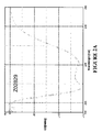
- a 280 (mg/ml) Isoelectric point Total amount (mg) Z02674 7321.1 1.05 6.5 30 Z02726 7321.2 1.05 7.7 nd Z02742 7341.2 1.05 10.3 60
- Lyophilized proteins were dissolved in PBS and 20 ⁇ g was analyzed with SDS-PAGE. All protein preparations contained IgG Fc-binding molecules together with some contaminating proteins. The size of the IgG Fc-binding molecules was confirmed with HPLC-MS.
- All three protein preparations contained precipitated contaminating material.
- undissolved material was removed by centrifugation and the supernatant was kept at +4 °C over night. A new visual inspection was performed, and no new precipitation could be seen. The concentration was measured with A 280 after centrifugation and found to be 17.4 mg/ml.
- the pH of the solutions was raised to approximately 10 with 50 % NaOH, which resulted in a clear solution for both proteins.
- Z02829 is a derivative of Z02674 containing two substitutions at the beginning of the protein (A8N and W11 N with respect to the sequence of the entire expressed molecule, i.e. A3N and W6N with respect to SEQ ID NO:7).
- the IgG Fc-binding motif of Z02829 is the same as that of Z02674.
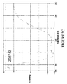
- the determined melting points of the IgG Fc-binding molecules are given in Table 4. Table 4. Determined melting points. Protein T m (°C) Z02829 62 Z02726 62 Z02742 63
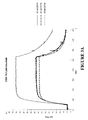
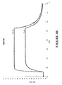
- Binding of the purified polypeptides to human IgG Fc was analyzed using surface plasmon resonance on a Biacore 2000 instrument. Palivizumab (without VH3 domain), trastuzumab (with VH3 domain) and etanercept (TNF ⁇ r-Fc fusion) were immobilized on chip surfaces with amine-coupling. The Z protein Z00000 (SEQ ID NO:10) and the earlier obtained variant Z01730 (SEQ ID NO:11) were used as controls. Binding diagrams for IgG Fc-binding molecules injected at 25 nM over immobilized target proteins are shown in Figures 3A-3C and Figures 4A-4C .
- the diagrams obtained from binding to palivizumab were analyzed with the BIAevaluation software provided by the manufacturer. The results are presented in Table 5. As shown in this Table, the IgG Fc-binding polypeptides exhibit binding affinities for IgG which are comparable to the positive control Z00000, which is a well known IgG Fc-binding molecule. Table 5.
- Binding constants for selected IgG Fc-binding molecules Protein k a (1/Ms) k d (1/s) K D (nM) Z02674 2.4 x 10 6 5.2 x 10 -2 22 Z02726 2.6 x 10 6 4.5 x 10 -2 18 Z02742 9.6 x 10 6 4.3 x 10 -2 5 Z00000 (control) 3.6 x 10 5 4.9 x 10 -3 14
- Example 2 individual IgG Fc-binding polypeptides from the selection described in Example 1 were coupled to chromatoghraphic media, and their elution conditions and binding capacities were studied in affinity chromatography experiments.
- the inventive IgG Fc-binding polypeptides Z02742, Z02674 and Z02726 and the reference molecule Z00000 were each immobilized on NHS-activated HiTrapTM columns (0.962 ml, GE Healthcare). The immobilization, ligand coupling via primary amines, was performed in accordance with the manufacturer's instructions. Each polypeptide was immobilized on four columns, of which two were used for the elution pH study and two were used for the capacity study.
- Citric acid and NaCl were dissolved in water to final concentrations of 0.1 M and 0.9 percent by weight (% wt/wt) respectively.
- Two buffers were prepared from this solution by adjusting pH to 6.2 for one part of the solution (buffer A) and to 2.5 for the other part of the solution (buffer B). pH adjustments were made by addition of NaOH. The buffers were filtered prior to use.
- Elution pH was studied for three different samples run on columns comprising IgG Fc-binding polypeptide ligands.
- the samples were trastuzumab (trade name Herceptin®, Apoteket article no. 573477), etanercept (trade name Enbrel®, Apoteket article no. 566661) and palivizumab (trade name Synagis®, Apoteket article no. 549113).
- the samples were prepared according to the manufacturer's instructions and were thereafter diluted to 1 mg/ml solutions in buffer A.
- the columns were attached to an ⁇ KTATM explorer 10 S chromatography system (GE Healthcare) and equilibrated (4 column volumes (CV) buffer A, flow rate 1 ml/min).
- Sample solution was injected into a SuperloopTM (50 ml, GE Healthcare) and 2 ml were loaded on each column at a flow rate of 0.4 ml/min.
- the columns were washed (3 CV buffer A, 1 ml/min) and the sample was eluted by an acidic pH gradient (25 CV, 0-100 % buffer B, 1 ml/min). After the acidic pH gradient, the columns were washed (4 CV 100 % buffer B, 1 ml/min) and re-equilibrated (4 CV buffer A, 1 ml/min).
- Dynamic binding capacity for a chromatographic medium is usually defined as the amount of sample applied to the medium when the absorbance at 280 nm reaches 10 % of the sample absorbance at 280 nm.
- the capacity was determined by loading sample on columns comprising immobilized IgG Fc-binding polypeptides. To determine the capacity, the dead volume (i.e. tubing and column volume) was subtracted from the sample volume required for 10 % breakthrough. The dead volume was measured by running sample through a column comprising no IgG Fc-binding polypeptide.
- Human polyclonal IgG (trade name Gammanorm®, Apoteket article no. 096169, comprising a mix of VH3 subfamily and non-VH3 subfamily antibodies) was used for determining capacity.
- the IgG sample was prepared by diluting 165 mg/ml Gammanorm® to 0.75 mg/ml with 1 X PBS.
- the columns were attached to an ⁇ KTATMexplorer 10 S chromatography system (GE Healthcare) and equilibrated (4 CV buffer A, 1 ml/min).
- the sample was loaded on the columns with a flow rate of 0.241 ml/min (residence time 4 min) until A 280 reached 10 % (in this case 128.2 mAU) of the sample absorbance.
- Bound protein was eluted (10 CV buffer B, 1 ml/min) and the columns were re-equilibrated (4 CV buffer A, 1 ml/min).
- the tested IgG Fc-binding polypeptides according to the invention bind to IgG and exhibit elution profiles in affinity chromatography which are comparable to, or better than, those of the column-coupled reference molecule Z00000.
- Capacities of the column-coupled IgG Fc binding polypeptides according to the invention ranged from 0.23 to 0.33 moles of IgG / moles polypeptide ligand and were comparable with the capacities of column-coupled Z00000 (see Figure 6 ).
- the dynamic binding capacity of columns comprising immobilized Z02674 was, however, approximately 20-30% higher than for the other columns.
Description
- This invention relates to a polypeptide which binds to immunoglobulin G Fc (IgG Fc). The polypeptide has industrial application for example in affinity separation and/or purification in the production of antibodies and/or Fc fusion proteins.
- In the industrial production of monoclonal antibodies and Fc fusion proteins, purification is frequently carried out using affinity chromatography. Protein A from Staphylococcus aureus has long been used as affinity ligand in such applications, due to the native affinity of Protein A for the Fc portion of IgG. Protein A in its entirety, as well as the individual Fc-binding domains thereof, have subsequently served as starting points for the rational design of engineered affinity ligands with improved properties. Despite the comparable success of currently used IgG Fc affinity ligands, there is a continued need for improvement. The continued provision of agents having an affinity for IgG Fc that is comparable with, or higher than, that exhibited by Protein A remains a matter of substantial interest. For example, Protein A affinity chromatography typically uses low pH conditions, which may lead to loss of yield due to the sensitivity of several antibodies and Fc fusion proteins to low pH conditions.
- A method to identify polypeptides that bind to epidermal growth factor receptor is disclosed in
WO2007/065635 . This method is based on phage display selection from libraries of mutants of protein A from Staphylococcus aureus. - The creation of IgG Fc-binding molecules based on protein A by site-directed mutagenesis is described in
EP1992692 andWO03/080655 EP1992692 discloses an IgG-binding ligand which is the Z or C-domain of staphylococcal protein A, wherein lysine residues are introduced C-terminally by site-directed mutagenesis.WO03/080655 - The provision of new IgG Fc-binding agents that allow elution at a higher pH as compared to Protein A during affinity chromatography would therefore be beneficial.
- It is an object of the invention to provide new IgG Fc-binding agents, that could for example be used in the production of antibodies or Fc fusion proteins, e.g. for affinity separation and/or purification.
- According to one aspect thereof, the invention provides an immunoglobulin G Fc (IgG Fc) binding polypeptide, comprising an IgG Fc-binding motif, BM, which motif consists of an amino acid sequence selected from:
- i) EQQX4AFYEIL HLPNLTEX18QX20 X21AFIX25X26LRX29,
wherein, independently of each other,- X4 is selected from H and N;
- X18 is selected from D and G;
- X20 is selected from R and K;
- X21 is selected from H and Q;
- X25 is selected from R, A and G;
- X26 is selected from A, S and T; and
- X29 is selected from G, K and A;
- ii) an amino acid sequence which has at least 93 % identity to the sequence defined in i) over a window corresponding to said binding motif;
wherein said IgG Fc-binding polypeptide binds to IgG Fc such that the KD value of the interaction is at most 1x 10⁻⁶ M. - The above definition of a class of sequence related, IgG Fc-binding polypeptides according to the invention is based on an analysis of a number of random polypeptide variants of a parent scaffold, that were selected from a combinatorial protein library for their interaction with IgG Fc in phage display selection experiments (Examples 1 and 2). The identified IgG Fc-binding motif, or "BM", corresponds to the target binding region of the parent scaffold, which region constitutes two alpha helices within a three-helical bundle protein domain. In the parent scaffold, the varied amino acid residues of the two BM helices include amino acid residues that participate in the binding surface for interaction with Fc. In the present invention, the random variation of surface residues and the subsequent selection of variants have modified the original Fc interaction capacity.
- As the skilled person will realize, the function of any polypeptide, such as the IgG Fc-binding capacity of the polypeptides according to the invention, is dependent on the tertiary structure of the polypeptide. It is therefore possible to make minor changes to the sequence of amino acids in a polypeptide without affecting the function thereof. Thus, the invention encompasses modified variants of the BM of i), which are such that the resulting sequence is at least 93 % identical to a sequence belonging to the class defined by i). For example, it is possible that an amino acid residue belonging to a certain functional grouping of amino acid residues (e.g. hydrophobic, hydrophilic, polar etc) could be exchanged for another amino acid residue from the same functional group.
- In one embodiment of the polypeptide according to the invention, X4 is H.
- In one embodiment of the polypeptide according to the invention, X18 is G.
- In one embodiment of the polypeptide according to the invention, X20 is K.
- In one embodiment of the polypeptide according to the invention, X21 is H.
- In one embodiment of the polypeptide according to the invention, X25 is R.
- In one embodiment of the polypeptide according to the invention, X26 is A.
- In one embodiment of the polypeptide according to the invention, X29 is G.
- As described in detail in the experimental section to follow, the selection of IgG Fc-binding variants has led to the identification of individual IgG Fc-binding motif (BM) sequences. These sequences constitute individual embodiments of the BM sequence i) in the definition of IgG Fc-binding polypeptides according to this aspect of the present invention. The sequences of individual IgG Fc-binding motifs are presented in
Figure 1 and as SEQ ID NO:1-3 (Figure 1 ). In embodiments of this aspect of the invention, the BM sequence i) may in particular be SEQ ID NO:1. - In embodiments of the present invention, the BM may form part of a three-helix bundle protein domain. For example, the BM may essentially constitute or form part of two alpha helices with an interconnecting loop, within said three-helix bundle protein domain.
- In particular embodiments of the invention, such a three-helix bundle protein domain is selected from domains of bacterial receptor proteins. Non-limiting examples of such domains are the five different three-helical domains of protein A from Staphylococcus aureus, and derivatives thereof. Thus, an IgG Fc-binding polypeptide according to the invention may comprise an amino acid sequence selected from:
- ADNNFNK-[BM]-DPSQSANLLSEAKKLNESQAPK (BM within domain A of staphylococcal protein A);
- ADNKFNK-[BM]-DPSQSANLLAEAKKLNDAQAPK (BM within domain B of staphylococcal protein A);
- ADNKFNK-[BM]-DPSVSKEILAEAKKLNDAQAPK (BM within domain C of staphylococcal protein A);
- ADAQQNNFNK-[BM]-DPSQSTNVLGEAKKLNESQAPK (BM within domain D of staphylococcal protein A);
- AQHDE-[BM]-DPSQSANVLGEAQKLNDSQAPK (BM within domain E of staphylococcal protein A); and
- VDNKFNK-[BM]-DPSQSANLLAEAKKLNDAQAPK (BM within the protein Z derivative of domain B of staphylococcal protein A);
- In alternative embodiments of the present invention, wherein the BM again essentially constitutes or forms part of two alpha helices with, an interconnecting loop, within said three-helix bundle protein domain, the IgG Fc-binding polypeptide comprises the amino acid sequence:
- FWK-[BM]-DPSQSARLLAXaAKKLDDQ,
- For example, the IgG Fc-binding polypeptide may comprise the amino acid sequence:
- VDAKFWK-[BM]-DPSQSARLLAXaAKKLDDQAPK
- In some examples of these embodiments, Xa is R.
- The IgG Fc-binding polypeptide may for example comprise an amino acid sequence selected from SEQ ID NO:4-6, such as SEQ ID NO:4 (
Figure 1 ). - According to another alternative aspect thereof, the invention provides an IgG Fc-binding polypeptide as described herein, whose amino acid sequence comprises a sequence selected from SEQ ID NO:7-9 (
Figure 1 ). In embodiments of this aspect of the invention, the IgG Fc-binding polypeptide may in particular comprise SEQ ID NO:7 - When reference is made herein to the degree of identity between the amino acid sequences of different polypeptides, the lower limit of 93 % identity to a sequence disclosed herein is given. In some embodiments, the inventive polypeptide may have a sequence which is at at least 94 %, at least 95 %, at least 96 %, at least 97 %, at least 98 % or at least 99 % identical to the sequence described herein. The comparison is performed over a window corresponding to an IgG Fc-binding motif in at least one of the sequences being compared.
- An IgG Fc-binding polypeptide according to any aspect of the invention may bind to IgG Fc such that the KD value of the interaction is at most 1 x 10⁻⁶ M, for example at most 1 x 10⁻⁷ M, such as at most 5 x 10⁻⁸ M.
- The polypeptide is advantageous in that it binds well to an IgG Fc. In particular, the polypeptide may be capable of binding to the Fc portion of a human IgG molecule. In some embodiments of the invention, the polypeptide is capable of binding to
classes class 3. In some embodiments, the polypeptide is capable of binding to the interface between the CH2 and CH3 domains of IgG Fc. In some embodiments, the polypeptide is capable of binding to an area on the Fc molecular surface made up by the Fc amino acid residues T250-S254, T256, L309-L312, L314, D315, E430 and L432-Y436 (numbering according to Deisenhofer, Biochemistry (1981) 20(9):2361-70). - The skilled addressee will appreciate that various modifications and/or additions can be made to a polypeptide according to the invention in order to tailor the polypeptide to a specific application without departing from the scope of the present invention. These modifications and additions are described in more detail below and may include additional amino acids in the same polypeptide chain, or labels and/or therapeutic agents that may be chemically conjugated or otherwise bound to the polypeptide of the invention.
- Furthermore, the invention also encompasses fragments of IgG Fc-binding polypeptides according to the invention that retain IgG Fc-binding. The possibility of creating fragments of a wild-type Staphylococcus aureus protein A domain with retained binding specificity was shown by Braisted AC et al in Proc Natl Acad Sci USA 93:5688-5692 (1996). In the experiments described in that paper, using a structure-based design and phage display methods, the binding domain of a three-helix bundle of 59 residues was reduced to a resulting two-helix derivative of 33 residues. This was achieved by stepwise selection of random mutations from different regions, which caused the stability and binding affinity to be iteratively improved. Following the same reasoning, with the polypeptides of the present invention, the skilled addressee will be able to obtain a "minimized" IgG Fc-binding polypeptide with the same binding properties as that of the "parent" IgG Fc-binding polypeptide. Thus, a polypeptide constituting a fragment of a polypeptide according to the invention is within the scope of the invention.
- The terms "IgG Fc-binding" and "binding affinity for IgG Fc" as used in this specification refers to a property of a polypeptide which may be tested for example by the use of surface plasmon resonance technology, such as in a Biacore instrument (GE Healthcare). For example as described in the examples below, IgG Fc-binding affinity may be tested in an experiment in which IgG Fc, or a fragment of IgG Fc, is immobilized on a sensor chip of the instrument, and the sample containing the polypeptide to be tested is passed over the chip. Alternatively, the polypeptide to be tested is immobilized on a sensor chip of the instrument, and a sample containing IgG Fc, or fragment thereof, is passed over the chip. The skilled person may then interpret the results obtained by such experiments to establish at least a qualitative measure of the binding affinity of the polypeptide for IgG Fc. If a quantitative measure is desired, for example to determine a KD value for the interaction, surface plasmon resonance methods may also be used. Binding values may for example be defined in a Biacore 2000 instrument (GE Healthcare). IgG Fc is immobilized on a sensor chip of the measurement, and samples of the polypeptide whose affinity is to be determined are prepared by serial dilution and injected in random order. KD values may then be calculated from the results using for example the 1:1 Langmuir binding model of the BIAevaluation 4.1 software provided by the instrument manufacturer.
- Where amino acid substitutions are introduced, these should not affect the basic structure of the polypeptide. For example, the overall folding of the Cα backbone of the polypeptide can be essentially the same as that of a domain of protein A, i.e. having the same elements of secondary structure in the same order. Thus, polypeptides having this basic structure will have similar CD spectra to the wild-type protein A domain. The skilled addressee is aware of other parameters that may be relevant. The requirement of conserving the basic structure, places restrictions on which positions of the amino acid sequence may be subject to substitution. For example, it is preferred that amino acid residues located on the surface of the polypeptide are substituted, whereas amino acid residues buried within the core of the polypeptide "three-helix bundle" should be kept constant in order to preserve the structural properties of the molecule. The same reasoning applies to fragments of polypeptides of the invention.
- The invention also covers polypeptides in which the IgG Fc-binding polypeptide described above is present as an IgG Fc-binding domain to which additional amino acid residues have been added at either terminal. These additional amino acid residues may play a role in the binding of IgG Fc by the polypeptide, but may equally well serve other purposes, related for example to one or more of the production, purification, stabilization in vivo and/or in vitro, coupling or detection of the polypeptide. Such additional amino acid residues may comprise one or more amino acid residues added for the purpose of chemical coupling. One example of this is the addition of a cysteine residue N-terminally or C-terminally with respect to the binding motif, e.g. close to or at the N or C terminus. Such additional amino acid residues may also provide a "tag" for purification or detection of the polypeptide such as a His6 tag or a "myc" (c-myc) tag or a "FLAG" tag for interaction with antibodies specific to the tag.
- The present invention also covers IgG Fc-binding polypeptides in which an IgG Fc-binding polypeptide as described above is present as an IgG Fc-binding domain to which additional peptides or proteins or other functional groups are coupled N- or C-terminally or to any other residues (specifically or non-specifically) by means of chemical conjugation (using known organic chemistry methods).
- The "additional amino acid residues" discussed above may also provide one or more polypeptide domains with any desired function, such as the same binding function as the first, IgG Fc-binding domain, or another binding function, or an enzymatic function, toxic function (e.g. an immunotoxin), or a fluorescent signaling function, or combinations thereof.
- The polypeptide of the invention may be in monomeric or multimeric forms. Multimeric forms of the polypeptide may be advantageous in that they may have enhanced binding properties. Preferred multimeric forms include dimeric, trimeric and tetrameric forms. Multimeric forms of the polypeptides may comprise a suitable number of polypeptides of the invention. These polypeptides essentially form domains within the multimer. These domains may all have the same amino acid sequence, but alternatively, they may have different amino acid sequences. The polypeptides may be joined by covalent coupling using known organic chemistry methods, or expressed as one or more fusion polypeptides in a system for recombinant expression of polypeptides, or joined in any other fashion, either directly or via a linker, for example an amino acid linker.
- Additionally, fusion polypeptides, in which the IgG Fc-binding polypeptide of the invention provides a first domain or moiety, and second or further moieties have other functions than binding IgG Fc are also contemplated and within the scope of the present invention. The second or further moieties of such a fusion polypeptide may comprise a binding domain with an affinity for another target molecule than IgG Fc. Such a binding domain may be another, similar polypeptide binder. For example, the polypeptide binder may be a variant of protein Z derived from domain B of protein A. This makes it possible to create multi-specific reagents that may be used in several types of applications such as medicine, veterinary medicine, diagnosis, separation, and imaging. The preparation of such multi-specific fusion polypeptides may be performed using methods well known in the art of molecular biology.
- In other embodiments of the invention, the second or further moieties may comprise an unrelated, naturally occurring or recombinant protein (or a fragment thereof which retains the binding or other ability of the naturally-occurring or recombinant protein) having a binding affinity for a target. For example, an IgG Fc-binding polypeptide in accordance with the invention may be joined to an albumin-binding domain, such as the albumin binding domain GA3 of protein G from Streptococcus strain G148 ("ABD"), or any other polypeptide with affinity for a serum protein.
- The IgG Fc-binding polypeptides of the present invention may be provided in the form of other fusion polypeptides. For example the IgG Fc-binding polypeptide, or fragment thereof, may be covalently coupled to a second or further moiety or moieties, which in addition to, or instead of target binding, exhibit other functions. One example would be a fusion between one or more IgG Fc-binding polypeptides and an enzymatically active polypeptide serving as a reporter or effector moiety. Examples of reporter enzymes, which may be coupled to the IgG Fc-binding polypeptide to form a fusion protein, are well-known to the skilled person and include enzymes such as β-galactosidase, alkaline phosphatase, horseradish peroxidase, carboxypeptidase. Other options for the second and further moiety or moieties of a fusion polypeptide according to the invention include fluorescent polypeptides, such as green fluorescent protein, red fluorescent protein, luciferase and variants thereof.
- A polypeptide according to the invention may be useful in any method which relies on affinity for IgG Fc of a reagent. Thus, the polypeptide may be used as a detection reagent, a capture reagent or a separation reagent in such methods. In particular, the polypeptide exhibits several characteristics which make it useful as an affinity reagent in affinity chromatography, wherein the goal is to separate, purify and/or produce antibodies or Fc fusion proteins from a heterogeneous mixture. The polypeptide can be bound to a matrix and e.g. used for the purification of IgG Fc-containing therapeutic compounds in industrial production. Due to properties such as a high target affinity, a high stability both in acidic and basic environments and a high selectivity for the IgG Fc fragment over the IgG Fab fragment, the IgG Fc-binding polypeptide according to the invention is thought to present a very attractive affinity reagent.
- Thus, another aspect of the present invention is a method of isolating molecules comprising IgG Fc from a sample, which method comprises the steps:
- (i) providing a sample containing molecules comprising IgG Fc;
- (ii) contacting the sample with an IgG Fc-binding polypeptide as described herein, whereby said molecules comprising IgG Fc bind to the polypeptide;
- (iii) isolating bound molecules comprising IgG Fc from the sample.
- In the inventive isolation method, the sample may be derived from a culture of prokaryotic or eukaryotic, such as mammalian or plant, cells expressing molecules comprising IgG Fc, or from expression of such molecules in an alternative expression system, for example a vesicular system. Alternatively, the sample may be derived from transgenic expression in a host, such as a plant or mammalian host.
- In some embodiments, said molecules comprising IgG Fc are IgG molecules or fragments thereof. For example, they can be human IgG molecules or fragments thereof. In some embodiments, said molecules comprising IgG Fc are monoclonal IgG antibodies. In particular, such monoclonal IgG antibodies may be human monoclonal IgG antibodies. For example, they are human monoclonal IgG antibodies from
class - In other embodiments, said molecules comprising IgG Fc are Fc fusion proteins. The Fc domain in such a fusion protein may thus, advantageously, be used as an "affinity handle" in the isolation of the fusion protein. A large variety of Fc fusion proteins have been created. For example, Fc fusion proteins having therapeutic applications include etanercept, which is a fusion between soluble TNF-α receptor and Fc, and VEGF Trap, which is a fusion between VEGF receptor domains and Fc (Holash et al, Proc Natl Acad Sci USA (2002) 99(17):11393-11398). While these two are illustrative examples of great interest, the listing of them is non-limiting, and it is in principle possible to fuse an Fc domain to any desired protein in order to modify its properties and facilitate affinity purification thereof using the inventive IgG Fc-binding polypeptide described herein as affinity ligand.
- Yet another aspect of the present invention concerns a method of producing molecules comprising IgG Fc, which method comprises the steps:
- (i) expressing desired molecules comprising IgG Fc;
- (ii) obtaining a sample of molecules comprising IgG Fc from said expression;
- (iii) contacting the sample with an IgG Fc-binding polypeptide as described herein, whereby molecules comprising IgG Fc bind to the polypeptide;
- (iv) isolating bound molecules comprising IgG Fc from the sample, and
- (v) recovering bound molecules comprising IgG Fc through elution thereof from the IgG Fc-binding polypeptide.
- Expression step (i) may be performed using any known expression system, for example recombinant expression in prokaryotic or eukaryotic, such as mammalian or plant, cells, or in a vesicular system. The sample may also be derived from transgenic expression in a host, such as a plant or mammalian host.
- In some embodiments, said molecules comprising IgG Fc are IgG molecules or fragments thereof. For example, they can be human IgG molecules or fragments thereof. In some embodiments, said molecules comprising IgG Fc are monoclonal IgG antibodies. In particular, such monoclonal IgG antibodies may be human monoclonal IgG antibodies. For example, they are human monoclonal IgG antibodies from
class - In other embodiments, said molecules comprising IgG Fc are Fc fusion proteins.
- In some embodiments of the inventive methods of isolating and producing, the IgG Fc-binding polypeptide is immobilized on a chromatography medium. In general, methods that employ the polypeptides in accordance with the invention in vitro may be performed in different formats, such as on filters or membranes, microtitre plates, in protein arrays, on biosensor surfaces, on beads, in flow cytometry, on tissue sections, and so on. In a specific aspect, the invention provides an affinity chromatography medium, which has an IgG Fc-binding polypeptide as described herein immobilized thereon. Such a medium may be based on any known chromatography material as a matrix, and coupling of the polypeptide to the matrix may be performed using any one of several known procedures.
- The numbering of amino acid residues and any use of the term "position" in the sequence of the polypeptide according to the invention is relative. In a polypeptide in accordance with the invention which has as many amino acid residues as a specifically disclosed polypeptide, i.e. those described above, the positions of amino acids in the polypeptide correspond exactly to those in the disclosed polypeptides. In a situation where there is, for example, an N terminal extension compared to the disclosed polypeptides, those amino acid residues in the extended peptide that correspond to those of the non-extended peptide have the same position numbers. For example, if there is a six amino acid residue extension on the extended polypeptide, then amino acid number seven of that modified polypeptide, counting from the N terminus, corresponds to the amino acid in position number one of the disclosed polypeptide.
- With regard to the description above of fusion polypeptides and proteins incorporating an IgG Fc-binding polypeptide of the invention, it should be noted that the designation of first, second and further moieties is made for the purposes of clarity to distinguish between the IgG Fc-binding moiety or moieties on the one hand, and moieties exhibiting other functions on the other hand. These designations are not intended to refer to the actual order of the different domains in the polypeptide chain of the fusion protein or polypeptide. Thus, for example, a first moiety may appear at the N-terminal end, in the middle, or at the C-terminal end of the fusion protein or polypeptide.
- The invention is further illustrated by the following non-limiting examples.
-
-
Figure 1 is a listing of the amino acid sequences of examples of IgG Fc-binding motifs comprised in IgG Fc-binding polypeptides of the invention (SEQ ID NO:1-3), examples of IgG Fc-binding polypeptides according to the invention (SEQ ID NO:4-9), the protein Z derivative of domain B of Staphylococcus aureus protein A (SEQ ID NO:10), and the Z variant Z01730 previously obtained by phage display selection from a combinatorial protein library (SEQ ID NO:11). -
Figure 2 shows overlay of CD spectra taken at 195-250 nm before and after variable temperature measurement (VTM) involving heating at 90°C for A: Z02829, B: Z02726 and C: Z02742. Z02829 is a derivative of Z02674 containing two substitutions at the beginning of the protein (A8N and W11 N with respect to the sequence of the entire expressed molecule, i.e. A3N and W6N with respect to SEQ ID NO:7). Thus, the IgG Fc-binding motif of Z02829 is the same as that of Z02674. -
Figure 3 shows sensorgrams obtained from Biacore analysis of IgG Fc-binding molecules according to the invention. Sensorgrams obtained after injection of 25 nM of Z02674 (solid line), Z02726 (dashed line) or Z02742 (dotted line) over CM5 sensor-chip surfaces containing immobilized palivizumab (A; 1280 response units, RU); trastuzumab (B; 1200 RU) and etanercept (C; 1500 RU). Signal from a blank sensor-chip surface was subtracted. -
Figure 4 shows sensorgrams obtained from kinetic Biacore analysis of IgG Fc-binding molecules according to the invention. The overlay plots show sensorgrams obtained after injections over immobilized palivizumab of 25 nM or 100 nM of Z02674 (A); Z02726 (B) and Z02742 (C) (dotted lines). The response curves were fitted to a 1:1 binding model (solid lines). -
Figure 5 shows chromatograms for columns comprising immobilized IgG Fc-binding polypeptides according to the invention. The overlay chromatograms show elution profiles for etanercept (A), trastuzumab (B) and palivizumab (C) when eluted with an acidic pH gradient from columns comprising Z00000, Z02742, Z02674 or Z02726, as indicated. -
Figure 6 shows a histogram of dynamic binding capacities (moles of IgG / moles of polypeptide) for columns comprising immobilized Z00000, Z02742, Z02674 or Z02726, as indicated. - In this Example, a Z variant library denoted ZLib2007-IgG, created following an initial selection of IgG binding Z variants from the library ZLib2002 and evaluation of results, was used for selection of IgG Fc-binding polypeptides according to the invention. Details of library construction and selection procedures were generally as described in Grönwall et al, J Biotechnol 128:162-183, 2007. Four different phage display selections from ZLib2007-IgG were made against various IgG and IgG-like molecules. Clones were sequenced, the sequences analyzed by clustering and the amino acid sequence of each clone compared to the distribution of variable amino acids among all selected clones and the distribution in the library design. IgG Fc-binding molecules were chosen for further characterization after four selection rounds, whereupon a fifth round was carried out with more stringent washing conditions, elution at a higher pH and with a higher temperature during selection. Additional IgG Fc-binding molecules derived from
selection round 5 were selected for further characterization. - Five selection rounds were performed in each one of four selection setups, and new phage stocks were prepared between each round. Target protein was alternated between selection rounds in some selection setups. Targets used were human immunoglobulin G2κ (IgG2κ) from myeloma serum (Meridian Life Science, cat. no. A50184H), human immunoglobulin G3λ (IgG3λ) from myeloma serum (Meridian Life Science, cat. no. A50186H), human immunoglobulin G4λ (IgG4λ) from myeloma serum (Meridian Life Science, cat. no. A50947H), etanercept (trade name Enbrel®; Apoteket cat. no. 566661, producer Wyeth, lot 21032), biotinylated human immunoglobulin G, Fc fragment (IgG-Fc) (Jackson Immunoresearch, cat. no. 009-060-008, lot 66321), and biotinylated human immunoglobulin G1κ (IgG1κ) (Ancell, cat. no. 295-030, lot 141605). An overview of target proteins in each round of each selection setup is presented in Table 1. The selections were performed against biotinylated target protein in liquid phase for selection against IgG variants, or against target immobilized on a solid phase in the form of the surface of an immunotube for selection against etanercept.
Table 1. Selection setups Setup Selection round Target Concentration No. of washes Time/wash (min) Elution pH IgG_22-Sel1 1 Poly-IgG-Fc 100 nM 2 0 3.5 2 Etanercept 6 µg/ml 2 0 3.5 3 Poly-IgG-Fc 20 nM 4 1 3.5 4 Etanercept 3µg/ml 5 2 3.5 5 Poly-IgG-Fc 20 nM 9 5 3.8 IgG_21-Sel1 1 Poly-IgG1&2&4 33+33+33 nM 2 0 3.5 2 Poly-IgG1&2&4 17+17+17 nM 2 0 3.5 3 Poly-IgG1&2&4 7+7+7 nM 4 1 3.5 4 Poly-IgG1&2&4 7+7+7 nM 5 2 3.5 5 Poly-IgG1&2&4 7+7+7 nM 9 5 3.8/4.5 IgG_23-Sel1 1 Poly-IgG1 100 nM 2 0 2.2 2 Poly-IgG4 50 nM 2 0 2.2 3 Poly-IgG2 20 nM 5 2 2.2 4 Poly-IgG2 20 nM 7 3 2.2 5 Poly-IgG1 20 nM 13 7 3.8 IgG_21-Sel2 1 Poly-IgG1&2&4 20+20+20 nM 2 0 2.2 2 Poly-IgG1&2&4 8+8+8 nM 2 0 2.2 3 Poly-IgG1&2&4 4+4+4 nM 4 1 2.2 4 Poly-IgG1&2&4 2+2+2 nM 5 2 2.2 5 Poly-IgG1&2&4 1+1+1 nM 9 5 3.8 - Warm conditions for selection (37 °C) and wash (37-45 °C) were used in
round 5 in all setups. - Phage library stock was PEG/NaCl precipitated twice and dissolved in 1 ml selection buffer (PBS: 2.68 mM KCl, 1.47 mM KH2PO4, 137 mM NaCl, 8.1 mM Na2HPO4 pH 7.4; supplemented with 0.1 % Tween20 (Acros Organics cat. no. 2333 62500) and 0.1 % gelatine (Prolabo, cat. no. 24 360.233)).
- Liquid phase selection: Phages were pre-incubated with streptavidin coated beads (Dynabeads® M-280; Dynal cat. no. 112.06) for 1 hour at room temperature. Pre-clearing against Fab was made in a Maxisorp immunotube (Nunc, cat. no. 444202) coated with Fab. All tubes and beads used in the selection procedure were pre-blocked in selection buffer. Phages were incubated with biotinylated target under agitation for up to 3 hours. Then, the phages were transferred to pre-blocked streptavidin beads and incubated for 15 min with agitation, and the beads were washed in selection buffer according to Table 1.
- Solid phase selection: Target protein was immobilized onto immunotubes. Phages were pre-incubated in an immunotube coated with Fab. All tubes, including tubes coated with target, were blocked in selection buffer prior to selection. Phages were incubated with the immobilized target molecules under agitation and the tube was thereafter washed in selection buffer.
- Elution and infection: Phages from either solid or liquid phase selection were eluted with elution buffer (0.05 M glycine-HCl at pH 2.2, or 0.05 M NaAc buffer at pH 3.5, 3.8 or 4.5 as outlined in Table 1), followed by immediate neutralization with neutralization buffer (1 M Tris-HCl, pH 8.0). The eluted phages (95 % of the volume) were used to infect log phase E. coli RR1ΔM15 cells (Rüther, Nucleic Acids Res 10:5765-5772, 1982) after each round of selection (approximately 500 times excess of cells compared to eluted phages). After 25 min incubation at 37 °C, the cells were centrifuged. The pellet was dissolved in a small volume of TSB-YE (30 g/I tryptic soy broth, 5 g/l yeast extract) and spread on a TYE plate (15 g/I agar, 10 g/l tryptone water (Merck), 5 g/I yeast extract, 3 g/l NaCl, 2 % glucose and 0.1 g/I ampicillin) and thereafter incubated over night at 37 °C.
- Preparation of phage stocks: Phage infected cells grown over night on TYE plates were re-suspended in TSB medium (30 g/I tryptic soy broth). An amount of suspended cells corresponding to approximately 100 infected cells of each eluted phage was inoculated in TSB-YE medium supplemented with 2 % glucose and 100 mg/ml ampicillin. These cells were grown to log phase at 37 °C and a volume of them resembling the same amount of cells prior to growth were infected with 20 times excess of M13K07 helper phage (New England Biolabs, cat. no. NO315S) Cells and helper phage were incubated for 30 min at 37 °C, and then pelleted by centrifugation, re-suspended in TSB-YE medium supplemented with 100 mM IPTG (isopropyl-β-D-1-thiogalactopyranoside), 25 µg/ml kanamycin and 100 µg/ml ampicillin and grown over night at 30 °C. An aliquot of the re-suspended cells was stored at -20 °C as a glycerol stock.
- The induced culture was centrifuged and phages in the supernatant were precipitated twice with a PEG/NaCl buffer (20 % polyethyleneglycol, 2.5 M NaCl). The phages were re-suspended in selection buffer.
- Phage stock and eluted phage were titrated after each round of selection.
- Proteins from clones obtained after four or five rounds of selection were produced in 96-well plates and screened for target binding activity using an ELISA setup.
- Proteins were produced by inoculating single colonies in 1 ml TSB-YE medium supplemented with 100 µg/ml ampicillin and 1 mM IPTG in deep-well plates (Nunc, cat. no. 278752) and grown for 18-24 h at 37 °C. A small amount of each culture was transferred to 96-well plates (Costar, cat. no. 9018) and stored at -20 °C as glycerol stocks. Remaining cells were pelleted by centrifugation, re-suspended in 400 µl PBS-T0.05 (PBS + 0.05 % Tween20) and frozen at -80 °C to release the periplasmic fraction of the cells. Frozen samples were thawed in a water bath and cells were pelleted by centrifugation. Supernatants containing soluble candidate IgG Fc-binding molecules fused to the albumin binding domain ABD from Streptococcus strain G148 were assayed for binding in an ELISA as follows.
- Microtiter wells were coated with 100 µl of HSA at 6 µg/ml (Sigma, cat. no. J-1010) in coating buffer (0.1 M sodium carbonate, pH 9.5). The wells were blocked with 200 µl PBS-T0.05 complemented with 2 % dried milk for 1 h at room temperature. After removal of blocking, 100 ml of candidate IgG Fc-binding molecule solution was added in each well and the plates were incubated for 1.5 h at room temperature. Biotinylated IgG1K (at a concentration of 0.05 and 0.5 µg/ml for clones derived from
round 4 and 0.01 µg/ml for clones from round 5) or IgG Fc (at a concentration of 0.5 µg/ml; Jackson Immunoresearch, cat. no. 009-008, lot 66321) in 100 µl PBS-T0.05 was added to the wells and incubated for 1.5 h. Bound target was detected with SA-HRP (Dako, cat. no. P0397), diluted 1:5000 in PBS-T0.05, and incubated for 1 h at room temperature. Plates were washed four times with PBS-T0.05 before incubation with the biotinylated target, SA-HRP and developing solution. Developing solution was prepared by mixing of equal volumes of ImmunoPure TMB kit substrates TMB and H2O2 (Pierce, cat. no. 34021), and 100 µl were added to each well. After 30 min incubation in darkness, 100 µl stop solution (2 M H2SO4) was added. The plates were read at 450 nm in an ELISA spectrophotometer. All steps from blocking to reading were performed in aTecan Genesis Freedom 200 robot. - Three controls were used:
- Well F12: Positive control treated as above, but for plates with clones from
selection round 4, a mixture of Z00000 (SEQ ID NO:10) and Z01730 (SEQ ID NO:11) as periplasmic fractions was used. For plates with clones fromselection round 5, periplasmic fraction of Z00000 was used. - Well G12: Positive control. As described for well F12 but with biotinylated IgG1κ at a concentration of 1 µg/ml after
round 4 and 0.5 µg/ml afterround 5. - Well H12: Blank. PBS-T0.05 used instead of periplasmic fractions.
- Based on the ELISA results, clones were chosen for sequencing. For clones taken from
selection round 4, clones with absorbance values similar to the positive control (well F12) were given priority. For clones taken fromselection round 5, clones with the highest absorbance values were given priority. A high diversity among picked clones was desirable, and therefore many clones with different absorbance values were chosen from both screens. - PCR fragments were amplified from the chosen colonies using the oligonucleotides AFFI-21 (5'-tgcttccggctcgtatgttgtgtg-3') and AFFI-22 (5'-cggaaccagagccaccaccgg-3'). Sequencing of amplified fragments was performed using BigDye® Terminator v3.1 Cycle Sequencing Kit (Applied Biosystems, cat. no. 4336919) and the biotinylated oligonucleotide AFFI-72 (5'-biotin-cggaaccagagccaccaccgg-3') according to the manufacturer's recommendations. The sequencing reactions were purified by binding to magnetic streptavidin-coated beads (Magnetic Biosolutions, cat. no. 11103) using a Magnatrix 8000 (Magnetic Biosolutions), and analyzed on ABI PRISM® 3100 Genetic Analyzer (Applied Biosystems). The sequencing results were imported and analyzed with Nautilus software (Thermo Electronics Corporation).
- Four different selection setups were applied and five selection rounds were made for each selection setup. Increasing numbers of washes and elution at different pH values were used in the selection setups.
- Clones obtained after four and five rounds of selection were produced in 96-well plates and screened for target binding activity using an ELISA setup. The putative IgG Fc-binding molecules were in periplasmic fractions obtained by freeze thawing.
- In the ELISA screening of clones derived from the fourth selection rounds, IgG1κ and IgG Fc were used as targets at concentrations of 0.05 µg/ml and 0.5 µg/ml for IgG1κ and 0.5 µg/ml for IgG Fc. The ELISA results for
round 4 clones indicated that absorbance values corresponded well between IgG1 and IgG Fc and were very high even at the low concentration of 0.05 µg/ml IgG1. - In the ELISA screening of clones derived from the fifth selection rounds, IgG1κ was used as target at a concentration of 0.01 µg/ml. The number of background binders was much higher among clones derived from
round 5 as compared to clones fromround 4. The responses were lowest among clones from the IgG_21 selection eluted with pH 4.5. - Clones from
round Figure 1 ) and two clones derived from round 5 (designated Z02726 and Z02742, SEQ ID NO:8 and SEQ ID NO:9, respectively; seeFigure 1 ) were chosen for further characterization. - In summary, the selections from the library ZLib2007-IgG were successful and suitable candidates were chosen for further characterization.
- In this Example, a group of IgG Fc-binding polypeptides from the selection described in Example 1 were subcloned and expressed in monomeric form, and their binding characteristics studied.
- IgG Fc-binding polypeptides Z02674, Z02726 and Z02742, as well as a modified version of Z02674 denoted Z02829, were sub-cloned as monomers into an expression vector in which expression is regulated by a T7 promoter. The IgG Fc-binding polypeptides were expressed with the additional N-terminal amino acid sequence GSSLQ and the additional C-terminal amino acid sequence VD. Thus, the expressed Z02674, Z02726 and Z02742 molecules have the sequence GSSLQ-[SEQ ID NO:#]-VD, wherein # corresponds to 7, 8 or 9 (see
Figure 1 ). - E. coli BL21 (DE3) cells (Novagen) were transformed with the plasmids and cultivated at 37 °C in 1 I of TSB + YE medium (tryptic soy broth with yeast extract) supplemented with 50 µg/ml kanamycin. At OD600 = 1, IPTG was added to induce protein expression at a final concentration of 1 mM and the cultivation was incubated at 37 °C for another 5 hours. The cells were harvested by centrifugation, re-suspended in 200 ml of binding buffer (50 mM sodium phosphate, 150 mM NaCl, pH 7.0) and sonicated to release the expressed protein. Cell debris was removed by centrifugation and the supernatant was applied on 40 ml IgG-sepharose in an XK26 column (GE Healthcare). Contaminants were washed away with binding buffer followed by elution of IgG Fc-binding molecules with elution buffer (0.1 M HAc). The purified IgG Fc-binding molecules were transferred to 10 mM NH4HCO3 by gel filtration and thereafter lyophilized. Concentration was determined using absorption at 280 nm and the extinction coefficient of the respective protein. The purity of the final product was analyzed on SDS PAGE stained with Coomassie Blue. The identity of the purified IgG Fc-binding molecules was confirmed using HPLC-MS.
- Lyophilized protein was dissolved in PBS. Protein solution was transferred to a plastic cuvette and examined for undissolved protein by visual inspection.
- CD analysis was performed with 0.5 mg/ml protein in PBS. A spectrum measurement at 195-250 nm was performed at 20 °C. The melting point (Tm) of the purified proteins was determined by a variable temperature measurement (VTM) where 220 nm was monitored during heating of the sample to 90 °C. After re-equilibrating the sample to 20 °C, a new spectrum was taken. An overlay of spectrums before and after VTM showed if the structure was regained after heating to 90 °C.
- Binding of the purified molecules to human IgG was analyzed using surface plasmon resonance on a Biacore 2000 instrument (GE Healthcare). Etanercept (trade name Enbrel®, a fusion protein containing the Fc region of human IgG; Apoteket article no. 566661) and two human monoclonal IgG antibodies, palivizumab (trade name Synagis®, does not comprise a VH3 domain; Apoteket article no. 549170) and trastuzumab (trade name Herceptin®, comprises a VH3 domain: Apoteket article no. 573477) were used as target proteins. Target proteins were immobilized in different flow cells by amine coupling onto the carboxylated dextran layer on surfaces of CM-5 chips according to the manufacturer's recommendations. To analyze their binding to the immobilized target proteins, the purified IgG Fc-binding molecules were diluted in HBS-EP (0.01 M HEPES, 0.15 M NaCl, 3 mM EDTA, 0.005 % surfactant P20, pH 7.2) and injected at 25 nM and 100 nM at a constant flow-rate of 25 µl/min for 4 minutes. The surfaces were regenerated with an injection of 0.3 M HAc, pH 3.2. An estimate of the dissociation equilibrium constant (KD) was made using BIAevaluation 4.1 (GE Healthcare), assuming a one-to-one Langmuir binding model and taking mass transfer effects into account.
- Size exclusion chromatography (SEC) was performed to check for aggregates. The purified IgG Fc-binding molecules were diluted to 0.5 mg/ml in PBS and 50 µl was injected at the flow rate 0.5 ml/minute on a Superdex 75 10/300 GL column (GE Healthcare) equilibrated with PBS.
- Monomeric IgG Fc-binding molecules were expressed from plasmid vectors in E. coli. The total amount of IgG sepharose-purified protein from 1 liter-cultivations was determined spectrophotometrically at A280 nm and is given in Table 2.
Table 2. Characteristics and amounts of purified proteins Protein Molecular weight (Da) 1 A280 = (mg/ml) Isoelectric point Total amount (mg) Z02674 7321.1 1.05 6.5 30 Z02726 7321.2 1.05 7.7 nd Z02742 7341.2 1.05 10.3 60 - Lyophilized proteins were dissolved in PBS and 20 µg was analyzed with SDS-PAGE. All protein preparations contained IgG Fc-binding molecules together with some contaminating proteins. The size of the IgG Fc-binding molecules was confirmed with HPLC-MS.
- PBS was added to the lyophilized IgG Fc-binding molecules molecules. Expected concentration based on the amount of protein in each vial is shown in Table 3.
Table 3. Expected concentration of dissolved molecules. Protein Concentration (mg/ml) Z02674 18 Z02726 30 Z02742 30 - All three protein preparations contained precipitated contaminating material. For Z02674, undissolved material was removed by centrifugation and the supernatant was kept at +4 °C over night. A new visual inspection was performed, and no new precipitation could be seen. The concentration was measured with A280 after centrifugation and found to be 17.4 mg/ml. For Z02726 and Z02742, the pH of the solutions was raised to approximately 10 with 50 % NaOH, which resulted in a clear solution for both proteins.
- CD analysis was performed with the proteins Z02726, Z02742 and Z02829. Z02829 is a derivative of Z02674 containing two substitutions at the beginning of the protein (A8N and W11 N with respect to the sequence of the entire expressed molecule, i.e. A3N and W6N with respect to SEQ ID NO:7). Thus, the IgG Fc-binding motif of Z02829 is the same as that of Z02674.
- The determined melting points of the IgG Fc-binding molecules are given in Table 4.
Table 4. Determined melting points. Protein Tm (°C) Z02829 62 Z02726 62 Z02742 63 - Overlay of spectra taken at 195-250 nm before and after VTM are shown in
Figure 2A-2C . As is evident from these overlay diagrams, all three IgG Fc-binding polypeptides completely regained their structure after heating at 90 °C. - Binding of the purified polypeptides to human IgG Fc was analyzed using surface plasmon resonance on a Biacore 2000 instrument. Palivizumab (without VH3 domain), trastuzumab (with VH3 domain) and etanercept (TNFαr-Fc fusion) were immobilized on chip surfaces with amine-coupling. The Z protein Z00000 (SEQ ID NO:10) and the earlier obtained variant Z01730 (SEQ ID NO:11) were used as controls. Binding diagrams for IgG Fc-binding molecules injected at 25 nM over immobilized target proteins are shown in
Figures 3A-3C andFigures 4A-4C . - As evidenced in these Figures, all tested polypeptides bind to IgG.
- In order to calculate an estimated binding affinity, the diagrams obtained from binding to palivizumab were analyzed with the BIAevaluation software provided by the manufacturer. The results are presented in Table 5. As shown in this Table, the IgG Fc-binding polypeptides exhibit binding affinities for IgG which are comparable to the positive control Z00000, which is a well known IgG Fc-binding molecule.
Table 5. Binding constants for selected IgG Fc-binding molecules Protein ka (1/Ms) kd (1/s) KD (nM) Z02674 2.4 x 10⁶ 5.2 x 10⁻² 22 Z02726 2.6 x 10⁶ 4.5 x 10⁻² 18 Z02742 9.6 x 10⁶ 4.3 x 10⁻² 5 Z00000 (control) 3.6 x 10⁵ 4.9 x 10⁻³ 14 - SEC analysis was performed by injecting 100 µg of purified protein on a Superdex 75 10/300 GL column equilibrated with PBS. All IgG Fc-binding molecules eluted in single peaks. The shape of peaks differed between the IgG Fc-binding molecules and they elute at different times. However, they all elute later than Z00000, which indicates that there are no aggregates.
- In this Example, individual IgG Fc-binding polypeptides from the selection described in Example 1 were coupled to chromatoghraphic media, and their elution conditions and binding capacities were studied in affinity chromatography experiments.
- The inventive IgG Fc-binding polypeptides Z02742, Z02674 and Z02726 and the reference molecule Z00000 were each immobilized on NHS-activated HiTrap™ columns (0.962 ml, GE Healthcare). The immobilization, ligand coupling via primary amines, was performed in accordance with the manufacturer's instructions. Each polypeptide was immobilized on four columns, of which two were used for the elution pH study and two were used for the capacity study.
- Citric acid and NaCl (Merck) were dissolved in water to final concentrations of 0.1 M and 0.9 percent by weight (% wt/wt) respectively. Two buffers were prepared from this solution by adjusting pH to 6.2 for one part of the solution (buffer A) and to 2.5 for the other part of the solution (buffer B). pH adjustments were made by addition of NaOH. The buffers were filtered prior to use.
- Elution pH was studied for three different samples run on columns comprising IgG Fc-binding polypeptide ligands. The samples were trastuzumab (trade name Herceptin®, Apoteket article no. 573477), etanercept (trade name Enbrel®, Apoteket article no. 566661) and palivizumab (trade name Synagis®, Apoteket article no. 549113). The samples were prepared according to the manufacturer's instructions and were thereafter diluted to 1 mg/ml solutions in buffer A.
- The columns were attached to an ÄKTA™ explorer 10 S chromatography system (GE Healthcare) and equilibrated (4 column volumes (CV) buffer A,
flow rate 1 ml/min). Sample solution was injected into a Superloop™ (50 ml, GE Healthcare) and 2 ml were loaded on each column at a flow rate of 0.4 ml/min. The columns were washed (3 CV buffer A, 1 ml/min) and the sample was eluted by an acidic pH gradient (25 CV, 0-100 % buffer B, 1 ml/min). After the acidic pH gradient, the columns were washed (4CV 100 % buffer B, 1 ml/min) and re-equilibrated (4 CV buffer A, 1 ml/min). - Eluted samples were collected with a fraction collector (Frac-950, GE Healthcare) in order to allow pH measurements in the eluted fractions. Peaks were collected when the absorbance at 280 nm (A280) exceeded 5 % of A280 for the 1 mg/ml sample solution. Peak collection stopped when the absorbance fell below the same 5 % threshold.
- Dynamic binding capacity for a chromatographic medium is usually defined as the amount of sample applied to the medium when the absorbance at 280 nm reaches 10 % of the sample absorbance at 280 nm. The capacity was determined by loading sample on columns comprising immobilized IgG Fc-binding polypeptides. To determine the capacity, the dead volume (i.e. tubing and column volume) was subtracted from the sample volume required for 10 % breakthrough. The dead volume was measured by running sample through a column comprising no IgG Fc-binding polypeptide.
- Human polyclonal IgG (trade name Gammanorm®, Apoteket article no. 096169, comprising a mix of VH3 subfamily and non-VH3 subfamily antibodies) was used for determining capacity. The IgG sample was prepared by diluting 165 mg/ml Gammanorm® to 0.75 mg/ml with 1 X PBS.
- The columns were attached to an ÄKTA™explorer 10 S chromatography system (GE Healthcare) and equilibrated (4 CV buffer A, 1 ml/min). The sample was loaded on the columns with a flow rate of 0.241 ml/min (
residence time 4 min) until A280 reached 10 % (in this case 128.2 mAU) of the sample absorbance. Bound protein was eluted (10 CV buffer B, 1 ml/min) and the columns were re-equilibrated (4 CV buffer A, 1 ml/min). - All samples were eluted at a higher pH (i.e. earlier in the gradient) from the columns comprising immobilized Z02674 than from the columns comprising the other polypeptide ligands. Trastuzumab was eluted from columns comprising Z00000 at a lower pH (i.e. later in the gradient) than from the other columns. Thus, pH in eluted fractions of trastuzumab were higher from the columns comprising the IgG Fc-binding polypeptides according to the invention than from the columns comprising the reference molecule Z00000. Overlays of chromatograms for different columns are shown in
Figures 5A-C . - Thus, the tested IgG Fc-binding polypeptides according to the invention bind to IgG and exhibit elution profiles in affinity chromatography which are comparable to, or better than, those of the column-coupled reference molecule Z00000.
- Capacities of the column-coupled IgG Fc binding polypeptides according to the invention ranged from 0.23 to 0.33 moles of IgG / moles polypeptide ligand and were comparable with the capacities of column-coupled Z00000 (see
Figure 6 ). The dynamic binding capacity of columns comprising immobilized Z02674 was, however, approximately 20-30% higher than for the other columns. -
- <110> affibody AB
- <120> Polypeptides
- <130> 21038436
- <150>
US 61/130992
<151> 2008-06-05 - <160> 11
- <170> PatentIn version 3.5
- <210> 1
<211> 29
<212> PRT
<213> Artificial Sequence - <220>
<223> IgG Fc-binding polypeptide - <400> 1

- <210> 2
<211> 29
<212> PRT
<213> Artificial Sequence - <220>
<223> IgG Fc-binding polypeptide - <400> 2

- <210> 3
<211> 29
<212> PRT
<213> Artificial Sequence - <220>
<223> IgG Fc-binding polypeptide - <400> 3

- <210> 4
<211> 51
<212> PRT
<213> Artificial Sequence - <220>
<223> IgG Fc-binding polypeptide - <400> 4

- <210> 5
<211> 51
<212> PRT
<213> Artificial Sequence - <220>
<223> IgG Fc-binding polypeptide - <400> 5

- <210> 6
<211> 51
<212> PRT
<213> Artificial Sequence - <220>
<223> IgG Fc-binding polypeptide - <400> 6


- <210> 7
<211> 58
<212> PRT
<213> Artificial sequence - <220>
<223> IgG Fc-binding polypeptide - <400> 7

- <210> 8
<211> 58
<212> PRT
<213> Artificial Sequence - <220>
<223> IgG Fc-binding polypeptide - <400> 8

- <210> 9
<211> 58
<212> PRT
<213> Artificial sequence - <220>
<223> IgG Fc-binding polypeptide - <400> 9

- <210> 10
<211> 58
<212> PRT
<213> Artificial Sequence - <220>
<223> IgG Fc-binding polypeptide - <400> 10

- <210> 11
<211> 58
<212> PRT
<213> Artificial Sequence - <220>
<223> IgG Fc-binding polypeptide - <400> 11

Claims (14)
- Immunoglobulin G Fc region binding polypeptide, comprising an immunoglobulin G Fc region binding motif, BM, which motif consists of an amino acid sequence selected from:i) EQQX4AFYEIL HLPNLTEX18QX20 X21AFIX25X26LRX29,
wherein, independently of each other,X4 is selected from H and N;X18 is selected from D and G;X20 is selected from R and K;X21 is selected from H and Q;X25 is selected from R, A and G;X26 is selected from A, S and T; andX29 is selected from G, K and A;andii) an amino acid sequence which has at least 93 % identity to the sequence defined in i) over a window corresponding to said binding motif;
wherein said IgG Fc-binding polypeptide binds to IgG Fc such that the KD value of the interaction is at most 1 x 10⁻⁶ M. - IgG Fc-binding polypeptide according to claim 1, wherein the amino acid sequence i) is selected from SEQ ID NO:1-3.
- IgG Fc-binding polypeptide according to any preceding claim, in which said IgG Fc-binding motif forms part of a three-helix bundle protein domain.
- IgG Fc-binding polypeptide according to claim 3, which comprises the amino acid sequence:FWK-[BM]-DPSQSARLLAXaAKKLDDAQ
wherein:[BM] is an IgG Fc-binding motif as defined in anyone of claims 1-2; andXa is selected from R, G and Q. - IgG Fc-binding polypeptide according to claim 4, which comprises an amino acid sequence selected from SEQ ID NO:4-6.
- IgG Fc-binding polypeptide according to any preceding claim, which comprises an amino acid sequence selected from SEQ ID NO:7-9.
- IgG Fc-binding polypeptide according to any preceding claim in multimeric form, comprising at least two IgG Fc-binding polypeptide monomer units, whose amino acid sequences may be the same or different.
- A polynucleotide encoding a polypeptide according to any preceding claim.
- Method of isolating molecules comprising IgG Fc from a sample, which method comprises the steps:(i) providing a sample containing molecules comprising IgG Fc;(ii) contacting the sample with an IgG Fc-binding polypeptide according to any one of claims 1-7 whereby said molecules comprising IgG Fc bind to the polypeptide;(iii) isolating bound molecules comprising IgG Fc from the sample.
- Method of producing molecules comprising IgG Fc, which method comprises the steps:(i) expressing desired molecules comprising IgG Fc;(ii) obtaining a sample of molecules comprising IgG Fc from said expression;(iii) contacting the sample with an IgG Fc-binding polypeptide according to any one of claims 1-7, whereby molecules comprising IgG Fc bind to the polypeptide;(iv) isolating bound molecules comprising IgG Fc from the sample, and(v) recovering bound molecules comprising IgG Fc through elution thereof from the IgG Fc-binding polypeptide.
- Method according to claim 9 or 10, in which said molecules comprising IgG Fc are IgG molecules or fragments thereof.
- Method according to claim 11, in which said IgG are monoclonal IgG antibodies.
- Method according to claim 9 or 10, in which said molecules comprising IgG Fc are Fc fusion proteins.
- Affinity chromatography medium, comprising an IgG Fc-binding polypeptide according to any one of claims 1-7.
Applications Claiming Priority (2)
| Application Number | Priority Date | Filing Date | Title |
|---|---|---|---|
| US13099208P | 2008-06-05 | 2008-06-05 | |
| PCT/EP2008/062754 WO2009146755A1 (en) | 2008-06-05 | 2008-09-24 | Polypeptide |
Publications (2)
| Publication Number | Publication Date |
|---|---|
| EP2288617A1 EP2288617A1 (en) | 2011-03-02 |
| EP2288617B1 true EP2288617B1 (en) | 2014-03-26 |
Family
ID=40430040
Family Applications (1)
| Application Number | Title | Priority Date | Filing Date |
|---|---|---|---|
| EP08874526.0A Not-in-force EP2288617B1 (en) | 2008-06-05 | 2008-09-24 | Polypeptide |
Country Status (4)
| Country | Link |
|---|---|
| US (1) | US9238810B2 (en) |
| EP (1) | EP2288617B1 (en) |
| JP (1) | JP5677943B2 (en) |
| WO (1) | WO2009146755A1 (en) |
Families Citing this family (11)
| Publication number | Priority date | Publication date | Assignee | Title |
|---|---|---|---|---|
| US8592555B2 (en) | 2008-08-11 | 2013-11-26 | Emd Millipore Corporation | Immunoglobulin-binding proteins with improved specificity |
| US20120165650A1 (en) | 2010-12-22 | 2012-06-28 | General Electric Company | Her2 binders |
| SG195555A1 (en) | 2008-12-24 | 2013-12-30 | Emd Millipore Corp | Caustic stable chromatography ligands |
| DK2977088T3 (en) | 2010-03-08 | 2017-10-09 | Ge Healthcare Bioprocess R&D Ab | IMMUNOGLOBULIN G FC REGION BINDING POLYPEPTIDE |
| EP2728000B1 (en) * | 2011-06-03 | 2018-12-05 | National Institute of Advanced Industrial Science And Technology | Protein a mutant protein having reduced affinity in acidic region, and antibody capture agent |
| SG186552A1 (en) | 2011-06-08 | 2013-01-30 | Emd Millipore Corp | Chromatography matrices including novel staphylococcus aureus protein a based ligands |
| CN104902914B (en) | 2012-09-11 | 2019-01-01 | 科荣生生物科学公司 | The Etanercept of high-purity and excellent yield correctly folded |
| CN106795202B (en) * | 2014-09-29 | 2020-10-30 | 富士胶片株式会社 | Antibody-binding polypeptide, antibody-binding fusion polypeptide, and adsorption material |
| US11643474B2 (en) | 2018-08-01 | 2023-05-09 | Kagoshima University | Peptide fusion protein |
| GB202203478D0 (en) * | 2022-03-14 | 2022-04-27 | Cytiva Bioprocess R & D Ab | Vh3 binding polypeptides |
| WO2023232912A1 (en) * | 2022-05-31 | 2023-12-07 | Oncopeptides Innovation 1 Ab | Novel polypeptides |
Family Cites Families (5)
| Publication number | Priority date | Publication date | Assignee | Title |
|---|---|---|---|---|
| GB9917027D0 (en) * | 1999-07-20 | 1999-09-22 | Affibody Technology Sweeden Ab | In vitro selection and optional identification of polypeptides using solid support carriers |
| SE0200943D0 (en) * | 2002-03-25 | 2002-03-25 | Amersham Biosciences Ab | Mutant protein |
| GB0524788D0 (en) | 2005-12-05 | 2006-01-11 | Affibody Ab | Polypeptides |
| EP1992692B1 (en) | 2006-02-21 | 2013-01-09 | Protenova Co., Ltd. | Immunoglobulin affinity ligand |
| JP5522723B2 (en) * | 2007-05-21 | 2014-06-18 | ノマディックバイオサイエンス株式会社 | Novel polypeptide, material for affinity chromatography, and method for separation and / or purification of immunoglobulin |
-
2008
- 2008-09-24 EP EP08874526.0A patent/EP2288617B1/en not_active Not-in-force
- 2008-09-24 US US12/996,260 patent/US9238810B2/en not_active Expired - Fee Related
- 2008-09-24 JP JP2011511987A patent/JP5677943B2/en not_active Expired - Fee Related
- 2008-09-24 WO PCT/EP2008/062754 patent/WO2009146755A1/en active Application Filing
Also Published As
| Publication number | Publication date |
|---|---|
| EP2288617A1 (en) | 2011-03-02 |
| US20110144302A1 (en) | 2011-06-16 |
| JP2011521653A (en) | 2011-07-28 |
| WO2009146755A1 (en) | 2009-12-10 |
| US9238810B2 (en) | 2016-01-19 |
| JP5677943B2 (en) | 2015-02-25 |
Similar Documents
| Publication | Publication Date | Title |
|---|---|---|
| EP2288617B1 (en) | Polypeptide | |
| EP2231860B1 (en) | Polypeptide derived from protein a and able to bind pdgf | |
| JP4089920B2 (en) | Bacterial receptor structure | |
| KR102151289B1 (en) | Abd binding polypeptide | |
| US6740734B1 (en) | Bacterial receptor structures | |
| EP2544785B1 (en) | Immunoglobulin g fc region binding polypeptide | |
| WO2005075507A1 (en) | Amyloid beta peptide binding polypeptide | |
| US9187535B2 (en) | Polypeptide derived from protein A and able to bind PDGF | |
| Gräslund et al. | A novel affinity gene fusion system allowing protein A-based recovery of non-immunoglobulin gene products | |
| JP6396303B2 (en) | Method for separating protein containing albumin bond | |
| WO2005000883A1 (en) | Polypeptides having binding affinity for insulin |
Legal Events
| Date | Code | Title | Description |
|---|---|---|---|
| PUAI | Public reference made under article 153(3) epc to a published international application that has entered the european phase |
Free format text: ORIGINAL CODE: 0009012 |
|
| 17P | Request for examination filed |
Effective date: 20101207 |
|
| AK | Designated contracting states |
Kind code of ref document: A1 Designated state(s): AT BE BG CH CY CZ DE DK EE ES FI FR GB GR HR HU IE IS IT LI LT LU LV MC MT NL NO PL PT RO SE SI SK TR |
|
| AX | Request for extension of the european patent |
Extension state: AL BA MK RS |
|
| DAX | Request for extension of the european patent (deleted) | ||
| 17Q | First examination report despatched |
Effective date: 20120626 |
|
| GRAP | Despatch of communication of intention to grant a patent |
Free format text: ORIGINAL CODE: EPIDOSNIGR1 |
|
| INTG | Intention to grant announced |
Effective date: 20131030 |
|
| RIN1 | Information on inventor provided before grant (corrected) |
Inventor name: ABRAHMSEN, LARS Inventor name: BERGMAN, THOMAS Inventor name: BLOMQVIST, ANDERS Inventor name: LENDEL, CHRISTOFER Inventor name: NORD, KARIN |
|
| GRAS | Grant fee paid |
Free format text: ORIGINAL CODE: EPIDOSNIGR3 |
|
| GRAA | (expected) grant |
Free format text: ORIGINAL CODE: 0009210 |
|
| AK | Designated contracting states |
Kind code of ref document: B1 Designated state(s): AT BE BG CH CY CZ DE DK EE ES FI FR GB GR HR HU IE IS IT LI LT LU LV MC MT NL NO PL PT RO SE SI SK TR |
|
| REG | Reference to a national code |
Ref country code: GB Ref legal event code: FG4D |
|
| REG | Reference to a national code |
Ref country code: CH Ref legal event code: EP |
|
| REG | Reference to a national code |
Ref country code: AT Ref legal event code: REF Ref document number: 658958 Country of ref document: AT Kind code of ref document: T Effective date: 20140415 |
|
| REG | Reference to a national code |
Ref country code: DE Ref legal event code: R096 Ref document number: 602008031188 Country of ref document: DE Effective date: 20140430 |
|
| REG | Reference to a national code |
Ref country code: IE Ref legal event code: FG4D |
|
| REG | Reference to a national code |
Ref country code: CH Ref legal event code: NV Representative=s name: OFFICE ERNEST T. FREYLINGER S.A., CH |
|
| REG | Reference to a national code |
Ref country code: SE Ref legal event code: TRGR |
|
| PG25 | Lapsed in a contracting state [announced via postgrant information from national office to epo] |
Ref country code: NO Free format text: LAPSE BECAUSE OF FAILURE TO SUBMIT A TRANSLATION OF THE DESCRIPTION OR TO PAY THE FEE WITHIN THE PRESCRIBED TIME-LIMIT Effective date: 20140626 Ref country code: LT Free format text: LAPSE BECAUSE OF FAILURE TO SUBMIT A TRANSLATION OF THE DESCRIPTION OR TO PAY THE FEE WITHIN THE PRESCRIBED TIME-LIMIT Effective date: 20140326 |
|
| REG | Reference to a national code |
Ref country code: AT Ref legal event code: MK05 Ref document number: 658958 Country of ref document: AT Kind code of ref document: T Effective date: 20140326 |
|
| REG | Reference to a national code |
Ref country code: NL Ref legal event code: VDEP Effective date: 20140326 |
|
| REG | Reference to a national code |
Ref country code: LT Ref legal event code: MG4D |
|
| PG25 | Lapsed in a contracting state [announced via postgrant information from national office to epo] |
Ref country code: FI Free format text: LAPSE BECAUSE OF FAILURE TO SUBMIT A TRANSLATION OF THE DESCRIPTION OR TO PAY THE FEE WITHIN THE PRESCRIBED TIME-LIMIT Effective date: 20140326 |
|
| PG25 | Lapsed in a contracting state [announced via postgrant information from national office to epo] |
Ref country code: HR Free format text: LAPSE BECAUSE OF FAILURE TO SUBMIT A TRANSLATION OF THE DESCRIPTION OR TO PAY THE FEE WITHIN THE PRESCRIBED TIME-LIMIT Effective date: 20140326 Ref country code: LV Free format text: LAPSE BECAUSE OF FAILURE TO SUBMIT A TRANSLATION OF THE DESCRIPTION OR TO PAY THE FEE WITHIN THE PRESCRIBED TIME-LIMIT Effective date: 20140326 |
|
| PG25 | Lapsed in a contracting state [announced via postgrant information from national office to epo] |
Ref country code: BE Free format text: LAPSE BECAUSE OF FAILURE TO SUBMIT A TRANSLATION OF THE DESCRIPTION OR TO PAY THE FEE WITHIN THE PRESCRIBED TIME-LIMIT Effective date: 20140326 Ref country code: EE Free format text: LAPSE BECAUSE OF FAILURE TO SUBMIT A TRANSLATION OF THE DESCRIPTION OR TO PAY THE FEE WITHIN THE PRESCRIBED TIME-LIMIT Effective date: 20140326 Ref country code: BG Free format text: LAPSE BECAUSE OF FAILURE TO SUBMIT A TRANSLATION OF THE DESCRIPTION OR TO PAY THE FEE WITHIN THE PRESCRIBED TIME-LIMIT Effective date: 20140626 Ref country code: NL Free format text: LAPSE BECAUSE OF FAILURE TO SUBMIT A TRANSLATION OF THE DESCRIPTION OR TO PAY THE FEE WITHIN THE PRESCRIBED TIME-LIMIT Effective date: 20140326 Ref country code: RO Free format text: LAPSE BECAUSE OF FAILURE TO SUBMIT A TRANSLATION OF THE DESCRIPTION OR TO PAY THE FEE WITHIN THE PRESCRIBED TIME-LIMIT Effective date: 20140326 Ref country code: CY Free format text: LAPSE BECAUSE OF FAILURE TO SUBMIT A TRANSLATION OF THE DESCRIPTION OR TO PAY THE FEE WITHIN THE PRESCRIBED TIME-LIMIT Effective date: 20140326 Ref country code: CZ Free format text: LAPSE BECAUSE OF FAILURE TO SUBMIT A TRANSLATION OF THE DESCRIPTION OR TO PAY THE FEE WITHIN THE PRESCRIBED TIME-LIMIT Effective date: 20140326 Ref country code: IS Free format text: LAPSE BECAUSE OF FAILURE TO SUBMIT A TRANSLATION OF THE DESCRIPTION OR TO PAY THE FEE WITHIN THE PRESCRIBED TIME-LIMIT Effective date: 20140726 |
|
| PG25 | Lapsed in a contracting state [announced via postgrant information from national office to epo] |
Ref country code: SK Free format text: LAPSE BECAUSE OF FAILURE TO SUBMIT A TRANSLATION OF THE DESCRIPTION OR TO PAY THE FEE WITHIN THE PRESCRIBED TIME-LIMIT Effective date: 20140326 Ref country code: PL Free format text: LAPSE BECAUSE OF FAILURE TO SUBMIT A TRANSLATION OF THE DESCRIPTION OR TO PAY THE FEE WITHIN THE PRESCRIBED TIME-LIMIT Effective date: 20140326 Ref country code: ES Free format text: LAPSE BECAUSE OF FAILURE TO SUBMIT A TRANSLATION OF THE DESCRIPTION OR TO PAY THE FEE WITHIN THE PRESCRIBED TIME-LIMIT Effective date: 20140326 Ref country code: AT Free format text: LAPSE BECAUSE OF FAILURE TO SUBMIT A TRANSLATION OF THE DESCRIPTION OR TO PAY THE FEE WITHIN THE PRESCRIBED TIME-LIMIT Effective date: 20140326 |
|
| PG25 | Lapsed in a contracting state [announced via postgrant information from national office to epo] |
Ref country code: PT Free format text: LAPSE BECAUSE OF FAILURE TO SUBMIT A TRANSLATION OF THE DESCRIPTION OR TO PAY THE FEE WITHIN THE PRESCRIBED TIME-LIMIT Effective date: 20140728 |
|
| REG | Reference to a national code |
Ref country code: DE Ref legal event code: R097 Ref document number: 602008031188 Country of ref document: DE |
|
| PG25 | Lapsed in a contracting state [announced via postgrant information from national office to epo] |
Ref country code: DK Free format text: LAPSE BECAUSE OF FAILURE TO SUBMIT A TRANSLATION OF THE DESCRIPTION OR TO PAY THE FEE WITHIN THE PRESCRIBED TIME-LIMIT Effective date: 20140326 |
|
| PLBE | No opposition filed within time limit |
Free format text: ORIGINAL CODE: 0009261 |
|
| STAA | Information on the status of an ep patent application or granted ep patent |
Free format text: STATUS: NO OPPOSITION FILED WITHIN TIME LIMIT |
|
| 26N | No opposition filed |
Effective date: 20150106 |
|
| PG25 | Lapsed in a contracting state [announced via postgrant information from national office to epo] |
Ref country code: IT Free format text: LAPSE BECAUSE OF FAILURE TO SUBMIT A TRANSLATION OF THE DESCRIPTION OR TO PAY THE FEE WITHIN THE PRESCRIBED TIME-LIMIT Effective date: 20140326 |
|
| REG | Reference to a national code |
Ref country code: DE Ref legal event code: R097 Ref document number: 602008031188 Country of ref document: DE Effective date: 20150106 |
|
| PG25 | Lapsed in a contracting state [announced via postgrant information from national office to epo] |
Ref country code: MC Free format text: LAPSE BECAUSE OF FAILURE TO SUBMIT A TRANSLATION OF THE DESCRIPTION OR TO PAY THE FEE WITHIN THE PRESCRIBED TIME-LIMIT Effective date: 20140326 Ref country code: LU Free format text: LAPSE BECAUSE OF FAILURE TO SUBMIT A TRANSLATION OF THE DESCRIPTION OR TO PAY THE FEE WITHIN THE PRESCRIBED TIME-LIMIT Effective date: 20140924 |
|
| REG | Reference to a national code |
Ref country code: IE Ref legal event code: MM4A |
|
| PG25 | Lapsed in a contracting state [announced via postgrant information from national office to epo] |
Ref country code: SI Free format text: LAPSE BECAUSE OF FAILURE TO SUBMIT A TRANSLATION OF THE DESCRIPTION OR TO PAY THE FEE WITHIN THE PRESCRIBED TIME-LIMIT Effective date: 20140326 |
|
| PG25 | Lapsed in a contracting state [announced via postgrant information from national office to epo] |
Ref country code: IE Free format text: LAPSE BECAUSE OF NON-PAYMENT OF DUE FEES Effective date: 20140924 |
|
| PG25 | Lapsed in a contracting state [announced via postgrant information from national office to epo] |
Ref country code: MT Free format text: LAPSE BECAUSE OF FAILURE TO SUBMIT A TRANSLATION OF THE DESCRIPTION OR TO PAY THE FEE WITHIN THE PRESCRIBED TIME-LIMIT Effective date: 20140326 Ref country code: GR Free format text: LAPSE BECAUSE OF FAILURE TO SUBMIT A TRANSLATION OF THE DESCRIPTION OR TO PAY THE FEE WITHIN THE PRESCRIBED TIME-LIMIT Effective date: 20140627 |
|
| PG25 | Lapsed in a contracting state [announced via postgrant information from national office to epo] |
Ref country code: HU Free format text: LAPSE BECAUSE OF FAILURE TO SUBMIT A TRANSLATION OF THE DESCRIPTION OR TO PAY THE FEE WITHIN THE PRESCRIBED TIME-LIMIT; INVALID AB INITIO Effective date: 20080924 Ref country code: TR Free format text: LAPSE BECAUSE OF FAILURE TO SUBMIT A TRANSLATION OF THE DESCRIPTION OR TO PAY THE FEE WITHIN THE PRESCRIBED TIME-LIMIT Effective date: 20140326 |
|
| REG | Reference to a national code |
Ref country code: FR Ref legal event code: PLFP Year of fee payment: 9 |
|
| REG | Reference to a national code |
Ref country code: FR Ref legal event code: PLFP Year of fee payment: 10 |
|
| REG | Reference to a national code |
Ref country code: FR Ref legal event code: PLFP Year of fee payment: 11 |
|
| PGFP | Annual fee paid to national office [announced via postgrant information from national office to epo] |
Ref country code: FR Payment date: 20210818 Year of fee payment: 14 |
|
| PGFP | Annual fee paid to national office [announced via postgrant information from national office to epo] |
Ref country code: CH Payment date: 20210820 Year of fee payment: 14 Ref country code: SE Payment date: 20210817 Year of fee payment: 14 Ref country code: DE Payment date: 20210818 Year of fee payment: 14 Ref country code: GB Payment date: 20210819 Year of fee payment: 14 |
|
| REG | Reference to a national code |
Ref country code: DE Ref legal event code: R119 Ref document number: 602008031188 Country of ref document: DE |
|
| REG | Reference to a national code |
Ref country code: CH Ref legal event code: PL |
|
| REG | Reference to a national code |
Ref country code: SE Ref legal event code: EUG |
|
| GBPC | Gb: european patent ceased through non-payment of renewal fee |
Effective date: 20220924 |
|
| PG25 | Lapsed in a contracting state [announced via postgrant information from national office to epo] |
Ref country code: LI Free format text: LAPSE BECAUSE OF NON-PAYMENT OF DUE FEES Effective date: 20220930 Ref country code: FR Free format text: LAPSE BECAUSE OF NON-PAYMENT OF DUE FEES Effective date: 20220930 Ref country code: DE Free format text: LAPSE BECAUSE OF NON-PAYMENT OF DUE FEES Effective date: 20230401 Ref country code: CH Free format text: LAPSE BECAUSE OF NON-PAYMENT OF DUE FEES Effective date: 20220930 |
|
| PG25 | Lapsed in a contracting state [announced via postgrant information from national office to epo] |
Ref country code: SE Free format text: LAPSE BECAUSE OF NON-PAYMENT OF DUE FEES Effective date: 20220925 |
|
| PG25 | Lapsed in a contracting state [announced via postgrant information from national office to epo] |
Ref country code: GB Free format text: LAPSE BECAUSE OF NON-PAYMENT OF DUE FEES Effective date: 20220924 |