EP2247718B1 - Synthetic surfaces for culturing stem cell derived cardiomyocytes - Google Patents
Synthetic surfaces for culturing stem cell derived cardiomyocytes Download PDFInfo
- Publication number
- EP2247718B1 EP2247718B1 EP09705923.2A EP09705923A EP2247718B1 EP 2247718 B1 EP2247718 B1 EP 2247718B1 EP 09705923 A EP09705923 A EP 09705923A EP 2247718 B1 EP2247718 B1 EP 2247718B1
- Authority
- EP
- European Patent Office
- Prior art keywords
- dimethacrylate
- stem cell
- cells
- cell
- glycerol dimethacrylate
- Prior art date
- Legal status (The legal status is an assumption and is not a legal conclusion. Google has not performed a legal analysis and makes no representation as to the accuracy of the status listed.)
- Not-in-force
Links
Images


Classifications
-
- C—CHEMISTRY; METALLURGY
- C12—BIOCHEMISTRY; BEER; SPIRITS; WINE; VINEGAR; MICROBIOLOGY; ENZYMOLOGY; MUTATION OR GENETIC ENGINEERING
- C12N—MICROORGANISMS OR ENZYMES; COMPOSITIONS THEREOF; PROPAGATING, PRESERVING, OR MAINTAINING MICROORGANISMS; MUTATION OR GENETIC ENGINEERING; CULTURE MEDIA
- C12N5/00—Undifferentiated human, animal or plant cells, e.g. cell lines; Tissues; Cultivation or maintenance thereof; Culture media therefor
- C12N5/06—Animal cells or tissues; Human cells or tissues
- C12N5/0602—Vertebrate cells
- C12N5/0652—Cells of skeletal and connective tissues; Mesenchyme
- C12N5/0657—Cardiomyocytes; Heart cells
-
- C—CHEMISTRY; METALLURGY
- C12—BIOCHEMISTRY; BEER; SPIRITS; WINE; VINEGAR; MICROBIOLOGY; ENZYMOLOGY; MUTATION OR GENETIC ENGINEERING
- C12N—MICROORGANISMS OR ENZYMES; COMPOSITIONS THEREOF; PROPAGATING, PRESERVING, OR MAINTAINING MICROORGANISMS; MUTATION OR GENETIC ENGINEERING; CULTURE MEDIA
- C12N5/00—Undifferentiated human, animal or plant cells, e.g. cell lines; Tissues; Cultivation or maintenance thereof; Culture media therefor
- C12N5/0068—General culture methods using substrates
-
- C—CHEMISTRY; METALLURGY
- C12—BIOCHEMISTRY; BEER; SPIRITS; WINE; VINEGAR; MICROBIOLOGY; ENZYMOLOGY; MUTATION OR GENETIC ENGINEERING
- C12N—MICROORGANISMS OR ENZYMES; COMPOSITIONS THEREOF; PROPAGATING, PRESERVING, OR MAINTAINING MICROORGANISMS; MUTATION OR GENETIC ENGINEERING; CULTURE MEDIA
- C12N2500/00—Specific components of cell culture medium
- C12N2500/90—Serum-free medium, which may still contain naturally-sourced components
-
- C—CHEMISTRY; METALLURGY
- C12—BIOCHEMISTRY; BEER; SPIRITS; WINE; VINEGAR; MICROBIOLOGY; ENZYMOLOGY; MUTATION OR GENETIC ENGINEERING
- C12N—MICROORGANISMS OR ENZYMES; COMPOSITIONS THEREOF; PROPAGATING, PRESERVING, OR MAINTAINING MICROORGANISMS; MUTATION OR GENETIC ENGINEERING; CULTURE MEDIA
- C12N2506/00—Differentiation of animal cells from one lineage to another; Differentiation of pluripotent cells
- C12N2506/02—Differentiation of animal cells from one lineage to another; Differentiation of pluripotent cells from embryonic cells
-
- C—CHEMISTRY; METALLURGY
- C12—BIOCHEMISTRY; BEER; SPIRITS; WINE; VINEGAR; MICROBIOLOGY; ENZYMOLOGY; MUTATION OR GENETIC ENGINEERING
- C12N—MICROORGANISMS OR ENZYMES; COMPOSITIONS THEREOF; PROPAGATING, PRESERVING, OR MAINTAINING MICROORGANISMS; MUTATION OR GENETIC ENGINEERING; CULTURE MEDIA
- C12N2533/00—Supports or coatings for cell culture, characterised by material
- C12N2533/30—Synthetic polymers
Definitions
- the present disclosure relates to a cell culture of a stem cell-derived cardiomyocyte and a method for culturing such cells.
- Pluripotent stem cells such as human embryonic stem cells (hESCs) have the ability to differentiate into any of the three germ layers, giving rise to any adult cell type in the human body.
- This unique property provides a potential for developing new treatments for a number of serious cell degenerative diseases, such as diabetes, spinal cord injury, heart diseases and the like.
- the heart unlike organs such as the skin or liver, the heart is not capable of regenerate sufficient cardiomyocytes to undergo extensive repair. Therefore cardiac repair may benefit from cardiomyocytes, which can be differentiated from hESCs or other pluripotent stem cells, being transplanted into the heart.
- hESC-based treatments include obtaining and maintaining adequate numbers of undifferentiated hESCs in tissue culture and controlling their differentiation in order to produce specific cell types.
- Stem cell cultures such as hES cell cultures are typically seeded with a small number of cells from a cell bank or stock and then amplified in the undifferentiated state until differentiation is desired for a given therapeutic application.
- the hESC or their differentiated cells are currently cultured in the presence of surfaces or media containing animal-derived components, such as feeder layers, fetal bovine serum, or MATRIGEL.
- animal-derived additions to the culture environment expose the cells to potentially harmful viruses or other infectious agents which could be transferred to patients or compromise general culture and maintenance of the hESCs.
- biological products are vulnerable to batch variation, immune response and limited shelf-life.
- hESC-derived cardiomyocytes have been cultured in defined serum-free medium. While such culture systems are not completely xeno-free culture systems when the matrices employed contain animal-derived components, such as gelatin and MATRIGEL, they do provide a step toward the eventual clinical application of hESC-derived cardiomyocytes.
- some synthetic surfaces have been identified that can support differentiation of human epithelial stem cells into epithelial cells.
- the present disclosure describes, inter alia, synthetic surfaces useful in the culture of stem cell-derived cardiomyocytes in chemically defined media.
- a cell culture of a stem cell-derived cardiomyocyte comprising: an article comprising an acrylate material disposed on a surface; the stem cell-derived cardiomyocyte disposed on the acrylate material; and a culture medium in which the stem cell-derived cardiomyocyte is cultured, wherein the acrylate material comprises a
- the acrylate material may comprise a homopolymer formed from glycerol dimethacrylate or a copolymer formed from (i) glycerol dimethacrylate and tri(ethylene glycol) dimethacrylate, or (ii) glycerol dimethacrylate and 1,4-butanediol dimethacrylate.
- the acrylate material may consist essentially of a homopolymer formed from glycerol dimethacrylate or a copolymer formed from (i) glycerol dimethacrylate and tri(ethylene glycol) dimethacrylate, or (ii) glycerol dimethacrylate and 1,4-butanediol dimethacrylate.
- the culture medium can be a chemically defined medium.
- the stem cell-derived cardiomyocyte may be a human embryonic stem cell-derived cardiomyocyte.
- a stem cell-derived cardiomyocyte comprising:
- the culturing the deposited stem cell-derived cardiomyocyte in a cell culture medium may comprise culturing the cardiomyocyte in a chemically defined medium.
- the acrylate material may comprise a homopolymer formed from glycerol dimethacrylate.
- the acrylate material may comprise a copolymer formed from glycerol dimethacrylate and tri(ethylene glycol) dimethacrylate.
- the acrylate material may comprise a copolymer formed from glycerol dimethacrylate and 1,4-butanediol dimethacrylate.
- the stem cell-derived cardiomyocyte may be a human embryonic stem cell-derived cardiomyocyte.
- the synthetic surfaces reduce potential contamination issues associated with surfaces having components obtained from or derived from animal sources. Such surfaces may also provide for improved shelf life compared to those surfaces with biological components.
- the ability to culture stem cell-derived cardiomyocytes in chemically-defined media further reduces potential contamination issues.
- ratios of compounds in a composition are stated on a by volume basis.
- acrylate includes compounds containing an acrylate moiety or a methacrylate moiety.
- An acrylate moiety is moiety of the following formula: CH 2 CHC(O)O-.
- a methacrylate moiety is a moiety of the following formula: CH 2 C(CH 3 )C(O)O-.
- the term “acrylate” includes specific compounds disclosed in Table 1. "Acrylate” and “methacrylate” are used herein interchangeable, except when content clearly dictates otherwise; e.g. when a specific compound or group of compounds are named.
- the present disclosure describes, inter alia, articles having synthetic surfaces for culturing stem cell-derived cardiomyocytes and methods for culturing stem cell-derived cardiomyocytes on such surfaces.
- the synthetic surfaces are used in combination with a chemically defined medium to culture stem cell-derived cardiomyocytes.
- the surfaces may be useful in differentiating stem cells, such as hESCs, into cardiomyocytes.
- the article 100 includes a base material substrate 10 having a surface 15 .
- a synthetic polymer coating layer 20 is disposed on the surface 15 of the base material 10 . While not shown, it will be understood that synthetic polymer coating 20 may be disposed on a portion of base material 10 .
- the base material 10 may be any material suitable for culturing cells, including a ceramic substance, a glass, a plastic, a polymer or co-polymer, any combinations thereof, or a coating of one material on another.
- Such base materials 10 include glass materials such as soda-lime glass, pyrex glass, vycor glass, quartz glass; silicon; plastics or polymers, including dendritic polymers, such as poly(vinyl chloride), poly(vinyl alcohol), poly(methyl methacrylate), poly(vinyl acetate-maleic anhydride), poly(dimethylsiloxane) monomethacrylate, cyclic olefin polymers, fluorocarbon polymers, polystyrenes, polypropylene, polyethyleneimine; copolymers such as poly(vinyl acetate-co-maleic anhydride), poly(styrene-co-maleic anhydride), poly(ethylene-co-acrylic acid) or derivatives of these or the like.
- dendritic polymers such as poly(vinyl chloride), poly(vinyl alcohol), poly(methyl methacrylate), poly(vinyl acetate-maleic anhydride), poly(dimethylsiloxane)
- Examples of articles 100 suitable for cell culture include single and multi-well plates, such as 6, 12, 96, 384, and 1536 well plates, jars, petri dishes, flasks, beakers, plates, roller bottles, slides, such as chambered and multichambered culture slides, tubes, cover slips, cups, spinner bottles, perfusion chambers, bioreactors, and fermenters.
- single and multi-well plates such as 6, 12, 96, 384, and 1536 well plates, jars, petri dishes, flasks, beakers, plates, roller bottles, slides, such as chambered and multichambered culture slides, tubes, cover slips, cups, spinner bottles, perfusion chambers, bioreactors, and fermenters.
- Synthetic polymer coating 20 provides a surface 25 on which cells may be cultured.
- the synthetic polymer surface 20 includes polymerized (meth)acrylate monomers, selected from the group of monomers provided in Table 1 below. Other materials (not shown), such as peptides, may be incorporated into or conjugated to synthetic polymer surface to produce a biomimetic surface.
- Table 1 List of acrylate and methacrylate monomers. Acrylate material used in the invention is indicated “*" thus.
- Monomer name Monomer structure * Tetra(ethylene glycol) diacrylate * Glycerol dimethacrylate * Triethylene glycol dimethacrylate * 1,4-Butanediol dimethacrylate Poly(ethylene glycol) diacrylate (average M n ⁇ 258) Di(ethylene glycol) dimethacrylate Tetra(ethylene glycol) dimethacrylate 1,6-Hexanediol propoxylate diacrylate Neopentyl glycol diacrylate Neopentyl glycol dimethacrylate Trimethylolpropane benzoate diacrylate Trimethylolpropane ethoxylate (1 EO/OH) methyl Tricyclo[5.2.1.0 2,6 ]decanedimethanol diacrylate Neopentyl glycol ethoxylate diacrylate Trimethylolpropane triacrylate
- the acrylates listed in Table 1 may be synthesized as known in the art or obtained from a commercial vendor, such as Polysciences, Inc., Sigma Aldrich, Inc., and Sartomer, Inc.
- an intermediate layer 30 may be disposed between surface 15 of base material 10 and the synthetic polymer coating 20 .
- Intermediate layer 30 may be configured to improve binding of coating 20 to substrate 10 , to facilitate monomer spreading, to render portions of the surface 10 that are uncoated cytophobic to encourage cell growth on coated areas, to provide a substrate compatible with a monomer or solvent where the monomer or solvent is incompatible with the base material 10 , to provide topographical features if desired through, for example, patterned printing, or the like.
- substrate 10 is a glass substrate, it may be desirable to treat a surface of the glass substrate with an epoxy coating or a silane coating.
- intermediate layer 30 of polyamide, polyimide, polypropylene, polyethtylene, or polyacrylate. While not shown, it will be understood that synthetic polymer coating 20 may be disposed on a portion of intermediate layer 30 . It will be further understood that intermediate layer 30 may be disposed on a portion of base material 10 .
- Surface 15 of base material 10 is treated, either physically or chemically, to impart a desirable property or characteristic to the surface 15 .
- surface 15 may be corona treated or plasma treated.
- vacuum or atmospheric pressure plasma include radio frequency (RF) and microwave plasmas both primary and secondary, dielectric barrier discharge, and corona discharge generated in molecular or mixed gases including air, oxygen, nitrogen, argon, carbon dioxide, nitrous oxide, or water vapor.
- RF radio frequency
- microwave plasmas both primary and secondary, dielectric barrier discharge, and corona discharge generated in molecular or mixed gases including air, oxygen, nitrogen, argon, carbon dioxide, nitrous oxide, or water vapor.
- Synthetic polymer coating layer 20 whether disposed on an intermediate layer 30 or base material 10 , preferably uniformly coats the underlying substrate.
- uniformly coated it is meant that the layer 20 in a given area, for example a surface of a well of a culture plate, completely coats the area at a thickness of about 5 nm or greater. While the thickness of a uniformly coated surface may vary across the surface, there are no areas of the uniformly coated surfaces through which the underlying layer (either intermediate layer 30 or base material 10 ) is exposed. Cell responses across non-uniform surfaces tend to be more variable than cell responses across uniform surfaces.
- Synthetic polymer coating layer 20 may have any desirable thickness. However, it has been found that thicker coatings, e.g. coatings of greater than about 10 micrometers, tend to have unevenness around the periphery of the coating due to surface tension.
- the thickness of the coating layer 20 is less than about 10 micrometers.
- the thickness may be less than about 5 micrometers, less than about 2 micrometers, less than about 1 micrometers, less than about 0.5 micrometers or less than about 0.1 micrometers.
- the polymer material forming synthetic polymer layer 20 may be cross-linked to any suitable degree. Higher degrees of cross-linking may result in reduced waste product and reduced cell toxicity.
- Article 100 is cell culture ware having a well, such as a petri dish, a multi-well plate, a flask, a beaker or other container having a well.
- article 100 formed from base material 10 may include one or more wells 50 .
- Well 50 includes a sidewall 55 and a surface 15 .
- a synthetic polymer coating 20 may be disposed on surface 15 or sidewalls 55 (or, as discussed above with regard to FIG. 1 one or more intermediate layer 30 may be disposed between surface 15 or sidewall 55 and synthetic polymer coating 20 ) or a portion thereof.
- Article 100 includes a uniformly coated layer 20 having a surface 25 with an area greater than about 5 mm 2 .
- the area of the surface 15 is too small, reliable cell responses may not be readily observable because some cells, such as human embryonic stem cells, are seeded as colonies or clusters of cells (e.g., having a diameter of about 0.5 mm) and adequate surface is desirable to ensure attachment of sufficient numbers of colonies to produce a quantitative cell response.
- An article 100 has a well 50 having a uniformly coated surface 15 , where the surface 15 has an area greater than about 0.1 cm 2 , greater than about 0.3 cm 2 , greater than about 0.9 cm 2 , or greater than about 1cm 2 .
- a synthetic polymer layer may be disposed on a surface of a cell culture article via any known or future developed process.
- the synthetic polymer layer provides a uniform layer that does not delaminate during typical cell culture conditions.
- the synthetic polymer surface may be associated with the base material substrate via covalent or non-covalent interactions. Examples of non-covalent interactions that may associate the synthetic polymer surface with the substrate include chemical adsorption, hydrogen bonding, surface interpenetration, ionic bonding, van der Waals forces, hydrophobic interactions, dipole-dipole interactions, mechanical interlocking, and combinations thereof.
- Monomers are deposited on a surface of a cell culture article and polymerized in situ.
- the base material will be referred to herein as the "substrate" on which the synthetic polymer material is deposited.
- Polymerization may be done in solution phase or in bulk phase.
- the solvent is compatible with the material forming the cell culture article and the monomers. It may be desirable to select a monomer that is non-toxic to the cells to be cultured and that does not interfere with the polymerization reaction. Alternatively, or in addition, selection of a solvent that can be substantially completely removed or removed to an extent that it is non-toxic or no longer interferes with polymerization may be desirable. In such circumstances, it may be desirable that the solvent be readily removable without harsh conditions, such as vacuum or extreme heat. Volatile solvents are examples of such readily removable solvents.
- solvents that may be suitable in various situations for coating articles as described herein include ethanol, isopropanol, acetyl acetate, dimethylformamide (DMF), and dimethylsulfoxide (DMSO).
- Ethanol may be a particularly suitable solvent when it is desired to remove solvent prior to polymerization.
- the monomers may be diluted with solvent by any suitable amount to achieve the desired viscosity and monomer concentration.
- the monomer compositions used according to the teachings presented herein contain between about 1% to about 99% monomer.
- the monomer may be diluted with an ethanol solvent to provide a composition having between about 1% and about 50% monomer, or from about 1% to about 10% monomer by volume.
- the monomers may be diluted with solvent so that the polymer layer 20 achieves a desired thickness. As discussed above, if the deposited monomers are too thick, a non-uniform surface may result.
- non-uniform surfaces may be observed when the monomer-solvent composition is deposited on a surface 15 of a well 50 at a volume of greater than about 8 microliters per square centimeter of the surface 15 .
- the monomer-solvent compositions are deposited on a surface 15 of a well 50 in a volume of about 7 microliters or less per square centimeter of the surface 15 .
- the monomer-solvent compositions may be deposited on a surface 15 of a well 50 in a volume of about 5 microliters or less per square centimeter of the surface 15 , or about 2 microliters or less per square centimeter of the surface 15 .
- Synthetic polymer surface is deposited on a surface of an intermediate layer that is associated with the base material via covalent or non-covalent interactions, either directly or via one or more additional intermediate layers (not shown).
- the intermediate layer will be referred to herein as the "substrate" onto which the synthetic polymer surface is deposited.
- the surface of the base material is treated.
- the surface may be treated to improve binding of the synthetic polymer surface to the base material surface, to facilitate monomer spreading on the base material surface, or the like.
- the base material may be treated for similar purposes with regard to an intermediate layer.
- the surface is corona treated or vacuum plasma treated. High surface energy obtainable from such treatments may facilitate monomer spreading and uniform coating.
- vacuum plasma treatment examples include microwave vacuum plasma treatments and radio frequency vacuum plasma treatments.
- the vacuum plasma treatments may be performed in the presence of reactive gases, such as oxygen, nitrogen, ammonia or nitric oxide.
- one or more monomers presented in Table 1 above are polymerized. If one monomer is used, the polymer will be referred to as a homopolymer of the monomer. If two or more different monomers are used, the polymer will be referred to as a copolymer of the monomers.
- the monomers employed may be monofunctional, difunctional, or higher-functional. When two or more monomers are used, the ratio of the monomers may be varied. For example, two monomers are used and the ratio, by volume of the first monomer to the second monomer ranges from between about 5:95 to about 95:5.
- the ratio of the first monomer to the second monomer ranges from between about 10:90 to about 90:10, about 20:80 to about 80:20, from about 30:70 to about 70:30.
- the ratio of the first monomer to the second monomer is about 50:50, 30:70, or 10:90.
- the molecular weight of the polymer may be controlled by varying the concentration of monomers or the ratios of difunctional or higher-functional monomers to monofunctional monomers. Increased concentrations of difunctional or higher-functional monomers will increase the degree of cross-linking in the chains.
- a composition forming the layer may include one or more additional compounds such as surfactants, wetting agents, photoinitiators, thermal initiators, catalysts, activators, and cross-linking agents.
- Any suitable polymerization initiator may be employed.
- a suitable initiator e.g. a radical initiator or a cationic initiator, suitable for use with the monomers listed in Table 1 .
- UV light is used to generate free radical monomers to initiate chain polymerization.
- Any suitable initiator may be used.
- polymerization initiators include organic peroxides, azo compounds, quinones, nitroso compounds, acyl halides, hydrazones, mercapto compounds, pyrylium compounds, imidazoles, chlorotriazines, benzoin, benzoin alkyl ethers, diketones, phenones, or mixtures thereof.
- Suitable commercially available, ultraviolet-activated and visible light-activated photoinitiators have tradenames such as IRGACURE 651, IRGACURE 184, IRGACURE 369, IRGACURE 819, DAROCUR 4265 and DAROCUR 1173 commercially available from Ciba Specialty Chemicals, Tarrytown, N.Y. and LUCIRIN TPO and LUCIRIN TPO-L commercially available from BASF (Charlotte, N.C.)
- a photosensitizer may also be included in a suitable initiator system.
- Representative photosensitizers have carbonyl groups or tertiary amino groups or mixtures thereof.
- Photosensitizers having a carbonyl groups include benzophenone, acetophenone, benzil, benzaldehyde, o-chlorobenzaldehyde, xanthone, thioxanthone, 9,10-anthraquinone, and other aromatic ketones.
- Photosensitizers having tertiary amines include methyldiethanolamine, ethyldiethanolamine, triethanolamine, phenylmethylethanolamine, and dimethylaminoethylbenzoate.
- Commercially available photosensitizers include QUANTICURE ITX, QUANTICURE QTX, QUANTICURE PTX, QUANTICURE EPD from Biddle Sawyer Corp.
- the amount of photosensitizer or photoinitiator system may vary from about 0.01 to 10% by weight.
- cationic initiators include salts of onium cations, such as arylsulfonium salts, as well as organometallic salts such as ion arene systems.
- the solvent is removed prior to polymerizing.
- the solvent may be removed by any suitable mechanism or process.
- the monomers are polymerized via an appropriate initiation mechanism.
- initiation mechanism Many of such mechanisms are well known in the art. For example, temperature may be increased to activate a thermal initiator, photoinitiators may be activated by exposure to appropriate wavelength of light, or the like.
- the monomer or monomer mixture is cured using UV light.
- the curing preferably occurs under inert gas protection, such as nitrogen protection, to prevent oxygen inhibition. Suitable UV light combined with gas protection may increase polymer conversion, insure coating integrity and reduce cytotoxicity.
- the cured synthetic polymer layer may be washed with solvent one or more times to remove impurities such as unreacted monomers or low molecular weight polymer species.
- the layer is washed with an ethanol solvent, e.g. greater than about 70% ethanol, greater than about 90% ethanol, greater than about 95% ethanol or greater than about 99% ethanol. Washing with an ethanol solvent may not only serve to remove impurities, which may be cytotoxic, but also can serve to sterilize the surface prior to incubation with cells.
- Stem cell-derived cardiomyocytes may be cultured on a synthetic polymer layer, as described above, according to any suitable protocol.
- stem cell derived cardiomyocyte means a cardiomyocyte obtained from differentiation of a stem cell.
- the stem cells are multipotent, or pluripotent stem cells.
- the cells may be present in an organ or tissue of a subject.
- the stem cells are embryonic stem cells, such as reference human embryonic stem cells.
- hES human embryonic stem cells
- the hES cells may be obtained from an established cell line.
- Examples of reference human embryonic stem cell lines that have been established include, but are not limited to, H1, H7, H9, H13 or H14 (available from WiCell established by the University of Wisconsin) ( Thompson (1998) Science 282:1145 ); hESBGN-01, hESBGN-02, hESBGN-03 (BresaGen, Inc., Athens, GA); HES-1, HES-2, HES-3, HES-4, HES-5, HES-6 (from ES Cell International, Inc., Singapore); HSF-1, HSF-6 (from University of California at San Francisco); I 3, I 3.2, I 3.3, I 4, I 6, I 6.2, J 3, J 3.2 (derived at the Technion-Israel Institute of Technology, Haifa, Israel); UCSF-1 and UCSF-2 ( Genbacev et al.
- Cardiomyocytes may also be differentiated from induced primate pluripotent stem (iPS) cells.
- iPS cells refer to cells, obtained from a juvenile or adult mammal such as a human, that are genetically modified, e.g., by transfection with one or more appropriate vectors, such that they are reprogrammed to attain the phenotype of a pluripotent stem cell such as an hES cell.
- Phenotypic traits attained by these reprogrammed cells include morphology resembling stem cells isolated from a blastocyst as well as surface antigen expression, gene expression and telomerase activity resembling blastocyst derived embryonic stem cells.
- the iPS cells typically have the ability to differentiate into at least one cell type from each of the primary germ layers: ectoderm, endoderm and mesoderm and thus are suitable for differentiation into cardiomyocytes.
- the iPS cells like hES cells, also form teratomas when injected into immuno-deficient mice, e.g., SCID mice. ( Takahashi et al., (2007) Cell 131(5):861 ; Yu et al., (2007) Science318:5858 ).
- Stem cell derived cardiomyocytes may be obtained by any suitable methods. One way to obtain such cells is described in Laflamme et al., "Cardiomyocytes derived from human embryonic stem cells in pro-survival factors enhance function of infracted rats, Nature Biotechnology, 25: 1015 - 1024 (2007 ).
- undifferentiated human embryonic stem cells such as those derived from the reference female H7 human embryonic stem cell line, may be seeded on MATRIGEL-coated plates at a density of about 100,000 cells/cm 2 and refed daily with hES cell growth medium (KO DMEM + 20% Serum replacement, 1 mM 1-glutamine, 1% NEAA, 0.1 mM 2-ME plus hbFGF at 80 ng/ml and TGFb1 at 0.5 ng/ml)
- growth media may be replaced with RPMI-B27 medium (available from Invitrogen) supplemented with about 100 ng/ml human recombinant activin A (available from R&D Systems) for about 24 hours, followed by 10 ng/ml human recombinant BMP4 (available from R&D Systems) for four days.
- RPMI-B27 medium available from Invitrogen
- human recombinant activin A available from R&D Systems
- BMP4 available from R&D Systems
- the cells Prior to seeding cells, the cells may be harvested and suspended in a suitable medium, such as a growth medium in which the cells are to be cultured once seeded onto the surface.
- a suitable medium such as a growth medium in which the cells are to be cultured once seeded onto the surface.
- the cells may be suspended in and cultured in serum-containing medium, a conditioned medium, or a chemically-defined medium.
- chemically-defined medium means cell culture media that contains no components of unknown composition. Chemically defined media may contain no proteins, hydrosylates, or peptides of unknown origin.
- conditioned media contains polypeptides or proteins of known composition, such as recombinant growth hormones. Because all components of chemically-defined media have a known chemical structure and composition, variability in culture conditions can be reduced and thus cell response may be more reproducible.
- Chemically defined cell culture media are commercially available from Invitrogen (Invitrogen Corporation, 1600 Faraday Avenue, PO Box 6482, Carlsbad, California 92008) as StemPro® a fully defined, serum- and feeder-free medium (SFM) specially formulated for the growth and expansion of human embryonic stem cells (hESCs) and StemCell Technologies, Inc as mTeSRTM1 maintenance media for human embryonic stem cells.
- SFM serum- and feeder-free medium
- the cells may be seeded at any suitable concentration. Typically, the cells are seeded at about 10,000 cells/cm 2 of substrate to about 500,000 cells/cm 2 . For example, cells may be seeded at about 50,000 cells/cm 2 of substrate to about 150,000 cells/cm 2 . However, higher and lower concentrations may readily be used.
- the incubation time and conditions such as temperature CO 2 and O 2 levels, growth medium, and the like, will depend on the nature of the cells being cultured and can be readily modified. The amount of time that the cells are incubated on the surface may vary depending on the cell response being studied or the cell response desired.
- any suitable method may be used, if desired, to confirm that the stem cell derived cardiomyocytes are indeed cardiomyocytes or that the stem cells employed have successfully differentiated into cardiomyocytes.
- the presence of certain cardiomyocyte-selective markers may be investigated.
- markers include Nkx2.5 and ⁇ -actinin, cardiac troponin I.
- Antibodies to such markers may be used in standard immunocytochemical or flow cytometry techniques.
- cellular morphology or functionality by observing beating cardiomyocytes in culture or by performing various electrophysiological analyses to determine whether the cells have characteristics of cardiomyocytes.
- the cultured stem cell derived cardiomyocytes may be used for any suitable purpose, including investigational studies in culture, in animals, for developing therapeutic uses, for drug discovery and toxicology or for therapeutic purposes.
- One potential therapeutic or investigational purpose is repairing cardiac damage due to an infarct, e.g., as described in Laflamme et al., Nature Biotechnology, 25: 1015 - 1024 (2007 ).
- the cells may also be used to create cDNA libraries according to known methods.
- the cDNA libraries may be used to study gene expression in the differentiated cardiomyocytes.
- the library obtained from the differentiated cardiomyocytes may be compared to a cDNA library from the undifferentiated stem cells from which the cardiomyocytes were derived, thus allowing for the identification and isolation of genes related to the differentiation and development of cardiomyocytes.
- Acrylic coating surfaces were prepared from homomonomers or copolymers of various acrylate and methacrylate monomers.
- two different acrylate or methacrylate monomers were used. Briefly, the monomers were mixed with 1% w/w of photoinitiator Irgacure 819 (Ciba Specialty Chemiscals, Inc.) and used along or blended with other monomer formulation (monomer with 1% w/w of photoinitiator) according to volume ratio of 70:30. Then formulation was placed in a well of a vacuum plasma treated cyclic olefin copolymer plate (provided by Corning Life Science Development group) at a volume of 2.5 ⁇ L.
- the plate was allowed to lay horizontally flat for 30 min for the formulation to spread out.
- the coatings were cured with 13mW/cm 2 pulsed (100 Hz) UV light (Xenon RC-700) for 1 min in N 2 purged box (with fused silica window). All the plates were sterilized by 25-35 kGy Gamma radiation prior to cell culture.
- H7 hES cells were maintained in the undifferentiated state on MATRIGEL-coated plates in SR medium (KO-DMEM, 20 % KO-serum replacement, 1 mM L-glutamine, 0.1 mM ⁇ -mercaptoethanol, 1% non- essential amino acids, 80 ng/ml hbFGF and 0.5 ng/ml TGFb1).
- hESC-derived cardiomyocytes were generated using direct differentiation protocol. Briefly, undifferentiated H7 cells were harvested by 200 U/ml collagenase IV and seeded on MATRIGEL-coated plates at the density of 100,000 cells/cm 2 in SR medium. Cells were cultured for 6 days with daily medium exchange.
- Cardiac differentiation was initiated by replacing SR medium with RPMI-B27 medium, supplemented with 100 ng/ml human recombinant activin A for 24 h, followed by 10 ng/ml human recombinant BMP4 for 4 days.
- cells were detached by Accutase treatment, re-suspended in RPMI-B27 medium w/o growth factors, and seeded at the density of100,000 cells/cm 2 onto Corning CB/TOPAS 96-well plates coated with different binary mixtures of acrylates or MATRIGEL as positive control. Cells were cultured for another 2-3 weeks with the same medium exchange every 2-3 days. The wells were microscopically examined, and spontaneous beating activity was recorded.
- CM cardiomyocyte
- DAPI 4'-6-Diamidino-2-phenylindole
- “Sim MA” is a note that indicates that cells growing on that surface were similar to cells growing on MatrigelTM.
- Sim TOP is a note that indicates that the cells growing on that surface were similar to cells growing on plasma-treated cyclic olefin copolymer surface, sold as TOPAS® surface by TOPAS, Florence, Kentucky. The notes also indicate that some of the surfaces supported differentiated embryonic myocytes that began to beat in culture. Table 2 . Example compositions of acrylic polymers which support the culture of hES cell derived cardiomyocytes in chemically defined medium. Acrylate material used in the invention is indicated “*" thus.
- Formuation ID Monomer (1) Monomer (2) 95-1 Tetra(ethylene glycol) diacrylate 100% * 95-2 Glycerol dimethacrylate 100% 90-2 Triethylene glycol dimethacrylate 100% 90-4 1,4-Butanediol dimethacrylate 100% 95-3 Poly(ethylene glycol) diacrylate 100% 122-1 Tetra(ethylene glycol) diacrylate 70% Glycerol dimethacrylate 30% 122-3 Tetra(ethylene glycol) diacrylate 70% 1,4-Butanediol dimethacrylate 30% 27-2 Tetra(ethylene glycol) diacrylate 70% Trimethylolpropane triacrylate 30% * 123-1 Glycerol dimethacrylate 70% Tetra(ethylene glycol) diacrylate 30% * 123-2 Glycerol dimethacrylate 70% Tri(ethylene glycol) dimethacrylate 30% * 123-3 Glycerol dimethacrylate70% 1,4-Butanediol dimethacrylate 30% 123-4 Glycerol dimethacrylate 70% Poly(ethylene glycol
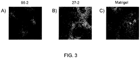
- FIG. 3 shows fluorescent images of differentiated cardiomyocytes cultured on surfaces of formulation 95-2 ( A ), formulation 27-2 ( B ), and MATRIGEL ( C ).
- Cell morphology, Nkx 2.5 marker expression (green) and alpha-actinin marker expression (red) are similar to MATRIGEL.
- the cells depicted in FIG. 3 exhibited beating prior to fixing with paraformaldehyde.
Description
- The present disclosure relates to a cell culture of a stem cell-derived cardiomyocyte and a method for culturing such cells.
- Pluripotent stem cells such as human embryonic stem cells (hESCs) have the ability to differentiate into any of the three germ layers, giving rise to any adult cell type in the human body. This unique property provides a potential for developing new treatments for a number of serious cell degenerative diseases, such as diabetes, spinal cord injury, heart diseases and the like. For example, unlike organs such as the skin or liver, the heart is not capable of regenerate sufficient cardiomyocytes to undergo extensive repair. Therefore cardiac repair may benefit from cardiomyocytes, which can be differentiated from hESCs or other pluripotent stem cells, being transplanted into the heart.
- However there remain obstacles in the development of such hESC-based treatments. Such obstacles include obtaining and maintaining adequate numbers of undifferentiated hESCs in tissue culture and controlling their differentiation in order to produce specific cell types. Stem cell cultures, such as hES cell cultures are typically seeded with a small number of cells from a cell bank or stock and then amplified in the undifferentiated state until differentiation is desired for a given therapeutic application. To accomplish this, the hESC or their differentiated cells are currently cultured in the presence of surfaces or media containing animal-derived components, such as feeder layers, fetal bovine serum, or MATRIGEL. These animal-derived additions to the culture environment expose the cells to potentially harmful viruses or other infectious agents which could be transferred to patients or compromise general culture and maintenance of the hESCs. In addition, such biological products are vulnerable to batch variation, immune response and limited shelf-life.
- Some steps have been taken to culture hESCs either in media or on surfaces that are free of animal-derived components. However, the response of hESCs or their differentiated derivatives is difficult to predict as components of the surface or culture medium change. Yet some advances have been made. For example, hESC-derived cardiomyocytes have been cultured in defined serum-free medium. While such culture systems are not completely xeno-free culture systems when the matrices employed contain animal-derived components, such as gelatin and MATRIGEL, they do provide a step toward the eventual clinical application of hESC-derived cardiomyocytes. By way of further example, some synthetic surfaces have been identified that can support differentiation of human epithelial stem cells into epithelial cells. However, the systems employed relied on serum medium for the cell culture, which still potentially causes problem as described before for all biological animal derived components. To date, a completely animal free system employing a chemically defined medium and a synthetic surface has not yet been identified for culturing stem cells or cells derived from stem cells.
- The present disclosure describes, inter alia, synthetic surfaces useful in the culture of stem cell-derived cardiomyocytes in chemically defined media.
- According to a first aspect of the invention, there is a provided a cell culture of a stem cell-derived cardiomyocyte, comprising: an article comprising an acrylate material disposed on a surface; the stem cell-derived cardiomyocyte disposed on the acrylate material; and a culture medium in which the stem cell-derived cardiomyocyte is cultured, wherein the acrylate material comprises a
- (i) homopolymer formed from glycerol dimethacrylate, or
- (ii) a copolymer formed from
glycerol dimethacrylate and tetra(ethylene glycol) diacrylate,
glycerol dimethacrylate and tri(ethylene glycol) dimethacrylate,
glycerol dimethacrylate and 1,4-butanediol dimethacrylate. - The acrylate material may comprise a homopolymer formed from glycerol dimethacrylate or a copolymer formed from (i) glycerol dimethacrylate and tri(ethylene glycol) dimethacrylate, or (ii) glycerol dimethacrylate and 1,4-butanediol dimethacrylate. Alternatively, the acrylate material may consist essentially of a homopolymer formed from glycerol dimethacrylate or a copolymer formed from (i) glycerol dimethacrylate and tri(ethylene glycol) dimethacrylate, or (ii) glycerol dimethacrylate and 1,4-butanediol dimethacrylate.
- Suitably, the culture medium can be a chemically defined medium. The stem cell-derived cardiomyocyte may be a human embryonic stem cell-derived cardiomyocyte.
- According to a second aspect of the invention, there is provided a method for culturing a stem cell-derived cardiomyocyte, comprising:
- depositing a suspension comprising the stem cell-derived cardiomyocyte on a acrylate material; and
- culturing the deposited stem cell-derived cardiomyocyte in a cell culture medium wherein the acrylate material comprises
- (i) a homopolymer formed from glycerol dimethacrylate, or
- (ii) a copolymer formed from
glycerol dimethacrylate and tetra(ethylene glycol) diacrylate,
glycerol dimethacrylate and tri(ethylene glycol) dimethacrylate,
glycerol dimethacrylate and 1,4-butanediol dimethacrylate.
- The culturing the deposited stem cell-derived cardiomyocyte in a cell culture medium may comprise culturing the cardiomyocyte in a chemically defined medium. The acrylate material may comprise a homopolymer formed from glycerol dimethacrylate.
- The acrylate material may comprise a copolymer formed from glycerol dimethacrylate and tri(ethylene glycol) dimethacrylate. The acrylate material may comprise a copolymer formed from glycerol dimethacrylate and 1,4-butanediol dimethacrylate. The stem cell-derived cardiomyocyte may be a human embryonic stem cell-derived cardiomyocyte.
- One or more of the various disclosures presented herein provide one or more advantages over prior surfaces for culturing stem cell-derived cardiomyocytes. For example, the synthetic surfaces reduce potential contamination issues associated with surfaces having components obtained from or derived from animal sources. Such surfaces may also provide for improved shelf life compared to those surfaces with biological components. The ability to culture stem cell-derived cardiomyocytes in chemically-defined media further reduces potential contamination issues. In addition, there will likely be less batch to batch variation in the ability of the synthetic surfaces or chemically defined media, resulting in improved reproducibility of culture results and expectations. These and other advantages will be readily understood from the following detailed descriptions when read in conjunction with the accompanying drawings.
-
-
FIG. 1A-B are schematic diagrams of side views of synthetic polymer layer coated articles. -
FIG. 2A-C are schematic diagrams of cross sections of a multi-well cell culture plate. The plate is uncoated inFIG. 2A and coated inFIGs. 2B-C . -
FIG. 3 is a fluorescent image of stem cell derived cardiomyocytes cultured in chemically defined medium and on a surface of formulation 1 (A), formulation 18 (B), and MATRIGEL (C) as described in Example 1. Green: Nkx 2.5. Red: Alpha-actinin. - The drawings are not necessarily to scale. Like numbers used in the figures refer to like components, steps and the like. However, it will be understood that the use of a number to refer to a component in a given figure is not intended to limit the component in another figure labeled with the same number. In addition, the use of different numbers to refer to components is not intended to indicate that the different numbered components cannot be the same or similar.
- In the following detailed description, reference is made to the accompanying drawings that form a part hereof, and in which are shown by way of illustration several specific embodiments of devices, systems and methods. It is to be understood that other embodiments are contemplated and may be made without departing from the scope or spirit of the present disclosure. The following detailed description, therefore, is not to be taken in a limiting sense.
- All scientific and technical terms used herein have meanings commonly used in the art unless otherwise specified. The definitions provided herein are to facilitate understanding of certain terms used frequently herein and are not meant to limit the scope of the present disclosure.
- As used in this specification and the appended claims, the singular forms "a", "an", and "the" encompass embodiments having plural referents, unless the content clearly dictates otherwise. As used in this specification and the appended claims, the term "or" is generally employed in its sense including "and/or" unless the content clearly dictates otherwise.
- Unless stated otherwise, ratios of compounds in a composition, such as a solution, are stated on a by volume basis.
- As used herein, "have", "having", "include", "including", "comprise", "comprising" or the like are used in their open ended sense, and generally mean "including, but not limited to".
- As used herein the term "acrylate" includes compounds containing an acrylate moiety or a methacrylate moiety. An acrylate moiety is moiety of the following formula: CH2CHC(O)O-. A methacrylate moiety is a moiety of the following formula: CH2C(CH3)C(O)O-. For the purposes of this disclosure, the term "acrylate" includes specific compounds disclosed in Table 1. "Acrylate" and "methacrylate" are used herein interchangeable, except when content clearly dictates otherwise; e.g. when a specific compound or group of compounds are named.
- The present disclosure describes, inter alia, articles having synthetic surfaces for culturing stem cell-derived cardiomyocytes and methods for culturing stem cell-derived cardiomyocytes on such surfaces. The synthetic surfaces are used in combination with a chemically defined medium to culture stem cell-derived cardiomyocytes. The surfaces may be useful in differentiating stem cells, such as hESCs, into cardiomyocytes.
- Referring to
FIG. 1 , a schematic diagram ofarticle 100 for culturing cells is shown. Thearticle 100 includes abase material substrate 10 having asurface 15. A syntheticpolymer coating layer 20 is disposed on thesurface 15 of thebase material 10. While not shown, it will be understood thatsynthetic polymer coating 20 may be disposed on a portion ofbase material 10. Thebase material 10 may be any material suitable for culturing cells, including a ceramic substance, a glass, a plastic, a polymer or co-polymer, any combinations thereof, or a coating of one material on another.Such base materials 10 include glass materials such as soda-lime glass, pyrex glass, vycor glass, quartz glass; silicon; plastics or polymers, including dendritic polymers, such as poly(vinyl chloride), poly(vinyl alcohol), poly(methyl methacrylate), poly(vinyl acetate-maleic anhydride), poly(dimethylsiloxane) monomethacrylate, cyclic olefin polymers, fluorocarbon polymers, polystyrenes, polypropylene, polyethyleneimine; copolymers such as poly(vinyl acetate-co-maleic anhydride), poly(styrene-co-maleic anhydride), poly(ethylene-co-acrylic acid) or derivatives of these or the like. - Examples of
articles 100 suitable for cell culture include single and multi-well plates, such as 6, 12, 96, 384, and 1536 well plates, jars, petri dishes, flasks, beakers, plates, roller bottles, slides, such as chambered and multichambered culture slides, tubes, cover slips, cups, spinner bottles, perfusion chambers, bioreactors, and fermenters. -
Synthetic polymer coating 20 provides asurface 25 on which cells may be cultured. Thesynthetic polymer surface 20 includes polymerized (meth)acrylate monomers, selected from the group of monomers provided in Table 1 below. Other materials (not shown), such as peptides, may be incorporated into or conjugated to synthetic polymer surface to produce a biomimetic surface.Table 1: List of acrylate and methacrylate monomers. Acrylate material used in the invention is indicated "*" thus. Monomer name Monomer structure * Tetra(ethylene glycol) diacrylate 
* Glycerol dimethacrylate 
* Triethylene glycol dimethacrylate 
* 1,4-Butanediol dimethacrylate 
Poly(ethylene glycol) diacrylate (average Mn ∼258) 
Di(ethylene glycol) dimethacrylate 
Tetra(ethylene glycol) dimethacrylate 
1,6-Hexanediol propoxylate diacrylate 
Neopentyl glycol diacrylate 
Neopentyl glycol dimethacrylate 
Trimethylolpropane benzoate diacrylate 
Trimethylolpropane ethoxylate (1 EO/OH) methyl 
Tricyclo[5.2.1.02,6]decanedimethanol diacrylate 
Neopentyl glycol ethoxylate diacrylate 
Trimethylolpropane triacrylate 
- The acrylates listed in Table 1 may be synthesized as known in the art or obtained from a commercial vendor, such as Polysciences, Inc., Sigma Aldrich, Inc., and Sartomer, Inc.
- As shown in
FIG. 1B , anintermediate layer 30 may be disposed betweensurface 15 ofbase material 10 and thesynthetic polymer coating 20.Intermediate layer 30 may be configured to improve binding ofcoating 20 tosubstrate 10, to facilitate monomer spreading, to render portions of thesurface 10 that are uncoated cytophobic to encourage cell growth on coated areas, to provide a substrate compatible with a monomer or solvent where the monomer or solvent is incompatible with thebase material 10, to provide topographical features if desired through, for example, patterned printing, or the like. For example, ifsubstrate 10 is a glass substrate, it may be desirable to treat a surface of the glass substrate with an epoxy coating or a silane coating. For variouspolymer base materials 10 it may be desirable to provide anintermediate layer 30 of polyamide, polyimide, polypropylene, polyethtylene, or polyacrylate. While not shown, it will be understood thatsynthetic polymer coating 20 may be disposed on a portion ofintermediate layer 30. It will be further understood thatintermediate layer 30 may be disposed on a portion ofbase material 10. -
Surface 15 ofbase material 10 is treated, either physically or chemically, to impart a desirable property or characteristic to thesurface 15. For example, and as discussed below,surface 15 may be corona treated or plasma treated. Examples of vacuum or atmospheric pressure plasma include radio frequency (RF) and microwave plasmas both primary and secondary, dielectric barrier discharge, and corona discharge generated in molecular or mixed gases including air, oxygen, nitrogen, argon, carbon dioxide, nitrous oxide, or water vapor. - Synthetic
polymer coating layer 20, whether disposed on anintermediate layer 30 orbase material 10, preferably uniformly coats the underlying substrate. By "uniformly coated", it is meant that thelayer 20 in a given area, for example a surface of a well of a culture plate, completely coats the area at a thickness of about 5 nm or greater. While the thickness of a uniformly coated surface may vary across the surface, there are no areas of the uniformly coated surfaces through which the underlying layer (eitherintermediate layer 30 or base material 10) is exposed. Cell responses across non-uniform surfaces tend to be more variable than cell responses across uniform surfaces. - Synthetic
polymer coating layer 20 may have any desirable thickness. However, it has been found that thicker coatings, e.g. coatings of greater than about 10 micrometers, tend to have unevenness around the periphery of the coating due to surface tension. - The thickness of the
coating layer 20 is less than about 10 micrometers. For example, the thickness may be less than about 5 micrometers, less than about 2 micrometers, less than about 1 micrometers, less than about 0.5 micrometers or less than about 0.1 micrometers. - The polymer material forming
synthetic polymer layer 20 may be cross-linked to any suitable degree. Higher degrees of cross-linking may result in reduced waste product and reduced cell toxicity. -
Article 100 is cell culture ware having a well, such as a petri dish, a multi-well plate, a flask, a beaker or other container having a well. Referring now toFIG. 2 ,article 100 formed frombase material 10 may include one ormore wells 50. Well 50 includes asidewall 55 and asurface 15. Referring toFIG. 2B-C , asynthetic polymer coating 20 may be disposed onsurface 15 or sidewalls 55 (or, as discussed above with regard toFIG. 1 one or moreintermediate layer 30 may be disposed betweensurface 15 orsidewall 55 and synthetic polymer coating 20) or a portion thereof. -
Article 100 includes a uniformly coatedlayer 20 having asurface 25 with an area greater than about 5 mm2. When the area of thesurface 15 is too small, reliable cell responses may not be readily observable because some cells, such as human embryonic stem cells, are seeded as colonies or clusters of cells (e.g., having a diameter of about 0.5 mm) and adequate surface is desirable to ensure attachment of sufficient numbers of colonies to produce a quantitative cell response.
Anarticle 100 has a well 50 having a uniformlycoated surface 15, where thesurface 15 has an area greater than about 0.1 cm2, greater than about 0.3 cm2, greater than about 0.9 cm2, or greater than about 1cm2. - A synthetic polymer layer may be disposed on a surface of a cell culture article via any known or future developed process. Preferably, the synthetic polymer layer provides a uniform layer that does not delaminate during typical cell culture conditions. The synthetic polymer surface may be associated with the base material substrate via covalent or non-covalent interactions. Examples of non-covalent interactions that may associate the synthetic polymer surface with the substrate include chemical adsorption, hydrogen bonding, surface interpenetration, ionic bonding, van der Waals forces, hydrophobic interactions, dipole-dipole interactions, mechanical interlocking, and combinations thereof.
- Monomers are deposited on a surface of a cell culture article and polymerized in situ. The base material will be referred to herein as the "substrate" on which the synthetic polymer material is deposited. Polymerization may be done in solution phase or in bulk phase.
- As many of the monomers identified in Table 1 above are viscous, it may be desirable to dilute the monomers in a suitable solvent to reduce viscosity prior to being dispensed on the surface. Reducing viscosity may allow for thinner and more uniform layers of the synthetic polymer material to be formed. One of skill in the art will be able to readily select a suitable solvent. Preferably the solvent is compatible with the material forming the cell culture article and the monomers. It may be desirable to select a monomer that is non-toxic to the cells to be cultured and that does not interfere with the polymerization reaction. Alternatively, or in addition, selection of a solvent that can be substantially completely removed or removed to an extent that it is non-toxic or no longer interferes with polymerization may be desirable. In such circumstances, it may be desirable that the solvent be readily removable without harsh conditions, such as vacuum or extreme heat. Volatile solvents are examples of such readily removable solvents.
- Some solvents that may be suitable in various situations for coating articles as described herein include ethanol, isopropanol, acetyl acetate, dimethylformamide (DMF), and dimethylsulfoxide (DMSO).
- Ethanol may be a particularly suitable solvent when it is desired to remove solvent prior to polymerization.
- The monomers may be diluted with solvent by any suitable amount to achieve the desired viscosity and monomer concentration. Generally the monomer compositions used according to the teachings presented herein contain between about 1% to about 99% monomer. By way of example, the monomer may be diluted with an ethanol solvent to provide a composition having between about 1% and about 50% monomer, or from about 1% to about 10% monomer by volume. The monomers may be diluted with solvent so that the
polymer layer 20 achieves a desired thickness. As discussed above, if the deposited monomers are too thick, a non-uniform surface may result. As described in further details in the Examples, non-uniform surfaces may be observed when the monomer-solvent composition is deposited on asurface 15 of a well 50 at a volume of greater than about 8 microliters per square centimeter of thesurface 15. In various embodiments, the monomer-solvent compositions are deposited on asurface 15 of a well 50 in a volume of about 7 microliters or less per square centimeter of thesurface 15. For example, the monomer-solvent compositions may be deposited on asurface 15 of a well 50 in a volume of about 5 microliters or less per square centimeter of thesurface 15, or about 2 microliters or less per square centimeter of thesurface 15. - Synthetic polymer surface is deposited on a surface of an intermediate layer that is associated with the base material via covalent or non-covalent interactions, either directly or via one or more additional intermediate layers (not shown). In such embodiments, the intermediate layer will be referred to herein as the "substrate" onto which the synthetic polymer surface is deposited.
- The surface of the base material is treated. The surface may be treated to improve binding of the synthetic polymer surface to the base material surface, to facilitate monomer spreading on the base material surface, or the like. Of course, the base material may be treated for similar purposes with regard to an intermediate layer.
- The surface is corona treated or vacuum plasma treated. High surface energy obtainable from such treatments may facilitate monomer spreading and uniform coating. Examples of vacuum plasma treatment that may be employed include microwave vacuum plasma treatments and radio frequency vacuum plasma treatments. The vacuum plasma treatments may be performed in the presence of reactive gases, such as oxygen, nitrogen, ammonia or nitric oxide.
- To form the synthetic polymer surface, one or more monomers presented in Table 1 above are polymerized. If one monomer is used, the polymer will be referred to as a homopolymer of the monomer. If two or more different monomers are used, the polymer will be referred to as a copolymer of the monomers. The monomers employed may be monofunctional, difunctional, or higher-functional. When two or more monomers are used, the ratio of the monomers may be varied. For example, two monomers are used and the ratio, by volume of the first monomer to the second monomer ranges from between about 5:95 to about 95:5. For example, the ratio of the first monomer to the second monomer ranges from between about 10:90 to about 90:10, about 20:80 to about 80:20, from about 30:70 to about 70:30. For example, the ratio of the first monomer to the second monomer is about 50:50, 30:70, or 10:90. It will be understood that the molecular weight of the polymer may be controlled by varying the concentration of monomers or the ratios of difunctional or higher-functional monomers to monofunctional monomers. Increased concentrations of difunctional or higher-functional monomers will increase the degree of cross-linking in the chains.
- In addition to the monomers that form the polymer layer, a composition forming the layer may include one or more additional compounds such as surfactants, wetting agents, photoinitiators, thermal initiators, catalysts, activators, and cross-linking agents.
- Any suitable polymerization initiator may be employed. One of skill in the art will readily be able to select a suitable initiator, e.g. a radical initiator or a cationic initiator, suitable for use with the monomers listed in Table 1. In various embodiments, UV light is used to generate free radical monomers to initiate chain polymerization.
- Any suitable initiator may be used. Examples of polymerization initiators include organic peroxides, azo compounds, quinones, nitroso compounds, acyl halides, hydrazones, mercapto compounds, pyrylium compounds, imidazoles, chlorotriazines, benzoin, benzoin alkyl ethers, diketones, phenones, or mixtures thereof. Examples of suitable commercially available, ultraviolet-activated and visible light-activated photoinitiators have tradenames such as IRGACURE 651, IRGACURE 184, IRGACURE 369, IRGACURE 819, DAROCUR 4265 and DAROCUR 1173 commercially available from Ciba Specialty Chemicals, Tarrytown, N.Y. and LUCIRIN TPO and LUCIRIN TPO-L commercially available from BASF (Charlotte, N.C.)
- A photosensitizer may also be included in a suitable initiator system. Representative photosensitizers have carbonyl groups or tertiary amino groups or mixtures thereof. Photosensitizers having a carbonyl groups include benzophenone, acetophenone, benzil, benzaldehyde, o-chlorobenzaldehyde, xanthone, thioxanthone, 9,10-anthraquinone, and other aromatic ketones. Photosensitizers having tertiary amines include methyldiethanolamine, ethyldiethanolamine, triethanolamine, phenylmethylethanolamine, and dimethylaminoethylbenzoate. Commercially available photosensitizers include QUANTICURE ITX, QUANTICURE QTX, QUANTICURE PTX, QUANTICURE EPD from Biddle Sawyer Corp.
- In general, the amount of photosensitizer or photoinitiator system may vary from about 0.01 to 10% by weight.
- Examples of cationic initiators include salts of onium cations, such as arylsulfonium salts, as well as organometallic salts such as ion arene systems.
- Where the monomers are diluted in solvent before being deposited on the substrate surface, the solvent is removed prior to polymerizing. The solvent may be removed by any suitable mechanism or process.
- It has been found that removal of substantially all of the solvent prior to curing, allows for better control of curing kinetics and the amount of monomer converted. When conversion rates of the monomers are increased, waste generation and cytotoxicity are reduced.
- Whether polymerized in bulk phase (substantially solvent free) or solvent phase, the monomers are polymerized via an appropriate initiation mechanism. Many of such mechanisms are well known in the art. For example, temperature may be increased to activate a thermal initiator, photoinitiators may be activated by exposure to appropriate wavelength of light, or the like. According to numerous embodiments, the monomer or monomer mixture is cured using UV light. The curing preferably occurs under inert gas protection, such as nitrogen protection, to prevent oxygen inhibition. Suitable UV light combined with gas protection may increase polymer conversion, insure coating integrity and reduce cytotoxicity.
- The cured synthetic polymer layer may be washed with solvent one or more times to remove impurities such as unreacted monomers or low molecular weight polymer species. In various embodiments, the layer is washed with an ethanol solvent, e.g. greater than about 70% ethanol, greater than about 90% ethanol, greater than about 95% ethanol or greater than about 99% ethanol. Washing with an ethanol solvent may not only serve to remove impurities, which may be cytotoxic, but also can serve to sterilize the surface prior to incubation with cells.
- Stem cell-derived cardiomyocytes may be cultured on a synthetic polymer layer, as described above, according to any suitable protocol. As used herein, "stem cell derived cardiomyocyte" means a cardiomyocyte obtained from differentiation of a stem cell.
- The stem cells are multipotent, or pluripotent stem cells. The cells may be present in an organ or tissue of a subject.
- For example, the stem cells are embryonic stem cells, such as reference human embryonic stem cells.
- By way of reference, because human embryonic stem cells (hES) have the ability to grown continually in culture in an undifferentiated state, the hES cells may be obtained from an established cell line. Examples of reference human embryonic stem cell lines that have been established include, but are not limited to, H1, H7, H9, H13 or H14 (available from WiCell established by the University of Wisconsin) (Thompson (1998) Science 282:1145 ); hESBGN-01, hESBGN-02, hESBGN-03 (BresaGen, Inc., Athens, GA); HES-1, HES-2, HES-3, HES-4, HES-5, HES-6 (from ES Cell International, Inc., Singapore); HSF-1, HSF-6 (from University of California at San Francisco); I 3, I 3.2, I 3.3, I 4, I 6, I 6.2, J 3, J 3.2 (derived at the Technion-Israel Institute of Technology, Haifa, Israel); UCSF-1 and UCSF-2 (Genbacev et al., Fertil. Steril. 83(5):1517-29, 2005); lines HUES 1-17 (Cowan et al., NEJM 350(13):1353-56, 2004); and line ACT-14 (Klimanskaya et al., Lancet, 365(9471):1636-41, 2005).
- Cardiomyocytes may also be differentiated from induced primate pluripotent stem (iPS) cells. iPS cells refer to cells, obtained from a juvenile or adult mammal such as a human, that are genetically modified, e.g., by transfection with one or more appropriate vectors, such that they are reprogrammed to attain the phenotype of a pluripotent stem cell such as an hES cell. Phenotypic traits attained by these reprogrammed cells include morphology resembling stem cells isolated from a blastocyst as well as surface antigen expression, gene expression and telomerase activity resembling blastocyst derived embryonic stem cells. The iPS cells typically have the ability to differentiate into at least one cell type from each of the primary germ layers: ectoderm, endoderm and mesoderm and thus are suitable for differentiation into cardiomyocytes. The iPS cells, like hES cells, also form teratomas when injected into immuno-deficient mice, e.g., SCID mice. (Takahashi et al., (2007) Cell 131(5):861; Yu et al., (2007) Science318:5858).
- Stem cell derived cardiomyocytes may be obtained by any suitable methods. One way to obtain such cells is described in Laflamme et al., "Cardiomyocytes derived from human embryonic stem cells in pro-survival factors enhance function of infracted rats, Nature Biotechnology, 25: 1015 - 1024 (2007). Briefly, undifferentiated human embryonic stem cells, such as those derived from the reference female H7 human embryonic stem cell line, may be seeded on MATRIGEL-coated plates at a density of about 100,000 cells/cm2 and refed daily with hES cell growth medium (KO DMEM + 20% Serum replacement, 1 mM 1-glutamine, 1% NEAA, 0.1 mM 2-ME plus hbFGF at 80 ng/ml and TGFb1 at 0.5 ng/ml) To induce differentiation, growth media may be replaced with RPMI-B27 medium (available from Invitrogen) supplemented with about 100 ng/ml human recombinant activin A (available from R&D Systems) for about 24 hours, followed by 10 ng/ml human recombinant BMP4 (available from R&D Systems) for four days. Of course, any other suitable method may be employed (see, e.g.,
US Patent No. 7,425,448 ). - Prior to seeding cells, the cells may be harvested and suspended in a suitable medium, such as a growth medium in which the cells are to be cultured once seeded onto the surface. For example, the cells may be suspended in and cultured in serum-containing medium, a conditioned medium, or a chemically-defined medium. As used herein, "chemically-defined medium" means cell culture media that contains no components of unknown composition. Chemically defined media may contain no proteins, hydrosylates, or peptides of unknown origin. For example, conditioned media contains polypeptides or proteins of known composition, such as recombinant growth hormones. Because all components of chemically-defined media have a known chemical structure and composition, variability in culture conditions can be reduced and thus cell response may be more reproducible. In addition, the possibility of contamination is reduced. Further, the ability to scale up is made easier due, at least in part, to the factors discussed above. Chemically defined cell culture media are commercially available from Invitrogen (Invitrogen Corporation, 1600 Faraday Avenue, PO Box 6482, Carlsbad, California 92008) as StemPro® a fully defined, serum- and feeder-free medium (SFM) specially formulated for the growth and expansion of human embryonic stem cells (hESCs) and StemCell Technologies, Inc as mTeSR™1 maintenance media for human embryonic stem cells.
- The cells may be seeded at any suitable concentration. Typically, the cells are seeded at about 10,000 cells/cm2 of substrate to about 500,000 cells/cm2. For example, cells may be seeded at about 50,000 cells/cm2 of substrate to about 150,000 cells/cm2. However, higher and lower concentrations may readily be used. The incubation time and conditions, such as temperature CO2 and O2 levels, growth medium, and the like, will depend on the nature of the cells being cultured and can be readily modified. The amount of time that the cells are incubated on the surface may vary depending on the cell response being studied or the cell response desired.
- Any suitable method may be used, if desired, to confirm that the stem cell derived cardiomyocytes are indeed cardiomyocytes or that the stem cells employed have successfully differentiated into cardiomyocytes. For example, the presence of certain cardiomyocyte-selective markers may be investigated. Such markers include Nkx2.5 and α-actinin, cardiac troponin I. Antibodies to such markers may be used in standard immunocytochemical or flow cytometry techniques. In addition or alternatively, cellular morphology or functionality, by observing beating cardiomyocytes in culture or by performing various electrophysiological analyses to determine whether the cells have characteristics of cardiomyocytes.
- The cultured stem cell derived cardiomyocytes may be used for any suitable purpose, including investigational studies in culture, in animals, for developing therapeutic uses, for drug discovery and toxicology or for therapeutic purposes. One potential therapeutic or investigational purpose is repairing cardiac damage due to an infarct, e.g., as described in Laflamme et al., Nature Biotechnology, 25: 1015 - 1024 (2007). The cells may also be used to create cDNA libraries according to known methods. The cDNA libraries may be used to study gene expression in the differentiated cardiomyocytes. For example the library obtained from the differentiated cardiomyocytes may be compared to a cDNA library from the undifferentiated stem cells from which the cardiomyocytes were derived, thus allowing for the identification and isolation of genes related to the differentiation and development of cardiomyocytes.
- In the following, non-limiting examples are presented, which describe various embodiments of the cell culture and methods discussed above.
- Identification of Acrylic Coating Surfaces Suitable for Culturing Stem Cell Derived Cardiomyocytes in a Chemically Defined Medium
- Acrylic coating surfaces were prepared from homomonomers or copolymers of various acrylate and methacrylate monomers. For copolymers two different acrylate or methacrylate monomers were used. Briefly, the monomers were mixed with 1% w/w of photoinitiator Irgacure 819 (Ciba Specialty Chemiscals, Inc.) and used along or blended with other monomer formulation (monomer with 1% w/w of photoinitiator) according to volume ratio of 70:30. Then formulation was placed in a well of a vacuum plasma treated cyclic olefin copolymer plate (provided by Corning Life Science Development group) at a volume of 2.5 µL. The plate was allowed to lay horizontally flat for 30 min for the formulation to spread out. The coatings were cured with 13mW/cm2 pulsed (100 Hz) UV light (Xenon RC-700) for 1 min in N2 purged box (with fused silica window). All the plates were sterilized by 25-35 kGy Gamma radiation prior to cell culture.
- Prior to the experiments, H7 hES cells were maintained in the undifferentiated state on MATRIGEL-coated plates in SR medium (KO-DMEM, 20 % KO-serum replacement, 1 mM L-glutamine, 0.1 mM β-mercaptoethanol, 1% non- essential amino acids, 80 ng/ml hbFGF and 0.5 ng/ml TGFb1). hESC-derived cardiomyocytes were generated using direct differentiation protocol. Briefly, undifferentiated H7 cells were harvested by 200 U/ml collagenase IV and seeded on MATRIGEL-coated plates at the density of 100,000 cells/cm2 in SR medium. Cells were cultured for 6 days with daily medium exchange. Cardiac differentiation was initiated by replacing SR medium with RPMI-B27 medium, supplemented with 100 ng/ml human recombinant activin A for 24 h, followed by 10 ng/ml human recombinant BMP4 for 4 days. At this point, cells were detached by Accutase treatment, re-suspended in RPMI-B27 medium w/o growth factors, and seeded at the density of100,000 cells/cm2 onto Corning CB/TOPAS 96-well plates coated with different binary mixtures of acrylates or MATRIGEL as positive control. Cells were cultured for another 2-3 weeks with the same medium exchange every 2-3 days. The wells were microscopically examined, and spontaneous beating activity was recorded.
- At the end of the differentiation protocol, cells were fixed with 4% paraformaldehyde immunostained for cardiomyocyte (CM) specific markers, Nkx2.5 and α-actinin, and counterstained with 4'-6-Diamidino-2-phenylindole (DAPI) to stain the nucleus. After scanning each plate with ArrayScan, the following quantitative analyses were performed for each surface: 1) TNC: total number of cells based on DAPI positive cell number, 2) TNC: total number of CM, based on Nkx2.5-positive cell number, 3) CM yield = TNC/TNC.
- It was found that only a small portion of the tested surfaces supported the cell growth while maintaining some key characteristics and functions. Examples of the coating surfaces which supported growth of differentiated human embryonic cardiomyocytes in chemically defined medium are listed in Table 2, where the volume ratio of monomer (1) to monomer (2) is 70:30. These surfaces were characterized and given a "grade" based on qualitative assessments of the morphology of the cells and their adhesion to the surfaces. Acrylate surface rating was performed based on consideration of the following criteria: 1) total cell number, 2) total CM number, 3) CM yield, 4) presence of beating areas, 5) similarity to Matrigel derived CM morphology. Notes on the qualities of the differentiated embryonic myocytes that were observed are shown in Table 2, as well as the grade that was assessed. For example, "Sim MA" is a note that indicates that cells growing on that surface were similar to cells growing on Matrigel™. "Sim TOP" is a note that indicates that the cells growing on that surface were similar to cells growing on plasma-treated cyclic olefin copolymer surface, sold as TOPAS® surface by TOPAS, Florence, Kentucky. The notes also indicate that some of the surfaces supported differentiated embryonic myocytes that began to beat in culture.
Table 2. Example compositions of acrylic polymers which support the culture of hES cell derived cardiomyocytes in chemically defined medium. Acrylate material used in the invention is indicated "*" thus. Formuation ID Monomer (1) Monomer (2) 95-1 Tetra(ethylene glycol) diacrylate 100% * 95-2 Glycerol dimethacrylate 100% 90-2 Triethylene glycol dimethacrylate 100% 90-4 1,4-Butanediol dimethacrylate 100% 95-3 Poly(ethylene glycol) diacrylate 100% 122-1 Tetra(ethylene glycol) diacrylate 70% Glycerol dimethacrylate 30% 122-3 Tetra(ethylene glycol) diacrylate 70% 1,4-Butanediol dimethacrylate 30% 27-2 Tetra(ethylene glycol) diacrylate 70% Trimethylolpropane triacrylate 30% * 123-1 Glycerol dimethacrylate 70% Tetra(ethylene glycol) diacrylate 30% * 123-2 Glycerol dimethacrylate 70% Tri(ethylene glycol) dimethacrylate 30% * 123-3 Glycerol dimethacrylate70% 1,4-Butanediol dimethacrylate 30% 123-4 Glycerol dimethacrylate 70% Poly(ethylene glycol) diacrylate 30% * 123-6 Triethylene glycol dimethacrylate 70% Glycerol dimethacrylate 30% 123-7 Triethylene glycol dimethacrylate 70% 1,4-Butanediol dimethacrylate 30% * 124-2 1,4-Butanediol dimethacrylate 70% Glycerol dimethacrylate 30% 124-3 1,4-Butanediol dimethacrylate 70% Triethylene glycol dimethacrylate 30% 124-5 Poly(ethylene glycol) diacrylate 70% Tetra(ethylene glycol) diacrylate 30% 133-1 Di(ethylene glycol) dimethacrylate 70% Tetraethylene glycol dimethacrylate 30% 134-1 Tetraethylene glycol dimethacrylate 70% Di(ethylene glycol) dimethacrylate 30% 140-1 Trimethylolpropane triacrylate 70% Neopentyl glycol ethoxylate diacrylate 30% TABLE 3. Cell count, cell surface Marker, and functions of h7 differentiated cardiomyocyted cultured on acrylate polymer surfaces. Acrylate material used in the invention is indicated "*" thus. ID Rating Beating Average Cell Count (per well) Average % Nkx2.5 Cardio yield (based on count*Nkx2.5) Note * 95-2 A y 19970 7 1397.9 Sim MA 90-2 C- 5420 14 758.8 Sim MA, peeled lumpy 90-4 B- y 6907 24 1657.68 Sim TOP 95-3 C y 3788 24 909.12 122-3 B 6061 3 181.83 Sim as MA, not beating * 123-1 B+ y 5250 5 262.5 Sim MA, beating * 123-2 A 6327 7 442.89 Good attachment, no beating * 123-3 A 6223 8 497.84 Good attachment, no beating 123-4 B- 4954 4 198.16 Many attached, dying 123-7 C 4246 15 636.9 peeled * 124-2 A y 3798 21 797.58 Sim TOP beating 124-3 B y 2267 8 497.84 Sim TOP beating 124-5 C y 940 27 253.8 Beating but poor attachment 140-1 B- y 2886 33 952.38 Sim TOP - For other tested homopolymers and copolymer combinations, very low or no cardiomyocytes yield was observed at the end of the differentiation procedure, due to either low total cell number or low Nkx2.5-positive cells present on those surfaces
-
FIG. 3 shows fluorescent images of differentiated cardiomyocytes cultured on surfaces of formulation 95-2 (A), formulation 27-2 (B), and MATRIGEL (C). Cell morphology, Nkx 2.5 marker expression (green) and alpha-actinin marker expression (red) are similar to MATRIGEL. In addition, the cells depicted inFIG. 3 exhibited beating prior to fixing with paraformaldehyde.
Claims (11)
- A cell culture of a stem cell-derived cardiomyocyte, comprising:an article comprising an acrylate material disposed on a surface;the stem cell-derived cardiomyocyte disposed on the acrylate material; anda culture medium in which the stem cell-derived cardiomyocyte is cultured, wherein the acrylate material comprises a(i) homopolymer formed from glycerol dimethacrylate, or(ii) a copolymer formed from
glycerol dimethacrylate and tetra(ethylene glycol) diacrylate,
glycerol dimethacrylate and tri(ethylene glycol) dimethacrylate, or
glycerol dimethacrylate and 1,4-butanediol dimethacrylate. - The culture of claim 1, wherein the acrylate material comprises a homopolymer formed from glycerol dimethacrylate or a copolymer formed from (i) glycerol dimethacrylate and tri(ethylene glycol) dimethacrylate, or (ii) glycerol dimethacrylate and 1,4-butanediol dimethacrylate.
- The culture of claim 1, wherein the acrylate material consists essentially of a homopolymer formed from glycerol dimethacrylate or a copolymer formed from (i) glycerol dimethacrylate and tri(ethylene glycol) dimethacrylate, or (ii) glycerol dimethacrylate and 1,4-butanediol dimethacrylate.
- The culture of claim 1, wherein culture medium is a chemically defined medium.
- The culture of claim 1, wherein the stem cell-derived cardiomyocyte is a human embryonic stem cell-derived cardiomyocyte.
- A method for culturing a stem cell-derived cardiomyocyte, comprising:depositing a suspension comprising the stem cell-derived cardiomyocyte on a acrylate material; andculturing the deposited stem cell-derived cardiomyocyte in a cell culture medium wherein the acrylate material comprises(i) a homopolymer formed from glycerol dimethacrylate, or(ii) a copolymer formed from
glycerol dimethacrylate and tetra(ethylene glycol) diacrylate,
glycerol dimethacrylate and tri(ethylene glycol) dimethacrylate, or
glycerol dimethacrylate and 1,4-butanediol dimethacrylate. - The method of claim 6, wherein culturing the deposited stem cell-derived cardiomyocyte in a cell culture medium comprises culturing the cardiomyocyte in a chemically defined medium.
- The method of claim 6, wherein the acrylate material comprises a homopolymer formed from glycerol dimethacrylate.
- The method of claim 6, wherein the acrylate material comprises a copolymer formed from glycerol dimethacrylate and tri(ethylene glycol) dimethacrylate.
- The method of claim 6, wherein the acrylate material comprises a copolymer formed from glycerol dimethacrylate and 1,4-butanediol dimethacrylate.
- The method of claim 6, wherein the stem cell-derived cardiomyocyte is a human embryonic stem cell-derived cardiomyocyte.
Applications Claiming Priority (2)
| Application Number | Priority Date | Filing Date | Title |
|---|---|---|---|
| US6292108P | 2008-01-30 | 2008-01-30 | |
| PCT/US2009/032411 WO2009097411A1 (en) | 2008-01-30 | 2009-01-29 | Synthetic surfaces for culturing stem cell derived cardiomyocytes |
Publications (3)
| Publication Number | Publication Date |
|---|---|
| EP2247718A1 EP2247718A1 (en) | 2010-11-10 |
| EP2247718A4 EP2247718A4 (en) | 2015-07-22 |
| EP2247718B1 true EP2247718B1 (en) | 2019-01-16 |
Family
ID=40899641
Family Applications (1)
| Application Number | Title | Priority Date | Filing Date |
|---|---|---|---|
| EP09705923.2A Not-in-force EP2247718B1 (en) | 2008-01-30 | 2009-01-29 | Synthetic surfaces for culturing stem cell derived cardiomyocytes |
Country Status (10)
| Country | Link |
|---|---|
| US (4) | US8241907B2 (en) |
| EP (1) | EP2247718B1 (en) |
| JP (4) | JP2011510658A (en) |
| KR (2) | KR101809863B1 (en) |
| CN (2) | CN101939418A (en) |
| AU (1) | AU2009209157B2 (en) |
| CA (1) | CA2712891A1 (en) |
| IL (1) | IL207083A (en) |
| SG (1) | SG188098A1 (en) |
| WO (1) | WO2009097411A1 (en) |
Families Citing this family (23)
| Publication number | Priority date | Publication date | Assignee | Title |
|---|---|---|---|---|
| CN101939362A (en) | 2008-01-30 | 2011-01-05 | 杰龙公司 | Synthetic surfaces for culturing stem cell derived oligodendrocyte progenitor cells |
| JP2011510658A (en) | 2008-01-30 | 2011-04-07 | ジェロン・コーポレーション | Synthetic surface for culturing stem cell-derived cardiomyocytes |
| CA3080762A1 (en) | 2009-11-12 | 2011-05-19 | Technion Research & Development Foundation Limited | Culture media, cell cultures and methods of culturing pluripotent stem cells in an undifferentiated state |
| US9453197B2 (en) | 2010-12-16 | 2016-09-27 | General Electric Company | Methods of making cell carrier |
| US9453196B2 (en) | 2010-12-16 | 2016-09-27 | General Electric Company | Cell carrier, methods of making and use |
| US9534206B2 (en) | 2010-12-16 | 2017-01-03 | General Electric Company | Cell carrier, associated methods for making cell carrier and culturing cells using the same |
| US9926523B2 (en) | 2010-12-16 | 2018-03-27 | General Electric Company | Cell carriers and methods for culturing cells |
| US9518249B2 (en) | 2010-12-16 | 2016-12-13 | General Electric Company | Cell carrier, associated methods for making cell carrier and culturing cells using the same |
| EP2712365B1 (en) | 2011-03-17 | 2016-09-21 | Corning Incorporated | Synthetic coating for cell culture |
| CA2829804A1 (en) | 2011-03-29 | 2012-10-04 | Geron Corporation | Enriched populations of cardiomyocyte lineage cells from pluripotent stem cells |
| US10813630B2 (en) | 2011-08-09 | 2020-10-27 | Corquest Medical, Inc. | Closure system for atrial wall |
| US10307167B2 (en) | 2012-12-14 | 2019-06-04 | Corquest Medical, Inc. | Assembly and method for left atrial appendage occlusion |
| US10314594B2 (en) | 2012-12-14 | 2019-06-11 | Corquest Medical, Inc. | Assembly and method for left atrial appendage occlusion |
| WO2013163187A1 (en) | 2012-04-23 | 2013-10-31 | The Regents Of The University Of Michigan | Systems and methods for targeted imaging and ablation of cardiac cells |
| US20140141503A1 (en) * | 2012-11-20 | 2014-05-22 | Corning Incorporated | Cell culture substrate having uniform surface coating |
| US20140142689A1 (en) | 2012-11-21 | 2014-05-22 | Didier De Canniere | Device and method of treating heart valve malfunction |
| US9566443B2 (en) | 2013-11-26 | 2017-02-14 | Corquest Medical, Inc. | System for treating heart valve malfunction including mitral regurgitation |
| US20150330979A1 (en) | 2014-05-15 | 2015-11-19 | Kellbenx Incorporated | PREPARATION OF FETAL NUCLEATED RED BLOOD CELLS (NRBCs) FOR DIAGNOSTIC TESTING |
| US10842626B2 (en) | 2014-12-09 | 2020-11-24 | Didier De Canniere | Intracardiac device to correct mitral regurgitation |
| WO2018008985A2 (en) * | 2016-07-05 | 2018-01-11 | 한국과학기술원 | Production method for and use of polymer thin-film culture plate for production method for and application of cell sheet |
| US20230258663A9 (en) | 2019-05-02 | 2023-08-17 | Kellbenx Incorporated | Filtration-based methods for preparing fetal nucleated red blood cells (nrbcs) for diagnostic testing |
| CN110791416A (en) * | 2019-11-11 | 2020-02-14 | 浙江赛宁生物科技有限公司 | Culture dish and culture dish manufacturing method |
| KR102553945B1 (en) * | 2021-05-25 | 2023-07-11 | 서울대학교산학협력단 | Device for cell culture and manufacturing method of the same |
Family Cites Families (131)
| Publication number | Priority date | Publication date | Assignee | Title |
|---|---|---|---|---|
| US3502545A (en) | 1968-09-27 | 1970-03-24 | Monsanto Co | Process for the separation of water-soluble polymeric materials from unbound protein or peptide using semiporous gel |
| AR208398A1 (en) | 1974-03-29 | 1976-12-27 | Smith & Nephew Res | A LIGHTLY INTERLACED HYDROGEL COPOLYMER |
| JPS58146280A (en) | 1982-02-26 | 1983-08-31 | Japan Atom Energy Res Inst | Immobilized yeast having proliferation activity and its preparation |
| US4792525A (en) | 1982-08-04 | 1988-12-20 | La Jolla Cancer Research Foundation | Tetrapeptide |
| US4578079A (en) | 1982-08-04 | 1986-03-25 | La Jolla Cancer Research Foundation | Tetrapeptide |
| US5695997A (en) | 1982-08-04 | 1997-12-09 | La Jolla Cancer Research Foundation | Tetrapeptide |
| JPS5993733A (en) | 1982-11-19 | 1984-05-30 | Mitsui Petrochem Ind Ltd | Curing resin composition for coating |
| US5916875A (en) | 1983-11-22 | 1999-06-29 | La Jolla Cancer Research Foundation | Tetrapeptide |
| US4908236A (en) | 1986-12-04 | 1990-03-13 | Millipore Corporation | Transparent porous membrane having hydrophilic surface and process |
| EP0355112A1 (en) | 1987-04-22 | 1990-02-28 | Michael Bay | Cell culture processes, materials and products |
| US5173421A (en) * | 1988-12-14 | 1992-12-22 | Mitsubishi Kasei Corporation | Cell culture carriers |
| US5278063A (en) | 1989-09-28 | 1994-01-11 | Board Of Regents The University Of Texas System | Chemical modification of promote animal cell adhesion on surfaces |
| US5330911A (en) | 1989-09-28 | 1994-07-19 | Board Of Regents, The University Of Texas System | Surfaces having desirable cell adhesive effects |
| EP0450254A1 (en) | 1990-04-03 | 1991-10-09 | Ciba-Geigy Ag | Photocurable compositions |
| DE69118826T2 (en) | 1990-11-27 | 1996-11-14 | Fuji Photo Film Co Ltd | Propenamide derivatives, their polymers, copolymers and their use |
| WO1992013066A1 (en) | 1991-01-24 | 1992-08-06 | Loyola University Of Chicago | Mammalian cardiac myocyte cell line |
| US5543318A (en) | 1991-06-12 | 1996-08-06 | Smith; David A. | Method of isolation, culture and proliferation of human atrial myocytes |
| JP3182662B2 (en) | 1992-02-26 | 2001-07-03 | 三菱レイヨン株式会社 | Coating composition |
| DK0627911T3 (en) | 1992-02-28 | 2000-11-20 | Univ Texas | Photopolymerizable biodegradable hydrogels as tissue contact materials and controlled release carriers |
| EP0567886A3 (en) | 1992-04-21 | 1994-11-02 | Kurashiki Boseki Kk | Coating composition for culturing animal adhesive cells and method for culturing of the cells in serum-free condition. |
| CA2124579A1 (en) | 1992-09-29 | 1994-04-14 | Toray Industries, Inc. | Hydrophilic material and semipermeable membrane made therefrom |
| US5523226A (en) * | 1993-05-14 | 1996-06-04 | Biotechnology Research And Development Corp. | Transgenic swine compositions and methods |
| JPH078273A (en) | 1993-06-14 | 1995-01-13 | Kurabo Ind Ltd | Serumfree culture of adhesive animal cell |
| US6284503B1 (en) | 1993-08-20 | 2001-09-04 | University Of Utah Research Foundation | Composition and method for regulating the adhesion of cells and biomolecules to hydrophobic surfaces |
| US5602301A (en) | 1993-11-16 | 1997-02-11 | Indiana University Foundation | Non-human mammal having a graft and methods of delivering protein to myocardial tissue |
| US6015671A (en) | 1995-06-07 | 2000-01-18 | Indiana University Foundation | Myocardial grafts and cellular compositions |
| DE4441327C1 (en) | 1994-11-22 | 1995-11-09 | Inst Pflanzengenetik & Kultur | Embryonic heart muscle cells, their production and their use |
| US5843780A (en) | 1995-01-20 | 1998-12-01 | Wisconsin Alumni Research Foundation | Primate embryonic stem cells |
| US6074614A (en) | 1995-06-07 | 2000-06-13 | Molecular Devices Corporation | Multi-assay plate cover for elimination of meniscus |
| US20020061837A1 (en) | 1996-09-05 | 2002-05-23 | Lough John W. | Bone morphogenetic protein and fibroblast growth factor compositions and methods for the induction of cardiogenesis |
| US6261836B1 (en) | 1996-10-01 | 2001-07-17 | Geron Corporation | Telomerase |
| US6331406B1 (en) | 1997-03-31 | 2001-12-18 | The John Hopkins University School Of Medicine | Human enbryonic germ cell and methods of use |
| US6110459A (en) | 1997-05-28 | 2000-08-29 | Mickle; Donald A. G. | Transplants for myocardial scars and methods and cellular preparations |
| US6099832A (en) | 1997-05-28 | 2000-08-08 | Genzyme Corporation | Transplants for myocardial scars |
| CA2296704C (en) | 1997-07-14 | 2010-10-19 | Osiris Therapeutics, Inc. | Cardiac muscle regeneration using mesenchymal stem cells |
| US5986020A (en) | 1997-08-05 | 1999-11-16 | Campbell; J. David | Process for producing hyperbranched polymers |
| US6121027A (en) | 1997-08-15 | 2000-09-19 | Surmodics, Inc. | Polybifunctional reagent having a polymeric backbone and photoreactive moieties and bioactive groups |
| US6316522B1 (en) | 1997-08-18 | 2001-11-13 | Scimed Life Systems, Inc. | Bioresorbable hydrogel compositions for implantable prostheses |
| AU1197699A (en) | 1997-10-23 | 1999-05-10 | Geron Corporation | Methods and materials for the growth of primate-derived primordial stem cells |
| CA2310747A1 (en) | 1997-11-20 | 1999-06-03 | Karin Maag | Method for multi-layered coating of substrates |
| DE19755800A1 (en) | 1997-12-16 | 1999-06-17 | Merck Patent Gmbh | Cyclopeptide derivatives |
| JP2002507407A (en) | 1998-03-23 | 2002-03-12 | ザイモジェネティクス,インコーポレイティド | Heart-derived stem cells |
| DE69937888T2 (en) | 1998-07-31 | 2009-01-02 | Genzyme Corp., Cambridge | Process for the preparation of mesenchymal stem cells |
| US7368420B1 (en) | 1998-10-02 | 2008-05-06 | Caritas St. Elizabeth's Medical Center Of Boston, Inc. | Akt compositions for enhancing survival of cells |
| US6667176B1 (en) | 2000-01-11 | 2003-12-23 | Geron Corporation | cDNA libraries reflecting gene expression during growth and differentiation of human pluripotent stem cells |
| IL129966A (en) | 1999-05-14 | 2009-12-24 | Technion Res & Dev Foundation | ISOLATED HUMAN EMBRYOID BODIES (hEB) DERIVED FROM HUMAN EMBRYONIC STEM CELLS |
| CA2377270A1 (en) | 1999-06-18 | 2000-12-28 | Advanced Research And Technology Institute, Inc. | Cardiomyocytes with enhanced proliferative potential |
| AU7500400A (en) | 1999-09-30 | 2001-04-30 | Mcgill University | Autologous marrow stem cell (msc) transplantation for myocardial regeneration |
| US20020142457A1 (en) | 1999-12-28 | 2002-10-03 | Akihiro Umezawa | Cell having the potentiality of differentiation into cardiomyocytes |
| AU784618B2 (en) | 1999-12-28 | 2006-05-18 | Kyowa Hakko Kogyo Co. Ltd. | Cells capable of differentiating into heart muscle cells |
| CA2394653A1 (en) | 2000-01-05 | 2001-07-12 | Novartis Ag | Hydrogels |
| WO2001053465A1 (en) | 2000-01-21 | 2001-07-26 | The Johns Hopkins University School Of Medicine | Human embryoid body-derived cells |
| EP1265987A2 (en) | 2000-03-10 | 2002-12-18 | ADVANCED RESEARCH AND TECHNOLOGY INSTITUTE, Inc. | Multipotent cell and cardiomyocyte cell populations, and routes to and uses of same |
| JP2001309682A (en) | 2000-04-20 | 2001-11-02 | Matsushita Electric Ind Co Ltd | Spindle motor driving method and spindle motor driving circuit, and magnetic disk driving method and magnetic disk unit |
| US6458589B1 (en) | 2000-04-27 | 2002-10-01 | Geron Corporation | Hepatocyte lineage cells derived from pluripotent stem cells |
| WO2002006373A1 (en) | 2000-07-17 | 2002-01-24 | University Of Utah Research Foundation | Hydrogel films and methods of making and using therefor |
| WO2002013760A2 (en) | 2000-07-31 | 2002-02-21 | New York Medical College | Methods and compositions for the repair and/or regeneration of damaged myocardium |
| AU2001284695A1 (en) | 2000-07-31 | 2002-02-13 | New York Medical College | Methods and compositions for the repair and/or regeneration of damaged myocardium |
| AU2001286173B2 (en) | 2000-08-01 | 2007-10-25 | Yissum Research Development Company | Directed differentiation of embryonic cells |
| US6534052B1 (en) | 2000-09-05 | 2003-03-18 | Yong-Fu Xiao | Cardiac function comprising implantation of embryonic stem cell in which differentiation has been initiated |
| US6607720B1 (en) | 2000-09-05 | 2003-08-19 | Yong-Fu Xiao | Genetically altered mammalian embryonic stem cells, their living progeny, and their therapeutic application for improving cardiac function after myocardial infarction |
| US20040224283A1 (en) | 2000-09-26 | 2004-11-11 | Sun Benjamin J. | Method of forming a dental product |
| JP2002191353A (en) | 2000-10-17 | 2002-07-09 | Sanyo Chem Ind Ltd | Cell culture support and method for producing the same |
| US7575939B2 (en) | 2000-10-30 | 2009-08-18 | Sru Biosystems, Inc. | Optical detection of label-free biomolecular interactions using microreplicated plastic sensor elements |
| JP5057629B2 (en) | 2001-02-06 | 2012-10-24 | マサチューセッツ インスティテュート オブ テクノロジー | Encapsulation of tissue cells and their use in peptide scaffolds |
| IL158368A0 (en) | 2001-04-13 | 2004-05-12 | Anterogen Co Ltd | Methods and reagents for cell transplantation |
| US20050276858A1 (en) | 2001-04-23 | 2005-12-15 | Kao Weiyuan J | Bifunctional-modified hydrogels |
| US7615593B2 (en) | 2001-04-23 | 2009-11-10 | Wisconsin Alumni Research Foundation | Bifunctional-modified hydrogels |
| US6844028B2 (en) | 2001-06-26 | 2005-01-18 | Accelr8 Technology Corporation | Functional surface coating |
| US7732199B2 (en) | 2001-07-12 | 2010-06-08 | Geron Corporation | Process for making transplantable cardiomyocytes from human embryonic stem cells |
| JP2004535199A (en) | 2001-07-12 | 2004-11-25 | ジェロン コーポレイション | Cardiomyocyte lineage cells produced from human pluripotent stem cells |
| US6598586B2 (en) | 2001-07-17 | 2003-07-29 | Murray, Inc. | Dual arm choke and throttle control |
| IL159895A0 (en) | 2001-07-20 | 2004-06-20 | Technion Res & Dev Foundation | Methods of generating human cardiac cells and tissues and uses thereof |
| AU2002317039B2 (en) | 2001-07-24 | 2007-10-04 | Es Cell International Pte Ltd | Methods of inducing differentiation of stem cells |
| US20030113812A1 (en) | 2001-10-02 | 2003-06-19 | Hemperly John J. | Proliferation and differentiation of stem cells using extracellular matrix and other molecules |
| WO2003053216A2 (en) | 2001-12-06 | 2003-07-03 | University Of Washington | Biodegradable, porous structures useful for growing living tissue, and methods of manufacture |
| US20030180268A1 (en) | 2002-02-05 | 2003-09-25 | Anthony Atala | Tissue engineered construct for supplementing or replacing a damaged organ |
| MXPA04008824A (en) | 2002-03-15 | 2005-06-17 | Wicell Res Inst Inc | Method for generating primate trophoblasts. |
| US7247477B2 (en) | 2002-04-16 | 2007-07-24 | Technion Research & Development Foundation Ltd. | Methods for the in-vitro identification, isolation and differentiation of vasculogenic progenitor cells |
| CA2489712C (en) | 2002-06-21 | 2016-07-12 | University Of Utah Research Foundation | Crosslinked compounds and methods of making and using thereof |
| US6898272B2 (en) | 2002-08-01 | 2005-05-24 | Spirent Communications | System and method for testing telecommunication devices |
| US20050019747A1 (en) * | 2002-08-07 | 2005-01-27 | Anderson Daniel G. | Nanoliter-scale synthesis of arrayed biomaterials and screening thereof |
| ATE356838T1 (en) | 2002-08-09 | 2007-04-15 | Ottawa Health Research Inst | BIOSYNTHETIC MATRIX AND THEIR USE |
| JP2004129561A (en) | 2002-10-10 | 2004-04-30 | Masahiro Sakanaka | Neural precursor cell and method for making the same |
| US7569222B2 (en) | 2002-11-18 | 2009-08-04 | Woerly Stephane | Hydrogel membrane composition and use thereof |
| WO2004055057A1 (en) | 2002-12-18 | 2004-07-01 | The University Of Melbourne | Hydrogel preparation and process of manufacture thereof |
| ATE448866T1 (en) | 2003-02-19 | 2009-12-15 | Natrix Separations Inc | SUPPORTED POROUS GELS COMPRISING COMPOSITE MATERIALS |
| WO2005021634A2 (en) | 2003-02-20 | 2005-03-10 | Texas Research International, Inc. | Ultraviolet light curing compositions for composite repair |
| AU2003901099A0 (en) | 2003-03-11 | 2003-03-27 | Es Cell International Pte Ltd. | Methods of inducing differentiation of stem cells |
| WO2004085606A1 (en) | 2003-03-24 | 2004-10-07 | National Institute For Environmental Studies | Cell culture medium and solidified preparation of cell adhesion protein or peptide |
| US20040197557A1 (en) | 2003-03-27 | 2004-10-07 | Eshraghi Ray R | Process for manufacturing hollow fibers |
| CN1835763B (en) | 2003-06-20 | 2011-04-20 | 妙甯公司 | CCN1 compositions and methods |
| AR046076A1 (en) | 2003-07-18 | 2005-11-23 | Otsuka Pharma Co Ltd | PROCEDURE FOR OBTAINING A HOMOGENOUS POPULATION OF OLIGODENDROCIT PRECURSOR CELLS AND OBTAINED POPULATION |
| CN1832984B (en) | 2003-08-06 | 2010-06-09 | 宝洁公司 | Coated water-swellable material |
| US7074615B2 (en) | 2003-08-15 | 2006-07-11 | Becton, Dickinson And Company | Peptides for enhanced cell attachment and cell growth |
| WO2005021580A2 (en) | 2003-08-28 | 2005-03-10 | Arizona Board Of Regents | Hydrogels for modulating cell migration and matrix deposition |
| EP1675943A4 (en) * | 2003-09-15 | 2007-12-05 | Massachusetts Inst Technology | Nanoliter-scale synthesis of arrayed biomaterials and screening thereof |
| US20050136536A1 (en) * | 2003-09-15 | 2005-06-23 | Anderson Daniel G. | Embryonic epithelial cells |
| US20050059150A1 (en) | 2003-09-17 | 2005-03-17 | Becton, Dickinson And Company | Environments that maintain function of primary liver cells |
| US7384984B2 (en) | 2003-12-10 | 2008-06-10 | 3M Innovative Properties Company | Reactive hydrophilic oligomers |
| US20050266554A1 (en) | 2004-04-27 | 2005-12-01 | D Amour Kevin A | PDX1 expressing endoderm |
| US20050214938A1 (en) | 2004-03-26 | 2005-09-29 | Gold Joseph D | Cardiac bodies: clusters of spontaneously contracting cells for regenerating cardiac function |
| US7452718B2 (en) | 2004-03-26 | 2008-11-18 | Geron Corporation | Direct differentiation method for making cardiomyocytes from human embryonic stem cells |
| WO2005113751A1 (en) | 2004-05-14 | 2005-12-01 | Becton, Dickinson And Company | Cell culture environments for the serum-free expansion of mesenchymal stem cells |
| US20050281857A1 (en) | 2004-05-25 | 2005-12-22 | Heyer Toni M | Methods and reagents for preparing biomolecule-containing coatings |
| JP2006042794A (en) | 2004-06-28 | 2006-02-16 | Sanyo Chem Ind Ltd | Resin bead for cell culture |
| JP2006174826A (en) | 2004-11-24 | 2006-07-06 | Sanyo Chem Ind Ltd | Temperature-sensitive and swelling resin bead for cell culture |
| WO2006087809A1 (en) | 2005-02-18 | 2006-08-24 | Shishiai-Kabushikigaisha | Liquid heat carrier composition |
| US20060228386A1 (en) | 2005-02-22 | 2006-10-12 | University Of Tennessee Research Foundation | Polymeric microstructures |
| EP1869169A4 (en) | 2005-03-29 | 2015-11-04 | Univ California | Controlling stem cell destiny destiny with tunable networks |
| JP4710008B2 (en) * | 2005-04-20 | 2011-06-29 | 国立大学法人東北大学 | Method for producing cultured myocytes with high metabolic capacity |
| US8932620B2 (en) | 2005-06-17 | 2015-01-13 | Drexel University | Three-dimensional scaffolds for tissue engineering made by processing complex extracts of natural extracellular matrices |
| JP2009502242A (en) | 2005-07-20 | 2009-01-29 | サーモディクス,インコーポレイティド | Polymer-coated nanofibril structures and methods for cell maintenance and differentiation |
| GB2443370A (en) | 2005-07-29 | 2008-04-30 | Australian Stem Cell Ct Ltd | Compositions and methods for growth of pluripotent cells |
| US20070029924A1 (en) | 2005-08-03 | 2007-02-08 | Nobuyuki Ushifusa | Image display device |
| WO2007120999A2 (en) | 2006-03-06 | 2007-10-25 | Bio-Rad Laboratories, Inc. | Chelating monomers and polymers |
| JP2009529589A (en) | 2006-03-15 | 2009-08-20 | ザ・ユニバーシティ・オブ・シドニー | Activated polymers that bind to biomolecules |
| US8241908B2 (en) | 2006-05-16 | 2012-08-14 | Becton, Dickinson And Company | Extracellular matrix coated surface for culturing cells |
| WO2008083390A2 (en) | 2006-12-29 | 2008-07-10 | University Of Washington | Dual-functional nonfouling surfaces and materials |
| BRPI0807481A2 (en) | 2007-02-09 | 2014-05-13 | Mitsubishi Rayon Co | TRANSPARENT MOLD BODY AND ARTICLE AVOIDING REFLECTION USING THE SAME |
| US8038013B2 (en) | 2007-03-06 | 2011-10-18 | E.I. Du Pont De Nemours And Company | Liquid filtration media |
| US8501905B2 (en) | 2007-03-22 | 2013-08-06 | The Regents Of The University Of California | Synthetic cell platforms and methods of use thereof |
| US8574906B2 (en) | 2007-08-09 | 2013-11-05 | Corning Incorporated | Cell culture surfaces having hydrogel supported proteins |
| EP2183289A2 (en) | 2007-08-31 | 2010-05-12 | Corning Incorporated | Reactive surface on a polymeric substrate |
| US20090110907A1 (en) | 2007-10-29 | 2009-04-30 | Jiang Dayue D | Membranes Based On Poly (Vinyl Alcohol-Co-Vinylamine) |
| CN101939362A (en) * | 2008-01-30 | 2011-01-05 | 杰龙公司 | Synthetic surfaces for culturing stem cell derived oligodendrocyte progenitor cells |
| US8329469B2 (en) | 2008-01-30 | 2012-12-11 | Geron Corporation | Swellable (meth)acrylate surfaces for culturing cells in chemically defined media |
| JP2011510658A (en) | 2008-01-30 | 2011-04-07 | ジェロン・コーポレーション | Synthetic surface for culturing stem cell-derived cardiomyocytes |
| BRPI0906724A2 (en) | 2008-01-30 | 2019-05-07 | Geron Corp | cell culture article and screening |
| JP5271579B2 (en) | 2008-03-25 | 2013-08-21 | ライオン株式会社 | Absorbent articles for pets |
| JP5818785B2 (en) | 2009-05-28 | 2015-11-18 | コーニング インコーポレイテッド | Synthetic microcarriers for cell culture |
-
2009
- 2009-01-29 JP JP2010545155A patent/JP2011510658A/en active Pending
- 2009-01-29 KR KR1020167026656A patent/KR101809863B1/en active IP Right Grant
- 2009-01-29 WO PCT/US2009/032411 patent/WO2009097411A1/en active Application Filing
- 2009-01-29 CN CN2009801039222A patent/CN101939418A/en active Pending
- 2009-01-29 US US12/362,190 patent/US8241907B2/en active Active
- 2009-01-29 AU AU2009209157A patent/AU2009209157B2/en not_active Ceased
- 2009-01-29 CN CN201510866478.7A patent/CN105400735A/en active Pending
- 2009-01-29 KR KR1020107019066A patent/KR20110005682A/en active Application Filing
- 2009-01-29 EP EP09705923.2A patent/EP2247718B1/en not_active Not-in-force
- 2009-01-29 CA CA2712891A patent/CA2712891A1/en not_active Abandoned
- 2009-01-29 SG SG2013007612A patent/SG188098A1/en unknown
-
2010
- 2010-07-19 IL IL207083A patent/IL207083A/en active IP Right Grant
-
2012
- 2012-07-11 US US13/546,381 patent/US8563312B2/en not_active Expired - Fee Related
-
2013
- 2013-09-17 US US14/028,808 patent/US9243229B2/en not_active Expired - Fee Related
-
2014
- 2014-08-04 JP JP2014158599A patent/JP5933647B2/en not_active Expired - Fee Related
-
2016
- 2016-01-25 US US15/005,397 patent/US9745550B2/en not_active Expired - Fee Related
- 2016-05-02 JP JP2016092437A patent/JP6596381B2/en not_active Expired - Fee Related
-
2019
- 2019-09-30 JP JP2019178458A patent/JP2020022466A/en active Pending
Non-Patent Citations (1)
| Title |
|---|
| None * |
Also Published As
| Publication number | Publication date |
|---|---|
| IL207083A (en) | 2017-10-31 |
| JP2016171808A (en) | 2016-09-29 |
| SG188098A1 (en) | 2013-03-28 |
| IL207083A0 (en) | 2010-12-30 |
| US9745550B2 (en) | 2017-08-29 |
| US8563312B2 (en) | 2013-10-22 |
| CN101939418A (en) | 2011-01-05 |
| US20120276626A1 (en) | 2012-11-01 |
| WO2009097411A1 (en) | 2009-08-06 |
| AU2009209157A1 (en) | 2009-08-06 |
| US9243229B2 (en) | 2016-01-26 |
| JP2011510658A (en) | 2011-04-07 |
| JP6596381B2 (en) | 2019-10-23 |
| US20140134725A1 (en) | 2014-05-15 |
| JP2014236741A (en) | 2014-12-18 |
| JP2020022466A (en) | 2020-02-13 |
| JP5933647B2 (en) | 2016-06-15 |
| EP2247718A4 (en) | 2015-07-22 |
| US8241907B2 (en) | 2012-08-14 |
| AU2009209157B2 (en) | 2015-06-11 |
| CA2712891A1 (en) | 2009-08-06 |
| EP2247718A1 (en) | 2010-11-10 |
| KR20160117630A (en) | 2016-10-10 |
| CN105400735A (en) | 2016-03-16 |
| KR20110005682A (en) | 2011-01-18 |
| US20090191633A1 (en) | 2009-07-30 |
| KR101809863B1 (en) | 2017-12-15 |
| US20160137983A1 (en) | 2016-05-19 |
Similar Documents
| Publication | Publication Date | Title |
|---|---|---|
| US9745550B2 (en) | Synthetic surfaces for culturing stem cell derived cardiomyocytes | |
| US10221390B2 (en) | Synthetic surfaces for culturing stem cell derived oligodendrocyte progenitor cells | |
| AU2015203433B2 (en) | Synthetic surfaces for culturing stem cell derived oligodendrocyte progenitor cells |
Legal Events
| Date | Code | Title | Description |
|---|---|---|---|
| PUAI | Public reference made under article 153(3) epc to a published international application that has entered the european phase |
Free format text: ORIGINAL CODE: 0009012 |
|
| 17P | Request for examination filed |
Effective date: 20100820 |
|
| AK | Designated contracting states |
Kind code of ref document: A1 Designated state(s): AT BE BG CH CY CZ DE DK EE ES FI FR GB GR HR HU IE IS IT LI LT LU LV MC MK MT NL NO PL PT RO SE SI SK TR |
|
| AX | Request for extension of the european patent |
Extension state: AL BA RS |
|
| DAX | Request for extension of the european patent (deleted) | ||
| RAP1 | Party data changed (applicant data changed or rights of an application transferred) |
Owner name: ASTERIAS BIOTHERAPEUTICS, INC. |
|
| RIC1 | Information provided on ipc code assigned before grant |
Ipc: C12N 5/077 20100101ALI20150312BHEP Ipc: C12N 5/00 20060101AFI20150312BHEP |
|
| RA4 | Supplementary search report drawn up and despatched (corrected) |
Effective date: 20150622 |
|
| RIC1 | Information provided on ipc code assigned before grant |
Ipc: C12N 5/00 20060101AFI20150616BHEP Ipc: C12N 5/077 20100101ALI20150616BHEP |
|
| 17Q | First examination report despatched |
Effective date: 20160603 |
|
| STAA | Information on the status of an ep patent application or granted ep patent |
Free format text: STATUS: EXAMINATION IS IN PROGRESS |
|
| GRAP | Despatch of communication of intention to grant a patent |
Free format text: ORIGINAL CODE: EPIDOSNIGR1 |
|
| STAA | Information on the status of an ep patent application or granted ep patent |
Free format text: STATUS: GRANT OF PATENT IS INTENDED |
|
| INTG | Intention to grant announced |
Effective date: 20180803 |
|
| GRAS | Grant fee paid |
Free format text: ORIGINAL CODE: EPIDOSNIGR3 |
|
| GRAA | (expected) grant |
Free format text: ORIGINAL CODE: 0009210 |
|
| STAA | Information on the status of an ep patent application or granted ep patent |
Free format text: STATUS: THE PATENT HAS BEEN GRANTED |
|
| RAP1 | Party data changed (applicant data changed or rights of an application transferred) |
Owner name: ASTERIAS BIOTHERAPEUTICS, INC. |
|
| AK | Designated contracting states |
Kind code of ref document: B1 Designated state(s): AT BE BG CH CY CZ DE DK EE ES FI FR GB GR HR HU IE IS IT LI LT LU LV MC MK MT NL NO PL PT RO SE SI SK TR |
|
| REG | Reference to a national code |
Ref country code: GB Ref legal event code: FG4D |
|
| REG | Reference to a national code |
Ref country code: CH Ref legal event code: EP |
|
| REG | Reference to a national code |
Ref country code: IE Ref legal event code: FG4D |
|
| REG | Reference to a national code |
Ref country code: DE Ref legal event code: R096 Ref document number: 602009056720 Country of ref document: DE |
|
| REG | Reference to a national code |
Ref country code: AT Ref legal event code: REF Ref document number: 1089713 Country of ref document: AT Kind code of ref document: T Effective date: 20190215 |
|
| PGFP | Annual fee paid to national office [announced via postgrant information from national office to epo] |
Ref country code: DE Payment date: 20190228 Year of fee payment: 11 Ref country code: GB Payment date: 20190131 Year of fee payment: 11 Ref country code: CH Payment date: 20190315 Year of fee payment: 11 Ref country code: IE Payment date: 20190319 Year of fee payment: 11 |
|
| REG | Reference to a national code |
Ref country code: NL Ref legal event code: MP Effective date: 20190116 |
|
| REG | Reference to a national code |
Ref country code: LT Ref legal event code: MG4D |
|
| PG25 | Lapsed in a contracting state [announced via postgrant information from national office to epo] |
Ref country code: NL Free format text: LAPSE BECAUSE OF FAILURE TO SUBMIT A TRANSLATION OF THE DESCRIPTION OR TO PAY THE FEE WITHIN THE PRESCRIBED TIME-LIMIT Effective date: 20190116 |
|
| REG | Reference to a national code |
Ref country code: AT Ref legal event code: MK05 Ref document number: 1089713 Country of ref document: AT Kind code of ref document: T Effective date: 20190116 |
|
| PG25 | Lapsed in a contracting state [announced via postgrant information from national office to epo] |
Ref country code: PL Free format text: LAPSE BECAUSE OF FAILURE TO SUBMIT A TRANSLATION OF THE DESCRIPTION OR TO PAY THE FEE WITHIN THE PRESCRIBED TIME-LIMIT Effective date: 20190116 Ref country code: SE Free format text: LAPSE BECAUSE OF FAILURE TO SUBMIT A TRANSLATION OF THE DESCRIPTION OR TO PAY THE FEE WITHIN THE PRESCRIBED TIME-LIMIT Effective date: 20190116 Ref country code: ES Free format text: LAPSE BECAUSE OF FAILURE TO SUBMIT A TRANSLATION OF THE DESCRIPTION OR TO PAY THE FEE WITHIN THE PRESCRIBED TIME-LIMIT Effective date: 20190116 Ref country code: LT Free format text: LAPSE BECAUSE OF FAILURE TO SUBMIT A TRANSLATION OF THE DESCRIPTION OR TO PAY THE FEE WITHIN THE PRESCRIBED TIME-LIMIT Effective date: 20190116 Ref country code: PT Free format text: LAPSE BECAUSE OF FAILURE TO SUBMIT A TRANSLATION OF THE DESCRIPTION OR TO PAY THE FEE WITHIN THE PRESCRIBED TIME-LIMIT Effective date: 20190516 Ref country code: NO Free format text: LAPSE BECAUSE OF FAILURE TO SUBMIT A TRANSLATION OF THE DESCRIPTION OR TO PAY THE FEE WITHIN THE PRESCRIBED TIME-LIMIT Effective date: 20190416 Ref country code: FI Free format text: LAPSE BECAUSE OF FAILURE TO SUBMIT A TRANSLATION OF THE DESCRIPTION OR TO PAY THE FEE WITHIN THE PRESCRIBED TIME-LIMIT Effective date: 20190116 |
|
| PG25 | Lapsed in a contracting state [announced via postgrant information from national office to epo] |
Ref country code: IS Free format text: LAPSE BECAUSE OF FAILURE TO SUBMIT A TRANSLATION OF THE DESCRIPTION OR TO PAY THE FEE WITHIN THE PRESCRIBED TIME-LIMIT Effective date: 20190516 Ref country code: BG Free format text: LAPSE BECAUSE OF FAILURE TO SUBMIT A TRANSLATION OF THE DESCRIPTION OR TO PAY THE FEE WITHIN THE PRESCRIBED TIME-LIMIT Effective date: 20190416 Ref country code: HR Free format text: LAPSE BECAUSE OF FAILURE TO SUBMIT A TRANSLATION OF THE DESCRIPTION OR TO PAY THE FEE WITHIN THE PRESCRIBED TIME-LIMIT Effective date: 20190116 Ref country code: GR Free format text: LAPSE BECAUSE OF FAILURE TO SUBMIT A TRANSLATION OF THE DESCRIPTION OR TO PAY THE FEE WITHIN THE PRESCRIBED TIME-LIMIT Effective date: 20190417 Ref country code: LV Free format text: LAPSE BECAUSE OF FAILURE TO SUBMIT A TRANSLATION OF THE DESCRIPTION OR TO PAY THE FEE WITHIN THE PRESCRIBED TIME-LIMIT Effective date: 20190116 |
|
| PG25 | Lapsed in a contracting state [announced via postgrant information from national office to epo] |
Ref country code: LU Free format text: LAPSE BECAUSE OF NON-PAYMENT OF DUE FEES Effective date: 20190129 |
|
| REG | Reference to a national code |
Ref country code: BE Ref legal event code: MM Effective date: 20190131 Ref country code: DE Ref legal event code: R097 Ref document number: 602009056720 Country of ref document: DE |
|
| PG25 | Lapsed in a contracting state [announced via postgrant information from national office to epo] |
Ref country code: SK Free format text: LAPSE BECAUSE OF FAILURE TO SUBMIT A TRANSLATION OF THE DESCRIPTION OR TO PAY THE FEE WITHIN THE PRESCRIBED TIME-LIMIT Effective date: 20190116 Ref country code: MC Free format text: LAPSE BECAUSE OF FAILURE TO SUBMIT A TRANSLATION OF THE DESCRIPTION OR TO PAY THE FEE WITHIN THE PRESCRIBED TIME-LIMIT Effective date: 20190116 Ref country code: DK Free format text: LAPSE BECAUSE OF FAILURE TO SUBMIT A TRANSLATION OF THE DESCRIPTION OR TO PAY THE FEE WITHIN THE PRESCRIBED TIME-LIMIT Effective date: 20190116 Ref country code: AT Free format text: LAPSE BECAUSE OF FAILURE TO SUBMIT A TRANSLATION OF THE DESCRIPTION OR TO PAY THE FEE WITHIN THE PRESCRIBED TIME-LIMIT Effective date: 20190116 Ref country code: CZ Free format text: LAPSE BECAUSE OF FAILURE TO SUBMIT A TRANSLATION OF THE DESCRIPTION OR TO PAY THE FEE WITHIN THE PRESCRIBED TIME-LIMIT Effective date: 20190116 Ref country code: IT Free format text: LAPSE BECAUSE OF FAILURE TO SUBMIT A TRANSLATION OF THE DESCRIPTION OR TO PAY THE FEE WITHIN THE PRESCRIBED TIME-LIMIT Effective date: 20190116 Ref country code: RO Free format text: LAPSE BECAUSE OF FAILURE TO SUBMIT A TRANSLATION OF THE DESCRIPTION OR TO PAY THE FEE WITHIN THE PRESCRIBED TIME-LIMIT Effective date: 20190116 Ref country code: EE Free format text: LAPSE BECAUSE OF FAILURE TO SUBMIT A TRANSLATION OF THE DESCRIPTION OR TO PAY THE FEE WITHIN THE PRESCRIBED TIME-LIMIT Effective date: 20190116 |
|
| PLBE | No opposition filed within time limit |
Free format text: ORIGINAL CODE: 0009261 |
|
| STAA | Information on the status of an ep patent application or granted ep patent |
Free format text: STATUS: NO OPPOSITION FILED WITHIN TIME LIMIT |
|
| PG25 | Lapsed in a contracting state [announced via postgrant information from national office to epo] |
Ref country code: BE Free format text: LAPSE BECAUSE OF NON-PAYMENT OF DUE FEES Effective date: 20190131 |
|
| 26N | No opposition filed |
Effective date: 20191017 |
|
| PG25 | Lapsed in a contracting state [announced via postgrant information from national office to epo] |
Ref country code: SI Free format text: LAPSE BECAUSE OF FAILURE TO SUBMIT A TRANSLATION OF THE DESCRIPTION OR TO PAY THE FEE WITHIN THE PRESCRIBED TIME-LIMIT Effective date: 20190116 |
|
| PG25 | Lapsed in a contracting state [announced via postgrant information from national office to epo] |
Ref country code: TR Free format text: LAPSE BECAUSE OF FAILURE TO SUBMIT A TRANSLATION OF THE DESCRIPTION OR TO PAY THE FEE WITHIN THE PRESCRIBED TIME-LIMIT Effective date: 20190116 |
|
| PG25 | Lapsed in a contracting state [announced via postgrant information from national office to epo] |
Ref country code: MT Free format text: LAPSE BECAUSE OF NON-PAYMENT OF DUE FEES Effective date: 20190129 |
|
| PGFP | Annual fee paid to national office [announced via postgrant information from national office to epo] |
Ref country code: FR Payment date: 20200526 Year of fee payment: 12 |
|
| REG | Reference to a national code |
Ref country code: DE Ref legal event code: R119 Ref document number: 602009056720 Country of ref document: DE |
|
| REG | Reference to a national code |
Ref country code: CH Ref legal event code: PL |
|
| GBPC | Gb: european patent ceased through non-payment of renewal fee |
Effective date: 20200129 |
|
| PG25 | Lapsed in a contracting state [announced via postgrant information from national office to epo] |
Ref country code: GB Free format text: LAPSE BECAUSE OF NON-PAYMENT OF DUE FEES Effective date: 20200129 Ref country code: DE Free format text: LAPSE BECAUSE OF NON-PAYMENT OF DUE FEES Effective date: 20200801 |
|
| PG25 | Lapsed in a contracting state [announced via postgrant information from national office to epo] |
Ref country code: LI Free format text: LAPSE BECAUSE OF NON-PAYMENT OF DUE FEES Effective date: 20200131 Ref country code: CH Free format text: LAPSE BECAUSE OF NON-PAYMENT OF DUE FEES Effective date: 20200131 |
|
| PG25 | Lapsed in a contracting state [announced via postgrant information from national office to epo] |
Ref country code: IE Free format text: LAPSE BECAUSE OF NON-PAYMENT OF DUE FEES Effective date: 20200129 |
|
| PG25 | Lapsed in a contracting state [announced via postgrant information from national office to epo] |
Ref country code: CY Free format text: LAPSE BECAUSE OF FAILURE TO SUBMIT A TRANSLATION OF THE DESCRIPTION OR TO PAY THE FEE WITHIN THE PRESCRIBED TIME-LIMIT Effective date: 20190116 |
|
| PG25 | Lapsed in a contracting state [announced via postgrant information from national office to epo] |
Ref country code: HU Free format text: LAPSE BECAUSE OF FAILURE TO SUBMIT A TRANSLATION OF THE DESCRIPTION OR TO PAY THE FEE WITHIN THE PRESCRIBED TIME-LIMIT; INVALID AB INITIO Effective date: 20090129 |
|
| PG25 | Lapsed in a contracting state [announced via postgrant information from national office to epo] |
Ref country code: FR Free format text: LAPSE BECAUSE OF NON-PAYMENT OF DUE FEES Effective date: 20210131 |
|
| PG25 | Lapsed in a contracting state [announced via postgrant information from national office to epo] |
Ref country code: MK Free format text: LAPSE BECAUSE OF FAILURE TO SUBMIT A TRANSLATION OF THE DESCRIPTION OR TO PAY THE FEE WITHIN THE PRESCRIBED TIME-LIMIT Effective date: 20190116 |