EP2245458B1 - Nouveau procédé de mesure et de caractérisation de microvésicules dans des liquides organiques humain - Google Patents
Nouveau procédé de mesure et de caractérisation de microvésicules dans des liquides organiques humain Download PDFInfo
- Publication number
- EP2245458B1 EP2245458B1 EP09704302.0A EP09704302A EP2245458B1 EP 2245458 B1 EP2245458 B1 EP 2245458B1 EP 09704302 A EP09704302 A EP 09704302A EP 2245458 B1 EP2245458 B1 EP 2245458B1
- Authority
- EP
- European Patent Office
- Prior art keywords
- exosomes
- plasma
- body fluid
- antibody
- tumor
- Prior art date
- Legal status (The legal status is an assumption and is not a legal conclusion. Google has not performed a legal analysis and makes no representation as to the accuracy of the status listed.)
- Not-in-force
Links
- 238000000034 method Methods 0.000 title claims description 48
- 210000001124 body fluid Anatomy 0.000 title claims description 21
- 239000010839 body fluid Substances 0.000 title claims description 21
- 210000001808 exosome Anatomy 0.000 claims description 200
- 206010028980 Neoplasm Diseases 0.000 claims description 90
- 201000001441 melanoma Diseases 0.000 claims description 49
- 210000004027 cell Anatomy 0.000 claims description 33
- 238000001514 detection method Methods 0.000 claims description 28
- 238000002360 preparation method Methods 0.000 claims description 25
- 238000012360 testing method Methods 0.000 claims description 19
- 102000003727 Caveolin 1 Human genes 0.000 claims description 17
- 108090000026 Caveolin 1 Proteins 0.000 claims description 17
- 239000000427 antigen Substances 0.000 claims description 11
- 108091007433 antigens Proteins 0.000 claims description 11
- 102000036639 antigens Human genes 0.000 claims description 11
- 230000004614 tumor growth Effects 0.000 claims description 8
- 210000005260 human cell Anatomy 0.000 claims description 7
- 239000012530 fluid Substances 0.000 claims description 5
- 238000000746 purification Methods 0.000 claims description 3
- 238000005406 washing Methods 0.000 claims description 3
- 206010003445 Ascites Diseases 0.000 claims description 2
- 210000001185 bone marrow Anatomy 0.000 claims description 2
- 230000002490 cerebral effect Effects 0.000 claims description 2
- 210000003608 fece Anatomy 0.000 claims description 2
- 210000002700 urine Anatomy 0.000 claims description 2
- 102000004190 Enzymes Human genes 0.000 claims 4
- 108090000790 Enzymes Proteins 0.000 claims 4
- 239000000758 substrate Substances 0.000 claims 3
- 210000002381 plasma Anatomy 0.000 description 69
- 101000934368 Homo sapiens CD63 antigen Proteins 0.000 description 31
- 102100025222 CD63 antigen Human genes 0.000 description 30
- 238000011579 SCID mouse model Methods 0.000 description 25
- 102000004169 proteins and genes Human genes 0.000 description 22
- 108090000623 proteins and genes Proteins 0.000 description 22
- 101150104494 CAV1 gene Proteins 0.000 description 19
- 238000011002 quantification Methods 0.000 description 18
- 238000004458 analytical method Methods 0.000 description 16
- 201000011510 cancer Diseases 0.000 description 13
- 206010009944 Colon cancer Diseases 0.000 description 11
- 239000012228 culture supernatant Substances 0.000 description 11
- 238000001262 western blot Methods 0.000 description 11
- LOKCTEFSRHRXRJ-UHFFFAOYSA-I dipotassium trisodium dihydrogen phosphate hydrogen phosphate dichloride Chemical compound P(=O)(O)(O)[O-].[K+].P(=O)(O)([O-])[O-].[Na+].[Na+].[Cl-].[K+].[Cl-].[Na+] LOKCTEFSRHRXRJ-UHFFFAOYSA-I 0.000 description 10
- 239000002953 phosphate buffered saline Substances 0.000 description 10
- 238000004113 cell culture Methods 0.000 description 9
- 210000004881 tumor cell Anatomy 0.000 description 8
- 239000011324 bead Substances 0.000 description 7
- 238000012512 characterization method Methods 0.000 description 7
- 201000010099 disease Diseases 0.000 description 7
- 208000037265 diseases, disorders, signs and symptoms Diseases 0.000 description 7
- 239000012528 membrane Substances 0.000 description 7
- 238000002965 ELISA Methods 0.000 description 6
- 102000003855 L-lactate dehydrogenase Human genes 0.000 description 6
- 108700023483 L-lactate dehydrogenases Proteins 0.000 description 6
- 241001465754 Metazoa Species 0.000 description 6
- 241000699670 Mus sp. Species 0.000 description 6
- 238000002474 experimental method Methods 0.000 description 6
- 238000001727 in vivo Methods 0.000 description 6
- 230000035945 sensitivity Effects 0.000 description 6
- 210000002966 serum Anatomy 0.000 description 6
- 238000012413 Fluorescence activated cell sorting analysis Methods 0.000 description 5
- 238000000684 flow cytometry Methods 0.000 description 5
- 210000002540 macrophage Anatomy 0.000 description 5
- 230000036470 plasma concentration Effects 0.000 description 5
- 201000009030 Carcinoma Diseases 0.000 description 4
- 238000003556 assay Methods 0.000 description 4
- 210000004369 blood Anatomy 0.000 description 4
- 239000008280 blood Substances 0.000 description 4
- 230000001413 cellular effect Effects 0.000 description 4
- 239000004816 latex Substances 0.000 description 4
- 229920000126 latex Polymers 0.000 description 4
- 238000004393 prognosis Methods 0.000 description 4
- 239000000243 solution Substances 0.000 description 4
- 239000006228 supernatant Substances 0.000 description 4
- 239000000439 tumor marker Substances 0.000 description 4
- 108091003079 Bovine Serum Albumin Proteins 0.000 description 3
- 208000001333 Colorectal Neoplasms Diseases 0.000 description 3
- 101000578784 Homo sapiens Melanoma antigen recognized by T-cells 1 Proteins 0.000 description 3
- 102100028389 Melanoma antigen recognized by T-cells 1 Human genes 0.000 description 3
- 206010027476 Metastases Diseases 0.000 description 3
- 206010060862 Prostate cancer Diseases 0.000 description 3
- 230000000903 blocking effect Effects 0.000 description 3
- 230000002596 correlated effect Effects 0.000 description 3
- 210000004443 dendritic cell Anatomy 0.000 description 3
- 238000003745 diagnosis Methods 0.000 description 3
- 210000001163 endosome Anatomy 0.000 description 3
- 239000012894 fetal calf serum Substances 0.000 description 3
- 210000003712 lysosome Anatomy 0.000 description 3
- 230000001868 lysosomic effect Effects 0.000 description 3
- 230000036210 malignancy Effects 0.000 description 3
- 239000003550 marker Substances 0.000 description 3
- 230000001394 metastastic effect Effects 0.000 description 3
- 206010061289 metastatic neoplasm Diseases 0.000 description 3
- 230000003287 optical effect Effects 0.000 description 3
- 210000003463 organelle Anatomy 0.000 description 3
- 239000008188 pellet Substances 0.000 description 3
- 238000005199 ultracentrifugation Methods 0.000 description 3
- 206010061818 Disease progression Diseases 0.000 description 2
- 101150048357 Lamp1 gene Proteins 0.000 description 2
- 108010010995 MART-1 Antigen Proteins 0.000 description 2
- 102000016200 MART-1 Antigen Human genes 0.000 description 2
- 206010027480 Metastatic malignant melanoma Diseases 0.000 description 2
- 208000000236 Prostatic Neoplasms Diseases 0.000 description 2
- 210000001744 T-lymphocyte Anatomy 0.000 description 2
- 210000000170 cell membrane Anatomy 0.000 description 2
- 238000005119 centrifugation Methods 0.000 description 2
- 201000010989 colorectal carcinoma Diseases 0.000 description 2
- 230000001086 cytosolic effect Effects 0.000 description 2
- 239000010432 diamond Substances 0.000 description 2
- 230000005750 disease progression Effects 0.000 description 2
- 238000001378 electrochemiluminescence detection Methods 0.000 description 2
- 238000005516 engineering process Methods 0.000 description 2
- 239000000284 extract Substances 0.000 description 2
- 238000000338 in vitro Methods 0.000 description 2
- 239000012139 lysis buffer Substances 0.000 description 2
- 238000005259 measurement Methods 0.000 description 2
- 230000001404 mediated effect Effects 0.000 description 2
- 230000009401 metastasis Effects 0.000 description 2
- 208000021039 metastatic melanoma Diseases 0.000 description 2
- 210000001616 monocyte Anatomy 0.000 description 2
- 210000002487 multivesicular body Anatomy 0.000 description 2
- YBYRMVIVWMBXKQ-UHFFFAOYSA-N phenylmethanesulfonyl fluoride Chemical compound FS(=O)(=O)CC1=CC=CC=C1 YBYRMVIVWMBXKQ-UHFFFAOYSA-N 0.000 description 2
- 230000004962 physiological condition Effects 0.000 description 2
- 230000008569 process Effects 0.000 description 2
- 238000012797 qualification Methods 0.000 description 2
- 238000004445 quantitative analysis Methods 0.000 description 2
- 238000000611 regression analysis Methods 0.000 description 2
- 238000003118 sandwich ELISA Methods 0.000 description 2
- UCSJYZPVAKXKNQ-HZYVHMACSA-N streptomycin Chemical compound CN[C@H]1[C@H](O)[C@@H](O)[C@H](CO)O[C@H]1O[C@@H]1[C@](C=O)(O)[C@H](C)O[C@H]1O[C@@H]1[C@@H](NC(N)=N)[C@H](O)[C@@H](NC(N)=N)[C@H](O)[C@H]1O UCSJYZPVAKXKNQ-HZYVHMACSA-N 0.000 description 2
- VZSRBBMJRBPUNF-UHFFFAOYSA-N 2-(2,3-dihydro-1H-inden-2-ylamino)-N-[3-oxo-3-(2,4,6,7-tetrahydrotriazolo[4,5-c]pyridin-5-yl)propyl]pyrimidine-5-carboxamide Chemical compound C1C(CC2=CC=CC=C12)NC1=NC=C(C=N1)C(=O)NCCC(N1CC2=C(CC1)NN=N2)=O VZSRBBMJRBPUNF-UHFFFAOYSA-N 0.000 description 1
- QKNYBSVHEMOAJP-UHFFFAOYSA-N 2-amino-2-(hydroxymethyl)propane-1,3-diol;hydron;chloride Chemical compound Cl.OCC(N)(CO)CO QKNYBSVHEMOAJP-UHFFFAOYSA-N 0.000 description 1
- 108010039627 Aprotinin Proteins 0.000 description 1
- 102100027221 CD81 antigen Human genes 0.000 description 1
- BVKZGUZCCUSVTD-UHFFFAOYSA-L Carbonate Chemical compound [O-]C([O-])=O BVKZGUZCCUSVTD-UHFFFAOYSA-L 0.000 description 1
- KCXVZYZYPLLWCC-UHFFFAOYSA-N EDTA Chemical compound OC(=O)CN(CC(O)=O)CCN(CC(O)=O)CC(O)=O KCXVZYZYPLLWCC-UHFFFAOYSA-N 0.000 description 1
- 238000012286 ELISA Assay Methods 0.000 description 1
- 101150029707 ERBB2 gene Proteins 0.000 description 1
- 206010063045 Effusion Diseases 0.000 description 1
- 108010039471 Fas Ligand Protein Proteins 0.000 description 1
- 108010017213 Granulocyte-Macrophage Colony-Stimulating Factor Proteins 0.000 description 1
- 102100039620 Granulocyte-macrophage colony-stimulating factor Human genes 0.000 description 1
- 102100026122 High affinity immunoglobulin gamma Fc receptor I Human genes 0.000 description 1
- 241000282412 Homo Species 0.000 description 1
- 101000914479 Homo sapiens CD81 antigen Proteins 0.000 description 1
- 101000913074 Homo sapiens High affinity immunoglobulin gamma Fc receptor I Proteins 0.000 description 1
- 108010001336 Horseradish Peroxidase Proteins 0.000 description 1
- 206010061598 Immunodeficiency Diseases 0.000 description 1
- GDBQQVLCIARPGH-UHFFFAOYSA-N Leupeptin Natural products CC(C)CC(NC(C)=O)C(=O)NC(CC(C)C)C(=O)NC(C=O)CCCN=C(N)N GDBQQVLCIARPGH-UHFFFAOYSA-N 0.000 description 1
- 101710116782 Lysosome-associated membrane glycoprotein 1 Proteins 0.000 description 1
- 102100035133 Lysosome-associated membrane glycoprotein 1 Human genes 0.000 description 1
- NPPQSCRMBWNHMW-UHFFFAOYSA-N Meprobamate Chemical compound NC(=O)OCC(C)(CCC)COC(N)=O NPPQSCRMBWNHMW-UHFFFAOYSA-N 0.000 description 1
- 241001529936 Murinae Species 0.000 description 1
- 241000699666 Mus <mouse, genus> Species 0.000 description 1
- 239000000020 Nitrocellulose Substances 0.000 description 1
- 229930182555 Penicillin Natural products 0.000 description 1
- JGSARLDLIJGVTE-MBNYWOFBSA-N Penicillin G Chemical compound N([C@H]1[C@H]2SC([C@@H](N2C1=O)C(O)=O)(C)C)C(=O)CC1=CC=CC=C1 JGSARLDLIJGVTE-MBNYWOFBSA-N 0.000 description 1
- 208000024777 Prion disease Diseases 0.000 description 1
- 101150030875 RAB7A gene Proteins 0.000 description 1
- 239000012980 RPMI-1640 medium Substances 0.000 description 1
- 239000012722 SDS sample buffer Substances 0.000 description 1
- QAOWNCQODCNURD-UHFFFAOYSA-L Sulfate Chemical compound [O-]S([O-])(=O)=O QAOWNCQODCNURD-UHFFFAOYSA-L 0.000 description 1
- QAOWNCQODCNURD-UHFFFAOYSA-N Sulfuric acid Chemical compound OS(O)(=O)=O QAOWNCQODCNURD-UHFFFAOYSA-N 0.000 description 1
- 229920004890 Triton X-100 Polymers 0.000 description 1
- 239000013504 Triton X-100 Substances 0.000 description 1
- 102100031988 Tumor necrosis factor ligand superfamily member 6 Human genes 0.000 description 1
- 208000025865 Ulcer Diseases 0.000 description 1
- 150000001299 aldehydes Chemical class 0.000 description 1
- 230000001093 anti-cancer Effects 0.000 description 1
- 230000030741 antigen processing and presentation Effects 0.000 description 1
- 230000014102 antigen processing and presentation of exogenous peptide antigen via MHC class I Effects 0.000 description 1
- 230000000890 antigenic effect Effects 0.000 description 1
- 230000006907 apoptotic process Effects 0.000 description 1
- 229960004405 aprotinin Drugs 0.000 description 1
- 239000013060 biological fluid Substances 0.000 description 1
- 230000015572 biosynthetic process Effects 0.000 description 1
- 239000012496 blank sample Substances 0.000 description 1
- 239000000872 buffer Substances 0.000 description 1
- 229940022399 cancer vaccine Drugs 0.000 description 1
- 239000000969 carrier Substances 0.000 description 1
- 210000004323 caveolae Anatomy 0.000 description 1
- 239000006143 cell culture medium Substances 0.000 description 1
- 230000003915 cell function Effects 0.000 description 1
- 230000010307 cell transformation Effects 0.000 description 1
- 238000012733 comparative method Methods 0.000 description 1
- 210000000805 cytoplasm Anatomy 0.000 description 1
- 230000034994 death Effects 0.000 description 1
- 230000001627 detrimental effect Effects 0.000 description 1
- 238000011161 development Methods 0.000 description 1
- 238000001085 differential centrifugation Methods 0.000 description 1
- 239000012895 dilution Substances 0.000 description 1
- 238000010790 dilution Methods 0.000 description 1
- 229940042399 direct acting antivirals protease inhibitors Drugs 0.000 description 1
- 238000009826 distribution Methods 0.000 description 1
- 231100000673 dose–response relationship Toxicity 0.000 description 1
- 238000013399 early diagnosis Methods 0.000 description 1
- 230000000694 effects Effects 0.000 description 1
- 230000028023 exocytosis Effects 0.000 description 1
- 210000001723 extracellular space Anatomy 0.000 description 1
- MHMNJMPURVTYEJ-UHFFFAOYSA-N fluorescein-5-isothiocyanate Chemical compound O1C(=O)C2=CC(N=C=S)=CC=C2C21C1=CC=C(O)C=C1OC1=CC(O)=CC=C21 MHMNJMPURVTYEJ-UHFFFAOYSA-N 0.000 description 1
- 235000013305 food Nutrition 0.000 description 1
- 230000006870 function Effects 0.000 description 1
- ZDXPYRJPNDTMRX-UHFFFAOYSA-N glutamine Natural products OC(=O)C(N)CCC(N)=O ZDXPYRJPNDTMRX-UHFFFAOYSA-N 0.000 description 1
- 230000012010 growth Effects 0.000 description 1
- 239000001963 growth medium Substances 0.000 description 1
- 210000003630 histaminocyte Anatomy 0.000 description 1
- 108091008147 housekeeping proteins Proteins 0.000 description 1
- 102000057640 human CD63 Human genes 0.000 description 1
- 210000000987 immune system Anatomy 0.000 description 1
- 230000001506 immunosuppresive effect Effects 0.000 description 1
- 238000011534 incubation Methods 0.000 description 1
- ZPNFWUPYTFPOJU-LPYSRVMUSA-N iniprol Chemical compound C([C@H]1C(=O)NCC(=O)NCC(=O)N[C@H]2CSSC[C@H]3C(=O)N[C@@H](CCCCN)C(=O)N[C@@H](C)C(=O)N[C@@H](CCCNC(N)=N)C(=O)N[C@H](C(N[C@H](C(=O)N[C@@H](CCCNC(N)=N)C(=O)N[C@@H](CC=4C=CC(O)=CC=4)C(=O)N[C@@H](CC=4C=CC=CC=4)C(=O)N[C@@H](CC=4C=CC(O)=CC=4)C(=O)N[C@@H](CC(N)=O)C(=O)N[C@@H](C)C(=O)N[C@@H](CCCCN)C(=O)N[C@@H](C)C(=O)NCC(=O)N[C@@H](CC(C)C)C(=O)N[C@@H](CSSC[C@H](NC(=O)[C@H](CC(O)=O)NC(=O)[C@H](CCC(O)=O)NC(=O)[C@H](C)NC(=O)[C@H](CO)NC(=O)[C@H](CCCCN)NC(=O)[C@H](CC=4C=CC=CC=4)NC(=O)[C@H](CC(N)=O)NC(=O)[C@H](CC(N)=O)NC(=O)[C@H](CCCNC(N)=N)NC(=O)[C@H](CCCCN)NC(=O)[C@H](C)NC(=O)[C@H](CCCNC(N)=N)NC2=O)C(=O)N[C@@H](CCSC)C(=O)N[C@@H](CCCNC(N)=N)C(=O)N[C@@H]([C@@H](C)O)C(=O)N[C@@H](CSSC[C@H](NC(=O)[C@H](CC=2C=CC=CC=2)NC(=O)[C@H](CC(O)=O)NC(=O)[C@H]2N(CCC2)C(=O)[C@@H](N)CCCNC(N)=N)C(=O)N[C@@H](CC(C)C)C(=O)N[C@@H](CCC(O)=O)C(=O)N2[C@@H](CCC2)C(=O)N2[C@@H](CCC2)C(=O)N[C@@H](CC=2C=CC(O)=CC=2)C(=O)N[C@@H]([C@@H](C)O)C(=O)NCC(=O)N2[C@@H](CCC2)C(=O)N3)C(=O)NCC(=O)NCC(=O)N[C@@H](C)C(O)=O)C(=O)N[C@@H](CCC(N)=O)C(=O)N[C@H](C(=O)N[C@@H](CC=2C=CC=CC=2)C(=O)N[C@H](C(=O)N1)C(C)C)[C@@H](C)O)[C@@H](C)CC)=O)[C@@H](C)CC)C1=CC=C(O)C=C1 ZPNFWUPYTFPOJU-LPYSRVMUSA-N 0.000 description 1
- 230000035992 intercellular communication Effects 0.000 description 1
- 230000003834 intracellular effect Effects 0.000 description 1
- 230000003902 lesion Effects 0.000 description 1
- GDBQQVLCIARPGH-ULQDDVLXSA-N leupeptin Chemical compound CC(C)C[C@H](NC(C)=O)C(=O)N[C@@H](CC(C)C)C(=O)N[C@H](C=O)CCCN=C(N)N GDBQQVLCIARPGH-ULQDDVLXSA-N 0.000 description 1
- 108010052968 leupeptin Proteins 0.000 description 1
- 239000003446 ligand Substances 0.000 description 1
- 210000004185 liver Anatomy 0.000 description 1
- 210000001165 lymph node Anatomy 0.000 description 1
- 210000004698 lymphocyte Anatomy 0.000 description 1
- 239000006166 lysate Substances 0.000 description 1
- 230000003211 malignant effect Effects 0.000 description 1
- 239000000463 material Substances 0.000 description 1
- 230000008883 metastatic behaviour Effects 0.000 description 1
- 238000000386 microscopy Methods 0.000 description 1
- 239000000203 mixture Substances 0.000 description 1
- 238000012986 modification Methods 0.000 description 1
- 230000004048 modification Effects 0.000 description 1
- 238000012544 monitoring process Methods 0.000 description 1
- 210000004980 monocyte derived macrophage Anatomy 0.000 description 1
- 238000000465 moulding Methods 0.000 description 1
- 210000004985 myeloid-derived suppressor cell Anatomy 0.000 description 1
- 230000001338 necrotic effect Effects 0.000 description 1
- 239000013642 negative control Substances 0.000 description 1
- 230000001613 neoplastic effect Effects 0.000 description 1
- 229920001220 nitrocellulos Polymers 0.000 description 1
- 231100000590 oncogenic Toxicity 0.000 description 1
- 230000002246 oncogenic effect Effects 0.000 description 1
- 239000002245 particle Substances 0.000 description 1
- 230000008506 pathogenesis Effects 0.000 description 1
- 230000037361 pathway Effects 0.000 description 1
- 229940049954 penicillin Drugs 0.000 description 1
- 239000000137 peptide hydrolase inhibitor Substances 0.000 description 1
- 230000003836 peripheral circulation Effects 0.000 description 1
- 210000000680 phagosome Anatomy 0.000 description 1
- 210000004180 plasmocyte Anatomy 0.000 description 1
- 230000001737 promoting effect Effects 0.000 description 1
- 201000005825 prostate adenocarcinoma Diseases 0.000 description 1
- 201000001514 prostate carcinoma Diseases 0.000 description 1
- 238000002331 protein detection Methods 0.000 description 1
- 239000012521 purified sample Substances 0.000 description 1
- 238000012113 quantitative test Methods 0.000 description 1
- 108020003175 receptors Proteins 0.000 description 1
- 102000005962 receptors Human genes 0.000 description 1
- 238000011160 research Methods 0.000 description 1
- 239000000523 sample Substances 0.000 description 1
- 238000012216 screening Methods 0.000 description 1
- 238000002415 sodium dodecyl sulfate polyacrylamide gel electrophoresis Methods 0.000 description 1
- 238000010561 standard procedure Methods 0.000 description 1
- 239000007858 starting material Substances 0.000 description 1
- 229960005322 streptomycin Drugs 0.000 description 1
- 230000036269 ulceration Effects 0.000 description 1
- 210000003934 vacuole Anatomy 0.000 description 1
- 230000003612 virological effect Effects 0.000 description 1
- XLYOFNOQVPJJNP-UHFFFAOYSA-N water Substances O XLYOFNOQVPJJNP-UHFFFAOYSA-N 0.000 description 1
Images


Classifications
-
- G—PHYSICS
- G01—MEASURING; TESTING
- G01N—INVESTIGATING OR ANALYSING MATERIALS BY DETERMINING THEIR CHEMICAL OR PHYSICAL PROPERTIES
- G01N33/00—Investigating or analysing materials by specific methods not covered by groups G01N1/00 - G01N31/00
- G01N33/48—Biological material, e.g. blood, urine; Haemocytometers
- G01N33/50—Chemical analysis of biological material, e.g. blood, urine; Testing involving biospecific ligand binding methods; Immunological testing
- G01N33/53—Immunoassay; Biospecific binding assay; Materials therefor
- G01N33/566—Immunoassay; Biospecific binding assay; Materials therefor using specific carrier or receptor proteins as ligand binding reagents where possible specific carrier or receptor proteins are classified with their target compounds
- G01N33/567—Immunoassay; Biospecific binding assay; Materials therefor using specific carrier or receptor proteins as ligand binding reagents where possible specific carrier or receptor proteins are classified with their target compounds utilising isolate of tissue or organ as binding agent
-
- G—PHYSICS
- G01—MEASURING; TESTING
- G01N—INVESTIGATING OR ANALYSING MATERIALS BY DETERMINING THEIR CHEMICAL OR PHYSICAL PROPERTIES
- G01N33/00—Investigating or analysing materials by specific methods not covered by groups G01N1/00 - G01N31/00
- G01N33/48—Biological material, e.g. blood, urine; Haemocytometers
- G01N33/50—Chemical analysis of biological material, e.g. blood, urine; Testing involving biospecific ligand binding methods; Immunological testing
- G01N33/53—Immunoassay; Biospecific binding assay; Materials therefor
- G01N33/569—Immunoassay; Biospecific binding assay; Materials therefor for microorganisms, e.g. protozoa, bacteria, viruses
- G01N33/56983—Viruses
- G01N33/56988—HIV or HTLV
-
- G—PHYSICS
- G01—MEASURING; TESTING
- G01N—INVESTIGATING OR ANALYSING MATERIALS BY DETERMINING THEIR CHEMICAL OR PHYSICAL PROPERTIES
- G01N33/00—Investigating or analysing materials by specific methods not covered by groups G01N1/00 - G01N31/00
- G01N33/48—Biological material, e.g. blood, urine; Haemocytometers
- G01N33/50—Chemical analysis of biological material, e.g. blood, urine; Testing involving biospecific ligand binding methods; Immunological testing
- G01N33/53—Immunoassay; Biospecific binding assay; Materials therefor
- G01N33/574—Immunoassay; Biospecific binding assay; Materials therefor for cancer
- G01N33/57407—Specifically defined cancers
- G01N33/5743—Specifically defined cancers of skin, e.g. melanoma
-
- G—PHYSICS
- G01—MEASURING; TESTING
- G01N—INVESTIGATING OR ANALYSING MATERIALS BY DETERMINING THEIR CHEMICAL OR PHYSICAL PROPERTIES
- G01N2333/00—Assays involving biological materials from specific organisms or of a specific nature
- G01N2333/005—Assays involving biological materials from specific organisms or of a specific nature from viruses
- G01N2333/08—RNA viruses
- G01N2333/15—Retroviridae, e.g. bovine leukaemia virus, feline leukaemia virus, feline leukaemia virus, human T-cell leukaemia-lymphoma virus
- G01N2333/155—Lentiviridae, e.g. visna-maedi virus, equine infectious virus, FIV, SIV
- G01N2333/16—HIV-1, HIV-2
-
- G—PHYSICS
- G01—MEASURING; TESTING
- G01N—INVESTIGATING OR ANALYSING MATERIALS BY DETERMINING THEIR CHEMICAL OR PHYSICAL PROPERTIES
- G01N2333/00—Assays involving biological materials from specific organisms or of a specific nature
- G01N2333/005—Assays involving biological materials from specific organisms or of a specific nature from viruses
- G01N2333/08—RNA viruses
- G01N2333/18—Togaviridae; Flaviviridae
-
- G—PHYSICS
- G01—MEASURING; TESTING
- G01N—INVESTIGATING OR ANALYSING MATERIALS BY DETERMINING THEIR CHEMICAL OR PHYSICAL PROPERTIES
- G01N2800/00—Detection or diagnosis of diseases
- G01N2800/28—Neurological disorders
- G01N2800/2814—Dementia; Cognitive disorders
- G01N2800/2828—Prion diseases
Definitions
- the present invention relates generally to the field of cancer diagnosis. More specifically, the invention relates to a method to quantify and qualify exosomes in the human body fluids.
- Exosomes are micro vesicles of a size ranging between 30-120 nm, actively secreted through an exocytosis pathway normally used for receptor discharge and intercellular cross-talk. Exosomes may be detected in cell culture supernatants and some body fluids, following multistep ultracentrifugation. In addition to major hisotcompatibility complex proteins (MHCI, MHC II) and proteins involved in antigen presentation, exosomes may carry membrane and cytocolic proteins involved in many cellular functions. Exosomes are secreted under specific physiological conditions from different cell types such as dendritic cells (DC), lymphocytes, mast cells and epithelical cells.
- DC dendritic cells
- lymphocytes lymphocytes
- mast cells epithelical cells.
- MHC class III antigens This process leads to the formation of basket- like cellular reservoirs that contain these multifusion-derived micro vesicles, also called Multivesicular Bodies (MVB).
- MVB Multivesicular Bodies
- This process has been resolved through utrastructure observations, particularly in normal cells.
- immunoelectron microscopy and western blot analysis of exosome preparations it has been shown that these microvesicles co- express markers of different intracellular vacuoles, such as early endosomes (e.g. Rab5), lysosomes (e.g. CD63, CD81, LAMP-1) and late phagosomes (Rab7), but also some other more cell-specific proteins (e.g. MHC class III antigens).
- Tumor derived exosomes are in many aspects comparable to the exosomes of normal cells, except for the expression of some tumor markers, such as CEA for colonic carcinoma, MART-1 or gp-100 for melanoma. Moreover, the origin of tumor exosomes seems to be in many aspects dissimilar to that of normal exosomes. This is probably because malignant cells have more dynamic vesicle traffic from the cytoplasm to the extracellular space and vice versa.
- tumor exosomes The role of tumor exosomes in cancer progression is a recently emerged reserach area. Inital data suggests that these organelles act as carriers of tumor antigenic material for DC-mediated T cell crosspriming. Such results give support to clinical attempts to use tumor exosomes as anti-cancer vaccines. Growing evidence concerning a vast array of supressive effects exerted by these microvesicles on different components of the immune system is clearly supporting the involvement of tumor exosomes in disease progression. In particular, it has been recently shown that exosomes secreted by human tumor cells of various origins are able to induce apoptosis in activated T cells, through the expression of death ligands (e.g.
- FasL FasL, TRAIL
- FasL FasL, TRAIL
- NK functions inhibit NK functions and promote the generation of myeloid-derived suppressor cells from normal monocytes.
- a central problem in obtaining useful in vivo data on exosomes is the low level of efficiency of currently available methods to obtain exosomes in order to quantify and characterize them from human body fluids, particularly from plasma.
- the body fluids may also be ascite, cerebral fluids, bone marrow, urine, faeces or bronco-alveolar washing.
- this disclosure provides a simple a reliable method to detect and quantify exosomes from body fluids, especially from human plasma.
- a first aspect of the invention is a method to detect and quantify exosomes in human cell derived samples or in body fluid, as defined in appended claim 1.
- Another aspect of the invention is a non-invasive method to monitor tumor growth, as defined in appended claim 6.
- a further aspect of the invention is a test kit for quantifying and qualifying exosomes in human cell derived samples or in body fluid, as defined in appended claim 12.
- an ELISA based test allows quantification and characterization of exosomes from human plasma of both healthy donors and tumor patients. Through this test characterization of exosome-like microvesicles from plasma of both SCID mice engrafted with human melanoma or colon carcinoma cells and tumors patients is possible. This disclosure shows that plasmatic levels of exosomes are directly related to the tumor size and caveolin-1 is exclusively detectable from exosomes purified from plasma of tumor patients.
- tumor exosomes in plasma of human patients is useful in diagnosis and follow up of human tumor malignancies.
- the technology disclosed here allows a non-invasive test useful in clinical practice for diagnosis, follow up and screening of tumors.
- the technology according to this disclosure may also be used in clinical research of tumors.
- this invention provides tools to improve existing clinical tests based on proteins that are expressed on exosomes (e.g. tumor markers).
- an object of this invention is to provide a method to quantify and characterize exosomes, particularly in small volumes.
- Another object of this invention is to provide a method to quantify and characterize exosomes in plasma samples.
- Yet another object of this invention is to provide a method to measure exosome levels in less than 2ml of body fluids, together with a mostly complete characterization of the protein composition of exosomes. Moreover, an object of this invention is to provide a method to simultaneously quantify exosomes in several samples.
- test according to this disclosure shows that the quantity of exosomes in plasma is related to the size of the tumor. Accordingly another object of the invention is to provide a method to follow up and provide a prognosis of cancer patients.
- Exosomes are micro vesicles of a size ranging between 30-120 nm, actively secreted in the extracellular environment by normal as well as tumor cells. Given the increasing understanding of the role of exosomes in cancer progression and the fact that there is an increasing need to find new cures, improve diagnostics and follow up of malignancy and growth of tumors, there is accordingly a need for methods and tools to detect and measure exosomes in human fluids. Because of the potential involvement of exosomes in promoting disease progression through a series of detrimental effects on tumor microenvironment, the possibility of quantifying exosomes in human plasma or serum, through a sensitive, specific and feasible assay is becoming a crucial issue.
- TEM and WB are not quantitative or are only poorly quantitative, whereby there is a clear need for a method to detect and measure exosomes quantitatively in human fluids. Therefore the goal of this disclosure is to provide a new tool for clinical oncologists for diagnosing and follow up studies of cancer patients. Furthermore, a goal of this disclosure is to provide a novel method to be used to diagnose other human diseases, such as viral or prion diseases, where particles are transmitted in microvesicles.
- the novel quantitative test that is disclosed here is based on ELISA mediated detection of exosomes and it is the first easy, sensitive and reliable test for quantifying exosomes.
- the proteins that are detected by this method are not exosome specific but are exclusively shared with cytoplasmic organelles such as endosomes and lysosomes, whose membranes are not recycled as for plasma membrane structures. This feature excludes the possibility of detecting these proteins on debris derived from necrotic tumor cells, or in their soluble form.
- the assay of this disclosure preferably includes a universal tumor marker (caveolin1) which allows preferential detection of tumor-secreted exosomes.
- Koga et al. disclose a method to capture exosomes from purified samples with beads coated against Her2/Neu.
- Admyre et al. J Allegry Clim Imuunol 120, 1418-1423,200 ) disclose an Elisa assay whereby the exeosmes are immobilized with an anti-MHUC-II antibody and detected via a horse radish peroxidase conjugate.
- CA 2453198 discloses an ELISA assay based on the capture of exosomes with anti-MHC-I antibody and the detection with an anti-FAsL antibody
- the novel method of this disclosure is based on sandwich ELISA test that we call ExoTest, to capture and quantify plasmatic exosomes based on expression of housekeeping proteins (CD63 and Rab-5) and tumor-associated marker caveolin-1.
- ExoTest uses an anti-Rab5 antibody for capturing exosomes present in purified exosome preparations.
- As detecting antibody ExoTest uses an antibody recognizing either an exosome antigen (CD63) or other antigens expressed by the cellular source of exosomes like caveolin-1, a protein associated to metastatic behavior of tumors.
- CD63 exosome antigen
- caveolin-1 a protein associated to metastatic behavior of tumors.
- exosome levels in the plasma are significantly correlated to the tumor size and increased with time after engraft.
- circulating exosomes, together with the typical proteins presented tumor markers expressed by the human tumors growing in the mice (not shown).
- ExoTest using the CD63 as detecting antigen was able to quantify exosomes in plasma of patients with melanoma and of healthy subjects. Since tumor cells secrete large amount of exosomes, we were actually surprised not to find a difference between the levels of CD63 -positive exosomes in plasma of melanoma patients and healthy control persons.
- ExoTest we could observe a significant increase in the plasma levels of caveolin-1 -positive exosomes in tumor patients as compared to plasma of healthy individuals, suggesting that caveolin-1 may represent a specific tumor marker, and that ExoTest is a successful test to quantify such increase.
- the results show that an exosome-detecting ExoTest is working and useful for detection and quantification of circulating exosomes in humans.
- the test offers a possibility of detecting different proteins in plasma exosome preparations, with a potential application to specific type of tumor patients.
- This disclosure also provides a novel prognostic/diagnostic tool for tumor patients based on quantification and characterization of plasma exosomes. This is particularly relevant for melanoma patients, because sensitive and reliable serum markers are still limited and LDH (lactate dehydrogenase) levels remain the only prognostic serum factor for assessment of disease course and prognosis.
- melanoma and colon carcinoma were used two types of human tumor cell lines, i.e. melanoma and colon carcinoma.
- Mel 501 and Mel BS are two metastatic melanoma cell lines obtained from melanoma lesions of patients, surgically resected (Istituto Nazionale dei Tumori, Milan, Italy).
- MDM Human monocytes-derived macrophages
- Human plasma samples were collected from EDTA-treated whole blood from patients with primary or metastatic melanoma and from age and sex-matched healthy donors. Samples were stored at -70° C until analysis.
- the supernatant was filtered by using a 0,22 ⁇ m filter (StericupTM, Millipore Corp., Bedford, Massachusetts, USA) and then centrifuged at 100,000 g for 1 h in a Beckman ultracentrifuge (Beckman Coulter) in order to pellet exosomes. After one washing in a large volume of phosphate-buffered saline (PBS), exosomes were suspended in a small volume of PBS or in the proper lysis buffer, and stored at -80°C for further experimental analysis.
- PBS phosphate-buffered saline
- exosomes were suspended in a small volume of PBS or in the proper lysis buffer, and stored at -80°C for further experimental analysis.
- heparinized blood from SCID mice engrafted with human tumors or from tumor patients and healthy donors were centrifuged at 400 x g for 20 minutes. Plasma was then collected, aliquoted and stored at -70°C until analysis. Plasma samples were subject
- Exosome preparations (5-10 ⁇ g) were incubated with 5 ⁇ l 4- ⁇ m-diameter aldehyde/sulfate latex beads (Interfacial Dynamics, Portland, OR) and resuspended into 400 ⁇ l PBS containing 2% FCS.
- Exosomes-coated beads (20 ⁇ l) were incubated with the following antibodies: anti- Rab5 (Santa Cruz), anti- CD63-FITC (Pharmigen), anti-CD81-PE (Pharmingen), anti-cavelolin-1 (clone N-20, Santa Cruz) for 30 minutes at 4°C, followed, when needed, by incubation with PE- or FITC-conjugated secondary antibody and analyzed on a FACSCalibur flow cytometer (BD Biosciences).
- Exosome protein concentration was determined by Bradford microassay method (Bio-Rad Laboratories, Hercules, CA).
- the novel ELISA test (ExoTest) according to this disclosure is based on the presence of specific proteins in the exosomes. These proteins are shared with cytoplasmic organelles such as endosomes and lysosomes (Rab-5 and CD63, respectively), whose membranes are not shed or recycled as for plasma membrane structures, thus excluding the possible presence of structures deriving from membrane disruption.
- the appropriate detection antibody anti-CD63 Mab (clone H5C6 Pharmingen) or anti-caveolin-1 Mab (clone 2297, Pharmingen)
- the plate was incubated with 1OQJI of HRP -conjugated anti-mouse-peroxidase secondary antibody (Pierce, Milan, Italy) diluted 1:50,000 in blocking solution for 1 hour at room temperature.
- ExoTest provides quantification of exosomes present in cell culture preparations and has higher sensitivity for exosome protein detection than Western Blot analysis.
- ExoTest was able to clearly detect exosomes starting from a minimum amount of 3 ⁇ g of purified exosome preparations.
- ExoTest needed a significantly smaller amount of exosome preparations to obtain results comparable to those using WB analysis.
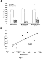
- exosomes are known to represent an important and specific route of intercellular communication, we reasoned that tumor-derived exosomes may differ from circulating exosomes in normal physiological conditions. Recently, it has been reported that prostasomes (membrane vesicles secreted by prostate cancer cells) isolated from prostate carcinoma PC-3 cell line contain caveolin-1 protein, the major component of caveolae. It is also known that the serum levels of caveolin-1 are elevated in prostate cancer patients compared with healthy subjects (Tahir, 2003 #21). However, there is no prior art suggesting association of this protein with membrane vesicles in the blood. Therefore, we evaluated the presence of caveolin-1 on exosomes obtained from plasma of SCID mice engrafted with melanoma tumors.
- Cav1 is strongly expressed on exosomes secreted by human melanoma cells in vitro, while undetectable on both cellular extracts and exosomes from normal human cells such as for instance primary monocyte-derived macrophages (MDM).
- MDM primary monocyte-derived macrophages
- Cav 1 was detected in exosome preparations derived from plasma of SCID mice engrafted with melanoma tumors by Western blot ( Fig. 3B ), flow cytometry ( Fig 3C ) and ExoTest ( Fig. 3D ), while Cav1 was undetectable in plasma-derived exosomes from control animals ( Figs. 3B, 3D ).
- other tumor markers such as MelanA/Mart-1 for melanoma and CEA for CRC, could be used for detecting the in vivo release of tumor exosomes in tumor-bearing SCID mice by ExoTest, with results comparable with those obtained with Cavl.
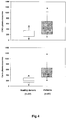
- CD63+ and Cav1+ exo are plasmatic exosomes expressed as OD450 x 1000.
- ExoTest test allowed the detection of exosomal proteins in plasma-purified exosomes from both melanoma patients and healthy donors ( Fig. 4A ), with up to 4 folds exosome levels in plasma of melanoma patients as compared to healthy donors (p ⁇ 0.001).
- cut-off mean +/- 2SD in healthy donors' samples
- stage III-IV Most of the patients included into the analysis (57/62) were affected by advance disease (stage III-IV). A wide distribution of plasmatic exosome levels was detected by ExoTest in all disease stages, which suggest that the variability in the amount of exosomes present in peripheral circulation of different patients may reflect diverse levels of tumor aggressiveness and may therefore become a novel independent prognostic factor for melanoma.
- AJCC American Joint Committee on Cancer
- Whole plasma can be used for exosome quantification.
- ExoTest for clinical purposes prompted us to verify whether ExoTest could be used for exosome detection in unfractionated biological fluids that would allow an easy and reproducible analysis avoiding the steps of ultracentrifugation. Therefore, we compared the detection and quantification of CD63+ exosomes from unfractionated samples (cell culture supernatants from human macrophages and melanoma cells, and human plasma) and exosomes purified from the same samples. In order to increase the sensitivity of the test, for these specific experiments the HRP-conjugated Mab was incubated for 30 minutes instead of 15 minutes. As is shown in Fig.
Landscapes
- Health & Medical Sciences (AREA)
- Life Sciences & Earth Sciences (AREA)
- Engineering & Computer Science (AREA)
- Immunology (AREA)
- Biomedical Technology (AREA)
- Molecular Biology (AREA)
- Chemical & Material Sciences (AREA)
- Hematology (AREA)
- Urology & Nephrology (AREA)
- Physics & Mathematics (AREA)
- General Physics & Mathematics (AREA)
- Cell Biology (AREA)
- Food Science & Technology (AREA)
- Medicinal Chemistry (AREA)
- Biotechnology (AREA)
- Analytical Chemistry (AREA)
- Biochemistry (AREA)
- General Health & Medical Sciences (AREA)
- Microbiology (AREA)
- Pathology (AREA)
- Oncology (AREA)
- Virology (AREA)
- Hospice & Palliative Care (AREA)
- AIDS & HIV (AREA)
- Tropical Medicine & Parasitology (AREA)
- Measuring Or Testing Involving Enzymes Or Micro-Organisms (AREA)
- Investigating Or Analysing Biological Materials (AREA)
- Peptides Or Proteins (AREA)
Claims (12)
- Procédé permettant de quantifier et de qualifier des exosomes dans des échantillons dérivés de cellules humaines ou dans un fluide corporel, ledit procédé comprenant les étapes suivantes :a) la capture des exosomes de l'échantillon dérivé de cellules humaines ou du fluide corporel avec un anticorps primaire anti-Rab 5b ;b) la détection des exosomes liés avec un anticorps de détection, ledit anticorps de détection se liant à un antigène exosomique ;c) l'étape permettant à un anticorps secondaire lié à une enzyme de réagir avec l'anticorps de détection ;d) l'addition de substrat ; ete) la détection de la réaction.
- Procédé selon la revendication 1, dans lequel l'anticorps de détection est un anticorps anti-CD63 ou anti-cavéoline-1.
- Procédé selon la revendication 1 ou 2, le procédé comprenant une étape de purification d'une préparation exosomique à partir de l'échantillon dérivé de cellules humaines ou du fluide corporel, et l'étape a) étant réalisée avec des exosomes purifiés.
- Procédé selon la revendication 1 ou 2, dans lequel le fluide corporel est un échantillon de plasma humain, l'ascite, les fluides cérébraux, la moelle osseuse, l'urine, les selles ou le liquide de lavage bronchoalvéolaire.
- Procédé selon la revendication 4, dans lequel le volume du fluide corporel utilisé est inférieur à 2 ml.
- Procédé non invasif de surveillance de la croissance tumorale, ledit procédé comprenant les étapes suivantes :a) l'analyse périodique d'un échantillon de fluide corporel d'un patient ;b) la capture des exosomes de l'échantillon de fluide corporel avec un anticorps primaire, ledit anticorps primaire étant un anticorps anti-Rab-5b ;c) la détection des exosomes liés avec un anticorps de détection, ledit anticorps de détection étant un anticorps anti-CD63 ou anti-cavéoline-1 ;d) l'étape permettant à un anticorps secondaire lié à une enzyme de réagir avec l'anticorps de détection ;e) l'addition de substrat ;f) la détection de la réaction ; etg) l'établissement d'une corrélation entre la quantité des exosomes détectés et la taille de la tumeur.
- Procédé selon la revendication 6, le procédé comprenant la purification de la préparation exosomique à partir de l'échantillon de fluide corporel et l'étape c) étant réalisée avec la préparation exosomique purifiée.
- Procédé selon la revendication 6, dans lequel l'échantillon de fluide corporel est un échantillon de plasma.
- Procédé selon la revendication 6, dans lequel la tumeur est une tumeur de mélanome.
- Procédé de diagnostic d'une tumeur, par la détection des exosomes porteurs de la cavéoline-1 en utilisant le procédé selon la revendication 1, dans lequel l'anticorps de détection est un anticorps anti-cavéoline-1.
- Procédé selon la revendication 10, dans lequel la tumeur est une tumeur de mélanome.
- Kit de test pour la quantification et la qualification des exosomes dans des échantillons dérivés de cellules humaines ou dans un fluide corporel, ledit kit comprenant :a) une préparation d'anticorps primaire pour la capture des exosomes des échantillons dérivés de cellules, ou du fluide corporel, ledit anticorps primaire étant un anticorps anti-Rab 5b ;b) une préparation d'anticorps de détection pour la détection des exosomes liés, ledit anticorps de détection étant un anticorps anti-CD63 ou anti-cavéoline-1 ;c) une préparation d'anticorps secondaire lié à une enzyme pour une réaction avec l'anticorps de détection ; etd) un substrat pour l'enzyme.
Priority Applications (1)
| Application Number | Priority Date | Filing Date | Title |
|---|---|---|---|
| PL09704302T PL2245458T3 (pl) | 2008-01-25 | 2009-01-26 | Nowy sposób pomiaru i charakterystyki mikropęcherzyków w ludzkich płynach ustrojowych |
Applications Claiming Priority (3)
| Application Number | Priority Date | Filing Date | Title |
|---|---|---|---|
| US6252808P | 2008-01-25 | 2008-01-25 | |
| US12/321,412 US8617806B2 (en) | 2008-01-25 | 2009-01-21 | Method to measure and characterize microvesicles in the human body fluids |
| PCT/EE2009/000002 WO2009092386A2 (fr) | 2008-01-25 | 2009-01-26 | Nouveau procédé de mesure et de caractérisation de microvésicules dans des liquides organiques humain |
Publications (2)
| Publication Number | Publication Date |
|---|---|
| EP2245458A2 EP2245458A2 (fr) | 2010-11-03 |
| EP2245458B1 true EP2245458B1 (fr) | 2016-10-26 |
Family
ID=41064551
Family Applications (1)
| Application Number | Title | Priority Date | Filing Date |
|---|---|---|---|
| EP09704302.0A Not-in-force EP2245458B1 (fr) | 2008-01-25 | 2009-01-26 | Nouveau procédé de mesure et de caractérisation de microvésicules dans des liquides organiques humain |
Country Status (12)
| Country | Link |
|---|---|
| US (1) | US8617806B2 (fr) |
| EP (1) | EP2245458B1 (fr) |
| JP (1) | JP5583600B2 (fr) |
| CN (1) | CN102317778B (fr) |
| AU (1) | AU2009207927B2 (fr) |
| BR (1) | BRPI0906683B8 (fr) |
| CA (1) | CA2713196C (fr) |
| ES (1) | ES2613670T3 (fr) |
| MX (1) | MX2010008164A (fr) |
| PL (1) | PL2245458T3 (fr) |
| RU (1) | RU2520741C2 (fr) |
| WO (1) | WO2009092386A2 (fr) |
Cited By (2)
| Publication number | Priority date | Publication date | Assignee | Title |
|---|---|---|---|---|
| DE102021208893B3 (de) | 2021-08-13 | 2022-11-17 | Hahn-Schickard-Gesellschaft für angewandte Forschung e.V. | Isolieren von Analyten unterschiedlicher Analyt-Klassen |
| EP3978925A4 (fr) * | 2019-05-24 | 2023-06-28 | Sol Bio Corporation | Système et procédé de séparation par affinité mettant en oeuvre une réaction d'adhérence de type substituable |
Families Citing this family (54)
| Publication number | Priority date | Publication date | Assignee | Title |
|---|---|---|---|---|
| CA2733672C (fr) | 2007-08-16 | 2018-09-11 | The Royal Institution For The Advancement Of Learning/Mcgill University | Microvesicules issues d'une cellule tumorale |
| US20100255514A1 (en) * | 2007-08-16 | 2010-10-07 | The Royal Institution For The Advancement Of Learning/Mcgill University | Tumor cell-derived microvesicles |
| AU2009212543B2 (en) * | 2008-02-01 | 2015-07-09 | The General Hospital Corporation | Use of microvesicles in diagnosis, prognosis and treatment of medical diseases and conditions |
| US10545149B2 (en) * | 2008-10-06 | 2020-01-28 | Morehouse School Of Medicine | Detection of HIV-related proteins in urine |
| US9487837B2 (en) | 2008-10-06 | 2016-11-08 | Morehouse School Of Medicine | Exosome-mediated diagnosis of hepatitis virus infections and diseases |
| WO2010062706A2 (fr) | 2008-10-30 | 2010-06-03 | Caris Mpi, Inc. | Procédés d'évaluation de motifs arn |
| BRPI0921043A2 (pt) | 2008-11-12 | 2018-08-07 | Caris Life Sciences Luxembourg Holdings | métodos e sistemas para usar exossomas para determinar fenótipos |
| EP3461912B1 (fr) | 2009-09-09 | 2022-07-13 | The General Hospital Corporation | Utilisation de microvésicules dans l'analyse de profils d'acides nucléiques |
| US20130029339A1 (en) | 2009-09-09 | 2013-01-31 | The General Hospital Corporation | Use of microvesicles in analyzing kras mutations |
| US20130203061A1 (en) * | 2009-11-30 | 2013-08-08 | Michael KLASS | Methods and systems for isolating, storing, and analyzing vesicles |
| EP2542696B1 (fr) * | 2010-03-01 | 2016-09-28 | Caris Life Sciences Switzerland Holdings GmbH | Biomarqueurs pour théranostique |
| EP2556172A4 (fr) | 2010-04-06 | 2013-10-30 | Caris Life Sciences Luxembourg Holdings | Biomarqueurs circulants pour une maladie |
| EP2591359B1 (fr) * | 2010-07-07 | 2017-03-01 | Aethlon Medical Inc | Procédés permettant de quantifier des exosomes |
| WO2012031008A2 (fr) | 2010-08-31 | 2012-03-08 | The General Hospital Corporation | Matières biologiques liées au cancer dans des microvésicules |
| WO2012048372A1 (fr) * | 2010-10-11 | 2012-04-19 | Medsaic Pty Ltd | Essai pour la détection d'une maladie |
| US20130295574A1 (en) | 2010-11-10 | 2013-11-07 | Exosome Diagnostics, Inc. | Method for Isolation of Nucleic Acid Containing Particles and Extraction of Nucleic Acids Therefrom |
| FR2973029B1 (fr) * | 2011-03-23 | 2013-04-26 | Centre Nat Rech Scient | Complexes metalliques dinucleaires greffes, et leur utilisation en tant que capteurs de microparticules cellulaires |
| US9816998B2 (en) | 2011-04-01 | 2017-11-14 | Cornell University | Circulating exosomes as diagnostic/prognostic indicators and therapeutic targets of melanoma and other cancers |
| WO2012162563A2 (fr) * | 2011-05-24 | 2012-11-29 | The Regents Of The University Of California | Procédé de détection de biomarqueur exosomal par libération et mesure induites par un champ électrique |
| KR20140067001A (ko) * | 2011-08-08 | 2014-06-03 | 카리스 라이프 사이언스 룩셈부르크 홀딩스, 에스.에이.알.엘. | 생물지표 조성물 및 방법 |
| EP2801822B1 (fr) * | 2011-12-22 | 2017-08-30 | Theoria Science Inc. | Procédé d'analyse d'exosome, réactif pour l'utilisation dans l'analyse d'exosome, et appareil d'analyse d'exosome |
| WO2014030590A1 (fr) * | 2012-08-24 | 2014-02-27 | 国立大学法人東京大学 | Méthode d'analyse d'un exosome, appareil d'analyse d'un exosome, complexe anticorps-exosome et puce pour l'électrophorèse d'un exosome |
| KR101933620B1 (ko) * | 2012-09-18 | 2018-12-28 | 삼성전자주식회사 | 소포를 검출하기 위한 조성물, 키트 및 이를 이용하여 소포를 분석하는 방법 |
| WO2014078420A1 (fr) * | 2012-11-13 | 2014-05-22 | Allan Wu | Procédés et systèmes de traitement d'exosomes |
| KR102257912B1 (ko) | 2013-03-13 | 2021-05-27 | 메소 스케일 테크놀러지즈, 엘엘시 | 개선된 분석 방법 |
| PT2972193T (pt) | 2013-03-13 | 2020-04-23 | Univ Miami | Método para isolamento e purificação de microvesículas de sobrenadantes de cultura celular e fluidos biológicos |
| US10114015B2 (en) | 2013-03-13 | 2018-10-30 | Meso Scale Technologies, Llc. | Assay methods |
| JP6210004B2 (ja) | 2013-04-09 | 2017-10-11 | 株式会社Jvcケンウッド | 試料分析用デバイス及びエクソソームの捕捉方法 |
| JPWO2015029979A1 (ja) * | 2013-08-30 | 2017-03-02 | 国立大学法人 東京大学 | エキソソームの分析方法、エキソソーム分析チップ、及びエキソソーム分析装置 |
| JPWO2015045666A1 (ja) | 2013-09-25 | 2017-03-09 | 国立大学法人 東京大学 | 流体デバイス、エキソソームの分析方法、生体分子分析方法及び生体分子検出方法 |
| US10139402B2 (en) | 2013-11-06 | 2018-11-27 | Jsr Corporation | Separation method, detection method, signal measurement method, method for determining disease, method for evaluating drug efficacy of disease treatment drug, kit, and liquid composition |
| EP3078889B1 (fr) * | 2013-12-06 | 2019-09-18 | The University of Tokyo | Clapet, structure de régulation de fluide, dispositif à fluide et procédé de fabrication de clapet |
| CN112239774A (zh) | 2014-05-15 | 2021-01-19 | 中尺度技术有限责任公司 | 改进的测定方法 |
| EP3152576B1 (fr) * | 2014-06-06 | 2018-09-12 | Exosomics Siena S.p.A. | Utilisation de tm9sf4 comme biomarqueur pour les exosomes associeés aux tumeurs |
| RU2556825C1 (ru) * | 2014-09-15 | 2015-07-20 | Федеральное государственное бюджетное учреждение науки Институт химической биологии и фундаментальной медицины Сибирского отделения Российской академии наук (ИХБФМ СО РАН) | Способ получения экзосом из крови |
| US20160320390A1 (en) * | 2015-05-01 | 2016-11-03 | Morehouse School Of Medicine | Compositions and methods for capturing exosomes |
| CN108271413A (zh) * | 2015-05-20 | 2018-07-10 | Jsr株式会社 | 分离方法、检测方法、信号测定方法、疾病的判定方法、药效评价方法、试剂盒、液态组合物以及检体稀释液 |
| US10345310B2 (en) | 2015-06-09 | 2019-07-09 | The Board Of Regents Of The University Of Texas System | Diagnostic test for early stage cancer |
| JP6606916B2 (ja) | 2015-08-21 | 2019-11-20 | 株式会社Jvcケンウッド | エクソソームの捕捉方法 |
| GB201518466D0 (en) * | 2015-10-19 | 2015-12-02 | Cizzle Biotechnology Ltd | Use |
| CN106093412A (zh) * | 2016-06-28 | 2016-11-09 | 叶瑞东 | 一种多肽分子在制备诊断急性脑梗死后微出血的试剂盒中的应用 |
| CN109891236A (zh) * | 2016-10-13 | 2019-06-14 | 合同会社明乐康中央研究所 | 回收胞外囊泡的方法 |
| RU2682721C2 (ru) * | 2016-11-17 | 2019-03-21 | Федеральное Государственное Бюджетное Учреждение Науки Институт Молекулярной Биологии Им. В.А. Энгельгардта Российской Академии Наук (Имб Ран) | Биологический микрочип для обнаружения опухолевых экзосом в сыворотке крови человека для диагностики колоректального рака |
| JP6797427B2 (ja) * | 2017-01-12 | 2020-12-09 | テオリアサイエンス株式会社 | 被検者が膵臓癌に罹患している可能性を試験する方法 |
| CN106906294A (zh) * | 2017-03-31 | 2017-06-30 | 浙江大学 | 一种胞外囊泡/脂质体的定量方法 |
| CN107144688B (zh) * | 2017-04-07 | 2018-09-11 | 浙江大学 | Cd19阳性外泌体作为分子标记在制备肿瘤诊断试剂盒中的应用及试剂盒 |
| JP6573351B2 (ja) | 2017-05-29 | 2019-09-11 | 国立大学法人神戸大学 | 検出対象の分析用センサ作製用基材、検出対象の分析用センサ、及び検出対象の分析法 |
| DE102017005543A1 (de) * | 2017-06-13 | 2018-12-13 | Forschungszentrum Jülich GmbH | Verfahren zum Nachweis von Extrazellulären Vesikeln in einer Probe |
| CN107907689A (zh) * | 2017-10-10 | 2018-04-13 | 北京大学 | 外泌体蛋白cd5l的检测方法 |
| KR101968046B1 (ko) * | 2018-07-19 | 2019-04-11 | (주) 바이오인프라생명과학 | 암의 조기 진단을 위한 복합 바이오마커 |
| JP6677284B2 (ja) * | 2018-10-09 | 2020-04-08 | 凸版印刷株式会社 | 分析対象物の検出方法及びラテラルフロー用テストストリップ |
| CN109991427A (zh) * | 2019-04-08 | 2019-07-09 | 王延博 | 一种用于检测血清中外泌体表面蛋白标志物的试剂盒 |
| CN110095604B (zh) * | 2019-04-12 | 2022-03-18 | 南方医科大学南方医院 | Caveolin-1蛋白阳性外泌体作为非小细胞肺癌诊断标志物的应用 |
| CN111537725A (zh) * | 2020-05-29 | 2020-08-14 | 武汉大学 | 高效定量检测细胞外囊泡中pd-1水平的方法、elisa试剂盒及使用方法 |
Family Cites Families (12)
| Publication number | Priority date | Publication date | Assignee | Title |
|---|---|---|---|---|
| JPH081438B2 (ja) * | 1986-05-13 | 1996-01-10 | 三洋化成工業株式会社 | 酵素免疫測定法 |
| EP1004664A1 (fr) * | 1998-11-24 | 2000-05-31 | Institut National De La Sante Et De La Recherche Medicale (Inserm) | Compositions et procédés utilisant la lactadhérine ou des dérivés de celle-ci |
| GB9927320D0 (en) * | 1999-11-18 | 2000-01-12 | Chiron Spa | Exosome separation |
| US6812023B1 (en) * | 2000-04-27 | 2004-11-02 | Anosys, Inc. | Methods of producing membrane vesicles |
| US20040241176A1 (en) * | 2000-04-27 | 2004-12-02 | Ap Cells. Inc. | Method of producing membrane vesicles |
| US7442777B2 (en) * | 2000-11-29 | 2008-10-28 | Arius Research Inc. | Cytotoxicity mediation of cells evidencing surface expression of CD63 |
| US20030190602A1 (en) * | 2001-03-12 | 2003-10-09 | Monogen, Inc. | Cell-based detection and differentiation of disease states |
| WO2003063690A2 (fr) | 2002-01-31 | 2003-08-07 | Baylor College Of Medicine | Caveoline secretee tenant lieu de marqueur pour le cancer de la prostate |
| CA2453198A1 (fr) * | 2004-01-07 | 2005-07-07 | Wei-Ping Min | Quantification et production d'exosomes immunosuppresseurs |
| JP2005237234A (ja) * | 2004-02-25 | 2005-09-08 | Japan Science & Technology Agency | カベオリン抗原とその用途 |
| AU2007243184A1 (en) * | 2006-04-28 | 2007-11-08 | University Of Iowa Research Foundation | Methods and compounds to modulate parvovirus transduction of mammalian cells or to alter virus infection, method to identify a viral receptor or co-receptor |
| US20120058492A1 (en) * | 2008-01-25 | 2012-03-08 | Hansabiomed Ou | Method and a Kit To Detect Malignant Tumors and Provide a Prognosis |
-
2009
- 2009-01-21 US US12/321,412 patent/US8617806B2/en not_active Expired - Fee Related
- 2009-01-26 WO PCT/EE2009/000002 patent/WO2009092386A2/fr active Application Filing
- 2009-01-26 EP EP09704302.0A patent/EP2245458B1/fr not_active Not-in-force
- 2009-01-26 BR BRPI0906683A patent/BRPI0906683B8/pt not_active IP Right Cessation
- 2009-01-26 ES ES09704302.0T patent/ES2613670T3/es active Active
- 2009-01-26 MX MX2010008164A patent/MX2010008164A/es active IP Right Grant
- 2009-01-26 CA CA2713196A patent/CA2713196C/fr not_active Expired - Fee Related
- 2009-01-26 PL PL09704302T patent/PL2245458T3/pl unknown
- 2009-01-26 AU AU2009207927A patent/AU2009207927B2/en not_active Ceased
- 2009-01-26 RU RU2010135526/15A patent/RU2520741C2/ru active
- 2009-01-26 JP JP2010543379A patent/JP5583600B2/ja not_active Expired - Fee Related
- 2009-01-26 CN CN200980111452.4A patent/CN102317778B/zh not_active Expired - Fee Related
Cited By (3)
| Publication number | Priority date | Publication date | Assignee | Title |
|---|---|---|---|---|
| EP3978925A4 (fr) * | 2019-05-24 | 2023-06-28 | Sol Bio Corporation | Système et procédé de séparation par affinité mettant en oeuvre une réaction d'adhérence de type substituable |
| DE102021208893B3 (de) | 2021-08-13 | 2022-11-17 | Hahn-Schickard-Gesellschaft für angewandte Forschung e.V. | Isolieren von Analyten unterschiedlicher Analyt-Klassen |
| WO2023017049A1 (fr) | 2021-08-13 | 2023-02-16 | Hahn-Schickard-Gesellschaft für angewandte Forschung e.V. | Isolement d'analytes de différentes classes d'analytes |
Also Published As
| Publication number | Publication date |
|---|---|
| EP2245458A2 (fr) | 2010-11-03 |
| US20090220944A1 (en) | 2009-09-03 |
| RU2520741C2 (ru) | 2014-06-27 |
| CN102317778B (zh) | 2015-05-13 |
| CA2713196C (fr) | 2014-06-17 |
| WO2009092386A3 (fr) | 2009-09-17 |
| CA2713196A1 (fr) | 2009-07-30 |
| MX2010008164A (es) | 2010-11-25 |
| ES2613670T3 (es) | 2017-05-25 |
| BRPI0906683B8 (pt) | 2021-07-27 |
| AU2009207927B2 (en) | 2015-04-09 |
| BRPI0906683A2 (pt) | 2017-08-22 |
| AU2009207927A1 (en) | 2009-07-30 |
| CN102317778A (zh) | 2012-01-11 |
| WO2009092386A2 (fr) | 2009-07-30 |
| US8617806B2 (en) | 2013-12-31 |
| PL2245458T3 (pl) | 2017-08-31 |
| RU2010135526A (ru) | 2012-02-27 |
| WO2009092386A8 (fr) | 2010-10-14 |
| JP5583600B2 (ja) | 2014-09-03 |
| JP2011510309A (ja) | 2011-03-31 |
| BRPI0906683B1 (pt) | 2021-03-02 |
Similar Documents
| Publication | Publication Date | Title |
|---|---|---|
| EP2245458B1 (fr) | Nouveau procédé de mesure et de caractérisation de microvésicules dans des liquides organiques humain | |
| Dusoswa et al. | Glycan modification of glioblastoma-derived extracellular vesicles enhances receptor-mediated targeting of dendritic cells | |
| Muhsin-Sharafaldine et al. | Procoagulant and immunogenic properties of melanoma exosomes, microvesicles and apoptotic vesicles | |
| Boukouris et al. | Exosomes in bodily fluids are a highly stable resource of disease biomarkers | |
| Ludwig et al. | Suppression of lymphocyte functions by plasma exosomes correlates with disease activity in patients with head and neck cancer | |
| Szczepanski et al. | Increased frequency and suppression by regulatory T cells in patients with acute myelogenous leukemia | |
| Laurent et al. | Distribution, function, and prognostic value of cytotoxic T lymphocytes in follicular lymphoma: a 3-D tissue-imaging study | |
| Chang et al. | Inflammation-associated lysophospholipids as ligands for CD1d-restricted T cells in human cancer | |
| Schuler et al. | Human CD4+ CD39+ regulatory T cells produce adenosine upon co-expression of surface CD73 or contact with CD73+ exosomes or CD73+ cells | |
| Welton et al. | Proteomics analysis of bladder cancer exosomes | |
| Jahrsdörfer et al. | Granzyme B produced by human plasmacytoid dendritic cells suppresses T-cell expansion | |
| Bergmann et al. | Tumor‐derived microvesicles in sera of patients with head and neck cancer and their role in tumor progression | |
| Frisullo et al. | Regulatory T cells fail to suppress CD4+ T‐bet+ T cells in relapsing multiple sclerosis patients | |
| Emara et al. | Retagging identifies dendritic cell-specific intercellular adhesion molecule-3 (ICAM3)-grabbing non-integrin (DC-SIGN) protein as a novel receptor for a major allergen from house dust mite | |
| US20160370265A1 (en) | Method for isolating exosomes | |
| US11561223B2 (en) | Method and system for identifying membrane proteins on extracellular vesicles | |
| Kim et al. | Expression of pro-and antiapoptotic proteins in circulating CD8+ T cells of patients with squamous cell carcinoma of the head and neck | |
| Nguyen et al. | Novel characterisation of mast cell phenotypes from peripheral blood mononuclear cells in chronic fatigue syndrome/myalgic encephalomyelitis patients | |
| Tzaridis et al. | A novel serum extracellular vesicle protein signature to monitor glioblastoma tumor progression | |
| Ismail et al. | Pretransplant detection of anti-endothelial cell antibodies could predict renal allograft outcome | |
| Siegel et al. | Internalization of therapeutic antibodies into Dendritic cells as a risk factor for immunogenicity | |
| NL2033706B1 (en) | Identification and/or isolation of T cell-comprising cell-cell complexes and use thereof. | |
| WO2020162441A1 (fr) | Biomarqueur de maladie granulomateuse | |
| Gómez-Herranz et al. | Major Histocompatibility Complex class I heavy chains localize in both cytoplasmic and nuclear compartment | |
| Ham | The role of breast cancer derived-exosomes in the tumour micro-environment |
Legal Events
| Date | Code | Title | Description |
|---|---|---|---|
| PUAI | Public reference made under article 153(3) epc to a published international application that has entered the european phase |
Free format text: ORIGINAL CODE: 0009012 |
|
| 17P | Request for examination filed |
Effective date: 20100809 |
|
| AK | Designated contracting states |
Kind code of ref document: A2 Designated state(s): AT BE BG CH CY CZ DE DK EE ES FI FR GB GR HR HU IE IS IT LI LT LU LV MC MK MT NL NO PL PT RO SE SI SK TR |
|
| AX | Request for extension of the european patent |
Extension state: AL BA RS |
|
| DAX | Request for extension of the european patent (deleted) | ||
| 17Q | First examination report despatched |
Effective date: 20121026 |
|
| GRAP | Despatch of communication of intention to grant a patent |
Free format text: ORIGINAL CODE: EPIDOSNIGR1 |
|
| RIC1 | Information provided on ipc code assigned before grant |
Ipc: G01N 33/567 20060101ALI20160223BHEP Ipc: G01N 33/569 20060101ALI20160223BHEP Ipc: G01N 33/53 20060101AFI20160223BHEP Ipc: G01N 33/574 20060101ALI20160223BHEP |
|
| INTG | Intention to grant announced |
Effective date: 20160331 |
|
| GRAS | Grant fee paid |
Free format text: ORIGINAL CODE: EPIDOSNIGR3 |
|
| GRAA | (expected) grant |
Free format text: ORIGINAL CODE: 0009210 |
|
| AK | Designated contracting states |
Kind code of ref document: B1 Designated state(s): AT BE BG CH CY CZ DE DK EE ES FI FR GB GR HR HU IE IS IT LI LT LU LV MC MK MT NL NO PL PT RO SE SI SK TR |
|
| REG | Reference to a national code |
Ref country code: GB Ref legal event code: FG4D |
|
| REG | Reference to a national code |
Ref country code: CH Ref legal event code: EP |
|
| REG | Reference to a national code |
Ref country code: AT Ref legal event code: REF Ref document number: 840379 Country of ref document: AT Kind code of ref document: T Effective date: 20161115 |
|
| REG | Reference to a national code |
Ref country code: IE Ref legal event code: FG4D |
|
| REG | Reference to a national code |
Ref country code: DE Ref legal event code: R096 Ref document number: 602009041927 Country of ref document: DE |
|
| REG | Reference to a national code |
Ref country code: RO Ref legal event code: EPE |
|
| RAP2 | Party data changed (patent owner data changed or rights of a patent transferred) |
Owner name: EXOSOMICS SIENA S.P.A. |
|
| REG | Reference to a national code |
Ref country code: DE Ref legal event code: R081 Ref document number: 602009041927 Country of ref document: DE Owner name: EXOSOMICS S.P.A., IT Free format text: FORMER OWNER: HANSABIOMED OUE, HAAPSALU, EE Ref country code: DE Ref legal event code: R081 Ref document number: 602009041927 Country of ref document: DE Owner name: EXOSOMICS SIENA S.P.A., IT Free format text: FORMER OWNER: HANSABIOMED OUE, HAAPSALU, EE |
|
| REG | Reference to a national code |
Ref country code: FR Ref legal event code: PLFP Year of fee payment: 9 |
|
| REG | Reference to a national code |
Ref country code: CH Ref legal event code: NV Representative=s name: JACOBACCI AND PARTNERS SA, CH Ref country code: CH Ref legal event code: PUE Owner name: EXOSOMICS SIENA S.P.A., IT Free format text: FORMER OWNER: HANSABIOMED OUE, EE |
|
| REG | Reference to a national code |
Ref country code: SE Ref legal event code: TRGR |
|
| REG | Reference to a national code |
Ref country code: LT Ref legal event code: MG4D |
|
| PG25 | Lapsed in a contracting state [announced via postgrant information from national office to epo] |
Ref country code: LV Free format text: LAPSE BECAUSE OF FAILURE TO SUBMIT A TRANSLATION OF THE DESCRIPTION OR TO PAY THE FEE WITHIN THE PRESCRIBED TIME-LIMIT Effective date: 20161026 |
|
| REG | Reference to a national code |
Ref country code: NL Ref legal event code: MP Effective date: 20161026 |
|
| REG | Reference to a national code |
Ref country code: AT Ref legal event code: MK05 Ref document number: 840379 Country of ref document: AT Kind code of ref document: T Effective date: 20161026 |
|
| PG25 | Lapsed in a contracting state [announced via postgrant information from national office to epo] |
Ref country code: LT Free format text: LAPSE BECAUSE OF FAILURE TO SUBMIT A TRANSLATION OF THE DESCRIPTION OR TO PAY THE FEE WITHIN THE PRESCRIBED TIME-LIMIT Effective date: 20161026 Ref country code: GR Free format text: LAPSE BECAUSE OF FAILURE TO SUBMIT A TRANSLATION OF THE DESCRIPTION OR TO PAY THE FEE WITHIN THE PRESCRIBED TIME-LIMIT Effective date: 20170127 Ref country code: NO Free format text: LAPSE BECAUSE OF FAILURE TO SUBMIT A TRANSLATION OF THE DESCRIPTION OR TO PAY THE FEE WITHIN THE PRESCRIBED TIME-LIMIT Effective date: 20170126 |
|
| REG | Reference to a national code |
Ref country code: GB Ref legal event code: 732E Free format text: REGISTERED BETWEEN 20170406 AND 20170412 |
|
| REG | Reference to a national code |
Ref country code: ES Ref legal event code: FG2A Ref document number: 2613670 Country of ref document: ES Kind code of ref document: T3 Effective date: 20170525 |
|
| PG25 | Lapsed in a contracting state [announced via postgrant information from national office to epo] |
Ref country code: IS Free format text: LAPSE BECAUSE OF FAILURE TO SUBMIT A TRANSLATION OF THE DESCRIPTION OR TO PAY THE FEE WITHIN THE PRESCRIBED TIME-LIMIT Effective date: 20170226 Ref country code: FI Free format text: LAPSE BECAUSE OF FAILURE TO SUBMIT A TRANSLATION OF THE DESCRIPTION OR TO PAY THE FEE WITHIN THE PRESCRIBED TIME-LIMIT Effective date: 20161026 Ref country code: PT Free format text: LAPSE BECAUSE OF FAILURE TO SUBMIT A TRANSLATION OF THE DESCRIPTION OR TO PAY THE FEE WITHIN THE PRESCRIBED TIME-LIMIT Effective date: 20170227 Ref country code: AT Free format text: LAPSE BECAUSE OF FAILURE TO SUBMIT A TRANSLATION OF THE DESCRIPTION OR TO PAY THE FEE WITHIN THE PRESCRIBED TIME-LIMIT Effective date: 20161026 Ref country code: BE Free format text: LAPSE BECAUSE OF FAILURE TO SUBMIT A TRANSLATION OF THE DESCRIPTION OR TO PAY THE FEE WITHIN THE PRESCRIBED TIME-LIMIT Effective date: 20161026 Ref country code: NL Free format text: LAPSE BECAUSE OF FAILURE TO SUBMIT A TRANSLATION OF THE DESCRIPTION OR TO PAY THE FEE WITHIN THE PRESCRIBED TIME-LIMIT Effective date: 20161026 Ref country code: HR Free format text: LAPSE BECAUSE OF FAILURE TO SUBMIT A TRANSLATION OF THE DESCRIPTION OR TO PAY THE FEE WITHIN THE PRESCRIBED TIME-LIMIT Effective date: 20161026 |
|
| REG | Reference to a national code |
Ref country code: DE Ref legal event code: R097 Ref document number: 602009041927 Country of ref document: DE |
|
| PG25 | Lapsed in a contracting state [announced via postgrant information from national office to epo] |
Ref country code: SK Free format text: LAPSE BECAUSE OF FAILURE TO SUBMIT A TRANSLATION OF THE DESCRIPTION OR TO PAY THE FEE WITHIN THE PRESCRIBED TIME-LIMIT Effective date: 20161026 Ref country code: DK Free format text: LAPSE BECAUSE OF FAILURE TO SUBMIT A TRANSLATION OF THE DESCRIPTION OR TO PAY THE FEE WITHIN THE PRESCRIBED TIME-LIMIT Effective date: 20161026 Ref country code: CZ Free format text: LAPSE BECAUSE OF FAILURE TO SUBMIT A TRANSLATION OF THE DESCRIPTION OR TO PAY THE FEE WITHIN THE PRESCRIBED TIME-LIMIT Effective date: 20161026 Ref country code: EE Free format text: LAPSE BECAUSE OF FAILURE TO SUBMIT A TRANSLATION OF THE DESCRIPTION OR TO PAY THE FEE WITHIN THE PRESCRIBED TIME-LIMIT Effective date: 20161026 |
|
| PG25 | Lapsed in a contracting state [announced via postgrant information from national office to epo] |
Ref country code: BG Free format text: LAPSE BECAUSE OF FAILURE TO SUBMIT A TRANSLATION OF THE DESCRIPTION OR TO PAY THE FEE WITHIN THE PRESCRIBED TIME-LIMIT Effective date: 20170126 |
|
| PLBE | No opposition filed within time limit |
Free format text: ORIGINAL CODE: 0009261 |
|
| STAA | Information on the status of an ep patent application or granted ep patent |
Free format text: STATUS: NO OPPOSITION FILED WITHIN TIME LIMIT |
|
| PG25 | Lapsed in a contracting state [announced via postgrant information from national office to epo] |
Ref country code: MC Free format text: LAPSE BECAUSE OF FAILURE TO SUBMIT A TRANSLATION OF THE DESCRIPTION OR TO PAY THE FEE WITHIN THE PRESCRIBED TIME-LIMIT Effective date: 20161026 |
|
| 26N | No opposition filed |
Effective date: 20170727 |
|
| REG | Reference to a national code |
Ref country code: IE Ref legal event code: MM4A |
|
| PG25 | Lapsed in a contracting state [announced via postgrant information from national office to epo] |
Ref country code: SI Free format text: LAPSE BECAUSE OF FAILURE TO SUBMIT A TRANSLATION OF THE DESCRIPTION OR TO PAY THE FEE WITHIN THE PRESCRIBED TIME-LIMIT Effective date: 20161026 Ref country code: LU Free format text: LAPSE BECAUSE OF NON-PAYMENT OF DUE FEES Effective date: 20170126 |
|
| REG | Reference to a national code |
Ref country code: FR Ref legal event code: PLFP Year of fee payment: 10 |
|
| PG25 | Lapsed in a contracting state [announced via postgrant information from national office to epo] |
Ref country code: IE Free format text: LAPSE BECAUSE OF NON-PAYMENT OF DUE FEES Effective date: 20170126 |
|
| PG25 | Lapsed in a contracting state [announced via postgrant information from national office to epo] |
Ref country code: MT Free format text: LAPSE BECAUSE OF NON-PAYMENT OF DUE FEES Effective date: 20170126 |
|
| PG25 | Lapsed in a contracting state [announced via postgrant information from national office to epo] |
Ref country code: HU Free format text: LAPSE BECAUSE OF FAILURE TO SUBMIT A TRANSLATION OF THE DESCRIPTION OR TO PAY THE FEE WITHIN THE PRESCRIBED TIME-LIMIT; INVALID AB INITIO Effective date: 20090126 |
|
| REG | Reference to a national code |
Ref country code: ES Ref legal event code: PC2A Owner name: EXOSOMICS S.P.A. Effective date: 20191022 |
|
| PG25 | Lapsed in a contracting state [announced via postgrant information from national office to epo] |
Ref country code: CY Free format text: LAPSE BECAUSE OF NON-PAYMENT OF DUE FEES Effective date: 20161026 |
|
| REG | Reference to a national code |
Ref country code: DE Ref legal event code: R082 Ref document number: 602009041927 Country of ref document: DE Representative=s name: KRAMER BARSKE SCHMIDTCHEN PATENTANWAELTE PARTG, DE Ref country code: DE Ref legal event code: R081 Ref document number: 602009041927 Country of ref document: DE Owner name: EXOSOMICS S.P.A., IT Free format text: FORMER OWNER: EXOSOMICS SIENA S.P.A., SIENA, IT |
|
| REG | Reference to a national code |
Ref country code: CH Ref legal event code: PFA Owner name: EXOSOMICS S.P.A., IT Free format text: FORMER OWNER: EXOSOMICS SIENA S.P.A., IT |
|
| PG25 | Lapsed in a contracting state [announced via postgrant information from national office to epo] |
Ref country code: MK Free format text: LAPSE BECAUSE OF FAILURE TO SUBMIT A TRANSLATION OF THE DESCRIPTION OR TO PAY THE FEE WITHIN THE PRESCRIBED TIME-LIMIT Effective date: 20161026 |
|
| PGFP | Annual fee paid to national office [announced via postgrant information from national office to epo] |
Ref country code: RO Payment date: 20201224 Year of fee payment: 13 |
|
| PGFP | Annual fee paid to national office [announced via postgrant information from national office to epo] |
Ref country code: PL Payment date: 20201221 Year of fee payment: 13 |
|
| PGFP | Annual fee paid to national office [announced via postgrant information from national office to epo] |
Ref country code: FR Payment date: 20210128 Year of fee payment: 13 Ref country code: IT Payment date: 20201127 Year of fee payment: 13 Ref country code: CH Payment date: 20210120 Year of fee payment: 13 |
|
| PGFP | Annual fee paid to national office [announced via postgrant information from national office to epo] |
Ref country code: DE Payment date: 20210129 Year of fee payment: 13 Ref country code: GB Payment date: 20210121 Year of fee payment: 13 Ref country code: ES Payment date: 20210201 Year of fee payment: 13 Ref country code: SE Payment date: 20210118 Year of fee payment: 13 Ref country code: TR Payment date: 20210124 Year of fee payment: 13 |
|
| REG | Reference to a national code |
Ref country code: DE Ref legal event code: R119 Ref document number: 602009041927 Country of ref document: DE |
|
| REG | Reference to a national code |
Ref country code: SE Ref legal event code: EUG |
|
| REG | Reference to a national code |
Ref country code: CH Ref legal event code: PL |
|
| GBPC | Gb: european patent ceased through non-payment of renewal fee |
Effective date: 20220126 |
|
| PG25 | Lapsed in a contracting state [announced via postgrant information from national office to epo] |
Ref country code: SE Free format text: LAPSE BECAUSE OF NON-PAYMENT OF DUE FEES Effective date: 20220127 Ref country code: RO Free format text: LAPSE BECAUSE OF NON-PAYMENT OF DUE FEES Effective date: 20220126 Ref country code: GB Free format text: LAPSE BECAUSE OF NON-PAYMENT OF DUE FEES Effective date: 20220126 Ref country code: DE Free format text: LAPSE BECAUSE OF NON-PAYMENT OF DUE FEES Effective date: 20220802 |
|
| PG25 | Lapsed in a contracting state [announced via postgrant information from national office to epo] |
Ref country code: FR Free format text: LAPSE BECAUSE OF NON-PAYMENT OF DUE FEES Effective date: 20220131 |
|
| PG25 | Lapsed in a contracting state [announced via postgrant information from national office to epo] |
Ref country code: LI Free format text: LAPSE BECAUSE OF NON-PAYMENT OF DUE FEES Effective date: 20220131 Ref country code: CH Free format text: LAPSE BECAUSE OF NON-PAYMENT OF DUE FEES Effective date: 20220131 |
|
| PG25 | Lapsed in a contracting state [announced via postgrant information from national office to epo] |
Ref country code: IT Free format text: LAPSE BECAUSE OF NON-PAYMENT OF DUE FEES Effective date: 20220126 |
|
| REG | Reference to a national code |
Ref country code: ES Ref legal event code: FD2A Effective date: 20230329 |
|
| PG25 | Lapsed in a contracting state [announced via postgrant information from national office to epo] |
Ref country code: ES Free format text: LAPSE BECAUSE OF NON-PAYMENT OF DUE FEES Effective date: 20220127 |
|
| PG25 | Lapsed in a contracting state [announced via postgrant information from national office to epo] |
Ref country code: PL Free format text: LAPSE BECAUSE OF NON-PAYMENT OF DUE FEES Effective date: 20220126 |
|
| PG25 | Lapsed in a contracting state [announced via postgrant information from national office to epo] |
Ref country code: TR Free format text: LAPSE BECAUSE OF NON-PAYMENT OF DUE FEES Effective date: 20220126 |