EP2244083A1 - Procédé, appareil, récipient et cyto-centrifuge pour préparation cellulaire à base de liquide - Google Patents
Procédé, appareil, récipient et cyto-centrifuge pour préparation cellulaire à base de liquide Download PDFInfo
- Publication number
- EP2244083A1 EP2244083A1 EP09158694A EP09158694A EP2244083A1 EP 2244083 A1 EP2244083 A1 EP 2244083A1 EP 09158694 A EP09158694 A EP 09158694A EP 09158694 A EP09158694 A EP 09158694A EP 2244083 A1 EP2244083 A1 EP 2244083A1
- Authority
- EP
- European Patent Office
- Prior art keywords
- vessel
- cell
- cells
- liquid
- centrifugation step
- Prior art date
- Legal status (The legal status is an assumption and is not a legal conclusion. Google has not performed a legal analysis and makes no representation as to the accuracy of the status listed.)
- Ceased
Links
Images


Classifications
-
- G—PHYSICS
- G01—MEASURING; TESTING
- G01N—INVESTIGATING OR ANALYSING MATERIALS BY DETERMINING THEIR CHEMICAL OR PHYSICAL PROPERTIES
- G01N1/00—Sampling; Preparing specimens for investigation
- G01N1/28—Preparing specimens for investigation including physical details of (bio-)chemical methods covered elsewhere, e.g. G01N33/50, C12Q
- G01N1/2813—Producing thin layers of samples on a substrate, e.g. smearing, spinning-on
-
- G—PHYSICS
- G01—MEASURING; TESTING
- G01N—INVESTIGATING OR ANALYSING MATERIALS BY DETERMINING THEIR CHEMICAL OR PHYSICAL PROPERTIES
- G01N1/00—Sampling; Preparing specimens for investigation
- G01N1/28—Preparing specimens for investigation including physical details of (bio-)chemical methods covered elsewhere, e.g. G01N33/50, C12Q
- G01N1/34—Purifying; Cleaning
Definitions
- the present invention relates to a method and an apparatus for liquid based cell preparation. Further, the present invention relates to a vessel and a cytocentrifuge comprising such a vessel, in particular for the use in a method for liquid based cell preparation.
- Cytology is a big and fast growing branch of pathology. For instance, millions of microscope slides are prepared with cells in screening programs for cervical cancer. These preparations are used to be called “smears", after the way they are prepared. A brush is used to collect cells and this brush is smeared out on a microscope slide. Disadvantages are contamination of the brush with blood, mucus and also loss of cells that stayed behind on the bristles of the brush after making the smear.
- the cells are either collected by a special filtration technique (ThinPrep®) or a centrifugation technique (SurePath® and Cytospin®).
- ThinPrep® a special filtration technique
- SurePath® and Cytospin® a centrifugation technique
- ThinPrep® (Cytyc Corporation, Malborough, USA, www.cytyc.de) slides can be processed very fast, within approximately two minutes.
- the cells are thereby mixed with a fixative solution by a special preparation machine (ThinPrep® Processor) that produces a negative pressure so that the cell suspension is automatically sucked out on a micropore-filter (TransCyt Filter). Particles that are smaller than the cells can cross the filter, so that the remaining material, which is fixed on a microscope slide, mostly consists of cells.
- ThinPrep® method is therefore a very fast and standardized method to obtain clean slides.
- the Cytospin® technique (Shandon, Thermofisher Scientific, Waldham, Massachusetts, USA, www.thermofisher.com) relies on the technical principal of depositing cellular material on a slide using centrifugal forces. This centrifugation accelerates the continuous sedimentation process, so that the cellular material can directly be deposited and fixed on a slide in a shorter time than by simple sedimentation.
- One of the main disadvantages of this technique is, that a. o. due to the high centrifugal forces that are needed to attach the cells to the slide, not only the cells, but also most of the contaminating particles (such as blood and mucus) are deposited on the slide.
- Cytospin® method mostly unsatisfactory for a cell preparation of cells obtained during surgery.
- the slides are even more unsatisfactory than conventional smears, because the same amount of cells and non-cellular components is dispersed on much less space than on normal smears, which means overlap caused by cellular and non-cellular material is common.
- the third liquid based preparation technique known in the prior art is SurePath® also known under the name PrepStain® (Becton and Dickenson, New Jersey, USA, formerly Tripath Imaging, www.bd.com). This technique is based on cell sedimentation combined with a cleaning step through differential centrifugation. The preparation process itself is partly automated. According to Sweeney SurePath® has so far been the best method regarding removal of blood contamination. On the other hand, the main disadvantage of SurePath® is that it is very time consuming. Moreover, the SurePath® method is quite elaborate, which is the reason that even in procedures where time is no issue it is often not used.
- a method for liquid based cell preparation comprises the steps of:
- a vessel in particular for the use in a method according to the present invention, is presented, wherein the vessel is a centrifugation-tube provided with a microscope slide coated with a coating for binding cells of a cell-suspension.
- a cytocentrifuge which comprises such a vessel for carrying out the first and/or second centrifugation step of the method of the present invention.
- the present invention is based on the idea that the above mentioned SurePath® method can be significantly improved by replacing some method steps and introducing the technical principle of cell deposition through cytocentrifugation instead of only making use of the cell sedimentation principle.
- the SurePath® method requires to centrifuge the cells through a density medium until a pellet is formed. This pellet then needs to be treated, i.e. it first needs to be removed, then suspended in a buffer solution and afterwards the suspension has to be put into a sedimentation chamber for about 30 minutes until the cellular material is deposited on a slide.
- the cells are, according to the present invention, directly deposited on the microscope slide which is connected to or is a part of the vessel.
- the time duration for performing the centrifugation steps can thereby be significantly shortened since the cells are directly fixed on the microscope slide, so that it is no longer necessary to perform the second centrifugation step until a cell pellet is formed on the bottom of the collection vial, as this is done in the SurePath® method.
- the sedimentation process that needs to be performed at the end of the SurePath® method which is at the same time also the most time consuming method step (approximately about 30 minutes), can be omitted.
- the sedimentation process step of the SurePath® method is quite elaborate, so that by omitting this step the whole preparation process is not only faster, but also easier to handle in practice.
- the diagnostic results achieved with the method according to the present invention show that the prepared slides are as clean as in the SurePath® method, even though the preparation time is reduced by more than 80 % compared to the SurePath® method. Not only the number of method steps is reduced, but also the whole method has become easier than the SurePath® method.
- the shorter preparation time of the method according to the present invention is especially advantageous if cellular samples are taken during surgery. In this case, the patient is still undergoing surgery while the cellular samples have to be examined. Therefore, a very fast cell preparation method is of utmost importance.
- the collection liquid is an alcohol-based liquid, in particular an ethanol-based liquid, an isotonic salt solution with one or more alcohols or a water-based liquid with a density equal to or lower than the density of the density liquid.
- the collection liquid has the function to preserve the cells and to maintain their morphological structure.
- the cell sample can for example be suspended in a 15 ml centrifuge tube with 3 ml of this collection liquid.
- a possible collection liquid is SurePath® preservative fluid. This was at first developed for cervical cytology, but can as well be used in oral cytology. It contains 24 % ethanol and small amounts of methanol and isopropanol.
- the cell sample can be obtained for example by a brush, which is then suspended and mixed with the collection liquid to obtain a cell-suspension.
- the microscope slide is coated with a coating for binding cells of the cell-suspension.
- This coating is preferably a Poly-L-lysine coating, a silane coating or a gelatin coating.
- the coating of the microscope slide supports the adhesion of the cellular material on the microscope slide.
- the main function of the coating is to prevent cells to be flushed away when removing the density liquid. Since blood, mucus and other unwanted material is less or not adhered to the coating, mainly (or even only) the cellular material, which has to be examined, is deposited on the microscope slide. The cellular material is therefore actively pushed to the microscope slide due to centrifugal forces during the centrifugation step, so that the cells of the cell-suspension can be bound by the coating of the microscope slide.
- the inventors therefore have also overcome another problem, since it has been always believed that the density medium where the cell-suspension is deposited onto does interfere with the deposition process. This means that the coating might loose its adhesive properties upon contact with the density medium, or the density medium might in one or another way prevent sticking of the cells to the substrate. Furthermore, after the removal of the supernatant, remaining red blood cells might be deposited on the coated substrate in between the cells. However, surprisingly this turned out not to be a problem.
- a coating on the slide it is also possible in an alternative embodiment to use a slide made of a material that does not need a coating for binding cells, e.g. a special polymer material.
- the cells are epithelial cells, in particular cuboidal epithelia, squamous epithelia, columnar epithelia or transitional epithelia or liver cells, white blood cells or fibroplasts.
- the cell preparation method according to the present invention is predominantly used in screening programs for the detection of cervical cancer. Therefore, mainly epithelial cells are prepared with the method according to the present invention, but also liver cells, white blood cells or fibroplasts can be prepared with the presented method.
- any human or animal cellular material can be prepared with the presented method. The method presented here helps to separate the cellular material from blood, mucus and other contaminations surrounding the collected cellular material.
- the method according to the present invention comprises the step of rinsing the removed microscope slide in deionized water following step f).
- the deionized water reduces the number of remaining erythrocytes and flushes strongly overlapping cells away. This step is advised when a monolayer of cells is desired. Mainly the first layer adheres to the coating. This step is therefore performed at the end of the method after removing the microscope slide from the vessel. It has to be noted, that instead of deionized water also any other rinsing liquid, which reduces the number of remaining erythrocytes and flushes strongly overlapping cells away, is possible.
- the second centrifugation step has a higher rotation frequency than the first centrifugation step.
- the first centrifugation step is performed with a centrifugal force of less than 200 g and/or the second centrifugation step is performed with a centrifugal force of more than 900 g.
- the epithelial cells are collected in the density liquid due to their higher ratio of centrifugal force and flow resistance, while less dense structures like erythrocytes, mucus and very small protein-particles remain predominantly in the supernatant and the upper part of the density liquid.
- the supernatant is removed during the first and/or the second centrifugation step, or, more preferably, at the end or after the first centrifugation step. This has the advantage that after the removal of the supernatant the suspension mainly contains the desired cellular material while the contamination of the suspension with blood and mucus is significantly reduced.
- the removal of the supernatant at the end or after the first centrifugation step is carried out by a suction system or by an overflow system in the vessel.
- the supernatant can for example be pipetted off by a vacuum pump.
- the supernatant is released through a valve in the upper part of the vessel which opens near the end of the first centrifugation step. This simplifies the method and also speeds up the whole procedure.
- any other suction or overflow system is possible to use in order to remove the supernatant. Removal of the supernatant without stopping the centrifuge after the first step saves much time that is otherwise wasted with stopping and restarting the centrifuge. If the supernatant is removed in a running centrifuge, the second centrifugation step can be started from an already running centrifuge, thus gaining time.
- a second centrifugation step is performed with the remaining cell suspension. During this second centrifugation step the epithelial cells are deposited on the microscope slide and adhered to the coating of the slide.
- the second centrifugation step is carried out for a time duration in the range from 1 - 5 minutes.
- This is compared to the SurePath® method a significant time reduction, since the second centrifugation step in the SurePath® method is carried out for a time duration of at least 10 minutes until a cell pellet is formed on the bottom of the collection vial.
- this is, as already mentioned above, not necessary anymore, so that the time duration for carrying out the second centrifugation step can be reduced to 1 - 5 minutes, at least at the g-forces preferably used according to the present invention.
- Another gain in time is that the method of the present invention is finished after the second centrifuge step, whereas the SurePath method is not.
- the total preparation time for carrying out the method is shorter than 10 minutes. Tests have shown that the method according to the present invention can be even performed within 5 to 6 minutes. Compared to the SurePath® method (usual preparation time of about 45 minutes) this is a time reduction of more than 80 %. At the same time, this time reduction goes along with a significantly easier and less elaborate method compared to the SurePath® method. Nevertheless, the results obtained so far have shown a comparably high quality.
- the invention is not only embodied in the claimed method, a vessel and the cytocentrifuge, but also in all utility devices that are necessary to carry out the method according to the present invention.
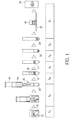
- FIG. 1 illustrates a method according to the prior art
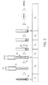
- Fig. 2 illustrates a method according of the present invention
- the method shown in Fig. 1 corresponds to the SurePath® method known in the art.
- the SurePath® method mainly comprises seven subsequently performed method steps S 1 to S 7 .
- a brush 10 which carries a collected cell sample, is suspended in a collection liquid 12.
- the cell sample is also contaminated with blood and mucus, which is also suspended in the collection liquid 12.
- Collection liquid 12 usually SurePath® preservative fluid, which is an ethanol-based liquid, is used.
- the brush 10 is then mixed with the collection liquid 12 for at least 1 minute in a special machine (PrepMate®) to form a cell-suspension 16.
- PrepMate® a special machine
- Step S 1 is similarly performed in the method according to the present invention (see Fig. 2 ), except the fact that the brush 10 does not necessarily have to remain in the collection liquid 12 and also the mixing step in the PrepMate® machine is not absolutely necessary. Therefore, also less collection liquid 12 is needed (3 ml instead of 10 ml).
- step S 2 the cell suspension 16 is collected by a syringe 18 and then deposited onto a density liquid 20.
- the density liquid 20 is provided in a regular centrifuge tube 22, whereas the density liquid 20 is, according to the present invention, provided in a vessel 24 which is provided with a microscope slide 26.
- the slide 26 can be lying on the bottom of the vessel 24 (as shown here), but the vessel 24 may as well be mounted on the slide 26 and sealed with a sealing, e.g. an O-ring, as shown in Figs. 4 and 5 .
- Another difference between the two methods is, that in the present invention only 1 - 1.5 ml of the cell-suspension 16 (depending on the cell concentration and the diameter of the vessel) are needed, whereas in the SurePath® method at least 6 ml of the cell suspension 16 has to be deposited onto the density liquid 20.
- PrepStain® density reagent is preferably used as density liquid 20. Further, preferably smaller vessels are used according to the present invention.
- step S 3 a first centrifugation step is performed, where the cells 14 are collected in the density liquid 20 due to higher ratio of centrifugal forces over flow resistance forces on them, while less dense structures like erythrocyte fragments, mucus and very small protein-particles remain in a supernatant 28 or in the upper part of the density liquid.
- this first centrifugation step is only performed for a time duration of approximately 1 minute instead of 2 minutes, and the centrifugal forces used are generally smaller than those used in the SurePath® method (approximately 80 to 90 g instead of 200 g).
- the supernatant 28 is in both methods removed until the border between the density liquid 20 and the collection liquid 12 (S 4 ). According to the present invention it is also possible to remove a small amount of the density liquid (the upper part), if the cells 14 are strongly spoiled with blood and mucus. The contaminant particles may enter the upper layer of the density liquid and this becomes especially apparent in case of a large concentration.
- the supernatant 28 does not necessarily need to be removed after the first centrifugation step, it can also be removed at the beginning of the second or in a break between the first and the second step. Preferably, this is done near the end of the first step, otherwise cells to be examined may be removed unwantedly. Furthermore, it is possible to remove the supernatant 28 during the first and/or the second centrifugation step. This is, for instance, done by a suction system with a tube or by an overflow system in the vessel. An overflow system is for example possible through a valve in the upper part of the vessel 24, which opens near the end of the first centrifugation step and releases the supernatant 28.
- Step S 5 shows the main difference between the SurePath® method and the method according to the present invention.
- a second centrifugation step with approximately 800 g centrifugal force is performed for a time duration of about 10 minutes until a cell pellet 30 is formed on the bottom of the centrifugetube 22.
- the second centrifugation step according to the present invention is in contrast thereto performed in a cytocentrifuge for only 2 minutes with 900 g centrifugal force. Since the cell suspension 16 is in a vessel 24, which is provided with a microscope slide 26, the cells 14 are directly deposited on the microscope slide 26 and adhered by a coating 32. This results in a significant time reduction, not only for the second centrifugation step (S 5 ), but also for the following method steps.
- the supernatant 28 needs to be decanted again and afterwards the cell pellet 30 is vortexed and resuspended in a buffer solution 34 (S 6 ). Then, the cleaned cell suspension is deposited into a settling chamber 36 on a microscope slide 38 for half an hour, so that the cells 14 can sedimentate spontaneously according to their gravity force (S 7 ).
- step S 7 which takes half an hour, can be skipped.
- step S 6 of the presented method the liquid only needs to be discarded and the microscope slide 26 is removed from the vessel 24. This last process step only takes 15 seconds instead of 30 minutes.
- a rinsing step can be performed after step S 6 . However, this is not obligatory.
- the microscope slide 26 is rinsed in deionized water, whereby the number of remaining erythrocytes is reduced and strongly overlapping cells are flushed away. This step is only advised when a monolayer of cells is desired. In this case only the first layer adheres to the coating 32.
- Fig. 3 shows an example of a vessel according to the present invention.
- the vessel 24 is a modified centrifugation tube which is provided with a microscope slide 26.
- the microscope slide 26 is carried by a cap 40 which is attached to the vessel 24.
- the cap 40 can be either clamped or bold together with the vessel 24 in order to be demountable. It has to be noted, that also other mounting options are possible as long as they are demountable.
- the microscope slide 26 is coated with a coating 32.
- the coating has the function to adhere and bind the cells 14 of the cell-suspension 16 during the second centrifugation step.
- a possible material for the coating 32 is for example poly-L-lysine. However, also other materials are possible for the coating 32. It also has to be noted, that the coating 32 may be arranged at any other position within the vessel, but preferably it is arranged at the lower part of the vessel 24 since the cells generally move there. Depending on the cell material or substrate material, also other embodiments are possible where no coating 32 is needed at all.
- the vessel is a centrifugation tube with a circular profile.
- this centrifugation tube is a 50 ml tube, so that it is large enough that the fluid within the vessel 24 can easily be removed by a pipette.
- the upper part 42 of the vessel 24 is also covered by a cap during the centrifugation steps.
- the size of the vessel 24 is exactly adapted to a centrifuge carrying bucket so that the vessels 24 are securely fastened during the centrifugation.
- a centrifuge carrying bucket usually carries multiple centrifugation tubes 24.
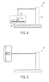
- a cytocentrifuge which is used for the first and/or the second centrifugation step, is shown in Figs. 4 and 5.
- Fig. 4 thereby shows the cytocentrifuge 44 in a first position while not operating, whereas Fig. 5 shows the cytocentrifuge 44 in the operating state.
- the vessel 24 carrying the cell-suspension 16 and provided with a microscope slide 26 is mounted in a fixative device 46 within the cytocentrifuge 44, e.g. in a centrifuge carrying bucket.
- a centrifuge rotor 48 is turned with high velocity so that the vessel 24 is accelerated on a horizontal circular path. This centrifugation accelerates the spontaneous sedimentation process, so that the cells 14 that are included in the cell-suspension 16 are accelerated towards the microscope slide 26, where they are bound on the coating 32.
- Figs. 6 and 7 show a microscope image of a cell sample.
- Fig. 6 shows a microscope image of a cell sample prepared by the smear-technique according to the prior art and
- Fig. 7 shows the same cell sample prepared by the method according to the present invention.
- the extraordinary results of the preparation method according to the present invention is visualized.
- the cell sample in Fig. 6 is still contaminated with blood, mucus and very small protein particles
- the cell sample is almost completely clean and only shows the cellular material 14 if it is prepared with the method according to the present invention (see Fig. 7 ).
- Such a clean cell sample enables to examine the cells 14 without the interference of distracting blood and mucus.
Priority Applications (1)
| Application Number | Priority Date | Filing Date | Title |
|---|---|---|---|
| EP09158694A EP2244083A1 (fr) | 2009-04-24 | 2009-04-24 | Procédé, appareil, récipient et cyto-centrifuge pour préparation cellulaire à base de liquide |
Applications Claiming Priority (1)
| Application Number | Priority Date | Filing Date | Title |
|---|---|---|---|
| EP09158694A EP2244083A1 (fr) | 2009-04-24 | 2009-04-24 | Procédé, appareil, récipient et cyto-centrifuge pour préparation cellulaire à base de liquide |
Publications (1)
| Publication Number | Publication Date |
|---|---|
| EP2244083A1 true EP2244083A1 (fr) | 2010-10-27 |
Family
ID=40996750
Family Applications (1)
| Application Number | Title | Priority Date | Filing Date |
|---|---|---|---|
| EP09158694A Ceased EP2244083A1 (fr) | 2009-04-24 | 2009-04-24 | Procédé, appareil, récipient et cyto-centrifuge pour préparation cellulaire à base de liquide |
Country Status (1)
| Country | Link |
|---|---|
| EP (1) | EP2244083A1 (fr) |
Cited By (5)
| Publication number | Priority date | Publication date | Assignee | Title |
|---|---|---|---|---|
| CN102680290A (zh) * | 2011-03-09 | 2012-09-19 | 湖北欣立达科技有限公司 | 一种自动制作液基细胞的制片方法 |
| CN105115799A (zh) * | 2015-08-30 | 2015-12-02 | 江苏健友医疗科技有限公司 | 液基细胞自然沉降制片装置 |
| CN111051847A (zh) * | 2017-07-20 | 2020-04-21 | 贝克顿·迪金森公司 | 将样品直接沉积到用于液基细胞学的载玻片上的手动方法 |
| CN113109124A (zh) * | 2021-04-09 | 2021-07-13 | 兴宏业(武汉)科技有限公司 | 一种负压平衡离心制片方法 |
| CN113332790A (zh) * | 2021-05-28 | 2021-09-03 | 暨南大学 | 一种新型的样品过滤机 |
Citations (6)
| Publication number | Priority date | Publication date | Assignee | Title |
|---|---|---|---|---|
| US4250830A (en) * | 1979-10-03 | 1981-02-17 | Leif Robert C | Swinging buckets |
| WO1993008895A1 (fr) * | 1991-11-05 | 1993-05-13 | Wescor, Inc. | Dispositif, appareil et procede ameliores de cytocentrifugation |
| WO1994025873A1 (fr) | 1993-04-23 | 1994-11-10 | Cellpro, Incorporated | Procedes d'enrichissement de cellules souches f×tales provenant du sang maternel |
| US6210889B1 (en) * | 1998-01-28 | 2001-04-03 | The Universite Laval | Method for enrichment of fetal cells from maternal blood and use of same in determination of fetal sex and detection of chromosomal abnormalities |
| US20050260100A1 (en) * | 2002-04-13 | 2005-11-24 | Leif Robert C | Centrifugal cytology system, chamber block and method for the preparation of treated monolayers of sample material |
| US20070161051A1 (en) * | 2006-01-12 | 2007-07-12 | Biocept, Inc. | Device for cell separation and analysis and method of using |
-
2009
- 2009-04-24 EP EP09158694A patent/EP2244083A1/fr not_active Ceased
Patent Citations (6)
| Publication number | Priority date | Publication date | Assignee | Title |
|---|---|---|---|---|
| US4250830A (en) * | 1979-10-03 | 1981-02-17 | Leif Robert C | Swinging buckets |
| WO1993008895A1 (fr) * | 1991-11-05 | 1993-05-13 | Wescor, Inc. | Dispositif, appareil et procede ameliores de cytocentrifugation |
| WO1994025873A1 (fr) | 1993-04-23 | 1994-11-10 | Cellpro, Incorporated | Procedes d'enrichissement de cellules souches f×tales provenant du sang maternel |
| US6210889B1 (en) * | 1998-01-28 | 2001-04-03 | The Universite Laval | Method for enrichment of fetal cells from maternal blood and use of same in determination of fetal sex and detection of chromosomal abnormalities |
| US20050260100A1 (en) * | 2002-04-13 | 2005-11-24 | Leif Robert C | Centrifugal cytology system, chamber block and method for the preparation of treated monolayers of sample material |
| US20070161051A1 (en) * | 2006-01-12 | 2007-07-12 | Biocept, Inc. | Device for cell separation and analysis and method of using |
Non-Patent Citations (1)
| Title |
|---|
| SWEENEY ET AL.: "Comparison of the effectiveness of two Liquid-Based Papanicolaou systems in the handling of adverse limiting factors, such as excessive blood", CANCER CYTOPATHOLOGY, vol. 108, no. 1, 25 February 2006 (2006-02-25), pages 27 - 31 |
Cited By (8)
| Publication number | Priority date | Publication date | Assignee | Title |
|---|---|---|---|---|
| CN102680290A (zh) * | 2011-03-09 | 2012-09-19 | 湖北欣立达科技有限公司 | 一种自动制作液基细胞的制片方法 |
| CN102680290B (zh) * | 2011-03-09 | 2015-11-25 | 湖北欣立达科技有限公司 | 一种自动制作液基细胞的制片方法 |
| CN105115799A (zh) * | 2015-08-30 | 2015-12-02 | 江苏健友医疗科技有限公司 | 液基细胞自然沉降制片装置 |
| CN111051847A (zh) * | 2017-07-20 | 2020-04-21 | 贝克顿·迪金森公司 | 将样品直接沉积到用于液基细胞学的载玻片上的手动方法 |
| US20200150008A1 (en) * | 2017-07-20 | 2020-05-14 | Becton Dickinson And Company | Manual method for depositing a sample directly onto a slide for liquid based cytology |
| CN113109124A (zh) * | 2021-04-09 | 2021-07-13 | 兴宏业(武汉)科技有限公司 | 一种负压平衡离心制片方法 |
| CN113109124B (zh) * | 2021-04-09 | 2022-04-01 | 兴宏业(武汉)科技有限公司 | 一种负压平衡离心制片方法 |
| CN113332790A (zh) * | 2021-05-28 | 2021-09-03 | 暨南大学 | 一种新型的样品过滤机 |
Similar Documents
| Publication | Publication Date | Title |
|---|---|---|
| EP0654972B1 (fr) | Procede et appareil permettant d'obtenir des monocouches cytologiques | |
| KR101858056B1 (ko) | 다수의 세포 현탁액 제조 및 분석용 자동 방법 및 자동화 디바이스 | |
| EP2244083A1 (fr) | Procédé, appareil, récipient et cyto-centrifuge pour préparation cellulaire à base de liquide | |
| US7687032B2 (en) | Filter assembly for molecular testing | |
| JP2001522042A (ja) | 流体試料と粒子物質を混合および流体試料から粒子物質を分離をする方法、およびその装置 | |
| JPWO2003050532A1 (ja) | 血液細胞分離システム | |
| US20140127745A1 (en) | Method, compositions and device for preparing cytological specimens | |
| JP2781513B2 (ja) | 細胞学的材料の単層の調製法 | |
| US11906405B2 (en) | Methods and systems for preparing cytological samples | |
| JP3996736B2 (ja) | 液体試料から粒子状物質を分離するための方法及び装置 | |
| EP0483506A1 (fr) | Méthode et appareil pour essai de pap automatisé | |
| US4767602A (en) | Apparatus for redepositing particulate matter | |
| US6106483A (en) | Apparatus for obtaining a cytology monolayer | |
| JP2000146782A (ja) | 自動固定標本作製装置および方法 | |
| Saqi et al. | Cell blocks: evolution, modernization, and assimilation into emerging technologies | |
| US20220268671A1 (en) | A device for immobilizing rare cells for cytology | |
| EP3389813B1 (fr) | Quantification à haute précision de particules subvisibles | |
| Leif | Methods for preparing sorted cells as monolayer specimens | |
| WO2023091933A1 (fr) | Support pour cellules d'imagerie provenant d'un échantillon par aspiration à l'aiguille | |
| JP2021006774A (ja) | 試料中に含まれる目的細胞の定量方法 | |
| WO2013016743A1 (fr) | Dispositif et procédé pour une analyse rapide et précise de fluides corporels | |
| MXPA00001286A (en) | Method and apparatus for separating particulate matter from a liquid specimen |
Legal Events
| Date | Code | Title | Description |
|---|---|---|---|
| PUAI | Public reference made under article 153(3) epc to a published international application that has entered the european phase |
Free format text: ORIGINAL CODE: 0009012 |
|
| AK | Designated contracting states |
Kind code of ref document: A1 Designated state(s): AT BE BG CH CY CZ DE DK EE ES FI FR GB GR HR HU IE IS IT LI LT LU LV MC MK MT NL NO PL PT RO SE SI SK TR |
|
| AX | Request for extension of the european patent |
Extension state: AL BA RS |
|
| STAA | Information on the status of an ep patent application or granted ep patent |
Free format text: STATUS: THE APPLICATION HAS BEEN REFUSED |
|
| 18R | Application refused |
Effective date: 20100825 |