EP2240904B1 - Ermitteln eines quantitativen masses der instabilität von kalkablagerungen eines blutgefässes - Google Patents
Ermitteln eines quantitativen masses der instabilität von kalkablagerungen eines blutgefässes Download PDFInfo
- Publication number
- EP2240904B1 EP2240904B1 EP09709737A EP09709737A EP2240904B1 EP 2240904 B1 EP2240904 B1 EP 2240904B1 EP 09709737 A EP09709737 A EP 09709737A EP 09709737 A EP09709737 A EP 09709737A EP 2240904 B1 EP2240904 B1 EP 2240904B1
- Authority
- EP
- European Patent Office
- Prior art keywords
- calcific
- deposits
- calcific deposits
- deposit
- aorta
- Prior art date
- Legal status (The legal status is an assumption and is not a legal conclusion. Google has not performed a legal analysis and makes no representation as to the accuracy of the status listed.)
- Not-in-force
Links
Images


Classifications
-
- G—PHYSICS
- G06—COMPUTING OR CALCULATING; COUNTING
- G06T—IMAGE DATA PROCESSING OR GENERATION, IN GENERAL
- G06T7/00—Image analysis
- G06T7/0002—Inspection of images, e.g. flaw detection
- G06T7/0012—Biomedical image inspection
-
- G—PHYSICS
- G06—COMPUTING OR CALCULATING; COUNTING
- G06T—IMAGE DATA PROCESSING OR GENERATION, IN GENERAL
- G06T7/00—Image analysis
- G06T7/60—Analysis of geometric attributes
- G06T7/62—Analysis of geometric attributes of area, perimeter, diameter or volume
-
- G—PHYSICS
- G06—COMPUTING OR CALCULATING; COUNTING
- G06T—IMAGE DATA PROCESSING OR GENERATION, IN GENERAL
- G06T2207/00—Indexing scheme for image analysis or image enhancement
- G06T2207/10—Image acquisition modality
- G06T2207/10116—X-ray image
-
- G—PHYSICS
- G06—COMPUTING OR CALCULATING; COUNTING
- G06T—IMAGE DATA PROCESSING OR GENERATION, IN GENERAL
- G06T2207/00—Indexing scheme for image analysis or image enhancement
- G06T2207/30—Subject of image; Context of image processing
- G06T2207/30004—Biomedical image processing
- G06T2207/30101—Blood vessel; Artery; Vein; Vascular
Definitions
- the present inventors have found that, in biological terms, a greater number of small calcific deposits distributed over a large portion of a blood vessel indicate a greater risk of developing cardiovascular disease than fewer larger deposits over the same area.
- the inventors have also found that the risk that a patient may suffer an episode of cardiovascular disease is high while calcific deposits are growing as during growth, the calcific deposits are relatively unstable. Because of its size relative to the size of a blood vessel a big, dense calcific deposit might be thought to be of grave concern, but could be quite stable and safe in terms of resulting in an episode of cardiovascular disease.
- the present inventors take into consideration one or both of the spread of calcific deposits in a blood vessel and the scope of growth of the deposits to provide an indication of how stable the calcifications are.
- locating and annotating the one or more calcific deposits comprises locating and annotating the boundary of each said calcific deposit and calculating a measure reflecting a combination of a) and b) is obtained by calculating the area occupied by the calcific deposits, expanding the boundary of each said calcific deposit outwards by a distance x corresponding to between 4mm and 20mm of a life-size image, calculating the area occupied by the expanded calcific deposits, and calculating a comparative index by comparing the area of expanded calcific deposits with the area of unexpanded calcific deposits to derive said measure.
- the typical diameter of a healthy aorta is approximately 20mm-25mm.
- the typical diameter of a diseased aorta may be up to 60mm-65mm.
- boundaries of the calcifications are expanded by approximately 1 6 to 1 2 of the diameter of the aorta.
- the step of expanding the boundary of the one or more calcific deposits comprises dilating the boundary of each calcific deposit.
- the boundaries of the calcific deposits may be dilated using any suitable structuring element resulting in expansion of the boundaries by the approximate distance x.
- points along the boundary of each respective calcific deposit are moved outwards by the fixed distance x or, if closer, up to an aortic wall or unexpanded boundary of an adjacent calcific deposit. Preventing expansion of the boundaries of respective areas of calcification beyond either the arterial walls or adjacent calcific deposits gives a realistic prediction of the likely growth of the calcific deposits.
- the method for calculating a measure reflecting b) comprises identifying a convex hull of the calcific deposits and deriving a value representative of the convex hull by calculating one of the perimeter of the convex hull and the area within the convex hull.
- the convex hull of the calcific deposits defines the shortest path around the calcific deposits that encloses each of the calcific deposits.
- the convex hull increases as the calcific deposits are more spread out throughout the blood vessel.
- the method further comprises calculating a value indicative of the total area of the calcific deposits and dividing the value representative of the convex hull by the total area.
- the method further comprises counting the number of calcific deposits and deriving a value indicative of the product obtained by multiplying the number of calcific deposits with the value representative of the convex hull.
- the method for calculating a measure reflecting a) comprises identifying a convex hull of each individual calcific deposit, deriving a value representative of each convex hull by calculating one of the perimeter of the convex hull and the area within the convex hull, summing the values representative of the convex hulls, calculating a value indicative of the total area of the calcific deposits and dividing the sum of values representative of the convex hulls by the total area of calcific deposits.
- calculating a measure reflecting a) comprises deriving a value indicative of the result of calculating the ratio of the square of the perimeter to the area for each calcific deposit and summing the ratios.
- calculating a measure reflecting a) comprises deriving a value indicative of the result of calculating the ratio of the square of the sum of the perimeters of the calcific deposits to the sum of the areas of the calcific deposits.
- the present inventors have found that the rate of growth of individual calcifications is likely to increase as the periphery of the individual calcific deposits become more irregular and as the individual calcific deposits deviate from being round. As the calcific deposits deviate from being round and as the periphery becomes more irregular the ratio of perimeter to area increases.
- calculating a measure reflecting a) or b) further comprises calculating a value indicative of a fractal dimension of the calcific deposits.
- the method further comprises calculating the Hausdorff Dimension or using a box-counting method to calculate the value indicative of the fractal dimension.
- an indication of the spread of calcific deposits within an aorta is determined as the fractal dimension will increase as the calcific deposits are more spread out. If the grid is relatively large, a greater percentage of boxes of the grid will be occupied by at least some part of a calcific deposit if the calcific deposits are spread out.
- an indication of the irregularity of the periphery of individual calcifications can be determined and the fractal dimension will increase as the periphery of calcific deposits become more irregular. If the grid is relatively fine, a greater percentage of boxes of the grid will be occupied by at least some part of a calcific deposit if the periphery of the calcific deposit is irregular.
- calculating a measure reflecting b) further comprises calculating a value indicative of the entropy of the calcific deposits.
- the blood vessel is an artery and in a more preferred embodiment the blood vessel is an aorta.
- a high score generally indicates a lack of stability of the calcifications and indicates a higher risk that a patient may suffer an episode of cardiovascular disease. It will, however, be appreciated that an inverse of the measure could be obtained, or other known mathematical techniques could be applied to the measure, such that a lower score would indicate a lack of stability.
- the present invention will hereinafter be described with particular reference to the analysis of x-ray images of an aorta. It will, however, be appreciated that the described method could be applied to other medical images of an aorta for example, DXA, Computer Tomography (CT) or Magnetic Resonance. Furthermore, the invention is not limited to analysis of images of an aorta and may also be applied to other blood vessels.
- the first step in preparing the image for analysis is to outline the walls of the lumbar aorta in an image.
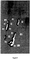
- Figure 2 shows an image of part of a lumbar spine and lumbar aorta where there are calcific deposits 4 in the lumbar aorta.
- the six points for vertebral height measurements are annotated on L1 to L4 of the lumbar vertebrae as shown in Figure 3 and from this the lumbar aorta can be identified and annotated. Further information about how the outline of the aorta is found is given by Lauze F et al.
- the second step is to outline each individual calcific deposit located in the aorta. Annotation of the boundaries may be done manually or using a particle filtering technique as discussed by de Bruijne in ("Shape particle guided tissue classification” in Mathematical Methods in Biomedical Image Analysis (MMBIA), 2006 ) and Conrad-Hansen et al. in ("A pixelwise inpainting-based refinement scheme for quantizing calcification in the lumbar aorta on 2D lateral x-ray images", SPIE Medical Imaging - Image Processing, 2006 ).
- the following severity scores relating to the geometrical outline of the calcific deposits and aorta may be computed.
- known calcification severity scores for example the AC24, can also be calculated.
- the annotations of the calcifications are used to calculate the following measures:
- the total area of visible calcific deposits i.e. the total area of calcification located within the respective annotated boundaries is calculated.
- the annotated boundary of each distinct calcific deposit is then expanded by a uniform amount and the total area of expanded calcific deposits, i.e. the total area of calcification located within the respective expanded annotated boundaries, calculated.
- the MAD factor is the result of the total expanded area of calcifications divided by the total area of visible (unexpanded) calcifications.
- each calcific deposit If points along the boundary of each calcific deposit are moved outwards from the centre of the calcific deposit by a distance x, the expanded boundaries of neighbouring calcific deposits or neighbouring portions of a particular calcific deposit that are within a distance x of each other will overlap.
- the MAD factor relates to the relative potential growth of the calcified plaque by counting only once overlapping expansions of two or more nearby calcific deposits.
- expansion of the boundaries can be limited to expansion within the aortic walls. Therefore, if a calcific deposit is less than a distance x from an aortic wall, the boundary may only be expanded up to the aortic wall. This enables a measure to be derived that provides a realistic indication of the likely expansion of the calcific deposits.
- the MAD factor will in general be large when the individual areas are distributed over a large portion of the aorta.
- relative proximity of the calcific deposits will only be considered if at least two of the calcific deposits are less than a distance x apart from each other. If the calcific deposits are all more than a distance x apart from each other, the expanded boundaries will not overlap.
- the MAD factor also takes into account the morphology of individual calcific deposits. Specifically, as the shape of an individual calcific deposit deviates from being round, the percentage by which the calcified deposit will be expanded will be greater than for a rounder calcific deposit of the same area. Likewise, as the periphery of a calcific deposit becomes more irregular, the percentage by which the calcific deposit will expand will be greater than for a calcific deposit with a smoother periphery. Accordingly, a far stretched deposit yields a worse prognosis compared to a circular deposit of the same area.
- FIG. 4 An example of an embodiment of the present invention illustrating the potential growth of areas of calcification is shown in Figure 4 .
- the first step is to locate the aortic walls 22 and annotate the boundaries of each area of calcification 24.
- Figure 4 shows seven distinct areas of calcification 24. Points (not shown) along the respective boundary of each area of calcification are extended outwards in a direction substantially perpendicular to the tangent of each respective point. The points are moved a uniform distance away from the original boundary resulting in the expanded boundaries 26 shown in Figure 4 .
- Expansion of the respective boundaries is restricted by the aortic walls and by other neighbouring calcifications.
- the boundary of a first calcification 28 is located in close proximity to an aortic wall 22. Accordingly, in that direction, the boundary is only expanded up to the aortic wall 22.
- the respective boundaries are only expanded up to the original unexpanded boundaries of the neighbouring calcifications and overlapping areas are only considered once.
- a grass-fire equation implemented by iterated morphological dilations with a combined radius of 200 pixels corresponding to 8.9 mm in real size simulates the total extent of the atherosclerotic process.
- the boundaries may be expanded by the number of pixels that correspond to life-size distances of, for example, between 4mm and 20mm, or 7mm and 10mm.
- a healthy aorta has a diameter of approximately 20mm to 25mm.
- a diseased may have a diameter approximately 40mm to 50mm wider than a healthy aorta.
- boundaries of the calcifications may be expanded by approximately 1 6 to 1 2 of the diameter of the aorta. If the resolution of the image being analysed varies, the boundaries may be dilated by an appropriate number of pixels to correspond to an appropriate life-size expansion within the range specified above.
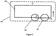
- Figure 5 illustrates schematically dilation of a general shape 40.
- the centre of, for example, a circle 42 having radius r is rolled along the periphery of the shape 40 in a direction A.
- the dilated shape 44 is determined by the circumference of the circle 42 and its periphery is a distance r away from that of the original shape 40.
- predicted growth of an area of calcification could be based on models of growth learned from previous examples of development of calcific deposits as set out by Kuhl, R Maas, G Himpel, A Menzel ("Computational modelling of artherosclerosis - A first approach towards a patient specific simulation based on computer topography", BMMB 6, 321-331, 2007 ).
- the moment of inertia may be multiplied by a measure of the total area of calcific deposits or by the NCD.
- the convex hull of individual calcific deposits may also be calculated.
- the convex hull of an individual calcific deposit would equate to the shortest path around that calcific deposit and may therefore give some indication of the irregularity of individual calcific deposits.
- a measure may be derived of the sum of perimeters or areas representative of the convex hulls of individual calcific deposits divided by the total area of calcific deposits.
- an indication of the irregularity of the periphery of individual calcifications can be determined and the fractal dimension will increase as the periphery of calcific deposits become more irregular.
- box counting method if the grid is relatively fine, a greater percentage of boxes of the grid will be occupied by at least some part of a calcific deposit if the periphery of the calcific deposit is irregular.
- Each of the measures described above may be used in isolation to provide a measure indicative of the severity of calcification in an aorta. To verify results, however, or to provide repeatable results, the different methods may be used in combination.
- Scores obtained for the various measures as they are described above are expected to increase as stability of the calcifications decreases and therefore the risk of suffering an episode of CVD increases.
- similarly useful results may be obtained with different mathematical formulae to obtain a result that may decrease or behave in a different way as stability of the calcifications in an aorta decreases.
- the calcification of aortic plaque is the end stage of a long range of molecular events resulting in maturation into a calcified fibro-fatty plaque that includes but is not restricted to: inflammation, macrophage infiltration, foam cells generation, lipid accumulation and processing and smooth muscle cells apoptosis. This results in imparted collagen synthesis and vascular integrity, and later results in weakened fibrous cap and generates atherosclerotic plaques that are more prone to rupture.
- the calcifications detected and analysed on x-rays are restricted only to the calcified core, and do not include the surrounding necrotic tissue and area of high remodelling and fibrosis. Thus the pathological area is underestimated by simple calcification measurements on x-rays.
- the study population consisted of 308 women aged 48 to 76 years who previously participated in epidemiologic cohorts. The original population was recruited by questionnaire. These women were invited for a follow up visit in 2000-2001. Among those 8593 women invited for a revisit, 308 were randomly selected that all had an interval of 8-9 years since their first visit, were post menopausal, and had the lumbar aorta visible on a single radiograph in the examinations. Among these 308 women, 52 had died before the revisit. Of these 52, 20 died from CVD (38%), 27 died from cancer (52%) and 5 died from other causes (10%). Information of the 52 individuals who died in the observation period was obtained via the Central Registry of the Danish Ministry of Health with a follow up rate of 100%.
- Demographic characteristics and risk parameters collected at baseline were age, weight, height, body mass index (BMI), waist and hip circumferences, systolic and diastolic blood pressure, treated hypertension, treated diabetes, smoking, regular alcohol and daily coffee consumption, and weekly fitness activity.
- BMI body mass index
- systolic and diastolic blood pressure treated hypertension
- treated diabetes smoking, regular alcohol and daily coffee consumption
- weekly fitness activity Using a blood analyser, measurements of fasting glucose and lipid profile (total cholesterol, triglycerides, HDL0cholesterol (HDL0C), LDL-cholesterol (LDL-C), apolipoprotein (apoA and apoB) were obtained.
- Lateral x-rays of the lumbar aorta were recorded.
- the images were digitised using a Vidar Dosimetry Pro Advantage scanner providing an image resolution of 9651 times 4008 pixels on 12-bit gray scale using a pixel size of 44.6 ⁇ m squared.
- Trained radiologists annotated the digitised images on a Sectra radiological reading unit with annotation software written using the Matlab programming environment. The radiologists were instructed to annotate the 4 corner points and 2 mediolateral points used for vertebral height measurements on L1 to L4, then to delineate the aorta and finally to delineate every individual calcified deposit visible in the lumber aorta.
- the software used had the ability to edit annotations and to perform a digital zoom for precise annotation. Finally it was noted if the calcified deposit was associated to the anterior and/or posterior aorta wall.
- the comparison of markers was performed by adjusting one marker for the influence of the other. When the adjusted marker may significantly (p ⁇ 0.05) differentiate survivor group from deceased group the marker is assumed to carry additional information. Markers are compared by mutually adjusting for the other marker and testing for additional information as above. Markers are furthermore compared by odds-ratio of the 90% fractile using the Mantel-Haenszel 95% confidence interval ( Mantel N, Haenszel "Statistical aspects of the analysis of data from retrospective studies of disease" J National Cancer Inst 1959; 22(4):710-748 ).
- Odds ratio differences are tested by Tarone's ( Tarone RE "On heterogenenity tests based on efficient scores" Biometrika 1985; 72(1):91-95 ) adjustment of the Breslow-Day ( Breslow NE, Day NE "Statistical methods in cancer research. Volume I - the analysis of case-control studies" IARC Sci Publications 1980;(32):5-338 ) test of heterogeneous odds ratio. Markers are combined linearly using Fisher's linear discriminant analysis (LDA). When combining LDA with fractile analysis, the LDA weights and fractile threshold are computed and evaluated in a leave-one-out fashion. Tests are considered statistically significant when p ⁇ 0.05.
- LDA linear discriminant analysis
- the aortic calcification markers performed significantly better in all the stratified deceased groups (except the other-cause death group), and unlike the metabolic/physical, all the aortic calcification markers scored significantly higher in the deceased versus the survivors, even after adjustment by age, waist and triglycerides.
- NCD number of calcified deposits
- Trained radiologists annotated the digitized images on a Sectra radiological reading unit with annotation software implemented in Matlab (Mathworks, MA, USA). The radiologists were instructed to annotate the 6 points used for vertebral height measurements on L1 to L4, delineate the aorta, and outline every individual calcified deposit visible in the lumbar aorta and noting association to anterior and/or posterior walls.
- the software allowed editing and digital zoom.
Landscapes
- Engineering & Computer Science (AREA)
- Physics & Mathematics (AREA)
- Theoretical Computer Science (AREA)
- Computer Vision & Pattern Recognition (AREA)
- General Physics & Mathematics (AREA)
- Health & Medical Sciences (AREA)
- Geometry (AREA)
- General Health & Medical Sciences (AREA)
- Medical Informatics (AREA)
- Nuclear Medicine, Radiotherapy & Molecular Imaging (AREA)
- Radiology & Medical Imaging (AREA)
- Quality & Reliability (AREA)
- Apparatus For Radiation Diagnosis (AREA)
- Image Processing (AREA)
- Magnetic Resonance Imaging Apparatus (AREA)
Claims (6)
- Computer-implementiertes Verfahren zum Verarbeiten eines Bildes zumindest eines Teils eines Blutgefäßes, um ein für die Instabilität von Kalkablagerungen in dem Blutgefäß kennzeichnendes Maß herzuleiten, wobei das Blutgefäß zumindest eine Kalkablagerung aufweist, und wobei das Verfahren umfasst:- das Lokalisieren und Annotieren einer oder mehrerer Kalkablagerung(en);- das Verwenden der durch die Annotierung der Kalkablagerungen hergeleiteten Informationen zum Berechnen eines Maßes, das eine Kombination widerspiegelt: aus a) den Abweichungen der einzelnen Kalkablagerungen insgesamt von einem runden Erscheinungsbild [eng/.: roundness] und b) bis hin zu zumindestens einem Schwellenwert, bis zu dem die einzelnen Kalkablagerungen voneinander beabstandet sind; wobei das Lokalisieren und Annotieren der Kalkablagerung(en) das Lokalisieren und Annotieren der Grenze einer jeden Kalkablagerung umfasst und das Berechnen eines Maßes, das eine Kombination aus a) und b) widerspiegelt, erhalten wird durch:- das Berechnen des von den Kalkablagerungen eingenommenen Bereichs;- das Nachaußenaufweiten der Grenze jeder Kalkablagerung um eine Entfernung x, die zwischen 4mm und 20mm eines lebensgroßen Bildes entspricht;- das Berechnen des von den aufgeweiteten Kalkablagerungen eingenommenen Bereichs; und- das Berechnen eines Vergleichsindexes durch Vergleichen des Bereichs der aufgeweiteten Kalkablagerungen mit dem Bereich der nichtaufgeweiteten Kalkablagerungen, um das Ausmaß herzuleiten.
- Verfahren nach Anspruch 1, bei dem der Schritt des Nachaußenaufweitens der Grenze jeder Kalkablagerung das Dilatieren der Grenze jeder Kalkablagerung umfasst.
- Verfahren nach Anspruch 2, weiterhin umfassend:das Dilatieren der Grenze jeder Kalkablagerung unter Verwendung eines Kreisradius x.
- Verfahren nach Anspruch 1, weiterhin umfassend:das Zählen der Anzahl der Kalkablagerungen und Gewichten des Vergleichsindexes anhand dieser Anzahl.
- Verfahren nach einem der vorherigen Ansprüche, bei dem das Blutgefäß eine Arterie ist.
- Verfahren nach Anspruch 5, bei dem das Blutgefäß eine Aorta ist.
Applications Claiming Priority (2)
| Application Number | Priority Date | Filing Date | Title |
|---|---|---|---|
| US12/069,894 US20090204338A1 (en) | 2008-02-13 | 2008-02-13 | Method of deriving a quantitative measure of the instability of calcific deposits of a blood vessel |
| PCT/EP2009/051626 WO2009101128A1 (en) | 2008-02-13 | 2009-02-12 | A method of deriving a quantitative measure of the instability of calcific deposits of a blood vessel |
Publications (2)
| Publication Number | Publication Date |
|---|---|
| EP2240904A1 EP2240904A1 (de) | 2010-10-20 |
| EP2240904B1 true EP2240904B1 (de) | 2012-04-25 |
Family
ID=40467300
Family Applications (1)
| Application Number | Title | Priority Date | Filing Date |
|---|---|---|---|
| EP09709737A Not-in-force EP2240904B1 (de) | 2008-02-13 | 2009-02-12 | Ermitteln eines quantitativen masses der instabilität von kalkablagerungen eines blutgefässes |
Country Status (7)
| Country | Link |
|---|---|
| US (1) | US20090204338A1 (de) |
| EP (1) | EP2240904B1 (de) |
| JP (1) | JP2011514186A (de) |
| KR (1) | KR20100133972A (de) |
| CN (1) | CN101946263B (de) |
| AT (1) | ATE555456T1 (de) |
| WO (1) | WO2009101128A1 (de) |
Families Citing this family (8)
| Publication number | Priority date | Publication date | Assignee | Title |
|---|---|---|---|---|
| WO2013115422A1 (ko) * | 2012-02-02 | 2013-08-08 | 부산대학교 산학협력단 | 디지털 이미지의 다중 프랙탈 분석을 이용한 대상체 검출방법 |
| KR101984247B1 (ko) | 2012-03-15 | 2019-05-30 | 삼성전자 주식회사 | 관상동맥석회화 수준 변화 예측장치 및 예측방법 |
| CN108171712B (zh) * | 2016-12-07 | 2022-02-11 | 富士通株式会社 | 确定图像相似度的方法和装置 |
| CN109288536B (zh) * | 2018-09-30 | 2021-01-29 | 数坤(北京)网络科技有限公司 | 获取冠脉钙化区域分类的方法、装置及系统 |
| CN109461143B (zh) * | 2018-10-12 | 2021-01-12 | 上海联影医疗科技股份有限公司 | 图像显示方法、装置、计算机设备和存储介质 |
| JP2024529795A (ja) * | 2021-08-04 | 2024-08-09 | カリフォルニア インスティチュート オブ テクノロジー | 血流中の血栓の超音波検出 |
| CA3267732A1 (en) * | 2022-09-16 | 2024-03-21 | Edith Cowan University | METHOD, SYSTEM AND USES FOR DETERMINING ABDOMINAL AORTIC CALCIFICATION |
| CN120241039B (zh) * | 2025-05-28 | 2025-10-17 | 四川吉晟生物医药有限公司 | 一种人体圆度指数计算装置 |
Family Cites Families (43)
| Publication number | Priority date | Publication date | Assignee | Title |
|---|---|---|---|---|
| US6282317B1 (en) * | 1998-12-31 | 2001-08-28 | Eastman Kodak Company | Method for automatic determination of main subjects in photographic images |
| US6890350B1 (en) * | 1999-07-28 | 2005-05-10 | Scimed Life Systems, Inc. | Combination self-expandable, balloon-expandable endoluminal device |
| US7231071B2 (en) * | 2001-09-13 | 2007-06-12 | Fujifilm Corporation | Abnormal shadow detecting system |
| US7127096B2 (en) * | 2001-11-20 | 2006-10-24 | Accuimage Diagnostics Corp. | Method and software for improving coronary calcium scoring consistency |
| US6882743B2 (en) * | 2001-11-29 | 2005-04-19 | Siemens Corporate Research, Inc. | Automated lung nodule segmentation using dynamic programming and EM based classification |
| US20030190063A1 (en) * | 2002-03-08 | 2003-10-09 | Acharya Kishore C. | Method and system for performing coronary artery calcification scoring |
| US20030215240A1 (en) * | 2002-04-03 | 2003-11-20 | Grann Eric B. | Optical WDM with single mode tolerance and low profile |
| US7295691B2 (en) * | 2002-05-15 | 2007-11-13 | Ge Medical Systems Global Technology Company, Llc | Computer aided diagnosis of an image set |
| US20060008843A1 (en) * | 2002-05-17 | 2006-01-12 | Automated Cell, Inc | Determination of protein function |
| US7418123B2 (en) * | 2002-07-12 | 2008-08-26 | University Of Chicago | Automated method and system for computerized image analysis for prognosis |
| US20040133100A1 (en) * | 2002-08-23 | 2004-07-08 | Morteza Naghavi | Novel risk assessment method based upon coronary calcification distribution pattern imaged by computed tomography |
| US7074188B2 (en) * | 2002-08-26 | 2006-07-11 | The Cleveland Clinic Foundation | System and method of characterizing vascular tissue |
| US7149331B1 (en) * | 2002-09-03 | 2006-12-12 | Cedara Software Corp. | Methods and software for improving thresholding of coronary calcium scoring |
| DE10249643A1 (de) * | 2002-10-24 | 2004-05-13 | Siemens Ag | Verfahren zur Unterstützung der Diagnose und/oder Therapie einer krankhaften Veränderung eines Blutgefäßes und hierzu hergerichtete Datenverarbeitungseinrichtung |
| US7379574B2 (en) * | 2002-11-27 | 2008-05-27 | The Board Of Trustees Of The Leland Stanford Junior University | Quantification of vascular irregularity |
| US7298883B2 (en) * | 2002-11-29 | 2007-11-20 | University Of Chicago | Automated method and system for advanced non-parametric classification of medical images and lesions |
| GB0310818D0 (en) * | 2003-05-10 | 2003-06-18 | Astrazeneca Ab | Assay |
| US20050002548A1 (en) * | 2003-06-20 | 2005-01-06 | Novak Carol L. | Automatic detection of growing nodules |
| US7912528B2 (en) * | 2003-06-25 | 2011-03-22 | Siemens Medical Solutions Usa, Inc. | Systems and methods for automated diagnosis and decision support for heart related diseases and conditions |
| EP1642234A1 (de) * | 2003-07-09 | 2006-04-05 | Humanitas Mirasole S.p.A. | Verfahren und vorrichtung zum analysieren biologischer gewebe |
| JP4212564B2 (ja) * | 2005-02-28 | 2009-01-21 | ザイオソフト株式会社 | 画像処理方法および画像処理プログラム |
| US7561727B2 (en) * | 2005-06-02 | 2009-07-14 | Nordic Bioscience Imaging A/S | Method of deriving a quantitative measure of a degree of calcification of an aorta |
| US20070099239A1 (en) * | 2005-06-24 | 2007-05-03 | Raymond Tabibiazar | Methods and compositions for diagnosis and monitoring of atherosclerotic cardiovascular disease |
| US7340083B2 (en) * | 2005-06-29 | 2008-03-04 | University Of Washington | Method and system for atherosclerosis risk scoring |
| US8014576B2 (en) * | 2005-11-23 | 2011-09-06 | The Medipattern Corporation | Method and system of computer-aided quantitative and qualitative analysis of medical images |
| US20070232883A1 (en) * | 2006-02-15 | 2007-10-04 | Ilegbusi Olusegun J | Systems and methods for determining plaque vulnerability to rupture |
| WO2007103568A2 (en) * | 2006-03-09 | 2007-09-13 | Biosite, Inc. | Methods and compositions for the diagnosis of diseases of the aorta |
| US7894664B2 (en) * | 2006-03-22 | 2011-02-22 | University Of Washington | Conditional shape model for image processing |
| US7627156B2 (en) * | 2006-03-22 | 2009-12-01 | Volcano Corporation | Automated lesion analysis based upon automatic plaque characterization according to a classification criterion |
| US20070242863A1 (en) * | 2006-04-13 | 2007-10-18 | Bernice Eland Hoppel | Methods and Apparatus for Contouring at Least One Vessel |
| US7860290B2 (en) * | 2006-04-21 | 2010-12-28 | Siemens Medical Solutions Usa, Inc. | Three-dimensional (3D) modeling of coronary arteries |
| US20090142259A1 (en) * | 2006-05-12 | 2009-06-04 | Genentech, Inc. | Compositions and methods for the diagnosis and treatment of bladder and urinary tract tumors |
| US9111372B2 (en) * | 2006-08-11 | 2015-08-18 | Visionary Technologies, Inc. | System and method for object identification and anomaly detection |
| US7986821B2 (en) * | 2006-08-15 | 2011-07-26 | General Electric Company | Processes and apparatus for imaging protocols and analysis |
| US7804992B2 (en) * | 2006-10-02 | 2010-09-28 | Hologic, Inc. | Cardiovascular risk assessments using aortic calcification information derived from x-ray measurements taken with a dual energy x-ray densitometer |
| WO2008042934A1 (en) * | 2006-10-03 | 2008-04-10 | The Brigham And Women's Hospital, Inc. | Measurement of thin-layered structures in x-ray computer tomography |
| AU2007310958A1 (en) * | 2006-10-19 | 2008-04-24 | Entelos, Inc. | Method and apparatus for modeling atherosclerosis |
| US7940970B2 (en) * | 2006-10-25 | 2011-05-10 | Rcadia Medical Imaging, Ltd | Method and system for automatic quality control used in computerized analysis of CT angiography |
| US7873194B2 (en) * | 2006-10-25 | 2011-01-18 | Rcadia Medical Imaging Ltd. | Method and system for automatic analysis of blood vessel structures and pathologies in support of a triple rule-out procedure |
| US7940977B2 (en) * | 2006-10-25 | 2011-05-10 | Rcadia Medical Imaging Ltd. | Method and system for automatic analysis of blood vessel structures to identify calcium or soft plaque pathologies |
| US7912270B2 (en) * | 2006-11-21 | 2011-03-22 | General Electric Company | Method and system for creating and using an impact atlas |
| US7957574B2 (en) * | 2006-11-22 | 2011-06-07 | General Electric Company | Methods and apparatus for generating a risk metric for soft plaque in vessels |
| EP2106536B1 (de) * | 2007-01-05 | 2015-08-12 | Malvern Instruments Incorporated | Spektrometrische untersuchung von heterogenität |
-
2008
- 2008-02-13 US US12/069,894 patent/US20090204338A1/en not_active Abandoned
-
2009
- 2009-02-12 WO PCT/EP2009/051626 patent/WO2009101128A1/en not_active Ceased
- 2009-02-12 AT AT09709737T patent/ATE555456T1/de active
- 2009-02-12 CN CN2009801048452A patent/CN101946263B/zh active Active
- 2009-02-12 EP EP09709737A patent/EP2240904B1/de not_active Not-in-force
- 2009-02-12 JP JP2010546327A patent/JP2011514186A/ja active Pending
- 2009-02-12 KR KR1020107020176A patent/KR20100133972A/ko not_active Withdrawn
Also Published As
| Publication number | Publication date |
|---|---|
| JP2011514186A (ja) | 2011-05-06 |
| CN101946263A (zh) | 2011-01-12 |
| US20090204338A1 (en) | 2009-08-13 |
| EP2240904A1 (de) | 2010-10-20 |
| ATE555456T1 (de) | 2012-05-15 |
| CN101946263B (zh) | 2012-12-26 |
| KR20100133972A (ko) | 2010-12-22 |
| WO2009101128A1 (en) | 2009-08-20 |
Similar Documents
| Publication | Publication Date | Title |
|---|---|---|
| EP2240904B1 (de) | Ermitteln eines quantitativen masses der instabilität von kalkablagerungen eines blutgefässes | |
| Sahiner et al. | Effect of CAD on radiologists' detection of lung nodules on thoracic CT scans: analysis of an observer performance study by nodule size | |
| Jacob et al. | Automated quantitative computed tomography versus visual computed tomography scoring in idiopathic pulmonary fibrosis: validation against pulmonary function | |
| US10339648B2 (en) | Quantitative predictors of tumor severity | |
| Budai et al. | Three-dimensional CT texture analysis of anatomic liver segments can differentiate between low-grade and high-grade fibrosis | |
| JP7612693B2 (ja) | 2次元及び3次元画像データを分析するためのシステム及び方法 | |
| Zhang et al. | Differentiation of focal organising pneumonia and peripheral adenocarcinoma in solid lung lesions using thin-section CT-based radiomics | |
| Yoshiyasu et al. | Radiomics technology for identifying early-stage lung adenocarcinomas suitable for sublobar resection | |
| JP7383698B2 (ja) | 脂肪の放射線シグネチャ | |
| CN111247592B (zh) | 用于随时间量化组织的系统和方法 | |
| Nannini et al. | A fully automated deep learning approach for coronary artery segmentation and comprehensive characterization | |
| Mundt et al. | Periaortic adipose radiomics texture features associated with increased coronary calcium score—first results on a photon-counting-CT | |
| Suman et al. | Recent advancements in computed tomography assessment of fibrotic interstitial lung diseases | |
| Corti et al. | Enhancing cardiovascular risk stratification: Radiomics of coronary plaque and perivascular adipose tissue–current insights and future perspectives | |
| Garzelli et al. | Improving the prediction of lung adenocarcinoma invasive component on CT: Value of a vessel removal algorithm during software segmentation of subsolid nodules | |
| Wang et al. | CTA-based radiomics and area change rate predict infrarenal abdominal aortic aneurysms patients events: A multicenter study | |
| Zuo et al. | Peritumoral and intratumoral radiomics for predicting visceral pleural invasion in lung adenocarcinoma based on preoperative computed tomography (CT) | |
| Li et al. | Prediction and verification of survival in patients with non-small-cell lung cancer based on an integrated radiomics nomogram | |
| Wang et al. | Hybrid clinical-radiomics model based on fully automatic segmentation for predicting the early expansion of spontaneous intracerebral hemorrhage: a multi-center study | |
| Shi et al. | Computed tomography enterography radiomics and machine learning for identification of Crohn’s disease | |
| Chi et al. | Stenosis detection and quantification on cardiac CTCA using panoramic MIP of coronary arteries | |
| Parrish | Volume CT: state-of-the-art reporting | |
| Song et al. | Pericoronary adipose tissue feature analysis in CT calcium score images with comparison to coronary CTA | |
| Sinsuat et al. | Influence of slice thickness on diagnoses of pulmonary nodules using low-dose CT: potential dependence of detection and diagnostic agreement on features and location of nodule | |
| Rathore et al. | Fully automated coronary artery calcium score and risk categorization from chest CT using deep learning and multiorgan segmentation: A validation study from National Lung Screening Trial (NLST) |
Legal Events
| Date | Code | Title | Description |
|---|---|---|---|
| PUAI | Public reference made under article 153(3) epc to a published international application that has entered the european phase |
Free format text: ORIGINAL CODE: 0009012 |
|
| 17P | Request for examination filed |
Effective date: 20100723 |
|
| AK | Designated contracting states |
Kind code of ref document: A1 Designated state(s): AT BE BG CH CY CZ DE DK EE ES FI FR GB GR HR HU IE IS IT LI LT LU LV MC MK MT NL NO PL PT RO SE SI SK TR |
|
| AX | Request for extension of the european patent |
Extension state: AL BA RS |
|
| DAX | Request for extension of the european patent (deleted) | ||
| RAP1 | Party data changed (applicant data changed or rights of an application transferred) |
Owner name: SYNARC INC. |
|
| GRAP | Despatch of communication of intention to grant a patent |
Free format text: ORIGINAL CODE: EPIDOSNIGR1 |
|
| RTI1 | Title (correction) |
Free format text: DERIVING A QUANTITATIVE MEASURE OF THE INSTABILITY OF CALCIFIC DEPOSITS OF A BLOOD VESSEL |
|
| GRAS | Grant fee paid |
Free format text: ORIGINAL CODE: EPIDOSNIGR3 |
|
| GRAA | (expected) grant |
Free format text: ORIGINAL CODE: 0009210 |
|
| AK | Designated contracting states |
Kind code of ref document: B1 Designated state(s): AT BE BG CH CY CZ DE DK EE ES FI FR GB GR HR HU IE IS IT LI LT LU LV MC MK MT NL NO PL PT RO SE SI SK TR |
|
| REG | Reference to a national code |
Ref country code: GB Ref legal event code: FG4D |
|
| REG | Reference to a national code |
Ref country code: CH Ref legal event code: EP |
|
| REG | Reference to a national code |
Ref country code: AT Ref legal event code: REF Ref document number: 555456 Country of ref document: AT Kind code of ref document: T Effective date: 20120515 |
|
| REG | Reference to a national code |
Ref country code: IE Ref legal event code: FG4D |
|
| REG | Reference to a national code |
Ref country code: DE Ref legal event code: R096 Ref document number: 602009006551 Country of ref document: DE Effective date: 20120621 |
|
| REG | Reference to a national code |
Ref country code: NL Ref legal event code: VDEP Effective date: 20120425 |
|
| REG | Reference to a national code |
Ref country code: AT Ref legal event code: MK05 Ref document number: 555456 Country of ref document: AT Kind code of ref document: T Effective date: 20120425 |
|
| LTIE | Lt: invalidation of european patent or patent extension |
Effective date: 20120425 |
|
| PG25 | Lapsed in a contracting state [announced via postgrant information from national office to epo] |
Ref country code: PL Free format text: LAPSE BECAUSE OF FAILURE TO SUBMIT A TRANSLATION OF THE DESCRIPTION OR TO PAY THE FEE WITHIN THE PRESCRIBED TIME-LIMIT Effective date: 20120425 Ref country code: LT Free format text: LAPSE BECAUSE OF FAILURE TO SUBMIT A TRANSLATION OF THE DESCRIPTION OR TO PAY THE FEE WITHIN THE PRESCRIBED TIME-LIMIT Effective date: 20120425 Ref country code: CY Free format text: LAPSE BECAUSE OF FAILURE TO SUBMIT A TRANSLATION OF THE DESCRIPTION OR TO PAY THE FEE WITHIN THE PRESCRIBED TIME-LIMIT Effective date: 20120425 Ref country code: IS Free format text: LAPSE BECAUSE OF FAILURE TO SUBMIT A TRANSLATION OF THE DESCRIPTION OR TO PAY THE FEE WITHIN THE PRESCRIBED TIME-LIMIT Effective date: 20120825 Ref country code: FI Free format text: LAPSE BECAUSE OF FAILURE TO SUBMIT A TRANSLATION OF THE DESCRIPTION OR TO PAY THE FEE WITHIN THE PRESCRIBED TIME-LIMIT Effective date: 20120425 Ref country code: SE Free format text: LAPSE BECAUSE OF FAILURE TO SUBMIT A TRANSLATION OF THE DESCRIPTION OR TO PAY THE FEE WITHIN THE PRESCRIBED TIME-LIMIT Effective date: 20120425 Ref country code: NO Free format text: LAPSE BECAUSE OF FAILURE TO SUBMIT A TRANSLATION OF THE DESCRIPTION OR TO PAY THE FEE WITHIN THE PRESCRIBED TIME-LIMIT Effective date: 20120725 |
|
| PG25 | Lapsed in a contracting state [announced via postgrant information from national office to epo] |
Ref country code: LV Free format text: LAPSE BECAUSE OF FAILURE TO SUBMIT A TRANSLATION OF THE DESCRIPTION OR TO PAY THE FEE WITHIN THE PRESCRIBED TIME-LIMIT Effective date: 20120425 Ref country code: PT Free format text: LAPSE BECAUSE OF FAILURE TO SUBMIT A TRANSLATION OF THE DESCRIPTION OR TO PAY THE FEE WITHIN THE PRESCRIBED TIME-LIMIT Effective date: 20120827 Ref country code: GR Free format text: LAPSE BECAUSE OF FAILURE TO SUBMIT A TRANSLATION OF THE DESCRIPTION OR TO PAY THE FEE WITHIN THE PRESCRIBED TIME-LIMIT Effective date: 20120726 Ref country code: SI Free format text: LAPSE BECAUSE OF FAILURE TO SUBMIT A TRANSLATION OF THE DESCRIPTION OR TO PAY THE FEE WITHIN THE PRESCRIBED TIME-LIMIT Effective date: 20120425 Ref country code: HR Free format text: LAPSE BECAUSE OF FAILURE TO SUBMIT A TRANSLATION OF THE DESCRIPTION OR TO PAY THE FEE WITHIN THE PRESCRIBED TIME-LIMIT Effective date: 20120425 |
|
| PG25 | Lapsed in a contracting state [announced via postgrant information from national office to epo] |
Ref country code: BE Free format text: LAPSE BECAUSE OF FAILURE TO SUBMIT A TRANSLATION OF THE DESCRIPTION OR TO PAY THE FEE WITHIN THE PRESCRIBED TIME-LIMIT Effective date: 20120425 |
|
| PG25 | Lapsed in a contracting state [announced via postgrant information from national office to epo] |
Ref country code: DK Free format text: LAPSE BECAUSE OF FAILURE TO SUBMIT A TRANSLATION OF THE DESCRIPTION OR TO PAY THE FEE WITHIN THE PRESCRIBED TIME-LIMIT Effective date: 20120425 Ref country code: EE Free format text: LAPSE BECAUSE OF FAILURE TO SUBMIT A TRANSLATION OF THE DESCRIPTION OR TO PAY THE FEE WITHIN THE PRESCRIBED TIME-LIMIT Effective date: 20120425 Ref country code: SK Free format text: LAPSE BECAUSE OF FAILURE TO SUBMIT A TRANSLATION OF THE DESCRIPTION OR TO PAY THE FEE WITHIN THE PRESCRIBED TIME-LIMIT Effective date: 20120425 Ref country code: RO Free format text: LAPSE BECAUSE OF FAILURE TO SUBMIT A TRANSLATION OF THE DESCRIPTION OR TO PAY THE FEE WITHIN THE PRESCRIBED TIME-LIMIT Effective date: 20120425 Ref country code: CZ Free format text: LAPSE BECAUSE OF FAILURE TO SUBMIT A TRANSLATION OF THE DESCRIPTION OR TO PAY THE FEE WITHIN THE PRESCRIBED TIME-LIMIT Effective date: 20120425 Ref country code: AT Free format text: LAPSE BECAUSE OF FAILURE TO SUBMIT A TRANSLATION OF THE DESCRIPTION OR TO PAY THE FEE WITHIN THE PRESCRIBED TIME-LIMIT Effective date: 20120425 Ref country code: NL Free format text: LAPSE BECAUSE OF FAILURE TO SUBMIT A TRANSLATION OF THE DESCRIPTION OR TO PAY THE FEE WITHIN THE PRESCRIBED TIME-LIMIT Effective date: 20120425 |
|
| PG25 | Lapsed in a contracting state [announced via postgrant information from national office to epo] |
Ref country code: IT Free format text: LAPSE BECAUSE OF FAILURE TO SUBMIT A TRANSLATION OF THE DESCRIPTION OR TO PAY THE FEE WITHIN THE PRESCRIBED TIME-LIMIT Effective date: 20120425 |
|
| PLBE | No opposition filed within time limit |
Free format text: ORIGINAL CODE: 0009261 |
|
| STAA | Information on the status of an ep patent application or granted ep patent |
Free format text: STATUS: NO OPPOSITION FILED WITHIN TIME LIMIT |
|
| 26N | No opposition filed |
Effective date: 20130128 |
|
| PG25 | Lapsed in a contracting state [announced via postgrant information from national office to epo] |
Ref country code: ES Free format text: LAPSE BECAUSE OF FAILURE TO SUBMIT A TRANSLATION OF THE DESCRIPTION OR TO PAY THE FEE WITHIN THE PRESCRIBED TIME-LIMIT Effective date: 20120805 |
|
| REG | Reference to a national code |
Ref country code: DE Ref legal event code: R097 Ref document number: 602009006551 Country of ref document: DE Effective date: 20130128 |
|
| PG25 | Lapsed in a contracting state [announced via postgrant information from national office to epo] |
Ref country code: BG Free format text: LAPSE BECAUSE OF FAILURE TO SUBMIT A TRANSLATION OF THE DESCRIPTION OR TO PAY THE FEE WITHIN THE PRESCRIBED TIME-LIMIT Effective date: 20120725 |
|
| PG25 | Lapsed in a contracting state [announced via postgrant information from national office to epo] |
Ref country code: MC Free format text: LAPSE BECAUSE OF NON-PAYMENT OF DUE FEES Effective date: 20130228 |
|
| REG | Reference to a national code |
Ref country code: CH Ref legal event code: PL |
|
| PG25 | Lapsed in a contracting state [announced via postgrant information from national office to epo] |
Ref country code: CH Free format text: LAPSE BECAUSE OF NON-PAYMENT OF DUE FEES Effective date: 20130228 Ref country code: LI Free format text: LAPSE BECAUSE OF NON-PAYMENT OF DUE FEES Effective date: 20130228 |
|
| REG | Reference to a national code |
Ref country code: IE Ref legal event code: MM4A |
|
| PG25 | Lapsed in a contracting state [announced via postgrant information from national office to epo] |
Ref country code: IE Free format text: LAPSE BECAUSE OF NON-PAYMENT OF DUE FEES Effective date: 20130212 |
|
| PG25 | Lapsed in a contracting state [announced via postgrant information from national office to epo] |
Ref country code: MT Free format text: LAPSE BECAUSE OF FAILURE TO SUBMIT A TRANSLATION OF THE DESCRIPTION OR TO PAY THE FEE WITHIN THE PRESCRIBED TIME-LIMIT Effective date: 20120425 |
|
| PG25 | Lapsed in a contracting state [announced via postgrant information from national office to epo] |
Ref country code: TR Free format text: LAPSE BECAUSE OF FAILURE TO SUBMIT A TRANSLATION OF THE DESCRIPTION OR TO PAY THE FEE WITHIN THE PRESCRIBED TIME-LIMIT Effective date: 20120425 |
|
| PG25 | Lapsed in a contracting state [announced via postgrant information from national office to epo] |
Ref country code: LU Free format text: LAPSE BECAUSE OF NON-PAYMENT OF DUE FEES Effective date: 20130212 Ref country code: HU Free format text: LAPSE BECAUSE OF FAILURE TO SUBMIT A TRANSLATION OF THE DESCRIPTION OR TO PAY THE FEE WITHIN THE PRESCRIBED TIME-LIMIT; INVALID AB INITIO Effective date: 20090212 Ref country code: MK Free format text: LAPSE BECAUSE OF FAILURE TO SUBMIT A TRANSLATION OF THE DESCRIPTION OR TO PAY THE FEE WITHIN THE PRESCRIBED TIME-LIMIT Effective date: 20120425 |
|
| REG | Reference to a national code |
Ref country code: FR Ref legal event code: PLFP Year of fee payment: 8 |
|
| REG | Reference to a national code |
Ref country code: FR Ref legal event code: PLFP Year of fee payment: 9 |
|
| PGFP | Annual fee paid to national office [announced via postgrant information from national office to epo] |
Ref country code: DE Payment date: 20170831 Year of fee payment: 9 |
|
| REG | Reference to a national code |
Ref country code: FR Ref legal event code: PLFP Year of fee payment: 10 |
|
| REG | Reference to a national code |
Ref country code: DE Ref legal event code: R119 Ref document number: 602009006551 Country of ref document: DE |
|
| PG25 | Lapsed in a contracting state [announced via postgrant information from national office to epo] |
Ref country code: DE Free format text: LAPSE BECAUSE OF NON-PAYMENT OF DUE FEES Effective date: 20180901 |
|
| PGFP | Annual fee paid to national office [announced via postgrant information from national office to epo] |
Ref country code: GB Payment date: 20231221 Year of fee payment: 16 |
|
| PGFP | Annual fee paid to national office [announced via postgrant information from national office to epo] |
Ref country code: FR Payment date: 20231212 Year of fee payment: 16 |
|
| GBPC | Gb: european patent ceased through non-payment of renewal fee |
Effective date: 20250212 |
|
| PG25 | Lapsed in a contracting state [announced via postgrant information from national office to epo] |
Ref country code: GB Free format text: LAPSE BECAUSE OF NON-PAYMENT OF DUE FEES Effective date: 20250212 |
|
| PG25 | Lapsed in a contracting state [announced via postgrant information from national office to epo] |
Ref country code: FR Free format text: LAPSE BECAUSE OF NON-PAYMENT OF DUE FEES Effective date: 20250228 |