EP2235485B1 - Method for the non-invasive optic determination of the temperature of flowing blood inside a living body - Google Patents
Method for the non-invasive optic determination of the temperature of flowing blood inside a living body Download PDFInfo
- Publication number
- EP2235485B1 EP2235485B1 EP09704736A EP09704736A EP2235485B1 EP 2235485 B1 EP2235485 B1 EP 2235485B1 EP 09704736 A EP09704736 A EP 09704736A EP 09704736 A EP09704736 A EP 09704736A EP 2235485 B1 EP2235485 B1 EP 2235485B1
- Authority
- EP
- European Patent Office
- Prior art keywords
- temperature
- absorption
- measurement
- light
- wavelength
- Prior art date
- Legal status (The legal status is an assumption and is not a legal conclusion. Google has not performed a legal analysis and makes no representation as to the accuracy of the status listed.)
- Not-in-force
Links
- 238000000034 method Methods 0.000 title claims description 33
- 239000008280 blood Substances 0.000 title claims description 23
- 210000004369 blood Anatomy 0.000 title claims description 23
- 238000010521 absorption reaction Methods 0.000 claims description 84
- 238000005259 measurement Methods 0.000 claims description 59
- XLYOFNOQVPJJNP-UHFFFAOYSA-N water Substances O XLYOFNOQVPJJNP-UHFFFAOYSA-N 0.000 claims description 23
- 230000003287 optical effect Effects 0.000 claims description 14
- 230000005855 radiation Effects 0.000 claims description 13
- 230000000694 effects Effects 0.000 claims description 12
- 230000001419 dependent effect Effects 0.000 claims description 8
- 238000009529 body temperature measurement Methods 0.000 claims description 7
- 238000012937 correction Methods 0.000 claims description 5
- 239000002609 medium Substances 0.000 description 16
- 238000012544 monitoring process Methods 0.000 description 8
- 238000011156 evaluation Methods 0.000 description 7
- 238000002604 ultrasonography Methods 0.000 description 6
- 239000000470 constituent Substances 0.000 description 5
- WQZGKKKJIJFFOK-GASJEMHNSA-N Glucose Natural products OC[C@H]1OC(O)[C@H](O)[C@@H](O)[C@@H]1O WQZGKKKJIJFFOK-GASJEMHNSA-N 0.000 description 4
- 239000012736 aqueous medium Substances 0.000 description 4
- 239000008103 glucose Substances 0.000 description 4
- 238000000862 absorption spectrum Methods 0.000 description 3
- 239000012503 blood component Substances 0.000 description 3
- 238000006073 displacement reaction Methods 0.000 description 3
- 238000004497 NIR spectroscopy Methods 0.000 description 2
- 230000005540 biological transmission Effects 0.000 description 2
- 210000004204 blood vessel Anatomy 0.000 description 2
- 238000011065 in-situ storage Methods 0.000 description 2
- 238000001228 spectrum Methods 0.000 description 2
- 230000002123 temporal effect Effects 0.000 description 2
- 206010028980 Neoplasm Diseases 0.000 description 1
- 238000002835 absorbance Methods 0.000 description 1
- 230000015572 biosynthetic process Effects 0.000 description 1
- 230000036760 body temperature Effects 0.000 description 1
- 238000011088 calibration curve Methods 0.000 description 1
- 239000000306 component Substances 0.000 description 1
- 238000000315 cryotherapy Methods 0.000 description 1
- 230000003247 decreasing effect Effects 0.000 description 1
- 238000011161 development Methods 0.000 description 1
- 238000000502 dialysis Methods 0.000 description 1
- 239000003814 drug Substances 0.000 description 1
- 230000005670 electromagnetic radiation Effects 0.000 description 1
- 238000002474 experimental method Methods 0.000 description 1
- 238000005534 hematocrit Methods 0.000 description 1
- 230000001678 irradiating effect Effects 0.000 description 1
- 238000002372 labelling Methods 0.000 description 1
- 239000007788 liquid Substances 0.000 description 1
- 239000000523 sample Substances 0.000 description 1
- 238000012360 testing method Methods 0.000 description 1
- 238000002560 therapeutic procedure Methods 0.000 description 1
Images


Classifications
-
- G—PHYSICS
- G01—MEASURING; TESTING
- G01J—MEASUREMENT OF INTENSITY, VELOCITY, SPECTRAL CONTENT, POLARISATION, PHASE OR PULSE CHARACTERISTICS OF INFRARED, VISIBLE OR ULTRAVIOLET LIGHT; COLORIMETRY; RADIATION PYROMETRY
- G01J5/00—Radiation pyrometry, e.g. infrared or optical thermometry
- G01J5/60—Radiation pyrometry, e.g. infrared or optical thermometry using determination of colour temperature
- G01J5/602—Radiation pyrometry, e.g. infrared or optical thermometry using determination of colour temperature using selective, monochromatic or bandpass filtering
-
- A—HUMAN NECESSITIES
- A61—MEDICAL OR VETERINARY SCIENCE; HYGIENE
- A61B—DIAGNOSIS; SURGERY; IDENTIFICATION
- A61B5/00—Measuring for diagnostic purposes; Identification of persons
- A61B5/0059—Measuring for diagnostic purposes; Identification of persons using light, e.g. diagnosis by transillumination, diascopy, fluorescence
-
- A—HUMAN NECESSITIES
- A61—MEDICAL OR VETERINARY SCIENCE; HYGIENE
- A61B—DIAGNOSIS; SURGERY; IDENTIFICATION
- A61B5/00—Measuring for diagnostic purposes; Identification of persons
- A61B5/01—Measuring temperature of body parts ; Diagnostic temperature sensing, e.g. for malignant or inflamed tissue
-
- G—PHYSICS
- G01—MEASURING; TESTING
- G01J—MEASUREMENT OF INTENSITY, VELOCITY, SPECTRAL CONTENT, POLARISATION, PHASE OR PULSE CHARACTERISTICS OF INFRARED, VISIBLE OR ULTRAVIOLET LIGHT; COLORIMETRY; RADIATION PYROMETRY
- G01J5/00—Radiation pyrometry, e.g. infrared or optical thermometry
- G01J5/58—Radiation pyrometry, e.g. infrared or optical thermometry using absorption; using extinction effect
-
- A—HUMAN NECESSITIES
- A61—MEDICAL OR VETERINARY SCIENCE; HYGIENE
- A61B—DIAGNOSIS; SURGERY; IDENTIFICATION
- A61B5/00—Measuring for diagnostic purposes; Identification of persons
- A61B5/145—Measuring characteristics of blood in vivo, e.g. gas concentration, pH value; Measuring characteristics of body fluids or tissues, e.g. interstitial fluid, cerebral tissue
- A61B5/14532—Measuring characteristics of blood in vivo, e.g. gas concentration, pH value; Measuring characteristics of body fluids or tissues, e.g. interstitial fluid, cerebral tissue for measuring glucose, e.g. by tissue impedance measurement
-
- A—HUMAN NECESSITIES
- A61—MEDICAL OR VETERINARY SCIENCE; HYGIENE
- A61B—DIAGNOSIS; SURGERY; IDENTIFICATION
- A61B5/00—Measuring for diagnostic purposes; Identification of persons
- A61B5/48—Other medical applications
- A61B5/4806—Sleep evaluation
Definitions
- the invention relates to a method for noninvasive, optical determination of the temperature of blood according to claim 1 wherein the medium to be examined is illuminated with infrared and / or visible light in the region of an absorption line, the position of which depends on the temperature of the medium and wherein the absorption of the light measured in the absorption line and the temperature is determined from this measurement by comparison with calibration data.
- - Medium means in a water-containing medium, e.g. living tissue and blood according to the invention (flowing) within a human body.
- Absorption means on the one hand the e.g. In transmission measured absorption behavior, but on the other hand also dependent on the absorption backscattering behavior.
- the determination of the temperature plays an important role in medicine in various fields, e.g. in the temperature monitoring of intensive care patients.
- the noninvasive measurement of body temperature by means of ear thermometers is frequently used, this type of measurement being restricted to the "discrete" application, i.e. the measurement at regular intervals.
- far invasive measuring methods have been used in practice in which probes or catheters with integrated sensors are introduced or introduced into the body.
- NIRS near-infrared spectroscopy
- the living tissue is largely transparent to electromagnetic radiation in the red and infrared regions, so that it is possible to "look inside” tissue within depths of a few millimeters to a few centimeters within this "biological window".
- the target tissue can be localized so that targeted optical absorption measurements in the localized tissue can be carried out in a relatively large depth of the body (cf. DE 103 11 408 B3 and DE 10 2006 036 920 ).
- Further prior art is bekaunt from US2004099815 A1 and De 10348958A1 ,
- the present invention seeks to provide a method for noninvasive, optical determination of the temperature of blood, which allows a simple and non-invasive way an accurate determination of the temperature of the blood.
- the method is intended to measure the temperature inside a body, e.g. suitable for measuring the temperature of tissue or flowing blood inside a body.
- the method should advantageously be used with the known methods for noninvasive determination of the concentration of blood constituents, e.g. the measurement of the glucose concentration in pulsating blood, combine.
- the invention teaches in a generic method for noninvasive, optical determination of the temperature of a medium of the type described above, that the medium is illuminated with (at least) two discrete wavelengths of light, which lie in the region of the absorption line on different sides of the absorption maximum, in that at least one temperature-dependent measured value is determined from the ratio of these two determined absorption values relative to one another, and that the temperature is determined from this measured value by comparison with the previously recorded calibration data.
- ratio of the two determined absorption values
- a predetermined “relation” is meant, which is to be applied to the two measured values.
- Forming the difference of the two sides of the maximum absorption values meant is especially preferred.
- the invention first of all starts from the (known) knowledge that there are several absorption lines of the water in the region of the biological window whose height and in particular also position (or wavelength) are sensitively dependent on the temperature of the water-containing medium.
- the absorption values on both sides of the maximum change very sensitively, so that if, for example, the difference of these two values is determined, this difference is particularly sensitive to the temperature of the Medium depends.
- a straight line can be passed through in the course of the evaluation by the two absorption points of the two fixed wavelengths, and as the measured value, for example, the slope of this straight line is determined, into which the difference of these two absorption values flows.
- the slope of this line and in particular the sign of this slope now depend very sensitive on the temperature, so that an accurate temperature determination is possible without exact determination of the maximum displacement. It is only necessary to measure two absorbance values for two fixed wavelengths and to evaluate them in the manner described.
- the measurement according to the invention is preferably carried out with infrared and / or visible light having a wavelength between 600 and 2500 nm, preferably 800 to 1600 nm.
- infrared and / or visible light having a wavelength between 600 and 2500 nm, preferably 800 to 1600 nm.
- the measurement of the temperature by means of infrared light in the range of the water absorption band around 970 nm leads to excellent results.
- at least one wavelength between, for example, 950 and 970 nm and at least one wavelength between, for example, 975 and 1000 nm is then used.
- it is also possible to work with other water absorption bands within the biological window for example in the region of the water absorption band around 1450 nm.
- any absorption line is considered whose position (wavelength of the maximum) depends on the temperature.
- the optimum range for the measurement ie the two optimally used wavelengths, can be determined experimentally in practice. There must always be one wavelength below and one wavelength above the absorption maximum. Care must be taken to ensure that the distance from the maximum is sufficiently large that the observed effect actually occurs, that the absorption values change with opposite signs when the temperature changes, that is, greater on one side of the maximum and smaller on the other side of the maximum become. With a temperature increase, the absorption should always grow at one wavelength and always decrease at the other wavelength. If the temperature is lowered, the opposite behavior should be shown. However, the distance of the selected wavelength must not be too far away from the absorption maximum, because there the danger of superposition with other lines or effects. It has proven expedient first to determine a specific temperature range, for example 30 ° C.
- the selected wavelengths ⁇ 1 , ⁇ 2 for the measurement should then be, for example, about 5 to 30 nm, preferably 5 to 15 nm, above or below this wavelength ⁇ 0 . This applies in particular to the region of the absorption line at 970 nm. In the case of the absorption line around 1450 nm, it is optionally possible to measure at a greater distance from the maximum.
- the method according to the invention it is initially possible to carry out the temperature of liquids at a defined location, for example in the laboratory or outside a body, without any further disturbing effects occurring there.
- the method according to the invention is suitable for measuring the temperature on or in a living body "in situ".
- the measurement succeeds even in low-lying areas, for example, the temperature of flowing blood in a bloodstream in a body can be measured.
- the invention proposes to mark the place where the temperature measurement is to take place, targeted by appropriate measures. This succeeds with the help of ultrasonic radiation, as this example in the DE 103 11 408 B3 and the DE 10 2006 036 920 is described.
- the tissue to be examined or the bloodstream can be "marked” with ultrasound radiation by focusing (pulsed) ultrasound radiation on the location or the bloodstream.
- ultrasound radiation by focusing (pulsed) ultrasound radiation on the location or the bloodstream.
- the proportions of the incident light into the detector are taken into account, which are in temporal relation to the ultrasound radiation, so that quite specifically an optical measurement and consequently a temperature determination in a low-lying area of a body can be performed.
- the invention recommends in a preferred development that initially takes a measurement of the temperature at the surface of the body.
- This reference measurement can likewise be carried out in the manner according to the invention, wherein a marking by means of ultrasonic focus may also be expedient there.
- a conventional reference measurement could also be made on the body surface, e.g. with a temperature sensor.
- the intensity of the light backscattered by the surface depends only on the temperature of the surface, because the light does not have to pass through further intermediate layers. Thus, first of all, the temperature of the surface is uniquely determined.
- the measurement can then take place in the interior of the body, in which case the temperature difference or the temperature gradient based on the temperature at the surface of the body is determined.
- the measurement in the region of the surface of the body thus forms a reference measurement in order to eliminate the possible dependence of the temperature gradient during the subsequent measurement in the interior of the body.
- the spectroscopic measurement on human tissue it may be expedient to take account of an influence on the spectroscopic measurement on human tissue by various factors, such as skin color, skin moisture, thickness and structure of the intermediate tissue sections, hematocrit values (which may vary in each individual) and fat levels in the blood, which can vary by the hour Blood changes.
- Such an isosbestic wavelength is remarkable characterized in that the absorption or backscatter depends exclusively on different scattering effects in the intermediate layers and not on the absorption behavior of the medium, for example water. With the aid of such a measurement at an isosbestic wavelength, the absorption-independent scattering effects can consequently be compensated or filtered out, so that altogether a particularly exact measurement can be achieved, even in low-lying layers of a body.
- an isosbestic wavelength of about 808 nm can be used.
- the temperature of a medium can be determined very precisely (for example with a precision of ⁇ 0.01 ° C.) in a simple manner.
- the temperature at body surfaces, or more preferably the temperature inside a body can be determined in a non-invasive and optical manner. These measures allow e.g. Also, an exact determination of the concentration of blood components and in particular the glucose concentration in blood, as in the course of a known measurement of concentration (at the same time) can also be a non-invasive measurement of the temperature, and precisely at the place where the concentration is determined ,
- the method according to the invention can also be used advantageously in other areas, e.g. in temperature monitoring of intensive care patients and in temperature monitoring in cryotherapy and medical tumor therapy.
- a temperature monitoring of neonates or a temperature monitoring of persons in the course of activities in thermally exposed environments can take place. Further applications are temperature monitoring in sleep diagnostics, during dialysis or even temperature monitoring of athletes. Also, the temperature measurement in industrial applications, e.g. the determination of the thermal distribution in the clothing industry is a possible application.
- FIG. 1 an experimental setup for determining the temperature T of an aqueous medium is shown optically.
- optical absorption spectra can be performed on a water-containing medium M.
- the water-containing medium M is arranged in a container 1 in this laboratory setup.
- laser light of the desired wavelength is radiated into the medium M via a coupler 3 and an input optical waveguide 4.
- the light emerging on the opposite side of the container 1 is coupled out via an output optical waveguide 5 and applied to a detector 6.
- the detector 6 is connected to an evaluation unit 7, which may have a computer and / or an oscilloscope. In the computer, the described evaluation algorithm is deposited, which will be discussed in more detail below.
- thermometer 10 is indicated, which measures the actual temperature of the aqueous medium exactly, so that the temperature data obtained in the manner according to the invention can be verified. It should be noted that it is the schematic hint of a laboratory setup, which serves primarily to prove the functionality of the method according to the invention.
- the optical determination of the temperature T takes place in a comparable manner by irradiating laser light into the body. However, it is then appropriate not to measure in transmission, as in the laboratory, but to measure the backscattered light, whereby the backscattered component also indicates the absorption behavior of the medium.
- Fig. 2 shows first in an overview by way of example and only schematically a conventional absorption spectrum of water in a wavelength range of about 700 nm to 2400 nm It is already the water absorption band B in the range of a wavelength of ⁇ 0 ⁇ 970 nm recognizable.
- ⁇ 0 thus means the wavelength of the absorption maximum at a certain temperature, ie, ⁇ 0 is temperature-dependent.
- the absorption line B shifts towards higher wavelengths at higher temperatures.
- the absorption in the region of this absorption line B is now measured, specifically only for two fixed wavelengths ⁇ 1 and ⁇ 2 , which lie on different sides of the absorption maximum (A 0 , ⁇ 0 ). These wavelengths are in Fig. 3 also marked. It should be noted that the position of the maximum and consequently ⁇ 0 itself is temperature dependent. Taking into account the selected temperature range, the wavelengths ⁇ 1 and ⁇ 2 are to be selected so that they are always on different sides of the (shifting) maximum for all temperatures of this range. It is now in Fig.
- infrared light is now radiated at a certain temperature, namely only two discrete wavelengths ⁇ 1 , ⁇ 2, which are respectively arranged on different sides of the absorption maximum ⁇ 0 .
- the measured absorption values are set in a relation, for example as in the exemplary embodiment, subtracted from one another, wherein the difference formed forms the determined measured value, which is sensitive to the temperature.
- This measured value which in the exemplary embodiment represents the difference of the absorption values or the slope of the drawn straight line G through the two measuring points, is compared with previously recorded calibration data.
- These calibration data are available for a variety of temperatures in Fig. 4 shown. There, the absorption values for different temperatures at the wavelengths ⁇ 1 and ⁇ 2 are shown in each case. Furthermore, for the purpose of illustration, straight lines have also been passed through the pairs assigned to each pair. Fig. 4 makes it particularly clear that the difference of the measured values and consequently also the slope of the straight line depends sensitively on the temperature, since with increasing or decreasing temperature in particular also a sign change can occur. It Consequently, the measurement is carried out at unknown temperature according to Fig.
- the Fig. 1 to 4 illustrate the basic operation of the method according to the invention and illustrate the implementation in the laboratory. Since it is an optical and non-invasive method of measurement, the measurement of the temperature within a body, for example the determination of the temperature of tissue, eg blood, inside a body K is possible in a comparable manner.
- tissue e.g blood
- the target area of the measurement is marked according to the invention by means of ultrasound radiation.
- Such a method is in another context in the DE 103 11 408 B3 been described.
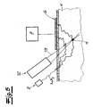
- the marking of a region in the interior of a body described therein can also be carried out in a corresponding manner to mark a region in the course of the temperature measurement. This is exemplified on Fig. 5 directed.
- the infrared light of a laser 2 is irradiated in the manner described (for the wavelengths ⁇ 1 and ⁇ 2 ) into the interior of a body K and the backscattered photons, which represent the absorption, are measured with a detector 6.
- the detector 6 now registers not only the photons scattered back in the region of the blood vessel 11, but also a large number of other photons which were scattered in other areas.
- a marking or selection is now possible by irradiation of ultrasonic radiation 13 with the in Fig. 5 This is focused on the target area, namely the blood vessel 11.
- the ultrasonic radiation source 12 generates pulsed ultrasonic radiation with fixed pulse length and fixed repetition time. About the evaluation can then taking into account this Pulse behavior of the light fraction can be extracted from the detector 6, which actually contributes to the volume of the ultrasonic focus.
- the amount of light marked by the ultrasound in practice may not only depend on the temperature of the observed location, but may also depend to some extent on the gradient of the temperature of the surface and the location to be observed, it may be expedient to provide a reference measurement beforehand the surface of the measuring body, eg to make on the skin, and there may be useful labeling by means of ultrasonic focus.
- the measurement made there depends exclusively on the local temperature and not on the temperature of any intermediate layers or a temperature gradient, so that subsequently a temperature measurement can take place in the desired depth of the body and a temperature differential measurement takes place.
- an isosbestic wavelength is characterized in that the backscattered photon current is influenced only by scattering effects in the intermediate layers and in the observed location and is completely independent of the (optical) absorption capacity of the water. The scattering behavior can consequently be "corrected out” from the measurement made.
- These reference and correction measurements can be carried out in practice in immediate (temporal) connection with the temperature measurement carried out and immediately flow into the evaluation, so that a device for performing the method according to the invention as it were calibrated by itself.
Landscapes
- Health & Medical Sciences (AREA)
- Life Sciences & Earth Sciences (AREA)
- Physics & Mathematics (AREA)
- Surgery (AREA)
- General Health & Medical Sciences (AREA)
- Engineering & Computer Science (AREA)
- Biomedical Technology (AREA)
- Heart & Thoracic Surgery (AREA)
- Medical Informatics (AREA)
- Molecular Biology (AREA)
- Biophysics (AREA)
- Animal Behavior & Ethology (AREA)
- Pathology (AREA)
- Public Health (AREA)
- Veterinary Medicine (AREA)
- General Physics & Mathematics (AREA)
- Spectroscopy & Molecular Physics (AREA)
- Investigating Or Analysing Materials By Optical Means (AREA)
- Measurement Of The Respiration, Hearing Ability, Form, And Blood Characteristics Of Living Organisms (AREA)
- Radiation Pyrometers (AREA)
- Measuring And Recording Apparatus For Diagnosis (AREA)
Description
Die Erfindung betrifft ein Verfahren zur nichtinvasiven, optischen Bestimmung der Temperatur von Blut gemäβ Anspruch 1 wobei das zu untersuchende Medium mit infrarotem und/oder sichtbaren Licht im Bereich einer Absorptionslinie beleuchtet wird, deren Lage von der Temperatur des Mediums abhängt und wobei die Absorption des Lichtes im Bereich der Absorptionslinie gemessen und aus dieser Messung durch Vergleich mit Kalibrierungsdaten die Temperatur ermittelt wird. - Medium meint im ein wasserhaltiges Medium z.B. lebendes Gewebe und erfindungsgemäβ (fließendes) Blut innerhalb eines menschlichen Körpers. Absorption meint einerseits das z.B. in Transmission gemessene Absorptionsverhalten, aber andererseits auch das von der Absorption abhängige Rückstreuverhalten.The invention relates to a method for noninvasive, optical determination of the temperature of blood according to claim 1 wherein the medium to be examined is illuminated with infrared and / or visible light in the region of an absorption line, the position of which depends on the temperature of the medium and wherein the absorption of the light measured in the absorption line and the temperature is determined from this measurement by comparison with calibration data. - Medium means in a water-containing medium, e.g. living tissue and blood according to the invention (flowing) within a human body. Absorption means on the one hand the e.g. In transmission measured absorption behavior, but on the other hand also dependent on the absorption backscattering behavior.
Die Bestimmung der Temperatur, z.B. eines menschlichen Körpers, spielt in der Medizin in unterschiedlichsten Bereichen eine bedeutende Rolle, z.B. bei der Temperaturüberwachung von Intensivpatienten. Dabei wird in der Praxis häufig die nichtinvasive Messung der Körpertemperatur mittels Ohrthermometern angewendet, wobei diese Art der Messung auf die "diskrete" Anwendung, d.h., die Messung in regelmäßigen Zeitabständen beschränkt ist. Für eine kontinuierliche Temperaturüberwachung werden in der Praxis bislang invasive Meßmethoden angewendet, bei denen Sonden oder Katheter mit integrierten Sensoren in den Körper eingeführt oder eingebracht werden.The determination of the temperature, e.g. of a human body, plays an important role in medicine in various fields, e.g. in the temperature monitoring of intensive care patients. In practice, the noninvasive measurement of body temperature by means of ear thermometers is frequently used, this type of measurement being restricted to the "discrete" application, i.e. the measurement at regular intervals. For continuous temperature monitoring, so far invasive measuring methods have been used in practice in which probes or catheters with integrated sensors are introduced or introduced into the body.
Außerdem besteht im Zusammenhang mit der nichtinvasiven Messung der Konzentration von Blutbestandteilen und insbesondere im Zusammenhang mit der Messung der Glukosekonzentration in fließendem bzw. pulsierendem Blut das Bedürfnis der Temperaturbestimmung "in situ", da derartige Messungen unter Einsatz von Kalibrierkurven in der Regel von der Temperatur abhängig sind (vgl.
Dabei ist zu beachten, dass im Bereich dieses so genannten biologischen Fensters "diskrete" Wasserabsorptionsbanden liegen, welche bei den oben beschriebenen Messungen der Konzentration von Blutbestandteilen in der Regel gemieden werden. Es ist jedoch bekannt, dass die Lage (und folglich die Wellenlänge) dieser Absorptionsmaxima und auch die Höhe der Absorptionslinie (und folglich die Größe bzw. das Maß der Absorption) von der Temperatur des Mediums, z.B. des Wassers abhängen. Aus diesem Grunde wurde bereits vorgeschlagen, die Temperaturabhängigkeit der Absorption im Bereich dieser Wasser-Absorptionsbanden auszunutzen, um die Temperatur des wasserhaltigen Mediums zu bestimmen. Dazu wurde vorgeschlagen, die Verschiebung der Absorptionslinie spektroskopisch aufzunehmen (vgl.
Ein ähnliches Verfahren ist aus der
Ausgehend von dem bekannten Stand der Technik liegt der Erfindung die Aufgabe zugrunde ein Verfahren zur nichtinvasiven, optischen Bestimmung der Temperatur von Blut zu schaffen, welches auf einfache und nichtinvasive Weise eine exakte Bestimmung der Temperatur des Bluts ermöglicht. Das Verfahren soll sich zur Messung der Temperatur im Innern eines Körpers, z.B. zur Messung der Temperatur von Gewebe oder fließendem Blut im Innern eines Körpers eignen. Außerdem soll sich das Verfahren in vorteilhafter Weise mit den bekannten Verfahren zur nichtinvasiven Bestimmung der Konzentration von Blutbestandteilen, z.B. der Messung der Glukosekonzentration in pulsierendem Blut, kombinieren lassen.Based on the known prior art, the present invention seeks to provide a method for noninvasive, optical determination of the temperature of blood, which allows a simple and non-invasive way an accurate determination of the temperature of the blood. The method is intended to measure the temperature inside a body, e.g. suitable for measuring the temperature of tissue or flowing blood inside a body. In addition, the method should advantageously be used with the known methods for noninvasive determination of the concentration of blood constituents, e.g. the measurement of the glucose concentration in pulsating blood, combine.
Zur Lösung dieser Aufgabe lehrt die Erfindung bei einem gattungsgemäßen Verfahren zur nichtinvasiven, optischen Bestimmung der Temperatur eines Mediums der eingangs beschriebenen Art, dass das Medium mit (zumindest) zwei diskreten Lichtwellenlängen beleuchtet wird, welche im Bereich der Absorptionslinie auf unterschiedlichen Seiten des Absorptionsmaximums liegen, dass aus dem Verhältnis dieser beiden ermittelten Absorptionswerte zueinander zumindest ein von der Temperatur abhängiger Messwert bestimmt wird und dass aus diesem Messwert durch Vergleich mit den zuvor aufgenommen Kalibrierungsdaten die Temperatur bestimmt wird. Mit dem "Verhältnis" der beiden ermittelten Absorptionswerte ist eine vorgegebene "Relation" gemeint, welche auf die beiden Messwerte angewendet werden soll. Besonders bevorzugt ist damit die Bildung der Differenz der beiden beidseitig des Maximums liegenden Absorptionswerte gemeint.To solve this problem, the invention teaches in a generic method for noninvasive, optical determination of the temperature of a medium of the type described above, that the medium is illuminated with (at least) two discrete wavelengths of light, which lie in the region of the absorption line on different sides of the absorption maximum, in that at least one temperature-dependent measured value is determined from the ratio of these two determined absorption values relative to one another, and that the temperature is determined from this measured value by comparison with the previously recorded calibration data. By the "ratio" of the two determined absorption values, a predetermined "relation" is meant, which is to be applied to the two measured values. Especially preferred is the Forming the difference of the two sides of the maximum absorption values meant.
Dabei geht die Erfindung zunächst einmal von der (bekannten) Erkenntnis aus, dass sich im Bereich des biologischen Fensters mehrere Absorptionslinien des Wassers befinden, deren Höhe und insbesondere auch Lage (bzw. Wellenlänge) empfindlich von der Temperatur des wasserhaltigen Mediums abhängt. Es ist nun im Rahmen der Erfindung jedoch nicht erforderlich, die Absorptionslinie vollständig zu vermessen und oder die Lage des Absorptionsmaximums exakt festzustellen. Vielmehr wird im Rahmen der Erfindung auf einfache Weise eine Messung mit zumindest zwei und vorzugsweise lediglich zwei diskreten Lichtwellenlängen durchgeführt, weiche jeweils auf unterschiedlichen Seiten des Absorptionsmaximums liegen. Denn die Erfindung hat erkannt, dass sich bei Änderung der Temperatur durch die Verschiebung des Maximums die Absorptionswerte beidseitig des Maximums in sehr unterschiedlicher Weise empfindlich ändern, so dass - wenn beispielsweise die Differenz dieser beiden Werte ermittelt wird - diese Differenz besonders empfindlich von der Temperatur des Mediums abhängt. Mit anderen Worten kann im Zuge der Auswertung durch die beiden Absorptionspunkte der beiden fest vorgegebenen Wellenlängen eine Gerade hindurchgelegt werden und als Messwert wird z.B. die Steigung dieser Geraden ermittelt, in welche die Differenz dieser beiden Absorptionswerte einfließt. Die Steigung dieser Geraden und insbesondere auch das Vorzeichen dieser Steigung hängen nun ganz empfindlich von der Temperatur ab, so dass eine exakte Temperaturbestimmung auch ohne exakte Bestimmung der Maximum-Verschiebung möglich wird. Es ist lediglich erforderlich, zwei Absorptionswerte für zwei fest vorgegebene Wellenlängen zu messen und diese in der beschriebenen Weise auszuwerten. Dieses wird im Folgenden noch in der Figurenbeschreibung weiter verdeutlicht. Es versteht sich, dass im Zuge der Temperaturbestimmung nach bzw. bei Ermittlung der Absorptionswerte und der daraus gewonnenen Messwerte ein Vergleich mit aufgenommenen Kalibrierungsdaten erfolgt. So lassen sich entsprechende Messungen im Labor bei bekannten Temperaturen durchführen und die bekannten Differenzwerte oder Steigungen lassen sich als Kalibrierungsdaten speichern, so dass sie im Zuge der Messung dann automatisch berücksichtigt werden können. Es sei jedoch darauf hingewiesen, dass es sich bei der beschriebenen "Bildung der Differenz" bzw. Bestimmung der Steigung der Verbindungsgeraden um eine bevorzugte Ausführungsform der Auswertung unter Berücksichtigung der beiden beidseitig des Maximums liegenden Absorptionswerte handelt. Die Erfindung umfasst grundsätzlich andere "Relationen", in welche zwei oder auch mehr Messwerte einfließen, die beidseitig des Absorptionsmaximums liegen.The invention first of all starts from the (known) knowledge that there are several absorption lines of the water in the region of the biological window whose height and in particular also position (or wavelength) are sensitively dependent on the temperature of the water-containing medium. However, it is not necessary within the scope of the invention to measure the absorption line completely and to determine the exact location of the absorption maximum. Rather, within the scope of the invention, a measurement is carried out in a simple manner with at least two and preferably only two discrete wavelengths of light, which lie in each case on different sides of the absorption maximum. For the invention has recognized that when the temperature changes due to the displacement of the maximum, the absorption values on both sides of the maximum change very sensitively, so that if, for example, the difference of these two values is determined, this difference is particularly sensitive to the temperature of the Medium depends. In other words, a straight line can be passed through in the course of the evaluation by the two absorption points of the two fixed wavelengths, and as the measured value, for example, the slope of this straight line is determined, into which the difference of these two absorption values flows. The slope of this line and in particular the sign of this slope now depend very sensitive on the temperature, so that an accurate temperature determination is possible without exact determination of the maximum displacement. It is only necessary to measure two absorbance values for two fixed wavelengths and to evaluate them in the manner described. This will be further clarified below in the description of the figures. It is understood that in the course of determining the temperature after or when determining the absorption values and the measured values obtained therefrom, a comparison is made with recorded calibration data. Thus, corresponding measurements can be carried out in the laboratory at known temperatures and the known difference values or slopes can be described as Save calibration data so that they can be automatically taken into account during the measurement. It should be noted, however, that the described "formation of the difference" or determination of the slope of the connecting straight line is a preferred embodiment of the evaluation taking into account the two absorption values lying on both sides of the maximum. The invention basically comprises other "relations" in which two or more measured values flow, which lie on both sides of the absorption maximum.
Bevorzugt wird die erfindungsgemäße Messung mit infrarotem und/oder sichtbarem Licht mit einer Wellenlänge zwischen 600 und 2500 nm, vorzugsweise 800 bis 1600 nm durchgeführt. Versuche haben gezeigt, dass die Messung der Temperatur mit Hilfe von Infrarot-Licht im Bereich der WasserAbsorptionsbande um 970 nm zu hervorragenden Ergebnissen führt. In diesem Fall wird dann zumindest eine Wellenlänge zwischen z.B. 950 und 970 nm und zumindest eine Wellenlänge zwischen z.B. 975 und 1000 nm eingesetzt. Es besteht aber auch die Möglichkeit, mit anderen Wasser-Absorptionsbanden innerhalb des biologischen Fensters zu arbeiten, z.B. im Bereich der WasserAbsorptionsbande um 1450 nm. Grundsätzlich kommt jede Absorptionslinie in Betracht, deren Lage (Wellenlänge des Maximums) von der Temperatur abhängt. Der optimale Bereich für die Messung, d.h., die beiden optimal einzusetzenden Wellenlängen, lassen sich in der Praxis experimentell ermitteln. Es sind stets jeweils eine Wellenlänge unterhalb und eine Wellenlänge oberhalb des Absorptionsmaximums zu wählen. Dabei ist darauf zu achten, dass der Abstand vom Maximum hinreichend groß ist, so dass tatsächlich der beobachtete Effekt eintritt, dass sich die Absorptionswerte bei Temperaturveränderung mit entgegengesetzten Vorzeichen verändern, d.h., auf einer Seite des Maximums größer und auf der anderen Seite des Maximums kleiner werden. Bei einer Temperaturerhöhung soll die Absorption bei einer der Wellenlänge stets wachsen und bei der anderen Wellenlänge stets sinken. Bei einer Temperaturemiedrigung soll sich das gegenteilige Verhalten zeigen. Allerdings darf der Abstand der ausgewählten Wellenlänge nicht zu weit vom Absorptionsmaximum entfernt sein, da dort die Gefahr der Überlagerung mit anderen Linien bzw. Effekten besteht. Es hat sich als zweckmäßig erwiesen, zunächst einen bestimmten Temperaturbereich, z.B. 30°C bis 43°C festzulegen und dann eine mittlere (typische) Temperatur (z.B. 37°C) festzulegen und dort die Wellenlänge des Absorptionsmaximums zu bestimmen. Die ausgewählten Wellenlängen λ1, λ2 für die Messung sollten dann z.B. etwa 5 bis 30 nm, vorzugsweise 5 bis 15 nm oberhalb bzw. unterhalb dieser Wellenlänger λ0 liegen. Dieses gilt insbesondere für den Bereich der Absorptionslinie bei 970 nm. Im Falle der Absorptionslinie um 1450 nm kann gegebenenfalls mit größerem Abstand zum Maximum gemessen werden.The measurement according to the invention is preferably carried out with infrared and / or visible light having a wavelength between 600 and 2500 nm, preferably 800 to 1600 nm. Experiments have shown that the measurement of the temperature by means of infrared light in the range of the water absorption band around 970 nm leads to excellent results. In this case, at least one wavelength between, for example, 950 and 970 nm and at least one wavelength between, for example, 975 and 1000 nm is then used. However, it is also possible to work with other water absorption bands within the biological window, for example in the region of the water absorption band around 1450 nm. In principle, any absorption line is considered whose position (wavelength of the maximum) depends on the temperature. The optimum range for the measurement, ie the two optimally used wavelengths, can be determined experimentally in practice. There must always be one wavelength below and one wavelength above the absorption maximum. Care must be taken to ensure that the distance from the maximum is sufficiently large that the observed effect actually occurs, that the absorption values change with opposite signs when the temperature changes, that is, greater on one side of the maximum and smaller on the other side of the maximum become. With a temperature increase, the absorption should always grow at one wavelength and always decrease at the other wavelength. If the temperature is lowered, the opposite behavior should be shown. However, the distance of the selected wavelength must not be too far away from the absorption maximum, because there the danger of superposition with other lines or effects. It has proven expedient first to determine a specific temperature range, for example 30 ° C. to 43 ° C., and then to determine an average (typical) temperature (for example 37 ° C.) and there determine the wavelength of the absorption maximum. The selected wavelengths λ 1 , λ 2 for the measurement should then be, for example, about 5 to 30 nm, preferably 5 to 15 nm, above or below this wavelength λ 0 . This applies in particular to the region of the absorption line at 970 nm. In the case of the absorption line around 1450 nm, it is optionally possible to measure at a greater distance from the maximum.
Mit dem erfindungsgemäßen Verfahren besteht zunächst einmal die Möglichkeit, die Temperatur von Flüssigkeiten an einem definierten Ort, z.B. im Labor bzw. außerhalb eines Körpers durchzuführen, ohne dass dort weitere störende Effekte auftreten. Von besonderer Bedeutung ist jedoch die Tatsache, dass sich das erfindungsgemäße Verfahren eignet, um die Temperatur an oder in einem lebenden Körper "in situ" zu messen. Insbesondere gelingt die Messung auch in tief liegenden Bereichen, z.B. kann die Temperatur von fließendem Blut in einer Blutbahn in einem Körper gemessen werden. Dazu schlägt die Erfindung vor, den Ort, an welchem die Temperaturmessung stattfinden soll, gezielt durch geeignete Maßnahmen zu markieren. Dieses gelingt mit Hilfe von Ultraschallstrahlung, so wie dieses beispielsweise in der
Um der Tatsache Rechnung zu tragen, dass der z.B. durch Ultraschallschallung markierte Lichtanteil nicht ausschließlich von der Temperatur des beobachteten Ortes abhängt, sondern auch von der Temperaturdifferenz zwischen der Körperobertläche und dem zu beobachtenden Ort, empfiehlt die Erfindung in einer bevorzugten Weiterbildung, dass zunächst eine Messung der Temperatur an der Oberfläche des Köpers erfolgt. Diese Referenzmessung kann ebenfalls auf die erfindungsgemäße Weise erfolgen, wobei auch dort eine Markierung mittels Ultraschallfokus zweckmäßig sein kann. An der Körperoberfläche könnte aber auch eine herkömmliche Referenzmessung vorgenommen werden, z.B. mit einem Temperaturfühler. An der Oberfläche ist die Intensität des von der Oberfläche rückgestreuten Lichtes nur von der Temperatur der Oberfläche abhängig, weil das Licht keine weiteren Zwischenlagen passieren muss. Damit ist zunächst einmal die Temperatur der Oberfläche eindeutig bestimmt. Anschließend kann dann die Messung im Innern des Körpers erfolgen, wobei dann die Temperaturdifferenz bzw. der Temperaturgradient bezogen auf die Temperatur an der Oberfläche des Körpers ermittelt wird. Die Messung im Bereich der Oberfläche des Körpers bildet folglich eine Referenzmessung, um beim anschließenden Messen im Körperinnem die eventuelle Abhängigkeit des Temperaturgradienten zu eliminieren.To take account of the fact that the e.g. characterized by ultrasonic radiation not exclusively on the temperature of the observed location, but also on the temperature difference between the body surface and the observed location, the invention recommends in a preferred development that initially takes a measurement of the temperature at the surface of the body. This reference measurement can likewise be carried out in the manner according to the invention, wherein a marking by means of ultrasonic focus may also be expedient there. However, a conventional reference measurement could also be made on the body surface, e.g. with a temperature sensor. On the surface, the intensity of the light backscattered by the surface depends only on the temperature of the surface, because the light does not have to pass through further intermediate layers. Thus, first of all, the temperature of the surface is uniquely determined. Then, the measurement can then take place in the interior of the body, in which case the temperature difference or the temperature gradient based on the temperature at the surface of the body is determined. The measurement in the region of the surface of the body thus forms a reference measurement in order to eliminate the possible dependence of the temperature gradient during the subsequent measurement in the interior of the body.
Im Übrigen kann es zweckmäßig sein eine Beeinflussung der spektroskopischen Messung an menschlichem Gewebe durch verschiedene Faktoren zu berücksichtigen, z.B. Hautfarbe, Hautfeuchtigkeit, Dicke und Struktur der zwischenliegenden Gewebeabschnitte, Hämatokritwerte (welche bei jedem Menschen variieren können) sowie Fettpegel im Blut, welcher sich stundenweise im Blut verändert. Aus diesem Grunde kann es zweckmäßig sein, zusätzlich zu der beschriebenen Absorptionsmessung und gegebenenfalls der Referenzmessung an der Körperoberfläche eine Korrekturmessung vorzunehmen, mit welcher sich die beschriebenen Effekte und insbesondere verschiedene Streueffekte an den Zwischenlagen eliminieren lassen. Dazu ist es zweckmäßig, Licht mit einer so genannten "isosbestischen Wellenlänge" in den Körper, z.B. in das Gewebe, einzustrahlen und die Absorption bzw. Rückstreuung zu messen. Eine solche isosbestische Wellenlänge zeichnet sich dadurch aus, dass die Absorption bzw. Rückstreuung ausschließlich von verschiedenen Streueffekten in den Zwischenlagen und nicht vom Absorptionsverhalten des Mediums, z.B. Wasser, abhängt. Mit Hilfe einer solchen Messung bei einer isosbestischen Wellenlänge lassen sich folglich die Absorptionsunabhängigen Streueffekte kompensieren bzw. herausfiltern, so dass insgesamt eine besonders exakte Messung auch in tief liegenden Schichten eines Körpers gelingt. Im Falle eines wasserhaltigen Mediums kann beispielsweise eine isosbestische Wellenlänge von etwa 808 nm verwendet werden.Incidentally, it may be expedient to take account of an influence on the spectroscopic measurement on human tissue by various factors, such as skin color, skin moisture, thickness and structure of the intermediate tissue sections, hematocrit values (which may vary in each individual) and fat levels in the blood, which can vary by the hour Blood changes. For this reason, it may be expedient, in addition to the described absorption measurement and optionally the reference measurement on the body surface, to perform a correction measurement with which the described effects and in particular various scattering effects on the intermediate layers can be eliminated. For this purpose, it is expedient to irradiate light with a so-called "isosbestic wavelength" into the body, for example into the tissue, and to measure the absorption or backscatter. Such an isosbestic wavelength is remarkable characterized in that the absorption or backscatter depends exclusively on different scattering effects in the intermediate layers and not on the absorption behavior of the medium, for example water. With the aid of such a measurement at an isosbestic wavelength, the absorption-independent scattering effects can consequently be compensated or filtered out, so that altogether a particularly exact measurement can be achieved, even in low-lying layers of a body. In the case of an aqueous medium, for example, an isosbestic wavelength of about 808 nm can be used.
Insgesamt lässt sich mit dem erfindungsgemäßen Verfahren auf einfache Weise die Temperatur eines Mediums besonders exakt (z.B. mit einer Präzision ± 0,01 °C) bestimmen. Es kann die Temperatur an Körperoberflächen oder besonders bevorzugt auch die Temperatur im Innern eines Körpers ermittelt werden, und zwar auf nichtinvasive und optische Weise. Diese Maßnahmen ermöglichen z.B. auch eine exakte Bestimmung der Konzentration von Blutbestandteilen und insbesondere auch der Glukosekonzentration in Blut, da im Zuge einer bekannten Messung der Konzentration (zeitgleich) auch eine nichtinvasive Messung der Temperatur erfolgen kann, und zwar exakt an dem Ort, an dem auch die Konzentration bestimmt wird. Das erfindungsgemäße Verfahren lässt sich jedoch auch in anderen Bereichen vorteilhaft anwenden, z.B. bei der Temperaturüberwachung von Intensivpatienten und bei der Temperaturüberwachung bei der Kryotherapie sowie medizinischen Tumortherapie. Ferner kann eine Temperaturüberwachung bei Neonaten oder auch eine Temperaturüberwachung von Personen im Zuge von Tätigkeiten in thermisch exponierten Umgebungen erfolgen. Weitere Anwendungsfälle sind das Temperaturmonitoring in der Schlafdiagnostik, während der Dialyse oder auch die Temperaturüberwachung von Sportlern. Auch die Temperaturmessung in industriellen Anwendungen, z.B. die Bestimmung der Thermoverteilung in der Bekleidungsindustrie ist ein möglicher Anwendungsfall.Overall, with the method according to the invention, the temperature of a medium can be determined very precisely (for example with a precision of ± 0.01 ° C.) in a simple manner. The temperature at body surfaces, or more preferably the temperature inside a body, can be determined in a non-invasive and optical manner. These measures allow e.g. Also, an exact determination of the concentration of blood components and in particular the glucose concentration in blood, as in the course of a known measurement of concentration (at the same time) can also be a non-invasive measurement of the temperature, and precisely at the place where the concentration is determined , However, the method according to the invention can also be used advantageously in other areas, e.g. in temperature monitoring of intensive care patients and in temperature monitoring in cryotherapy and medical tumor therapy. Furthermore, a temperature monitoring of neonates or a temperature monitoring of persons in the course of activities in thermally exposed environments can take place. Further applications are temperature monitoring in sleep diagnostics, during dialysis or even temperature monitoring of athletes. Also, the temperature measurement in industrial applications, e.g. the determination of the thermal distribution in the clothing industry is a possible application.
Im Folgenden wird die Erfindung anhand einer lediglich ein Ausführungsbeispiel darstellenden Zeichnung näher erläutert. Es zeigen:
- Fig. 1
- einen Versuchsaufbau zur Durchführung eines Verfahrene zur Temperaturbestrimmung
- Fig. 2.
- schematisch ein Wasser-Absorptionsspektrum in einem Wellenlängenbereich von etwa 660 nm bis etwa 2400 nm,
- Fig. 3
- die Wasserabsorption im Bereich einer Wasserabsorptionsbande um 970 nm bei zwei verschiedenen Temperaturen,
- Fig. 4
- Kalibrierungsdaten für eine Messung gemäß
Fig. 3 , und - Fig. 5
- eine schematische Darstellung des Verfahrens zur Bestimmung der Temperatur im Innern eines Körpers.
- Fig. 1
- a test setup for carrying out a method for temperature determination
- Fig. 2.
- schematically a water absorption spectrum in a wavelength range from about 660 nm to about 2400 nm,
- Fig. 3
- the water absorption in the region of a water absorption band around 970 nm at two different temperatures,
- Fig. 4
- Calibration data for a measurement according to
Fig. 3 , and - Fig. 5
- a schematic representation of the method for determining the temperature inside a body.
In
Die physikalischen Zusammenhänge und die Funktionsweise des erfindungsgemäßen Verfahrens sollen nun anhand der
Die
Dazu wird erfindungsgemäβ das Zielgebiet der Messung mit Hilfe von Ultraschallstrahlung markiert. Ein solches Verfahren ist in anderem Zusammenhang in der
Da der durch den Ultraschall markierte Lichtanteil in der Praxis eventuell nicht nur von der Temperatur des beobachteten Ortes abhängt, sondern in gewissem Maß auch von dem Gradient der Temperatur der Oberfläche und des zu beobachteten Ortes abhängen kann, kann es zweckmäßig sein, zuvor eine Referenzmessung an der Oberfläche des Messkörpers, z.B. an der Haut vorzunehmen, wobei auch dort eine Markierung mittels Ultraschallfokus zweckmäßig sein kann. Die dort vorgenommene Messung hängt ausschließlich von der dortigen Temperatur und nicht von der Temperatur eventueller Zwischenlagen oder einem Temperaturgradienten ab, so dass anschließend eine Temperatumessung in der gewünschten Tiefe des Körpers stattfinden kann und dabei eine Temperatur-Differenzmessung erfolgt.Since the amount of light marked by the ultrasound in practice may not only depend on the temperature of the observed location, but may also depend to some extent on the gradient of the temperature of the surface and the location to be observed, it may be expedient to provide a reference measurement beforehand the surface of the measuring body, eg to make on the skin, and there may be useful labeling by means of ultrasonic focus. The measurement made there depends exclusively on the local temperature and not on the temperature of any intermediate layers or a temperature gradient, so that subsequently a temperature measurement can take place in the desired depth of the body and a temperature differential measurement takes place.
Schließlich kann es ergänzend zur Korrektur zweckmäßig sein, eine Korrekturmessung mit Hilfe einer isosbestischen Wellenlänge vorzunehmen. Einzelheiten sind dazu in den Figuren nicht dargestellt. Eine solche isosbestische Wellenlänge zeichnet sich dadurch aus, dass der rückgestreute Photonenstrom nur durch Streueffekte in den Zwischenlagen sowie in dem beobachteten Ort beeinflusst wird und von dem (optischen) Absorptionsvermögen des Wassers vollkommen unabhängig ist. Das Streuverhalten kann folglich aus der vorgenommenen Messung "heraus korrigiert" werden. Diese Referenz- und Korrekturmessungen können in der Praxis in unmittelbarem (zeitlichen) Zusammenhang mit der durchgeführten Temperaturmessung erfolgen und sofort in die Auswertung einfließen, so dass sich eine Vorrichtung zur Durchführung des erfindungsgemäßen Verfahrens gleichsam von selbst kalibriert.Finally, in addition to the correction, it may be expedient to carry out a correction measurement with the aid of an isosbestic wavelength. Details are not shown in the figures. Such an isosbestic wavelength is characterized in that the backscattered photon current is influenced only by scattering effects in the intermediate layers and in the observed location and is completely independent of the (optical) absorption capacity of the water. The scattering behavior can consequently be "corrected out" from the measurement made. These reference and correction measurements can be carried out in practice in immediate (temporal) connection with the temperature measurement carried out and immediately flow into the evaluation, so that a device for performing the method according to the invention as it were calibrated by itself.
Claims (7)
- A method for the non-invasive, optical determination of the temperature of flowing blood inside a body,
wherein the blood which is to be examined is illuminated with infrared and/or visible light in the region of an absorption line, the position of which depends on the temperature of the blood, and wherein the absorption of the light in the region of the absorption line is measured and from this measurement the temperature is determined by comparison with calibration data,
wherein the blood is illuminated with at least two discrete light wavelengths λ1, λ2 which lie in the region of the absorption line on different sides of the absorption maximum,
wherein at least one measured value dependent on the temperature (T) or a measuring function dependent on the temperature is determined from the ratio or respectively a functional relation of these two determined absorption values A(λ1), A(λ2) to one another,
wherein the temperature is determined from this measured value or respectively this measuring function by comparison with the previously recorded calibration data,
wherein for measuring the temperature inside a body, the site of the measurement in the bloodstream is marked by means of pulsed ultrasonic radiation, by the pulsed ultrasonic radiation being focussed on the bloodstream and in the course of measurement of the absorption of the light for the temperature measurement only the portions of the incident light into the detector are taken into consideration, which are in time relationship to the ultrasonic radiation. - The method according to Claim 1, wherein the measured value is determined by forming the difference of the two absorption values lying on both sides of the maximum or by determining the inclination of a straight line running through the measuring points.
- The method according to Claim 1 or 2, wherein the medium is illuminated with infrared light and/or visible light with wavelength λ1, λ2 between 600 nm and 2500 nm, preferably 800 nm to 1600 nm.
- The method according to Claim 3, wherein the measurement takes place in the region of the water absorption line at about 970 nm, wherein preferably on the one hand light of a first wavelength λ1 between 950 and 970 nm is used, and on the other hand light of a second wavelength λ2 between 975 and 1000 nm is used.
- The method according to Claim 3, wherein the measurement is carried out in the region of the water absorption line at about 1450 nm.
- The method according to one of Claims 1 to 5, wherein for determining the temperature inside a body firstly a reference measurement of the temperature on the surface of the body is carried out and wherein subsequently the measurement of the temperature is carried out at a site inside the body.
- The method according to one of Claims 1 to 6, wherein for determining the temperature inside a body a correction measurement with one or more isosbestic wavelengths is carried out, wherein the medium or respectively the body is illuminated with light with an isosbestic wavelength, in which the backscattered light portion is dependent exclusively on scatter effects in the interior of the body and not on the absorption behaviour of the medium, e.g. water.
Applications Claiming Priority (2)
| Application Number | Priority Date | Filing Date | Title |
|---|---|---|---|
| DE102008006245A DE102008006245A1 (en) | 2008-01-25 | 2008-01-25 | Method for the noninvasive, optical determination of the temperature of a medium |
| PCT/EP2009/000440 WO2009092603A2 (en) | 2008-01-25 | 2009-01-23 | Method for the non-invasive optic determination of the temperature of a medium |
Publications (2)
| Publication Number | Publication Date |
|---|---|
| EP2235485A2 EP2235485A2 (en) | 2010-10-06 |
| EP2235485B1 true EP2235485B1 (en) | 2013-02-27 |
Family
ID=40527910
Family Applications (1)
| Application Number | Title | Priority Date | Filing Date |
|---|---|---|---|
| EP09704736A Not-in-force EP2235485B1 (en) | 2008-01-25 | 2009-01-23 | Method for the non-invasive optic determination of the temperature of flowing blood inside a living body |
Country Status (8)
| Country | Link |
|---|---|
| US (1) | US8426819B2 (en) |
| EP (1) | EP2235485B1 (en) |
| JP (1) | JP2011510312A (en) |
| CN (1) | CN101981422B (en) |
| DE (1) | DE102008006245A1 (en) |
| ES (1) | ES2408857T3 (en) |
| RU (1) | RU2489689C2 (en) |
| WO (1) | WO2009092603A2 (en) |
Families Citing this family (13)
| Publication number | Priority date | Publication date | Assignee | Title |
|---|---|---|---|---|
| US8376955B2 (en) * | 2009-09-29 | 2013-02-19 | Covidien Lp | Spectroscopic method and system for assessing tissue temperature |
| CA2891041C (en) * | 2012-12-04 | 2017-10-17 | F. Hoffmann-La Roche Ag | Method for hematocrit correction and glucose meter adapted therefor |
| JP5964773B2 (en) * | 2013-03-22 | 2016-08-03 | 日本電信電話株式会社 | Temperature measuring method and apparatus |
| DE102014107261A1 (en) * | 2014-05-22 | 2015-11-26 | Nirlus Engineering Ag | Method for the noninvasive optical measurement of properties of flowing blood |
| DE102014107250A1 (en) | 2014-05-22 | 2015-11-26 | Nirlus Engineering Ag | Method and apparatus for noninvasive in vivo optical determination of glucose concentration in flowing blood |
| EP3170446A1 (en) | 2015-11-20 | 2017-05-24 | NIRLUS Engineering AG | Method and device for the non-invasive optical in-vivo determination of glucose concentration in flowing blood |
| RU2672379C1 (en) * | 2016-07-06 | 2018-11-14 | Сергей Маркович Полозов | Method of non-invasive remote temperature control of deeply located organs and tissues |
| DE102018124531A1 (en) | 2018-10-04 | 2020-04-09 | Nirlus Engineering Ag | Method and device for non-invasive optical measurement of properties of living tissue |
| DE102018124537A1 (en) | 2018-10-04 | 2020-04-09 | Nirlus Engineering Ag | Method and device for non-invasive optical measurement of properties of living tissue |
| CN111713746B (en) * | 2020-06-08 | 2024-03-15 | 深圳市康泓威科技有限公司 | Method for detecting and controlling solution temperature of electronic atomization device and electronic atomization device |
| CN116568214A (en) * | 2020-10-07 | 2023-08-08 | 光谱公司 | Health analysis using spectral sensor system |
| KR20220070792A (en) * | 2020-11-23 | 2022-05-31 | 삼성전자주식회사 | Apparatus and method for estimating body core temperature, and healthcare device |
| DE102020134911A1 (en) | 2020-12-23 | 2022-06-23 | Nirlus Engineering Ag | Method and device for non-invasive optical in vivo determination of glucose concentration |
Family Cites Families (19)
| Publication number | Priority date | Publication date | Assignee | Title |
|---|---|---|---|---|
| SE466157B (en) * | 1989-04-25 | 1992-01-07 | Migrata Uk Ltd | DETERMINED TO DETERMINE THE GLUCOSE CONTENT OF WHOLE BLOOD AND DISPOSABLE BEFORE THIS |
| CA2028261C (en) * | 1989-10-28 | 1995-01-17 | Won Suck Yang | Non-invasive method and apparatus for measuring blood glucose concentration |
| DE4203202A1 (en) * | 1992-02-05 | 1993-08-12 | Boehringer Mannheim Gmbh | DEVICE FOR ANALYZING A MEDICAL SAMPLE |
| CA2194232A1 (en) * | 1994-07-01 | 1996-01-18 | Hans Van Der Woord | A method for monitoring performance of an incubator module, said incubator module being comprised in an automated system for assaying multiple samples and a kit suitable for use in said method |
| GB9415869D0 (en) * | 1994-08-05 | 1994-09-28 | Univ Mcgill | Substrate measurement by infrared spectroscopy |
| JPH11511658A (en) * | 1995-08-16 | 1999-10-12 | パーカー,ダウッド | Non-invasive blood analyte sensor |
| JP3826479B2 (en) * | 1997-03-19 | 2006-09-27 | 松下電器産業株式会社 | Blood glucose meter |
| JPH11183377A (en) * | 1997-12-17 | 1999-07-09 | Matsushita Electric Ind Co Ltd | Optical content meter |
| JP2002531820A (en) * | 1998-11-27 | 2002-09-24 | デーウー・エレクトロニクス・カンパニー・リミテッド | Bolometer with zinc oxide bolometer element |
| IL137447A (en) * | 2000-07-23 | 2007-03-08 | Israel Atomic Energy Comm | Apparatus and method for probing light absorbing agents in biological tissues |
| US6640117B2 (en) * | 2000-09-26 | 2003-10-28 | Sensys Medical, Inc. | Method and apparatus for minimizing spectral effects attributable to tissue state variations during NIR-based non-invasive blood analyte determination |
| JP4054853B2 (en) * | 2000-10-17 | 2008-03-05 | 独立行政法人農業・食品産業技術総合研究機構 | Blood analysis using near infrared spectroscopy |
| JP4009046B2 (en) * | 2001-04-10 | 2007-11-14 | 浜松ホトニクス株式会社 | Infrared sensor |
| US7077565B2 (en) | 2001-11-15 | 2006-07-18 | Glucon, Inc. | Method for measuring temperature of substances from measurement of absorption coefficients |
| WO2003048704A1 (en) * | 2001-11-15 | 2003-06-12 | Glucon Inc. | Method and apparatus for measuring temperature |
| DE10311408B3 (en) | 2003-03-13 | 2004-09-02 | Universität Zu Lübeck | Non-invasive measurement of blood component concentrations, e.g. for monitoring patients in an emergency, comprises using light with a pulsed ultrasonic beam to detect backscattered light for evaluation |
| DE10348958B4 (en) * | 2003-10-13 | 2008-04-17 | Friedrich-Schiller-Universität Jena | Method for determining the temperature of aqueous liquids by optical means |
| JP2007309767A (en) * | 2006-05-18 | 2007-11-29 | Techno Medica Co Ltd | Flow cell for spectrometry |
| DE102006036920B3 (en) | 2006-08-04 | 2007-11-29 | Nirlus Engineering Ag | Measuring glucose concentration in pulsating blood involves determining concentration in first measurement cycle, repeating, measuring transmission, scattering for near infrared wavelengths, computing indicator value, comparing with table |
-
2008
- 2008-01-25 DE DE102008006245A patent/DE102008006245A1/en not_active Ceased
-
2009
- 2009-01-23 EP EP09704736A patent/EP2235485B1/en not_active Not-in-force
- 2009-01-23 WO PCT/EP2009/000440 patent/WO2009092603A2/en active Application Filing
- 2009-01-23 ES ES09704736T patent/ES2408857T3/en active Active
- 2009-01-23 CN CN2009801030711A patent/CN101981422B/en not_active Expired - Fee Related
- 2009-01-23 JP JP2010543437A patent/JP2011510312A/en active Pending
- 2009-01-23 US US12/812,992 patent/US8426819B2/en active Active
- 2009-01-23 RU RU2010135518/28A patent/RU2489689C2/en active
Also Published As
| Publication number | Publication date |
|---|---|
| WO2009092603A8 (en) | 2010-09-02 |
| US8426819B2 (en) | 2013-04-23 |
| WO2009092603A2 (en) | 2009-07-30 |
| CN101981422A (en) | 2011-02-23 |
| US20110108730A1 (en) | 2011-05-12 |
| CN101981422B (en) | 2013-10-23 |
| RU2489689C2 (en) | 2013-08-10 |
| EP2235485A2 (en) | 2010-10-06 |
| WO2009092603A3 (en) | 2009-10-01 |
| ES2408857T3 (en) | 2013-06-21 |
| DE102008006245A1 (en) | 2009-07-30 |
| RU2010135518A (en) | 2012-02-27 |
| JP2011510312A (en) | 2011-03-31 |
Similar Documents
| Publication | Publication Date | Title |
|---|---|---|
| EP2235485B1 (en) | Method for the non-invasive optic determination of the temperature of flowing blood inside a living body | |
| DE102006036920B3 (en) | Measuring glucose concentration in pulsating blood involves determining concentration in first measurement cycle, repeating, measuring transmission, scattering for near infrared wavelengths, computing indicator value, comparing with table | |
| DE102014108424B3 (en) | Non-invasive substance analysis | |
| DE10311452B4 (en) | Analysis system for the reagent-free determination of the concentration of an analyte in living tissue | |
| EP0876596B1 (en) | Process and device for determining an analyte contained in a scattering matrix | |
| EP1292220B1 (en) | Method and device for detecting substances in body fluids by raman spectroscopy | |
| DE60310286T2 (en) | Apparatus and method for the non-invasive determination of the concentrations of biological fluids by means of photoacoustic spectroscopy | |
| EP1889039B1 (en) | Method and apparatus for optical characterization of tissue | |
| DE69737363T2 (en) | MONITORING OF TISSUE INGREDIENTS BY INFRARED RADIATION | |
| EP0774658B1 (en) | Method and apparatus for obtaining analytical data on the interior of a scattering medium | |
| DE69903306T2 (en) | METHOD FOR TISSUE MODULATION FOR QUANTITATIVE NON-INVASIVE IN VIVO SPECTROSCOPIC ANALYSIS OF TISSUE | |
| DE60312737T2 (en) | Method and device for measuring blood components | |
| CH699338B1 (en) | Device for measuring the blood flow in a body tissue. | |
| WO2017085110A1 (en) | Method and device for the non-invasive optical in-vivo determining of the glucose concentration in flowing blood | |
| DE19504174A1 (en) | Method for the spectroscopic examination of a biological tissue | |
| DE102015009863A1 (en) | Method and device for the non-invasive determination of a measurand of an analyte in a biological body | |
| DE102014107250A1 (en) | Method and apparatus for noninvasive in vivo optical determination of glucose concentration in flowing blood | |
| DE102015009864B4 (en) | Method and device for the non-invasive determination of a measured variable of an analyte in a biological body | |
| WO2020070228A1 (en) | Method and measuring device for the non-invasive optical measurement of characteristics of living tissue | |
| WO2005094670A1 (en) | Method and device for detecting a dye bolus injected into the body of a living being | |
| WO2013131637A1 (en) | Photo-acoustic device | |
| EP1014849A1 (en) | Method for evaluating the distribution of scattered light resulting from the local transillumination of a living organism, by determining characteristic values | |
| EP1365681A2 (en) | Method for the determination of a light transport parameter in a biological matrix | |
| Kamanzi et al. | Blood Oxygen Saturation Measurements using Photoacoustic Z-scan Technique |
Legal Events
| Date | Code | Title | Description |
|---|---|---|---|
| PUAI | Public reference made under article 153(3) epc to a published international application that has entered the european phase |
Free format text: ORIGINAL CODE: 0009012 |
|
| 17P | Request for examination filed |
Effective date: 20100806 |
|
| AK | Designated contracting states |
Kind code of ref document: A2 Designated state(s): AT BE BG CH CY CZ DE DK EE ES FI FR GB GR HR HU IE IS IT LI LT LU LV MC MK MT NL NO PL PT RO SE SI SK TR |
|
| AX | Request for extension of the european patent |
Extension state: AL BA RS |
|
| DAX | Request for extension of the european patent (deleted) | ||
| 17Q | First examination report despatched |
Effective date: 20111223 |
|
| GRAP | Despatch of communication of intention to grant a patent |
Free format text: ORIGINAL CODE: EPIDOSNIGR1 |
|
| GRAS | Grant fee paid |
Free format text: ORIGINAL CODE: EPIDOSNIGR3 |
|
| GRAA | (expected) grant |
Free format text: ORIGINAL CODE: 0009210 |
|
| AK | Designated contracting states |
Kind code of ref document: B1 Designated state(s): AT BE BG CH CY CZ DE DK EE ES FI FR GB GR HR HU IE IS IT LI LT LU LV MC MK MT NL NO PL PT RO SE SI SK TR |
|
| REG | Reference to a national code |
Ref country code: GB Ref legal event code: FG4D Free format text: NOT ENGLISH |
|
| REG | Reference to a national code |
Ref country code: CH Ref legal event code: EP |
|
| REG | Reference to a national code |
Ref country code: AT Ref legal event code: REF Ref document number: 598741 Country of ref document: AT Kind code of ref document: T Effective date: 20130315 |
|
| REG | Reference to a national code |
Ref country code: IE Ref legal event code: FG4D Free format text: LANGUAGE OF EP DOCUMENT: GERMAN |
|
| REG | Reference to a national code |
Ref country code: DE Ref legal event code: R096 Ref document number: 502009006363 Country of ref document: DE Effective date: 20130425 |
|
| REG | Reference to a national code |
Ref country code: CH Ref legal event code: NV Representative=s name: KELLER AND PARTNER PATENTANWAELTE AG, CH |
|
| REG | Reference to a national code |
Ref country code: ES Ref legal event code: FG2A Ref document number: 2408857 Country of ref document: ES Kind code of ref document: T3 Effective date: 20130621 |
|
| REG | Reference to a national code |
Ref country code: NL Ref legal event code: T3 |
|
| REG | Reference to a national code |
Ref country code: LT Ref legal event code: MG4D |
|
| PG25 | Lapsed in a contracting state [announced via postgrant information from national office to epo] |
Ref country code: LT Free format text: LAPSE BECAUSE OF FAILURE TO SUBMIT A TRANSLATION OF THE DESCRIPTION OR TO PAY THE FEE WITHIN THE PRESCRIBED TIME-LIMIT Effective date: 20130227 Ref country code: BG Free format text: LAPSE BECAUSE OF FAILURE TO SUBMIT A TRANSLATION OF THE DESCRIPTION OR TO PAY THE FEE WITHIN THE PRESCRIBED TIME-LIMIT Effective date: 20130527 Ref country code: SE Free format text: LAPSE BECAUSE OF FAILURE TO SUBMIT A TRANSLATION OF THE DESCRIPTION OR TO PAY THE FEE WITHIN THE PRESCRIBED TIME-LIMIT Effective date: 20130227 Ref country code: NO Free format text: LAPSE BECAUSE OF FAILURE TO SUBMIT A TRANSLATION OF THE DESCRIPTION OR TO PAY THE FEE WITHIN THE PRESCRIBED TIME-LIMIT Effective date: 20130527 Ref country code: IS Free format text: LAPSE BECAUSE OF FAILURE TO SUBMIT A TRANSLATION OF THE DESCRIPTION OR TO PAY THE FEE WITHIN THE PRESCRIBED TIME-LIMIT Effective date: 20130627 |
|
| PG25 | Lapsed in a contracting state [announced via postgrant information from national office to epo] |
Ref country code: FI Free format text: LAPSE BECAUSE OF FAILURE TO SUBMIT A TRANSLATION OF THE DESCRIPTION OR TO PAY THE FEE WITHIN THE PRESCRIBED TIME-LIMIT Effective date: 20130227 Ref country code: SI Free format text: LAPSE BECAUSE OF FAILURE TO SUBMIT A TRANSLATION OF THE DESCRIPTION OR TO PAY THE FEE WITHIN THE PRESCRIBED TIME-LIMIT Effective date: 20130227 Ref country code: PT Free format text: LAPSE BECAUSE OF FAILURE TO SUBMIT A TRANSLATION OF THE DESCRIPTION OR TO PAY THE FEE WITHIN THE PRESCRIBED TIME-LIMIT Effective date: 20130627 Ref country code: LV Free format text: LAPSE BECAUSE OF FAILURE TO SUBMIT A TRANSLATION OF THE DESCRIPTION OR TO PAY THE FEE WITHIN THE PRESCRIBED TIME-LIMIT Effective date: 20130227 Ref country code: GR Free format text: LAPSE BECAUSE OF FAILURE TO SUBMIT A TRANSLATION OF THE DESCRIPTION OR TO PAY THE FEE WITHIN THE PRESCRIBED TIME-LIMIT Effective date: 20130528 Ref country code: PL Free format text: LAPSE BECAUSE OF FAILURE TO SUBMIT A TRANSLATION OF THE DESCRIPTION OR TO PAY THE FEE WITHIN THE PRESCRIBED TIME-LIMIT Effective date: 20130227 |
|
| PG25 | Lapsed in a contracting state [announced via postgrant information from national office to epo] |
Ref country code: HR Free format text: LAPSE BECAUSE OF FAILURE TO SUBMIT A TRANSLATION OF THE DESCRIPTION OR TO PAY THE FEE WITHIN THE PRESCRIBED TIME-LIMIT Effective date: 20130227 |
|
| PG25 | Lapsed in a contracting state [announced via postgrant information from national office to epo] |
Ref country code: EE Free format text: LAPSE BECAUSE OF FAILURE TO SUBMIT A TRANSLATION OF THE DESCRIPTION OR TO PAY THE FEE WITHIN THE PRESCRIBED TIME-LIMIT Effective date: 20130227 Ref country code: SK Free format text: LAPSE BECAUSE OF FAILURE TO SUBMIT A TRANSLATION OF THE DESCRIPTION OR TO PAY THE FEE WITHIN THE PRESCRIBED TIME-LIMIT Effective date: 20130227 Ref country code: RO Free format text: LAPSE BECAUSE OF FAILURE TO SUBMIT A TRANSLATION OF THE DESCRIPTION OR TO PAY THE FEE WITHIN THE PRESCRIBED TIME-LIMIT Effective date: 20130227 Ref country code: CZ Free format text: LAPSE BECAUSE OF FAILURE TO SUBMIT A TRANSLATION OF THE DESCRIPTION OR TO PAY THE FEE WITHIN THE PRESCRIBED TIME-LIMIT Effective date: 20130227 Ref country code: DK Free format text: LAPSE BECAUSE OF FAILURE TO SUBMIT A TRANSLATION OF THE DESCRIPTION OR TO PAY THE FEE WITHIN THE PRESCRIBED TIME-LIMIT Effective date: 20130227 |
|
| PG25 | Lapsed in a contracting state [announced via postgrant information from national office to epo] |
Ref country code: CY Free format text: LAPSE BECAUSE OF FAILURE TO SUBMIT A TRANSLATION OF THE DESCRIPTION OR TO PAY THE FEE WITHIN THE PRESCRIBED TIME-LIMIT Effective date: 20130227 |
|
| PLBE | No opposition filed within time limit |
Free format text: ORIGINAL CODE: 0009261 |
|
| STAA | Information on the status of an ep patent application or granted ep patent |
Free format text: STATUS: NO OPPOSITION FILED WITHIN TIME LIMIT |
|
| 26N | No opposition filed |
Effective date: 20131128 |
|
| REG | Reference to a national code |
Ref country code: DE Ref legal event code: R097 Ref document number: 502009006363 Country of ref document: DE Effective date: 20131128 |
|
| PG25 | Lapsed in a contracting state [announced via postgrant information from national office to epo] |
Ref country code: LU Free format text: LAPSE BECAUSE OF FAILURE TO SUBMIT A TRANSLATION OF THE DESCRIPTION OR TO PAY THE FEE WITHIN THE PRESCRIBED TIME-LIMIT Effective date: 20140123 Ref country code: MC Free format text: LAPSE BECAUSE OF FAILURE TO SUBMIT A TRANSLATION OF THE DESCRIPTION OR TO PAY THE FEE WITHIN THE PRESCRIBED TIME-LIMIT Effective date: 20130227 |
|
| REG | Reference to a national code |
Ref country code: IE Ref legal event code: MM4A |
|
| PG25 | Lapsed in a contracting state [announced via postgrant information from national office to epo] |
Ref country code: IE Free format text: LAPSE BECAUSE OF NON-PAYMENT OF DUE FEES Effective date: 20140123 |
|
| REG | Reference to a national code |
Ref country code: CH Ref legal event code: PCAR Free format text: NEW ADDRESS: EIGERSTRASSE 2 POSTFACH, 3000 BERN 14 (CH) |
|
| REG | Reference to a national code |
Ref country code: FR Ref legal event code: PLFP Year of fee payment: 8 |
|
| PG25 | Lapsed in a contracting state [announced via postgrant information from national office to epo] |
Ref country code: MT Free format text: LAPSE BECAUSE OF FAILURE TO SUBMIT A TRANSLATION OF THE DESCRIPTION OR TO PAY THE FEE WITHIN THE PRESCRIBED TIME-LIMIT Effective date: 20130227 |
|
| PG25 | Lapsed in a contracting state [announced via postgrant information from national office to epo] |
Ref country code: HU Free format text: LAPSE BECAUSE OF FAILURE TO SUBMIT A TRANSLATION OF THE DESCRIPTION OR TO PAY THE FEE WITHIN THE PRESCRIBED TIME-LIMIT; INVALID AB INITIO Effective date: 20090123 Ref country code: TR Free format text: LAPSE BECAUSE OF FAILURE TO SUBMIT A TRANSLATION OF THE DESCRIPTION OR TO PAY THE FEE WITHIN THE PRESCRIBED TIME-LIMIT Effective date: 20130227 |
|
| REG | Reference to a national code |
Ref country code: FR Ref legal event code: PLFP Year of fee payment: 9 |
|
| REG | Reference to a national code |
Ref country code: FR Ref legal event code: PLFP Year of fee payment: 10 |
|
| PG25 | Lapsed in a contracting state [announced via postgrant information from national office to epo] |
Ref country code: MK Free format text: LAPSE BECAUSE OF FAILURE TO SUBMIT A TRANSLATION OF THE DESCRIPTION OR TO PAY THE FEE WITHIN THE PRESCRIBED TIME-LIMIT Effective date: 20130227 |
|
| REG | Reference to a national code |
Ref country code: CH Ref legal event code: PFA Owner name: NIRLUS ENGINEERING AG, DE Free format text: FORMER OWNER: NIRLUS ENGINEERING AG, DE |
|
| PGFP | Annual fee paid to national office [announced via postgrant information from national office to epo] |
Ref country code: FR Payment date: 20210216 Year of fee payment: 13 Ref country code: NL Payment date: 20210204 Year of fee payment: 13 Ref country code: CH Payment date: 20210217 Year of fee payment: 13 Ref country code: IT Payment date: 20210222 Year of fee payment: 13 |
|
| PGFP | Annual fee paid to national office [announced via postgrant information from national office to epo] |
Ref country code: ES Payment date: 20210326 Year of fee payment: 13 Ref country code: GB Payment date: 20210219 Year of fee payment: 13 Ref country code: DE Payment date: 20210309 Year of fee payment: 13 Ref country code: BE Payment date: 20210217 Year of fee payment: 13 Ref country code: AT Payment date: 20210218 Year of fee payment: 13 |
|
| REG | Reference to a national code |
Ref country code: DE Ref legal event code: R119 Ref document number: 502009006363 Country of ref document: DE |
|
| REG | Reference to a national code |
Ref country code: CH Ref legal event code: PL |
|
| REG | Reference to a national code |
Ref country code: NL Ref legal event code: MM Effective date: 20220201 |
|
| REG | Reference to a national code |
Ref country code: AT Ref legal event code: MM01 Ref document number: 598741 Country of ref document: AT Kind code of ref document: T Effective date: 20220123 |
|
| GBPC | Gb: european patent ceased through non-payment of renewal fee |
Effective date: 20220123 |
|
| REG | Reference to a national code |
Ref country code: BE Ref legal event code: MM Effective date: 20220131 |
|
| PG25 | Lapsed in a contracting state [announced via postgrant information from national office to epo] |
Ref country code: NL Free format text: LAPSE BECAUSE OF NON-PAYMENT OF DUE FEES Effective date: 20220201 Ref country code: GB Free format text: LAPSE BECAUSE OF NON-PAYMENT OF DUE FEES Effective date: 20220123 Ref country code: DE Free format text: LAPSE BECAUSE OF NON-PAYMENT OF DUE FEES Effective date: 20220802 Ref country code: AT Free format text: LAPSE BECAUSE OF NON-PAYMENT OF DUE FEES Effective date: 20220123 |
|
| PG25 | Lapsed in a contracting state [announced via postgrant information from national office to epo] |
Ref country code: FR Free format text: LAPSE BECAUSE OF NON-PAYMENT OF DUE FEES Effective date: 20220131 Ref country code: BE Free format text: LAPSE BECAUSE OF NON-PAYMENT OF DUE FEES Effective date: 20220131 |
|
| PG25 | Lapsed in a contracting state [announced via postgrant information from national office to epo] |
Ref country code: LI Free format text: LAPSE BECAUSE OF NON-PAYMENT OF DUE FEES Effective date: 20220131 Ref country code: CH Free format text: LAPSE BECAUSE OF NON-PAYMENT OF DUE FEES Effective date: 20220131 |
|
| PG25 | Lapsed in a contracting state [announced via postgrant information from national office to epo] |
Ref country code: IT Free format text: LAPSE BECAUSE OF NON-PAYMENT OF DUE FEES Effective date: 20220123 |
|
| REG | Reference to a national code |
Ref country code: ES Ref legal event code: FD2A Effective date: 20230329 |
|
| PG25 | Lapsed in a contracting state [announced via postgrant information from national office to epo] |
Ref country code: ES Free format text: LAPSE BECAUSE OF NON-PAYMENT OF DUE FEES Effective date: 20220124 |