EP2213224A1 - Diagnosis support device, method for controlling diagnosis support device, and its program - Google Patents
Diagnosis support device, method for controlling diagnosis support device, and its program Download PDFInfo
- Publication number
- EP2213224A1 EP2213224A1 EP08840701A EP08840701A EP2213224A1 EP 2213224 A1 EP2213224 A1 EP 2213224A1 EP 08840701 A EP08840701 A EP 08840701A EP 08840701 A EP08840701 A EP 08840701A EP 2213224 A1 EP2213224 A1 EP 2213224A1
- Authority
- EP
- European Patent Office
- Prior art keywords
- diagnosis support
- processing
- lesion
- diagnosis
- examination
- Prior art date
- Legal status (The legal status is an assumption and is not a legal conclusion. Google has not performed a legal analysis and makes no representation as to the accuracy of the status listed.)
- Withdrawn
Links
Images




Classifications
-
- G—PHYSICS
- G16—INFORMATION AND COMMUNICATION TECHNOLOGY [ICT] SPECIALLY ADAPTED FOR SPECIFIC APPLICATION FIELDS
- G16H—HEALTHCARE INFORMATICS, i.e. INFORMATION AND COMMUNICATION TECHNOLOGY [ICT] SPECIALLY ADAPTED FOR THE HANDLING OR PROCESSING OF MEDICAL OR HEALTHCARE DATA
- G16H50/00—ICT specially adapted for medical diagnosis, medical simulation or medical data mining; ICT specially adapted for detecting, monitoring or modelling epidemics or pandemics
- G16H50/20—ICT specially adapted for medical diagnosis, medical simulation or medical data mining; ICT specially adapted for detecting, monitoring or modelling epidemics or pandemics for computer-aided diagnosis, e.g. based on medical expert systems
-
- A—HUMAN NECESSITIES
- A61—MEDICAL OR VETERINARY SCIENCE; HYGIENE
- A61B—DIAGNOSIS; SURGERY; IDENTIFICATION
- A61B6/00—Apparatus or devices for radiation diagnosis; Apparatus or devices for radiation diagnosis combined with radiation therapy equipment
-
- G—PHYSICS
- G06—COMPUTING OR CALCULATING; COUNTING
- G06T—IMAGE DATA PROCESSING OR GENERATION, IN GENERAL
- G06T7/00—Image analysis
- G06T7/0002—Inspection of images, e.g. flaw detection
- G06T7/0012—Biomedical image inspection
-
- G—PHYSICS
- G16—INFORMATION AND COMMUNICATION TECHNOLOGY [ICT] SPECIALLY ADAPTED FOR SPECIFIC APPLICATION FIELDS
- G16H—HEALTHCARE INFORMATICS, i.e. INFORMATION AND COMMUNICATION TECHNOLOGY [ICT] SPECIALLY ADAPTED FOR THE HANDLING OR PROCESSING OF MEDICAL OR HEALTHCARE DATA
- G16H10/00—ICT specially adapted for the handling or processing of patient-related medical or healthcare data
- G16H10/60—ICT specially adapted for the handling or processing of patient-related medical or healthcare data for patient-specific data, e.g. for electronic patient records
-
- G—PHYSICS
- G16—INFORMATION AND COMMUNICATION TECHNOLOGY [ICT] SPECIALLY ADAPTED FOR SPECIFIC APPLICATION FIELDS
- G16H—HEALTHCARE INFORMATICS, i.e. INFORMATION AND COMMUNICATION TECHNOLOGY [ICT] SPECIALLY ADAPTED FOR THE HANDLING OR PROCESSING OF MEDICAL OR HEALTHCARE DATA
- G16H15/00—ICT specially adapted for medical reports, e.g. generation or transmission thereof
-
- G—PHYSICS
- G16—INFORMATION AND COMMUNICATION TECHNOLOGY [ICT] SPECIALLY ADAPTED FOR SPECIFIC APPLICATION FIELDS
- G16H—HEALTHCARE INFORMATICS, i.e. INFORMATION AND COMMUNICATION TECHNOLOGY [ICT] SPECIALLY ADAPTED FOR THE HANDLING OR PROCESSING OF MEDICAL OR HEALTHCARE DATA
- G16H30/00—ICT specially adapted for the handling or processing of medical images
- G16H30/20—ICT specially adapted for the handling or processing of medical images for handling medical images, e.g. DICOM, HL7 or PACS
-
- G—PHYSICS
- G16—INFORMATION AND COMMUNICATION TECHNOLOGY [ICT] SPECIALLY ADAPTED FOR SPECIFIC APPLICATION FIELDS
- G16H—HEALTHCARE INFORMATICS, i.e. INFORMATION AND COMMUNICATION TECHNOLOGY [ICT] SPECIALLY ADAPTED FOR THE HANDLING OR PROCESSING OF MEDICAL OR HEALTHCARE DATA
- G16H50/00—ICT specially adapted for medical diagnosis, medical simulation or medical data mining; ICT specially adapted for detecting, monitoring or modelling epidemics or pandemics
- G16H50/70—ICT specially adapted for medical diagnosis, medical simulation or medical data mining; ICT specially adapted for detecting, monitoring or modelling epidemics or pandemics for mining of medical data, e.g. analysing previous cases of other patients
-
- G—PHYSICS
- G06—COMPUTING OR CALCULATING; COUNTING
- G06T—IMAGE DATA PROCESSING OR GENERATION, IN GENERAL
- G06T2207/00—Indexing scheme for image analysis or image enhancement
- G06T2207/30—Subject of image; Context of image processing
- G06T2207/30004—Biomedical image processing
-
- G—PHYSICS
- G16—INFORMATION AND COMMUNICATION TECHNOLOGY [ICT] SPECIALLY ADAPTED FOR SPECIFIC APPLICATION FIELDS
- G16H—HEALTHCARE INFORMATICS, i.e. INFORMATION AND COMMUNICATION TECHNOLOGY [ICT] SPECIALLY ADAPTED FOR THE HANDLING OR PROCESSING OF MEDICAL OR HEALTHCARE DATA
- G16H50/00—ICT specially adapted for medical diagnosis, medical simulation or medical data mining; ICT specially adapted for detecting, monitoring or modelling epidemics or pandemics
- G16H50/30—ICT specially adapted for medical diagnosis, medical simulation or medical data mining; ICT specially adapted for detecting, monitoring or modelling epidemics or pandemics for calculating health indices; for individual health risk assessment
Definitions
- the present invention relates to a medical diagnosis support system which performs computer processing of data obtained from an object to be examined and presents obtained diagnostic information.
- a doctor displays a medical image obtained by imaging a patient on a monitor, interprets the displayed medical image, and observes the state of a lesion and its change with time.
- the following are available as apparatuses designed to generate this type of medical images:
- a diagnosis support apparatus which automatically detects a lesion or the like by digitizing a medical image and performing image analysis, and performs computer-aided diagnosis ( Kawata, Niki, and Ohmatsu, "Curvature Based Internal Structure Analysis of Pulmonary Nodules Using Thoracic 3-D CT Images", the transactions of the Institute of Electronics, Information and Communication Engineers D-II, Vol. J83-D-II, No. 1, pp. 209 - 218, January 2000 ).
- CAD Computer-Aided Diagnosis
- CAD is designed to automatically detect an abnormal shadow candidate as a lesion.
- performing computer processing of image data representing a radiographic image will detect an abnormal tumor shadow indicating a cancer or the like, a high-density minute calcified shadow, or the like. Presenting this detection result can reduce the load of interpretation on a doctor and improve the accuracy of the interpretation result.
- NPO The Japanese Society of CT Screening
- a diagnosis support apparatus to perform computer support diagnosis calculates an abnormal shadow candidate always considering the balance between "sensitivity” and “misdiagnosis detection”, which contradict each other (Japanese Patent No. 3417595 ).
- a false positive lesion candidate is called an FP (false positive).
- the computer-aided diagnosis system disclosed in Japanese Patent No. 3417595 performs computer-aided diagnosis by using only data obtained from an object to be examined. That is, this system has given no consideration to the examination history of an object and has not met the requirement for more accurate diagnosis.
- the present invention provides a mechanism for obtaining diagnostic information by computer processing with consideration also given to the examination history of an object.
- a diagnosis support apparatus comprises diagnosis support processing means for processing data obtained from an object to extract diagnosis support information, storage means for storing an examination history of the object, and changing means for changing a processing method in the diagnosis support processing means in accordance with the examination history of the object stored in the storage means.
- the arrangement of the present invention can provide a mechanism for providing diagnostic information by computer processing with consideration given to the examination history of an object.
- a CPU 100 mainly controls the operation of each constituent element of a diagnosis support apparatus 1.
- a main memory 101 stores control programs to be executed by the CPU 100 or provides a work area when the CPU 100 executes a program.
- a magnetic disk 102 stores an operating system (OS), device drives for peripheral devices, various kinds of application software including programs for executing, for example, diagnosis support processing (to be described later), and the like.
- a display memory 103 temporarily stores display data for a monitor 104.
- the monitor 104 is, for example, a CRT monitor or a liquid crystal monitor, and displays images based on data from the display memory 103.
- a mouse 105 and a keyboard 106 are operated by the user to perform pointing input operation and input characters and the like.
- the respective constituent elements described above are connected to each other via a common bus 107 so as to be communicable with each other.
- the diagnosis support apparatus 1 can read out image data and the like from a database 3 via a LAN 4.
- storage devices such as an FDD, CD-RW drive, MO drive, and ZIP drive to the diagnosis support apparatus 1 to allow it to read image data and the like from the drives.
- the diagnosis support apparatus can directly acquire medical images and the like from a medical imaging apparatus 2 via the LAN 4.
- the diagnosis support apparatus 1 includes a medical examination data input unit 201, a case database 202, a medical knowledge database 203, a diagnosis support processing unit 204 as a diagnosis support processing means, a processing method changing unit 207, a history recording unit 208 as a storage means, and a data output unit 209.
- the medical examination data input unit 201 acquires data concerning an object to be examined, for example, image data and measurement data such as an electrocardiogram, brain wave data, and leukocyte count, which are acquired from, for example, an X-ray imaging apparatus, CT apparatus, MR apparatus, and ultrasonic or sound diagnosis apparatus.
- image data and measurement data such as an electrocardiogram, brain wave data, and leukocyte count, which are acquired from, for example, an X-ray imaging apparatus, CT apparatus, MR apparatus, and ultrasonic or sound diagnosis apparatus.
- the medical examination data input unit may also be configured to input medical examination data including information associated with the acquisition of lesion candidates, for example, medical record information.
- this unit may be configured to either allow a user to directly input these data or read them from various types of storage media such as an FDD, CD-RW drive, MO drive, and ZIP drive on which information is recorded.
- the unit may be configured to be connected to a database storing these data via a LAN so as to receive them therefrom.
- the case database 202 stores image data captured by the medical examination data acquisition apparatus 2, numerical data including electrocardiograms and leukocyte counts, and text data such as medical records on objects to be examined. Each case data can include data processing result values and a confirmed diagnosis result obtained by the diagnosis support apparatus. It is possible to use these pieces of information for an analogous case search.
- the case database stores templates classified into categories used for recognition/identification processing. Such a template is used for pattern recognition processing to determine the type of lesion (e.g., malignant or benign).
- the template is used to extract a region which is a lesion based on the similarity (e.g., a correlation value) with data extracted from an object.
- the medical knowledge database 203 stores diagnosis criteria for lesion regions and information of diseases as examination targets including metastatic lesions and complications. This database may also store data including a diagnosis sequence such as a sequence for checking, upon detection of a primary lesion, the presence/absence of an associated disease.
- the diagnosis support processing unit 204 acquires information for supporting diagnosis from data obtained from the object.
- a processing unit 205 acquires information concerning a lesion from the data acquired from the object. This unit extracts, for example, a lesion such as a candidate region of a tumor shadow or a candidate region of a calculus. When acquiring a lesion candidate as a candidate of a lesion, this unit can also compare similarities with past case data by referring to the case database 202. In this case, the processing unit 205 calculates a feature amount from an image, and acquires a lesion candidate from the coincidence of the feature amount for each case stored in the case database 202.
- This processing unit may refer to data concerning diagnosis criteria and diagnosis sequences stored in the medical knowledge database 203.
- a diagnosis criterion when CT images are used.
- a CT value range corresponding to a lesion is set. It is highly possible that a lesion exists in the range. A lesion is therefore extracted from the range.
- a diagnosis sequence a diagnosis trial by a doctor is grammaticalized as a flowchart. For example, in the case of two-dimensional image data, a processing sequence required for the extraction of a region of interest from an object and computer processing such as the digitization of texture features in the region of interest is grammaticalized in accordance with the way of thinking of a doctor.
- processing targets of the processing unit 205 are not limited to image data.
- the processing targets include measurement data other than images concerning an object or medical record data.
- the processing unit can be configured to acquire lesion candidates based on these data.
- an output processing unit 206 acquires information for supporting diagnosis of the likelihood of a lesion or the like with respect to a lesion candidate extracted by the processing unit 205.
- the output processing unit directly analyzes data obtained from the object and acquires and outputs information for supporting diagnosis.
- the history recording unit 208 records diagnosis history information concerning the object.
- diagnosis history information this unit records examination methods performed in the past, diagnosis results obtained by the methods, and the like.
- the unit also stores an onset probability estimated in the past, a next diagnosis period corresponding to the onset probability, and the like.
- an examination method this unit also records the purpose information of an examination such as screening, close examination, or follow-up of a lesion.
- the processing method changing unit 207 determines processing methods for the processing unit 204 and the output processing unit 205 in accordance with the history information of the object which is obtained from the history recording unit 208.
- the data output unit 209 outputs the data of a lesion candidate obtained by the processing unit 205 and the determination information of the lesion candidate obtained by the output processing unit 206 in a designated format.
- step S31 the CPU 100 inputs medical examination data from the medical examination data input unit 201 to the diagnosis support apparatus 1.
- the CPU acquires the examination history of the object (e.g., the history of examination from the day of previous imaging to the day of examination) and the information of an examination purpose from the history recording unit 208.
- step S32 the processing method changing unit 207 estimates a current onset probability under the control of the CPU 100 in accordance with the history information of the object recorded on the history recording unit 208.
- the onset probability is the probability of the onset of a lesion. It also refers to the probability at which a lesion will change from benignity to malignancy.
- a symptomatic change that can occur in accordance with the lapse of time after examination of the object is stored as probabilistically significant information according to past medical findings. Therefore, a symptomatic change can be predicted within a probabilistically significant range in accordance with information in the course of time. That is, it is possible to predict the symptomatic change of the object within a probabilistically significant range in accordance with the examination history of the object.
- the CPU calculates the progress degree of a lesion candidate by using the past and current examination data of the object, the time intervals of diagnosis such as a checkup count, history information such as an onset probability estimated by a past checkup, lesion candidate data as an image processing result, and the like which are recorded on the history recording unit 208.
- the CPU predicts the current size of a lesion (a tumor or calculus) of the object from the information of the lesion recorded on the history recording unit 208. This prediction is performed by using the past size and growth rate of the lesion. In this case, the CPU uses, as a growth rate, a statistic recorded on the medical knowledge database 203 by using the past size and type of a lesion. In addition, if a change in the size of a lesion is recorded on the history recording unit 208, it is possible to obtain the size of the lesion at a given time point in the past. Furthermore, if, for example, the predicted size is 5 mm or more, the CPU determines that the onset probability is high.
- the onset probability is the probability at which the extracted lesion candidate is malignant.
- the processing method changing unit 207 changes the size of a lesion to be extracted by the processing unit 205 in accordance with this onset probability as well. If this onset probability is high, since there is a possibility that a new lesion will develop, the processing unit extracts lesions of sizes in the entire range. In addition, in some cases, the processing function is changed in accordance with the size of a lesion.
- the processing unit extracts lesions of sizes in the entire range, considering that the onset probabilities are high.
- the onset probability changes in accordance with the lapse of time after the previous diagnosis. If not much time has elapsed since the previous examination, the probability of the development of a large lesion significantly decreases. For this reason, the CPU changes a parameter in the processing method changing unit 207 to prevent the processing unit 205 from extracting a large lesion, considering that the onset probability is low. This can perform extraction in accordance with a specific size, and hence decreases the false positive rate of lesions extracted by the processing unit 205.
- the processing method changing unit 207 changes the processing method used by the diagnosis support processing unit 204 based on the history of the object recorded on the history recording unit 208.
- the ways of changing the processing method used by the diagnosis support processing unit 204 include, for example, the following methods: adjusting the probability distribution of reliability; adjusting a threshold for the probability of recognition of abnormality; changing a parameter of a recognition function such as a Bayesian discriminator; and moving an interface in a feature space in a discriminator.
- the above settings are merely an example, and do not limit this embodiment.
- step S33 the processing unit 205 extracts a lesion candidate, under the control of the CPU 100, by the processing method determined in step S32.
- the CPU obtains lesion candidate data such as a reliability indicating the likelihood of a lesion and the progress degree of the lesion from the feature amount calculated from the lesion candidate by using the information of a diagnosis criterion stored in the medical knowledge database 203.
- the reliability indicates, for example, the likelihood of a lesion based on a correlation value with the feature amount obtained from a past lesion and the like.
- the processing unit 205 segments the image into regions such as a lung field region, diaphragm region, bronchus region, pulmonary artery region, and pulmonary vein region, and classifies the lung field into a superior lobe segment, median lobe segment, and inferior lobe segment.
- a level set method which is a kind of dynamic contour method will be described below.
- the level set method defines a level set function which is one dimension higher than a detection target region, and regards a region which is to be extracted as its zero contour line. Updating this function based on the following evolution equation called a level set equation will control a contour and detect a region.
- ⁇ t represents the value obtained by calculating the first derivative of the level set function
- F represents the growth rate of the contour
- represents the absolute value of the gradient of the level set function.
- an organ region can be detected from the medical image.
- the detection of an organ region has been described above by exemplifying the level set method.
- a region detection method the following is available: a method based on threshold processing, region growing, dynamic contour method, clustering, graph minimum cut algorithm, or the like.
- An organ region is detected by using one of these methods or other techniques. These methods can be switched and used in accordance with the region to be detected. It suffices to perform region detection by using a probability atlas, a human body shape model, and the like as prior knowledge as well as an image feature amount.
- image feature amounts for specifying a lung field tumor include a shape index and curvedness obtained from the CT value of each pixel inside the tumor and a three-dimensional curvature (a Gaussian curvature, average curvature, or principal curvature).
- a lesion candidate is detected by using one of the above techniques or other techniques.
- step S35 the CPU 100 causes the output processing unit 206 to evaluate the lesion candidate detected by the processing unit 205 in accordance with the processing method determined by the processing method changing unit 207.
- malignancy probabilities probability values
- a discriminator using a discrimination method (e.g., a linear discriminant function, support vector machine, AdaBoost, Bayesian discriminator, or neural network).
- the method to be used for the abnormality detection, disease classification, and benign/malignant discrimination is not limited to the above technique.
- the following is a case in which benign/malignant discrimination of a lung field tumor candidate is performed by applying the feature amount extracted from an X-ray CT image to the following linear discriminant function. That is, lesion candidates are classified into true lesions and false positives.
- x represents a feature vector of one pattern
- m1 and m2 represent the average vectors of classes 1 and 2
- ⁇ W represents a within-class covariance matrix.
- the value of a linear discriminant function f is regarded as a discriminant score. When this value is negative, the lesion candidate is classified into class 1 (benign). When the value is positive, the lesion candidate is classified into class 2 (malignant).
- the CPU then changes the parameter ⁇ W of the above linear discriminant function in accordance with the onset probability. That is, when determining that the onset probability is high, the CPU sets ⁇ W so as to allow f to output more positive values. When determining that the onset probability is low, the CPU sets ⁇ W so as to allow f to
- the CPU upon estimating that the onset probability is high, the CPU makes more lesion candidates attract attention. That is, the CPU increases the sensitivity of the output processing unit 206. In contrast, upon estimating that the onset probability is low, in order to reduce the number of false positive lesion candidates, the CPU decreases the sensitivity to present fewer lesion candidates which are not relatively important.
- step S36 the CPU narrows down the lesion candidate data detected in step S32 to the lesion candidate data which are determined in step S35 to satisfy the criterion of recognition of a lesion, and outputs them.
- the data output unit 209 converts the above data into output data corresponding to an output destination under the control of the CPU 100.
- the output destination includes a sheet, a storage device such as a memory or a hard disk, and a monitor.
- Fig. 4 shows an output example of lesion candidate data.
- a mark indicating a lesion candidate is displayed on medical examination data.
- Image feature amounts and patient attribute/time-varying data are displayed next to the medical examination data.
- lesion candidate data is to be output to a display device such as a monitor, it suffices to display the data in a popup form or in another window.
- the history recording unit 208 records a predicted size as history information of the object to allow the information to be used for the next diagnosis processing or the determination of a next diagnosis timing.
- a prediction method based on the size and growth rate of a tumor as an example will be described below.
- the longest diameter is used as the size of a tumor.
- step S51 the processing method changing unit acquires the tumor detected in step S32 from the current chest image data, and calculates the longest diameter of the tumor.
- step S52 the processing method changing unit checks whether the past chest image data or past diagnosis report of the object includes a description of a lesion corresponding to the tumor acquired in step S51. If there is a description of the lesion in the past, the process advances to step S53.
- step S51 If the lesion candidate detected in step S51 is detected for the first time by this checkup, the process advances to step S55.
- step S53 the processing method changing unit acquires the past information of the tumor from the history recording unit 208.
- the information to be acquired includes the size (the longest diameter in this case), diagnosis on the case, and the reliability of the diagnosis.
- the diagnosis support processing unit 204 may calculate this information if it is not described in the past diagnosis report.
- step S54 the processing method changing unit calculates a growth rate from the current and past longest diameter values of the corresponding tumor and the time period from the past checkup according to the following equation.
- the following equation is used as an example in this case, the present invention is not limited to this.
- VC MRpresent - MRpast / t
- VC growth rate
- MRpresent is the current longest diameter value of the tumor
- MRpast is the longest diameter value of the corresponding tumor at the time of a past examination
- t is the time elapsed since the past examination.
- step S55 if there is no past information of the detected tumor, the processing method changing unit uses the value of the growth rate of the tumor which is empirically known in the medical field. Obviously, it suffices to use a mathematical expression or method of deriving the growth rate of the tumor in accordance with other examination data of the object or information such as a past case.
- step S56 the processing method changing unit acquires a next scheduled checkup date (next checkup timing) for the object which is recorded on the history recording unit 208, and estimates the size of the tumor at the time of the next checkup based on the current size of the tumor, the growth rate, and the number of days to the next checkup.
- a next scheduled checkup date next checkup timing
- the processing method changing unit acquires a next scheduled checkup date (next checkup timing) for the object which is recorded on the history recording unit 208, and estimates the size of the tumor at the time of the next checkup based on the current size of the tumor, the growth rate, and the number of days to the next checkup.
- the processing method changing unit acquires a next scheduled checkup date (next checkup timing) for the object which is recorded on the history recording unit 208, and estimates the size of the tumor at the time of the next checkup based on the current size of the tumor, the growth rate, and the number of days to the next checkup.
- the processing method changing unit acquires
- step S57 the processing method changing unit estimates an onset probability by comparing the prediction of the size of the tumor at the time of the next checkup, which is calculated in step S56, with a medical guideline. If, for example, the size of the tumor is predicted to be less than 5 mm, it can be determined that the onset probability is low. If the size of the tumor is predicted to be 10 mm or more, it can be determined that the onset probability is high.
- the size of a tumor is expressed by its longest diameter.
- the present invention is not limited to this as long as the size and growth rate of a tumor can be expressed.
- the volume of the tumor can be used.
- step S32 As a method of estimating an onset probability in step S32, an example using medical importances will be described next.
- the processing method changing unit 207 estimates an onset probability in accordance with the medical importance of a lesion candidate acquired by the processing unit 205. Alternatively, it is possible to estimate an onset probability in accordance with an interval change in the medical importance of a lesion candidate.
- a medical importance I calculated by the processing method changing unit 207 will be described below.
- the processing unit 205 is to detect a plurality of kinds of diseases
- Fig. 6 shows examples of the important morbidity A and the progress degree B.
- the processing unit 205 has detected one malignant tumor (stage 0) in the lung field as a lesion candidate.
- a method of estimating an onset probability by using the probability (called a certainty factor in some cases) of a lesion candidate being a lesion will be described next.
- a method of calculating the probability of a lesion candidate being a lesion includes detecting a lesion candidate by a similar image search.
- a processing sequence for a similar image search will be described with reference to Fig. 7 .
- step S71 the processing unit 205 calculates image feature amounts by processing medical examination data.
- Image feature amounts include, for example, the luminance distribution obtained from general image processing and the 2D or 3D feature amount of a region of interest in an image. Alternatively, it suffices to use a "shape index" value and a "curvedness" value.
- step S72 a case represented by an image having a feature amount similar to the feature amount of the image calculated by the processing unit 205 is acquired by searching the case database 202 using the calculated feature amount.
- cases are stored in advance in association with feature amounts, and the information of a case is acquired from a feature amount with a case.
- step S73 found images are arranged in decreasing order of similarity of feature amounts, and the similarities of the images are calculated in decreasing order of similarity.
- a value used as an example of similarity is the inner product value of the normalized feature vectors of a reference image and a similar image in a feature space used for a search. That is, in the feature space, the closer the feature amounts of a reference image and similar image, the more similar the two images. Note that the method of calculating similarities described above is merely an example, and the present invention is not limited to the method in this embodiment.
- step S74 a finding associated with a case represented by the found image or the feature amount obtained from the image is acquired.
- Elements A, B, and C for obtaining the above medical importances are respectively attached to findings.
- cancers include cancers with high progress rates and highly metastatic cancers, which are malignant and high in medical importance.
- the certainty factor of a selected case is acquired when it is selected by pattern matching.
- a certainty factor indicates the probability of a case being malignant when a plurality of doctors diagnose the case.
- a support vector machine or another discriminator used for pattern recognition other than the above method, to calculate the probability of a lesion candidate. That is, the calculation method to be used is not limited to the above method using similarities.
- the first embodiment has exemplified the case in which the sensitivity of diagnosis support is adjusted based on the onset probability of a patient.
- examination data obtained from a medical examination data acquisition apparatus is the one obtained with standard parameters.
- the second embodiment will describe a way of changing an acquisition parameter in a medical examination data acquisition apparatus in accordance with the onset probability of a patient.
- This embodiment changes an acquisition parameter in the medical examination data acquisition apparatus in accordance with the onset probability of a patient which is calculated in the first embodiment.
- the dose of the X-ray source is reduced or the slice intervals are increased. If it is determined that the onset probability is high, the X-ray dose is increased to obtain sharper medical images or the slice intervals are decreased. If the onset probability at a specific region is high, it is possible to image only the region with high sharpness or examine other regions by a method with a less load.
- an onset probability can be used to select a medical examination data acquisition apparatus. For example, if it is determined that the onset probability is high, it is possible to perform examination more suitable for the case (e.g., PET).
- adjusting an acquisition parameter in the examination apparatus in accordance with the onset probability estimated from the history of a patient can acquire more detailed features of the case.
- the second embodiment has described that it is possible to detect a lesion with a high onset probability more accurately by changing an acquisition parameter in the medical examination data acquisition apparatus in accordance with the onset probability of a patient.
- the image diagnosis support apparatus needs to present not only the information of a lesion candidate but also the information of a criterion for regarding a lesion as a candidate.
- an image diagnosis support apparatus 1 presents the doctor the onset probability of a patient, a change in sensitivity parameter (a changed parameter, its value, or a changed algorithm), and the reason and ground of the change, together with the related information of the detected lesion candidate and an interpretation report.
- a change in sensitivity parameter a changed parameter, its value, or a changed algorithm
- the reason and ground of the change together with the related information of the detected lesion candidate and an interpretation report.
- presenting the onset probability of a sensitivity parameter and change information together with a diagnosis report makes it possible to perform more accurate diagnosis.
- a doctor can comprehend the health of a patient better and manage patient information.
- the object of the present invention is also implemented by supplying a computer-readable storage medium storing programs for implementing the functions of the above embodiments to a system or apparatus, and causing the computer (or a CPU or an MPU) of the system or apparatus to read out and execute the program codes stored in the storage medium.
- the program codes read out from the storage medium implement the functions of the above embodiments by themselves, and the storage medium storing the program codes constitutes the present invention.
- a storage medium for supplying the program codes a floppy disk, hard disk, optical disk, magnetooptical disk, CD-ROM, CD-R, magnetic tape, nonvolatile memory card, ROM, or the like can be used.
- the present invention also incorporates a case in which the functions of the above embodiments are also implemented when the program codes read out from the storage medium are written in the memory of a function expansion board inserted into the computer or a function expansion unit connected to the computer, and the CPU 100 or the like of the function expansion board or function expansion unit performs part or all of actual processing based on the instructions of the program codes.
- the arrangement of the present invention can provide a mechanism for obtaining diagnostic information by computer processing with consideration given to the examination history of an object.
Landscapes
- Engineering & Computer Science (AREA)
- Health & Medical Sciences (AREA)
- Medical Informatics (AREA)
- Public Health (AREA)
- General Health & Medical Sciences (AREA)
- Primary Health Care (AREA)
- Epidemiology (AREA)
- Biomedical Technology (AREA)
- Data Mining & Analysis (AREA)
- Pathology (AREA)
- Radiology & Medical Imaging (AREA)
- Nuclear Medicine, Radiotherapy & Molecular Imaging (AREA)
- Databases & Information Systems (AREA)
- Life Sciences & Earth Sciences (AREA)
- Physics & Mathematics (AREA)
- General Physics & Mathematics (AREA)
- Theoretical Computer Science (AREA)
- Quality & Reliability (AREA)
- Computer Vision & Pattern Recognition (AREA)
- Biophysics (AREA)
- High Energy & Nuclear Physics (AREA)
- Optics & Photonics (AREA)
- Heart & Thoracic Surgery (AREA)
- Molecular Biology (AREA)
- Surgery (AREA)
- Animal Behavior & Ethology (AREA)
- Veterinary Medicine (AREA)
- Measuring And Recording Apparatus For Diagnosis (AREA)
- Business, Economics & Management (AREA)
- Apparatus For Radiation Diagnosis (AREA)
- Tourism & Hospitality (AREA)
- Medical Treatment And Welfare Office Work (AREA)
- Economics (AREA)
- Human Resources & Organizations (AREA)
- Marketing (AREA)
- Strategic Management (AREA)
- General Business, Economics & Management (AREA)
- Child & Adolescent Psychology (AREA)
Abstract
Description
- The present invention relates to a medical diagnosis support system which performs computer processing of data obtained from an object to be examined and presents obtained diagnostic information.
- In the medical field, a doctor displays a medical image obtained by imaging a patient on a monitor, interprets the displayed medical image, and observes the state of a lesion and its change with time. The following are available as apparatuses designed to generate this type of medical images:
- · Computed Radiography (CR)
- · Computed Tomography (CT)
- · Magnetic Resonance Imaging (MRI)
- · Ultrasound System (US)
- With the purpose of reducing the load of such interpretation on a doctor, there has been developed a diagnosis support apparatus which automatically detects a lesion or the like by digitizing a medical image and performing image analysis, and performs computer-aided diagnosis (Kawata, Niki, and Ohmatsu, "Curvature Based Internal Structure Analysis of Pulmonary Nodules Using Thoracic 3-D CT Images", the transactions of the Institute of Electronics, Information and Communication Engineers D-II, Vol. J83-D-II, No. 1, pp. 209 - 218, January 2000).
- Computer-Aided Diagnosis will be referred to as CAD hereinafter. CAD is designed to automatically detect an abnormal shadow candidate as a lesion. In this abnormal shadow detection processing, performing computer processing of image data representing a radiographic image will detect an abnormal tumor shadow indicating a cancer or the like, a high-density minute calcified shadow, or the like. Presenting this detection result can reduce the load of interpretation on a doctor and improve the accuracy of the interpretation result.
- The Japanese Society of CT Screening (NPO) provides determination criteria and follow-up guidelines for lung cancer CT screening by single slice helical CT to help doctors avoid misdiagnosis in interpretation.
- A diagnosis support apparatus to perform computer support diagnosis calculates an abnormal shadow candidate always considering the balance between "sensitivity" and "misdiagnosis detection", which contradict each other (Japanese Patent No.
3417595 - For example, increasing "sensitivity" as a parameter for adjusting the number of tumor shadow candidates to be extracted will increase the number of times of "misdiagnosis detection", that is, extraction of shadows which are not actually tumors.
- As described above, increasing "sensitivity" will reduce oversight but increase "misdiagnosis detection". A false positive lesion candidate is called an FP (false positive).
-
- Patent reference 1: Japanese Patent No.
3417595 - Non-patent reference 1: Kawata, Niki, and Ohmatsu, "Curvature Based Internal Structure Analysis of Pulmonary Nodules Using Thoracic 3-D CT Images", the transactions of the Institute of Electronics, Information and Communication Engineers D-II, Vol. J83-D-II, No. 1, pp. 209 - 218, January 2000
- Non-patent reference 2: determination criteria and follow-up guidelines for lung cancer CT screening by single slice helical CT, "The Japanese Society of CT Screening (NPO)"
- The computer-aided diagnosis system disclosed in Japanese Patent No.
3417595 - In consideration of the above situation, the present invention provides a mechanism for obtaining diagnostic information by computer processing with consideration also given to the examination history of an object.
- In order to achieve the above object, a diagnosis support apparatus according to an aspect of the present invention comprises diagnosis support processing means for processing data obtained from an object to extract diagnosis support information, storage means for storing an examination history of the object, and changing means for changing a processing method in the diagnosis support processing means in accordance with the examination history of the object stored in the storage means.
- The arrangement of the present invention can provide a mechanism for providing diagnostic information by computer processing with consideration given to the examination history of an object.
- Other features and advantages of the present invention will be apparent from the following description taken in conjunction with the accompanying drawings, in which like reference characters designate the same or similar parts throughout the figures thereof.
-
-
Fig. 1 is a block diagram showing the configuration of a diagnosis support apparatus system; -
Fig. 2 is a block diagram showing the functional arrangement of a diagnosis support apparatus; -
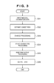
Fig. 3 is a flowchart showing a processing sequence in the diagnosis support apparatus; -
Fig. 4 is a view showing an output example from the diagnosis support apparatus; -
Fig. 5 is a flowchart showing a processing sequence for calculating the size of a lesion candidate in the diagnosis support apparatus; -
Fig. 6 is a view showing an example of data stored in a medical knowledge database in the diagnosis support apparatus; and -
Fig. 7 is a flowchart showing a processing sequence for searching for a similar image of a case in the diagnosis support apparatus. - The preferred embodiments of the present invention will be exemplarily described in detail below with reference to the accompanying drawings. Note however that the constituent elements described in the embodiments are merely examples, and the scope of the present invention is not limited to them.
- A
CPU 100 mainly controls the operation of each constituent element of adiagnosis support apparatus 1. Amain memory 101 stores control programs to be executed by theCPU 100 or provides a work area when theCPU 100 executes a program. Amagnetic disk 102 stores an operating system (OS), device drives for peripheral devices, various kinds of application software including programs for executing, for example, diagnosis support processing (to be described later), and the like. Adisplay memory 103 temporarily stores display data for amonitor 104. Themonitor 104 is, for example, a CRT monitor or a liquid crystal monitor, and displays images based on data from thedisplay memory 103. Amouse 105 and akeyboard 106 are operated by the user to perform pointing input operation and input characters and the like. The respective constituent elements described above are connected to each other via a common bus 107 so as to be communicable with each other. - In this embodiment, the diagnosis support
apparatus 1 can read out image data and the like from adatabase 3 via aLAN 4. Alternatively, it suffices to connect storage devices such as an FDD, CD-RW drive, MO drive, and ZIP drive to thediagnosis support apparatus 1 to allow it to read image data and the like from the drives. In addition, the diagnosis support apparatus can directly acquire medical images and the like from amedical imaging apparatus 2 via theLAN 4. - An example of the arrangement of the
diagnosis support apparatus 1 will be described with reference toFig. 2 .
Referring toFig. 2 , thediagnosis support apparatus 1 includes a medical examinationdata input unit 201, acase database 202, amedical knowledge database 203, a diagnosissupport processing unit 204 as a diagnosis support processing means, a processingmethod changing unit 207, ahistory recording unit 208 as a storage means, and adata output unit 209. - The medical examination
data input unit 201 acquires data concerning an object to be examined, for example, image data and measurement data such as an electrocardiogram, brain wave data, and leukocyte count, which are acquired from, for example, an X-ray imaging apparatus, CT apparatus, MR apparatus, and ultrasonic or sound diagnosis apparatus. - The medical examination data input unit may also be configured to input medical examination data including information associated with the acquisition of lesion candidates, for example, medical record information. In this case, this unit may be configured to either allow a user to directly input these data or read them from various types of storage media such as an FDD, CD-RW drive, MO drive, and ZIP drive on which information is recorded. Alternatively, the unit may be configured to be connected to a database storing these data via a LAN so as to receive them therefrom.
- The
case database 202 stores image data captured by the medical examinationdata acquisition apparatus 2, numerical data including electrocardiograms and leukocyte counts, and text data such as medical records on objects to be examined. Each case data can include data processing result values and a confirmed diagnosis result obtained by the diagnosis support apparatus. It is possible to use these pieces of information for an analogous case search.
In addition, the case database stores templates classified into categories used for recognition/identification processing. Such a template is used for pattern recognition processing to determine the type of lesion (e.g., malignant or benign). In addition, when a lesion is to be extracted, the template is used to extract a region which is a lesion based on the similarity (e.g., a correlation value) with data extracted from an object. - The
medical knowledge database 203 stores diagnosis criteria for lesion regions and information of diseases as examination targets including metastatic lesions and complications. This database may also store data including a diagnosis sequence such as a sequence for checking, upon detection of a primary lesion, the presence/absence of an associated disease.
The diagnosissupport processing unit 204 acquires information for supporting diagnosis from data obtained from the object. - When processing image data, a
processing unit 205 acquires information concerning a lesion from the data acquired from the object. This unit extracts, for example, a lesion such as a candidate region of a tumor shadow or a candidate region of a calculus.
When acquiring a lesion candidate as a candidate of a lesion, this unit can also compare similarities with past case data by referring to thecase database 202. In this case, theprocessing unit 205 calculates a feature amount from an image, and acquires a lesion candidate from the coincidence of the feature amount for each case stored in thecase database 202. - This processing unit may refer to data concerning diagnosis criteria and diagnosis sequences stored in the
medical knowledge database 203. Consider, for example, a diagnosis criterion when CT images are used. In this case, a CT value range corresponding to a lesion is set. It is highly possible that a lesion exists in the range. A lesion is therefore extracted from the range. In addition, for a diagnosis sequence, a diagnosis trial by a doctor is grammaticalized as a flowchart. For example, in the case of two-dimensional image data, a processing sequence required for the extraction of a region of interest from an object and computer processing such as the digitization of texture features in the region of interest is grammaticalized in accordance with the way of thinking of a doctor. - Note that the processing targets of the
processing unit 205 are not limited to image data. For example, the processing targets include measurement data other than images concerning an object or medical record data. The processing unit can be configured to acquire lesion candidates based on these data. - If image data is a target, an
output processing unit 206 acquires information for supporting diagnosis of the likelihood of a lesion or the like with respect to a lesion candidate extracted by theprocessing unit 205. In the case of measurement data, the output processing unit directly analyzes data obtained from the object and acquires and outputs information for supporting diagnosis. - The
history recording unit 208 records diagnosis history information concerning the object. As diagnosis history information, this unit records examination methods performed in the past, diagnosis results obtained by the methods, and the like. The unit also stores an onset probability estimated in the past, a next diagnosis period corresponding to the onset probability, and the like.
As an examination method, this unit also records the purpose information of an examination such as screening, close examination, or follow-up of a lesion. - The processing
method changing unit 207 determines processing methods for theprocessing unit 204 and theoutput processing unit 205 in accordance with the history information of the object which is obtained from thehistory recording unit 208.
Thedata output unit 209 outputs the data of a lesion candidate obtained by theprocessing unit 205 and the determination information of the lesion candidate obtained by theoutput processing unit 206 in a designated format. - The manner of how the
CPU 100 controls thediagnosis support apparatus 1 will be described next with reference to the flowchart ofFig. 3 .
In step S31, theCPU 100 inputs medical examination data from the medical examinationdata input unit 201 to thediagnosis support apparatus 1.
In addition, since different lesion detection criteria and different lesion detection sequences are used to acquire lesion candidates in accordance with examination purposes, the CPU acquires the examination history of the object (e.g., the history of examination from the day of previous imaging to the day of examination) and the information of an examination purpose from thehistory recording unit 208. - In step S32, the processing
method changing unit 207 estimates a current onset probability under the control of theCPU 100 in accordance with the history information of the object recorded on thehistory recording unit 208. In this case, the onset probability is the probability of the onset of a lesion. It also refers to the probability at which a lesion will change from benignity to malignancy. - A symptomatic change that can occur in accordance with the lapse of time after examination of the object is stored as probabilistically significant information according to past medical findings. Therefore, a symptomatic change can be predicted within a probabilistically significant range in accordance with information in the course of time. That is, it is possible to predict the symptomatic change of the object within a probabilistically significant range in accordance with the examination history of the object.
- When estimating an onset probability, the CPU calculates the progress degree of a lesion candidate by using the past and current examination data of the object, the time intervals of diagnosis such as a checkup count, history information such as an onset probability estimated by a past checkup, lesion candidate data as an image processing result, and the like which are recorded on the
history recording unit 208. - For example, the CPU predicts the current size of a lesion (a tumor or calculus) of the object from the information of the lesion recorded on the
history recording unit 208. This prediction is performed by using the past size and growth rate of the lesion. In this case, the CPU uses, as a growth rate, a statistic recorded on themedical knowledge database 203 by using the past size and type of a lesion. In addition, if a change in the size of a lesion is recorded on thehistory recording unit 208, it is possible to obtain the size of the lesion at a given time point in the past. Furthermore, if, for example, the predicted size is 5 mm or more, the CPU determines that the onset probability is high. In this case, the onset probability is the probability at which the extracted lesion candidate is malignant. The processingmethod changing unit 207 changes the size of a lesion to be extracted by theprocessing unit 205 in accordance with this onset probability as well. If this onset probability is high, since there is a possibility that a new lesion will develop, the processing unit extracts lesions of sizes in the entire range. In addition, in some cases, the processing function is changed in accordance with the size of a lesion. - If it is determined from the history of the object recorded on the
history recording unit 208 that the object is taking the first medical examination, the processing unit extracts lesions of sizes in the entire range, considering that the onset probabilities are high. - If the history of the object recorded on the
history recording unit 208 has no history of extraction of a lesion, the onset probability changes in accordance with the lapse of time after the previous diagnosis. If not much time has elapsed since the previous examination, the probability of the development of a large lesion significantly decreases. For this reason, the CPU changes a parameter in the processingmethod changing unit 207 to prevent theprocessing unit 205 from extracting a large lesion, considering that the onset probability is low. This can perform extraction in accordance with a specific size, and hence decreases the false positive rate of lesions extracted by theprocessing unit 205. - In this manner, the processing
method changing unit 207 changes the processing method used by the diagnosissupport processing unit 204 based on the history of the object recorded on thehistory recording unit 208. - The ways of changing the processing method used by the diagnosis
support processing unit 204 include, for example, the following methods: adjusting the probability distribution of reliability; adjusting a threshold for the probability of recognition of abnormality; changing a parameter of a recognition function such as a Bayesian discriminator; and moving an interface in a feature space in a discriminator. In addition, it is possible to change the determination algorithm itself. Selecting a support vector machine, Bayesian discriminator, neural network, or AdaBoost, other than a linear discriminant function, is also equivalent to changing the processing method. - For example, the following settings are made as onset probabilities: onset probability = 1.0: which is an onset probability set when no onset probability can be estimated or is regarded as a default, in which state, the parameter of a discriminator which is used as a default is used; onset probability = 0.9: to which the discriminator is set by decreasing the sensitivity of the discriminator such that false positives appear at a rate of 90% of that in the default state when the onset probability is low; and onset probability = 1.1: to which the discriminator is set by increasing the sensitivity of the discriminator such that false positives appear at a rate of 110% of that in the default state when the onset probability is high. The above settings are merely an example, and do not limit this embodiment.
- In step S33, the
processing unit 205 extracts a lesion candidate, under the control of theCPU 100, by the processing method determined in step S32. In this case, the CPU obtains lesion candidate data such as a reliability indicating the likelihood of a lesion and the progress degree of the lesion from the feature amount calculated from the lesion candidate by using the information of a diagnosis criterion stored in themedical knowledge database 203. In this case, the reliability indicates, for example, the likelihood of a lesion based on a correlation value with the feature amount obtained from a past lesion and the like. - In the case of a chest CT image, the
processing unit 205 segments the image into regions such as a lung field region, diaphragm region, bronchus region, pulmonary artery region, and pulmonary vein region, and classifies the lung field into a superior lobe segment, median lobe segment, and inferior lobe segment. As an example of a method of detecting an organ region from a medical image, a level set method which is a kind of dynamic contour method will be described below. The level set method defines a level set function which is one dimension higher than a detection target region, and regards a region which is to be extracted as its zero contour line. Updating this function based on the following evolution equation called a level set equation will control a contour and detect a region.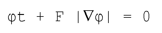
where ϕt represents the value obtained by calculating the first derivative of the level set function, F represents the growth rate of the contour, and |∇ϕ| represents the absolute value of the gradient of the level set function. - In this manner, an organ region can be detected from the medical image. The detection of an organ region has been described above by exemplifying the level set method. However, as a region detection method, the following is available: a method based on threshold processing, region growing, dynamic contour method, clustering, graph minimum cut algorithm, or the like. An organ region is detected by using one of these methods or other techniques.
These methods can be switched and used in accordance with the region to be detected. It suffices to perform region detection by using a probability atlas, a human body shape model, and the like as prior knowledge as well as an image feature amount. - The following are available as methods of detecting an abnormality such as a lung field tumor from an organ region: filter processing for detecting an abnormality, pattern matching, abnormality detection using a discriminator, and difference detection processing by registration between, for example, past images or average shape images and diagnosis images.
In addition, image feature amounts for specifying a lung field tumor include a shape index and curvedness obtained from the CT value of each pixel inside the tumor and a three-dimensional curvature (a Gaussian curvature, average curvature, or principal curvature). - A lesion candidate is detected by using one of the above techniques or other techniques.
- In step S35, the
CPU 100 causes theoutput processing unit 206 to evaluate the lesion candidate detected by theprocessing unit 205 in accordance with the processing method determined by the processingmethod changing unit 207.
In order to perform disease classification and benign/malignant discrimination of abnormalities, malignancy probabilities (probability values) are subjected to disease classification and benign/malignant discrimination using a discriminator using a discrimination method (e.g., a linear discriminant function, support vector machine, AdaBoost, Bayesian discriminator, or neural network). The method to be used for the abnormality detection, disease classification, and benign/malignant discrimination is not limited to the above technique. - It is further possible to give more consideration to a feature amount itself or replacing it as well as changing the weight of a feature amount used in the discriminator.
- The following is a case in which benign/malignant discrimination of a lung field tumor candidate is performed by applying the feature amount extracted from an X-ray CT image to the following linear discriminant function. That is, lesion candidates are classified into true lesions and false positives.

classes
The value of a linear discriminant function f is regarded as a discriminant score. When this value is negative, the lesion candidate is classified into class 1 (benign). When the value is positive, the lesion candidate is classified into class 2 (malignant). The CPU then changes the parameter ΣW of the above linear discriminant function in accordance with the onset probability. That is, when determining that the onset probability is high, the CPU sets ΣW so as to allow f to output more positive values. When determining that the onset probability is low, the CPU sets ΣW so as to allow f to output more negative values. - That is, upon estimating that the onset probability is high, the CPU makes more lesion candidates attract attention. That is, the CPU increases the sensitivity of the
output processing unit 206. In contrast, upon estimating that the onset probability is low, in order to reduce the number of false positive lesion candidates, the CPU decreases the sensitivity to present fewer lesion candidates which are not relatively important. - It suffices to perform the processing in the
output processing unit 206 in conjunction with the sensitivity of theprocessing unit 205 or independently. That is, it is possible to extract many tumor shadows by maximizing the sensitivity of theprocessing unit 205 and change the threshold in theoutput processing unit 206 so as to decrease the number of false positive lesion candidates. Alternatively, it is possible to perform this processing in conjunction with the sensitivity of theprocessing unit 205. - In step S36, the CPU narrows down the lesion candidate data detected in step S32 to the lesion candidate data which are determined in step S35 to satisfy the criterion of recognition of a lesion, and outputs them.
- In this case, the
data output unit 209 converts the above data into output data corresponding to an output destination under the control of theCPU 100. The output destination includes a sheet, a storage device such as a memory or a hard disk, and a monitor. -
Fig. 4 shows an output example of lesion candidate data. A mark indicating a lesion candidate is displayed on medical examination data. Image feature amounts and patient attribute/time-varying data are displayed next to the medical examination data. In addition, when lesion candidate data is to be output to a display device such as a monitor, it suffices to display the data in a popup form or in another window. - A method of predicting the size of a lesion candidate in step S33 which is executed by the processing
method changing unit 207 described above will be described next with reference toFig. 5 . Thehistory recording unit 208 records a predicted size as history information of the object to allow the information to be used for the next diagnosis processing or the determination of a next diagnosis timing. - A prediction method based on the size and growth rate of a tumor as an example will be described below. In this case, the longest diameter is used as the size of a tumor.
- In step S51, the processing method changing unit acquires the tumor detected in step S32 from the current chest image data, and calculates the longest diameter of the tumor.
- In step S52, the processing method changing unit checks whether the past chest image data or past diagnosis report of the object includes a description of a lesion corresponding to the tumor acquired in step S51. If there is a description of the lesion in the past, the process advances to step S53.
- If the lesion candidate detected in step S51 is detected for the first time by this checkup, the process advances to step S55.
- In step S53, the processing method changing unit acquires the past information of the tumor from the
history recording unit 208. The information to be acquired includes the size (the longest diameter in this case), diagnosis on the case, and the reliability of the diagnosis. The diagnosissupport processing unit 204 may calculate this information if it is not described in the past diagnosis report. - In step S54, the processing method changing unit calculates a growth rate from the current and past longest diameter values of the corresponding tumor and the time period from the past checkup according to the following equation. Although the following equation is used as an example in this case, the present invention is not limited to this. For example, it suffices to use another method of calculating a growth rate, which is accepted in the medical field, like a method using a mathematical expression additionally including a weight and an adjustment term in accordance with the region to be processed.

where VC is a growth rate, MRpresent is the current longest diameter value of the tumor, MRpast is the longest diameter value of the corresponding tumor at the time of a past examination, and t is the time elapsed since the past examination. - In step S55, if there is no past information of the detected tumor, the processing method changing unit uses the value of the growth rate of the tumor which is empirically known in the medical field. Obviously, it suffices to use a mathematical expression or method of deriving the growth rate of the tumor in accordance with other examination data of the object or information such as a past case.
- In step S56, the processing method changing unit acquires a next scheduled checkup date (next checkup timing) for the object which is recorded on the
history recording unit 208, and estimates the size of the tumor at the time of the next checkup based on the current size of the tumor, the growth rate, and the number of days to the next checkup. For example, as a method of estimating the size of a tumor, the following equation is available. However, the method is not limited to the equation, and the size of the tumor may be estimated in accordance with empirical values summarized in a table as medical knowledge.
where MRfuture is the prediction of the size of the tumor at the time of the next scheduled checkup, and tf is the time to the next scheduled checkup. - In step S57, the processing method changing unit estimates an onset probability by comparing the prediction of the size of the tumor at the time of the next checkup, which is calculated in step S56, with a medical guideline. If, for example, the size of the tumor is predicted to be less than 5 mm, it can be determined that the onset probability is low. If the size of the tumor is predicted to be 10 mm or more, it can be determined that the onset probability is high.
- With the above processing shown in
Fig. 5 , it is possible to predict the size of a lesion at the time of a next checkup. Using this information allows the processingmethod changing unit 207 to estimate an onset probability. - In the above description, the size of a tumor is expressed by its longest diameter. However, the present invention is not limited to this as long as the size and growth rate of a tumor can be expressed. For example, the volume of the tumor can be used.
- As a method of estimating an onset probability in step S32, an example using medical importances will be described next.
- The processing
method changing unit 207 estimates an onset probability in accordance with the medical importance of a lesion candidate acquired by theprocessing unit 205. Alternatively, it is possible to estimate an onset probability in accordance with an interval change in the medical importance of a lesion candidate. - A medical importance I calculated by the processing
method changing unit 207 will be described below.
The medical importance I can be defined as follows:
where
A: important morbidity (relative disease severity among different kinds of diseases)
B: progress degree (stage) (disease severity within same kind of disease)
C: associated morbidity - If, for example, the
processing unit 205 is to detect a plurality of kinds of diseases, the associated morbidity C is set for each lesion in accordance with the examination purpose as follows. That is
Fig. 6 shows examples of the important morbidity A and the progress degree B. - Assume that in the case of follow-up of a lung cancer, the
processing unit 205 has detected one malignant tumor (stage 0) in the lung field as a lesion candidate. In this case, a medical importance IC concerning the malignant tumor is obtained as follows:
- Subsequently, in order to estimate an onset probability, the processing method changing unit compares this medical importance with the medical importance of a corresponding lesion of the patient in the past. If the
history recording unit 208 indicates that it was determined in a past examination that the same tumor was determined as a benign tumor, the processing method changing unit obtains a past medical importance IP of the tumor as follows:
That is, since the medical importance has increased within a given time period, it is estimated that the onset probability is high. Alternatively, even if the medical importance has not changed, it is estimated that the onset probability is high, when the medical importance is kept at a value equal to or more than a given value for a predetermined time period. - Furthermore, in order to estimate an onset probability based on changes in medical importance described above, it is also possible to use a table (medical importances, changes in medical importance, and time periods) formed from cases collected by medical researches.
- Note that the contents of medical importances described above are only an example, and are not limited by the definition in this embodiment. For example, it suffices to give consideration to examination purpose information concerning screening, close examination, and follow-up when estimating an onset probability based on a medical importance.
- A method of estimating an onset probability by using the probability (called a certainty factor in some cases) of a lesion candidate being a lesion will be described next.
In this case, a method of calculating the probability of a lesion candidate being a lesion includes detecting a lesion candidate by a similar image search. A processing sequence for a similar image search will be described with reference toFig. 7 . - In step S71, the
processing unit 205 calculates image feature amounts by processing medical examination data. Image feature amounts include, for example, the luminance distribution obtained from general image processing and the 2D or 3D feature amount of a region of interest in an image. Alternatively, it suffices to use a "shape index" value and a "curvedness" value. - In step S72, a case represented by an image having a feature amount similar to the feature amount of the image calculated by the
processing unit 205 is acquired by searching thecase database 202 using the calculated feature amount. In addition, cases are stored in advance in association with feature amounts, and the information of a case is acquired from a feature amount with a case. - In step S73, found images are arranged in decreasing order of similarity of feature amounts, and the similarities of the images are calculated in decreasing order of similarity. In this case, a value used as an example of similarity is the inner product value of the normalized feature vectors of a reference image and a similar image in a feature space used for a search. That is, in the feature space, the closer the feature amounts of a reference image and similar image, the more similar the two images. Note that the method of calculating similarities described above is merely an example, and the present invention is not limited to the method in this embodiment.
- In step S74, a finding associated with a case represented by the found image or the feature amount obtained from the image is acquired. Elements A, B, and C for obtaining the above medical importances are respectively attached to findings. For example, cancers include cancers with high progress rates and highly metastatic cancers, which are malignant and high in medical importance. In addition, the certainty factor of a selected case is acquired when it is selected by pattern matching. In this case, a certainty factor indicates the probability of a case being malignant when a plurality of doctors diagnose the case. In addition, it suffices to set a success ratio (the probability that a finding from a doctor coincides with an output finding from a discriminant function) obtained when a case is determined by the discriminant function as a certainty factor which is a statistic. A change T in lesion similarity is defined based on the obtained similarity and the certainty factor of the case as follows:

where Spresent is the similarity obtained by the current examination, Cpresent is the certainty factor obtained by the current examination, Spast is the similarity of a corresponding case of the patient in the past which is recorded on the history recording unit, and Cpast is the certainty factor of the corresponding case of the patient in the past which is recorded on the history recording unit. If the change T described above has increased within a given time period, it is estimated that the onset probability is high. - It is also possible to use, for example, a support vector machine or another discriminator used for pattern recognition, other than the above method, to calculate the probability of a lesion candidate. That is, the calculation method to be used is not limited to the above method using similarities.
- As described above, according to the first embodiment, considering the history of a patient can adjust the sensitivity of diagnosis support and reduce the load of interpretation on a doctor. This makes it possible to reduce the load on the patient because oversight or unnecessary biopsies are reduced.
- The first embodiment has exemplified the case in which the sensitivity of diagnosis support is adjusted based on the onset probability of a patient. However, it is assumed that examination data obtained from a medical examination data acquisition apparatus is the one obtained with standard parameters.
- The second embodiment will describe a way of changing an acquisition parameter in a medical examination data acquisition apparatus in accordance with the onset probability of a patient.
- Although it depends on the type of medical examination data acquisition apparatus, increasing the sharpness of acquired examination data may increase the load (the invasiveness of examination) on the patient. In the case of an X-ray CT apparatus, for example, suppressing the exposure dose of the patient by decreasing the voltage or current amount of an X-ray tube will increase noise in an obtained slice image. As a method of reducing the overall exposure dose of the patient without reducing the dose of the X-ray tube (X-ray source), it is conceivable to increase the moving speed of the bed. However, the intervals between slice images increase, and hence there is a possibility that a lesion may not be depicted, falling outside a slice image.
- This embodiment changes an acquisition parameter in the medical examination data acquisition apparatus in accordance with the onset probability of a patient which is calculated in the first embodiment.
- In the X-ray CT apparatus, when it is determined that the onset probability is low, the dose of the X-ray source is reduced or the slice intervals are increased. If it is determined that the onset probability is high, the X-ray dose is increased to obtain sharper medical images or the slice intervals are decreased.
If the onset probability at a specific region is high, it is possible to image only the region with high sharpness or examine other regions by a method with a less load. - It is also conceivable to change the calculation algorithm for examination data. In the case of an X-ray CT apparatus, since the reconstruction algorithm can be changed, an algorithm that can obtain sharper images may be selected, even if it is slow, when the onset probability is high, whereas when the onset probability is low, a fast algorithm may be selected.
- In addition, it suffices to adjust an acquisition parameter such as the amount of contrast medium for MRI. Furthermore, an onset probability can be used to select a medical examination data acquisition apparatus. For example, if it is determined that the onset probability is high, it is possible to perform examination more suitable for the case (e.g., PET).
- As described above, according to the second embodiment, adjusting an acquisition parameter in the examination apparatus in accordance with the onset probability estimated from the history of a patient can acquire more detailed features of the case.
- The second embodiment has described that it is possible to detect a lesion with a high onset probability more accurately by changing an acquisition parameter in the medical examination data acquisition apparatus in accordance with the onset probability of a patient.
- Note that final diagnosis is performed by a doctor qualified to perform diagnosis such as an interpreting doctor or an attending physician. In order to confirm the diagnosis, therefore, the image diagnosis support apparatus needs to present not only the information of a lesion candidate but also the information of a criterion for regarding a lesion as a candidate.
- In this case, an image
diagnosis support apparatus 1 presents the doctor the onset probability of a patient, a change in sensitivity parameter (a changed parameter, its value, or a changed algorithm), and the reason and ground of the change, together with the related information of the detected lesion candidate and an interpretation report. As a result output method, it suffices to display the result on the screen of a terminal used for interpretation as well as an interpretation report as a paper medium or to use other presentation methods. - As described above, according to the third embodiment, presenting the onset probability of a sensitivity parameter and change information together with a diagnosis report makes it possible to perform more accurate diagnosis. In addition, a doctor can comprehend the health of a patient better and manage patient information.
- Obviously, the object of the present invention is also implemented by supplying a computer-readable storage medium storing programs for implementing the functions of the above embodiments to a system or apparatus, and causing the computer (or a CPU or an MPU) of the system or apparatus to read out and execute the program codes stored in the storage medium. In this case, the program codes read out from the storage medium implement the functions of the above embodiments by themselves, and the storage medium storing the program codes constitutes the present invention.
- As a storage medium for supplying the program codes, a floppy disk, hard disk, optical disk, magnetooptical disk, CD-ROM, CD-R, magnetic tape, nonvolatile memory card, ROM, or the like can be used.
- The functions of the above embodiments are implemented not only when the readout program codes are executed by the computer but also when the OS (Operating System) running on the computer performs part or all of actual processing based on the instructions of the program codes. Obviously, the present invention incorporates a case in which the function of the above embodiments are implemented by this processing.
- Obviously, the present invention also incorporates a case in which the functions of the above embodiments are also implemented when the program codes read out from the storage medium are written in the memory of a function expansion board inserted into the computer or a function expansion unit connected to the computer, and the
CPU 100 or the like of the function expansion board or function expansion unit performs part or all of actual processing based on the instructions of the program codes. - Note that the description of this embodiment is a preferred example of the diagnosis support apparatus according to the present invention, and the present invention is not limited to this.
- The arrangement of the present invention can provide a mechanism for obtaining diagnostic information by computer processing with consideration given to the examination history of an object.
- The present invention is not limited to the above embodiments and various changes and modifications can be made within the spirit and scope of the present invention. Therefore, to apprise the public of the scope of the present invention, the following claims are made.
- This application claims the benefit of Japanese Patent Application No.
2007-271300, filed October 18, 2007
Claims (10)
- A diagnosis support apparatus comprising:diagnosis support processing means for processing data obtained from an object to extract diagnosis support information;storage means for storing an examination history of the object; andchanging means for changing a processing method in said diagnosis support processing means in accordance with the examination history of the object stored in said storage means.
- The diagnosis support apparatus according to claim 1, characterized by further comprising a case database which associates and stores a feature amount of a lesion and a finding,
wherein said diagnosis support processing means calculates a feature amount from an extracted lesion and obtains a medical importance of the lesion from a similarity of a feature amount with a case stored in said case database. - The diagnosis support apparatus according to claim 2, characterized in that
said case database stores an important morbidity which is a relative disease severity among different kinds of diseases as A, a progress degree as B, and an associated morbidity as C, and
said diagnosis support processing means calculates a medical importance I from an equation of I = A*B*C. - The diagnosis support apparatus according to claim 1, characterized in that said changing means changes a size of a lesion as a processing target in said diagnosis support processing means in accordance with the examination history of the object.
- The diagnosis support apparatus according to claim 1, characterized in that said changing means calculates a size of a lesion as a processing target in accordance with a size of the lesion of the object stored in said storage means and an elapsed time from a day when the lesion is imaged to an examination day, and changes the size of the lesion as the processing target as a processing method in said diagnosis support processing means.
- The diagnosis support apparatus according to claim 1, characterized in that said changing means calculates an onset probability of the lesion in accordance with the examination history of the object and changes a processing method in said diagnosis support processing means in accordance with the onset probability.
- The diagnosis support apparatus according to claim 1, characterized in that said changing means changes one of a parameter of a processing function used in said diagnosis support processing means, an algorithm of the processing function, and a threshold for an output value from the processing function.
- The diagnosis support apparatus according to claim 1, characterized by further comprising a medical examination data acquisition apparatus which acquires data from the object,
wherein said changing means changes an acquisition parameter in said medical examination data acquisition apparatus. - The diagnosis support apparatus according to claim 1, characterized by further comprising data output means,
wherein one of change information of the processing method in said diagnosis support processing means and a processing result obtained by said diagnosis support processing means is output. - A control method for a diagnosis support apparatus including storage means for storing an examination history of an object, characterized by comprising:a changing step of changing a processing method in accordance with the examination history of the object stored in the storage means; anda diagnosis support processing step of processing data obtained from the object to extract diagnosis support information by using the changed processing method.
Applications Claiming Priority (2)
| Application Number | Priority Date | Filing Date | Title |
|---|---|---|---|
| JP2007271300A JP5159242B2 (en) | 2007-10-18 | 2007-10-18 | Diagnosis support device, diagnosis support device control method, and program thereof |
| PCT/JP2008/066238 WO2009050962A1 (en) | 2007-10-18 | 2008-09-09 | Diagnosis support device, method for controlling diagnosis support device, and its program |
Publications (2)
| Publication Number | Publication Date |
|---|---|
| EP2213224A1 true EP2213224A1 (en) | 2010-08-04 |
| EP2213224A4 EP2213224A4 (en) | 2016-12-07 |
Family
ID=40567238
Family Applications (1)
| Application Number | Title | Priority Date | Filing Date |
|---|---|---|---|
| EP08840701.0A Withdrawn EP2213224A4 (en) | 2007-10-18 | 2008-09-09 | Diagnosis support device, method for controlling diagnosis support device, and its program |
Country Status (6)
| Country | Link |
|---|---|
| US (2) | US9852269B2 (en) |
| EP (1) | EP2213224A4 (en) |
| JP (1) | JP5159242B2 (en) |
| KR (1) | KR101216259B1 (en) |
| CN (1) | CN101742961B (en) |
| WO (1) | WO2009050962A1 (en) |
Cited By (2)
| Publication number | Priority date | Publication date | Assignee | Title |
|---|---|---|---|---|
| JP2009095550A (en) * | 2007-10-18 | 2009-05-07 | Canon Inc | Diagnosis support device, diagnosis support device control method, and program thereof |
| US11238588B2 (en) * | 2016-04-13 | 2022-02-01 | Canon Kabushiki Kaisha | Medical diagnosis support apparatus, information processing method, medical diagnosis support system, and program |
Families Citing this family (75)
| Publication number | Priority date | Publication date | Assignee | Title |
|---|---|---|---|---|
| JP5178119B2 (en) | 2007-09-28 | 2013-04-10 | キヤノン株式会社 | Image processing apparatus and image processing method |
| CN108181457B (en) | 2009-03-12 | 2021-03-30 | 癌症预防和治疗有限公司 | Methods and kits for identifying, assessing, preventing and treating lung diseases |
| JP5478328B2 (en) * | 2009-09-30 | 2014-04-23 | 富士フイルム株式会社 | Diagnosis support system, diagnosis support program, and diagnosis support method |
| JP5523891B2 (en) | 2009-09-30 | 2014-06-18 | 富士フイルム株式会社 | Lesion region extraction device, its operating method and program |
| JP5582772B2 (en) * | 2009-12-08 | 2014-09-03 | キヤノン株式会社 | Image processing apparatus and image processing method |
| JP5655327B2 (en) * | 2010-03-09 | 2015-01-21 | 富士ゼロックス株式会社 | Program and information processing apparatus |
| JP5320335B2 (en) * | 2010-03-31 | 2013-10-23 | 富士フイルム株式会社 | Diagnosis support system, diagnosis support apparatus, diagnosis support method, and diagnosis support program |
| CN101877035B (en) * | 2010-04-22 | 2015-09-09 | 无锡市优特科科技有限公司 | Based on the electrocardiograph system of goldstandard database |
| CN102883660A (en) * | 2010-09-20 | 2013-01-16 | 德克萨斯州大学系统董事会 | Advanced multimedia structured reporting |
| JP5670695B2 (en) * | 2010-10-18 | 2015-02-18 | ソニー株式会社 | Information processing apparatus and method, and program |
| JP5557687B2 (en) * | 2010-10-21 | 2014-07-23 | 株式会社日立メディコ | Inspection plan creation device |
| JP2013545520A (en) * | 2010-10-29 | 2013-12-26 | ザ・ジョンズ・ホプキンス・ユニバーシティ | Image search engine |
| AU2012249288C1 (en) * | 2011-04-29 | 2017-12-21 | Lung Cancer Proteomics Llc | Methods of identification and diagnosis of lung diseases using classification systems and kits thereof |
| JP5744631B2 (en) * | 2011-06-06 | 2015-07-08 | キヤノン株式会社 | Medical support device, medical support method |
| JP6193225B2 (en) * | 2011-06-29 | 2017-09-06 | インナー モンゴリア フルイ メディカル サイエンス カンパニー リミテッドInner Mongolia Furui Medical Science Co., Ltd | Liver fibrosis detection device and detection system |
| US20140306992A1 (en) * | 2011-12-26 | 2014-10-16 | Canon Kabushiki Kaisha | Image processing apparatus, image processing system and image processing method |
| JP6226510B2 (en) | 2012-01-27 | 2017-11-08 | キヤノン株式会社 | Image processing system, processing method, and program |
| JP5750381B2 (en) * | 2012-02-13 | 2015-07-22 | 株式会社日立製作所 | Region extraction processing system |
| JP6021346B2 (en) * | 2012-02-14 | 2016-11-09 | キヤノン株式会社 | Diagnosis support apparatus and control method thereof |
| JP5802144B2 (en) * | 2012-02-16 | 2015-10-28 | 株式会社日立製作所 | Image diagnosis support system, program, and storage medium |
| BR112014020040A8 (en) * | 2012-02-17 | 2017-07-11 | Koninklijke Philips Nv | NON-TRANSITORY STORAGE MEDIA STORING INSTRUCTIONS EXECUTED BY AN ELECTRONIC DATA PROCESSING DEVICE INCLUDING A DISPLAY FOR MONITORING A PATIENT WITH ACUTE LUNG INJURY, APPARATUS, AND METHOD |
| JP6106259B2 (en) * | 2012-03-21 | 2017-03-29 | コーニンクレッカ フィリップス エヌ ヴェKoninklijke Philips N.V. | Clinical workstation integrating medical imaging and biopsy data and method of using the same |
| WO2014062648A1 (en) * | 2012-10-15 | 2014-04-24 | ClinicalBox, Inc. | Adaptive medical testing |
| US20150269339A1 (en) * | 2012-10-15 | 2015-09-24 | ClinicalBox, Inc. | Adaptive Medical Testing |
| KR102043130B1 (en) | 2012-11-16 | 2019-11-11 | 삼성전자주식회사 | The method and apparatus for computer aided diagnosis |
| US9378551B2 (en) * | 2013-01-03 | 2016-06-28 | Siemens Aktiengesellschaft | Method and system for lesion candidate detection |
| KR20140091176A (en) | 2013-01-10 | 2014-07-21 | 삼성전자주식회사 | Apparatus and method for lesion diagnosis |
| US9730655B2 (en) * | 2013-01-21 | 2017-08-15 | Tracy J. Stark | Method for improved detection of nodules in medical images |
| JP6208243B2 (en) * | 2013-08-27 | 2017-10-04 | 株式会社日立製作所 | Morbidity assessment device, morbidity assessment method, and morbidity assessment program |
| JP6612861B2 (en) * | 2014-07-02 | 2019-11-27 | コーニンクレッカ フィリップス エヌ ヴェ | System and method for identifying an organization |
| US11147652B2 (en) | 2014-11-13 | 2021-10-19 | Align Technology, Inc. | Method for tracking, predicting, and proactively correcting malocclusion and related issues |
| CN104484845B (en) * | 2014-12-30 | 2019-03-05 | 天津迈沃医药技术股份有限公司 | Disease autoanalysis platform based on medical information ontology database |
| US20160188823A1 (en) * | 2014-12-31 | 2016-06-30 | General Electric Company | System for providing electrocardiogram (ecg) analytics for electronic medical records (emr) |
| US9824189B2 (en) * | 2015-01-23 | 2017-11-21 | Panasonic Intellectual Property Management Co., Ltd. | Image processing apparatus, image processing method, image display system, and storage medium |
| KR101748019B1 (en) | 2015-02-03 | 2017-06-15 | 부산대학교 산학협력단 | Device and method for providing of medical information |
| JP6301277B2 (en) * | 2015-03-20 | 2018-03-28 | 富士フイルム株式会社 | Diagnostic auxiliary image generation apparatus, diagnostic auxiliary image generation method, and diagnostic auxiliary image generation program |
| JP6408436B2 (en) * | 2015-07-29 | 2018-10-17 | 富士フイルム株式会社 | Medical support device, its operating method and operating program, and medical support system |
| JP6755130B2 (en) * | 2016-06-21 | 2020-09-16 | 株式会社日立製作所 | Image processing equipment and method |
| US20180060535A1 (en) * | 2016-08-31 | 2018-03-01 | International Business Machines Corporation | Updating probabilities of conditions based on annotations on medical images |
| US11045663B2 (en) * | 2016-11-02 | 2021-06-29 | Shimadzu Corporation | X-ray fluoroscopy device and x-ray fluoroscopy method |
| CN110168657B (en) * | 2016-12-05 | 2024-03-12 | 皇家飞利浦有限公司 | Tumor tracking with smart tumor size change notifications |
| JP6706345B2 (en) * | 2016-12-19 | 2020-06-03 | 富士フイルム株式会社 | Similar case retrieval device, its operating method and operating program, and similar case retrieval system |
| US11397655B2 (en) * | 2017-02-24 | 2022-07-26 | Hitachi, Ltd. | Abnormality diagnosis system that reconfigures a diagnostic program based on an optimal diagnosis procedure found by comparing a plurality of diagnosis procedures |
| CN110709936A (en) | 2017-04-04 | 2020-01-17 | 肺癌蛋白质组学有限责任公司 | Plasma-based protein profiling for early-stage lung cancer prognosis |
| RU2667879C1 (en) * | 2017-05-30 | 2018-09-24 | Общество с ограниченной ответственностью "Аби Продакшн" | Processing and analysis of data on computer-assisted tomography images |
| JP7258846B2 (en) * | 2017-07-31 | 2023-04-17 | コーニンクレッカ フィリップス エヌ ヴェ | Devices, Systems, and Methods for Determining When to Select Selective Echocardiography |
| JP7224757B2 (en) * | 2017-10-13 | 2023-02-20 | キヤノン株式会社 | Diagnosis support device, information processing method, diagnosis support system and program |
| JP6872470B2 (en) * | 2017-11-14 | 2021-05-19 | 株式会社日立製作所 | Image processing equipment, image processing method, image processing program and ultrasonic imaging equipment |
| CN108888247B (en) * | 2018-05-22 | 2021-11-05 | 广东乐心医疗电子股份有限公司 | Aerobic capacity testing method, device and system and data acquisition equipment |
| CN109003670A (en) * | 2018-06-29 | 2018-12-14 | 武汉圣大东高科技有限公司 | Big data medical information processing method, system, terminal device and storage medium |
| JP7467033B2 (en) * | 2018-07-04 | 2024-04-15 | キヤノンメディカルシステムズ株式会社 | Medical information processing device |
| US11003939B2 (en) | 2018-07-06 | 2021-05-11 | Canon Kabushiki Kaisha | Information processing apparatus, information processing method, and storage medium |
| JP7091174B2 (en) * | 2018-07-09 | 2022-06-27 | キヤノン株式会社 | System, system control method and program |
| CN110867241B (en) * | 2018-08-27 | 2023-11-03 | 卡西欧计算机株式会社 | Image-like display control device, system, method, and recording medium |
| WO2020044735A1 (en) * | 2018-08-31 | 2020-03-05 | 富士フイルム株式会社 | Similarity determination device, method, and program |
| JP7187244B2 (en) * | 2018-10-10 | 2022-12-12 | キヤノンメディカルシステムズ株式会社 | Medical image processing device, medical image processing system and medical image processing program |
| JP7109345B2 (en) | 2018-11-20 | 2022-07-29 | 富士フイルム株式会社 | Priority determination device, method and program |
| CN113164141A (en) * | 2018-11-27 | 2021-07-23 | 富士胶片株式会社 | Similarity determination device, method, and program |
| DE112019005902T5 (en) * | 2018-11-27 | 2021-09-30 | Fujifilm Corporation | SIMILARITY DETERMINATION DEVICE, SIMILARITY DETERMINATION METHOD, AND SIMILARITY DETERMINATION PROGRAM |
| JP7289638B2 (en) * | 2018-11-29 | 2023-06-12 | キヤノン株式会社 | Medical information processing system and program |
| CN109712705A (en) * | 2018-12-19 | 2019-05-03 | 中国石油大学(华东) | A kind of cholelithiasis intelligent diagnostics APP based on deep learning |
| KR102231677B1 (en) * | 2019-02-26 | 2021-03-24 | 사회복지법인 삼성생명공익재단 | Device for predicting Coronary Arterial Calcification Using Probabilistic Model, the prediction Method and Recording Medium |
| CN110085299B (en) * | 2019-04-19 | 2020-12-08 | 合肥中科离子医学技术装备有限公司 | Image identification dryness removal method and system and image library |
| US11790524B2 (en) * | 2019-06-20 | 2023-10-17 | Ohio State Innovation Foundation | System and method for quantitative volumetric assessment and modeling of tumor lesions |
| CN110309915B (en) * | 2019-07-05 | 2021-12-10 | 中国人民解放军国防科技大学 | Histogram-based neuron positioning abnormal layer processing method, system and medium |
| WO2021246047A1 (en) * | 2020-06-04 | 2021-12-09 | 富士フイルム株式会社 | Progression prediction device, method, and program |
| JP7483018B2 (en) | 2020-09-04 | 2024-05-14 | 富士フイルム株式会社 | Image processing device, image processing method and program, and image processing system |
| CN112256754A (en) * | 2020-10-19 | 2021-01-22 | 柳州市妇幼保健院 | Ultrasonic detection and analysis system and method based on standard model |
| US20220164951A1 (en) | 2020-11-20 | 2022-05-26 | Hologic, Inc. | Systems and methods for using ai to identify regions of interest in medical images |
| US12150776B2 (en) | 2021-04-26 | 2024-11-26 | Wisconsin Alumni Research Foundation & AIQ Solutions, Inc. | System and method for monitoring multiple lesions |
| JP7852212B2 (en) * | 2021-06-29 | 2026-04-28 | 大日本印刷株式会社 | Methods for operating diagnostic support devices, computer programs, and medical devices |
| CN113496774A (en) * | 2021-07-22 | 2021-10-12 | 河北北方学院 | Digital medical information management method and related device |
| US12567496B2 (en) | 2022-12-08 | 2026-03-03 | Optellum Limited | Method and apparatus for displaying and analyzing medical scan images |
| JP2025048914A (en) * | 2023-09-21 | 2025-04-03 | ソフトバンクグループ株式会社 | system |
| CN117116472B (en) * | 2023-10-25 | 2024-10-11 | 首都医科大学附属北京友谊医院 | Medical diagnostic apparatus, electronic device, and storage medium |
Family Cites Families (35)
| Publication number | Priority date | Publication date | Assignee | Title |
|---|---|---|---|---|
| US5260871A (en) * | 1991-07-31 | 1993-11-09 | Mayo Foundation For Medical Education And Research | Method and apparatus for diagnosis of breast tumors |
| US5289374A (en) | 1992-02-28 | 1994-02-22 | Arch Development Corporation | Method and system for analysis of false positives produced by an automated scheme for the detection of lung nodules in digital chest radiographs |
| US6075879A (en) * | 1993-09-29 | 2000-06-13 | R2 Technology, Inc. | Method and system for computer-aided lesion detection using information from multiple images |
| US5483443A (en) * | 1994-04-08 | 1996-01-09 | Promt Medical Systems | Method for computing current procedural terminology codes from physician generated documentation |
| US6317617B1 (en) * | 1997-07-25 | 2001-11-13 | Arch Development Corporation | Method, computer program product, and system for the automated analysis of lesions in magnetic resonance, mammogram and ultrasound images |
| US6418238B1 (en) * | 1997-09-22 | 2002-07-09 | Olympus Optical Co., Ltd. | Image detection apparatus and image detection method capable of detecting roundish shape |
| US6941323B1 (en) * | 1999-08-09 | 2005-09-06 | Almen Laboratories, Inc. | System and method for image comparison and retrieval by enhancing, defining, and parameterizing objects in images |
| US6901156B2 (en) * | 2000-02-04 | 2005-05-31 | Arch Development Corporation | Method, system and computer readable medium for an intelligent search workstation for computer assisted interpretation of medical images |
| JP2001357381A (en) * | 2000-06-12 | 2001-12-26 | Fuji Photo Film Co Ltd | Method and system for detecting abnormal shade candidate |
| US6909792B1 (en) * | 2000-06-23 | 2005-06-21 | Litton Systems, Inc. | Historical comparison of breast tissue by image processing |
| US7343305B2 (en) * | 2001-05-03 | 2008-03-11 | University Of Florida Research Foundation, Inc. | Method and system for recording carious lesions |
| WO2003040990A2 (en) | 2001-11-02 | 2003-05-15 | Siemens Medical Solutions Usa, Inc. | Patient data mining for quality adherence |
| US20050207630A1 (en) * | 2002-02-15 | 2005-09-22 | The Regents Of The University Of Michigan Technology Management Office | Lung nodule detection and classification |
| JP4087640B2 (en) * | 2002-05-14 | 2008-05-21 | 富士フイルム株式会社 | Disease candidate information output system |
| JP2004046594A (en) * | 2002-07-12 | 2004-02-12 | Konica Minolta Holdings Inc | Image diagnosis support device |
| US20040122702A1 (en) * | 2002-12-18 | 2004-06-24 | Sabol John M. | Medical data processing system and method |
| EP1636731A2 (en) * | 2003-06-25 | 2006-03-22 | Siemens Medical Solutions USA, Inc. | Systems and methods for automated diagnosis and decision support for breast imaging |
| US7162063B1 (en) * | 2003-07-29 | 2007-01-09 | Western Research Company, Inc. | Digital skin lesion imaging system and method |
| US7406187B2 (en) * | 2004-02-23 | 2008-07-29 | Canon Kabushiki Kaisha | Method and system for processing an image |
| JP2005270635A (en) * | 2004-02-23 | 2005-10-06 | Canon Inc | Image processing method and processing apparatus |
| JP2005284846A (en) | 2004-03-30 | 2005-10-13 | Fuji Photo Film Co Ltd | Diagnosis support system and method and server used therefor |
| CN1976629A (en) * | 2004-04-26 | 2007-06-06 | D·F·杨克洛维茨 | Medical Imaging System for Accurate Determination of Oriented Tumor Changes |
| JP4731127B2 (en) * | 2004-05-26 | 2011-07-20 | 株式会社日立メディコ | Image diagnosis support apparatus and method |
| CA2575859A1 (en) * | 2004-08-11 | 2006-02-23 | Aureon Laboratories, Inc. | Systems and methods for automated diagnosis and grading of tissue images |
| US8045805B2 (en) * | 2005-03-15 | 2011-10-25 | Applied Visual Sciences, Inc. | Method for determining whether a feature of interest or an anomaly is present in an image |
| WO2006114003A1 (en) * | 2005-04-27 | 2006-11-02 | The Governors Of The University Of Alberta | A method and system for automatic detection and segmentation of tumors and associated edema (swelling) in magnetic resonance (mri) images |
| EP1938274A2 (en) * | 2005-09-12 | 2008-07-02 | D.V.P. Technologies Ltd. | Medical image processing |
| US7876938B2 (en) * | 2005-10-06 | 2011-01-25 | Siemens Medical Solutions Usa, Inc. | System and method for whole body landmark detection, segmentation and change quantification in digital images |
| US7599893B2 (en) * | 2005-10-13 | 2009-10-06 | Aureon Laboratories, Inc. | Methods and systems for feature selection in machine learning based on feature contribution and model fitness |
| US7805385B2 (en) * | 2006-04-17 | 2010-09-28 | Siemens Medical Solutions Usa, Inc. | Prognosis modeling from literature and other sources |
| JP4979334B2 (en) * | 2006-10-18 | 2012-07-18 | 富士フイルム株式会社 | Medical image interpretation support system and program |
| US8116849B2 (en) * | 2007-08-08 | 2012-02-14 | Siemens Medical Solutions Usa, Inc. | Non-invasive temperature scanning and analysis for cardiac ischemia characterization |
| JP5159242B2 (en) * | 2007-10-18 | 2013-03-06 | キヤノン株式会社 | Diagnosis support device, diagnosis support device control method, and program thereof |
| US8401260B2 (en) * | 2008-09-22 | 2013-03-19 | General Electric Company | Systems and methods for analyzing growth of computer detected patterns on digital medical images |
| US9462941B2 (en) * | 2011-10-17 | 2016-10-11 | The Board Of Trustees Of The Leland Stanford Junior University | Metamorphopsia testing and related methods |
-
2007
- 2007-10-18 JP JP2007271300A patent/JP5159242B2/en active Active
-
2008
- 2008-09-09 EP EP08840701.0A patent/EP2213224A4/en not_active Withdrawn
- 2008-09-09 WO PCT/JP2008/066238 patent/WO2009050962A1/en not_active Ceased
- 2008-09-09 KR KR1020107008464A patent/KR101216259B1/en not_active Expired - Fee Related
- 2008-09-09 CN CN2008800246235A patent/CN101742961B/en not_active Expired - Fee Related
- 2008-09-09 US US12/738,577 patent/US9852269B2/en not_active Expired - Fee Related
-
2017
- 2017-11-30 US US15/827,447 patent/US10297352B2/en active Active
Cited By (2)
| Publication number | Priority date | Publication date | Assignee | Title |
|---|---|---|---|---|
| JP2009095550A (en) * | 2007-10-18 | 2009-05-07 | Canon Inc | Diagnosis support device, diagnosis support device control method, and program thereof |
| US11238588B2 (en) * | 2016-04-13 | 2022-02-01 | Canon Kabushiki Kaisha | Medical diagnosis support apparatus, information processing method, medical diagnosis support system, and program |
Also Published As
| Publication number | Publication date |
|---|---|
| CN101742961A (en) | 2010-06-16 |
| US9852269B2 (en) | 2017-12-26 |
| JP2009095550A (en) | 2009-05-07 |
| CN101742961B (en) | 2011-09-14 |
| US10297352B2 (en) | 2019-05-21 |
| WO2009050962A1 (en) | 2009-04-23 |
| KR20100065194A (en) | 2010-06-15 |
| KR101216259B1 (en) | 2012-12-28 |
| US20180137942A1 (en) | 2018-05-17 |
| JP5159242B2 (en) | 2013-03-06 |
| US20100250275A1 (en) | 2010-09-30 |
| EP2213224A4 (en) | 2016-12-07 |
Similar Documents
| Publication | Publication Date | Title |
|---|---|---|
| US10297352B2 (en) | Diagnosis support apparatus, method of controlling diagnosis support apparatus, and program therefor | |
| CN112529834B (en) | Spatial distribution of pathological image patterns in 3D image data | |
| JP5456132B2 (en) | Diagnosis support device, diagnosis support device control method, and program thereof | |
| US11455723B2 (en) | Second reader suggestion | |
| JP5954769B2 (en) | Medical image processing apparatus, medical image processing method, and abnormality detection program | |
| US6748044B2 (en) | Computer assisted analysis of tomographic mammography data | |
| KR20220076283A (en) | Medical image reading assistant apparatus and method for adjusting threshold of diagnostic assistant information based on follow-up exam | |
| Azhari et al. | Tumor detection in medical imaging: a survey | |
| JP2009502230A (en) | Detection of wounds in medical images | |
| JP2003524492A (en) | Method, system, and computer-readable medium for an intelligent search workstation for computer-aided interpretation of medical images | |
| EP2208183B1 (en) | Computer-aided detection (cad) of a disease | |
| KR20120072961A (en) | Method and apparatus for aiding image diagnosis using medical image, image diagnosis aiding system for performing the same | |
| CN115705644B (en) | Methods and systems for early detection and localization of lesions | |
| US20060025671A1 (en) | Image display apparatus, image display method and the program | |
| CN111226287A (en) | Method for analyzing medical imaging dataset, system for analyzing medical imaging dataset, computer program product, and computer readable medium | |
| WO2014107402A1 (en) | Classification of biological tissue by multi-mode data registration, segmentation and characterization | |
| Gopinath et al. | Enhanced lung cancer classification and prediction based on hybrid neural network approach | |
| US20050002548A1 (en) | Automatic detection of growing nodules | |
| CN110892448A (en) | Methods for the detection and quantification of arterial calcification | |
| Wang et al. | 3D texture feature-based lymph node automated detection in head and neck cancer analysis | |
| CN111265234A (en) | Method and system for judging properties of lung mediastinal lymph nodes | |
| JP2006181025A (en) | Abnormal shadow detecting method, device and program | |
| Jaglan et al. | Escalate the efficacy of breast tumor detection through magnetic resonance imaging: a framework | |
| Watanabe et al. | Detection of Driver Gene Mutations from Thoracic CT Images Based on LightGBM with Radiomics Features | |
| Robert et al. | Current Technology Development in Cancer Detection using Artificial Intelligence: Methodical Exploration and Interpretation |
Legal Events
| Date | Code | Title | Description |
|---|---|---|---|
| PUAI | Public reference made under article 153(3) epc to a published international application that has entered the european phase |
Free format text: ORIGINAL CODE: 0009012 |
|
| 17P | Request for examination filed |
Effective date: 20100518 |
|
| AK | Designated contracting states |
Kind code of ref document: A1 Designated state(s): AT BE BG CH CY CZ DE DK EE ES FI FR GB GR HR HU IE IS IT LI LT LU LV MC MT NL NO PL PT RO SE SI SK TR |
|
| AX | Request for extension of the european patent |
Extension state: AL BA MK RS |
|
| DAX | Request for extension of the european patent (deleted) | ||
| RA4 | Supplementary search report drawn up and despatched (corrected) |
Effective date: 20161107 |
|
| RIC1 | Information provided on ipc code assigned before grant |
Ipc: A61B 6/00 20060101ALI20161031BHEP Ipc: A61B 5/00 20060101AFI20161031BHEP Ipc: A61B 6/03 20060101ALI20161031BHEP Ipc: G06F 19/00 20110101ALI20161031BHEP Ipc: G06Q 50/00 20120101ALI20161031BHEP |
|
| 17Q | First examination report despatched |
Effective date: 20180206 |
|
| STAA | Information on the status of an ep patent application or granted ep patent |
Free format text: STATUS: THE APPLICATION HAS BEEN WITHDRAWN |
|
| 18W | Application withdrawn |
Effective date: 20190103 |