EP2194993B1 - Wirkstoff-Screening Verfahren mittels nicht-invasiver in-vivo Darstellung - Google Patents
Wirkstoff-Screening Verfahren mittels nicht-invasiver in-vivo Darstellung Download PDFInfo
- Publication number
- EP2194993B1 EP2194993B1 EP08785781.9A EP08785781A EP2194993B1 EP 2194993 B1 EP2194993 B1 EP 2194993B1 EP 08785781 A EP08785781 A EP 08785781A EP 2194993 B1 EP2194993 B1 EP 2194993B1
- Authority
- EP
- European Patent Office
- Prior art keywords
- eye
- cells
- islets
- anterior chamber
- target cells
- Prior art date
- Legal status (The legal status is an assumption and is not a legal conclusion. Google has not performed a legal analysis and makes no representation as to the accuracy of the status listed.)
- Active
Links
Images


Classifications
-
- A—HUMAN NECESSITIES
- A61—MEDICAL OR VETERINARY SCIENCE; HYGIENE
- A61K—PREPARATIONS FOR MEDICAL, DENTAL OR TOILETRY PURPOSES
- A61K49/00—Preparations for testing in vivo
- A61K49/0004—Screening or testing of compounds for diagnosis of disorders, assessment of conditions, e.g. renal clearance, gastric emptying, testing for diabetes, allergy, rheuma, pancreas functions
- A61K49/0008—Screening agents using (non-human) animal models or transgenic animal models or chimeric hosts, e.g. Alzheimer disease animal model, transgenic model for heart failure
-
- A—HUMAN NECESSITIES
- A61—MEDICAL OR VETERINARY SCIENCE; HYGIENE
- A61K—PREPARATIONS FOR MEDICAL, DENTAL OR TOILETRY PURPOSES
- A61K35/00—Medicinal preparations containing materials or reaction products thereof with undetermined constitution
- A61K35/12—Materials from mammals; Compositions comprising non-specified tissues or cells; Compositions comprising non-embryonic stem cells; Genetically modified cells
- A61K35/37—Digestive system
- A61K35/39—Pancreas; Islets of Langerhans
-
- A—HUMAN NECESSITIES
- A01—AGRICULTURE; FORESTRY; ANIMAL HUSBANDRY; HUNTING; TRAPPING; FISHING
- A01K—ANIMAL HUSBANDRY; AVICULTURE; APICULTURE; PISCICULTURE; FISHING; REARING OR BREEDING ANIMALS, NOT OTHERWISE PROVIDED FOR; NEW BREEDS OF ANIMALS
- A01K67/00—Rearing or breeding animals, not otherwise provided for; New or modified breeds of animals
- A01K67/027—New or modified breeds of vertebrates
- A01K67/0271—Chimeric vertebrates, e.g. comprising exogenous cells
-
- A—HUMAN NECESSITIES
- A61—MEDICAL OR VETERINARY SCIENCE; HYGIENE
- A61P—SPECIFIC THERAPEUTIC ACTIVITY OF CHEMICAL COMPOUNDS OR MEDICINAL PREPARATIONS
- A61P3/00—Drugs for disorders of the metabolism
- A61P3/08—Drugs for disorders of the metabolism for glucose homeostasis
- A61P3/10—Drugs for disorders of the metabolism for glucose homeostasis for hyperglycaemia, e.g. antidiabetics
-
- A—HUMAN NECESSITIES
- A61—MEDICAL OR VETERINARY SCIENCE; HYGIENE
- A61P—SPECIFIC THERAPEUTIC ACTIVITY OF CHEMICAL COMPOUNDS OR MEDICINAL PREPARATIONS
- A61P31/00—Antiinfectives, i.e. antibiotics, antiseptics, chemotherapeutics
-
- A—HUMAN NECESSITIES
- A61—MEDICAL OR VETERINARY SCIENCE; HYGIENE
- A61P—SPECIFIC THERAPEUTIC ACTIVITY OF CHEMICAL COMPOUNDS OR MEDICINAL PREPARATIONS
- A61P5/00—Drugs for disorders of the endocrine system
- A61P5/48—Drugs for disorders of the endocrine system of the pancreatic hormones
- A61P5/50—Drugs for disorders of the endocrine system of the pancreatic hormones for increasing or potentiating the activity of insulin
-
- A—HUMAN NECESSITIES
- A61—MEDICAL OR VETERINARY SCIENCE; HYGIENE
- A61P—SPECIFIC THERAPEUTIC ACTIVITY OF CHEMICAL COMPOUNDS OR MEDICINAL PREPARATIONS
- A61P7/00—Drugs for disorders of the blood or the extracellular fluid
- A61P7/12—Antidiuretics, e.g. drugs for diabetes insipidus
-
- C—CHEMISTRY; METALLURGY
- C12—BIOCHEMISTRY; BEER; SPIRITS; WINE; VINEGAR; MICROBIOLOGY; ENZYMOLOGY; MUTATION OR GENETIC ENGINEERING
- C12N—MICROORGANISMS OR ENZYMES; COMPOSITIONS THEREOF; PROPAGATING, PRESERVING, OR MAINTAINING MICROORGANISMS; MUTATION OR GENETIC ENGINEERING; CULTURE MEDIA
- C12N5/00—Undifferentiated human, animal or plant cells, e.g. cell lines; Tissues; Cultivation or maintenance thereof; Culture media therefor
- C12N5/06—Animal cells or tissues; Human cells or tissues
- C12N5/0602—Vertebrate cells
- C12N5/0676—Pancreatic cells
-
- A—HUMAN NECESSITIES
- A01—AGRICULTURE; FORESTRY; ANIMAL HUSBANDRY; HUNTING; TRAPPING; FISHING
- A01K—ANIMAL HUSBANDRY; AVICULTURE; APICULTURE; PISCICULTURE; FISHING; REARING OR BREEDING ANIMALS, NOT OTHERWISE PROVIDED FOR; NEW BREEDS OF ANIMALS
- A01K2217/00—Genetically modified animals
- A01K2217/05—Animals comprising random inserted nucleic acids (transgenic)
-
- A—HUMAN NECESSITIES
- A01—AGRICULTURE; FORESTRY; ANIMAL HUSBANDRY; HUNTING; TRAPPING; FISHING
- A01K—ANIMAL HUSBANDRY; AVICULTURE; APICULTURE; PISCICULTURE; FISHING; REARING OR BREEDING ANIMALS, NOT OTHERWISE PROVIDED FOR; NEW BREEDS OF ANIMALS
- A01K2227/00—Animals characterised by species
- A01K2227/10—Mammal
- A01K2227/105—Murine
-
- A—HUMAN NECESSITIES
- A01—AGRICULTURE; FORESTRY; ANIMAL HUSBANDRY; HUNTING; TRAPPING; FISHING
- A01K—ANIMAL HUSBANDRY; AVICULTURE; APICULTURE; PISCICULTURE; FISHING; REARING OR BREEDING ANIMALS, NOT OTHERWISE PROVIDED FOR; NEW BREEDS OF ANIMALS
- A01K2267/00—Animals characterised by purpose
- A01K2267/03—Animal model, e.g. for test or diseases
- A01K2267/0306—Animal model for genetic diseases
- A01K2267/0325—Animal model for autoimmune diseases
-
- C—CHEMISTRY; METALLURGY
- C12—BIOCHEMISTRY; BEER; SPIRITS; WINE; VINEGAR; MICROBIOLOGY; ENZYMOLOGY; MUTATION OR GENETIC ENGINEERING
- C12N—MICROORGANISMS OR ENZYMES; COMPOSITIONS THEREOF; PROPAGATING, PRESERVING, OR MAINTAINING MICROORGANISMS; MUTATION OR GENETIC ENGINEERING; CULTURE MEDIA
- C12N2830/00—Vector systems having a special element relevant for transcription
- C12N2830/008—Vector systems having a special element relevant for transcription cell type or tissue specific enhancer/promoter combination
Definitions
- LSM confocal and two-photon laser-scanning microscopy
- the present invention provides methods for drug development comprising:
- the present invention provides methods for drug development comprising:
- the target cells to be transplanted may be individual cells, a plurality of cells of the same type, or a plurality of different cell types, such as tissues/tissue portions.
- the cells may be of any type desired to be assessed, including but not limited to endocrine cells (including but not limited to pancreatic beta cells), and cells derived from any tissue type, including but not limited to fat, muscle, brain, liver, kidney, heart, and lungs.
- the methods of the invention provide a novel platform for non-invasive in vivo drug development studies that take into account the physiology and pathophysiology of any cell or tissue.
- the anterior chamber of the eye can be used as a versatile natural body window to clarify, for the first time, the integration of complex signaling networks at the cellular level under in vivo conditions.
- Employing the anterior chamber of the eye as an in vivo model for assessing activity of one or more cellular components enables the continuous monitoring of, for example, morphology, vascularization, innervation, cell death, cell proliferation, cell development (including but not limited to stem cell development, tumor cell development, etc.), gene expression and cell signaling.
- this platform can be used, for example, to elucidate the effects of modulatory inputs from, for example, the hormonal and neuronal system, as well as from autocrine/paracrine signals of endocrine or vascular cells. Furthermore, it serves as a novel approach for non-invasive in vivo studies of cell/tissue function and survival under healthy and non-healthy conditions.
- the model system is ideal for use, for example, in drug discovery and testing of drug candidates (including but mot limited to drug candidates for treating cancer, diabetes, etc.) in vivo.
- the test mammal can be any suitable test mammal in which cells can be transplanted into the anterior chamber of the eye, selected from mice, monkeys, rabbits, dogs, rats, and pigs.
- the anterior chamber of the eye comprises the front portion of the eye, and includes the structure in front of the vitreous humour, as well as the cornea, iris, ciliary body, and lens.
- Transplantation of target cells into the anterior chamber of the eye can comprise placement of the cells into any one or more of these anterior eye chamber compartments, so long as the target cells can be observed and fluorescent signals from the cells can be non-invasively visualized.
- target cells are transplanted via injection through the cornea, allowing engraftment of the transplanted target cells onto the iris, permitting observation and imaging through the cornea.
- the one or more cellular components of interest in the transplanted test cells are fluorescently labeled.
- the labeling may be direct (ie: covalent interaction) or indirect (noncovalent interaction) cells may be labeled before or after transplantation.
- Pre-transplantation labeling can be accomplished by any means known in the art, including but not limited to recombinant DNA techniques, such as transfection of the cells with an expression construct that will express a fluorescent protein-labeled cellular component of interest. Any fluorescent protein can be used including but not limited to green fluorescent protein and all of its variants.
- Post-transplantation labeling can also be by any means known to those of skill in the art, including but not limited to injection of fluorescent stains of interest and contact with labeled antibodies.
- Fluorescence imaging on the anterior eye chamber can be accomplished by any technique known to those of skill in the art, including but not limited to laser scanning microscopy.
- the methods involve stimulating fluorescence from the labeled cellular components of interest by laser stimulation at appropriate wavelength(s) to non-invasively obtain fluorescence images of the cellular components in the transplanted cells.
- any cellular component of interest can be assessed via the methods of the present invention.
- Such activity can be any characteristic of the cellular component of interest that can be assessed based on detection of fluorescent signals from the cellular component of interest, including but not limited to expression, distribution, localization, amount, kinetics/dynamic changes, modifications, and oscillations.
- the methods comprise assessing activity of the cellular component of interest over time.
- the methods comprise performing fluorescent imaging at multiple time points, and changes in activity of the cellular component of interest can be assessed.
- the assessment is done within individual cells.
- the cellular component of interest comprises a component of a signal transduction pathway
- the methods comprise assessing activity of the signal transduction pathway by assessing activity of the one or more cellular components of interest over time.
- the test cells comprise pancreatic beta cells.
- the methods comprise contacting the target cells with one or more test compounds and assessing the activity of the fluorescently labeled cellular components of interest in the target cells in response to the one or more test compounds.
- Such contacting of the transplanted cells with the one or more test compounds can be pre or post-transplantation; preferably post-transplantation.
- pancreatic islets of Langerhans are composed of several different cell types, including alpha-, beta- and delta-cells. These clusters of cells represent the endocrine pancreas and are of major importance for glucose homeostasis. Insufficient release of insulin from beta-cells in response to elevated blood glucose levels, leads to diabetes. The regulation of glucose induced insulin secretion from beta-cells is a complex process, modulated by autocrine, paracrine, hormonal and neuronal factors. Therefore, studies on vascularized and innervated islets of Langerhans are necessary to understand the mechanisms leading to hormone secretion in health and disease.
- Diabetic mice could be rendered normoglycemic by transplanting islets of Langerhans to the anterior chamber of the eye and these mice showed identical responses to glucose tolerance tests compared to control mice.
- This platform enables non-invasive assessment of multiple morphological and functional parameters in vascularized and innervated tissue.
- Different types of tissues, transplanted to the anterior chamber of the eye have been shown to attract vessels and nerves and establish organotypic vascularization 10,12 and innervation 4,5,13. This allows studying tissues in a setting comparable to their natural surrounding without having to access the tissue in an invasive manner.
- the target cells and their regulatory input can be modulated not only systemically but also locally without difficulty. Substances can be applied topically onto the eye or injected into the anterior chamber. Additionally, perfusion of the anterior chamber repetitively allows exchange of the aqueous humor or loading of the graft with fluorescent indicators.
- the anterior chamber of the eye When utilizing the anterior chamber of the eye as a transplantation site, the special local features expressed to optimize visualization and to make the anterior chamber an immune privileged site are taken into consideration. Differences in the composition of the aqueous humor inside the anterior chamber and the blood plasma are kept in mind when interpreting observations made in the anterior chamber. However, our studies have shown no obvious effects on normal graft function by the local environment. One possible explanation might be that the induced neovascularization leads to an adjustment of aqueous humor and blood plasma composition. Thus, we have demonstrated that the anterior chamber of the eye enables studies of complex biological interactions in an in vivo system at single-cell resolution. Its current form is further extendible and implementation of newly developed fluorescent proteins, blosensors and transgenic animals will help to investigate numerous parameters important for development, function and survival under both physiological and pathophysiological conditions.
- pancreatic ⁇ cells are any population of cells that contains pancreatic ⁇ islet cells. Such pancreatic ⁇ islet cell populations include the pancreas, isolated pancreatic islets of Langerhans ("pancreatic islets”) and isolated pancreatic ⁇ islet cells. Methods for pancreatic isolation are well known in the art, and methods for isolating pancreatic islets, can be found, for example, in Cejvan et al., Diabetes 52:1176-1181 (2003 ); Zambre et al., Biochem. Pharmacol. 57:1159-1164 (1999 ), and Fagan et al., Surgery 124:254-259 (1998 ), and references cited therein. Once implanted in the host eye, the beta cells in these islets begin to make and release insulin
- the inventors have disclosed a new method for monitoring real-time events in cells such as pancreatic ⁇ cells by transplanting cells into the anterior chamber of the eye.
- transplantation of pancreatic ⁇ cells into the eye has not led to any serious complication, such as vision loss or excessive eye irritation.
- the eye provides a somewhat immunopriviliged compartment
- transplantation of insulin-producing cells into the eye of patients with type I diabetes can provide the necessary insulin production, while allowing a lowered dosage of immunosuppressant drugs to be used, reducing the potentially severe side effects of immunosuppression.
- transplantation into the eye will reduce any period of anoxia during transplantation, resulting in an increased survival of the transplanted cells.
- Transplantation into the eye preferably involves transplantation into the anterior chamber of the eye.
- the anterior chamber of the eye comprises the front portion of the eye, and includes the structure in front of the vitreous humour, as well as the cornea, iris, ciliary body, and lens.
- Transplantation of the insulin-producing cells into the anterior chamber of the eye can comprise placement of the cells into any one or more of these anterior eye chamber compartments.
- test cells are transplanted via injection through the cornea, allowing engraftment of the transplanted cells onto the iris, permitting observation and imaging through the cornea.
- Insulin producing cells such as pancreatic beta islets, transplanted into the anterior chamber of the eye engrafted on the iris, became vascularized, retain their cellular composition, and respond to stimulation. Furthermore, they can be monitored by non-invasive laser scanning microscopy (LSM) allowed in vivo imaging of islet vascularization, as well as beta-cell function and insulin release.
- LSM laser scanning microscopy
- the insulin-producing cells or components thereof can be fluorescently labeled, and fluorescence imaging can be used to monitor cell activity.
- Fluorescence imaging on the anterior eye chamber can be accomplished by any technique known to those of skill in the art, including but not limited to laser scanning microscopy.
- the methods involve stimulating fluorescence from the labeled cellular components of interest by laser stimulation at appropriate wavelength(s) to non-invasively obtain fluorescence images of the cellular components in the transplanted cells.
- Impaired insulin release and thereby defect glucose homeostasis in the body is a hallmark of diabetes (1).
- insulin release from the pancreatic ⁇ -cell is regulated by the complex and concerted actions of cell metabolic activity, autocrine/paracrine signalling, and continuous input from hormones and neurotransmitters (2).
- the ⁇ -cells, together with other endocrine cell types, are situated within the endocrine pancreas, the islets of Langerhans, which are densely vascularised (3) and abundantly innervated (4). Therefore, to fully understand the complexity of ⁇ -cell signal-transduction and the mechanisms controlling insulin release in health and disease, studies need to be conducted in vascularised and innervated islets in vivo.
- C57BL6 and Tie2-GFP mice (STOCK Tg(TIE2GFP)287Sato/J) were purchased from the Jackson Laboratories (Bar Harbor, ME).
- RIP-GFP mice were generated at a core-facility at Karolinska Institutet and were characterized by a normal glucose tolerance and ⁇ -cell restricted expression of GFP (see Methods). All experiments were approved by the local animal ethics committees at Karolinska Institutet and the University of Miami.
- pancreatic islets were isolated and cultured as described (25). Thirty to three hundred islets were transferred from culture media to sterile PBS and aspirated into a 27G eye cannula connected to a 1 ml Hamilton syringe (Hamilton, Reno, NV) via a 0.4 mm polythene tubing (Portex Limited, Kent, England). Mice were anesthetized using isoflurane (Isoflurane, Abott Scandinavia AB, Solna, Sweden) and 0.1 ml/kg of Temgesic (Schering-Plough, NJ) was subcutaneously injected to relieve post-operative pain.
- isoflurane Isoflurane, Abott Scandinavia AB, Solna, Sweden
- Temgesic Schering-Plough, NJ
- the cornea was punctured close to the sclera at the bottom part of the eye with a 27G needle. Great care was taken not to damage the iris and to avoid bleeding.
- the blunt eye cannula was gently inserted and the islets were slowly injected into the anterior chamber where they settled on the iris. After injection, the cannula was carefully withdrawn and the animal was left lying on the side before awakening. The mice quickly recovered and showed no signs of stress or irritation from the transplanted eye.
- Intravital imaging of islets transplanted to the anterior chamber of the eye is transplanted to the anterior chamber of the eye.
- mice Previously transplanted mice were anesthetized with a 40% oxygen and ⁇ 2% isoflurane (Isoflurane) mixture, placed on a heating pad.
- the mouse head was restrained with a stereotaxic headholder (SG-4N, Narishige, Tokyo, Japan) and positioned with the eye containing the engrafted islets facing up.
- the eyelid was carefully pulled back and the eye was gently held at the corneoscleral junction with a pair of tweezers attached to a UST-2 Solid Universal Joint (Narishige).
- the tips of the tweezers were covered with a single piece of polythene tubing creating a loop between the two tips.
- GFP and Texas Red were excited at 890 nm and emission light was collected and separated onto two nondescanned detectors using a dicronic mirror (RSP560) and emission filters (BP 525/50 and BP 640/20).
- the images captured with TPLSM were denoised using wavelet filtering as previously described (27).
- 100 ⁇ l of annexin V-APC (Molecular Probes) were intravenously injected via the tail vein.
- GFP was excited at 488 nm (35% AOTF) and emission light was collected between 495-530 nm. Reflected light was imaged by illumination at 546 nm (35% AOTF) and collection between 539-547 nm.
- APC was excited at 633 nm (75% AOTF), with collection of emission light between 644-680 nm.
- Initial studies showed weak annexin V-APC labelling of RIPGFP islet grafts ⁇ 40 min after administration with a gradual increase.
- the islet grafts were imaged 4-6 h after administration of annexin V-APC.
- the image stacks captured with CLSM were denoised using median filtering. All displayed fluorescence images have been subjected to changes in brightness and contrast for optimal visualization.
- RIP-GFP mice were generated by injections of the RIP1.EGFP expression cassette (rat insulin-1 promoter -410/+1bp-EGFPSV40polyA) into one-cell stage embryos from B6CBAF1/Crl donors. The obtained F0 generation was scored for RIP1.EGFP genomic integration by PCR analysis. The RIPI.EGFP transgene was observed in seven potential transgenic founders (17.5%), which were mated with inbred C57BI/6NCrl mice to generate F1 animals. The founder lines were screened with regards to 1) the expression of GFP in ⁇ -cells as determined by immunostaining, and 2) animal and cell physiology. The RIP1.EGFP founder line #29 was found to have a normal glucose tolerance when compared to control animals and ⁇ -cell restricted expression of GFP, and was selected for homozygote breeding.
- RIP1.EGFP founder line #29 was found to have a normal glucose tolerance when compared to control animals and ⁇ -cell restricted expression of GFP, and was selected for homozy
- the graft-bearing eyes were removed and postfixed for 1h in 4% paraformaldehyde. After cryoprotection by sucrose substitution (10%, 20%, and 30% in PBS), vertical sections of the eyes were cut on a cryostat (14 ⁇ m). Sections were washed in PBS (3 X 10 min) and incubated in PBS containing 5% bovine serum albumin and 0.1% triton (1h).
- the vessel density was determined as the number of vessel segments per graft area.
- a vessel segment was defined as a single vessel or a branch of a vessel.
- the ⁇ -cell GFP fluorescence was used to define the graft area.
- Two optical sections were quantified from each islet graft. The optical sections were selected from image series of z-stacks. The first section was selected at the deepest level in the graft, without loss of signal. The second section was selected in the middle of the graft, between the surface and the deepest section.
- the images for quantification were captured using 10x or 20x lenses and a zoom factor of 2.0 or greater. Data are presented as mean ⁇ SEM.
- the quantification of the vasculature was made with the Leica Confocal Software (version 2.61).
- mice were anesthetized with 10 ml/kg of a mixture of 1 part Hypnorm (0.315 mg/ml fentanyl and 10 mg/ml fluanisone, VetaPharma, Leeds, UK), 1 part Dormicum (5mg/ml, Roche, Basel Switzerland) and 2 parts sterile water. To prolong anesthesia, subsequent injections of the mixture (4 ml/kg) were administered every 30 - 40 min. Mice were placed on the microscope setup as described above, and body temperature was controlled via a rectal probe connected to the control unit of the heating pad.
- Hypnorm 0.315 mg/ml fentanyl and 10 mg/ml fluanisone, VetaPharma, Leeds, UK
- Dormicum 5mg/ml, Roche, Basel Switzerland
- mice were placed on the microscope setup as described above, and body temperature was controlled via a rectal probe connected to the control unit of the heating pad.
- Micropipettes were pulled from borosilicate glass capillaries on a horizontal programmable puller (DMZ Universal Puller, Zeitz-Instrumente, Augsburg, Germany) and broken to a tip outer diameter of 60-100 ⁇ m. Adjacent micropipette tips were bevelled at an angle of 30° using a rotating wheel grinder (model BV10, Sutter Instruments Co., Novato, CA, USA). Micropipettes were introduced into the anterior chamber by penetrating the cornea at shallow angles on opposite sides of the eye using two micromanipulator units (Eppendorf, Hamburg, Germany). Care was taken not to damage the iris or islet grafts. One micropipette was attached to a reservoir filled with extracellular solution via polyethylene tubing.
- the height of the reservoir was adjusted to assure a constant intracameral pressure of about 15 mmHg.
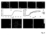
- the second micropipette was connected to a 1 ml syringe. Rate and volume of inflow was controlled by a syringe pump (Univector). Islet cells were loaded with Ca2+ indicators by perfusing the anterior chamber with extracellular solution containing Fluo-4 and Fura-Red AM esters for 40 min at 3 ⁇ l/min. Subsequently, the dye containing solution was washed out by perfusion with extracellular solution for 10 min at 5-6 ⁇ l/min.
- Imaging was performed as described above exciting Fluo-4 and Fura-Red at 488 nm (25% AOTF) and collecting emission light for Fluo-4 between 495-535 nm and for Fura-Red between 600-700 nm.
- Systemic stimulation of insulin release was achieved by intravenous injection of glibenclamide (1 mg/kg) via the tail vein.
- mice were subcutaneously injected with 0.1 ml/kg of Temgesic (Schering-Plough, NJ) to relieve post-operative pain.
- Pancreatic islets are hardly accessible for in vivo monitoring because they are deeply embedded and scattered in the exocrine tissue of the pancreas, constituting 1- 2% of the pancreatic volume5.
- Isolated islets (6) and especially pancreatic slices (7) allow functional studies of ⁇ -cells in a multi-cellular environment.
- these preparations are restricted to defined end-points and partially lack input from vascular and nervous connections.
- pancreatic islets After islet transplantation, islets recruit a new vasculature (11) as well as nervous Connections (12), and are capable of maintaining glucose homeostasis via pulsatile insulin release (12,13).
- pancreatic islets into the anterior chamber of the eye because the cornea acts as a natural body window that allows noninvasive imaging of engrafted tissue.
- the anterior chamber of the eye has been frequently used as a transplantation site to study a variety of tissues including pancreas because it is immune privileged (14-17).
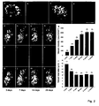
- Mouse islets were transplanted into the anterior chamber of the eye via injection through the cornea ( Figure 1a ). After transplantation, the islets engrafted on the iris and were readily observed and imaged through the cornea ( Figure 1b-c ).
- the transplanted islets engrafted either as single islets or in groups as islet clusters. Immunohistochemical staining of engrafted islets showed that the proportion of the insulin-containing ⁇ -cells and glucagon-containing ⁇ -cells did not change after transplantation and that this proportion was similar to that of islets in the pancreas ( Figure 1d-h ), which is in agreement with earlier Studies (14,16,17).
- mice that were rendered diabetic with streptozotocin transplanting islets into the anterior chamber reversed hyperglycaemia. These mice further showed physiological responses to glucose challenges ( Figure 1i ), demonstrating that islets engrafted in the anterior chamber of the eye are functional.
- islets isolated from transgenic mice expressing the enhanced green fluorescent protein (GFP) under the control of the rat insulin 1 promoter (RIP-GFP) were transplanted into the anterior chamber of the eye.
- TPLSM two-photon LSM
- non-invasive TPLSM enabled longitudinal in vivo imaging of ⁇ -cells and the vasculature in islets engrafted in the anterior chamber of the eye.
- ⁇ -cell death is a characteristics of type 1 diabetes (21) and is implicated in the pathology of type 2 diabetes (22).
- Annexin V has been used as a reporter of cell death both under experimental and clinical conditions (23) and has been validated as a marker for ⁇ -cell apoptosis after systemic administration (24).
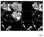
- RIP-GFP islets into the anterior chamber of the eye and, after complete engraftment and revascularization, we monitored cell death following intravenously administered annexin V conjugated to allophycocyanin (APC).
- mice transplanted with RIP-GFP islets by intravenous administration of alloxan (75mg/kg), a well-characterized diabetogenic compound that is taken up by ⁇ -cells via the glucose transporter 2 (26).
- alloxan 75mg/kg
- substantial loss of GFP fluorescence and structural changes in the reflection of the islet grafts were observed ( Figure 4e-f ), indicating loss of ⁇ -cells.
- ⁇ -cell death can be imaged non-invasively and longitudinally under in vivo conditions in islets engrafted in the anterior chamber of the eye.
- the anterior chamber of the eye can be used as a versatile natural body window to clarify, for the first time, the integration of complex signalling networks at the cellular level under in vivo conditions.
- the tissue of interest is transplanted into the anterior chamber of the eye and cell biological parameters are assessed by LSM through the cornea.
- the anterior chamber of the eye has been frequently used as a transplantation site to study a variety of tissues 3-7 . While originally the anterior chamber of the eye was selected as a transplantation site because of its properties as an immune privileged site 6 , most studies utilized the anterior chamber in a syngeneic transplantation setting because it is easily accessible and the cornea allows macroscopic observation of the engrafted tissue.
- the iris which forms the base of the anterior chamber, has one of the highest concentrations of blood vessels and autonomic nerves in the body, and thereby enables fast innervation 9 and vascularization 10 of the graft.
- studies utilizing the anterior chamber as a transplantation site mainly employed macroscopic observations 5 to investigate graft physiology. This restricts longitudinal studies to parameters observable at low resolution.
- In vivo electrophysiology 7 as well as histology 3 and various other in vitro techniques after graft removal enable assessment of morphology and cellular function, however setting an endpoint to the study and hereby preventing longitudinal monitoring.
- Extracellular solution 140 NaCl, 5 KCI, 2 NaHCO 3 , 1 NaH 2 PO 4 , 1.2 MgCl 2 , 2.5 CaCl 2 , 10 HEPES, 3 glucose (pH 7.4 with NaOH).
- LSM Leica TCS-SP2-AOBS confocal laser scanner equipped with Argon and HeNe lasers connected to a Leica DMLFSA microscope.
- Two-photon excitation is achieved using a TI:Sapphire laser (Tsunami; Spectra-Physics, USA) for ⁇ 100 fs excitation at ⁇ 82 MHz.
- the microscope stage is customized for the use of head holding adaptor and mouse.
- a head holding adapter SG-4N-S manufactured by Narishige, Japan.
- the head holder is equipped with a gas mask (GM-4-S) or a nosepiece.
- GM-4-S gas mask
- the head holder is attached to a metal plate which fits onto the customized stage of the microscope.
- the metal plate is covered by a heating pad.
- Body temperature is controlled via a rectal probe which regulates the temperature of the heating pad.
- Outflow Connect a - 35 cm piece of polythene tubing 0.9 mm i.d. and 1.2 mm o.d. with one end to a capillary holder and connect the other end of the tubing to a 10 ml syringe without a plunger (open reservoir).
- Inflow Connect a - 35 cm piece of tygon tubing 0.76 mm i.d. and 0.86 mm wall thickness with one end to a capillary holder and connect the other end of the tubing to a 1 ml syringe.
- the here introduced platform enables longitudinal assessment of several biological parameters in vivo without the need of invasive surgical procedures to access the tissue of interest.
- the current protocol easily enables detection of islet graft morphology by reflection or fluorescence imaging.
- Morphological characterization of a pancreatic islet graft by imaging reflection and GFP was carried out in which. islets of mice expressing GFP under the insulin promoter (green beta-cells) were transplanted to mice expressing GFP under the Tie2 promoter (green endothelial cells).
- GFP was excited with 488 nm at 35% laser power and emission measured between 495-530 nm. Reflection was imaged by exciting with 633 nm at 35% laser power and measuring emission between 632-639 nm..
- vascularization and cell death can be followed longitudinally by systemic injections of fluorescent dyes.
- Blood vessels were visualized by an intravenous injection of a 70 kDa Texas Red labeled dextran. GFP and Texas Red were excited with a two-photon laser at 890 nm at minimal necessary laser power required and emission collected onto non-descanned detectors using a dichroic mirror (RSP 560) and emission filters (BP 525/50 and BP 640/20).
- RSP 560 dichroic mirror
- BP 525/50 and BP 640/20 emission filters
- beta-cell death was induced by intravenous injection of alloxan.
- Apoptotic and dead cells were visualized by intravenous injection of annexin V-APC.
- Reflection was imaged by exciting with 543 nm and emission measured between 539-547 nm at 35% laser power.
- GFP was excited at 488 nm and emission measured between 495-530 nm at 35% laser power.
- APC was excited at 633 nm with collection of emission light between 645-680 nm at 75% laser power.
- islet-cells can be loaded repetitively with Ca 2+ indicators, by perfusion of the anterior chamber, to measure systemically induced changes in cytoplasmic free Ca 2+ concentration.
- the anterior chamber was perfused with Fluo-4 and Fura-Red. Fluo-4 and Fura-Red were excited with 488 nm at 25% laser power and emission measured for Fluo-4 between 495-535 nm and for Fura-Red between 600-700 nm. Reflection was imaged by exciting with 543 nm and emission measured between 539-547 nm at 15% laser power.
- Step 12 Movement of the head because the head holder is not properly fixed. Fix the clamping of the head holder. Movement of the eye during normal breathing. Adjust the eye stabilizer carefully. Gasping of the mouse. Make sure that the airflow of the anesthesia unit and isofluorane levels are set properly. No fluorescence can be detected In the blood vessels (Step 13 B ii). Tail vein injection failed. Repeat tail vein injection. Excitation and emission settings are wrong. Adjust excitation and emission settings. Blood circulation to the eye is disrupted due to false setting of eye stabilizer. Adjust the position of the eye stabilizer. Perfusion does not work (Step 13 D x). Air bubbles block the pipettes.
- capillary holder or tubing make sure that there are no air bubbles in the tubing, capillary holders or capillaries, prior to the experiment. Dirt blocks the pipette tips. Be sure to clean the pipettes after beveling, right before the experiment.
Landscapes
- Health & Medical Sciences (AREA)
- Life Sciences & Earth Sciences (AREA)
- Engineering & Computer Science (AREA)
- Animal Behavior & Ethology (AREA)
- General Health & Medical Sciences (AREA)
- Chemical & Material Sciences (AREA)
- Diabetes (AREA)
- Cell Biology (AREA)
- Zoology (AREA)
- Veterinary Medicine (AREA)
- Public Health (AREA)
- Biomedical Technology (AREA)
- Medicinal Chemistry (AREA)
- Pharmacology & Pharmacy (AREA)
- Organic Chemistry (AREA)
- Bioinformatics & Cheminformatics (AREA)
- Biotechnology (AREA)
- Environmental Sciences (AREA)
- General Chemical & Material Sciences (AREA)
- Chemical Kinetics & Catalysis (AREA)
- Nuclear Medicine, Radiotherapy & Molecular Imaging (AREA)
- Epidemiology (AREA)
- Gastroenterology & Hepatology (AREA)
- Endocrinology (AREA)
- Physiology (AREA)
- Nutrition Science (AREA)
- Developmental Biology & Embryology (AREA)
- Immunology (AREA)
- Virology (AREA)
- Animal Husbandry (AREA)
- Biodiversity & Conservation Biology (AREA)
- Wood Science & Technology (AREA)
- Genetics & Genomics (AREA)
- Hematology (AREA)
- Pathology (AREA)
- Urology & Nephrology (AREA)
- Toxicology (AREA)
- Rheumatology (AREA)
- Biochemistry (AREA)
- General Engineering & Computer Science (AREA)
Claims (8)
- Eine Methode zur Arzneimittelentwicklung, umfassend(a) Einpflanzen von Zielzellen in die vordere Kammer des Auges eines Versuchstieres ausgewählt aus der Gruppe, bestehend aus Maus, Affe, Hund, Ratte und Schwein, wobei eine oder mehrere zelluläre Bestandteile von therapeutischem Interesse in den transplantierten Zielzellen fluoreszierend markiert sind;(b) In-Kontakt-Bringen der Zielzellen mit einer oder mehreren Testverbindungen; und(c) Durchführen von nicht-invasiver Fluoreszenz Bildgebung auf dem Auge des Versuchstieres, wobei die Fluoreszenz Bildgebung dafür verwendet wird, um Testverbindung-induzierte Veränderungen in einer oder mehrerer, der Aktivität, der Position und der Menge der fluoreszierend markierten zellulären Komponenten von therapeutischem Interesse in den transplantierten Zielzellen zu detektieren, wobei die Veränderungen solche Testverbindungen identifizieren, die einen therapeutischen Vorteil für die Zielzellen bereitstellen können.
- Die Methode nach Anspruch 1, wobei die Zielzellen vom Gewebe ausgewählt aus der Gruppe, bestehend aus Endokrin, Fett, Muskel, Gehirn, Leber, Herz, Niere und Lunge abstammen.
- Die Methode von einem der Ansprüche 1-2, wobei eine oder mehrere Testverbindungen topisch auf das Auge des Versuchstieres appliziert werden.
- Die Methode von einem der Ansprüche 1-2, wobei die eine oder mehrere Testverbindungen in die vordere Kammer des Auges des Versuchstieres injiziert werden.
- Die Methode von einem der Ansprüche 1-4, wobei die Fluoreszenz Bildgebung Laserrastermikroskopie umfasst.
- Die Methode einer der Ansprüche 1-5, worin die Fluoreszenz Bildgebung dazu verwendet wird, Testverbindung-induzierte Veränderungen in einer oder mehrerer, der Aktivität, der Position und der Menge der fluoreszierend markierten zellulären Verbindungen von therapeutischem Interesse in den transplantierten Zielzellen zu vielen Zeitpunkten zu detektieren.
- Die Methode von einem der Ansprüche 1-6, wobei die Zielzellen pankreatische Beta-Zellen umfassen.
- Die Methode von einem der Ansprüche 1-7, wobei die Zielzellen individuelle Zellen sind.
Priority Applications (4)
| Application Number | Priority Date | Filing Date | Title |
|---|---|---|---|
| EP12000170.6A EP2441462B1 (de) | 2007-08-31 | 2008-09-01 | Verfahren zur Behandlung von Diabetes Typ I |
| PL08785781T PL2194993T3 (pl) | 2007-08-31 | 2008-09-01 | Metody badania przesiewowego leków z użyciem nieinwazyjnego obrazowania in vivo |
| PL12000170T PL2441462T3 (pl) | 2007-08-31 | 2008-09-01 | Sposób leczenia cukrzycy typu I |
| DK12000170.6T DK2441462T3 (da) | 2007-08-31 | 2008-09-01 | Fremgangsmåde til behandling af type I-diabetes |
Applications Claiming Priority (4)
| Application Number | Priority Date | Filing Date | Title |
|---|---|---|---|
| US96943707P | 2007-08-31 | 2007-08-31 | |
| US98903807P | 2007-11-19 | 2007-11-19 | |
| US4248208P | 2008-04-04 | 2008-04-04 | |
| PCT/EP2008/007130 WO2009027106A2 (en) | 2007-08-31 | 2008-09-01 | Non-invasive in vivo imaging and methods for treating type i diabetes |
Related Child Applications (2)
| Application Number | Title | Priority Date | Filing Date |
|---|---|---|---|
| EP12000170.6A Division EP2441462B1 (de) | 2007-08-31 | 2008-09-01 | Verfahren zur Behandlung von Diabetes Typ I |
| EP12000170.6 Division-Into | 2012-01-12 |
Publications (2)
| Publication Number | Publication Date |
|---|---|
| EP2194993A2 EP2194993A2 (de) | 2010-06-16 |
| EP2194993B1 true EP2194993B1 (de) | 2013-12-18 |
Family
ID=40025468
Family Applications (2)
| Application Number | Title | Priority Date | Filing Date |
|---|---|---|---|
| EP08785781.9A Active EP2194993B1 (de) | 2007-08-31 | 2008-09-01 | Wirkstoff-Screening Verfahren mittels nicht-invasiver in-vivo Darstellung |
| EP12000170.6A Active EP2441462B1 (de) | 2007-08-31 | 2008-09-01 | Verfahren zur Behandlung von Diabetes Typ I |
Family Applications After (1)
| Application Number | Title | Priority Date | Filing Date |
|---|---|---|---|
| EP12000170.6A Active EP2441462B1 (de) | 2007-08-31 | 2008-09-01 | Verfahren zur Behandlung von Diabetes Typ I |
Country Status (7)
| Country | Link |
|---|---|
| US (5) | US20090060843A1 (de) |
| EP (2) | EP2194993B1 (de) |
| JP (4) | JP5883221B2 (de) |
| DK (2) | DK2441462T3 (de) |
| ES (2) | ES2732561T3 (de) |
| PL (2) | PL2441462T3 (de) |
| WO (1) | WO2009027106A2 (de) |
Families Citing this family (10)
| Publication number | Priority date | Publication date | Assignee | Title |
|---|---|---|---|---|
| US20090060843A1 (en) * | 2007-08-31 | 2009-03-05 | Biocrine Ab | Non-Invasive In Vivo Imaging and Methods for Treating Type I Diabetes |
| EP2478113B1 (de) | 2009-09-18 | 2020-09-02 | Caliper Life Sciences, Inc. | Zusammensetzungen und verfahren zur in-vivo-evaluation einer biolumineszenz |
| US20130226292A1 (en) * | 2010-10-12 | 2013-08-29 | Jean-Marie Parel | Islet storing intraocular implant, method of using the same and kit including the same |
| JP6092191B2 (ja) | 2011-05-13 | 2017-03-08 | バイオクライン アーベーBiocrine Ab | 電動式注入および吸引用システムおよび方法 |
| US8557513B2 (en) | 2011-06-27 | 2013-10-15 | Biocrine Ab | Methods for treating and/or limiting development of diabetes |
| US9462973B2 (en) | 2012-07-27 | 2016-10-11 | Biocrine Ab | Methods for monitoring physiological status of a body organ |
| JP6161845B1 (ja) * | 2016-11-14 | 2017-07-12 | 英次 湊 | まつ毛増毛用器具、まつ毛の増毛方法、まゆ毛増毛用器具、及びまゆ毛の増毛方法 |
| EP3823648B1 (de) | 2018-07-19 | 2023-06-07 | BioCrine AB | Verfahren zur behandlung von diabetes |
| US12685490B2 (en) | 2020-01-28 | 2026-07-21 | The Board Of Trustees Of The Leland Stanford Junior University | Method of preventing or treating pancreatic dysfunction or diabetes by upregulating human cathelicidin LL-37 to inhibit islet amyloid polypeptide (IAPP) self-assembly |
| SE2351151A1 (en) * | 2023-10-06 | 2025-04-07 | Anna Herland | Medical device |
Family Cites Families (11)
| Publication number | Priority date | Publication date | Assignee | Title |
|---|---|---|---|---|
| SU1369039A1 (ru) * | 1982-07-16 | 1988-09-07 | Институт биологической физики АН СССР | Способ моделировани компенсации сахарного диабета |
| US5650435A (en) * | 1991-04-01 | 1997-07-22 | Madara; James L. | Modulation of inflammation related to columnar epithelia |
| US7255851B2 (en) * | 1994-07-01 | 2007-08-14 | The Board Of Trustees Of The Leland Stanford Junior University | Non-invasive localization of a light-emitting conjugate in a mammal |
| US5650135A (en) * | 1994-07-01 | 1997-07-22 | The Board Of Trustees Of The Leland Stanford Junior University | Non-invasive localization of a light-emitting conjugate in a mammal |
| DE19703734A1 (de) * | 1997-01-31 | 1998-08-06 | Univ Magdeburg Tech | Anordnung und Verfahren zur nicht-invasiven Bestimmung und/oder Erzeugung funktioneller und/oder morphologischer Eigenschaften von Zellen des Auges im lebenden Organismus |
| KR19990014353A (ko) * | 1998-10-17 | 1999-02-25 | 윤태욱 | 췌도의 대량 배양증식방법 |
| CA2403090C (en) * | 2000-03-17 | 2014-06-10 | Anticancer, Inc. | Whole-body optical imaging of gene expression and uses thereof |
| US20050180959A1 (en) * | 2002-02-06 | 2005-08-18 | Karsten Eulenberg | Kinases involved in the regulation of energy homeostasis |
| WO2004006838A2 (en) * | 2002-07-15 | 2004-01-22 | Sugen, Inc. | Novel kinases |
| WO2006130851A2 (en) * | 2005-06-02 | 2006-12-07 | Arizona Board Of Regents On Behalf Of The University Of Arizona | Prevascularized devices and related methods |
| US20090060843A1 (en) * | 2007-08-31 | 2009-03-05 | Biocrine Ab | Non-Invasive In Vivo Imaging and Methods for Treating Type I Diabetes |
-
2008
- 2008-08-27 US US12/199,473 patent/US20090060843A1/en not_active Abandoned
- 2008-09-01 EP EP08785781.9A patent/EP2194993B1/de active Active
- 2008-09-01 WO PCT/EP2008/007130 patent/WO2009027106A2/en not_active Ceased
- 2008-09-01 EP EP12000170.6A patent/EP2441462B1/de active Active
- 2008-09-01 DK DK12000170.6T patent/DK2441462T3/da active
- 2008-09-01 PL PL12000170T patent/PL2441462T3/pl unknown
- 2008-09-01 PL PL08785781T patent/PL2194993T3/pl unknown
- 2008-09-01 DK DK08785781.9T patent/DK2194993T3/da active
- 2008-09-01 ES ES12000170T patent/ES2732561T3/es active Active
- 2008-09-01 JP JP2010522257A patent/JP5883221B2/ja active Active
- 2008-09-01 ES ES08785781.9T patent/ES2447069T3/es active Active
-
2011
- 2011-09-02 JP JP2011191839A patent/JP5882640B2/ja active Active
-
2014
- 2014-06-04 JP JP2014116161A patent/JP2014167023A/ja active Pending
- 2014-10-15 US US14/515,000 patent/US9463205B2/en active Active
-
2015
- 2015-12-08 JP JP2015239327A patent/JP2016074726A/ja active Pending
-
2016
- 2016-09-14 US US15/265,212 patent/US10207012B2/en active Active
-
2018
- 2018-11-06 US US16/181,853 patent/US10493108B2/en active Active
-
2019
- 2019-10-31 US US16/669,840 patent/US20200061121A1/en not_active Abandoned
Non-Patent Citations (4)
| Title |
|---|
| ACTA ENDOCRINOLOGICA 1959, vol. 31, no. 1, 1959, pages 123 - 129, ISSN: 0001-5598 * |
| DATABASE EMBASE [online] ELSEVIER SCIENCE PUBLISHERS, AMSTERDAM, NL; 1959, KULLANDER S: "Studies on the histogenesis of experimental granulosa cell tumours in rats", Database accession no. EMB-0007862129 * |
| DATABASE EMBASE [online] ELSEVIER SCIENCE PUBLISHERS, AMSTERDAM, NL; 2002, GRISANTI S ET AL: "Xenotransplantation of retinal pigment epithelial cells into RCS rats", Database accession no. EMB-2002089108 * |
| JAPANESE JOURNAL OF OPHTHALMOLOGY 2002 JP LNKD- DOI:10.1016/S0021-5155(01)00464-6, vol. 46, no. 1, 2002, pages 36 - 44, ISSN: 0021-5155 * |
Also Published As
| Publication number | Publication date |
|---|---|
| JP5883221B2 (ja) | 2016-03-09 |
| US10493108B2 (en) | 2019-12-03 |
| HK1144252A1 (en) | 2011-02-11 |
| EP2194993A2 (de) | 2010-06-16 |
| JP2014167023A (ja) | 2014-09-11 |
| JP2011256202A (ja) | 2011-12-22 |
| JP5882640B2 (ja) | 2016-03-09 |
| PL2441462T3 (pl) | 2020-01-31 |
| US10207012B2 (en) | 2019-02-19 |
| JP2016074726A (ja) | 2016-05-12 |
| US20150064144A1 (en) | 2015-03-05 |
| US20200061121A1 (en) | 2020-02-27 |
| JP2010536908A (ja) | 2010-12-02 |
| US20090060843A1 (en) | 2009-03-05 |
| US20170000907A1 (en) | 2017-01-05 |
| US9463205B2 (en) | 2016-10-11 |
| DK2194993T3 (da) | 2014-01-27 |
| PL2194993T3 (pl) | 2014-04-30 |
| EP2441462A1 (de) | 2012-04-18 |
| EP2441462B1 (de) | 2019-05-08 |
| ES2447069T3 (es) | 2014-03-11 |
| WO2009027106A3 (en) | 2009-07-23 |
| ES2732561T3 (es) | 2019-11-25 |
| WO2009027106A2 (en) | 2009-03-05 |
| US20190070316A1 (en) | 2019-03-07 |
| DK2441462T3 (da) | 2019-06-24 |
Similar Documents
| Publication | Publication Date | Title |
|---|---|---|
| US10493108B2 (en) | Non-invasive in vivo imaging and methods for treating diabetes | |
| Speier et al. | Noninvasive high-resolution in vivo imaging of cell biology in the anterior chamber of the mouse eye | |
| JP2010536908A5 (ja) | 1型糖尿病の薬品開発方法 | |
| Tracy et al. | Intravitreal implantation of TPP1-transduced stem cells delays retinal degeneration in canine CLN2 neuronal ceroid lipofuscinosis | |
| JP6234453B2 (ja) | 臓器の生理学的状態をモニタリングする方法 | |
| Yang et al. | The anterior chamber of the eye technology and its anatomical, optical, and immunological bases | |
| Baffet et al. | Cellular and subcellular imaging of motor protein-based behavior in embryonic rat brain | |
| Rosmus et al. | Redefining the ontogeny of hyalocytes as yolk sac-derived tissue-resident macrophages of the vitreous body | |
| Fang et al. | Methods for imaging inflammation and transendothelial migration in vivo and ex vivo | |
| Peglion et al. | Live imaging of microtubule dynamics in glioblastoma cells invading the zebrafish brain | |
| HK1144252B (en) | Methods for drug screening using non-invasive in vivo imaging | |
| HK1169047A (en) | Method for treating type i diabetes | |
| HK1169047B (en) | Method for treating type i diabetes | |
| Vadrevu et al. | Studying the role of alveolar macrophages in breast cancer metastasis | |
| KR102949566B1 (ko) | 세포 기반 첨단 바이오융복합 제품의 비임상시험 방법 | |
| KR20160115522A (ko) | 활막 조직 이식을 통한 류마티스 관절염 동물모델인 아바타라의 제조방법 | |
| EP4534053A1 (de) | Medizinische vorrichtung | |
| JP7193865B2 (ja) | 短期間造腫瘍性スクリーニングシステム | |
| Singh et al. | Confocal Imaging of Intracellular calcium Cycling in the Intact Heart | |
| Valentino et al. | Hypoglycemia causes widespread white matter injury–an imaging study in transgenic mice | |
| Vallieres et al. | Neda Haghayegh Jahromi1, Heidi Tardent1, Gaby Enzmann1, Urban Deutsch1, Naoto Kawakami2, 3, Stefan Bittner4, Dietmar Vestweber5, Frauke Zipp4, Jens V. Stein1 and Britta Engelhardt1 | |
| Vaas | Imaging the effects of the transient middle cerebral artery occlusion filament model on the recruitment of neutrophils with non‐invasive imaging techniques | |
| Valentino | Two-photon microscopy: sequential imaging studies in vivo | |
| Sahoo et al. | Angiogenesis Assays |
Legal Events
| Date | Code | Title | Description |
|---|---|---|---|
| PUAI | Public reference made under article 153(3) epc to a published international application that has entered the european phase |
Free format text: ORIGINAL CODE: 0009012 |
|
| 17P | Request for examination filed |
Effective date: 20100330 |
|
| AK | Designated contracting states |
Kind code of ref document: A2 Designated state(s): AT BE BG CH CY CZ DE DK EE ES FI FR GB GR HR HU IE IS IT LI LT LU LV MC MT NL NO PL PT RO SE SI SK TR |
|
| AX | Request for extension of the european patent |
Extension state: AL BA MK RS |
|
| 17Q | First examination report despatched |
Effective date: 20101203 |
|
| REG | Reference to a national code |
Ref country code: HK Ref legal event code: DE Ref document number: 1144252 Country of ref document: HK |
|
| RTI1 | Title (correction) |
Free format text: METHODS FOR DRUG SCREENING USING NON-INVASIVE IN VIVO IMAGING |
|
| GRAP | Despatch of communication of intention to grant a patent |
Free format text: ORIGINAL CODE: EPIDOSNIGR1 |
|
| DAX | Request for extension of the european patent (deleted) | ||
| RIC1 | Information provided on ipc code assigned before grant |
Ipc: A61P 31/00 20060101ALI20120404BHEP Ipc: A01K 67/027 20060101ALI20120404BHEP Ipc: A61K 35/39 20060101AFI20120404BHEP Ipc: G01N 33/50 20060101ALI20120404BHEP Ipc: C12N 5/07 20100101ALI20120404BHEP |
|
| GRAJ | Information related to disapproval of communication of intention to grant by the applicant or resumption of examination proceedings by the epo deleted |
Free format text: ORIGINAL CODE: EPIDOSDIGR1 |
|
| GRAS | Grant fee paid |
Free format text: ORIGINAL CODE: EPIDOSNIGR3 |
|
| GRAP | Despatch of communication of intention to grant a patent |
Free format text: ORIGINAL CODE: EPIDOSNIGR1 |
|
| GRAP | Despatch of communication of intention to grant a patent |
Free format text: ORIGINAL CODE: EPIDOSNIGR1 |
|
| INTG | Intention to grant announced |
Effective date: 20130715 |
|
| GRAA | (expected) grant |
Free format text: ORIGINAL CODE: 0009210 |
|
| AK | Designated contracting states |
Kind code of ref document: B1 Designated state(s): AT BE BG CH CY CZ DE DK EE ES FI FR GB GR HR HU IE IS IT LI LT LU LV MC MT NL NO PL PT RO SE SI SK TR |
|
| REG | Reference to a national code |
Ref country code: GB Ref legal event code: FG4D |
|
| REG | Reference to a national code |
Ref country code: CH Ref legal event code: EP Ref country code: CH Ref legal event code: NV Representative=s name: HANS ULRICH SEIFERT SEIFERT AND PARTNER, CH |
|
| REG | Reference to a national code |
Ref country code: AT Ref legal event code: REF Ref document number: 645271 Country of ref document: AT Kind code of ref document: T Effective date: 20140115 |
|
| REG | Reference to a national code |
Ref country code: DK Ref legal event code: T3 Effective date: 20140120 |
|
| REG | Reference to a national code |
Ref country code: IE Ref legal event code: FG4D |
|
| REG | Reference to a national code |
Ref country code: DE Ref legal event code: R096 Ref document number: 602008029427 Country of ref document: DE Effective date: 20140213 |
|
| REG | Reference to a national code |
Ref country code: NL Ref legal event code: T3 |
|
| REG | Reference to a national code |
Ref country code: SE Ref legal event code: TRGR |
|
| REG | Reference to a national code |
Ref country code: ES Ref legal event code: FG2A Ref document number: 2447069 Country of ref document: ES Kind code of ref document: T3 Effective date: 20140311 |
|
| REG | Reference to a national code |
Ref country code: HK Ref legal event code: GR Ref document number: 1144252 Country of ref document: HK |
|
| PG25 | Lapsed in a contracting state [announced via postgrant information from national office to epo] |
Ref country code: FI Free format text: LAPSE BECAUSE OF FAILURE TO SUBMIT A TRANSLATION OF THE DESCRIPTION OR TO PAY THE FEE WITHIN THE PRESCRIBED TIME-LIMIT Effective date: 20131218 Ref country code: HR Free format text: LAPSE BECAUSE OF FAILURE TO SUBMIT A TRANSLATION OF THE DESCRIPTION OR TO PAY THE FEE WITHIN THE PRESCRIBED TIME-LIMIT Effective date: 20131218 Ref country code: NO Free format text: LAPSE BECAUSE OF FAILURE TO SUBMIT A TRANSLATION OF THE DESCRIPTION OR TO PAY THE FEE WITHIN THE PRESCRIBED TIME-LIMIT Effective date: 20140318 Ref country code: LT Free format text: LAPSE BECAUSE OF FAILURE TO SUBMIT A TRANSLATION OF THE DESCRIPTION OR TO PAY THE FEE WITHIN THE PRESCRIBED TIME-LIMIT Effective date: 20131218 |
|
| REG | Reference to a national code |
Ref country code: PL Ref legal event code: T3 |
|
| REG | Reference to a national code |
Ref country code: LT Ref legal event code: MG4D |
|
| PG25 | Lapsed in a contracting state [announced via postgrant information from national office to epo] |
Ref country code: LV Free format text: LAPSE BECAUSE OF FAILURE TO SUBMIT A TRANSLATION OF THE DESCRIPTION OR TO PAY THE FEE WITHIN THE PRESCRIBED TIME-LIMIT Effective date: 20131218 |
|
| PG25 | Lapsed in a contracting state [announced via postgrant information from national office to epo] |
Ref country code: EE Free format text: LAPSE BECAUSE OF FAILURE TO SUBMIT A TRANSLATION OF THE DESCRIPTION OR TO PAY THE FEE WITHIN THE PRESCRIBED TIME-LIMIT Effective date: 20131218 Ref country code: IS Free format text: LAPSE BECAUSE OF FAILURE TO SUBMIT A TRANSLATION OF THE DESCRIPTION OR TO PAY THE FEE WITHIN THE PRESCRIBED TIME-LIMIT Effective date: 20140418 |
|
| PG25 | Lapsed in a contracting state [announced via postgrant information from national office to epo] |
Ref country code: CZ Free format text: LAPSE BECAUSE OF FAILURE TO SUBMIT A TRANSLATION OF THE DESCRIPTION OR TO PAY THE FEE WITHIN THE PRESCRIBED TIME-LIMIT Effective date: 20131218 Ref country code: PT Free format text: LAPSE BECAUSE OF FAILURE TO SUBMIT A TRANSLATION OF THE DESCRIPTION OR TO PAY THE FEE WITHIN THE PRESCRIBED TIME-LIMIT Effective date: 20140418 Ref country code: SK Free format text: LAPSE BECAUSE OF FAILURE TO SUBMIT A TRANSLATION OF THE DESCRIPTION OR TO PAY THE FEE WITHIN THE PRESCRIBED TIME-LIMIT Effective date: 20131218 Ref country code: CY Free format text: LAPSE BECAUSE OF FAILURE TO SUBMIT A TRANSLATION OF THE DESCRIPTION OR TO PAY THE FEE WITHIN THE PRESCRIBED TIME-LIMIT Effective date: 20131218 Ref country code: RO Free format text: LAPSE BECAUSE OF FAILURE TO SUBMIT A TRANSLATION OF THE DESCRIPTION OR TO PAY THE FEE WITHIN THE PRESCRIBED TIME-LIMIT Effective date: 20131218 |
|
| REG | Reference to a national code |
Ref country code: DE Ref legal event code: R097 Ref document number: 602008029427 Country of ref document: DE |
|
| PLBE | No opposition filed within time limit |
Free format text: ORIGINAL CODE: 0009261 |
|
| STAA | Information on the status of an ep patent application or granted ep patent |
Free format text: STATUS: NO OPPOSITION FILED WITHIN TIME LIMIT |
|
| 26N | No opposition filed |
Effective date: 20140919 |
|
| REG | Reference to a national code |
Ref country code: DE Ref legal event code: R097 Ref document number: 602008029427 Country of ref document: DE Effective date: 20140919 |
|
| PG25 | Lapsed in a contracting state [announced via postgrant information from national office to epo] |
Ref country code: LU Free format text: LAPSE BECAUSE OF FAILURE TO SUBMIT A TRANSLATION OF THE DESCRIPTION OR TO PAY THE FEE WITHIN THE PRESCRIBED TIME-LIMIT Effective date: 20140901 Ref country code: MC Free format text: LAPSE BECAUSE OF FAILURE TO SUBMIT A TRANSLATION OF THE DESCRIPTION OR TO PAY THE FEE WITHIN THE PRESCRIBED TIME-LIMIT Effective date: 20131218 |
|
| PG25 | Lapsed in a contracting state [announced via postgrant information from national office to epo] |
Ref country code: SI Free format text: LAPSE BECAUSE OF FAILURE TO SUBMIT A TRANSLATION OF THE DESCRIPTION OR TO PAY THE FEE WITHIN THE PRESCRIBED TIME-LIMIT Effective date: 20131218 |
|
| PG25 | Lapsed in a contracting state [announced via postgrant information from national office to epo] |
Ref country code: BG Free format text: LAPSE BECAUSE OF FAILURE TO SUBMIT A TRANSLATION OF THE DESCRIPTION OR TO PAY THE FEE WITHIN THE PRESCRIBED TIME-LIMIT Effective date: 20131218 |
|
| PG25 | Lapsed in a contracting state [announced via postgrant information from national office to epo] |
Ref country code: MT Free format text: LAPSE BECAUSE OF FAILURE TO SUBMIT A TRANSLATION OF THE DESCRIPTION OR TO PAY THE FEE WITHIN THE PRESCRIBED TIME-LIMIT Effective date: 20131218 Ref country code: GR Free format text: LAPSE BECAUSE OF FAILURE TO SUBMIT A TRANSLATION OF THE DESCRIPTION OR TO PAY THE FEE WITHIN THE PRESCRIBED TIME-LIMIT Effective date: 20140319 |
|
| PG25 | Lapsed in a contracting state [announced via postgrant information from national office to epo] |
Ref country code: HU Free format text: LAPSE BECAUSE OF FAILURE TO SUBMIT A TRANSLATION OF THE DESCRIPTION OR TO PAY THE FEE WITHIN THE PRESCRIBED TIME-LIMIT; INVALID AB INITIO Effective date: 20080901 |
|
| REG | Reference to a national code |
Ref country code: FR Ref legal event code: PLFP Year of fee payment: 9 |
|
| REG | Reference to a national code |
Ref country code: FR Ref legal event code: PLFP Year of fee payment: 10 |
|
| REG | Reference to a national code |
Ref country code: FR Ref legal event code: PLFP Year of fee payment: 11 |
|
| PGFP | Annual fee paid to national office [announced via postgrant information from national office to epo] |
Ref country code: IT Payment date: 20190920 Year of fee payment: 12 Ref country code: TR Payment date: 20190821 Year of fee payment: 12 Ref country code: NL Payment date: 20190926 Year of fee payment: 12 Ref country code: SE Payment date: 20190927 Year of fee payment: 12 Ref country code: IE Payment date: 20190927 Year of fee payment: 12 Ref country code: DK Payment date: 20190927 Year of fee payment: 12 |
|
| REG | Reference to a national code |
Ref country code: DE Ref legal event code: R082 Ref document number: 602008029427 Country of ref document: DE Representative=s name: ZWICKER SCHNAPPAUF & PARTNER PATENTANWAELTE PA, DE |
|
| PGFP | Annual fee paid to national office [announced via postgrant information from national office to epo] |
Ref country code: PL Payment date: 20190821 Year of fee payment: 12 |
|
| PGFP | Annual fee paid to national office [announced via postgrant information from national office to epo] |
Ref country code: AT Payment date: 20190821 Year of fee payment: 12 |
|
| PGFP | Annual fee paid to national office [announced via postgrant information from national office to epo] |
Ref country code: ES Payment date: 20191001 Year of fee payment: 12 Ref country code: BE Payment date: 20190927 Year of fee payment: 12 |
|
| PGFP | Annual fee paid to national office [announced via postgrant information from national office to epo] |
Ref country code: CH Payment date: 20191002 Year of fee payment: 12 |
|
| REG | Reference to a national code |
Ref country code: DK Ref legal event code: EBP Effective date: 20200930 |
|
| REG | Reference to a national code |
Ref country code: CH Ref legal event code: PL |
|
| REG | Reference to a national code |
Ref country code: NL Ref legal event code: MM Effective date: 20201001 |
|
| REG | Reference to a national code |
Ref country code: AT Ref legal event code: MM01 Ref document number: 645271 Country of ref document: AT Kind code of ref document: T Effective date: 20200901 |
|
| REG | Reference to a national code |
Ref country code: BE Ref legal event code: MM Effective date: 20200930 |
|
| PG25 | Lapsed in a contracting state [announced via postgrant information from national office to epo] |
Ref country code: NL Free format text: LAPSE BECAUSE OF NON-PAYMENT OF DUE FEES Effective date: 20201001 |
|
| PG25 | Lapsed in a contracting state [announced via postgrant information from national office to epo] |
Ref country code: AT Free format text: LAPSE BECAUSE OF NON-PAYMENT OF DUE FEES Effective date: 20200901 Ref country code: BE Free format text: LAPSE BECAUSE OF NON-PAYMENT OF DUE FEES Effective date: 20200930 Ref country code: CH Free format text: LAPSE BECAUSE OF NON-PAYMENT OF DUE FEES Effective date: 20200930 Ref country code: IE Free format text: LAPSE BECAUSE OF NON-PAYMENT OF DUE FEES Effective date: 20200901 Ref country code: LI Free format text: LAPSE BECAUSE OF NON-PAYMENT OF DUE FEES Effective date: 20200930 Ref country code: SE Free format text: LAPSE BECAUSE OF NON-PAYMENT OF DUE FEES Effective date: 20200902 |
|
| REG | Reference to a national code |
Ref country code: SE Ref legal event code: EUG |
|
| PG25 | Lapsed in a contracting state [announced via postgrant information from national office to epo] |
Ref country code: IT Free format text: LAPSE BECAUSE OF NON-PAYMENT OF DUE FEES Effective date: 20200901 |
|
| PG25 | Lapsed in a contracting state [announced via postgrant information from national office to epo] |
Ref country code: DK Free format text: LAPSE BECAUSE OF NON-PAYMENT OF DUE FEES Effective date: 20200930 |
|
| REG | Reference to a national code |
Ref country code: ES Ref legal event code: FD2A Effective date: 20220114 |
|
| PG25 | Lapsed in a contracting state [announced via postgrant information from national office to epo] |
Ref country code: ES Free format text: LAPSE BECAUSE OF NON-PAYMENT OF DUE FEES Effective date: 20200902 |
|
| PG25 | Lapsed in a contracting state [announced via postgrant information from national office to epo] |
Ref country code: TR Free format text: LAPSE BECAUSE OF NON-PAYMENT OF DUE FEES Effective date: 20200901 |
|
| PG25 | Lapsed in a contracting state [announced via postgrant information from national office to epo] |
Ref country code: PL Free format text: LAPSE BECAUSE OF NON-PAYMENT OF DUE FEES Effective date: 20200901 |
|
| PGFP | Annual fee paid to national office [announced via postgrant information from national office to epo] |
Ref country code: DE Payment date: 20250820 Year of fee payment: 18 |
|
| PGFP | Annual fee paid to national office [announced via postgrant information from national office to epo] |
Ref country code: GB Payment date: 20250822 Year of fee payment: 18 |
|
| PGFP | Annual fee paid to national office [announced via postgrant information from national office to epo] |
Ref country code: FR Payment date: 20250821 Year of fee payment: 18 |