EP2191767A1 - In-examinee image acquisition system, in-examinee image processing method, and in-examinee introduction device - Google Patents
In-examinee image acquisition system, in-examinee image processing method, and in-examinee introduction device Download PDFInfo
- Publication number
- EP2191767A1 EP2191767A1 EP08828127A EP08828127A EP2191767A1 EP 2191767 A1 EP2191767 A1 EP 2191767A1 EP 08828127 A EP08828127 A EP 08828127A EP 08828127 A EP08828127 A EP 08828127A EP 2191767 A1 EP2191767 A1 EP 2191767A1
- Authority
- EP
- European Patent Office
- Prior art keywords
- image
- information
- processed
- unit
- image processing
- Prior art date
- Legal status (The legal status is an assumption and is not a legal conclusion. Google has not performed a legal analysis and makes no representation as to the accuracy of the status listed.)
- Granted
Links
Images




Classifications
-
- A—HUMAN NECESSITIES
- A61—MEDICAL OR VETERINARY SCIENCE; HYGIENE
- A61B—DIAGNOSIS; SURGERY; IDENTIFICATION
- A61B1/00—Instruments for performing medical examinations of the interior of cavities or tubes of the body by visual or photographical inspection, e.g. endoscopes; Illuminating arrangements therefor
- A61B1/04—Instruments for performing medical examinations of the interior of cavities or tubes of the body by visual or photographical inspection, e.g. endoscopes; Illuminating arrangements therefor combined with photographic or television appliances
- A61B1/041—Capsule endoscopes for imaging
-
- A—HUMAN NECESSITIES
- A61—MEDICAL OR VETERINARY SCIENCE; HYGIENE
- A61B—DIAGNOSIS; SURGERY; IDENTIFICATION
- A61B1/00—Instruments for performing medical examinations of the interior of cavities or tubes of the body by visual or photographical inspection, e.g. endoscopes; Illuminating arrangements therefor
- A61B1/00002—Operational features of endoscopes
- A61B1/00004—Operational features of endoscopes characterised by electronic signal processing
- A61B1/00009—Operational features of endoscopes characterised by electronic signal processing of image signals during a use of endoscope
- A61B1/000095—Operational features of endoscopes characterised by electronic signal processing of image signals during a use of endoscope for image enhancement
-
- A—HUMAN NECESSITIES
- A61—MEDICAL OR VETERINARY SCIENCE; HYGIENE
- A61B—DIAGNOSIS; SURGERY; IDENTIFICATION
- A61B1/00—Instruments for performing medical examinations of the interior of cavities or tubes of the body by visual or photographical inspection, e.g. endoscopes; Illuminating arrangements therefor
- A61B1/00002—Operational features of endoscopes
- A61B1/00011—Operational features of endoscopes characterised by signal transmission
- A61B1/00016—Operational features of endoscopes characterised by signal transmission using wireless means
Definitions
- the present invention relates to an in-vivo image acquiring system which acquires an image of an inside of a subject, an in-vivo image processing method, and a body-insertable apparatus.
- the capsule endoscope has an imaging function and a wireless-transmission function, and has a mechanism such that the capsule endoscope is swallowed from a mouth of a patient for observing an inside of a body cavity, and successively captures an inside of organs such as an esophagus, a stomach, and a small intestine due to a peristalsis until naturally excreted (e.g., see Patent Document 1).
- Image data captured by the capsule endoscope inside a body is successively transmitted to an outside via a wireless transmission while the capsule endoscope moves through the body cavity, and then stored in a memory in a receiving apparatus arranged outside the body.
- a doctor or a nurse can have a diagnosis with the image data stored in the memory being displayed on a display.
- Patent Document 1 Japanese Patent Application Laid-Open No. 2003-19111
- the capsule endoscope described has a lens and a signal processing function which are designed with optical performance corresponding to the applied area.
- the lens and the signal processing function are designed with the optical performance where a focus meets at a near point for observing a wall surface of the small intestine having a small inner diameter.
- the capsule endoscope generally captures organs other than the applied area of the capsule endoscope, and transmits a huge number of images until naturally excreted.
- the optical performance is designed corresponding to the applied area, and the appropriate optical performance is not guaranteed for organs other than the applied area. Images of the organs other than the applied area are captured not properly enough to be able to be examined, and the images cannot be used for the examination. Thus, all images but those of the applied area in the huge number of images captured by the capsule endoscope are wasted. Further, when the images of organs other than the applied area need to be taken, the subject has to swallow the capsule endoscope which is designed with the optical performance to be able to capture the organs to be captured again, and the burden of the subject is increased.
- the present invention is made in view of the above drawbacks of the conventional technology, and an object of the present invention is to provide an in-vivo image acquiring system, an in-vivo image processing method, and a body-insertable apparatus, which are able to use the captured images of organs other than the applied area of the capsule endoscope, thereby decreasing the number of wasted images.
- an in-vivo image acquiring system includes: a body-insertable apparatus that is introduced into a subject, and wirelessly transmits image information including a captured in-vivo image of the subject to outside; and a processing apparatus that processes the image information transmitted from the body-insertable apparatus.
- the body-insertable apparatus includes an imaging unit that captures the in-vivo image of the subject, and a transmitting unit which attaches type information corresponding to optical information of the imaging unit to the image information including the image captured by the imaging unit, and transmits the image information.
- the processing apparatus includes a storage unit that stores therein combinations of pieces of the optical information corresponding respectively to pieces of the type information, and image processing programs corresponding respectively to pieces of the optical information, and an image processing unit that acquires the optical information corresponding to the type information which is attached to the image information to be processed from the optical information stored in the storage unit, and processes the image information to be processed using the image processing program, of the image processing programs stored in the storage unit, which corresponds to the acquired optical information.
- the type information indicates an applied portion of the body-insertable apparatus
- the image processing unit processes, as the image information to be processed, the image information corresponding to a portion other than a portion to which the body-insertable apparatus is applied.
- the optical information indicates a magnification ratio of the imaging, a number of pixels, or a luminance in the imaging unit
- each of the image processing programs is a magnification-ratio-adjusting program which changes the magnification ratio of the image to be processed, a sharpening program which changes sharpness of the image to be processed, or a luminance-adjusting program which changes luminance of the image to be processed
- the image processing unit processes the image information to be processed using the magnification-ratio-adjusting program, the sharpening program, or the luminance-adjusting program based on the magnification ratio of the imaging, the number of pixels, or the luminance corresponding to the type information of the image information to be processed.
- An in-vivo image processing method is for processing image information including an in-vivo image of a subject which is transmitted from a body-insertable apparatus introduced inside the subject.
- the in-vivo image processing method includes the steps of: capturing the in-vivo image of the subject by an imaging unit in the body-insertable apparatus; transmitting the image information to which the type information corresponding to the optical information in the imaging unit is attached from the body-insertable apparatus; receiving the image information transmitted from the body-insertable apparatus; and acquiring the optical information corresponding to the type information attached to the image information to be processed from the received image information, and processing the image information to be processed using an image processing program corresponding to the acquired optical information.
- the type information indicates the applied portion of the body-insertable apparatus
- the processing step includes processing, as the image information to be processed, the image information corresponding to a portion other than a portion to which the body-insertable apparatus is applied.
- the optical information indicates a magnification ratio of the imaging, a number of pixels, or a luminance in the imaging unit
- each of the image processing programs is a magnification-ratio-adjusting program which changes the magnification ratio of the image to be processed, a sharpening program which changes sharpness of the image to be processed, or a luminance-adjusting program which changes luminance of the image to be processed
- the processing step includes processing the image information to be processed using the magnification-ratio-adjusting program, the sharpening program, or the luminance-adjusting program based on the magnification ratio of the imaging, the number of pixels, or the luminance corresponding to the type information of the image information to be processed.
- a body-insertable apparatus is introduced inside a subject, and wirelessly transmits image information including a captured in-vivo image of a subject to outside.
- the body-insertable apparatus includes: an imaging unit that captures the in-vivo image of the subject; and a transmitting unit that attaches type information which corresponds to optical information of the imaging unit to the image information including the image captured by the imaging unit, and transmits the image information.
- the body-insertable apparatus attaches the type information corresponding to the optical information of the imaging unit of the body-insertable apparatus to the in-vivo image information, and transmits the image information.
- the processing apparatus acquires the optical information corresponding to the type information attached to the image information to be processed, and processes the image information to be processed using the image-processing program corresponding to the acquired optical information.
- the processing apparatus can also process the image information to be processed that corresponds to portions other than the applied area of the body-insertable apparatus so that the image information to be processed can be used.
- the image which corresponds to portions other than the applied area and which is captured by the body-insertable apparatus can be used for the diagnosis. Further, the subject does not have to swallow the capsule endoscope again.
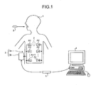
- FIG. 1 is a schematic diagram of an overall configuration of an in-vivo image acquiring system according to an embodiment.
- the in-vivo image acquiring system employs a monocular capsule endoscope as an example of a body-inserting apparatus. As shown in FIG.
- the wireless in-vivo image acquiring system includes a capsule endoscope 2 which is introduced inside a subject 1 and captures intracelomic images, and wirelessly transmits data such as an image signal to a receiving apparatus 3, a receiving apparatus which receives the data of the intracelomic images wirelessly transmitted from the capsule endoscope 2, a processing apparatus 4 which displays the intracelomic images based on the image signal received by the receiving apparatus 3, and a portable storage medium 5 used for transferring data between the receiving apparatus 3 and the processing apparatus 4.
- a capsule endoscope 2 which is introduced inside a subject 1 and captures intracelomic images, and wirelessly transmits data such as an image signal to a receiving apparatus 3, a receiving apparatus which receives the data of the intracelomic images wirelessly transmitted from the capsule endoscope 2, a processing apparatus 4 which displays the intracelomic images based on the image signal received by the receiving apparatus 3, and a portable storage medium 5 used for transferring data between the receiving apparatus 3 and the processing apparatus 4.
- the receiving apparatus 3 includes a wireless-transmission unit 3a which includes plural receiving antennas A1 to An attached to an outer surface of the subject 1, and a main receiving unit 3b which processes a wireless-transmission signal received via the plural receiving antennas A1 to An and performs other processes. Those units are detachably connected with the receiving apparatus 3 via a connector or the like.
- the receiving antennas A1 to An may be attached to, for example, a jacket which can be worn by the subject 1 and the subject 1 may wear the jacket so that the receiving antennas are attached to the subject 1. Further, in this case, the receiving antennas A1 to An may be detachably attached to the jacket.
- the processing apparatus 4 displays the intracelomic images captured by the capsule endoscope 2.
- the processing apparatus 4 may be realized by a workstation or the like which displays the images based on the data acquired via the portable storage medium 5.
- the processing apparatus 4 may be realized by a CRT display or an LCD display or the like which displays the image thereon, or a printer or the like which outputs the image on other mediums.
- the portable storage medium 5 may be realized by a compact flash (registered trademark) or the like, and detachably attached to the main receiving unit 3b and the processing apparatus 4.
- the portable storage medium 5 can output or store information when attached to the main receiving unit 3b and the processing apparatus 4.
- the portable storage medium 5 may be attached to the main receiving unit 3b and while the capsule endoscope 2 moves through the body cavity of the subject 1, and the portable storage medium 5 stores therein the data transmitted from the capsule endoscope 2. Further, after the capsule endoscope 2 is excreted from the subject 1, that is, after the capturing of the inside of the subject 1 is finished, the portable storage medium 5 is detached from the main receiving unit 3b and attached to the processing apparatus 4 so that the data may be read out by the processing apparatus 4.
- the subject 1 can act freely while the inside of the body cavity is captured, and the time for transferring the data to the processing apparatus 4 can be shortened. Further, data transfer between the main receiving unit 3b and the processing apparatus 4 may be performed by other storage apparatuses built into the main receiving unit 3b and the main receiving unit 3b may have a wired or wireless connection with the processing apparatus 4.
- FIG. 2 is a schematic diagram showing an example of the configuration of the capsule endoscope 2.
- the capsule endoscope 2 includes an imaging unit 22 which captures the body cavity of the subject 1, and the power unit 26 which supplies power for each component forming the capsule endoscope 2 inside a capsule-shaped casing 20.
- the imaging unit 22 shown in FIG. 2 is monocular, the imaging unit 22 may be binocular with plural imaging units 22.
- the capsule-shaped casing 20 includes a transparent dome-shaped front cover 20a, and a casing 20b which is kept watertight with the front cover 20a.
- the capsule-shaped casing 20 is of a size to be swallowed by the mouth of the subject 1.
- the front cover 20a is attached to one end of the casing 20b.
- the casing 20b is made of a colored material which does not transmit light.
- the casing 20b contains a control unit 21 which controls driving of each component of the capsule endoscope 2 and controls input and output of signals of each component, an imaging unit 22 which captures an inside of the body cavity, a signal processing unit 23 which processes the image captured by the imaging unit 22, a storage unit 24 which stores the information needed for the wireless transmission, a communication processing unit 25 which modulates each signal to be transmitted to the processing apparatus 4 arranged outside into a wireless-transmission signal and demodulates the wireless-transmission signal received via an antenna 25a, and a power unit 26 which supplies driving power for each component of the capsule endoscope 2.
- the communication processing unit 25 includes the antenna 25a which may be realized by a coiled antenna, and transmits and receives the wireless-transmission signal to and from the antenna arranged outside.
- the imaging unit 22 captures the image of the body cavity of the subject 1.
- the imaging unit 22 is realized by an imaging device such as a CCD and CMOS, a light-emitting device such as an LED which illuminates an imaged field of the imaging device, and an optical components such as a lens 22a which forms an image of the reflected light which is transmitted from the imaged field to the imaging device.
- the imaging unit 22 is fixated on an edge of the casing 20b.
- the imaging unit 22 forms the image of the reflected light which is received from the imaged field via the front cover 20a, and captures the image inside the body cavity of the subject 1.
- the imaging device, the light-emitting device, and the optical components such as the lens 22a in the imaging unit 22 have a function and a lens which are designed with optical performance corresponding to an applied area of the capsule endoscope 2.
- the lens and the signal processing function and the like in the imaging unit 22 are designed with the optical performance to focus on a point nearby so that the wall surface of the small intestine, which has a small inner diameter, can be captured.
- the lens and the signal processing function are designed with optical performance to focus on a point far from the capsule endoscope 2 compared with the case of the small intestine so that the inside of the stomach, which has a large volume, can be captured.
- the capsule endoscope 2 attached type information corresponding to the optical information to image information including in-vivo images, and transmits the image information.
- the imaging unit 22 of the capsule endoscope 2 is designed with optical performance corresponding to the applied area of the capsule endoscope 2, and thus each pieces of optical information of the imaging unit 22 differs corresponding to the applied area.
- the capsule endoscope 2 attaches information of the applied area of the capsule endoscope 2 to the image information as the type information corresponding to the optical information of the imaging unit 22 in the capsule endoscope 2, and transmits the image information.
- the type information is previously stored in the storage unit 24.
- the control unit 21 acquires the type information corresponding to the applied area of the imaging unit 22 from the storage unit 24, and outputs the type information to the signal processing unit 23.
- the signal processing unit 23 processes the image captured by the imaging unit 22, and attaches the type information which is output from the control unit 21 to the image signal including the image.
- the signal processing unit 23 processes an image G1 captured by the imaging unit 22 for every unit of scan-line data. Further, the signal processing unit 23 appends unique information Da including information which is identical for the capsule endoscope 2 to an end of last scan-line data.
- the unique information Da includes white-balance information, a serial number of the capsule endoscope 2, and the like, and further includes the above-described type information.
- the signal processing unit 23 appends the unique information Da including the white-balance information, the serial number, and the type information to an end of the last scan-line data similarly for images G2 to Gn captured by the imaging unit 22.
- the communication processing unit 25 wirelessly transmits the information generated by the signal processing unit 23 via the antenna 25a . Specifically, the communication processing unit 25 attaches the type information corresponding to the optical information in the imaging unit 22 to the image information including images captured by the imaging unit 22, and wirelessly transmits the image information to an outside.
- the capsule endoscope 2 attaches the type information, which corresponds to the optical information in the imaging unit 22 of the capsule endoscope 2 and indicates the applied area of the capsule endoscope 2, to the image information captured by the imaging unit 22.
- the information transmitted from the capsule endoscope 2 is received by the receiving apparatus 3, and stored in the portable storage medium 5.
- the processing apparatus 4 can acquire the type information with the image information including the images captured by the capsule endoscope 2 by reading the information stored in the portable storage medium 5.
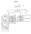
- FIG. 4 is a block diagram of an overall configuration of the processing apparatus 4 shown in FIG. 1 .
- the processing apparatus 4 includes a control unit 41, an input unit 42, a storage unit 44, an image processing unit 45, and a display unit 47.
- the control unit 41 has a connection with database Db where image groups Pa, Pb captured by the capsule endoscope 2 are stored in folders Fa, Fb, or the like.
- the control unit 41 includes a CPU or the like which has a controlling function.
- the control unit 41 controls operations and procedures of the input unit 42, the storage unit 44, the image processing unit 45, and the display unit 47.
- the input unit 42 is realized by a keyboard to input various information, or a mouse to point to any location on a display screen of the display unit 47.
- the input unit 42 acquires various information needed for an analysis of the specimen, and instruction information of an analyzing operation, or the like from an outside.
- the storage unit 44 includes a hard disk, which magnetically stores information, and a memory, which loads a variety of programs for a process of the processing apparatus 4 from the hard disk and electrically stores the programs therein when the processing apparatus 4 performs the process.
- the storage unit 44 stores various information including an optical-information set Dr and an image-processing-program set Dp.
- the storage unit 44 may include an auxiliary storage apparatus which can read information stored in a storage medium such as a CD-ROM, a DVD-ROM, and a PC card.
- the imaging unit 22 of the capsule endoscope 2 is designed corresponding to the applied area of the capsule endoscope 2.
- each optical information of the imaging unit 22 differs depending on the applied area of the capsule endoscope 2, i.e., the type of capsule endoscope 2.
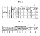
- the storage unit 44 stores a table T1 which shows combinations of optical information corresponding to each type of the capsule endoscope 2 as an optical-information set Dr.
- the table T1 shows a magnification ratio of the imaging, a distortion (DT) value, the number of pixels, a luminance, an aberration amount for a height of an image, a gamma value, and a spectral sensitivity in the imaging unit corresponding to each type of the capsule endoscope 2, i.e., each applied area of the capsule endoscope 2 such as a small intestine as optical information. Further, when optical information differs for each version of each type of the capsule endoscope 2, the storage unit 44 stores each optical information corresponding to each version of each type as the optical-information set Dr similarly to the table T1.
- the storage unit 44 stores a magnification-ratio-adjusting program which adjusts a magnification ratio of an image to be processed, a sharpening program which adjusts sharpness of the image to be processed, and a luminance-adjusting program which adjusts luminance of the image to be processed, and the like as an image-processing-program set Dp. Specifically, as shown in a table T2 in FIG.
- the storage unit 44 stores the magnification-ratio-adjusting program which adjusts the magnification ratio of the image based on the magnification ratio of imaging of the imaging unit 22 which has captured the image to be processed, the sharpening program which adjusts sharpness of the image based on the number of pixels of the imaging unit 22 which has captured the image to be processed, and the luminance-adjusting program which adjusts luminance of the image based on the luminance of the imaging unit 22 which has captured the image to be processed as the image processing programs.
- the storage unit 44 stores a DT-value-correcting program which corrects the image based on a DT value of the imaging unit 22 which has captured the image to be processed, an aberration-correcting program which corrects the image based on an aberration amount of the imaging unit 22 which has captured the image to be processed, a gamma-correcting program which corrects the image based on a gamma value of the imaging unit 22, and a color-correcting program which corrects colors of the image based on spectral sensitivity of the imaging unit 22 which has captured the image to be processed.
- the storage unit 44 may store plural image processing programs as the image-processing-program set Dp, respectively. As shown in the table T2 in FIG.
- the storage unit 44 may store the magnification-ratio-adjusting programs a1, a2, the DT-value-correcting programs b1, b2, the sharpening programs c1, c2, the luminance-adjusting programs d1, d2, the aberration-correcting programs e1, e2, the gamma-correcting programs f1, f2, and the color-correcting programs g1, g2.
- the image processing unit 45 includes a process setting unit 46.
- the process setting unit 46 acquires the optical information corresponding to the type information attached to the image information to be processed from the optical information stored in the storage unit 44. Further, the process setting unit 46 sets the image processing program, of the image processing programs stored in the storage unit 44, that corresponds to the optical information acquired from the image processing program stored in the storage unit 44as the image processing program to process the image information to be processed.
- the image processing unit 45 processes the image to be processed using the image processing program which is set by the process setting unit and corresponds to the optical information of the image information to be processed.
- the image processing unit 45 processes the image information including the images corresponding to portions other than the applied area of the capsule endoscope 2 as the image information to be processed.
- the image processing unit 45 processes the image information to be processed using the magnification-ratio-adjusting program, the sharpening program, the luminance-adjusting program, and the like corresponding to the optical information such as the magnification ratio, the number of pixels, and the luminance corresponding to the type information of the image information to be processed.
- the image processing unit 45 acquires the optical information corresponding to the type information of the images which are captured by the capsule endoscope 2 and correspond to portions other than the applied area of the capsule endoscope 2, and further the image processing unit 45 processes the image using the image processing program corresponding to the optical information so that the images which correspond to portions other than the applied area and could not be used otherwise as they were can be used.
- the display unit 47 is realized by a CRT display, an LCD display, and the like.
- the display unit 47 displays the instruction information, an instruction result, and the like of the input unit 42.
- the display unit 47 displays the images captured by the capsule endoscope 2 and the images processed by the image processing unit 45 under the control by the control unit 41.
- FIG. 7 is a flowchart showing each procedure of the image processing in the processing apparatus 4 shown in FIG. 4 .
- the image processing unit 45 acquires via the control unit 41 instruction information which is input from the input unit 42 and which instructs the image processing unit 45 to process the image (Step S2).
- the instruction information includes information of the image information to be processed and the contents of the process on the image information to be processed, and the like.
- the instruction information instructs the image processing unit 45 to process the image information, of the image groups acquired via the portable storage medium 5, which corresponds to a predetermined number of the images corresponding to portions other than the applied area of the capsule endoscope 2.
- the instruction information instructs the image processing unit 45 to process the image information, of image groups Pa, Pb stored in database Db, that corresponds to the predetermined number of images corresponding to portions other than the applied area of the capsule endoscope 2 which has captured the image group Pa, Pb. Then, the instruction information instructs the image processing unit 45 to perform the image processing which allows the portion captured in the image which is selected as the image information to be processed according to the instruction information to be used for a diagnosis.
- the instruction information selects the images inside the stomach observed by the capsule endoscope 2 for observing the small intestine as the image information to be processed, and instructs the image processing unit 45 to process the images so as to allow the images to be observed as easily as the images captured by the capsule endoscope 2 for observing the stomach.
- the image processing unit 45 acquires the image information to be processed according to the instruction information (Step S4).
- the process setting unit 46 acquires the type information attached to the image information from the image information to be processed (Step S6).
- the type information is included in the unique information Da which is appended to the end of the last scan-line data as previously described.
- the process setting unit 46 acquires the optical information that corresponds to the type information of the image information to be processed that is acquired from the unique information Da appended to the end of the last scan-line data at Step 6 (Step 8).
- the process setting unit 46 sets the image processing program, of the image processing programs set by the process setting unit 46, that corresponds to the acquired optical information as the image processing program to process the image information to be processed (Step S10).
- the image processing unit 45 processes the image information to be processed using the image processing program set by the process setting unit 46 (Step S12).
- the display unit outputs and displays the image processed by the image processing unit 45 (Step S14).
- the control unit 41 determines whether the image processed by the image processing unit 45 is to be saved based on the instruction information which is input from the input unit 42 (Step S16). For example, when an operator of the processing apparatus 4 selects via a mouse a selection field to save the image on a selection menu where the operator can choose to save the processed image, instruction information to save the processed image is input from the input unit 42 to the control unit 41. Further, when the operator of the processing apparatus 4 selects via a mouse a selection field not to save the image, instruction information not to save the processed image is input from the input unit 42 to the control unit 41.
- Step S16 If the control unit 41 determines that the image processed by the image processing unit 45 is not to be saved (Step S16: No), the processing apparatus 4 finishes the image processing. Further, if the control unit 41 determines that the image processed by the image processing unit 45 is to be saved (Step S16: Yes), the processing apparatus 4 saves the processed image in a specified destination to save (Step S18), and finished the image processing in the processing apparatus 4.
- Step S12 The image processing performed in the image processing unit 45 (Step S12) is described below in a specific manner. Firstly, the image processing where the image processing unit 45 processes the image using the magnification-ratio-adjusting program of the image processing programs shown in FIG. 6 is described as an example with reference to FIG. 8 .
- the image processing unit 45 acquired view-angle information and LED light-emitting amount information of the imaging unit 22 in the capsule endoscope 2 which has captured the image information to be processed from the type information acquired by the process setting unit 46 according to the procedure of the magnification-ratio-adjusting program (Step S22).
- the image processing unit 45 calculates observation distance in the capsule endoscope 2 based on the view-angle information and the LED light-emitting amount information (Step S24).
- the view angle is a range of the angle for the imaging unit 22 of the capsule endoscope 2 to capture clear images.
- the observation distance can be calculated based on the view angle.
- the imaging unit 22 automatically adjusts light. When the imaged area is too dark as an object to be captured is far, the imaging unit 22 automatically increases a light-emitting amount of an LED. When the imaged area is too light as the object to be captured is close, the imaging unit 22 automatically decreases the light-emitting amount of the LED.
- the image processing unit 45 can calculate the distance between the object to be captured and the capsule endoscope 2 based on information of the LED light-emitting amount, specifically, the light-emitting amount of the LED at the time when the image information to be processed is captured.
- the image processing unit 45 calculates the magnification ratio of the imaging unit 22 in the capsule endoscope 2 which has captured the image to be processed based on the calculated observation distance and the size of the imaged object in the image information to be processed (Step S26).
- the image processing unit 45 determines whether the calculated magnification ratio is higher than a predetermined magnification ratio corresponding to an intended usage (Step S28).
- the image processing unit 45 compares the calculated magnification ratio with the magnification ratio of the capsule endoscope 2 for observing the stomach.
- the compared magnification ratios may be included in the magnification-ratio-adjusting program, or acquired from the optical-information set Dr in the storage unit 44.
- the image processing unit 45 determines that the calculated magnification ratio is not higher than the predetermined magnification ratio (Step S28: No), i.e., that the calculated magnification ratio is lower than the predetermined magnification ratio, the image processing unit 45 magnifies the image to be processed in correspondence with the predetermined magnification ratio (Step S30). For example, when the image to be processed is the image of the inside of the stomach captured by the capsule endoscope 2 for observing the small intestine, and the magnification ratio is lower than the magnification ratio of the capsule endoscope 2 for observing the inside of the stomach, the image processing unit 45 magnifies the capsule endoscope 2 for observing the inside of the stomach.
- Step S28 determines that the calculated magnification ratio is higher than the predetermined magnification ratio
- Step S32 demagnifies the image to be processed in correspondence with the predetermined magnification ratio
- the image processing unit 45 determines whether there is a following image to be processed. If the image processing unit 45 determines that there is the following image to be processed (Step S34: Yes), the image processing unit 45 proceeds to Step S24 and calculates observation distance on the image to be processed. On the other hand, if the image processing unit 45 determines that there is not the following image to be processed (Step S34: No), the image processing unit 45 outputs a series of processed images to the control unit 41 (Step S36), and finishes the image processing. The series of images processed by the image processing unit 45 are displayed by the display unit 47, and stored in the database Db and the like via the control unit 41.
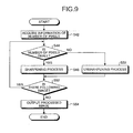
- the image processing unit 45 processes images using the sharpening program of the image-processing programs shown in FIG. 6 is described with reference to FIG. 9 .
- the image processing unit 45 acquires information of the number of pixels which indicates the number of pixels of the imaging unit 22 of the capsule endoscope 2 which has captured the image to be processed from the type information acquired by the process setting unit 46 (Step S42).
- the image processing unit 45 determines, based on the acquired information of the number of pixels, whether the number of pixels of the imaging unit 22 of the capsule endoscope 2 which has captured the image to be processed is smaller than a predetermined number of pixel corresponding to an intended usage (Step S44). For example, when the image processing unit 45 processes the image of the inside of the stomach observed by the capsule endoscope 2 for observing the small intestine for the image to be used for the diagnosis, the image processing unit 45 compares the number of pixels with the number of pixels of the capsule endoscope 2 for observing the inside of the stomach. The compared number of pixels may be included in the sharpening program, or acquired from the optical-information set Dr stored in the storage unit 44.
- the image processing unit 45 determines that the number of pixels of the imaging unit 22 of the capsule endoscope 2 which has captured the image to be processed is smaller than the predetermined number of pixels (Step 44: Yes), the image processing unit 45 sharpens edges of the image to be processed by adjusting the sharpness of the image (Step S46).
- the image processing unit 45 sharpens the image in correspondence with the sharpness of the capsule endoscope 2 because the edge of the image is unclear compared with the image captured by the capsule endoscope 2 for observing the inside of the stomach.
- Step S44 determines that the number of pixels of the imaging unit 22 of the capsule endoscope 2 which has captured the image to be processed is larger than the predetermined number of pixels (Step S44: No).
- the image processing unit 45 does not sharpen the edge because the sharpness of the image to be processed is clear enough for the intended usage in the diagnosis.
- the image processing unit 45 determines the case as "unsharp" (Step S50).
- the image to be processed is the image of the inside of the stomach captured by the capsule endoscope 2 for observing the small intestine, and the image is captured with the larger number of pixels than the number of pixels of the capsule endoscope 2 for observing the inside of the stomach, it is not necessary for the image processing unit 45 to sharpen the image in correspondence with the sharpness of the capsule endoscope 2 for observing the inside of the stomach because the edge of the image to be processed is no less clear than the image captured by the capsule endoscope 2 for observing the inside of the stomach.
- the image processing unit 45 determines whether there is a following image to be processed (Step S52). If the image processing unit 45 determines that there is the following image to be processed (Step S52: Yes), the image processing unit 45 proceeds to Step S44, and performs a determination process on the number of pixels of the following image to be processed. On the other hand, if the image processing unit 45 determines that there is not the following image to be processed (Step S52: No), the image processing unit 45 outputs the series of processed images to the control unit 41 (Step S54), and finishes the image processing. The series of images processed by the image processing unit 45 are displayed by the display unit 47, and stored in the database Db and the like via the control unit 41.
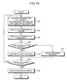
- the image processing unit 45 processes images using the luminance-adjusting program of the image-processing programs shown in FIG. 6 is described as an example with reference to FIG. 10 .
- the image processing unit 45 acquires information related to luminance such as the F-number, the LED light-emitting amount, the LED light-emitting efficiency, and luminance around the lens in the imaging unit 22 of the capsule endoscope 2 which has captured the image information to be processed from the type information acquired by the process setting unit 46 (Step S62).
- the image processing unit 45 calculates luminance of a central of the image to be processed based on the F-number, the LED light-emitting amount, the LED light-emitting efficiency, and the luminance around the lens (Step S64). Then, the image processing unit 45 calculates luminance of a predetermined periphery of the center of the image to be processed (Step S66).
- the image processing unit 45 compares the calculated luminance of central with the calculated luminance of periphery to determine whether the luminance of center is higher than the luminance of periphery (Step S68). When the image processing unit 45 determines that the luminance of center is higher than the luminance of center (Step S68: Yes), the image processing unit 45 increases a gain of the entire image because the periphery of the image is darker than the center of the image (Step S70) so that the entire image becomes lighter and can be clearly observed.
- Step S68 determines that the luminance of center is lower than the luminance of periphery (Step S68: No)
- the image processing unit 45 decreases the gain of the entire image because the periphery of the image is lighter than the center of the image (Step S72) so that the entire image becomes darker and can be clearly observed.
- the image processing unit 45 determines whether there is the following image to be processed (Step S74). If the image processing unit 45 determines that there is the following image to be processed (Step S74: Yes), the image processing unit 45 proceeds to Step S62, and acquires information related to luminance of the image to be processed to adjust the luminance. On the other hand, if the image processing unit 45 determines that there is not the following image to be processed (Step S74: No), the image processing unit 45 outputs the series of processed images to the control unit 41 (Step S76), and finishes the image processing. The series of images processed by the image processing unit 45 are displayed by the display unit 47, and stored in the database Db and the like via the control unit 41.
- the capsule endoscope 2 attaches the type information corresponding to the optical information of the imaging unit 22 in the capsule endoscope 2 to each image, and transmits the images.
- the processing apparatus 4 processes the image to be processed using the image-processing program corresponding to the optical information of the imaging unit 22 which has captured the image based on the type information attached to the image to be processed.
- the processing apparatus 4 can process the images which correspond to portions other than the applied area of the capsule endoscope 2, whereby the images which could not be used as they were can be used for the diagnosis.
- the images corresponding to portions other than the applied area which have been wasted conventionally are not wasted and can be used, and a smaller number of images are wasted.
- the subject has to swallow the capsule endoscope corresponding to the organ to be captured so that the images corresponding to portions other than the applied area could be acquired.
- the subject does not have to swallow the capsule endoscope again, whereby the subject takes less burden.
- the images corresponding to portions other than the applied area of the capsule endoscope 2 in the images stored in the database Db are processed so that the images can be used.
- the images in the database Db can be efficiently used.
- the processing apparatus 4 processes the image of the small intestine captured by the capsule endoscope 2 for previously observing the inside of the stomach of the subject so that the images of the small intestine of the subject currently observed can be compared with the processed image.
- the previous observation images can be used as history information of the subject.
- the image at the boundary between the small intestine and the large intestine is processed in correspondence with the optical information of the capsule endoscope 2 for observing the large intestine, whereby even the image captured toward the large intestine, where the inner diameter is smaller than that of the small intestine and the object to be observed is far than that of a case in the small intestine, can be used for the diagnosis more efficiently.
- the processing apparatus 4 can process the image selecting the image-processing program which processes the image corresponding to portions other than the applied area of the capsule endoscope 2 to be used.
- the image of the inside of the large intestine observed by the capsule endoscope 2 for observing the small intestine may be processed in correspondence with the optical information of the capsule endoscope 2 for observing the large intestine.
- most of the images of the enteric cavities which are captured by the capsule endoscope 2 can be used for diagnosis.
- version information may also be used with the type information.
- the capsule endoscope 2 attaches the type information of the imaging unit 22 and the version information of the capsule endoscope 2 to each piece of image information, and transmits the image information.
- the process setting unit 46 acquires the type information and also version information attached to the image information to be processed, and acquires the optical information corresponding to the acquired type information and the acquired version information with reference to the table T1, for example.
- the process setting unit 46 selects the image-processing program corresponding to the acquired optical information from the image-processing-program set Dp, and sets the image-processing program as the image program to process the image information to be processed.
- the type information and the version information may be used for the image processing.
- the storage unit 44 stores the combinations of the optical information corresponding to the version information of the capsule endoscope 2 as the optical-information set Dr, respectively. Then, the process setting unit 46 acquires the version information attached to the image information to be processed, and acquires the optical information corresponding to the acquired version information from the optical-information set Dr.
- the process setting unit 46 selects the image-processing program corresponding to the acquired optical information from the image-processing-program set Dp, and sets the image-processing program as the image program to process the image information to be processed.
- the image processing of the image to be processed may be determined only by the version information.
- the processing apparatus 4 processes the image stored in the database Db or the images acquired via the portable storage medium 5.
- the present invention is not limited to the case above.
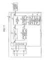
- the receiving apparatus 3 shown in FIG. 1 may process approximately in real time the images which are successively transmitted from the capsule endoscope 2 approximately real time, and the processed images may be stored in the storage unit in the receiving apparatus 3 or the portable storage medium 5.
- the receiving apparatus 3 stores the optical-information set Dr and the image-processing-program set Dp, in the main receiving unit 3b which performs the process on the wireless-transmission signal received via the receiving antennas A1 to An in the wireless-transmission unit 3a.
- the receiving apparatus 3 processes the image to be processed corresponding to portions other than the applied area of the capsule endoscope 2 using the optical-information-set Dr and the image-processing-program set Dp so that the image can be used.
- the main receiving unit 3b includes a receiving unit 31, a converting unit 32, a synchronizing-signal detection unit 33, an image processing unit 34, and a storage unit 36.
- the receiving unit 31 switches an antenna A for receiving the wireless-transmission signal.
- the receiving unit 31 performs the receiving process to perform demodulation, analog/digital conversion, and the like on the wireless-transmission signal received via the switched antenna A, and outputs a signal Sa.
- the converting unit 32 converts the signal Sa which is output from the receiving unit 31 into a signal Si having a signal format which can be processed by the image processing unit 34.
- the converting unit 32 outputs the signal Si at the same timing of a synchronizing signal output of the synchronizing-signal detection unit 33.
- the synchronizing-signal detection unit 33 detects each synchronizing signal from the signal Sa, and outputs synchronizing-signal information Sd corresponding to the detected synchronizing signal to the image processing unit.
- the image processing unit 34 performs a predetermined process on the signal Si which is output from the converting unit 32, and outputs image data Sf corresponding to a single frame of the image.
- the storage unit 36 stores the optical-information-set Dr and the image-processing-program set Dp along with the information needed for the image processing in the receiving apparatus 3.
- the image processing unit 34 includes the process setting unit 46.
- the process setting unit 46 acquires the optical information corresponding to the type information attached to the image information to be processed from the optical information stored in the storage unit 36.
- the process setting unit 46 sets the image-processing program, of the image-processing programs stored in the storage unit 44, which corresponds to the acquired optical information as the image-processing program to process the image information to be processed.
- the image processing unit 34 processes the image to be processed in approximately real time using the image-processing program corresponding to the optical information of the image information to be processed that is set by the process setting unit.
- the image which corresponds to portions other than the applied area of the capsule endoscope 2, and which could not be used as they were can be used.
- an in-vivo image acquiring system, an in-vivo image processing method, and a body-insertable apparatus according to the present invention are useful for acquiring images of organs which are captured inside the subject, and particularly suitable for decreasing the number of wasted images of organs since the images of organs corresponding to portions other than the applied area can be used for diagnosis as well as the images of organs of the applied area inside the subject that can be properly captured by the imaging unit.
Landscapes
- Health & Medical Sciences (AREA)
- Life Sciences & Earth Sciences (AREA)
- Surgery (AREA)
- Engineering & Computer Science (AREA)
- Biomedical Technology (AREA)
- Molecular Biology (AREA)
- Pathology (AREA)
- Radiology & Medical Imaging (AREA)
- Nuclear Medicine, Radiotherapy & Molecular Imaging (AREA)
- Biophysics (AREA)
- Physics & Mathematics (AREA)
- Heart & Thoracic Surgery (AREA)
- Medical Informatics (AREA)
- Optics & Photonics (AREA)
- Animal Behavior & Ethology (AREA)
- General Health & Medical Sciences (AREA)
- Public Health (AREA)
- Veterinary Medicine (AREA)
- Computer Networks & Wireless Communication (AREA)
- Signal Processing (AREA)
- Endoscopes (AREA)
- Measurement Of The Respiration, Hearing Ability, Form, And Blood Characteristics Of Living Organisms (AREA)
- Eye Examination Apparatus (AREA)
Abstract
Description
- The present invention relates to an in-vivo image acquiring system which acquires an image of an inside of a subject, an in-vivo image processing method, and a body-insertable apparatus.
- Recently, a swallowable capsule endoscope has been developed in a field of endoscope. The capsule endoscope has an imaging function and a wireless-transmission function, and has a mechanism such that the capsule endoscope is swallowed from a mouth of a patient for observing an inside of a body cavity, and successively captures an inside of organs such as an esophagus, a stomach, and a small intestine due to a peristalsis until naturally excreted (e.g., see Patent Document 1).
- Image data captured by the capsule endoscope inside a body is successively transmitted to an outside via a wireless transmission while the capsule endoscope moves through the body cavity, and then stored in a memory in a receiving apparatus arranged outside the body. A doctor or a nurse can have a diagnosis with the image data stored in the memory being displayed on a display.
- Patent Document 1: Japanese Patent Application Laid-Open No.
2003-19111 - The capsule endoscope described has a lens and a signal processing function which are designed with optical performance corresponding to the applied area. For example, in the capsule endoscope for a small intestine, the lens and the signal processing function are designed with the optical performance where a focus meets at a near point for observing a wall surface of the small intestine having a small inner diameter. Further, the capsule endoscope generally captures organs other than the applied area of the capsule endoscope, and transmits a huge number of images until naturally excreted.
- In the conventional capsule endoscope, however, the optical performance is designed corresponding to the applied area, and the appropriate optical performance is not guaranteed for organs other than the applied area. Images of the organs other than the applied area are captured not properly enough to be able to be examined, and the images cannot be used for the examination. Thus, all images but those of the applied area in the huge number of images captured by the capsule endoscope are wasted. Further, when the images of organs other than the applied area need to be taken, the subject has to swallow the capsule endoscope which is designed with the optical performance to be able to capture the organs to be captured again, and the burden of the subject is increased.
- The present invention is made in view of the above drawbacks of the conventional technology, and an object of the present invention is to provide an in-vivo image acquiring system, an in-vivo image processing method, and a body-insertable apparatus, which are able to use the captured images of organs other than the applied area of the capsule endoscope, thereby decreasing the number of wasted images.
- To solve the problems described above and achieve the object, an in-vivo image acquiring system according to the present invention includes: a body-insertable apparatus that is introduced into a subject, and wirelessly transmits image information including a captured in-vivo image of the subject to outside; and a processing apparatus that processes the image information transmitted from the body-insertable apparatus. The body-insertable apparatus includes an imaging unit that captures the in-vivo image of the subject, and a transmitting unit which attaches type information corresponding to optical information of the imaging unit to the image information including the image captured by the imaging unit, and transmits the image information. The processing apparatus includes a storage unit that stores therein combinations of pieces of the optical information corresponding respectively to pieces of the type information, and image processing programs corresponding respectively to pieces of the optical information, and an image processing unit that acquires the optical information corresponding to the type information which is attached to the image information to be processed from the optical information stored in the storage unit, and processes the image information to be processed using the image processing program, of the image processing programs stored in the storage unit, which corresponds to the acquired optical information.
- In the in-vivo image acquiring system according to the present invention, the type information indicates an applied portion of the body-insertable apparatus, and the image processing unit processes, as the image information to be processed, the image information corresponding to a portion other than a portion to which the body-insertable apparatus is applied.
- In the in-vivo image acquiring system according to the present invention, the optical information indicates a magnification ratio of the imaging, a number of pixels, or a luminance in the imaging unit, each of the image processing programs is a magnification-ratio-adjusting program which changes the magnification ratio of the image to be processed, a sharpening program which changes sharpness of the image to be processed, or a luminance-adjusting program which changes luminance of the image to be processed, and the image processing unit processes the image information to be processed using the magnification-ratio-adjusting program, the sharpening program, or the luminance-adjusting program based on the magnification ratio of the imaging, the number of pixels, or the luminance corresponding to the type information of the image information to be processed.
- An in-vivo image processing method according to the present invention is for processing image information including an in-vivo image of a subject which is transmitted from a body-insertable apparatus introduced inside the subject. The in-vivo image processing method includes the steps of: capturing the in-vivo image of the subject by an imaging unit in the body-insertable apparatus; transmitting the image information to which the type information corresponding to the optical information in the imaging unit is attached from the body-insertable apparatus; receiving the image information transmitted from the body-insertable apparatus; and acquiring the optical information corresponding to the type information attached to the image information to be processed from the received image information, and processing the image information to be processed using an image processing program corresponding to the acquired optical information.
- In the in-vivo image processing method according to the present invention, the type information indicates the applied portion of the body-insertable apparatus, and the processing step includes processing, as the image information to be processed, the image information corresponding to a portion other than a portion to which the body-insertable apparatus is applied.
- In the in-vivo image processing method according to the present invention, the optical information indicates a magnification ratio of the imaging, a number of pixels, or a luminance in the imaging unit, each of the image processing programs is a magnification-ratio-adjusting program which changes the magnification ratio of the image to be processed, a sharpening program which changes sharpness of the image to be processed, or a luminance-adjusting program which changes luminance of the image to be processed, and the processing step includes processing the image information to be processed using the magnification-ratio-adjusting program, the sharpening program, or the luminance-adjusting program based on the magnification ratio of the imaging, the number of pixels, or the luminance corresponding to the type information of the image information to be processed.
- A body-insertable apparatus according to the present invention is introduced inside a subject, and wirelessly transmits image information including a captured in-vivo image of a subject to outside. The body-insertable apparatus includes: an imaging unit that captures the in-vivo image of the subject; and a transmitting unit that attaches type information which corresponds to optical information of the imaging unit to the image information including the image captured by the imaging unit, and transmits the image information.
- According to the present invention, the body-insertable apparatus attaches the type information corresponding to the optical information of the imaging unit of the body-insertable apparatus to the in-vivo image information, and transmits the image information. The processing apparatus acquires the optical information corresponding to the type information attached to the image information to be processed, and processes the image information to be processed using the image-processing program corresponding to the acquired optical information. The processing apparatus can also process the image information to be processed that corresponds to portions other than the applied area of the body-insertable apparatus so that the image information to be processed can be used. The image which corresponds to portions other than the applied area and which is captured by the body-insertable apparatus can be used for the diagnosis. Further, the subject does not have to swallow the capsule endoscope again.
-
-
FIG. 1 is a schematic diagram of an overall configuration of an in-vivo image acquiring system according to an embodiment. -
FIG. 2 is a block diagram of a configuration of a capsule endoscope shown inFIG. 1 . -
FIG. 3 is an explanatory diagram of an image signal transmitted from the capsule endoscope shown inFIG. 1 . -
FIG. 4 is a block diagram of a configuration of a processing apparatus shown inFIG. 1 . -
FIG. 5 is a diagram illustrating an optical-information-set stored in a storage unit shown inFIG. 4 . -
FIG. 6 is a diagram illustrating an image-processing-program set stored in the storage unit shown inFIG. 4 . -
FIG. 7 is a flowchart showing a procedure of an image processing of a processing apparatus shown inFIG. 4 . -
FIG. 8 is a flowchart showing an example of an image processing procedure performed by an image processing unit shown inFIG. 4 . -
FIG. 9 is a flowchart showing an example of the image processing procedure performed by the image processing unit shown inFIG. 4 . -
FIG. 10 is a flowchart showing an example of the image processing procedure performed by the image processing unit shown inFIG. 4 . -
FIG. 11 is a block diagram of a configuration of a receiving apparatus shown inFIG. 1 . -
- 1
- Subject
- 2
- Capsule endoscope
- 3
- Receiving apparatus
- 3a
- Wireless-transmission unit
- 3b
- Main receiving unit
- 4
- Processing apparatus
- 5
- Portable storage medium
- 20
- Capsule-shaped casing
- 20a
- Front cover
- 20b
- Casing
- 21
- Control unit
- 22
- Imaging unit
- 23
- Signal processing unit
- 24
- Storage unit
- 25
- Communication processing unit
- 25a
- Antenna
- 26
- Power unit
- 31
- Receiving unit
- 32
- Converting unit
- 33
- Synchronizing-signal detection unit
- 34
- Image processing unit
- 36
- Storage unit
- 41
- Control unit
- 42
- Input unit
- 44
- Storage unit
- 45
- Image processing unit
- 46
- Process setting unit
- 47
- Display unit
- Best modes for carrying a wireless-transmission in-vivo image acquiring system and a body-inserting apparatus according to the present invention (referred to below simply as "embodiments") are described with reference to the drawings. The present invention is not limited to the embodiments. Further, same numerals are attached to identical components.
- An embodiment of the present invention is described.
FIG. 1 is a schematic diagram of an overall configuration of an in-vivo image acquiring system according to an embodiment. The in-vivo image acquiring system employs a monocular capsule endoscope as an example of a body-inserting apparatus. As shown inFIG. 1 , the wireless in-vivo image acquiring system includes acapsule endoscope 2 which is introduced inside asubject 1 and captures intracelomic images, and wirelessly transmits data such as an image signal to a receivingapparatus 3, a receiving apparatus which receives the data of the intracelomic images wirelessly transmitted from thecapsule endoscope 2, aprocessing apparatus 4 which displays the intracelomic images based on the image signal received by the receivingapparatus 3, and aportable storage medium 5 used for transferring data between the receivingapparatus 3 and theprocessing apparatus 4. - Further, the receiving
apparatus 3 includes a wireless-transmission unit 3a which includes plural receiving antennas A1 to An attached to an outer surface of the subject 1, and amain receiving unit 3b which processes a wireless-transmission signal received via the plural receiving antennas A1 to An and performs other processes. Those units are detachably connected with the receivingapparatus 3 via a connector or the like. Alternatively, the receiving antennas A1 to An may be attached to, for example, a jacket which can be worn by thesubject 1 and the subject 1 may wear the jacket so that the receiving antennas are attached to thesubject 1. Further, in this case, the receiving antennas A1 to An may be detachably attached to the jacket. - The
processing apparatus 4 displays the intracelomic images captured by thecapsule endoscope 2. Theprocessing apparatus 4 may be realized by a workstation or the like which displays the images based on the data acquired via theportable storage medium 5. Specifically, theprocessing apparatus 4 may be realized by a CRT display or an LCD display or the like which displays the image thereon, or a printer or the like which outputs the image on other mediums. - The
portable storage medium 5 may be realized by a compact flash (registered trademark) or the like, and detachably attached to themain receiving unit 3b and theprocessing apparatus 4. Theportable storage medium 5 can output or store information when attached to themain receiving unit 3b and theprocessing apparatus 4. Specifically, theportable storage medium 5 may be attached to themain receiving unit 3b and while thecapsule endoscope 2 moves through the body cavity of the subject 1, and theportable storage medium 5 stores therein the data transmitted from thecapsule endoscope 2. Further, after thecapsule endoscope 2 is excreted from thesubject 1, that is, after the capturing of the inside of the subject 1 is finished, theportable storage medium 5 is detached from themain receiving unit 3b and attached to theprocessing apparatus 4 so that the data may be read out by theprocessing apparatus 4. Since the data is transferred via theportable storage medium 5 between themain receiving unit 3b and theprocessing apparatus 4, the subject 1 can act freely while the inside of the body cavity is captured, and the time for transferring the data to theprocessing apparatus 4 can be shortened. Further, data transfer between themain receiving unit 3b and theprocessing apparatus 4 may be performed by other storage apparatuses built into themain receiving unit 3b and themain receiving unit 3b may have a wired or wireless connection with theprocessing apparatus 4. - A configuration of the
capsule endoscope 2 which is an example of the body-insertable apparatus according to the present invention is described.FIG. 2 is a schematic diagram showing an example of the configuration of thecapsule endoscope 2. As shown inFIG. 2 , thecapsule endoscope 2 includes animaging unit 22 which captures the body cavity of the subject 1, and thepower unit 26 which supplies power for each component forming thecapsule endoscope 2 inside a capsule-shapedcasing 20. Further, although theimaging unit 22 shown inFIG. 2 is monocular, theimaging unit 22 may be binocular withplural imaging units 22. - The capsule-shaped
casing 20 includes a transparent dome-shapedfront cover 20a, and acasing 20b which is kept watertight with thefront cover 20a. The capsule-shapedcasing 20 is of a size to be swallowed by the mouth of thesubject 1. Thefront cover 20a is attached to one end of thecasing 20b. Thecasing 20b is made of a colored material which does not transmit light. Thecasing 20b contains acontrol unit 21 which controls driving of each component of thecapsule endoscope 2 and controls input and output of signals of each component, animaging unit 22 which captures an inside of the body cavity, asignal processing unit 23 which processes the image captured by theimaging unit 22, astorage unit 24 which stores the information needed for the wireless transmission, acommunication processing unit 25 which modulates each signal to be transmitted to theprocessing apparatus 4 arranged outside into a wireless-transmission signal and demodulates the wireless-transmission signal received via anantenna 25a, and apower unit 26 which supplies driving power for each component of thecapsule endoscope 2. Thecommunication processing unit 25 includes theantenna 25a which may be realized by a coiled antenna, and transmits and receives the wireless-transmission signal to and from the antenna arranged outside. - The
imaging unit 22 captures the image of the body cavity of thesubject 1. Specifically, theimaging unit 22 is realized by an imaging device such as a CCD and CMOS, a light-emitting device such as an LED which illuminates an imaged field of the imaging device, and an optical components such as alens 22a which forms an image of the reflected light which is transmitted from the imaged field to the imaging device. Theimaging unit 22 is fixated on an edge of thecasing 20b. Theimaging unit 22 forms the image of the reflected light which is received from the imaged field via thefront cover 20a, and captures the image inside the body cavity of thesubject 1. The imaging device, the light-emitting device, and the optical components such as thelens 22a in theimaging unit 22 have a function and a lens which are designed with optical performance corresponding to an applied area of thecapsule endoscope 2. For example, when the capsule endoscope is to be used for a small intestine, the lens and the signal processing function and the like in theimaging unit 22 are designed with the optical performance to focus on a point nearby so that the wall surface of the small intestine, which has a small inner diameter, can be captured. Further, when thecapsule endoscope 2 is to be used for a stomach, the lens and the signal processing function are designed with optical performance to focus on a point far from thecapsule endoscope 2 compared with the case of the small intestine so that the inside of the stomach, which has a large volume, can be captured. - The
capsule endoscope 2 attached type information corresponding to the optical information to image information including in-vivo images, and transmits the image information. Theimaging unit 22 of thecapsule endoscope 2 is designed with optical performance corresponding to the applied area of thecapsule endoscope 2, and thus each pieces of optical information of theimaging unit 22 differs corresponding to the applied area. Thecapsule endoscope 2 attaches information of the applied area of thecapsule endoscope 2 to the image information as the type information corresponding to the optical information of theimaging unit 22 in thecapsule endoscope 2, and transmits the image information. - The type information is previously stored in the
storage unit 24. Thecontrol unit 21 acquires the type information corresponding to the applied area of theimaging unit 22 from thestorage unit 24, and outputs the type information to thesignal processing unit 23. Thesignal processing unit 23 processes the image captured by theimaging unit 22, and attaches the type information which is output from thecontrol unit 21 to the image signal including the image. - Specifically, as shown in
FIG. 3 , thesignal processing unit 23 processes an image G1 captured by theimaging unit 22 for every unit of scan-line data. Further, thesignal processing unit 23 appends unique information Da including information which is identical for thecapsule endoscope 2 to an end of last scan-line data. The unique information Da includes white-balance information, a serial number of thecapsule endoscope 2, and the like, and further includes the above-described type information. Thesignal processing unit 23 appends the unique information Da including the white-balance information, the serial number, and the type information to an end of the last scan-line data similarly for images G2 to Gn captured by theimaging unit 22. - The
communication processing unit 25 wirelessly transmits the information generated by thesignal processing unit 23 via theantenna 25a . Specifically, thecommunication processing unit 25 attaches the type information corresponding to the optical information in theimaging unit 22 to the image information including images captured by theimaging unit 22, and wirelessly transmits the image information to an outside. - Thus, the
capsule endoscope 2 attaches the type information, which corresponds to the optical information in theimaging unit 22 of thecapsule endoscope 2 and indicates the applied area of thecapsule endoscope 2, to the image information captured by theimaging unit 22. The information transmitted from thecapsule endoscope 2 is received by the receivingapparatus 3, and stored in theportable storage medium 5. Theprocessing apparatus 4 can acquire the type information with the image information including the images captured by thecapsule endoscope 2 by reading the information stored in theportable storage medium 5. - The
processing apparatus 4 shown inFIG. 1 is described with reference toFIG. 4. FIG. 4 is a block diagram of an overall configuration of theprocessing apparatus 4 shown inFIG. 1 . As shown inFIG. 4 , theprocessing apparatus 4 includes acontrol unit 41, aninput unit 42, astorage unit 44, animage processing unit 45, and adisplay unit 47. Further, thecontrol unit 41 has a connection with database Db where image groups Pa, Pb captured by thecapsule endoscope 2 are stored in folders Fa, Fb, or the like. - The
control unit 41 includes a CPU or the like which has a controlling function. Thecontrol unit 41 controls operations and procedures of theinput unit 42, thestorage unit 44, theimage processing unit 45, and thedisplay unit 47. Theinput unit 42 is realized by a keyboard to input various information, or a mouse to point to any location on a display screen of thedisplay unit 47. Theinput unit 42 acquires various information needed for an analysis of the specimen, and instruction information of an analyzing operation, or the like from an outside. - The
storage unit 44 includes a hard disk, which magnetically stores information, and a memory, which loads a variety of programs for a process of theprocessing apparatus 4 from the hard disk and electrically stores the programs therein when theprocessing apparatus 4 performs the process. Thestorage unit 44 stores various information including an optical-information set Dr and an image-processing-program set Dp. Thestorage unit 44 may include an auxiliary storage apparatus which can read information stored in a storage medium such as a CD-ROM, a DVD-ROM, and a PC card. - The
imaging unit 22 of thecapsule endoscope 2 is designed corresponding to the applied area of thecapsule endoscope 2. Thus, each optical information of theimaging unit 22 differs depending on the applied area of thecapsule endoscope 2, i.e., the type ofcapsule endoscope 2. As shown inFIG. 5 , thestorage unit 44 stores a table T1 which shows combinations of optical information corresponding to each type of thecapsule endoscope 2 as an optical-information set Dr. The table T1 shows a magnification ratio of the imaging, a distortion (DT) value, the number of pixels, a luminance, an aberration amount for a height of an image, a gamma value, and a spectral sensitivity in the imaging unit corresponding to each type of thecapsule endoscope 2, i.e., each applied area of thecapsule endoscope 2 such as a small intestine as optical information. Further, when optical information differs for each version of each type of thecapsule endoscope 2, thestorage unit 44 stores each optical information corresponding to each version of each type as the optical-information set Dr similarly to the table T1. - The
storage unit 44 stores a magnification-ratio-adjusting program which adjusts a magnification ratio of an image to be processed, a sharpening program which adjusts sharpness of the image to be processed, and a luminance-adjusting program which adjusts luminance of the image to be processed, and the like as an image-processing-program set Dp. Specifically, as shown in a table T2 inFIG. 6 , thestorage unit 44 stores the magnification-ratio-adjusting program which adjusts the magnification ratio of the image based on the magnification ratio of imaging of theimaging unit 22 which has captured the image to be processed, the sharpening program which adjusts sharpness of the image based on the number of pixels of theimaging unit 22 which has captured the image to be processed, and the luminance-adjusting program which adjusts luminance of the image based on the luminance of theimaging unit 22 which has captured the image to be processed as the image processing programs. Further, thestorage unit 44 stores a DT-value-correcting program which corrects the image based on a DT value of theimaging unit 22 which has captured the image to be processed, an aberration-correcting program which corrects the image based on an aberration amount of theimaging unit 22 which has captured the image to be processed, a gamma-correcting program which corrects the image based on a gamma value of theimaging unit 22, and a color-correcting program which corrects colors of the image based on spectral sensitivity of theimaging unit 22 which has captured the image to be processed. Thestorage unit 44 may store plural image processing programs as the image-processing-program set Dp, respectively. As shown in the table T2 inFIG. 6 , thestorage unit 44 may store the magnification-ratio-adjusting programs a1, a2, the DT-value-correcting programs b1, b2, the sharpening programs c1, c2, the luminance-adjusting programs d1, d2, the aberration-correcting programs e1, e2, the gamma-correcting programs f1, f2, and the color-correcting programs g1, g2. - The
image processing unit 45 includes aprocess setting unit 46. Theprocess setting unit 46 acquires the optical information corresponding to the type information attached to the image information to be processed from the optical information stored in thestorage unit 44. Further, theprocess setting unit 46 sets the image processing program, of the image processing programs stored in thestorage unit 44, that corresponds to the optical information acquired from the image processing program stored in the storage unit 44as the image processing program to process the image information to be processed. Theimage processing unit 45 processes the image to be processed using the image processing program which is set by the process setting unit and corresponds to the optical information of the image information to be processed. Theimage processing unit 45 processes the image information including the images corresponding to portions other than the applied area of thecapsule endoscope 2 as the image information to be processed. Theimage processing unit 45 processes the image information to be processed using the magnification-ratio-adjusting program, the sharpening program, the luminance-adjusting program, and the like corresponding to the optical information such as the magnification ratio, the number of pixels, and the luminance corresponding to the type information of the image information to be processed. Theimage processing unit 45 acquires the optical information corresponding to the type information of the images which are captured by thecapsule endoscope 2 and correspond to portions other than the applied area of thecapsule endoscope 2, and further theimage processing unit 45 processes the image using the image processing program corresponding to the optical information so that the images which correspond to portions other than the applied area and could not be used otherwise as they were can be used. - The
display unit 47 is realized by a CRT display, an LCD display, and the like. Thedisplay unit 47 displays the instruction information, an instruction result, and the like of theinput unit 42. Thedisplay unit 47 displays the images captured by thecapsule endoscope 2 and the images processed by theimage processing unit 45 under the control by thecontrol unit 41. - The procedure of image processing in the
processing apparatus 4 is described with reference toFIG. 7. FIG. 7 is a flowchart showing each procedure of the image processing in theprocessing apparatus 4 shown inFIG. 4 . As shown inFIG. 7 , theimage processing unit 45 acquires via thecontrol unit 41 instruction information which is input from theinput unit 42 and which instructs theimage processing unit 45 to process the image (Step S2). The instruction information includes information of the image information to be processed and the contents of the process on the image information to be processed, and the like. The instruction information instructs theimage processing unit 45 to process the image information, of the image groups acquired via theportable storage medium 5, which corresponds to a predetermined number of the images corresponding to portions other than the applied area of thecapsule endoscope 2. Furthermore, the instruction information instructs theimage processing unit 45 to process the image information, of image groups Pa, Pb stored in database Db, that corresponds to the predetermined number of images corresponding to portions other than the applied area of thecapsule endoscope 2 which has captured the image group Pa, Pb. Then, the instruction information instructs theimage processing unit 45 to perform the image processing which allows the portion captured in the image which is selected as the image information to be processed according to the instruction information to be used for a diagnosis. For example, the instruction information selects the images inside the stomach observed by thecapsule endoscope 2 for observing the small intestine as the image information to be processed, and instructs theimage processing unit 45 to process the images so as to allow the images to be observed as easily as the images captured by thecapsule endoscope 2 for observing the stomach. - The
image processing unit 45 acquires the image information to be processed according to the instruction information (Step S4). Theprocess setting unit 46 acquires the type information attached to the image information from the image information to be processed (Step S6). The type information is included in the unique information Da which is appended to the end of the last scan-line data as previously described. Theprocess setting unit 46 acquires the optical information that corresponds to the type information of the image information to be processed that is acquired from the unique information Da appended to the end of the last scan-line data at Step 6 (Step 8). - The
process setting unit 46 sets the image processing program, of the image processing programs set by theprocess setting unit 46, that corresponds to the acquired optical information as the image processing program to process the image information to be processed (Step S10). - The
image processing unit 45 processes the image information to be processed using the image processing program set by the process setting unit 46 (Step S12). The display unit outputs and displays the image processed by the image processing unit 45 (Step S14). Thecontrol unit 41 determines whether the image processed by theimage processing unit 45 is to be saved based on the instruction information which is input from the input unit 42 (Step S16). For example, when an operator of theprocessing apparatus 4 selects via a mouse a selection field to save the image on a selection menu where the operator can choose to save the processed image, instruction information to save the processed image is input from theinput unit 42 to thecontrol unit 41. Further, when the operator of theprocessing apparatus 4 selects via a mouse a selection field not to save the image, instruction information not to save the processed image is input from theinput unit 42 to thecontrol unit 41. - If the
control unit 41 determines that the image processed by theimage processing unit 45 is not to be saved (Step S16: No), theprocessing apparatus 4 finishes the image processing. Further, if thecontrol unit 41 determines that the image processed by theimage processing unit 45 is to be saved (Step S16: Yes), theprocessing apparatus 4 saves the processed image in a specified destination to save (Step S18), and finished the image processing in theprocessing apparatus 4. - The image processing performed in the image processing unit 45 (Step S12) is described below in a specific manner. Firstly, the image processing where the
image processing unit 45 processes the image using the magnification-ratio-adjusting program of the image processing programs shown inFIG. 6 is described as an example with reference toFIG. 8 . - The
image processing unit 45 acquired view-angle information and LED light-emitting amount information of theimaging unit 22 in thecapsule endoscope 2 which has captured the image information to be processed from the type information acquired by theprocess setting unit 46 according to the procedure of the magnification-ratio-adjusting program (Step S22). - The
image processing unit 45 calculates observation distance in thecapsule endoscope 2 based on the view-angle information and the LED light-emitting amount information (Step S24). The view angle is a range of the angle for theimaging unit 22 of thecapsule endoscope 2 to capture clear images. The observation distance can be calculated based on the view angle. Further, in thecapsule endoscope 2, theimaging unit 22 automatically adjusts light. When the imaged area is too dark as an object to be captured is far, theimaging unit 22 automatically increases a light-emitting amount of an LED. When the imaged area is too light as the object to be captured is close, theimaging unit 22 automatically decreases the light-emitting amount of the LED. Thus, theimage processing unit 45 can calculate the distance between the object to be captured and thecapsule endoscope 2 based on information of the LED light-emitting amount, specifically, the light-emitting amount of the LED at the time when the image information to be processed is captured. - The
image processing unit 45 calculates the magnification ratio of theimaging unit 22 in thecapsule endoscope 2 which has captured the image to be processed based on the calculated observation distance and the size of the imaged object in the image information to be processed (Step S26). - The
image processing unit 45 determines whether the calculated magnification ratio is higher than a predetermined magnification ratio corresponding to an intended usage (Step S28). When theimage processing unit 45 processes an image of the inside of the stomach observed by thecapsule endoscope 2 for observing the small intestine for the image to be used for the diagnosis, theimage processing unit 45 compares the calculated magnification ratio with the magnification ratio of thecapsule endoscope 2 for observing the stomach. The compared magnification ratios may be included in the magnification-ratio-adjusting program, or acquired from the optical-information set Dr in thestorage unit 44. - If the
image processing unit 45 determines that the calculated magnification ratio is not higher than the predetermined magnification ratio (Step S28: No), i.e., that the calculated magnification ratio is lower than the predetermined magnification ratio, theimage processing unit 45 magnifies the image to be processed in correspondence with the predetermined magnification ratio (Step S30). For example, when the image to be processed is the image of the inside of the stomach captured by thecapsule endoscope 2 for observing the small intestine, and the magnification ratio is lower than the magnification ratio of thecapsule endoscope 2 for observing the inside of the stomach, theimage processing unit 45 magnifies thecapsule endoscope 2 for observing the inside of the stomach. - On the other hand, if the
image processing unit 45 determines that the calculated magnification ratio is higher than the predetermined magnification ratio (Step S28: Yes), theimage processing unit 45 demagnifies the image to be processed in correspondence with the predetermined magnification ratio (Step S32). - After the magnification process or the demagnification process, the
image processing unit 45 determines whether there is a following image to be processed. If theimage processing unit 45 determines that there is the following image to be processed (Step S34: Yes), theimage processing unit 45 proceeds to Step S24 and calculates observation distance on the image to be processed. On the other hand, if theimage processing unit 45 determines that there is not the following image to be processed (Step S34: No), theimage processing unit 45 outputs a series of processed images to the control unit 41 (Step S36), and finishes the image processing. The series of images processed by theimage processing unit 45 are displayed by thedisplay unit 47, and stored in the database Db and the like via thecontrol unit 41. - A case where the
image processing unit 45 processes images using the sharpening program of the image-processing programs shown inFIG. 6 is described with reference toFIG. 9 . According to the procedure of the sharpening program, theimage processing unit 45 acquires information of the number of pixels which indicates the number of pixels of theimaging unit 22 of thecapsule endoscope 2 which has captured the image to be processed from the type information acquired by the process setting unit 46 (Step S42). - The
image processing unit 45 determines, based on the acquired information of the number of pixels, whether the number of pixels of theimaging unit 22 of thecapsule endoscope 2 which has captured the image to be processed is smaller than a predetermined number of pixel corresponding to an intended usage (Step S44). For example, when theimage processing unit 45 processes the image of the inside of the stomach observed by thecapsule endoscope 2 for observing the small intestine for the image to be used for the diagnosis, theimage processing unit 45 compares the number of pixels with the number of pixels of thecapsule endoscope 2 for observing the inside of the stomach. The compared number of pixels may be included in the sharpening program, or acquired from the optical-information set Dr stored in thestorage unit 44. - When the
image processing unit 45 determines that the number of pixels of theimaging unit 22 of thecapsule endoscope 2 which has captured the image to be processed is smaller than the predetermined number of pixels (Step 44: Yes), theimage processing unit 45 sharpens edges of the image to be processed by adjusting the sharpness of the image (Step S46). When the image to be processed is an image of the inside of the stomach captured by thecapsule endoscope 2 for observing the small intestine, and the image is captured with the number of pixels which is smaller than the number of pixels of the capsule endoscope for observing the inside of the stomach, theimage processing unit 45 sharpens the image in correspondence with the sharpness of thecapsule endoscope 2 because the edge of the image is unclear compared with the image captured by thecapsule endoscope 2 for observing the inside of the stomach. - On the other hand, when the
image processing unit 45 determines that the number of pixels of theimaging unit 22 of thecapsule endoscope 2 which has captured the image to be processed is larger than the predetermined number of pixels (Step S44: No), theimage processing unit 45 does not sharpen the edge because the sharpness of the image to be processed is clear enough for the intended usage in the diagnosis. Thus, theimage processing unit 45 determines the case as "unsharp" (Step S50). When the image to be processed is the image of the inside of the stomach captured by thecapsule endoscope 2 for observing the small intestine, and the image is captured with the larger number of pixels than the number of pixels of thecapsule endoscope 2 for observing the inside of the stomach, it is not necessary for theimage processing unit 45 to sharpen the image in correspondence with the sharpness of thecapsule endoscope 2 for observing the inside of the stomach because the edge of the image to be processed is no less clear than the image captured by thecapsule endoscope 2 for observing the inside of the stomach. - After the determination process of the sharpening or unsharpening, the
image processing unit 45 determines whether there is a following image to be processed (Step S52). If theimage processing unit 45 determines that there is the following image to be processed (Step S52: Yes), theimage processing unit 45 proceeds to Step S44, and performs a determination process on the number of pixels of the following image to be processed. On the other hand, if theimage processing unit 45 determines that there is not the following image to be processed (Step S52: No), theimage processing unit 45 outputs the series of processed images to the control unit 41 (Step S54), and finishes the image processing. The series of images processed by theimage processing unit 45 are displayed by thedisplay unit 47, and stored in the database Db and the like via thecontrol unit 41. - A care where the
image processing unit 45 processes images using the luminance-adjusting program of the image-processing programs shown inFIG. 6 is described as an example with reference toFIG. 10 . According to the procedure of the luminance-adjusting program, theimage processing unit 45 acquires information related to luminance such as the F-number, the LED light-emitting amount, the LED light-emitting efficiency, and luminance around the lens in theimaging unit 22 of thecapsule endoscope 2 which has captured the image information to be processed from the type information acquired by the process setting unit 46 (Step S62). - The
image processing unit 45 calculates luminance of a central of the image to be processed based on the F-number, the LED light-emitting amount, the LED light-emitting efficiency, and the luminance around the lens (Step S64). Then, theimage processing unit 45 calculates luminance of a predetermined periphery of the center of the image to be processed (Step S66). - The
image processing unit 45 compares the calculated luminance of central with the calculated luminance of periphery to determine whether the luminance of center is higher than the luminance of periphery (Step S68). When theimage processing unit 45 determines that the luminance of center is higher than the luminance of center (Step S68: Yes), theimage processing unit 45 increases a gain of the entire image because the periphery of the image is darker than the center of the image (Step S70) so that the entire image becomes lighter and can be clearly observed. On the other hand, if theimage processing unit 45 determines that the luminance of center is lower than the luminance of periphery (Step S68: No), theimage processing unit 45 decreases the gain of the entire image because the periphery of the image is lighter than the center of the image (Step S72) so that the entire image becomes darker and can be clearly observed. - After the process of increasing the gain or decreasing the gain, the
image processing unit 45 determines whether there is the following image to be processed (Step S74). If theimage processing unit 45 determines that there is the following image to be processed (Step S74: Yes), theimage processing unit 45 proceeds to Step S62, and acquires information related to luminance of the image to be processed to adjust the luminance. On the other hand, if theimage processing unit 45 determines that there is not the following image to be processed (Step S74: No), theimage processing unit 45 outputs the series of processed images to the control unit 41 (Step S76), and finishes the image processing. The series of images processed by theimage processing unit 45 are displayed by thedisplay unit 47, and stored in the database Db and the like via thecontrol unit 41. - Thus, in the embodiment, the
capsule endoscope 2 attaches the type information corresponding to the optical information of theimaging unit 22 in thecapsule endoscope 2 to each image, and transmits the images. Theprocessing apparatus 4 processes the image to be processed using the image-processing program corresponding to the optical information of theimaging unit 22 which has captured the image based on the type information attached to the image to be processed. As a result, theprocessing apparatus 4 can process the images which correspond to portions other than the applied area of thecapsule endoscope 2, whereby the images which could not be used as they were can be used for the diagnosis. Thus, according to the present embodiment, the images corresponding to portions other than the applied area which have been wasted conventionally are not wasted and can be used, and a smaller number of images are wasted. Further, conventionally, the subject has to swallow the capsule endoscope corresponding to the organ to be captured so that the images corresponding to portions other than the applied area could be acquired. However, in the present invention, the subject does not have to swallow the capsule endoscope again, whereby the subject takes less burden. - Further, in the present embodiment, the images corresponding to portions other than the applied area of the
capsule endoscope 2 in the images stored in the database Db are processed so that the images can be used. Thus, the images in the database Db can be efficiently used. For example, theprocessing apparatus 4 processes the image of the small intestine captured by thecapsule endoscope 2 for previously observing the inside of the stomach of the subject so that the images of the small intestine of the subject currently observed can be compared with the processed image. Thus, the previous observation images can be used as history information of the subject. - Further, in the present embodiment, of the images captured by the
capsule endoscope 2 for observing the small intestine, the image at the boundary between the small intestine and the large intestine is processed in correspondence with the optical information of thecapsule endoscope 2 for observing the large intestine, whereby even the image captured toward the large intestine, where the inner diameter is smaller than that of the small intestine and the object to be observed is far than that of a case in the small intestine, can be used for the diagnosis more efficiently. - Further, in the present embodiment, the case where the image of the inside of the stomach observed by the capsule endoscope for observing the small intestine is processed as an example. The present invention, however, is not limited to the embodiment. The
processing apparatus 4 can process the image selecting the image-processing program which processes the image corresponding to portions other than the applied area of thecapsule endoscope 2 to be used. For example, the image of the inside of the large intestine observed by thecapsule endoscope 2 for observing the small intestine may be processed in correspondence with the optical information of thecapsule endoscope 2 for observing the large intestine. As a result, according to the present embodiment, most of the images of the enteric cavities which are captured by thecapsule endoscope 2 can be used for diagnosis. - In the present embodiment, as shown in the table T1, when each version of each type of the
capsule endoscope 2 differs corresponding to the optical information, version information may also be used with the type information. In this case, thecapsule endoscope 2 attaches the type information of theimaging unit 22 and the version information of thecapsule endoscope 2 to each piece of image information, and transmits the image information. In theprocessing apparatus 4, theprocess setting unit 46 acquires the type information and also version information attached to the image information to be processed, and acquires the optical information corresponding to the acquired type information and the acquired version information with reference to the table T1, for example. Then, theprocess setting unit 46 selects the image-processing program corresponding to the acquired optical information from the image-processing-program set Dp, and sets the image-processing program as the image program to process the image information to be processed. Thus, in the present embodiment, when the optical information differs depending on each piece of version information, the type information and the version information may be used for the image processing. - Further, when the different version information is attached depending on each different optical performance of the
imaging unit 22, i.e., when the different version information is attached to the different optical information of theimaging unit 22 regardless of the type of thecapsule endoscope 2, the optical information can determined by the version information, and thecapsule endoscope 2 does not need to further attach the type information. Thus, thecapsule endoscope 2 attaches only the version information to the image information, and transmits the image information. In theprocessing apparatus 4, thestorage unit 44 stores the combinations of the optical information corresponding to the version information of thecapsule endoscope 2 as the optical-information set Dr, respectively. Then, theprocess setting unit 46 acquires the version information attached to the image information to be processed, and acquires the optical information corresponding to the acquired version information from the optical-information set Dr. Theprocess setting unit 46 selects the image-processing program corresponding to the acquired optical information from the image-processing-program set Dp, and sets the image-processing program as the image program to process the image information to be processed. Thus, in the present embodiment, when the optical information can be determined only by the version information, the image processing of the image to be processed may be determined only by the version information. - Further, in the present embodiment, the case where the
processing apparatus 4 processes the image stored in the database Db or the images acquired via theportable storage medium 5. The present invention, however, is not limited to the case above. For example, the receivingapparatus 3 shown inFIG. 1 may process approximately in real time the images which are successively transmitted from thecapsule endoscope 2 approximately real time, and the processed images may be stored in the storage unit in the receivingapparatus 3 or theportable storage medium 5. - In this case, as shown in
FIG. 11 , the receivingapparatus 3 stores the optical-information set Dr and the image-processing-program set Dp, in themain receiving unit 3b which performs the process on the wireless-transmission signal received via the receiving antennas A1 to An in the wireless-transmission unit 3a. The receivingapparatus 3 processes the image to be processed corresponding to portions other than the applied area of thecapsule endoscope 2 using the optical-information-set Dr and the image-processing-program set Dp so that the image can be used. - The
main receiving unit 3b is described in detail. As shown inFIG. 11 , themain receiving unit 3b includes a receivingunit 31, a convertingunit 32, a synchronizing-signal detection unit 33, animage processing unit 34, and astorage unit 36. The receivingunit 31 switches an antenna A for receiving the wireless-transmission signal. The receivingunit 31 performs the receiving process to perform demodulation, analog/digital conversion, and the like on the wireless-transmission signal received via the switched antenna A, and outputs a signal Sa. The convertingunit 32 converts the signal Sa which is output from the receivingunit 31 into a signal Si having a signal format which can be processed by theimage processing unit 34. The convertingunit 32 outputs the signal Si at the same timing of a synchronizing signal output of the synchronizing-signal detection unit 33. The synchronizing-signal detection unit 33 detects each synchronizing signal from the signal Sa, and outputs synchronizing-signal information Sd corresponding to the detected synchronizing signal to the image processing unit. - The
image processing unit 34 performs a predetermined process on the signal Si which is output from the convertingunit 32, and outputs image data Sf corresponding to a single frame of the image. Thestorage unit 36 stores the optical-information-set Dr and the image-processing-program set Dp along with the information needed for the image processing in the receivingapparatus 3. - As shown in
FIG. 11 , theimage processing unit 34 includes theprocess setting unit 46. Theprocess setting unit 46 acquires the optical information corresponding to the type information attached to the image information to be processed from the optical information stored in thestorage unit 36. Theprocess setting unit 46 sets the image-processing program, of the image-processing programs stored in thestorage unit 44, which corresponds to the acquired optical information as the image-processing program to process the image information to be processed. Theimage processing unit 34 processes the image to be processed in approximately real time using the image-processing program corresponding to the optical information of the image information to be processed that is set by the process setting unit. Thus, the image which corresponds to portions other than the applied area of thecapsule endoscope 2, and which could not be used as they were can be used. - As described above, an in-vivo image acquiring system, an in-vivo image processing method, and a body-insertable apparatus according to the present invention are useful for acquiring images of organs which are captured inside the subject, and particularly suitable for decreasing the number of wasted images of organs since the images of organs corresponding to portions other than the applied area can be used for diagnosis as well as the images of organs of the applied area inside the subject that can be properly captured by the imaging unit.
Claims (7)
- An in-vivo image acquiring system, comprising:a body-insertable apparatus that is introduced into a subject, and wirelessly transmits image information including a captured in-vivo image of the subject to outside; anda processing apparatus that processes the image information transmitted from the body-insertable apparatus, andthe body-insertable apparatus comprises
an imaging unit that captures the in-vivo image of the subject, and
a transmitting unit which attaches type information corresponding to optical information of the imaging unit to the image information including the image captured by the imaging unit, and transmits the image information, andthe processing apparatus comprises
a storage unit that stores therein combinations of pieces of the optical information corresponding respectively to pieces of the type information, and image processing programs corresponding respectively to pieces of the optical information, and
an image processing unit that acquires the optical information corresponding to the type information which is attached to the image information to be processed from the optical information stored in the storage unit, and processes the image information to be processed using the image processing program, of the image processing programs stored in the storage unit, which corresponds to the acquired optical information. - The in-vivo image acquiring system according to claim 1, wherein
the type information indicates an applied portion of the body-insertable apparatus, and
the image processing unit processes, as the image information to be processed, the image information corresponding to a portion other than a portion to which the body-insertable apparatus is applied. - The in-vivo image acquiring system according to claim 1, wherein
the optical information indicates a magnification ratio of the imaging, a number of pixels, or a luminance in the imaging unit,
each of the image processing programs is a magnification-ratio-adjusting program which changes the magnification ratio of the image to be processed, a sharpening program which changes sharpness of the image to be processed, or a luminance-adjusting program which changes luminance of the image to be processed, and
the image processing unit processes the image information to be processed using the magnification-ratio-adjusting program, the sharpening program, or the luminance-adjusting program based on the magnification ratio of the imaging, the number of pixels, or the luminance corresponding to the type information of the image information to be processed. - An in-vivo image processing method for processing image information including an in-vivo image of a subject which is transmitted from a body-insertable apparatus introduced inside the subject, the in-vivo image processing method comprising the steps of:capturing the in-vivo image of the subject by an imaging unit in the body-insertable apparatus;transmitting the image information to which the type information corresponding to the optical information in the imaging unit is attached from the body-insertable apparatus;receiving the image information transmitted from the body-insertable apparatus; andacquiring the optical information corresponding to the type information attached to the image information to be processed from the received image information, and processing the image information to be processed using an image processing program corresponding to the acquired optical information.
- The in-vivo image processing method according to claim 4, wherein
the type information indicates the applied portion of the body-insertable apparatus, and
the processing step includes processing, as the image information to be processed, the image information corresponding to a portion other than a portion to which the body-insertable apparatus is applied. - The in-vivo image processing method according to claim 4, wherein
the optical information indicates a magnification ratio of the imaging, a number of pixels, or a luminance in the imaging unit,
each of the image processing programs is a magnification-ratio-adjusting program which changes the magnification ratio of the image to be processed, a sharpening program which changes sharpness of the image to be processed, or a luminance-adjusting program which changes luminance of the image to be processed, and
the processing step includes processing the image information to be processed using the magnification-ratio-adjusting program, the sharpening program, or the luminance-adjusting program based on the magnification ratio of the imaging, the number of pixels, or the luminance corresponding to the type information of the image information to be processed. - A body-insertable apparatus which is introduced inside a subject, and wirelessly transmits image information including a captured in-vivo image of a subject to outside, the body-insertable apparatus comprising:an imaging unit that captures the in-vivo image of the subject; anda transmitting unit that attaches type information which corresponds to optical information of the imaging unit to the image information including the image captured by the imaging unit, and transmits the image information.
Applications Claiming Priority (2)
| Application Number | Priority Date | Filing Date | Title |
|---|---|---|---|
| JP2007226973A JP5259141B2 (en) | 2007-08-31 | 2007-08-31 | In-subject image acquisition system, in-subject image processing method, and in-subject introduction device |
| PCT/JP2008/065210 WO2009028508A1 (en) | 2007-08-31 | 2008-08-26 | In-examinee image acquisition system, in-examinee image processing method, and in-examinee introduction device |
Publications (3)
| Publication Number | Publication Date |
|---|---|
| EP2191767A1 true EP2191767A1 (en) | 2010-06-02 |
| EP2191767A4 EP2191767A4 (en) | 2014-10-01 |
| EP2191767B1 EP2191767B1 (en) | 2017-05-10 |
Family
ID=40387227
Family Applications (1)
| Application Number | Title | Priority Date | Filing Date |
|---|---|---|---|
| EP08828127.4A Not-in-force EP2191767B1 (en) | 2007-08-31 | 2008-08-26 | In-examinee image acquisition system and in-examinee image processing method |
Country Status (5)
| Country | Link |
|---|---|
| US (1) | US8300090B2 (en) |
| EP (1) | EP2191767B1 (en) |
| JP (1) | JP5259141B2 (en) |
| CN (1) | CN101790344B (en) |
| WO (1) | WO2009028508A1 (en) |
Families Citing this family (7)
| Publication number | Priority date | Publication date | Assignee | Title |
|---|---|---|---|---|
| JP5015363B2 (en) * | 2010-09-24 | 2012-08-29 | オリンパスメディカルシステムズ株式会社 | Image display device and capsule endoscope system |
| JPWO2012042966A1 (en) * | 2010-09-28 | 2014-02-06 | オリンパスメディカルシステムズ株式会社 | Image display device, image display method, and image display program |
| JP2012249956A (en) * | 2011-06-06 | 2012-12-20 | Toshiba Corp | Capsule endoscope image processing apparatus and capsule endoscope system |
| CN103513413B (en) * | 2012-06-18 | 2017-05-24 | 奥林巴斯株式会社 | Endoscope device and endoscope image recording destination folder change method |
| JP6581923B2 (en) * | 2016-03-03 | 2019-09-25 | 富士フイルム株式会社 | Image processing apparatus, operating method thereof and operating program |
| JPWO2018020558A1 (en) * | 2016-07-25 | 2019-05-09 | オリンパス株式会社 | Image processing apparatus, image processing method and program |
| AU2019217992B2 (en) * | 2018-02-09 | 2024-06-13 | Gyrus Acmi, Inc. D.B.A. Olympus Surgical Technologies America | Medical laser apparatus and system |
Family Cites Families (26)
| Publication number | Priority date | Publication date | Assignee | Title |
|---|---|---|---|---|
| JP3958526B2 (en) * | 2001-02-28 | 2007-08-15 | ペンタックス株式会社 | Observation site display system for electronic endoscope apparatus |
| IL143260A (en) | 2001-05-20 | 2006-09-05 | Given Imaging Ltd | Array system and method for locating an in vivo signal source |
| JP4744026B2 (en) * | 2001-07-30 | 2011-08-10 | オリンパス株式会社 | Capsule endoscope and capsule endoscope system |
| JP3810381B2 (en) * | 2003-04-25 | 2006-08-16 | オリンパス株式会社 | Image display device, image display method, and image display program |
| US7623904B2 (en) * | 2003-08-06 | 2009-11-24 | Olympus Corporation | Medical apparatus, medical apparatus guide system, capsule type medical apparatus, and capsule type medical apparatus guide apparatus |
| US20050029437A1 (en) * | 2003-08-08 | 2005-02-10 | Akira Hasegawa | Capsule optical sensor |
| JP3993550B2 (en) * | 2003-09-30 | 2007-10-17 | オリンパス株式会社 | Gastrointestinal inspection device |
| US20050075537A1 (en) * | 2003-10-06 | 2005-04-07 | Eastman Kodak Company | Method and system for real-time automatic abnormality detection for in vivo images |
| JP4009581B2 (en) * | 2003-11-18 | 2007-11-14 | オリンパス株式会社 | Capsule medical system |
| JP4445799B2 (en) * | 2004-05-24 | 2010-04-07 | オリンパス株式会社 | Intra-subject introduction device and medical device |
| JP2006075331A (en) * | 2004-09-09 | 2006-03-23 | Sanyo Electric Co Ltd | Imaging apparatus for endoscope |
| US7627189B2 (en) * | 2004-10-20 | 2009-12-01 | Fujinon Corporation | Sharpness adjustment method and program and electronic endoscope apparatus |
| JP2006115965A (en) | 2004-10-20 | 2006-05-11 | Fujinon Corp | Electronic endoscope apparatus |
| JP4615963B2 (en) * | 2004-10-29 | 2011-01-19 | オリンパス株式会社 | Capsule endoscope device |
| JP4709579B2 (en) * | 2005-04-26 | 2011-06-22 | オリンパスメディカルシステムズ株式会社 | Capsule endoscope |
| JP4602828B2 (en) * | 2005-04-26 | 2010-12-22 | オリンパスメディカルシステムズ株式会社 | In-subject information acquisition system |
| JP4472585B2 (en) * | 2005-06-14 | 2010-06-02 | オリンパス株式会社 | Transmission device and in-subject information acquisition system |
| JP2007082664A (en) * | 2005-09-21 | 2007-04-05 | Fujifilm Corp | Capsule endoscope |
| US7567692B2 (en) * | 2005-09-30 | 2009-07-28 | Given Imaging Ltd. | System and method for detecting content in-vivo |
| JP4855759B2 (en) * | 2005-10-19 | 2012-01-18 | オリンパス株式会社 | Receiving apparatus and in-subject information acquisition system using the same |
| WO2007066288A2 (en) * | 2005-12-07 | 2007-06-14 | Koninklijke Philips Electronics, N.V. | Electronic gastrointestinal screening |
| JP4847161B2 (en) | 2006-03-02 | 2011-12-28 | キヤノン株式会社 | Image transmitting apparatus and imaging apparatus |
| JP2007319442A (en) * | 2006-06-01 | 2007-12-13 | Fujifilm Corp | Capsule endoscope system and image processing apparatus |
| JP4932588B2 (en) * | 2007-05-08 | 2012-05-16 | オリンパス株式会社 | Image processing apparatus and image processing program |
| JP5467754B2 (en) * | 2008-07-08 | 2014-04-09 | Hoya株式会社 | Signal processing apparatus for electronic endoscope and electronic endoscope apparatus |
| JP2010187756A (en) * | 2009-02-16 | 2010-09-02 | Olympus Corp | Image processing apparatus, image processing method, and image processing program |
-
2007
- 2007-08-31 JP JP2007226973A patent/JP5259141B2/en not_active Expired - Fee Related
-
2008
- 2008-08-26 EP EP08828127.4A patent/EP2191767B1/en not_active Not-in-force
- 2008-08-26 CN CN2008801044989A patent/CN101790344B/en not_active Expired - Fee Related
- 2008-08-26 WO PCT/JP2008/065210 patent/WO2009028508A1/en not_active Ceased
- 2008-08-27 US US12/199,679 patent/US8300090B2/en active Active
Also Published As
| Publication number | Publication date |
|---|---|
| WO2009028508A1 (en) | 2009-03-05 |
| EP2191767A4 (en) | 2014-10-01 |
| US20090058996A1 (en) | 2009-03-05 |
| CN101790344B (en) | 2012-07-18 |
| CN101790344A (en) | 2010-07-28 |
| JP5259141B2 (en) | 2013-08-07 |
| JP2009056160A (en) | 2009-03-19 |
| EP2191767B1 (en) | 2017-05-10 |
| US8300090B2 (en) | 2012-10-30 |
Similar Documents
| Publication | Publication Date | Title |
|---|---|---|
| EP2191767B1 (en) | In-examinee image acquisition system and in-examinee image processing method | |
| AU2006312639B2 (en) | In vivo image acquisition device, receive device, and in vivo information acquisition system | |
| JP5622350B2 (en) | Intra-subject introduction apparatus and intra-subject information acquisition system | |
| EP2556788B1 (en) | Image display apparatus and capsule endoscopy system | |
| EP2386239A1 (en) | Subject in-vivo imaging system and subject in-vivo introducing device | |
| JP4956694B2 (en) | Information processing apparatus and capsule endoscope system | |
| US20190082936A1 (en) | Image processing apparatus | |
| EP3318175A1 (en) | Image processing apparatus and imaging system | |
| US8915839B2 (en) | In-vivo information acquiring system | |
| JP5096115B2 (en) | In-subject information acquisition system and in-subject introduction device | |
| WO2020178962A1 (en) | Endoscope system and image processing device | |
| JP6308673B2 (en) | Image processing apparatus for electronic endoscope and electronic endoscope system | |
| EP2345359A1 (en) | Image generating device, endoscopic system, and image generating method | |
| AU2006285872B2 (en) | Receiver apparatus | |
| US10462440B2 (en) | Image processing apparatus | |
| US8692869B2 (en) | Image processing device, image processing method, machine readable recording medium, endoscope system | |
| US11259691B2 (en) | Body-insertable apparatus, transmission method, and non-transitory computer readable medium | |
| WO2021176708A1 (en) | Antenna system, capsule endoscope system, and operating method of antenna system | |
| JP2019136241A (en) | Image processing device, image processing method, and image processing program | |
| CN101031231A (en) | Receiving apparatus and receiving system | |
| US20200373955A1 (en) | Receiving device and receiving method | |
| JP2016077683A (en) | Receiving device and capsule-type endoscope system | |
| JP5896877B2 (en) | Light control device |
Legal Events
| Date | Code | Title | Description |
|---|---|---|---|
| PUAI | Public reference made under article 153(3) epc to a published international application that has entered the european phase |
Free format text: ORIGINAL CODE: 0009012 |
|
| 17P | Request for examination filed |
Effective date: 20100322 |
|
| AK | Designated contracting states |
Kind code of ref document: A1 Designated state(s): AT BE BG CH CY CZ DE DK EE ES FI FR GB GR HR HU IE IS IT LI LT LU LV MC MT NL NO PL PT RO SE SI SK TR |
|
| AX | Request for extension of the european patent |
Extension state: AL BA MK RS |
|
| DAX | Request for extension of the european patent (deleted) | ||
| A4 | Supplementary search report drawn up and despatched |
Effective date: 20140828 |
|
| RIC1 | Information provided on ipc code assigned before grant |
Ipc: A61B 5/07 20060101ALI20140822BHEP Ipc: A61B 1/00 20060101AFI20140822BHEP |
|
| 17Q | First examination report despatched |
Effective date: 20150729 |
|
| RAP1 | Party data changed (applicant data changed or rights of an application transferred) |
Owner name: OLYMPUS CORPORATION |
|
| RAP1 | Party data changed (applicant data changed or rights of an application transferred) |
Owner name: OLYMPUS CORPORATION |
|
| RAP1 | Party data changed (applicant data changed or rights of an application transferred) |
Owner name: OLYMPUS CORPORATION |
|
| RAP1 | Party data changed (applicant data changed or rights of an application transferred) |
Owner name: OLYMPUS CORPORATION |
|
| GRAP | Despatch of communication of intention to grant a patent |
Free format text: ORIGINAL CODE: EPIDOSNIGR1 |
|
| INTG | Intention to grant announced |
Effective date: 20161206 |
|
| GRAS | Grant fee paid |
Free format text: ORIGINAL CODE: EPIDOSNIGR3 |
|
| GRAA | (expected) grant |
Free format text: ORIGINAL CODE: 0009210 |
|
| AK | Designated contracting states |
Kind code of ref document: B1 Designated state(s): AT BE BG CH CY CZ DE DK EE ES FI FR GB GR HR HU IE IS IT LI LT LU LV MC MT NL NO PL PT RO SE SI SK TR |
|
| REG | Reference to a national code |
Ref country code: GB Ref legal event code: FG4D |
|
| REG | Reference to a national code |
Ref country code: AT Ref legal event code: REF Ref document number: 891425 Country of ref document: AT Kind code of ref document: T Effective date: 20170515 Ref country code: CH Ref legal event code: EP |
|
| REG | Reference to a national code |
Ref country code: IE Ref legal event code: FG4D |
|
| REG | Reference to a national code |
Ref country code: DE Ref legal event code: R096 Ref document number: 602008050245 Country of ref document: DE |
|
| REG | Reference to a national code |
Ref country code: NL Ref legal event code: MP Effective date: 20170510 |
|
| REG | Reference to a national code |
Ref country code: LT Ref legal event code: MG4D |
|
| REG | Reference to a national code |
Ref country code: AT Ref legal event code: MK05 Ref document number: 891425 Country of ref document: AT Kind code of ref document: T Effective date: 20170510 |
|
| PG25 | Lapsed in a contracting state [announced via postgrant information from national office to epo] |
Ref country code: NO Free format text: LAPSE BECAUSE OF FAILURE TO SUBMIT A TRANSLATION OF THE DESCRIPTION OR TO PAY THE FEE WITHIN THE PRESCRIBED TIME-LIMIT Effective date: 20170810 Ref country code: GR Free format text: LAPSE BECAUSE OF FAILURE TO SUBMIT A TRANSLATION OF THE DESCRIPTION OR TO PAY THE FEE WITHIN THE PRESCRIBED TIME-LIMIT Effective date: 20170811 Ref country code: AT Free format text: LAPSE BECAUSE OF FAILURE TO SUBMIT A TRANSLATION OF THE DESCRIPTION OR TO PAY THE FEE WITHIN THE PRESCRIBED TIME-LIMIT Effective date: 20170510 Ref country code: LT Free format text: LAPSE BECAUSE OF FAILURE TO SUBMIT A TRANSLATION OF THE DESCRIPTION OR TO PAY THE FEE WITHIN THE PRESCRIBED TIME-LIMIT Effective date: 20170510 Ref country code: ES Free format text: LAPSE BECAUSE OF FAILURE TO SUBMIT A TRANSLATION OF THE DESCRIPTION OR TO PAY THE FEE WITHIN THE PRESCRIBED TIME-LIMIT Effective date: 20170510 Ref country code: HR Free format text: LAPSE BECAUSE OF FAILURE TO SUBMIT A TRANSLATION OF THE DESCRIPTION OR TO PAY THE FEE WITHIN THE PRESCRIBED TIME-LIMIT Effective date: 20170510 Ref country code: FI Free format text: LAPSE BECAUSE OF FAILURE TO SUBMIT A TRANSLATION OF THE DESCRIPTION OR TO PAY THE FEE WITHIN THE PRESCRIBED TIME-LIMIT Effective date: 20170510 |
|
| PG25 | Lapsed in a contracting state [announced via postgrant information from national office to epo] |
Ref country code: LV Free format text: LAPSE BECAUSE OF FAILURE TO SUBMIT A TRANSLATION OF THE DESCRIPTION OR TO PAY THE FEE WITHIN THE PRESCRIBED TIME-LIMIT Effective date: 20170510 Ref country code: IS Free format text: LAPSE BECAUSE OF FAILURE TO SUBMIT A TRANSLATION OF THE DESCRIPTION OR TO PAY THE FEE WITHIN THE PRESCRIBED TIME-LIMIT Effective date: 20170910 Ref country code: BG Free format text: LAPSE BECAUSE OF FAILURE TO SUBMIT A TRANSLATION OF THE DESCRIPTION OR TO PAY THE FEE WITHIN THE PRESCRIBED TIME-LIMIT Effective date: 20170810 Ref country code: NL Free format text: LAPSE BECAUSE OF FAILURE TO SUBMIT A TRANSLATION OF THE DESCRIPTION OR TO PAY THE FEE WITHIN THE PRESCRIBED TIME-LIMIT Effective date: 20170510 Ref country code: SE Free format text: LAPSE BECAUSE OF FAILURE TO SUBMIT A TRANSLATION OF THE DESCRIPTION OR TO PAY THE FEE WITHIN THE PRESCRIBED TIME-LIMIT Effective date: 20170510 Ref country code: PL Free format text: LAPSE BECAUSE OF FAILURE TO SUBMIT A TRANSLATION OF THE DESCRIPTION OR TO PAY THE FEE WITHIN THE PRESCRIBED TIME-LIMIT Effective date: 20170510 |
|
| PG25 | Lapsed in a contracting state [announced via postgrant information from national office to epo] |
Ref country code: EE Free format text: LAPSE BECAUSE OF FAILURE TO SUBMIT A TRANSLATION OF THE DESCRIPTION OR TO PAY THE FEE WITHIN THE PRESCRIBED TIME-LIMIT Effective date: 20170510 Ref country code: CZ Free format text: LAPSE BECAUSE OF FAILURE TO SUBMIT A TRANSLATION OF THE DESCRIPTION OR TO PAY THE FEE WITHIN THE PRESCRIBED TIME-LIMIT Effective date: 20170510 Ref country code: DK Free format text: LAPSE BECAUSE OF FAILURE TO SUBMIT A TRANSLATION OF THE DESCRIPTION OR TO PAY THE FEE WITHIN THE PRESCRIBED TIME-LIMIT Effective date: 20170510 Ref country code: RO Free format text: LAPSE BECAUSE OF FAILURE TO SUBMIT A TRANSLATION OF THE DESCRIPTION OR TO PAY THE FEE WITHIN THE PRESCRIBED TIME-LIMIT Effective date: 20170510 Ref country code: SK Free format text: LAPSE BECAUSE OF FAILURE TO SUBMIT A TRANSLATION OF THE DESCRIPTION OR TO PAY THE FEE WITHIN THE PRESCRIBED TIME-LIMIT Effective date: 20170510 |
|
| REG | Reference to a national code |
Ref country code: DE Ref legal event code: R097 Ref document number: 602008050245 Country of ref document: DE |
|
| PG25 | Lapsed in a contracting state [announced via postgrant information from national office to epo] |
Ref country code: IT Free format text: LAPSE BECAUSE OF FAILURE TO SUBMIT A TRANSLATION OF THE DESCRIPTION OR TO PAY THE FEE WITHIN THE PRESCRIBED TIME-LIMIT Effective date: 20170510 |
|
| PLBE | No opposition filed within time limit |
Free format text: ORIGINAL CODE: 0009261 |
|
| STAA | Information on the status of an ep patent application or granted ep patent |
Free format text: STATUS: NO OPPOSITION FILED WITHIN TIME LIMIT |
|
| REG | Reference to a national code |
Ref country code: CH Ref legal event code: PL |
|
| PG25 | Lapsed in a contracting state [announced via postgrant information from national office to epo] |
Ref country code: MC Free format text: LAPSE BECAUSE OF FAILURE TO SUBMIT A TRANSLATION OF THE DESCRIPTION OR TO PAY THE FEE WITHIN THE PRESCRIBED TIME-LIMIT Effective date: 20170510 |
|
| 26N | No opposition filed |
Effective date: 20180213 |
|
| GBPC | Gb: european patent ceased through non-payment of renewal fee |
Effective date: 20170826 |
|
| PG25 | Lapsed in a contracting state [announced via postgrant information from national office to epo] |
Ref country code: LI Free format text: LAPSE BECAUSE OF NON-PAYMENT OF DUE FEES Effective date: 20170831 Ref country code: CH Free format text: LAPSE BECAUSE OF NON-PAYMENT OF DUE FEES Effective date: 20170831 |
|
| REG | Reference to a national code |
Ref country code: FR Ref legal event code: ST Effective date: 20180430 |
|
| REG | Reference to a national code |
Ref country code: IE Ref legal event code: MM4A |
|
| PG25 | Lapsed in a contracting state [announced via postgrant information from national office to epo] |
Ref country code: SI Free format text: LAPSE BECAUSE OF FAILURE TO SUBMIT A TRANSLATION OF THE DESCRIPTION OR TO PAY THE FEE WITHIN THE PRESCRIBED TIME-LIMIT Effective date: 20170510 |
|
| REG | Reference to a national code |
Ref country code: BE Ref legal event code: MM Effective date: 20170831 |
|
| PG25 | Lapsed in a contracting state [announced via postgrant information from national office to epo] |
Ref country code: LU Free format text: LAPSE BECAUSE OF NON-PAYMENT OF DUE FEES Effective date: 20170826 |
|
| PG25 | Lapsed in a contracting state [announced via postgrant information from national office to epo] |
Ref country code: GB Free format text: LAPSE BECAUSE OF NON-PAYMENT OF DUE FEES Effective date: 20170826 Ref country code: IE Free format text: LAPSE BECAUSE OF NON-PAYMENT OF DUE FEES Effective date: 20170826 |
|
| PG25 | Lapsed in a contracting state [announced via postgrant information from national office to epo] |
Ref country code: FR Free format text: LAPSE BECAUSE OF NON-PAYMENT OF DUE FEES Effective date: 20170831 Ref country code: BE Free format text: LAPSE BECAUSE OF NON-PAYMENT OF DUE FEES Effective date: 20170831 |
|
| PG25 | Lapsed in a contracting state [announced via postgrant information from national office to epo] |
Ref country code: MT Free format text: LAPSE BECAUSE OF NON-PAYMENT OF DUE FEES Effective date: 20170826 |
|
| PG25 | Lapsed in a contracting state [announced via postgrant information from national office to epo] |
Ref country code: HU Free format text: LAPSE BECAUSE OF FAILURE TO SUBMIT A TRANSLATION OF THE DESCRIPTION OR TO PAY THE FEE WITHIN THE PRESCRIBED TIME-LIMIT; INVALID AB INITIO Effective date: 20080826 |
|
| PG25 | Lapsed in a contracting state [announced via postgrant information from national office to epo] |
Ref country code: CY Free format text: LAPSE BECAUSE OF NON-PAYMENT OF DUE FEES Effective date: 20170510 |
|
| PG25 | Lapsed in a contracting state [announced via postgrant information from national office to epo] |
Ref country code: TR Free format text: LAPSE BECAUSE OF FAILURE TO SUBMIT A TRANSLATION OF THE DESCRIPTION OR TO PAY THE FEE WITHIN THE PRESCRIBED TIME-LIMIT Effective date: 20170510 |
|
| PG25 | Lapsed in a contracting state [announced via postgrant information from national office to epo] |
Ref country code: PT Free format text: LAPSE BECAUSE OF FAILURE TO SUBMIT A TRANSLATION OF THE DESCRIPTION OR TO PAY THE FEE WITHIN THE PRESCRIBED TIME-LIMIT Effective date: 20170510 |
|
| PGFP | Annual fee paid to national office [announced via postgrant information from national office to epo] |
Ref country code: DE Payment date: 20220819 Year of fee payment: 15 |
|
| P01 | Opt-out of the competence of the unified patent court (upc) registered |
Effective date: 20230528 |
|
| REG | Reference to a national code |
Ref country code: DE Ref legal event code: R119 Ref document number: 602008050245 Country of ref document: DE |
|
| PG25 | Lapsed in a contracting state [announced via postgrant information from national office to epo] |
Ref country code: DE Free format text: LAPSE BECAUSE OF NON-PAYMENT OF DUE FEES Effective date: 20240301 |