EP2187983B1 - Matériaux remodelables améliorés servant à oblitérer des vaisseaux corporels - Google Patents
Matériaux remodelables améliorés servant à oblitérer des vaisseaux corporels Download PDFInfo
- Publication number
- EP2187983B1 EP2187983B1 EP08831044.6A EP08831044A EP2187983B1 EP 2187983 B1 EP2187983 B1 EP 2187983B1 EP 08831044 A EP08831044 A EP 08831044A EP 2187983 B1 EP2187983 B1 EP 2187983B1
- Authority
- EP
- European Patent Office
- Prior art keywords
- construct
- extracellular matrix
- submucosa
- collagenous
- materials
- Prior art date
- Legal status (The legal status is an assumption and is not a legal conclusion. Google has not performed a legal analysis and makes no representation as to the accuracy of the status listed.)
- Active
Links
- 239000000463 material Substances 0.000 title claims description 254
- 102000010834 Extracellular Matrix Proteins Human genes 0.000 claims description 92
- 108010037362 Extracellular Matrix Proteins Proteins 0.000 claims description 92
- 210000002744 extracellular matrix Anatomy 0.000 claims description 92
- 210000004876 tela submucosa Anatomy 0.000 claims description 44
- 239000011159 matrix material Substances 0.000 claims description 40
- 210000001519 tissue Anatomy 0.000 claims description 29
- 230000009969 flowable effect Effects 0.000 claims description 26
- 102000008186 Collagen Human genes 0.000 claims description 25
- 108010035532 Collagen Proteins 0.000 claims description 25
- 229920001436 collagen Polymers 0.000 claims description 25
- 239000003229 sclerosing agent Substances 0.000 claims description 23
- 229940127554 medical product Drugs 0.000 claims description 22
- 230000000975 bioactive effect Effects 0.000 claims description 20
- PEDCQBHIVMGVHV-UHFFFAOYSA-N Glycerine Chemical compound OCC(O)CO PEDCQBHIVMGVHV-UHFFFAOYSA-N 0.000 claims description 12
- 239000000243 solution Substances 0.000 claims description 11
- LFQSCWFLJHTTHZ-UHFFFAOYSA-N Ethanol Chemical compound CCO LFQSCWFLJHTTHZ-UHFFFAOYSA-N 0.000 claims description 9
- 239000003102 growth factor Substances 0.000 claims description 9
- 102000010780 Platelet-Derived Growth Factor Human genes 0.000 claims description 7
- 108010038512 Platelet-Derived Growth Factor Proteins 0.000 claims description 7
- ZRKFYGHZFMAOKI-QMGMOQQFSA-N tgfbeta Chemical compound C([C@H](NC(=O)[C@H](C(C)C)NC(=O)CNC(=O)[C@H](CCC(O)=O)NC(=O)[C@H](CCCNC(N)=N)NC(=O)[C@H](CC(N)=O)NC(=O)[C@H](CC(C)C)NC(=O)[C@H]([C@@H](C)O)NC(=O)[C@H](CCC(O)=O)NC(=O)[C@H]([C@@H](C)O)NC(=O)[C@H](CC(C)C)NC(=O)CNC(=O)[C@H](C)NC(=O)[C@H](CO)NC(=O)[C@H](CCC(N)=O)NC(=O)[C@@H](NC(=O)[C@H](C)NC(=O)[C@H](C)NC(=O)[C@@H](NC(=O)[C@H](CC(C)C)NC(=O)[C@@H](N)CCSC)C(C)C)[C@@H](C)CC)C(=O)N[C@@H]([C@@H](C)O)C(=O)N[C@@H](C(C)C)C(=O)N[C@@H](CC=1C=CC=CC=1)C(=O)N[C@@H](C)C(=O)N1[C@@H](CCC1)C(=O)N[C@@H]([C@@H](C)O)C(=O)N[C@@H](CC(N)=O)C(=O)N[C@@H](CCC(O)=O)C(=O)N[C@@H](C)C(=O)N[C@@H](CC=1C=CC=CC=1)C(=O)N[C@@H](CCCNC(N)=N)C(=O)N[C@@H](C)C(=O)N[C@@H](CC(C)C)C(=O)N1[C@@H](CCC1)C(=O)N1[C@@H](CCC1)C(=O)N[C@@H](CCCNC(N)=N)C(=O)N[C@@H](CCC(O)=O)C(=O)N[C@@H](CCCNC(N)=N)C(=O)N[C@@H](CO)C(=O)N[C@@H](CCCNC(N)=N)C(=O)N[C@@H](CC(C)C)C(=O)N[C@@H](CC(C)C)C(O)=O)C1=CC=C(O)C=C1 ZRKFYGHZFMAOKI-QMGMOQQFSA-N 0.000 claims description 7
- QTBSBXVTEAMEQO-UHFFFAOYSA-N Acetic acid Chemical compound CC(O)=O QTBSBXVTEAMEQO-UHFFFAOYSA-N 0.000 claims description 6
- 102000003974 Fibroblast growth factor 2 Human genes 0.000 claims description 6
- 108090000379 Fibroblast growth factor 2 Proteins 0.000 claims description 6
- 102000004887 Transforming Growth Factor beta Human genes 0.000 claims description 6
- 108090001012 Transforming Growth Factor beta Proteins 0.000 claims description 6
- 230000033115 angiogenesis Effects 0.000 claims description 6
- 210000002469 basement membrane Anatomy 0.000 claims description 6
- 239000006261 foam material Substances 0.000 claims description 6
- 239000007787 solid Substances 0.000 claims description 6
- 235000011187 glycerol Nutrition 0.000 claims description 5
- 210000000813 small intestine Anatomy 0.000 claims description 5
- 229960000776 sodium tetradecyl sulfate Drugs 0.000 claims description 5
- 229920001363 Polidocanol Polymers 0.000 claims description 4
- 239000000835 fiber Substances 0.000 claims description 4
- 239000000203 mixture Substances 0.000 claims description 4
- ONJQDTZCDSESIW-UHFFFAOYSA-N polidocanol Chemical compound CCCCCCCCCCCCOCCOCCOCCOCCOCCOCCOCCOCCOCCO ONJQDTZCDSESIW-UHFFFAOYSA-N 0.000 claims description 4
- 229960002226 polidocanol Drugs 0.000 claims description 4
- 230000000717 retained effect Effects 0.000 claims description 4
- VBEQCZHXXJYVRD-GACYYNSASA-N uroanthelone Chemical compound C([C@@H](C(=O)N[C@H](C(=O)N[C@@H](CS)C(=O)N[C@@H](CC(N)=O)C(=O)N[C@@H](CS)C(=O)N[C@H](C(=O)N[C@@H]([C@@H](C)CC)C(=O)NCC(=O)N[C@@H](CC=1C=CC(O)=CC=1)C(=O)N[C@@H](CO)C(=O)NCC(=O)N[C@@H](CC(O)=O)C(=O)N[C@@H](CCCNC(N)=N)C(=O)N[C@@H](CS)C(=O)N[C@@H](CCC(N)=O)C(=O)N[C@@H]([C@@H](C)O)C(=O)N[C@@H](CCCNC(N)=N)C(=O)N[C@@H](CC(O)=O)C(=O)N[C@@H](CC(C)C)C(=O)N[C@@H](CCCNC(N)=N)C(=O)N[C@@H](CC=1C2=CC=CC=C2NC=1)C(=O)N[C@@H](CC=1C2=CC=CC=C2NC=1)C(=O)N[C@@H](CCC(O)=O)C(=O)N[C@@H](CC(C)C)C(=O)N[C@@H](CCCNC(N)=N)C(O)=O)C(C)C)[C@@H](C)O)NC(=O)[C@H](CO)NC(=O)[C@H](CC(O)=O)NC(=O)[C@H](CC(C)C)NC(=O)[C@H](CO)NC(=O)[C@H](CCC(O)=O)NC(=O)[C@@H](NC(=O)[C@H](CC=1NC=NC=1)NC(=O)[C@H](CCSC)NC(=O)[C@H](CS)NC(=O)[C@@H](NC(=O)CNC(=O)CNC(=O)[C@H](CC(N)=O)NC(=O)[C@H](CC(C)C)NC(=O)[C@H](CS)NC(=O)[C@H](CC=1C=CC(O)=CC=1)NC(=O)CNC(=O)[C@H](CC(O)=O)NC(=O)[C@H](CC=1C=CC(O)=CC=1)NC(=O)[C@H](CO)NC(=O)[C@H](CO)NC(=O)[C@H]1N(CCC1)C(=O)[C@H](CS)NC(=O)CNC(=O)[C@H]1N(CCC1)C(=O)[C@H](CC=1C=CC(O)=CC=1)NC(=O)[C@H](CO)NC(=O)[C@@H](N)CC(N)=O)C(C)C)[C@@H](C)CC)C1=CC=C(O)C=C1 VBEQCZHXXJYVRD-GACYYNSASA-N 0.000 claims description 4
- WQZGKKKJIJFFOK-GASJEMHNSA-N Glucose Natural products OC[C@H]1OC(O)[C@H](O)[C@@H](O)[C@@H]1O WQZGKKKJIJFFOK-GASJEMHNSA-N 0.000 claims description 3
- 239000007864 aqueous solution Substances 0.000 claims description 3
- GRIXGZQULWMCLU-UHFFFAOYSA-L disodium;7-[[2-carboxylato-2-(4-hydroxyphenyl)acetyl]amino]-7-methoxy-3-[(1-methyltetrazol-5-yl)sulfanylmethyl]-8-oxo-5-oxa-1-azabicyclo[4.2.0]oct-2-ene-2-carboxylate Chemical compound [Na+].[Na+].C12OCC(CSC=3N(N=NN=3)C)=C(C([O-])=O)N2C(=O)C1(OC)NC(=O)C(C([O-])=O)C1=CC=C(O)C=C1 GRIXGZQULWMCLU-UHFFFAOYSA-L 0.000 claims description 3
- UPUIQOIQVMNQAP-UHFFFAOYSA-M sodium;tetradecyl sulfate Chemical compound [Na+].CCCCCCCCCCCCCCOS([O-])(=O)=O UPUIQOIQVMNQAP-UHFFFAOYSA-M 0.000 claims description 3
- 210000002784 stomach Anatomy 0.000 claims description 3
- 210000003932 urinary bladder Anatomy 0.000 claims description 3
- 108010006654 Bleomycin Proteins 0.000 claims description 2
- 102000009024 Epidermal Growth Factor Human genes 0.000 claims description 2
- 101800003838 Epidermal growth factor Proteins 0.000 claims description 2
- KGWDUNBJIMUFAP-KVVVOXFISA-N Ethanolamine Oleate Chemical compound NCCO.CCCCCCCC\C=C/CCCCCCCC(O)=O KGWDUNBJIMUFAP-KVVVOXFISA-N 0.000 claims description 2
- 239000004098 Tetracycline Substances 0.000 claims description 2
- 108010038182 alcoholic prolamine solution Proteins 0.000 claims description 2
- 229960001561 bleomycin Drugs 0.000 claims description 2
- OYVAGSVQBOHSSS-UAPAGMARSA-O bleomycin A2 Chemical compound N([C@H](C(=O)N[C@H](C)[C@@H](O)[C@H](C)C(=O)N[C@@H]([C@H](O)C)C(=O)NCCC=1SC=C(N=1)C=1SC=C(N=1)C(=O)NCCC[S+](C)C)[C@@H](O[C@H]1[C@H]([C@@H](O)[C@H](O)[C@H](CO)O1)O[C@@H]1[C@H]([C@@H](OC(N)=O)[C@H](O)[C@@H](CO)O1)O)C=1N=CNC=1)C(=O)C1=NC([C@H](CC(N)=O)NC[C@H](N)C(N)=O)=NC(N)=C1C OYVAGSVQBOHSSS-UAPAGMARSA-O 0.000 claims description 2
- 210000001951 dura mater Anatomy 0.000 claims description 2
- 229940116977 epidermal growth factor Drugs 0.000 claims description 2
- 229940031124 ethanolamine oleate Drugs 0.000 claims description 2
- 239000008103 glucose Substances 0.000 claims description 2
- 229960005150 glycerol Drugs 0.000 claims description 2
- 210000003516 pericardium Anatomy 0.000 claims description 2
- 210000004303 peritoneum Anatomy 0.000 claims description 2
- URLJMZWTXZTZRR-UHFFFAOYSA-N sodium myristyl sulfate Chemical compound CCCCCCCCCCCCCCOS(O)(=O)=O URLJMZWTXZTZRR-UHFFFAOYSA-N 0.000 claims description 2
- FVEFRICMTUKAML-UHFFFAOYSA-M sodium tetradecyl sulfate Chemical compound [Na+].CCCCC(CC)CCC(CC(C)C)OS([O-])(=O)=O FVEFRICMTUKAML-UHFFFAOYSA-M 0.000 claims description 2
- 229940010746 sotradecol Drugs 0.000 claims description 2
- 239000000454 talc Substances 0.000 claims description 2
- 229910052623 talc Inorganic materials 0.000 claims description 2
- 229960002180 tetracycline Drugs 0.000 claims description 2
- 229930101283 tetracycline Natural products 0.000 claims description 2
- 235000019364 tetracycline Nutrition 0.000 claims description 2
- 150000003522 tetracyclines Chemical class 0.000 claims description 2
- 210000000936 intestine Anatomy 0.000 claims 2
- SFNALCNOMXIBKG-UHFFFAOYSA-N ethylene glycol monododecyl ether Chemical compound CCCCCCCCCCCCOCCO SFNALCNOMXIBKG-UHFFFAOYSA-N 0.000 claims 1
- 238000000034 method Methods 0.000 description 60
- 239000003795 chemical substances by application Substances 0.000 description 48
- 210000003462 vein Anatomy 0.000 description 39
- 210000002073 venous valve Anatomy 0.000 description 22
- 239000000126 substance Substances 0.000 description 18
- 238000011282 treatment Methods 0.000 description 17
- 210000003752 saphenous vein Anatomy 0.000 description 15
- 238000002513 implantation Methods 0.000 description 12
- -1 formaldehyde, carbodiimides Chemical class 0.000 description 10
- 210000002414 leg Anatomy 0.000 description 10
- 230000018044 dehydration Effects 0.000 description 9
- 238000006297 dehydration reaction Methods 0.000 description 9
- 238000001035 drying Methods 0.000 description 9
- 230000002792 vascular Effects 0.000 description 9
- XLYOFNOQVPJJNP-UHFFFAOYSA-N water Chemical compound O XLYOFNOQVPJJNP-UHFFFAOYSA-N 0.000 description 9
- 206010046996 Varicose vein Diseases 0.000 description 8
- 230000002491 angiogenic effect Effects 0.000 description 8
- 238000004108 freeze drying Methods 0.000 description 8
- 238000012360 testing method Methods 0.000 description 8
- 239000008280 blood Substances 0.000 description 7
- 210000004369 blood Anatomy 0.000 description 7
- 230000006835 compression Effects 0.000 description 7
- 238000007906 compression Methods 0.000 description 7
- 239000006260 foam Substances 0.000 description 7
- HEMHJVSKTPXQMS-UHFFFAOYSA-M Sodium hydroxide Chemical compound [OH-].[Na+] HEMHJVSKTPXQMS-UHFFFAOYSA-M 0.000 description 6
- 239000003431 cross linking reagent Substances 0.000 description 6
- 230000006870 function Effects 0.000 description 6
- 230000008569 process Effects 0.000 description 6
- 238000012545 processing Methods 0.000 description 6
- 229920002994 synthetic fiber Polymers 0.000 description 6
- 229920002683 Glycosaminoglycan Polymers 0.000 description 5
- 239000004952 Polyamide Substances 0.000 description 5
- FAPWRFPIFSIZLT-UHFFFAOYSA-M Sodium chloride Chemical compound [Na+].[Cl-] FAPWRFPIFSIZLT-UHFFFAOYSA-M 0.000 description 5
- 230000015572 biosynthetic process Effects 0.000 description 5
- 230000017531 blood circulation Effects 0.000 description 5
- 239000007943 implant Substances 0.000 description 5
- 229920002647 polyamide Polymers 0.000 description 5
- 239000004810 polytetrafluoroethylene Substances 0.000 description 5
- 229920001343 polytetrafluoroethylene Polymers 0.000 description 5
- 238000002360 preparation method Methods 0.000 description 5
- 238000003825 pressing Methods 0.000 description 5
- 239000011780 sodium chloride Substances 0.000 description 5
- 102000018233 Fibroblast Growth Factor Human genes 0.000 description 4
- 108050007372 Fibroblast Growth Factor Proteins 0.000 description 4
- 102000016359 Fibronectins Human genes 0.000 description 4
- 108010067306 Fibronectins Proteins 0.000 description 4
- HTTJABKRGRZYRN-UHFFFAOYSA-N Heparin Chemical compound OC1C(NC(=O)C)C(O)OC(COS(O)(=O)=O)C1OC1C(OS(O)(=O)=O)C(O)C(OC2C(C(OS(O)(=O)=O)C(OC3C(C(O)C(O)C(O3)C(O)=O)OS(O)(=O)=O)C(CO)O2)NS(O)(=O)=O)C(C(O)=O)O1 HTTJABKRGRZYRN-UHFFFAOYSA-N 0.000 description 4
- 210000001367 artery Anatomy 0.000 description 4
- 230000008901 benefit Effects 0.000 description 4
- 239000000560 biocompatible material Substances 0.000 description 4
- 230000008859 change Effects 0.000 description 4
- 229940126864 fibroblast growth factor Drugs 0.000 description 4
- 239000000499 gel Substances 0.000 description 4
- 210000002216 heart Anatomy 0.000 description 4
- 230000000968 intestinal effect Effects 0.000 description 4
- 210000003127 knee Anatomy 0.000 description 4
- 238000004519 manufacturing process Methods 0.000 description 4
- 102000039446 nucleic acids Human genes 0.000 description 4
- 108020004707 nucleic acids Proteins 0.000 description 4
- 150000007523 nucleic acids Chemical class 0.000 description 4
- 108090000623 proteins and genes Proteins 0.000 description 4
- 102000004169 proteins and genes Human genes 0.000 description 4
- 239000007858 starting material Substances 0.000 description 4
- 208000027185 varicose disease Diseases 0.000 description 4
- 108090000790 Enzymes Proteins 0.000 description 3
- 102000004190 Enzymes Human genes 0.000 description 3
- 102000003886 Glycoproteins Human genes 0.000 description 3
- 108090000288 Glycoproteins Proteins 0.000 description 3
- 241001465754 Metazoa Species 0.000 description 3
- 239000004698 Polyethylene Substances 0.000 description 3
- 102000016611 Proteoglycans Human genes 0.000 description 3
- 108010067787 Proteoglycans Proteins 0.000 description 3
- 210000003423 ankle Anatomy 0.000 description 3
- 239000012620 biological material Substances 0.000 description 3
- 238000010382 chemical cross-linking Methods 0.000 description 3
- 238000005056 compaction Methods 0.000 description 3
- 150000001875 compounds Chemical class 0.000 description 3
- 230000002939 deleterious effect Effects 0.000 description 3
- 238000004925 denaturation Methods 0.000 description 3
- 230000036425 denaturation Effects 0.000 description 3
- 229940088598 enzyme Drugs 0.000 description 3
- RTZKZFJDLAIYFH-UHFFFAOYSA-N ether Substances CCOCC RTZKZFJDLAIYFH-UHFFFAOYSA-N 0.000 description 3
- 230000012010 growth Effects 0.000 description 3
- 238000010438 heat treatment Methods 0.000 description 3
- 150000002632 lipids Chemical class 0.000 description 3
- 239000007788 liquid Substances 0.000 description 3
- 230000004048 modification Effects 0.000 description 3
- 238000012986 modification Methods 0.000 description 3
- 238000004806 packaging method and process Methods 0.000 description 3
- 229920000647 polyepoxide Polymers 0.000 description 3
- 229920000573 polyethylene Polymers 0.000 description 3
- 229920000642 polymer Polymers 0.000 description 3
- 229920002635 polyurethane Polymers 0.000 description 3
- 239000004814 polyurethane Substances 0.000 description 3
- 230000004044 response Effects 0.000 description 3
- 238000002791 soaking Methods 0.000 description 3
- KIUKXJAPPMFGSW-DNGZLQJQSA-N (2S,3S,4S,5R,6R)-6-[(2S,3R,4R,5S,6R)-3-Acetamido-2-[(2S,3S,4R,5R,6R)-6-[(2R,3R,4R,5S,6R)-3-acetamido-2,5-dihydroxy-6-(hydroxymethyl)oxan-4-yl]oxy-2-carboxy-4,5-dihydroxyoxan-3-yl]oxy-5-hydroxy-6-(hydroxymethyl)oxan-4-yl]oxy-3,4,5-trihydroxyoxane-2-carboxylic acid Chemical compound CC(=O)N[C@H]1[C@H](O)O[C@H](CO)[C@@H](O)[C@@H]1O[C@H]1[C@H](O)[C@@H](O)[C@H](O[C@H]2[C@@H]([C@@H](O[C@H]3[C@@H]([C@@H](O)[C@H](O)[C@H](O3)C(O)=O)O)[C@H](O)[C@@H](CO)O2)NC(C)=O)[C@@H](C(O)=O)O1 KIUKXJAPPMFGSW-DNGZLQJQSA-N 0.000 description 2
- 241000283690 Bos taurus Species 0.000 description 2
- SXRSQZLOMIGNAQ-UHFFFAOYSA-N Glutaraldehyde Chemical compound O=CCCCC=O SXRSQZLOMIGNAQ-UHFFFAOYSA-N 0.000 description 2
- 229920002971 Heparan sulfate Polymers 0.000 description 2
- 239000004677 Nylon Substances 0.000 description 2
- KFSLWBXXFJQRDL-UHFFFAOYSA-N Peracetic acid Chemical compound CC(=O)OO KFSLWBXXFJQRDL-UHFFFAOYSA-N 0.000 description 2
- 229920002732 Polyanhydride Polymers 0.000 description 2
- 208000005392 Spasm Diseases 0.000 description 2
- 241000282887 Suidae Species 0.000 description 2
- 241000251539 Vertebrata <Metazoa> Species 0.000 description 2
- 238000002679 ablation Methods 0.000 description 2
- WQZGKKKJIJFFOK-VFUOTHLCSA-N beta-D-glucose Chemical compound OC[C@H]1O[C@@H](O)[C@H](O)[C@@H](O)[C@@H]1O WQZGKKKJIJFFOK-VFUOTHLCSA-N 0.000 description 2
- 230000002201 biotropic effect Effects 0.000 description 2
- 210000004204 blood vessel Anatomy 0.000 description 2
- 239000007767 bonding agent Substances 0.000 description 2
- 210000004027 cell Anatomy 0.000 description 2
- 230000036755 cellular response Effects 0.000 description 2
- ZPUCINDJVBIVPJ-LJISPDSOSA-N cocaine Chemical compound O([C@H]1C[C@@H]2CC[C@@H](N2C)[C@H]1C(=O)OC)C(=O)C1=CC=CC=C1 ZPUCINDJVBIVPJ-LJISPDSOSA-N 0.000 description 2
- 238000001816 cooling Methods 0.000 description 2
- 229920001577 copolymer Polymers 0.000 description 2
- 238000004132 cross linking Methods 0.000 description 2
- 239000008367 deionised water Substances 0.000 description 2
- 229910021641 deionized water Inorganic materials 0.000 description 2
- 230000029087 digestion Effects 0.000 description 2
- 230000010339 dilation Effects 0.000 description 2
- 229940088679 drug related substance Drugs 0.000 description 2
- 238000010981 drying operation Methods 0.000 description 2
- 239000002158 endotoxin Substances 0.000 description 2
- 150000002118 epoxides Chemical group 0.000 description 2
- 210000003191 femoral vein Anatomy 0.000 description 2
- 230000014509 gene expression Effects 0.000 description 2
- 230000035876 healing Effects 0.000 description 2
- 229920000669 heparin Polymers 0.000 description 2
- 229960002897 heparin Drugs 0.000 description 2
- 229920002674 hyaluronan Polymers 0.000 description 2
- 229960003160 hyaluronic acid Drugs 0.000 description 2
- 238000003384 imaging method Methods 0.000 description 2
- 230000001976 improved effect Effects 0.000 description 2
- 238000002955 isolation Methods 0.000 description 2
- 210000004185 liver Anatomy 0.000 description 2
- 229920001778 nylon Polymers 0.000 description 2
- 210000000056 organ Anatomy 0.000 description 2
- 238000012856 packing Methods 0.000 description 2
- 239000002245 particle Substances 0.000 description 2
- BASFCYQUMIYNBI-UHFFFAOYSA-N platinum Chemical compound [Pt] BASFCYQUMIYNBI-UHFFFAOYSA-N 0.000 description 2
- 230000035755 proliferation Effects 0.000 description 2
- 230000001737 promoting effect Effects 0.000 description 2
- 230000002829 reductive effect Effects 0.000 description 2
- 238000007634 remodeling Methods 0.000 description 2
- 238000007632 sclerotherapy Methods 0.000 description 2
- 229910001220 stainless steel Inorganic materials 0.000 description 2
- 239000010935 stainless steel Substances 0.000 description 2
- 238000001356 surgical procedure Methods 0.000 description 2
- 239000000725 suspension Substances 0.000 description 2
- 229910052715 tantalum Inorganic materials 0.000 description 2
- GUVRBAGPIYLISA-UHFFFAOYSA-N tantalum atom Chemical compound [Ta] GUVRBAGPIYLISA-UHFFFAOYSA-N 0.000 description 2
- 238000011144 upstream manufacturing Methods 0.000 description 2
- 239000005526 vasoconstrictor agent Substances 0.000 description 2
- 201000002282 venous insufficiency Diseases 0.000 description 2
- SFLSHLFXELFNJZ-QMMMGPOBSA-N (-)-norepinephrine Chemical compound NC[C@H](O)C1=CC=C(O)C(O)=C1 SFLSHLFXELFNJZ-QMMMGPOBSA-N 0.000 description 1
- UCTWMZQNUQWSLP-VIFPVBQESA-N (R)-adrenaline Chemical compound CNC[C@H](O)C1=CC=C(O)C(O)=C1 UCTWMZQNUQWSLP-VIFPVBQESA-N 0.000 description 1
- 229930182837 (R)-adrenaline Natural products 0.000 description 1
- KATAXDCYPGGJNJ-UHFFFAOYSA-N 1,3-bis(oxiran-2-ylmethoxy)propan-2-ol Chemical compound C1OC1COCC(O)COCC1CO1 KATAXDCYPGGJNJ-UHFFFAOYSA-N 0.000 description 1
- UWFRVQVNYNPBEF-UHFFFAOYSA-N 1-(2,4-dimethylphenyl)propan-1-one Chemical compound CCC(=O)C1=CC=C(C)C=C1C UWFRVQVNYNPBEF-UHFFFAOYSA-N 0.000 description 1
- AOBIOSPNXBMOAT-UHFFFAOYSA-N 2-[2-(oxiran-2-ylmethoxy)ethoxymethyl]oxirane Chemical compound C1OC1COCCOCC1CO1 AOBIOSPNXBMOAT-UHFFFAOYSA-N 0.000 description 1
- 206010002091 Anaesthesia Diseases 0.000 description 1
- 241000326710 Argiope lobata Species 0.000 description 1
- 206010010356 Congenital anomaly Diseases 0.000 description 1
- 239000004971 Cross linker Substances 0.000 description 1
- IAYPIBMASNFSPL-UHFFFAOYSA-N Ethylene oxide Chemical compound C1CO1 IAYPIBMASNFSPL-UHFFFAOYSA-N 0.000 description 1
- 108010049003 Fibrinogen Proteins 0.000 description 1
- 102000008946 Fibrinogen Human genes 0.000 description 1
- 241000233866 Fungi Species 0.000 description 1
- 108010010803 Gelatin Proteins 0.000 description 1
- AZKVWQKMDGGDSV-BCMRRPTOSA-N Genipin Chemical compound COC(=O)C1=CO[C@@H](O)[C@@H]2C(CO)=CC[C@H]12 AZKVWQKMDGGDSV-BCMRRPTOSA-N 0.000 description 1
- 239000004705 High-molecular-weight polyethylene Substances 0.000 description 1
- 102000003839 Human Proteins Human genes 0.000 description 1
- 108090000144 Human Proteins Proteins 0.000 description 1
- 206010061218 Inflammation Diseases 0.000 description 1
- NNJVILVZKWQKPM-UHFFFAOYSA-N Lidocaine Chemical compound CCN(CC)CC(=O)NC1=C(C)C=CC=C1C NNJVILVZKWQKPM-UHFFFAOYSA-N 0.000 description 1
- 239000000020 Nitrocellulose Substances 0.000 description 1
- 208000008589 Obesity Diseases 0.000 description 1
- 229910019142 PO4 Inorganic materials 0.000 description 1
- 108090000526 Papain Proteins 0.000 description 1
- 241001494479 Pecora Species 0.000 description 1
- 102000057297 Pepsin A Human genes 0.000 description 1
- 108090000284 Pepsin A Proteins 0.000 description 1
- 239000004695 Polyether sulfone Substances 0.000 description 1
- 229920000954 Polyglycolide Polymers 0.000 description 1
- 229920001710 Polyorthoester Polymers 0.000 description 1
- 239000004743 Polypropylene Substances 0.000 description 1
- 239000004365 Protease Substances 0.000 description 1
- 108010003894 Protein-Lysine 6-Oxidase Proteins 0.000 description 1
- 102100026858 Protein-lysine 6-oxidase Human genes 0.000 description 1
- 229920002472 Starch Polymers 0.000 description 1
- 239000004830 Super Glue Substances 0.000 description 1
- 239000004809 Teflon Substances 0.000 description 1
- 229920006362 Teflon® Polymers 0.000 description 1
- 108090000190 Thrombin Proteins 0.000 description 1
- 208000007536 Thrombosis Diseases 0.000 description 1
- 108060008539 Transglutaminase Proteins 0.000 description 1
- 108090000631 Trypsin Proteins 0.000 description 1
- 102000004142 Trypsin Human genes 0.000 description 1
- 208000000558 Varicose Ulcer Diseases 0.000 description 1
- 241000700605 Viruses Species 0.000 description 1
- FJWGYAHXMCUOOM-QHOUIDNNSA-N [(2s,3r,4s,5r,6r)-2-[(2r,3r,4s,5r,6s)-4,5-dinitrooxy-2-(nitrooxymethyl)-6-[(2r,3r,4s,5r,6s)-4,5,6-trinitrooxy-2-(nitrooxymethyl)oxan-3-yl]oxyoxan-3-yl]oxy-3,5-dinitrooxy-6-(nitrooxymethyl)oxan-4-yl] nitrate Chemical compound O([C@@H]1O[C@@H]([C@H]([C@H](O[N+]([O-])=O)[C@H]1O[N+]([O-])=O)O[C@H]1[C@@H]([C@@H](O[N+]([O-])=O)[C@H](O[N+]([O-])=O)[C@@H](CO[N+]([O-])=O)O1)O[N+]([O-])=O)CO[N+](=O)[O-])[C@@H]1[C@@H](CO[N+]([O-])=O)O[C@@H](O[N+]([O-])=O)[C@H](O[N+]([O-])=O)[C@H]1O[N+]([O-])=O FJWGYAHXMCUOOM-QHOUIDNNSA-N 0.000 description 1
- 238000010521 absorption reaction Methods 0.000 description 1
- 230000002378 acidificating effect Effects 0.000 description 1
- 230000002730 additional effect Effects 0.000 description 1
- 239000000853 adhesive Substances 0.000 description 1
- 230000001070 adhesive effect Effects 0.000 description 1
- 230000001800 adrenalinergic effect Effects 0.000 description 1
- 230000002411 adverse Effects 0.000 description 1
- 239000000556 agonist Substances 0.000 description 1
- 238000007605 air drying Methods 0.000 description 1
- 150000001298 alcohols Chemical class 0.000 description 1
- 230000004075 alteration Effects 0.000 description 1
- 230000037005 anaesthesia Effects 0.000 description 1
- 239000003242 anti bacterial agent Substances 0.000 description 1
- 229940088710 antibiotic agent Drugs 0.000 description 1
- 239000008365 aqueous carrier Substances 0.000 description 1
- 230000000712 assembly Effects 0.000 description 1
- 238000000429 assembly Methods 0.000 description 1
- 229910052788 barium Inorganic materials 0.000 description 1
- DSAJWYNOEDNPEQ-UHFFFAOYSA-N barium atom Chemical compound [Ba] DSAJWYNOEDNPEQ-UHFFFAOYSA-N 0.000 description 1
- 230000009286 beneficial effect Effects 0.000 description 1
- 230000003115 biocidal effect Effects 0.000 description 1
- 229920000249 biocompatible polymer Polymers 0.000 description 1
- 229920002988 biodegradable polymer Polymers 0.000 description 1
- 239000004621 biodegradable polymer Substances 0.000 description 1
- 229910052797 bismuth Inorganic materials 0.000 description 1
- JCXGWMGPZLAOME-UHFFFAOYSA-N bismuth atom Chemical compound [Bi] JCXGWMGPZLAOME-UHFFFAOYSA-N 0.000 description 1
- 239000003114 blood coagulation factor Substances 0.000 description 1
- 230000036770 blood supply Effects 0.000 description 1
- 238000007664 blowing Methods 0.000 description 1
- 210000001124 body fluid Anatomy 0.000 description 1
- 239000010839 body fluid Substances 0.000 description 1
- 230000036760 body temperature Effects 0.000 description 1
- 210000000988 bone and bone Anatomy 0.000 description 1
- 244000309466 calf Species 0.000 description 1
- 239000002775 capsule Substances 0.000 description 1
- 238000005266 casting Methods 0.000 description 1
- 229960001139 cefazolin Drugs 0.000 description 1
- MLYYVTUWGNIJIB-BXKDBHETSA-N cefazolin Chemical compound S1C(C)=NN=C1SCC1=C(C(O)=O)N2C(=O)[C@@H](NC(=O)CN3N=NN=C3)[C@H]2SC1 MLYYVTUWGNIJIB-BXKDBHETSA-N 0.000 description 1
- 230000010261 cell growth Effects 0.000 description 1
- 230000001413 cellular effect Effects 0.000 description 1
- 239000001913 cellulose Substances 0.000 description 1
- 229920002678 cellulose Polymers 0.000 description 1
- 235000010980 cellulose Nutrition 0.000 description 1
- 229920002301 cellulose acetate Polymers 0.000 description 1
- 201000002816 chronic venous insufficiency Diseases 0.000 description 1
- 230000004087 circulation Effects 0.000 description 1
- 229960003920 cocaine Drugs 0.000 description 1
- 239000000512 collagen gel Substances 0.000 description 1
- 230000001332 colony forming effect Effects 0.000 description 1
- 210000002808 connective tissue Anatomy 0.000 description 1
- 230000008602 contraction Effects 0.000 description 1
- 239000008121 dextrose Substances 0.000 description 1
- 238000010586 diagram Methods 0.000 description 1
- 230000003292 diminished effect Effects 0.000 description 1
- 238000007598 dipping method Methods 0.000 description 1
- 201000010099 disease Diseases 0.000 description 1
- 208000037265 diseases, disorders, signs and symptoms Diseases 0.000 description 1
- 229940079593 drug Drugs 0.000 description 1
- 239000003814 drug Substances 0.000 description 1
- 230000002500 effect on skin Effects 0.000 description 1
- 230000000694 effects Effects 0.000 description 1
- 238000005516 engineering process Methods 0.000 description 1
- 230000007613 environmental effect Effects 0.000 description 1
- 230000006862 enzymatic digestion Effects 0.000 description 1
- 229960005139 epinephrine Drugs 0.000 description 1
- 150000002170 ethers Chemical class 0.000 description 1
- 239000004744 fabric Substances 0.000 description 1
- 230000002349 favourable effect Effects 0.000 description 1
- 229940012952 fibrinogen Drugs 0.000 description 1
- 239000012530 fluid Substances 0.000 description 1
- 239000004872 foam stabilizing agent Substances 0.000 description 1
- 239000007789 gas Substances 0.000 description 1
- 210000001035 gastrointestinal tract Anatomy 0.000 description 1
- 239000008273 gelatin Substances 0.000 description 1
- 229920000159 gelatin Polymers 0.000 description 1
- 235000019322 gelatine Nutrition 0.000 description 1
- 235000011852 gelatine desserts Nutrition 0.000 description 1
- AZKVWQKMDGGDSV-UHFFFAOYSA-N genipin Natural products COC(=O)C1=COC(O)C2C(CO)=CCC12 AZKVWQKMDGGDSV-UHFFFAOYSA-N 0.000 description 1
- 239000003292 glue Substances 0.000 description 1
- PCHJSUWPFVWCPO-UHFFFAOYSA-N gold Chemical compound [Au] PCHJSUWPFVWCPO-UHFFFAOYSA-N 0.000 description 1
- 229910052737 gold Inorganic materials 0.000 description 1
- 239000010931 gold Substances 0.000 description 1
- 238000003306 harvesting Methods 0.000 description 1
- 230000000887 hydrating effect Effects 0.000 description 1
- 230000036571 hydration Effects 0.000 description 1
- 238000006703 hydration reaction Methods 0.000 description 1
- 239000000017 hydrogel Substances 0.000 description 1
- 238000001727 in vivo Methods 0.000 description 1
- 230000001939 inductive effect Effects 0.000 description 1
- 239000011261 inert gas Substances 0.000 description 1
- 230000002757 inflammatory effect Effects 0.000 description 1
- 230000004054 inflammatory process Effects 0.000 description 1
- 230000002401 inhibitory effect Effects 0.000 description 1
- 230000000977 initiatory effect Effects 0.000 description 1
- 238000003780 insertion Methods 0.000 description 1
- 230000037431 insertion Effects 0.000 description 1
- 230000009545 invasion Effects 0.000 description 1
- PNDPGZBMCMUPRI-UHFFFAOYSA-N iodine Chemical compound II PNDPGZBMCMUPRI-UHFFFAOYSA-N 0.000 description 1
- 229960004194 lidocaine Drugs 0.000 description 1
- 239000006193 liquid solution Substances 0.000 description 1
- 239000006194 liquid suspension Substances 0.000 description 1
- 238000002690 local anesthesia Methods 0.000 description 1
- 230000033001 locomotion Effects 0.000 description 1
- 210000003141 lower extremity Anatomy 0.000 description 1
- 230000014759 maintenance of location Effects 0.000 description 1
- 235000013372 meat Nutrition 0.000 description 1
- 239000012567 medical material Substances 0.000 description 1
- 210000004379 membrane Anatomy 0.000 description 1
- 239000012528 membrane Substances 0.000 description 1
- 229910052751 metal Inorganic materials 0.000 description 1
- 239000002184 metal Substances 0.000 description 1
- 238000002156 mixing Methods 0.000 description 1
- 239000000178 monomer Substances 0.000 description 1
- 238000006386 neutralization reaction Methods 0.000 description 1
- 229920001220 nitrocellulos Polymers 0.000 description 1
- 229960002748 norepinephrine Drugs 0.000 description 1
- SFLSHLFXELFNJZ-UHFFFAOYSA-N norepinephrine Natural products NCC(O)C1=CC=C(O)C(O)=C1 SFLSHLFXELFNJZ-UHFFFAOYSA-N 0.000 description 1
- 235000020824 obesity Nutrition 0.000 description 1
- 239000007800 oxidant agent Substances 0.000 description 1
- 206010033675 panniculitis Diseases 0.000 description 1
- 229940055729 papain Drugs 0.000 description 1
- 235000019834 papain Nutrition 0.000 description 1
- 239000006072 paste Substances 0.000 description 1
- 229940111202 pepsin Drugs 0.000 description 1
- 230000035699 permeability Effects 0.000 description 1
- 150000004965 peroxy acids Chemical class 0.000 description 1
- 229960001802 phenylephrine Drugs 0.000 description 1
- SONNWYBIRXJNDC-VIFPVBQESA-N phenylephrine Chemical compound CNC[C@H](O)C1=CC=CC(O)=C1 SONNWYBIRXJNDC-VIFPVBQESA-N 0.000 description 1
- NBIIXXVUZAFLBC-UHFFFAOYSA-K phosphate Chemical compound [O-]P([O-])([O-])=O NBIIXXVUZAFLBC-UHFFFAOYSA-K 0.000 description 1
- 239000010452 phosphate Substances 0.000 description 1
- 229910052697 platinum Inorganic materials 0.000 description 1
- 239000005014 poly(hydroxyalkanoate) Substances 0.000 description 1
- 239000005015 poly(hydroxybutyrate) Substances 0.000 description 1
- 229920000747 poly(lactic acid) Polymers 0.000 description 1
- 239000002745 poly(ortho ester) Substances 0.000 description 1
- 229920001610 polycaprolactone Polymers 0.000 description 1
- 239000004632 polycaprolactone Substances 0.000 description 1
- 239000004417 polycarbonate Substances 0.000 description 1
- 229920000515 polycarbonate Polymers 0.000 description 1
- 229920000728 polyester Polymers 0.000 description 1
- 229920006393 polyether sulfone Polymers 0.000 description 1
- 239000004633 polyglycolic acid Substances 0.000 description 1
- 229920000903 polyhydroxyalkanoate Polymers 0.000 description 1
- 239000004626 polylactic acid Substances 0.000 description 1
- 229920001155 polypropylene Polymers 0.000 description 1
- 229920001296 polysiloxane Polymers 0.000 description 1
- 229920002451 polyvinyl alcohol Polymers 0.000 description 1
- 238000011176 pooling Methods 0.000 description 1
- 239000011148 porous material Substances 0.000 description 1
- 239000000843 powder Substances 0.000 description 1
- 230000035935 pregnancy Effects 0.000 description 1
- 230000005855 radiation Effects 0.000 description 1
- 238000011084 recovery Methods 0.000 description 1
- 238000010992 reflux Methods 0.000 description 1
- 210000005000 reproductive tract Anatomy 0.000 description 1
- 238000011160 research Methods 0.000 description 1
- 230000000241 respiratory effect Effects 0.000 description 1
- 210000002345 respiratory system Anatomy 0.000 description 1
- 230000002441 reversible effect Effects 0.000 description 1
- 238000005096 rolling process Methods 0.000 description 1
- 230000037390 scarring Effects 0.000 description 1
- 210000002460 smooth muscle Anatomy 0.000 description 1
- 241000894007 species Species 0.000 description 1
- 238000005507 spraying Methods 0.000 description 1
- 239000003381 stabilizer Substances 0.000 description 1
- 239000008107 starch Substances 0.000 description 1
- 235000019698 starch Nutrition 0.000 description 1
- 230000001954 sterilising effect Effects 0.000 description 1
- 238000004659 sterilization and disinfection Methods 0.000 description 1
- 238000007920 subcutaneous administration Methods 0.000 description 1
- 210000004304 subcutaneous tissue Anatomy 0.000 description 1
- 235000000346 sugar Nutrition 0.000 description 1
- 150000008163 sugars Chemical class 0.000 description 1
- 230000008961 swelling Effects 0.000 description 1
- 229920001059 synthetic polymer Polymers 0.000 description 1
- 208000009056 telangiectasis Diseases 0.000 description 1
- 229960004072 thrombin Drugs 0.000 description 1
- 230000002885 thrombogenetic effect Effects 0.000 description 1
- 201000005665 thrombophilia Diseases 0.000 description 1
- 102000003601 transglutaminase Human genes 0.000 description 1
- 230000007704 transition Effects 0.000 description 1
- 239000012588 trypsin Substances 0.000 description 1
- 238000002604 ultrasonography Methods 0.000 description 1
- 238000009281 ultraviolet germicidal irradiation Methods 0.000 description 1
- 230000002485 urinary effect Effects 0.000 description 1
- 210000001635 urinary tract Anatomy 0.000 description 1
- 238000001291 vacuum drying Methods 0.000 description 1
- 229940070710 valerate Drugs 0.000 description 1
- NQPDZGIKBAWPEJ-UHFFFAOYSA-N valeric acid Chemical compound CCCCC(O)=O NQPDZGIKBAWPEJ-UHFFFAOYSA-N 0.000 description 1
- 210000005166 vasculature Anatomy 0.000 description 1
- 125000000391 vinyl group Chemical group [H]C([*])=C([H])[H] 0.000 description 1
- 229920002554 vinyl polymer Polymers 0.000 description 1
- 238000009736 wetting Methods 0.000 description 1
Images

Classifications
-
- A—HUMAN NECESSITIES
- A61—MEDICAL OR VETERINARY SCIENCE; HYGIENE
- A61B—DIAGNOSIS; SURGERY; IDENTIFICATION
- A61B17/00—Surgical instruments, devices or methods, e.g. tourniquets
- A61B17/12—Surgical instruments, devices or methods, e.g. tourniquets for ligaturing or otherwise compressing tubular parts of the body, e.g. blood vessels, umbilical cord
- A61B17/12022—Occluding by internal devices, e.g. balloons or releasable wires
-
- A—HUMAN NECESSITIES
- A61—MEDICAL OR VETERINARY SCIENCE; HYGIENE
- A61B—DIAGNOSIS; SURGERY; IDENTIFICATION
- A61B17/00—Surgical instruments, devices or methods, e.g. tourniquets
- A61B17/00008—Vein tendon strippers
-
- A—HUMAN NECESSITIES
- A61—MEDICAL OR VETERINARY SCIENCE; HYGIENE
- A61B—DIAGNOSIS; SURGERY; IDENTIFICATION
- A61B17/00—Surgical instruments, devices or methods, e.g. tourniquets
- A61B17/12—Surgical instruments, devices or methods, e.g. tourniquets for ligaturing or otherwise compressing tubular parts of the body, e.g. blood vessels, umbilical cord
- A61B17/12022—Occluding by internal devices, e.g. balloons or releasable wires
- A61B17/12099—Occluding by internal devices, e.g. balloons or releasable wires characterised by the location of the occluder
- A61B17/12109—Occluding by internal devices, e.g. balloons or releasable wires characterised by the location of the occluder in a blood vessel
-
- A—HUMAN NECESSITIES
- A61—MEDICAL OR VETERINARY SCIENCE; HYGIENE
- A61B—DIAGNOSIS; SURGERY; IDENTIFICATION
- A61B17/00—Surgical instruments, devices or methods, e.g. tourniquets
- A61B17/12—Surgical instruments, devices or methods, e.g. tourniquets for ligaturing or otherwise compressing tubular parts of the body, e.g. blood vessels, umbilical cord
- A61B17/12022—Occluding by internal devices, e.g. balloons or releasable wires
- A61B17/12131—Occluding by internal devices, e.g. balloons or releasable wires characterised by the type of occluding device
- A61B17/12181—Occluding by internal devices, e.g. balloons or releasable wires characterised by the type of occluding device formed by fluidized, gelatinous or cellular remodelable materials, e.g. embolic liquids, foams or extracellular matrices
- A61B17/12186—Occluding by internal devices, e.g. balloons or releasable wires characterised by the type of occluding device formed by fluidized, gelatinous or cellular remodelable materials, e.g. embolic liquids, foams or extracellular matrices liquid materials adapted to be injected
-
- A—HUMAN NECESSITIES
- A61—MEDICAL OR VETERINARY SCIENCE; HYGIENE
- A61B—DIAGNOSIS; SURGERY; IDENTIFICATION
- A61B17/00—Surgical instruments, devices or methods, e.g. tourniquets
- A61B17/12—Surgical instruments, devices or methods, e.g. tourniquets for ligaturing or otherwise compressing tubular parts of the body, e.g. blood vessels, umbilical cord
- A61B17/12022—Occluding by internal devices, e.g. balloons or releasable wires
- A61B17/12131—Occluding by internal devices, e.g. balloons or releasable wires characterised by the type of occluding device
- A61B17/12181—Occluding by internal devices, e.g. balloons or releasable wires characterised by the type of occluding device formed by fluidized, gelatinous or cellular remodelable materials, e.g. embolic liquids, foams or extracellular matrices
- A61B17/1219—Occluding by internal devices, e.g. balloons or releasable wires characterised by the type of occluding device formed by fluidized, gelatinous or cellular remodelable materials, e.g. embolic liquids, foams or extracellular matrices expandable in contact with liquids
-
- A—HUMAN NECESSITIES
- A61—MEDICAL OR VETERINARY SCIENCE; HYGIENE
- A61L—METHODS OR APPARATUS FOR STERILISING MATERIALS OR OBJECTS IN GENERAL; DISINFECTION, STERILISATION OR DEODORISATION OF AIR; CHEMICAL ASPECTS OF BANDAGES, DRESSINGS, ABSORBENT PADS OR SURGICAL ARTICLES; MATERIALS FOR BANDAGES, DRESSINGS, ABSORBENT PADS OR SURGICAL ARTICLES
- A61L27/00—Materials for grafts or prostheses or for coating grafts or prostheses
- A61L27/36—Materials for grafts or prostheses or for coating grafts or prostheses containing ingredients of undetermined constitution or reaction products thereof, e.g. transplant tissue, natural bone, extracellular matrix
- A61L27/3604—Materials for grafts or prostheses or for coating grafts or prostheses containing ingredients of undetermined constitution or reaction products thereof, e.g. transplant tissue, natural bone, extracellular matrix characterised by the human or animal origin of the biological material, e.g. hair, fascia, fish scales, silk, shellac, pericardium, pleura, renal tissue, amniotic membrane, parenchymal tissue, fetal tissue, muscle tissue, fat tissue, enamel
- A61L27/3633—Extracellular matrix [ECM]
-
- A—HUMAN NECESSITIES
- A61—MEDICAL OR VETERINARY SCIENCE; HYGIENE
- A61L—METHODS OR APPARATUS FOR STERILISING MATERIALS OR OBJECTS IN GENERAL; DISINFECTION, STERILISATION OR DEODORISATION OF AIR; CHEMICAL ASPECTS OF BANDAGES, DRESSINGS, ABSORBENT PADS OR SURGICAL ARTICLES; MATERIALS FOR BANDAGES, DRESSINGS, ABSORBENT PADS OR SURGICAL ARTICLES
- A61L27/00—Materials for grafts or prostheses or for coating grafts or prostheses
- A61L27/36—Materials for grafts or prostheses or for coating grafts or prostheses containing ingredients of undetermined constitution or reaction products thereof, e.g. transplant tissue, natural bone, extracellular matrix
- A61L27/3641—Materials for grafts or prostheses or for coating grafts or prostheses containing ingredients of undetermined constitution or reaction products thereof, e.g. transplant tissue, natural bone, extracellular matrix characterised by the site of application in the body
- A61L27/3645—Connective tissue
-
- A—HUMAN NECESSITIES
- A61—MEDICAL OR VETERINARY SCIENCE; HYGIENE
- A61L—METHODS OR APPARATUS FOR STERILISING MATERIALS OR OBJECTS IN GENERAL; DISINFECTION, STERILISATION OR DEODORISATION OF AIR; CHEMICAL ASPECTS OF BANDAGES, DRESSINGS, ABSORBENT PADS OR SURGICAL ARTICLES; MATERIALS FOR BANDAGES, DRESSINGS, ABSORBENT PADS OR SURGICAL ARTICLES
- A61L27/00—Materials for grafts or prostheses or for coating grafts or prostheses
- A61L27/50—Materials characterised by their function or physical properties, e.g. injectable or lubricating compositions, shape-memory materials, surface modified materials
- A61L27/54—Biologically active materials, e.g. therapeutic substances
-
- A—HUMAN NECESSITIES
- A61—MEDICAL OR VETERINARY SCIENCE; HYGIENE
- A61L—METHODS OR APPARATUS FOR STERILISING MATERIALS OR OBJECTS IN GENERAL; DISINFECTION, STERILISATION OR DEODORISATION OF AIR; CHEMICAL ASPECTS OF BANDAGES, DRESSINGS, ABSORBENT PADS OR SURGICAL ARTICLES; MATERIALS FOR BANDAGES, DRESSINGS, ABSORBENT PADS OR SURGICAL ARTICLES
- A61L31/00—Materials for other surgical articles, e.g. stents, stent-grafts, shunts, surgical drapes, guide wires, materials for adhesion prevention, occluding devices, surgical gloves, tissue fixation devices
- A61L31/005—Ingredients of undetermined constitution or reaction products thereof
-
- A—HUMAN NECESSITIES
- A61—MEDICAL OR VETERINARY SCIENCE; HYGIENE
- A61L—METHODS OR APPARATUS FOR STERILISING MATERIALS OR OBJECTS IN GENERAL; DISINFECTION, STERILISATION OR DEODORISATION OF AIR; CHEMICAL ASPECTS OF BANDAGES, DRESSINGS, ABSORBENT PADS OR SURGICAL ARTICLES; MATERIALS FOR BANDAGES, DRESSINGS, ABSORBENT PADS OR SURGICAL ARTICLES
- A61L31/00—Materials for other surgical articles, e.g. stents, stent-grafts, shunts, surgical drapes, guide wires, materials for adhesion prevention, occluding devices, surgical gloves, tissue fixation devices
- A61L31/04—Macromolecular materials
-
- A—HUMAN NECESSITIES
- A61—MEDICAL OR VETERINARY SCIENCE; HYGIENE
- A61L—METHODS OR APPARATUS FOR STERILISING MATERIALS OR OBJECTS IN GENERAL; DISINFECTION, STERILISATION OR DEODORISATION OF AIR; CHEMICAL ASPECTS OF BANDAGES, DRESSINGS, ABSORBENT PADS OR SURGICAL ARTICLES; MATERIALS FOR BANDAGES, DRESSINGS, ABSORBENT PADS OR SURGICAL ARTICLES
- A61L31/00—Materials for other surgical articles, e.g. stents, stent-grafts, shunts, surgical drapes, guide wires, materials for adhesion prevention, occluding devices, surgical gloves, tissue fixation devices
- A61L31/14—Materials characterised by their function or physical properties, e.g. injectable or lubricating compositions, shape-memory materials, surface modified materials
- A61L31/16—Biologically active materials, e.g. therapeutic substances
-
- A—HUMAN NECESSITIES
- A61—MEDICAL OR VETERINARY SCIENCE; HYGIENE
- A61L—METHODS OR APPARATUS FOR STERILISING MATERIALS OR OBJECTS IN GENERAL; DISINFECTION, STERILISATION OR DEODORISATION OF AIR; CHEMICAL ASPECTS OF BANDAGES, DRESSINGS, ABSORBENT PADS OR SURGICAL ARTICLES; MATERIALS FOR BANDAGES, DRESSINGS, ABSORBENT PADS OR SURGICAL ARTICLES
- A61L2300/00—Biologically active materials used in bandages, wound dressings, absorbent pads or medical devices
- A61L2300/40—Biologically active materials used in bandages, wound dressings, absorbent pads or medical devices characterised by a specific therapeutic activity or mode of action
- A61L2300/412—Tissue-regenerating or healing or proliferative agents
-
- A—HUMAN NECESSITIES
- A61—MEDICAL OR VETERINARY SCIENCE; HYGIENE
- A61L—METHODS OR APPARATUS FOR STERILISING MATERIALS OR OBJECTS IN GENERAL; DISINFECTION, STERILISATION OR DEODORISATION OF AIR; CHEMICAL ASPECTS OF BANDAGES, DRESSINGS, ABSORBENT PADS OR SURGICAL ARTICLES; MATERIALS FOR BANDAGES, DRESSINGS, ABSORBENT PADS OR SURGICAL ARTICLES
- A61L2430/00—Materials or treatment for tissue regeneration
- A61L2430/36—Materials or treatment for tissue regeneration for embolization or occlusion, e.g. vaso-occlusive compositions or devices
Definitions
- the present invention resides generally in the field of devices useful for the deployment of prosthetic devices, and in a particular aspect relates to the deployment of prosthetic devices within the vasculature of a patient to treat complications, such as a varicose vein condition, resultant of venous reflux.
- vascular vessels are comprised of tissue and are the conduit for circulating blood through a mammalian body.
- a vascular vessel that carries blood from the heart is known as an artery.
- a vascular vessel that returns blood to the heart is known as a vein.
- venous vascular vessels contain venous valves.
- Each venous valve is located inside the vein and typically includes at least two valve leaflets, which are disposed annularly along the inside wall of the vein. These leaflets open to permit blood flow toward the heart and close, upon a change in pressure, such as a transition from systole to diastole, to restrict the back flow of blood.
- the venous pressure forces the valve leaflets to move apart in a downstream flexing motion, thereby creating an open path for blood flow.
- the leaflets normally flex together when moving in the upstream direction; therefore, they return to a closed position to restrict or prevent blood flow in the upstream, or retrograde, direction after the venous pressure is relieved.
- the leaflets when functioning properly, extend radially inward toward one another such that the leaflet tips, or cusps contact each other when the valve is closed.
- valves in a vein will allow deleterious retrograde flow to occur.
- blood When a valve allows such retrograde flow, blood will collect, or pool in vessels beneath the valve. This pooling of blood causes an increase in the venous pressure below the valve.
- Venous valves that allow such deleterious retrograde flow are known as incompetent or inadequate venous valves. The condition resulting from such incompetent venous valves is known as venous valve insufficiency.
- venous valve leaflets In the condition of venous valve insufficiency, the venous valve leaflets do not function properly. Incompetent venous valves can cause the veins to bulge, can cause swelling in the patient's lower extremities, and can result in varicose veins and/or chronic venous insufficiency. If left untreated, venous valve insufficiency can cause venous stasis ulcers of the skin and subcutaneous tissue.
- a common method of treatment for venous valve insufficiency is the placement of an elastic stocking around the patient's leg to apply external pressure to the vein, forcing the walls radially inward to force the leaflets into apposition.
- the tight stocking is quite uncomfortable, especially in warm weather, because the stocking must be constantly worn to keep the leaflets in apposition.
- the elastic stocking also affects the patient's physical appearance, thereby potentially having an adverse psychological affect. This physical and/or psychological discomfort can lead to the patient removing the stocking, thereby inhibiting treatment.
- Surgical methods for treatment of venous valve insufficiency have also been developed.
- a vein with incompetent venous valves can be surgically constricted to bring incompetent leaflets into closer proximity in hopes of restoring natural valve function.
- Methods for surgical constriction of an incompetent vein include implanting a frame around the outside of the vessel, placing a constricting suture around the vessel (e.g., valvuloplasty), or other types of treatment to the outside of the vessel to induce vessel contraction.
- Other surgical venous valve insufficiency treatment methods include bypassing or replacing damaged venous valves with autologous sections of veins containing competent valves.
- Another surgical method includes vein stripping and ligation.
- the femoral vein and other major venous tributaries are disconnected from the greater saphenous vein (GSV) and tied off.
- the GSV is removed from the leg by advancing a wire through the vein, tying the wire to a saphenous vein end, and then pulling the wire, and vein, out through an incision in the upper calf or ankle.
- the above surgeries require at least one incision and have several undesirable side effects and risks, such as a long patient recovery time, the potential for scarring, and numerous other risks inherent with surgery, such as those associated with the administration of anesthesia.
- venous valve insufficiency various implantable prosthetic devices and minimally invasive methods for implantation of these devices have been suggested to treat venous valve insufficiency.
- Such prosthetic devices can be inserted intravascularly, for example from an implantation catheter.
- Prosthetic devices can function as a replacement venous valve, or enhance venous valve function by bringing incompetent valve leaflets into closer proximity.
- venous valve function can be enhanced by clipping the valve leaflets together with a clip made from a biocompatible material, such as a metal or polymer.
- valve leaflets can be fastened together with electrodes delivering RF energy.
- a catheter having an electrode tip can be used to apply RF energy to cause localized heating and corresponding shrinkage of venous tissue. After treatment of one venous section is complete, the catheter can be repositioned to treat a different venous section.
- sclerotherapy involves the delivery of one or more sclerosing agents to the lumen of a varicose, or other smaller diameter vein, which induce the vein to collapse and the venous walls to fuse, thereby closing the vein.
- the present invention provides a medical product for occluding a venous vessel that includes a remodelable material that is configured for deployment within a venous vessel and that is effective to promote the ingrowth of patient tissue into the lumen of the venous vessel.
- the remodelable material includes one or more sclerosants that are effective to injure the lining of the venous vessel so as to enhance the ingrowth of patient tissue into the remodelable material and promote occlusion of the venous vessel.
- the present invention provides a medical product for occluding a bodily vessel that includes one or more sclerosing agents and an angiogenic remodelable material.
- the sclerosing agents and the remodelable material are configured for conjunctive or cooperative use to enhance closure of the bodily vessel.
- Advantageous such angiogenic remodelable materials can include extracellular matrix materials, such as porcine small intestine submucosa.
- the present invention provides a medical product for occluding a venous vessel that includes providing a flowable remodelable material and one or more sclerosive agents.
- the flowable material is configured for placement within a venous vessel and is effective to promote the ingrowth of patient tissue within the lumen of the venous vessel.
- the sclerosive agents are configured for placement within the venous vessel in conjunction with the provided flowable material and are effective to injure the lining of the venous vessel.
- the present invention provides a medical product for treating varicosities associated with venous valve insufficiency comprising providing a sponge form remodelable prosthesis that includes one or more sclerosive agents.
- the present invention provides a medical kit that includes a medical product as discussed herein enclosed in sterile medical packaging.
- the present invention provides improved devices for occluding venous and other bodily vessels. Additional embodiments as well as features and advantages of the invention will be apparent from the further descriptions herein.
- certain embodiments of the invention provide for the treatment of venous valve insufficiency with the cooperative emplacement of a remodelable material and one or more sclerosants within a venous vessel, such as the greater saphenous vein.
- the procedure can include injuring a venous wall segment with a sclerosing agent and contacting an angiogenic remodelable material to the injured venous tissue so as to occlude the vein.
- the angiogenic remodelable material can include an extracellular matrix material, such as a sponge form material or a flowable material, and the sclerosive agent can be contained within the material, such as by soaking, mixing, or containing the sclerosive agent within the material, immediately prior to implantation, if desirable.
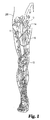
- FIG. 1 shown in Figure 1 is a diagram of a human leg showing certain venous structures therein.
- human leg 200 having greater saphenous vein 10 and the femoral vein 11 which adjoin at the sapheno-femoral junction 12.
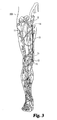
- greater saphenous vein 10 can be occluded in a region constituting substantially all of the passage between a point 13 occurring near the medial side of the knee to a point 14 occurring prior to the sapheno-femoral junction 12, as illustrated by the shaded area in Figure 2 .
- such occlusion is effective to prevent reflux of venous blood from the sapheno-femoral junction 12 in a direction down toward the medial side of the knee (e.g. at point 13).
- Such occlusion is effective to treat varicosities that commonly occur in lower portions of the leg, e.g. portions occurring below the knee.
- the occlusive material may include an elongate sponge form material that may include an end portion 16 that traverses the wall of the greater saphenous vein 10.
- the occlusive material may include a flowable remodelable material that can continuously or intermittently extend between points 13 and 14 within the greater saphenous vein 10.
- the sclerosive material can be contained within the occlusive material at implantation, or can be emplaced within the vein 10 before, after, or during implantation of the occlusive material.
- percutaneous access to the GSV can be achieved at point 13 using the Seldinger or any other suitable technique.
- an access needle can be passed through the skin to access the GSV 10
- a wire guide can be passed through the access needle and into the vein 10.
- the wire guide Prior to the deployment of any occlusive or sclerosive material, the wire guide can be used for any number of conventional procedures including catheterization and imaging procedures to locate the sapheno-femoral junction 12, or vein dilation and/or straightening procedures to prepare the vein for an occlusive implant.
- the wire guide can be used to place a cannulated delivery device within the vein 10. The wire guide can then be removed from the vein 10, thereby providing an open lumen through the cannulated device for delivery of one or more sclerosive agents and occlusive materials, as are discussed herein.
- an everting delivery sheath, peel away sheath, or a sheath having a reversible sleeve component can be used to deploy the occlusive device within the vascular vessel 10.
- the occlusive material and sheath can be delivered using any suitable method, such as over a wire guide or through another cannulated device.
- the occluder in alternative embodiments, such as when a foam material has sufficient column strength, the occluder, optionally containing a sclerosant, can be introduced into the vein and thereafter openly advanced (without a sheath) through the vein to implant the device, over a guide wire if desirable.
- the sclerosive agents and certain flowable occlusive materials can be injected at one or more locations between points 13 and 14 within the GSV 10 using one or more needle/syringe combinations until suitable occlusion is achieved.
- suitable endoluminal delivery techniques reference can be made, for example to U.S. Pat. Pub. No. 2003/0051735 and/or WO2005/053547 .
- occlusive device materials can include any suitable biocompatible material.
- the occlusion materials may include synthetic materials or reconstituted or naturally-derived collagenous materials.
- biocompatible materials that are at least bioresorbable will provide advantage in embodiments of the invention, with materials that are bioremodelable and promote cellular invasion and ingrowth providing particular advantage.
- remodelable materials may be used in this context to promote cellular growth within the occlusive materials to promote the closure of an occluded passageway.
- Bioremodelable materials of the invention can be provided by collagenous extracellular matrix (ECM) materials possessing biotropic properties, including in certain forms angiogenic collagenous ECM materials.
- suitable collagenous materials include ECUM materials, such as submucosa, renal capsule membrane, dermal collagen, dura mater, pericardium, serosa, facia lata, peritoneum, or basement membrane layers including liver basement membrane.
- the preferred medical graft products of the invention will include submucosa, such as submucosa derived from a warm-blooded vertebrate.
- Suitable submucosa materials for these purposes include, for instance, intestinal submucosa, including small intestinal submucosa, stomach submucosa, urinary bladder submucosa, and uterine submucosa.
- Mammalian submucosa materials are preferred.
- submucosa materials derived from animals raised for meat or other product production e.g. pigs, cattle or sheep, will be advantageous.
- Porcine submucosa provides a particularly preferred material for use in the present invention, especially porcine small intestine submucosa (SIS), more especially porcine small intestine submucosa retaining substantially its native cross-linking.
- SIS porcine small intestine submucosa
- the submucosa or other ECM material can be derived from any suitable organ or other biological structure, including for example submucosa derived from the alimentary, respiratory, intestinal, urinary or genital tracts of warm-blooded vertebrates.
- Submucosa useful in the present invention can be obtained by harvesting such tissue sources and delaminating the submucosa from smooth muscle layers, mucosal layers, and/or other layers occurring in the tissue source.
- Submucosa or other ECM materials can be derived from any suitable organ or other tissue source, usually sources containing connective tissues.
- the ECM materials processed for use in certain embodiments will typically include abundant collagen, most commonly being constituted of at least about 80% by weight collagen on a dry weight basis.
- Such naturally-derived ECM materials will for the most part include collagen fibers that are non-randomly oriented, for instance occurring as generally uniaxial or multi-axial but regularly oriented fibers.
- the ECM material can retain these factors interspersed as solids between, upon and/or within the collagen fibers.
- Particularly desirable naturally-derived ECM materials for use in embodiments of the invention will include significant amounts of such interspersed, non-collagenous solids that are readily ascertainable under light microscopic examination.
- non-collagenous solids can constitute a significant percentage of the dry weight of the ECM material in certain embodiments, for example at least about 1%, at least about 3%, and at least about 5% by weight in various embodiments of the invention.
- the submucosa or other ECM material used in illustrative embodiments may also exhibit an angiogenic character and thus be effective to induce angiogenesis in a host engrafted with the material.
- angiogenesis is the process through which the body makes new blood vessels to generate increased blood supply to tissues.
- angiogenic materials when contacted with host tissues, promote or encourage the formation of new blood vessels.
- Methods for measuring in vivo angiogenesis in response to biomaterial implantation have recently been developed. For example, one such method uses a subcutaneous implant model to determine the angiogenic character of a material. See, C. Heeschen et al., Nature Medicine 7 (2001), No. 7, 833-839 . When combined with a fluorescence microangiography technique, this model can provide both quantitative and qualitative measures of angiogenesis into biomaterials. C. Johnson et al., Circulation Research 94 (2004), No. 2, 262-268 .
- the submucosa material or any other ECM material retain and/or include growth factors or other bioactive components native to the source tissue.
- the submucosa or other ECM material may include one or more growth factors such as basic fibroblast growth factor (FGF-2), transforming growth factor beta (TGF-beta), epidermal growth factor (EGF), and/or platelet derived growth factor (PDGF).
- FGF-2 basic fibroblast growth factor
- TGF-beta transforming growth factor beta
- EGF epidermal growth factor
- PDGF platelet derived growth factor
- submucosa or other ECM material used in embodiments of the invention may include other biological materials such as heparin, heparin sulfate, hyaluronic acid, fibronectin and the like.
- the submucosa or other ECM material may include a bioactive component that induces, directly or indirectly, a cellular response such as a change in cell morphology, proliferation, growth, protein or gene expression.
- the ECM material will exhibit the capacity to promote angiogenesis.
- non-native bioactive components such as those synthetically produced by recombinant technology or other methods, may be incorporated into the submucosa or other ECM material.
- These non-native bioactive components may be naturally-derived or recombinantly produced proteins that correspond to those natively occurring in the ECM material, but perhaps of a different species (e.g. human proteins applied to collagenous ECMs from other animals, such as pigs).
- the non-native bioactive components may also be drug substances.
- Illustrative drug substances that may be incorporated into and/or onto the ECM material can include, for example, antibiotics and/or thrombus-promoting substances such as blood clotting factors, e.g.
- thrombin, fibrinogen, and the like may be applied to the ECM material as a premanufactured step, immediately prior to the procedure (e.g. by soaking the material in a solution containing a suitable antibiotic such as cefazolin), or during or after engraftment of the ECM material within the patient.
- a suitable antibiotic such as cefazolin
- Submucosa or other ECM material used in embodiments of the invention is preferably highly purified, for example, as described in U.S. Patent No. 6,206,931 to Cook et al .
- preferred ECM material will exhibit an endotoxin level of less than about 12 endotoxin units (EU) per gram, more preferably less than about 5 EU per gram, and most preferably less than about 1 EU per gram.
- EU endotoxin units
- the submucosa or other ECM material may have a bioburden of less than about 1 colony forming units (CFU) per gram, more preferably less than about 0.5 CFU per gram.
- CFU colony forming units
- Fungus levels are desirably similarly low, for example less than about 1 CFU per gram, more preferably less than about 0.5 CPU per gram.
- Nucleic acid levels are preferably less than about 5 ⁇ g/mg, more preferably less than about 2 ⁇ g/mg, and virus levels are preferably less than about 50 plaque forming units (PFU) per gram, more preferably less than about 5 PFU per gram.
- the ECM material used in embodiments of the invention is preferably disinfected with an oxidizing agent, particularly a peracid, such as peracetic acid.
- Such materials may include any suitable biocompatible material.
- Illustrative sponge or foam matrices will generally comprise porous, three-dimensionally stable bodies formed from suitable biocompatible matrix materials.
- suitable biocompatible matrix materials include naturally-occurring polymers and/or synthetic polymers or materials, such as hydrogels, gel foam, and/or polyvinyl (alcohol) foam.
- More preferred sponge compositions will comprise collagen as a matrix-forming material, either alone or in combination with one or more other matrix forming materials.
- sponge matrices useful in embodiments of the invention can be formed by providing a liquid solution or suspension of a matrix-forming material, and causing the material to form a porous three-dimensionally stable structure; however, a sponge or foam material can be formed using any suitable formation method, as is known in the art.
- a collagen solution or suspension can be prepared.
- the collagen may be derived from mammalian or other animal sources, for example, bovine, porcine or human sources, and desirably is derived from remodelable ECM materials as discussed herein. Synthetically-derived collagen may also be used.
- suitable collagen concentrations in the solution will be within the purview of those skilled in the art, with concentration ranges of about 0.05 g/ml to about 0.2 g/ml being typical.
- Digestion of the collagen to form the collagen solution is usually carried out under acidic conditions, starting with ground, minced or otherwise comminuted collagen-containing tissue.
- enzymatic digestion may be utilized using known enzymes for this purpose such as pepsin, trypsin, and/or papain. After digestion, the enzymes can be removed by suitable, known techniques.
- the collagenous solution and/or suspension can be employed as a moldable or castable material in the formation of the foam or sponge.
- the cast material can be dried directly without chemical crosslinking or can be crosslinked with a suitable crosslinking agent and then dried.
- suitable crosslinking agent for these purposes include glutaraldehyde, formaldehyde, carbodiimides, UV irradiation, or other crosslinking agents.
- the crosslinking agent will contain polar groups that impart a hydrophilic character to the final sponge matrix material.
- a polyepoxide crosslinker is utilized for this purpose, especially a polyglycidyl ether compound.
- Suitable such compounds include ethylene glycol diglycidyl ether, available under the trade name Denacol EX810 from Nagese Chemical Co., Osaka, Japan, and glycerol polyglycidyl ether available under the trade name Denacol EX313 also from Nagese Chemical Co.
- polyglycidyl ethers or other polyepoxide compounds utilized in the invention will have from 2 to about 10 epoxide groups per molecule.
- the use of such epoxides and/or other crosslinking agents which impart polar groups and a hydrophilic character to the resulting matrix will provide for good wettability and rapid hydration and expansion of closure devices of the invention.
- Preferred sources of collagen for forming illustrative sponge matrices include extracellular matrix materials as discussed herein, such as collagenous submucosal tissues, and other collagenous basement membrane materials. These include, for example, small intestinal submucosa, stomach submucosa, urinary bladder submucosa, liver basement membrane, and other basement membrane materials.
- extracellular matrix materials such as collagenous submucosal tissues, and other collagenous basement membrane materials. These include, for example, small intestinal submucosa, stomach submucosa, urinary bladder submucosa, liver basement membrane, and other basement membrane materials.
- these materials are preferably processed and utilized under conditions which retain their favorable growth properties. This may include, for example, processing under conditions in which native proteins and/or other materials, for instance biotropic agents, are retained in their bioactive form.
- the collagen sources, and resulting sponge matrices may include active native substances such as one or more growth factors, e.g. basic fibroblast growth factor (FGF-2); transforming growth factor beta (TGF-beta); epidermal growth factor (EFG); platelet derived growth factor (PDGF); and/or other substances such as glycosaminoglycans (GAGs); and/or fibronectin (FN).
- FGF-2 basic fibroblast growth factor
- TGF-beta transforming growth factor beta
- EGF-beta epidermal growth factor
- PDGF platelet derived growth factor
- GAGs glycosaminoglycans
- FN fibronectin
- Sponge matrix materials that can be used to form illustrative devices can be highly expandable when wetted, so as to achieve an expanded configuration.
- expandable sponge materials can exhibit the capacity to expand at least 100% by volume, more preferably at least about 200% by volume, and typically in the range of about 300% by volume to about 1000% by volume, when wetted to saturation with deionized water.
- Sponge materials used in aspects of the invention can also exhibit advantageous rates of expansion, achieving volume expansions as noted above in less than about 10 seconds, more preferably less than about 5 seconds, when immersed in deionized water.
- Highly compact dense sponge matrices can be prepared by first hydrating or otherwise wetting a porous sponge matrix, and then compressing and drying the element. Such preparative processes generally provide a more dense, rigid and stably compressed sponge matrix than processes such as simple compaction of the dry sponge matrix. Drying can be conducted sufficiently to stabilize the sponge matrix. For example, preferred drying procedures will reduce the liquid (e.g. water) content of the matrix to less than about 20% by weight, more preferably less than about 10% by weight. Compression forces can be applied so as to achieve a final density and/or desirable configuration, and can be applied in one, two or three dimensions, including radially, such as can be provided by a radial compression device.
- liquid e.g. water
- the drying of the compacted element can involve lyophilization (or freeze drying) or vacuum drying at ambient or elevated temperatures.
- lyophilization or freeze drying
- vacuum drying at ambient or elevated temperatures.
- the sponge matrix is stabilized structurally and remains in its highly dense and compacted state until contacted with a liquid susceptible to absorption by the matrix, for example body fluids.
- the pores of the matrix are thereby stably retained at a volume substantially reduced from their maximum volume, but return to a partially or fully expanded state when the matrix material is wetted.
- Compressed sponge matrices forming occlusive bodies of the invention can be highly dense, typically having densities of at least about 0.05 g/cm 3 , preferably in the range of about 0.05 g/cm 3 to about 0.2 g/cm 3 , and more preferably about 0.075 g/cm 3 to about 0.2 g/cm 3 .
- the compacted sponge matrix can have sufficient column strength to be deployed by passage through needles, catheters, sheaths, or bodily vessels for example by utilizing a push rod or other pusher element (gloved hand) to force the sponge matrix body through the vessel, needle and/or catheter cannula.
- Expanded sponge densities (dry) will generally be less than the corresponding compacted densities. Typical expanded densities (dry) will range from about 0.01 g/cm 3 to about 0.1 g/cm 3 , more preferably about 0.02 g/cm 3 to about 0.07 g/cm 3 .
- Compressed sponge materials may also contain agents which promote further retention of the compressed, high density form of the matrices. These may include for example starch, cellulose, sugars such as dextrose, or glycerin. Such agents can optionally be included in the liquid (preferably aqueous) used to hydrate or otherwise wet the sponge prior to compaction and drying.
- agents which promote further retention of the compressed, high density form of the matrices. These may include for example starch, cellulose, sugars such as dextrose, or glycerin. Such agents can optionally be included in the liquid (preferably aqueous) used to hydrate or otherwise wet the sponge prior to compaction and drying.
- occlusion devices of the invention can be made from ECM's or other collagenous materials that have been subjected to processes that expand the materials.
- such expanded materials can be formed by the controlled contact of an ECM material with one or more alkaline substances until the material expands, and the isolation of the expanded material.
- the contacting can be sufficient to expand the ECM material to at least 120% of (i.e. 1.2 times) its original bulk volume, or in some forms to at least about two times its original volume.
- the expanded material can optionally be isolated from the alkaline medium, e.g. by neutralization and/or rinsing.
- the collected, expanded material can be used in any suitable manner in the preparation of a graft device.
- the expanded material can be enriched with bioactive components, dried, and/or molded, etc., in the formation of a graft construct of a desired shape or configuration.
- a dried graft construct formed with the expanded ECM material can be highly compressible (or expandable) such that the material can be compressed for delivery, such as from within the lumen of a cannulated delivery device, and thereafter expand upon deployment from the device so as to become anchored within a patient and/or cause closure of a bodily segment within the patient.
- Expanded collagenous or ECM materials can be formed by the controlled contact of a collagenous or ECM material with an aqueous solution or other medium containing sodium hydroxide.
- Alkaline treatment of the material can cause changes in the physical structure of the material that in turn cause it to expand. Such changes may include denaturation of the collagen in the material.
- the magnitude of the expansion is related to several factors, including for instance the concentration or pH of the alkaline medium, exposure time, and temperature used in the treatment of the material to be expanded.
- ECM materials that can be processed to make expanded materials can include any of those disclosed herein or other suitable ECM's. Typical such ECM materials will include a network of collagen fibrils having naturally-occurring intramolecular cross links and naturally-occurring intermolecular cross links. Upon expansion processing as described herein, the naturally-occurring intramolecular cross links and naturally-occurring intermolecular cross links can be retained in the processed collagenous matrix material sufficiently to maintain the collagenous matrix material as an intact collagenous sheet material; however, collagen fibrils in the collagenous sheet material can be denatured, and the collagenous sheet material can have an alkaline-processed thickness that is greater than the thickness of the starting material, for example at least 120% of the original thickness, or at least twice the original thickness.
- the concentration of the alkaline substance for treatment of the remodelable material can be in the range of about 0.5 to about 2 M, with a concentration of about 1 M being more preferable.
- the pH of the alkaline substance can in certain embodiments range from about 8 to about 14.
- the alkaline substance will have a pH of from about 10 to about 14, and most preferably of from about 12 to about 14.
- the exposure of the collagenous material to the alkaline substance is performed at a temperature of about 4 to about 45 °C. In preferred embodiments, the exposure is performed at a temperature of about 25 to about 40 °C, with 37 °C being most preferred.
- the exposure time can range from at least about one minute up to about 5 hours or more. In some embodiments, the exposure time is about 1 to about 2 hours. In a particularly preferred embodiment, the collagenous material is exposed to a 1 M solution of. NaOH having a pH of 14 at a temperature of about 37 °C for about 1.5 to 2 hours.
- Such treatment results in collagen denaturation and a substantial expansion of the remodelable material.
- Denaturation of the collagen matrix of the material can be observed as a change in the collagen packing characteristics of the material, for example a substantial disruption of a tightly bound collagenous network of the starting material.
- a non-expanded ECM or other collagenous material can have a tightly bound collagenous network presenting a substantially uniform, continuous surface when viewed by the naked eye or under moderate magnification, e.g. 100x magnification.
- an expanded collagenous material can have a surface that is quite different, in that the surface is not continuous but rather presents collagen strands or bundles in many regions that are separated by substantial gaps in material between the strands or bundles when viewed under the same magnification, e.g.
- an expanded collagenous material typically appears more porous than a corresponding non-expanded collagenous material.
- the expanded collagenous material can be demonstrated as having increased porosity, e.g. by measuring for an increased permeability to water or other fluid passage as compared to the non-treated starting material.
- the more foamy and porous structure of an expanded ECM or other collagenous material can allow the material to be cast or otherwise prepared into a variety of three-dimensionally stable shapes for use in the preparation of medical materials and devices. It can further allow for the preparation of constructs that are highly compressible and which expand after compression. Such properties can be useful, for example, when the prepared graft construct is to be compressed and loaded into a deployment device (e.g. a lumen thereof) for delivery into a patient, and thereafter deployed to expand at the implant site.
- a deployment device e.g. a lumen thereof
- the material can be isolated from the alkaline medium and processed for further use.
- the collected material can be neutralized and/or rinsed with water to remove the alkalinity from the material, prior to further processing of the material to form a graft construct.
- a starting ECM material can optionally include a variety of bioactive or other non-collagenous components including, for example, growth factors, glycoproteins, glycosaminoglycans, proteoglycans, nucleic acids, and lipids. Treating the material with an alkaline substance may reduce the quantity of one, some or all of such non-collagenous components contained within the material.
- controlled treatment of the remodelable material with an alkaline substance will be sufficient to create a remodelable collagenous material which is substantially devoid of nucleic acids and lipids, and potentially also of growth factors, glycoproteins, glycosaminoglycans, and proteoglycans.
- one or more bioactive components can be returned to the material.
- an expanded material can include a collagenous material which has been depleted of nucleic acids and lipids, but which has been replenished with growth factors, glycoproteins, glycosaminoglycans, and/or proteoglycans.
- These bioactive components can be returned to the material by any suitable method. For instance, in certain forms a tissue extract, such as is discussed in U.S. Patent No. 6,375,989 , containing these components can be prepared and applied to an expanded collagenous material.
- the expanded collagenous material can be incubated in a tissue extract for a sufficient time to allow bioactive components contained therein to associate with the expanded collagenous material.
- the tissue extract may, for example, be obtained from non-expanded collagenous tissue of the same type used to prepare the expanded material.
- Other means for returning or introducing bioactive components to an expanded remodelable collagenous material include spraying, impregnating, dipping, etc. as known in the art.
- an expanded collagenous material may be modified by the addition of one or more growth factors such as basic fibroblast growth factor (FGF-2), transforming growth factor beta (TGF beta), epidermal growth factor (EGF), platelet derived growth factor (PDGF), and/or cartilage derived growth factor (CDGF).
- FGF-2 basic fibroblast growth factor
- TGF beta transforming growth factor beta
- EGF epidermal growth factor
- PDGF platelet derived growth factor
- CDGF cartilage derived growth factor
- an expanded collagenous material may include a bioactive component that induces, directly or indirectly, a cellular response such as a change in cell morphology, proliferation, growth, protein or gene expression.
- Expanded collagenous materials can be used to prepare a wide variety of occlusive devices.
- Methods for preparing such occlusive devices can include contacting an ECM or other collagenous starting material with an alkaline substance in an amount effective to expand the material, casting or otherwise forming the expanded collagenous material into an occlusive shape (e.g. an elongate tube or cylinder), and lyophilizing the expanded material to form a dried occlusive device.
- an occlusive shape e.g. an elongate tube or cylinder
- flowable occlusive materials can include any suitable biocompatible flowable material that will promote the closure of a venous vessel.
- a flowable extracellular matrix material can be used to fill bodily vessels and promote tissue ingrowth to close the vessels.
- the flowable material can be delivered in any suitable fashion, including for example forcible ejection from cannulated members such as catheters, sheaths, or needles.
- Suitable flowable, remodelable ECM materials for use in these aspects of the invention can be prepared, for example, as described in U.S. Patent Nos. 5,275,826 , 5,516,533 , 6,206,931 , 6,444,229 and/or in International Publication No. WO2005020847 (Cook Biotech Incorporated) published March 10, 2005.
- Such flowable materials can include solubilized and/or particulate ECM components, and in preferred forms include ECM gels having suspended therein ECM particles, for example having an average particle size of about 50 microns to about 500 microns, more preferably about 100 microns to about 400 microns.
- the ECM particulate can be added in any suitable amount relative to the solubilized ECM components, with preferred ECM particulate to ECM solubilized component weight ratios (based on dry solids) being about 0.1:1 to about 200:1, more preferably in the range of 1:1 to about 100:1.
- the inclusion of such ECM particulates in the ultimate gel can serve to provide additional material that can function to provide bioactivity to the gel (e.g. itself including FGF-2 and/or other growth factors or bioactive substances as discussed herein) and/or serve as scaffolding material for tissue ingrowth.
- Flowable ECM materials can also be used in conjunction with other occlusive devices as described herein, or otherwise.
- a flowable material can be emplaced with a formed occlusive construct, such as to help seal any voids between the construct and a vessel wall or to provide a cap at one or both device ends.
- flowable material optionally containing agents (as discussed herein) may be emplaced in between two or more other occlusive constructs to fill any desired amount of vessel space between the formed devices.
- agents or a solution thereof
- such synthetic materials may include nonresorbable synthetic biocompatible polymers, such as cellulose acetate, cellulose nitrate, silicone, polyethylene teraphthalate, polyurethane, polyamide, polyester, polyorthoester, polyanhydride, polyether sulfone, polycarbonate, polypropylene, high molecular weight polyethylene, polytetrafluoroethylene, or mixtures or copolymers thereof.
- nonresorbable synthetic biocompatible polymers such as cellulose acetate, cellulose nitrate, silicone, polyethylene teraphthalate, polyurethane, polyamide, polyester, polyorthoester, polyanhydride, polyether sulfone, polycarbonate, polypropylene, high molecular weight polyethylene, polytetrafluoroethylene, or mixtures or copolymers thereof.
- Illustrative resorbable synthetic materials can include polylactic acid, polyglycolic acid or copolymers thereof, a polyanhydride, polycaprolactone, polyhydroxy-butyrate valerate, polyhydroxyalkanoate, or another biodegradable polymer or mixture thereof.
- suitable synthetic materials both biodegradable and nonbiodegradable
- suitable synthetic materials See U.S. Utility Pat. App. No. 2005/0228486 entitled, "Implantable Frame with Variable Compliance,” filed on April 11, 2005.
- agents can include any substance that is capable of bringing about or inducing constriction, spasm, or closure in a bodily vessel of a patient and/or causing the de-epithelialization or inflammation (either dilative or constrictive), and/or initiating a healing response in certain patient tissue, such as a wall segment of a venous vessel.
- Illustrative such agents can include any suitable vasoconstrictive agent, sclerosive agent, thrombogenic agent, inflammatory agent, hypercoagulable agent, or any suitable combination of one or more of any of the above or other suitable agents.
- suitable vasoconstrictive agents can include any suitable alpha adrenergic direct or indirect agonist, such as norepinephrine, epinephrine, phenylephrine, and/or cocaine, or lidocaine, hypertonic saline, or any suitable combination thereof.
- suitable sclerosive agents can include, for example, polidocanol, sodium tetradecyl sulfate, e.g.
- SOTRADECOL® morrhuate sodium, ethanolamine oleate, tetradecyl sulfate, tetracycline, glycerin, hypertonic glucose, talc, acetic acid, alcohol, bleomycin, picibanil, ethibloc, deoxycycline, and/or any suitable microfoam that contains a sclerosive agent, such as VARISOLVE®, manufactured by Provensis, Ltd. of London, England, or any other suitable agent as disclosed in U.S. Pat. Nos. 5,676,962 and/or 6,572,873 , for example.
- VARISOLVE® sclerosive agent
- occlusive constructs useful in embodiments of the invention, can include any material, expandable or non, that occupies a volumetric shape or space that is suitable for promoting closure of a vascular or other bodily vessel.
- occlusive devices can include one or more boluses of a flowable remodelable material placed within a bodily lumen and/or an elongate sponge form material and/or any other suitable volumetric construct, such as can be formed by folding or rolling, or otherwise overlaying one or more portions of a biocompatible sheet material, for example.
- the overlaid sheet material or only portions or segments thereof can be compressed and dried or otherwise bonded such that an occlusive construct, e.g. a substantially unitary construct, is formed.
- the occlusive construct can then be placed within a vascular vessel in a manner such that the construct fills at least a luminal segment of the vessel.
- Occlusive constructs can include multilaminate materials, such as sheet form material or volumetric constructs formed from such multilaminate materials.
- multilaminate materials such as sheet form material or volumetric constructs formed from such multilaminate materials.
- two or more ECM segments can be rolled or stacked, or one ECM segment folded over itself at least one time, and then the layers can be fused or bonded together using a bonding technique, such as chemical cross-linking or vacuum pressing during dehydrating conditions.
- An adhesive, glue or other bonding agent may also be used in achieving a bond between ECM layers.
- Suitable bonding agents may include, for example, collagen gels or pastes, gelatin, or other agents including reactive monomers or polymers, for example cyanoacrylate adhesives.
- bonding can be achieved or facilitated using chemical cross-linking agents, such as glutaraldehyde, formaldehyde, epoxides, genipin or derivatives thereof, carbodiimide compounds, polyepoxide compounds, or other similar agents, including those others identified in the discussions above.
- Cross-linking of ECM materials can also be catalyzed by exposing the matrix to UV radiation, by treating the collagen-based matrix with enzymes such as transglutaminase and lysyl oxidase, and by photocross-linking. The combination of one or more of these with dehydration-induced bonding may also be used.
- dehydration-induced bonding methods can be used to fuse ECM portions of the bioremodelable material.
- the multiple layers of ECM material are compressed under dehydrating conditions.
- dehydrating conditions can include any mechanical or environmental condition which promotes or induces the removal of water from the ECM material.
- at least one of the two surfaces compressing the matrix structure can be water permeable.
- Dehydration of the ECM material can optionally be further enhanced by applying blotting material, heating the matrix structure or blowing air, or other inert gas, across the exterior of the compressing surfaces.
- One particularly useful method of dehydration bonding ECM materials is lyophilization, e.g. freeze-drying or evaporative cooling conditions.
- Another method of dehydration bonding comprises pulling a vacuum on the assembly while simultaneously pressing the assembly together. This method is known as vacuum pressing.
- vacuum pressing dehydration of the ECM materials in forced contact with one another effectively bonds the materials to one another, even in the absence of other agents for achieving a bond, although such agents can be used while also taking advantage at least in part of the dehydration-induced bonding.
- the ECM materials can be caused to form a generally unitary ECM structure.
- drying operations conducted with no or substantially no duration of exposure to temperatures above human body temperature or slightly higher, say, no higher than about 38° C, will preferably be used in some forms of the present invention.
- drying operations conducted with no or substantially no duration of exposure to temperatures above human body temperature or slightly higher, say, no higher than about 38° C, will preferably be used in some forms of the present invention.
- These include, for example, vacuum pressing operations at less than about 38° C, forced air drying at less than about 38° C, or either of these processes with no active heating - at about room temperature (about 25° C) or with cooling.
- Relatively low temperature conditions also, of course, include lyophilization conditions.
- illustrative occlusive constructs of the invention can be formed by randomly or regularly packing one or more pieces of single or multilayer ECM sheet material within a mold and thereafter processing the packed material.
- suitable processing can include, for example, providing the packed ECM sheet material in a partially or otherwise completely wetted or hydrated form and can complete, at least in part, by partially or completely dehydrothermally bonding the hydrated packed sheet material to establish a substantially unitary construct.
- a randomly packed construct can be formed by placing folded, wadded, gathered, or otherwise packed ECM sheet material within a mold, and thereafter drying the randomly configured material to form a substantially unitary occlusive construct.
- Packed, molded graft constructs can also include suitable flowable, comminuted, and/or sponge form materials, each of which can be ECM based, interspersed within rolled, folded, or otherwise randomly packed and/or covered ECM material. Additionally, these materials can be formed into any suitable shape, configuration, size and/or length as disclosed herein.
- such cast occlusive constructs may include one or more surface protuberances and/or perforations.
- Such protuberances may include a plurality of such protuberances axially aligned on the surface at one or more locations, e.g. two.
- Such protuberances may correspond with apertures in the mold used to form the occlusive construct, such procedures typically including a drying or bonding step, e.g. lyophilization.
- Such perforations will typically at least partially penetrate a transverse length of occlusive construct's body, and in certain embodiments will completely transversely penetrate the occlusive constructs body.
- Such perforations may form a plurality of surface apertures on the construct, which may be axially aligned at one or more locations, e.g. two, and in certain embodiments such apertures can correspond with at least a portion of such protuberances.
- Such perforations may be initially formed as part of the process of making the occlusive constructs, such as prior to or during a drying step, e.g. lyophilization, and as such, certain segments of the perforated tract may close as the manufacture of the product completes.
- a drying step e.g. lyophilization
- Occlusive devices for use in aspects of the invention may include any occlusive device that is pushable or guidable through a bodily vessel or bodily lumen as discussed herein and that will close or occlude the vessel after implantation.
- Illustrative such devices can occupy any suitable volumetric shape, form, size, and material.
- Such devices can include single or multilaminate sheet material, such as in a fan folded configuration that can be delivered and deployed at an implantation site in a folded over, such as folded in half lengthwise, configuration.
- Additional such guidable devices can include elongate sponge form devices that can be delivered and deployed in a folded-over, such as folded in half over a deployment device, or non-folded configuration.
- Such self guidable devices can include an expandable sponge form material having sufficient column strength such that it is advancable through a vein or other vascular vessel.
- Alternative such self-guidable devices can include a sheet material that is processed to itself such that it provides sufficient stiffness to the material to be advancable through a vein or other bodily vessel, or a material that incorporates certain rigid or semi-rigid materials or objects that enhance the stiffness of the occlusive material to make the material guidable through a bodily lumen.
- suitable such self guidable devices can include occlusive devices that exhibit a column strength, such as described herein, that includes any value between about 200 kPA or less to about 12,000 kPA or more. Additional such column strengths can include any value within the range of from about 700 kPA to about 11,000 kPA, and still additional such column strengths may include any value from about 1,000 kPA to about 10,000 kPA. Illustrative such column strength values can be measured using an Instron compressive strength testing machine. A sample of occlusive material, 5 cm in length, can be secured between to two test fixtures such that 0.5 cm of material is held within in each fixture. This test assembly results in a span of 4 cm of occlusive material between each fixture face. Thereafter, the fixtured sample can be placed in the Instron testing machine and compressed at a rate of 30 mm/min until the sample buckles. The force recorded at the point of buckling is the column strength or pushability number of the occlusive material.
- oxidized foam material made from small intestine submucosa, each having a pre-compressed diameter of 16 mm and a length of 5 cm where compressed down to a diameter of 4 mm using a radial compression machine.
- column strength of each sample was individually determined using an Instron compressive strength testing machine. Each test was executed by securing 0.5 cm of each end of each sample within a test fixture such that a span of 4 cm of compressed foam material extended between the faces of the fixtures. The fixed sample was then compressed by the Instron machine at a rate of 30 mm/min and the compressive force was recorded. The compressive force at the point each test sample buckled was recorded as the column strength or pushability number for each sample.
- the column strength for each oxidized foam sample was determined to be 1488.5 kPA, 1849.6 kPA, and 1628.8 kPA, respectively.
- Occlusion materials will generally be of sufficient character and/or dimension to achieve occlusion of the desired stretch of vascular vessel, either alone or in combination with other similar or differing devices.
- the occlusion device (or implanted flowable material) will have a length of at least about 10 cm, and in many situations at least about 20 cm. Indeed, for preferred occlusion procedures involving a significant stretch of an artery or vein, occlusion devices having lengths greater than 20 cm will be used.
- occlusion devices having lengths of at about 40 cm or 50 cm can be used.
- the sclerosive or other agents can be conjunctively used with one or more occlusive devices or materials during a percutaneous ablation procedure.
- the sclerosive agents and occlusive material can be provided separately and mixed or cooperatively implanted within a patient during an occlusion procedure.
- Such cooperative emplacement can include injecting or infusing one or more agents within a venous vessel before, during, or after implantation of a remodelable material, such as an elongate sponge form material.
- such conjunctive placement can include contacting a remodelable material with a sclerosive agent soon before the remodelable material is implanted within a patient, such as by soaking the remodelable material in a bath containing sclerosive agents.
- one or more sclerosants can be mixed within a flowable ECM material before the material is injected (via syringe) or infused (percutaneously via a catheter) within a venous vessel.
- the agents can be mixed within the flowable material in preparation for an ablation procedure, or during the manufacture of the flowable material (such as by forming the flowable material from an ECM graft material that contains one or more sclerosive agents).
- the sclerosive agents may be contained within the occlusive material in any suitable concentration to bring about a healing response, spasm, constriction, and/or a de-epithelielization in patient tissue that is in contact with or near the implant location.
- the sclerosive agent can be carried within an aqueous carrier solution, such as water, saline, and the like.
- the concentration of sclerosant in the solution is from about 0.1 volume % to about 10 volume % or greater.
- the water or saline can contain from about 2 to about 4 volume % physiologically acceptable alcohol, e.g. ethanol.
- Saline can also be buffered, e.g. phosphate buffered.
- the pH of the buffer can be adjusted to be physiological, e.g. from pH 6.0 to pH 8.0, more preferably about pH 7.0.
- Sclerosants may also contain additional components, such as stabilizing agents, e.g. foam stabilizing agents, such as glycerol. Further components may include alcohols, such as ethanol.
- stabilizing agents e.g. foam stabilizing agents, such as glycerol.
- Further components may include alcohols, such as ethanol.
- agent concentration used in illustrative occlusive procedures can be varied depending, for example, on the amount of vessel dilation and/or the type of cooperative emplacement procedure techniques and devices as can be selected by the physician. Additionally, when doping an agent into an occlusive construct, higher agent concentrations may be initially used to impart the agent into the construct than will be present within or effectively delivered to the patient at or after implantation.
- Illustrative dry occlusive products can be formed that contain one or more sclerosive or other agents throughout one or more regions of the construct or within or on one or more surfaces of the construct.
- sclerosive agents can be evenly dispersed throughout the construct, or, alternatively, can be differentially concentrated throughout differing regions or surfaces of the construct, such as for example to form an occlusive construct having higher sclerosive agent concentrations at surface areas while having reduced or diminished sclerosive concentrations at core or inner areas.
- Such dry form ECM occluders can serve to continually deliver sclerosive agents into patient tissue after implantation so as to enhance occlusion and remodeling of the occlusive device.
- Such occlusive devices can be deployed in dry form within a patient and thereafter optionally hydrated with a suitable hydrant, e.g. saline, or in alternative embodiments can be hydrated before implantation occurs.
- Illustrative dry occlusive devices can be formed by contacting an occlusive material with one or more sclerosing agents and thereafter drying the material so as to form a dry occlusive material that is doped with or otherwise contains sclerosive agents.
- an ECM sheet material can be soaked within a bath containing a sclerosive agent and can thereafter be rolled and pressed into a mold and thereafter lyophilized (freeze dried) so as to form a substantially unitary occlusive construct containing sclerosive agents.
- the construct can be compressed or recompressed after lyophilization is complete.
- the greater saphenous vein may also be accessed at a higher, e.g. jugular or lower level, e.g. near the ankle. During such access, any or all of the saphenous vein occurring between the ankle and the sapheno-femoral junction may be subjected to occlusion.
- Other veins in the leg(s) that may be involved in the varicose vein condition e.g. spider veins, may also be occluded, alternatively or in addition to the saphenous vein.
- the lesser saphenous vein, or varicose veins themselves may be occluded and obliterated in accordance with the invention.
- other veins or arteries in the leg(s) or elsewhere in the body may be occluded within the scope of the present invention.
- Percutaneously-conducted occlusion procedures can be performed under local anesthesia.
- it may be beneficial to use graduated compression stockings in the occluded area for example for a week or more. Compression of the occluded area may serve to facilitate permanent closure of the occluded vessel, for example when applied during a remodeling period during which tissue ingrowth into the occluded lumen occurs.
- Sheaths, dilators, pushers, wire guides and needles used in the present invention can all be conventional marketed products or modifications thereof.
- sheaths can be formed from PTFE (e.g. Teflon) or polyamide (e.g. Nylon) material, or a combination of materials such as an assembly including an inner layer of PTFE, a flat wire coil over the PTFE for kink resistance, and a polyamide (Nylon) outer layer to provide integrity to the overall structure and a smooth surface (e.g. as in the Flexor sheath, Cook, Inc.).
- Dilators and pushers can be made from conventional dilator/catheter type materials such as polyethylene, polyamide, stainless steel, polyurethane or vinyl, or any combination of these materials.
- Fittings provided for sheath/dilator assemblies can be conventional elements such as luer locks, and the dilator can have a fitting allowing it to be locked to the sheath during insertion and manipulation.
- Catheters can be made from conventional materials such as polyethylene, polyamide, PTFE, polyurethane, and other materials.
- Delivery sheaths used in the invention will have a lumen diameter sized to allow for the introduction of a sufficient amount of occlusion material to occlude the artery or vein of interest.
- the inner diameter (I.D.) of the final delivery sheath can range from about 4 French up to about 40 French.
- the distal ends of the catheters, sheaths, dilators, wires or other components, e.g. occlusive constructs, used in percutaneous procedures can include markers that can be X-ray, sonographically, or otherwise non-invasively visualized to identify their location during the procedure.
- Metallic bands of stainless steel, tantalum, platinum, gold, or other suitable materials, which include a dimple pattern, can serve the purpose for both ultrasound and X-ray identification.
- distal and/or proximal ends and/or other locations on occluder devices of the invention may include markers for non-invasive imaging, including imageable materials such as those discussed above as well as substances that can be applied to ECMs or other collagenous materials, e.g. substances containing tantalum, barium, iodine, or bismuth, e.g. in powder form.
- the invention also provides medical kits that include medical products described herein sealed within medical packaging potentially in combination with other components, such as for example a sheath and/or guidewire.
- the final, packaged products are provided in a sterile condition. This may be achieved, for example, by gamma, e-beam or other irradiation techniques, ethylene oxide gas, or any other suitable sterilization technique, and the materials and other properties of the medical packaging will be selected accordingly.
Claims (14)
- Produit médical (15) pour oblitérer un vaisseau veineux (10), comprenant:un produit d'assemblage oblitérant comprenant un matériau remodelable configuré pour se déployer à l'intérieur d'un vaisseau veineux pour remplir au moins un segment luminal du vaisseau veineux (10) de façon à oblitérer le vaisseau veineux (10), dans lequel le matériau remodelable est un matériau de matrice extracellulaire collagéneux retenant des constituants bioactifs natifs d'un tissu source pour le matériau de matrice extracellulaire collagéneux et qui est efficace pour induire une angiogenèse lorsqu'il est déployé à l'intérieur du vaisseau (10), dans lequel le matériau de matrice extracellulaire collagéneux est efficace pour activer la croissance de tissu du patient jusque dans la lumière du vaisseau veineux (10);caractérisé en ce que:le produit d'assemblage oblitérant inclut un ou plusieurs sclérosants qui sont efficaces pour blesser la paroi du vaisseau veineux (10) de façon à activer la croissance de tissu du patient jusque dans le produit d'assemblage oblitérant et activer l'oblitération du vaisseau veineux (10);et dans lequel:(i) ledit produit est un produit sec avec lesdits un ou plusieurs sclérosants dispersés uniformément à travers le produit d'assemblage ou plus fortement concentrés aux aires de surface dudit produit d'assemblage qu'aux aires internes du produit d'assemblage; ou(ii) lesdits un ou plusieurs sclérosants sont véhiculés à l'intérieur d'une solution aqueuse; ou(iii) ledit matériau remodelable est un matériau de matrice extracellulaire fluide.
- Produit médical (15) selon la revendication 1, dans lequel le matériau de matrice extracellulaire comprend au moins 80% en poids de collagène sur la base d'un poids sec, inclut des fibres orientées uniaxiales ou multiaxiales, et inclut au moins environ 3% de matières solides non collagéneuses retenues du tissu source, les matières solides non collagéneuses incluant lesdits constituants bioactifs, lesdits constituants bioactifs comprenant de préférence des facteurs de croissance.
- Produit médical (15) selon la revendication 2, dans lequel le matériau de matrice extracellulaire comprend une sous-muqueuse, une séreuse, un péricarde, une dure-mère, un péritoine ou une membrane basilaire, de préférence une sous-muqueuse de mammifère, la sous-muqueuse de mammifère étant de préférence une sous-muqueuse intestinale, une sous-muqueuse stomacale, une sous-muqueuse de vessie, ou une sous-muqueuse utérine, la sous-muqueuse intestinale comprenant de préférence une sous-muqueuse d'intestin grêle de porcin adulte.
- Produit médical (15) selon la revendication 1, dans lequel le ou les sclérosants comprennent du tétradécyl sulfate de sodium, du morrhuate sodium, de l'oléate d'éthanolamine, du sulfate de tétradécyle, de la tétracycline, de la glycérine, du glucose hypertonique, du talc, de l'acide acétique, un alcool, de la bléomycine, du picibanil, de l'éthibloc, du désoxycycline polidocanol, du sotradécol, de l'éthoxysclérol, ou tout mélange approprié de ceux-ci.
- Produit médical (15) selon la revendication 2, dans lequel le ou les sclérosants comprennent du morrhuate sodium.
- Produit médical (15) selon la revendication 5, dans lequel le ou les sclérosants comprennent du polidocanol ou du tétradécyl sulfate de sodium, de préférence du polidocanol.
- Produit médical (15) selon la revendication 1, dans lequel le matériau de matrice extracellulaire collagéneux comprend un matériau en forme d'éponge ou de mousse, lorsque ledit produit est un produit sec avec lesdits sclérosants dispersés uniformément à travers le produit d'assemblage ou plus fortement concentrés aux aires de surface dudit produit d'assemblage qu'aux aires internes du produit d'assemblage.
- Produit médical (15) selon la revendication 1, dans lequel le matériau de matrice extracellulaire collagéneux retient un facteur de croissance des fibroblastes basique, un facteur de croissance transformant bêta, un facteur de croissance épidermique, et/ou un facteur de croissance dérivé des plaquettes, natif d'un tissu source pour le matériau de matrice extracellulaire collagéneux.
- Produit médical (15) selon la revendication 1, dans lequel le matériau de matrice extracellulaire comprend un matériau de feuille de matrice extracellulaire, lorsque ledit produit est un produit sec avec lesdits sclérosants dispersés uniformément à travers le produit d'assemblage ou plus fortement concentrés aux aires de surface dudit produit d'assemblage qu'aux aires internes du produit d'assemblage.
- Produit médical (15) selon la revendication 9, dans lequel le matériau de feuille est enroulé pour former un corps cylindrique de matériau de feuille chevauchant ou stratifié.
- Produit médical (15) selon la revendication 10, dans lequel les couches se chevauchant sont liées déshydrothermiquement.
- Produit médical (15) selon la revendication 1, dans lequel la concentration de sclérosant dans la solution est de 0,1% en volume à 10% en volume, lorsque lesdits un ou plusieurs sclérosants sont véhiculés à l'intérieur d'une solution aqueuse.
- Produit médical (15) selon la revendication 1, dans lequel le produit d'assemblage oblitérant a une longueur d'au moins environ 10cm, de préférence d'au moins environ 20cm, lorsque ledit produit est un produit sec avec lesdits sclérosants dispersés uniformément à travers le produit d'assemblage ou plus fortement concentrés aux aires de surface dudit produit d'assemblage qu'aux aires internes du produit d'assemblage.
- Produit médical (15) selon la revendication 1, dans lequel le matériau de matrice extracellulaire fluide inclut des constituants de matrice extracellulaire particulaires.
Applications Claiming Priority (2)
| Application Number | Priority Date | Filing Date | Title |
|---|---|---|---|
| US97172407P | 2007-09-12 | 2007-09-12 | |
| PCT/US2008/076139 WO2009036250A1 (fr) | 2007-09-12 | 2008-09-12 | Matériaux remodelables améliorés servant à oblitérer des vaisseaux corporels et procédés et systèmes associés |
Publications (2)
| Publication Number | Publication Date |
|---|---|
| EP2187983A1 EP2187983A1 (fr) | 2010-05-26 |
| EP2187983B1 true EP2187983B1 (fr) | 2014-04-16 |
Family
ID=39967266
Family Applications (1)
| Application Number | Title | Priority Date | Filing Date |
|---|---|---|---|
| EP08831044.6A Active EP2187983B1 (fr) | 2007-09-12 | 2008-09-12 | Matériaux remodelables améliorés servant à oblitérer des vaisseaux corporels |
Country Status (3)
| Country | Link |
|---|---|
| US (2) | US8029560B2 (fr) |
| EP (1) | EP2187983B1 (fr) |
| WO (1) | WO2009036250A1 (fr) |
Cited By (1)
| Publication number | Priority date | Publication date | Assignee | Title |
|---|---|---|---|---|
| US9238090B1 (en) | 2014-12-24 | 2016-01-19 | Fettech, Llc | Tissue-based compositions |
Families Citing this family (10)
| Publication number | Priority date | Publication date | Assignee | Title |
|---|---|---|---|---|
| CN105212984B (zh) * | 2009-02-20 | 2017-12-22 | 柯惠有限合伙公司 | 用于治疗静脉机能不全的静脉闭塞的方法和装置 |
| US10143455B2 (en) | 2011-07-20 | 2018-12-04 | Covidien LLP | Enhanced ultrasound visualization of intravascular devices |
| US8758427B2 (en) | 2011-12-02 | 2014-06-24 | Vascular Solutions, Inc. | Elongated expandable member for occluding varicose veins |
| US20120109191A1 (en) | 2011-12-13 | 2012-05-03 | Vascular Insights Llc | Adhesive-based varicose vein treatment |
| US8808620B1 (en) | 2012-02-22 | 2014-08-19 | Sapheon, Inc. | Sterilization process design for a medical adhesive |
| ES2843482T3 (es) * | 2012-04-24 | 2021-07-19 | Angioclinic Ag | Composición para tratar la insuficiencia venosa y las venas varicosas |
| US9801983B2 (en) | 2014-12-18 | 2017-10-31 | Cook Medical Technologies Llc | Medical devices for delivering a bioactive to a point of treatment and methods of making medical devices |
| US9943314B2 (en) | 2015-04-14 | 2018-04-17 | Teleflex Innovations S.À.R.L. | Magnetically-driven delivery assembly and method |
| US11109867B2 (en) | 2018-03-16 | 2021-09-07 | Boston Scientific Scimed, Inc. | Devices and methods for vein closure |
| US20230190648A1 (en) * | 2020-05-15 | 2023-06-22 | Mayo Foundation For Medical Education And Research | Bioactive Tissue Derived Nanocomposite Hydrogels for Permanent Arterial Embolization and Enhanced Vascular Healing |
Family Cites Families (45)
| Publication number | Priority date | Publication date | Assignee | Title |
|---|---|---|---|---|
| FR2516927B1 (fr) * | 1981-11-26 | 1986-05-23 | Merieux Fond | Procede de preparation industrielle de materiaux collageniques a partir de tissus placentaires humains, materiaux collageniques humains obtenus, leur application comme biomateriaux |
| US4956178A (en) * | 1988-07-11 | 1990-09-11 | Purdue Research Foundation | Tissue graft composition |
| US4902508A (en) * | 1988-07-11 | 1990-02-20 | Purdue Research Foundation | Tissue graft composition |
| AU4926193A (en) * | 1992-09-21 | 1994-04-12 | Vitaphore Corporation | Embolization plugs for blood vessels |
| US5275826A (en) * | 1992-11-13 | 1994-01-04 | Purdue Research Foundation | Fluidized intestinal submucosa and its use as an injectable tissue graft |
| ES2068151B1 (es) * | 1993-06-23 | 1995-11-16 | Cabrera Garrido Juan | Microespuma inyectable para esclerosis. |
| DE19508058A1 (de) * | 1995-02-21 | 1996-08-22 | Schering Ag | Verfahren zur Herstellung von DTPA-Tetraestern der terminalen Carbonsäuren und deren Verwendung zur Herstellung pharmazeutischer Mittel |
| US5554389A (en) * | 1995-04-07 | 1996-09-10 | Purdue Research Foundation | Urinary bladder submucosa derived tissue graft |
| US5711969A (en) * | 1995-04-07 | 1998-01-27 | Purdue Research Foundation | Large area submucosal tissue graft constructs |
| ES2306841T3 (es) * | 1996-08-23 | 2008-11-16 | Cook Biotech, Inc. | Protesis para injertos con base de colageno. |
| ES2263185T3 (es) | 1996-12-10 | 2006-12-01 | Purdue Research Foundation | Biomaterial derivado de tejido hepatico de vertebrados. |
| EP0944647B1 (fr) * | 1996-12-10 | 2008-11-12 | Purdue Research Foundation | Extraits de sous-muqueuses |
| EP0946186B1 (fr) * | 1996-12-10 | 2003-03-26 | Purdue Research Foundation | Greffon issu de la sous-muqueuse gastrique |
| US5993844A (en) * | 1997-05-08 | 1999-11-30 | Organogenesis, Inc. | Chemical treatment, without detergents or enzymes, of tissue to form an acellular, collagenous matrix |
| US6264992B1 (en) * | 1998-02-27 | 2001-07-24 | Purdue Research Foundation | Submucosa as a growth substrate for cells |
| US7452371B2 (en) * | 1999-06-02 | 2008-11-18 | Cook Incorporated | Implantable vascular device |
| JP4399585B2 (ja) * | 1998-06-02 | 2010-01-20 | クック インコーポレイティド | 多辺管内医療器具 |
| JP2002529521A (ja) * | 1998-11-12 | 2002-09-10 | ノヴォリティックス インコーポレイテッド | 血管閉塞を生成する組成物および方法 |
| JP2002531181A (ja) | 1998-12-01 | 2002-09-24 | クック・バイオテック・インコーポレーテッド | 多型コラーゲン性バイオマテリアル医療装置 |
| US6645167B1 (en) * | 1999-05-21 | 2003-11-11 | Micro Therapeutics, Inc. | Methods for embolizing vascular sites with an embolizing composition |
| AU5143300A (en) * | 1999-05-21 | 2000-12-12 | Micro Therapeutics, Inc. | Methods for delivering in vivo uniform dispersed embolic compositions of high viscosity |
| JP5148030B2 (ja) * | 1999-05-21 | 2013-02-20 | タイコ ヘルスケア グループ リミテッド パートナーシップ | 新規の高粘度塞栓形成組成物 |
| GB9912356D0 (en) * | 1999-05-26 | 1999-07-28 | Btg Int Ltd | Generation of microfoam |
| WO2001003666A2 (fr) * | 1999-07-12 | 2001-01-18 | Scimed Life Systems, Inc. | Compositions pour occlusion vasculaire a base de liquide |
| US6592566B2 (en) * | 2000-02-03 | 2003-07-15 | Arizona Board Of Regents | Method for forming an endovascular occlusion |
| CA2422852C (fr) * | 2000-09-18 | 2012-06-26 | Organogenesis Inc. | Procedes de traitement de patients a l'aide de protheses greffees a feuilles planes cultivees par genie genetique |
| AU2002320182B2 (en) * | 2001-06-29 | 2008-02-21 | Cook Biotech Incorporated | Porous sponge matrix medical devices and methods |
| US8465516B2 (en) * | 2001-07-26 | 2013-06-18 | Oregon Health Science University | Bodily lumen closure apparatus and method |
| KR100947468B1 (ko) * | 2001-07-26 | 2010-03-17 | 쿠크 바이오텍, 인코포레이티드 | 혈관 폐쇄 부재 및 전달 장치 |
| NZ532243A (en) * | 2001-09-12 | 2006-03-31 | Virexx Medical Corp | Vascular occlusion solid-phase agent with immobilised platelet binding agent |
| DK1435877T3 (da) * | 2001-10-15 | 2009-08-03 | Hemoteq Ag | Overtrækning af stents for at forhindre restenose |
| FR2832307B1 (fr) * | 2001-11-19 | 2004-08-27 | Sofradim Production | Dispositif d'occlusion d'un conduit corporel, notamment d'une veine variqueuse |
| ATE515981T1 (de) | 2003-05-15 | 2011-07-15 | Cook Biotech Inc | Verschluss für körperöffnungen |
| GB2424586B (en) | 2003-08-25 | 2008-05-28 | Cook Biotech Inc | Graft materials containing bioactive substances, and methods for their manufacture |
| CA2547088C (fr) * | 2003-11-28 | 2011-10-18 | Cook Biotech Incorporated | Procedes, systemes et dispositifs d'occlusion vasculaire |
| US7582110B2 (en) * | 2004-04-13 | 2009-09-01 | Cook Incorporated | Implantable frame with variable compliance |
| US20060052822A1 (en) * | 2004-08-31 | 2006-03-09 | Mirizzi Michael S | Apparatus and material composition for permanent occlusion of a hollow anatomical structure |
| EP1827527B1 (fr) * | 2004-12-06 | 2016-08-24 | Cook Incorporated | Dispositifs d'occlusion gonflables, procedes, et systemes |
| US8066732B2 (en) * | 2004-12-30 | 2011-11-29 | Cook Incorporated | Inverting occlusion devices, methods, and systems |
| US7972354B2 (en) * | 2005-01-25 | 2011-07-05 | Tyco Healthcare Group Lp | Method and apparatus for impeding migration of an implanted occlusive structure |
| AU2006242214B2 (en) * | 2005-04-29 | 2012-02-09 | Cook Biotech Incorporated | Volumetric grafts for treatment of fistulae and related methods and systems |
| CA2637450A1 (fr) | 2006-01-31 | 2007-08-09 | Cook Biotech Incorporated | Greffons de fistules et procedes et systemes pour traiter des fistules |
| US20070179507A1 (en) * | 2006-01-31 | 2007-08-02 | Bhavin Shah | Fistula graft deployment systems and methods |
| EP2043531B1 (fr) * | 2006-06-15 | 2013-01-02 | Cook Medical Technologies LLC | Systèmes et appareils pour la délivrance de prothèses endoluminales |
| US8540760B2 (en) * | 2007-07-11 | 2013-09-24 | Cook Medical Technologies Llc | Tubular devices having reversible components for deployment of endoluminal occluders and related methods and systems |
-
2008
- 2008-09-12 WO PCT/US2008/076139 patent/WO2009036250A1/fr active Search and Examination
- 2008-09-12 US US12/209,483 patent/US8029560B2/en active Active
- 2008-09-12 EP EP08831044.6A patent/EP2187983B1/fr active Active
-
2011
- 2011-08-10 US US13/206,729 patent/US8518104B2/en active Active
Cited By (2)
| Publication number | Priority date | Publication date | Assignee | Title |
|---|---|---|---|---|
| US9238090B1 (en) | 2014-12-24 | 2016-01-19 | Fettech, Llc | Tissue-based compositions |
| US11938246B2 (en) | 2014-12-24 | 2024-03-26 | Fettech, Llc | Tissue-based compositions and methods of use thereof |
Also Published As
| Publication number | Publication date |
|---|---|
| US20110295233A1 (en) | 2011-12-01 |
| EP2187983A1 (fr) | 2010-05-26 |
| WO2009036250A8 (fr) | 2010-04-22 |
| US20090069758A1 (en) | 2009-03-12 |
| US8029560B2 (en) | 2011-10-04 |
| WO2009036250A1 (fr) | 2009-03-19 |
| US8518104B2 (en) | 2013-08-27 |
Similar Documents
| Publication | Publication Date | Title |
|---|---|---|
| US9980732B2 (en) | Methods, systems, and devices for the delivery of endoluminal prostheses | |
| EP2717790B1 (fr) | Dispositifs d'occlusion vasculaire | |
| EP2187983B1 (fr) | Matériaux remodelables améliorés servant à oblitérer des vaisseaux corporels | |
| US8540760B2 (en) | Tubular devices having reversible components for deployment of endoluminal occluders and related methods and systems | |
| US10639061B2 (en) | Devices and methods for modifying veins and other bodily vessels | |
| US11064987B2 (en) | Volumetric grafts for treatment of fistulae and related methods and systems | |
| US9788821B2 (en) | Physically modified extracellular matrix materials and uses thereof | |
| EP2720619B1 (fr) | Dispositifs de fermeture de fistule | |
| US8518064B2 (en) | Method for anchoring occlusion plug | |
| US20090069838A1 (en) | Inflatable occlusion devices, methods, and systems | |
| AU2013257459B2 (en) | Volumetric grafts for treatment of fistulae and related methods and systems |
Legal Events
| Date | Code | Title | Description |
|---|---|---|---|
| PUAI | Public reference made under article 153(3) epc to a published international application that has entered the european phase |
Free format text: ORIGINAL CODE: 0009012 |
|
| 17P | Request for examination filed |
Effective date: 20100331 |
|
| AK | Designated contracting states |
Kind code of ref document: A1 Designated state(s): AT BE BG CH CY CZ DE DK EE ES FI FR GB GR HR HU IE IS IT LI LT LU LV MC MT NL NO PL PT RO SE SI SK TR |
|
| AX | Request for extension of the european patent |
Extension state: AL BA MK RS |
|
| RIN1 | Information on inventor provided before grant (corrected) |
Inventor name: PAUL, RAM, H., JR Inventor name: FLAGLE, JACOB, A. Inventor name: BATES, BRIAN, L. |
|
| DAX | Request for extension of the european patent (deleted) | ||
| 17Q | First examination report despatched |
Effective date: 20120925 |
|
| GRAP | Despatch of communication of intention to grant a patent |
Free format text: ORIGINAL CODE: EPIDOSNIGR1 |
|
| INTG | Intention to grant announced |
Effective date: 20131115 |
|
| GRAS | Grant fee paid |
Free format text: ORIGINAL CODE: EPIDOSNIGR3 |
|
| GRAA | (expected) grant |
Free format text: ORIGINAL CODE: 0009210 |
|
| AK | Designated contracting states |
Kind code of ref document: B1 Designated state(s): AT BE BG CH CY CZ DE DK EE ES FI FR GB GR HR HU IE IS IT LI LT LU LV MC MT NL NO PL PT RO SE SI SK TR |
|
| REG | Reference to a national code |
Ref country code: GB Ref legal event code: FG4D |
|
| RAP2 | Party data changed (patent owner data changed or rights of a patent transferred) |
Owner name: COOK MEDICAL TECHNOLOGIES LLC |
|
| REG | Reference to a national code |
Ref country code: CH Ref legal event code: EP |
|
| REG | Reference to a national code |
Ref country code: AT Ref legal event code: REF Ref document number: 662114 Country of ref document: AT Kind code of ref document: T Effective date: 20140515 |
|
| REG | Reference to a national code |
Ref country code: IE Ref legal event code: FG4D |
|
| REG | Reference to a national code |
Ref country code: DE Ref legal event code: R096 Ref document number: 602008031584 Country of ref document: DE Effective date: 20140528 |
|
| REG | Reference to a national code |
Ref country code: AT Ref legal event code: MK05 Ref document number: 662114 Country of ref document: AT Kind code of ref document: T Effective date: 20140416 |
|
| REG | Reference to a national code |
Ref country code: NL Ref legal event code: VDEP Effective date: 20140416 |
|
| REG | Reference to a national code |
Ref country code: LT Ref legal event code: MG4D |
|
| PG25 | Lapsed in a contracting state [announced via postgrant information from national office to epo] |
Ref country code: NL Free format text: LAPSE BECAUSE OF FAILURE TO SUBMIT A TRANSLATION OF THE DESCRIPTION OR TO PAY THE FEE WITHIN THE PRESCRIBED TIME-LIMIT Effective date: 20140416 Ref country code: LT Free format text: LAPSE BECAUSE OF FAILURE TO SUBMIT A TRANSLATION OF THE DESCRIPTION OR TO PAY THE FEE WITHIN THE PRESCRIBED TIME-LIMIT Effective date: 20140416 Ref country code: FI Free format text: LAPSE BECAUSE OF FAILURE TO SUBMIT A TRANSLATION OF THE DESCRIPTION OR TO PAY THE FEE WITHIN THE PRESCRIBED TIME-LIMIT Effective date: 20140416 Ref country code: GR Free format text: LAPSE BECAUSE OF FAILURE TO SUBMIT A TRANSLATION OF THE DESCRIPTION OR TO PAY THE FEE WITHIN THE PRESCRIBED TIME-LIMIT Effective date: 20140717 Ref country code: CY Free format text: LAPSE BECAUSE OF FAILURE TO SUBMIT A TRANSLATION OF THE DESCRIPTION OR TO PAY THE FEE WITHIN THE PRESCRIBED TIME-LIMIT Effective date: 20140416 Ref country code: NO Free format text: LAPSE BECAUSE OF FAILURE TO SUBMIT A TRANSLATION OF THE DESCRIPTION OR TO PAY THE FEE WITHIN THE PRESCRIBED TIME-LIMIT Effective date: 20140716 Ref country code: IS Free format text: LAPSE BECAUSE OF FAILURE TO SUBMIT A TRANSLATION OF THE DESCRIPTION OR TO PAY THE FEE WITHIN THE PRESCRIBED TIME-LIMIT Effective date: 20140816 Ref country code: BG Free format text: LAPSE BECAUSE OF FAILURE TO SUBMIT A TRANSLATION OF THE DESCRIPTION OR TO PAY THE FEE WITHIN THE PRESCRIBED TIME-LIMIT Effective date: 20140716 |
|
| PG25 | Lapsed in a contracting state [announced via postgrant information from national office to epo] |
Ref country code: LV Free format text: LAPSE BECAUSE OF FAILURE TO SUBMIT A TRANSLATION OF THE DESCRIPTION OR TO PAY THE FEE WITHIN THE PRESCRIBED TIME-LIMIT Effective date: 20140416 Ref country code: AT Free format text: LAPSE BECAUSE OF FAILURE TO SUBMIT A TRANSLATION OF THE DESCRIPTION OR TO PAY THE FEE WITHIN THE PRESCRIBED TIME-LIMIT Effective date: 20140416 Ref country code: PL Free format text: LAPSE BECAUSE OF FAILURE TO SUBMIT A TRANSLATION OF THE DESCRIPTION OR TO PAY THE FEE WITHIN THE PRESCRIBED TIME-LIMIT Effective date: 20140416 Ref country code: HR Free format text: LAPSE BECAUSE OF FAILURE TO SUBMIT A TRANSLATION OF THE DESCRIPTION OR TO PAY THE FEE WITHIN THE PRESCRIBED TIME-LIMIT Effective date: 20140416 Ref country code: ES Free format text: LAPSE BECAUSE OF FAILURE TO SUBMIT A TRANSLATION OF THE DESCRIPTION OR TO PAY THE FEE WITHIN THE PRESCRIBED TIME-LIMIT Effective date: 20140416 Ref country code: SE Free format text: LAPSE BECAUSE OF FAILURE TO SUBMIT A TRANSLATION OF THE DESCRIPTION OR TO PAY THE FEE WITHIN THE PRESCRIBED TIME-LIMIT Effective date: 20140416 |
|
| PG25 | Lapsed in a contracting state [announced via postgrant information from national office to epo] |
Ref country code: PT Free format text: LAPSE BECAUSE OF FAILURE TO SUBMIT A TRANSLATION OF THE DESCRIPTION OR TO PAY THE FEE WITHIN THE PRESCRIBED TIME-LIMIT Effective date: 20140818 |
|
| REG | Reference to a national code |
Ref country code: DE Ref legal event code: R097 Ref document number: 602008031584 Country of ref document: DE |
|
| PG25 | Lapsed in a contracting state [announced via postgrant information from national office to epo] |
Ref country code: CZ Free format text: LAPSE BECAUSE OF FAILURE TO SUBMIT A TRANSLATION OF THE DESCRIPTION OR TO PAY THE FEE WITHIN THE PRESCRIBED TIME-LIMIT Effective date: 20140416 Ref country code: EE Free format text: LAPSE BECAUSE OF FAILURE TO SUBMIT A TRANSLATION OF THE DESCRIPTION OR TO PAY THE FEE WITHIN THE PRESCRIBED TIME-LIMIT Effective date: 20140416 Ref country code: RO Free format text: LAPSE BECAUSE OF FAILURE TO SUBMIT A TRANSLATION OF THE DESCRIPTION OR TO PAY THE FEE WITHIN THE PRESCRIBED TIME-LIMIT Effective date: 20140416 Ref country code: BE Free format text: LAPSE BECAUSE OF FAILURE TO SUBMIT A TRANSLATION OF THE DESCRIPTION OR TO PAY THE FEE WITHIN THE PRESCRIBED TIME-LIMIT Effective date: 20140416 Ref country code: SK Free format text: LAPSE BECAUSE OF FAILURE TO SUBMIT A TRANSLATION OF THE DESCRIPTION OR TO PAY THE FEE WITHIN THE PRESCRIBED TIME-LIMIT Effective date: 20140416 Ref country code: DK Free format text: LAPSE BECAUSE OF FAILURE TO SUBMIT A TRANSLATION OF THE DESCRIPTION OR TO PAY THE FEE WITHIN THE PRESCRIBED TIME-LIMIT Effective date: 20140416 |
|
| PLBE | No opposition filed within time limit |
Free format text: ORIGINAL CODE: 0009261 |
|
| STAA | Information on the status of an ep patent application or granted ep patent |
Free format text: STATUS: NO OPPOSITION FILED WITHIN TIME LIMIT |
|
| 26N | No opposition filed |
Effective date: 20150119 |
|
| PG25 | Lapsed in a contracting state [announced via postgrant information from national office to epo] |
Ref country code: IT Free format text: LAPSE BECAUSE OF FAILURE TO SUBMIT A TRANSLATION OF THE DESCRIPTION OR TO PAY THE FEE WITHIN THE PRESCRIBED TIME-LIMIT Effective date: 20140416 |
|
| PG25 | Lapsed in a contracting state [announced via postgrant information from national office to epo] |
Ref country code: LU Free format text: LAPSE BECAUSE OF FAILURE TO SUBMIT A TRANSLATION OF THE DESCRIPTION OR TO PAY THE FEE WITHIN THE PRESCRIBED TIME-LIMIT Effective date: 20140912 Ref country code: MC Free format text: LAPSE BECAUSE OF FAILURE TO SUBMIT A TRANSLATION OF THE DESCRIPTION OR TO PAY THE FEE WITHIN THE PRESCRIBED TIME-LIMIT Effective date: 20140416 |
|
| REG | Reference to a national code |
Ref country code: DE Ref legal event code: R097 Ref document number: 602008031584 Country of ref document: DE Effective date: 20150119 Ref country code: CH Ref legal event code: PL |
|
| REG | Reference to a national code |
Ref country code: FR Ref legal event code: ST Effective date: 20150529 |
|
| PG25 | Lapsed in a contracting state [announced via postgrant information from national office to epo] |
Ref country code: LI Free format text: LAPSE BECAUSE OF NON-PAYMENT OF DUE FEES Effective date: 20140930 Ref country code: CH Free format text: LAPSE BECAUSE OF NON-PAYMENT OF DUE FEES Effective date: 20140930 Ref country code: SI Free format text: LAPSE BECAUSE OF FAILURE TO SUBMIT A TRANSLATION OF THE DESCRIPTION OR TO PAY THE FEE WITHIN THE PRESCRIBED TIME-LIMIT Effective date: 20140416 |
|
| PG25 | Lapsed in a contracting state [announced via postgrant information from national office to epo] |
Ref country code: FR Free format text: LAPSE BECAUSE OF NON-PAYMENT OF DUE FEES Effective date: 20140930 |
|
| PG25 | Lapsed in a contracting state [announced via postgrant information from national office to epo] |
Ref country code: MT Free format text: LAPSE BECAUSE OF FAILURE TO SUBMIT A TRANSLATION OF THE DESCRIPTION OR TO PAY THE FEE WITHIN THE PRESCRIBED TIME-LIMIT Effective date: 20140416 |
|
| PG25 | Lapsed in a contracting state [announced via postgrant information from national office to epo] |
Ref country code: TR Free format text: LAPSE BECAUSE OF FAILURE TO SUBMIT A TRANSLATION OF THE DESCRIPTION OR TO PAY THE FEE WITHIN THE PRESCRIBED TIME-LIMIT Effective date: 20140416 Ref country code: HU Free format text: LAPSE BECAUSE OF FAILURE TO SUBMIT A TRANSLATION OF THE DESCRIPTION OR TO PAY THE FEE WITHIN THE PRESCRIBED TIME-LIMIT; INVALID AB INITIO Effective date: 20080912 |
|
| P01 | Opt-out of the competence of the unified patent court (upc) registered |
Effective date: 20230602 |
|
| PGFP | Annual fee paid to national office [announced via postgrant information from national office to epo] |
Ref country code: IE Payment date: 20230828 Year of fee payment: 16 Ref country code: GB Payment date: 20230810 Year of fee payment: 16 |
|
| PGFP | Annual fee paid to national office [announced via postgrant information from national office to epo] |
Ref country code: DE Payment date: 20230808 Year of fee payment: 16 |