EP2172219B1 - Anti-cancer agent comprising an iNKT ligand and anti-PD-1 antibody or anti-PD-L1 antibody - Google Patents
Anti-cancer agent comprising an iNKT ligand and anti-PD-1 antibody or anti-PD-L1 antibody Download PDFInfo
- Publication number
- EP2172219B1 EP2172219B1 EP09004806.7A EP09004806A EP2172219B1 EP 2172219 B1 EP2172219 B1 EP 2172219B1 EP 09004806 A EP09004806 A EP 09004806A EP 2172219 B1 EP2172219 B1 EP 2172219B1
- Authority
- EP
- European Patent Office
- Prior art keywords
- cancer
- antibody
- inkt
- cells
- carcinoma
- Prior art date
- Legal status (The legal status is an assumption and is not a legal conclusion. Google has not performed a legal analysis and makes no representation as to the accuracy of the status listed.)
- Active
Links
Images




Classifications
-
- A—HUMAN NECESSITIES
- A61—MEDICAL OR VETERINARY SCIENCE; HYGIENE
- A61K—PREPARATIONS FOR MEDICAL, DENTAL OR TOILETRY PURPOSES
- A61K39/00—Medicinal preparations containing antigens or antibodies
- A61K39/395—Antibodies; Immunoglobulins; Immune serum, e.g. antilymphocytic serum
-
- C—CHEMISTRY; METALLURGY
- C07—ORGANIC CHEMISTRY
- C07K—PEPTIDES
- C07K16/00—Immunoglobulins [IG], e.g. monoclonal or polyclonal antibodies
- C07K16/18—Immunoglobulins [IG], e.g. monoclonal or polyclonal antibodies against material from animals or humans
- C07K16/28—Immunoglobulins [IG], e.g. monoclonal or polyclonal antibodies against material from animals or humans against receptors, cell surface antigens or cell surface determinants
- C07K16/2803—Immunoglobulins [IG], e.g. monoclonal or polyclonal antibodies against material from animals or humans against receptors, cell surface antigens or cell surface determinants against the immunoglobulin superfamily
- C07K16/2818—Immunoglobulins [IG], e.g. monoclonal or polyclonal antibodies against material from animals or humans against receptors, cell surface antigens or cell surface determinants against the immunoglobulin superfamily against CD28 or CD152
-
- A—HUMAN NECESSITIES
- A61—MEDICAL OR VETERINARY SCIENCE; HYGIENE
- A61K—PREPARATIONS FOR MEDICAL, DENTAL OR TOILETRY PURPOSES
- A61K39/00—Medicinal preparations containing antigens or antibodies
- A61K39/395—Antibodies; Immunoglobulins; Immune serum, e.g. antilymphocytic serum
- A61K39/39533—Antibodies; Immunoglobulins; Immune serum, e.g. antilymphocytic serum against materials from animals
- A61K39/39558—Antibodies; Immunoglobulins; Immune serum, e.g. antilymphocytic serum against materials from animals against tumor tissues, cells, antigens
-
- A—HUMAN NECESSITIES
- A61—MEDICAL OR VETERINARY SCIENCE; HYGIENE
- A61P—SPECIFIC THERAPEUTIC ACTIVITY OF CHEMICAL COMPOUNDS OR MEDICINAL PREPARATIONS
- A61P35/00—Antineoplastic agents
-
- A—HUMAN NECESSITIES
- A61—MEDICAL OR VETERINARY SCIENCE; HYGIENE
- A61P—SPECIFIC THERAPEUTIC ACTIVITY OF CHEMICAL COMPOUNDS OR MEDICINAL PREPARATIONS
- A61P35/00—Antineoplastic agents
- A61P35/02—Antineoplastic agents specific for leukemia
-
- A—HUMAN NECESSITIES
- A61—MEDICAL OR VETERINARY SCIENCE; HYGIENE
- A61P—SPECIFIC THERAPEUTIC ACTIVITY OF CHEMICAL COMPOUNDS OR MEDICINAL PREPARATIONS
- A61P37/00—Drugs for immunological or allergic disorders
- A61P37/02—Immunomodulators
-
- A—HUMAN NECESSITIES
- A61—MEDICAL OR VETERINARY SCIENCE; HYGIENE
- A61P—SPECIFIC THERAPEUTIC ACTIVITY OF CHEMICAL COMPOUNDS OR MEDICINAL PREPARATIONS
- A61P43/00—Drugs for specific purposes, not provided for in groups A61P1/00-A61P41/00
-
- C—CHEMISTRY; METALLURGY
- C07—ORGANIC CHEMISTRY
- C07K—PEPTIDES
- C07K16/00—Immunoglobulins [IG], e.g. monoclonal or polyclonal antibodies
- C07K16/18—Immunoglobulins [IG], e.g. monoclonal or polyclonal antibodies against material from animals or humans
- C07K16/28—Immunoglobulins [IG], e.g. monoclonal or polyclonal antibodies against material from animals or humans against receptors, cell surface antigens or cell surface determinants
- C07K16/2803—Immunoglobulins [IG], e.g. monoclonal or polyclonal antibodies against material from animals or humans against receptors, cell surface antigens or cell surface determinants against the immunoglobulin superfamily
- C07K16/2827—Immunoglobulins [IG], e.g. monoclonal or polyclonal antibodies against material from animals or humans against receptors, cell surface antigens or cell surface determinants against the immunoglobulin superfamily against B7 molecules, e.g. CD80, CD86
-
- A—HUMAN NECESSITIES
- A61—MEDICAL OR VETERINARY SCIENCE; HYGIENE
- A61K—PREPARATIONS FOR MEDICAL, DENTAL OR TOILETRY PURPOSES
- A61K39/00—Medicinal preparations containing antigens or antibodies
- A61K2039/505—Medicinal preparations containing antigens or antibodies comprising antibodies
-
- C—CHEMISTRY; METALLURGY
- C07—ORGANIC CHEMISTRY
- C07K—PEPTIDES
- C07K2317/00—Immunoglobulins specific features
- C07K2317/70—Immunoglobulins specific features characterized by effect upon binding to a cell or to an antigen
- C07K2317/76—Antagonist effect on antigen, e.g. neutralization or inhibition of binding
Definitions
- the present invention relates to a use of a composition comprising alpha-galactosyl ceramide and an anti-PD-1 antibody or an anti-PD-L1 antibody which blocks the signaling of PD-1 or PD-L1 to prevent the iNKT cell ligand induced anergy of iNKT cells for the manufacture of a medicament for the treatment of cancer.
- WO 2008/085562 A2 describes uses of an agent that reduces PD-L1 interaction with PD-1, and particularly monoclonal antibodies that bind to PD-L1 and interfere with PD-L1 interaction with PD-1 in combination with a vaccine to provide synergistic effects.
- This agent can find application as an antitumor in combination with a vaccine to overcome T cell inactivation.
- Natural killer T (NKT) cells co-expressing a T cell receptor and NK cell markers, are essential for several aspects of immunity, such as immunomodulation and immunuopotentiation, in various immune diseases including autoimmune diseases, infectious diseases, cancer, etc.
- NKT cells exists at high levels in the thymus, the liver, and the bone marrow, but at low levels in the spleen, lymph nodes and blood.
- NKT cells recognize glycolipid antigens presented by CD1d, a MHC class 1-like molecule.
- a major subset of NKT cells called type 1 NKT cells or invariant natural T (iNKT) cells, express an invariant natural T cell receptor (TCR) composed of V ⁇ 14-J ⁇ 18 chains in mice (V ⁇ 24-J ⁇ 18 in humans).
- TCR invariant natural T cell receptor
- iNKT cells Upon TCR stimulation with a ligand, such as ⁇ -galactosylceramide ( ⁇ -GC), iNKT cells rapidly produce a wide range of cytokines including IL-4, IFN- ⁇ , IL-12, and GM-CSF.
- iNKT cells This rapid and potent response to a ligand enables iNKT cells to enhance or regulate the activity of various immune cells in innate and acquired immunity. Found in diverse diseases and promoting tumor rejection or regulating autoimmune disorders, these immunomodulatory roles of iNKT cells are studied for use in immunotherapy treatments for cancer and autoimmune diseases.
- iNKT cells tend to greatly decrease in responsiveness following repeated stimulation after a first stimulation with their ligands via the T cell receptor.
- iNKT cells that have been stimulated in vivo with ⁇ -GC have reduced proliferation and cytokine production upon secondary stimulation with the same ligand. This iNKT cell anergy is a major obstacle in immunotherapeutic trials targeting iNKT cells.
- T cells are known to become anergic when they receive a TCR signal with insufficient co-stimulatory signals.
- Co-stimulatory molecules such as CD28, CD40L and ICOS are known to be involved in the development and activation of iNKT cells.
- 4-1BB contributes to promote the activation of iNKT cells as a co-stimulatory molecule and can affect iNKT cell-mediated allergic lung inflammation ( Kim, D. H., W. S. Chang, Y. S. Lee, K. A. Lee, Y. K. Kim, B. S. Kwon, and C. Y. Kang. 2008.
- PD-1 is a 55 KDa type I transmebrane protein of the immunoglobulin superfamily, and is known as a co-inhibitory molecule on T cells. That is, PD-1 is a member of the co-inhibitory molecules of the CD28 family (e.g., CD28, CTLA-4, ICOS and BTLA) expressed on activated B cells, T cells and bone marrow cells. Two ligands for PD-1, PD-L1 and PD-L2, have been identified thus far. The interaction of PD-1 with the PD ligands can transduce inhibitory or co-stimulatory signals into the T cells.
- PD-1 is not expressed on naive T cells, but is inducibly expressed after T cell activation.
- PD-L1 it is expressed to some degree on naive T cells and its level is increased on activated T cells.
- PD-L1 is found at high levels in various human cancers and interacts with PD-1 to transduce inhibitory or co-stimulatory signals from entering into the T cells. For example, the interaction between PD-1 and PD-L1 induces a decrease in the level of tumor invasive lymphocytes and in T cell receptor-mediated proliferation and causes the immune evasion of tumor cells.
- PD-1 develops various autoimmune phenotypes, such as autoimmune cardiac infarction and lupus-like syndromes with arthritis and nephritis, and plays an important role in the development of autoimmune encephalomyelitis, systemic lupus erythematosus, graft-versus-host disease (GVHD), type 1 diabetes and rheumatic arthritis.
- GVHD graft-versus-host disease
- Aged PD-1-deficient mice develop autoimmune diseases, indicating that PD-1 plays a critical role in the regulation of autoimmunity and immune tolerance.
- PD-1 signals are essential for inducing T cell exhaustion during chronic infection.
- the NKT cell ligand ⁇ -GC has conventionally been used as an anticancer agent.
- repeated stimulation of iNKT cells with ⁇ -GC induces anergy leading to a great decrease in responsiveness, it cannot achieve effective anticancer effects. Therefore, there is a pressing need for an anticancer agent that can restore the responsiveness of iNKT cells even in the state of anergy caused by stimulation with iNKT cell ligands.
- the anti-PD1 or anti-PD-L1 antibody was found to induce potent anti-tumor activity of iNKT cells as demonstrated by a significant reduction in the number of metastatic nodules in B16F10 melanoma metastasis models in vivo.
- the present invention provides use of a composition comprising alpha-galactosyl ceramide and an anti-PD-1 antibody or anti-PD-L1 antibody, which blocks the signaling of PD-1 or PD-L1 to prevent the iNKT cell ligand induced anergy of iNKT cells for the manufacture of a medicament for the treatment of cancer.
- a method for reversing the unresponsiveness of iNKT cells with anergy induced therein by iNKT cell ligand treatment comprising treating the anergic iNKT cells with an anti-PD-1 antibody or an anti-PD-L1 antibody.
- the iNKT cell ligand may be selected from a group consisting of alpha-galactosyl ceramide, alpha-glucuronosyl ceramide, phosphatidylinositoltetramannoside, isoglobotrihexosylceramide, ganglioside GD3, phsphatidylcholine, phosphatidylethanolamine, phosphatidylinositol, sulfatide, betagalactosylceramide, lipophosphoglycan, glycoinositol phospholipid, alpha-galactosylceramide analogs including betaanomer galactoceramide and alpha-anomer galactosylceramide, and bacterial lipid antigens.
- the anti-PD-1 antibody or the anti-PD-L1 antibody may be a monoclonal antibody or a polyclonal antibody.
- the anti-PD-1 antibody or anti-PD-L1 antibody according to the present invention can block the signaling of PD-1 or PD-L1 to prevent the iNKT cell ligand-induced anergy of iNKT cells and can provide cytokine secretion ability for even anergic iNKT cells to restore their responsiveness.
- the anti-PD-1 antibody or anti-PD-L1 antibody of the present invention inhibits the anergy induction of iNKT cells to significantly decrease the number of pulmonary nodules in a lung metastatis model of B16F10 melanoma, thus effectively eliciting anticancer immune responses of iNKT cells and showing anti-tumor effects of iNKT cells against cancer metastasis. Therefore, an anticancer agent comprising the anti-PD-1 antibody or anti-PD-L1 antibody of the present invention can be very useful in the treatment of cancer, particularly, metastatic cancer.
- the cancer to which the anticancer agent according to the present invention is therapeutically applicable may be gynecologic tumor, endocrine gland cancer, CNS (central nervous system) tumor or ureter cancer.
- the cancer include lung cancer, stomach cancer, liver cancer, bone cancer, pancreatic cancer, skin cancer, head and neck cancer, melanoma, uterine cancer, ovarian cancer, rectal cancer, colorectal cancer, colon cancer, breast cancer, uterine sarcoma, Fallopian tube carcinoma, endometrial carcinoma, uterine cervical carcinoma, vaginal carcinoma, vulva carcinoma, esophageal cancer, larynx cancer, small intestine cancer, thyroid cancer, parathyroid cancer, soft tissue sarcoma, uterine cancer, penis cancer, prostate cancer, chronic or acute leukemia, pediatric solid tumor, differentiated lymphoma, bladder cancer, kidney cancer, renal cell carcinoma, renal pelvic carcinoma, primary CNS lymphoma, spinal cord tumor, brainstem glioma, and pitu
- the anticancer agent of the present invention may be formulated into a pharmaceutical composition with at least one conventional anticancer ingredient.
- the pharmaceutical composition may further comprise a pharmaceutically acceptable carrier.
- a pharmaceutically acceptable carrier include saline, sterile water, Ringer's solution, buffered saline, dextrose solution, maltodextrin solution, glycerol, ethanol, etc.
- conventional additives such as antioxidants, buffers, bacteriostatic agents, etc., may be added to the composition.
- the active ingredients may be admixed with a diluent, a dispersant, a surfactant, a binder and/or a lubricant.
- composition of the present invention may be administered via oral routes or parenteral routes (e.g., intravenous, subcutaneous, intraperitoneal, topical, etc.).
- parenteral routes e.g., intravenous, subcutaneous, intraperitoneal, topical, etc.
- the effective dosage of the anticancer agent in accordance with the present invention depends on various factors, including the patient's weight, age, gender, state of health, diet, the time of administration, route of administration, excretion rate, severity of diseases, etc. In general, it may be administered in a single dose, and preferably in multiple doses per day at a daily dose ranging from 0.01 to 1000 mg/day, and preferably from 0.1 to 100 mg/kg of the anti-PD-1 antibody or anti-PD-L1 antibody.
- composition according to the present invention may be used alone or in combination with surgical operation, hormonal therapy, chemotherapy, and/or biological response controllers.
- EXAMPLE 1 Expression of PD-1 and PD Ligands in iNKT Cells
- mice Six- to eight-week-old female C56BL/6 mice were purchased from Orient Bio. All mice were bred and maintained in specific pathogen-free conditions.
- Hybridoma clones producing antibodies to mouse PD-1 (RMP1-14, rat IgG2a), PD-L1 (MIH-5, rat IgG2a), and PD-L2 (Ty25, rat IgG2a) were produced according to methods well known in the art [ Yamazaki, T., H. Akiba, H. Iwai, H. Matsuda, M. Aoki, Y. Tanno, T. Shin, H. Tsuchiya, D. M. Pardoll, K. Okumura, M. Azuma, and H. Yagita. 2002.
- Splenocytes were isolated from C57BL/6 mice administered with 2 ⁇ g of ⁇ -GC at different time points (0 hr, 6 hrs, 72 hrs, 7 days, one month and two months) after the administration.
- the cells were stained with a PE-conjugated anti-PD-1 monoclonal antibody, a PE-conjugated anti-PD-L1 monoclonal antibody, a PE-conjugated anti-PD-L2 monoclonal antibody and a PE-conjugated isotype control monoclonal antibody, respectively.
- iNKT cells were gated on B220- TCR- ⁇ int ⁇ -GC/CDld:Ig+ population.
- PD-1, PD-L1 and PD-L2 expression was analyzed by FACS. The results are given in FIG. 1 . Shaded histograms indicate staining with isotype control mAbs.
- PD-1 was constitutively expressed on iNKT cells at a low level and its expression was upregulated after ⁇ -GC stimulation, followed by the persistence of the upregulated level for two months.
- PD-L1 expression was temporarily increased on ⁇ -GC-stimulated iNKT cells but declined toward naive state levels within 72 hrs.
- PD-L2 its expression was not found on iNKT cells regardless of their activation status.
- spelenocytes After being prepared from naive C57BL/6 mice, 5 ⁇ 105 spelenocytes were incubated for 3 days with 100 ng/ml of ⁇ -GC in the presence of 50 ⁇ g/ml of the control rat IgG, the anti-PD-1 mAb, the anti-PD-L1 mAb, or the anti-PD-L2 mAb. Then, the supernatants were obtained and assayed for IFN- ⁇ and IL-4 levels by ELISA.
- anti-PD-1 mAb significantly increased the production of IFN- ⁇ and, to a lesser extent, IL-4.
- Anti-PD-L1 mAb also induced greater production of IFN- ⁇ , but anti-PD-L2 mAb did not,
- C57BL/6 mice was injected with 200 ⁇ g of the control rat IgG, the anti-PD-1 mAb, the anti-PD-L1 mAb or the anti-PD-L2 mAb 24 hrs before treatment with 2 ⁇ g of ⁇ -GC.
- Sera were obtained at 0, 2, 6, 12, 24, 48 and 72 hrs after the treatment, followed by ELISA analysis for IFN- ⁇ and IL-4 levels.
- intracellular cytokine staining was followed by flow cytometry analysis.
- splenocytes were isolated 2 hrs after ⁇ -GC treatment and 5 ⁇ 106 cells were incubated with Golgi plug for 2 hrs to accumulate cytokines. Intracellular cytokine staining was performed using BD Cytofix/Cytoperm Plus with Golgiplug kit according to the manufacturer's protocol (BD Biosciences). iNKT cells were gated on B220-TCR- ⁇ int ⁇ -GC/CDld:Ig+ population and IFN- ⁇ + or IL-4+ iNKT cells were analyzed by flow cytometry.
- IFN- ⁇ and IL-4 levels in blood are depicted in FIG. 3 and flow histograms of IFN- ⁇ + and IL-4+ iNKT cells are given in FIG. 4 .
- the blood IFN- ⁇ and IL-4 levels were observed to peak, respectively, within 12 hrs and 2 hrs after ⁇ -GC treatment, indicating iNKT cell activation.
- IFN- ⁇ levels in the sera of anti-PD-1 or anti-PD-L1 mAb-treated mice were significantly enhanced compared with levels in the group treated with control IgG. Slightly different IL-4 production was observed after treatment with the blocking Abs, but with no significance.
- iNKT cells producing IFN- ⁇ increased in number because they blocked PD-1/PD-L1 interaction with the anti-PD-1 mAb or anti-PD-L1 mAb in comparison with control cells.
- no significant differences were found between populations of IL-4+ iNKT cells and control cells after mAb treatment.
- ⁇ -GC-induced unresponsiveness of iNKT cells was detected as early as 3 days after primary stimulation and observed to persist until 7 - 30 days after ⁇ -GC stimulation.
- C57BL/6 mice were injected with 2 ⁇ g of ⁇ -GC to induce iNKT cell anergy.
- splenocytes were isolated from the mice and 5x10⁵ cells were incubated for 3 days with 100ng/ml of ⁇ -GC in the presence of 50 ⁇ g/ml of the control rat IgG, the anti-PD-1 mAb, the anti-PD-L1 mAb, or the anti-PD-L2 mAb.
- mice splenocytes isolated from the mice were incubated for 3 days with 100ng/ml of ⁇ -GC without mAb, and these were represented by 'activation'. The supernatants were then assayed for IFN- ⁇ and IL-4 levels by ELISA [*: p ⁇ 0.05 and **: p ⁇ 0.01 (vs. control rat IgG)].
- Splenocytes (5 ⁇ 106 cells), prepared two and twelve hours after the second injection of ⁇ -GC, were incubated with Golgi plug for 2 hrs to accumulate cytokines. Intracellular cytokine staining was performed on the splenocytes prepared 2 hrs after the secondary GC treatment, using BD Cytofix/Cytoperm Plus with Golgiplug kit according to the manufacturer's protocol (BD Biosciences). The splenocytes prepared 12 hrs later were used in an assay for CD69 expression in iNKT and NK cells.
- iNKT cells were gated on B220-TCR- ⁇ int ⁇ -GC/CDld:Ig+ population and NK cells were gated on B220- TCR- ⁇ - NK1.1high population.
- IFN- ⁇ + or IL-4+ iNKT cells were analyzed by flow cytometry.
- CD69 expression on iNKT and NK cells was analyzed by flow cytometry.
- IFN- ⁇ and IL-4 levels in blood are graphed in FIG. 6 and FACS results of IFN- ⁇ + or IL-4+ iNKT cells and flow histograms of CD69 expression on iNKT and NK cells are given in FIGS. 7 and 8 , respectively.
- mice pretreated with ⁇ -GC produced significantly low IFN- ⁇ and IL-4 upon secondary ⁇ -GC injection.
- mice treated with the anti-PD-1 antibody during the primary ⁇ -GC injection produced remarkably higher IFN- ⁇ and IL-4 upon the secondary ⁇ -GC injection than did the control IgG-treated group.
- CD69 expression upon secondary ⁇ -GC injection was increased on iNKT and NK cells in a similar manner by the treatment with anti-PD-1 mAbs, as shown in FIG. 8 .
- PD-1/PD-L1 interaction is essential for the induction of iNKT cell anergy in vivo.
- Skin tumor cells (B16F10, ATCC) were cultured in DMEM media supplemented with 10% FBS and 1% penicillin/streptomycin.
- C56BL/6 mice were intraperitoneally injected with 200 ⁇ g of the control rat IgG, the anti-PD-1 mAb or the anti-PD-L1 mAb 24 hrs before i.v. inoculation with 2 ⁇ 105 B16F10 tumor cells.
- the mice On Days zero, 4 and 8, the mice were treated with 500ng of ⁇ -GC plus 200 ⁇ g of the control rat IgG, the anti-PD-1 mAb or the anti-PD-L1 mAb.
- the lungs were weighed and weight differences between metastatic and normal lungs are graphed in FIG. 9 .
- the B16F10 metastatic lungs from the group treated with ⁇ -GC plus the anti-PD-1 mAb were significantly reduced in weight compared with those from the group treated with ⁇ -GC plus the control rat IgG.
- mice were intraperitoneally injected with 200 ⁇ g of the control rat IgG, the anti-PD-1 antibody or the anti-PD-L1 antibody 24 hrs after which injection with 2 ⁇ g of ⁇ -GC induced iNKT cell anergy. 7 days later, 5 ⁇ 105 B16F10 tumor cells were i.v. injected.
- the mice were treated with 500 ng of ⁇ -GC to induce anti-tumor activity of iNKT cells. 14 days later, the lungs were excised and the nodules formed by cancer metastasis were counted (A). The isolated lungs were observed under an optical microscope (B). The results are given in FIG. 10 .
- the data obtained above indicates that the blockage of PD-1/PD-L1 interaction by treatment with the anti-PD-1 antibody or the anti-PD-L1 antibody during the induction phase of iNKT cell anergy leads to the persistence of the anti-tumor effects of iNKT cells.
- Formulation examples are given to illustrate dosage preparations containing the anticancer agent of the present invention.
- the anti-PD-1 or anti-PD-L1 antibody blocks the PD-1/PD-L1-mediated signaling pathway not only to prevent the iNKT cell ligand-induced iNKT cell anergy, but also to reverse the unresponsiveness of already anergic iNKT cells to produce cytokines.
- the anti-PD1 or anti-PD-L1 antibody ensures the potent anti-tumor activity of iNKT cells as demonstrated by a significant reduction in the number of metastatic nodules in B16F10 melanoma metastasis models in vivo.
- the anticancer agent comprising an anti-PD-1 or anti-PD-L1 antibody in accordance with the present invention can be very useful in the treatment of cancer, particularly metastatic cancer.
Landscapes
- Health & Medical Sciences (AREA)
- Chemical & Material Sciences (AREA)
- Immunology (AREA)
- Medicinal Chemistry (AREA)
- Life Sciences & Earth Sciences (AREA)
- Organic Chemistry (AREA)
- General Health & Medical Sciences (AREA)
- Animal Behavior & Ethology (AREA)
- Pharmacology & Pharmacy (AREA)
- Public Health (AREA)
- Veterinary Medicine (AREA)
- Engineering & Computer Science (AREA)
- Bioinformatics & Cheminformatics (AREA)
- Chemical Kinetics & Catalysis (AREA)
- Proteomics, Peptides & Aminoacids (AREA)
- General Chemical & Material Sciences (AREA)
- Nuclear Medicine, Radiotherapy & Molecular Imaging (AREA)
- Molecular Biology (AREA)
- Genetics & Genomics (AREA)
- Biophysics (AREA)
- Biochemistry (AREA)
- Oncology (AREA)
- Microbiology (AREA)
- Mycology (AREA)
- Epidemiology (AREA)
- Biomedical Technology (AREA)
- Hematology (AREA)
- Medicines Containing Antibodies Or Antigens For Use As Internal Diagnostic Agents (AREA)
- Pharmaceuticals Containing Other Organic And Inorganic Compounds (AREA)
- Medicines That Contain Protein Lipid Enzymes And Other Medicines (AREA)
Description
- The present invention relates to a use of a composition comprising alpha-galactosyl ceramide and an anti-PD-1 antibody or an anti-PD-L1 antibody which blocks the signaling of PD-1 or PD-L1 to prevent the iNKT cell ligand induced anergy of iNKT cells for the manufacture of a medicament for the treatment of cancer.
- The article "Role of the programmed death-1 (PD-1) pathway in glycolipid-induced iNKT cell anergy" by Parekh et al. (The FASEB Journal, Vol. 22, p. LB397) teaches the use of blocking antibodies binding to PD-1, PD-L1 or PD-L2 to avoid α-GalCer induced anergy of iNKT cells.
-
WO 2008/085562 A2 describes uses of an agent that reduces PD-L1 interaction with PD-1, and particularly monoclonal antibodies that bind to PD-L1 and interfere with PD-L1 interaction with PD-1 in combination with a vaccine to provide synergistic effects. This agent can find application as an antitumor in combination with a vaccine to overcome T cell inactivation. - The article "Sustained activation and tumor targeting of NKT cells using a CD1d-anti-HER2-scFv fusion protein induce antitumor effects in mice" by Stirnemann et al. (Journal of Clinical Investigation, Vol. 118, no. 3, pages 994-1005) teaches the use of αGalCer/sCD1d fused to anti-HER2-scFv to overcome the iNKT anergy observed in cancer therapy. That is, iNKT cells remain responsive to repated injections of α-GalCer/sCD1d fused to anti-HER2-scFv. Also, the antitumor effect of α-GalCer/sCD1d fused to anti-HER2-scFv was associated with HER2-specific tumor localization and accumulation of iNKT cell at the tumor site.
- The article "Invariant natural killer T cells and immunotherapy of cancer" by Molling et al. (Clinical Immunology, Vol. 129, no. 2, pages 182-194) reviews the pre-clinical findings providing evidence for a distinct contribution of iNKT cells to natural anti-tumor immune responses in humans. Among others, this article describes the use of αGalCer loaded dendritic cells to stimulate iNKT cells and to overcome the anergy of iNKT cells following stimulation with αGalCer in cancer therapy.
- Natural killer T (NKT) cells, co-expressing a T cell receptor and NK cell markers, are essential for several aspects of immunity, such as immunomodulation and immunuopotentiation, in various immune diseases including autoimmune diseases, infectious diseases, cancer, etc. NKT cells exists at high levels in the thymus, the liver, and the bone marrow, but at low levels in the spleen, lymph nodes and blood.
- Unlike conventional T cells that recognize small peptide antigens presented by major histocompatibility
complex MHC class 1 orMHC class 2, NKT cells recognize glycolipid antigens presented by CD1d, a MHC class 1-like molecule. A major subset of NKT cells, calledtype 1 NKT cells or invariant natural T (iNKT) cells, express an invariant natural T cell receptor (TCR) composed of Vα14-Jα18 chains in mice (Vα24-Jα18 in humans). Upon TCR stimulation with a ligand, such as α-galactosylceramide (α-GC), iNKT cells rapidly produce a wide range of cytokines including IL-4, IFN-γ, IL-12, and GM-CSF. This rapid and potent response to a ligand enables iNKT cells to enhance or regulate the activity of various immune cells in innate and acquired immunity. Found in diverse diseases and promoting tumor rejection or regulating autoimmune disorders, these immunomodulatory roles of iNKT cells are studied for use in immunotherapy treatments for cancer and autoimmune diseases. - However, iNKT cells tend to greatly decrease in responsiveness following repeated stimulation after a first stimulation with their ligands via the T cell receptor. For instance, iNKT cells that have been stimulated in vivo with α -GC have reduced proliferation and cytokine production upon secondary stimulation with the same ligand. This iNKT cell anergy is a major obstacle in immunotherapeutic trials targeting iNKT cells.
- Conventional T cells are known to become anergic when they receive a TCR signal with insufficient co-stimulatory signals. Co-stimulatory molecules such as CD28, CD40L and ICOS are known to be involved in the development and activation of iNKT cells. Recently, it has been reported that 4-1BB contributes to promote the activation of iNKT cells as a co-stimulatory molecule and can affect iNKT cell-mediated allergic lung inflammation (Kim, D. H., W. S. Chang, Y. S. Lee, K. A. Lee, Y. K. Kim, B. S. Kwon, and C. Y. Kang. 2008. 4-1BB engagement co-stimulates NKT cell activation and exacerbates NKT cell ligand-induced airway hyperresponsiveness and inflammation. J Immunol 180:2062-2068.). On the other hand, it has recently been suggested that coinhibitory molecules, such as PD-1, B7H3, and B7H4, may actively anergize or tolerize T cells by delivering inhibitory signals into TCR-stimulated T cells. In a lymphocytic choriomeningitis virus (LCMV) infected model, CD8 T cells are tolerized by LCMV epitope-presenting dendritic cells. However, the blockade of the PD-1 signal can reverse the anergic phenotype of CD8 T cells. It has also been reported that the inhibition of PD-1/PD-L1 restores the function of exhausted CD8 T cells in a chronic infection model.
- Programmed death-1 (PD-1) is a 55 KDa type I transmebrane protein of the immunoglobulin superfamily, and is known as a co-inhibitory molecule on T cells. That is, PD-1 is a member of the co-inhibitory molecules of the CD28 family (e.g., CD28, CTLA-4, ICOS and BTLA) expressed on activated B cells, T cells and bone marrow cells. Two ligands for PD-1, PD-L1 and PD-L2, have been identified thus far. The interaction of PD-1 with the PD ligands can transduce inhibitory or co-stimulatory signals into the T cells. In conventional T cells, PD-1 is not expressed on naive T cells, but is inducibly expressed after T cell activation. As for PD-L1, it is expressed to some degree on naive T cells and its level is increased on activated T cells. PD-L1 is found at high levels in various human cancers and interacts with PD-1 to transduce inhibitory or co-stimulatory signals from entering into the T cells. For example, the interaction between PD-1 and PD-L1 induces a decrease in the level of tumor invasive lymphocytes and in T cell receptor-mediated proliferation and causes the immune evasion of tumor cells. In PD-1-deficient animals, PD-1 develops various autoimmune phenotypes, such as autoimmune cardiac infarction and lupus-like syndromes with arthritis and nephritis, and plays an important role in the development of autoimmune encephalomyelitis, systemic lupus erythematosus, graft-versus-host disease (GVHD),
type 1 diabetes and rheumatic arthritis. Aged PD-1-deficient mice develop autoimmune diseases, indicating that PD-1 plays a critical role in the regulation of autoimmunity and immune tolerance. In particular, PD-1 signals are essential for inducing T cell exhaustion during chronic infection. - With the ability thereof to stimulate the T cell receptor to rapidly produce various cytokines of iNKT cells, the NKT cell ligand α-GC has conventionally been used as an anticancer agent. However, since repeated stimulation of iNKT cells with α-GC induces anergy leading to a great decrease in responsiveness, it cannot achieve effective anticancer effects. Therefore, there is a pressing need for an anticancer agent that can restore the responsiveness of iNKT cells even in the state of anergy caused by stimulation with iNKT cell ligands.
- Leading to the present invention, intensive and thorough research into an anticancer agent taking advantage of the responsiveness of iNKT cells, conducted by the present inventors, resulted in the finding that PD-1 expressed on iNKT cells is upregulated after stimulation and that blocking of the PD-1/PD-L1 signaling pathway by an anti-PD-1 or anti-PD-L1 antibody allows iNKT cells under an iNKT cell ligand-induced anergy condition to recover their responsiveness, such as the production of cytokines. Also, the anti-PD1 or anti-PD-L1 antibody was found to induce potent anti-tumor activity of iNKT cells as demonstrated by a significant reduction in the number of metastatic nodules in B16F10 melanoma metastasis models in vivo.
- It is therefore an object of the present invention to provide a use of a composition which prevents the iNKT cell ligand induced anergy of iNKT cells for the manufacture of a medicament for the treatment of cancer.
- The above and other objects, features and advantages of the present invention will be more clearly understood from the following detailed description taken in conjunction with the accompanying drawings, in which:
-
FIG. 1 is of flow histograms showing the expression levels of PD-1, PD-L1 and PD-L2 on iNKT cells of the splenocytes isolated from α-GC-treated C57BL/6 mice at indicated time points (0 hr, 6 hrs, 72 hrs, 7 days, 1 month and 2 months) after treatment. -
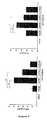
FIG. 2 is of histograms showing IFN-γ and IL-4 levels in the supernatants obtained after incubating splenocytes isolated from naive C57BL/6 mice with α-GC in the presence of control rat IgG, anti-PD-1 monoclonal antibody, anti-PD-L1 monoclonal antibody or anti-PD-L2 monoclonal antibody as analyzed by ELISA (in vitro). -
FIG. 3 is of plots showing IFN-γ and IL-4 levels in the sera obtained from the C57BL/6 mice, which were intraperitoneally injected with control rat IgG, anti-PD-1 monoclonal antibody, anti-PD-L1 monoclonal antibody or anti-PD-L2 monoclonal antibody 24 hrs before α-GC treatment, at 0, 2, 6, 12, 24, 48 and 72 hrs after the α-GC treatment as analyzed by ELISA (in vivo). -
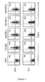
FIG. 4 shows FACS results of IFN-γ+ or IL-4+ iNKT cells on which intracellular cytokine staining was performed to examine if the increased production of IFN-γ and IL-4 in the sera ofFIG. 3 comes from iNKT cells. -
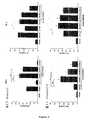
FIG. 5 is of histograms showing IFN-γ and IL-4 levels in the supernatants obtained after the splenocytes of C57BL/6 mice, in which iNKT cell anergy was induced by α-GC treatment 7 days and one month before splenocyte isolation therefrom, were incubated with α-GC in the presence of control rat IgG, anti-PD-1 monoclonal antibody, anti-PD-L1 monoclonal antibody or anti-PD-L2 monoclonal antibody, as analyzed by ELISA (in vitro). -
FIG. 6 is of histograms showing IFN-γ and IL-4 levels in sera from C57BL/6 mice, as analyzed by ELISA. The mice were intraperitoneally injected with control rat IgG, anti-PD-1 monoclonal antibody, anti-PD-L1 monoclonal antibody or anti-PD-L2 monoclonal antibody, followed by double injection with α-GC 24 hrs and 14 days later. The sera were obtained 2 and 12 hrs after the secondary injection of α-GC (in vivo). -
FIG. 7 shows FACS results of IFN-γ+ or IL-4+ iNKT cells after intracellular cytokine staining was performed on the splenocytes isolated 2 hrs after the secondary injection (in vivo). -
FIG. 8 shows CD69 expression levels on iNKT and NK cells as measured by flow cytometry using the splenocytes isolated 12 hrs after the secondary injection (in vivo). -
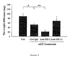
FIG. 9 is a histogram showing weights of normal and metastatic lungs of C57BL/6 mice. The mice were intraperitoneally injected with control rat IgG, anti-PD-1 monoclonal antibody or anti-PD-L1 antibody 24 hrs before intravenous injection with skin tumor cells. OnDays -
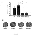
FIG. 10 shows effects of the blockage of PD-1/PD-L1 interaction on the antitumor activity of iNKT cells in terms of the number of metastatic pulmonary nodules (A) and optical microscopic views (B). C57BL/6 mice were intraperitoneally injected with control rat IgG, anti-PD-1 antibody or anti-PD-L1 antibody 24 hrs before α-GC treatment. 7 days later, the mice were intravenously injected with skin tumor cells and re-injected with α-GC onDays - In accordance with an aspect thereof, the present invention provides use of a composition comprising alpha-galactosyl ceramide and an anti-PD-1 antibody or anti-PD-L1 antibody, which blocks the signaling of PD-1 or PD-L1 to prevent the iNKT cell ligand induced anergy of iNKT cells for the manufacture of a medicament for the treatment of cancer.
- Further, it is disclosed a method for reversing the unresponsiveness of iNKT cells with anergy induced therein by iNKT cell ligand treatment, comprising treating the anergic iNKT cells with an anti-PD-1 antibody or an anti-PD-L1 antibody.
- The iNKT cell ligand may be selected from a group consisting of alpha-galactosyl ceramide, alpha-glucuronosyl ceramide, phosphatidylinositoltetramannoside, isoglobotrihexosylceramide, ganglioside GD3, phsphatidylcholine, phosphatidylethanolamine, phosphatidylinositol, sulfatide, betagalactosylceramide, lipophosphoglycan, glycoinositol phospholipid, alpha-galactosylceramide analogs including betaanomer galactoceramide and alpha-anomer galactosylceramide, and bacterial lipid antigens.
- The anti-PD-1 antibody or the anti-PD-L1 antibody may be a monoclonal antibody or a polyclonal antibody.
- The anti-PD-1 antibody or anti-PD-L1 antibody according to the present invention can block the signaling of PD-1 or PD-L1 to prevent the iNKT cell ligand-induced anergy of iNKT cells and can provide cytokine secretion ability for even anergic iNKT cells to restore their responsiveness. In addition, the anti-PD-1 antibody or anti-PD-L1 antibody of the present invention inhibits the anergy induction of iNKT cells to significantly decrease the number of pulmonary nodules in a lung metastatis model of B16F10 melanoma, thus effectively eliciting anticancer immune responses of iNKT cells and showing anti-tumor effects of iNKT cells against cancer metastasis. Therefore, an anticancer agent comprising the anti-PD-1 antibody or anti-PD-L1 antibody of the present invention can be very useful in the treatment of cancer, particularly, metastatic cancer.
- The cancer to which the anticancer agent according to the present invention is therapeutically applicable may be gynecologic tumor, endocrine gland cancer, CNS (central nervous system) tumor or ureter cancer. Concrete examples of the cancer include lung cancer, stomach cancer, liver cancer, bone cancer, pancreatic cancer, skin cancer, head and neck cancer, melanoma, uterine cancer, ovarian cancer, rectal cancer, colorectal cancer, colon cancer, breast cancer, uterine sarcoma, Fallopian tube carcinoma, endometrial carcinoma, uterine cervical carcinoma, vaginal carcinoma, vulva carcinoma, esophageal cancer, larynx cancer, small intestine cancer, thyroid cancer, parathyroid cancer, soft tissue sarcoma, uterine cancer, penis cancer, prostate cancer, chronic or acute leukemia, pediatric solid tumor, differentiated lymphoma, bladder cancer, kidney cancer, renal cell carcinoma, renal pelvic carcinoma, primary CNS lymphoma, spinal cord tumor, brainstem glioma, and pituitary adenoma.
- The anticancer agent of the present invention may be formulated into a pharmaceutical composition with at least one conventional anticancer ingredient.
- In addition to the active ingredients, the pharmaceutical composition may further comprise a pharmaceutically acceptable carrier. Examples of the pharmaceutically acceptable carrier include saline, sterile water, Ringer's solution, buffered saline, dextrose solution, maltodextrin solution, glycerol, ethanol, etc. Optionally, conventional additives, such as antioxidants, buffers, bacteriostatic agents, etc., may be added to the composition. For the preparation of dosage forms including injections, such as aqueous solutions, suspensions and emulsions, pills, capsules, granules and tablets, the active ingredients may be admixed with a diluent, a dispersant, a surfactant, a binder and/or a lubricant. Reference may be made to literature (Remington's Pharmaceutical Science (recent edition), Mack Publishing Company, Easton PA) upon the formulation of the pharmaceutical composition into suitable dosage forms.
- The composition of the present invention may be administered via oral routes or parenteral routes (e.g., intravenous, subcutaneous, intraperitoneal, topical, etc.). The effective dosage of the anticancer agent in accordance with the present invention depends on various factors, including the patient's weight, age, gender, state of health, diet, the time of administration, route of administration, excretion rate, severity of diseases, etc. In general, it may be administered in a single dose, and preferably in multiple doses per day at a daily dose ranging from 0.01 to 1000 mg/day, and preferably from 0.1 to 100 mg/kg of the anti-PD-1 antibody or anti-PD-L1 antibody.
- For the effective prophylaxis and treatment of cancer, the composition according to the present invention may be used alone or in combination with surgical operation, hormonal therapy, chemotherapy, and/or biological response controllers.
- Six- to eight-week-old female C56BL/6 mice were purchased from Orient Bio. All mice were bred and maintained in specific pathogen-free conditions.
- α-GC was dissolved in PBS containing 0.5
% Tween 20. Hybridoma clones producing antibodies to mouse PD-1 (RMP1-14, rat IgG2a), PD-L1 (MIH-5, rat IgG2a), and PD-L2 (Ty25, rat IgG2a) were produced according to methods well known in the art [Yamazaki, T., H. Akiba, H. Iwai, H. Matsuda, M. Aoki, Y. Tanno, T. Shin, H. Tsuchiya, D. M. Pardoll, K. Okumura, M. Azuma, and H. Yagita. 2002. Expression of programmed .; Tsushima, F., H. Iwai, N. Otsuki, M. Abe, S. Hirose, T. Yamazaki, H. Akiba, H. Yagita, Y. Takahashi, K. Omura, K. Okumura, and M. Azuma. 2003. Preferential contribution of B7-H1 to programmed death-1-mediated regulation of hapten-specific allergic inflammatory responses. Eur J Immunol 33:2773-2782.; Yamazaki, T., H. Akiba, A. Koyanagi, M. Azuma, H. Yagita, and K. Okumura. 2005. Blockade of B7-H1 on macrophages suppresses CD4+ T cell proliferation by augmenting IFN-gamma-induced nitric oxide production. J Immunol 175:1586-1592.]. All clones were cultured in RPMI 1640 (Gibco) with 10% FBS (Gibco) and 1% penicillin/streptomycin (BioWhittaker). All antibodies were prepared from the ascites of nude mice using caprylic acid purification. Control rat IgG was also prepared by caprylic acid purification from sera of naive rats. - Splenocytes were isolated from C57BL/6 mice administered with 2 µg of α-GC at different time points (0 hr, 6 hrs, 72 hrs, 7 days, one month and two months) after the administration. The cells were stained with a PE-conjugated anti-PD-1 monoclonal antibody, a PE-conjugated anti-PD-L1 monoclonal antibody, a PE-conjugated anti-PD-L2 monoclonal antibody and a PE-conjugated isotype control monoclonal antibody, respectively. iNKT cells were gated on B220- TCR-βint α -GC/CDld:Ig+ population. PD-1, PD-L1 and PD-L2 expression (open histograms) was analyzed by FACS. The results are given in
FIG. 1 . Shaded histograms indicate staining with isotype control mAbs. - As depicted in
FIG. 1 , PD-1 was constitutively expressed on iNKT cells at a low level and its expression was upregulated after α-GC stimulation, followed by the persistence of the upregulated level for two months. On the other hand, PD-L1 expression was temporarily increased on α -GC-stimulated iNKT cells but declined toward naive state levels within 72 hrs. As for PD-L2, its expression was not found on iNKT cells regardless of their activation status. - The following in vitro and in vivo experiments were conducted to examine the effects of PD-1, PD-L1 and PD-L2 on iNKT cell activation.
- After being prepared from naive C57BL/6 mice, 5×10⁵ spelenocytes were incubated for 3 days with 100 ng/ml of α-GC in the presence of 50 µg/ml of the control rat IgG, the anti-PD-1 mAb, the anti-PD-L1 mAb, or the anti-PD-L2 mAb. Then, the supernatants were obtained and assayed for IFN-γ and IL-4 levels by ELISA.
- The results are given in
FIG. 2 . - Compared with control IgG treatment, as is apparent from
FIG. 2 , anti-PD-1 mAb significantly increased the production of IFN-γ and, to a lesser extent, IL-4. Anti-PD-L1 mAb also induced greater production of IFN-γ, but anti-PD-L2 mAb did not, - C57BL/6 mice was injected with 200 µg of the control rat IgG, the anti-PD-1 mAb, the anti-PD-L1 mAb or the anti-PD-L2 mAb 24 hrs before treatment with 2 µg of α-GC. Sera were obtained at 0, 2, 6, 12, 24, 48 and 72 hrs after the treatment, followed by ELISA analysis for IFN-γ and IL-4 levels. In order to determine whether the increased production of IFN-γ was distinctively attributed to iNKT cells, intracellular cytokine staining was followed by flow cytometry analysis. In this regard, splenocytes were isolated 2 hrs after α-GC treatment and 5×10⁶ cells were incubated with Golgi plug for 2 hrs to accumulate cytokines. Intracellular cytokine staining was performed using BD Cytofix/Cytoperm Plus with Golgiplug kit according to the manufacturer's protocol (BD Biosciences). iNKT cells were gated on B220-TCR-βint α-GC/CDld:Ig+ population and IFN-γ+ or IL-4+ iNKT cells were analyzed by flow cytometry.
- IFN-γ and IL-4 levels in blood are depicted in
FIG. 3 and flow histograms of IFN-γ+ and IL-4+ iNKT cells are given inFIG. 4 . - As shown in
FIG. 3 , the blood IFN-γ and IL-4 levels were observed to peak, respectively, within 12 hrs and 2 hrs after α-GC treatment, indicating iNKT cell activation. IFN-γ levels in the sera of anti-PD-1 or anti-PD-L1 mAb-treated mice were significantly enhanced compared with levels in the group treated with control IgG. Slightly different IL-4 production was observed after treatment with the blocking Abs, but with no significance. - As shown in
FIG. 4 , iNKT cells producing IFN-γ increased in number because they blocked PD-1/PD-L1 interaction with the anti-PD-1 mAb or anti-PD-L1 mAb in comparison with control cells. On the other hand, no significant differences were found between populations of IL-4+ iNKT cells and control cells after mAb treatment. These data indicate that the blockage of PD-1/PD-L1 interaction allows the delivery of the co-inhibitory signal during iNKT activation, resulting in increased IFN-γ secretion from iNKT cells. - The following in vitro and in vivo experiments were performed in order to examine whether the blockage of PD-1/PD-L1 interaction reverses iNKT cell anergy.
- The α-GC-induced unresponsiveness of iNKT cells was detected as early as 3 days after primary stimulation and observed to persist until 7 - 30 days after α-GC stimulation. Thus, C57BL/6 mice were injected with 2 µg of α-GC to induce iNKT cell anergy. One week and one month later, splenocytes were isolated from the mice and 5x10⁵ cells were incubated for 3 days with 100ng/ml of α-GC in the presence of 50 µg/ml of the control rat IgG, the anti-PD-1 mAb, the anti-PD-L1 mAb, or the anti-PD-L2 mAb. Also, splenocytes isolated from the mice were incubated for 3 days with 100ng/ml of α-GC without mAb, and these were represented by 'activation'. The supernatants were then assayed for IFN-γ and IL-4 levels by ELISA [*: p<0.05 and **: p<0.01 (vs. control rat IgG)].
- The results are depicted in
FIG. 5 . - As seen in the graphs of
FIG. 5 , when restimulated with α -GC in the presence of the control IgG, the splenocytes of the mice in which iNKT cell anergy was induced by pretreatment with α-GC 7 days before the restimulation were found to greatly decrease in the production of IFN-γ and IL-4. In contrast, the restimulation of iNKT cell anergy-induced splenocytes with α-GC in the presence of the anti-PD-1 antibody or anti-PD-L1 antibody resulted in a great increase in the production of IFN-γ and IL-4 (A). This upregulation was observed to persist for one to two months after α-GC treatment (B). Therefore, the blockage of PD-1/PD-L1 interaction during restimulation with α-GC reverses the established anergic phenotype of iNKT cells. - Splenocytes (5×10⁶ cells), prepared two and twelve hours after the second injection of α-GC, were incubated with Golgi plug for 2 hrs to accumulate cytokines. Intracellular cytokine staining was performed on the splenocytes prepared 2 hrs after the secondary GC treatment, using BD Cytofix/Cytoperm Plus with Golgiplug kit according to the manufacturer's protocol (BD Biosciences). The splenocytes prepared 12 hrs later were used in an assay for CD69 expression in iNKT and NK cells. iNKT cells were gated on B220-TCR-βint α-GC/CDld:Ig+ population and NK cells were gated on B220- TCR-β- NK1.1high population. IFN-γ+ or IL-4+ iNKT cells were analyzed by flow cytometry. Also, CD69 expression on iNKT and NK cells was analyzed by flow cytometry. IFN-γ and IL-4 levels in blood are graphed in
FIG. 6 and FACS results of IFN-γ+ or IL-4+ iNKT cells and flow histograms of CD69 expression on iNKT and NK cells are given inFIGS. 7 and8 , respectively. - Like mice treated with control rat IgG, as seen in
FIG. 6 , mice pretreated with α-GC produced significantly low IFN-γ and IL-4 upon secondary α-GC injection. In contrast, mice treated with the anti-PD-1 antibody during the primary α-GC injection produced remarkably higher IFN-γ and IL-4 upon the secondary α-GC injection than did the control IgG-treated group. - As seen in
FIG. 7 , the increased cytokine production was attributed to iNKT cells. - It was also found that CD69 expression upon secondary α-GC injection was increased on iNKT and NK cells in a similar manner by the treatment with anti-PD-1 mAbs, as shown in
FIG. 8 . Thus, PD-1/PD-L1 interaction is essential for the induction of iNKT cell anergy in vivo. - The following experiments were performed to investigate the effects of the anti-PD-1 antibody or the anti-PD-L1 antibody on the anticancer activity of activated iNKT cells in a B16F10 melanoma metastasis model.
- Skin tumor cells (B16F10, ATCC) were cultured in DMEM media supplemented with 10% FBS and 1% penicillin/streptomycin. C56BL/6 mice were intraperitoneally injected with 200 µg of the control rat IgG, the anti-PD-1 mAb or the anti-PD-L1 mAb 24 hrs before i.v. inoculation with 2×10⁵ B16F10 tumor cells. On Days zero, 4 and 8, the mice were treated with 500ng of α-GC plus 200 µg of the control rat IgG, the anti-PD-1 mAb or the anti-PD-L1 mAb. On
Day 14, the lungs were weighed and weight differences between metastatic and normal lungs are graphed inFIG. 9 . - As depicted in
FIG. 9 , the B16F10 metastatic lungs from the group treated with α-GC plus the anti-PD-1 mAb were significantly reduced in weight compared with those from the group treated with α-GC plus the control rat IgG. - Anergy-induced iNKT cells were assayed for the recovery of anti-tumor activity by the antibodies of the present invention. In this regard, naive C57BL/6 mice were intraperitoneally injected with 200 µg of the control rat IgG, the anti-PD-1 antibody or the anti-PD-L1 antibody 24 hrs after which injection with 2 µg of α-GC induced iNKT cell anergy. 7 days later, 5×10⁵ B16F10 tumor cells were i.v. injected. On
Day FIG. 10 . - As seen in
FIG. 10 , when induced by treatment with the control IgG and α-GC, iNKT cell anergy was unchanged with time to show a metastasis result comparable to that of the normal metastasis control group, indicating that α-GC treatment during tumor inodulation did not suppress tumor growth. In contrast, when the anergy induction of α-GC was prevented by treatment with the anti-PD-1 antibody or the anti-PD-L1, the numbers of tumor nodules in the B16F10 melanoma metastasis model were remarkably reduced compared with those upon treatment with the control IgG, demonstrating the superior anti-tumor activity of the antibodies of the present invention. Taken together, the data obtained above indicates that the blockage of PD-1/PD-L1 interaction by treatment with the anti-PD-1 antibody or the anti-PD-L1 antibody during the induction phase of iNKT cell anergy leads to the persistence of the anti-tumor effects of iNKT cells. - Formulation examples are given to illustrate dosage preparations containing the anticancer agent of the present invention.
-
- Anti-PD-1 or Anti-PD-L1 Antibody
- 0.1 g
- Lactose
- 1.5 g
- Talc
- 0.5 g
- These ingredients were mixed and loaded into an airtight sac to give a powder.
-
- Anti-PD-1 or Anti-PD-L1 Antibody
- 0.1 g
- Lactose
- 7.9 g
- Crystalline cellulose
- 1.5 g
- Magnesium stearate
- 0.5 g
- These ingredients were mixed and directly compressed into a tablet.
-
- Anti-PD-1 or Anti-PD-L1 Antibody
- 0.1 g
- Corn starch
- 5 g
- Carboxycellulose
- 4.9 g
- These ingredients were admixed together and the admixture was loaded into a conventional capsule using a suitable device.
-
- Anti-PD-1 or Anti-PD-L1 Antibody
- 0.1 g
- Sterile water for injection
- proper quantity
- pH Adjuster
- proper quantity
- Using a conventional method, these ingredients were put into an ampule (2 ml) to give an injection.
-
- Anti-PD-1 or Anti-PD-L1 Antibody
- 0.1 g
- Isomerized sugar
- 10 g
- Mannitol
- 5 g
- Purified water
- proper quantity
- Each ingredient was dissolved in purified water and flavored with lemon before admixing together. Purified water was added to the admixture to form a final volume of 100 ml which was then loaded into a brown vial and sterilized.
- As described hitherto, the anti-PD-1 or anti-PD-L1 antibody according to the present invention blocks the PD-1/PD-L1-mediated signaling pathway not only to prevent the iNKT cell ligand-induced iNKT cell anergy, but also to reverse the unresponsiveness of already anergic iNKT cells to produce cytokines. In addition, the anti-PD1 or anti-PD-L1 antibody ensures the potent anti-tumor activity of iNKT cells as demonstrated by a significant reduction in the number of metastatic nodules in B16F10 melanoma metastasis models in vivo. Collectively, the anticancer agent comprising an anti-PD-1 or anti-PD-L1 antibody in accordance with the present invention can be very useful in the treatment of cancer, particularly metastatic cancer.
Claims (3)
- Use of a composition comprising alpha-galactosyl ceramide and an anti-PD-1 antibody or anti-PD-L1 antibody, which blocks the signaling of PD-1 or PD-L1 to prevent the iNKT cell ligand induced anergy of iNKT cells for the manufacture of a medicament for the treatment of cancer.
- Use of a composition as defined in claim 1, wherein the anti-PD-1 antibody or the anti-PD-L1 antibody is a monoclonal antibody or a polyclonal antibody.
- Use of a composition as defined in claim 1 or 2, wherein the cancer is selected from a group consisting of lung cancer, stomach cancer, liver cancer, bone cancer, pancreatic cancer, skin cancer, head and neck cancer, melanoma, uterine cancer, ovarian cancer, rectal cancer, colorectal cancer, colon cancer, breast cancer, uterine sarcoma, Fallopian tube carcinoma, endometrial carcinoma, uterine cervical carcinoma, vaginal carcinoma, vulva carcinoma, esophageal cancer, larynx cancer, small intestine cancer, thyroid cancer, parathyroid cancer, soft tissue sarcoma, uterine cancer, penis cancer, prostate cancer, chronic or acute leukemia, pediatric solid tumor, differentiated lymphoma, bladder cancer, kidney cancer, renal cell carcinoma, renal pelvic carcinoma, primary CNS lymphoma, spinal cord tumor, brainstem glioma, and pituitary adenoma.
Applications Claiming Priority (1)
| Application Number | Priority Date | Filing Date | Title |
|---|---|---|---|
| KR1020080097236A KR101050829B1 (en) | 2008-10-02 | 2008-10-02 | Anticancer agents comprising an anti-PD-1 antibody or an anti-PD-L1 antibody |
Publications (2)
| Publication Number | Publication Date |
|---|---|
| EP2172219A1 EP2172219A1 (en) | 2010-04-07 |
| EP2172219B1 true EP2172219B1 (en) | 2013-09-11 |
Family
ID=41171109
Family Applications (1)
| Application Number | Title | Priority Date | Filing Date |
|---|---|---|---|
| EP09004806.7A Active EP2172219B1 (en) | 2008-10-02 | 2009-04-01 | Anti-cancer agent comprising an iNKT ligand and anti-PD-1 antibody or anti-PD-L1 antibody |
Country Status (4)
| Country | Link |
|---|---|
| US (2) | US20100086550A1 (en) |
| EP (1) | EP2172219B1 (en) |
| JP (1) | JP5054057B2 (en) |
| KR (1) | KR101050829B1 (en) |
Cited By (6)
| Publication number | Priority date | Publication date | Assignee | Title |
|---|---|---|---|---|
| CN108752476A (en) * | 2016-03-04 | 2018-11-06 | 四川科伦博泰生物医药股份有限公司 | A kind of PDL-1 antibody, its medical composition and its use |
| US10336824B2 (en) | 2015-03-13 | 2019-07-02 | Cytomx Therapeutics, Inc. | Anti-PDL1 antibodies, activatable anti-PDL1 antibodies, and methods of thereof |
| US10450385B2 (en) | 2014-08-07 | 2019-10-22 | Memorial Sloan Kettering Cancer Center | Anti-ceramide antibodies |
| US10513558B2 (en) | 2015-07-13 | 2019-12-24 | Cytomx Therapeutics, Inc. | Anti-PD1 antibodies, activatable anti-PD1 antibodies, and methods of use thereof |
| US11168144B2 (en) | 2017-06-01 | 2021-11-09 | Cytomx Therapeutics, Inc. | Activatable anti-PDL1 antibodies, and methods of use thereof |
| EP4011389A4 (en) * | 2019-08-09 | 2024-09-04 | Riken | COMBINED USE OF ARTIFICIAL ADJUVANT VECTOR CELLS AND IMMUNOSTIMULANT |
Families Citing this family (151)
| Publication number | Priority date | Publication date | Assignee | Title |
|---|---|---|---|---|
| EP4209510B1 (en) | 2008-12-09 | 2024-01-31 | F. Hoffmann-La Roche AG | Anti-pd-l1 antibodies and their use to enhance t-cell function |
| NO2504364T3 (en) | 2009-11-24 | 2018-01-06 | ||
| CA2849259A1 (en) * | 2011-09-19 | 2013-03-28 | The Johns Hopkins University | Cancer immunotherapy |
| US9624298B2 (en) | 2011-11-28 | 2017-04-18 | Merck Patent Gmbh | Anti-PD-L1 antibodies and uses thereof |
| JP6312659B2 (en) * | 2012-05-31 | 2018-04-18 | ジェネンテック, インコーポレイテッド | Cancer treatment method using PD-1 binding antagonist and VEGF antagonist |
| LT2904011T (en) * | 2012-10-02 | 2017-11-10 | Bristol-Myers Squibb Company | Combination of anti-kir antibodies and anti-pd-1 antibodies to treat cancer |
| JP6742903B2 (en) | 2013-05-02 | 2020-08-19 | アナプティスバイオ インコーポレイティッド | Antibodies to programmed death-1 (PD-1) |
| WO2014194293A1 (en) * | 2013-05-30 | 2014-12-04 | Amplimmune, Inc. | Improved methods for the selection of patients for pd-1 or b7-h4 targeted therapies, and combination therapies thereof |
| EP3293275B1 (en) | 2013-06-06 | 2021-08-04 | Dana-Farber Cancer Institute, Inc. | Compositions and methods for identification, assessment prevention, and treatment of cancer using pd-l1 isoforms |
| CN104250302B (en) | 2013-06-26 | 2017-11-14 | 上海君实生物医药科技股份有限公司 | The anti-antibody of PD 1 and its application |
| CN105873606B (en) | 2013-09-11 | 2020-12-29 | 米迪缪尼有限公司 | Anti-B7-H1 Antibodies for Tumor Treatment |
| LT3702373T (en) | 2013-09-13 | 2022-11-10 | Beigene Switzerland Gmbh | Anti-pd1 antibodies and their use as therapeutics and diagnostics |
| EP3757130A1 (en) | 2013-09-26 | 2020-12-30 | Costim Pharmaceuticals Inc. | Methods for treating hematologic cancers |
| CN104698059B (en) * | 2013-12-04 | 2017-07-21 | 苏州中赢医疗科技有限公司 | A kind of glioma tumor markers and its application |
| HUE046249T2 (en) | 2013-12-12 | 2020-02-28 | Shanghai hengrui pharmaceutical co ltd | PD-1 antibody, antigen-binding fragment thereof, and its therapeutic use |
| EP4124624A3 (en) * | 2013-12-20 | 2023-05-03 | Intervet International B.V. | Antibodies against canine pd-1 |
| TWI681969B (en) | 2014-01-23 | 2020-01-11 | 美商再生元醫藥公司 | Human antibodies to pd-1 |
| TWI680138B (en) * | 2014-01-23 | 2019-12-21 | 美商再生元醫藥公司 | Human antibodies to pd-l1 |
| CA2935423C (en) | 2014-01-24 | 2023-11-07 | Dana-Farber Cancer Institute, Inc. | Antibody molecules to pd-1 and uses thereof |
| AU2015210750B2 (en) | 2014-01-31 | 2020-08-20 | Children's Medical Center Corporation | Antibody molecules to TIM-3 and uses thereof |
| SG10201806297VA (en) * | 2014-02-04 | 2018-08-30 | Incyte Corp | Combination of a pd-1 antagonist and an ido1 inhibitor for treating cancer |
| WO2015138920A1 (en) | 2014-03-14 | 2015-09-17 | Novartis Ag | Antibody molecules to lag-3 and uses thereof |
| WO2015157162A1 (en) | 2014-04-06 | 2015-10-15 | H. Lee Moffitt Cancer Center And Research Institute, Inc. | Histone deacetylase as a modulator of pdl1 expression and activity |
| WO2015179615A1 (en) | 2014-05-23 | 2015-11-26 | Eisai R&D Management Co., Ltd | Combination therapies for the treatment of cancer |
| TWI726608B (en) | 2014-07-03 | 2021-05-01 | 英屬開曼群島商百濟神州有限公司 | Anti-pd-l1 antibodies and their use as therapeutics and diagnostics |
| WO2016006241A1 (en) | 2014-07-09 | 2016-01-14 | 日本全薬工業株式会社 | Anti-canine pd-1 antibody or anti-canine pd-l1 antibody |
| MX388181B (en) | 2014-07-22 | 2025-03-19 | Cb Therapeutics Inc | ANTI-PD-1 ANTIBODIES. |
| EP3190415B1 (en) | 2014-07-25 | 2019-05-01 | Riken | Memory invariant nkt cell marker |
| JP6909153B2 (en) | 2014-08-05 | 2021-07-28 | アポロミクス インコーポレイテッド | Anti-PD-L1 antibody |
| WO2016040882A1 (en) | 2014-09-13 | 2016-03-17 | Novartis Ag | Combination therapies of egfr inhibitors |
| WO2016061142A1 (en) | 2014-10-14 | 2016-04-21 | Novartis Ag | Antibody molecules to pd-l1 and uses thereof |
| CN105622753B (en) * | 2014-11-04 | 2019-04-09 | 博生吉医药科技(苏州)有限公司 | A kind of PD-1 monoclonal antibody and its application |
| JP7305300B2 (en) | 2014-11-05 | 2023-07-10 | ザ リージェンツ オブ ザ ユニバーシティ オブ カリフォルニア | Combination immunotherapy |
| US20170058043A1 (en) | 2014-12-06 | 2017-03-02 | H. Lee Moffitt Cancer Center And Research Institute, Inc. | Bispecific antibody for cancer immunotherapy |
| AU2016219161A1 (en) * | 2015-02-12 | 2017-08-31 | The Johns Hopkins University | Inhibition of Yap for breaking tumor immune tolerance |
| CN116327915A (en) | 2015-04-03 | 2023-06-27 | 佐马技术有限公司 | Treatment of cancer using TGF-beta inhibitors and PD-1 inhibitors |
| DK3277325T3 (en) | 2015-04-03 | 2021-01-18 | H Lee Moffitt Cancer Ct & Res | COMBINATION IMMUNE THERAPY FOR CANCER |
| EP3892284B1 (en) | 2015-05-29 | 2024-05-22 | Merck Sharp & Dohme LLC | Combination of a pd-1 antagonist and a cpg-c type oligonucleotide for treating cancer |
| WO2016197067A1 (en) | 2015-06-05 | 2016-12-08 | H. Lee Moffitt Cancer Center And Research Institute, Inc. | Gm-csf/cd40l vaccine and checkpoint inhibitor combination therapy |
| ES2878188T3 (en) | 2015-07-29 | 2021-11-18 | Novartis Ag | Combination therapies comprising antibody molecules against LAG-3 |
| AR105654A1 (en) | 2015-08-24 | 2017-10-25 | Lilly Co Eli | ANTIBODIES PD-L1 (LINKING 1 OF PROGRAMMED CELL DEATH) |
| EP3344656A1 (en) | 2015-09-01 | 2018-07-11 | Agenus Inc. | Anti-pd-1 antibodies and methods of use thereof |
| ES2839212T3 (en) | 2015-09-29 | 2021-07-05 | Inst Nat Sante Rech Med | Methods to determine the metabolic status of B lymphomas |
| AU2016329251B2 (en) | 2015-10-02 | 2023-02-02 | F. Hoffmann-La Roche Ag | Anti-PD1 antibodies and methods of use |
| KR20250099459A (en) | 2015-11-03 | 2025-07-01 | 얀센 바이오테크 인코포레이티드 | Antibodies specifically binding pd-1 and their uses |
| CA3004804A1 (en) | 2015-11-17 | 2017-05-26 | Jiangsu Hengrui Medicine Co., Ltd. | Pd-l1 antibody, antigen-binding fragment thereof and medical application thereof |
| HUE063377T2 (en) | 2015-12-22 | 2024-01-28 | Regeneron Pharma | Combination of anti-pd-1 antibodies and bispecific anti-cd20/anti-cd3 antibodies to treat cancer |
| WO2017118634A1 (en) | 2016-01-04 | 2017-07-13 | INSERM (Institut National de la Santé et de la Recherche Médicale) | Use of pd-1 and tim-3 as a measure for cd8+ cells in predicting and treating renal cell carcinoma |
| TWI786044B (en) | 2016-05-13 | 2022-12-11 | 美商再生元醫藥公司 | Methods of treating skin cancer by administering a pd-1 inhibitor |
| US10059769B2 (en) * | 2016-06-13 | 2018-08-28 | I-Mab | Anti-PD-L1 antibodies and uses thereof |
| KR20190019121A (en) * | 2016-06-17 | 2019-02-26 | 오사카 유니버시티 | Intratumoral intravenous growth promoter |
| WO2018007885A1 (en) | 2016-07-05 | 2018-01-11 | Beigene, Ltd. | COMBINATION OF A PD-l ANTAGONIST AND A RAF INHIBITOR FOR TREATING CANCER |
| AU2017313085B2 (en) | 2016-08-19 | 2024-06-20 | Beone Medicines I Gmbh | Use of a combination comprising a Btk inhibitor for treating cancers |
| EP4342978A3 (en) | 2016-09-01 | 2024-07-03 | Chimera Bioengineering Inc. | Gold optimized car t-cells |
| WO2018050782A1 (en) | 2016-09-14 | 2018-03-22 | Abivax | Combinations including abx196 for the treatment of cancer |
| US11524988B2 (en) | 2016-09-19 | 2022-12-13 | H. Lee Moffitt Cancer Center And Research Institute, Inc. | Artificial antigen presenting cells for genetic engineering of immune cells |
| US12098202B2 (en) | 2016-10-10 | 2024-09-24 | The National Institute for Biotechnology in the Negev Ltd. | Non-cytotoxic modified cells and use thereof |
| EP3666794A1 (en) | 2016-11-01 | 2020-06-17 | AnaptysBio, Inc. | Antibodies directed against programmed death- 1 (pd-1) |
| US10342785B2 (en) | 2016-11-04 | 2019-07-09 | Askat Inc. | Use of EP4 receptor antagonists for the treatment of NASH-associated liver cancer |
| CN118634323A (en) | 2016-12-07 | 2024-09-13 | 艾吉纳斯公司 | Antibodies and methods of use thereof |
| EP3565844B8 (en) | 2017-01-09 | 2023-04-12 | Tesaro, Inc. | Methods of treating cancer with anti-pd-1 antibodies |
| CN106699891B (en) * | 2017-01-25 | 2019-04-09 | 北京天广实生物技术股份有限公司 | A kind of anti-PD-L1 antibody, its pharmaceutical composition and use |
| WO2018137681A1 (en) | 2017-01-25 | 2018-08-02 | Beigene, Ltd. | Crystalline forms of (s) -7- (1- (but-2-ynoyl) piperidin-4-yl) -2- (4-phenoxyphenyl) -4, 5, 6, 7-tetrahy dropyrazolo [1, 5-a] pyrimidine-3-carboxamide, preparation, and uses thereof |
| WO2018156735A1 (en) | 2017-02-22 | 2018-08-30 | H. Lee Moffitt Cancer Center And Research Institute, Inc. | Bispecific antibody for cancer immunotherapy |
| US11458169B2 (en) | 2017-02-22 | 2022-10-04 | H. Lee Moffitt Cancer Center And Research Institute, Inc. | TIM3-binding chimeric antigen receptors |
| EP3600427A1 (en) | 2017-03-24 | 2020-02-05 | INSERM - Institut National de la Santé et de la Recherche Médicale | Methods and compositions for treating melanoma |
| US11603407B2 (en) | 2017-04-06 | 2023-03-14 | Regeneron Pharmaceuticals, Inc. | Stable antibody formulation |
| US11738009B2 (en) | 2017-04-18 | 2023-08-29 | Tempest Therapeutics, Inc. | Bicyclic compounds and their use in the treatment of cancer |
| KR102757960B1 (en) | 2017-06-26 | 2025-01-22 | 베이진 엘티디 | Immunotherapy for hepatocellular carcinoma (HCC) |
| BR112020000118A2 (en) | 2017-07-07 | 2020-07-07 | H. Lee Moffitt Cancer Center And Research Institute, Inc. | chimeric antigen receptors with mutated cd28 costimulatory domains |
| WO2019083971A1 (en) | 2017-10-23 | 2019-05-02 | Children's Medical Center Corporation | Methods of treating cancer using lsd1 inhibitors in combination with immunotherapy |
| CN111801334B (en) | 2017-11-29 | 2023-06-09 | 百济神州瑞士有限责任公司 | Treatment of Indolent or Aggressive B-Cell Lymphoma Using Combinations Containing BTK Inhibitors |
| EP3735590A1 (en) | 2018-01-04 | 2020-11-11 | INSERM (Institut National de la Santé et de la Recherche Médicale) | Methods and compositions for treating melanoma resistant |
| CN112040957A (en) | 2018-01-08 | 2020-12-04 | H·李·莫菲特癌症中心和研究所公司 | Compositions and methods for targeting CD99-expressing cancers |
| US11951129B2 (en) | 2018-01-09 | 2024-04-09 | H. Lee Moffitt Cancer Center And Research Institute, Inc. | Compositions and methods for targeting CLEC12A-expressing cancers |
| AU2019206958A1 (en) | 2018-01-10 | 2020-07-16 | Jiangsu Hengrui Medicine Co., Ltd. | PD-L1 antibody, antigen-binding fragment thereof, and pharmaceutical use thereof |
| US12398209B2 (en) | 2018-01-22 | 2025-08-26 | Janssen Biotech, Inc. | Methods of treating cancers with antagonistic anti-PD-1 antibodies |
| CN117643587A (en) | 2018-02-05 | 2024-03-05 | 深圳市原力生命科学有限公司 | Heterobicyclic carboxylic acids for the treatment of cancer |
| CA3091373A1 (en) * | 2018-02-17 | 2019-08-22 | Apollomics Inc. | Cancer treatment using combination of neutrophil modulator with modulator of immune checkpoint |
| EP3756012A1 (en) | 2018-02-21 | 2020-12-30 | INSERM (Institut National de la Santé et de la Recherche Médicale) | Use of sk1 as biomarker for predicting response to immunecheckpoint inhibitors |
| SG11202010153WA (en) | 2018-04-17 | 2020-11-27 | Tempest Therapeutics Inc | Bicyclic carboxamides and methods of use thereof |
| WO2020023361A1 (en) | 2018-07-23 | 2020-01-30 | H. Lee Moffitt Cancer Center And Research Institute Inc. | Enhancing anti-tumor response in melanoma cells with defective sting signaling |
| CN112041348B (en) | 2018-07-25 | 2023-09-22 | 天境生物科技(上海)有限公司 | Anti-CD73 anti-PD-L1 bispecific antibody |
| WO2020104479A1 (en) | 2018-11-20 | 2020-05-28 | INSERM (Institut National de la Santé et de la Recherche Médicale) | Methods and compositions for treating cancers and resistant cancers with anti transferrin receptor 1 antibodies |
| US20230183379A1 (en) | 2018-11-20 | 2023-06-15 | INSERM (Institut National de la Santé et de la Recherche Médicale) | Bispecific antibody targeting transferrin receptor 1 and soluble antigen |
| WO2020115261A1 (en) | 2018-12-07 | 2020-06-11 | INSERM (Institut National de la Santé et de la Recherche Médicale) | Methods and compositions for treating melanoma |
| WO2020120592A1 (en) | 2018-12-12 | 2020-06-18 | INSERM (Institut National de la Santé et de la Recherche Médicale) | Methods and compositions for predicting and treating melanoma |
| WO2020127411A1 (en) | 2018-12-19 | 2020-06-25 | INSERM (Institut National de la Santé et de la Recherche Médicale) | Methods and compositions for treating cancers by immuno-modulation using antibodies against cathespin-d |
| WO2020127885A1 (en) | 2018-12-21 | 2020-06-25 | INSERM (Institut National de la Santé et de la Recherche Médicale) | Compositions for treating cancers and resistant cancers |
| EP3918332B1 (en) | 2019-01-30 | 2025-03-19 | INSERM (Institut National de la Santé et de la Recherche Médicale) | Methods and compositions for identifying whether a subject suffering from a cancer will achieve a response with an immune-checkpoint inhibitor |
| KR102942600B1 (en) | 2019-02-03 | 2026-03-20 | 지앙수 헨그루이 파마슈티컬스 컴퍼니 리미티드 | Anti-PD-1 antibody, its antigen-binding fragment, and its pharmaceutical uses |
| US20220117911A1 (en) | 2019-02-04 | 2022-04-21 | INSERM (Institut National de la Santé et de la Recherche Médicale) | Methods and compositions for modulating blood-brain barrier |
| US20220098674A1 (en) | 2019-02-13 | 2022-03-31 | Inserm (Institut National De La Santé Et Dr La Recherch Médicale) | Methods and compositions for selecting a cancer treatment in a subject suffering from cancer |
| WO2020221796A1 (en) | 2019-04-30 | 2020-11-05 | INSERM (Institut National de la Santé et de la Recherche Médicale) | Methods and compositions for treating melanoma |
| AU2020296372A1 (en) | 2019-06-18 | 2022-02-17 | Janssen Sciences Ireland Unlimited Company | Combination of hepatitis B virus (HBV) vaccines and anti-PD-1 antibody |
| KR20220041080A (en) | 2019-06-18 | 2022-03-31 | 얀센 사이언시즈 아일랜드 언리미티드 컴퍼니 | Combination of hepatitis B virus (HBV) vaccine and anti-PD-1 or anti-PC-L1 antibody |
| WO2021048292A1 (en) | 2019-09-11 | 2021-03-18 | INSERM (Institut National de la Santé et de la Recherche Médicale) | Methods and compositions for treating melanoma |
| WO2021064180A1 (en) | 2019-10-03 | 2021-04-08 | INSERM (Institut National de la Santé et de la Recherche Médicale) | Methods and compositions for modulating macrophages polarization |
| US20240301497A1 (en) | 2019-10-17 | 2024-09-12 | INSERM (Institut National de la Santé et de la Recherche Médicale) | Methods for diagnosing nasal intestinal type adenocarcinomas |
| US20240122938A1 (en) | 2019-10-29 | 2024-04-18 | INSERM (Institut National de la Santé et de la Recherche Médicale) | Methods and compositions for treating uveal melanoma |
| CA3164754A1 (en) | 2019-12-19 | 2021-06-24 | Inserm (Institut National De La Sante Et De La Recherche Medicale) | Methods and vaccine compositions to treat cancers |
| EP4090770A1 (en) | 2020-01-17 | 2022-11-23 | INSERM (Institut National de la Santé et de la Recherche Médicale) | Methods and compositions for treating melanoma |
| WO2021156360A1 (en) | 2020-02-05 | 2021-08-12 | INSERM (Institut National de la Santé et de la Recherche Médicale) | Methods for discontinuing a treatment with a tyrosine kinase inhibitor (tki) |
| CA3172894A1 (en) | 2020-02-27 | 2021-09-02 | H. Lee Moffitt Cancer Center And Research Institute Inc. | Tumor-infiltrating lymphocytes with enhanced tumor reactivity |
| WO2021170777A1 (en) | 2020-02-28 | 2021-09-02 | INSERM (Institut National de la Santé et de la Recherche Médicale) | Methods for diagnosing, prognosing and managing treatment of breast cancer |
| BR112022022335A2 (en) | 2020-05-05 | 2023-01-10 | Teon Therapeutics Inc | TYPE 2 CANNABINOID RECEPTOR MODULATORS AND USES THEREOF |
| JP7648654B2 (en) | 2020-05-05 | 2025-03-18 | リジェネロン・ファーマシューティカルズ・インコーポレイテッド | CAR Comprising CD28 Zeta and CD3 Zeta |
| CR20230025A (en) | 2020-06-26 | 2023-10-05 | Amgen Inc | Il-10 muteins and fusion proteins thereof |
| JP7856627B2 (en) | 2020-07-28 | 2026-05-11 | アンスティチュ ナショナル ドゥ ラ サンテ エ ドゥ ラ ルシェルシュ メディカル | Methods and compositions for preventing and treating cancer |
| CN111920948B (en) * | 2020-09-25 | 2021-02-02 | 安可瑞(山西)生物细胞有限公司 | Pharmaceutical composition comprising immune cells for treating cancer |
| CN116368154A (en) | 2020-10-08 | 2023-06-30 | 阿菲姆德股份有限公司 | trispecific binder |
| WO2022084531A1 (en) | 2020-10-23 | 2022-04-28 | INSERM (Institut National de la Santé et de la Recherche Médicale) | Methods and compositions for treating glioma |
| EP4244392A1 (en) | 2020-11-16 | 2023-09-20 | Inserm (Institut National De La Sante Et De La Recherche Medicale) | Methods and compositions for predicting and treating uveal melanoma |
| EP4244391A1 (en) | 2020-11-16 | 2023-09-20 | INSERM (Institut National de la Santé et de la Recherche Médicale) | Methods and compositions for predicting and treating uveal melanoma |
| TWI877433B (en) | 2020-11-30 | 2025-03-21 | 大陸商杭州阿諾生物醫藥科技有限公司 | Combination therapy for the treatment of pik3ca mutant cancer |
| WO2022194908A1 (en) | 2021-03-17 | 2022-09-22 | INSERM (Institut National de la Santé et de la Recherche Médicale) | Methods and compositions for treating melanoma |
| EP4326903A1 (en) | 2021-04-23 | 2024-02-28 | Inserm (Institut National De La Sante Et De La Recherche Medicale) | Methods and compositions for treating cell senescence accumulation related disease |
| AU2022320948A1 (en) | 2021-07-30 | 2024-01-18 | Affimed Gmbh | Duplexbodies |
| US20250215103A1 (en) | 2021-08-03 | 2025-07-03 | Hoffmann-La Roche Inc. | Bispecific antibodies and methods of use |
| TW202325306A (en) | 2021-09-02 | 2023-07-01 | 美商天恩治療有限公司 | Methods of improving growth and function of immune cells |
| WO2023081730A1 (en) | 2021-11-03 | 2023-05-11 | Teon Therapeutics, Inc. | 4-hydroxy-2-oxo-1,2-dihydro-1,8-naphthyridine-3-carboxamide derivatives as cannabinoid cb2 receptor modulators for the treatment of cancer |
| US20240426823A1 (en) | 2021-11-03 | 2024-12-26 | Institut National de la Santé et de la Recherche Médicale | Methods and compositions for treating triple negative breast cancer (tnbc) |
| JP2024539432A (en) | 2021-11-15 | 2024-10-28 | 中国科学院生物物理研究所 | Fusion protein construct containing interleukin 15 as an active ingredient and its use |
| WO2023097211A1 (en) | 2021-11-24 | 2023-06-01 | The University Of Southern California | Methods for enhancing immune checkpoint inhibitor therapy |
| KR102819428B1 (en) | 2021-12-17 | 2025-06-13 | 한국과학기술연구원 | composition comprising PD-L1-inhibiting prodrug for the prevention or treatment of cancer |
| WO2023118165A1 (en) | 2021-12-21 | 2023-06-29 | INSERM (Institut National de la Santé et de la Recherche Médicale) | Methods and compositions for treating melanoma |
| EP4554943A1 (en) | 2022-07-14 | 2025-05-21 | Teon Therapeutics, Inc. | Adenosine receptor antagonists and uses thereof |
| WO2024033399A1 (en) | 2022-08-10 | 2024-02-15 | Institut National de la Santé et de la Recherche Médicale | Sigmar1 ligand for the treatment of pancreatic cancer |
| WO2024033400A1 (en) | 2022-08-10 | 2024-02-15 | Institut National de la Santé et de la Recherche Médicale | Sk2 inhibitor for the treatment of pancreatic cancer |
| WO2024056716A1 (en) | 2022-09-14 | 2024-03-21 | Institut National de la Santé et de la Recherche Médicale | Methods and pharmaceutical compositions for the treatment of dilated cardiomyopathy |
| WO2024084034A1 (en) | 2022-10-21 | 2024-04-25 | Institut National de la Santé et de la Recherche Médicale | Methods and pharmaceutical compositions for the treatment of osteoarthritis |
| EP4637731A1 (en) | 2022-12-21 | 2025-10-29 | Gilead Sciences, Inc. | Combination therapy for treating cancer |
| WO2024150525A1 (en) | 2023-01-12 | 2024-07-18 | 国立大学法人山口大学 | Feline anti-pd-1 antibody |
| EP4687959A1 (en) | 2023-03-24 | 2026-02-11 | Cornell University | Utilizing t cells derived from tumor draining lymph nodes for chimeric antigen receptor (car) t cell therapy for the treatment of cancer |
| WO2024200571A1 (en) | 2023-03-28 | 2024-10-03 | Institut National de la Santé et de la Recherche Médicale | Method for discriminating mono-immunotherapy from combined immunotherapy in cancers |
| EP4704807A1 (en) | 2023-05-03 | 2026-03-11 | IOX Therapeutics Limited | Inkt cell modulator liposomal compositions and methods of use |
| WO2024231384A1 (en) | 2023-05-10 | 2024-11-14 | Institut National de la Santé et de la Recherche Médicale | Compositions for treating senescence related disease |
| WO2024245951A1 (en) | 2023-05-26 | 2024-12-05 | Institut National de la Santé et de la Recherche Médicale | Combination of slc8a1 inhibitor and mitochondria-targeted antioxidant for treating melanoma |
| EP4470542A1 (en) | 2023-05-31 | 2024-12-04 | Fundación Miguel Servet | Oleuropein in the management of cancer |
| WO2024256635A1 (en) | 2023-06-15 | 2024-12-19 | Institut National de la Santé et de la Recherche Médicale | Dpm1 inhibitor for treating cancer |
| WO2025073765A1 (en) | 2023-10-03 | 2025-04-10 | Institut National de la Santé et de la Recherche Médicale | Methods of prognosis and treatment of patients suffering from melanoma |
| WO2025078632A1 (en) | 2023-10-12 | 2025-04-17 | Institut National de la Santé et de la Recherche Médicale | Methods of prognosis and treatment of patients suffering from cancer |
| WO2025132479A1 (en) | 2023-12-18 | 2025-06-26 | Institut National de la Santé et de la Recherche Médicale | Flt3 inhibitor for modulating macrophages polarization |
| WO2025132770A1 (en) | 2023-12-22 | 2025-06-26 | Institut National de la Santé et de la Recherche Médicale | Affitins for the treatment of cancer |
| WO2025210123A1 (en) | 2024-04-03 | 2025-10-09 | Institut National de la Santé et de la Recherche Médicale | Methods and pharmaceutical composition for treating cancers |
| WO2025219330A1 (en) | 2024-04-15 | 2025-10-23 | Institut National de la Santé et de la Recherche Médicale | Detection of ppix for use in methods for melanoma ferroptosis sensitivity and targeted therapy resistance prediction |
| WO2025223372A1 (en) | 2024-04-22 | 2025-10-30 | Fujian Haixi Pharmaceuticals Co., Ltd. | Combination comprising a pd-1 inhibitor and a pyridine compound for use in treating solid tumors |
| WO2025228998A1 (en) | 2024-04-30 | 2025-11-06 | Institut National de la Santé et de la Recherche Médicale | Use of hdac4 inhibitors for the treatment of melanoma |
| WO2025247829A1 (en) | 2024-05-27 | 2025-12-04 | Institut National de la Santé et de la Recherche Médicale | Methods and pharmaceutical composition for treating prostate cancer |
| WO2026013071A1 (en) | 2024-07-09 | 2026-01-15 | Institut National de la Santé et de la Recherche Médicale | Setdb1 inhibitor for use in the treatment of uveal melanoma |
| WO2026035860A1 (en) | 2024-08-07 | 2026-02-12 | Teon Therapeutics, Inc. | Formulations of adenosine receptor antagonist |
Family Cites Families (4)
| Publication number | Priority date | Publication date | Assignee | Title |
|---|---|---|---|---|
| KR100549866B1 (en) * | 2001-08-22 | 2006-02-08 | 고려대학교 산학협력단 | Cancer Treatment and Prevention Agents |
| PT1537878E (en) | 2002-07-03 | 2010-11-18 | Ono Pharmaceutical Co | IMMUNOPOTENTIAL COMPOSITIONS |
| CN101426523B (en) * | 2006-04-27 | 2013-03-27 | 财团法人首尔大学校产学协力财团 | B cell-based vaccine loaded with the ligand of natural killer T cell and antigen |
| US20090304711A1 (en) * | 2006-09-20 | 2009-12-10 | Drew Pardoll | Combinatorial Therapy of Cancer and Infectious Diseases with Anti-B7-H1 Antibodies |
-
2008
- 2008-10-02 KR KR1020080097236A patent/KR101050829B1/en active Active
-
2009
- 2009-03-25 US US12/410,732 patent/US20100086550A1/en not_active Abandoned
- 2009-03-30 JP JP2009082501A patent/JP5054057B2/en active Active
- 2009-04-01 EP EP09004806.7A patent/EP2172219B1/en active Active
-
2012
- 2012-02-17 US US13/398,915 patent/US8617546B2/en active Active
Cited By (11)
| Publication number | Priority date | Publication date | Assignee | Title |
|---|---|---|---|---|
| US10450385B2 (en) | 2014-08-07 | 2019-10-22 | Memorial Sloan Kettering Cancer Center | Anti-ceramide antibodies |
| RU2717651C2 (en) * | 2014-08-07 | 2020-03-24 | Мемориал Слоан-Кеттеринг Кэнсер Сентер | Antibodies to ceramide |
| US11447572B2 (en) | 2014-08-07 | 2022-09-20 | Memorial Sloan Kettering Cancer Center | Anti-ceramide antibodies |
| US10336824B2 (en) | 2015-03-13 | 2019-07-02 | Cytomx Therapeutics, Inc. | Anti-PDL1 antibodies, activatable anti-PDL1 antibodies, and methods of thereof |
| US10513558B2 (en) | 2015-07-13 | 2019-12-24 | Cytomx Therapeutics, Inc. | Anti-PD1 antibodies, activatable anti-PD1 antibodies, and methods of use thereof |
| CN108752476A (en) * | 2016-03-04 | 2018-11-06 | 四川科伦博泰生物医药股份有限公司 | A kind of PDL-1 antibody, its medical composition and its use |
| CN108752476B (en) * | 2016-03-04 | 2019-09-20 | 四川科伦博泰生物医药股份有限公司 | A kind of PDL-1 antibody, its pharmaceutical composition and use thereof |
| US10465014B2 (en) | 2016-03-04 | 2019-11-05 | Sichuan Kelun-Biotech Biopharmaceutical Co., Ltd. | PDL-1 antibody, pharmaceutical composition thereof, and uses thereof |
| US11136413B2 (en) | 2016-03-04 | 2021-10-05 | Sichuan Kelun-Biotech Biopharmaceutical Co., Ltd. | PDL-1 antibody, pharmaceutical composition thereof, and uses thereof |
| US11168144B2 (en) | 2017-06-01 | 2021-11-09 | Cytomx Therapeutics, Inc. | Activatable anti-PDL1 antibodies, and methods of use thereof |
| EP4011389A4 (en) * | 2019-08-09 | 2024-09-04 | Riken | COMBINED USE OF ARTIFICIAL ADJUVANT VECTOR CELLS AND IMMUNOSTIMULANT |
Also Published As
| Publication number | Publication date |
|---|---|
| US8617546B2 (en) | 2013-12-31 |
| US20100086550A1 (en) | 2010-04-08 |
| US20120237522A1 (en) | 2012-09-20 |
| KR101050829B1 (en) | 2011-07-20 |
| EP2172219A1 (en) | 2010-04-07 |
| JP2010083863A (en) | 2010-04-15 |
| KR20100037899A (en) | 2010-04-12 |
| JP5054057B2 (en) | 2012-10-24 |
Similar Documents
| Publication | Publication Date | Title |
|---|---|---|
| US8617546B2 (en) | Anticancer agent comprising anti-PD-1 antibody or anti-PD-L1 antibody | |
| US11866509B2 (en) | Humanized antibodies against CEACAM1 | |
| US8475790B2 (en) | Combination of CD137 antibody and CTLA-4 antibody for the treatment of proliferative diseases | |
| US10696744B2 (en) | Means and methods for treating DLBCL | |
| US20210230276A1 (en) | Treatment of cancer with combinations of immunoregulatory agents | |
| KR100510604B1 (en) | Identification of unique binding interactions between certain antibodies and the human b7.1 and b7.2 co-stimulatory antigens | |
| JP2022513778A (en) | Chimeric antigen receptor and T cell receptor and how to use | |
| US10005843B2 (en) | Anti-TNFRSF25 antibodies | |
| WO2013169693A1 (en) | Methods of treating cancer using an il-21 polypeptide and an anti-pd-1 antibody | |
| US20070243184A1 (en) | Prophylaxis and treatment of enterocolitis associated with anti-ctla-4 antibody therapy | |
| CN109476754A (en) | Use of anti-PD-1 antibodies in the treatment of colorectal cancer patients | |
| CN109476752A (en) | Use of a combination of an anti-PD-1 antibody and an anti-CD30 antibody in the treatment of lymphoma | |
| WO2018101448A1 (en) | Method of treating cancer using anti-ccr4 antibody and anti-pd-1 antibody | |
| WO2001089567A1 (en) | Identification of unique binding interactions between certain antibodies and the human b7.1 and b7.2 co-stimulatory antigens | |
| US20240052050A1 (en) | Multispecific antibodies for the treatment of cancer | |
| CA3181677A1 (en) | Combination therapy comprising anti-cd137 antibodies | |
| EP4342492A1 (en) | Pharmaceutical combination and use thereof | |
| US20200405806A1 (en) | Combination of a tetanus toxoid, anti-ox40 antibody and/or anti-pd-1 antibody to treat tumors | |
| HK1262611B (en) | Methods for treating dlbcl | |
| HK1262611A1 (en) | Methods for treating dlbcl | |
| HK1224678A1 (en) | Means and methods for treating dlbcl | |
| HK1188229B (en) | Means and methods for treating dlbcl |
Legal Events
| Date | Code | Title | Description |
|---|---|---|---|
| PUAI | Public reference made under article 153(3) epc to a published international application that has entered the european phase |
Free format text: ORIGINAL CODE: 0009012 |
|
| 17P | Request for examination filed |
Effective date: 20090401 |
|
| AK | Designated contracting states |
Kind code of ref document: A1 Designated state(s): AT BE BG CH CY CZ DE DK EE ES FI FR GB GR HR HU IE IS IT LI LT LU LV MC MK MT NL NO PL PT RO SE SI SK TR |
|
| AX | Request for extension of the european patent |
Extension state: AL BA RS |
|
| 17Q | First examination report despatched |
Effective date: 20100531 |
|
| RIC1 | Information provided on ipc code assigned before grant |
Ipc: A61P 35/00 20060101ALI20130327BHEP Ipc: A61K 31/70 20060101ALI20130327BHEP Ipc: A61K 39/395 20060101AFI20130327BHEP Ipc: C07K 16/28 20060101ALN20130327BHEP |
|
| GRAP | Despatch of communication of intention to grant a patent |
Free format text: ORIGINAL CODE: EPIDOSNIGR1 |
|
| INTG | Intention to grant announced |
Effective date: 20130506 |
|
| GRAS | Grant fee paid |
Free format text: ORIGINAL CODE: EPIDOSNIGR3 |
|
| GRAA | (expected) grant |
Free format text: ORIGINAL CODE: 0009210 |
|
| RIN1 | Information on inventor provided before grant (corrected) |
Inventor name: CHANG, WOO SUNG Inventor name: KANG, CHANG YUIL Inventor name: KIM, JI YEON |
|
| AK | Designated contracting states |
Kind code of ref document: B1 Designated state(s): AT BE BG CH CY CZ DE DK EE ES FI FR GB GR HR HU IE IS IT LI LT LU LV MC MK MT NL NO PL PT RO SE SI SK TR |
|
| REG | Reference to a national code |
Ref country code: GB Ref legal event code: FG4D |
|
| REG | Reference to a national code |
Ref country code: CH Ref legal event code: EP |
|
| REG | Reference to a national code |
Ref country code: AT Ref legal event code: REF Ref document number: 631260 Country of ref document: AT Kind code of ref document: T Effective date: 20130915 |
|
| REG | Reference to a national code |
Ref country code: IE Ref legal event code: FG4D |
|
| REG | Reference to a national code |
Ref country code: DE Ref legal event code: R096 Ref document number: 602009018651 Country of ref document: DE Effective date: 20131107 |
|
| PG25 | Lapsed in a contracting state [announced via postgrant information from national office to epo] |
Ref country code: LT Free format text: LAPSE BECAUSE OF FAILURE TO SUBMIT A TRANSLATION OF THE DESCRIPTION OR TO PAY THE FEE WITHIN THE PRESCRIBED TIME-LIMIT Effective date: 20130911 Ref country code: HR Free format text: LAPSE BECAUSE OF FAILURE TO SUBMIT A TRANSLATION OF THE DESCRIPTION OR TO PAY THE FEE WITHIN THE PRESCRIBED TIME-LIMIT Effective date: 20130911 Ref country code: SE Free format text: LAPSE BECAUSE OF FAILURE TO SUBMIT A TRANSLATION OF THE DESCRIPTION OR TO PAY THE FEE WITHIN THE PRESCRIBED TIME-LIMIT Effective date: 20130911 Ref country code: CY Free format text: LAPSE BECAUSE OF FAILURE TO SUBMIT A TRANSLATION OF THE DESCRIPTION OR TO PAY THE FEE WITHIN THE PRESCRIBED TIME-LIMIT Effective date: 20130821 Ref country code: NO Free format text: LAPSE BECAUSE OF FAILURE TO SUBMIT A TRANSLATION OF THE DESCRIPTION OR TO PAY THE FEE WITHIN THE PRESCRIBED TIME-LIMIT Effective date: 20131211 |
|
| REG | Reference to a national code |
Ref country code: NL Ref legal event code: VDEP Effective date: 20130911 |
|
| REG | Reference to a national code |
Ref country code: AT Ref legal event code: MK05 Ref document number: 631260 Country of ref document: AT Kind code of ref document: T Effective date: 20130911 |
|
| REG | Reference to a national code |
Ref country code: LT Ref legal event code: MG4D |
|
| PG25 | Lapsed in a contracting state [announced via postgrant information from national office to epo] |
Ref country code: FI Free format text: LAPSE BECAUSE OF FAILURE TO SUBMIT A TRANSLATION OF THE DESCRIPTION OR TO PAY THE FEE WITHIN THE PRESCRIBED TIME-LIMIT Effective date: 20130911 Ref country code: GR Free format text: LAPSE BECAUSE OF FAILURE TO SUBMIT A TRANSLATION OF THE DESCRIPTION OR TO PAY THE FEE WITHIN THE PRESCRIBED TIME-LIMIT Effective date: 20131212 Ref country code: ES Free format text: LAPSE BECAUSE OF FAILURE TO SUBMIT A TRANSLATION OF THE DESCRIPTION OR TO PAY THE FEE WITHIN THE PRESCRIBED TIME-LIMIT Effective date: 20130911 Ref country code: LV Free format text: LAPSE BECAUSE OF FAILURE TO SUBMIT A TRANSLATION OF THE DESCRIPTION OR TO PAY THE FEE WITHIN THE PRESCRIBED TIME-LIMIT Effective date: 20130911 Ref country code: SI Free format text: LAPSE BECAUSE OF FAILURE TO SUBMIT A TRANSLATION OF THE DESCRIPTION OR TO PAY THE FEE WITHIN THE PRESCRIBED TIME-LIMIT Effective date: 20130911 |
|
| PG25 | Lapsed in a contracting state [announced via postgrant information from national office to epo] |
Ref country code: BE Free format text: LAPSE BECAUSE OF FAILURE TO SUBMIT A TRANSLATION OF THE DESCRIPTION OR TO PAY THE FEE WITHIN THE PRESCRIBED TIME-LIMIT Effective date: 20130911 Ref country code: CY Free format text: LAPSE BECAUSE OF FAILURE TO SUBMIT A TRANSLATION OF THE DESCRIPTION OR TO PAY THE FEE WITHIN THE PRESCRIBED TIME-LIMIT Effective date: 20130911 |
|
| PG25 | Lapsed in a contracting state [announced via postgrant information from national office to epo] |
Ref country code: RO Free format text: LAPSE BECAUSE OF FAILURE TO SUBMIT A TRANSLATION OF THE DESCRIPTION OR TO PAY THE FEE WITHIN THE PRESCRIBED TIME-LIMIT Effective date: 20130911 Ref country code: EE Free format text: LAPSE BECAUSE OF FAILURE TO SUBMIT A TRANSLATION OF THE DESCRIPTION OR TO PAY THE FEE WITHIN THE PRESCRIBED TIME-LIMIT Effective date: 20130911 Ref country code: IS Free format text: LAPSE BECAUSE OF FAILURE TO SUBMIT A TRANSLATION OF THE DESCRIPTION OR TO PAY THE FEE WITHIN THE PRESCRIBED TIME-LIMIT Effective date: 20140111 Ref country code: CZ Free format text: LAPSE BECAUSE OF FAILURE TO SUBMIT A TRANSLATION OF THE DESCRIPTION OR TO PAY THE FEE WITHIN THE PRESCRIBED TIME-LIMIT Effective date: 20130911 Ref country code: SK Free format text: LAPSE BECAUSE OF FAILURE TO SUBMIT A TRANSLATION OF THE DESCRIPTION OR TO PAY THE FEE WITHIN THE PRESCRIBED TIME-LIMIT Effective date: 20130911 Ref country code: NL Free format text: LAPSE BECAUSE OF FAILURE TO SUBMIT A TRANSLATION OF THE DESCRIPTION OR TO PAY THE FEE WITHIN THE PRESCRIBED TIME-LIMIT Effective date: 20130911 |
|
| PG25 | Lapsed in a contracting state [announced via postgrant information from national office to epo] |
Ref country code: PL Free format text: LAPSE BECAUSE OF FAILURE TO SUBMIT A TRANSLATION OF THE DESCRIPTION OR TO PAY THE FEE WITHIN THE PRESCRIBED TIME-LIMIT Effective date: 20130911 Ref country code: AT Free format text: LAPSE BECAUSE OF FAILURE TO SUBMIT A TRANSLATION OF THE DESCRIPTION OR TO PAY THE FEE WITHIN THE PRESCRIBED TIME-LIMIT Effective date: 20130911 |
|
| REG | Reference to a national code |
Ref country code: DE Ref legal event code: R097 Ref document number: 602009018651 Country of ref document: DE |
|
| PG25 | Lapsed in a contracting state [announced via postgrant information from national office to epo] |
Ref country code: PT Free format text: LAPSE BECAUSE OF FAILURE TO SUBMIT A TRANSLATION OF THE DESCRIPTION OR TO PAY THE FEE WITHIN THE PRESCRIBED TIME-LIMIT Effective date: 20140113 |
|
| PLBE | No opposition filed within time limit |
Free format text: ORIGINAL CODE: 0009261 |
|
| STAA | Information on the status of an ep patent application or granted ep patent |
Free format text: STATUS: NO OPPOSITION FILED WITHIN TIME LIMIT |
|
| 26N | No opposition filed |
Effective date: 20140612 |
|
| PG25 | Lapsed in a contracting state [announced via postgrant information from national office to epo] |
Ref country code: IT Free format text: LAPSE BECAUSE OF FAILURE TO SUBMIT A TRANSLATION OF THE DESCRIPTION OR TO PAY THE FEE WITHIN THE PRESCRIBED TIME-LIMIT Effective date: 20130911 |
|
| REG | Reference to a national code |
Ref country code: DE Ref legal event code: R097 Ref document number: 602009018651 Country of ref document: DE Effective date: 20140612 |
|
| PG25 | Lapsed in a contracting state [announced via postgrant information from national office to epo] |
Ref country code: DK Free format text: LAPSE BECAUSE OF FAILURE TO SUBMIT A TRANSLATION OF THE DESCRIPTION OR TO PAY THE FEE WITHIN THE PRESCRIBED TIME-LIMIT Effective date: 20130911 |
|
| PG25 | Lapsed in a contracting state [announced via postgrant information from national office to epo] |
Ref country code: MC Free format text: LAPSE BECAUSE OF FAILURE TO SUBMIT A TRANSLATION OF THE DESCRIPTION OR TO PAY THE FEE WITHIN THE PRESCRIBED TIME-LIMIT Effective date: 20130911 Ref country code: LU Free format text: LAPSE BECAUSE OF FAILURE TO SUBMIT A TRANSLATION OF THE DESCRIPTION OR TO PAY THE FEE WITHIN THE PRESCRIBED TIME-LIMIT Effective date: 20140401 |
|
| REG | Reference to a national code |
Ref country code: CH Ref legal event code: PL |
|
| REG | Reference to a national code |
Ref country code: FR Ref legal event code: ST Effective date: 20141231 |
|
| REG | Reference to a national code |
Ref country code: IE Ref legal event code: MM4A |
|
| PG25 | Lapsed in a contracting state [announced via postgrant information from national office to epo] |
Ref country code: LI Free format text: LAPSE BECAUSE OF NON-PAYMENT OF DUE FEES Effective date: 20140430 Ref country code: CH Free format text: LAPSE BECAUSE OF NON-PAYMENT OF DUE FEES Effective date: 20140430 |
|
| PG25 | Lapsed in a contracting state [announced via postgrant information from national office to epo] |
Ref country code: FR Free format text: LAPSE BECAUSE OF NON-PAYMENT OF DUE FEES Effective date: 20140430 |
|
| PG25 | Lapsed in a contracting state [announced via postgrant information from national office to epo] |
Ref country code: IE Free format text: LAPSE BECAUSE OF NON-PAYMENT OF DUE FEES Effective date: 20140401 |
|
| PG25 | Lapsed in a contracting state [announced via postgrant information from national office to epo] |
Ref country code: MT Free format text: LAPSE BECAUSE OF FAILURE TO SUBMIT A TRANSLATION OF THE DESCRIPTION OR TO PAY THE FEE WITHIN THE PRESCRIBED TIME-LIMIT Effective date: 20130911 |
|
| PG25 | Lapsed in a contracting state [announced via postgrant information from national office to epo] |
Ref country code: BG Free format text: LAPSE BECAUSE OF FAILURE TO SUBMIT A TRANSLATION OF THE DESCRIPTION OR TO PAY THE FEE WITHIN THE PRESCRIBED TIME-LIMIT Effective date: 20130911 |
|
| PG25 | Lapsed in a contracting state [announced via postgrant information from national office to epo] |
Ref country code: TR Free format text: LAPSE BECAUSE OF FAILURE TO SUBMIT A TRANSLATION OF THE DESCRIPTION OR TO PAY THE FEE WITHIN THE PRESCRIBED TIME-LIMIT Effective date: 20130911 Ref country code: HU Free format text: LAPSE BECAUSE OF FAILURE TO SUBMIT A TRANSLATION OF THE DESCRIPTION OR TO PAY THE FEE WITHIN THE PRESCRIBED TIME-LIMIT; INVALID AB INITIO Effective date: 20090401 |
|
| REG | Reference to a national code |
Ref country code: DE Ref legal event code: R082 Ref document number: 602009018651 Country of ref document: DE Representative=s name: GLAWE DELFS MOLL PARTNERSCHAFT MBB VON PATENT-, DE Ref country code: DE Ref legal event code: R082 Ref document number: 602009018651 Country of ref document: DE Representative=s name: RAIBLE, DEISSLER, LEHMANN PATENTANWAELTE PARTG, DE |
|
| REG | Reference to a national code |
Ref country code: DE Ref legal event code: R082 Ref document number: 602009018651 Country of ref document: DE Representative=s name: RAIBLE, DEISSLER, LEHMANN PATENTANWAELTE PARTG, DE |
|
| PG25 | Lapsed in a contracting state [announced via postgrant information from national office to epo] |
Ref country code: MK Free format text: LAPSE BECAUSE OF FAILURE TO SUBMIT A TRANSLATION OF THE DESCRIPTION OR TO PAY THE FEE WITHIN THE PRESCRIBED TIME-LIMIT Effective date: 20130911 |
|
| PGFP | Annual fee paid to national office [announced via postgrant information from national office to epo] |
Ref country code: DE Payment date: 20250320 Year of fee payment: 17 |
|
| PGFP | Annual fee paid to national office [announced via postgrant information from national office to epo] |
Ref country code: GB Payment date: 20260324 Year of fee payment: 18 |