JP7305300B2 - Combination immunotherapy - Google Patents
Combination immunotherapy Download PDFInfo
- Publication number
- JP7305300B2 JP7305300B2 JP2017525120A JP2017525120A JP7305300B2 JP 7305300 B2 JP7305300 B2 JP 7305300B2 JP 2017525120 A JP2017525120 A JP 2017525120A JP 2017525120 A JP2017525120 A JP 2017525120A JP 7305300 B2 JP7305300 B2 JP 7305300B2
- Authority
- JP
- Japan
- Prior art keywords
- slc
- cells
- tumor
- cancer
- cell
- Prior art date
- Legal status (The legal status is an assumption and is not a legal conclusion. Google has not performed a legal analysis and makes no representation as to the accuracy of the status listed.)
- Active
Links
- 238000011220 combination immunotherapy Methods 0.000 title 1
- 206010028980 Neoplasm Diseases 0.000 claims description 425
- 210000004027 cell Anatomy 0.000 claims description 368
- 229940076838 Immune checkpoint inhibitor Drugs 0.000 claims description 107
- 239000012274 immune-checkpoint protein inhibitor Substances 0.000 claims description 107
- 102000040430 polynucleotide Human genes 0.000 claims description 98
- 108091033319 polynucleotide Proteins 0.000 claims description 98
- 239000002157 polynucleotide Substances 0.000 claims description 98
- 239000013598 vector Substances 0.000 claims description 95
- 108091008026 Inhibitory immune checkpoint proteins Proteins 0.000 claims description 92
- 102000037984 Inhibitory immune checkpoint proteins Human genes 0.000 claims description 92
- 239000003112 inhibitor Substances 0.000 claims description 63
- 210000004443 dendritic cell Anatomy 0.000 claims description 51
- 239000003814 drug Substances 0.000 claims description 40
- 208000002154 non-small cell lung carcinoma Diseases 0.000 claims description 40
- 208000029729 tumor suppressor gene on chromosome 11 Diseases 0.000 claims description 40
- 210000000612 antigen-presenting cell Anatomy 0.000 claims description 32
- 229940124597 therapeutic agent Drugs 0.000 claims description 24
- 208000037265 diseases, disorders, signs and symptoms Diseases 0.000 claims description 19
- 201000010099 disease Diseases 0.000 claims description 16
- 125000003275 alpha amino acid group Chemical group 0.000 claims description 14
- 229940121655 pd-1 inhibitor Drugs 0.000 claims description 14
- 229960002621 pembrolizumab Drugs 0.000 claims description 13
- 239000012270 PD-1 inhibitor Substances 0.000 claims description 12
- 239000012668 PD-1-inhibitor Substances 0.000 claims description 12
- 229960003301 nivolumab Drugs 0.000 claims description 9
- 229950009791 durvalumab Drugs 0.000 claims description 5
- 229950010773 pidilizumab Drugs 0.000 claims description 5
- 229950007213 spartalizumab Drugs 0.000 claims description 4
- 241001529453 unidentified herpesvirus Species 0.000 claims description 3
- 241000710960 Sindbis virus Species 0.000 claims description 2
- 241000700618 Vaccinia virus Species 0.000 claims description 2
- FWMNVWWHGCHHJJ-SKKKGAJSSA-N 4-amino-1-[(2r)-6-amino-2-[[(2r)-2-[[(2r)-2-[[(2r)-2-amino-3-phenylpropanoyl]amino]-3-phenylpropanoyl]amino]-4-methylpentanoyl]amino]hexanoyl]piperidine-4-carboxylic acid Chemical compound C([C@H](C(=O)N[C@H](CC(C)C)C(=O)N[C@H](CCCCN)C(=O)N1CCC(N)(CC1)C(O)=O)NC(=O)[C@H](N)CC=1C=CC=CC=1)C1=CC=CC=C1 FWMNVWWHGCHHJJ-SKKKGAJSSA-N 0.000 claims 1
- 230000000069 prophylactic effect Effects 0.000 claims 1
- 108090000765 processed proteins & peptides Proteins 0.000 description 199
- 102000004196 processed proteins & peptides Human genes 0.000 description 189
- 229920001184 polypeptide Polymers 0.000 description 179
- 241000699670 Mus sp. Species 0.000 description 148
- 108090000695 Cytokines Proteins 0.000 description 138
- 102000004127 Cytokines Human genes 0.000 description 136
- 238000000034 method Methods 0.000 description 136
- 201000011510 cancer Diseases 0.000 description 108
- 108010074328 Interferon-gamma Proteins 0.000 description 107
- 102100037850 Interferon gamma Human genes 0.000 description 106
- 108090000623 proteins and genes Proteins 0.000 description 105
- 210000001744 T-lymphocyte Anatomy 0.000 description 89
- 210000001165 lymph node Anatomy 0.000 description 84
- 230000014509 gene expression Effects 0.000 description 66
- 102000004169 proteins and genes Human genes 0.000 description 65
- 235000018102 proteins Nutrition 0.000 description 63
- 210000004881 tumor cell Anatomy 0.000 description 60
- 238000002347 injection Methods 0.000 description 59
- 239000007924 injection Substances 0.000 description 59
- 102100036170 C-X-C motif chemokine 9 Human genes 0.000 description 58
- 101710085500 C-X-C motif chemokine 9 Proteins 0.000 description 58
- 230000028993 immune response Effects 0.000 description 58
- 241000124008 Mammalia Species 0.000 description 54
- 230000000694 effects Effects 0.000 description 52
- 102100039620 Granulocyte-macrophage colony-stimulating factor Human genes 0.000 description 49
- 230000002601 intratumoral effect Effects 0.000 description 49
- 108010017213 Granulocyte-Macrophage Colony-Stimulating Factor Proteins 0.000 description 48
- 102000004887 Transforming Growth Factor beta Human genes 0.000 description 48
- 108090001012 Transforming Growth Factor beta Proteins 0.000 description 48
- 239000003085 diluting agent Substances 0.000 description 48
- 238000001727 in vivo Methods 0.000 description 46
- 238000011282 treatment Methods 0.000 description 43
- ZRKFYGHZFMAOKI-QMGMOQQFSA-N tgfbeta Chemical compound C([C@H](NC(=O)[C@H](C(C)C)NC(=O)CNC(=O)[C@H](CCC(O)=O)NC(=O)[C@H](CCCNC(N)=N)NC(=O)[C@H](CC(N)=O)NC(=O)[C@H](CC(C)C)NC(=O)[C@H]([C@@H](C)O)NC(=O)[C@H](CCC(O)=O)NC(=O)[C@H]([C@@H](C)O)NC(=O)[C@H](CC(C)C)NC(=O)CNC(=O)[C@H](C)NC(=O)[C@H](CO)NC(=O)[C@H](CCC(N)=O)NC(=O)[C@@H](NC(=O)[C@H](C)NC(=O)[C@H](C)NC(=O)[C@@H](NC(=O)[C@H](CC(C)C)NC(=O)[C@@H](N)CCSC)C(C)C)[C@@H](C)CC)C(=O)N[C@@H]([C@@H](C)O)C(=O)N[C@@H](C(C)C)C(=O)N[C@@H](CC=1C=CC=CC=1)C(=O)N[C@@H](C)C(=O)N1[C@@H](CCC1)C(=O)N[C@@H]([C@@H](C)O)C(=O)N[C@@H](CC(N)=O)C(=O)N[C@@H](CCC(O)=O)C(=O)N[C@@H](C)C(=O)N[C@@H](CC=1C=CC=CC=1)C(=O)N[C@@H](CCCNC(N)=N)C(=O)N[C@@H](C)C(=O)N[C@@H](CC(C)C)C(=O)N1[C@@H](CCC1)C(=O)N1[C@@H](CCC1)C(=O)N[C@@H](CCCNC(N)=N)C(=O)N[C@@H](CCC(O)=O)C(=O)N[C@@H](CCCNC(N)=N)C(=O)N[C@@H](CO)C(=O)N[C@@H](CCCNC(N)=N)C(=O)N[C@@H](CC(C)C)C(=O)N[C@@H](CC(C)C)C(O)=O)C1=CC=C(O)C=C1 ZRKFYGHZFMAOKI-QMGMOQQFSA-N 0.000 description 42
- 108090000174 Interleukin-10 Proteins 0.000 description 41
- 102000003814 Interleukin-10 Human genes 0.000 description 41
- 108010021064 CTLA-4 Antigen Proteins 0.000 description 39
- 102000008203 CTLA-4 Antigen Human genes 0.000 description 39
- 102100040678 Programmed cell death protein 1 Human genes 0.000 description 39
- 239000012634 fragment Substances 0.000 description 39
- 108010065805 Interleukin-12 Proteins 0.000 description 37
- 102000013462 Interleukin-12 Human genes 0.000 description 37
- 102100034922 T-cell surface glycoprotein CD8 alpha chain Human genes 0.000 description 37
- 108010073929 Vascular Endothelial Growth Factor A Proteins 0.000 description 36
- 102000005789 Vascular Endothelial Growth Factors Human genes 0.000 description 36
- 108010019530 Vascular Endothelial Growth Factors Proteins 0.000 description 36
- 239000003795 chemical substances by application Substances 0.000 description 36
- 210000000952 spleen Anatomy 0.000 description 36
- 239000000427 antigen Substances 0.000 description 34
- 108091007433 antigens Proteins 0.000 description 33
- 102000036639 antigens Human genes 0.000 description 33
- 239000000203 mixture Substances 0.000 description 32
- 230000001965 increasing effect Effects 0.000 description 31
- 230000002269 spontaneous effect Effects 0.000 description 31
- 102100036846 C-C motif chemokine 21 Human genes 0.000 description 30
- 235000001014 amino acid Nutrition 0.000 description 30
- 101710089372 Programmed cell death protein 1 Proteins 0.000 description 29
- 230000006023 anti-tumor response Effects 0.000 description 29
- 208000020816 lung neoplasm Diseases 0.000 description 29
- 101710165473 Tumor necrosis factor receptor superfamily member 4 Proteins 0.000 description 28
- 102100022153 Tumor necrosis factor receptor superfamily member 4 Human genes 0.000 description 28
- 229940024606 amino acid Drugs 0.000 description 28
- 101000851370 Homo sapiens Tumor necrosis factor receptor superfamily member 9 Proteins 0.000 description 27
- 102100036856 Tumor necrosis factor receptor superfamily member 9 Human genes 0.000 description 27
- 150000001413 amino acids Chemical class 0.000 description 27
- 150000001875 compounds Chemical class 0.000 description 27
- 230000001404 mediated effect Effects 0.000 description 27
- 210000001519 tissue Anatomy 0.000 description 27
- 241000700605 Viruses Species 0.000 description 26
- 238000011830 transgenic mouse model Methods 0.000 description 26
- 230000004614 tumor growth Effects 0.000 description 26
- 241000701161 unidentified adenovirus Species 0.000 description 26
- 230000000259 anti-tumor effect Effects 0.000 description 25
- 238000000338 in vitro Methods 0.000 description 25
- 210000004698 lymphocyte Anatomy 0.000 description 25
- 101001137987 Homo sapiens Lymphocyte activation gene 3 protein Proteins 0.000 description 24
- 230000009467 reduction Effects 0.000 description 24
- 238000012360 testing method Methods 0.000 description 24
- 102000019034 Chemokines Human genes 0.000 description 23
- 108010012236 Chemokines Proteins 0.000 description 23
- 102100020862 Lymphocyte activation gene 3 protein Human genes 0.000 description 23
- 241001529936 Murinae Species 0.000 description 23
- 230000003247 decreasing effect Effects 0.000 description 22
- 230000004044 response Effects 0.000 description 22
- 206010058467 Lung neoplasm malignant Diseases 0.000 description 21
- 241001465754 Metazoa Species 0.000 description 21
- 238000004519 manufacturing process Methods 0.000 description 21
- 241000699666 Mus <mouse, genus> Species 0.000 description 20
- 108020004414 DNA Proteins 0.000 description 19
- 238000002474 experimental method Methods 0.000 description 19
- 210000004072 lung Anatomy 0.000 description 19
- 201000005202 lung cancer Diseases 0.000 description 19
- -1 IP-10 Proteins 0.000 description 18
- 239000013604 expression vector Substances 0.000 description 18
- 230000003389 potentiating effect Effects 0.000 description 18
- 230000008595 infiltration Effects 0.000 description 17
- 238000001764 infiltration Methods 0.000 description 17
- 108010083702 Chemokine CCL21 Proteins 0.000 description 16
- 241000699660 Mus musculus Species 0.000 description 16
- 238000000684 flow cytometry Methods 0.000 description 16
- 210000004962 mammalian cell Anatomy 0.000 description 16
- 230000002829 reductive effect Effects 0.000 description 16
- 230000003442 weekly effect Effects 0.000 description 16
- 201000009030 Carcinoma Diseases 0.000 description 15
- 230000007423 decrease Effects 0.000 description 15
- 238000001415 gene therapy Methods 0.000 description 15
- 230000001506 immunosuppresive effect Effects 0.000 description 15
- 239000013612 plasmid Substances 0.000 description 15
- 238000002560 therapeutic procedure Methods 0.000 description 15
- 101000713085 Homo sapiens C-C motif chemokine 21 Proteins 0.000 description 14
- 230000001772 anti-angiogenic effect Effects 0.000 description 14
- 230000001419 dependent effect Effects 0.000 description 14
- XEYBRNLFEZDVAW-ARSRFYASSA-N dinoprostone Chemical compound CCCCC[C@H](O)\C=C\[C@H]1[C@H](O)CC(=O)[C@@H]1C\C=C/CCCC(O)=O XEYBRNLFEZDVAW-ARSRFYASSA-N 0.000 description 14
- 230000012010 growth Effects 0.000 description 14
- 210000000987 immune system Anatomy 0.000 description 14
- XEYBRNLFEZDVAW-UHFFFAOYSA-N prostaglandin E2 Natural products CCCCCC(O)C=CC1C(O)CC(=O)C1CC=CCCCC(O)=O XEYBRNLFEZDVAW-UHFFFAOYSA-N 0.000 description 14
- 239000002246 antineoplastic agent Substances 0.000 description 13
- 239000006285 cell suspension Substances 0.000 description 13
- 229940127089 cytotoxic agent Drugs 0.000 description 13
- 229960002986 dinoprostone Drugs 0.000 description 13
- 229940079593 drug Drugs 0.000 description 13
- 230000005764 inhibitory process Effects 0.000 description 13
- 239000003446 ligand Substances 0.000 description 13
- 210000004988 splenocyte Anatomy 0.000 description 13
- 238000003556 assay Methods 0.000 description 12
- 230000001461 cytolytic effect Effects 0.000 description 12
- 238000004806 packaging method and process Methods 0.000 description 12
- 102000005962 receptors Human genes 0.000 description 12
- 108020003175 receptors Proteins 0.000 description 12
- 238000002965 ELISA Methods 0.000 description 11
- 230000033115 angiogenesis Effects 0.000 description 11
- 238000013459 approach Methods 0.000 description 11
- 230000036039 immunity Effects 0.000 description 11
- 208000037841 lung tumor Diseases 0.000 description 11
- 230000006798 recombination Effects 0.000 description 11
- 238000005215 recombination Methods 0.000 description 11
- 230000004083 survival effect Effects 0.000 description 11
- 230000003612 virological effect Effects 0.000 description 11
- 108010067902 Peptide Library Proteins 0.000 description 10
- 230000003399 chemotactic effect Effects 0.000 description 10
- 230000002401 inhibitory effect Effects 0.000 description 10
- 239000002609 medium Substances 0.000 description 10
- 229920001223 polyethylene glycol Polymers 0.000 description 10
- 150000003384 small molecules Chemical group 0.000 description 10
- 230000000638 stimulation Effects 0.000 description 10
- 239000000126 substance Substances 0.000 description 10
- 239000006228 supernatant Substances 0.000 description 10
- 206010006187 Breast cancer Diseases 0.000 description 9
- 208000026310 Breast neoplasm Diseases 0.000 description 9
- 108010042650 Solute Carrier Proteins Proteins 0.000 description 9
- 102000004589 Solute Carrier Proteins Human genes 0.000 description 9
- 208000009956 adenocarcinoma Diseases 0.000 description 9
- 238000010171 animal model Methods 0.000 description 9
- 239000000463 material Substances 0.000 description 9
- 238000010172 mouse model Methods 0.000 description 9
- 210000000056 organ Anatomy 0.000 description 9
- 230000001575 pathological effect Effects 0.000 description 9
- 230000001177 retroviral effect Effects 0.000 description 9
- 230000001225 therapeutic effect Effects 0.000 description 9
- 238000012546 transfer Methods 0.000 description 9
- 108091003079 Bovine Serum Albumin Proteins 0.000 description 8
- AOJJSUZBOXZQNB-TZSSRYMLSA-N Doxorubicin Chemical compound O([C@H]1C[C@@](O)(CC=2C(O)=C3C(=O)C=4C=CC=C(C=4C(=O)C3=C(O)C=21)OC)C(=O)CO)[C@H]1C[C@H](N)[C@H](O)[C@H](C)O1 AOJJSUZBOXZQNB-TZSSRYMLSA-N 0.000 description 8
- PEDCQBHIVMGVHV-UHFFFAOYSA-N Glycerine Chemical compound OCC(O)CO PEDCQBHIVMGVHV-UHFFFAOYSA-N 0.000 description 8
- 102100022297 Integrin alpha-X Human genes 0.000 description 8
- 108700019146 Transgenes Proteins 0.000 description 8
- 230000000735 allogeneic effect Effects 0.000 description 8
- 230000004071 biological effect Effects 0.000 description 8
- 230000010261 cell growth Effects 0.000 description 8
- 230000016396 cytokine production Effects 0.000 description 8
- 238000011156 evaluation Methods 0.000 description 8
- 239000012091 fetal bovine serum Substances 0.000 description 8
- 230000002068 genetic effect Effects 0.000 description 8
- 238000001990 intravenous administration Methods 0.000 description 8
- 150000007523 nucleic acids Chemical class 0.000 description 8
- 230000035755 proliferation Effects 0.000 description 8
- 238000010186 staining Methods 0.000 description 8
- 239000012271 PD-L1 inhibitor Substances 0.000 description 7
- 102100024216 Programmed cell death 1 ligand 1 Human genes 0.000 description 7
- 230000002146 bilateral effect Effects 0.000 description 7
- 230000001413 cellular effect Effects 0.000 description 7
- 239000002299 complementary DNA Substances 0.000 description 7
- 239000012636 effector Substances 0.000 description 7
- 230000008029 eradication Effects 0.000 description 7
- 238000009472 formulation Methods 0.000 description 7
- 210000002443 helper t lymphocyte Anatomy 0.000 description 7
- 230000006698 induction Effects 0.000 description 7
- 210000002540 macrophage Anatomy 0.000 description 7
- 230000004048 modification Effects 0.000 description 7
- 238000012986 modification Methods 0.000 description 7
- 238000006386 neutralization reaction Methods 0.000 description 7
- 102000039446 nucleic acids Human genes 0.000 description 7
- 108020004707 nucleic acids Proteins 0.000 description 7
- 229940121656 pd-l1 inhibitor Drugs 0.000 description 7
- 239000008194 pharmaceutical composition Substances 0.000 description 7
- 229920005862 polyol Polymers 0.000 description 7
- 150000003077 polyols Chemical class 0.000 description 7
- 239000000243 solution Substances 0.000 description 7
- 238000006467 substitution reaction Methods 0.000 description 7
- 230000009885 systemic effect Effects 0.000 description 7
- 206010005003 Bladder cancer Diseases 0.000 description 6
- 102100028990 C-X-C chemokine receptor type 3 Human genes 0.000 description 6
- 206010009944 Colon cancer Diseases 0.000 description 6
- 102000004190 Enzymes Human genes 0.000 description 6
- 108090000790 Enzymes Proteins 0.000 description 6
- 101000916050 Homo sapiens C-X-C chemokine receptor type 3 Proteins 0.000 description 6
- 101001117317 Homo sapiens Programmed cell death 1 ligand 1 Proteins 0.000 description 6
- 102100024213 Programmed cell death 1 ligand 2 Human genes 0.000 description 6
- 108010071390 Serum Albumin Proteins 0.000 description 6
- 102000007562 Serum Albumin Human genes 0.000 description 6
- 208000007097 Urinary Bladder Neoplasms Diseases 0.000 description 6
- 235000004279 alanine Nutrition 0.000 description 6
- 230000030741 antigen processing and presentation Effects 0.000 description 6
- 230000005975 antitumor immune response Effects 0.000 description 6
- 230000015572 biosynthetic process Effects 0.000 description 6
- 230000005907 cancer growth Effects 0.000 description 6
- 230000002950 deficient Effects 0.000 description 6
- 230000006870 function Effects 0.000 description 6
- 238000001476 gene delivery Methods 0.000 description 6
- 210000002865 immune cell Anatomy 0.000 description 6
- 230000010354 integration Effects 0.000 description 6
- 230000003993 interaction Effects 0.000 description 6
- 201000007270 liver cancer Diseases 0.000 description 6
- 208000014018 liver neoplasm Diseases 0.000 description 6
- 239000012071 phase Substances 0.000 description 6
- UCSJYZPVAKXKNQ-HZYVHMACSA-N streptomycin Chemical compound CN[C@H]1[C@H](O)[C@@H](O)[C@H](CO)O[C@H]1O[C@@H]1[C@](C=O)(O)[C@H](C)O[C@H]1O[C@@H]1[C@@H](NC(N)=N)[C@H](O)[C@@H](NC(N)=N)[C@H](O)[C@H]1O UCSJYZPVAKXKNQ-HZYVHMACSA-N 0.000 description 6
- 238000003786 synthesis reaction Methods 0.000 description 6
- 238000010361 transduction Methods 0.000 description 6
- 230000026683 transduction Effects 0.000 description 6
- 230000009261 transgenic effect Effects 0.000 description 6
- 238000002054 transplantation Methods 0.000 description 6
- 201000005112 urinary bladder cancer Diseases 0.000 description 6
- 238000009010 Bradford assay Methods 0.000 description 5
- 239000012275 CTLA-4 inhibitor Substances 0.000 description 5
- 229940045513 CTLA4 antagonist Drugs 0.000 description 5
- 208000005623 Carcinogenesis Diseases 0.000 description 5
- 230000004543 DNA replication Effects 0.000 description 5
- GHASVSINZRGABV-UHFFFAOYSA-N Fluorouracil Chemical compound FC1=CNC(=O)NC1=O GHASVSINZRGABV-UHFFFAOYSA-N 0.000 description 5
- QNAYBMKLOCPYGJ-REOHCLBHSA-N L-alanine Chemical compound C[C@H](N)C(O)=O QNAYBMKLOCPYGJ-REOHCLBHSA-N 0.000 description 5
- 206010062049 Lymphocytic infiltration Diseases 0.000 description 5
- 241000829100 Macaca mulatta polyomavirus 1 Species 0.000 description 5
- 206010027476 Metastases Diseases 0.000 description 5
- 101100407308 Mus musculus Pdcd1lg2 gene Proteins 0.000 description 5
- 206010033128 Ovarian cancer Diseases 0.000 description 5
- 206010061535 Ovarian neoplasm Diseases 0.000 description 5
- 239000004698 Polyethylene Substances 0.000 description 5
- 108700030875 Programmed Cell Death 1 Ligand 2 Proteins 0.000 description 5
- 206010038389 Renal cancer Diseases 0.000 description 5
- 208000006265 Renal cell carcinoma Diseases 0.000 description 5
- FAPWRFPIFSIZLT-UHFFFAOYSA-M Sodium chloride Chemical compound [Na+].[Cl-] FAPWRFPIFSIZLT-UHFFFAOYSA-M 0.000 description 5
- 102000003848 Uteroglobin Human genes 0.000 description 5
- 108090000203 Uteroglobin Proteins 0.000 description 5
- 230000004913 activation Effects 0.000 description 5
- 125000000539 amino acid group Chemical group 0.000 description 5
- 230000003321 amplification Effects 0.000 description 5
- 210000004369 blood Anatomy 0.000 description 5
- 239000008280 blood Substances 0.000 description 5
- AIYUHDOJVYHVIT-UHFFFAOYSA-M caesium chloride Chemical compound [Cl-].[Cs+] AIYUHDOJVYHVIT-UHFFFAOYSA-M 0.000 description 5
- 230000036952 cancer formation Effects 0.000 description 5
- 238000002619 cancer immunotherapy Methods 0.000 description 5
- 231100000504 carcinogenesis Toxicity 0.000 description 5
- 230000004663 cell proliferation Effects 0.000 description 5
- 230000035605 chemotaxis Effects 0.000 description 5
- DQLATGHUWYMOKM-UHFFFAOYSA-L cisplatin Chemical compound N[Pt](N)(Cl)Cl DQLATGHUWYMOKM-UHFFFAOYSA-L 0.000 description 5
- 229960004316 cisplatin Drugs 0.000 description 5
- 208000029742 colonic neoplasm Diseases 0.000 description 5
- 230000001472 cytotoxic effect Effects 0.000 description 5
- 238000012217 deletion Methods 0.000 description 5
- 230000037430 deletion Effects 0.000 description 5
- 238000011161 development Methods 0.000 description 5
- 230000018109 developmental process Effects 0.000 description 5
- 239000002158 endotoxin Substances 0.000 description 5
- 229960002949 fluorouracil Drugs 0.000 description 5
- ZDXPYRJPNDTMRX-UHFFFAOYSA-N glutamine Natural products OC(=O)C(N)CCC(N)=O ZDXPYRJPNDTMRX-UHFFFAOYSA-N 0.000 description 5
- 208000014829 head and neck neoplasm Diseases 0.000 description 5
- 238000009169 immunotherapy Methods 0.000 description 5
- 230000000977 initiatory effect Effects 0.000 description 5
- 229960003130 interferon gamma Drugs 0.000 description 5
- 229960005386 ipilimumab Drugs 0.000 description 5
- 230000035800 maturation Effects 0.000 description 5
- 238000005259 measurement Methods 0.000 description 5
- 230000009401 metastasis Effects 0.000 description 5
- 230000003278 mimic effect Effects 0.000 description 5
- 238000003199 nucleic acid amplification method Methods 0.000 description 5
- 230000035790 physiological processes and functions Effects 0.000 description 5
- 210000005212 secondary lymphoid organ Anatomy 0.000 description 5
- 230000035945 sensitivity Effects 0.000 description 5
- 230000003393 splenic effect Effects 0.000 description 5
- 238000007920 subcutaneous administration Methods 0.000 description 5
- 208000024891 symptom Diseases 0.000 description 5
- 208000003170 Bronchiolo-Alveolar Adenocarcinoma Diseases 0.000 description 4
- 238000011740 C57BL/6 mouse Methods 0.000 description 4
- 208000000461 Esophageal Neoplasms Diseases 0.000 description 4
- DHMQDGOQFOQNFH-UHFFFAOYSA-N Glycine Chemical compound NCC(O)=O DHMQDGOQFOQNFH-UHFFFAOYSA-N 0.000 description 4
- 108010063738 Interleukins Proteins 0.000 description 4
- 208000006552 Lewis Lung Carcinoma Diseases 0.000 description 4
- 108700019961 Neoplasm Genes Proteins 0.000 description 4
- 102000048850 Neoplasm Genes Human genes 0.000 description 4
- 206010061309 Neoplasm progression Diseases 0.000 description 4
- 206010030155 Oesophageal carcinoma Diseases 0.000 description 4
- 108010043958 Peptoids Proteins 0.000 description 4
- 206010060862 Prostate cancer Diseases 0.000 description 4
- 208000000236 Prostatic Neoplasms Diseases 0.000 description 4
- 206010039491 Sarcoma Diseases 0.000 description 4
- 208000024770 Thyroid neoplasm Diseases 0.000 description 4
- 229960003852 atezolizumab Drugs 0.000 description 4
- 239000000969 carrier Substances 0.000 description 4
- 238000002648 combination therapy Methods 0.000 description 4
- 231100000433 cytotoxic Toxicity 0.000 description 4
- 239000003937 drug carrier Substances 0.000 description 4
- 210000003743 erythrocyte Anatomy 0.000 description 4
- 201000004101 esophageal cancer Diseases 0.000 description 4
- MHMNJMPURVTYEJ-UHFFFAOYSA-N fluorescein-5-isothiocyanate Chemical compound O1C(=O)C2=CC(N=C=S)=CC=C2C21C1=CC=C(O)C=C1OC1=CC(O)=CC=C21 MHMNJMPURVTYEJ-UHFFFAOYSA-N 0.000 description 4
- 230000002163 immunogen Effects 0.000 description 4
- 208000015181 infectious disease Diseases 0.000 description 4
- 238000003780 insertion Methods 0.000 description 4
- 230000037431 insertion Effects 0.000 description 4
- NOESYZHRGYRDHS-UHFFFAOYSA-N insulin Chemical compound N1C(=O)C(NC(=O)C(CCC(N)=O)NC(=O)C(CCC(O)=O)NC(=O)C(C(C)C)NC(=O)C(NC(=O)CN)C(C)CC)CSSCC(C(NC(CO)C(=O)NC(CC(C)C)C(=O)NC(CC=2C=CC(O)=CC=2)C(=O)NC(CCC(N)=O)C(=O)NC(CC(C)C)C(=O)NC(CCC(O)=O)C(=O)NC(CC(N)=O)C(=O)NC(CC=2C=CC(O)=CC=2)C(=O)NC(CSSCC(NC(=O)C(C(C)C)NC(=O)C(CC(C)C)NC(=O)C(CC=2C=CC(O)=CC=2)NC(=O)C(CC(C)C)NC(=O)C(C)NC(=O)C(CCC(O)=O)NC(=O)C(C(C)C)NC(=O)C(CC(C)C)NC(=O)C(CC=2NC=NC=2)NC(=O)C(CO)NC(=O)CNC2=O)C(=O)NCC(=O)NC(CCC(O)=O)C(=O)NC(CCCNC(N)=N)C(=O)NCC(=O)NC(CC=3C=CC=CC=3)C(=O)NC(CC=3C=CC=CC=3)C(=O)NC(CC=3C=CC(O)=CC=3)C(=O)NC(C(C)O)C(=O)N3C(CCC3)C(=O)NC(CCCCN)C(=O)NC(C)C(O)=O)C(=O)NC(CC(N)=O)C(O)=O)=O)NC(=O)C(C(C)CC)NC(=O)C(CO)NC(=O)C(C(C)O)NC(=O)C1CSSCC2NC(=O)C(CC(C)C)NC(=O)C(NC(=O)C(CCC(N)=O)NC(=O)C(CC(N)=O)NC(=O)C(NC(=O)C(N)CC=1C=CC=CC=1)C(C)C)CC1=CN=CN1 NOESYZHRGYRDHS-UHFFFAOYSA-N 0.000 description 4
- 230000003834 intracellular effect Effects 0.000 description 4
- 238000011813 knockout mouse model Methods 0.000 description 4
- 230000021633 leukocyte mediated immunity Effects 0.000 description 4
- 201000001441 melanoma Diseases 0.000 description 4
- 108020004999 messenger RNA Proteins 0.000 description 4
- 239000002245 particle Substances 0.000 description 4
- 239000000047 product Substances 0.000 description 4
- 238000011160 research Methods 0.000 description 4
- 230000003248 secreting effect Effects 0.000 description 4
- 230000028327 secretion Effects 0.000 description 4
- 238000010254 subcutaneous injection Methods 0.000 description 4
- 239000007929 subcutaneous injection Substances 0.000 description 4
- 201000002510 thyroid cancer Diseases 0.000 description 4
- 230000005751 tumor progression Effects 0.000 description 4
- 210000003171 tumor-infiltrating lymphocyte Anatomy 0.000 description 4
- 229950005972 urelumab Drugs 0.000 description 4
- 108091032973 (ribonucleotides)n+m Proteins 0.000 description 3
- 208000031261 Acute myeloid leukaemia Diseases 0.000 description 3
- 208000010507 Adenocarcinoma of Lung Diseases 0.000 description 3
- 238000011725 BALB/c mouse Methods 0.000 description 3
- 206010008342 Cervix carcinoma Diseases 0.000 description 3
- 108091026890 Coding region Proteins 0.000 description 3
- 238000012286 ELISA Assay Methods 0.000 description 3
- 206010014733 Endometrial cancer Diseases 0.000 description 3
- 206010014759 Endometrial neoplasm Diseases 0.000 description 3
- 241001135569 Human adenovirus 5 Species 0.000 description 3
- 108090000978 Interleukin-4 Proteins 0.000 description 3
- 108010002616 Interleukin-5 Proteins 0.000 description 3
- 208000008839 Kidney Neoplasms Diseases 0.000 description 3
- KZSNJWFQEVHDMF-BYPYZUCNSA-N L-valine Chemical compound CC(C)[C@H](N)C(O)=O KZSNJWFQEVHDMF-BYPYZUCNSA-N 0.000 description 3
- 229940125563 LAG3 inhibitor Drugs 0.000 description 3
- 108010050619 Monokines Proteins 0.000 description 3
- 102000013967 Monokines Human genes 0.000 description 3
- 208000033776 Myeloid Acute Leukemia Diseases 0.000 description 3
- 238000005481 NMR spectroscopy Methods 0.000 description 3
- 229930012538 Paclitaxel Natural products 0.000 description 3
- 229930182555 Penicillin Natural products 0.000 description 3
- JGSARLDLIJGVTE-MBNYWOFBSA-N Penicillin G Chemical compound N([C@H]1[C@H]2SC([C@@H](N2C1=O)C(O)=O)(C)C)C(=O)CC1=CC=CC=C1 JGSARLDLIJGVTE-MBNYWOFBSA-N 0.000 description 3
- DNIAPMSPPWPWGF-UHFFFAOYSA-N Propylene glycol Chemical compound CC(O)CO DNIAPMSPPWPWGF-UHFFFAOYSA-N 0.000 description 3
- 239000012980 RPMI-1640 medium Substances 0.000 description 3
- 208000015634 Rectal Neoplasms Diseases 0.000 description 3
- 238000011579 SCID mouse model Methods 0.000 description 3
- 101100289792 Squirrel monkey polyomavirus large T gene Proteins 0.000 description 3
- 230000006044 T cell activation Effects 0.000 description 3
- 230000024932 T cell mediated immunity Effects 0.000 description 3
- 108060008682 Tumor Necrosis Factor Proteins 0.000 description 3
- 102000000852 Tumor Necrosis Factor-alpha Human genes 0.000 description 3
- 208000006105 Uterine Cervical Neoplasms Diseases 0.000 description 3
- KZSNJWFQEVHDMF-UHFFFAOYSA-N Valine Natural products CC(C)C(N)C(O)=O KZSNJWFQEVHDMF-UHFFFAOYSA-N 0.000 description 3
- 108020005202 Viral DNA Proteins 0.000 description 3
- 239000012190 activator Substances 0.000 description 3
- 239000013543 active substance Substances 0.000 description 3
- 229940009456 adriamycin Drugs 0.000 description 3
- QWCKQJZIFLGMSD-UHFFFAOYSA-N alpha-aminobutyric acid Chemical compound CCC(N)C(O)=O QWCKQJZIFLGMSD-UHFFFAOYSA-N 0.000 description 3
- 230000001195 anabolic effect Effects 0.000 description 3
- 238000004458 analytical method Methods 0.000 description 3
- 230000005904 anticancer immunity Effects 0.000 description 3
- 230000000890 antigenic effect Effects 0.000 description 3
- 239000012298 atmosphere Substances 0.000 description 3
- 210000003719 b-lymphocyte Anatomy 0.000 description 3
- 230000008901 benefit Effects 0.000 description 3
- 230000000903 blocking effect Effects 0.000 description 3
- 239000000872 buffer Substances 0.000 description 3
- 238000004113 cell culture Methods 0.000 description 3
- 201000010881 cervical cancer Diseases 0.000 description 3
- 230000008859 change Effects 0.000 description 3
- 208000006990 cholangiocarcinoma Diseases 0.000 description 3
- 210000000349 chromosome Anatomy 0.000 description 3
- 230000009089 cytolysis Effects 0.000 description 3
- 208000035475 disorder Diseases 0.000 description 3
- 230000009977 dual effect Effects 0.000 description 3
- VJJPUSNTGOMMGY-MRVIYFEKSA-N etoposide Chemical compound COC1=C(O)C(OC)=CC([C@@H]2C3=CC=4OCOC=4C=C3[C@@H](O[C@H]3[C@@H]([C@@H](O)[C@@H]4O[C@H](C)OC[C@H]4O3)O)[C@@H]3[C@@H]2C(OC3)=O)=C1 VJJPUSNTGOMMGY-MRVIYFEKSA-N 0.000 description 3
- 210000004602 germ cell Anatomy 0.000 description 3
- 235000011187 glycerol Nutrition 0.000 description 3
- 206010073071 hepatocellular carcinoma Diseases 0.000 description 3
- 230000002519 immonomodulatory effect Effects 0.000 description 3
- 230000008076 immune mechanism Effects 0.000 description 3
- 230000037451 immune surveillance Effects 0.000 description 3
- 239000003547 immunosorbent Substances 0.000 description 3
- 238000002513 implantation Methods 0.000 description 3
- 230000001976 improved effect Effects 0.000 description 3
- 238000011065 in-situ storage Methods 0.000 description 3
- 238000011081 inoculation Methods 0.000 description 3
- 238000007912 intraperitoneal administration Methods 0.000 description 3
- 238000010253 intravenous injection Methods 0.000 description 3
- 201000010982 kidney cancer Diseases 0.000 description 3
- 230000000670 limiting effect Effects 0.000 description 3
- 239000007788 liquid Substances 0.000 description 3
- 201000005249 lung adenocarcinoma Diseases 0.000 description 3
- 210000000207 lymphocyte subset Anatomy 0.000 description 3
- 210000003563 lymphoid tissue Anatomy 0.000 description 3
- 238000002703 mutagenesis Methods 0.000 description 3
- 231100000350 mutagenesis Toxicity 0.000 description 3
- 125000003729 nucleotide group Chemical group 0.000 description 3
- 229960001592 paclitaxel Drugs 0.000 description 3
- 230000037361 pathway Effects 0.000 description 3
- 230000006320 pegylation Effects 0.000 description 3
- 229940049954 penicillin Drugs 0.000 description 3
- 210000005105 peripheral blood lymphocyte Anatomy 0.000 description 3
- 229920000642 polymer Polymers 0.000 description 3
- 229920001451 polypropylene glycol Polymers 0.000 description 3
- 230000008569 process Effects 0.000 description 3
- 108091006082 receptor inhibitors Proteins 0.000 description 3
- 238000003259 recombinant expression Methods 0.000 description 3
- 206010038038 rectal cancer Diseases 0.000 description 3
- 201000001275 rectum cancer Diseases 0.000 description 3
- 230000001105 regulatory effect Effects 0.000 description 3
- 238000012216 screening Methods 0.000 description 3
- 239000011780 sodium chloride Substances 0.000 description 3
- 229960005322 streptomycin Drugs 0.000 description 3
- 239000000758 substrate Substances 0.000 description 3
- 238000007910 systemic administration Methods 0.000 description 3
- 230000008685 targeting Effects 0.000 description 3
- RCINICONZNJXQF-MZXODVADSA-N taxol Chemical compound O([C@@H]1[C@@]2(C[C@@H](C(C)=C(C2(C)C)[C@H](C([C@]2(C)[C@@H](O)C[C@H]3OC[C@]3([C@H]21)OC(C)=O)=O)OC(=O)C)OC(=O)[C@H](O)[C@@H](NC(=O)C=1C=CC=CC=1)C=1C=CC=CC=1)O)C(=O)C1=CC=CC=C1 RCINICONZNJXQF-MZXODVADSA-N 0.000 description 3
- 238000010257 thawing Methods 0.000 description 3
- 230000014616 translation Effects 0.000 description 3
- 230000032258 transport Effects 0.000 description 3
- 230000005740 tumor formation Effects 0.000 description 3
- 241001430294 unidentified retrovirus Species 0.000 description 3
- 238000002255 vaccination Methods 0.000 description 3
- 239000004474 valine Substances 0.000 description 3
- 239000013607 AAV vector Substances 0.000 description 2
- 206010061424 Anal cancer Diseases 0.000 description 2
- CIWBSHSKHKDKBQ-JLAZNSOCSA-N Ascorbic acid Chemical compound OC[C@H](O)[C@H]1OC(=O)C(O)=C1O CIWBSHSKHKDKBQ-JLAZNSOCSA-N 0.000 description 2
- IJGRMHOSHXDMSA-UHFFFAOYSA-N Atomic nitrogen Chemical compound N#N IJGRMHOSHXDMSA-UHFFFAOYSA-N 0.000 description 2
- 108090001008 Avidin Proteins 0.000 description 2
- 206010005949 Bone cancer Diseases 0.000 description 2
- 208000018084 Bone neoplasm Diseases 0.000 description 2
- 241000283690 Bos taurus Species 0.000 description 2
- 206010006417 Bronchial carcinoma Diseases 0.000 description 2
- 102100036301 C-C chemokine receptor type 7 Human genes 0.000 description 2
- 102100021943 C-C motif chemokine 2 Human genes 0.000 description 2
- 101150040382 CCL21 gene Proteins 0.000 description 2
- 241000282472 Canis lupus familiaris Species 0.000 description 2
- 108090000565 Capsid Proteins Proteins 0.000 description 2
- OKTJSMMVPCPJKN-UHFFFAOYSA-N Carbon Chemical compound [C] OKTJSMMVPCPJKN-UHFFFAOYSA-N 0.000 description 2
- 102100023321 Ceruloplasmin Human genes 0.000 description 2
- 108010035563 Chloramphenicol O-acetyltransferase Proteins 0.000 description 2
- 208000003322 Coinfection Diseases 0.000 description 2
- 208000001333 Colorectal Neoplasms Diseases 0.000 description 2
- 108020004635 Complementary DNA Proteins 0.000 description 2
- 102000053602 DNA Human genes 0.000 description 2
- 239000012624 DNA alkylating agent Substances 0.000 description 2
- 230000005778 DNA damage Effects 0.000 description 2
- 231100000277 DNA damage Toxicity 0.000 description 2
- 241000702421 Dependoparvovirus Species 0.000 description 2
- 238000011510 Elispot assay Methods 0.000 description 2
- 241000283086 Equidae Species 0.000 description 2
- 241000282326 Felis catus Species 0.000 description 2
- 206010017993 Gastrointestinal neoplasms Diseases 0.000 description 2
- 101710103262 Glandular kallikrein Proteins 0.000 description 2
- 201000010915 Glioblastoma multiforme Diseases 0.000 description 2
- WQZGKKKJIJFFOK-GASJEMHNSA-N Glucose Natural products OC[C@H]1OC(O)[C@H](O)[C@@H](O)[C@@H]1O WQZGKKKJIJFFOK-GASJEMHNSA-N 0.000 description 2
- 239000004471 Glycine Substances 0.000 description 2
- HTTJABKRGRZYRN-UHFFFAOYSA-N Heparin Chemical compound OC1C(NC(=O)C)C(O)OC(COS(O)(=O)=O)C1OC1C(OS(O)(=O)=O)C(O)C(OC2C(C(OS(O)(=O)=O)C(OC3C(C(O)C(O)C(O3)C(O)=O)OS(O)(=O)=O)C(CO)O2)NS(O)(=O)=O)C(C(O)=O)O1 HTTJABKRGRZYRN-UHFFFAOYSA-N 0.000 description 2
- 208000017604 Hodgkin disease Diseases 0.000 description 2
- 208000021519 Hodgkin lymphoma Diseases 0.000 description 2
- 208000010747 Hodgkins lymphoma Diseases 0.000 description 2
- 241000282412 Homo Species 0.000 description 2
- 101000716065 Homo sapiens C-C chemokine receptor type 7 Proteins 0.000 description 2
- 101001136592 Homo sapiens Prostate stem cell antigen Proteins 0.000 description 2
- 241000598171 Human adenovirus sp. Species 0.000 description 2
- MHAJPDPJQMAIIY-UHFFFAOYSA-N Hydrogen peroxide Chemical compound OO MHAJPDPJQMAIIY-UHFFFAOYSA-N 0.000 description 2
- 102000037982 Immune checkpoint proteins Human genes 0.000 description 2
- 108091008036 Immune checkpoint proteins Proteins 0.000 description 2
- 108060003951 Immunoglobulin Proteins 0.000 description 2
- 102000004877 Insulin Human genes 0.000 description 2
- 108090001061 Insulin Proteins 0.000 description 2
- 102100034349 Integrase Human genes 0.000 description 2
- 108010002350 Interleukin-2 Proteins 0.000 description 2
- 102000000588 Interleukin-2 Human genes 0.000 description 2
- 102000015696 Interleukins Human genes 0.000 description 2
- AGPKZVBTJJNPAG-WHFBIAKZSA-N L-isoleucine Chemical compound CC[C@H](C)[C@H](N)C(O)=O AGPKZVBTJJNPAG-WHFBIAKZSA-N 0.000 description 2
- ROHFNLRQFUQHCH-YFKPBYRVSA-N L-leucine Chemical compound CC(C)C[C@H](N)C(O)=O ROHFNLRQFUQHCH-YFKPBYRVSA-N 0.000 description 2
- 208000031671 Large B-Cell Diffuse Lymphoma Diseases 0.000 description 2
- ROHFNLRQFUQHCH-UHFFFAOYSA-N Leucine Natural products CC(C)CC(N)C(O)=O ROHFNLRQFUQHCH-UHFFFAOYSA-N 0.000 description 2
- 206010025323 Lymphomas Diseases 0.000 description 2
- 108091054438 MHC class II family Proteins 0.000 description 2
- 102000043131 MHC class II family Human genes 0.000 description 2
- 241001436793 Meru Species 0.000 description 2
- 206010027406 Mesothelioma Diseases 0.000 description 2
- 206010027480 Metastatic malignant melanoma Diseases 0.000 description 2
- 241000204031 Mycoplasma Species 0.000 description 2
- NWIBSHFKIJFRCO-WUDYKRTCSA-N Mytomycin Chemical compound C1N2C(C(C(C)=C(N)C3=O)=O)=C3[C@@H](COC(N)=O)[C@@]2(OC)[C@@H]2[C@H]1N2 NWIBSHFKIJFRCO-WUDYKRTCSA-N 0.000 description 2
- 208000015914 Non-Hodgkin lymphomas Diseases 0.000 description 2
- 108700020796 Oncogene Proteins 0.000 description 2
- 206010061902 Pancreatic neoplasm Diseases 0.000 description 2
- 229930040373 Paraformaldehyde Natural products 0.000 description 2
- 229920001213 Polysorbate 20 Polymers 0.000 description 2
- 102100036735 Prostate stem cell antigen Human genes 0.000 description 2
- 102100038358 Prostate-specific antigen Human genes 0.000 description 2
- 102100031770 SH2B adapter protein 1 Human genes 0.000 description 2
- MTCFGRXMJLQNBG-UHFFFAOYSA-N Serine Natural products OCC(N)C(O)=O MTCFGRXMJLQNBG-UHFFFAOYSA-N 0.000 description 2
- 208000000102 Squamous Cell Carcinoma of Head and Neck Diseases 0.000 description 2
- 208000005718 Stomach Neoplasms Diseases 0.000 description 2
- 230000006052 T cell proliferation Effects 0.000 description 2
- 210000000662 T-lymphocyte subset Anatomy 0.000 description 2
- 102000057032 Tissue Kallikreins Human genes 0.000 description 2
- 108010067390 Viral Proteins Proteins 0.000 description 2
- 208000004354 Vulvar Neoplasms Diseases 0.000 description 2
- 239000002253 acid Substances 0.000 description 2
- 150000007513 acids Chemical class 0.000 description 2
- RJURFGZVJUQBHK-UHFFFAOYSA-N actinomycin D Natural products CC1OC(=O)C(C(C)C)N(C)C(=O)CN(C)C(=O)C2CCCN2C(=O)C(C(C)C)NC(=O)C1NC(=O)C1=C(N)C(=O)C(C)=C2OC(C(C)=CC=C3C(=O)NC4C(=O)NC(C(N5CCCC5C(=O)N(C)CC(=O)N(C)C(C(C)C)C(=O)OC4C)=O)C(C)C)=C3N=C21 RJURFGZVJUQBHK-UHFFFAOYSA-N 0.000 description 2
- 208000020990 adrenal cortex carcinoma Diseases 0.000 description 2
- 208000007128 adrenocortical carcinoma Diseases 0.000 description 2
- 238000013019 agitation Methods 0.000 description 2
- 238000000137 annealing Methods 0.000 description 2
- 229940121375 antifungal agent Drugs 0.000 description 2
- 239000003429 antifungal agent Substances 0.000 description 2
- 230000003305 autocrine Effects 0.000 description 2
- 239000011324 bead Substances 0.000 description 2
- UCMIRNVEIXFBKS-UHFFFAOYSA-N beta-alanine Chemical compound NCCC(O)=O UCMIRNVEIXFBKS-UHFFFAOYSA-N 0.000 description 2
- 239000011230 binding agent Substances 0.000 description 2
- 230000008827 biological function Effects 0.000 description 2
- 238000001815 biotherapy Methods 0.000 description 2
- 201000000053 blastoma Diseases 0.000 description 2
- 238000004159 blood analysis Methods 0.000 description 2
- 208000003362 bronchogenic carcinoma Diseases 0.000 description 2
- 229910052799 carbon Inorganic materials 0.000 description 2
- 208000011892 carcinosarcoma of the corpus uteri Diseases 0.000 description 2
- 238000000423 cell based assay Methods 0.000 description 2
- 230000003833 cell viability Effects 0.000 description 2
- 238000012512 characterization method Methods 0.000 description 2
- 125000003636 chemical group Chemical group 0.000 description 2
- 230000031902 chemoattractant activity Effects 0.000 description 2
- 230000000973 chemotherapeutic effect Effects 0.000 description 2
- 239000011651 chromium Substances 0.000 description 2
- 230000002759 chromosomal effect Effects 0.000 description 2
- 230000008045 co-localization Effects 0.000 description 2
- 210000001072 colon Anatomy 0.000 description 2
- 230000001268 conjugating effect Effects 0.000 description 2
- 230000000875 corresponding effect Effects 0.000 description 2
- XVOYSCVBGLVSOL-UHFFFAOYSA-N cysteic acid Chemical compound OC(=O)C(N)CS(O)(=O)=O XVOYSCVBGLVSOL-UHFFFAOYSA-N 0.000 description 2
- 235000018417 cysteine Nutrition 0.000 description 2
- 125000000151 cysteine group Chemical group N[C@@H](CS)C(=O)* 0.000 description 2
- 230000002380 cytological effect Effects 0.000 description 2
- 230000000120 cytopathologic effect Effects 0.000 description 2
- 230000001086 cytosolic effect Effects 0.000 description 2
- 238000004925 denaturation Methods 0.000 description 2
- 230000036425 denaturation Effects 0.000 description 2
- 238000001514 detection method Methods 0.000 description 2
- 238000003745 diagnosis Methods 0.000 description 2
- 238000000502 dialysis Methods 0.000 description 2
- 230000004069 differentiation Effects 0.000 description 2
- 206010012818 diffuse large B-cell lymphoma Diseases 0.000 description 2
- 239000012154 double-distilled water Substances 0.000 description 2
- 230000003828 downregulation Effects 0.000 description 2
- 201000008184 embryoma Diseases 0.000 description 2
- 230000003511 endothelial effect Effects 0.000 description 2
- 210000002919 epithelial cell Anatomy 0.000 description 2
- 229960005420 etoposide Drugs 0.000 description 2
- 230000017188 evasion or tolerance of host immune response Effects 0.000 description 2
- 108020001507 fusion proteins Proteins 0.000 description 2
- 102000037865 fusion proteins Human genes 0.000 description 2
- 108700004026 gag Genes Proteins 0.000 description 2
- BTCSSZJGUNDROE-UHFFFAOYSA-N gamma-aminobutyric acid Chemical compound NCCCC(O)=O BTCSSZJGUNDROE-UHFFFAOYSA-N 0.000 description 2
- 206010017758 gastric cancer Diseases 0.000 description 2
- 238000007429 general method Methods 0.000 description 2
- 239000011521 glass Substances 0.000 description 2
- 208000005017 glioblastoma Diseases 0.000 description 2
- 239000001963 growth medium Substances 0.000 description 2
- 201000010536 head and neck cancer Diseases 0.000 description 2
- 201000000459 head and neck squamous cell carcinoma Diseases 0.000 description 2
- 238000007490 hematoxylin and eosin (H&E) staining Methods 0.000 description 2
- 229960002897 heparin Drugs 0.000 description 2
- 229920000669 heparin Polymers 0.000 description 2
- 231100000844 hepatocellular carcinoma Toxicity 0.000 description 2
- 230000002209 hydrophobic effect Effects 0.000 description 2
- 125000002887 hydroxy group Chemical group [H]O* 0.000 description 2
- 230000003463 hyperproliferative effect Effects 0.000 description 2
- 230000005746 immune checkpoint blockade Effects 0.000 description 2
- 238000003018 immunoassay Methods 0.000 description 2
- 102000018358 immunoglobulin Human genes 0.000 description 2
- 230000003259 immunoinhibitory effect Effects 0.000 description 2
- 230000003308 immunostimulating effect Effects 0.000 description 2
- 229960003444 immunosuppressant agent Drugs 0.000 description 2
- 239000003018 immunosuppressive agent Substances 0.000 description 2
- 230000001024 immunotherapeutic effect Effects 0.000 description 2
- 230000005917 in vivo anti-tumor Effects 0.000 description 2
- 230000001939 inductive effect Effects 0.000 description 2
- 229940125396 insulin Drugs 0.000 description 2
- 229940076144 interleukin-10 Drugs 0.000 description 2
- 238000010212 intracellular staining Methods 0.000 description 2
- 229960000310 isoleucine Drugs 0.000 description 2
- AGPKZVBTJJNPAG-UHFFFAOYSA-N isoleucine Natural products CCC(C)C(N)C(O)=O AGPKZVBTJJNPAG-UHFFFAOYSA-N 0.000 description 2
- 210000003292 kidney cell Anatomy 0.000 description 2
- 208000032839 leukemia Diseases 0.000 description 2
- 210000000265 leukocyte Anatomy 0.000 description 2
- 238000007798 limiting dilution analysis Methods 0.000 description 2
- 208000030173 low grade glioma Diseases 0.000 description 2
- 201000005296 lung carcinoma Diseases 0.000 description 2
- 201000005243 lung squamous cell carcinoma Diseases 0.000 description 2
- 230000000527 lymphocytic effect Effects 0.000 description 2
- 230000036210 malignancy Effects 0.000 description 2
- 208000015486 malignant pancreatic neoplasm Diseases 0.000 description 2
- 239000011159 matrix material Substances 0.000 description 2
- 208000021039 metastatic melanoma Diseases 0.000 description 2
- 238000002156 mixing Methods 0.000 description 2
- 238000010369 molecular cloning Methods 0.000 description 2
- 238000000329 molecular dynamics simulation Methods 0.000 description 2
- 230000001613 neoplastic effect Effects 0.000 description 2
- 230000003472 neutralizing effect Effects 0.000 description 2
- 238000011275 oncology therapy Methods 0.000 description 2
- 238000002559 palpation Methods 0.000 description 2
- 201000002528 pancreatic cancer Diseases 0.000 description 2
- 208000008443 pancreatic carcinoma Diseases 0.000 description 2
- 201000008129 pancreatic ductal adenocarcinoma Diseases 0.000 description 2
- 229920002866 paraformaldehyde Polymers 0.000 description 2
- 239000008188 pellet Substances 0.000 description 2
- 239000000816 peptidomimetic Substances 0.000 description 2
- 230000010412 perfusion Effects 0.000 description 2
- 210000005259 peripheral blood Anatomy 0.000 description 2
- 239000011886 peripheral blood Substances 0.000 description 2
- 102000013415 peroxidase activity proteins Human genes 0.000 description 2
- 108040007629 peroxidase activity proteins Proteins 0.000 description 2
- 230000026731 phosphorylation Effects 0.000 description 2
- 238000006366 phosphorylation reaction Methods 0.000 description 2
- 230000004962 physiological condition Effects 0.000 description 2
- 239000000256 polyoxyethylene sorbitan monolaurate Substances 0.000 description 2
- 235000010486 polyoxyethylene sorbitan monolaurate Nutrition 0.000 description 2
- 210000002729 polyribosome Anatomy 0.000 description 2
- 229920000136 polysorbate Polymers 0.000 description 2
- 239000002243 precursor Substances 0.000 description 2
- 230000002265 prevention Effects 0.000 description 2
- 230000000750 progressive effect Effects 0.000 description 2
- 230000002035 prolonged effect Effects 0.000 description 2
- 201000005825 prostate adenocarcinoma Diseases 0.000 description 2
- 238000000746 purification Methods 0.000 description 2
- 238000011002 quantification Methods 0.000 description 2
- 238000010791 quenching Methods 0.000 description 2
- 230000000171 quenching effect Effects 0.000 description 2
- 238000001959 radiotherapy Methods 0.000 description 2
- 201000010174 renal carcinoma Diseases 0.000 description 2
- 230000010076 replication Effects 0.000 description 2
- 230000004043 responsiveness Effects 0.000 description 2
- 238000010839 reverse transcription Methods 0.000 description 2
- FSYKKLYZXJSNPZ-UHFFFAOYSA-N sarcosine Chemical compound C[NH2+]CC([O-])=O FSYKKLYZXJSNPZ-UHFFFAOYSA-N 0.000 description 2
- 239000013605 shuttle vector Substances 0.000 description 2
- 238000009097 single-agent therapy Methods 0.000 description 2
- 238000002741 site-directed mutagenesis Methods 0.000 description 2
- 241000894007 species Species 0.000 description 2
- 210000004989 spleen cell Anatomy 0.000 description 2
- 206010041823 squamous cell carcinoma Diseases 0.000 description 2
- 238000010561 standard procedure Methods 0.000 description 2
- 230000004936 stimulating effect Effects 0.000 description 2
- 201000011549 stomach cancer Diseases 0.000 description 2
- 238000003860 storage Methods 0.000 description 2
- 238000001356 surgical procedure Methods 0.000 description 2
- 238000013268 sustained release Methods 0.000 description 2
- 239000012730 sustained-release form Substances 0.000 description 2
- 230000002195 synergetic effect Effects 0.000 description 2
- 230000000699 topical effect Effects 0.000 description 2
- 230000002463 transducing effect Effects 0.000 description 2
- 238000013519 translation Methods 0.000 description 2
- 230000003827 upregulation Effects 0.000 description 2
- 238000011144 upstream manufacturing Methods 0.000 description 2
- 201000005290 uterine carcinosarcoma Diseases 0.000 description 2
- 210000002845 virion Anatomy 0.000 description 2
- MTCFGRXMJLQNBG-REOHCLBHSA-N (2S)-2-Amino-3-hydroxypropansäure Chemical compound OC[C@H](N)C(O)=O MTCFGRXMJLQNBG-REOHCLBHSA-N 0.000 description 1
- BVAUMRCGVHUWOZ-ZETCQYMHSA-N (2s)-2-(cyclohexylazaniumyl)propanoate Chemical compound OC(=O)[C@H](C)NC1CCCCC1 BVAUMRCGVHUWOZ-ZETCQYMHSA-N 0.000 description 1
- MRTPISKDZDHEQI-YFKPBYRVSA-N (2s)-2-(tert-butylamino)propanoic acid Chemical compound OC(=O)[C@H](C)NC(C)(C)C MRTPISKDZDHEQI-YFKPBYRVSA-N 0.000 description 1
- NPDBDJFLKKQMCM-SCSAIBSYSA-N (2s)-2-amino-3,3-dimethylbutanoic acid Chemical compound CC(C)(C)[C@H](N)C(O)=O NPDBDJFLKKQMCM-SCSAIBSYSA-N 0.000 description 1
- VRYALKFFQXWPIH-PBXRRBTRSA-N (3r,4s,5r)-3,4,5,6-tetrahydroxyhexanal Chemical compound OC[C@@H](O)[C@@H](O)[C@H](O)CC=O VRYALKFFQXWPIH-PBXRRBTRSA-N 0.000 description 1
- GEYOCULIXLDCMW-UHFFFAOYSA-N 1,2-phenylenediamine Chemical compound NC1=CC=CC=C1N GEYOCULIXLDCMW-UHFFFAOYSA-N 0.000 description 1
- SVUOLADPCWQTTE-UHFFFAOYSA-N 1h-1,2-benzodiazepine Chemical compound N1N=CC=CC2=CC=CC=C12 SVUOLADPCWQTTE-UHFFFAOYSA-N 0.000 description 1
- NHBKXEKEPDILRR-UHFFFAOYSA-N 2,3-bis(butanoylsulfanyl)propyl butanoate Chemical compound CCCC(=O)OCC(SC(=O)CCC)CSC(=O)CCC NHBKXEKEPDILRR-UHFFFAOYSA-N 0.000 description 1
- SGTNSNPWRIOYBX-UHFFFAOYSA-N 2-(3,4-dimethoxyphenyl)-5-{[2-(3,4-dimethoxyphenyl)ethyl](methyl)amino}-2-(propan-2-yl)pentanenitrile Chemical compound C1=C(OC)C(OC)=CC=C1CCN(C)CCCC(C#N)(C(C)C)C1=CC=C(OC)C(OC)=C1 SGTNSNPWRIOYBX-UHFFFAOYSA-N 0.000 description 1
- FUOOLUPWFVMBKG-UHFFFAOYSA-N 2-Aminoisobutyric acid Chemical compound CC(C)(N)C(O)=O FUOOLUPWFVMBKG-UHFFFAOYSA-N 0.000 description 1
- QKNYBSVHEMOAJP-UHFFFAOYSA-N 2-amino-2-(hydroxymethyl)propane-1,3-diol;hydron;chloride Chemical compound Cl.OCC(N)(CO)CO QKNYBSVHEMOAJP-UHFFFAOYSA-N 0.000 description 1
- SLXKOJJOQWFEFD-UHFFFAOYSA-N 6-aminohexanoic acid Chemical compound NCCCCCC(O)=O SLXKOJJOQWFEFD-UHFFFAOYSA-N 0.000 description 1
- 206010001258 Adenoviral infections Diseases 0.000 description 1
- 241000272525 Anas platyrhynchos Species 0.000 description 1
- 241001156002 Anthonomus pomorum Species 0.000 description 1
- 208000007860 Anus Neoplasms Diseases 0.000 description 1
- 239000004475 Arginine Substances 0.000 description 1
- DCXYFEDJOCDNAF-UHFFFAOYSA-N Asparagine Natural products OC(=O)C(N)CC(N)=O DCXYFEDJOCDNAF-UHFFFAOYSA-N 0.000 description 1
- 208000023275 Autoimmune disease Diseases 0.000 description 1
- 241000271566 Aves Species 0.000 description 1
- 208000010839 B-cell chronic lymphocytic leukemia Diseases 0.000 description 1
- 208000032791 BCR-ABL1 positive chronic myelogenous leukemia Diseases 0.000 description 1
- 229940125565 BMS-986016 Drugs 0.000 description 1
- 208000003174 Brain Neoplasms Diseases 0.000 description 1
- 206010058354 Bronchioloalveolar carcinoma Diseases 0.000 description 1
- 102100036841 C-C motif chemokine 1 Human genes 0.000 description 1
- 101710155835 C-C motif chemokine 1 Proteins 0.000 description 1
- 101710155857 C-C motif chemokine 2 Proteins 0.000 description 1
- 102100032367 C-C motif chemokine 5 Human genes 0.000 description 1
- 102000001902 CC Chemokines Human genes 0.000 description 1
- 108010040471 CC Chemokines Proteins 0.000 description 1
- 108700012434 CCL3 Proteins 0.000 description 1
- 210000001266 CD8-positive T-lymphocyte Anatomy 0.000 description 1
- 102100035793 CD83 antigen Human genes 0.000 description 1
- 108050006947 CXC Chemokine Proteins 0.000 description 1
- 102000019388 CXC chemokine Human genes 0.000 description 1
- KLWPJMFMVPTNCC-UHFFFAOYSA-N Camptothecin Natural products CCC1(O)C(=O)OCC2=C1C=C3C4Nc5ccccc5C=C4CN3C2=O KLWPJMFMVPTNCC-UHFFFAOYSA-N 0.000 description 1
- 206010007279 Carcinoid tumour of the gastrointestinal tract Diseases 0.000 description 1
- 102000000844 Cell Surface Receptors Human genes 0.000 description 1
- 108010001857 Cell Surface Receptors Proteins 0.000 description 1
- 206010057248 Cell death Diseases 0.000 description 1
- 241000282693 Cercopithecidae Species 0.000 description 1
- 102000000013 Chemokine CCL3 Human genes 0.000 description 1
- 108010055166 Chemokine CCL5 Proteins 0.000 description 1
- 102000009410 Chemokine receptor Human genes 0.000 description 1
- 108050000299 Chemokine receptor Proteins 0.000 description 1
- VYZAMTAEIAYCRO-UHFFFAOYSA-N Chromium Chemical compound [Cr] VYZAMTAEIAYCRO-UHFFFAOYSA-N 0.000 description 1
- 208000006545 Chronic Obstructive Pulmonary Disease Diseases 0.000 description 1
- 208000010833 Chronic myeloid leukaemia Diseases 0.000 description 1
- 208000035473 Communicable disease Diseases 0.000 description 1
- 108010069514 Cyclic Peptides Proteins 0.000 description 1
- 102000001189 Cyclic Peptides Human genes 0.000 description 1
- 201000003883 Cystic fibrosis Diseases 0.000 description 1
- SHZGCJCMOBCMKK-UHFFFAOYSA-N D-mannomethylose Natural products CC1OC(O)C(O)C(O)C1O SHZGCJCMOBCMKK-UHFFFAOYSA-N 0.000 description 1
- 108010092160 Dactinomycin Proteins 0.000 description 1
- 206010012735 Diarrhoea Diseases 0.000 description 1
- 206010061818 Disease progression Diseases 0.000 description 1
- 241000725618 Duck hepatitis B virus Species 0.000 description 1
- KCXVZYZYPLLWCC-UHFFFAOYSA-N EDTA Chemical compound OC(=O)CN(CC(O)=O)CCN(CC(O)=O)CC(O)=O KCXVZYZYPLLWCC-UHFFFAOYSA-N 0.000 description 1
- 241000196324 Embryophyta Species 0.000 description 1
- 101710121417 Envelope glycoprotein Proteins 0.000 description 1
- 101710091045 Envelope protein Proteins 0.000 description 1
- 208000010201 Exanthema Diseases 0.000 description 1
- 208000001382 Experimental Melanoma Diseases 0.000 description 1
- PXGOKWXKJXAPGV-UHFFFAOYSA-N Fluorine Chemical compound FF PXGOKWXKJXAPGV-UHFFFAOYSA-N 0.000 description 1
- 108091006027 G proteins Proteins 0.000 description 1
- 102000030782 GTP binding Human genes 0.000 description 1
- 108091000058 GTP-Binding Proteins 0.000 description 1
- 208000022072 Gallbladder Neoplasms Diseases 0.000 description 1
- 201000003741 Gastrointestinal carcinoma Diseases 0.000 description 1
- 108700028146 Genetic Enhancer Elements Proteins 0.000 description 1
- 206010071602 Genetic polymorphism Diseases 0.000 description 1
- WHUUTDBJXJRKMK-UHFFFAOYSA-N Glutamic acid Natural products OC(=O)C(N)CCC(O)=O WHUUTDBJXJRKMK-UHFFFAOYSA-N 0.000 description 1
- 241000700721 Hepatitis B virus Species 0.000 description 1
- 101000897480 Homo sapiens C-C motif chemokine 2 Proteins 0.000 description 1
- 101000946856 Homo sapiens CD83 antigen Proteins 0.000 description 1
- 101000746373 Homo sapiens Granulocyte-macrophage colony-stimulating factor Proteins 0.000 description 1
- 101000914514 Homo sapiens T-cell-specific surface glycoprotein CD28 Proteins 0.000 description 1
- OAKJQQAXSVQMHS-UHFFFAOYSA-N Hydrazine Chemical compound NN OAKJQQAXSVQMHS-UHFFFAOYSA-N 0.000 description 1
- PMMYEEVYMWASQN-DMTCNVIQSA-N Hydroxyproline Chemical compound O[C@H]1CN[C@H](C(O)=O)C1 PMMYEEVYMWASQN-DMTCNVIQSA-N 0.000 description 1
- 206010021042 Hypopharyngeal cancer Diseases 0.000 description 1
- 206010056305 Hypopharyngeal neoplasm Diseases 0.000 description 1
- 206010062016 Immunosuppression Diseases 0.000 description 1
- 206010061218 Inflammation Diseases 0.000 description 1
- 102100026720 Interferon beta Human genes 0.000 description 1
- 108010047761 Interferon-alpha Proteins 0.000 description 1
- 102000006992 Interferon-alpha Human genes 0.000 description 1
- 108090000467 Interferon-beta Proteins 0.000 description 1
- 102000014150 Interferons Human genes 0.000 description 1
- 108010050904 Interferons Proteins 0.000 description 1
- 108010002352 Interleukin-1 Proteins 0.000 description 1
- 108090000177 Interleukin-11 Proteins 0.000 description 1
- 108090000176 Interleukin-13 Proteins 0.000 description 1
- 108010002386 Interleukin-3 Proteins 0.000 description 1
- 108090001005 Interleukin-6 Proteins 0.000 description 1
- 108010002586 Interleukin-7 Proteins 0.000 description 1
- 108090001007 Interleukin-8 Proteins 0.000 description 1
- 108010002335 Interleukin-9 Proteins 0.000 description 1
- 241000581650 Ivesia Species 0.000 description 1
- SNDPXSYFESPGGJ-BYPYZUCNSA-N L-2-aminopentanoic acid Chemical compound CCC[C@H](N)C(O)=O SNDPXSYFESPGGJ-BYPYZUCNSA-N 0.000 description 1
- XUJNEKJLAYXESH-REOHCLBHSA-N L-Cysteine Chemical compound SC[C@H](N)C(O)=O XUJNEKJLAYXESH-REOHCLBHSA-N 0.000 description 1
- ZGUNAGUHMKGQNY-ZETCQYMHSA-N L-alpha-phenylglycine zwitterion Chemical compound OC(=O)[C@@H](N)C1=CC=CC=C1 ZGUNAGUHMKGQNY-ZETCQYMHSA-N 0.000 description 1
- DCXYFEDJOCDNAF-REOHCLBHSA-N L-asparagine Chemical compound OC(=O)[C@@H](N)CC(N)=O DCXYFEDJOCDNAF-REOHCLBHSA-N 0.000 description 1
- CKLJMWTZIZZHCS-REOHCLBHSA-N L-aspartic acid Chemical compound OC(=O)[C@@H](N)CC(O)=O CKLJMWTZIZZHCS-REOHCLBHSA-N 0.000 description 1
- RHGKLRLOHDJJDR-BYPYZUCNSA-N L-citrulline Chemical compound NC(=O)NCCC[C@H]([NH3+])C([O-])=O RHGKLRLOHDJJDR-BYPYZUCNSA-N 0.000 description 1
- FFEARJCKVFRZRR-BYPYZUCNSA-N L-methionine Chemical compound CSCC[C@H](N)C(O)=O FFEARJCKVFRZRR-BYPYZUCNSA-N 0.000 description 1
- SNDPXSYFESPGGJ-UHFFFAOYSA-N L-norVal-OH Natural products CCCC(N)C(O)=O SNDPXSYFESPGGJ-UHFFFAOYSA-N 0.000 description 1
- LRQKBLKVPFOOQJ-YFKPBYRVSA-N L-norleucine Chemical compound CCCC[C@H]([NH3+])C([O-])=O LRQKBLKVPFOOQJ-YFKPBYRVSA-N 0.000 description 1
- 102000017578 LAG3 Human genes 0.000 description 1
- 206010023825 Laryngeal cancer Diseases 0.000 description 1
- 208000031422 Lymphocytic Chronic B-Cell Leukemia Diseases 0.000 description 1
- 102100035304 Lymphotactin Human genes 0.000 description 1
- 239000004472 Lysine Substances 0.000 description 1
- KDXKERNSBIXSRK-UHFFFAOYSA-N Lysine Natural products NCCCCC(N)C(O)=O KDXKERNSBIXSRK-UHFFFAOYSA-N 0.000 description 1
- 108010046938 Macrophage Colony-Stimulating Factor Proteins 0.000 description 1
- 102000007651 Macrophage Colony-Stimulating Factor Human genes 0.000 description 1
- 108700018351 Major Histocompatibility Complex Proteins 0.000 description 1
- 206010025537 Malignant anorectal neoplasms Diseases 0.000 description 1
- 102000029749 Microtubule Human genes 0.000 description 1
- 108091022875 Microtubule Proteins 0.000 description 1
- 229940121849 Mitotic inhibitor Drugs 0.000 description 1
- 208000034578 Multiple myelomas Diseases 0.000 description 1
- 241000714177 Murine leukemia virus Species 0.000 description 1
- 208000033761 Myelogenous Chronic BCR-ABL Positive Leukemia Diseases 0.000 description 1
- 206010028729 Nasal cavity cancer Diseases 0.000 description 1
- 208000001894 Nasopharyngeal Neoplasms Diseases 0.000 description 1
- 206010061306 Nasopharyngeal cancer Diseases 0.000 description 1
- RHGKLRLOHDJJDR-UHFFFAOYSA-N Ndelta-carbamoyl-DL-ornithine Natural products OC(=O)C(N)CCCNC(N)=O RHGKLRLOHDJJDR-UHFFFAOYSA-N 0.000 description 1
- 241001028048 Nicola Species 0.000 description 1
- 208000010505 Nose Neoplasms Diseases 0.000 description 1
- 108091028043 Nucleic acid sequence Proteins 0.000 description 1
- 108091034117 Oligonucleotide Proteins 0.000 description 1
- 240000007594 Oryza sativa Species 0.000 description 1
- 235000007164 Oryza sativa Nutrition 0.000 description 1
- 208000012868 Overgrowth Diseases 0.000 description 1
- 108091006006 PEGylated Proteins Proteins 0.000 description 1
- 229910019142 PO4 Inorganic materials 0.000 description 1
- 206010033701 Papillary thyroid cancer Diseases 0.000 description 1
- 206010061332 Paraganglion neoplasm Diseases 0.000 description 1
- 108010033276 Peptide Fragments Proteins 0.000 description 1
- 102000007079 Peptide Fragments Human genes 0.000 description 1
- 208000009565 Pharyngeal Neoplasms Diseases 0.000 description 1
- 206010034811 Pharyngeal cancer Diseases 0.000 description 1
- 108010004729 Phycoerythrin Proteins 0.000 description 1
- 206010035226 Plasma cell myeloma Diseases 0.000 description 1
- 239000002202 Polyethylene glycol Substances 0.000 description 1
- 101710094000 Programmed cell death 1 ligand 1 Proteins 0.000 description 1
- 101710188315 Protein X Proteins 0.000 description 1
- 241000125945 Protoparvovirus Species 0.000 description 1
- 101710185720 Putative ethidium bromide resistance protein Proteins 0.000 description 1
- 108020004511 Recombinant DNA Proteins 0.000 description 1
- 206010038019 Rectal adenocarcinoma Diseases 0.000 description 1
- 241001068263 Replication competent viruses Species 0.000 description 1
- 241001068295 Replication defective viruses Species 0.000 description 1
- 206010038997 Retroviral infections Diseases 0.000 description 1
- 241000283984 Rodentia Species 0.000 description 1
- 208000004337 Salivary Gland Neoplasms Diseases 0.000 description 1
- 206010061934 Salivary gland cancer Diseases 0.000 description 1
- 108010077895 Sarcosine Proteins 0.000 description 1
- 101000965899 Simian virus 40 Large T antigen Proteins 0.000 description 1
- 206010041067 Small cell lung cancer Diseases 0.000 description 1
- DWAQJAXMDSEUJJ-UHFFFAOYSA-M Sodium bisulfite Chemical compound [Na+].OS([O-])=O DWAQJAXMDSEUJJ-UHFFFAOYSA-M 0.000 description 1
- 208000032383 Soft tissue cancer Diseases 0.000 description 1
- 101000582398 Staphylococcus aureus Replication initiation protein Proteins 0.000 description 1
- 230000005867 T cell response Effects 0.000 description 1
- 102100027213 T-cell-specific surface glycoprotein CD28 Human genes 0.000 description 1
- 108010006785 Taq Polymerase Proteins 0.000 description 1
- 241000202349 Taxus brevifolia Species 0.000 description 1
- 201000008754 Tenosynovial giant cell tumor Diseases 0.000 description 1
- 208000024313 Testicular Neoplasms Diseases 0.000 description 1
- 206010057644 Testis cancer Diseases 0.000 description 1
- 239000004098 Tetracycline Substances 0.000 description 1
- AYFVYJQAPQTCCC-UHFFFAOYSA-N Threonine Natural products CC(O)C(N)C(O)=O AYFVYJQAPQTCCC-UHFFFAOYSA-N 0.000 description 1
- 239000004473 Threonine Substances 0.000 description 1
- 208000033781 Thyroid carcinoma Diseases 0.000 description 1
- 206010052779 Transplant rejections Diseases 0.000 description 1
- GLNADSQYFUSGOU-GPTZEZBUSA-J Trypan blue Chemical compound [Na+].[Na+].[Na+].[Na+].C1=C(S([O-])(=O)=O)C=C2C=C(S([O-])(=O)=O)C(/N=N/C3=CC=C(C=C3C)C=3C=C(C(=CC=3)\N=N\C=3C(=CC4=CC(=CC(N)=C4C=3O)S([O-])(=O)=O)S([O-])(=O)=O)C)=C(O)C2=C1N GLNADSQYFUSGOU-GPTZEZBUSA-J 0.000 description 1
- 108090000704 Tubulin Proteins 0.000 description 1
- 102000004243 Tubulin Human genes 0.000 description 1
- 208000023915 Ureteral Neoplasms Diseases 0.000 description 1
- 206010046392 Ureteric cancer Diseases 0.000 description 1
- 208000006593 Urologic Neoplasms Diseases 0.000 description 1
- 229940122803 Vinca alkaloid Drugs 0.000 description 1
- 108700005077 Viral Genes Proteins 0.000 description 1
- 206010047741 Vulval cancer Diseases 0.000 description 1
- 230000005856 abnormality Effects 0.000 description 1
- 238000010521 absorption reaction Methods 0.000 description 1
- 150000001242 acetic acid derivatives Chemical class 0.000 description 1
- RJURFGZVJUQBHK-IIXSONLDSA-N actinomycin D Chemical compound C[C@H]1OC(=O)[C@H](C(C)C)N(C)C(=O)CN(C)C(=O)[C@@H]2CCCN2C(=O)[C@@H](C(C)C)NC(=O)[C@H]1NC(=O)C1=C(N)C(=O)C(C)=C2OC(C(C)=CC=C3C(=O)N[C@@H]4C(=O)N[C@@H](C(N5CCC[C@H]5C(=O)N(C)CC(=O)N(C)[C@@H](C(C)C)C(=O)O[C@@H]4C)=O)C(C)C)=C3N=C21 RJURFGZVJUQBHK-IIXSONLDSA-N 0.000 description 1
- 230000009471 action Effects 0.000 description 1
- 230000001154 acute effect Effects 0.000 description 1
- 238000011360 adjunctive therapy Methods 0.000 description 1
- 230000002411 adverse Effects 0.000 description 1
- 239000011543 agarose gel Substances 0.000 description 1
- 239000003905 agrochemical Substances 0.000 description 1
- 125000003295 alanine group Chemical group N[C@@H](C)C(=O)* 0.000 description 1
- 238000012867 alanine scanning Methods 0.000 description 1
- 229930013930 alkaloid Natural products 0.000 description 1
- 125000000217 alkyl group Chemical group 0.000 description 1
- PMMURAAUARKVCB-UHFFFAOYSA-N alpha-D-ara-dHexp Natural products OCC1OC(O)CC(O)C1O PMMURAAUARKVCB-UHFFFAOYSA-N 0.000 description 1
- 150000001408 amides Chemical group 0.000 description 1
- 229960002684 aminocaproic acid Drugs 0.000 description 1
- 210000002255 anal canal Anatomy 0.000 description 1
- 201000007696 anal canal cancer Diseases 0.000 description 1
- 230000002491 angiogenic effect Effects 0.000 description 1
- 239000005557 antagonist Substances 0.000 description 1
- 239000003242 anti bacterial agent Substances 0.000 description 1
- 230000000844 anti-bacterial effect Effects 0.000 description 1
- 230000001093 anti-cancer Effects 0.000 description 1
- 230000005809 anti-tumor immunity Effects 0.000 description 1
- 239000003963 antioxidant agent Substances 0.000 description 1
- 235000006708 antioxidants Nutrition 0.000 description 1
- 201000011165 anus cancer Diseases 0.000 description 1
- 239000012736 aqueous medium Substances 0.000 description 1
- ODKSFYDXXFIFQN-UHFFFAOYSA-N arginine Natural products OC(=O)C(N)CCCNC(N)=N ODKSFYDXXFIFQN-UHFFFAOYSA-N 0.000 description 1
- 235000010323 ascorbic acid Nutrition 0.000 description 1
- 229960005070 ascorbic acid Drugs 0.000 description 1
- 239000011668 ascorbic acid Substances 0.000 description 1
- 235000009582 asparagine Nutrition 0.000 description 1
- 229960001230 asparagine Drugs 0.000 description 1
- 235000003704 aspartic acid Nutrition 0.000 description 1
- 230000003416 augmentation Effects 0.000 description 1
- 230000001363 autoimmune Effects 0.000 description 1
- 230000009286 beneficial effect Effects 0.000 description 1
- 229940049706 benzodiazepine Drugs 0.000 description 1
- 229940000635 beta-alanine Drugs 0.000 description 1
- OQFSQFPPLPISGP-UHFFFAOYSA-N beta-carboxyaspartic acid Natural products OC(=O)C(N)C(C(O)=O)C(O)=O OQFSQFPPLPISGP-UHFFFAOYSA-N 0.000 description 1
- 230000033228 biological regulation Effects 0.000 description 1
- 229960000074 biopharmaceutical Drugs 0.000 description 1
- 238000001574 biopsy Methods 0.000 description 1
- 210000004556 brain Anatomy 0.000 description 1
- 238000009395 breeding Methods 0.000 description 1
- 230000001488 breeding effect Effects 0.000 description 1
- 210000000233 bronchiolar non-ciliated Anatomy 0.000 description 1
- 239000011575 calcium Substances 0.000 description 1
- 239000001506 calcium phosphate Substances 0.000 description 1
- 229910000389 calcium phosphate Inorganic materials 0.000 description 1
- 235000011010 calcium phosphates Nutrition 0.000 description 1
- 229940127093 camptothecin Drugs 0.000 description 1
- VSJKWCGYPAHWDS-FQEVSTJZSA-N camptothecin Chemical compound C1=CC=C2C=C(CN3C4=CC5=C(C3=O)COC(=O)[C@]5(O)CC)C4=NC2=C1 VSJKWCGYPAHWDS-FQEVSTJZSA-N 0.000 description 1
- 230000009702 cancer cell proliferation Effects 0.000 description 1
- 208000035269 cancer or benign tumor Diseases 0.000 description 1
- 239000002775 capsule Substances 0.000 description 1
- UHBYWPGGCSDKFX-UHFFFAOYSA-N carboxyglutamic acid Chemical compound OC(=O)C(N)CC(C(O)=O)C(O)=O UHBYWPGGCSDKFX-UHFFFAOYSA-N 0.000 description 1
- 238000012219 cassette mutagenesis Methods 0.000 description 1
- 101150055766 cat gene Proteins 0.000 description 1
- 230000020411 cell activation Effects 0.000 description 1
- 230000032823 cell division Effects 0.000 description 1
- 230000006037 cell lysis Effects 0.000 description 1
- 230000036978 cell physiology Effects 0.000 description 1
- 108091092356 cellular DNA Proteins 0.000 description 1
- 230000010001 cellular homeostasis Effects 0.000 description 1
- 238000005119 centrifugation Methods 0.000 description 1
- 239000002738 chelating agent Substances 0.000 description 1
- 239000003153 chemical reaction reagent Substances 0.000 description 1
- 229910052804 chromium Inorganic materials 0.000 description 1
- 208000032852 chronic lymphocytic leukemia Diseases 0.000 description 1
- 150000001860 citric acid derivatives Chemical class 0.000 description 1
- 235000013477 citrulline Nutrition 0.000 description 1
- 229960002173 citrulline Drugs 0.000 description 1
- 238000011281 clinical therapy Methods 0.000 description 1
- 238000011260 co-administration Methods 0.000 description 1
- 230000007699 co-inhibitory pathway Effects 0.000 description 1
- 238000000576 coating method Methods 0.000 description 1
- 201000010897 colon adenocarcinoma Diseases 0.000 description 1
- 230000001332 colony forming effect Effects 0.000 description 1
- 238000011284 combination treatment Methods 0.000 description 1
- 229940046044 combinations of antineoplastic agent Drugs 0.000 description 1
- 230000002860 competitive effect Effects 0.000 description 1
- 230000000295 complement effect Effects 0.000 description 1
- 230000021615 conjugation Effects 0.000 description 1
- 239000000470 constituent Substances 0.000 description 1
- 238000010276 construction Methods 0.000 description 1
- 238000007796 conventional method Methods 0.000 description 1
- 230000002596 correlated effect Effects 0.000 description 1
- 239000007822 coupling agent Substances 0.000 description 1
- 238000002425 crystallisation Methods 0.000 description 1
- 230000008025 crystallization Effects 0.000 description 1
- 210000004748 cultured cell Anatomy 0.000 description 1
- 238000012258 culturing Methods 0.000 description 1
- 208000035250 cutaneous malignant susceptibility to 1 melanoma Diseases 0.000 description 1
- 208000030381 cutaneous melanoma Diseases 0.000 description 1
- XUJNEKJLAYXESH-UHFFFAOYSA-N cysteine Natural products SCC(N)C(O)=O XUJNEKJLAYXESH-UHFFFAOYSA-N 0.000 description 1
- 239000000824 cytostatic agent Substances 0.000 description 1
- 230000001085 cytostatic effect Effects 0.000 description 1
- 229960000640 dactinomycin Drugs 0.000 description 1
- 230000034994 death Effects 0.000 description 1
- 230000007547 defect Effects 0.000 description 1
- 230000007812 deficiency Effects 0.000 description 1
- 239000003405 delayed action preparation Substances 0.000 description 1
- 230000003111 delayed effect Effects 0.000 description 1
- 239000008121 dextrose Substances 0.000 description 1
- 229940039227 diagnostic agent Drugs 0.000 description 1
- 239000000032 diagnostic agent Substances 0.000 description 1
- 238000012631 diagnostic technique Methods 0.000 description 1
- 208000035647 diffuse type tenosynovial giant cell tumor Diseases 0.000 description 1
- 102000038379 digestive enzymes Human genes 0.000 description 1
- 108091007734 digestive enzymes Proteins 0.000 description 1
- 230000005750 disease progression Effects 0.000 description 1
- 239000002612 dispersion medium Substances 0.000 description 1
- 238000002224 dissection Methods 0.000 description 1
- VSJKWCGYPAHWDS-UHFFFAOYSA-N dl-camptothecin Natural products C1=CC=C2C=C(CN3C4=CC5=C(C3=O)COC(=O)C5(O)CC)C4=NC2=C1 VSJKWCGYPAHWDS-UHFFFAOYSA-N 0.000 description 1
- PMMYEEVYMWASQN-UHFFFAOYSA-N dl-hydroxyproline Natural products OC1C[NH2+]C(C([O-])=O)C1 PMMYEEVYMWASQN-UHFFFAOYSA-N 0.000 description 1
- 229960004679 doxorubicin Drugs 0.000 description 1
- 241001493065 dsRNA viruses Species 0.000 description 1
- 210000000959 ear middle Anatomy 0.000 description 1
- 230000002124 endocrine Effects 0.000 description 1
- 201000003914 endometrial carcinoma Diseases 0.000 description 1
- 238000005516 engineering process Methods 0.000 description 1
- 230000002708 enhancing effect Effects 0.000 description 1
- 108700004025 env Genes Proteins 0.000 description 1
- 230000002255 enzymatic effect Effects 0.000 description 1
- 238000003114 enzyme-linked immunosorbent spot assay Methods 0.000 description 1
- YJGVMLPVUAXIQN-UHFFFAOYSA-N epipodophyllotoxin Natural products COC1=C(OC)C(OC)=CC(C2C3=CC=4OCOC=4C=C3C(O)C3C2C(OC3)=O)=C1 YJGVMLPVUAXIQN-UHFFFAOYSA-N 0.000 description 1
- ZMMJGEGLRURXTF-UHFFFAOYSA-N ethidium bromide Chemical compound [Br-].C12=CC(N)=CC=C2C2=CC=C(N)C=C2[N+](CC)=C1C1=CC=CC=C1 ZMMJGEGLRURXTF-UHFFFAOYSA-N 0.000 description 1
- 229960005542 ethidium bromide Drugs 0.000 description 1
- LYCAIKOWRPUZTN-UHFFFAOYSA-N ethylene glycol Natural products OCCO LYCAIKOWRPUZTN-UHFFFAOYSA-N 0.000 description 1
- 210000003527 eukaryotic cell Anatomy 0.000 description 1
- 201000005884 exanthem Diseases 0.000 description 1
- 230000001747 exhibiting effect Effects 0.000 description 1
- 208000024519 eye neoplasm Diseases 0.000 description 1
- 206010016256 fatigue Diseases 0.000 description 1
- 239000010408 film Substances 0.000 description 1
- 229910052731 fluorine Inorganic materials 0.000 description 1
- 239000011737 fluorine Substances 0.000 description 1
- 239000012737 fresh medium Substances 0.000 description 1
- 230000005714 functional activity Effects 0.000 description 1
- 230000004927 fusion Effects 0.000 description 1
- 101150098622 gag gene Proteins 0.000 description 1
- 210000000232 gallbladder Anatomy 0.000 description 1
- 201000007487 gallbladder carcinoma Diseases 0.000 description 1
- 229960003692 gamma aminobutyric acid Drugs 0.000 description 1
- 201000006585 gastric adenocarcinoma Diseases 0.000 description 1
- 230000002496 gastric effect Effects 0.000 description 1
- 238000012239 gene modification Methods 0.000 description 1
- 231100000024 genotoxic Toxicity 0.000 description 1
- 230000001738 genotoxic effect Effects 0.000 description 1
- 231100000734 genotoxic potential Toxicity 0.000 description 1
- 235000013922 glutamic acid Nutrition 0.000 description 1
- 239000004220 glutamic acid Substances 0.000 description 1
- 150000004676 glycans Chemical class 0.000 description 1
- 230000013595 glycosylation Effects 0.000 description 1
- 238000006206 glycosylation reaction Methods 0.000 description 1
- 210000003128 head Anatomy 0.000 description 1
- 210000003958 hematopoietic stem cell Anatomy 0.000 description 1
- 210000003494 hepatocyte Anatomy 0.000 description 1
- 238000013537 high throughput screening Methods 0.000 description 1
- 230000013632 homeostatic process Effects 0.000 description 1
- 238000002744 homologous recombination Methods 0.000 description 1
- 230000006801 homologous recombination Effects 0.000 description 1
- 229940088597 hormone Drugs 0.000 description 1
- 239000005556 hormone Substances 0.000 description 1
- 230000005745 host immune response Effects 0.000 description 1
- 230000007236 host immunity Effects 0.000 description 1
- 210000005260 human cell Anatomy 0.000 description 1
- 210000003917 human chromosome Anatomy 0.000 description 1
- 229920001600 hydrophobic polymer Polymers 0.000 description 1
- WGCNASOHLSPBMP-UHFFFAOYSA-N hydroxyacetaldehyde Natural products OCC=O WGCNASOHLSPBMP-UHFFFAOYSA-N 0.000 description 1
- 229960002591 hydroxyproline Drugs 0.000 description 1
- 201000006866 hypopharynx cancer Diseases 0.000 description 1
- 230000001900 immune effect Effects 0.000 description 1
- 230000036737 immune function Effects 0.000 description 1
- 230000000899 immune system response Effects 0.000 description 1
- 230000005847 immunogenicity Effects 0.000 description 1
- 238000003364 immunohistochemistry Methods 0.000 description 1
- 230000007233 immunological mechanism Effects 0.000 description 1
- 230000001771 impaired effect Effects 0.000 description 1
- 238000000099 in vitro assay Methods 0.000 description 1
- 238000005462 in vivo assay Methods 0.000 description 1
- 238000011534 incubation Methods 0.000 description 1
- 239000000411 inducer Substances 0.000 description 1
- 230000001524 infective effect Effects 0.000 description 1
- 230000036512 infertility Effects 0.000 description 1
- 230000004054 inflammatory process Effects 0.000 description 1
- 238000001802 infusion Methods 0.000 description 1
- 239000004615 ingredient Substances 0.000 description 1
- 108091008042 inhibitory receptors Proteins 0.000 description 1
- 239000002054 inoculum Substances 0.000 description 1
- 229940079322 interferon Drugs 0.000 description 1
- 229940117681 interleukin-12 Drugs 0.000 description 1
- 201000002313 intestinal cancer Diseases 0.000 description 1
- 230000004068 intracellular signaling Effects 0.000 description 1
- 201000007450 intrahepatic cholangiocarcinoma Diseases 0.000 description 1
- 238000007918 intramuscular administration Methods 0.000 description 1
- 238000010255 intramuscular injection Methods 0.000 description 1
- 239000007927 intramuscular injection Substances 0.000 description 1
- 239000007928 intraperitoneal injection Substances 0.000 description 1
- 230000009545 invasion Effects 0.000 description 1
- 239000007951 isotonicity adjuster Substances 0.000 description 1
- 208000003849 large cell carcinoma Diseases 0.000 description 1
- 206010023841 laryngeal neoplasm Diseases 0.000 description 1
- 239000002502 liposome Substances 0.000 description 1
- 210000004185 liver Anatomy 0.000 description 1
- 230000004807 localization Effects 0.000 description 1
- 210000005210 lymphoid organ Anatomy 0.000 description 1
- 108010019677 lymphotactin Proteins 0.000 description 1
- 125000003588 lysine group Chemical group [H]N([H])C([H])([H])C([H])([H])C([H])([H])C([H])([H])C([H])(N([H])[H])C(*)=O 0.000 description 1
- 230000002101 lytic effect Effects 0.000 description 1
- 230000003211 malignant effect Effects 0.000 description 1
- 208000006178 malignant mesothelioma Diseases 0.000 description 1
- 208000026037 malignant tumor of neck Diseases 0.000 description 1
- 230000010311 mammalian development Effects 0.000 description 1
- 239000003550 marker Substances 0.000 description 1
- 230000000873 masking effect Effects 0.000 description 1
- 230000007246 mechanism Effects 0.000 description 1
- 229910052751 metal Inorganic materials 0.000 description 1
- 239000002184 metal Substances 0.000 description 1
- 230000001394 metastastic effect Effects 0.000 description 1
- 230000008883 metastatic behaviour Effects 0.000 description 1
- 206010061289 metastatic neoplasm Diseases 0.000 description 1
- 229930182817 methionine Natural products 0.000 description 1
- 125000000956 methoxy group Chemical group [H]C([H])([H])O* 0.000 description 1
- 239000011859 microparticle Substances 0.000 description 1
- 238000000386 microscopy Methods 0.000 description 1
- 210000004688 microtubule Anatomy 0.000 description 1
- 201000003956 middle ear cancer Diseases 0.000 description 1
- 230000005012 migration Effects 0.000 description 1
- 238000013508 migration Methods 0.000 description 1
- 229960004857 mitomycin Drugs 0.000 description 1
- 230000011278 mitosis Effects 0.000 description 1
- 230000000394 mitotic effect Effects 0.000 description 1
- 108091005601 modified peptides Proteins 0.000 description 1
- 230000020654 modulation by virus of host translation Effects 0.000 description 1
- 210000001616 monocyte Anatomy 0.000 description 1
- 210000000214 mouth Anatomy 0.000 description 1
- 210000000663 muscle cell Anatomy 0.000 description 1
- OHDXDNUPVVYWOV-UHFFFAOYSA-N n-methyl-1-(2-naphthalen-1-ylsulfanylphenyl)methanamine Chemical compound CNCC1=CC=CC=C1SC1=CC=CC2=CC=CC=C12 OHDXDNUPVVYWOV-UHFFFAOYSA-N 0.000 description 1
- 210000003928 nasal cavity Anatomy 0.000 description 1
- 201000007425 nasal cavity carcinoma Diseases 0.000 description 1
- 210000000822 natural killer cell Anatomy 0.000 description 1
- 238000013188 needle biopsy Methods 0.000 description 1
- 230000007896 negative regulation of T cell activation Effects 0.000 description 1
- 230000014399 negative regulation of angiogenesis Effects 0.000 description 1
- 210000005170 neoplastic cell Anatomy 0.000 description 1
- 210000004498 neuroglial cell Anatomy 0.000 description 1
- 230000007935 neutral effect Effects 0.000 description 1
- 229910052757 nitrogen Inorganic materials 0.000 description 1
- 230000036963 noncompetitive effect Effects 0.000 description 1
- 231100000252 nontoxic Toxicity 0.000 description 1
- 230000003000 nontoxic effect Effects 0.000 description 1
- 239000000346 nonvolatile oil Substances 0.000 description 1
- 238000001668 nucleic acid synthesis Methods 0.000 description 1
- 239000002773 nucleotide Substances 0.000 description 1
- 229920002113 octoxynol Polymers 0.000 description 1
- 201000008106 ocular cancer Diseases 0.000 description 1
- 210000004789 organ system Anatomy 0.000 description 1
- 230000008520 organization Effects 0.000 description 1
- 230000002611 ovarian Effects 0.000 description 1
- 201000010302 ovarian serous cystadenocarcinoma Diseases 0.000 description 1
- 230000001590 oxidative effect Effects 0.000 description 1
- 239000012188 paraffin wax Substances 0.000 description 1
- 208000007312 paraganglioma Diseases 0.000 description 1
- 230000008506 pathogenesis Effects 0.000 description 1
- 230000007170 pathology Effects 0.000 description 1
- 238000003909 pattern recognition Methods 0.000 description 1
- 230000002093 peripheral effect Effects 0.000 description 1
- 210000004303 peritoneum Anatomy 0.000 description 1
- 238000002823 phage display Methods 0.000 description 1
- 239000008177 pharmaceutical agent Substances 0.000 description 1
- 230000000144 pharmacologic effect Effects 0.000 description 1
- 208000028591 pheochromocytoma Diseases 0.000 description 1
- 235000021317 phosphate Nutrition 0.000 description 1
- 150000003013 phosphoric acid derivatives Chemical class 0.000 description 1
- 238000005222 photoaffinity labeling Methods 0.000 description 1
- 230000001766 physiological effect Effects 0.000 description 1
- 210000005134 plasmacytoid dendritic cell Anatomy 0.000 description 1
- 239000004033 plastic Substances 0.000 description 1
- 229920003023 plastic Polymers 0.000 description 1
- 210000004224 pleura Anatomy 0.000 description 1
- 201000003437 pleural cancer Diseases 0.000 description 1
- 229960001237 podophyllotoxin Drugs 0.000 description 1
- YJGVMLPVUAXIQN-XVVDYKMHSA-N podophyllotoxin Chemical compound COC1=C(OC)C(OC)=CC([C@@H]2C3=CC=4OCOC=4C=C3[C@H](O)[C@@H]3[C@@H]2C(OC3)=O)=C1 YJGVMLPVUAXIQN-XVVDYKMHSA-N 0.000 description 1
- YVCVYCSAAZQOJI-UHFFFAOYSA-N podophyllotoxin Natural products COC1=C(O)C(OC)=CC(C2C3=CC=4OCOC=4C=C3C(O)C3C2C(OC3)=O)=C1 YVCVYCSAAZQOJI-UHFFFAOYSA-N 0.000 description 1
- 108700004029 pol Genes Proteins 0.000 description 1
- 229920000233 poly(alkylene oxides) Polymers 0.000 description 1
- 229920001282 polysaccharide Polymers 0.000 description 1
- 239000005017 polysaccharide Substances 0.000 description 1
- 238000001556 precipitation Methods 0.000 description 1
- 238000002360 preparation method Methods 0.000 description 1
- 238000002203 pretreatment Methods 0.000 description 1
- 238000012545 processing Methods 0.000 description 1
- 230000001737 promoting effect Effects 0.000 description 1
- 230000001902 propagating effect Effects 0.000 description 1
- 238000000159 protein binding assay Methods 0.000 description 1
- 235000004252 protein component Nutrition 0.000 description 1
- 230000004850 protein–protein interaction Effects 0.000 description 1
- 239000002510 pyrogen Substances 0.000 description 1
- 239000013608 rAAV vector Substances 0.000 description 1
- 230000005855 radiation Effects 0.000 description 1
- 206010037844 rash Diseases 0.000 description 1
- 238000003753 real-time PCR Methods 0.000 description 1
- 230000008707 rearrangement Effects 0.000 description 1
- 230000007115 recruitment Effects 0.000 description 1
- 201000001281 rectum adenocarcinoma Diseases 0.000 description 1
- 230000006884 regulation of angiogenesis Effects 0.000 description 1
- 230000037425 regulation of transcription Effects 0.000 description 1
- 231100000916 relative toxicity Toxicity 0.000 description 1
- 230000000717 retained effect Effects 0.000 description 1
- 230000002441 reversible effect Effects 0.000 description 1
- 238000012552 review Methods 0.000 description 1
- 235000009566 rice Nutrition 0.000 description 1
- 150000003839 salts Chemical class 0.000 description 1
- 229940043230 sarcosine Drugs 0.000 description 1
- 238000007423 screening assay Methods 0.000 description 1
- 238000005204 segregation Methods 0.000 description 1
- 230000009919 sequestration Effects 0.000 description 1
- 208000005893 serous cystadenoma Diseases 0.000 description 1
- 230000035939 shock Effects 0.000 description 1
- 230000011664 signaling Effects 0.000 description 1
- 230000007781 signaling event Effects 0.000 description 1
- 208000017520 skin disease Diseases 0.000 description 1
- 201000003708 skin melanoma Diseases 0.000 description 1
- 208000000649 small cell carcinoma Diseases 0.000 description 1
- 208000000587 small cell lung carcinoma Diseases 0.000 description 1
- 235000010267 sodium hydrogen sulphite Nutrition 0.000 description 1
- 239000007787 solid Substances 0.000 description 1
- 239000007790 solid phase Substances 0.000 description 1
- 239000002904 solvent Substances 0.000 description 1
- 230000010473 stable expression Effects 0.000 description 1
- 239000007858 starting material Substances 0.000 description 1
- 238000012916 structural analysis Methods 0.000 description 1
- 230000008093 supporting effect Effects 0.000 description 1
- 238000011477 surgical intervention Methods 0.000 description 1
- 239000000725 suspension Substances 0.000 description 1
- 230000002381 testicular Effects 0.000 description 1
- 201000003120 testicular cancer Diseases 0.000 description 1
- 208000002918 testicular germ cell tumor Diseases 0.000 description 1
- 229930101283 tetracycline Natural products 0.000 description 1
- 229960002180 tetracycline Drugs 0.000 description 1
- 235000019364 tetracycline Nutrition 0.000 description 1
- 150000003522 tetracyclines Chemical class 0.000 description 1
- 208000013077 thyroid gland carcinoma Diseases 0.000 description 1
- 208000030045 thyroid gland papillary carcinoma Diseases 0.000 description 1
- 230000001256 tonic effect Effects 0.000 description 1
- 231100000331 toxic Toxicity 0.000 description 1
- 230000002588 toxic effect Effects 0.000 description 1
- FGMPLJWBKKVCDB-UHFFFAOYSA-N trans-L-hydroxy-proline Natural products ON1CCCC1C(O)=O FGMPLJWBKKVCDB-UHFFFAOYSA-N 0.000 description 1
- 238000013518 transcription Methods 0.000 description 1
- 230000035897 transcription Effects 0.000 description 1
- 108091008023 transcriptional regulators Proteins 0.000 description 1
- 238000001890 transfection Methods 0.000 description 1
- 230000001131 transforming effect Effects 0.000 description 1
- QORWJWZARLRLPR-UHFFFAOYSA-H tricalcium bis(phosphate) Chemical compound [Ca+2].[Ca+2].[Ca+2].[O-]P([O-])([O-])=O.[O-]P([O-])([O-])=O QORWJWZARLRLPR-UHFFFAOYSA-H 0.000 description 1
- 239000001226 triphosphate Substances 0.000 description 1
- 235000011178 triphosphate Nutrition 0.000 description 1
- UNXRWKVEANCORM-UHFFFAOYSA-N triphosphoric acid Chemical compound OP(O)(=O)OP(O)(=O)OP(O)(O)=O UNXRWKVEANCORM-UHFFFAOYSA-N 0.000 description 1
- 230000005747 tumor angiogenesis Effects 0.000 description 1
- 230000004565 tumor cell growth Effects 0.000 description 1
- 230000005748 tumor development Effects 0.000 description 1
- 230000005909 tumor killing Effects 0.000 description 1
- 230000037455 tumor specific immune response Effects 0.000 description 1
- 230000001875 tumorinhibitory effect Effects 0.000 description 1
- 238000009281 ultraviolet germicidal irradiation Methods 0.000 description 1
- 210000004291 uterus Anatomy 0.000 description 1
- 229960005486 vaccine Drugs 0.000 description 1
- 239000003981 vehicle Substances 0.000 description 1
- 210000003462 vein Anatomy 0.000 description 1
- 210000000264 venule Anatomy 0.000 description 1
- 229960001722 verapamil Drugs 0.000 description 1
- 210000003501 vero cell Anatomy 0.000 description 1
- 108700001624 vesicular stomatitis virus G Proteins 0.000 description 1
- 230000006648 viral gene expression Effects 0.000 description 1
- 239000013603 viral vector Substances 0.000 description 1
- 201000005102 vulva cancer Diseases 0.000 description 1
- XLYOFNOQVPJJNP-UHFFFAOYSA-N water Chemical compound O XLYOFNOQVPJJNP-UHFFFAOYSA-N 0.000 description 1
- 239000008215 water for injection Substances 0.000 description 1
- 210000004885 white matter Anatomy 0.000 description 1
- XOOUIPVCVHRTMJ-UHFFFAOYSA-L zinc stearate Chemical compound [Zn+2].CCCCCCCCCCCCCCCCCC([O-])=O.CCCCCCCCCCCCCCCCCC([O-])=O XOOUIPVCVHRTMJ-UHFFFAOYSA-L 0.000 description 1
Images
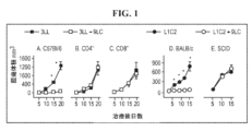
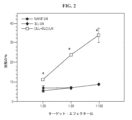

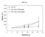
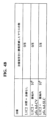
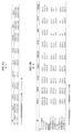
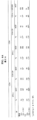

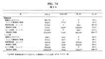

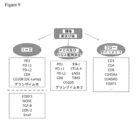
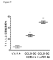
Classifications
-
- C—CHEMISTRY; METALLURGY
- C07—ORGANIC CHEMISTRY
- C07K—PEPTIDES
- C07K14/00—Peptides having more than 20 amino acids; Gastrins; Somatostatins; Melanotropins; Derivatives thereof
- C07K14/435—Peptides having more than 20 amino acids; Gastrins; Somatostatins; Melanotropins; Derivatives thereof from animals; from humans
- C07K14/52—Cytokines; Lymphokines; Interferons
- C07K14/521—Chemokines
-
- A—HUMAN NECESSITIES
- A61—MEDICAL OR VETERINARY SCIENCE; HYGIENE
- A61K—PREPARATIONS FOR MEDICAL, DENTAL OR TOILETRY PURPOSES
- A61K38/00—Medicinal preparations containing peptides
- A61K38/16—Peptides having more than 20 amino acids; Gastrins; Somatostatins; Melanotropins; Derivatives thereof
- A61K38/17—Peptides having more than 20 amino acids; Gastrins; Somatostatins; Melanotropins; Derivatives thereof from animals; from humans
- A61K38/19—Cytokines; Lymphokines; Interferons
- A61K38/195—Chemokines, e.g. RANTES
-
- A—HUMAN NECESSITIES
- A61—MEDICAL OR VETERINARY SCIENCE; HYGIENE
- A61K—PREPARATIONS FOR MEDICAL, DENTAL OR TOILETRY PURPOSES
- A61K39/00—Medicinal preparations containing antigens or antibodies
- A61K39/0005—Vertebrate antigens
- A61K39/0011—Cancer antigens
-
- A—HUMAN NECESSITIES
- A61—MEDICAL OR VETERINARY SCIENCE; HYGIENE
- A61K—PREPARATIONS FOR MEDICAL, DENTAL OR TOILETRY PURPOSES
- A61K39/00—Medicinal preparations containing antigens or antibodies
- A61K39/395—Antibodies; Immunoglobulins; Immune serum, e.g. antilymphocytic serum
- A61K39/39533—Antibodies; Immunoglobulins; Immune serum, e.g. antilymphocytic serum against materials from animals
- A61K39/3955—Antibodies; Immunoglobulins; Immune serum, e.g. antilymphocytic serum against materials from animals against proteinaceous materials, e.g. enzymes, hormones, lymphokines
-
- A—HUMAN NECESSITIES
- A61—MEDICAL OR VETERINARY SCIENCE; HYGIENE
- A61K—PREPARATIONS FOR MEDICAL, DENTAL OR TOILETRY PURPOSES
- A61K39/00—Medicinal preparations containing antigens or antibodies
- A61K39/395—Antibodies; Immunoglobulins; Immune serum, e.g. antilymphocytic serum
- A61K39/39533—Antibodies; Immunoglobulins; Immune serum, e.g. antilymphocytic serum against materials from animals
- A61K39/39558—Antibodies; Immunoglobulins; Immune serum, e.g. antilymphocytic serum against materials from animals against tumor tissues, cells, antigens
-
- A—HUMAN NECESSITIES
- A61—MEDICAL OR VETERINARY SCIENCE; HYGIENE
- A61K—PREPARATIONS FOR MEDICAL, DENTAL OR TOILETRY PURPOSES
- A61K39/00—Medicinal preparations containing antigens or antibodies
- A61K39/46—Cellular immunotherapy
- A61K39/461—Cellular immunotherapy characterised by the cell type used
- A61K39/4615—Dendritic cells
-
- A—HUMAN NECESSITIES
- A61—MEDICAL OR VETERINARY SCIENCE; HYGIENE
- A61K—PREPARATIONS FOR MEDICAL, DENTAL OR TOILETRY PURPOSES
- A61K39/00—Medicinal preparations containing antigens or antibodies
- A61K39/46—Cellular immunotherapy
- A61K39/462—Cellular immunotherapy characterized by the effect or the function of the cells
- A61K39/4622—Antigen presenting cells
-
- A—HUMAN NECESSITIES
- A61—MEDICAL OR VETERINARY SCIENCE; HYGIENE
- A61K—PREPARATIONS FOR MEDICAL, DENTAL OR TOILETRY PURPOSES
- A61K39/00—Medicinal preparations containing antigens or antibodies
- A61K39/46—Cellular immunotherapy
- A61K39/463—Cellular immunotherapy characterised by recombinant expression
- A61K39/4636—Immune checkpoint inhibitors
-
- A—HUMAN NECESSITIES
- A61—MEDICAL OR VETERINARY SCIENCE; HYGIENE
- A61K—PREPARATIONS FOR MEDICAL, DENTAL OR TOILETRY PURPOSES
- A61K39/00—Medicinal preparations containing antigens or antibodies
- A61K39/46—Cellular immunotherapy
- A61K39/464—Cellular immunotherapy characterised by the antigen targeted or presented
- A61K39/4643—Vertebrate antigens
- A61K39/4644—Cancer antigens
- A61K39/464436—Cytokines
- A61K39/464442—Chemokines
-
- A—HUMAN NECESSITIES
- A61—MEDICAL OR VETERINARY SCIENCE; HYGIENE
- A61K—PREPARATIONS FOR MEDICAL, DENTAL OR TOILETRY PURPOSES
- A61K45/00—Medicinal preparations containing active ingredients not provided for in groups A61K31/00 - A61K41/00
- A61K45/06—Mixtures of active ingredients without chemical characterisation, e.g. antiphlogistics and cardiaca
-
- A—HUMAN NECESSITIES
- A61—MEDICAL OR VETERINARY SCIENCE; HYGIENE
- A61K—PREPARATIONS FOR MEDICAL, DENTAL OR TOILETRY PURPOSES
- A61K48/00—Medicinal preparations containing genetic material which is inserted into cells of the living body to treat genetic diseases; Gene therapy
-
- A—HUMAN NECESSITIES
- A61—MEDICAL OR VETERINARY SCIENCE; HYGIENE
- A61K—PREPARATIONS FOR MEDICAL, DENTAL OR TOILETRY PURPOSES
- A61K9/00—Medicinal preparations characterised by special physical form
- A61K9/0012—Galenical forms characterised by the site of application
- A61K9/0019—Injectable compositions; Intramuscular, intravenous, arterial, subcutaneous administration; Compositions to be administered through the skin in an invasive manner
-
- A—HUMAN NECESSITIES
- A61—MEDICAL OR VETERINARY SCIENCE; HYGIENE
- A61P—SPECIFIC THERAPEUTIC ACTIVITY OF CHEMICAL COMPOUNDS OR MEDICINAL PREPARATIONS
- A61P35/00—Antineoplastic agents
-
- C—CHEMISTRY; METALLURGY
- C07—ORGANIC CHEMISTRY
- C07K—PEPTIDES
- C07K16/00—Immunoglobulins [IGs], e.g. monoclonal or polyclonal antibodies
- C07K16/18—Immunoglobulins [IGs], e.g. monoclonal or polyclonal antibodies against material from animals or humans
- C07K16/24—Immunoglobulins [IGs], e.g. monoclonal or polyclonal antibodies against material from animals or humans against cytokines, lymphokines or interferons
- C07K16/249—Interferons
-
- C—CHEMISTRY; METALLURGY
- C07—ORGANIC CHEMISTRY
- C07K—PEPTIDES
- C07K16/00—Immunoglobulins [IGs], e.g. monoclonal or polyclonal antibodies
- C07K16/18—Immunoglobulins [IGs], e.g. monoclonal or polyclonal antibodies against material from animals or humans
- C07K16/28—Immunoglobulins [IGs], e.g. monoclonal or polyclonal antibodies against material from animals or humans against receptors, cell surface antigens or cell surface determinants
- C07K16/2803—Immunoglobulins [IGs], e.g. monoclonal or polyclonal antibodies against material from animals or humans against receptors, cell surface antigens or cell surface determinants against the immunoglobulin superfamily
- C07K16/2818—Immunoglobulins [IGs], e.g. monoclonal or polyclonal antibodies against material from animals or humans against receptors, cell surface antigens or cell surface determinants against the immunoglobulin superfamily against CD28 or CD152
-
- C—CHEMISTRY; METALLURGY
- C12—BIOCHEMISTRY; BEER; SPIRITS; WINE; VINEGAR; MICROBIOLOGY; ENZYMOLOGY; MUTATION OR GENETIC ENGINEERING
- C12N—MICROORGANISMS OR ENZYMES; COMPOSITIONS THEREOF; PROPAGATING, PRESERVING, OR MAINTAINING MICROORGANISMS; MUTATION OR GENETIC ENGINEERING; CULTURE MEDIA
- C12N7/00—Viruses; Bacteriophages; Compositions thereof; Preparation or purification thereof
-
- A—HUMAN NECESSITIES
- A61—MEDICAL OR VETERINARY SCIENCE; HYGIENE
- A61K—PREPARATIONS FOR MEDICAL, DENTAL OR TOILETRY PURPOSES
- A61K39/00—Medicinal preparations containing antigens or antibodies
- A61K2039/51—Medicinal preparations containing antigens or antibodies comprising whole cells, viruses or DNA/RNA
- A61K2039/515—Animal cells
- A61K2039/5154—Antigen presenting cells [APCs], e.g. dendritic cells or macrophages
-
- A—HUMAN NECESSITIES
- A61—MEDICAL OR VETERINARY SCIENCE; HYGIENE
- A61K—PREPARATIONS FOR MEDICAL, DENTAL OR TOILETRY PURPOSES
- A61K39/00—Medicinal preparations containing antigens or antibodies
- A61K2039/51—Medicinal preparations containing antigens or antibodies comprising whole cells, viruses or DNA/RNA
- A61K2039/525—Virus
- A61K2039/5256—Virus expressing foreign proteins
-
- A—HUMAN NECESSITIES
- A61—MEDICAL OR VETERINARY SCIENCE; HYGIENE
- A61K—PREPARATIONS FOR MEDICAL, DENTAL OR TOILETRY PURPOSES
- A61K39/00—Medicinal preparations containing antigens or antibodies
- A61K2039/54—Medicinal preparations containing antigens or antibodies characterised by the route of administration
-
- A—HUMAN NECESSITIES
- A61—MEDICAL OR VETERINARY SCIENCE; HYGIENE
- A61K—PREPARATIONS FOR MEDICAL, DENTAL OR TOILETRY PURPOSES
- A61K39/00—Medicinal preparations containing antigens or antibodies
- A61K2039/545—Medicinal preparations containing antigens or antibodies characterised by the dose, timing or administration schedule
-
- A—HUMAN NECESSITIES
- A61—MEDICAL OR VETERINARY SCIENCE; HYGIENE
- A61K—PREPARATIONS FOR MEDICAL, DENTAL OR TOILETRY PURPOSES
- A61K2239/00—Indexing codes associated with cellular immunotherapy of group A61K39/46
- A61K2239/31—Indexing codes associated with cellular immunotherapy of group A61K39/46 characterized by the route of administration
-
- A—HUMAN NECESSITIES
- A61—MEDICAL OR VETERINARY SCIENCE; HYGIENE
- A61K—PREPARATIONS FOR MEDICAL, DENTAL OR TOILETRY PURPOSES
- A61K2239/00—Indexing codes associated with cellular immunotherapy of group A61K39/46
- A61K2239/38—Indexing codes associated with cellular immunotherapy of group A61K39/46 characterised by the dose, timing or administration schedule
-
- A—HUMAN NECESSITIES
- A61—MEDICAL OR VETERINARY SCIENCE; HYGIENE
- A61K—PREPARATIONS FOR MEDICAL, DENTAL OR TOILETRY PURPOSES
- A61K2239/00—Indexing codes associated with cellular immunotherapy of group A61K39/46
- A61K2239/46—Indexing codes associated with cellular immunotherapy of group A61K39/46 characterised by the cancer treated
- A61K2239/55—Lung
-
- C—CHEMISTRY; METALLURGY
- C07—ORGANIC CHEMISTRY
- C07K—PEPTIDES
- C07K2317/00—Immunoglobulins specific features
- C07K2317/70—Immunoglobulins specific features characterized by effect upon binding to a cell or to an antigen
- C07K2317/76—Antagonist effect on antigen, e.g. neutralization or inhibition of binding
-
- C—CHEMISTRY; METALLURGY
- C12—BIOCHEMISTRY; BEER; SPIRITS; WINE; VINEGAR; MICROBIOLOGY; ENZYMOLOGY; MUTATION OR GENETIC ENGINEERING
- C12N—MICROORGANISMS OR ENZYMES; COMPOSITIONS THEREOF; PROPAGATING, PRESERVING, OR MAINTAINING MICROORGANISMS; MUTATION OR GENETIC ENGINEERING; CULTURE MEDIA
- C12N2710/00—MICROORGANISMS OR ENZYMES; COMPOSITIONS THEREOF; PROPAGATING, PRESERVING, OR MAINTAINING MICROORGANISMS; MUTATION OR GENETIC ENGINEERING; CULTURE MEDIA dsDNA viruses
- C12N2710/00011—Details
- C12N2710/10011—Adenoviridae
- C12N2710/10041—Use of virus, viral particle or viral elements as a vector
- C12N2710/10043—Use of virus, viral particle or viral elements as a vector viral genome or elements thereof as genetic vector
-
- C—CHEMISTRY; METALLURGY
- C12—BIOCHEMISTRY; BEER; SPIRITS; WINE; VINEGAR; MICROBIOLOGY; ENZYMOLOGY; MUTATION OR GENETIC ENGINEERING
- C12N—MICROORGANISMS OR ENZYMES; COMPOSITIONS THEREOF; PROPAGATING, PRESERVING, OR MAINTAINING MICROORGANISMS; MUTATION OR GENETIC ENGINEERING; CULTURE MEDIA
- C12N2710/00—MICROORGANISMS OR ENZYMES; COMPOSITIONS THEREOF; PROPAGATING, PRESERVING, OR MAINTAINING MICROORGANISMS; MUTATION OR GENETIC ENGINEERING; CULTURE MEDIA dsDNA viruses
- C12N2710/00011—Details
- C12N2710/10011—Adenoviridae
- C12N2710/10071—Demonstrated in vivo effect
Description
関連出願
本願は、2014年11月5日に出願された米国仮特許出願第62/075,532号の優先権を主張する。その内容は参照により本明細書において援用する。
Related Application This application claims priority to US Provisional Patent Application No. 62/075,532, filed November 5, 2014. The contents of which are incorporated herein by reference.
発明の分野
本発明は、癌などの病理学的状態に関連するものを含めた哺乳動物生理学的プロセスを調節するために二次リンパ器官ケモカインを使用する方法に関する。
FIELD OF THE INVENTION The present invention relates to methods of using secondary lymphoid organ chemokines to modulate mammalian physiological processes, including those associated with pathological conditions such as cancer.
発明の背景
発癌性、癌退行、再発及び転移に影響を与える免疫機構を理解することは、新規免疫療法の開発の重要な側面である。この状況において、当業者であれば、免疫応答の基本的な側面は、生物の免疫細胞が自己免疫抗原と非自己抗原とを区別することができることであると理解する。したがって、癌における免疫機構を解明しようとする臨床関連モデルは、腫瘍細胞が宿主免疫系の細胞と遺伝的背景を共有する(すなわち、同系である)という事実を考慮しなければならない。残念ながら、動物に癌細胞系を導入する癌の多くの動物モデルは、宿主動物の遺伝的背景と評価されている癌細胞系との間の差異に影響を受ける免疫応答によって混乱を来してしまう。具体的には、宿主動物及び癌細胞株が本質的に同一の遺伝的背景を共有しない癌モデルでは、個体間での移植臓器の拒絶反応に見られるものに類似する宿主の免疫系による「非自己」免疫応答に関連する問題を含めて、様々な問題がある。自発癌細胞上の非自己抗原(癌では当然には生じない現象)の宿主免疫系認識から生じる場合のある非自己免疫応答は、ヒト癌では起こらない癌細胞に対する免疫応答を生じさせる。したがって、癌の発生及び進行を忠実に模倣し、それによって免疫機構の臨床的に関連する分析を行うことができるようにする癌モデルが絶えず必要とされている。
BACKGROUND OF THE INVENTION Understanding the immune mechanisms that influence carcinogenesis, cancer regression, recurrence and metastasis is an important aspect of the development of novel immunotherapies. In this context, those skilled in the art understand that a fundamental aspect of the immune response is the ability of an organism's immune cells to distinguish between autoimmune and non-self antigens. Therefore, clinically relevant models seeking to elucidate immune mechanisms in cancer must take into account the fact that tumor cells share a genetic background (ie, are syngeneic) with cells of the host immune system. Unfortunately, many animal models of cancer that introduce cancer cell lines into animals are confounded by immune responses influenced by differences between the genetic background of the host animal and the cancer cell line being evaluated. put away. Specifically, in cancer models in which the host animal and cancer cell line do not share essentially the same genetic background, the host's immune system is responsible for "non-human" responses similar to those seen in transplanted organ rejection among individuals. There are a variety of problems, including those associated with auto'immune responses. Non-autoimmune responses, which may result from host immune system recognition of non-self antigens on spontaneous cancer cells (a phenomenon that does not occur naturally in cancer), generate an immune response against cancer cells that does not occur in human cancers. Therefore, there is a continuing need for cancer models that faithfully mimic cancer development and progression, thereby allowing clinically relevant analyzes of the immune system to be performed.
腫瘍細胞に対する効果的な免疫応答は、APCエフェクター及びリンパ球エフェクターの両方を必要とする(例えば、Huang外,Science,264:961-965,1994参照)。腫瘍細胞は、MHC抗原の発現が制限され、共刺激分子を欠失する場合が多いため、それらは効果的でないAPCである(例えば、Restifo外,J.Exp.Med.,177:265-272,1993参照)。さらに、腫瘍細胞は、宿主免疫監視の回避に寄与する免疫抑制メディエーターを分泌する(例えば、Huang外,Cancer Res.,58:1208-1216,1998;Sharma外,J.Immunol.,163:5020-5028,1999;及びUzzo,J.Clin.Investig.,104:769-776,1999参照)。この問題を回避するために、研究者は、生体外で生成されたDCを使用して生体内で抗腫瘍免疫応答を刺激している。実験マウスモデルでは、腫瘍関連抗原ペプチドがパルスされたDC(Nair外,Eur.J.Immunol.,27:589-597,1997)や腫瘍RNAがトランスフェクトされたDCは、生体内で抗原特異的抗腫瘍応答を誘導することが示されている(Boczkowski外,J.Exp.Med.,184:465-472,1996)。同様に、DCと腫瘍細胞との融合又はサイトカイン修飾DCの腫瘍内注射も、抗腫瘍免疫を増強することが示されている(Gong外,Nat.Med.,3:558-561,1997;Celluzzi外,J.Immunol.,160:3081-3085,1998;Miller外,Hum.Gene Ther.,11:53-65,2000)。結果として、効果的な抗癌免疫は、腫瘍抗原提示のために特殊分化宿主APCを動員して特異的T細胞活性化を促進することによって達成できることが示唆されている(Soto外,Annu.Rev.Immunol.,15:675-705,1997)。したがって、DC及びリンパ球エフェクターの両方をリンパ節及び腫瘍部位に引きつけるケモカインは、癌免疫療法において強力な薬剤として機能することがきると考えられる。 An effective immune response against tumor cells requires both APC effectors and lymphocyte effectors (see, eg, Huang et al., Science, 264:961-965, 1994). Because tumor cells often have restricted expression of MHC antigens and lack co-stimulatory molecules, they are ineffective APCs (eg, Restifo et al., J. Exp. Med., 177:265-272). , 1993). In addition, tumor cells secrete immunosuppressive mediators that contribute to evading host immune surveillance (see, eg, Huang et al., Cancer Res., 58:1208-1216, 1998; Sharma et al., J. Immunol., 163:5020- 5028, 1999; and Uzzo, J. Clin. Investig., 104:769-776, 1999). To circumvent this problem, researchers have used ex vivo-generated DCs to stimulate anti-tumor immune responses in vivo. In experimental mouse models, DCs pulsed with tumor-associated antigenic peptides (Nair et al., Eur. J. Immunol., 27:589-597, 1997) and DCs transfected with tumor RNA demonstrated antigen-specific activity in vivo. It has been shown to induce anti-tumor responses (Boczkowski et al., J. Exp. Med., 184:465-472, 1996). Similarly, fusion of DCs with tumor cells or intratumoral injection of cytokine-modified DCs has also been shown to enhance anti-tumor immunity (Gong et al., Nat. Med., 3:558-561, 1997; Celluzzi et al., J. Immunol., 160:3081-3085, 1998; Miller et al., Hum. Gene Ther., 11:53-65, 2000). Consequently, it has been suggested that effective anti-cancer immunity can be achieved by recruiting specialized host APCs for tumor antigen presentation to promote specific T-cell activation (Soto et al., Annu. Rev. Immunol., 15:675-705, 1997). Therefore, chemokines that attract both DCs and lymphocyte effectors to lymph nodes and tumor sites could serve as potent agents in cancer immunotherapy.
相同であるが機能的に異なるタンパク質の一群であるケモカインは、白血球の移動及び活性化を直接仲介し、血管新生の調節に所定の役割を果たす(Baggiolini外,Rev.Immunol.,15:675-705,1997)。また、ケモカインは、免疫ホメオスタシス及び二次リンパ器官の構造を維持するように機能する(Jung外,Curr.Opin.Immunol.,11:319-325,1999)。いくつかのケモカインは、抗腫瘍活性を有することが知られている。腫瘍細胞がMIP1α、RANTES、リンホタクチン、TCA3、JE/MCP-1/MCAF、MIP3α、MIP3β及びIP-10を含めたケモカインで修飾された様々なマウス腫瘍モデルにおいて腫瘍拒絶が注目されている(Lustre外,J.Exp.Med.,178:1057-1065,1993;Bottazzi外,J.Immunol.,148:1280-1285,1992;Kellermann外,J.Immunol.,162:3859-3864,1999;Sallusto外,Eur.J.Immunol.,28:2760-2769,1998;Sozzani外,J.Immunol.,161:1083-1086,1998;Dieu外,J.Exp.Med.,188:373-386,1998;Campell外,J.Cell Biol.,141:1053-1059,1998;Saeki外,J.Immunol.,162:2472-2475,1991;Nagira外,Eur.J.Immunol.,28:1516-1523,1998)。 Chemokines, a group of homologous but functionally distinct proteins, directly mediate leukocyte migration and activation and play a role in the regulation of angiogenesis (Baggiolini et al., Rev. Immunol., 15:675- 705, 1997). Chemokines also function to maintain immune homeostasis and the structure of secondary lymphoid organs (Jung et al., Curr. Opin. Immunol., 11:319-325, 1999). Several chemokines are known to have anti-tumor activity. Tumor rejection has been noted in various mouse tumor models in which tumor cells have been modified with chemokines including MIP1α, RANTES, lymphotactin, TCA3, JE/MCP-1/MCAF, MIP3α, MIP3β and IP-10 (Lustre et al. Bottazzi et al., J. Immunol., 148:1280-1285, 1992; Kellermann et al., J. Immunol., 162:3859-3864, 1999; Sozzani et al., J. Immunol., 161:1083-1086, 1998; Dieu et al., J. Exp. Med., 188:373-386, 1998; Campell et al., J. Cell Biol., 141:1053-1059, 1998; Saeki et al., J. Immunol., 162:2472-2475, 1991; ).
二次リンパ組織ケモカイン(SLC、Exodus2又は6Ckineとも呼ばれる)は、ナイーブT細胞及びDCを強力に誘引する、高内皮細静脈並びに脾臓及びリンパ節のT細胞ゾーンで通常発現される高内皮由来CCケモカインである(Cyster外,J.Exp.Med.,189:447-450,1999.24;Ogata外,Blood,93:3225-3232,1999;Chan外,Blood,93:3610-3616,1999;Hedrick外,J.Immunol.,159:1589-1593,1997;Hromas外,J.Immunol.,159:2554-2558,1997;Nagira外,J.Biol.Chem.,272:19518-19524,1997;Tanabe外,J.Immunol.,159:5671-5679,1997;Willimann外,Eur.J.Immunol.,28:2025-2034,1998)。SLCは、2つの特異的Gタンパク質共役型7回膜貫通ドメインケモカイン受容体のCCR7及びCXCR3を介してその効果を仲介する(Yoshida外,J.Biol.Chem.273:7118;Jenh外,J.Immunol.162:3765)。CCR7はナイーブT細胞及び成熟DC上で発現されるのに対し、CXCR3は記憶表現型を有するTh1サイトカイン産生リンパ球上で優先的に発現される(Yoshida外,J.Biol.Chem.273:7118;Jenh外,J.Immunol.162:3765)。 Secondary lymphoid tissue chemokines (SLC, also called Exodus2 or 6Ckine) are high endothelial-derived CC chemokines normally expressed in high endothelial venules and T-cell zones of the spleen and lymph nodes that potently attract naive T cells and DCs. (Cyster et al., J. Exp. Med., 189:447-450, 1999.24; Ogata et al., Blood, 93:3225-3232, 1999; Chan et al., Blood, 93:3610-3616, 1999; Hedrick Hromas et al., J. Immunol., 159:2554-2558, 1997; Nagira et al., J. Biol. et al., J. Immunol., 159:5671-5679, 1997; Willimann et al., Eur. J. Immunol., 28:2025-2034, 1998). SLC mediates its effects through CCR7 and CXCR3, two specific G protein-coupled seven-transmembrane domain chemokine receptors (Yoshida et al., J. Biol. Chem. 273:7118; Jenh et al., J. Am. Immunol. 162:3765). CCR7 is expressed on naive T cells and mature DCs, whereas CXCR3 is preferentially expressed on Th1 cytokine-producing lymphocytes with a memory phenotype (Yoshida et al., J. Biol. Chem. 273:7118 Jenh et al., J. Immunol. 162:3765).
DCを化学結合させるSLCの能力(Kellermann外,J.Immunol.,162:3859-3864,1999)は、他のケモカインと共有される特性である(Sallusto外,Eur.J.Immunol.,28:2760-2769,1998;Sozzani外,J.Immunol.,161:1083-1086,1998;Dieu外,J.Exp.Med.,188:373-386,1998)。しかし、SLCは、生体内での1型サイトカイン応答を誘発することができるため、明らかに有利な場合がある(Sharma外,J.Immunol.,164:4558-4563,2000)。DCは、免疫応答の開始に関与するユニークに強力なAPCである(Banchereau外,Nature(Lond.),392:245-252,1998)。免疫系のセンチネルとして機能するDCは、末梢における抗原の獲得、その後、特異的な免疫応答を開始するリンパ器官におけるT細胞領域への輸送を担当する。SLCは、ナイーブリンパ球及び抗原刺激DCの両方を二次リンパ器官のT細胞ゾーンに動員し、これらの初期免疫応答成分を共局在化させ、同族T細胞活性化を達成する(Cyster外,J.Exp.Med.,189:447-450,1999.24)。
The ability of SLC to chemically bind DCs (Kellermann et al., J. Immunol., 162:3859-3864, 1999) is a property shared with other chemokines (Sallusto et al., Eur. J. Immunol., 28: 2760-2769, 1998; Sozzani et al., J. Immunol., 161:1083-1086, 1998; Dieu et al., J. Exp. Med., 188:373-386, 1998). However, SLC may have a clear advantage because it can induce
当該技術分野においては、例えば宿主サイトカインプロファイルがSLCによってどのように調節されるのかのみならず、SLCが同系癌細胞に対する効果的な細胞仲介免疫応答を編成する能力を試験するために癌における免疫機構を忠実に模倣する癌モデルが必要とされている。さらに、このような臨床関連モデルに基づく免疫機能並びに免疫療法モダリティの新たなアッセイが必要とされている。ここで提供される発明は、これらのニーズを満たすものである。 The art is not only interested in how the host cytokine profile is modulated by SLC, but also the immune mechanisms in cancer to test the ability of SLC to orchestrate an effective cell-mediated immune response against syngeneic cancer cells. There is a need for cancer models that faithfully mimic . In addition, new assays of immune function and immunotherapeutic modalities based on such clinically relevant models are needed. The invention provided herein meets these needs.
発明の概要
本明細書に開示される発明は、本質的に同一の遺伝的背景を有する宿主動物及び癌細胞を利用することによって、癌における免疫機構を忠実に模倣する動物モデルを提供する。これらのモデルは、SLCが同系癌細胞に対する効果的な細胞仲介免疫応答を編成する能力を実証するために使用される。さらに、これらのモデルを使用して、同系癌細胞に対するSLC調節免疫応答に関連する宿主サイトカインプロファイルを評価することができる。
SUMMARY OF THE INVENTION The invention disclosed herein provides an animal model that faithfully mimics the immune system in cancer by utilizing host animals and cancer cells that have essentially the same genetic background. These models are used to demonstrate the ability of SLCs to orchestrate effective cell-mediated immune responses against syngeneic cancer cells. Additionally, these models can be used to assess host cytokine profiles associated with SLC-modulated immune responses against syngeneic cancer cells.
本明細書に開示するように、二次リンパ器官ケモカインの抗腫瘍効率を、腫瘍を自発的に発生するトランスジェニックマウスを含めた多数の同系モデルにおいて評価した。これらのトランスジェニックマウスでは、両側多巣性肺腺癌が臓器特異的に発生する。当該技術分野において知られている同種モデルと比較して、このトランスジェニックマウスモデルにおいて生じる自発性腫瘍は非自己抗原を発現しないため、ヒト癌に似ている。 As disclosed herein, the anti-tumor efficacy of secondary lymphoid organ chemokines was evaluated in a number of syngeneic models, including transgenic mice that spontaneously develop tumors. Bilateral multifocal lung adenocarcinoma develops organ-specifically in these transgenic mice. Compared to allogeneic models known in the art, the spontaneous tumors that arise in this transgenic mouse model do not express non-self antigens and thus resemble human cancers.
本明細書で開示される同系モデルにおいて、腫瘍内及び/又は腋窩リンパ節領域内における組換えSLCの注入により、腫瘍の広範囲にわたるリンパ球及びDC浸潤が著しく減少し、生存率が向上した。これらの同系ネズミモデルにSLC注射をすると、腫瘍部位、リンパ節及び脾臓でCD4及びCD8リンパ球並びにDCが有意に増加した。細胞浸潤は、1型サイトカイン及び抗血管新生ケモカインIFN-γ誘導性タンパク質10、及びIFN-γ(MIG)によって誘導されるモノカインの同化作用の向上を伴った。対照的に、免疫抑制性サイトカイン形質転換増殖因子βのリンパ節及び腫瘍部位産生は、SLC処置に応答して減少した。試験管内では、照射自己腫瘍による刺激後に、SLC処置マウスからの脾細胞は、より多くのIFN-γ及び顆粒球マクロファージコロニー刺激因子を有意に分泌したが、インターロイキン10のレベルを低下させた。腫瘍が器官特異的に発生するモデルにおける腫瘍負荷の有意な減少は、腫瘍免疫及び癌免疫療法を調節する際のSLCの使用方法を提供する。
In the syngeneic model disclosed herein, injection of recombinant SLC into the tumor and/or into the axillary lymph node region markedly reduced extensive lymphocyte and DC infiltration of the tumor and improved survival. SLC injection into these syngeneic murine models resulted in significant increases in CD4 and CD8 lymphocytes and DCs at the tumor site, lymph nodes and spleen. Cellular infiltration was accompanied by enhanced anabolic effects of monokines induced by the
本明細書で開示される発明は多数の実施形態を有する。本発明の典型的な実施形態は、哺乳動物に、癌細胞の増殖を阻害するのに十分な量の二次リンパ組織ケモカイン(SLC)ポリペプチドを投与することによって哺乳動物における自発癌の増殖を阻害する方法である。好ましい方法では、SLCは配列番号1で示されるポリペプチド配列を有する。これらの方法では、SLCポリペプチドは、典型的には、腫瘍内注射又はリンパ節注射によって哺乳動物に全身的に投与される。さらに別の投与態様では、SLCポリペプチドをコードするポリヌクレオチドを有する発現ベクターを哺乳動物に投与し、SLCポリペプチドは、SLC発現ベクターで形質導入された哺乳動物細胞によって産生される。 The invention disclosed herein has numerous embodiments. An exemplary embodiment of the invention inhibits spontaneous cancer growth in a mammal by administering to the mammal an amount of a secondary lymphoid tissue chemokine (SLC) polypeptide sufficient to inhibit cancer cell growth. It is a method of hindering. In a preferred method, the SLC has the polypeptide sequence shown in SEQ ID NO:1. In these methods, SLC polypeptides are administered systemically to mammals, typically by intratumoral or lymph node injection. In yet another mode of administration, an expression vector having a polynucleotide encoding an SLC polypeptide is administered to a mammal, and the SLC polypeptide is produced by mammalian cells transduced with the SLC expression vector.
本発明の関連する実施形態は、哺乳動物に二次リンパ組織ケモカイン(SLC)を投与することを含む、哺乳類における同系癌細胞(最も好ましくは自発的癌細胞)の増殖を阻害する方法であって、配列番号1に示されるSLCをコードするポリヌクレオチドを哺乳類の細胞に形質導入することによってSLCを哺乳動物に投与し、それによって、該形質導入された細胞が癌細胞の増殖を阻害するのに十分な量でSLCポリペプチドを発現するする方法である。好ましくは、ベクターは、腫瘍内注射又はリンパ節内注射によって哺乳動物に全身的に投与される。 A related embodiment of the invention is a method of inhibiting the growth of syngeneic cancer cells (most preferably spontaneous cancer cells) in a mammal comprising administering to the mammal a secondary lymphoid tissue chemokine (SLC) comprising: , administering SLC to a mammal by transducing mammalian cells with a polynucleotide encoding SLC shown in SEQ ID NO: 1, whereby the transduced cells inhibit the growth of cancer cells. A method for expressing SLC polypeptides in sufficient quantities. Preferably, the vector is administered systemically to the mammal by intratumoral or intralymphatic injection.
本発明の別の実施形態は、哺乳動物又は哺乳動物に由来する細胞の集団におけるサイトカイン発現を、同系の腫瘍細胞の増殖を阻害するのに十分な量の二次リンパ系組織ケモカイン(SLC)ポリペプチドに該細胞集団を曝露することによって達成する又は調節する(例えば、既存のサイトカインプロファイルを変化させる)方法である。本明細書に開示されるように、本明細書中に開示される同系モデルは、SLCの添加がどのようにサイトカイン発現を協調的に調節し、かつ、腫瘍細胞の増殖を阻害するのかを実証するため、これらの現象の観察(サイトカイン発現の調節及び腫瘍増殖の阻害)は、潜在的な免疫刺激性化合物又は免疫阻害性試験化合物の効果を評価するように設計された細胞系アッセイで使用できる。 Another embodiment of the present invention modifies cytokine expression in a mammal or a population of mammal-derived cells with a secondary lymphoid tissue chemokine (SLC) polysaccharide in an amount sufficient to inhibit the growth of syngeneic tumor cells. A method of achieving or modulating (eg, altering an existing cytokine profile) by exposing the cell population to peptides. As disclosed herein, the syngeneic model disclosed herein demonstrates how the addition of SLC coordinates cytokine expression and inhibits tumor cell proliferation. Therefore, observation of these phenomena (modulation of cytokine expression and inhibition of tumor growth) can be used in cell-based assays designed to assess the effects of potential immunostimulatory or immunoinhibitory test compounds. .
本発明の別の実施形態は、同系哺乳動物細胞の集団におけるインターフェロン-γ(IFN-γ)ポリペプチドの発現の増加及び形質転換増殖因子-β(TGF-β)ポリペプチドの発現の減少を、CD8陽性T細胞、CD4陽性T細胞、抗原提示細胞及び腫瘍細胞を含む腫瘍細胞の増殖を阻害するのに十分な量の二次リンパ組織ケモカイン(SLC)ポリペプチドに該細胞集団を曝露することによって達成する方法である。好ましい方法では、酵素結合免疫吸着(ELISA)アッセイによって測定したときに、インターフェロン-γ(IFN-γ)ポリペプチドの発現の増加は少なくとも約2倍であり、形質転換増殖因子-β(TGF-β)ポリペプチドの発現の減少は少なくとも約2倍である。 Another embodiment of the invention provides for increased expression of interferon-γ (IFN-γ) polypeptide and decreased expression of transforming growth factor-β (TGF-β) polypeptide in a population of syngeneic mammalian cells, by exposing the cell population to a secondary lymphoid tissue chemokine (SLC) polypeptide in an amount sufficient to inhibit proliferation of tumor cells, including CD8 positive T cells, CD4 positive T cells, antigen presenting cells and tumor cells; is the way to achieve it. In preferred methods, the increase in expression of interferon-gamma (IFN-γ) polypeptide is at least about 2-fold and transforming growth factor-β (TGF-β) as measured by an enzyme-linked immunosorbent (ELISA) assay. ) The decrease in expression of the polypeptide is at least about 2-fold.
様々な実施形態では、本発明は、被験体に(i)SLCポリペプチド、(ii)SLCポリペプチドをコードするポリヌクレオチド、(iii)ポリヌクレオチドを含む細胞、又は(iv)それらの組み合わせを投与し、そして該被験体に免疫チェックポイント阻害剤を投与し、それによって該被験体において癌又は固形腫瘍を治療することを含む、被験体において癌又は固形腫瘍を治療する方法も提供する。 In various embodiments, the present invention administers to a subject (i) a SLC polypeptide, (ii) a polynucleotide encoding a SLC polypeptide, (iii) a cell comprising a polynucleotide, or (iv) a combination thereof. and administering an immune checkpoint inhibitor to said subject, thereby treating cancer or solid tumor in said subject.
また、腫瘍増殖若しくは腫瘍体積を減少させる方法、腫瘍進行を緩徐化若しくは低下させる方法、又は被験体における腫瘍再発を予防又は改善する方法であって、(i)SLCポリペプチド、(ii)SLCポリペプチドをコードするポリヌクレオチド、(iii)ポリヌクレオチドを含む細胞、又は(iv)それらの組み合わせを投与することを含む方法も意図される。 Also methods of reducing tumor growth or tumor volume, slowing or reducing tumor progression, or preventing or ameliorating tumor recurrence in a subject, comprising: (i) a SLC polypeptide, (ii) an SLC polypeptide, Also contemplated are methods comprising administering a polynucleotide encoding a peptide, (iii) a cell comprising the polynucleotide, or (iv) a combination thereof.
様々な実施形態では、免疫チェックポイント阻害剤は、CTLA-4阻害剤、CTLA-4受容体阻害剤、プログラム細胞死1(PD-1)阻害剤、PD1-L1阻害剤、PD1-L2阻害剤、4-1BB阻害剤、OX40阻害剤、リンパ球活性化遺伝子3(LAG-3)阻害剤、T細胞免疫グロブリン及びムチンドメイン3(TIM-3)阻害剤又はそれらの組み合わせよりなる群から選択される。 In various embodiments, the immune checkpoint inhibitor is a CTLA-4 inhibitor, CTLA-4 receptor inhibitor, programmed cell death 1 (PD-1) inhibitor, PD1-L1 inhibitor, PD1-L2 inhibitor , 4-1BB inhibitors, OX40 inhibitors, lymphocyte activation gene 3 (LAG-3) inhibitors, T cell immunoglobulin and mucin domain 3 (TIM-3) inhibitors, or combinations thereof be.
様々な実施形態では、免疫チェックポイント阻害剤は、CTLA-4、CTLA-4受容体、PD-1、PD1-L1、PD1-L2、4-1BB、OX40、LAG-3、TIM-3又はそれらの組み合わせの1種以上に特異的な抗体、任意にモノクローナル抗体である。 In various embodiments, the immune checkpoint inhibitor is CTLA-4, CTLA-4 receptor, PD-1, PD1-L1, PD1-L2, 4-1BB, OX40, LAG-3, TIM-3 or optionally a monoclonal antibody, specific for one or more of the combinations of
様々な実施形態では、免疫チェックポイント阻害剤は、CTLA-4、CTLA-4受容体、PD-1、PD1-L1、PD1-L2、4-1BB、OX40、LAG-3、TIM-3又はそれらの組み合わせの1種以上の活性を阻害する小分子阻害剤である。 In various embodiments, the immune checkpoint inhibitor is CTLA-4, CTLA-4 receptor, PD-1, PD1-L1, PD1-L2, 4-1BB, OX40, LAG-3, TIM-3 or is a small molecule inhibitor that inhibits one or more activities of a combination of
様々な実施形態では、免疫チェックポイント阻害剤は、CTLA-4阻害剤、任意にイピリムマブ又はトレミリムマブである。 In various embodiments, the immune checkpoint inhibitor is a CTLA-4 inhibitor, optionally ipilimumab or tremilimumab.
様々な実施形態では、免疫チェックポイント阻害剤は、ニボルマブ、ペムブロリズマブ、ピジリズマブ、ラムブロリズマブ、BMS-936559、アテゾリズマブ並びにAMP-224、AMP224、AUNP12、BGB108、MCLA134、MEDI0680、PDR001、REGN2810、SHR1210、STIA110X、STIA1110及びTSR042よりなる群から選択されるPD1阻害剤である。 In various embodiments, the immune checkpoint inhibitor is nivolumab, pembrolizumab, pidilizumab, lambrolizumab, BMS-936559, atezolizumab and AMP-224, AMP224, AUNP12, BGB108, MCLA134, MEDI0680, PDR001, REGN2810, SHR1210, ST IA110X, STIA1110 and TSR042.
様々な実施形態では、免疫チェックポイント阻害剤は、BMS-936559、MPDL3280A、MEDI-4736、MSB0010718C、ALN-PDL、BGBA317、KD033、KY1003、STIA100X、STIA1010、STIA1011、STIA1012及びSTIA1014よりなる群から選択されるPD1-L1阻害剤である。 In various embodiments, the immune checkpoint inhibitor is from the group consisting of BMS-936559, MPDL3280A, MEDI-4736, MSB0010718C, ALN-PDL, BGBA317, KD033, KY1003, STIA100X, STIA1010, STIA1011, STIA1012 and STIA1014 selected is a PD1-L1 inhibitor.
様々な実施形態において、4-1BBの免疫チェックポイント阻害剤は、4-1BBに特異的に結合するモノクローナル抗体であり、BMS-663513及びPF-05082566が挙げられるが、これらに限定されない。 In various embodiments, the immune checkpoint inhibitor of 4-1BB is a monoclonal antibody that specifically binds 4-1BB, including but not limited to BMS-663513 and PF-05082566.
様々な実施形態では、免疫チェックポイント阻害剤はOX40の阻害剤である。例示的な態様では、OX40の阻害剤は、OX40に特異的に結合するモノクローナル抗体である。 In various embodiments, the immune checkpoint inhibitor is an inhibitor of OX40. In an exemplary aspect, the inhibitor of OX40 is a monoclonal antibody that specifically binds OX40.
様々な実施形態において、SLCポリペプチドは、配列番号1又は配列番号2のアミノ酸配列を含む。 In various embodiments, the SLC polypeptide comprises the amino acid sequence of SEQ ID NO:1 or SEQ ID NO:2.
様々な実施形態では、SLCポリペプチドをコードするポリヌクレオチドをベクターに挿入し、このベクターを被験体に投与する。様々な実施形態では、ベクターはアデノウイルスベクターである。様々な実施形態では、アデノウイルスベクターは、複製欠損アデノウイルスベクターである。 In various embodiments, the polynucleotide encoding the SLC polypeptide is inserted into a vector and the vector is administered to the subject. In various embodiments, the vector is an adenoviral vector. In various embodiments, the adenoviral vector is a replication-defective adenoviral vector.
様々な実施形態では、SLCポリペプチドをコードするポリヌクレオチドを含む細胞は抗原提示細胞(APC)である。様々な実施形態において、APCは樹状細胞である。様々な実施形態において、樹状細胞は、被験体に対して自己由来である。 In various embodiments, the cells comprising polynucleotides encoding SLC polypeptides are antigen presenting cells (APCs). In various embodiments, APCs are dendritic cells. In various embodiments, the dendritic cells are autologous to the subject.
様々な実施形態では、SLCポリペプチドをコードするポリヌクレオチドを含む少なくとも又は約1×10⁶個の細胞を被験体に投与する。様々な実施形態では、細胞は、24時間以内に1×10⁶個の細胞当たり少なくとも約0.25ngのCCL21を産生する。 In various embodiments, at least or about 1×10 6 cells comprising a polynucleotide encoding a SLC polypeptide are administered to the subject. In various embodiments, the cells produce at least about 0.25 ng of CCL21 per 1×10 6 cells within 24 hours.
様々な実施形態では、被験体は固形腫瘍を含み、細胞を該被験体に腫瘍内投与する。様々な実施形態では、固形腫瘍は非小細胞肺癌(NSCLC)固形腫瘍である。 In various embodiments, the subject comprises a solid tumor and the cells are administered intratumorally to the subject. In various embodiments, the solid tumor is a non-small cell lung cancer (NSCLC) solid tumor.
様々な実施形態において、(i)SLCポリペプチド、(ii)SLCポリペプチドをコードするポリヌクレオチド、(iii)ポリヌクレオチドを含む細胞、又は(iv)それらの組み合わせを、免疫チェックポイント阻害剤の前に被験体に投与する。様々な実施形態では、(i)SLCポリペプチド、(ii)SLCポリペプチドをコードするポリヌクレオチド、(iii)ポリヌクレオチドを含む細胞、又は(iv)それらの組み合わせを、免疫チェックポイント阻害剤の約2週間前に被験体に投与する。 In various embodiments, (i) a SLC polypeptide, (ii) a polynucleotide encoding a SLC polypeptide, (iii) a cell comprising a polynucleotide, or (iv) a combination thereof is administered prior to an immune checkpoint inhibitor. administered to the subject on . In various embodiments, (i) a SLC polypeptide, (ii) a polynucleotide encoding a SLC polypeptide, (iii) a cell comprising the polynucleotide, or (iv) a combination thereof, is about Subjects are dosed 2 weeks prior.
様々な実施形態において、(i)SLCポリペプチド、(ii)SLCポリペプチドをコードするポリヌクレオチド、(iii)ポリヌクレオチドを含む細胞、又は(iv)それらの組み合わせを被験体に2回以上投与する。 In various embodiments, (i) a SLC polypeptide, (ii) a polynucleotide encoding a SLC polypeptide, (iii) a cell comprising a polynucleotide, or (iv) a combination thereof is administered to a subject two or more times. .
様々な実施形態において、(i)SLCポリペプチド、(ii)SLCポリペプチドをコードするポリヌクレオチド、(iii)ポリヌクレオチドを含む細胞、又は(iv)それらの組み合わせを1ヶ月に1回被験体に投与する。 In various embodiments, (i) a SLC polypeptide, (ii) a polynucleotide encoding a SLC polypeptide, (iii) a cell comprising a polynucleotide, or (iv) a combination thereof is administered to a subject once a month. Administer.
様々な実施形態では、免疫チェックポイント阻害剤を被験体に2回以上投与する。様々な実施形態では、免疫チェックポイント阻害剤を2週間に1回被験体に投与する。 In various embodiments, the immune checkpoint inhibitor is administered to the subject more than once. In various embodiments, the immune checkpoint inhibitor is administered to the subject once every two weeks.
また、(i)SLCポリペプチド、(ii)SLCポリペプチドをコードするポリヌクレオチド、(iii)ポリヌクレオチドを含む細胞又は(iv)それらの組み合わせと免疫チェックポイント阻害剤とを含むキットも意図される。 Also contemplated are kits comprising (i) a SLC polypeptide, (ii) a polynucleotide encoding a SLC polypeptide, (iii) a cell comprising the polynucleotide, or (iv) a combination thereof and an immune checkpoint inhibitor. .
様々な実施形態において、キットは、CTLA-4阻害剤、CTLA-4受容体阻害剤、PD-1阻害剤、PD1-L1阻害剤、PD1-L2阻害剤、4-1BB阻害剤、OX40阻害剤、LAG-3阻害剤、TIM-3阻害剤又はそれらの組み合わせよりなる群から選択される免疫チェックポイント阻害剤を提供する。 In various embodiments, the kit comprises a CTLA-4 inhibitor, a CTLA-4 receptor inhibitor, a PD-1 inhibitor, a PD1-L1 inhibitor, a PD1-L2 inhibitor, a 4-1BB inhibitor, an OX40 inhibitor , LAG-3 inhibitors, TIM-3 inhibitors or combinations thereof.
様々な実施形態では、免疫チェックポイント阻害剤は、CTLA-4、CTLA-4受容体、PD-1、PD1-L1、PD1-L2、4-1BB、OX40、LAG-3、TIM-3又はそれらの組み合わせの1種以上に特異的な抗体、任意にモノクローナル抗体である。 In various embodiments, the immune checkpoint inhibitor is CTLA-4, CTLA-4 receptor, PD-1, PD1-L1, PD1-L2, 4-1BB, OX40, LAG-3, TIM-3 or optionally a monoclonal antibody, specific for one or more of the combinations of
様々な実施形態では、免疫チェックポイント阻害剤は、CTLA-4、CTLA-4受容体、PD-1、PD1-L1、PD1-L2、4-1BB、OX40、LAG-3、TIM-3又はそれらの組み合わせの1種以上の活性を阻害する小分子阻害剤である。 In various embodiments, the immune checkpoint inhibitor is CTLA-4, CTLA-4 receptor, PD-1, PD1-L1, PD1-L2, 4-1BB, OX40, LAG-3, TIM-3 or is a small molecule inhibitor that inhibits one or more activities of a combination of
様々な実施形態では、キット中の免疫チェックポイント阻害剤は、CTLA-4阻害剤、任意にイピリムマブ又はトレミリムマブである。 In various embodiments, the immune checkpoint inhibitor in the kit is a CTLA-4 inhibitor, optionally ipilimumab or tremilimumab.
様々な実施形態では、キット中の免疫チェックポイント阻害剤は、ニボルマブ、ペムブロリズマブ、ピジリズマブ、ラムブロリズマブ、BMS-936559、アテゾリズマブ並びにAMP-224、AMP224、AUNP12、BGB108、MCLA134、MEDI0680、PDR001、REGN2810、SHR1210、STIA110X、STIA1110及びTSR042よりなる群から選択されるPD1阻害剤である。 In various embodiments, the immune checkpoint inhibitor in the kit is nivolumab, pembrolizumab, pidilizumab, lambrolizumab, BMS-936559, atezolizumab and 0, A PD1 inhibitor selected from the group consisting of STIA110X, STIA1110 and TSR042.
様々な実施形態では、キット中の免疫チェックポイント阻害剤は、BMS-936559、MPDL3280A、MEDI-4736、MSB0010718C、ALN-PDL、BGBA317、KD033、KY1003、STIA100X、STIA1010、STIA1011、STIA1012及びSTIA1014よりなる群から選択されるPD1-L1阻害剤である。 In various embodiments, the immune checkpoint inhibitor in the kit is from BMS-936559, MPDL3280A, MEDI-4736, MSB0010718C, ALN-PDL, BGBA317, KD033, KY1003, STIA100X, STIA1010, STIA1011, STIA1012 and STIA1014 group of A PD1-L1 inhibitor selected from
様々な実施形態において、4-1BBの免疫チェックポイント阻害剤は、4-1BBに特異的に結合するモノクローナル抗体であり、BMS-663513及びPF-05082566が挙げられるが、これらに限定されない。 In various embodiments, the immune checkpoint inhibitor of 4-1BB is a monoclonal antibody that specifically binds 4-1BB, including but not limited to BMS-663513 and PF-05082566.
様々な実施形態では、免疫チェックポイント阻害剤はOX40の阻害剤である。様々な態様では、OX40の阻害剤は、OX40に特異的に結合するモノクローナル抗体である。 In various embodiments, the immune checkpoint inhibitor is an inhibitor of OX40. In various aspects, the inhibitor of OX40 is a monoclonal antibody that specifically binds OX40.
様々な実施形態では、チェックポイント阻害剤はLAG-3阻害剤である。様々な実施形態では、LAG-3の阻害剤は、LAG-3に特異的に結合するモノクローナル抗体である。 In various embodiments, the checkpoint inhibitor is a LAG-3 inhibitor. In various embodiments, the inhibitor of LAG-3 is a monoclonal antibody that specifically binds LAG-3.
様々な実施形態では、チェックポイント阻害剤はTIM-3阻害剤である。様々な実施形態では、TIM-3の阻害剤は、TIM-3に特異的に結合するモノクローナル抗体である。 In various embodiments, the checkpoint inhibitor is a TIM-3 inhibitor. In various embodiments, the inhibitor of TIM-3 is a monoclonal antibody that specifically binds TIM-3.
様々な実施形態において、SLCポリペプチドは、配列番号1又は配列番号2のアミノ酸配列を含む。 In various embodiments, the SLC polypeptide comprises the amino acid sequence of SEQ ID NO:1 or SEQ ID NO:2.
様々な実施形態では、SLCポリペプチドをコードするポリヌクレオチドをベクターに挿入し、このベクターを被験体に投与する。様々な実施形態では、ベクターはアデノウイルスベクターである。様々な実施形態では、アデノウイルスベクターは、複製欠損アデノウイルスベクターである。 In various embodiments, the polynucleotide encoding the SLC polypeptide is inserted into a vector and the vector is administered to the subject. In various embodiments, the vector is an adenoviral vector. In various embodiments, the adenoviral vector is a replication-defective adenoviral vector.
また、SLC(例えば、SLCポリペプチド、SLCポリペプチドをコードするポリヌクレオチド、ポリヌクレオチドを含む細胞若しくはそれらの組合せ)及び/又はチェックポイントタンパク質シグナル伝達を調節する上記抗体若しくはポリペプチド、例えばCTLA-4阻害剤、CTLA-4受容体阻害剤、プログラム細胞死1(PD-1)阻害剤、PD1-L1阻害剤、PD1-L2阻害剤、4-1BB阻害剤、OX40阻害剤、リンパ球活性化遺伝子3(LAG-3)阻害剤、T細胞免疫グロブリン及びムチンドメイン3(TIM-3)阻害剤若しくはそれらの組み合わせなどのチェックポイント阻害剤のいずれかの、本明細書に記載の障害のいずれかを治療するための医薬品の製造における使用も意図される。様々な実施形態では、免疫チェックポイント阻害剤は、CTLA-4、CTLA-4受容体、PD-1、PD1-L1、PD1-L2、4-1BB、OX40、LAG-3、TIM-3又はそれらの組み合わせの1種以上に特異的な抗体、任意にモノクローナル抗体である。 Also, antibodies or polypeptides described above that modulate SLC (eg, SLC polypeptides, polynucleotides encoding SLC polypeptides, cells comprising polynucleotides, or combinations thereof) and/or checkpoint protein signaling, such as CTLA-4 inhibitor, CTLA-4 receptor inhibitor, programmed cell death 1 (PD-1) inhibitor, PD1-L1 inhibitor, PD1-L2 inhibitor, 4-1BB inhibitor, OX40 inhibitor, lymphocyte activation gene Any of the disorders described herein, any of the checkpoint inhibitors such as 3 (LAG-3) inhibitors, T-cell immunoglobulin and mucin domain 3 (TIM-3) inhibitors or combinations thereof Use in the manufacture of medicaments for treatment is also contemplated. In various embodiments, the immune checkpoint inhibitor is CTLA-4, CTLA-4 receptor, PD-1, PD1-L1, PD1-L2, 4-1BB, OX40, LAG-3, TIM-3 or optionally a monoclonal antibody, specific for one or more of the combinations of
また、上記抗体又はポリペプチドのいずれかを任意に適切な使用説明書と共に含むシリンジ、例えば、一回使用シリンジ又は予め充填されたシリンジ、滅菌密封容器、例えば、バイアル、ボトル、容器、及び/又はキット若しくはパッケージも意図される。 Also, syringes, e.g., single-use syringes or pre-filled syringes, sterile sealed containers, e.g., vials, bottles, containers, and/or containing any of the above antibodies or polypeptides, optionally with appropriate instructions for use. Kits or packages are also contemplated.
本明細書に記載された上記抗体又はポリペプチドのいずれかを、補助療法として当該分野において知られている又は本明細書に記載されている任意の化学療法剤と同時に投与することができる。また、本発明の上記抗体又はポリペプチドのいずれかを化学療法剤と共に含む組成物も意図される。 Any of the above antibodies or polypeptides described herein can be administered concurrently with any chemotherapeutic agent known in the art or described herein as adjunctive therapy. Also contemplated are compositions comprising any of the above antibodies or polypeptides of the invention in combination with a chemotherapeutic agent.
本明細書に記載される各特徴若しくは実施形態又は組み合わせは、本発明の態様のいずれかの非限定的で例示的な実施形態であり、それ自体本明細書に記載された他の任意の特徴若しくは実施形態又は組み合わせと組み合わせることが可能であることを意味すると解される。例えば、特徴が「一実施形態」、「いくつかの実施形態」、「さらなる実施形態」、「特定の例示的実施形態」及び/又は「別の実施形態」などの用語で記載される場合には、これらのタイプの実施形態のそれぞれは、可能な全ての組み合わせを列挙することなく、本明細書に記載される任意の他の特徴又は特徴の組み合わせと組み合わせられることを目的とした特徴の非限定的な例である。 Each feature or embodiment or combination described herein is a non-limiting, exemplary embodiment of any aspect of the invention, and as such any other feature described herein. or can be combined with any embodiment or combination. For example, when features are described in terms such as "one embodiment," "some embodiments," "further embodiments," "certain exemplary embodiments," and/or "another embodiment," each of these types of embodiments is a non-exhaustive list of features intended to be combined with any other feature or combination of features described herein without listing all possible combinations. A limited example.
このような特徴又は特徴の組み合わせは、本発明のいずれの態様にも適用される。同様に、所定の方法において、所定の特徴によって特徴付けられる抗体などのポリペプチド結合剤を同定する記載がある場合には、それらの特徴によって特徴付けられるポリペプチド結合剤も本発明によって企図される。範囲内に入る値の例が開示されている場合には、これらの例のいずれも範囲の可能な終点として考えられ、そのような終点間の任意の数値及び全ての数値が考慮され、上限値及び下限値の任意かつ全ての組み合わせが想定される。 Any such feature or combination of features applies to any aspect of the invention. Similarly, where a given method describes identifying a polypeptide binding agent, such as an antibody, characterized by a given characteristic, a polypeptide binding agent characterized by those characteristics is also contemplated by the invention. . Where examples of values falling within a range are disclosed, any of these examples are considered possible endpoints of the range, any and all numerical values between such endpoints are considered, and the upper limit is and lower limits are envisioned.
発明の詳細な説明
本発明は、CCL21(SLC)をチェックポイント阻害剤と組み合わせて投与して、腫瘍進行に関与する免疫系を調節することを含む、腫瘍の治療方法、腫瘍体積の減少方法及び癌の進行の減少方法を提供する。
DETAILED DESCRIPTION OF THE INVENTION The present invention provides methods of treating tumors, methods of reducing tumor volume and methods comprising administering CCL21 (SLC) in combination with checkpoint inhibitors to modulate the immune system involved in tumor progression. A method of reducing cancer progression is provided.
特に定義しない限り、本明細書で使用される全ての技術用語、表記及び他の科学用語又は表現は、本発明が属する技術分野において通常の知識を有する者によって一般的に理解される意味を有するものとする。場合によっては、一般的に理解される意味を有する用語は、明瞭化及び/又は参照を容易にするために本明細書で定義され、このような定義の包含は、必ずしも当該技術分野で一般的に理解されるものに対して実質的な相違を表すものと解釈すべきではない。本明細書において記載されている又は参照されている技術及び手順の多くはよく理解されており、当業者により従来の方法、例えば、Ausubel外,Current Protocols in Molecular Biology,Wiley Interscience Publishers,(1995)及びSambrook外,Molecular Cloning:A Laboratory Manual第2版(1989) Cold Spring Harbor Laboratory Press,Cold Spring Harbor, N.Yに記載された幅広く使用されている分子クローニング方法などを使用して一般的に採用されている。必要に応じて、市販のキット及び試薬の使用を含む手順を、特記しない限り、一般に、製造者が定めたプロトコール及び/又はパラメーターに従って実施する。 Unless otherwise defined, all technical terms, notations and other scientific terms or expressions used herein have the meaning commonly understood by one of ordinary skill in the art to which this invention belongs. shall be In some cases, terms having commonly understood meanings are defined herein for clarity and/or ease of reference, and inclusion of such definitions does not necessarily imply that they are common in the art. should not be construed as representing material differences from what is understood in Many of the techniques and procedures described or referenced herein are well understood and conventional methods by those skilled in the art, such as Ausubel et al., Current Protocols in Molecular Biology, Wiley Interscience Publishers, (1995). and Sambrook et al., Molecular Cloning: A Laboratory Manual 2nd Edition (1989) Cold Spring Harbor Laboratory Press, Cold Spring Harbor, N.W. It is commonly employed using widely used molecular cloning methods such as those described in Y. As appropriate, procedures involving the use of commercially available kits and reagents are generally performed in accordance with manufacturer defined protocols and/or parameters unless otherwise specified.
本明細書で使用される略語としては次のものが挙げられる:APC、抗原提示細胞;SLC、二次リンパ器官ケモカイン;DC、樹状細胞;IP-10、IFN-γ誘導性タンパク質10;TGF-β、形質転換増殖因子β;GM-CSF、顆粒球マクロファージコロニー刺激因子;IL、インターロイキン;FBS、ウシ胎仔血清;mAb、モノクローナル抗体;VEGF、血管内皮増殖因子;EIA、酵素イムノアッセイ;SV40 TAg、シミアンウイルス40ラージT抗原;Ag、抗原;PGE2、プロスタグランジンE2;PE、フィコエリトリン;LN、リンパ節。
Abbreviations used herein include: APC, antigen-presenting cell; SLC, secondary lymphoid organ chemokine; DC, dendritic cell; IP-10, IFN-γ-
A.本発明の特徴の簡単なキャラクタリゼーション
本発明は、二次リンパ組織-組織ケモカイン(SLC)が、同系腫瘍細胞に対する免疫応答におけるサイトカインプロファイルを調節し、これらの細胞の増殖を阻害することができるという本明細書で開示された発見に基づくものである。ここで提供される開示は、マウスが自発的に腫瘍を発生させる臨床関連マウスモデルにおけるSLCの抗腫瘍効率を実証するものである。例えば、組換えSLCの注射(例えば、腋窩リンパ節領域内)により、腫瘍の広範なリンパ球及びDC浸潤と生存の延長とを伴ってこの同系腫瘍負荷が有意に減少する。SLC注射により、腫瘍部位、リンパ節及び脾臓におけるCD4及びCD8リンパ球並びにDCが有意に増加する。
A. BRIEF CHARACTERIZATION OF FEATURES OF THE PRESENT INVENTION The present invention demonstrates that secondary lymphoid tissue-tissue chemokines (SLCs) can modulate the cytokine profile in immune responses to syngeneic tumor cells and inhibit proliferation of these cells. It is based on the discoveries disclosed herein. The disclosure provided herein demonstrates the anti-tumor efficacy of SLC in a clinically relevant mouse model in which mice spontaneously develop tumors. For example, injection of recombinant SLC (eg, in the axillary lymph node region) significantly reduces this syngeneic tumor burden with extensive lymphocytic and DC infiltration of the tumor and prolonged survival. SLC injection significantly increases CD4 and CD8 lymphocytes and DCs in tumor sites, lymph nodes and spleen.
後述するように、同系腫瘍の部位で観察された細胞浸潤には、1型サイトカインと抗血管新生ケモカインIFN-γ誘導性タンパク質10との同化作用の増強と、IFN-γ(MIG)によって誘導されるモノカインとを伴った。対照的に、免疫抑制性サイトカイン形質転換増殖因子βのリンパ節及び腫瘍部位産生は、SLC処置に応答して減少した。試験管内では、照射自己腫瘍による刺激後に、SLC処置マウスからの脾細胞は、IFN-γ及び顆粒球マクロファージコロニー刺激因子を有意に分泌したが、インターロイキン10のレベルは低下した。器官特異的に腫瘍が発生するモデルにおける腫瘍負荷の有意な減少は、腫瘍免疫の調節及び肺癌免疫療法におけるその使用におけるSLCの追加評価のための強力な理論的根拠を提供する。
As discussed below, observed cellular infiltration at sites of syngeneic tumors included enhanced anabolic effects of
本明細書で提供される開示を考慮すると、DCはT細胞の主要な活性化因子として機能する強力なAPCであるため、SLCがDC及びT細胞の両方の共局在化を促進させることができることは、腫瘍仲介免疫抑制を逆転させ、同系の状況において有効な細胞仲介免疫応答を組織化することを示す。その免疫療法の可能性に加えて、SLCは強力な血管新生抑制作用を有することが分かっているため(Soto外,Annu.Rev.Immunol.,15:675-705,1997)、癌治療での使用に対する追加の根拠となる。 Given the disclosure provided herein, it is possible that SLC promotes the co-localization of both DCs and T cells, as DCs are potent APCs that function as primary activators of T cells. The ability to reverse tumor-mediated immunosuppression and to orchestrate an effective cell-mediated immune response in the syngeneic setting. In addition to its immunotherapeutic potential, SLC has been shown to have potent anti-angiogenic effects (Soto et al., Annu. Rev. Immunol., 15:675-705, 1997), making it a candidate for cancer therapy. Provides additional justification for use.
本出願人は、移植可能なマウス肺癌モデルを使用して、SLCの抗腫瘍効果がT細胞依存性であることを示す。これらの移植モデルでは、SLCの抗腫瘍効果を、皮下注射部位で増殖した移植可能な腫瘍を使用して決定した。この移植可能なモデルでは、腫瘍内投与された組換えSLCは、処置されたマウスの40%において完全な腫瘍根絶をもたらした。SLC仲介抗腫瘍応答は、CD4及びCD8リンパ球サブセットの両方に依存し、かつ、腫瘍のDC浸潤を伴った。このケモカインの抗血管形成能力を直接的に支持する最近の研究において、Arenberg外(Arenberg外,Cancer Immunol.Immunother.,49:587-592,2000)は、SLCがSCIDマウスモデルにおいてヒト肺癌成長及び血管形成を阻害することを報告している。 Applicants use a transplantable mouse lung cancer model to show that the anti-tumor effect of SLC is T-cell dependent. In these transplantation models, the anti-tumor efficacy of SLC was determined using transplantable tumors grown at the site of subcutaneous injection. In this transplantable model, recombinant SLC administered intratumorally resulted in complete tumor eradication in 40% of treated mice. SLC-mediated anti-tumor responses were dependent on both CD4 and CD8 lymphocyte subsets and involved DC infiltration of the tumor. In a recent study directly supporting the anti-angiogenic potential of this chemokine, Arenberg et al. reported to inhibit angiogenesis.
本明細書で検討する自発腫瘍モデルは、腺癌が臓器特異的に発症する癌の臨床関連モデルにおいてSLCの抗腫瘍特性を実証する。具体的には、このモデルでは、ネズミクララ細胞特異的プロモーターCC-10の制御下でSV40の大きなTAg導入遺伝子を発現するトランスジェニックマウスは、びまん性両側気管支肺胞癌を発症し、平均寿命が4ヶ月である(Magdaleno外,Cell Growth Differ.,8:145-155,1997)。SLCの抗腫瘍活性は、トランスジェニックマウスの腋窩リンパ節領域に組換えSLCを注射することによって肺癌のための自発的モデルで決定される。リンパ節領域にSLCを注射することに対する理論的根拠は、特異的な抗腫瘍免疫応答を開始することができるリンパ節のT細胞領域にDCを共局在化させることである。多くの臨床状況において、注射のためのリンパ節部位へのアクセスは、腫瘍内投与よりも容易に達成できる場合もある。これらの結果は、このアプローチが全身性抗腫瘍応答を生成する際に有効であることを示す。CC-10 TAgマウスの腋窩リンパ節領域に注射されたSLCは、希釈剤対照注射を受けたCC-10 TAgマウスと比較して、腫瘍負荷の減少及び生存利益を伴う強力な抗腫瘍応答を示した。SLC処置マウスにおける腫瘍負荷の減少は、腫瘍部位、リンパ節及び脾臓の広範なリンパ球浸潤のみならずDC浸潤を伴った。 The spontaneous tumor model discussed here demonstrates the anti-tumor properties of SLC in a clinically relevant model of cancer in which adenocarcinoma develops in an organ-specific manner. Specifically, in this model, transgenic mice expressing the SV40 large T Ag transgene under the control of the murine clara cell-specific promoter CC-10 develop diffuse bilateral bronchoalveolar carcinoma with a life expectancy of 4 months (Magdaleno et al., Cell Growth Differ., 8:145-155, 1997). The anti-tumor activity of SLC is determined in a spontaneous model for lung cancer by injecting recombinant SLC into the axillary lymph node region of transgenic mice. The rationale for injecting SLC into lymph node regions is to co-localize DCs to T-cell regions of lymph nodes that can initiate specific anti-tumor immune responses. In many clinical situations, access to the lymph node site for injection may be easier to achieve than intratumoral administration. These results demonstrate that this approach is effective in generating systemic anti-tumor responses. SLC injected into the axillary lymph node region of CC-10 T Ag mice exhibited a potent anti-tumor response with reduced tumor burden and survival benefit compared to CC-10 T Ag mice receiving diluent control injections. rice field. Reduced tumor burden in SLC-treated mice was accompanied by DC infiltration as well as extensive lymphocytic infiltration of the tumor site, lymph nodes and spleen.
SLC治療の結果として、CC-10 TAgマウスの腫瘍部位、リンパ節及び脾臓からのサイトカイン産生も変化した。次のサイトカイン:VEGF、IL-10、PGE-2、TGF-β、IFN-γ、GMCSF、IL-12、MIG及びIP-10を測定した(表1B)。これらのサイトカインの産生は、腫瘍部位がPGE-2、VEGF、IL-10及びTGF-βの豊富な供給源であることが明らかにされており、これらの分子の腫瘍部位における存在により免疫応答が抑制されることが示されている(Huang外,Cancer Res.,58:1208-1216,1998;Gabrilovich外,Nat.Med.,2:1096-1103,1996;Bellone外,Am J.Pathol.,155:537-547,1999)。また、VEGF、PGE-2及びTGF-βは、以前に血管形成を促進することが実証されている(Fajardo外,Lab.Investig.,74:600-608,1996;Ferrara,N. Breast Cancer Res.Treat.,36:127-137,1995;Tsujii外,Cell,93:705-716,1998)。VEGF、TGF-β、PGE-2及びIL-10に対する抗体は、生体内モデル系において腫瘍増殖を抑制する能力を有する。また、VEGFはDC成熟を妨げることも示されている(Gabrilovich外,Nat.Med.,2:1096-1103,1996)。IL-10及びTGF-βの両方は、Ag提示を強力に抑制し、かつ、CTL生成及びマクロファージ活性化に拮抗することのできる免疫抑制性サイトカインである(Sharma外,J.Immunol.,163:5020-5028,1999;Bellone外,Am J.Pathol.,155:537-547,1999)。より高い薬理学的濃度では、IL-10は腫瘍の減少を引き起こすことができるが、このサイトカインの生理学的濃度は抗腫瘍応答を抑制する(Sharma外,J.Immunol.,163:5020-5028,1999;Sun外,Int.J.Cancer,80:624-629,1999;Halak外,Cancer Res.,59:911-917,1999;Stolina外,J.Immunol.,164:361-370,2000)。トランスジェニック腫瘍保持マウスにおけるSLC処置の前に、免疫抑制タンパク質VEGF、PGE-2及びTGF-βのレベルは、正常対照マウスでのレベルと比較して上昇した。IL-10に関してはこのような増加はなかった。同様に、SLC治療後のIL-4及びIL-5の有意な変化はなかった。SLC処置CC-10 TAgマウスは、VEGF及びTGF-βの有意な減少を示した。免疫抑制性サイトカインの減少は肺に限定されず、全身的に明らかであった。CC-10 TAgトランスジェニックマウスのSLC処置により、リンパ節由来細胞においてTGF-βが減少し、脾細胞からのPGE-2及びVEGFのレベルが減少した。したがって、これらのサイトカインにおけるSLC仲介減少の利点としては、抗原提示及びCTL生成の促進(Sharma外,J.Immunol.,163:5020-5028,1999;Bellone外,Am.J.Pathol.,155:537-547,1999)並びに血管形成の制限(Fajardo外,Lab.Investig.,74:600-608,1996;Ferrara,N.Breast Cancer Res.Treat.,36:127 -137,1995;Tsujii外,Cell,93:705-716,1998)が挙げられる。 Cytokine production from tumor sites, lymph nodes and spleens of CC-10 T Ag mice was also altered as a result of SLC treatment. The following cytokines were measured: VEGF, IL-10, PGE-2, TGF-β, IFN-γ, GMCSF, IL-12, MIG and IP-10 (Table 1B). The production of these cytokines has been shown to make tumor sites a rich source of PGE-2, VEGF, IL-10 and TGF-β, and the presence of these molecules at the tumor site enhances the immune response. (Huang et al., Cancer Res., 58:1208-1216, 1998; Gabrilovich et al., Nat. Med., 2:1096-1103, 1996; Bellone et al., Am J. Pathol., 1998; 155:537-547, 1999). VEGF, PGE-2 and TGF-β have also been previously demonstrated to promote angiogenesis (Fajardo et al., Lab. Investig., 74:600-608, 1996; Ferrara, N. Breast Cancer Res. Treat., 36:127-137, 1995; Tsujii et al., Cell, 93:705-716, 1998). Antibodies to VEGF, TGF-β, PGE-2 and IL-10 have the ability to suppress tumor growth in in vivo model systems. VEGF has also been shown to interfere with DC maturation (Gabrilovich et al., Nat. Med., 2:1096-1103, 1996). Both IL-10 and TGF-β are immunosuppressive cytokines that potently suppress Ag presentation and can antagonize CTL generation and macrophage activation (Sharma et al., J. Immunol., 163: 5020-5028, 1999; Bellone et al., Am J. Pathol., 155:537-547, 1999). At higher pharmacological concentrations IL-10 can cause tumor reduction, whereas physiological concentrations of this cytokine suppress antitumor responses (Sharma et al., J. Immunol., 163:5020-5028, 1999; Sun et al., Int. J. Cancer, 80:624-629, 1999; Halak et al., Cancer Res., 59:911-917, 1999; . Prior to SLC treatment in transgenic tumor-bearing mice, levels of immunosuppressive proteins VEGF, PGE-2 and TGF-β were elevated compared to levels in normal control mice. There was no such increase for IL-10. Similarly, there were no significant changes in IL-4 and IL-5 after SLC treatment. SLC-treated CC-10 T Ag mice showed a significant reduction in VEGF and TGF-β. The reduction in immunosuppressive cytokines was not confined to the lung but was evident systemically. SLC treatment of CC-10 T Ag transgenic mice decreased TGF-β in lymph node-derived cells and decreased levels of PGE-2 and VEGF from splenocytes. Thus, benefits of SLC-mediated reduction in these cytokines include enhancement of antigen presentation and CTL generation (Sharma et al., J. Immunol., 163:5020-5028, 1999; Bellone et al., Am. J. Pathol., 155: 537-547, 1999) and restriction of angiogenesis (Fajardo et al., Lab. Investig., 74:600-608, 1996; Ferrara, N. Breast Cancer Res. Treat., 36:127-137, 1995; Tsujii et al., Cell, 93:705-716, 1998).
成功した免疫療法は、腫瘍特異的T細胞応答を2型から1型サイトカインプロファイルにシフトさせることが十分に立証されている(Hu外,J.Immunol.,161:3033-3041,1998)。応答は、抗癌免疫を促進する一連の生物学的効果を仲介するIL-12及びIFN-γに依存する。マクロファージによって産生されるサイトカインであるIL-12(Trinchieri外,70:83-243,1998)及びDC(Johnson外,J.Exp.Med.,186:1799-1802,1997)は、細胞免疫応答の誘導に重要な役割を果たす(Ma外,Chem.Immunol.,68:1,1997)。IL-12は、CTLの誘導、1型仲介免疫応答及びナチュラルキラー活性化(Trinchieri外,70:83-243,1998)並びに腫瘍血管新生の障害(Voest外,J.Natl.Cancer Inst.,87:581-586,1995)を伴ういくつかの作用の結果である強力な抗腫瘍効果を仲介することが分かっている。IP-10及びMIGは、CXCR3ケモカイン受容体を発現する活性化T細胞を化学誘引するCXCケモカインである(Loetscher外,J.Exp.Med.,184:963-969,1996)。IP-10及びMIGの両方は、強力な抗腫瘍特性及び抗血管新生特性を有することが知られている(Lustre外,J.Exp.Med.,178:1057-1065,1993;Brunda外,J.Exp.Med.,178:1223-1230,1993;Arenberg外,J.Exp.Med.,184:981-992,1996;Sgadari外,Blood,89:2635-2643,1997)。SLC処置CC-10 TAgマウスの肺は、IFN-γ、IL-12、IP-10、MIG及びGM-CSFの有意な増加を示した。MIG及びIP-10は、IFN-γによって誘導される強力な血管新生抑制因子であり(Arenberg外,J.Exp.Med.,184:981-992,1996;Strieter外,Biochem.Biophys.Res.Commun.,210:51-57,1995;Tannenbaum外,J.Immunol.,161:927-932,1998)、SLC投与後のCC-10 TAgマウスの腫瘍減少の一部を担う可能性がある。SLCは、直接的な抗血管新生効果を有することが立証されているため(Soto外,Annu.Rev.Immunol.,15:675-705,1997;Arenberg外,Am.J.Resp.Crit.Care Med.,159:A746,1999)、このモデルで観察された腫瘍減少は、T細胞依存性免疫並びに血管新生を阻害する際にIFN-γを分泌するT細胞による関与に起因する可能性がある(Tannenbaum外,J.Immunol.,161:927-932,1998)。したがって、SLC処置マウスの腫瘍部位でのIFN-γの増加は、IP-10及びMIGの相対的増加を説明するものであろう。MIG及びIP-10の両方は、腫瘍部位でIFN-γをさらに増幅することのできる刺激CXCR3発現Tリンパ球に対して走化性である(Farber外,J.Leukoc.Biol.,61:246-257,1997)。フローサイトメトリー測定から、CD4及びCD8細胞の両方がSLC処置マウスにおけるGM-CSF及びIFN-γの分泌の増加の原因であることが明らかになった。SLC処置マウスにおけるGM-CSFの増加は、DCの成熟及び抗原提示を増強することができた(Banchereau外,Nature(Lond.),392:245-252,1998)。SLC仲介抗腫瘍応答にとって重要な宿主サイトカインを正確に定義するためには、さらなる研究が必要である。
Successful immunotherapy is well documented to shift tumor-specific T cell responses from
1型サイトカインの増加は肺に限定されず、全身的に明らかであった。CC-10 TAgトランスジェニックマウスのSLC処置により、I型サイトカイン及び抗血管新生ケモカインが全身的に増加した。したがって、SLC処置CC-10 TAgマウス由来の脾細胞は、希釈剤処置CC-10 TAgマウスと比較して、GM-CSF、IL-12、MIG及びIP-10が増加した。同様に、SLC処置マウスからのリンパ節由来細胞は、有意に増強したIFN-γ、IP-10、MIG及びIL-12レベルを分泌した。最近の研究から、LN部位での1型応答の評価により、免疫療法を受けている患者の抗腫瘍応答についての洞察が得られる可能性があることが示唆されている(Chu外,Eur.J.Nuc.Med.,26:s50-53,1999)。SLC処置マウスの脾臓及びリンパ節におけるGM-CSF及びIFN-γの増加は、CD4及びCD8細胞がこれらのサイトカインを分泌する頻度の増加によって部分的に説明できた。1型サイトカインの増加は、部分的には、自己腫瘍に対する特異性の増加に起因していた;照射されたCC-10 TAg腫瘍細胞と共に培養した場合に、SLC処置CC-10 TAgマウス由来の脾細胞は有意に増加した量のGM-CSF及びIFN-γを分泌したが、IL-10のレベルは低下した。CC-10細胞の細胞表面染色、その後のフローサイトメトリーからは、検出可能なレベルのMHCクラスII分子が示されなかった。腫瘍はMHCクラスII発現を示さなかったが、アッセイにおいて脾臓APCが存在したためにCD4+1型サイトカイン産生が生じた可能性がある。SLC処置マウス由来の試験管内腫瘍刺激脾臓T細胞はIL-10の発現の減少を示したが、SLC治療は生体内でのIL-10レベルの低下をもたらさなかった。in situ微小環境は、IL-10の全レベルを決定するT細胞に加えて、細胞構成要素からの他の重要な因子を与えることができる。
The increase in
まとめると、本明細書で提供される開示は、SLC、例えば臨床的に関連する自発性肺癌モデルにおける腋窩リンパ節領域に注射されたSLCの投与がどのようにして全身性抗腫瘍応答を生じさせるのかを実証する。特定の理論に限定されないが、SLCの抗腫瘍特性は、DC及びT細胞の共局在化におけるその走化性能力並びにIFN-γ、IP-10、MIG及びIL-12などの重要なサイトカインの誘導に起因する場合がある。本明細書に開示されるモデルを使用して、さらなる研究は、SLC仲介抗腫瘍応答におけるこれらのサイトカインのそれぞれの重要性を説明することができる。この自発性気管支肺胞癌のモデルで実証された強力な抗腫瘍特性は、腫瘍免疫のSLC調節のさらなる評価及び肺癌などの癌に対する免疫療法におけるその使用に関する強力な理論的根拠となる。 Taken together, the disclosure provided herein demonstrates how administration of SLC, e.g., SLC injected into the axillary lymph node region in a clinically relevant spontaneous lung cancer model, produces a systemic anti-tumor response. to demonstrate. Without being limited to a particular theory, the anti-tumor properties of SLC are due to its chemotactic ability in co-localizing DCs and T cells as well as the release of key cytokines such as IFN-γ, IP-10, MIG and IL-12. It may be due to induction. Using the model disclosed herein, further studies can explain the importance of each of these cytokines in SLC-mediated anti-tumor responses. The potent anti-tumor properties demonstrated in this model of spontaneous bronchoalveolar carcinoma provide a strong rationale for further evaluation of SLC regulation of tumor immunity and its use in immunotherapy against cancers such as lung cancer.
以下に詳細に説明するように、本明細書に記載の発明は多数の実施形態を有する。典型的な実施形態としては、哺乳動物において同系の生理学的過程を調節する方法、例えば、CD8陽性T細胞、CD4陽性T細胞、抗原提示細胞及び腫瘍細胞を含む同系哺乳類細胞の集団中におけるインターフェロン-γ(IFN-γ)ポリペプチドなどの可溶性サイトカインの発現の増加及び形質転換増殖因子-β(TGF-β)ポリペプチドなどの可溶性サイトカインの発現の低下を、該細胞集団を腫瘍細胞の増殖を阻害するのに十分な量の二次リンパ組織ケモカイン(SLC)ポリペプチドにさらすことによって達成する方法が挙げられる。密接に関連する実施形態は、哺乳動物にSLCの治療有効量を投与することによって哺乳動物における癌又は過剰増殖性細胞増殖を治療する方法である。 As described in detail below, the invention described herein has numerous embodiments. Exemplary embodiments include methods of modulating syngeneic physiological processes in a mammal, e.g., interferon-injection in a population of syngeneic mammalian cells, including CD8-positive T-cells, CD4-positive T-cells, antigen-presenting cells and tumor cells. Increased expression of soluble cytokines such as gamma (IFN-γ) polypeptides and decreased expression of soluble cytokines such as transforming growth factor-β (TGF-β) polypeptides may be used to inhibit tumor cell proliferation in the cell population. by exposing to a secondary lymphoid tissue chemokine (SLC) polypeptide in an amount sufficient to achieve A closely related embodiment is a method of treating cancer or hyperproliferative cell growth in a mammal by administering to the mammal a therapeutically effective amount of SLC.
治療又は治療の目的のための「哺乳動物」とは、ヒト、飼育動物及び家畜、並びにイヌ、ウマ、ネコ、ウシなどの動物園の動物、スポーツ用動物又はペット動物を含めた哺乳類として分類される任意の動物をいう。好ましくは、哺乳動物はヒトである。 "Mammal" for treatment or therapeutic purposes is classified as mammals, including humans, domestic and farm animals, and zoo animals such as dogs, horses, cats, cattle, sport animals, or companion animals. Any animal. Preferably, the mammal is human.
用語「癌」、「癌性」又は「悪性」とは、典型的には調節されていない細胞増殖を特徴とする哺乳動物の生理学的状態をいう又は説明する。癌の例としては、癌腫、リンパ腫、芽腫、肉腫及び白血病が挙げられるが、これらに限定されない。このような癌の具体的な例としては、腺癌、乳癌、卵巣癌、結腸癌、結腸直腸癌、直腸癌、扁平上皮癌、小細胞肺癌、非小細胞肺癌、ホジキンリンパ腫及び非ホジキンリンパ腫、肝臓癌、膀胱癌、肝臓癌、唾液腺癌、腎臓癌、肝臓癌、前立腺癌、外陰癌、甲状腺癌、甲状腺癌、前立腺癌、前立腺癌、肝癌及び各種の頭頸部癌が挙げられる。 The terms "cancer," "cancerous," or "malignant" refer to or describe the physiological condition in mammals that is typically characterized by unregulated cell growth. Examples of cancer include, but are not limited to, carcinoma, lymphoma, blastoma, sarcoma and leukemia. Specific examples of such cancers include adenocarcinoma, breast cancer, ovarian cancer, colon cancer, colorectal cancer, rectal cancer, squamous cell carcinoma, small cell lung cancer, non-small cell lung cancer, Hodgkin's lymphoma and non-Hodgkin's lymphoma, Liver cancer, bladder cancer, liver cancer, salivary gland cancer, kidney cancer, liver cancer, prostate cancer, vulvar cancer, thyroid cancer, thyroid cancer, prostate cancer, prostate cancer, liver cancer and various head and neck cancers.
能動的癌免疫療法を設計する際の重大な問題の1つは、癌細胞が正常な宿主細胞に由来することである。したがって、癌細胞の抗原性プロフィールは、正常細胞のそれをよく模倣する。さらに、腫瘍抗原は真に外来のものではなく、腫瘍抗原は、感染症及び臓器移植で認識される抗原に対する非自己パラダイムと比較して、自己/改変自己パラダイムとより多く適合する(例えば、Lewis外,Semin Cancer Biol 6(6):321-327(1995))。これに関連して、本発明の重要な側面は、癌細胞が自発的であり、癌細胞に応答する免疫細胞が同系である動物モデルにおけるSLCの効果の特徴づけである。これに関連して、同系とは、特に抗原又は免疫学的反応に対して極めて近い遺伝的類似性又は同一性をいうことが当該技術分野において知られている。同系システムとしては、例えば、器官と細胞(例えば、癌細胞及びその非癌性対応物)が同じ個体に由来するモデル、及び/又は器官と細胞が同系交配株のものである異なる個々の動物に由来するモデルが挙げられる。同系モデルは、腫瘍形成及び化学療法分子の研究に特に有用である。同系モデルの具体例は、本明細書に記載の自発性気管支肺胞癌のCC-10 TAgトランスジェニックマウスモデルである。これに関して、免疫学の分野における当業者であれば、哺乳類の発育中に、免疫系が自己抗原(例えば、動物の生殖系列DNAにおける遺伝子によってコードされるもの)に対して寛容であることが分かる。T-Agがトランスジェニック動物の生殖系列に存在すると、トランスジェニック動物の免疫系は、免疫系の成熟中にこのタンパク質に対して寛容される。 One of the significant problems in designing active cancer immunotherapy is that cancer cells derive from normal host cells. Therefore, the antigenic profile of cancer cells closely mimics that of normal cells. Furthermore, tumor antigens are not truly foreign and tumor antigens are more compatible with the self/modified self paradigm compared to the non-self paradigm for antigens recognized in infectious diseases and organ transplantation (e.g., Lewis et al., Semin Cancer Biol 6(6):321-327 (1995)). In this context, an important aspect of the present invention is the characterization of the effects of SLC in animal models in which cancer cells are spontaneous and immune cells that respond to cancer cells are syngeneic. In this context, syngeneic is known in the art to refer to very close genetic similarity or identity, particularly to antigens or immunological responses. Syngeneic systems include, for example, models in which the organ and cells (e.g., cancer cells and their non-cancerous counterparts) are derived from the same individual, and/or in different individual animals in which the organ and cells are of inbred strains. derived models. Syngeneic models are particularly useful for the study of oncogenesis and chemotherapeutic molecules. A specific example of a syngeneic model is the CC-10 T Ag transgenic mouse model of spontaneous bronchoalveolar carcinoma described herein. In this regard, those skilled in the art of immunology know that during mammalian development, the immune system is tolerant to self-antigens (e.g., those encoded by genes in the animal's germline DNA). . The presence of T-Ag in the germline of transgenic animals makes the transgenic animal's immune system tolerant to this protein during maturation of the immune system.
同系とは対照的に、用語「同種異系」とは、組織又は細胞間の遺伝的相違であってそれぞれの組織又は細胞に存在する異なる抗原に対する所定のタイプの免疫学的メカニズム又は応答を達成するのに十分なものを暗示するために使用される。同種異系モデルの具体例は、マウスの一方の系統由来の癌細胞がマウスの他方の系統に移植されたものである。同種異系モデルは、移植免疫の研究と、移植組織上に存在する非自己抗原に対する免疫応答を抑制することができる分子の評価とに特に有用である。 The term "allogeneic," as opposed to syngeneic, refers to genetic differences between tissues or cells that achieve a given type of immunological mechanism or response to different antigens present in each tissue or cell. used to imply something sufficient to A specific example of an allogeneic model is one in which cancer cells from one strain of mouse are transplanted into another strain of mouse. Allogeneic models are particularly useful for studying transplant immunity and evaluating molecules that can suppress immune responses to non-self antigens present on transplanted tissue.
免疫系を含む様々な病態を研究するための臨床関連パラダイムを提供するために、免疫応答を評価するように設計された動物モデルは、外来(非自己)組織に対する免疫系応答の理解を前提としなければならない。これに関連して、移植免疫の分野の当業者であれば、同種異系組織に対する動物の免疫応答が同系組織に対する動物の免疫応答とは非常に異なること(すなわち応答がさらに生じることになるかどうか)を理解する。これは、例えば、同種異系臓器移植における免疫抑制剤の必要性(免疫抑制剤は、移植組織上に存在する非自己抗原に対する応答を阻害するために必要とされる)によって示される。したがって、臨床関連モデルは、このことが免疫応答に及ぼす深い意味を考慮しかつ特徴付けることなく異なる免疫表現型を混合することはできない。腫瘍細胞は、本明細書に記載された自発性気管支肺胞癌のCC-10 TAgトランスジェニックマウスモデルにおいて同系であるため、このモデルは、異なる免疫表現型を混合することに起因する交絡免疫応答に関連する問題を特に回避する。 Animal models designed to assess immune responses presuppose an understanding of immune system responses to foreign (non-self) tissues in order to provide a clinically relevant paradigm for studying various pathologies involving the immune system. There must be. In this regard, those skilled in the art of transplantation immunity are aware that an animal's immune response to allogeneic tissue is very different from an animal's immune response to syngeneic tissue (i.e., is there an additional response to occur? please). This is illustrated, for example, by the need for immunosuppressants in allogeneic organ transplantation, which are required to inhibit responses to non-self antigens present on the transplanted tissue. Therefore, clinically relevant models cannot mix different immune phenotypes without considering and characterizing the profound implications this has on the immune response. Because the tumor cells are syngeneic in the CC-10 T Ag transgenic mouse model of spontaneous bronchoalveolar carcinoma described herein, this model has a confounding immune response resulting from mixing different immunophenotypes. specifically to avoid problems related to
当該技術分野で知られているように、サイトカインは免疫応答の重要なメディエーターである。これに関連して、様々なサイトカイン、様々な濃度のサイトカイン及び/又はサイトカインの様々な組み合わせを使用して、特定の状況において特異的免疫応答を生成する。これに関して、当該技術分野では、様々な免疫応答が様々なサイトカインプロファイルを伴うことが知られている。したがって、自己組織に対する免疫応答と比較した非自己組織に対する免疫応答の固有の相違は、各応答に関与するサイトカインプロファイルの内在的な相違に部分的に起因する。 As is known in the art, cytokines are important mediators of the immune response. In this regard, different cytokines, different concentrations of cytokines and/or different combinations of cytokines are used to generate specific immune responses in specific situations. In this regard, it is known in the art that different immune responses are accompanied by different cytokine profiles. Therefore, the inherent differences in immune responses against non-self tissues compared to those against self tissues are due in part to intrinsic differences in the cytokine profiles involved in each response.
免疫応答の一般的な検査に関する臨床関連パラダイムも、多数の他の要因を考慮しなければならない。例えば、当該技術分野では、所定のネズミ株は、同一の薬剤に対するそれらの免疫応答が高い変動性を示すことが知られている。例えば、マウス株C57BL6、BALB/c及びBDF(1)が2-デオキシ-D-グルコース誘発ストレスに対する免疫応答において高い変動性を示すことを教示する、Dreau外、Physiolo Behav 2000 70(5):513-520を参照されたい。さらに、一般的なマウス株間の遺伝的多型が、刺激された未感作CD4 T細胞における早期サイトカイン産生に有意に影響を及ぼす場合があることが知られている(例えば、Lo外,Int Rev Immunol 1995,13(2):147-160参照)。したがって、免疫応答性の臨床関連モデルは、異なるマウス株からの組織及び細胞を混合するモデルによって生じる免疫応答が有する重大な示唆を考慮し特徴づけることなく、免疫応答において高い変動性を示すことが知られているマウス株由来の組織及び細胞を混合してはならない。このモデルは、本明細書に記載の自発性気管支肺胞癌のCC-10 TAgトランスジェニックマウスモデルにおいて異なるマウス株由来の組織及び細胞の混合がないため、このモデルは、異なる免疫表現型の混合に起因する混乱した免疫応答に関連する問題を特に回避する。
A clinically relevant paradigm for general testing of immune responses must also consider a number of other factors. For example, it is known in the art that certain murine strains exhibit high variability in their immune responses to the same drug. For example, Dreau et al.,
また、癌細胞に対する免疫応答の特異的評価のための臨床関連パラダイムは、いくつかの要因も考慮しなければならない。例えば、多くの腫瘍細胞株は、浸潤性及び転移性の挙動の向上などの特定の特徴を有するように選択されてきた(例えば、Poste外,Cancer Res.42(7):2770-2778(1982)参照)。当該技術分野で知られているように、このような特性の選択は、このような細胞株の免疫原性などの因子を変化させる場合があり、同様に、当該細胞系は、このような細胞株を利用する免疫応答のモデルを混乱させる可能性がある(例えば、De Baetselier外,Nature 1980 13;288(5787):179-181)。また、当該技術分野で知られているように、組織培養における細胞系の増殖は、生体内での腫瘍細胞増殖とは必ずしも一致しない特性である、培地中の最大の適合性に関連する特徴を有するクローンの派生物を選択する。本明細書に記載のCC-10 TAgトランスジェニックマウスモデルは自発性癌細胞を産生するので(細胞系と比較して)、このモデルは、組織培養におけるその期間中に特異的(及び非特異的)選択圧に供された細胞株の使用に関連する問題を特に回避する。 A clinically relevant paradigm for the specific assessment of immune responses to cancer cells must also consider several factors. For example, many tumor cell lines have been selected to possess specific characteristics such as enhanced invasive and metastatic behavior (see, eg, Poste et al., Cancer Res. 42(7):2770-2778 (1982). )reference). As is known in the art, selection of such properties may alter factors such as the immunogenicity of such cell lines; It can confound strain-based models of immune responses (eg, De Baetselier et al., Nature 1980 13;288(5787):179-181). Also, as is known in the art, the growth of cell lines in tissue culture has characteristics associated with maximal compatibility in culture, a property not necessarily consistent with tumor cell growth in vivo. Select a derivative of a clone that has Because the CC-10 T Ag transgenic mouse model described herein produces spontaneous cancer cells (compared to cell lines), this model provides specific (and non-specific) ) specifically avoids the problems associated with the use of cell lines that have been subjected to selective pressure.
腫瘍細胞に関する上記の問題に加えて、多くの培養腫瘍系が腫瘍増殖を促進するようなサイトカインを産生することができることに関連するこのようなモデルにおける細胞系の使用に関連する問題がある。具体的には、当該技術分野において、所定の腫瘍細胞株が、非癌性の対応物によっては産生されないサイトカイン又は正常組織において多量に産生されるサイトカインを発現することが知られている(例えば、B16メラノーマクローンのオートクリン増殖を検討するStackpole外,In Vitro Cell Dev Biol Anim 1995,31(3):244-251及び結腸癌結腸26クローンの自己分泌増殖を検討する清水外,Cancer Res 1996,56(14):3366-3370)。免疫応答において免疫応答を評価する又は免疫応答におけるサイトカインプロファイルを測定する状況において、癌モデルにおける細胞系の使用は、細胞系(これはこれらの細胞の環境においてサイトカインプロファイルを変化させることができる)によって産生されるサイトカインの存在によって混乱する場合がある。したがって、例えば免疫応答性の臨床関連モデルにおいてサイトカインプロファイルを評価及び/又は調節しようとする方法では、当業者は、サイトカインによって生じた細胞株の存在がモデルによって生成された免疫応答を示すという深刻な影響を考慮及び特徴付けることなく、サイトカイン生成細胞株をマウスに使用すべきではない。 In addition to the above problems with tumor cells, there are problems associated with the use of cell lines in such models related to the ability of many cultured tumor lines to produce cytokines that promote tumor growth. Specifically, it is known in the art that certain tumor cell lines express cytokines that are not produced by their non-cancerous counterparts or that are produced in abundance in normal tissues (e.g. Stackpole et al., In Vitro Cell Dev Biol Anim 1995, 31(3):244-251 examining autocrine proliferation of B16 melanoma clones and Shimizu et al., Cancer Res 1996, 56 examining autocrine proliferation of colon cancer colon 26 clones. (14):3366-3370). In the context of assessing immune responses or measuring cytokine profiles in immune responses, the use of cell lines in cancer models may be useful for cell lines, which can alter the cytokine profile in the environment of these cells. The presence of cytokines produced can be confusing. Thus, in methods that seek to assess and/or modulate cytokine profiles in, for example, clinically relevant models of immune responsiveness, one skilled in the art would appreciate the seriousness that the presence of cytokine-generated cell lines is indicative of the immune response generated by the model. Cytokine-producing cell lines should not be used in mice without considering and characterizing the effects.
上記のように、当業者であれば、免疫系は、自己組織(例えば、同系移植)に対するものとは異なって非自己組織(例えば、同種移植)に応答することを理解する。自己と非自己とを区別する能力は免疫の基本的な側面であるので、当業者であれば、外来組織に応答する際に観察される免疫反応は自己組織に対する免疫応答を予測する(すなわち、免疫応答がさらに起こることになる場合)ものではないことを理解する。これは、例えば、免疫抑制剤を摂取するために同種異系臓器移植を受けた個人の必要性によって説明される。結果として、免疫応答の臨床関連モデルは、この免疫の基本的な側面、特に、自己組織の異常増殖によって特徴付けられる病気である癌に対する免疫応答を評価するように設計されたものを考慮しなければならない。ここで使用されるトランスジェニックマウスモデルは動物の免疫系を非自己抗原に曝露せず、異なる免疫学的特徴を有することが観察されたマウス株由来の細胞及び組織を混合せず、しかもその代わりに自発性腫瘍に対する免疫応答を評価することに関連するため、このモデルによって提供されるデータは、ヒト癌の状況において臨床的に関連する。 As noted above, those skilled in the art understand that the immune system responds differently to non-autologous tissue (eg, allograft) than it does to self tissue (eg, syngeneic graft). Since the ability to discriminate between self and non-self is a fundamental aspect of immunity, it is understood by those skilled in the art that immune responses observed in response to foreign tissue are predictive of immune responses to self tissue (i.e. understand that if an immune response is to occur further) it is not a thing. This is illustrated, for example, by the need of individuals who have undergone allogeneic organ transplantation to receive immunosuppressants. As a result, clinically relevant models of the immune response must consider this fundamental aspect of immunity, particularly those designed to assess the immune response to cancer, a disease characterized by the overgrowth of self-tissue. must. The transgenic mouse model used herein does not expose the animal's immune system to non-self antigens, does not mix cells and tissues from mouse strains observed to have different immunological characteristics, and instead The data provided by this model are clinically relevant in the context of human cancer because they are relevant to assessing immune responses against spontaneous tumors.
「中和抗体」とは、結合する標的抗原の生物学的機能を排除し又は有意に低下させることができる抗体分子のことである。したがって、「中和」抗標的抗体は、酵素活性、リガンド結合又は細胞内シグナル伝達などの生物学的機能を排除する又は有意に低下させることができる。 A "neutralizing antibody" is an antibody molecule capable of eliminating or significantly reducing the biological function of the target antigen to which it binds. A “neutralizing” anti-target antibody can therefore eliminate or significantly reduce a biological function such as enzymatic activity, ligand binding or intracellular signaling.
「特異的に結合する」、「標的特異的」である、標的「に特異的な」又は標的抗原と「免疫反応性」である抗体とは、同様の抗原よりも高い親和性で標的抗原に結合する抗体又は抗体物質をいう。本発明の一態様では、標的結合ポリペプチド又はその断片、変異体若しくは誘導体は、他の種、すなわち非ヒト種の標的に対するその結合親和性と比較して、ヒト標的に対してより大きな親和性で結合するが、標的の相同分子種を認識し結合する結合ポリペプチドは、提供される範囲内にある。 An antibody that "binds specifically," is "target-specific," is "specific to" a target, or is "immunoreactive" with a target antigen is an antibody that binds to the target antigen with greater affinity than similar antigens. An antibody or antibody substance that binds. In one aspect of the invention, the target binding polypeptide or fragment, variant or derivative thereof has a greater affinity for the human target compared to its binding affinity for the target of other species, i.e. non-human species. Within the scope of provision are binding polypeptides that bind at , but recognize and bind the target orthologue.
例えば、その同族抗原に「特異的」な抗体又はその断片は、抗体の可変領域が検出可能な選択物で目的のポリペプチドを認識して結合させることを示す(すなわち、ファミリーメンバー間の局在配列同一性、相同性又は類似性の存在が可能性にもかかわらず、結合親和性における測定可能な相違に起因して同じファミリーの他の既知のポリペプチドから目的のポリペプチドを区別することができる)。また、特異的抗体は、抗体の可変領域の外側、特にその分子の定常領域内の配列との相互作用を介して、他のタンパク質(例えば、黄色ブドウ球菌プロテインA又はELISA技術における他の抗体)と相互作用することもできることが分かる。本発明の方法で使用するための抗体の結合特異性を決定するためのスクリーニングアッセイは、当該分野において周知でありかつ日常的に実施されている。このようなアッセイの包括的な議論については、Harlow外(著者),Antibodies A Laboratory Manual;Cold Spring Harbor Laboratory;Cold Spring Harbor,NY(1988)、第6章を参照されたい。
For example, an antibody or fragment thereof that is "specific" for its cognate antigen exhibits that the variable region of the antibody recognizes and binds the polypeptide of interest with a detectable selection (i.e., localization among family members). A polypeptide of interest can be distinguished from other known polypeptides of the same family due to measurable differences in binding affinities notwithstanding the possible presence of sequence identity, homology or similarity. can). A specific antibody may also be identified through interactions with sequences outside the variable region of the antibody, particularly within the constant region of the molecule, other proteins (e.g. Staphylococcus aureus protein A or other antibodies in ELISA techniques). It turns out that you can also interact with Screening assays to determine binding specificity of antibodies for use in the methods of the invention are well known and routinely practiced in the art. For a comprehensive discussion of such assays, see Harlow et al. (authors), Antibodies A Laboratory Manual; Cold Spring Harbor Laboratory; Cold Spring Harbor, NY (1988),
ここで、用語「治療有効量」は、治療される疾患の症状又は徴候を改善又は軽減するのに有効な本発明の標的特異的組成物の量を示すために使用される。 The term "therapeutically effective amount" is used herein to indicate an amount of the target-specific composition of the invention effective to ameliorate or alleviate symptoms or signs of the disease being treated.
本明細書における方法に関して使用するときに、用語「治療する」、「治療される」、「治療用」及び「治療」とは、事象、疾患又は状態の臨床的症状、兆候又は進行を一時的又は永久的に部分的に又は完全に無くす、低減する、抑制する又は改善することをいう。このような治療は絶対的に有用である必要はない。 The terms "treat," "treated," "therapeutic," and "treatment," as used with respect to the methods herein, refer to the temporary treatment of clinical symptoms, signs, or progression of an event, disease, or condition. or permanently partially or completely eliminate, reduce, suppress or improve. Such treatment need not be absolute to be useful.
B.本発明の実施形態を実施するための典型的な方法
本明細書中に開示されるいくつかの方法は、当該技術分野において、宿主動物に投与された遺伝的に非同一の癌細胞などの非自己(すなわち、同種異系)組織に対する免疫応答の状況においてSLCの効果を研究するために使用できることが知られている一般的な方法に関する。
B. Exemplary Methods for Practicing Embodiments of the Invention Some of the methods disclosed herein are known in the art for non-genetically non-identical cancer cells, such as genetically non-identical cancer cells, administered to a host animal. It relates to general methods that are known to be used to study the effects of SLC in the context of immune responses against autologous (ie, allogeneic) tissue.
本明細書に開示される方法は、哺乳動物における病的状態、例えば癌や免疫関連疾患を処置するためのプロトコールにおいて使用できる。典型的な方法では、SLCポリペプチドを哺乳動物に単独で又はさらに他の治療剤や技術と組み合わせて投与する。当業者であれば、本明細書に記載の様々な病的状態の哺乳動物における診断を行うことができる。例えば、哺乳動物における癌や免疫関連疾患の診断又は検出を可能にする診断技術が当該技術分野で利用可能である。例えば、癌は、触診、血液分析、X線、NMRなどを含めた(ただしこれらに限定されない)技術によって同定できる。例えば、悪性腫瘍に関連する遺伝子の発現(PSA、PSCA、PSM及びヒト腺カリクレインの発現を含む)並びに肉眼的な細胞学的な検査といった、癌に関連することが当該技術分野において知られている多種多様な診断因子を利用することができる(例えば、Bocking外,Anal Quant Cytol.6(2):74-88(1984);Eptsein,Hum Pathol.1995 Feb;26(2):223-9(1995);Thorson外,Mod Pathol.1998 Jun;11(6):543-51;Baisden外,Am J Surg Pathol.23(8):918-24 91999))。 The methods disclosed herein can be used in protocols for treating pathological conditions in mammals, such as cancer and immune-related diseases. In typical methods, SLC polypeptides are administered to mammals either alone or in combination with other therapeutic agents or techniques. Diagnosis in mammals of various pathological conditions described herein can be performed by one of ordinary skill in the art. For example, diagnostic techniques are available in the art that allow the diagnosis or detection of cancer and immune-related diseases in mammals. For example, cancer can be identified by techniques including, but not limited to, palpation, blood analysis, X-ray, NMR, and the like. known in the art to be associated with cancer, e.g. expression of genes associated with malignancy (including expression of PSA, PSCA, PSM and human glandular kallikrein) and gross cytological examination A wide variety of diagnostic factors are available (see, eg, Bocking et al., Anal Quant Cytol. 6(2):74-88 (1984); Eptsein, Hum Pathol. 1995 Feb; 26(2):223-9). 1995); Thorson et al., Mod Pathol. 1998 Jun; 11(6):543-51; Baisden et al., Am J Surg Pathol.
本発明の方法は、過剰増殖性障害及び癌の治療に有用であり、特に固形腫瘍の治療に特に有用である。本発明の方法に従って治療できる固形腫瘍のタイプとしては、肺癌、黒色腫、乳癌、頭頸部腫瘍、卵巣癌、子宮内膜癌、尿路癌、胃癌、白血病、リンパ腫、骨癌、肝臓癌、結腸癌、直腸癌、上記の転移及び原発不明の転移が挙げられるが、これらに限定されない。例えば、本発明の好ましい実施形態では、SLCを投与してサイトカインプロファイルを調節し、及び/又は腺癌系統の自発性腫瘍細胞の増殖を阻害する(本明細書においてトランスジェニックマウスモデルにおいて実証されるように)。当該技術分野で知られているように、腺癌系統の腫瘍細胞は、多数の異なる臓器系において自発的に生じることがある(例えば、Yagi外,Gan No Rinsho 1984 30(11):1392-1397参照)。 The methods of the invention are useful for treating hyperproliferative disorders and cancer, and are particularly useful for treating solid tumors. Types of solid tumors that can be treated according to the methods of the present invention include lung cancer, melanoma, breast cancer, head and neck tumors, ovarian cancer, endometrial cancer, urinary tract cancer, stomach cancer, leukemia, lymphoma, bone cancer, liver cancer, colon cancer. Examples include, but are not limited to, cancer, rectal cancer, metastasis as described above, and metastasis of unknown origin. For example, in a preferred embodiment of the invention, SLC is administered to modulate the cytokine profile and/or inhibit the proliferation of spontaneous tumor cells of the adenocarcinoma lineage (as demonstrated in transgenic mouse models herein). like). As is known in the art, tumor cells of the adenocarcinoma lineage can arise spontaneously in many different organ systems (eg Yagi et al., Gan No Rinsho 1984 30(11):1392-1397). reference).
本発明の方法において有用なポリペプチドは、天然に存在するタンパク質並びにその変異及び修飾形態の両方を包含する。上記のように、「SLCポリペプチド又はタンパク質」は、二次リンパ系組織ケモカインを意味する。SLCとしては、以下で定義するように、天然に存在する哺乳類SLC、並びにその変異体及び断片が挙げられる。好ましくは、SLCは、ヒト又はマウス由来のものである(例えば、それぞれ表4の配列番号1及び2参照)。最も好ましくは、SLCはヒトSLCである。 ヒトSLCはクローニングされかつ配列決定されている(例えば、Nagira外(1997)J Biol Chem 272:19518;その内容は参照により援用される)。その結果、ヒトSLCのcDNA及びアミノ酸配列は当該技術分野において知られている(例えば受託番号BAA21817及びAB002409参照)。また、マウスSLCもクローニングされかつ配列決定されている(例えば受託番号NP_035465及びNM_011335参照)。Hromas外(1997)J. Immunol 1.59:2554;Hedrick外(1997)J.Immunol 159:1589;及びTanabe外(1997)J. Immunol 1.59:5671;その内容は参照により本明細書において援用する。 Polypeptides useful in the methods of the invention include both naturally occurring proteins and mutant and modified forms thereof. As noted above, "SLC polypeptide or protein" refers to secondary lymphoid tissue chemokines. SLC includes naturally occurring mammalian SLC, and variants and fragments thereof, as defined below. Preferably, the SLC is of human or murine origin (see, eg, SEQ ID NOS: 1 and 2, respectively, Table 4). Most preferably, the SLC is human SLC. Human SLC has been cloned and sequenced (eg, Nagira et al. (1997) J Biol Chem 272:19518; the contents of which are incorporated by reference). As a result, the cDNA and amino acid sequences of human SLC are known in the art (see, eg, accession numbers BAA21817 and AB002409). Mouse SLC has also been cloned and sequenced (see, eg, accession numbers NP_035465 and NM_011335). Hromas et al. (1997) J. Am. Immunol 1.59:2554; Hedrick et al. (1997) J. Am. Immunol 159:1589; and Tanabe et al. (1997) J. Am. Immunol 1.59:5671; the contents of which are incorporated herein by reference.
本明細書に開示される方法における使用のためのSLCポリペプチドは、SLC変異体、SLC断片、アナログ及び誘導体とすることができる。「アナログ」は、1個以上のアミノ酸の置換、挿入又は欠失を有する天然のSLC配列及び構造を含むSLC又はSLC断片のいずれかのアナログを意図する。また、1個以上のペプトイド(ペプチド模倣物)を有するペプチドも用語「アナログ」に包含される(WO91/04282)。「誘導体」は、所望の活性が保持される限り、グリコシル化、リン酸化又は外来部分の他の付加(例えば、以下に記載されるようなペグ化)といったSLC、SLC断片又はそれらのそれぞれのアナログの任意の好適な修飾を意図する。当該技術分野では、SLC断片、アナログ及び誘導体をマスキングする方法が利用可能である。 SLC polypeptides for use in the methods disclosed herein can be SLC variants, SLC fragments, analogs and derivatives. By "analog" is intended any analog of SLC or SLC fragments, including native SLC sequences and structures with one or more amino acid substitutions, insertions or deletions. Also included in the term "analog" are peptides with one or more peptoids (peptide mimetics) (WO 91/04282). "Derivatives" include SLC, SLC fragments or their respective analogues, such as glycosylation, phosphorylation or other addition of foreign moieties (e.g. pegylation as described below), as long as the desired activity is retained. Any suitable modification of is contemplated. Methods for masking SLC fragments, analogs and derivatives are available in the art.
例示的なSLC誘導体では、例えばWO93/00109号に開示されているように、ポリオールがリシン残基を含めた1個以上のアミノ酸残基でSLC分子に結合することができる。使用されるポリオールは、任意の水溶性ポリ(アルキレンオキシド)重合体とすることができ、直鎖又は分岐鎖を有することができる。好適なポリオールとしては、1個以上のヒドロキシル位で化学基、例えば1個~4個の炭素を有するアルキル基で置換されたものが挙げられる。典型的には、ポリオールは、ポリ(エチレングリコール)(PEG)などのポリ(アルキレングリコール)であるため、説明を容易にするために、この議論の残りの部分は、使用されるポリオールがPEGであり、ポリオールをSLCタンパク質又は変異体に結合させるプロセスを「ペグ化」という例示的な実施形態に関する。しかし、当業者であれば、例えばポリ(プロピレングリコール)及びポリエチレン・ポリプロピレングリコール共重合体などの他のポリオールを、本明細書においてPEGについて記載したコンジュゲーション技術を用いて採用することができることが分かる。本発明のSLC変異体のペグ化度は、対応する非ペグ化タンパク質と比較して、望ましく増加した生体内半減期(以下、「半減期」)を与えるように調節できる。 In exemplary SLC derivatives, polyols can be attached to the SLC molecule at one or more amino acid residues, including lysine residues, as disclosed, for example, in WO93/00109. The polyols used can be any water-soluble poly(alkylene oxide) polymer and can have linear or branched chains. Suitable polyols include those substituted at one or more hydroxyl positions with chemical groups such as alkyl groups having from 1 to 4 carbons. Typically, the polyol is a poly(alkylene glycol), such as poly(ethylene glycol) (PEG), so for ease of explanation, the rest of this discussion will assume that the polyol used is PEG. , which relates to an exemplary embodiment referred to as "PEGylation", the process of conjugating a polyol to an SLC protein or variant. However, those skilled in the art will appreciate that other polyols, such as poly(propylene glycol) and polyethylene-polypropylene glycol copolymers, can be employed using the conjugation techniques described for PEG herein. . The degree of pegylation of the SLC variants of the invention can be adjusted to confer a desirably increased in vivo half-life (hereinafter "half-life") compared to the corresponding non-pegylated protein.
タンパク質をペグ化するための様々な方法が記載されている。例えば、生理学的に活性な非免疫原性組成物を産生するためにPEG及びポリプロピレングリコールに対して多数のホルモン及び酵素を結合させることが記載される米国特許第4,179,337号(Davis外に対して発行)を参照されたい。一般に、少なくとも1個の末端ヒドロキシ基を有するPEGをカップリング剤と反応させて末端反応性基を有する活性化PEGを形成する。次いで、この反応性基はタンパク質のα-及びε-アミンと反応して共有結合を形成することができる。都合のよいことに、PEG分子の他の末端をメトキシ基などの非反応性化学基で「ブロック」してタンパク質分子のPEG架橋複合体の形成を減少させることができる。 Various methods have been described for pegylating proteins. For example, US Pat. No. 4,179,337 (Davis et al.) describes conjugating a number of hormones and enzymes to PEG and polypropylene glycol to produce physiologically active non-immunogenic compositions. (published against). Generally, a PEG with at least one terminal hydroxy group is reacted with a coupling agent to form an activated PEG with a terminal reactive group. This reactive group can then react with α- and ε-amines of proteins to form covalent bonds. Conveniently, the other terminus of the PEG molecule can be "blocked" with non-reactive chemical groups such as methoxy groups to reduce formation of PEG cross-linked conjugates of protein molecules.
ここで使用するときに、SLC遺伝子及びSLCタンパク質には、本明細書に具体的に記載されるネズミ及びヒトSLC遺伝子及びタンパク質、並びにこれらの生物学的に活性な構造的及び/又は機能的に類似の変異体又はアナログが含まれる。SLCペプチドアナログは、一般に、少なくとも約50%、60%、70%、80%又は90%以上のアミノ酸相同性を共有する(BLAST基準を用いて)。例えば、%同一性値は、WU-BLAST-2によって生成できる(Altschul外,1996,Methods in Enzymology 266:460-480)。 As used herein, SLC genes and SLC proteins include the murine and human SLC genes and proteins specifically described herein, and their biologically active structurally and/or functionally. Similar variants or analogs are included. SLC peptide analogs generally share at least about 50%, 60%, 70%, 80% or 90% or more amino acid homology (using BLAST criteria). For example, % identity values can be generated by WU-BLAST-2 (Altschul et al., 1996, Methods in Enzymology 266:460-480).
SLCヌクレオチドアナログは、50%、60%、70%、80%又は90%以上の核酸相同性を共有することが好ましい(BLAST基準を使用)。しかし、いくつかの実施形態では、当業者であれば分かるように、種特異的コドン選好及び/又は特定の標的集団に合わせた最適ペプチドエピトープの観点から好ましい残基を選択するために、より低い相同性が好ましい。異なるSLCタンパク質又はその断片の部分、並びにSLCタンパク質及び異種ポリペプチドの融合タンパク質を組み合わせた融合タンパク質も含まれる。このようなSLCタンパク質をまとめてSLC関連タンパク質、本発明のタンパク質又はSLCという。 SLC nucleotide analogs preferably share 50%, 60%, 70%, 80% or 90% or more nucleic acid homology (using BLAST criteria). However, in some embodiments, lower Homology is preferred. Also included are fusion proteins that combine portions of different SLC proteins or fragments thereof, as well as fusion proteins of SLC proteins and heterologous polypeptides. Such SLC proteins are collectively referred to as SLC-related proteins, proteins of the invention or SLC.
用語「変異体」とは、具体的に記載されたタンパク質の対応する1以上の位置に1個以上の異なるアミノ酸残基を有するタンパク質などの、記載されたタイプ又は基準からの変化を示す分子をいう。アナログが変異体タンパク質の例である。ここで使用するときに、SLC関連遺伝子及びSLC関連タンパク質には、本明細書において具体的に記載されるSLC遺伝子及びタンパク質、並びにこれらの構造的及び/又は機能的に類似する変異体又はアナログが含まれる。SLCペプチドアナログは、一般に、少なくとも約50%、60%、70%、80%又は90%以上のアミノ酸相同性を共有する(BLAST基準を使用)。SLCヌクレオチドアナログは、50%、60%、70%、80%又は90%以上の核酸相同性を共有することが好ましい(BLAST基準を使用)。しかし、いくつかの実施形態では、当業者であれば分かるように、種特異的コドン選好及び/又は特定の標的集団に合わせた最適ペプチドエピトープの観点から好ましい残基を選択するために、より低い相同性が好ましい。 The term "variant" refers to a molecule that exhibits a change from the type or standard described, such as a protein having one or more different amino acid residues at corresponding one or more positions of the specifically described protein. say. Analogs are examples of variant proteins. As used herein, SLC-related genes and SLC-related proteins include the SLC genes and proteins specifically described herein, as well as structurally and/or functionally similar variants or analogs thereof. included. SLC peptide analogs generally share at least about 50%, 60%, 70%, 80% or 90% or more amino acid homology (using BLAST criteria). SLC nucleotide analogs preferably share 50%, 60%, 70%, 80% or 90% or more nucleic acid homology (using BLAST criteria). However, in some embodiments, as will be appreciated by those skilled in the art, lower Homology is preferred.
本明細書に開示される本発明の実施形態は、アミノ酸の挿入、欠失及び置換を有するポリペプチドなどのSLCタンパク質の広範囲の技術的に許容される変位体又はアナログを含む。SLC変異体は、部位特異的突然変異誘発、アラニンスキャニング、及びPCR突然変異誘発などの当該技術分野に知られている方法を用いて作製できる。部位特異的突然変異誘発(Carter外,Nucl.Acids Res.,13:4331(1986);Zoller外,Nucl.Acids Res.,10:6487(1987))、カセット突然変異誘発(Wells外,Gene,34:315 (1985))、制限選択突然変異誘発(Wells外,Philos.Trans.R.Soc.London SerA,317:415(1986))又は他の技術をクローニングされたDNAに対して実施して又は他の公知の技術を実施してSLC変異体DNAを生成させることができる。得られた突然変異体について生物学的活性を試験することができる。結合に重要な部位は、結晶化、光親和性標識、核磁気共鳴などの構造分析によって決定される。deVos外(1992)Science 255:306及びSmith外(1992)J.Mol.Biol.224:899参照。 Embodiments of the invention disclosed herein include a wide range of technically acceptable variants or analogs of SLC proteins, such as polypeptides with amino acid insertions, deletions and substitutions. SLC variants can be generated using methods known in the art such as site-directed mutagenesis, alanine scanning, and PCR mutagenesis. Site-directed mutagenesis (Carter et al., Nucl. Acids Res., 13:4331 (1986); Zoller et al., Nucl. Acids Res., 10:6487 (1987)), cassette mutagenesis (Wells et al., Gene, 34:315 (1985)), restricted selection mutagenesis (Wells et al., Philos. Trans. R. Soc. London SerA, 317:415 (1986)) or other techniques are performed on the cloned DNA. Alternatively, other known techniques can be implemented to generate SLC mutant DNA. The resulting mutants can be tested for biological activity. Sites important for binding are determined by structural analysis such as crystallization, photoaffinity labeling and nuclear magnetic resonance. deVos et al. (1992) Science 255:306 and Smith et al. (1992) J. Am. Mol. Biol. 224:899.
当該技術分野で知られているように、タンパク質の機能的活性を変化させることなく保存的アミノ酸置換をタンパク質内で行うことができる場合が多い。本発明のタンパク質は、保存的置換を含むことができる。このような変化は、典型的には、イソロイシン(I)、バリン(V)及びロイシン(L)のいずれかをこれらの疎水性アミノ酸のいずれか他のものに置換すること;アスパラギン酸(D)をグルタミン酸(E)に置換すること及びその逆;グルタミン(Q)をアスパラギン(N)に置換すること及びその逆;セリン(S)をスレオニン(T)に置換すること又はその逆を含む。また、他の置換も、特定のアミノ酸の環境及びタンパク質の三次元構造におけるその役割に依存して保存的であるとみなすことができる。例えば、アラニン(A)及びバリン(V)がそうであるように、グリシン(G)及びアラニン(A)は交換可能とすることができる場合が多い。比較的疎水性であるメチオニン(M)は、ロイシン及びイソロイシンと、場合によってはバリンと交換することができる場合が多い。リシン(K)及びアルギニン(R)は、アミノ酸残基の重要な特徴がその電荷であり、これら2つのアミノ酸残基の異なるpKが重要でない場所で交換可能な場合が多い。さらに他の変化は、特定の環境において「保存的」とみなすことができる。 As is known in the art, conservative amino acid substitutions can often be made within proteins without altering the protein's functional activity. Proteins of the invention may contain conservative substitutions. Such changes typically involve replacing any of isoleucine (I), valine (V) and leucine (L) with any other of these hydrophobic amino acids; aspartic acid (D). with glutamic acid (E) and vice versa; asparagine (N) with glutamine (Q) and vice versa; serine (S) with threonine (T) and vice versa. Other substitutions can also be considered conservative depending on the environment of the particular amino acid and its role in the three-dimensional structure of the protein. For example, glycine (G) and alanine (A) can often be interchangeable, as can alanine (A) and valine (V). Methionine (M), which is relatively hydrophobic, can often be exchanged for leucine and isoleucine and possibly valine. Lysine (K) and arginine (R) are often interchangeable where the important feature of the amino acid residue is its charge and the different pK's of these two amino acid residues are not important. Still other changes can be considered "conservative" in certain circumstances.
また、スキャニングアミノ酸分析を使用して、タンパク質・タンパク質相互作用などの特定の生物学的活性に関与する連続配列に沿って1個以上のアミノ酸を同定することもできる。好ましいスキャニングアミノ酸の中には比較的小さな中性アミノ酸がある。このようなアミノ酸としては、アラニン、グリシン、セリン及びシステインが挙げられる。この群のうち、アラニンが典型的に好ましいスキャニングアミノ酸である。というのは、このものは、ベータ炭素を超える側鎖を排除し、変異体の主鎖立体配座を変更する可能性が低いからである。また、アラニンは、最も一般的なアミノ酸であるため、典型的に好ましい。さらに、このものは、埋め込まれた位置及び露出した位置の両方に見出される場合が多い(Creighton,The Proteins(W.H. Freeman&Co.,N.Y.);Chothia,J.Mol.Biol.,150:1(1976))。アラニン置換が適切な量の変異体を生じさせない場合には、等価アミノ酸を使用することができる。 Scanning amino acid analysis can also be used to identify one or more amino acids along a contiguous sequence that are involved in a particular biological activity, such as protein-protein interactions. Among the preferred scanning amino acids are relatively small neutral amino acids. Such amino acids include alanine, glycine, serine and cysteine. Of this group, alanine is typically the preferred scanning amino acid. This is because it eliminates side chains beyond the beta carbon and is less likely to alter the main chain conformation of the mutant. Alanine is also typically preferred as it is the most common amino acid. Moreover, it is often found in both implanted and exposed locations (Creighton, The Proteins (W.H. Freeman & Co., N.Y.); Chothia, J. Mol. Biol., 150:1 (1976)). Equivalent amino acids can be used if the alanine substitution does not yield the appropriate amount of variant.
本発明の方法において有用な変異体SLCタンパク質及びSLCポリペプチド断片は、SLC生物活性を有していなければならない。具体的には、これらのものは、本来のタンパク質の所望の生物学的活性、すなわち、本明細書に記載の樹状細胞化学誘引物質活性、血管新生抑制活性又は抗腫瘍活性を有していなければならない。本発明の目的のために、「SLC変異体」は、SLCの樹状細胞・化学誘引物質活性、腫瘍阻害活性又は血管新生抑制活性の少なくとも30%を示す。より典型的には、変異体は、これらの活性の少なくとも1つの60%超を示す;より典型的には、変異体は、これらの活性の少なくとも1つの80%超を示す。ここで開示されるように、SLCタンパク質の生物学的活性は、インターフェロン-γ(IFN-γ)ポリペプチドの発現の増加とCD8陽性T細胞、CD4陽性T細胞、抗原提示細胞及び腫瘍細胞を含めた同系哺乳類細胞の集団における形質転換増殖因子-β(TGF-β)ポリペプチドの発現の減少とをもたらすといった、SLCが生体内でサイトカイン発現を調節する能力を試験することによっても評価できる。あるいは、SLCタンパク質の生物学的活性は、所定量の二次リンパ組織ケモカイン(SLC)ポリペプチドに細胞集団を曝露し、そしてこの分子が同系腫瘍細胞の増殖を阻害しなければならない能力を試験することによっても評価できる。 Mutant SLC proteins and SLC polypeptide fragments useful in the methods of the invention must possess SLC biological activity. Specifically, they must possess the desired biological activity of the native protein, i.e., dendritic cell chemoattractant activity, anti-angiogenic activity or anti-tumor activity as described herein. must. For the purposes of the present invention, a "SLC variant" exhibits at least 30% of the dendritic cell chemoattractant activity, tumor inhibitory activity or anti-angiogenic activity of SLC. More typically, the variants exhibit greater than 60% of at least one of these activities; more typically, the variants exhibit greater than 80% of at least one of these activities. As disclosed herein, the biological activities of SLC proteins include increased expression of interferon-γ (IFN-γ) polypeptides and CD8-positive T cells, CD4-positive T cells, antigen-presenting cells and tumor cells. It can also be evaluated by testing the ability of SLCs to modulate cytokine expression in vivo, such as by resulting in decreased expression of transforming growth factor-β (TGF-β) polypeptide in a population of syngeneic mammalian cells. Alternatively, the biological activity of SLC proteins is tested by exposing a cell population to a predetermined amount of a secondary lymphoid tissue chemokine (SLC) polypeptide and testing the ability of this molecule to inhibit the growth of syngeneic tumor cells. It can also be evaluated by
SLCは、SLCポリペプチド、SLC変異体又はSLC断片を被験体に又は被験体上に導入することによって直接投与できる。あるいは、SLCを、SLCポリペプチドをコードするポリヌクレオチドの投与後にその場で生成させることができ、SLC変異体又はSLC断片を被験体に導入することができる。 SLC can be administered directly by introducing an SLC polypeptide, SLC variant or SLC fragment to or onto a subject. Alternatively, SLC can be generated in situ following administration of a polynucleotide encoding the SLC polypeptide, and the SLC variant or SLC fragment can be introduced into the subject.
本発明のSLC剤は、天然SLCポリペプチド、天然SLC核酸配列、ポリペプチド及び核酸変異体、抗体、モノクローナル抗体、並びにSLC発現、活性及び受容体結合の操作によって免疫応答をブロックすることができる他の成分を含む。このような成分としては、例えば、SLCプロモーター活性を妨害又は増強するタンパク質又は小分子;転写調節因子を誘引するタンパク質又は小分子;SLC mRNAを安定化又は分解するポリヌクレオチド、タンパク質又は低分子;受容体結合を妨害するタンパク質又は小分子などが挙げられる。 The SLC agents of the present invention are capable of blocking immune responses by manipulation of native SLC polypeptides, native SLC nucleic acid sequences, polypeptide and nucleic acid variants, antibodies, monoclonal antibodies, and SLC expression, activity and receptor binding. Contains ingredients of Such components include, for example, proteins or small molecules that interfere with or enhance SLC promoter activity; proteins or small molecules that attract transcriptional regulators; polynucleotides, proteins or small molecules that stabilize or degrade SLC mRNA; Examples include proteins or small molecules that interfere with body binding.
本発明は任意の特定の方法に限定されないと認められる。SLCが成熟樹状細胞及びT細胞に対して走化性であると認識すると、例えば受容体結合を妨害すること、SLCプロモーター活性を妨害すること、遺伝子発現、mRNA安定性又はタンパク質安定性を妨げることなどによってSLC活性を抑制又は増強する任意の手段を使用して、一次免疫応答を調節することができ、これらは本発明に包含される。SLCのアミノ酸及びDNA配列は当該技術分野において知られている。例えば、Pachynski外(1998)J.Immunol.161:952;Yoshida外(1998)J.Biol.Chem.273:7118,Nagira外(1998)Eur.J.Immunol.28:1516-1523;Nagira外(1997)J Biol.Chem.2:19518を参照。これらの全ては、参照により本明細書で援用する。 It will be appreciated that the invention is not limited to any particular method. Recognizing that SLC is chemotactic for mature dendritic cells and T cells, for example, it interferes with receptor binding, interferes with SLC promoter activity, interferes with gene expression, mRNA stability or protein stability. Any means of suppressing or enhancing SLC activity, such as by regulating, can be used to modulate the primary immune response and are encompassed by the present invention. SLC amino acid and DNA sequences are known in the art. For example, Pachynski et al. (1998) J. Am. Immunol. 161:952; Yoshida et al. (1998) J. Am. Biol. Chem. 273:7118, Nagira et al. (1998) Eur. J. Immunol. 28:1516-1523; Nagira et al. (1997) J Biol. Chem. 2:19518. All of which are incorporated herein by reference.
本明細書に開示される方法で使用するためのポリヌクレオチドは、対立遺伝子変異体、ホモログ、オーソログなどの天然型であることができ、又は組換えDNA法や化学合成によって構築できる。あるいは、変異体ポリペプチドは、非天然型とすることができ、突然変異誘発を含めた当該技術分野で知られている技術によって作製できる。ポリヌクレオチド変異体は、ヌクレオチド置換、欠失、逆位及び挿入を含むことができる。 Polynucleotides for use in the methods disclosed herein can be naturally occurring forms, such as allelic variants, homologs, orthologs, or can be constructed by recombinant DNA methods or chemical synthesis. Alternatively, variant polypeptides can be non-naturally occurring and can be made by techniques known in the art, including mutagenesis. Polynucleotide variants can include nucleotide substitutions, deletions, inversions and insertions.
実施例8に示すように、SLCをコードする核酸分子をベクターに挿入し、これを遺伝子治療ベクターとして使用することができる。本明細書に開示される例示的なアデノウイルスベクターの他に、SLCタンパク質又はその断片の発現に適した広範囲の他の宿主・ベクター系が利用可能である(例えば、Sambrook他,1989,Current Protocols in Molecular Biology,1995前出参照)。遺伝子治療ベクターは、例えば、静脈注射、局所投与(米国特許第5,328,470号)、移植又は定位注射(例えば、Chen外,PNAS 91:3054-3057(1994)を参照)によって被験体に送達できる。哺乳動物宿主における発現用のベクターは、Wu外(1991)J.Biol.Chem.266:14338; Wu及びWu(1988)J.Biol.Chem.263:14621;並びにZenke外(1990)Proc.Nat’l.Acad Sci.USA 87:3655に記載されている。遺伝子治療ベクターの医薬製剤は、許容できる希釈剤中の遺伝子治療ベクターを含むことができ、又は遺伝子送達ビヒクルが埋め込まれた徐放性マトリックスを含むことができる。あるいは、完全な遺伝子送達ベクターを組換え細胞から無傷で製造することができる場合、例えば、レトロウイルスベクターの場合には、医薬製剤は、遺伝子送達系を産生する1種以上の細胞を含むことができる。 As shown in Example 8, a nucleic acid molecule encoding SLC can be inserted into a vector and used as a gene therapy vector. In addition to the exemplary adenoviral vectors disclosed herein, a wide variety of other host-vector systems suitable for expression of SLC proteins or fragments thereof are available (see, e.g., Sambrook et al., 1989, Current Protocols in Molecular Biology, 1995 supra). Gene therapy vectors are administered to a subject by, for example, intravenous injection, local administration (US Pat. No. 5,328,470), implantation or stereotactic injection (see, eg, Chen et al., PNAS 91:3054-3057 (1994)). can be delivered. Vectors for expression in mammalian hosts are described by Wu et al. (1991) J. Am. Biol. Chem. 266:14338; Wu and Wu (1988) J. Am. Biol. Chem. 263:14621; and Zenke et al. (1990) Proc. Nat'l. Acad Sci. USA 87:3655. Pharmaceutical formulations of gene therapy vectors can include the gene therapy vector in an acceptable diluent or can include a sustained release matrix in which the gene delivery vehicle is embedded. Alternatively, where the entire gene delivery vector can be produced intact from recombinant cells, e.g., in the case of retroviral vectors, the pharmaceutical formulation can include one or more cells that produce the gene delivery system. can.
本発明における使用には、アデノウイルスベクター、特にテトラサイクリン制御アデノウイルスベクターが好ましい。これらのベクターは、様々な遺伝子を送達及び発現させるために使用でき、インターロイキン遺伝子ファミリー及びインターロイキン遺伝子ファミリーのものなどのサイトカイン遺伝子が挙げられるが、これらに限定されない。 Adenoviral vectors, particularly tetracycline-controlled adenoviral vectors, are preferred for use in the present invention. These vectors can be used to deliver and express a variety of genes, including but not limited to the interleukin gene family and cytokine genes such as those of the interleukin gene family.
発現構築物の送達のための好ましい方法は、アデノウイルス発現ベクターの使用を伴う。アデノウイルスベクターは、ゲノムDNAへの組込みの能力が低いことが知られているが、この特徴は、これらのベクターによってもたらされる高い遺伝子導入効率によって相殺される。「アデノウイルス発現ベクター」とは、(a)相補的パッケージング機能を有する宿主細胞において構築物のパッケージングを支持する及び(b)その中でクローン化された目的の異種遺伝子を最終的に発現するのに十分なアデノウイルス配列を含む構築物を含むことを意味する。 A preferred method for delivery of expression constructs involves the use of adenoviral expression vectors. Adenoviral vectors are known for their low ability to integrate into genomic DNA, but this feature is offset by the high gene transfer efficiency provided by these vectors. An "adenoviral expression vector" is one that (a) supports packaging of a construct in a host cell with complementary packaging functions and (b) ultimately expresses a heterologous gene of interest cloned therein. is meant to include constructs containing adenoviral sequences sufficient to
発現ベクターは、アデノウイルスの遺伝子操作された形態を含む。36kbの線状二本鎖DNAウイルスであるアデノウイルスの遺伝子構成の知識から、アデノウイルスDNAの大きな断片を外来配列で置換することが可能になる(Grunhaus及びHorwitz,1992)。レトロウイルスとは対照的に、宿主細胞のアデノウイルス感染は、野生型アデノウイルスDNAが潜在的な遺伝毒性なしにエピソーム的に複製することができるため、染色体には組み込まれない。また、アデノウイルスは構造的に安定であり、広範な増幅後にゲノム再編成は検出されていない。 Expression vectors include genetically engineered forms of adenovirus. Knowledge of the genetic organization of adenovirus, a 36 kb linear double-stranded DNA virus, allows the replacement of large pieces of adenoviral DNA with foreign sequences (Grunhaus and Horwitz, 1992). In contrast to retroviruses, adenoviral infection of host cells is not chromosomally integrated because wild-type adenoviral DNA can replicate episomally without potential genotoxicity. Also, adenoviruses are structurally stable and no genome rearrangements have been detected after extensive amplification.
アデノウイルスは、その中型サイズのゲノム、操作の容易さ、高い力価、広い標的細胞範囲及び高い感染力のため、遺伝子導入ベクターとしての使用に特に好適えある。ウイルスゲノムの両末端は、ウイルスDNAの複製及びパッケージングに必要なシスエレメントである100~200塩基対の逆方向反復配列(ITR)を含む。ゲノムの初期(E)及び後期(L)領域は、ウイルスDNA複製の開始によって分割される異なる転写単位を含む。E1領域(E1A及びE1B)は、ウイルスゲノム及び少数の細胞遺伝子の転写の調節を担うタンパク質をコードする。E2領域(E2A及びE2B)の発現により、ウイルスDNA複製のためのタンパク質が合成される。これらのタンパク質は、DNA複製、後期遺伝子発現及び宿主細胞の遮断に関与する(Renan,1990)。ウイルスカプシドタンパク質の大部分を含めた後期遺伝子産物は、主要後期プロモーター(MLP)によって生じる単一の一次転写物の有意なプロセシングの後にのみ発現される。MLP(16.8m.u.に位置する)は、感染後期の間に特に有効であり、このプロモーターから生じる全てのmRNAは、それらを翻訳のための好ましいmRNAにする5’-三成分リーダー(TPL)配列を有する。 Adenoviruses are particularly suitable for use as gene transfer vectors because of their medium size genome, ease of manipulation, high titer, broad target cell range and high infectivity. Both ends of the viral genome contain 100-200 base pair inverted repeats (ITRs), cis-elements required for viral DNA replication and packaging. The early (E) and late (L) regions of the genome contain different transcription units separated by initiation of viral DNA replication. The E1 regions (E1A and E1B) encode proteins responsible for regulation of transcription of the viral genome and a few cellular genes. Expression of the E2 regions (E2A and E2B) synthesizes proteins for viral DNA replication. These proteins are involved in DNA replication, late gene expression and host cell shutoff (Renan, 1990). Late gene products, including most of the viral capsid proteins, are expressed only after significant processing of a single primary transcript driven by the major late promoter (MLP). The MLP (located at 16.8 mu) is particularly effective during the late stage of infection, and all mRNAs originating from this promoter follow the 5'-triple leader (located at 16.8 mu), making them preferred mRNAs for translation. TPL) sequence.
現在の系では、シャトルベクターとアデノウイルスゲノム骨格を含むマスタープラスミドとの間の相同組換えから組換えアデノウイルスが生成される。アデノウイルスゲノムの骨格とウイルスゲノムの欠損部分を含むヘルパー細胞の細胞DNAとの間の起こり得る組換えにより、このプロセスから野生型アデノウイルスが生成され得る。したがって、ウイルスの単一のクローンを個々のプラークから単離し、そのゲノム構造を調べることが重要である。 In current systems, recombinant adenovirus is generated from homologous recombination between a shuttle vector and a master plasmid containing the adenoviral genome backbone. Wild-type adenovirus can be generated from this process due to possible recombination between the backbone of the adenoviral genome and the cellular DNA of helper cells containing the defective portion of the viral genome. Therefore, it is important to isolate a single clone of virus from an individual plaque and study its genomic structure.
複製欠損である大部分のアデノウイルスベクターの生成及び増殖は、Ad5 DNA断片によってヒト胚腎細胞から形質転換され、かつ、E1タンパク質を構成的に発現する独特のヘルパー細胞株293に依存する。E3領域はアデノウイルスゲノム(Jones及びShenk,1978)からは不要であるため、現在のアデノウイルスベクターは、293細胞の助けを借りて、E1、E3又は両方の領域に外来DNAを保持する。本来、アデノウイルスは野生型ゲノムの約105%をパッケージングすることができ、約2kbの余分なDNAの容量を与えることができる。E1及びE3領域内で置換可能な約5.5kbのDNAと組み合わせると、大部分のアデノウイルスベクターの最大容量は、少なくとも7.5kb、又はベクターの全長の約15%である。アデノウイルスのウイルスゲノムの80%超がベクターの骨格に残存する。 The generation and propagation of most replication-deficient adenoviral vectors depend on a unique helper cell line 293, which is transformed from human embryonic kidney cells with an Ad5 DNA fragment and constitutively expresses the E1 protein. Since the E3 region is dispensable from the adenoviral genome (Jones and Shenk, 1978), current adenoviral vectors carry foreign DNA in the E1, E3 or both regions with the help of 293 cells. In nature, adenoviruses can package about 105% of the wild-type genome, giving an extra DNA capacity of about 2 kb. Combined with the approximately 5.5 kb of DNA that can be replaced within the E1 and E3 regions, the maximum capacity of most adenoviral vectors is at least 7.5 kb, or approximately 15% of the total length of the vector. More than 80% of the adenovirus viral genome remains in the vector backbone.
組換えE1欠損アデノウイルスを用いた生体内での遺伝子導入により、宿主免疫応答を誘発することのできる早期及び後期ウイルス遺伝子発現が生じ、それにより遺伝子治療のための導入遺伝子発現の持続期間及びアデノウイルスの使用が制限される。これらの潜在的な問題を回避するために、原核生物Cre-loxP組換え系は、免疫原性及び/又は細胞傷害性ウイルスタンパク質の発現を最小限に抑えるために、ウイルスゲノムにおいて広範囲の欠失を有する組換えアデノウイルスを生成するように適合されてきた。 In vivo gene transfer using recombinant E1-deficient adenovirus results in early and late viral gene expression that can elicit a host immune response, thereby improving the duration of transgene expression and adenovirus for gene therapy. Use of viruses is restricted. To circumvent these potential problems, the prokaryotic Cre-loxP recombination system employs extensive deletions in the viral genome to minimize expression of immunogenic and/or cytotoxic viral proteins. have been adapted to produce recombinant adenoviruses with
ヘルパー細胞株は、ヒト胚性腎臓細胞、筋肉細胞、造血細胞又は他のヒト胚間葉又は上皮細胞などのヒト細胞に由来することができる。あるいは、ヘルパー細胞は、ヒトアデノウイルスに対して寛容な他の哺乳動物種の細胞に由来することができる。このような細胞としては、例えば、ベロ細胞又は他のサル胚間葉系細胞若しくは上皮細胞が挙げられる。上記のように、好ましいヘルパー細胞株は293である。 Helper cell lines can be derived from human cells such as human embryonic kidney cells, muscle cells, hematopoietic cells or other human embryonic mesenchymal or epithelial cells. Alternatively, helper cells can be derived from cells of other mammalian species that are permissive to human adenovirus. Such cells include, for example, Vero cells or other monkey embryonic mesenchymal or epithelial cells. As noted above, a preferred helper cell line is 293.
近年、Racher外(1995)は、293細胞を培養し、アデノウイルスを増殖させるための改良方法を開示した。一方の形式では、100~200mlの培地を含む1リットルのシリコーン処理スピナーフラスコ(Techne,Cambridge,UK)に個々の細胞を接種することにより天然細胞凝集物を増殖させる。40rpmで撹拌した後に、トリパンブルーで細胞生存率を評価する。他方の形式では、以下のように、Fibra-Celマイクロキャリア(Bibby Sterlin,Stone,UK)(5g/l)を使用する。5mlの培地に再懸濁した細胞接種物を250ml三角フラスコ中のキャリア(50ml)に添加し、時々攪拌しながら1~4時間静置する。次いで、培地を50mlの新たな培地と交換し、振盪を開始する。ウイルス産生のために、細胞を約80%コンフルエンスまで増殖させ、その後培地を交換し(最終容量の25%まで)、アデノウイルスを0.05のMOIで添加する。培養物を一晩静置し、その後容量を100%に増加させ、振盪をさらに72時間開始する。 Recently, Racher et al. (1995) disclosed an improved method for culturing 293 cells and propagating adenovirus. In one format, natural cell aggregates are grown by inoculating individual cells into 1 liter siliconized spinner flasks (Techne, Cambridge, UK) containing 100-200 ml of medium. Cell viability is assessed with trypan blue after agitation at 40 rpm. The other format uses Fibra-Cel microcarriers (Bibby Sterlin, Stone, UK) (5 g/l) as follows. Cell inoculum resuspended in 5 ml medium is added to carrier (50 ml) in a 250 ml conical flask and allowed to stand for 1-4 hours with occasional agitation. The medium is then replaced with 50 ml of fresh medium and shaking initiated. For virus production, cells are grown to approximately 80% confluence, after which the medium is changed (to 25% of final volume) and adenovirus is added at an MOI of 0.05. The culture is allowed to stand overnight, then the volume is increased to 100% and shaking is initiated for another 72 hours.
場合によっては、複数の細胞型へのアデノウイルス仲介性遺伝子送達は、上皮由来細胞と比較してはるかに効率が低いことが分かっている。新たなアデノウイルスAdPKを構築してこの非効率性が克服されており(Wickham外,1996)、AdPKは、多くの細胞型で広範に発現するヘパリン含有細胞受容体に対するウイルスを標的とするヘパリン結合ドメインを含む。したがって、AdPKは、未修飾アデノウイルスよりも高い効率で複数の細胞型に遺伝子を送達し、それによって遺伝子導入効率を改善し、効率的なアデノウイルス仲介遺伝子治療に適した組織を拡大する。 In some cases, adenovirus-mediated gene delivery to multiple cell types has been found to be much less efficient compared to epithelial-derived cells. This inefficiency has been overcome by constructing a new adenoviral AdPK (Wickham et al., 1996), which targets heparin-binding viruses to heparin-containing cellular receptors that are widely expressed in many cell types. Contains domains. Thus, AdPK delivers genes to multiple cell types with greater efficiency than unmodified adenovirus, thereby improving gene transfer efficiency and expanding tissues suitable for efficient adenovirus-mediated gene therapy.
アデノウイルスベクターが複製欠損である又は少なくとも条件付きで欠損しているという要件以外に、アデノウイルスベクターの性質は、本発明の成功のために重要であるとは考えられていない。アデノウイルスは、42種の異なる既知の血清型又はサブグループA~Fのいずれかのものであることができる。サブグループCのアデノウイルス5型は、本発明で使用するための条件付き複製欠損アデノウイルスベクターを得るために好ましい出発物質である。これは、アデノウイルス5型が多くの生化学的及び遺伝的情報が知られているヒトアデノウイルスであり、アデノウイルスをベクターとして使用する大部分の構築のために歴史的に使用されているからである。
Other than the requirement that the adenoviral vector be replication-defective, or at least conditionally defective, no properties of the adenoviral vector are believed to be critical to the success of the present invention. Adenovirus can be of any of the 42 different known serotypes or subgroups AF.
上記のように、本発明の典型的なベクターは複製欠損であり、アデノウイルスE1領域を有さない。したがって、E1コード配列が除去された位置に外来遺伝子発現カセットを導入することが最も便利であろう。しかしながら、アデノウイルス配列内への構築物の挿入位置は、本発明にとって重要ではない。また、目的の遺伝子をコードするポリヌクレオチドは、Karlsson外(1986)によって記載されるようにE3置換ベクター中の欠失E3領域の代わりに又はヘルパー細胞株若しくはヘルパーウイルスがE4欠損を補完するE4領域にも挿入できる(Brough外,1996)。 As noted above, typical vectors of the invention are replication defective and do not have an adenoviral E1 region. Therefore, it will be most convenient to introduce the foreign gene expression cassette at the position from which the E1 coding sequences have been removed. However, the position of insertion of the construct within the adenoviral sequences is not critical to the invention. Alternatively, the polynucleotide encoding the gene of interest may be substituted for the deleted E3 region in the E3 replacement vector as described by Karlsson et al. (1986) or the E4 region where the helper cell line or helper virus complements the E4 deficiency. (Brough et al., 1996).
アデノウイルスの増殖及び操作は当業者に知られており、試験管内及び生体内で広い宿主範囲を示す。このウイルス群は、高い力価、例えば1mlあたり109~1011のプラーク形成単位で得ることができ、これらは高度に感染性である。アデノウイルスのライフサイクルは、宿主細胞ゲノムへの組み込みを必要としない。アデノウイルスベクターによって送達される外来遺伝子はエピソーム性であるため、宿主細胞に対する遺伝毒性が低い。野生型アデノウイルスによるワクチン接種の研究において重篤な副作用は報告されていない(Couch外,1963;Top外,1971)が、これは、ビバ遺伝子伝達ベクターと同様にそれらの安全性及び治療可能性を示すものである。 Adenovirus growth and manipulation is known to those of skill in the art and exhibits broad host range in vitro and in vivo. This group of viruses can be obtained in high titers, eg 10 9 to 10 11 plaque forming units per ml, and they are highly infective. The adenoviral life cycle does not require integration into the host cell genome. Foreign genes delivered by adenoviral vectors are episomal and therefore less genotoxic to host cells. Although no serious side effects have been reported in studies of vaccination with wild-type adenoviruses (Couch et al., 1963; Top et al., 1971), this is an indication of their safety and therapeutic potential as well as Viva gene transfer vectors. is shown.
アデノウイルスベクターは、真核生物の遺伝子発現(Levrero外,1991;Gomez-Foix外,1992)及びワクチン開発(Grunhaus及びHorwitz,1992;Graham及びPrevec,1992)に使用されてきた。近年、動物実験により、遺伝子治療のために組換えアデノウイルスを使用できることが示唆された(Stratford-Perricaudet及びPerricaudet,1991;Stratford-Perricaudet外,1991;Rich外,1993)。組換えアデノウイルスを異なる組織に投与する研究としては、気管注入(Rosenfeld外,1991;1992)、筋肉注射(Ragot外,1993)、末梢静脈注射(Herz及びGerard,1993)及び脳への定位的接種(Le Gal La Salle外,1993)が挙げられる。組換えアデノウイルス及びアデノ随伴ウイルス(下記参照)は、分裂していないヒト初代細胞に感染し、形質導入することができる。 Adenoviral vectors have been used for eukaryotic gene expression (Levrero et al., 1991; Gomez-Foix et al., 1992) and vaccine development (Grunhaus and Horwitz, 1992; Graham and Prevec, 1992). Animal studies have recently suggested that recombinant adenoviruses can be used for gene therapy (Stratford-Perricaudet and Perricaudet, 1991; Stratford-Perricaudet et al., 1991; Rich et al., 1993). Studies of administering recombinant adenovirus to different tissues include tracheal instillation (Rosenfeld et al., 1991; 1992), intramuscular injection (Ragot et al., 1993), peripheral intravenous injection (Herz and Gerard, 1993) and stereotaxic injection into the brain. inoculation (Le Gal La Salle et al., 1993). Recombinant adenoviruses and adeno-associated viruses (see below) can infect and transduce non-dividing human primary cells.
また、アデノ随伴ウイルス(AAV)は、遺伝子の送達及び発現のためのベクターの構築に使用するのに魅力的な系である。というのは、AAVは高頻度の組込みを有し、非分裂細胞に感染することができ、それによって、例えば組織培養(Muzyczka,1992)又は生体内において哺乳類細胞に遺伝子を送達するのを有用にするからである。AAVは、感染性のための広い宿主範囲を有する(Tratschin外,1984;Laughlin外,1986;Lebkowski外,1988;McLaughlin外,1988)。rAAVベクターの生成及び使用に関する詳細は、米国特許第5,139,941号及び米国特許第4,797,368号に記載されており、それぞれ参照により本明細書で援用する。 Adeno-associated virus (AAV) is also an attractive system for use in constructing vectors for gene delivery and expression. This is because AAV has a high frequency of integration and can infect non-dividing cells, making it useful for delivering genes to mammalian cells, for example in tissue culture (Muzyczka, 1992) or in vivo. Because it does. AAV has a broad host range for infectivity (Tratschin et al., 1984; Laughlin et al., 1986; Lebkowski et al., 1988; McLaughlin et al., 1988). Details regarding the production and use of rAAV vectors are described in US Pat. No. 5,139,941 and US Pat. No. 4,797,368, each incorporated herein by reference.
遺伝子送達におけるAAVの使用を実証する研究としては、LaFace外(1988);Zhou外(1993);Flotte外(1993);及びWalsh外(1994)が挙げられる。組換えAAVベクターは、マーカー遺伝子(Kaplitt外,1994;Lebkowski外,1988;Samulski外,1989;Yoder外,1994;Zhou外,1994;Hermonat及びMuzyczka,1984;Tratschin外,1985;McLaughlin外,1988)並びにヒト疾患に関与する遺伝子(Flotte外,1992;Luo外,1994;Ohi外,1990;Walsh外,1994;Wei外,1994)の試験管内及び生体内形質導入に使用することが成功している。近年、嚢胞性線維症の治療のための第I相ヒト試験についてAAVベクターが承認されている。 Studies demonstrating the use of AAV in gene delivery include LaFace et al. (1988); Zhou et al. (1993); Flotte et al. (1993); and Walsh et al. Recombinant AAV vectors contain marker genes (Kaplitt et al., 1994; Lebkowski et al., 1988; Samulski et al., 1989; Yoder et al., 1994; Zhou et al., 1994; Hermonat and Muzyczka, 1984; Tratschin et al., 1985; 88) and successfully used for in vitro and in vivo transduction of genes involved in human disease (Flotte et al., 1992; Luo et al., 1994; Ohi et al., 1990; Walsh et al., 1994; Wei et al., 1994). . Recently, AAV vectors have been approved for phase I human trials for the treatment of cystic fibrosis.
AAVは、培養細胞において生産的感染を受けるために別のウイルス(アデノウイルス又はヘルペスウイルスファミリーのメンバー)との同時感染を必要とするという点で依存性のパルボウイルスである(Muzyczka,1992)。ヘルパーウイルスとの共感染がない場合には、野生型AAVゲノムは、その末端を介してヒト19番染色体に組み込み、そこでプロウイルスとして潜伏状態で存在する(Kotin外,1990;Samulski外,1991)が、rAAVは、AAV Repタンパク質も発現しない限り、組込みについて19番染色体に限定されない(Shelling及びSmith,1994)。AAVプロウイルスを保有する細胞がヘルパーウイルスに重複感染すると、AAVゲノムは染色体又は組換えプラスミドから「レスキュー」され、正常な生産的感染が確立される(Samulski外,1989;McLaughlin外,1988;Kotin外,1990;Muzvczka,1992)。 AAV is a dependent parvovirus in that it requires co-infection with another virus (either an adenovirus or a member of the herpesvirus family) to undergo productive infection in cultured cells (Muzyczka, 1992). In the absence of co-infection with a helper virus, the wild-type AAV genome integrates via its termini into human chromosome 19, where it resides latently as a provirus (Kotin et al., 1990; Samulski et al., 1991). However, rAAV is not restricted to chromosome 19 for integration unless it also expresses the AAV Rep protein (Shelling and Smith, 1994). When a cell harboring an AAV provirus is superinfected with a helper virus, the AAV genome is "rescued" from the chromosome or recombinant plasmid and a normal productive infection is established (Samulski et al., 1989; McLaughlin et al., 1988; Kotin et al., 1989). et al., 1990; Muzvczka, 1992).
典型的には、組換えAAV(rAAV)ウイルスは、2つのAAV末端反復(McLaughlin外,1988:Samulski外,1989;それぞれ本明細書において参照により援用される)に隣接する目的の遺伝子を含むプラスミドと、末端反復を伴わない野生型AAVコード配列を含むプラスミド、例えば、plM4S(McCarty外,1991;参照により本明細書において援用される)とを同時トランスフェクトすることによって作製される。また、細胞に、AAVヘルパー機能のために必要とされるアデノウイルス遺伝子を保有するアデノウイルス又はプラスミドも感染又はトランスフェクトすると、このようにして作製されたrAAVウイルスストックは、熱ショックによって不活性化されなければならない又はrAAV粒子から物理的に分離されなければならない(例えば、塩化セシウム密度遠心分離によって)アデノウイルスで汚染される。あるいは、AAVコード領域を含むアデノウイルスベクター又はAAVコード領域とアデノウイルスヘルパー遺伝子のいくつか若しくは全てとを含む細胞株を使用することができるであろう(Yang外,1994;Clark外,1995)。また、組み込まれたプロウイルスとしてのrAAV DNAを保有する細胞株も使用することができる(Flotte外,1995)。 Typically, recombinant AAV (rAAV) viruses are plasmids containing a gene of interest flanked by two AAV terminal repeats (McLaughlin et al., 1988; Samulski et al., 1989; each incorporated herein by reference). and a plasmid containing a wild-type AAV coding sequence without terminal repeats, such as plM4S (McCarty et al., 1991; incorporated herein by reference). Cells are also infected or transfected with adenoviruses or plasmids carrying adenoviral genes required for AAV helper function, and rAAV virus stocks thus generated are inactivated by heat shock. or physically separated from the rAAV particles (eg, by cesium chloride density centrifugation) contaminated with adenovirus. Alternatively, adenoviral vectors containing the AAV coding region or cell lines containing the AAV coding region and some or all of the adenoviral helper genes could be used (Yang et al., 1994; Clark et al., 1995). Cell lines harboring rAAV DNA as an integrated provirus can also be used (Flotte et al., 1995).
本発明の特定の態様では、レトロウイルス感染の使用を通した標的細胞への選択された遺伝子の送達が望まれる。レトロウイルスは、逆転写プロセスによって感染細胞内においてそれらのRNAを二本鎖DNAに変換することができることを特徴とする一本鎖RNAウイルス群である(Coffin,1990)。その後、得られたDNAはプロウイルスとして細胞染色体に安定に組み込まれ、ウイルスタンパク質の合成を指示する。この組み込みにより、レシピエント細胞及びその子孫にウイルス遺伝子配列が保持される。レトロウイルスゲノムは、カプシドタンパク質、ポリメラーゼ酵素及びエンベロープ成分をそれぞれコードする3種の遺伝子gag、pol及びenvを含む。gag遺伝子の上流に見出される配列は、ゲノムをビリオンにパッケージングするためのシグナルを含む。ウイルスゲノムの5’及び3’末端には2つの長い末端反復配列(LTR)が存在する。これらは強力なプロモーター及びエンハンサー配列を含み、また宿主細胞ゲノムへの組込みに必要である(Coffin,1990)。 In certain aspects of the invention, delivery of selected genes to target cells through the use of retroviral infection is desired. Retroviruses are a group of single-stranded RNA viruses characterized by the ability to convert their RNA into double-stranded DNA in infected cells by a reverse transcription process (Coffin, 1990). The resulting DNA then stably integrates into cellular chromosomes as a provirus and directs synthesis of viral proteins. This integration retains the viral gene sequences in the recipient cell and its progeny. The retroviral genome contains three genes gag, pol and env, which encode capsid proteins, polymerase enzymes and envelope components, respectively. A sequence found upstream of the gag gene contains a signal for packaging the genome into virions. There are two long terminal repeats (LTRs) at the 5' and 3' ends of the viral genome. They contain strong promoter and enhancer sequences and are required for integration into the host cell genome (Coffin, 1990).
レトロウイルスベクターを構築するために、目的の遺伝子をコードする核酸を所定のウイルス配列の代わりにウイルスゲノムに挿入して複製欠損型ウイルスを産生する。ビリオンを産生するために、gag、pol及びenv遺伝子を含むがただしLTR及びパッケージング成分を含まないパッケージング細胞株を構築する(Mann外,1983)。レトロウイルスLTR及びパッケージング配列と共にcDNAを含む組換えプラスミドをこの細胞株に導入すると(例えばリン酸カルシウム沈殿によって)、パッケージング配列により、組換えプラスミドのRNA転写産物をウイルス粒子にパッケージングすることができ、その後、これは培養培地中に分泌される(Nicolas及びRubenstein,1988:Temin,1986;Mann外,1983)。次いで、組換えレトロウイルスを含む培地を収集し、任意に濃縮し、そして遺伝子導入のために使用する。レトロウイルスベクターは広範囲の細胞型に感染することができる。しかしながら、組込み及び安定発現は、宿主細胞の分裂を必要とする(Paskind外,1975)。 To construct a retroviral vector, a nucleic acid encoding a gene of interest is inserted into the viral genome in place of a given viral sequence to produce replication-defective virus. To produce virions, a packaging cell line is constructed that contains the gag, pol and env genes but lacks the LTR and packaging components (Mann et al., 1983). When a recombinant plasmid containing the cDNA together with the retroviral LTR and packaging sequences is introduced into this cell line (eg, by calcium phosphate precipitation), the packaging sequences enable the packaging of the RNA transcripts of the recombinant plasmid into viral particles. Afterwards, it is secreted into the culture medium (Nicolas and Rubenstein, 1988: Temin, 1986; Mann et al., 1983). The medium containing the recombinant retrovirus is then collected, optionally concentrated, and used for gene transfer. Retroviral vectors are capable of infecting a broad range of cell types. However, integration and stable expression require host cell division (Paskind et al., 1975).
欠損レトロウイルスベクターの使用に関連する懸念は、パッケージング細胞において野生型複製コンピテントウイルスが出現する可能性である。これは、組換えウイルスからのインタクトな配列が、宿主細胞ゲノムに組み込まれたgag、pol、env配列の上流に挿入される組換え事象により生じる場合がある。しかし、現在では、新たなパッケージング細胞株が利用可能であり、これは組換えの可能性を大幅に減少させるはずである(Markowitz外,1988;Hersdorffer外,1990)。 A concern associated with the use of defective retroviral vectors is the potential emergence of wild-type replication-competent virus in packaging cells. This may result from a recombination event in which the intact sequences from the recombinant virus are inserted upstream of the gag, pol, env sequences integrated into the host cell genome. However, new packaging cell lines are now available which should greatly reduce the likelihood of recombination (Markowitz et al., 1988; Hersdorffer et al., 1990).
場合によっては、限定的な宿主細胞範囲及び低力価のレトロウイルスベクターは、これらを真核細胞における安定した遺伝子伝達のために使用することを制限する場合がある。これらの潜在的な困難を克服するために、レトロウイルスエンベロープ糖タンパク質が水疱性口内炎ウイルスのG糖タンパク質によって完全に置換されたネズミ白血病ウイルス由来ベクターが開発されている(Burns外,1993)。これらのベクターは、非常に高い力価(109コロニー形成単位/ml)に濃縮することができ、かつ、通常はレトロウイルスエンベロープタンパク質を含むベクターによる感染に対して耐性の細胞に感染することができる。これらのベクターは、遺伝子治療モデル研究及び生体内でのベクターの直接送達を必要とする他の遺伝子導入研究を促進させることができる。 In some cases, the limited host cell range and low titer of retroviral vectors may limit their use for stable gene transfer in eukaryotic cells. To overcome these potential difficulties, a murine leukemia virus-derived vector has been developed in which the retroviral envelope glycoprotein is completely replaced by the vesicular stomatitis virus G glycoprotein (Burns et al., 1993). These vectors can be concentrated to very high titers (10 9 colony forming units/ml) and can infect cells that are normally resistant to infection by vectors containing retroviral envelope proteins. can. These vectors can facilitate gene therapy modeling studies and other gene transfer studies that require direct delivery of vectors in vivo.
本発明における発現ベクターの構築には、他のウイルスベクターを用いることができる。ワクシニアウイルス(Ridgeway,1988;Baichwal及びSugden,1986;Coupar外,1988)、シンドビスウイルス及びヘルペスウイルスなどのウイルス由来のベクターを用いることができる。これらは、様々な哺乳動物細胞にいくつかの魅力的な特徴を与える(Friedmann,1989;Ridgeway,1988;Baichwal及びSugden,1986;Coupar外,1988;Horwich外,1990)。 Other viral vectors can be used to construct expression vectors in the present invention. Vectors derived from viruses such as vaccinia virus (Ridgeway, 1988; Baichwal and Sugden, 1986; Coupar et al., 1988), Sindbis virus and herpes virus can be used. They endow a variety of mammalian cells with several attractive features (Friedmann, 1989; Ridgeway, 1988; Baichwal and Sugden, 1986; Coupar et al., 1988; Horwich et al., 1990).
B型肝炎ウイルスの欠損が最近認識され、様々なウイルス配列の構造・機能関係に新たな洞察が得られた。試験管内研究から、このウイルスは、そのゲノムの80%まで欠失しているにもかかわらず、ヘルパー依存性パッケージング及び逆転写の能力を保持することができることが示された(Horwich外,1990)。このことは、ゲノムの大部分が外来遺伝物質と置換され得ることを示唆した。近年、Chang外(1991)は、クロラムフェニコールアセチルトランスフェラーゼ(CAT)遺伝子を、ポリメラーゼ、表面及びプレ表面コード配列の代わりにアヒルB型肝炎ウイルスゲノムに導入した。鳥類の肝癌細胞株に野生型ウイルスを共トランスフェクトした。高力価の組換えウイルスを含有する培養培地を使用して一次アヒル肝細胞に感染させた。トランスフェクション後少なくとも24日間にわたって、安定したCAT遺伝子発現が検出された(Chang外,1991)。 The recent recognition of a defect in the hepatitis B virus has provided new insight into the structure-function relationships of various viral sequences. In vitro studies have shown that this virus is capable of retaining helper-dependent packaging and reverse transcription capabilities despite deletion of up to 80% of its genome (Horwich et al., 1990). ). This suggested that large portions of the genome could be replaced with foreign genetic material. Recently, Chang et al. (1991) introduced the chloramphenicol acetyltransferase (CAT) gene into the duck hepatitis B virus genome in place of the polymerase, surface and pre-surface coding sequences. Avian hepatoma cell lines were co-transfected with wild-type virus. Culture medium containing high titers of recombinant virus was used to infect primary duck hepatocytes. Stable CAT gene expression was detected for at least 24 days after transfection (Chang et al., 1991).
本発明の方法は、患者が示す特定の疾患又は障害の治療において一般的に使用されている任意の他の方法と組み合わせることができる。例えば、固形腫瘍の治療に関連して、本発明の方法は、手術、放射線療法などの古典的アプローチと組み合わせて使用することができる。特定の治療アプローチがそれ自体有害であることが知られていない限り又はSLC療法の有効性を妨げる限り、それと本発明との組み合わせが意図される。1種以上の薬剤をSLC療法と組み合わせて使用する場合には、明らかに望ましいものの、各治療を別々に行われた場合に観察される効果を組み合わせた結果に加味する必要はなく、また、確かに可能であり有利であるものの、組み合わせた治療が相乗効果を発揮する必要は特にない。 The method of the invention can be combined with any other method commonly used in the treatment of the particular disease or disorder that a patient presents with. For example, in connection with the treatment of solid tumors, the methods of the invention can be used in combination with classical approaches such as surgery, radiotherapy and the like. To the extent that a particular therapeutic approach is not known to be per se harmful or interferes with the efficacy of SLC therapy, combination thereof with the present invention is contemplated. Although clearly desirable, when more than one agent is used in combination with SLC therapy, it is not necessary, and certainly not necessary, to add to the combined results the effects observed when each treatment is administered separately. Although possible and advantageous in combination, it is not necessary for the combined treatment to be synergistic.
外科手術に関しては、任意の外科的介入を本発明と組み合わせて実施することができる。放射線療法に関しては、y線照射、X線、UV照射、マイクロ波、さらには電子放射などの腫瘍細胞内で局所的にDNA損傷を誘発するための任意のメカニズムが企図される。また、腫瘍細胞への放射性同位元素の指向性送達も意図されており、これは、標的化抗体その他の標的化手段に関連して使用できる。また、サイトカイン療法は、併用療法レジメンのための有効なパートナーであることも証明されている。様々なサイトカインをこのような併用アプローチで使用できる。サイトカインの例としては、IL-1α、IL-1β、IL-2、IL-3、IL-4、IL-5、IL-6、IL-7、IL-8、IL-9、IL-10、IL-11、IL-12、IL-13、TGF-β、GM-CSF、M-CSF、TNFα、TNFβ、LAF、TCGF、BCGF、TRF、BAF、BDG、MP、LIF、OSM、TMF、PDGF、IFN-α、IFN-β、IFN-γが挙げられる。サイトカインは、患者の状態及びサイトカインの相対毒性のような臨床適応と一致する標準的なレジメンに従って投与される。以下は、本発明の所定の実施形態で使用するためのサイトカイン遺伝子の例示的な表であるが、ただしこれらに限定されない。 With respect to surgery, any surgical intervention can be performed in conjunction with the present invention. With respect to radiation therapy, any mechanism for inducing DNA damage locally within tumor cells is contemplated, such as y-irradiation, X-rays, UV irradiation, microwaves, or even electron radiation. Also contemplated is the directed delivery of radioisotopes to tumor cells, which can be used in conjunction with targeting antibodies or other targeting means. Cytokine therapy has also proven to be an effective partner for combination therapy regimens. Various cytokines can be used in such combined approaches. Examples of cytokines include IL-1α, IL-1β, IL-2, IL-3, IL-4, IL-5, IL-6, IL-7, IL-8, IL-9, IL-10, IL-11, IL-12, IL-13, TGF-β, GM-CSF, M-CSF, TNFα, TNFβ, LAF, TCGF, BCGF, TRF, BAF, BDG, MP, LIF, OSM, TMF, PDGF, IFN-α, IFN-β, IFN-γ. Cytokines are administered according to standard regimens consistent with clinical indications such as the patient's condition and relative toxicity of the cytokines. The following is an exemplary but non-limiting table of cytokine genes for use in certain embodiments of the invention.


本発明の組成物は、本明細書に記載のチェックポイント阻害剤及び/又は以下に例示するような化学療法剤である化合物(第2薬剤)の有効量と組み合わせた治療投与のための操作されたウイルス又は細胞の有効量を有することができる。このような組成物は、一般に、薬学的に許容されるキャリア又は水性媒体に溶解又は分散される。多種多様な化学療法剤を本発明の治療遺伝子と併用することができる。これらのものは、例えば、DNAを直接架橋する薬剤、DNAにインターカレートする薬剤、及び核酸合成に影響を与えることによって染色体及び有糸分裂異常を引き起こす薬剤であることができる。 The compositions of the invention are engineered for therapeutic administration in combination with an effective amount of a compound (second agent) that is a checkpoint inhibitor as described herein and/or a chemotherapeutic agent as exemplified below. can have an effective amount of virus or cells. Such compositions are generally dissolved or dispersed in a pharmaceutically acceptable carrier or aqueous medium. A wide variety of chemotherapeutic agents can be used in combination with the therapeutic genes of the present invention. These can be, for example, agents that directly crosslink DNA, agents that intercalate into DNA, and agents that cause chromosomal and mitotic abnormalities by affecting nucleic acid synthesis.
様々な化学療法剤を、本明細書に開示される併用治療方法において使用することが意図される。例示の化学療法剤としては、エトポシド(VP-16)、アドリアマイシン、5-フルオロウラシル(5FU)、カンプトテシン、アクチノマイシンD、マイトマイシンC、シスプラチン(CDDP)、さらには過酸化水素が挙げられる。 A variety of chemotherapeutic agents are contemplated for use in the combination treatment methods disclosed herein. Exemplary chemotherapeutic agents include etoposide (VP-16), adriamycin, 5-fluorouracil (5FU), camptothecin, actinomycin D, mitomycin C, cisplatin (CDDP), and hydrogen peroxide.
当業者であれば分かるように、化学療法剤の適切な用量は、一般に、化学療法剤が単独で又は他の化学療法剤と組み合わせて投与される臨床療法において、およそ既に使用されている量であろう。ほんの一例として、シスプラチンなどの薬剤及び他のDNAアルキル化剤を使用することができる。シスプラチンは癌の治療に広く使用されており、臨床応用において使用される有効用量は、合計3コースで3週間毎に5日間にわたって20mg/in2である。シスプラチンは経口吸収されないため、静脈内注射、皮下注射、腫瘍内注射又は腹腔内注射によって送達されなければならない。 Appropriate doses of chemotherapeutic agents, as will be appreciated by those skilled in the art, are generally about those already used in clinical therapy where chemotherapeutic agents are administered alone or in combination with other chemotherapeutic agents. be. By way of example only, drugs such as cisplatin and other DNA alkylating agents can be used. Cisplatin is widely used in the treatment of cancer and the effective dose used in clinical applications is 20 mg/in 2 for 5 days every 3 weeks for a total of 3 courses. Cisplatin is not orally absorbed and must be delivered by intravenous, subcutaneous, intratumoral or intraperitoneal injection.
核酸、具体的にはDNAを直接架橋する薬剤は、DNA損傷を生じさせ、相乗的な抗新生物の組み合わせをもたらすことが想定され、これは本明細書に示されている。シスプラチンなどの薬剤及び他のDNAアルキル化剤を使用することができる。 Agents that directly cross-link nucleic acids, specifically DNA, are envisioned and shown herein to produce DNA damage and lead to synergistic antineoplastic combinations. Agents such as cisplatin and other DNA alkylating agents can be used.
さらに有用な薬剤としては、DNA複製、有糸分裂及び染色体分離を妨害する化合物が挙げられる。このような化学療法化合物としては、ドキソルビシン、エトポシド、ベラパミル、ポドフィロトキシンなどとしても知られるアドリアマイシンが挙げられる。新生物の治療のための臨床現場で広く使用されているこれらの化合物は、アドリアマイシンについては21日間隔で25~75mg/in2の範囲の用量で静脈内ボーラス投与によって投与され、エトポシドについては35~50mg/in2まで静脈内投与され又は静脈内投与量の2倍経口で投与される。 Further useful agents include compounds that interfere with DNA replication, mitosis and chromosomal segregation. Such chemotherapeutic compounds include doxorubicin, etoposide, verapamil, adriamycin, also known as podophyllotoxin, and the like. Widely used in clinical practice for the treatment of neoplasms, these compounds are administered by intravenous bolus administration at doses ranging from 25 to 75 mg/in 2 at 21-day intervals for adriamycin and 35 mg/in for etoposide. Up to ˜50 mg/in 2 or twice the intravenous dose orally.
ポリヌクレオチド前駆体の合成及び忠実度を破壊する薬剤も使用することができる。特に有用なのは、広範な試験を受け、かつ、容易に入手可能な薬剤である。このように、5-フルオロウラシル(5-FU)などの薬剤が新生物組織によって優先的に使用され、この薬剤を新生細胞に対する標的化のために特に有用なものにする。非常に毒性であるが、5-FUは、局所を含めて広範囲のキャリアに適用できるが、3~15mg/kg/日の範囲の用量を用いた静脈内投与が一般的に使用されている。 Agents that disrupt the synthesis and fidelity of polynucleotide precursors can also be used. Particularly useful are agents that have undergone extensive testing and are readily available. Thus, agents such as 5-fluorouracil (5-FU) are preferentially used by neoplastic tissues, making them particularly useful for targeting to neoplastic cells. Although highly toxic, 5-FU is applicable in a wide range of carriers, including topical, although intravenous administration with doses ranging from 3-15 mg/kg/day is commonly used.
また、タキソールのなどの植物アルカロイドも本発明の所定の態様において使用することが考えられる。タキソールは、トネリコであるタクサス・ブレビフォリア(Taxus brevifolia)の樹皮から単離される実験的な有糸分裂阻害剤である。このものはチューブリンに結合し(ビンカアルカロイドによって使用される部位とは異なる部位に)、微小管の集合を促進する。タキソールは現在臨床的に評価されている;このものは悪性黒色腫及び卵巣癌に対して活性を有する。最大投与量は、5日間にわたって1日当たり30mg/m2又は3週間ごとに1回210~250mg/m2である。言うまでもなく、これらの投薬量の全ては例示的なものであり、これらの点間の任意の投薬量も本発明での使用であると予想される。 Plant alkaloids, such as taxol, are also contemplated for use in certain aspects of the invention. Taxol is an experimental mitotic inhibitor isolated from the bark of the ash tree Taxus brevifolia. It binds tubulin (at a site different from that used by the vinca alkaloids) and promotes microtubule assembly. Taxol is currently being evaluated clinically; it has activity against malignant melanoma and ovarian cancer. The maximum dose is 30 mg/m 2 per day for 5 days or 210-250 mg/m 2 once every 3 weeks. Of course, all of these dosages are exemplary and any dosage between these points is also contemplated for use in the present invention.
併用療法に関連して有用な例示的な化学療法剤を表Bに列挙する。ここに列挙されている薬剤のそれぞれは例示的なものであり、限定ではない。「Remington’s Pharmaceutical Sciences」第15版、第33章、特に624~652ページを参照されたい。治療される被験体の状態によって投薬量のいくらかの変動が必然的に生じるであろう。いずれにしても、投与を担当する者は、個々の被験体に適切な用量を決定するであろう。さらに、ヒトへの投与について、製剤は、FDA Office of Biologics基準で要求されるように、無菌性、発熱原性、一般的安全性及び純度基準を満たさなければならない。 Exemplary chemotherapeutic agents useful in conjunction with combination therapy are listed in Table B. Each of the agents listed herein are exemplary and not limiting. See Remington's Pharmaceutical Sciences, 15th Edition, Chapter 33, especially pages 624-652. Some variation in dosage will necessarily occur depending on the condition of the subject being treated. In any event, the person responsible for administration will determine the appropriate dose for each individual subject. Moreover, for human administration, preparations should meet sterility, pyrogenicity, general safety and purity standards as required by FDA Office of Biologics standards.


チェックポイントタンパク質
細胞傷害性Tリンパ球抗原4(CTLA-4)(CD152)は、T細胞活性化のB7-1/B7-2調節経路に関与する周知の共刺激分子である。CTLA-4は、ヘルパーT細胞の表面上で発現し、T細胞に阻害シグナルを伝達する(例えば、Krummel外,J.Exp.Med.182(2):459-65,1995参照)。CTLA-4に結合する抗体としては、イピリムマブ及びトレミリムマブが挙げられる。
The checkpoint protein cytotoxic T lymphocyte antigen 4 (CTLA-4) (CD152) is a well-known co-stimulatory molecule involved in the B7-1/B7-2 regulatory pathway of T cell activation. CTLA-4 is expressed on the surface of helper T cells and transmits inhibitory signals to T cells (see, eg, Krummel et al., J. Exp. Med. 182(2):459-65, 1995). Antibodies that bind CTLA-4 include ipilimumab and tremilimumab.
分化クラスター279(CD279)としても知られているプログラム細胞死タンパク質1(PD-1)は、活性化T細胞、B細胞及びマクロファージ上で発現する細胞表面共抑制性受容体であり、免疫チェックポイント封鎖の成分である(Shinohara外,Genomics.,23(3):704(1994);Francisco外,Immunol Rev.,236:219(2010))。PD-1は、その2つのリガンドPD-L1(B7-H1;CD274としても知られる)及びPD-L2(B7-DC;CD273)との相互作用によりT細胞の活性を制限する(Postow外,J Clin Oncol.33:9(2015))。PD-1とPD-L1及びPD-L2との相互作用は、T細胞増殖、サイトカイン産生及び細胞傷害活性を低下させる(Freeman GJ外,J Exp Med.,192:1027-34(2000);Brown JA外.,J Immunol.,170:1257-66(2003))。 Programmed cell death protein 1 (PD-1), also known as cluster of differentiation 279 (CD279), is a cell surface co-inhibitory receptor expressed on activated T cells, B cells and macrophages and is an immune checkpoint. is a component of sequestration (Shinohara et al., Genomics., 23(3):704 (1994); Francisco et al., Immunol Rev., 236:219 (2010)). PD-1 limits the activity of T cells by interacting with its two ligands PD-L1 (B7-H1; also known as CD274) and PD-L2 (B7-DC; CD273) (Postow et al. J Clin Oncol. 33:9 (2015)). Interaction of PD-1 with PD-L1 and PD-L2 reduces T cell proliferation, cytokine production and cytotoxic activity (Freeman GJ et al., J Exp Med., 192:1027-34 (2000); Brown JA et al., J Immunol., 170:1257-66 (2003)).
2種のモノクローナル抗体がPD-1免疫療法の阻害について米国食品医薬品局(FDA)によって承認されている。転移性黒色腫での使用についてペムブロリズマブ(KEYTRUDA(登録商標)、Merck Sharp&Dohme Corp.)が承認され、転移性黒色腫及び転移性扁平上皮性非小細胞肺癌(NSCLC)での使用についてニボルマブ(Opdivo(登録商標)、Bristol-Myers Squibb)が承認されている。これらの抗体の両方はPD-1受容体に結合し、そのリガンドであるPD-L1及びPD-L2とのその相互作用を遮断する。 Two monoclonal antibodies have been approved by the US Food and Drug Administration (FDA) for inhibition of PD-1 immunotherapy. Pembrolizumab (KEYTRUDA®, Merck Sharp & Dohme Corp.) has been approved for use in metastatic melanoma, and nivolumab (Opdivo) has been approved for use in metastatic melanoma and metastatic squamous non-small cell lung cancer (NSCLC). ®, Bristol-Myers Squibb) are approved. Both of these antibodies bind to the PD-1 receptor and block its interaction with its ligands PD-L1 and PD-L2.
また、PD-L1の阻害剤は、膀胱癌、頭頸部癌及び胃腸癌における固形腫瘍の阻害に有効であることが示されている(Herbst RS外,J Clin Oncol.,31:3000(2013);Heery CR外,J Clin Oncol.,32:5s,3064(2014);Powles 外,J Clin Oncol,32:5s,5011(2014);Segal NH外,J Clin Oncol.,32:5s,3002(2014))。 Inhibitors of PD-L1 have also been shown to be effective in inhibiting solid tumors in bladder, head and neck and gastrointestinal cancers (Herbst RS et al., J Clin Oncol., 31:3000 (2013) Heery CR et al., J Clin Oncol., 32:5s, 3064 (2014); Powles et al., J Clin Oncol., 32:5s, 5011 (2014); Segal NH et al., J Clin Oncol., 32:5s, 3002 ( 2014)).
4-1BB(CD137)は、TNFスーパーファミリーの2型細胞表面受容体であり、活性化Tリンパ球及び樹状細胞上で発現する。4-1BBは、T細胞増殖を引き起こす共刺激分子として作用する。
4-1BB (CD137) is a
リンパ球活性化遺伝子3(LAG-3、CD223)は、活性化T細胞、ナチュラルキラー細胞、B細胞及び形質細胞様樹状細胞上で発現する細胞表面分子チェックポイントである。LAG-3は、CTLA-4及びPD-1と同様に、T細胞の細胞増殖、活性化及びホメオスタシスを負に調節する(Workman外,Journal of Immunology 172:5450-5,2004)。LAG-3モノクローナル抗体BMS-986016は、臨床試験の対象となっている。 Lymphocyte activation gene 3 (LAG-3, CD223) is a cell surface molecular checkpoint expressed on activated T cells, natural killer cells, B cells and plasmacytoid dendritic cells. LAG-3, like CTLA-4 and PD-1, negatively regulates cell proliferation, activation and homeostasis of T cells (Workman et al., Journal of Immunology 172:5450-5, 2004). The LAG-3 monoclonal antibody BMS-986016 is undergoing clinical trials.
TIM-3は、IFN-γ産生CD4+Tヘルパー1(Th1)及びCD8+T細胞傷害性1(Tc1)T細胞上で発現する受容体である(Anderson A.,Cancer Immunol Res 2;393,2014)。
TIM-3 is a receptor expressed on IFN-γ-producing CD4+ T helper 1 (Th1) and CD8+ T cytotoxic 1 (Tc1) T cells (Anderson A.,
例示的な実施形態では、SLCポリペプチド、SLC変異体、SLC断片、SLCアナログ、SLC誘導体、前記ポリペプチド、変異体又は断片をコードするSLCポリヌクレオチド、及び/又は本明細書に記載のSLC剤を免疫チェックポイント阻害剤と併用する。細胞性免疫は、T細胞が特定の混合組織適合複合体(MHC)分子に結合したAPCの被験体で発現する細胞内タンパク質の特異的ペプチド断片を認識するときに始まる。この相互作用は、CD28に結合する共刺激分子であるB7の存在を必要とする。この活性化の結果は、細胞傷害性Tリンパ球抗原4(CTLA-4)のアップレギュレーションである。CTLA-4がTリンパ球上のその受容体に結合すると、T細胞活性化の負の調節に至る。別の同時阻害経路は、活性化T細胞上に存在する別の阻害性受容体であるプログラム細胞死1受容体(PD-1)を使用する。PD-1がそのリガンド(PD-L1)に結合すると、有効な免疫応答を生成する活性化T細胞の能力が下方調節される。
In exemplary embodiments, SLC polypeptides, SLC variants, SLC fragments, SLC analogs, SLC derivatives, SLC polynucleotides encoding said polypeptides, variants or fragments, and/or SLC agents described herein with immune checkpoint inhibitors. Cell-mediated immunity begins when T cells recognize specific peptide fragments of intracellular proteins expressed in subjects with APCs bound to specific mixed histocompatibility complex (MHC) molecules. This interaction requires the presence of B7, a co-stimulatory molecule that binds to CD28. A consequence of this activation is the upregulation of cytotoxic T lymphocyte antigen 4 (CTLA-4). Binding of CTLA-4 to its receptor on T lymphocytes leads to negative regulation of T cell activation. Another co-inhibitory pathway uses another inhibitory receptor present on activated T cells, the programmed
したがって、本発明は、(a)被験体に、(i)SLCポリペプチド、(ii)SLCポリペプチドをコードするポリヌクレオチド、(iii)ポリヌクレオチドを含む細胞、又は(iv)それらの組み合わせを投与し、及び(b)被験体に免疫チェックポイント阻害剤を投与することを含む。例示的な態様では、本方法は、被験体における癌又は固形腫瘍の治療方法である。 Accordingly, the present invention provides for (a) administering to a subject (i) a SLC polypeptide, (ii) a polynucleotide encoding a SLC polypeptide, (iii) a cell comprising a polynucleotide, or (iv) a combination thereof. and (b) administering an immune checkpoint inhibitor to the subject. In exemplary aspects, the method is a method of treating cancer or a solid tumor in a subject.
免疫チェックポイント阻害剤は、当該技術分野において知られており(例えばBrahmer and Pardoll,Cancer Immunol Res 1;85(2013)及びそこに引用されている参考文献を参照)、また、免疫チェックポイント阻害剤のいずれか1種又はそれらの組合せは、SLCポリペプチド、SLC変異体、SLC断片、SLCアナログ、SLC誘導体、前記ポリペプチド、変異体若しくは断片をコードするSLCポリヌクレオチド、及び/又は本明細書に記載のSLC剤との併用療法に有用である。
Immune checkpoint inhibitors are known in the art (see, e.g., Brahmer and Pardoll,
様々な実施形態では、免疫チェックポイント阻害剤は、CTLA-4、CTLA-4受容体、PD-1、PD1-L1、PD1-L2、4-1BB、OX40、LAG-3、TIM-3又はそれらの組み合わせのうちの1種以上に特異的な抗体、任意にモノクローナル抗体である。 In various embodiments, the immune checkpoint inhibitor is CTLA-4, CTLA-4 receptor, PD-1, PD1-L1, PD1-L2, 4-1BB, OX40, LAG-3, TIM-3 or optionally a monoclonal antibody, specific for one or more of the combinations of
例示的な態様において、免疫チェックポイント阻害剤はCTLA-4の阻害剤である。例示的な態様において、免疫チェックポイント阻害剤はCTLA-4受容体の阻害剤である。例示的な態様において、免疫チェックポイント阻害剤はPD-1の阻害剤である。例示的な態様において、免疫チェックポイント阻害剤は、PD1-L1及びPD1-L2を含めた任意のPD-1リガンドの阻害剤である。例示的な態様において、免疫チェックポイント阻害剤は、CTLA-4又はCTLA-4受容体に特異的に結合するモノクローナル抗体である。例示的な態様において、CTLA-4に特異的に結合するモノクローナル抗体は、イピリムマブ及びトレミリムマブである。モノクローナル抗体を作製する方法は当該技術分野において知られている。例えば、Antibodies:A Laboratory Manual,Harlow及びLane著,CSHL Press,Cold Spring Harbor,New York,1988を参照されたい。 In exemplary embodiments, the immune checkpoint inhibitor is an inhibitor of CTLA-4. In exemplary embodiments, the immune checkpoint inhibitor is an inhibitor of the CTLA-4 receptor. In exemplary embodiments, the immune checkpoint inhibitor is an inhibitor of PD-1. In exemplary embodiments, the immune checkpoint inhibitor is an inhibitor of any PD-1 ligand, including PD1-L1 and PD1-L2. In exemplary embodiments, the immune checkpoint inhibitor is a monoclonal antibody that specifically binds to CTLA-4 or CTLA-4 receptor. In exemplary embodiments, the monoclonal antibodies that specifically bind CTLA-4 are ipilimumab and tremilimumab. Methods for making monoclonal antibodies are known in the art. See, for example, Antibodies: A Laboratory Manual, by Harlow and Lane, CSHL Press, Cold Spring Harbor, New York, 1988.
例示的な態様において、免疫チェックポイント阻害剤は、PD-1又はそのリガンドのいずれか1種に特異的に結合するモノクローナル抗体である。例示的な態様において、免疫チェックポイント阻害剤は、PD-L1に特異的に結合するモノクローナル抗体である。例示的な態様において、免疫チェックポイント阻害剤は、PD1-L2に特異的に結合するモノクローナル抗体である。 In exemplary embodiments, the immune checkpoint inhibitor is a monoclonal antibody that specifically binds PD-1 or any one of its ligands. In exemplary embodiments, the immune checkpoint inhibitor is a monoclonal antibody that specifically binds PD-L1. In exemplary embodiments, the immune checkpoint inhibitor is a monoclonal antibody that specifically binds to PD1-L2.
例示的な態様では、PD-1に特異的に結合するモノクローナル抗体は、ニボルマブ(BMS936558;Bristol Meyers Squibb)、ペムブロリズマブ(MK-3475;Merck)、ピジリズマブ(CT-011;CureTech)、ラムブロリズマブ、BMS-936559、アテゾリズマブ又はAMP-224(GSK/Amplimmune)、AMP224(MedImmune);AUNP12(Dr. Reddy’s Laboratories Ltd.);BGB108(BeiGene);MCLA134(Merus BV);MEDI0680(MedImmune);PDR001(Novartis);REGN2810(Regeneron/Sanofi);SHR1210(Jiangsu Hengrui Medicine/Incyte);STIA110X(Sorrento);STIA1110(Sorrento);TSR042(AnaptysBio/Tesaro)である。典型的な態様において、PD1-L1に特異的に結合するモノクローナル抗体は、BMS-936559(BMS/Ono)、MPDL3280A(Roche/Genentech)、又はMEDI-4736(MedImmune)、MSB0010718C(Merck/Serono)、ALN-PDL(Alnylam);BGBA317(BeiGene);KD033(Kadmon Corp.);KY1003(Kymab Ltd.);STIA100X(Sorrento);STIA1010(Sorrento);STIA1011(Sorrento);STIA1012(Sorrento)及びSTIA1014(Sorrento)である。 In an exemplary aspect, the monoclonal antibody that specifically binds to PD-1 is nivolumab (BMS936558; Bristol Meyers Squibb), pembrolizumab (MK-3475; Merck), pidilizumab (CT-011; CureTech), lambrolizumab, BMS- 936559, atezolizumab or AMP-224 (GSK/Amplimmune), AMP224 (MedImmune); AUNP12 (Dr. Reddy's Laboratories Ltd.); BGB108 (BeiGene); MCLA134 (Merus BV); MedImmune); PDR001 (Novartis) REGN2810 (Regeneron/Sanofi); SHR1210 (Jiangsu Hengrui Medicine/Incyte); STIA110X (Sorrento); STIA1110 (Sorrento); TSR042 (AnaptysBio/Tesaro). In exemplary embodiments, the monoclonal antibody that specifically binds to PD1-L1 is BMS-936559 (BMS/Ono), MPDL3280A (Roche/Genentech), or MEDI-4736 (MedImmune), MSB0010718C (Merck/Serono), BGBA317 (BeiGene); KD033 (Kadmon Corp.); KY1003 (Kymab Ltd.); STIA100X (Sorrento); STIA1010 (Sorrento); Sorrento) and STIA1014 (Sorrento) is.
例示的な態様において、免疫チェックポイント阻害剤は、4-1BB(CD137としても知られている)の阻害剤である。例示的な態様において、4-1BBの免疫チェックポイント阻害剤は、4-1BBに特異的に結合するモノクローナル抗体であり、BMS-663513(Bristol-Myers Squibb)及びPF-05082566(PF-2566)が挙げられるが、これらに限定されない。 In an exemplary aspect, the immune checkpoint inhibitor is an inhibitor of 4-1BB (also known as CD137). In an exemplary embodiment, the immune checkpoint inhibitor of 4-1BB is a monoclonal antibody that specifically binds to 4-1BB, BMS-663513 (Bristol-Myers Squibb) and PF-05082566 (PF-2566) include, but are not limited to.
例示的な態様において、免疫チェックポイント阻害剤はOX40の阻害剤である。例示的な態様において、OX40の阻害剤はOX40に特異的に結合するモノクローナル抗体である。例示的なOX40モノクローナル抗体は、Curti外,A phase I trial of monoclonal antibody to OX40 in patients with advanced cancer (要約). International Society for Biological Therapy of Cancer Annual Meeting,2007に記載されている。 In an exemplary aspect, the immune checkpoint inhibitor is an inhibitor of OX40. In exemplary embodiments, the inhibitor of OX40 is a monoclonal antibody that specifically binds OX40. Exemplary OX40 monoclonal antibodies are described in Curti et al., A phase I trial of monoclonal antibody to OX40 in patients with advanced cancer (abstract). International Society for Biological Therapy of Cancer Annual Meeting, 2007.
例示的な実施形態では、チェックポイント阻害剤はLAG-3阻害剤である。例示的な態様において、LAG-3の阻害剤は、LAG-3に特異的に結合するモノクローナル抗体である。 In exemplary embodiments, the checkpoint inhibitor is a LAG-3 inhibitor. In exemplary embodiments, the inhibitor of LAG-3 is a monoclonal antibody that specifically binds LAG-3.
例示的な実施形態では、チェックポイント阻害剤は、TIM-3阻害剤である。例示的な態様において、TIM-3の阻害剤は、TIM-3に特異的に結合するモノクローナル抗体である。 In exemplary embodiments, the checkpoint inhibitor is a TIM-3 inhibitor. In exemplary embodiments, the inhibitor of TIM-3 is a monoclonal antibody that specifically binds TIM-3.
様々な実施形態において、免疫チェックポイント阻害剤は、CTLA-4、CTLA-4受容体、PD-1、PD1-L1、PD1-L2、4-1BB、OX40、LAG-3、TIM-3又はそれらの組み合わせの1種以上の活性を阻害する小分子阻害剤である。 In various embodiments, the immune checkpoint inhibitor is CTLA-4, CTLA-4 receptor, PD-1, PD1-L1, PD1-L2, 4-1BB, OX40, LAG-3, TIM-3 or is a small molecule inhibitor that inhibits one or more activities of a combination of
例示的な態様において、免疫チェックポイント阻害剤、例えばPD1又はPD-L1阻害剤は、約1~20mg/kgの範囲内の用量で投与される。例示的な態様において、免疫チェックポイント阻害剤、例えばPD1又はPD-L1阻害剤は、約1~10mg/kgの範囲の用量で投与される。例示的な態様において、免疫チェックポイント阻害剤、例えばPD1又はPD-L1阻害剤は、静脈内など非経口的に投与される。例示的な態様において、免疫チェックポイント阻害剤、例えばPD1又はPD-L1阻害剤は、1~2週間ごとに投与される。例示的な態様では、免疫チェックポイント阻害剤、例えばPD1又はPD-L1を2週間に1回投与する。用量及びレジメンを調節して、被験体が例えば疲労、発疹、下痢、皮膚疾患、胃腸事象、内分泌障害を含めた有害事象を経験するのを防止する又は最小限に抑える。
In an exemplary aspect, the immune checkpoint inhibitor, eg, PD1 or PD-L1 inhibitor is administered at a dose within the range of about 1-20 mg/kg. In exemplary aspects, the immune checkpoint inhibitor, eg, PD1 or PD-L1 inhibitor, is administered at a dose in the range of about 1-10 mg/kg. In exemplary embodiments, immune checkpoint inhibitors, eg, PD1 or PD-L1 inhibitors, are administered parenterally, such as intravenously. In an exemplary aspect, an immune checkpoint inhibitor, such as a PD1 or PD-L1 inhibitor, is administered every 1-2 weeks. In exemplary aspects, an immune checkpoint inhibitor, eg, PD1 or PD-L1, is administered once every two weeks. The dose and regimen are adjusted to prevent or minimize subjects experiencing adverse events including, for example, fatigue, rash, diarrhea, skin disorders, gastrointestinal events, endocrine disturbances.
例示的な態様において、SLCポリペプチドは、免疫チェックポイント阻害剤と組み合わせて投与され、任意に、SLCポリペプチドは、配列番号1又は配列番号2のアミノ酸配列を含む。 In an exemplary aspect, the SLC polypeptide is administered in combination with an immune checkpoint inhibitor, optionally the SLC polypeptide comprises the amino acid sequence of SEQ ID NO:1 or SEQ ID NO:2.
例示的な態様において、SLCポリペプチドをコードするSLCポリヌクレオチドを免疫チェックポイント阻害剤と組み合わせて投与する。任意に、コードされたSLCポリペプチドは、配列番号1又は配列番号2のアミノ酸配列を含む。例示的な態様では、SLCポリヌクレオチドは、ベクター、例えば組換え発現ベクターに挿入され、すなわちその一部であり、このベクターが免疫チェックポイント阻害剤と組み合わせて被験体に投与される。例示的な態様において、ベクターはアデノウイルスベクターである。アデノウイルスベクターは、本明細書中に記載されるベクターのうちの任意のものであることができる。例示的な態様において、アデノウイルスベクターは、複製欠損アデノウイルスベクターである。例示的な態様では、被験体は固形腫瘍を含み、SLCポリヌクレオチドは被験体に腫瘍内投与される。別の態様において、SLCポリヌクレオチドは、被験体に対して非経口的に、例えば、静脈内又は皮下投与される。 In exemplary embodiments, SLC polynucleotides encoding SLC polypeptides are administered in combination with an immune checkpoint inhibitor. Optionally, the encoded SLC polypeptide comprises the amino acid sequence of SEQ ID NO:1 or SEQ ID NO:2. In exemplary aspects, the SLC polynucleotide is inserted into, or part of, a vector, eg, a recombinant expression vector, which is administered to a subject in combination with an immune checkpoint inhibitor. In exemplary embodiments, the vector is an adenoviral vector. The adenoviral vector can be any of the vectors described herein. In exemplary embodiments, the adenoviral vector is a replication-defective adenoviral vector. In an exemplary aspect, the subject comprises a solid tumor and the SLC polynucleotide is administered intratumorally to the subject. In another aspect, the SLC polynucleotide is administered to the subject parenterally, eg, intravenously or subcutaneously.
例示的な態様において、SLCポリペプチドをコードするSLCポリヌクレオチドを含み、発現する細胞は、免疫チェックポイント阻害剤と組み合わせて投与される。例示的な態様において、細胞は細胞集団の一部であり、細胞集団はSLCポリペプチドをコードするSLCポリヌクレオチドを含み、これを発現する。例示的な態様において、細胞の集団は、免疫チェックポイント阻害剤と組み合わせて投与される。例示的な態様において、細胞又は細胞集団は、配列番号1又は配列番号2のアミノ酸配列を含むSLCポリペプチドをコードするSLCポリヌクレオチドを含み、これを発現する。例示的な態様において、SLCポリヌクレオチドを含みかつこれを発現する細胞又は細胞集団は、APC又はその集団である。例示的な態様において、SLCポリヌクレオチドを含みかつこれを発現する細胞又は細胞集団は、樹状細胞又はその集団である。例示的な態様において、APC、例えば樹状細胞又はその集団は、治療される被験体に対して自己由来である。このような態様では、この方法はex vivo法である。 In exemplary embodiments, cells comprising and expressing SLC polynucleotides encoding SLC polypeptides are administered in combination with an immune checkpoint inhibitor. In exemplary embodiments, the cell is part of a cell population, and the cell population contains and expresses an SLC polynucleotide encoding an SLC polypeptide. In exemplary embodiments, the population of cells is administered in combination with an immune checkpoint inhibitor. In an exemplary embodiment, the cell or cell population comprises and expresses a SLC polynucleotide that encodes a SLC polypeptide comprising the amino acid sequence of SEQ ID NO:1 or SEQ ID NO:2. In an exemplary embodiment, the cell or cell population that contains and expresses SLC polynucleotides is an APC or population thereof. In an exemplary aspect, the cell or cell population comprising and expressing SLC polynucleotides is a dendritic cell or population thereof. In exemplary embodiments, the APCs, eg, dendritic cells or population thereof, are autologous to the subject being treated. In such aspects, the method is an ex vivo method.
例示的な態様において、SLCポリペプチドをコードするポリヌクレオチドを含みかつこれを発現する少なくとも約1×105個又は少なくとも約1×106個の細胞を被験体に投与する。例示的な態様では、SLCポリペプチドをコードするポリヌクレオチドを含みかつこれを発現する少なくとも約2×106個の細胞、少なくとも約3×106個の細胞、少なくとも約4×106個の細胞、少なくとも約5×106個の細胞、少なくとも約6×106個の細胞、少なくとも約7×106個の細胞、8×106個の細胞、少なくとも約9×106個の細胞、少なくとも約1×107個の細胞、少なくとも約2×107個の細胞、又は少なくとも約3×107個の細胞を被験体に投与する。例示的な態様では、細胞は所定の時間内に十分な量のSLCを産生する。例示的な態様では、細胞は、24時間内に1×106個の細胞当たり少なくとも約0.10ngのSLCを産生する。例示的な態様では、細胞は、24時間以内に1×10⁶個の細胞当たり少なくとも約0.15ngのSLCを産生する。例示的な態様では、細胞は、24時間以内に1×10⁶個の細胞当たり少なくとも約0.20ngのSLCを産生する。例示的な態様では、細胞は、24時間以内に1×106個の細胞当たり少なくとも約0.25ngのSLCを産生する。例示的な態様では、細胞は、24時間以内に1×106個の細胞当たり少なくとも約0.30ngのSLCを産生する。例示的な態様では、細胞は、24時間以内に1×106個の細胞当たり少なくとも約0.35ngのSLCを産生する。例示的な態様では、細胞は、24時間以内1×106個の細胞当たり少なくとも約0.40ngのSLCを産生する。例示的な態様では、細胞は、24時間以内に1×106細胞当たり少なくとも約0.45ngのSLCを産生する。例示的な態様では、細胞は、24時間以内に1×106個の細胞当たり少なくとも約0.50ngのSLCを産生する。例示的な態様では、1~3000万個の細胞は、24時間以内に1×106個の細胞当たり約0.2~0.45ng(例えば、0.292ng~約0.413ng)のSLCを産生する。 In exemplary embodiments, at least about 1×10 5 or at least about 1×10 6 cells containing and expressing a polynucleotide encoding a SLC polypeptide are administered to the subject. In exemplary embodiments, at least about 2×10 6 cells, at least about 3×10 6 cells, at least about 4×10 6 cells containing and expressing a polynucleotide encoding a SLC polypeptide. , at least about 5×10 6 cells, at least about 6×10 6 cells, at least about 7×10 6 cells, at least about 8×10 6 cells, at least about 9×10 6 cells, at least About 1×10 7 cells, at least about 2×10 7 cells, or at least about 3×10 7 cells are administered to the subject. In exemplary aspects, the cells produce a sufficient amount of SLC in a given amount of time. In exemplary aspects, the cells produce at least about 0.10 ng of SLC per 1×10 6 cells within 24 hours. In exemplary aspects, the cells produce at least about 0.15 ng of SLC per 1×10 6 cells within 24 hours. In exemplary aspects, the cells produce at least about 0.20 ng of SLC per 1×10 6 cells within 24 hours. In exemplary aspects, the cells produce at least about 0.25 ng of SLC per 1×10 6 cells within 24 hours. In exemplary aspects, the cells produce at least about 0.30 ng of SLC per 1×10 6 cells within 24 hours. In exemplary aspects, the cells produce at least about 0.35 ng of SLC per 1×10 6 cells within 24 hours. In exemplary aspects, the cells produce at least about 0.40 ng of SLC per 1×10 6 cells within 24 hours. In exemplary aspects, the cells produce at least about 0.45 ng of SLC per 1×10 6 cells within 24 hours. In exemplary aspects, the cells produce at least about 0.50 ng of SLC per 1×10 6 cells within 24 hours. In an exemplary aspect, the 1-30 million cells develop about 0.2-0.45 ng (eg, 0.292 ng to about 0.413 ng) of SLC per 1×10 6 cells within 24 hours. produce.
例示的な態様では、本明細書に記載されたSLCポリペプチド、SLC変異体、SLC断片、SLCアナログ、SLC誘導体、該ポリペプチド、変異体若しくは断片をコードするSLCポリヌクレオチド及び/又はSLC剤を、免疫チェックポイント阻害剤の投与前に投与する。例示的な態様では、本明細書に記載されたSLCポリペプチド、SLC変異体、SLC断片、SLCアナログ、SLC誘導体、該ポリペプチド、変異体若しくは断片をコードするSLCポリヌクレオチド及び/又はSLC剤を、約1日、約2日、約3日、約4日、約5日、約6日、約1週間、約2週間、約3週間又は約4週間投与することができる。 In exemplary aspects, the SLC polypeptides, SLC variants, SLC fragments, SLC analogs, SLC derivatives, SLC polynucleotides encoding the polypeptides, variants or fragments and/or SLC agents described herein. , before administration of immune checkpoint inhibitors. In exemplary aspects, the SLC polypeptides, SLC variants, SLC fragments, SLC analogs, SLC derivatives, SLC polynucleotides encoding the polypeptides, variants or fragments and/or SLC agents described herein. , about 1 day, about 2 days, about 3 days, about 4 days, about 5 days, about 6 days, about 1 week, about 2 weeks, about 3 weeks, or about 4 weeks.
例示的な態様では、本明細書に記載されたSLCポリペプチド、SLC変異体、SLC断片、SLCアナログ、SLC誘導体、該ポリペプチド、変異体若しくは断片をコードするSLCポリヌクレオチド及び/又はSLC剤は、免疫チェックポイント阻害剤の投与後に投与される。例示的な態様では、本明細書に記載されたSLCポリペプチド、SLC変異体、SLC断片、SLCアナログ、SLC誘導体、該ポリペプチド、変異体若しくは断片をコードするSLCポリヌクレオチド及び/又はSLC剤は、免疫チェックポイント阻害剤の投与の約1日、約2日、約3日、約4日、約5日、約6日、約1週間、約2週間、約3週間又は約4週間後に投与される。 In exemplary aspects, the SLC polypeptides, SLC variants, SLC fragments, SLC analogs, SLC derivatives, SLC polynucleotides encoding the polypeptides, variants or fragments and/or SLC agents described herein are , administered after administration of immune checkpoint inhibitors. In exemplary aspects, the SLC polypeptides, SLC variants, SLC fragments, SLC analogs, SLC derivatives, SLC polynucleotides encoding the polypeptides, variants or fragments and/or SLC agents described herein are , about 1 day, about 2 days, about 3 days, about 4 days, about 5 days, about 6 days, about 1 week, about 2 weeks, about 3 weeks or about 4 weeks after administration of the immune checkpoint inhibitor be done.
例示的な態様では、本明細書に記載されたSLCポリペプチド、SLC変異体、SLC断片、SLCアナログ、SLC誘導体、該ポリペプチド、変異体若しくは断片をコードするSLCポリヌクレオチド及び/又はSLC剤は、免疫チェックポイント阻害剤と同時に投与される。様々な実施形態において、これらの薬剤は、別々の処方で投与され、かつ、同時に投与され、ここで、「同時に」とは、複数の薬剤を互いに30分以内に投与することをいう。また、SLC組成物及びチェックポイント阻害剤を、第2薬剤、例えば、 SLC組成物及び/又はチェックポイント阻害剤のいずれかと共に投与する前に、SLC組成物及び/又はチェックポイント阻害剤のいずれかと共に投与した後に、又はSLC組成物及び/又はチェックポイント阻害剤のいずれかと同時に投与することができる化学療法剤と共に投与する方法も提供する。 In exemplary aspects, the SLC polypeptides, SLC variants, SLC fragments, SLC analogs, SLC derivatives, SLC polynucleotides encoding the polypeptides, variants or fragments and/or SLC agents described herein are , administered concurrently with an immune checkpoint inhibitor. In various embodiments, the agents are administered in separate formulations and are administered at the same time, where "simultaneously" refers to administering the agents within 30 minutes of each other. Also, prior to administering the SLC composition and the checkpoint inhibitor with a second agent, e.g., either the SLC composition and/or the checkpoint inhibitor, Also provided are methods of co-administration with chemotherapeutic agents, which can be co-administered, or co-administered with either the SLC composition and/or the checkpoint inhibitor.
様々な実施形態において、SLC剤及びチェックポイント阻害剤は、同じ処方で同時に与えることができると考えられる。 In various embodiments, the SLC agent and checkpoint inhibitor could be given simultaneously in the same formulation.
例示的な態様では、本明細書に記載されたSLCポリペプチド、SLC変異体、SLC断片、SLCアナログ、SLC誘導体、該ポリペプチド、変異体若しくは断片をコードするSLCポリヌクレオチド及び/又はSLC剤を、免疫チェックポイント阻害剤の投与前後に投与する。例示的な態様では、本明細書に記載されたSLCポリペプチド、SLC変異体、SLC断片、SLCアナログ、SLC誘導体、該ポリペプチド、変異体若しくは断片をコードするSLCポリヌクレオチド及び/又はSLC剤を、免疫チェックポイント阻害剤の投与前、免疫チェックポイント阻害剤の投与後、及び免疫チェックポイント阻害剤と同時に投与する。 In exemplary aspects, the SLC polypeptides, SLC variants, SLC fragments, SLC analogs, SLC derivatives, SLC polynucleotides encoding the polypeptides, variants or fragments and/or SLC agents described herein. , administered before and after administration of immune checkpoint inhibitors. In exemplary aspects, the SLC polypeptides, SLC variants, SLC fragments, SLC analogs, SLC derivatives, SLC polynucleotides encoding the polypeptides, variants or fragments and/or SLC agents described herein. , before administration of an immune checkpoint inhibitor, after administration of an immune checkpoint inhibitor, and concurrently with an immune checkpoint inhibitor.
例示的な態様では、(i)SLCポリペプチド、(ii)SLCポリペプチドをコードするポリヌクレオチド、(iii)ポリヌクレオチドを含む細胞又は(iv)それらの組み合わせを、被験体に2回以上投与する。典型的な態様では、(i)SLCポリペプチド、(ii)SLCポリペプチドをコードするポリヌクレオチド、(iii)ポリヌクレオチドを含む細胞又は(iv)それらの組み合わせを、被験体に対して、1週間に2回、1週間に1回、2週間に1回、3週間に1回又は毎月1回投与する。例示的な態様では、免疫チェックポイント阻害剤を被験体に2回以上投与する。典型的な態様では、免疫チェックポイント阻害剤を、被験体に対して1週間に2回、1週間に1回、2週間に1回、3週間に1回又は毎月1回投与する。 In exemplary aspects, (i) a SLC polypeptide, (ii) a polynucleotide encoding a SLC polypeptide, (iii) a cell comprising a polynucleotide, or (iv) a combination thereof is administered to a subject two or more times. . In typical embodiments, (i) a SLC polypeptide, (ii) a polynucleotide encoding a SLC polypeptide, (iii) a cell comprising a polynucleotide, or (iv) a combination thereof, is administered to a subject for one week. twice daily, once weekly, once every two weeks, once every three weeks, or once monthly. In exemplary aspects, the immune checkpoint inhibitor is administered to the subject more than once. In typical aspects, the immune checkpoint inhibitor is administered to the subject twice weekly, once weekly, once every two weeks, once every three weeks, or once monthly.
例示的な態様において、被験体は固形腫瘍を含み、細胞は被験体に腫瘍内投与される。別の態様では、細胞は、被験体に対して非経口的に、例えば静脈内又は皮下投与される。 In exemplary embodiments, the subject comprises a solid tumor and the cells are administered intratumorally to the subject. In another aspect, the cells are administered to the subject parenterally, eg, intravenously or subcutaneously.
典型的な態様では、この方法は、被験体に対して、免疫チェックポイント阻害剤を約1~約20mg/kgの用量で約2週間に1回静脈内投与し、被験体に対して、配列番号1又は配列番号2のアミノ酸配列を含むSLCポリペプチドをコードするSLCポリヌクレオチドを含みかつこれを発現する約1~約3,000万個の細胞を腫瘍内投与することを含む。典型的な態様では、この方法は、被験体に対して、免疫チェックポイント阻害剤を約1~約20mg/kgの用量で約2週間に1回静脈内投与し、及び、被験体に対して、配列番号1又は配列番号2のアミノ酸配列を含むSLCポリペプチドをコードするSLCポリヌクレオチドを腫瘍内投与することを含む。例示的な態様において、細胞又はSLCポリペプチドを、被験体に対して、免疫チェックポイント阻害剤の最初の投与の約2週間前に投与する。典型的な態様において、細胞又はSLCポリペプチドを、被験体に対して、細胞又はSLCポリペプチドの最初の投与後毎月投与する。例示的な態様において、免疫チェックポイント阻害剤を、免疫チェックポイント阻害剤の最初の投与後の2週間目に開始して2週間毎に投与する。 In an exemplary embodiment, the method comprises intravenously administering to the subject an immune checkpoint inhibitor at a dose of about 1 to about 20 mg/kg about once every two weeks; Intratumoral administration of about 1 to about 30 million cells comprising and expressing a SLC polynucleotide encoding an SLC polypeptide comprising the amino acid sequence of SEQ ID NO:1 or SEQ ID NO:2. In an exemplary embodiment, the method comprises intravenously administering to the subject an immune checkpoint inhibitor at a dose of about 1 to about 20 mg/kg about once every two weeks; , intratumoral administration of a SLC polynucleotide encoding a SLC polypeptide comprising the amino acid sequence of SEQ ID NO:1 or SEQ ID NO:2. In exemplary embodiments, the cells or SLC polypeptide are administered to the subject about two weeks prior to the first administration of the immune checkpoint inhibitor. In typical embodiments, the cells or SLC polypeptides are administered to the subject monthly after the first administration of the cells or SLC polypeptides. In an exemplary aspect, the immune checkpoint inhibitor is administered every two weeks beginning two weeks after the first administration of the immune checkpoint inhibitor.
例示的な態様において、この方法は、被験体において癌又は固形腫瘍を治療する方法である。癌は、本明細書に記載される又は当該分野において知られている癌のいずれかとすることができる。例示的な態様において、癌は、急性リンパ球性癌、急性骨髄性白血病、肺胞性横行性肉腫、骨癌、脳腫瘍、乳癌、肛門癌、肛門管又は肛門直腸癌、眼癌、 肝内胆管癌、関節の癌、頸部、胆嚢又は胸膜の癌、鼻、鼻腔又は中耳の癌、口腔癌、外陰部の癌、慢性リンパ球性白血病、慢性骨髄性白血病、結腸癌、食道癌、子宮頸癌、胃腸カルチノイド腫瘍、ホジキンリンパ腫、下咽頭癌、腎臓癌、喉頭癌、肝癌、肺癌、悪性中皮腫、メラノーマ、多発性骨髄腫、鼻咽頭癌、非ホジキンリンパ腫、卵巣癌、膵臓癌、腹膜、大網及び腸間膜癌、咽頭癌、前立腺癌、直腸癌、腎臓癌(例えば、腎細胞癌(RCC))、小腸癌、軟組織癌、胃癌、精巣癌、甲状腺癌、尿管癌、及び膀胱癌よりなる群から選択される。特定の態様において、癌は、頭部及び頸部癌、卵巣癌、子宮頸癌、膀胱癌及び食道癌、膵臓癌、胃腸癌、乳癌、子宮内膜癌及び結腸直腸癌、肝細胞癌、グリア芽細胞腫、膀胱癌、肺癌、例えば、非小細胞肺癌(NSCLC)、気管支肺胞癌よりなる群より選択される。 In exemplary embodiments, the method is a method of treating cancer or a solid tumor in a subject. The cancer can be any cancer described herein or known in the art. In exemplary embodiments, the cancer is acute lymphocytic carcinoma, acute myelogenous leukemia, alveolar transverse sarcoma, bone cancer, brain tumor, breast cancer, anal cancer, anal canal or anorectal cancer, eye cancer, intrahepatic cholangiocarcinoma. , cancer of the joints, cancer of the neck, gallbladder or pleura, cancer of the nose, nasal cavity or middle ear, cancer of the oral cavity, cancer of the vulva, chronic lymphocytic leukemia, chronic myelogenous leukemia, colon cancer, esophageal cancer, cervical cancer Cancer, gastrointestinal carcinoid tumor, Hodgkin's lymphoma, hypopharyngeal cancer, renal cancer, laryngeal cancer, liver cancer, lung cancer, malignant mesothelioma, melanoma, multiple myeloma, nasopharyngeal cancer, non-Hodgkin's lymphoma, ovarian cancer, pancreatic cancer, peritoneum , omental and mesenteric cancer, pharyngeal cancer, prostate cancer, rectal cancer, kidney cancer (e.g., renal cell carcinoma (RCC)), small bowel cancer, soft tissue cancer, gastric cancer, testicular cancer, thyroid cancer, ureteral cancer, and It is selected from the group consisting of bladder cancer. In particular aspects, the cancer is head and neck cancer, ovarian cancer, cervical cancer, bladder cancer and esophageal cancer, pancreatic cancer, gastrointestinal cancer, breast cancer, endometrial cancer and colorectal cancer, hepatocellular carcinoma, glia It is selected from the group consisting of blastoma, bladder cancer, lung cancer such as non-small cell lung cancer (NSCLC), bronchoalveolar carcinoma.
典型的な態様における固形腫瘍は、次の固形腫瘍である:腫瘍型データの状況急性骨髄性白血病(AML)、乳癌(BRCA)、発色団腎細胞癌(KICH)、明確な細胞腎癌(KIRC)、結腸及び直腸腺癌(COAD、READ)、皮膚黒色腫(SKCM)、多形性膠芽腫(GBM)、頭頸部扁平上皮癌(HNSC)、低悪性度グリアーマ(LGG)、肺腺癌(LUAD)、肺扁平上皮癌(LUSC)、卵巣漿液性嚢胞腺腫(OV)、乳頭状甲状腺癌(THCA)、胃腺癌(STAD)、前立腺腺癌(PRAD)、子宮体部子宮体癌(UCEC)、尿路上皮膀胱癌(BLCA)、乳頭状腎癌(KIRP)、肝臓肝細胞癌(LIHC)、子宮頸癌(CESC)、子宮癌肉腫(UCS)、副腎皮質癌(ACC)、食道癌(ESCA)、褐色細胞腫及びパラガングリオーマ(PCPG)、膵管腺癌(PAAD)、びまん性大細胞型B細胞リンパ腫(DLBC)、胆管癌(CHOL)、中皮腫(MESO)、肉腫(SARC)、精巣生殖細胞癌(TGCT)、白質黒色腫(UVM)。例示的な態様において、固形腫瘍は肺腫瘍である。例示的な態様において、固形腫瘍は非小細胞肺癌(NSCLC)固形腫瘍である。 Solid tumors in exemplary embodiments are the following solid tumors: tumor type data context acute myelogenous leukemia (AML), breast cancer (BRCA), chromophore renal cell carcinoma (KICH), definite cell renal carcinoma (KIRC). ), colon and rectal adenocarcinoma (COAD, READ), cutaneous melanoma (SKCM), glioblastoma multiforme (GBM), head and neck squamous cell carcinoma (HNSC), low-grade glioma (LGG), lung adenocarcinoma (LUAD), lung squamous cell carcinoma (LUSC), ovarian serous cystadenoma (OV), papillary thyroid carcinoma (THCA), gastric adenocarcinoma (STAD), prostatic adenocarcinoma (PRAD), endometrial carcinoma of the uterus (UCEC) ), urothelial bladder cancer (BLCA), papillary renal carcinoma (KIRP), liver hepatocellular carcinoma (LIHC), cervical cancer (CESC), uterine carcinosarcoma (UCS), adrenocortical carcinoma (ACC), esophageal cancer (ESCA), pheochromocytoma and paraganglioma (PCPG), pancreatic ductal adenocarcinoma (PAAD), diffuse large B-cell lymphoma (DLBC), cholangiocarcinoma (CHOL), mesothelioma (MESO), sarcoma (SARC) , testicular germ cell carcinoma (TGCT), white matter melanoma (UVM). In exemplary embodiments, the solid tumor is a lung tumor. In exemplary aspects, the solid tumor is a non-small cell lung cancer (NSCLC) solid tumor.
本発明の方法において有用なSLCポリペプチド、SLCポリペプチド変異体、SLCポリペプチド断片、該ポリペプチド、変異体及び断片をコードするSLCポリヌクレオチド並びにSLC剤は、哺乳動物への投与に適した医薬組成物に配合できる。ここで使用するときに、用語「哺乳動物」とは、ヒト、ウシ、ウマ、イヌ及びネコを含めて、哺乳動物として分類される任意の哺乳動物をいう。本発明の好ましい実施形態では、哺乳動物はヒトである。このような組成物は、典型的には、少なくとも1種のSLCポリペプチド、SLCポリペプチド変異体、SLCポリペプチド断片、該ポリペプチド、変異体若しくは断片をコードするSLCポリヌクレオチド、SLC剤、又はその組み合わせと、薬学的に許容されるキャリアとを含む。薬学的投与のために本発明のSLC化合物を処方するための方法は当業者に知られている。例えば、Remington:The Science and Practice of Pharmacy,第19版、Gennaro(著)1995,Mack Publishing Company,Easton,PAを参照されたい。 SLC polypeptides, SLC polypeptide variants, SLC polypeptide fragments, SLC polynucleotides encoding the polypeptides, variants and fragments, and SLC agents useful in the methods of the invention are pharmaceutical agents suitable for administration to mammals. It can be incorporated into the composition. As used herein, the term "mammal" refers to any mammal classified as a mammal, including humans, cows, horses, dogs and cats. In preferred embodiments of the invention, the mammal is a human. Such compositions typically comprise at least one SLC polypeptide, SLC polypeptide variant, SLC polypeptide fragment, SLC polynucleotide encoding said polypeptide, variant or fragment, SLC agent, or Including the combination and a pharmaceutically acceptable carrier. Methods for formulating the SLC compounds of the present invention for pharmaceutical administration are known to those skilled in the art. See, e.g., Remington: The Science and Practice of Pharmacy, 19th ed., Gennaro (author) 1995, Mack Publishing Company, Easton, PA.
ここで使用するときに、用語「薬学的に許容されるキャリア」には、薬学的投与に適合する任意の及び全ての溶媒、分散媒、被覆剤、抗菌剤及び抗真菌剤、等張剤及び吸収遅延剤などが含まれるものとする。薬学的に活性な物質のために当該媒体及び薬剤を使用することは当該技術分野において周知である。任意の従来の媒体又は薬剤が活性化合物と不相溶性である場合を除いて、このような媒体は、本発明の組成物において使用できる。追加の活性化合物も組成物に配合できる。本発明の医薬組成物は、その目的の投与経路に適合するように処方される。 As used herein, the term "pharmaceutically acceptable carrier" includes any and all solvents, dispersion media, coatings, antibacterial and antifungal agents, isotonic and antifungal agents that are compatible with pharmaceutical administration. Absorption retardants and the like are to be included. The use of such media and agents for pharmaceutically active substances is well known in the art. Except insofar as any conventional media or agent is incompatible with the active compound, such media can be used in the compositions of the present invention. Additional active compounds can also be incorporated into the compositions. A pharmaceutical composition of the invention is formulated to be compatible with its intended route of administration.
投与経路は所望の結果に応よって変わる。一般に、免疫応答の開始のために、炎症又は応答の所望の部位又はその付近での薬剤の注入が利用される。あるいは、疾患の状態に応じて他の投与経路を確保することもできる。すなわち、新生物又は腫瘍増殖の抑制のために、腫瘍部位又は付近での医薬組成物の注入が好ましい。あるいは、移植片拒絶の予防のために、全身投与を使用してもよい。 The route of administration will vary depending on the desired result. Generally, injection of drugs at or near the desired site of inflammation or response is utilized for initiation of an immune response. Alternatively, other routes of administration can be secured depending on the disease state. Thus, injection of the pharmaceutical composition at or near the tumor site is preferred for inhibition of neoplasm or tumor growth. Alternatively, systemic administration may be used for the prevention of graft rejection.
同様に、自己免疫疾患の治療又は予防のためには、全身投与が好ましい場合がある。全身投与の経路の例としては、非経口、例えば、静脈内、皮内、皮下、経口(例えば、吸入)、経皮(局所)、経粘膜及び直腸投与が挙げられる。非経口、皮内又は皮下適用に使用される溶液又は懸濁液は、次の成分を含むことができる:注射用水、生理食塩水などの滅菌希釈剤;固定油、ポリエチレングリコール、グリセリン、プロピレングリコールその他の合成溶媒;ベンジルアルコール又はメチルパラベンなどの抗菌剤; アスコルビン酸又は重亜硫酸ナトリウムなどの抗酸化剤;EDTAなどのキレート剤;酢酸塩、クエン酸塩又はリン酸塩などの緩衝剤、及び塩化ナトリウム又はデキストロースのような張性調整剤が挙げられる。 Similarly, systemic administration may be preferred for the treatment or prevention of autoimmune diseases. Examples of routes of systemic administration include parenteral, eg, intravenous, intradermal, subcutaneous, oral (eg, inhalation), transdermal (topical), transmucosal, and rectal administration. Solutions or suspensions used for parenteral, intradermal or subcutaneous application may contain the following components: sterile diluents such as water for injection, saline; fixed oils, polyethylene glycol, glycerine, propylene glycol. anti-oxidants such as ascorbic acid or sodium bisulfite; chelating agents such as EDTA; buffers such as acetates, citrates or phosphates, and sodium chloride. Or a tonicity adjusting agent such as dextrose.
一実施形態では、医薬組成物は、SLCタンパク質又はSLCタンパク質の発現に適したDNA構築物を含む徐放性製剤又はマトリックスを介して、身体内の部位又はその周囲に送達できる。このようにして、所望の移植部位に一時的にリンパ節を作製して免疫応答を開始する樹状細胞及びT細胞を誘引することができる。 In one embodiment, the pharmaceutical composition can be delivered to or around a site in the body via a sustained release formulation or matrix comprising the SLC protein or a DNA construct suitable for expression of the SLC protein. In this way, a temporary lymph node can be created at the desired implantation site to attract dendritic cells and T cells that initiate an immune response.
医薬組成物を、投与のための説明書と共に容器、パック又はディスペンサーに含めることができる。その結果、疾患の兆候、症状若しくは原因が軽減し及び/又は生物学的系の任意の他の所望の改変が生じることができる。SLCポリペプチド、SLCポリペプチド変異体、SLCポリペプチド断片、該SLCポリペプチド、変異体及び断片をコードするポリヌクレオチド、並びにSLC剤を含む本発明の薬学的組成物は、治療上有効な量で投与される。「治療有効量」とは、所望の生物学的結果(例えば、免疫応答の増強)を誘導するのに十分な非毒性投与量レベルをいう。癌の場合、薬剤の治療有効量は、癌細胞の数を減少させることができ;腫瘍のサイズを縮小させることができ;末梢器官への癌細胞の浸潤を阻害する(すなわち、ある程度まで遅らせ、好ましくは停止させる)ことができ;腫瘍転移を阻害する(すなわち、ある程度まで遅らせさせ、好ましくは停止させる)ことができ;腫瘍成長をある程度阻害することができ;及び/又は疾患に関連する1以上の症状をある程度緩和することができる。薬剤が既存の癌細胞の増殖を防止し及び/又は死滅させることができる範囲で、該薬剤は細胞増殖抑制性及び/又は細胞傷害性とすることができる。癌治療の場合、生体内での有効性は、例えば、腫瘍の負荷又は量、疾患進行までの時間(TTP)及び/又は応答率(RR)の決定によって測定できる。 The pharmaceutical composition can be included in a container, pack or dispenser together with instructions for administration. As a result, the signs, symptoms or causes of disease can be alleviated and/or any other desired modification of the biological system can occur. Pharmaceutical compositions of the invention comprising SLC polypeptides, SLC polypeptide variants, SLC polypeptide fragments, polynucleotides encoding the SLC polypeptides, variants and fragments, and SLC agents are administered in therapeutically effective amounts. administered. A "therapeutically effective amount" refers to a nontoxic dosage level sufficient to induce the desired biological result (eg, enhancement of an immune response). In the case of cancer, therapeutically effective amounts of drugs can reduce the number of cancer cells; can reduce the size of tumors; tumor metastasis can be inhibited (i.e., delayed to some extent, preferably stopped); tumor growth can be inhibited to some extent; and/or one or more disease-related symptoms can be alleviated to some extent. To the extent the drug can prevent growth and/or kill existing cancer cells, it can be cytostatic and/or cytotoxic. For cancer therapy, in vivo efficacy can be measured, for example, by determining tumor burden or mass, time to disease progression (TTP) and/or response rate (RR).
投与量は、所望の活性、治療される哺乳動物の疾患状態、投与形態、投与方法、患者の因子、例えば年齢、性別、及び疾患の重篤度に基づいて変更できる。治療有効量は、広範囲の濃度で提供されると認められる。このような範囲は、結合アッセイ、走化性アッセイ及び生体内アッセイに基づいて決定できる。 Dosages can vary based on the activity desired, the disease state of the mammal to be treated, mode of administration, method of administration, patient factors such as age, sex, and severity of disease. It is recognized that a therapeutically effective amount is provided over a wide range of concentrations. Such ranges can be determined based on binding assays, chemotaxis assays and in vivo assays.
投与レジメンは様々であることができる。薬剤の単回注射や複数回注射を使用することができる。同様に、発現ベクターは、薬剤の連続発現のために標的部位で使用できる。このようなレジメンは、疾患の重篤度及び所望の結果に依存して変化する。好ましい実施形態では、SLC又はLC組成物は、腫瘍又は腫瘍部位に直接注射される。腫瘍周囲部位とは、腫瘍の外縁から約15cm未満の部位を意味する。非常に好ましい実施形態において、SLC又はSLC組成物は、腫瘍の近位にあるリンパ節に注射される。SLC投与は、1以上の部位に行うことができる。好ましくは、SLC投与は、腫瘍内及び/又は腫瘍周辺の複数の部位で行う。 Dosage regimens can vary. Single or multiple injections of the drug can be used. Similarly, expression vectors can be used at target sites for continuous expression of agents. Such regimens will vary depending on the severity of the disease and the desired results. In preferred embodiments, the SLC or LC composition is injected directly into the tumor or tumor site. Peritumoral site means a site less than about 15 cm from the outer edge of the tumor. In a highly preferred embodiment, the SLC or SLC composition is injected into lymph nodes proximal to the tumor. SLC administration can be at one or more sites. Preferably, SLC administration occurs at multiple sites within and/or around the tumor.
SLCポリペプチドは、好ましくはキャリア、好ましくは薬学的に許容されるキャリア中で哺乳動物に投与される。好適なキャリア及びそれらの処方物は、Remington’s Pharmaceutical Sciences,第16版,1980,Mack Publishing Co.,Oslo外編に記載されている。典型的には、適量の薬学的に許容される塩を製剤に使用して製剤を等張にする。キャリアの例としては、生理食塩水、リンゲル液及びデキストロース溶液が挙げられる。溶液のpHは、好ましくは約5~約8、より好ましくは約7~約7.5である。さらなるキャリアとしては、例えばSLCポリペプチドを含む固体疎水性重合体の半透性マトリックスなどの徐放性製剤が挙げられ、このマトリックスは成形品、例えばフィルム、リポソーム又は微粒子の形態である。当業者であれば、所定のキャリアが、例えば投与経路及び投与されるSLCポリペプチドの濃度に依存することがさらに好ましいことは明らかであろう。 The SLC polypeptide is preferably administered to the mammal in a carrier, preferably a pharmaceutically acceptable carrier. Suitable carriers and their formulations are described in Remington's Pharmaceutical Sciences, 16th Edition, 1980, Mack Publishing Co.; , Oslo, ed. Typically, a suitable amount of a pharmaceutically acceptable salt is used in the formulation to render the formulation isotonic. Examples of carriers include saline, Ringer's solution, and dextrose solution. The pH of the solution is preferably from about 5 to about 8, more preferably from about 7 to about 7.5. Additional carriers include sustained-release preparations such as semipermeable matrices of solid hydrophobic polymers containing the SLC polypeptide, which matrices are in the form of shaped articles, eg films, liposomes or microparticles. Those skilled in the art will appreciate that a given carrier is more preferred depending, for example, on the route of administration and concentration of SLC polypeptide administered.
SLCポリペプチドは、注射(例えば、静脈内、腹腔内、皮下、筋肉内、門脈内)によって、又は効果的な形態で血流への送達を確実にする注入などの他の方法によって哺乳動物に投与できる。SLCポリペプチドを単離組織灌流などの単離灌流技術によって投与して、局所治療効果を発揮することもできる。局所又は静脈内注射が好ましい。 SLC polypeptides can be administered to mammals by injection (e.g., intravenous, intraperitoneal, subcutaneous, intramuscular, intraportal) or by other methods such as infusion that ensure delivery to the blood stream in an effective form. can be administered to SLC polypeptides can also be administered by isolated perfusion techniques, such as isolated tissue perfusion, to exert local therapeutic effects. Local or intravenous injection is preferred.
SLCポリペプチドを投与するのに有効な投薬量及びスケジュールは経験的に(例えば、本明細書に開示されるモデルを使用して)決定でき、このような決定を行うことは当業者の範囲内である。当業者であれば、投与されなければならないSLCポリペプチドの投与量は、例えば、SLCポリペプチドを受け入れる哺乳動物、投与経路、使用される特定のタイプの分子(例えば、ポリペプチド、ポリヌクレオチド等)及び哺乳動物に投与される他の薬物に依存して変化する。 Effective dosages and schedules for administering SLC polypeptides can be determined empirically (eg, using the models disclosed herein), and making such determinations is within the skill in the art. is. One skilled in the art will appreciate that the dosage of SLC polypeptide that must be administered depends on, for example, the mammal that receives the SLC polypeptide, the route of administration, the particular type of molecule (eg, polypeptide, polynucleotide, etc.) used. and will vary depending on other drugs administered to the mammal.
上記のように、SLCポリペプチドは、連続的に又は1種以上の他の治療剤と同時に投与できる。この分子及び治療剤の量は、例えば、使用される薬物のタイプ、治療される病理学的状態、投与スケジュール及び投与経路に依存するが、一般的に、個々に使用される場合よりも少ないであろう。また、アンタゴニスト又はブロッキングSLC抗体も治療において使用できると考えられる。例えば、SLC抗体を哺乳動物(上記のようなもの)に投与してSLC受容体結合をブロックすることができる。 As noted above, SLC polypeptides can be administered either sequentially or concurrently with one or more other therapeutic agents. The amount of the molecule and therapeutic agent will depend, for example, on the type of drug used, the pathological condition being treated, the schedule and route of administration, but will generally be less than when used individually. be. Antagonist or blocking SLC antibodies could also be used in therapy. For example, an SLC antibody can be administered to a mammal (as described above) to block SLC receptor binding.
哺乳動物へのSLCポリペプチドの投与後に、哺乳動物の生理学的状態を当業者に周知の様々な方法でモニターすることができる。本発明のSLCポリペプチドの治療効果は、試験管内アッセイにおいて及び生体内動物モデルを使用して試験できる。様々な周知の動物モデルを用いて、例えば免疫関連疾患や癌の発症及び病因におけるSLCの役割をさらに理解し、候補治療剤の有効性を試験することができる。このようなモデルの生体内での性質は、ヒト患者における応答を特に予測するのを可能にする。免疫関連疾患の動物モデルとしては、非組換え動物及び組換え(トランスジェニック)動物の両方が挙げられる。非組換え動物モデルとしては、例えば、ネズミモデルなどのげっ歯類が挙げられる。このようなモデルは、標準的な技術、例えば皮下注射、尾静脈注射、脾臓移植、腹腔内移植及び腎被膜下の移植を使用して同系マウスに細胞を導入することによって生成できる。 Following administration of the SLC polypeptide to the mammal, the mammal's physiological condition can be monitored in a variety of ways well known to those of skill in the art. The therapeutic efficacy of SLC polypeptides of the invention can be tested in in vitro assays and using in vivo animal models. Various well-known animal models can be used to further understand the role of SLCs in the development and pathogenesis of, for example, immune-related diseases and cancers, and to test the efficacy of candidate therapeutic agents. The in vivo nature of such models makes them particularly predictive of responses in human patients. Animal models of immune-related disease include both non-recombinant and recombinant (transgenic) animals. Non-recombinant animal models include, for example, rodents such as murine models. Such models can be generated by introducing cells into syngeneic mice using standard techniques such as subcutaneous injection, tail vein injection, splenic, intraperitoneal and subrenal capsule transplantation.
本発明のさらなる実施形態では、病理学的状態の治療又はSLCの検出若しくは精製に有用な物質を含有する製品及びキットが提供される。製品は、ラベル付きの容器を備える。適切な容器としては、例えば、ボトル、バイアル及び試験管が挙げられる。容器は、ガラスやプラスチックなどの様々な材料から形成できる。容器は、癌などの病的状態の治療に有効な活性剤を有する組成物を保持する。組成物中の活性剤は、好ましくはSLCである。容器上のラベルは、組成物が病理学的状態の治療又はSLCの検出若しくは精製に使用されることを示し、また、上記のような生体内又は試験管内使用のための指示を示すこともできる。 In further embodiments of the invention, articles of manufacture and kits containing materials useful for treating pathological conditions or detecting or purifying SLC are provided. The product comprises a labeled container. Suitable containers include, for example, bottles, vials and test tubes. The container can be formed from various materials such as glass or plastic. The container holds a composition having an active agent effective in treating pathological conditions such as cancer. The active agent in the composition is preferably SLC. The label on the container indicates that the composition is used for treating a pathological condition or detecting or purifying SLC, and can also indicate directions for in vivo or in vitro use as described above. .
本発明のキットは、上記の容器と、緩衝剤を含む第2容器とを備える。このものは、他の緩衝剤、希釈剤、フィルター、針、シリンジ及び使用説明書付きの添付文書を含めて、商業的観点及びユーザーの観点から望ましい他の物質をさらに含むことができる。 The kit of the present invention comprises the container described above and a second container containing a buffer. It can further include other materials desirable from a commercial and user standpoint, including other buffers, diluents, filters, needles, syringes and package inserts with instructions.
例示的な態様において、キットは、(i)SLCポリペプチド、(ii)該SLCポリペプチドをコードするポリヌクレオチド、(iii)該ポリヌクレオチドを含む細胞又は(iv)それらの組み合わせ、及び免疫チェックポイント阻害剤を含む。例示的な態様において、免疫チェックポイント阻害剤はCTLA-4の阻害剤である。例示的な態様において、免疫チェックポイント阻害剤はCTLA-4受容体の阻害剤である。例示的な態様において、免疫チェックポイント阻害剤はPD-1の阻害剤である。例示的な態様において、免疫チェックポイント阻害剤は、PD1-L1及びPD1-L2を含む任意のPD-1リガンドの阻害剤である。 In an exemplary embodiment, the kit comprises (i) a SLC polypeptide, (ii) a polynucleotide encoding said SLC polypeptide, (iii) a cell comprising said polynucleotide or (iv) a combination thereof, and an immune checkpoint. Contains inhibitors. In exemplary embodiments, the immune checkpoint inhibitor is an inhibitor of CTLA-4. In exemplary embodiments, the immune checkpoint inhibitor is an inhibitor of the CTLA-4 receptor. In exemplary embodiments, the immune checkpoint inhibitor is an inhibitor of PD-1. In an exemplary aspect, the immune checkpoint inhibitor is an inhibitor of any PD-1 ligand, including PD1-L1 and PD1-L2.
様々な実施形態において、免疫チェックポイント阻害剤は、CTLA-4、CTLA-4受容体、PD-1、PD1-L1、PD1-L2、4-1BB、OX40、LAG-3、TIM-3又はそれらの組み合わせの1種以上に対して特異的な抗体、任意にモノクローナル抗体である。 In various embodiments, the immune checkpoint inhibitor is CTLA-4, CTLA-4 receptor, PD-1, PD1-L1, PD1-L2, 4-1BB, OX40, LAG-3, TIM-3 or optionally a monoclonal antibody, specific for one or more of the combinations of
様々な実施形態において、免疫チェックポイント阻害剤は、CTLA-4、CTLA-4受容体、PD-1、PD1-L1、PD1-L2、4-1BB、OX40、LAG-3、TIM-3又はそれらの組み合わせの1種以上の活性を阻害する小分子阻害剤である。 In various embodiments, the immune checkpoint inhibitor is CTLA-4, CTLA-4 receptor, PD-1, PD1-L1, PD1-L2, 4-1BB, OX40, LAG-3, TIM-3 or is a small molecule inhibitor that inhibits one or more activities of a combination of
例示的な態様において、免疫チェックポイント阻害剤は、CTLA-4又はCTLA-4受容体に特異的に結合するモノクローナル抗体である。例示的な態様において、CTLA-4に特異的に結合するモノクローナル抗体は、イピリムマブ又はトレミリムマブである。 In exemplary embodiments, the immune checkpoint inhibitor is a monoclonal antibody that specifically binds to CTLA-4 or CTLA-4 receptor. In exemplary embodiments, the monoclonal antibody that specifically binds CTLA-4 is ipilimumab or tremilimumab.
例示的な態様において、免疫チェックポイント阻害剤は、PD-1に特異的に結合するモノクローナル抗体又はそのリガンドのいずれか1種である。例示的な態様において、免疫チェックポイント阻害剤は、PD-L1に特異的に結合するモノクローナル抗体である。例示的な態様において、免疫チェックポイント阻害剤は、PD1-L2に特異的に結合するモノクローナル抗体である。 In exemplary embodiments, the immune checkpoint inhibitor is a monoclonal antibody or any one of its ligands that specifically binds to PD-1. In exemplary embodiments, the immune checkpoint inhibitor is a monoclonal antibody that specifically binds PD-L1. In exemplary embodiments, the immune checkpoint inhibitor is a monoclonal antibody that specifically binds to PD1-L2.
例示的な態様では、PD-1に特異的に結合するモノクローナル抗体は、ニボルマブ(BMS936558;Bristol Meyers Squibb)、ペムブロリズマブ(MK-3475;Merck)、ピジリズマブ(CT-011;CureTech)、ラムブロリズマブ、BMS-936559、アテゾリズマブ又はAMP-224(GSK/Amplimmune)、AMP224(MedImmune);AUNP12(Dr. Reddy’s Laboratories Ltd.);BGB108(BeiGene);MCLA134(Merus BV);MEDI0680(MedImmune);PDR001(Novartis);REGN2810(Regeneron/Sanofi);SHR1210(Jiangsu Hengrui Medicine/Incyte);STIA110X(Sorrento);STIA1110(Sorrento);TSR042(AnaptysBio/Tesaro)である。 In an exemplary aspect, the monoclonal antibody that specifically binds to PD-1 is nivolumab (BMS936558; Bristol Meyers Squibb), pembrolizumab (MK-3475; Merck), pidilizumab (CT-011; CureTech), lambrolizumab, BMS- 936559, atezolizumab or AMP-224 (GSK/Amplimmune), AMP224 (MedImmune); AUNP12 (Dr. Reddy's Laboratories Ltd.); BGB108 (BeiGene); MCLA134 (Merus BV); MedImmune); PDR001 (Novartis) REGN2810 (Regeneron/Sanofi); SHR1210 (Jiangsu Hengrui Medicine/Incyte); STIA110X (Sorrento); STIA1110 (Sorrento); TSR042 (AnaptysBio/Tesaro).
例示的な態様において、PD1-L1に特異的に結合するモノクローナル抗体は、BMS-936559(BMS/Ono)、MPDL3280A(Roche/Genentech)、又はMEDI-4736(MedImmune)、MSB0010718C(Merck/Serono)、ALN-PDL(Alnylam);BGBA317(BeiGene);KD033(Kadmon Corp.);KY1003(Kymab Ltd.);STIA100X(Sorrento);STIA1010(Sorrento);STIA1011(Sorrento);STIA1012(Sorrento)及びSTIA1014(Sorrento)である。 In exemplary embodiments, the monoclonal antibody that specifically binds to PD1-L1 is BMS-936559 (BMS/Ono), MPDL3280A (Roche/Genentech), or MEDI-4736 (MedImmune), MSB0010718C (Merck/Serono), BGBA317 (BeiGene); KD033 (Kadmon Corp.); KY1003 (Kymab Ltd.); STIA100X (Sorrento); STIA1010 (Sorrento); Sorrento) and STIA1014 (Sorrento) is.
例示的な態様において、免疫チェックポイント阻害剤は、4-1BB(CD137としても知られている)の阻害剤である。例示的な態様において、4-1BBの免疫チェックポイント阻害剤は、4-1BBに特異的に結合するモノクローナル抗体であり、BMS-663513が挙げられるが、これらに限定されない。 In an exemplary aspect, the immune checkpoint inhibitor is an inhibitor of 4-1BB (also known as CD137). In an exemplary aspect, the immune checkpoint inhibitor of 4-1BB is a monoclonal antibody that specifically binds 4-1BB, including but not limited to BMS-663513.
例示的な態様において、免疫チェックポイント阻害剤はOX40の阻害剤である。例示的な態様において、OX40の阻害剤はOX40に特異的に結合するモノクローナル抗体である。例示的なOX40モノクローナル抗体は、Curti外,A phase I trial of monoclonal antibody to OX40 in patients with advanced cancer(要約).International Society for Biological Therapy of Cancer Annual Meeting,2007に記載されている。 In an exemplary aspect, the immune checkpoint inhibitor is an inhibitor of OX40. In exemplary embodiments, the inhibitor of OX40 is a monoclonal antibody that specifically binds OX40. Exemplary OX40 monoclonal antibodies are described in Curti et al., A phase I trial of monoclonal antibody to OX40 in patients with advanced cancer (abstract). International Society for Biological Therapy of Cancer Annual Meeting, 2007.
例示的な実施形態では、チェックポイント阻害剤はLAG-3阻害剤である。例示的な態様において、LAG-3の阻害剤は、LAG-3に特異的に結合するモノクローナル抗体である。 In exemplary embodiments, the checkpoint inhibitor is a LAG-3 inhibitor. In exemplary embodiments, the inhibitor of LAG-3 is a monoclonal antibody that specifically binds LAG-3.
例示的な実施形態では、チェックポイント阻害剤は、TIM-3阻害剤である。例示的な態様において、TIM-3の阻害剤は、TIM-3に特異的に結合するモノクローナル抗体である。 In exemplary embodiments, the checkpoint inhibitor is a TIM-3 inhibitor. In exemplary embodiments, the inhibitor of TIM-3 is a monoclonal antibody that specifically binds TIM-3.
例示的な態様において、免疫チェックポイント阻害剤、例えばPD1又はPD-L1阻害剤は、約1~20mg/kgの用量でキットに提供される。例示的な態様では、免疫チェックポイント阻害剤、例えばPD1又はPD-L1阻害剤は、約1~10mg/kgの用量でキットに提供される。例示的な態様において、免疫チェックポイント阻害剤、例えばPD1又はPD-L1阻害剤は、非経口、例えば、静脈内投与に適した形態でキットに提供される。 In exemplary embodiments, the immune checkpoint inhibitor, eg, PD1 or PD-L1 inhibitor, is provided in the kit at a dose of about 1-20 mg/kg. In exemplary aspects, the immune checkpoint inhibitor, eg, PD1 or PD-L1 inhibitor, is provided in the kit at a dose of about 1-10 mg/kg. In exemplary embodiments, the immune checkpoint inhibitor, eg, PD1 or PD-L1 inhibitor, is provided in the kit in a form suitable for parenteral, eg, intravenous administration.
典型的な態様において、キットは、SLCポリペプチド及び免疫チェックポイント阻害剤を含み、必要に応じて、SLCポリペプチドは、配列番号1又は配列番号2のアミノ酸配列を含む。 In typical embodiments, the kit comprises an SLC polypeptide and an immune checkpoint inhibitor, optionally the SLC polypeptide comprising the amino acid sequence of SEQ ID NO:1 or SEQ ID NO:2.
例示的な態様において、キットは、SLCポリペプチドをコードするSLCポリヌクレオチド及び免疫チェックポイント阻害剤を含む。場合により、コードされたSLCポリペプチドは、配列番号1又は配列番号2のアミノ酸配列を含む。例示的な態様において、SLCポリヌクレオチドは、ベクター、例えば組換え発現ベクターに挿入されるため、ベクターの一部であり、このベクターは本発明のキットの一部である。例示的な態様において、ベクターはアデノウイルスベクターである。アデノウイルスベクターは、本明細書に記載されるベクターのうちの任意のものであることができる。例示的な態様において、アデノウイルスベクターは、複製欠損アデノウイルスベクターである。例示的な態様において、SLCポリヌクレオチドは、腫瘍内投与に適した形態でキットに提供される。別の態様では、SLCポリヌクレオチドは、非経口、例えば、静脈内投与に適した形態でキットに提供される。 In an exemplary embodiment, the kit includes an SLC polynucleotide encoding an SLC polypeptide and an immune checkpoint inhibitor. Optionally, the encoded SLC polypeptide comprises the amino acid sequence of SEQ ID NO:1 or SEQ ID NO:2. In an exemplary embodiment, the SLC polynucleotide is inserted into a vector, eg, a recombinant expression vector, and thus is part of a vector, which vector is part of a kit of the invention. In exemplary embodiments, the vector is an adenoviral vector. The adenoviral vector can be any of the vectors described herein. In exemplary embodiments, the adenoviral vector is a replication-defective adenoviral vector. In exemplary aspects, the SLC polynucleotides are provided in the kit in a form suitable for intratumoral administration. In another aspect, the SLC polynucleotides are provided in the kit in a form suitable for parenteral, eg, intravenous administration.
例示的な態様において、キットは、SLCポリペプチドをコードするSLCポリヌクレオチドを含みかつこれを発現する細胞と免疫チェックポイント阻害剤とを含む。例示的な態様において、細胞は細胞集団の一部であり、細胞集団はSLCポリペプチドをコードするSLCポリヌクレオチドを含みかつこれを発現する。例示的な態様において、細胞の集団は、免疫チェックポイント阻害剤と組み合わせて投与される。例示的な態様において、細胞又は細胞集団は、配列番号1又は配列番号2のアミノ酸配列を含むSLCポリペプチドをコードするSLCポリヌクレオチドを含みかつ発現する。例示的な態様において、SLCポリヌクレオチドを含みかつ発現する細胞又は細胞集団は、APC又はその集団である。例示的な態様において、SLCポリヌクレオチドを含みかつ発現する細胞又は細胞集団は、樹状細胞又はその集団である。 In an exemplary embodiment, the kit includes cells that contain and express SLC polynucleotides encoding SLC polypeptides and immune checkpoint inhibitors. In exemplary embodiments, the cell is part of a cell population, and the cell population contains and expresses an SLC polynucleotide encoding an SLC polypeptide. In exemplary embodiments, the population of cells is administered in combination with an immune checkpoint inhibitor. In exemplary embodiments, the cell or population of cells contains and expresses a SLC polynucleotide that encodes a SLC polypeptide comprising the amino acid sequence of SEQ ID NO:1 or SEQ ID NO:2. In exemplary embodiments, the cell or cell population that contains and expresses the SLC polynucleotide is an APC or population thereof. In an exemplary aspect, the cell or cell population that contains and expresses the SLC polynucleotide is a dendritic cell or population thereof.
例示的な局面では、SLCポリペプチドをコードするポリヌクレオチドを含みかつ発現する少なくとも約1×10⁵個又は少なくとも約1×10⁶個の細胞がキットに提供される。例示的な態様では、SLCポリペプチドをコードするポリヌクレオチドを含みかつ発現する少なくとも約2×10⁶個の細胞、少なくとも約3×10⁶個の細胞、少なくとも約4×10⁶個の細胞、少なくとも約5×10⁶個の細胞、少なくとも約6×10⁶個の細胞、少なくとも約7×10⁶個の細胞、8×10⁶個の細胞、少なくとも約9×10⁶個の細胞、少なくとも約1×10⁷個の細胞、少なくとも約2×10⁷個の細胞、又は少なくとも約3×10⁷個の細胞がキットに提供される。例示的な態様では、細胞は所定の時間内に十分な量のSLCを産生する。 In exemplary aspects, at least about 1×10 5 or at least about 1×10 6 cells that contain and express a polynucleotide encoding a SLC polypeptide are provided in the kit. In exemplary embodiments, at least about 2×10 6 cells, at least about 3×10 6 cells, at least about 4×10 6 cells, at least about 4×10 6 cells, containing and expressing a polynucleotide encoding a SLC polypeptide, at least about about 5×10 6 cells, at least about 6×10 6 cells, at least about 7×10 6 cells, 8×10 6 cells, at least about 9×10 6 cells, at least about 1 xlO 7 cells, at least about 2 x 10 7 cells, or at least about 3 x 10 7 cells are provided in the kit. In exemplary aspects, the cells produce a sufficient amount of SLC in a given amount of time.
例示的な態様において、細胞は、24時間以内に1×10⁶個の細胞あたり少なくとも約0.10ngのSLCを産生する。例示的な態様において、細胞は、24時間以内に1×10⁶個の細胞あたり少なくとも約0.15ngのSLCを産生する。例示的な態様において、細胞は、24時間以内に1×10⁶個の細胞あたり少なくとも約0.20ngのSLCを産生する。例示的な態様において、細胞は、24時間以内に1×10⁶個の細胞あたり少なくとも約0.25ngのSLCを産生する。例示的な態様において、細胞は、24時間以内に1×10⁶個の細胞あたり少なくとも約0.30ngのSLCを産生する。例示的な態様において、細胞は、24時間以内に1×10⁶個の細胞あたり少なくとも約0.35ngのSLCを産生する。例示的な態様において、細胞は、24時間以内に1×10⁶個の細胞あたり少なくとも約0.40ngのSLCを産生する。例示的な態様において、細胞は、24時間以内に1×10⁶個の細胞あたり少なくとも約0.45ngのSLCを産生する。例示的な態様において、細胞は、24時間以内に1×10⁶個の細胞あたり少なくとも約0.50ngのSLCを産生する。 In exemplary embodiments, the cells produce at least about 0.10 ng of SLC per 1×10 6 cells within 24 hours. In exemplary embodiments, the cells produce at least about 0.15 ng of SLC per 1×10 6 cells within 24 hours. In exemplary embodiments, the cells produce at least about 0.20 ng of SLC per 1×10 6 cells within 24 hours. In exemplary embodiments, the cells produce at least about 0.25 ng of SLC per 1×10 6 cells within 24 hours. In exemplary embodiments, the cells produce at least about 0.30 ng of SLC per 1×10 6 cells within 24 hours. In exemplary embodiments, the cells produce at least about 0.35 ng of SLC per 1×10 6 cells within 24 hours. In exemplary embodiments, the cells produce at least about 0.40 ng of SLC per 1×10 6 cells within 24 hours. In exemplary embodiments, the cells produce at least about 0.45 ng of SLC per 1×10 6 cells within 24 hours. In exemplary embodiments, the cells produce at least about 0.50 ng of SLC per 1×10 6 cells within 24 hours.
本発明の例示的な実施形態
本明細書で開示される発明は多くの実施形態を有する。本発明の好ましい実施形態は、哺乳動物又は哺乳動物由来の細胞集団において、細胞集団を、本明細書に記載されるトランスジェニックマウスモデルにおいて生じる自発性癌腫細胞などの同系腫瘍細胞の増殖を阻害するのに十分な量の二次リンパ組織ケモカイン(SLC)に曝露することによってサイトカイン発現を達成又は調節する(例えば、既存のサイトカインプロファイルを変化させる)方法である。ここに開示されるように、本明細書に開示される同系モデルは、SLCの添加がどのようにサイトカイン発現を協調的に調節し、かつ、腫瘍細胞の増殖を阻害するかを示すため、これらの現象の観察(サイトカイン発現の調節及び腫瘍増殖の阻害)は見込まれる免疫刺激性又は免疫阻害性試験化合物の効果を評価するように設計された細胞系アッセイで使用できる。例えば、本明細書で提供される開示は、試験化合物がSLCのサイトカイン発現調節能力に及ぼす影響を試験すること、及び有利な態様でサイトカインプロファイルを調節する化合物を同定することを可能にする。
Exemplary Embodiments of the Invention The invention disclosed herein has many embodiments. A preferred embodiment of the present invention inhibits the growth of syngeneic tumor cells, such as spontaneous carcinoma cells, in mammals or mammal-derived cell populations that arise in the transgenic mouse models described herein. It is a method of achieving or modulating cytokine expression (eg, altering an existing cytokine profile) by exposure to a secondary lymphoid tissue chemokine (SLC) in an amount sufficient to achieve the desired effect. As disclosed herein, the syngeneic model disclosed herein demonstrates how the addition of SLC coordinately regulates cytokine expression and inhibits tumor cell proliferation. (modulation of cytokine expression and inhibition of tumor growth) can be used in cell-based assays designed to assess the effects of potential immunostimulatory or immunoinhibitory test compounds. For example, the disclosure provided herein allows testing the effects of test compounds on the ability of SLCs to modulate cytokine expression, and identifying compounds that modulate the cytokine profile in an advantageous manner.
本明細書に記載の方法は、多くの状況で使用することができる。例えば、上記の方法は、サイトカインプロファイルを調節する能力を有する化合物の効果を調査したときに連続的に実施できる。このような本発明の一実施形態では、所定の癌モデルにおけるSLCに応答するサイトカインプロファイル(及び/又は腫瘍増殖の阻害)を最初に調査して、その特定の状況におけるSLCの効果を決定する。その後、そのモデルにおけるSLCの効果を確認するために、このようなアッセイの結果を、SLCが既知の癌モデル(例えば、本明細書に記載のトランスジェニックマウスモデル)に及ぼす影響と比較することができる。次に、SLCの代わりに試験化合物を使用してこの方法の変形を繰り返し、次いでモデルにおける試験化合物に対する応答を有するサイトカインプロファイルを調べて、SLCと類似又は非類似の生理学的効果を生じることのできる分子を同定する(例えば、特定の方法でサイトカインプロファイル及び/又は腫瘍増殖の阻害を調節する)。関連する実施形態では、試験化合物がいくつかの臨床的適用性を有することができるようにSLCの効果を調節して、例えば、サイトカインプロファイルを、腫瘍増殖の阻害を向上させ、より少ない副作用で成長の阻害を可能にするように調節する場合には、SLC及び試験化合物を同時に添加することができる。これらのモデルがサイトカインプロファイル及び/又は腫瘍増殖の阻害を測定しかつ比較するときに、これらのものがリンクされていることが示されているので、モデルは新たな興味のある分子の同定及び解剖が細胞生理学に及ぼす影響を容易にする内部基準を提供する。 The methods described herein can be used in many situations. For example, the methods described above can be performed continuously when investigating the effects of compounds that have the ability to modulate the cytokine profile. In one such embodiment of the invention, the cytokine profile (and/or inhibition of tumor growth) in response to SLC in a given cancer model is first investigated to determine the effect of SLC in that particular setting. The results of such assays can then be compared to the effects of SLC on known cancer models (e.g., the transgenic mouse models described herein) to confirm the effects of SLC in that model. can. A variation of this method can then be repeated using the test compound in place of SLC, and the cytokine profile with response to the test compound in the model can then be examined to produce physiological effects similar or dissimilar to SLC. Molecules are identified (eg, modulate cytokine profile and/or inhibition of tumor growth in a particular way). In a related embodiment, the effects of SLC are modulated such that test compounds can have some clinical applicability, e.g., cytokine profile, improved inhibition of tumor growth, growth with fewer side effects SLC and test compound can be added simultaneously when adjusted to allow inhibition of . It has been shown that these are linked when these models measure and compare cytokine profiles and/or inhibition of tumor growth, thus allowing the identification and dissection of novel molecules of interest. provides an internal reference that facilitates the effects of s on cell physiology.
これらの方法は、治療法と平行しているため、特に有用な臨床モデルを提供する。具体的には、SLCによる癌の治療は、哺乳動物又は哺乳類由来の所定の細胞集団において、細胞集団を、同系腫瘍細胞の増殖を阻害するのに十分な量の二次リンパ組織ケモカイン(SLC)ポリペプチドに曝露することによってサイトカイン発現を達成又は調節する(例えば、既存のサイトカインプロファイルを変化させる)方法を必要とする。このような臨床状況において、所定の系におけるSLCの効果は、多数の方法で観察又は監視でき、例えば、SLCの効果を、サイトカインプロファイルの変化の評価、腫瘍増殖又は腫瘍死滅の阻害の評価(例えば、腫瘍サイズの減少及び/又は腫瘍及び/又は腫瘍増殖に伴う症状の重症度の低下を観察することによる)、生存率の増加(本明細書に開示されるトランスジェニックマウスモデルで観察されるときの)などによって観察できる。 These methods provide a particularly useful clinical model because they parallel therapy. Specifically, treatment of cancer with SLC involves treating the cell population with a secondary lymphoid tissue chemokine (SLC) in an amount sufficient to inhibit the growth of syngeneic tumor cells in a mammal or a defined cell population derived from a mammal. Methods are needed to achieve or modulate cytokine expression (eg, alter an existing cytokine profile) by exposure to polypeptides. In such clinical settings, the effect of SLC in a given system can be observed or monitored in a number of ways, e.g., assessing the effect of SLC on changes in cytokine profiles, assessing inhibition of tumor growth or tumor killing (e.g. , by observing a reduction in tumor size and/or a reduction in the severity of symptoms associated with tumors and/or tumor growth), an increase in survival (as observed in the transgenic mouse models disclosed herein) ) and so on.
本発明のこの実施形態の特定の実施形態は、腫瘍細胞の増殖を阻害するのに十分な量の二次リンパ系組織ケモカイン(SLC)ポリペプチドに細胞集団を曝露し、その後この方法を繰り返し、小分子又はポリペプチド剤からなる試験化合物に細胞集団を暴露することすることを含む、CD8陽性T細胞、CD4陽性T細胞、抗原提示細胞、及び腫瘍細胞を含めた同系哺乳動物細胞の集団におけるインターフェロン-γ(IFN-γ)ポリペプチドの発現の増加及び形質転換成長因子-β(TGF-β)ポリペプチドの発現の減少を達成する方法である。次いで、これらのアッセイからのデータを比較して、試験化合物がIFN-γポリペプチドの発現又はTGF-βポリペプチドの発現に及ぼす効果を観察することができる。 A specific embodiment of this embodiment of the invention is exposing the cell population to a secondary lymphoid tissue chemokine (SLC) polypeptide in an amount sufficient to inhibit proliferation of the tumor cells, and then repeating the method, Interferon in a population of syngeneic mammalian cells, including CD8 positive T cells, CD4 positive T cells, antigen presenting cells, and tumor cells, comprising exposing the cell population to a test compound consisting of a small molecule or polypeptide agent A method of achieving increased expression of a -γ (IFN-γ) polypeptide and decreased expression of a transforming growth factor-β (TGF-β) polypeptide. Data from these assays can then be compared to observe the effect of test compounds on IFN-γ polypeptide expression or TGF-β polypeptide expression.
当該分野で知られている任意の分子は、所定のサイトカインのレベルの変化によって検出されるSLCの活性を模倣又は調節(増加又は減少)するその能力について試験できる。SLC活性を模倣又は調節する分子を同定するために、サイトカイン発現の変化を検出するために候補分子を生体内又は試験管内で細胞又は被験体に直接与えることができる。さらに、当該分野で公知の任意の鉛活性化剤又は阻害剤構造を、本発明のスクリーニング及び治療方法と組み合わせて使用することができる。このような構造は、例えば、SLCの活性化剤及び/又は阻害剤の開発を助けるために使用できる。 Any molecule known in the art can be tested for its ability to mimic or modulate (increase or decrease) the activity of SLC as detected by changes in the levels of a given cytokine. To identify molecules that mimic or modulate SLC activity, candidate molecules can be given directly to cells or subjects in vivo or in vitro to detect changes in cytokine expression. Additionally, any lead activator or inhibitor structure known in the art can be used in conjunction with the screening and treatment methods of the present invention. Such structures can be used, for example, to aid in the development of SLC activators and/or inhibitors.
この本発明の態様は、サイトカインレベルの変化によって測定されるSLCの活性を調節する、例えば、阻害する、拮抗する、又は作用する、又は模倣する分子の化学的ライブラリーをスクリーニングするのに適している。この化学ライブラリーは、ペプチドライブラリー、ペプチド模倣ライブラリー、化学合成ライブラリー、組換え、例えばファージディスプレイライブラリー、及び試験管内翻訳ベースライブラリー、他の非ペプチド合成有機ライブラリーなどとすることができる。 This aspect of the invention is suitable for screening chemical libraries for molecules that modulate, e.g., inhibit, antagonize, or act on or mimic SLC activity as measured by changes in cytokine levels. there is The chemical library can be a peptide library, peptidomimetic library, chemically synthesized library, recombinant, e.g., phage display library, and in vitro translation-based library, other non-peptide synthetic organic library, and the like. can.
例示的なライブラリーは、いくつかの供給元(ArQule、Tripos/PanLabs、ChemDesign、Pharmacopoeia)から市販されている。場合によっては、これらの化学的ライブラリーは、メンバー化合物が結合している基質上のライブラリーの各メンバーの同一性をコードするコンビナトリアルストラテジーを用いて生成されるため、有効なモジュレーターである分子の直接的かつ即時の同定が可能になる。したがって、多くのコンビナトリアルアプローチにおいて、化合物のプレート上の位置は、その化合物の組成を特定する。また、一例では、単一のプレート位置は、目的の相互作用を含むウェルへの投与によってスクリーニングできる1~20種の化学物質を有することができる。したがって、調節が検出された場合には、相互作用対の一層小さなプールを調節活性についてアッセイすることができる。このような方法により、多くの候補分子をスクリーニングすることができる。 Exemplary libraries are commercially available from several sources (ArQule, Tripos/PanLabs, ChemDesign, Pharmacopoeia). In some cases, these chemical libraries are generated using a combinatorial strategy that encodes the identity of each member of the library on the substrate to which the member compounds are bound, thus reducing the number of molecules that are effective modulators. Direct and immediate identification is possible. Therefore, in many combinatorial approaches, the position of a compound on the plate specifies the composition of that compound. Also, in one example, a single plate position can have from 1 to 20 chemicals that can be screened by dosing to wells containing interactions of interest. Therefore, if modulation is detected, a smaller pool of interacting pairs can be assayed for modulatory activity. Many candidate molecules can be screened by such methods.
使用に適した多くの多様性ライブラリーが当該分野において公知であり、かつ、本発明に従って試験される化合物を提供するために使用できる。あるいは、標準的な方法を用いてライブラリーを構築することができる。化学(合成)ライブラリー、組換え発現ライブラリー、又はポリソーム系ライブラリーは、使用できる例示的なタイプのライブラリーである。 Many diversity libraries suitable for use are known in the art and can be used to provide compounds to be tested according to the present invention. Alternatively, libraries can be constructed using standard methods. Chemical (synthetic) libraries, recombinant expression libraries, or polysome-based libraries are exemplary types of libraries that can be used.
ライブラリーは、拘束性若しくは半剛性(ある程度の構造剛性を有する)又は線状若しくは非拘束性であることができる。ライブラリーは、cDNA又はゲノム発現ライブラリー、ランダムペプチド発現ライブラリー又は化学的に合成されたランダムペプチドライブラリー、又は非ペプチドライブラリーとすることができる。発現ライブラリーはアッセイが行われる細胞に導入され、そこでライブラリーの核酸が発現してそのコードされたタンパク質を産生する。 Libraries can be constrained or semi-rigid (having some degree of structural rigidity) or linear or unconstrained. Libraries can be cDNA or genomic expression libraries, random peptide expression libraries or chemically synthesized random peptide libraries, or non-peptide libraries. Expression libraries are introduced into cells in which the assay is to be performed, where the nucleic acids of the library are expressed to produce their encoded proteins.
一実施形態では、本発明で使用することができるペプチドライブラリーは、試験管内で化学的に合成されるライブラリーとすることができる。このようなライブラリーの例としては、各ペプチド中の第1及び第2残基が個々に及び特異的に定義された遊離のヘキサペプチドの混合物を記載するHoughten外,1991,Nature 354:84-86;収集中に各ビーズがその上にアミノ酸残基の単一のランダムな配列を固定したペプチドライブラリーを固相スプリット合成スキームが産生する「1ビーズ、1ペプチド」アプローチを記載するLam外,1991,Nature 354:82-84;スプリット合成法及びT-バッグ合成法を記載するMedynski,1994,Bio/Technology 12:709-710;Gallop外,1994,J.Medicinal Chemistry 37(9):1233-1251が挙げられる。単なる他の例として、コンビナトリアルライブラリーは、Ohlmeyer外,1993,Proc.Natl.Acad.Sci.USA 90:10922-10926;Erb外,1994,Proc.Natl.Acad.Sci.USA 91:11422-11426;Houghten外,1992,Biotechniques 13:412;Jayawickreme外,1994,Proc.Natl.Acad.Sci.USA 91:1614-1618;又はSalmon外,1993,Proc.Natl.Acad.Sci.USA 90:11708-11712の方法に従って使用のために調製できる。PCT出願公開第WO93/20242及びBrenner及びLerner、1992,Proc.Natl.Acad.Sci.USA 89:5381-5383には、各化学重合体ライブラリーメンバーについてのオリゴヌクレオチド識別子を含む「コードされたコンビナトリアル化学ライブラリー」が記載されている。 In one embodiment, a peptide library that can be used in the present invention can be a library that is chemically synthesized in vitro. An example of such a library is Houghten et al., 1991, Nature 354:84-, which describes a mixture of free hexapeptides in which the first and second residues in each peptide are individually and specifically defined. 86; Lam et al., describing a "one bead, one peptide" approach in which a solid-phase split synthesis scheme produces a peptide library on which each bead during collection has a single random sequence of amino acid residues fixed. 1991, Nature 354:82-84; Medynski, 1994, Bio/Technology 12:709-710 describing split synthesis and T-bag synthesis; Gallop et al., 1994, J. Am. Medicinal Chemistry 37(9):1233-1251. As just another example, combinatorial libraries are described in Ohlmeyer et al., 1993, Proc. Natl. Acad. Sci. USA 90:10922-10926; Erb et al., 1994, Proc. Natl. Acad. Sci. USA 91:11422-11426; Houghten et al., 1992, Biotechniques 13:412; Jayawickreme et al., 1994, Proc. Natl. Acad. Sci. USA 91:1614-1618; or Salmon et al., 1993, Proc. Natl. Acad. Sci. USA 90:11708-11712 can be prepared for use. PCT Application Publication No. WO 93/20242 and Brenner and Lerner, 1992, Proc. Natl. Acad. Sci. USA 89:5381-5383 describes an "encoded combinatorial chemical library" that contains an oligonucleotide identifier for each chemical polymer library member.
好ましい実施形態では、スクリーニングされたライブラリーは、ランダムペプチドファージディスプレイライブラリーである生物学的発現ライブラリーであり、その際、ランダムペプチドは制約される(例えば、ジスルフィド結合を有することにより)。 In preferred embodiments, the library screened is a biological expression library that is a random peptide phage display library, wherein the random peptides are constrained (eg, by having disulfide bonds).
さらに、より一般的な構造的に制約された有機多様性(例えば、非ペプチド)ライブラリーも使用することができる。一例として、ベンゾジアゼピンライブラリー(例えば、Bunin外,1994,Proc.Natl.Acad.Sci.USA 91:4708-4712を参照)を使用することができる。 In addition, more general structurally constrained organic diversity (eg, non-peptide) libraries can also be used. As an example, a benzodiazepine library (see, eg, Bunin et al., 1994, Proc. Natl. Acad. Sci. USA 91:4708-4712) can be used.
立体構造的に制約されたライブラリーとしては、酸化環境においてジスルフィド結合によって架橋してシステインを形成する不変システイン残基を含むもの、修飾ペプチド(例えば、フッ素、金属、同位体標識を取り込む、リン酸化されるなど)、1種以上の非天然アミノ酸を含むペプチド、非ペプチド構造、及びカルボキシグルタミン酸の有意な画分を含むペプチドを使用することができるが、これらに限定されない。 Conformational constrained libraries include those containing invariant cysteine residues that cross-link by disulfide bonds to form cysteines in an oxidizing environment, modified peptides (e.g., incorporating fluorine, metal, isotope labels, phosphorylation including, but not limited to, peptides containing one or more unnatural amino acids, non-peptide structures, and peptides containing a significant fraction of carboxyglutamic acid can be used.
非ペプチド、例えばペプチド誘導体(例えば、1種以上の非天然アミノ酸を含む)のライブラリーを使用することもできる。これらの一例はペプトイドライブラリーである(Simon外,1992,Proc.Natl.Acad.Sci.USA 89:9367-9371)。ペプトイドは、アルファ炭素ではなく主鎖アミノ窒素に結合した天然型側鎖を有する非天然アミノ酸の重合体である。ペプトイドはヒトの消化酵素によって容易には分解されないので、有利には薬物使用に容易に適合できる。ペプチド中のアミド官能基がペルメチル化されて化学的に変性されたコンビナトリアルライブラリーを生成した、使用できるライブラリーの別の例は、Ostresh外,1994,Proc.Natl.Acad.Sci.USA 91:11138-11142に記載されている。 Libraries of non-peptides, such as peptide derivatives (eg, containing one or more unnatural amino acids) can also be used. One example of these is the peptoid library (Simon et al., 1992, Proc. Natl. Acad. Sci. USA 89:9367-9371). Peptoids are polymers of unnatural amino acids with naturally occurring side chains attached to the backbone amino nitrogen rather than the alpha carbon. Peptoids are advantageously readily adaptable for drug use, as they are not readily degraded by human digestive enzymes. Another example of a library that can be used, in which amide functional groups in peptides have been permethylated to generate chemically modified combinatorial libraries, is Ostresh et al., 1994, Proc. Natl. Acad. Sci. USA 91:11138-11142.
本発明に従ってスクリーニングすることができるペプチドライブラリーのメンバーは、20種の天然アミノ酸を含むことに限定されない。特に、化学的に合成されたライブラリー及びポリソーム系ライブラリーは、20種の天然アミノ酸のほかにアミノ酸を使用することを可能にする(ライブラリー産生で使用される前駆体プールに含ませることによって)。特定の実施形態では、ライブラリーメンバーは、1種以上の非天然又は非古典的アミノ酸又は環状ペプチドを含む。非古典的アミノ酸としては、一般的なアミノ酸のD-異性体、”-アミノイソ酪酸、4-アミノ酪酸、Abu、2-アミノ酪酸;(-Abu、-Ahx、6-アミノヘキサン酸;Aib、2-アミノイソ酪酸;3-アミノプロピオン酸;オルニチン;ノルロイシン;ノルバリン、ヒドロキシプロリン、サルコシン、シトルリン、システイン酸、t-ブチルグリシン、t-ブチルアラニン、フェニルグリシン、シクロヘキシルアラニン、β-アラニン、デザイナーアミノ酸、例えば一般にβ-メチルアミノ酸、C”-メチルアミノ酸、N”-メチルアミノ酸、フルオロアミノ酸及びアミノ酸アナログが挙げられるが、これらに限定されない。さらに、アミノ酸はD(右旋性)又はL(左旋性)であることができる。 Peptide library members that can be screened according to the present invention are not limited to containing the 20 naturally occurring amino acids. In particular, chemically synthesized and polysome-based libraries allow the use of amino acids in addition to the 20 naturally occurring amino acids (by inclusion in the precursor pools used in library production). ). In certain embodiments, library members comprise one or more non-natural or non-classical amino acids or cyclic peptides. Non-classical amino acids include D-isomers of common amino acids, "-aminoisobutyric acid, 4-aminobutyric acid, Abu, 2-aminobutyric acid; (-Abu, -Ahx, 6-aminohexanoic acid; Aib, 2 norleucine; norvaline, hydroxyproline, sarcosine, citrulline, cysteic acid, t-butylglycine, t-butylalanine, phenylglycine, cyclohexylalanine, beta-alanine, designer amino acids such as In general, these include, but are not limited to, β-methyl amino acids, C″-methyl amino acids, N″-methyl amino acids, fluoro amino acids and amino acid analogs. Additionally, amino acids may be D (dextrorotatory) or L (levorotary). can be.
特定の実施形態では、本発明のタンパク質の断片及び/又はアナログ、特にペプチド模倣体は、活性の競合的又は非競合的阻害剤としての活性についてスクリーニングされる。 In certain embodiments, fragments and/or analogs, particularly peptidomimetics, of proteins of the invention are screened for activity as competitive or non-competitive inhibitors of activity.
本発明の別の実施形態では、コンビナトリアルケミストリーを用いてモジュレーターを同定することができる。コンビナトリアル化学は数十万の化合物を含むライブラリーを作成することができ、その多くは構造的に類似している場合がある。ハイスループットスクリーニングプログラムは、これらの巨大なライブラリーを既知の標的に対する親和性についてスクリーニングすることができるとともに、より小さな次元のライブラリーを達成するが、最大の化学的多様性をもたらす新規アプローチが開発されている。(例えば、Matter,1997,Journal of Medicinal Chemistry 40:1219-1229を参照)。 In another embodiment of the invention, modulators can be identified using combinatorial chemistry. Combinatorial chemistry can generate libraries containing hundreds of thousands of compounds, many of which may be structurally similar. High-throughput screening programs can screen these large libraries for affinity to known targets, and new approaches have been developed to achieve smaller dimensional libraries but yield maximum chemical diversity. It is (See, eg, Matter, 1997, Journal of Medicinal Chemistry 40:1219-1229).
従来、コンビナトリアルケミストリーの1つの方法である親和性フィンガープリンティングを使用して、規定されたタンパク質パネルに対する結合親和性のための小分子の別個のライブラリーが試験されていた。スクリーニングによって得られたフィンガープリントを用いて、個々のライブラリーメンバーの、他のタンパク質又は目的の受容体に対する親和性を予測する。フィンガープリントと、目的のタンパク質と反応することが知られている他の化合物から得られたフィンガープリントと比較して、ライブラリー化合物が同様に反応する場合があるかどうかを予測する。例えば、複合体又はタンパク質成分との相互作用について大きなライブラリー中における全てのリガンドを試験するのではなく、その活性を有することが知られている他の化合物と同様のフィンガープリントを有するリガンドのみを試験することができる。(例えば、Kauvar外,1995,Chemistry and Biology 2:107-118;Kauvar,1995,Affinity fingerprinting,Pharmaceutical Manufacturing International.8:25-28及びKauvar, Toxic-Chemical Detection by Pattern Recognition in New Frontiers in Agrochemical Immunoassay,D.Kurtz.L.Stanker and J.H.Skerritt.Editors,1995,AOAC:Washington,D.C.,305-312。)。 Previously, one method of combinatorial chemistry, affinity fingerprinting, was used to test separate libraries of small molecules for binding affinity to defined panels of proteins. Fingerprints obtained from screening are used to predict the affinity of individual library members for other proteins or receptors of interest. The fingerprint is compared to fingerprints obtained from other compounds known to react with the protein of interest to predict whether library compounds may react similarly. For example, rather than testing all ligands in a large library for interaction with a complex or protein component, only ligands with fingerprints similar to other compounds known to have that activity are tested. can be tested. (See, eg, Kauvar et al., 1995, Chemistry and Biology 2:107-118; Kauvar, 1995, Affinity fingerprinting, Pharmaceutical Manufacturing International. 8:25-28 and Kauvar, Toxic-Ch. emical Detection by Pattern Recognition in New Frontiers in Agrochemical Immunoassay, D. Kurtz, L. Stanker and JH Skerritt.Editors, 1995, AOAC: Washington, DC, 305-312.).
Kay外,1993,Gene 128:59-65(Kay)には、従来の任意のライブラリーよりも長い完全にランダムな配列のペプチドをコードするペプチドライブラリーを構築する方法が開示されている。Kayに開示されたライブラリーは、約20アミノ酸長を超える完全に合成されたランダムペプチドをコードする。このようなライブラリーは、複合モジュレーターを同定するために有利にスクリーニングできる(1996年3月12日付の米国特許第5,498,538号及び1994年8月18日付けのPCT公開第WO94/18318も参照)。 Kay et al., 1993, Gene 128:59-65 (Kay) disclose methods for constructing peptide libraries that encode peptides of completely random sequence longer than any conventional library. The library disclosed in Kay encodes fully synthetic random peptides over about 20 amino acids in length. Such libraries can be advantageously screened to identify compound modulators (U.S. Pat. see also).
様々のタイプのペプチドライブラリーの包括的な概説がGallop外,1994,J.Med.Chem.37:1233-1251に記載されている。 A comprehensive review of various types of peptide libraries is provided by Gallop et al., 1994, J. Am. Med. Chem. 37:1233-1251.
これらの方法で使用される同系哺乳類細胞の集団としては、典型的には、CD8陽性T細胞(すなわち、CD8抗原を発現するT細胞)、CD4陽性T細胞(すなわちCD8抗原を発現するT細胞)、抗原提示細胞(APC)及び腫瘍細胞が挙げられる。用語「抗原提示細胞」とは、クラスII MHC分子を構成的に発現し、かつ、TH細胞に刺激性抗原を提示する細胞をいう。APCとして機能する3つの主要クラスの細胞がある。これらのクラスは、マクロファージ、樹状細胞及びBリンパ球である。樹状細胞は、抗原提示細胞の中で最も強力であり、一次免疫応答の開始に不可欠であると考えられている(例えば、Lanzavecchia(1993)Science 260:937及びGrabbe外(1995)Immunology Today 16:117)。腫瘍細胞は、典型的には多種多様な技術によって同定され、当該技術としては触診、血液分析、X線、NMRなどが挙げられるが、これに限定されない。さらに、悪性に関連する遺伝子の発現(例えば、PSA、PSCA、PSM及びヒト腺カリクレイン発現)並びに総体的な細胞学的観察といった、癌に関連することが当該技術分野において知られている多種多様な診断因子を利用して腫瘍細胞を同定することができる(Bocking外,Anal Quant Cytol.6(2):74-88(1984);Eptsein,Hum Pathol.1995 Feb;26(2):223-9(1995);Thorson外,Mod Pathol.1998 Jun;11(6):543-51;Baisden外,Am J Surg Pathol.23(8):918-24(1999))。 Syngeneic mammalian cell populations used in these methods typically include CD8-positive T cells (i.e., T cells expressing the CD8 antigen), CD4-positive T cells (i.e., T cells expressing the CD8 antigen), , antigen presenting cells (APC) and tumor cells. The term "antigen-presenting cells" refers to cells that constitutively express class II MHC molecules and present stimulatory antigens to TH cells. There are three major classes of cells that function as APCs. These classes are macrophages, dendritic cells and B lymphocytes. Dendritic cells are the most potent of the antigen-presenting cells and are considered essential for the initiation of primary immune responses (e.g., Lanzavecchia (1993) Science 260:937 and Grabbe et al. (1995) Immunology Today 16). : 117). Tumor cells are typically identified by a wide variety of techniques, including but not limited to palpation, blood analysis, X-ray, NMR, and the like. In addition, a wide variety of studies known in the art to be associated with cancer, including expression of genes associated with malignancy (e.g., PSA, PSCA, PSM and human glandular kallikrein expression) and gross cytological observations. Diagnostic agents can be used to identify tumor cells (Bocking et al., Anal Quant Cytol. 6(2):74-88 (1984); Eptsein, Hum Pathol. 1995 Feb; 26(2):223-9). (1995); Thorson et al., Mod Pathol. 1998 June; 11(6):543-51; Baisden et al., Am J Surg Pathol.
本明細書に開示されるモデル及び方法を使用して、免疫反応におけるSLCの投与がサイトカインプロファイルをどのように調節するか、及び/又は様々な自発性腫瘍の増殖をどのように阻害するかを容易に評価することができる。本発明の好ましい実施形態において、SLCは、本明細書で実証されるように、サイトカインプロファイルを調節し及び/又は腺癌系統の自発性腫瘍細胞の増殖を阻害するために投与される。当該技術分野で知られているように、腺癌、扁平上皮細胞癌、小細胞癌及び大細胞癌を含めた肺癌の主要な形態は、共通の細胞系統内の分化の連続を表し、多数の腫瘍関連抗原を発現する(例えば、Berger外,J Clin Endocrinol Metab 1981 53(2):422-429及びNiho外,Gan To Kagaku Ryoho 2001:28(13):2089-93;Ohshio外,Tumori 1995 81(1):67-73及びHamasaki外,Anticancer Res 2001 21(2A):979-984)。その結果、共通の系統関係及び抗原プロファイルは、SLCが肺のこれらの癌(すなわち、腺癌関連肺癌)の増殖に対して非常に類似した効果を及ぼすことの証拠を提供する。 Using the models and methods disclosed herein, we will examine how administration of SLC in the immune response modulates the cytokine profile and/or inhibits the growth of various spontaneous tumors. can be easily evaluated. In a preferred embodiment of the invention, SLC is administered to modulate the cytokine profile and/or inhibit proliferation of spontaneous tumor cells of the adenocarcinoma lineage, as demonstrated herein. As is known in the art, the major forms of lung cancer, including adenocarcinoma, squamous cell carcinoma, small cell carcinoma and large cell carcinoma, represent a continuum of differentiation within common cell lineages and represent a multitude of cancers. express tumor-associated antigens (eg, Berger et al., J Clin Endocrinol Metab 1981 53(2):422-429 and Niho et al., Gan To Kagaku Ryoho 2001:28(13):2089-93; Ohshio et al., Tumori 1995 81). (1):67-73 and Hamasaki et al., Anticancer Res 2001 21(2A):979-984). Consequently, the common lineage and antigenic profile provide evidence that SLC exerts very similar effects on the growth of these cancers of the lung (ie, adenocarcinoma-associated lung cancer).
好ましくは、サイトカイン発現を達成する又は調節するこの方法は、同系哺乳動物細胞の集団におけるインターフェロン-γ(IFN-γ、例えば受託番号AAB59534及びP01580参照)ポリペプチドの発現の増加及び/又は形質転換成長因子-β(TGF-β、例えば、受託番号AAA50405及びAAK56116を参照)ポリペプチドの発現の減少を伴う。好ましい方法では、酵素結合免疫吸着(ELISA)アッセイによって測定したときに、インターフェロン-γ(IFN-γ)ポリペプチドの発現の増加は少なくとも約2倍であり、形質転換増殖因子-β(TGF-β)ポリペプチドの発現の減少は少なくとも約2倍である。所定の系におけるSLCの効果は、本明細書に記載されるELISAアッセイに加えて、多数の他の方法で観察できる。例えば、SLCの効果は、腫瘍増殖又は腫瘍死の阻害を評価し(腫瘍サイズの減少を観察することによって)、及び生存率の増加(本明細書に開示されるトランスジェニックマウスモデルで観察されるときの)などを評価することによって観察できる。 Preferably, this method of achieving or modulating cytokine expression comprises increasing expression and/or transforming growth of an interferon-γ (IFN-γ, see eg accession numbers AAB59534 and P01580) polypeptide in a population of syngeneic mammalian cells. Factor-β (TGF-β, see, eg, Accession Nos. AAA50405 and AAK56116) is accompanied by decreased expression of the polypeptide. In preferred methods, the increase in expression of interferon-gamma (IFN-γ) polypeptide is at least about 2-fold and transforming growth factor-β (TGF-β) as measured by an enzyme-linked immunosorbent (ELISA) assay. ) The decrease in expression of the polypeptide is at least about 2-fold. The effect of SLC in a given system can be observed in a number of other ways in addition to the ELISA assays described herein. For example, the effects of SLC can be assessed by inhibiting tumor growth or death (by observing a decrease in tumor size) and by increasing survival (observed in the transgenic mouse model disclosed herein). It can be observed by evaluating the time) etc.
本明細書に開示されるように、この細胞集団に対するSLCの添加は、顆粒球マクロファージコロニー刺激因子(GM-CSF、例えば受託番号:2144692及びgi:69708参照)ポリペプチド、IFN-γ(MIG、例えば受託番号P18340及びQ07325参照)ポリペプチドによって誘導されるモノカイン、 インターロイキン-12(IL-1、例えば受託番号NP_032377 AAD56385及びAAD56386参照)ポリペプチド又はIFN-γ誘導性タンパク質10(例えば、受託番号P02778及び AAA02968参照)ポリペプチドの増加;プロスタグランジンE(2)ポリペプチド又は血管内皮増殖因子(VEGF、例えば受託番号NP_003367及びNP_033531参照)ポリペプチドの減少に影響を及ぼす。したがって、好ましい方法としては、SLCの投与を介してこれらの分子のサイトカインプロファイルの変化を生じさせる方法が挙げられる。このポリペプチド発現の調節は、本明細書で開示されるELISAアッセイのような遺伝子発現を評価するために当該技術分野で使用される多種多様な方法のいずれか1つによって決定できる。好ましい方法では、ポリペプチドの発現の増加及び/又は減少は、酵素結合免疫吸着(ELISA)アッセイによって測定して少なくとも約2倍である。追加のプロファイリング技術は、当該技術分野で公知である(例えば、Peale外,J.Pathol 2001;195(1):7-19参照)。 As disclosed herein, the addition of SLC to this cell population stimulates granulocyte-macrophage colony-stimulating factor (GM-CSF, see e.g. accession numbers: 2144692 and gi:69708) polypeptide, IFN-gamma monokines induced by polypeptides, interleukin-12 (IL-1, see e.g. accession numbers NP_032377 AAD56385 and AAD56386) polypeptides or IFN-gamma inducible protein 10 (e.g. accession numbers P02778). and AAA02968) polypeptides; decrease prostaglandin E(2) polypeptides or vascular endothelial growth factor (VEGF, see eg accession numbers NP_003367 and NP_033531) polypeptides. Preferred methods, therefore, include those that produce changes in the cytokine profile of these molecules through administration of SLC. This modulation of polypeptide expression can be determined by any one of a wide variety of methods used in the art to assess gene expression, such as the ELISA assays disclosed herein. In preferred methods, the increase and/or decrease in expression of the polypeptide is at least about 2-fold as measured by an enzyme-linked immunosorbent (ELISA) assay. Additional profiling techniques are known in the art (see, eg, Peale et al., J. Pathol 2001; 195(1):7-19).
腫瘍増殖の阻害は、当該技術分野で知られている様々な方法のいずれか1つによって測定できる。好ましくは、同系腫瘍細胞の増殖の阻害は、腫瘍表面積の定量によって測定される。好ましい方法では、同系腫瘍細胞は自発性癌細胞である。本明細書に開示するように、ネズミクララ細胞特異的プロモーター制御下でSV40の大きなTAg導入遺伝子を発現するトランスジェニックは、びまん性両側気管支肺胞癌を発症する。このモデルは、本明細書に記載の方法に従って利用することができる当該技術分野において知られている多くの同系癌モデル動物モデルの1つである(Hakem外,Annu.Rev. Genet.2001;35:209-41;Mundy Semin.Oncol.2001 28(4 Suppl 11):2-8;Sills外,Toxicol Lett 2001 120(1-3):1887-198;Kitchin,Toxicol Appl Pharmacol 2001;172(3):249-61;及びD’Angelo外,J.Neurooncol 2000; 50(1-2):89-98)。
Inhibition of tumor growth can be measured by any one of a variety of methods known in the art. Preferably, inhibition of syngeneic tumor cell proliferation is measured by quantification of tumor surface area. In preferred methods, the syngeneic tumor cells are spontaneous cancer cells. As disclosed herein, transgenics expressing the SV40 large T Ag transgene under the control of a murine Clara cell-specific promoter develop diffuse bilateral bronchoalveolar carcinoma. This model is one of many syngeneic cancer model animal models known in the art that can be utilized according to the methods described herein (Hakem et al., Annu. Rev. Genet. 2001; 35 2001 28(4 Suppl 11): 2-8; Sills et al., Toxicol Lett 2001 120(1-3): 1887-198; Kitchen, Toxicol Appl Pharmacol 2001; ) : 249-61; and D'Angelo et al.,
上記に開示した方法において、同系細胞は、様々な方法、例えば、腫瘍内注射を介して哺乳動物にSLCポリペプチドを投与することによって、又はリンパ節内注射によってSLCポリペプチドを哺乳動物に投与することによってSLCに曝露できる。さらに別の投与様式では、SLCポリペプチドをコードするポリヌクレオチドを有する発現ベクターが哺乳動物に投与され、SLCポリペプチドは、SLCポリペプチドをコードする発現ベクターで形質導入された同系哺乳動物細胞によって産生される。 In the methods disclosed above, the syngeneic cells are administered the SLC polypeptide to the mammal in a variety of ways, e.g., by administering the SLC polypeptide to the mammal via intratumoral injection or by intralymph node injection. can be exposed to SLC by In yet another mode of administration, an expression vector having a polynucleotide encoding an SLC polypeptide is administered to a mammal, and the SLC polypeptide is produced by syngeneic mammalian cells transduced with the expression vector encoding the SLC polypeptide. be done.
本発明のさらに別の実施形態は、同系のCD8陽性T細胞、CD4陽性T細胞及び抗原提示細胞の集団における自発的哺乳動物癌細胞の増殖を、癌細胞の増殖を阻害するのに十分な量の二次リンパ組織ケモカイン(SLC)ポリペプチドに曝露することによって阻害する方法である。本発明の密接に関連した実施形態は、哺乳類被験体における同系癌を治療する方法であって、SLCの治療有効量を被験体に投与することを含む。好ましい方法において、SLCはヒトSLCである。非常に好ましい方法において、SLCは配列番号1に示されるポリペプチド配列を有する。好ましくは、SLCポリペプチドは、腫瘍内注射又はリンパ節内注入を介して哺乳動物に投与される。さらに別の投与様式では、SLCポリペプチドをコードするポリヌクレオチドを有する発現ベクターが哺乳動物に投与され、SLCポリペプチドは、SLCポリペプチドをコードする発現ベクターで形質導入された同系哺乳動物細胞によって産生される。非常に好ましい実施形態において、細胞は、SLCポリペプチドをコードする発現ベクターで形質導入された哺乳動物細胞によって発現するSLCポリペプチドに曝露される。本発明の関連する実施形態は、SLCポリペプチドをコードする発現ベクターで形質導入された同系宿主細胞からなる。この本発明の態様の非常に好ましい実施形態において、同系宿主細胞は、生体内でSLCポリペプチドをコードする発現ベクターで形質導入されている。 Yet another embodiment of the present invention provides an amount sufficient to inhibit the proliferation of spontaneous mammalian cancer cells in a population of syngeneic CD8-positive T cells, CD4-positive T cells and antigen-presenting cells. is a method of inhibiting by exposure to a secondary lymphoid tissue chemokine (SLC) polypeptide. A closely related embodiment of the invention is a method of treating syngeneic cancer in a mammalian subject comprising administering to the subject a therapeutically effective amount of SLC. In preferred methods, the SLC is human SLC. In a highly preferred method, the SLC has the polypeptide sequence shown in SEQ ID NO:1. Preferably, the SLC polypeptide is administered to the mammal via intratumoral injection or intralymph node injection. In yet another mode of administration, an expression vector having a polynucleotide encoding an SLC polypeptide is administered to a mammal, and the SLC polypeptide is produced by syngeneic mammalian cells transduced with the expression vector encoding the SLC polypeptide. be done. In highly preferred embodiments, cells are exposed to SLC polypeptides expressed by mammalian cells transduced with an expression vector encoding the SLC polypeptide. A related embodiment of the invention consists of a syngeneic host cell transduced with an expression vector encoding an SLC polypeptide. In a highly preferred embodiment of this aspect of the invention, the syngeneic host cell is transduced in vivo with an expression vector encoding an SLC polypeptide.
本発明のさらに別の実施形態は、哺乳動物に二次リンパ組織ケモカイン(SLC)を投与することを含む、哺乳動物における癌細胞(最も好ましくは自発性癌細胞)の増殖を阻害する方法である。ここで、SLCは、形質導入された細胞が癌細胞の増殖を阻害するのに十分な量でSLCポリペプチドを発現するように、配列番号1に示されるSLCをコードするポリヌクレオチドを哺乳動物の細胞に形質導入することによって哺乳動物に投与される。好ましくは、ベクターは、腫瘍内注射によって、又はその代わりにリンパ節内注射によって哺乳動物に投与される。 Yet another embodiment of the invention is a method of inhibiting the growth of cancer cells (most preferably spontaneous cancer cells) in a mammal comprising administering to the mammal a secondary lymphoid tissue chemokine (SLC). . wherein the SLC is a mammalian transduced cell that expresses the SLC polypeptide in an amount sufficient to inhibit cancer cell proliferation. It is administered to mammals by transducing cells. Preferably, the vector is administered to the mammal by intratumoral injection, or alternatively by intralymph node injection.
本発明のさらに別の実施形態は、哺乳動物細胞に二次リンパ組織ケモカイン(SLC)をex vivoで投与することを含む、哺乳動物における癌細胞(最も好ましくは自発性癌細胞)の増殖を阻害する方法である。実施例10に示すように、好ましい実施形態において、SLCは、形質導入細胞が同系癌細胞の増殖を阻害するのに十分な量のSLCポリペプチドを発現するようにSLCをコードするポリヌクレオチド(例えば、配列番号1に示すSLC)を哺乳動物の細胞に形質導入することによって哺乳動物に投与される。このような実施形態では、細胞の集団は、当該技術分野で公知の様々な方法のいずれか1つによって哺乳動物から除去できる。典型的には、細胞を腫瘍細胞の近位部位(例えば、腫瘍の部位又は腫瘍の近位のリンパ節)で哺乳動物から取り出し、続いてSLCの投与後に哺乳動物に再導入する(典型的には、腫瘍の部位又は腫瘍の近位のリンパ節などの癌細胞の近位部位に)。 Yet another embodiment of the present invention inhibits the growth of cancer cells (most preferably spontaneous cancer cells) in a mammal comprising ex vivo administration of a secondary lymphoid tissue chemokine (SLC) to mammalian cells. It is a way to As shown in Example 10, in a preferred embodiment, the SLC is a polynucleotide encoding SLC such that the transduced cells express a sufficient amount of the SLC polypeptide to inhibit the growth of syngeneic cancer cells (e.g. , SLC shown in SEQ ID NO: 1) into mammalian cells. In such embodiments, the population of cells can be removed from the mammal by any one of various methods known in the art. Typically, the cells are removed from the mammal at a site proximal to the tumor cells (e.g., the site of the tumor or a lymph node proximal to the tumor) and subsequently reintroduced into the mammal after administration of SLC (typically at a proximal site of cancer cells, such as the site of a tumor or a lymph node proximal to a tumor).
本発明のこのような一実施形態は、治療有効量のSLCを被験体に投与することを含む、哺乳動物被験体における同系の癌の生体外治療方法であり;SLCは、配列番号1又は3に示されるSLCポリペプチドをコードする発現ベクターを形質導入した哺乳動物細胞によって発現され、発現ベクターは、哺乳動物被験体に由来するDC細胞に形質導入された後に投与される。 One such embodiment of the invention is a method of ex vivo treatment of syngeneic cancer in a mammalian subject comprising administering to the subject a therapeutically effective amount of SLC; is expressed by mammalian cells transduced with an expression vector encoding the SLC polypeptide shown in Figure 1, wherein the expression vector is administered after transduction to DC cells derived from a mammalian subject.
本発明の関連する実施形態は、治療有効量のSLCを被験体に投与することを含む、哺乳動物被験体における同系の癌を治療する生体外方法であって、このように投与されたSLCは、配列番号1のSLCポリペプチドをコードするポリヌクレオチドが形質導入された自己細胞であり;該自己細胞を哺乳動物被験体に投与する方法である。 A related embodiment of the invention is an ex vivo method of treating syngeneic cancer in a mammalian subject comprising administering to the subject a therapeutically effective amount of SLC, wherein SLC so administered comprises is an autologous cell transduced with a polynucleotide encoding the SLC polypeptide of SEQ ID NO: 1; and a method of administering said autologous cell to a mammalian subject.
本発明のさらに別の実施形態は、配列番号1のSLCポリペプチドをコードするベクターを同系細胞に形質導入し、それによってSLCポリペプチドを同系細胞によって発現させ、及び腫瘍抗原を発現する同系腫瘍細胞の近位に同系細胞を配置することを含む、哺乳動物被験体における抗原提示細胞による生体内腫瘍抗原取り込み及び提示を促進する生体外方法である。同系細胞は自己DC細胞であることが好ましいが、当該分野で公知の類似の抗原提示細胞の使用も考えられる。 Yet another embodiment of the present invention is a syngeneic tumor cell that transduces a vector encoding the SLC polypeptide of SEQ ID NO: 1 into syngeneic cells, thereby expressing the SLC polypeptide by the syngeneic cell and expressing the tumor antigen. An ex vivo method of promoting in vivo tumor antigen uptake and presentation by antigen presenting cells in a mammalian subject comprising placing syngeneic cells proximal to the . Syngeneic cells are preferably autologous DC cells, although the use of analogous antigen-presenting cells known in the art is also contemplated.
本発明の別の実施形態は、Tリンパ球又は成熟宿主樹状細胞を哺乳類の同系腫瘍(例えば、腺癌)の部位に誘引する方法であって、次の工程:該哺乳動物から樹状細胞を得;該樹状細胞に、配列番号1に示す二次リンパ組織ケモカインをコードする外因性ポリヌクレオチドを導入し(例えば、この配列を含むベクターによる形質導入を介して)、それによって該細胞が二次リンパ組織ケモカインを発現するようにし;その後、このようにして生成された樹状細胞を哺乳類の同系腫瘍の部位に配置する(例えば、腫瘍内注射を介して)ことを含み;その後、該樹状細胞によって発現された二次リンパ組織ケモカインは、Tリンパ球又は成熟宿主樹状細胞を、走化性を介して哺乳動物の同系腫瘍の部位に引きつける方法である。実施例10に示すように、この方法は、生体内で末梢血リンパ球及び樹状細胞の腫瘍部位への有意な走化性を誘発させるために首尾よく使用できる。これに対応して、この方法で処置したマウスの60%は、この方法で処置した同系腫瘍の完全撲滅を示したが、未修飾又は対照樹状細胞で処置したマウスのわずか12%しか応答しなかった。この方法には多くの用途がある。例えば、この方法は、治療状況に(例えば、癌に罹患している個体の治療に)適用することができる。さらに、この方法は、免疫監視に関連する様々な生理学的プロセス、特に哺乳動物が癌に応答しなければならない自然の能力を解剖するためのモデルを提供する。さらに、このモデルは、様々な既知の化学療法剤の協調的使用、例えば、特定の化学療法剤が試験管内で末梢血リンパ球及び樹状細胞の腫瘍部位への走化性に関連する免疫応答に対して奏する効果を研究するために使用できる。 Another embodiment of the invention is a method of attracting T lymphocytes or mature host dendritic cells to a site of a syngeneic tumor (e.g., adenocarcinoma) in a mammal, comprising the step of: introducing into said dendritic cells (e.g., via transduction with a vector containing this sequence) an exogenous polynucleotide encoding a secondary lymphoid tissue chemokine shown in SEQ ID NO: 1, whereby said cells allowing to express a secondary lymphoid tissue chemokine; thereafter placing the dendritic cells thus generated at the site of a syngeneic mammalian tumor (e.g., via intratumoral injection); Secondary lymphoid tissue chemokines expressed by dendritic cells are a method of attracting T lymphocytes or mature host dendritic cells to the site of mammalian syngeneic tumors via chemotaxis. As shown in Example 10, this method can be used successfully in vivo to induce significant chemotaxis of peripheral blood lymphocytes and dendritic cells to tumor sites. Correspondingly, 60% of mice treated with this method showed complete eradication of syngeneic tumors treated with this method, whereas only 12% of mice treated with unmodified or control dendritic cells responded. I didn't. This method has many uses. For example, the method can be applied in a therapeutic setting (eg, treating an individual suffering from cancer). Furthermore, this method provides a model for dissecting various physiological processes associated with immune surveillance, particularly the natural ability of mammals to respond to cancer. In addition, this model demonstrates the coordinated use of various known chemotherapeutic agents, e.g., the immune response associated with the chemotaxis of certain chemotherapeutic agents to peripheral blood lymphocytes and dendritic cells to tumor sites in vitro. can be used to study the effects on
このような方法において、内因性SLCを発現する自己細胞は、当該技術分野で公知の様々な方法によって哺乳動物被験体に投与できる。好ましくは、内因性SLCを発現する自己細胞は、腫瘍内注射によって被験体に投与される。あるいは、内因性SLCを発現する自己細胞は、リンパ節内注射によって被験体に投与される。このような方法は、様々な癌、最も好ましくは腺癌の治療に使用できる。 In such methods, autologous cells expressing endogenous SLC can be administered to a mammalian subject by various methods known in the art. Preferably, autologous cells expressing endogenous SLC are administered to the subject by intratumoral injection. Alternatively, autologous cells expressing endogenous SLC are administered to the subject by intralymphatic injection. Such methods can be used to treat various cancers, most preferably adenocarcinoma.
本発明の別の実施形態では、SLCは、標的細胞における生理学的プロセスを調節する(例えば、免疫監視に関連するポリペプチドの発現をアップレギュレートする)のに十分な量のSLCポリペプチドとして投与され、このように調節された生理学的プロセスは、標的細胞の同系癌細胞の増殖の阻害を促進する。 In another embodiment of the invention, SLC is administered as an amount of SLC polypeptide sufficient to modulate a physiological process in a target cell (e.g., upregulate expression of a polypeptide associated with immune surveillance). Physiological processes regulated in this manner promote inhibition of syngeneic cancer cell growth of target cells.
本発明の他の実施形態は、病理学的状態を有する哺乳動物に投与するためのSLC組成物を製造することによって、癌を含む病理学的状態を治療するための薬剤の製造方法を含む。関連する方法は、癌の治療のための医薬の製造における有効量のSLCの使用であり、癌細胞は同系癌細胞である。このような方法は、典型的には、上記のサイトカインプロファイルを調節し及び/又は生体内での同系(好ましくは自発的)癌細胞の増殖を阻害するのに十分な量のSLCと、生理学的に許容されるキャリアの適切な量とを含める工程を伴う。当該技術分野で知られているように、場合により他の薬剤をこれらの製剤に含めることができる。 Other embodiments of the invention include methods of making medicaments for treating pathological conditions, including cancer, by manufacturing SLC compositions for administration to a mammal having the pathological condition. A related method is the use of an effective amount of SLC in the manufacture of a medicament for the treatment of cancer, wherein the cancer cells are syngeneic cancer cells. Such methods typically comprise an amount of SLC sufficient to modulate the cytokine profile described above and/or inhibit the growth of syngeneic (preferably spontaneous) cancer cells in vivo, and a physiological and the appropriate amount of acceptable carrier. Other agents can optionally be included in these formulations, as is known in the art.
この出願を通して、様々な刊行物を参照する(例えば括弧内)。これらの刊行物の開示は、参照によりその全体を本明細書で援用する。例えば、本明細書に開示される本発明で使用される方法に関連する所定の一般的方法は、国際特許出願公開第WO00/38706号に記載されており、その内容は参照により本明細書で援用する。本発明の様々な典型的な態様の理解を容易にするために、これらの援用された材料の所定の態様をここに再現する。 Throughout this application, various publications are referenced (eg, within parentheses). The disclosures of these publications are hereby incorporated by reference in their entireties. For example, certain general methods related to the methods used in the invention disclosed herein are described in International Patent Application Publication No. WO 00/38706, the contents of which are incorporated herein by reference. invoke. Certain aspects of these incorporated materials are reproduced here to facilitate understanding of the various exemplary aspects of the present invention.
本発明は、本発明の個々の態様の単なる例示を目的とする本明細書に開示される実施形態によって範囲が限定されるべきものではなく、機能的に均等なもののいずれも本発明の範囲内にある。本明細書に記載されたものに加えて、本発明のモデル及び方法に対する様々な改変は、当業者であれば上記の説明及び教示から明らかになるであろうし、同様に本発明の範囲内にあると考えられる。このような改変又は他の実施形態は、本発明の真の範囲及び精神から逸脱することなく実施できる。ただし、本発明は、特許請求の範囲によってのみ限定される。 The invention is not to be limited in scope by the embodiments disclosed herein, which are intended to be merely exemplary of individual aspects of the invention, and any functional equivalents are within the scope of the invention. It is in. Various modifications to the models and methods of the invention, in addition to those described herein, will become apparent to those skilled in the art from the foregoing description and teachings and are also within the scope of the invention. It is believed that there is. Such modifications or other embodiments can be made without departing from the true scope and spirit of the invention. However, the invention is limited only by the claims.
実施例1:同系移植性腫瘍モデルでSLCなどの免疫調節性分子を検査するための方法及び材料Example 1: Methods and Materials for Testing Immunomodulatory Molecules such as SLC in Syngeneic Graft Tumor Models
1.細胞培養及び腫瘍形成モデル
生体内での抗腫瘍応答の評価のために、2種の弱免疫原性肺癌、ライン1肺胞癌(L1C2、H-2d)及びルイス肺癌(3LL、H-2b)を利用した。これらの細胞を、10%FBS(Gemini Bioproducts、Calabasas、CA)、ペニシリン(100U/ml)、ストレプトマイシン、2mMグルタミン(JRH Biosciences、Lenexa、KS)を補充したRPMI1640(Irvine Scientific、Santa Ana、CA)を含む25cm3組織培養フラスコ中で単層として従来通り培養し、空気中5%CO2を含む加湿雰囲気中で37℃に維持した。細胞系はマイコプラズマ無添加であり、細胞を、液体N2から凍結ストック細胞を解凍する前に10継代まで使用した。腫瘍形成実験のために、105個の3LL又はL1C2腫瘍細胞に、C57BL/6又はBALB/cマウスの右上顎尖部領域に皮下注射によって接種し、腫瘍体積を週に3回監視した。5日齢の確立腫瘍を0.5μgのネズミ組換えSLC又はPBS希釈液(Pepro Tech、Rocky Hill、NJ)の腫瘍内注射により2週間にわたって週3回処置した。製造業者によって報告されたエンドトキシンレベルは、<0.1ng/μg(1EU/μg)のSLCであった。注射に使用されたSLCの量(0.5μg)は、製造業者によって提供された試験管内生物活性データによって決定された。総ネズミT細胞についてのSLCの最大走化活性は100ng/mlであった。SLC仲介性抗腫瘍特性の生体内評価のために、出願人は、各腫瘍内注射に対してこの量よりも5倍多く利用した。同量のマウス血清アルブミン(Sigma、St.Louis、MO)を対照注射用の無関係なタンパク質として利用した腫瘍形成実験も行った。腫瘍接種時にSLCを投与する実験も行った。SLC投与後の抗腫瘍応答を仲介する際の免疫系の重要性を決定するために、SCIDベージュCB17マウスにおいて腫瘍形成実験を行った。SLCを腫瘍接種の時点で、続いて週に3回皮下注射により投与した。CD4及びCD8ノックアウトマウスを利用して、腫瘍撲滅におけるCD4及びCD8細胞の寄与を決定した。各腫瘍の2つの二等分線直径をキャリパーで測定した。容積は、式(0.4)(ab2)を用いて計算し、ここでaはより大きい直径であり、bはより小さな直径である。
1. Cell culture and tumorigenesis models Two weakly immunogenic lung carcinomas,
2.腫瘍結節、リンパ節及び脾臓からのサイトカイン決定
腫瘍、リンパ節及び脾臓におけるサイトカインプロファイルを、以前に記載されたように(Sharma外,J.Immunol.163:5020)SLC及び希釈剤処置マウスの両方で測定した。非壊死性腫瘍を採取し、小片に切断し、ふるい(Bellco Glass、Vineland、NJ)を通過させた。腫瘍排出性リンパ節及び脾臓を、SLC処置腫瘍保有腫瘍、対照腫瘍保有マウス及び正常対照マウスから採取した。リンパ節と脾臓とを解きほぐし、RBCを二重蒸留水で枯渇させ、1×PBSで張性にした。腫瘍結節について、一晩培養した後の上清中においてIL-10、IL-12、GM-CSF、IFN-γ、TGF-β、血管内皮増殖因子(VEGF)、IFN-γ(MIG)によって誘導されるモノカインの産生、並びにELISAによりIP-10及び酵素免疫測定法(EIA)によりPGE2について評価した。腫瘍由来サイトカイン及びPGE2濃度をブラッドフォードアッセイ(Sigma、St.Louis、MO)により総タンパク質に対して補正した。照射腫瘍細胞(5×10⁶細胞/ml)による二次刺激後のサイトカイン測定のために、脾臓又はリンパ節由来のリンパ球を5mlの総容量で照射3LL(105細胞/ml)と共に50:1の比で共培養した。一晩培養した後に上清を回収し、ELISAによってGM-CSF、IFN-γ、IL-12及びIL-10を決定した。
2. Cytokine Determination from Tumor Nodes, Lymph Nodes and Spleen Cytokine profiles in tumors, lymph nodes and spleen were determined as previously described (Sharma et al., J. Immunol. 163:5020) in both SLC and diluent-treated mice. It was measured. Non-necrotic tumors were harvested, cut into small pieces and passed through sieves (Bellco Glass, Vineland, NJ). Tumor-draining lymph nodes and spleens were harvested from SLC-treated tumor-bearing tumors, control tumor-bearing mice and normal control mice. Lymph nodes and spleen were disaggregated, RBC depleted with double distilled water and made tonic with 1×PBS. Tumor nodules were induced by IL-10, IL-12, GM-CSF, IFN-γ, TGF-β, vascular endothelial growth factor (VEGF), IFN-γ (MIG) in the supernatant after overnight culture. Monokine production was assessed by ELISA and IP-10 by ELISA and PGE2 by enzyme-linked immunosorbent assay (EIA). Tumor-derived cytokine and PGE2 concentrations were corrected for total protein by Bradford assay (Sigma, St. Louis, Mo.). For cytokine measurements after secondary stimulation with irradiated tumor cells (5×10 6 cells/ml), lymphocytes from spleen or lymph nodes were 50:50 with irradiated 3LL (10 5 cells/ml) in a total volume of 5 ml: Co-cultured at a ratio of 1. Supernatants were harvested after overnight culture and GM-CSF, IFN-γ, IL-12 and IL-10 were determined by ELISA.
3.サイトカインELISA
腫瘍結節、リンパ節及び脾臓由来のサイトカインタンパク質濃度を、以前に記載されたように(Huang外,Cancer Res.58:1208)ELISAによって決定した。簡潔に説明すると、96ウェルCostar(Cambridge、MA)プレートを、測定されるサイトカインに対する4μg/mlの適切な抗マウスmAbで一晩被覆した。このプレートのウェルをPBS中の10%ウシ胎仔血清(Gemini Bioproducts)で30分間ブロックした。次いで、プレートをAgと共に1時間インキュベートし、過剰のAgをPBS-Tweenで洗い流した。プレートを適切なサイトカインに対する2μg/mlのビオチン化mAb(PharMingen、San Diego、CA)と共に30分間インキュベートし、そして過剰のAbをPBS-Tweenで洗い流した。プレートをアビジンペルオキシダーゼと共にインキュベートし、OPD基質中で所望の消光までインキュベートした後、その後の色の変化をMicroplate Reader(Molecular Dynamics、Sunnyvale、CA)を用いて490nmで読み取った。アッセイの標準物質として使用された組換えサイトカインはPharMingenから得た。IL-12(Biosource)及びVEGF(Oncogene Research Products、ケンブリッジ、マサチューセッツ州)を製造者の指示に従ってキットにより決定した。MIG及びIP-10を、以前に記載されたような二重リガンド法の改変によって定量した(Standiford外,J.Clin.Invest.86:1945)。MIG及びIP-10抗体及びタンパク質は、R&D(ミネソタ州ミネアポリス)から入手した。 IL-10、GM-CSF、IFN-γ、TGF-β、MIG及びIP-10 ELISAの感受性は15pg/mlであった。IL-12及びVEGFについて、感受性は5μg/mlであった。
3. Cytokine ELISA
Cytokine protein concentrations from tumor nodules, lymph nodes and spleens were determined by ELISA as previously described (Huang et al., Cancer Res. 58:1208). Briefly, 96-well Costar (Cambridge, Mass.) plates were coated overnight with 4 μg/ml of the appropriate anti-mouse mAb against the cytokines to be measured. The plate wells were blocked with 10% fetal bovine serum (Gemini Bioproducts) in PBS for 30 minutes. Plates were then incubated with Ag for 1 hour and excess Ag was washed away with PBS-Tween. Plates were incubated with 2 μg/ml biotinylated mAb (PharMingen, San Diego, Calif.) to the appropriate cytokine for 30 minutes and excess Ab was washed away with PBS-Tween. Plates were incubated with avidin peroxidase and incubated in OPD substrate until desired quenching, after which the subsequent color change was read at 490 nm using a Microplate Reader (Molecular Dynamics, Sunnyvale, Calif.). Recombinant cytokines used as assay standards were obtained from PharMingen. IL-12 (Biosource) and VEGF (Oncogene Research Products, Cambridge, MA) were determined by kit according to the manufacturer's instructions. MIG and IP-10 were quantified by a modification of the dual ligand method as previously described (Standiford et al., J. Clin. Invest. 86:1945). MIG and IP-10 antibodies and proteins were obtained from R&D (Minneapolis, MN). Sensitivity for IL-10, GM-CSF, IFN-γ, TGF-β, MIG and IP-10 ELISA was 15 pg/ml. For IL-12 and VEGF the sensitivity was 5 μg/ml.
4.PGE2 EIA
PGE 2濃度を、以前に記載されたように(Huang外,Cancer Res.58:1208)、メーカーの指示に従ってCayman Chemical(Ann Arbor、MI)製のキットを用いて決定した。EIAプレートをMolecular Dynamics Microplate Readerで読み取った。
4. PGE2 EIA
5.細胞溶解実験
細胞溶解活性を以前に記載されているように評価した(Sharma外,J.Immunol.163:5020)。照射された腫瘍細胞による二次刺激後の腫瘍細胞溶解を定量するために、SLC処置及び希釈剤腫瘍保持マウスからのリンパ節由来リンパ球(5×10⁶細胞/ml)を、照射3LL(105細胞/ml)と共に5mlの総体積中において50:1の比で培養した。5日間の培養後、リンパ節由来リンパ球の溶解能を、96ウェルプレートにおいて様々なE:Tでクロム標識した(51Cr、Amersham Arlington、Heights、IL;sp.act.250-500mCi/mg)3LL標的に対して4時間にわたって決定した。5%トリトンXによる自然放出及び最大放出も評価した。4時間のインキュベーション後に上清を除去し、ガンマカウンター(Beckman、Fullerton、CA)で活性を測定した。%特異的溶解を次式:%溶解=100×(実験cpm-自然放出)/(最大放出-自然放出)によって算出した。
5. Cytolytic experiments Cytolytic activity was assessed as previously described (Sharma et al., J. Immunol. 163:5020). To quantify tumor cell lysis after secondary stimulation with irradiated tumor cells, node-derived lymphocytes (5×10 6 cells/ml) from SLC-treated and diluent tumor-bearing mice were incubated with irradiated 3LL (10 5 cells/ml) at a ratio of 50:1 in a total volume of 5 ml. After 5 days of culture, the lytic capacity of lymph node-derived lymphocytes was chromium-labeled with various E:T (51Cr, Amersham Arlington, Heights, Ill.; sp.act. 250-500 mCi/mg) 3LL in 96-well plates. Targets were determined over 4 hours. Spontaneous and maximal release with 5% Triton X was also evaluated. Supernatants were removed after 4 hours of incubation and activity was measured in a gamma counter (Beckman, Fullerton, Calif.). Percent specific lysis was calculated by the following formula: % lysis = 100 x (experimental cpm - spontaneous release) / (maximum release - spontaneous release).
6.フローサイトメトリー
フローサイトメトリー実験のために、2種又は3種の蛍光色素(PE、FITC及びTri-color)(PharMingen)を使用して腫瘍結節単一細胞懸濁液のCD3 Tリンパ球集団をゲートした。DCを、腫瘍結節及びリンパ節内のCD11c及びDEC205の明るい集団として定義した。順方向及び側方散乱プロファイルに基づいてゲーティングすることにより、細胞をリンパ球又はDCとして同定した。フローサイトメトリー分析は、カリフォルニア大学ロサンゼルス校のJonsson Cancer Center Flow Cytometry Core FacilityのFACScanフローサイトメーター(Becton Dickinson、San Jose、CA)で行った。5,000~15,000個のゲートイベントを収集し、Cell Questソフトウェア(Becton Dickinson)を用いて分析した。
6. Flow Cytometry For flow cytometry experiments, CD3 T lymphocyte populations of tumor nodule single cell suspensions were analyzed using two or three fluorochromes (PE, FITC and Tri-color) (PharMingen). gated. DCs were defined as bright populations of CD11c and DEC205 within tumor nodules and lymph nodes. Cells were identified as lymphocytes or DCs by gating based on forward and side scatter profiles. Flow cytometry analysis was performed on a FACScan flow cytometer (Becton Dickinson, San Jose, Calif.) at the Jonsson Cancer Center Flow Cytometry Core Facility at the University of California, Los Angeles. 5,000-15,000 gated events were collected and analyzed using Cell Quest software (Becton Dickinson).
7.細胞内サイトカイン分析
SLC処置及び希釈剤処置3LL腫瘍保有マウスの腫瘍結節及びリンパ節の単一細胞懸濁液からのTリンパ球を、蒸留し脱イオンしたH2OでRBCを枯渇させ、細胞質内GM-CSF及びIFN-γの存在について評価した。細胞懸濁液を、製造者の指示に従ってタンパク質輸送阻害剤キットGolgiPlug(PharMingen)で処理した。細胞を採取し、2%FBS-PBSで2回洗浄した。細胞(5×10⁵)を、細胞表面抗原であるCD3、CD4及びCD8に特異的な0.5μgのFITC結合mAbと共に200μlの2%FBS-PBS中に4℃で30分間再懸濁した。2%FBS-PBSで2回洗浄した後に細胞を固定し、透過処理し、Cytofix/Cytopermキット(PharMingen)を用いて製造業者のプロトコールに従って洗浄した。細胞ペレットを100μlのPerm/Wash溶液に再懸濁し、細胞内染色のために0.25μgのPE結合抗GM-CSF抗体及び抗IFN-γm抗体で染色した。細胞を暗所において室温で30分間インキュベートし、2回洗浄し、300μlのPBS、2%パラホルムアルデヒド溶液に再懸濁し、そしてフローサイトメトリーで分析した。
7. Intracellular Cytokine Analysis T lymphocytes from single cell suspensions of tumor nodules and lymph nodes of SLC-treated and diluent-treated 3LL tumor-bearing mice were depleted of RBCs with distilled and deionized H 2 O and intracytoplasmically analyzed. The presence of GM-CSF and IFN-γ was assessed. Cell suspensions were treated with the protein transport inhibitor kit GolgiPlug (PharMingen) according to the manufacturer's instructions. Cells were harvested and washed twice with 2% FBS-PBS. Cells (5×10 5 ) were resuspended in 200 μl of 2% FBS-PBS for 30 minutes at 4° C. with 0.5 μg of FITC-conjugated mAbs specific for cell surface antigens CD3, CD4 and CD8. After two washes with 2% FBS-PBS, cells were fixed, permeabilized and washed using the Cytofix/Cytoperm kit (PharMingen) according to the manufacturer's protocol. Cell pellets were resuspended in 100 μl of Perm/Wash solution and stained with 0.25 μg of PE-conjugated anti-GM-CSF antibody and anti-IFN-γm antibody for intracellular staining. Cells were incubated for 30 minutes at room temperature in the dark, washed twice, resuspended in 300 μl of PBS, 2% paraformaldehyde solution and analyzed by flow cytometry.
典型的なSLCポリペプチド
以下の表4は、例示的なヒト及びネズミSLCポリペプチド配列を与える。
Exemplary SLC Polypeptides Table 4 below provides exemplary human and murine SLC polypeptide sequences.

実施例2:例示的な分子としてSLCを使用した同系移植性腫瘍モデルにおける免疫調節性分子の試験
ここで与える開示は、2種の同系移植マウス肺癌モデルを利用してSLCの抗腫瘍特性を試験する。いずれのモデルにおいても、腫瘍内SLC投与は、希釈剤処置腫瘍保有対照マウス(p<0.01)と比較して腫瘍体積の有意な減少をもたらし、40%のマウスが完全な腫瘍撲滅を示した(図1A及びD)。腫瘍体積の減少がL1C2及び3LLに及ぼすSLCの直接的な影響に起因するかどうかを決定するために、腫瘍細胞の試験管内増殖をSLCの存在下で評価した。12ウェルCostarプレートに播種された105個のL1C2細胞及び3LL細胞にSLC(200ng/ml)を添加し、細胞数を3日間にわたって毎日モニターした。SLCはこれらの腫瘍細胞の生体内増殖速度を変化させなかった。
Example 2 Testing Immunomodulatory Molecules in Syngeneic Graft Tumor Models Using SLC as an Exemplary Molecule The disclosure provided herein utilizes two syngeneic mouse lung cancer models to test the anti-tumor properties of SLC. do. In both models, intratumoral SLC administration resulted in a significant reduction in tumor volume compared to diluent-treated tumor-bearing control mice (p<0.01), with 40% of mice exhibiting complete tumor eradication. (FIGS. 1A and D). To determine whether the reduction in tumor volume was due to a direct effect of SLC on L1C2 and 3LL, in vitro growth of tumor cells was assessed in the presence of SLC. SLC (200 ng/ml) was added to 10 5 L1C2 and 3LL cells seeded in 12-well Costar plates and cell numbers were monitored daily for 3 days. SLC did not alter the in vivo growth rate of these tumor cells.
SLC仲介性抗腫瘍応答における宿主免疫の役割を評価するために、腫瘍を有するSCIDベージュCB17マウスにSLCを腫瘍内注射した。SLC投与はSCIDマウスの腫瘍体積を変化させなかった(図1E)。同様に、CD4及びCD8ノックアウトマウスでは、SLCは腫瘍体積を減少させることができなかったが、これは、SLC仲介性抗腫瘍応答がCD4及びCD8依存性であることを示す(図1B及びC)。 To assess the role of host immunity in SLC-mediated anti-tumor responses, tumor-bearing SCID-beige CB17 mice were injected intratumorally with SLC. SLC administration did not alter tumor volume in SCID mice (Fig. 1E). Similarly, in CD4 and CD8 knockout mice, SLC was unable to reduce tumor volume, indicating that SLC-mediated antitumor responses are CD4 and CD8 dependent (Fig. 1B and C). .
腫瘍の進行は、宿主サイトカインプロファイル(Alleva外,J.Immunol.153:1674;Rohrer外,J.Immunol.155:5719)によって改変される場合があるため、腫瘍内SLC投与後の腫瘍結節からのサイトカイン産生を調べた。次のサイトカインを測定した:VEGF、IL-10、PGE2、TGF-β、IFN-γ、GM-CSF、IL-12、MIG及びIP-10(表1A)。これらのサイトカインの産生は、以下の理由で評価した。腫瘍部位は、PGE-2、VEGF、IL-10及びTGF-βの豊富な供給源であることが証明されており、腫瘍部位におけるこれらの分子の存在は免疫応答を抑制することが示されている(Huang外, Cancer Res.58:1208;Bellone外,Am.J.Pathol.155:537;Gabrilovich外,Nat.Med.2:1096)。また、VEGF、PGE2及びTGF-βは、以前に、血管新生を促進することが証明されている(Fajardo外,Lab.Invest.74:600;Ferrara外,Breast Cancer Res.Treat.36:127;28;Tsujii外,Cell 93:705)。VEGF、TGF-β、PGE-2及びIL-10に対する抗体は、生体内モデル系において腫瘍増殖を抑制する能力を有する。VEGFはDCの成熟を妨げることも示されている(Gabrilovich外,Nat.Med.2:1096)。IL-10及びTGFβの両方は、抗原提示を強力に抑制し、CTL生成及びマクロファージ活性に拮抗することのできる免疫抑制性サイトカインであるため、腫瘍が免疫検出を免れることを可能にする(Sharma外,J.Immunol.163:5020;Bellone外,Am.J.Pathol.155:537)。SLCで腫瘍内処置したマウスは、希釈剤処置腫瘍保有対照からの腫瘍結節と比較して、PGE2(3.5倍)、VEGF(4倍)、IL-10(2倍)及びTGF-β(2.3倍)が有意に減少した(表1A)。SLC投与後の腫瘍部位におけるIL-10及びTGFβの全体的な減少は、抗原提示及びCTL生成を促進した可能性がある。SLC投与後の腫瘍部位におけるVEGF及びTGF-βの減少は、血管新生の阻害に寄与した可能性がある。これに対し、SLC投与後には、IFN-γ(5倍)、GM-CSF(10倍)、IL-12(2倍)、MIG(6.6倍)及びIP-10(2倍)が有意に減少した(表1A)。 Tumor progression may be modified by the host cytokine profile (Alleva et al., J. Immunol. 153:1674; Rohrer et al., J. Immunol. 155:5719), thus reducing tumor nodule progression after intratumoral SLC administration. Cytokine production was examined. The following cytokines were measured: VEGF, IL-10, PGE2, TGF-β, IFN-γ, GM-CSF, IL-12, MIG and IP-10 (Table 1A). Production of these cytokines was evaluated for the following reasons. Tumor sites have been shown to be rich sources of PGE-2, VEGF, IL-10 and TGF-β, and the presence of these molecules at tumor sites has been shown to suppress immune responses. (Huang et al., Cancer Res. 58:1208; Bellone et al., Am. J. Pathol. 155:537; Gabrilovich et al., Nat. Med. 2:1096). VEGF, PGE2 and TGF-β have also been previously demonstrated to promote angiogenesis (Fajardo et al., Lab. Invest. 74:600; Ferrara et al., Breast Cancer Res. Treat. 36:127; 28; Tsujii et al., Cell 93:705). Antibodies to VEGF, TGF-β, PGE-2 and IL-10 have the ability to suppress tumor growth in in vivo model systems. VEGF has also been shown to interfere with DC maturation (Gabrilovich et al., Nat. Med. 2:1096). Both IL-10 and TGFβ are immunosuppressive cytokines that potently suppress antigen presentation and can antagonize CTL generation and macrophage activity, thus allowing tumors to escape immunodetection (Sharma et al. , J. Immunol. 163:5020; Bellone et al., Am. J. Pathol. 155:537). Mice treated intratumorally with SLC showed higher levels of PGE2 (3.5-fold), VEGF (4-fold), IL-10 (2-fold) and TGF-β (2-fold) compared to tumor nodules from diluent-treated tumor-bearing controls. 2.3-fold) was significantly reduced (Table 1A). Overall reduction of IL-10 and TGFβ at the tumor site after SLC administration may have facilitated antigen presentation and CTL generation. The reduction of VEGF and TGF-β at the tumor site after SLC administration may have contributed to the inhibition of angiogenesis. In contrast, IFN-γ (5-fold), GM-CSF (10-fold), IL-12 (2-fold), MIG (6.6-fold) and IP-10 (2-fold) were significant after SLC administration. (Table 1A).
IL-12は1型サイトカインの重要な誘導物質であるが、IFN-γは細胞仲介性免疫を促進する1型サイトカインである。IL-12の増加(2倍)は、SLC処置マウスの腫瘍部位におけるIFN-γの相対的増加(5倍)を説明することができた(表1A)。この研究に用いた腫瘍細胞は検出可能なレベルのIL-12を作らない。したがって、本発明者は、マクロファージ及びDCが腫瘍部位におけるIL-12の主要な供給源であること推測する。
IL-12 is an important inducer of
MIG及びIP-10は、IFN-γによって誘導される強力な血管新生抑制因子であり、部分的にはIL-12仲介性腫瘍の縮小に関与する場合がある(Strieter外,Biochem.Biophys.Res.Commun.210:51;Tannenbaum外,J.Immunol.161:927;Arenberg外,J.Exp.Med.184:981)。したがって、SLC処置マウスの腫瘍部位におけるIFN-γの増加は、MIG(6.6倍)及びIP-10(2倍)の相対的増加を説明することができた(表1A)。MIGとIP-10の両方は、刺激されたCXCR3発現Tリンパ球に対して走化性であり、また、これは、腫瘍部位でIFN-γを増加させる可能性もある(Farber外,J.Leukocyte Biol.61:246)。SLC処置マウスの腫瘍結節におけるGM-CSFの増加(10倍)は、DC成熟及び抗原提示を増強する可能性がある(Banchereau外,Nature 392:245)。 MIG and IP-10 are potent inhibitors of angiogenesis induced by IFN-γ and may be partially responsible for IL-12-mediated tumor shrinkage (Strieter et al., Biochem. Biophys. Res. Tannenbaum et al., J. Immunol. 161:927; Arenberg et al., J. Exp. Med. 184:981). Thus, the increase in IFN-γ at tumor sites in SLC-treated mice could explain the relative increase in MIG (6.6-fold) and IP-10 (2-fold) (Table 1A). Both MIG and IP-10 are chemotactic for stimulated CXCR3-expressing T lymphocytes, and this may also increase IFN-γ at tumor sites (Farber et al., J. Med. Leukocyte Biol. 61:246). Increased GM-CSF (10-fold) in tumor nodules of SLC-treated mice may enhance DC maturation and antigen presentation (Banchereau et al., Nature 392:245).
現在の結果に基づけば、免疫抑制サイトカインの減少及び1型サイトカインの付随的増加は、腫瘍結節内に存在する細胞に及ぼすSLCの直接的な効果の可能性がある。あるいは、これらの変化は、SLC補充T細胞及びDCの結果の可能性がある。この問題に取り組むために、試験管内でSLCに応答した腫瘍及びリンパ節由来細胞からの1型及び免疫抑制性サイトカインの産生を評価した。腫瘍細胞(1×10⁶)又はリンパ節由来細胞(5×10⁶)をサイトカイン測定のためにSLC(200ng/ml)と24時間共培養した。SLCは、VEGF、TGF-β、IL-10又はPGE-2の腫瘍細胞産生に影響を与えなかった。対照未処置リンパ節細胞と比較して、SLCは、試験管内培養で一晩、リンパ節由来IL-12を有意に増加させた(288±15pg/ml対400±7pg/ml)のに対し、IL-10(110±5pg/ml対67±1pg/ml)、PGE2(210±4pg/ml対70±2pg/ml)及びTGF-β(258±9pg/ml対158±7pg/ml)の産生を減少させた。SLCは、試験管内でIFN-γ及びGM-CSFのリンパ節由来リンパ球産生を変化させなかった。SLCは抗血管新生効果を有することが証明されているので(Soto外,Proc.Natl.Acad.Sci.USA 95:8205;Arenberg外,Am.J.Respir.Crit.Care Med.159:A746)、これらのモデルで観察された腫瘍の減少は、T細胞依存性免疫並びに血管新生の阻害におけるT細胞の関与によるものである可能性がある(Tannenbaum外,J.Immunol.161:927)。SLC投与後の免疫抑制性サイトカインの減少及び1型サイトカインの増加に重要な細胞型及びタンパク質を描き出すためには、さらなる研究が必要であろう。
Based on current results, a reduction in immunosuppressive cytokines and a concomitant increase in
SLCに応答した腫瘍結節中のGM-CSF及びIFN-γの増加がこれらのサイトカインを分泌するCD4及びCD8 T細胞サブセットの頻度の増加によって説明できるかどうかを決定するために、フローサイトメトリー分析を行った。細胞表面マーカーCD4又はCD8に対して陽性に染色されたCD3 T細胞を、腫瘍結節からの単一細胞懸濁液で評価した。ゲートされたTリンパ球集団内のSLC処置マウスの腫瘍結節では、希釈剤処置マウスと比較してCD4及びCD8 Tリンパ球の頻度が有意に増加した(それぞれ25及び33%対15%及び11%;p<0.01)。腫瘍部位及びリンパ節におけるCD4及びCD8 T細胞のGM-CSF及びIFN-γプロファイルを細胞質内染色によって決定した。SLC投与により、GM-CSF及びIFN-γを分泌する腫瘍結節及びリンパ節からのCD4及びCD8 Tリンパ球の頻度が増加した(表2A)。 To determine whether the increase in GM-CSF and IFN-γ in tumor nodules in response to SLC can be explained by increased frequencies of CD4 and CD8 T cell subsets that secrete these cytokines, flow cytometric analysis was performed. gone. CD3 T cells that stained positive for the cell surface markers CD4 or CD8 were assessed in single cell suspensions from tumor nodules. Tumor nodules of SLC-treated mice within the gated T-lymphocyte population had significantly increased frequencies of CD4 and CD8 T-lymphocytes compared to diluent-treated mice (25 and 33% vs. 15% and 11%, respectively). ; p<0.01). GM-CSF and IFN-γ profiles of CD4 and CD8 T cells in tumor sites and lymph nodes were determined by intracytoplasmic staining. SLC administration increased the frequency of CD4 and CD8 T lymphocytes from tumor nodules and lymph nodes secreting GM-CSF and IFN-γ (Table 2A).
DCは、免疫応答の開始に関与する独特の強力なAPCであり、SLCが成熟DCを強く引きつけることが十分に実証されている(Chan外,Bloodhere 93:3610;Banchereau外,Nature 392:245)。腫瘍内SLC投与により腫瘍が有意に縮小したため、腫瘍内SLC投与により腫瘍結節及びリンパ節のDC浸潤が促進されるかどうかが疑われた。DC表面マーカーCD11c及びDEC205に対して、SLC及び希釈剤処置腫瘍保有マウス由来の腫瘍結節及びリンパ節の単一細胞懸濁液を染色した。SLC処置腫瘍保有マウスでは、希釈剤処置3LL保有腫瘍と比較して、腫瘍結節及びリンパ節におけるCD11c及びDEC205の細胞表面染色についてDCの頻度及び平均チャネル蛍光強度の両方が増加した(表2A)。これらの知見は、腫瘍内SLC投与が腫瘍部位にDCを効果的に動員したことを示している。 DCs are uniquely potent APCs involved in the initiation of immune responses, and it is well documented that SLCs strongly attract mature DCs (Chan et al., Bloodhere 93:3610; Banchereau et al., Nature 392:245). . Since intratumoral SLC administration resulted in significant tumor shrinkage, it was questioned whether intratumoral SLC administration promoted DC infiltration of tumor nodules and lymph nodes. Single-cell suspensions of tumor nodules and lymph nodes from SLC and diluent-treated tumor-bearing mice were stained for DC surface markers CD11c and DEC205. SLC-treated tumor-bearing mice had increased both DC frequency and mean channel fluorescence intensity for cell surface staining of CD11c and DEC205 in tumor nodules and lymph nodes compared to diluent-treated 3LL-bearing tumors (Table 2A). These findings indicate that intratumoral SLC administration effectively recruited DCs to the tumor site.
次に、腫瘍内SLC投与が有意な全身免疫反応を誘導するかどうかを検討した。この問題に対処するために、SLC及び希釈剤で処置した腫瘍を有するマウスからのリンパ節及び脾細胞を24時間にわたって照射腫瘍細胞と共培養し、そしてGM-CSF、IFN-γ、IL-10及びIL-12レベルをELISAで決定した。照射腫瘍細胞による二次刺激の後に、SLC処置腫瘍保有マウスからの脾細胞及びリンパ節由来細胞は、有意に増加したレベルのIFN-γ(13倍~28倍)、GM-CSF(3倍脾臓のみ)及びIL-12(1.3~4倍)を分泌した。対照的に、IL-10分泌は、SLC処置マウスにおいて減少した(6~9倍)(表3A)。さらに、腫瘍内SLC投与により、親腫瘍細胞に対するリンパ節由来リンパ球細胞溶解活性が増強された(図2)。本発明者は、SLCがSCIDマウスの腫瘍増殖に影響を与えなかったので、細胞溶解実験におけるエフェクター細胞集団の表現型がCD8+Tリンパ球であると推測する。しかしながら、CD4及びCD8ノックアウトマウスを利用した腫瘍形成実験から、効果的な腫瘍縮小のためにはCD4及びCD8 Tリンパ球サブセットが重要であることが示されている。CD4 Tリンパ球は、細胞溶解性エフェクターとしても働くことができるので(Sun外,Cell.Immunol.195:81;Semino外,Cell.Immunol.196:87)、SLC仲介性腫瘍減少におけるCD4 Tリンパ球の役割を描き出すためにはさらなる研究が必要となるであろう。 Next, we investigated whether intratumoral SLC administration induces a significant systemic immune response. To address this issue, lymph node and splenocytes from tumor-bearing mice treated with SLC and diluent were co-cultured with irradiated tumor cells for 24 hours and treated with GM-CSF, IFN-γ, IL-10. and IL-12 levels were determined by ELISA. After secondary stimulation with irradiated tumor cells, splenocytes and lymph node-derived cells from SLC-treated tumor-bearing mice showed significantly increased levels of IFN-γ (13-fold to 28-fold), GM-CSF (3-fold spleen only) and IL-12 (1.3-4 fold). In contrast, IL-10 secretion was decreased (6- to 9-fold) in SLC-treated mice (Table 3A). Furthermore, intratumoral SLC administration enhanced the cytolytic activity of lymph node-derived lymphocytes against parental tumor cells (Fig. 2). We speculate that the phenotype of the effector cell population in the cytolysis experiments is CD8+ T lymphocytes, as SLC did not affect tumor growth in SCID mice. However, tumorigenesis experiments using CD4 and CD8 knockout mice have shown that CD4 and CD8 T lymphocyte subsets are important for effective tumor shrinkage. Since CD4 T lymphocytes can also serve as cytolytic effectors (Sun et al., Cell. Immunol. 195:81; Semino et al., Cell. Immunol. 196:87), CD4 T lymphocytes in SLC-mediated tumor reduction Further research will be needed to delineate the role of spheres.
この研究の結果は、腫瘍内SLC投与により腫瘍結節及びT細胞依存性腫瘍拒絶反応内におけるDC及びTリンパ球が共局在することを示す。これらの知見は、腫瘍免疫におけるSLCのさらなる評価及び癌免疫療法におけるその使用のための強力な理論的根拠を提供する。 The results of this study demonstrate that intratumoral SLC administration co-localizes DCs and T lymphocytes within tumor nodules and T-cell dependent tumor rejection. These findings provide a strong rationale for further evaluation of SLC in tumor immunity and its use in cancer immunotherapy.
実施例3:自然発生腫瘍モデルにおけるSLCなどの免疫調節性分子を検査するための方法及び材料
1.細胞培養
10%FBS(GeminiBioproducts、Calabasas、CA)、ペニシリン(100単位/ml)、ストレプトマイシン(0.1mg/ml)及び2mMグルタミン(JRH Biosciences、Lenexa、KS)を補充したRPMI1640(Irvine Scientific、Santa Ana、CA)で増殖させた新たに切除した肺腫瘍からクララ細胞肺腫瘍細胞(CC-10タグ及びH-2q)から誘導し、空気中5%CO2を含む加湿雰囲気において37℃で維持した。2回の生体内継代後に、CC-10 TAg腫瘍クローンを単離した。細胞株はマイコプラズマフリーであり、液体窒素から凍結ストック細胞を解凍する前に細胞を10継代まで使用した。
Example 3: Methods and Materials for Testing Immunomodulatory Molecules such as SLC in Spontaneous Tumor Models1.
2. CC10TAgマウス
これらの研究では、SV40large TAgがネズミクララ細胞特異的プロモーターの制御下で発現するトランスジェニックCC-10 TAgマウスを使用した(Magdaleno外,Cell Growth Differ.,8:145-155,1997)。導入遺伝子を発現する全てのマウスは、びまん性両側気管支肺胞癌を発症した。腫瘍は、4週齢程度の早い段階で顕微鏡検査によって両側的に明らかであった。3ヵ月後、腫瘍増殖の気管支肺胞パターンが合体して、複数の両側性腫瘍結節を形成した。CC-10 TAgトランスジェニックマウスの平均寿命は4ヶ月であった。胸腔内転移は認められなかった。これらのマウスの繁殖ペアは、寛大にもFrancesco J.DeMayo(Baylor College of Medicine、TX、Houston)によって提供された。トランスジェニックマウスをWest Los Angeles Veteran Affairs動物施設で飼育し、動物研究施設で維持した。CC-10 TAgトランスジェニックマウスを用いた各実験の前に、導入遺伝子の存在をマウス尾生検のPCRによって確認した。5’プライマー配列は、SM19-TAG:5’-TGGACCTTCTAGGTCTTGAAAGG-3’(配列番号3)であり、3’プライマー配列は、SM36-TAG:5’-AGGCATTCCACCACTGCTCCCATT-3’(配列番号4)であった。得られたPCRフラグメントのサイズは650bpである。10mMのTris-HCl(pH8.3)、50mMのKCl、200μMの各デオキシヌクレオチドトリホスフェート、0.1μMのプライマー、2.5mMのMgCl2及び2.5単位のTaqポリメラーゼを含有する総容量50μlでDNA(1μg)を増幅させた。PCRはPerkin-Elmer製DNAサーマルサイクラー(Norwalk、CT)で行った。SV40導入遺伝子についての増幅プロファイルは、94℃で3分間の変性、58℃で1分間のアニーリング、72℃で1分間の伸長の40サイクル、続いて94℃で1分間の変性及び同じアニーリング及び伸長条件の39サイクルからなるものであった。最後のサイクルの伸長工程は10分であった。増幅後、臭化エチジウムで染色した1.5%アガロースゲル上で生成物を分子量基準に対して視覚化した。全ての実験では、病原体を含まないCC-10 TAgトランスジェニックマウスを4~5週齢で使用した。
2. CC10TAg Mice These studies used transgenic CC-10TAg mice in which the SV40large TAg is expressed under the control of a murine Clara cell-specific promoter (Magdaleno et al., Cell Growth Differ., 8:145-155, 1997). All mice expressing the transgene developed diffuse bilateral bronchoalveolar carcinoma. Tumors were evident bilaterally by microscopy as early as 4 weeks of age. After 3 months, the bronchoalveolar pattern of tumor growth coalesced to form multiple bilateral tumor nodules. CC-10 T Ag transgenic mice had an average lifespan of 4 months. No intrathoracic metastasis was observed. Breeding pairs of these mice were generously obtained from Francesco J. et al. Provided by DeMayo (Baylor College of Medicine, Houston, TX). Transgenic mice were bred at the West Los Angeles Veteran Affairs Animal Facility and maintained at the Animal Research Facility. Prior to each experiment with CC-10 T Ag transgenic mice, the presence of the transgene was confirmed by PCR of mouse tail biopsies. The 5′ primer sequence was SM19-TAG: 5′-TGGACCTTCTAGGTCTTGAAAGG-3′ (SEQ ID NO:3) and the 3′ primer sequence was SM36-TAG: 5′-AGGCATTCCACCACTGCTCCCATT-3′ (SEQ ID NO:4). . The size of the resulting PCR fragment is 650 bp. In a total volume of 50 μl containing 10 mM Tris-HCl (pH 8.3), 50 mM KCl, 200 μM each deoxynucleotide triphosphate, 0.1 μM primers, 2.5 mM MgCl 2 and 2.5 units Taq polymerase. DNA (1 μg) was amplified. PCR was performed on a Perkin-Elmer DNA thermal cycler (Norwalk, Conn.). The amplification profile for the SV40 transgene was 40 cycles of denaturation at 94°C for 3 minutes, annealing at 58°C for 1 minute, extension at 72°C for 1 minute, followed by denaturation at 94°C for 1 minute and the same annealing and extension. It consisted of 39 cycles of conditions. The extension step of the last cycle was 10 minutes. After amplification, products were visualized against molecular weight standards on a 1.5% agarose gel stained with ethidium bromide. All experiments used pathogen-free CC-10 T Ag transgenic mice at 4-5 weeks of age.
3.CC-10 TAgマウスにおけるSLC治療モデル
CC-10 TAgトランスジェニックマウスの腋窩リンパ節に、ネズミ組換えSLC(0.5μg/注射;Pepro Tech、Rocky Hill、NJ)又は等量のマウス血清アルブミン(Sigma Chemical Co.、ミズーリ州セントルイス)を対照注射のための無関係のタンパク質として含有する通常の生理食塩水希釈剤を注射した。4~5週齢から始めて、SLC又は対照注射液を8週間にわたって週に3回投与した。製造業者によって報告されたエンドトキシンレベルは、SLCの<0.1ng/μg(1エンドトキシン単位/μg)であった。SLCの用量は(0.5μg/注射)を、本発明者の以前の研究(Arenberg外,J.Exp.Med.184:981)及び製造業者によって提供された試験管内生体活性データに基づいて選択した。総ネズミT細胞についてのSLCの最大走化活性は100ng/mlであることが分かった。SLC仲介性抗腫瘍特性の生体内評価のために、各注射についてこの量よりも5倍多く使用した。4ヶ月後にマウスを屠殺し、肺を腫瘍表面積の定量のために単離した。腫瘍負荷は、較正された目盛り(1cm2の格子を100個の1mm2の正方形に細分した)でH&E染色切片の顕微鏡検査によって評価した。腫瘍がその面積の>50%を占めるグリッド正方形を陽性としてスコア化し、陽性の正方形の総数を以前に記載されたように決定した(Sharma外,J.Immunol.,163:5020-5028,1999)。肺の4つの組織学的切片からの10個の別々の視野を高倍率(×20対物レンズ)で検査した。各群の10匹のマウスを屠殺しなかったので、生存を評価することができた。
3. SLC Treatment Model in CC-10 T Ag Mice The axillary lymph nodes of CC-10 T Ag transgenic mice were injected with murine recombinant SLC (0.5 μg/injection; Pepro Tech, Rocky Hill, NJ) or an equivalent amount of mouse serum albumin (Sigma). Chemical Co., St. Louis, Mo.) was injected in normal saline diluent containing an irrelevant protein for control injections. Beginning at 4-5 weeks of age, SLC or control injections were administered three times weekly for 8 weeks. Endotoxin levels reported by the manufacturer were <0.1 ng/μg (1 endotoxin unit/μg) of SLC. The dose of SLC (0.5 μg/injection) was chosen based on our previous studies (Arenberg et al., J. Exp. Med. 184:981) and in vitro bioactivity data provided by the manufacturer. bottom. The maximal chemotactic activity of SLC for total murine T cells was found to be 100 ng/ml. Five times more than this amount was used for each injection for in vivo evaluation of SLC-mediated anti-tumor properties. Mice were sacrificed after 4 months and lungs were isolated for tumor surface area quantification. Tumor burden was assessed by microscopic examination of H&E-stained sections on a calibrated scale (1 cm 2 grid subdivided into 100 1
4.腫瘍結節、リンパ節及び脾臓からのサイトカイン決定
腫瘍、リンパ節及び脾臓におけるサイトカインプロファイルを、以前に記載されたように(Sharma外,J.Immunol.,163:5020-5028,1999)SLC及び希釈剤処置マウスの両方で測定した。非壊死性腫瘍を採取し、小片に切断し、ふるい(Bellco、Vineland、NJ)を通過させた。腋窩リンパ節及び脾臓を、SLC処置腫瘍保有マウス、対照腫瘍保有マウス及び正常対照マウスから採取した。リンパ節及び脾臓を解きほぐし、RBCをddH2Oで枯渇させ、1×PBSで張性にした。24時間の培養期間後に、腫瘍小結節の上澄をIL-10、IL-12、GM-CSF、IFN-γ、TGF-β、VEGF、MIG及びIP-10の産生についてELISAによって評価し及びEIAによってPGE-2の産生について評価した。腫瘍由来サイトカイン及びPGE-2濃度をブラッドフォードアッセイ(Sigma Chemical Co.)により全タンパク質について補正した。照射腫瘍細胞による二次刺激後のサイトカイン測定のために、脾臓細胞(5×10⁶細胞/ml)を照射(100Gy、Cs137X線)CC-10 TAg腫瘍細胞(105細胞/ml)と共に全量5mlにおいて50:1の比で共培養した。24時間培養した後に上清を回収し、GM-CSF、IFN-γ及びIL-10をELISAによって決定した。
4. Cytokine Determination from Tumor Nodes, Lymph Nodes and Spleen Cytokine profiles in tumors, lymph nodes and spleen were analyzed by SLC and diluent as previously described (Sharma et al., J. Immunol., 163:5020-5028, 1999). Measured in both treated mice. Non-necrotic tumors were harvested, cut into small pieces and passed through sieves (Bellco, Vineland, NJ). Axillary lymph nodes and spleens were harvested from SLC-treated tumor-bearing, control tumor-bearing and normal control mice. Lymph nodes and spleens were dissected and RBC depleted with ddH2O and tonicized with 1x PBS. After a 24 hour culture period, tumor nodule supernatants were assessed by ELISA and EIA for the production of IL-10, IL-12, GM-CSF, IFN-γ, TGF-β, VEGF, MIG and IP-10. The production of PGE-2 was assessed by. Tumor-derived cytokine and PGE-2 concentrations were corrected for total protein by Bradford assay (Sigma Chemical Co.). Splenocytes (5×10 6 cells/ml) were co-exposed with irradiated (100 Gy, Cs 137 X-ray) CC-10 T Ag tumor cells (10 5 cells/ml) for cytokine measurements after secondary stimulation with irradiated tumor cells. Co-cultured at a ratio of 50:1 in a total volume of 5 ml. Supernatants were harvested after 24 hours of culture and GM-CSF, IFN-γ and IL-10 were determined by ELISA.
5.サイトカインELISA
腫瘍結節、リンパ節及び脾臓由来のサイトカインタンパク質濃度を、以前に記載されているように(Sharma外,Gene Ther.,4:1361-1370,1997)ELISAによって決定した。簡潔に説明すると、96ウェルCostar(Cambridge、MA)プレートを、測定されるサイトカインに対する4μg/mlの適切な抗マウスmAbで一晩被覆した。プレートのウェルをPBS中10%FBS(ジェミニバイオプロダクツ社)で30分間ブロックした。次いで、プレートを抗原と共に1時間にわたってインキュベートし、過剰の抗原をPBS/Tween20で洗い流した。プレートを適切なサイトカインに対する2μg/mlビオチン化mAb(PharMingen)と30分間インキュベートし、余分な抗体をPBS/Tween20で洗い流した。プレートをアビジンペルオキシダーゼと共にインキュベートし、O-フェニレンジアミン基質中で所望の消光までインキュベートした後、後の色の変化をMolecular Devices Microplate Reader(Sunnyvale、CA)を用いて490nmで読み取った。アッセイにおいて基準物質として使用された組換えサイトカインはPharMingenから得た。IL-12(Biosource)及びVEGF(Oncogene Research Products、マサチューセッツ州ケンブリッジ)を、製造者の指示に従ってキットを用いて決定した。MIG及びIP-10を、以前に記載されているような二重リガンド法の改変を用いて定量した(Standiford外,J.Clin.Investig.,86:1945-1953,1990)。MIG及びIP-10の抗体及びタンパク質をR&D(Minneapolis、MN)から得た。IL-10、GM-CSF、IFN-γ、TGF-β、MIG及びIP-10のELISA感受性は15pg/mlであった。IL-12及びVEGFについて、ELISA感受性は5μg/mlであった。
5. Cytokine ELISA
Cytokine protein concentrations from tumor nodules, lymph nodes and spleens were determined by ELISA as previously described (Sharma et al., Gene Ther., 4:1361-1370, 1997). Briefly, 96-well Costar (Cambridge, Mass.) plates were coated overnight with 4 μg/ml of the appropriate anti-mouse mAb against the cytokines to be measured. Plate wells were blocked with 10% FBS (Gemini Bioproducts, Inc.) in PBS for 30 minutes. Plates were then incubated with antigen for 1 hour and excess antigen was washed away with PBS/Tween20. Plates were incubated with 2 μg/ml biotinylated mAb (PharMingen) against the appropriate cytokine for 30 minutes and excess antibody was washed away with PBS/Tween20. Plates were incubated with avidin peroxidase and incubated in O-phenylenediamine substrate until desired quenching, after which the subsequent color change was read at 490 nm using a Molecular Devices Microplate Reader (Sunnyvale, Calif.). Recombinant cytokines used as standards in the assay were obtained from PharMingen. IL-12 (Biosource) and VEGF (Oncogene Research Products, Cambridge, MA) were determined using kits according to the manufacturer's instructions. MIG and IP-10 were quantified using a modification of the dual ligand method as previously described (Standiford et al., J. Clin. Investig., 86:1945-1953, 1990). MIG and IP-10 antibodies and proteins were obtained from R&D (Minneapolis, Minn.). ELISA sensitivity for IL-10, GM-CSF, IFN-γ, TGF-β, MIG and IP-10 was 15 pg/ml. The ELISA sensitivity was 5 μg/ml for IL-12 and VEGF.
5.PGE2 EIA
PGE2濃度を、以前に記載されているように(Huang外,Cancer Res.,58:1208-1216,1998)メーカーの指示に従ってCayman Chemical Co.(Ann Arbor、MI)製のキットを用いて決定した。EIAプレートを、Molecular Devices Microplateリーダー(Sunnyvale、CA)によって読み取った。
5. PGE2 EIA
PGE2 concentrations were measured by Cayman Chemical Co. according to the manufacturer's instructions as previously described (Huang et al., Cancer Res., 58:1208-1216, 1998). (Ann Arbor, Mich.). EIA plates were read by a Molecular Devices Microplate reader (Sunnyvale, Calif.).
6.フローサイトメトリー
フローサイトメトリー実験のために、2種又は3種の蛍光色素(PE、FITC及びTri-color)(PharMingen)を使用して腫瘍結節、リンパ節及び脾臓単一細胞懸濁液のCD3 Tリンパ球集団をゲートした。DCを、腫瘍結節、リンパ節及び脾臓内のCD11c及びDEC205の明るい集団として定義した。順方向及び側方散乱プロファイルに基づいてゲーティングすることにより、細胞をリンパ球又はDCとして同定した。フローサイトメトリー分析は、カリフォルニア大学ロサンゼルス校のJonsson Cancer Center Flow Cytometry Core FacilityのFACScanフローサイトメーター(Becton Dickinson、San Jose、CA)で行った。5,000~15,000個のゲートイベントを収集し、Cell Questソフトウェア(Becton Dickinson)を用いて分析した。
6. Flow Cytometry For flow cytometry experiments, CD3 in tumor nodules, lymph nodes and spleen single cell suspensions using two or three fluorochromes (PE, FITC and Tri-color) (PharMingen). T lymphocyte populations were gated. DCs were defined as bright populations of CD11c and DEC205 within tumor nodules, lymph nodes and spleen. Cells were identified as lymphocytes or DCs by gating based on forward and side scatter profiles. Flow cytometry analysis was performed on a FACScan flow cytometer (Becton Dickinson, San Jose, Calif.) at the Jonsson Cancer Center Flow Cytometry Core Facility at the University of California, Los Angeles. 5,000-15,000 gated events were collected and analyzed using Cell Quest software (Becton Dickinson).
7.細胞内サイトカイン分析
SLC処置及び希釈剤処置CC-10 TAgトランスジェニックマウスの腫瘍結節、リンパ節及び脾臓の単一細胞懸濁液からのTリンパ球を、蒸留し脱イオンしたH2OでRBCを枯渇させ、細胞質内GM-CSF及びIFN-γの存在について評価した。細胞懸濁液を、製造者の指示に従ってタンパク質輸送阻害剤キットGolgiPlug(PharMingen)で処理した。細胞を採取し、2%FBS-PBSで2回洗浄した。細胞(5×10⁵)を、細胞表面抗原であるCD3、CD4及びCD8に特異的な0.5μgのFITC結合mAbと共に200μlの2%FBS-PBS中に4℃で30分間再懸濁した。2%FBS-PBSで2回洗浄した後に細胞を固定し、透過処理し、Cytofix/Cytopermキット(PharMingen)を用いて製造業者のプロトコールに従って洗浄した。細胞ペレットを100μlのPerm/Wash溶液に再懸濁し、細胞内染色のために0.25μgのPE結合抗GM-CSF抗体及び抗IFN-γm抗体で染色した。細胞を暗所において室温で30分間インキュベートし、2回洗浄し、300μlのPBS、2%パラホルムアルデヒド溶液に再懸濁し、そしてフローサイトメトリーで分析した。
7. Intracellular Cytokine Analysis T lymphocytes from single cell suspensions of tumor nodules, lymph nodes and spleens of SLC-treated and diluent-treated CC-10 T Ag transgenic mice were treated with distilled and deionized H 2 O to extract RBCs. Depleted and assessed for the presence of cytoplasmic GM-CSF and IFN-γ. Cell suspensions were treated with the protein transport inhibitor kit GolgiPlug (PharMingen) according to the manufacturer's instructions. Cells were harvested and washed twice with 2% FBS-PBS. Cells (5×10 5 ) were resuspended in 200 μl of 2% FBS-PBS for 30 minutes at 4° C. with 0.5 μg of FITC-conjugated mAbs specific for cell surface antigens CD3, CD4 and CD8. After two washes with 2% FBS-PBS, cells were fixed, permeabilized and washed using the Cytofix/Cytoperm kit (PharMingen) according to the manufacturer's protocol. Cell pellets were resuspended in 100 μl of Perm/Wash solution and stained with 0.25 μg of PE-conjugated anti-GM-CSF antibody and anti-IFN-γm antibody for intracellular staining. Cells were incubated for 30 minutes at room temperature in the dark, washed twice, resuspended in 300 μl of PBS, 2% paraformaldehyde solution and analyzed by flow cytometry.
実施例4:SLCは自発気管支癌のマウスモデルにおいて強力な抗腫瘍応答を仲介する
実施例3に記載の材料及び方法を使用して、SV40の大きなTAgがネズミクララ細胞特異的プロモーターの制御下で発現するトランスジェニックマウスにおける自発気管支肺胞細胞癌モデルにおけるSLCの抗腫瘍効力を評価した(Magdaleno外,Cell Growth Differ.,8:145-155,1997)。導入遺伝子を発現するマウスは、びまん性両側気管支肺胞癌を発症し、平均寿命は4ヶ月である。SLC(0.5μg/注射)又は同濃度のネズミ血清アルブミンを4週齢から始めて1週間あたり3回、8週間継続して腋窩リンパ節領域に注射した。対照マウスが進行性の肺腫瘍の増殖のため死亡し始めた4ヶ月目に、マウスを全ての処置群で屠殺し、肺を単離し、パラフィン包埋した。対照処置マウスからのパラフィン包埋肺腫瘍切片のH&E染色から、両肺全体に大きな腫瘍塊が明らかになり、リンパ球浸潤は最小限であった(図3A及びC)。対照的に、SLC処置マウスは、広範なリンパ球浸潤を伴う有意に小さな腫瘍結節を有していた(図3B及びD)。SLCで処置したマウスは、希釈剤処置した対照マウスと比較して、肺腫瘍の負荷が著しく減少した(図3E)。SLC処置マウスは対照注射を受けたマウスと比較して生存期間が延長した。対照処置マウスについて生存期間の中央値は18±2週間であったが、SLCで処置したマウスの生存期間の中央値は34±3週間であった(P<0.001)。
Example 4: SLC Mediates Potent Anti-Tumor Responses in a Mouse Model of Spontaneous Bronchial Carcinoma Using the materials and methods described in Example 3, the large T Ag of SV40 under the control of a murine clara cell-specific promoter We evaluated the antitumor efficacy of SLC in a spontaneous bronchoalveolar cell carcinoma model in expressing transgenic mice (Magdaleno et al., Cell Growth Differ., 8:145-155, 1997). Mice expressing the transgene develop diffuse bilateral bronchoalveolar carcinoma and have an average life span of 4 months. SLC (0.5 μg/injection) or the same concentration of murine serum albumin was injected into the axillary lymph node area three times per week starting at 4 weeks of age and continued for 8 weeks. At 4 months, when control mice began to die due to progressive lung tumor growth, mice were sacrificed in all treatment groups and lungs were isolated and paraffin-embedded. H&E staining of paraffin-embedded lung tumor sections from control-treated mice revealed large tumor masses throughout both lungs with minimal lymphocytic infiltration (FIGS. 3A and C). In contrast, SLC-treated mice had significantly smaller tumor nodules with extensive lymphocytic infiltration (FIGS. 3B and D). SLC-treated mice had significantly reduced lung tumor burden compared to diluent-treated control mice (Fig. 3E). SLC-treated mice had prolonged survival compared to mice receiving control injections. The median survival time for control-treated mice was 18±2 weeks, whereas the median survival time for mice treated with SLC was 34±3 weeks (P<0.001).
実施例5:CC-10タグマウスのSLC処置は1型サイトカイン及び抗血管形成性ケモカイン放出と免疫不全性細胞質TGF-β及びVEGFの衰退を促進する。
腫瘍進行が宿主サイトカインプロファイルによって改変され得ることを示す以前の報告に基づいて(Alleva外,J.Immunol.,153:1674-1686,1994;Rohrer外,J.Immunol.,155:5719- 5727,1995)、SLC療法後の腫瘍部位、リンパ節及び脾臓からのサイトカイン産生を評価した。組換えSLCで処置したCC-10 TAgマウスの肺、脾臓及びリンパ節におけるサイトカインプロファイルを、腫瘍を有する希釈剤処置対照マウス及び非腫瘍保有対照のマウスと比較した。CC-10 TAgマウスのSLC処置は1型サイトカインの全身性誘導をもたらしたが、免疫抑制性メディエーターの産生を減少させた。肺、リンパ節及び脾臓を採取し、24時間の培養期間後に、上清をVEGF、IL-10、IFN-γ、GM-CSF、IL-12、MIG、IP-10及びTGF-βの存在についてELISAにより評価し、PGE-2の存在についてEIAにより評価した。SLCで処置したCC-10 TAgマウスは、希釈剤処置群の肺と比較して、VEGF(3.5倍)及びTGF-β(1.83倍)の有意な減少を示したが、IFN-γ(160.5倍)、IP-10(1.7倍)、IL-12(2.1倍)、MIG(2.1倍)及びGM-CSF(8.3倍;表1B)の増加を示した。希釈剤処置群と比較して、SLC処置CC-10 TAgマウス由来の脾細胞は、PGE-2(14.6倍)及びVEGF(20.5倍)のレベルの減少を示したが、GM-CSF(2.4倍)、IL-12(2倍)、MIG(3.4倍)及びIP-10(4.1倍;表1B)の増加を示した。SLCで処置したマウス由来のリンパ節由来細胞は、希釈剤処置したCC-10 TAgマウスと比較して、有意に向上したレベルのIFN-γ(2.2倍)、IP-10(2.3倍)、MIG(2.3倍)及びIL-12倍(2.5倍)を分泌したが、TGF-βのレベルは低下した(1.8倍;表1B)。免疫抑制メディエーターPGE-2及びIL-10は、SLC処置マウスの腫瘍部位では変化しなかった。しかし、SLC処置マウスの脾臓におけるPGE-2レベルの有意な減少があった。SLC投与が有意な特異的全身性免疫応答を誘導するかどうかを決定するために、SLC及び希釈剤処置CC-10 TAgマウスからの脾臓細胞を、放射線照射したCC-10 TAg腫瘍細胞と共に試験管内で24時間共培養し、そしてGM-CSF、IFN-γ及びIL-10をELISAによって決定した。照射腫瘍細胞による二次刺激の後、SLC処置腫瘍保有マウス由来の脾細胞は、有意に増加したレベルのIFN-γ(5.9倍)及びGM-CSF(2.2倍)を分泌した。対照的に、IL-10分泌は5倍減少した(表3B)。
Example 5: SLC treatment of CC-10 tagged mice promotes
Based on previous reports showing that tumor progression can be modified by the host cytokine profile (Alleva et al., J. Immunol., 153:1674-1686, 1994; Rohrer et al., J. Immunol., 155:5719-5727, 1995) evaluated cytokine production from tumor sites, lymph nodes and spleen after SLC therapy. Cytokine profiles in the lung, spleen and lymph nodes of CC-10 T Ag mice treated with recombinant SLC were compared with tumor-bearing diluent-treated control mice and non-tumor bearing control mice. SLC treatment of CC-10 T Ag mice resulted in systemic induction of
実施例6:CC-10タグマウスのSLC処置は腫瘍部位、リンパ節及び脾臓のDC及びT細胞浸潤が向上する。
GM-CSF及びIFN-γの細胞供給源を決定するために、腫瘍、リンパ節及び脾臓の単一細胞懸濁液をSLC及び希釈剤対照処置CC-10 TAgマウスから単離した。Tリンパ球浸潤及び細胞内サイトカイン産生をフローサイトメトリーによって評価した。細胞を染色して、各部位のDC浸潤を定量した。希釈剤処置対照群と比較して、SLC処置CC-10TAgマウスは、腫瘍部位、リンパ節及び脾臓で細胞がDC表面マーカーのCD11c及びDEC205を発現する頻度の有意な増加を示した(表2B)。同様に、希釈剤処置対照群と比較して、SLC処置CC-10 TAgの腫瘍部位、リンパ節及び脾臓でCD4及びD8細胞がIFN-γ及びGM-CSFを発現する頻度の有意な増加があった(表2B)。
Example 6: SLC treatment of CC-10 tagged mice enhances DC and T cell infiltration of tumor sites, lymph nodes and spleen.
To determine the cellular sources of GM-CSF and IFN-γ, single cell suspensions of tumor, lymph node and spleen were isolated from SLC and diluent control treated CC-10 T Ag mice. T lymphocyte infiltration and intracellular cytokine production were assessed by flow cytometry. Cells were stained to quantify DC infiltration at each site. Compared to diluent-treated controls, SLC-treated CC-10TAg mice showed a significant increase in the frequency with which cells expressed the DC surface markers CD11c and DEC205 at tumor sites, lymph nodes and spleen (Table 2B). . Similarly, there was a significant increase in the frequency of CD4 and D8 cells expressing IFN-γ and GM-CSF at tumor sites, lymph nodes and spleens of SLC-treated CC-10 T Ag compared to diluent-treated controls. (Table 2B).
実施例7:SLC仲介抗腫瘍応答はIFN-γ、MIG及びIP-10を必要とする
ここで提示される研究は、SLC仲介抗腫瘍応答が腫瘍部位でのIFN-γ、IP-10及びMIGの同化作用の向上を伴うことを教示する。IP-10、MIG及びIFN-γは生体内で強力な抗腫瘍活性を有することが知られている。この文脈において、これらのサイトカインの増大がSLC仲介腫瘍減少におけるエフェクター分子として役立つかどうかを決定するために研究を行った。ここでは、SLC仲介抗腫瘍応答がサイトカインIP-10、MIG及びIFN-γを必要とすることを示す。
Example 7: SLC-mediated anti-tumor responses require IFN-γ, MIG and IP-10 The study presented here demonstrates that SLC-mediated anti-tumor responses require IFN-γ, IP-10 and MIG at the tumor site. is accompanied by an anabolic enhancement of IP-10, MIG and IFN-γ are known to have potent antitumor activity in vivo. In this context, studies were conducted to determine whether augmentation of these cytokines could serve as effector molecules in SLC-mediated tumor reduction. Here we show that SLC-mediated anti-tumor responses require the cytokines IP-10, MIG and IFN-γ.
生体内SLC仲介抗腫瘍応答におけるIFN-γ、IFN-γ誘導性タンパク質IP-10(IP-10)及びIFN-γ(MIG)によって誘導されるモノカインの役割を決定した。生体内でのIP-10、MIG及びIFN-γの枯渇は、SLCの抗腫瘍効果を有意に減少させた。腫瘍部位でのサイトカイン産生の評価から、IFN-γ、MIG及びIP-10の相互依存性が示された。これらのサイトカインのいずれか1つを生体内で中和すると、3つのサイトカインの全てが同時に減少した。これらの知見は、SLC仲介抗腫瘍応答が腫瘍部位でIP-10、MIG及びIFN-γの誘導を必要とすることを示す。 The role of monokines induced by IFN-γ, IFN-γ-inducible protein IP-10 (IP-10) and IFN-γ (MIG) in in vivo SLC-mediated anti-tumor responses was determined. Depletion of IP-10, MIG and IFN-γ in vivo significantly reduced the antitumor effect of SLC. Evaluation of cytokine production at the tumor site showed interdependence of IFN-γ, MIG and IP-10. Neutralization of any one of these cytokines in vivo resulted in a concomitant decrease in all three cytokines. These findings indicate that SLC-mediated anti-tumor responses require induction of IP-10, MIG and IFN-γ at the tumor site.
材料及び方法
細胞培養及び腫瘍形成モデル
弱い免疫原性肺癌であるLewis肺癌(3LL、H-2b)を、生体内でのSLC仲介抗腫瘍応答に重要なサイトカインの評価に利用した。細胞を、10%ウシ胎仔血清(FBS)(Gemini Bioproducts、Calabasas、CA)、ペニシリン(100U/ml)、ストレプトマイシン(0.1mg/ml)、2mMグルタミン(JRH Biosciences、Lenexa、KS)を補充したRPMI1640培地(Irvine Scientific、Santa Anna、CA)を含む25cm3組織培養フラスコ中で単層として通常通り培養し、空気中5%CO2を含む加湿雰囲気中で37℃に維持した。細胞系はマイコプラズマフリーであり、液体N2から凍結ストック細胞を解凍する前に10継代まで使用した。腫瘍形成実験のために、105個の3LL腫瘍細胞をC57Bl/6の右上肩甲上領域に皮下注射し、腫瘍体積を週に3回モニターした。5日目に、確立された腫瘍を、0.5μgのネズミ組換えSLC又はPBS希釈液(Pepro Tech、Rocky Hill、NJ)の腫瘍内注射で週に3回2週間にわたって投与して処置した。製造業者によって報告されたエンドトキシンレベルは、1μg(1EU/μg)あたり0.1ng未満のSLCであった。注射に使用したSLCの量(0.5μg)を、製造業者によって提供された試験管内生物活性データによって決定した。総ネズミT細胞に対するSLCの最大走化活性は100ng/mlであることが分かった。SLC仲介抗腫瘍特性の生体内評価のために、各腫瘍内注射についてこの量よりも5倍多く利用した。同量のマウス血清アルブミン(Sigma、St.Louis、MO)を対照注射のために無関係なタンパク質として利用した腫瘍形成実験も行った。SLC処置の24時間前、次いで週3回、マウスを実験期間中に35mg/用量の抗IP-10又は抗MIG、及び100μg/用量の精製IFN-γ(ATCC R4562)又は35mg/用量の対照抗体で腹腔内投与により処置した。そこで投与された抗体の用量では、腫瘍部位でのそれぞれのサイトカインの有意な生体内枯渇があった。各腫瘍の2つの二等分線直径をキャリパーで測定した。容量は、式(0.4)(ab2)を用いて計算した。ここで、「a」はより大きい直径、「b」をより小さい直径とした。
Materials and methods
Cell culture and tumorigenesis models Lewis lung carcinoma (3LL, H-2 b ), a weakly immunogenic lung carcinoma, was utilized to evaluate cytokines important for SLC-mediated anti-tumor responses in vivo. Cells were grown in RPMI 1640 supplemented with 10% fetal bovine serum (FBS) (Gemini Bioproducts, Calabasas, Calif.), penicillin (100 U/ml), streptomycin (0.1 mg/ml), 2 mM glutamine (JRH Biosciences, Lenexa, Kans.). They were routinely cultured as monolayers in 25 cm 3 tissue culture flasks containing medium (Irvine Scientific, Santa Anna, Calif.) and maintained at 37° C. in a humidified atmosphere containing 5% CO 2 in air. Cell lines are mycoplasma free and used up to 10 passages before thawing frozen stock cells from liquid N2 . For tumor formation experiments, 10 5 3LL tumor cells were injected subcutaneously into the right upper suprascapular region of C57B1/6 and tumor volume was monitored three times weekly. On
サイトカインELISA
MIG、IP-10及びIFN-γを、上記二重リガンド法の改変を用いて定量した。MIG及びIP10抗体並びに組換えサイトカインタンパク質は、R&D(Minneapolis、MN)から得た。IFN-γ抗体対及び組換えサイトカインはPharmingen製であった。IFN-γ、MIG及びIP-10のELISA感受性は15pg/mlであった。
Cytokine ELISA
MIG, IP-10 and IFN-γ were quantified using a modification of the dual ligand method described above. MIG and IP10 antibodies and recombinant cytokine proteins were obtained from R&D (Minneapolis, Minn.). IFN-γ antibody pair and recombinant cytokines were from Pharmingen. ELISA sensitivities for IFN-γ, MIG and IP-10 were 15 pg/ml.
結果
SLCは直接的な抗血管新生効果を有することが証明されているため、本発明のモデルで観察された腫瘍の減少は、血管新生を阻害する際におけるIFN-γを分泌するT細胞によるT細胞依存性免疫並びに関与が原因である可能性がある。IFN-γは、抗癌免疫を促進する一連の生物学的効果を仲介する。MIG及びIP-10は、IFN-γによって誘導される強力な血管新生抑制因子であるため、本発明者は、これらがIFN-γに加えてSLC投与後の腫瘍縮小の一部を担うと推定した。
Results Since SLC has been shown to have a direct anti-angiogenic effect, the tumor reduction observed in the present model is likely due to T cells secreting IFN-γ in inhibiting angiogenesis. Cell-dependent immunity as well as involvement may be the cause. IFN-γ mediates a range of biological effects that promote anticancer immunity. Since MIG and IP-10 are potent inhibitors of angiogenesis induced by IFN-γ, we speculate that they are responsible for some of the tumor shrinkage following SLC administration in addition to IFN-γ. bottom.
腫瘍部位へのDC及びT細胞の共局在がSLC仲介抗腫瘍応答にとって十分であったかどうか、及び/又は、腫瘍部位でのサイトカインMIG、IP-10及びIFN-γの付随的な相対的増加が腫瘍縮小の役割を果たすかどうかを決定するために、これらのサイトカインを、SLC処置マウスにおいて抗体で枯渇させた。抗IP-10、MIG及びIFN-γ抗体は、SLCの有効性を有意に阻害した(対照抗体群と比較して*p<0.01)。腫瘍部位でのサイトカイン測定は、IFN-γの中和がこれらのサイトカインの減少を引き起こしたため、腫瘍部位でのMIG及びIP-10の相対的増加がIFN-γ依存性であることを示した。したがって、SLC処置マウスの腫瘍部位でのIFN-γの増加により、IP-10及びMIGの相対的増加を説明することができた。また逆も観察された。これらのサイトカインの中和はIFN-γの減少を引き起こしたため、腫瘍部位におけるIFN-γ産生はMIG及びIP-10に依存することが分かった。したがって、SLC処置マウスにおけるIFN-γ、MIG及びIP-10は相互依存性を示した。というのは、これらのサイトカインのいずれか1種の生体内での中和は、3種のサイトカインの全ての付随する減少を引き起こしたからである。MIGとIP-10の両方は、腫瘍部位でさらにIFN-γを増幅することができる刺激CXCR3発現活性化Tリンパ球に対して走化性である。本発明の結果は、SLCの抗腫瘍特性がDC及びT細胞の共局在化におけるその走化性能力のみならずIFN-γ、IP-10、MIGなどの重要なサイトカインの誘導に起因する可能性があることを示唆する。 Whether co-localization of DCs and T cells to the tumor site was sufficient for SLC-mediated anti-tumor responses and/or concomitant relative increases in the cytokines MIG, IP-10 and IFN-γ at the tumor site To determine whether they play a role in tumor regression, these cytokines were depleted with antibodies in SLC-treated mice. Anti-IP-10, MIG and IFN-γ antibodies significantly inhibited the efficacy of SLC (*p<0.01 compared to control antibody group). Cytokine measurements at the tumor site indicated that the relative increase in MIG and IP-10 at the tumor site was IFN-γ dependent, as neutralization of IFN-γ caused a decrease in these cytokines. Therefore, an increase in IFN-γ at tumor sites in SLC-treated mice could explain the relative increase in IP-10 and MIG. The opposite was also observed. Neutralization of these cytokines caused a decrease in IFN-γ, indicating that IFN-γ production at the tumor site was dependent on MIG and IP-10. Thus, IFN-γ, MIG and IP-10 in SLC-treated mice showed interdependence. This is because in vivo neutralization of any one of these cytokines caused a concomitant decrease in all three cytokines. Both MIG and IP-10 are chemotactic for stimulated CXCR3-expressing activated T lymphocytes that can further amplify IFN-γ at the tumor site. The present results suggest that the anti-tumor properties of SLC may be attributed not only to its chemotactic ability in co-localizing DCs and T cells, but also to the induction of key cytokines such as IFN-γ, IP-10 and MIG. suggesting that there is
105個の3LL腫瘍をC57Bl/6マウスに移植した。腫瘍移植の5日後に、マウスを0.5μgの組換えマウスSLCで週3回腫瘍内処置した。SLC投与の1日前に、マウスにそれぞれのサイトカイン抗体を腹腔内注射した。抗体を週に3回投与した。SLC処置マウスは、希釈剤処置対照腫瘍保有マウスと比較して、IFN-γ、MIG及びIP-10を有意に誘導した(p<0.001)。生体内でのIFN-γの中和はIFN-γ、IP-10及びMIGを減少させたが、MIG及びIP-10の中和はこれらのサイトカインの相対的な減少をもたらした。また、MIGの中和は、IFN-γ及びIP-10の減少に至った。結果を総タンパク質のpg/mgとして表す。総タンパク質はBradfordアッセイによって決定した。これらの実験の結果を以下の表5に示す。 10 5 3LL tumors were implanted in C57B1/6 mice. Five days after tumor implantation, mice were treated intratumorally with 0.5 μg of recombinant mouse SLC three times weekly. One day before SLC administration, mice were injected intraperitoneally with the respective cytokine antibody. Antibodies were administered three times weekly. SLC-treated mice significantly induced IFN-γ, MIG and IP-10 compared to diluent-treated control tumor-bearing mice (p<0.001). Neutralization of IFN-γ in vivo decreased IFN-γ, IP-10 and MIG, whereas neutralization of MIG and IP-10 resulted in a relative reduction of these cytokines. Neutralization of MIG also led to a decrease in IFN-γ and IP-10. Results are expressed as pg/mg of total protein. Total protein was determined by Bradford assay. The results of these experiments are shown in Table 5 below.

実施例8:遺伝子療法系アプローチのマウスモデルにおけるSLC仲介抗腫瘍応答
上記の実施例で提供されたデータは、SLCポリペプチドが生体内で同系T細胞依存性抗腫瘍応答をどのように仲介するのかを実証するものである。直接注射可能なベクターを使用した遺伝子治療系抗腫瘍アプローチを探索するために、本発明者はネズミSLC cDNA(Ad-SLC)を発現するアデノウイルス構築物を作製した。これらの構築物では、ネズミ二次リンパ系ケモカインのcDNAをInvitrogen製pMH4プラスミドのCMVプロモーターの下流にクローニングし、シャトルベクターとして使用した。
Example 8: SLC-Mediated Anti-Tumor Responses in a Mouse Model of a Gene Therapy-Based Approach The data provided in the above examples demonstrate how SLC polypeptides mediate syngeneic T cell-dependent anti-tumor responses in vivo. is to demonstrate. To explore a gene therapy-based anti-tumor approach using a directly injectable vector, we generated an adenoviral construct expressing the murine SLC cDNA (Ad-SLC). In these constructs, cDNAs for murine secondary lymphoid chemokines were cloned into the Invitrogen pMH4 plasmid downstream of the CMV promoter and used as shuttle vectors.
全E1欠損Ad-5ゲノムを含むpJM17プラスミドを組換えベクターとして使用した(例示的な方法については、例えば、Cancer Gene Ther 1997 Jan-Feb;4(1):17-25を参照されたい)。マウスAdSLCを、マウスSLC cDNAを含むシャトルプラスミドpMH4とpJM17プラスミドとの間の組換え事象を介した293細胞における試験管内組換え事象によって調製した。 The pJM17 plasmid containing the entire E1-deleted Ad-5 genome was used as the recombination vector (for exemplary methods see, eg, Cancer Gene Ther 1997 Jan-Feb;4(1):17-25). Mouse AdSLC was prepared by an in vitro recombination event in 293 cells via a recombination event between shuttle plasmids pMH4 and pJM17 plasmids containing mouse SLC cDNA.
Ad SLCのクローンを、293細胞に及ぼす細胞変性効果を誘導する培地能力の希釈分析を制限することによって得、本発明者の研究室で開発したネズミSLC特異的ELISAによって確認した。ウイルスストックを、293細胞の増幅、その後CsCl精製、透析及び-80℃でのグリセロール(10%vol/vol)ストックとして保存によって得た(例えば、Cancer Gene Ther 1997 Jan-Feb;4(1):17-25)。 Clones of Ad SLC were obtained by limiting dilution analysis of medium's ability to induce cytopathic effects on 293 cells and confirmed by a murine SLC-specific ELISA developed in our laboratory. Virus stocks were obtained by amplification of 293 cells followed by CsCl purification, dialysis and storage as glycerol (10% vol/vol) stocks at −80° C. (eg Cancer Gene Ther 1997 Jan-Feb; 4(1): 17-25).
1系統肺胞癌細胞(L1C2)及びルイス肺癌細胞(C57BL/6由来の3LL)の試験管内形質導入は、これらの細胞系により、SLC特異的ELISAにより決定される時に100:1のMOIで10ng/ml/106細胞/24時間SLCの産生に至った。次に、移植可能なネズミL1C2肺腫瘍モデルを使用してAd-SLC構築物の生体内抗腫瘍効果を決定した。ウイルスストックの108pfuをC57BL/6マウスへの腫瘍内注射のために100μlのPBSに添加した。105個の細胞をマウスの上肩甲上部領域に注射し、5日後にAd-SLC又は対照Adベクターの腫瘍内注射で、107~109のpfu範囲で3週間にわたって1週間に1回処置した。インスリン注射器を用いてウイルスを腫瘍に注射し、その際、注射液をゆっくりと送達して、腫瘍内にウイルス粒子を均一に分布させた。
In vitro transduction of
図4に示すように、Ad-SLCベクターの腫瘍内注入により、マウスの60%において腫瘍が完全に退縮したが、対照Adベクターはこの効果を示さなかった。また、本発明者は、108pfuでのAd-SLCの単一腫瘍内用量の抗腫瘍効果を決定し、そして、これが3用量程度に有効であることを見出した。Ad-SLC療法に応答して腫瘍を拒絶するマウスは、5×10⁵の親腫瘍の二次的チャレンジを拒絶することができた。これらの結果は、生体内SLC遺伝子治療戦略により、同系肺癌モデルにおいて腫瘍を有意に縮小させることができることを示す。 As shown in FIG. 4, intratumoral injection of Ad-SLC vector resulted in complete tumor regression in 60% of mice, whereas control Ad vector did not show this effect. We also determined the anti-tumor effect of a single intratumoral dose of Ad-SLC at 10 8 pfu and found this to be effective as little as 3 doses. Mice that reject tumors in response to Ad-SLC therapy were able to reject a secondary challenge of 5×10 5 parental tumors. These results demonstrate that an in vivo SLC gene therapy strategy can significantly shrink tumors in a syngeneic lung cancer model.
本明細書に開示される生体内遺伝子導入方法は、癌を治療するための臨床関連モデルを提供する。特に、これらの生体内モデルは、癌が自発的に発生する(したがって、同系である)ため、直接的に関連する癌モデルである。さらに、本明細書に開示される遺伝子治療法は、臨床モデル、すなわちSLCポリペプチドをコードするポリヌクレオチドの投与と直接並行する。この遺伝子治療ベクターの投与が腫瘍負荷を低減することが示されるという事実は、癌を治療するための臨床的方法における当該ベクターの使用を強力に支持する直接的な証拠を提供する。結果として、このモデルは、SLC系遺伝子療法を最適化し特徴付けるのに特に有用なツールとなる。 The in vivo gene transfer methods disclosed herein provide a clinically relevant model for treating cancer. In particular, these in vivo models are directly relevant cancer models because cancers arise spontaneously (and are therefore syngeneic). Moreover, the gene therapy methods disclosed herein are directly paralleled in clinical models, ie, administration of polynucleotides encoding SLC polypeptides. The fact that administration of this gene therapy vector is shown to reduce tumor burden provides strong direct evidence to support its use in clinical methods to treat cancer. As a result, this model is a particularly useful tool for optimizing and characterizing SLC-based gene therapy.
実施例9:ヒト遺伝子治療系アプローチにおけるSLC仲介抗腫瘍応答
ヒトSLC cDNAを発現するアデノウイルス構築物などのベクターを使用して、ヒト遺伝子治療系抗腫瘍アプローチを採用できる。これらの構築物において、ヒト二次リンパ系ケモカイン用のcDNAを、CMVプロモーターなどの適切な程度の発現を可能にするプロモーターの下流にクローニングすることができる。
Example 9: SLC-mediated anti-tumor response in a human gene therapy-based approach A human gene therapy-based anti-tumor approach can be employed using vectors such as adenoviral constructs expressing human SLC cDNA. In these constructs, cDNAs for human secondary lymphoid chemokines can be cloned downstream of promoters that allow the appropriate degree of expression, such as the CMV promoter.
全E1欠損Ad-5ゲノムを含むpJM17プラスミドなどのプラスミドを組換えベクターとして使用することができる(例示方法については、例えば、Cancer Gene Ther 1997 Jan-Feb;4(1):17-25を参照されたい)。ヒトAdSLCを、ヒトSLCcDNAを含むシャトルプラスミドと組換えプラスミドとの間の組換え事象を介した293細胞における試験管内組換え事象によって調製することができる。 A plasmid such as the pJM17 plasmid containing the entire E1-deleted Ad-5 genome can be used as a recombination vector (for exemplary methods see, e.g., Cancer Gene Ther 1997 Jan-Feb;4(1):17-25). want to be). Human AdSLC can be prepared by an in vitro recombination event in 293 cells via a recombination event between a shuttle plasmid containing the human SLC cDNA and a recombination plasmid.
Ad SLCのクローンを、293細胞などの細胞に及ぼす細胞変性効果を誘導する培地能力の希釈分析を制限することによって得、本発明者の研究室で開発したヒトSLC特異的ELISAによって確認することができる。ウイルスストックを、細胞の増幅、その後CsCl精製、透析及び-80℃でのグリセロール(10%vol/vol)ストックとして保存によって得ることができる(例えば、Cancer Gene Ther 1997 Jan-Feb;4(1):17-25)。 Clones of Ad SLC can be obtained by limiting dilution analysis of the medium's ability to induce cytopathic effects on cells such as 293 cells and confirmed by a human SLC-specific ELISA developed in our laboratory. can. Virus stocks can be obtained by cell amplification followed by CsCl purification, dialysis and storage as glycerol (10% vol/vol) stocks at −80° C. (eg Cancer Gene Ther 1997 Jan-Feb; 4(1) : 17-25).
1系統肺胞癌細胞(L1C2)及びルイス肺癌細胞(3LL)などの系統の試験管内形質導入は、これらの細胞系により、SLC特異的ELISAにより決定されるときに100:1のMOIでSLCの産生に使用できる。移植可能なネズミL1C2肺腫瘍モデルと同等の細胞を使用してAdSLC構築物の生体内抗腫瘍効果を決定することができる。ウイルスストックの108pfuを腫瘍内注射のために100μlのPBSに添加することができる。105個の細胞を腫瘍近傍に注射し、5日後に腫瘍を、107~109のpfu範囲で3週間にわたって1週間に1回のSLCベクターの腫瘍内注射で処置した。一方法では、インスリン注射器を用いてウイルスを腫瘍に注射し、その際、注射液をゆっくりと送達して、腫瘍内にウイルス粒子を均一に分布させた。
In vitro transduction of lineages such as
実施例10:NSCLCにおける典型的な腫瘍内樹状細胞(DC)ベースのエクスビボ療法におけるアデノウイルスCCL-21/SLCの使用
この例では、ex vivoで生成された遺伝子改変DCの腫瘍内投与を利用して、その場での腫瘍抗原取り込み及び提示を達成する方法を実証する。この実施例では、成熟宿主DCを腫瘍部位に引きつけるために、DCにCCL-21(二次リンパ組織ケモカイン(SLC))を発現するアデノウイルスベクター構築物を形質導入した。CCL-21は成熟DC及び活性化T細胞を強力に引きつけるので、CCL-21を発現するDCの腫瘍内注射(i.t.)により肺癌モデルにおいて強力な抗腫瘍応答が得られる。
Example 10: Use of Adenoviral CCL-21/SLC in a Typical Intratumoral Dendritic Cell (DC)-Based Ex Vivo Therapy in NSCLC This example utilizes intratumoral administration of ex vivo generated genetically modified DCs. to demonstrate how to achieve in situ tumor antigen uptake and presentation. In this example, DCs were transduced with an adenoviral vector construct expressing CCL-21, a secondary lymphoid tissue chemokine (SLC), to attract mature host DCs to the tumor site. Intratumoral injection (it) of DCs expressing CCL-21 elicit potent anti-tumor responses in lung cancer models, as CCL-21 is a potent attraction to mature DCs and activated T cells.
この例示的なモデルでは、105個の系統1肺胞細胞癌(L1C2)細胞を利用して同系BALB/cマウスに皮下腫瘍を確立させた。確立された腫瘍を1週間間隔で106個のDC-Ad-CCL-21(CCL-21の10ng/ml/106細胞/24時間)で3週間にわたって処置した。DC-Ad-CCL-21で腫瘍内処置したマウスの60%が完全な腫瘍撲滅を示した。対照的に、非改変ベクター又は対照ベクター改変DC(DC-Ad-CV)で処置したマウスの12%しか応答しなかった。これらの結果に基づいて、本発明者は、ヒトCCL-21(Ad-CCL-21)を発現するアデノウイルスベクターを構築し特徴付けた。ヒト単球由来のDCを、GM-CSF及びIL-4を含む培地で培養した。6日目の形質導入後に、CCL-21タンパク質産生をELISAによって8日目に評価した。50:1又は100:1のMOIでAd-CCL-21が形質導入されたDCは、71±15ng/ml及び91±5ng/ml/106細胞/48時間を生じた。評価されたMOIでは、DCは、CD83又はCCR7R発現を有意に上方調節することなく細胞生存率及び未成熟表現型を維持した。さらに、DC-Ad-CCL-21は105個よりも少なくても末梢血リンパ球及びLPS刺激DCの有意な走化性を引き起こした。
In this exemplary model, 10 5
これらの研究は、NSCLCにおける腫瘍内DC-Ad-CCL-21療法の使用が成功することに関する証拠となる。 These studies provide evidence for the successful use of intratumoral DC-Ad-CCL-21 therapy in NSCLC.
実施例11:PDL1発現はCCL21遺伝子改変樹状細胞の第1相試験における免疫応答と相関する
背景
患者がCCL21を受けた(細胞仲介免疫応答を刺激しかつ血管新生を阻害することによって抗腫瘍応答を誘発するためにTech ID:20538/UC Case 2001-381-0ケモカインに記載されたCCL21遺伝子修飾DCの注射による腫瘍への導入を介して)最初の試験において、治療の開始時に肺癌細胞上でPDL1表面発現を上方調節した患者は、CCL21療法に対する特異的免疫応答が限られている又は全くないことが分かった。また、CCL21療法後にPDL1がさらに上昇した患者もいた。これは、抗体その他の手段によってPD1/PDL1経路を遮断することが、CCL21又はPD1/PDL1経路療法のいずれかを改善するための有効な手段であるという証拠となる。これは、併用療法が非常に有効であることを示している。
Example 11: PDL1 Expression Correlates with Immune Response in a
background
Patients received CCL21 (CCL21 gene modification described in Tech ID: 20538/UC Case 2001-381-0 chemokines to induce anti-tumor responses by stimulating cell-mediated immune responses and inhibiting angiogenesis). In initial trials, patients who upregulated PDL1 surface expression on lung cancer cells at the start of treatment (via introduction into tumors by injection of DCs) had limited or no specific immune response to CCL21 therapy. I found out. In some patients, PDL1 was further elevated after CCL21 therapy. This provides evidence that blocking the PD1/PDL1 pathway by antibodies or other means is an effective means to improve either CCL21 or PD1/PDL1 pathway therapy. This indicates that the combination therapy is highly effective.
肺癌患者の抗腫瘍免疫応答は、二次リンパ系ケモカイン(SLC/CCL21)遺伝子を発現するために複製欠損アデノウイルス(Ad)ベクターが形質導入された自己樹状細胞(DC)の腫瘍内(IT)投与によって誘発できる。ここで、腫瘍PD-L1発現の状況におけるCCL21遺伝子改変DC(Ad-CCL21-DC)投与後の腫瘍特異的免疫応答を分析した。 Anti-tumor immune responses in lung cancer patients depend on intratumoral (IT ) can be induced by administration. Here, we analyzed tumor-specific immune responses following CCL21 gene-modified DC (Ad-CCL21-DC) administration in the context of tumor PD-L1 expression.
方法
第I相非ランダム化用量漸増マルチコホート試験を実施してステージIIIB/IVのNSCLC患者を登録した。16人の患者がIT注射(0日目及び7日目)によりAd-CCL21-DCの用量で2回のワクチン接種を受けた(A、B、C又はD;1×106、5×106、1×107又は3×107個の細胞/注射)。末梢血を抗原特異的ELISPOTアッセイのために収集し、そして原発性肺癌のCT誘導針生検を、リアルタイムPCR及び免疫組織化学による細胞浸潤の評価によってPD-L1発現のために得た。
Methods A phase I, non-randomized, dose-escalating, multi-cohort trial was conducted to enroll patients with stage IIIB/IV NSCLC. Sixteen patients received two vaccinations with doses of Ad-CCL21-DC by IT injection (
結果
16人の被験体の末梢血をELISPOTアッセイによって評価した。陽性応答は、>20スポット/2×105細胞の絶対数(ポジティブレスポンダー;PR)を有するバックグラウンドを超えるスポット数の2倍の増加として定義した。混合応答は、ワクチン接種後の時点(混合レスポンダー;MR)と比較して0日目に高いIFN-γバックグラウンド発現を有する陽性応答として定義した。全レスポンダーの38%(6/16)に対して19%(3/16)のMR及び19%(3/16)MRがあった。平均PD-L1遺伝子コピー数は、7日目に394(MR)及び684(PR)と比較して1344(非レスポンダー;NR)であった。腫瘍CD8のT細胞浸潤は、40%(6/15;全被験者)、33%(3/9;NR)及び50%(3/6;MR及びPR)で誘導された。
Results Peripheral blood of 16 subjects was evaluated by ELISPOT assay. A positive response was defined as a two-fold increase in the number of spots over background with an absolute number of >20 spots/2×10 5 cells (positive responders; PR). A mixed response was defined as a positive response with high IFN-γ background expression at
結論
SLC/CCL21遺伝子を発現する自己樹状細胞の腫瘍内投与は、(1)抗腫瘍特異的免疫応答が誘発され、かつ、より低いPD-L1発現と相関すること、及び(2)腫瘍へのCD8 T細胞浸潤が誘導されることを実証した。
CONCLUSIONS: Intratumoral administration of autologous dendritic cells expressing the SLC/CCL21 gene (1) elicited an antitumor-specific immune response and correlated with lower PD-L1 expression, and (2) demonstrated that CD8 T-cell infiltration was induced.
実施例12:腫瘍内CCL21及びチェックポイント遮断療法の生体内モデル
組換えネズミCCL21(rmCCL21、SLC)又は関連する対照を、NSCLC肺腫瘍(それぞれ3LL又はLKR13)を有する同系マウス(C57/Bl6又は129/sv)に注射する。SLCを、腫瘍サイズが8×8mm(典型的には約d10)に達したときに腫瘍内に注射し、そして8日後に再度腫瘍内に注射して天然に存在する宿主抗腫瘍免疫応答を増大させる。以前の知見に基づいて、SLCはT細胞(ヘルパー及び細胞傷害性)及び有益なマクロファージを化学走性化するであろう。宿主の抗腫瘍免疫応答が上昇するにつれて、肺腫瘍は、T細胞上のPD-1受容体との結合についてのPD-L1及びPD-L2のアップレギュレーションを含めた免疫回避シグナル伝達事象を開始してT細胞を失活させ、かつ、腫瘍に対するT細胞の将来の動員を阻害するという証拠がある。このため、抗PD1(ネズミ)を、CCL21の最初の注射の2日後、その後実験の期間中4日ごとに腹腔内注射する。全体として、宿主抗腫瘍免疫応答はSLCで補強され、腫瘍の免疫回避は、抗PD1の適切なタイミングでの送達によって防止されることが予想される。これらのマウス研究の主要評価項目は腫瘍増殖率である。二次エンドポイントは、治療前浸潤(n=2マウス/群)に対する治療後(n=4マウス/群)の免疫細胞浸潤である。未処置対照群の腫瘍が22×22mm(典型的にはd28)に達したときに全ての処置後マウスを安楽死させる。これらの最初の併用研究の完了後に、抗PD1処置と組み合わせたときの同腹仔に適合したマウスからのCCL21遺伝子改変自己樹状細胞の腫瘍内注射の有効性を評価する。これらの前臨床試験の結果を使用して、進行期のNSCLC患者におけるこの組み合わせの安全性及び有効性を試験するように現在設計されている第I/II相臨床試験をガイドする。
Example 12: In vivo model of intratumoral CCL21 and checkpoint blockade therapy /sv). SLC is injected intratumorally when tumor size reaches 8×8 mm (typically about d10) and injected intratumorally again 8 days later to augment naturally occurring host anti-tumor immune responses. Let Based on previous findings, SLC will chemotactic T cells (helper and cytotoxic) and beneficial macrophages. As the host's anti-tumor immune response mounts, lung tumors initiate immune evasion signaling events, including the upregulation of PD-L1 and PD-L2 on binding to PD-1 receptors on T cells. There is evidence that it deactivates T cells and inhibits future recruitment of T cells to tumors. For this, anti-PD1 (murine) is injected intraperitoneally 2 days after the first injection of CCL21 and then every 4 days for the duration of the experiment. Overall, host anti-tumor immune responses are expected to be reinforced with SLC and tumor immune evasion is prevented by timely delivery of anti-PD1. The primary endpoint of these mouse studies is tumor growth rate. The secondary endpoint is immune cell infiltration post-treatment (n=4 mice/group) versus pre-treatment infiltration (n=2 mice/group). All post-treatment mice are euthanized when untreated control group tumors reach 22×22 mm (typically d28). After completion of these initial combination studies, the efficacy of intratumoral injection of CCL21 gene-modified autologous dendritic cells from littermate-matched mice when combined with anti-PD1 treatment will be evaluated. The results of these preclinical studies will be used to guide a Phase I/II clinical trial currently designed to test the safety and efficacy of this combination in advanced-stage NSCLC patients.
図8は、各群から処置前及び処置後のマウスにおいて調査される例示的な免疫細胞マーカーのチャートである。これらのマーカーは、IHC及び他の手段による腫瘍浸潤免疫細胞における測定値であることができる。 FIG. 8 is a chart of exemplary immune cell markers investigated in pre- and post-treatment mice from each group. These markers can be measured in tumor-infiltrating immune cells by IHC and other means.
実施例13:腫瘍内CCL21及びチェックポイント遮断はNSCLC腫瘍増殖を協同的に阻害する
CCL21-DCは、肺癌のよく特徴付けられた同系KRASG12Dネズミモデルにおいて、単独療法として以前に評価されていた。このモデルでは、腫瘍成長の減少、自己腫瘍に対する腫瘍浸潤リンパ球(TIL)細胞溶解活性の増加、並びに腫瘍における及び脾臓における全身的にIFNγ及びTNFαの増加が観察された。同じLKR13マウスモデルを使用して、CCL21療法とチェックポイントインヒビター療法との組み合わせを分析した。抗PD-1抗体(1μg/ml)又は対照抗体(1μg/ml)n=8マウス/群の存在下で、自己腫瘍に対する試験管内T細胞細胞溶解活性を評価した。TILは、希釈剤CCL21-DC処置群の腫瘍由来であった。
Example 13: Intratumoral CCL21 and Checkpoint Blockade Cooperatively Inhibit NSCLC Tumor Growth CCL21-DC was previously evaluated as a monotherapy in the well-characterized syngeneic KRASG12D murine model of lung cancer. In this model, decreased tumor growth, increased tumor infiltrating lymphocyte (TIL) cytolytic activity against autologous tumors, and increased IFNγ and TNFα systemically in tumors and in the spleen were observed. The same LKR13 mouse model was used to analyze the combination of CCL21 and checkpoint inhibitor therapy. In vitro T cell cytolytic activity against autologous tumors was assessed in the presence of anti-PD-1 antibody (1 μg/ml) or control antibody (1 μg/ml) n=8 mice/group. TILs were derived from tumors in the diluent CCL21-DC treated group.
抗PD-1単独療法も腫瘍の増殖を阻害し、自己腫瘍に対するTIL細胞溶解活性を増加させ、そして腫瘍内において及び脾臓において全身的にIFNγ及びTNFαを増加させることが観察された。CCL21-DC処置群からのTIL活性がPD-1遮断によって増強され得るかどうかを決定するために、試験管内細胞溶解アッセイを実施した。CCL21-DC群からのTILは、対照抗体と比較してPD-1抗体の存在下で自己腫瘍に対して有意に大きな細胞溶解活性を有していた(図9、希釈剤に対して*p<0.05、CCL21-DC+対照抗体に対して**p<0.05)。 Anti-PD-1 monotherapy was also observed to inhibit tumor growth, increase TIL cytolytic activity against autologous tumors, and increase IFNγ and TNFα systemically within the tumor and in the spleen. To determine whether TIL activity from CCL21-DC treated groups could be enhanced by PD-1 blockade, an in vitro cytolytic assay was performed. TILs from the CCL21-DC group had significantly greater cytolytic activity against autologous tumors in the presence of PD-1 antibody compared to control antibody (Figure 9, *p vs. diluent). <0.05, **p<0.05 versus CCL21-DC+control antibody).
組み合わせて投与した腫瘍内(IT)CCL21及び腹腔内抗PD-1を、LKR13腫瘍保有マウスにおいても評価した。両単独療法は、最終腫瘍体積を約3倍に減少させ、その組み合わせは、いずれかの薬剤単独よりも有効であることが判明した。同系3LLマウス肺癌モデルを利用して同様の結果が得られた。両単独療法は、腫瘍の増殖を有意に低下させ、それによって、剖検時の最終腫瘍体積は対照群の約半分であり、チェックポイント遮断とCCL21との併用は抗腫瘍活性をほぼ2倍に増強した。まとめると、これらのデータは、腫瘍内CCL21及びチェックポイント遮断がいずれかの単独療法よりもNSCLC腫瘍増殖を協同的に阻害するという仮説を支持するものである。 Intratumoral (IT) CCL21 and intraperitoneal anti-PD-1 administered in combination were also evaluated in LKR13 tumor-bearing mice. Both monotherapies reduced final tumor volume by approximately 3-fold, and the combination was found to be more effective than either agent alone. Similar results were obtained using a syngeneic 3LL mouse lung cancer model. Both monotherapies significantly reduced tumor growth, so that final tumor volume at autopsy was about half that of controls, and checkpoint blockade in combination with CCL21 almost doubled antitumor activity. bottom. Taken together, these data support the hypothesis that intratumoral CCL21 and checkpoint blockade cooperatively inhibit NSCLC tumor growth better than either monotherapy.
当業者であれば、上記の例示的な実施例に示された本発明の多くの変更及び変形が想起されると予想される。結果として、本発明は、特許請求の範囲においてのみ限定されるべきである。 It is expected that many modifications and variations of the present invention, shown in the above illustrative examples, will occur to those skilled in the art. As a result, the invention should only be limited by the scope of the claims.
Claims (19)
a.は、(i)SLCポリペプチド、(ii)該SLCポリペプチドをコードするポリヌクレオチドを発現可能なように含む組換えポリヌクレオチド、(iii)該組換えポリヌクレオチドを発現可能なように導入してなる細胞、又は(iv)それらの組み合わせから選択され、及び
b.は、PD-1阻害剤、PD1-L1阻害剤及びそれらの組み合わせから選択される免疫チェックポイント阻害剤である。 A therapeutic agent for concurrent use to treat or prevent recurrence of a non-small cell lung cancer (NSCLC) solid tumor in a subject, comprising: a. and b. Therapeutic agents containing:
a. (i) a SLC polypeptide, (ii) a recombinant polynucleotide operably comprising a polynucleotide encoding said SLC polypeptide, (iii) operably introducing said recombinant polynucleotide or (iv) combinations thereof, and b. is an immune checkpoint inhibitor selected from PD-1 inhibitors, PD1-L1 inhibitors and combinations thereof.
前記PD1-L1阻害剤がBMS-936559、MPDL3280A、MEDI-4736、MSB0010718C、ALN-PDL、KD033、KY1003、STIA100X、STIA1010、STIA1011、STIA1012及びSTIA1014よりなる群から選択される、請求項1に記載の治療剤。 wherein the PD-1 inhibitor is nivolumab, pembrolizumab, pidilizumab, rambrolizumab, BGBA317, AMP-224, AMP224, AUNP12, BGB108, MCLA134, MEDI0680, PDR001, REGN2810, SHR1210, STIA110X, STIA1110 and TSR042,
2. The PD1-L1 inhibitor is selected from the group consisting of BMS-936559, MPDL3280A, MEDI-4736, MSB0010718C, ALN-PDL , K D033, KY1003, STIA100X, STIA1010, STIA1011, STIA1012 and STIA1014. therapeutic agent.
Applications Claiming Priority (3)
| Application Number | Priority Date | Filing Date | Title |
|---|---|---|---|
| US201462075532P | 2014-11-05 | 2014-11-05 | |
| US62/075,532 | 2014-11-05 | ||
| PCT/US2015/059297 WO2016073759A1 (en) | 2014-11-05 | 2015-11-05 | Combination immunotherapy |
Publications (3)
| Publication Number | Publication Date |
|---|---|
| JP2017538680A JP2017538680A (en) | 2017-12-28 |
| JP2017538680A5 JP2017538680A5 (en) | 2018-12-13 |
| JP7305300B2 true JP7305300B2 (en) | 2023-07-10 |
Family
ID=55909820
Family Applications (1)
| Application Number | Title | Priority Date | Filing Date |
|---|---|---|---|
| JP2017525120A Active JP7305300B2 (en) | 2014-11-05 | 2015-11-05 | Combination immunotherapy |
Country Status (4)
| Country | Link |
|---|---|
| US (2) | US11236139B2 (en) |
| EP (1) | EP3215182B1 (en) |
| JP (1) | JP7305300B2 (en) |
| WO (1) | WO2016073759A1 (en) |
Families Citing this family (14)
| Publication number | Priority date | Publication date | Assignee | Title |
|---|---|---|---|---|
| CA2856839C (en) | 2011-12-01 | 2019-07-30 | Chemocentryx, Inc. | Substituted anilines as ccr(4) antagonists |
| WO2015109391A1 (en) | 2014-01-24 | 2015-07-30 | Children's Hospital Of Eastern Ontario Research Institute Inc. | Smc combination therapy for the treatment of cancer |
| JP7305300B2 (en) | 2014-11-05 | 2023-07-10 | ザ リージェンツ オブ ザ ユニバーシティ オブ カリフォルニア | Combination immunotherapy |
| SG11201708804WA (en) | 2015-05-07 | 2017-11-29 | Agenus Inc | Anti-ox40 antibodies and methods of use thereof |
| CA2987410A1 (en) | 2015-05-29 | 2016-12-08 | Bristol-Myers Squibb Company | Antibodies against ox40 and uses thereof |
| CN108883173B (en) | 2015-12-02 | 2022-09-06 | 阿吉纳斯公司 | Antibodies and methods of use thereof |
| TWI794171B (en) * | 2016-05-11 | 2023-03-01 | 美商滬亞生物國際有限公司 | Combination therapies of hdac inhibitors and pd-l1 inhibitors |
| EP3458053B1 (en) * | 2016-05-20 | 2021-12-08 | Biohaven Pharmaceutical Holding Company Ltd. | Use of riluzole, riluzole prodrugs or riluzole analogs with immunotherapies to treat cancers |
| ES2965957T3 (en) | 2016-06-02 | 2024-04-17 | Ultimovacs Asa | A vaccine along with an immune checkpoint inhibitor for use in cancer treatment |
| CA3041340A1 (en) | 2016-11-09 | 2018-05-17 | Agenus Inc. | Anti-ox40 antibodies, anti-gitr antibodies, and methods of use thereof |
| EP3684792A1 (en) | 2017-09-21 | 2020-07-29 | The University of Copenhagen | Chemotaxis-potentiating peptides and uses thereof |
| US20220177534A1 (en) * | 2018-05-10 | 2022-06-09 | The Regents Of The University Of California | Ccl21 and checkpoint inhibitors for the treatment of cancer |
| US11446289B2 (en) * | 2018-08-29 | 2022-09-20 | Chemocentryx, Inc. | Combination therapy using C-C chemokine receptor 4 (CCR4) antagonists and one or more immune checkpoint inhibitors |
| AU2020248422A1 (en) * | 2019-03-26 | 2021-11-11 | Translational Drug Development, Llc | Method of treating malignant rhabdoid tumor of the ovary and small cell cancer of the ovary of the hypercalcemic type |
Citations (1)
| Publication number | Priority date | Publication date | Assignee | Title |
|---|---|---|---|---|
| WO2013185052A1 (en) | 2012-06-08 | 2013-12-12 | Aduro Biotech | Compostions and methods for cancer immunotherapy |
Family Cites Families (28)
| Publication number | Priority date | Publication date | Assignee | Title |
|---|---|---|---|---|
| US4179337A (en) | 1973-07-20 | 1979-12-18 | Davis Frank F | Non-immunogenic polypeptides |
| US4797368A (en) | 1985-03-15 | 1989-01-10 | The United States Of America As Represented By The Department Of Health And Human Services | Adeno-associated virus as eukaryotic expression vector |
| US5139941A (en) | 1985-10-31 | 1992-08-18 | University Of Florida Research Foundation, Inc. | AAV transduction vectors |
| US5328470A (en) | 1989-03-31 | 1994-07-12 | The Regents Of The University Of Michigan | Treatment of diseases by site-specific instillation of cells or site-specific transformation of cells and kits therefor |
| EP0444210B1 (en) | 1989-09-21 | 1995-05-24 | Idemitsu Kosan Company Limited | Arylstyrene polymer and copolymer and process for preparing the same |
| WO1993000109A1 (en) | 1991-06-28 | 1993-01-07 | Genentech, Inc. | Method of stimulating immune response using growth hormone |
| US6808710B1 (en) | 1999-08-23 | 2004-10-26 | Genetics Institute, Inc. | Downmodulating an immune response with multivalent antibodies to PD-1 |
| US20030175801A1 (en) | 2001-04-18 | 2003-09-18 | Dubinett Steven M. | Methods of using secondary lymphoid organ chemokine to modulate physiological processes in mammals |
| FI2206517T3 (en) | 2002-07-03 | 2023-10-19 | Ono Pharmaceutical Co | Immunopotentiating compositions comprising anti-PD-L1 antibodies |
| JP4511943B2 (en) | 2002-12-23 | 2010-07-28 | ワイス エルエルシー | Antibody against PD-1 and use thereof |
| ES2729974T3 (en) | 2003-01-23 | 2019-11-07 | Ono Pharmaceutical Co | Specific antibody of human PD-1 and CD3 |
| LT2439273T (en) | 2005-05-09 | 2019-05-10 | Ono Pharmaceutical Co., Ltd. | Human monoclonal antibodies to programmed death 1(PD-1) and methods for treating cancer using anti-PD-1 antibodies alone or in combination with other immunotherapeutics |
| CN105330741B (en) | 2005-07-01 | 2023-01-31 | E.R.施贵宝&圣斯有限责任公司 | Human monoclonal antibodies to programmed death ligand 1 (PD-L1) |
| CA2647282A1 (en) * | 2006-04-05 | 2007-10-11 | Pfizer Products Inc. | Ctla4 antibody combination therapy |
| DK2170959T3 (en) | 2007-06-18 | 2014-01-13 | Merck Sharp & Dohme | ANTIBODIES AGAINST HUMAN PROGRAMMED DEATH RECEPTOR PD-1 |
| US20090028857A1 (en) | 2007-07-23 | 2009-01-29 | Cell Genesys, Inc. | Pd-1 antibodies in combination with a cytokine-secreting cell and methods of use thereof |
| US20110059137A1 (en) | 2008-03-21 | 2011-03-10 | H. Lee Moffitt Cancer Center And Research Institutute, Inc | Chemokine gene-modified cells for cancer immunotherapy |
| AU2009290544B2 (en) | 2008-09-12 | 2015-07-16 | Oxford University Innovation Limited | PD-1 specific antibodies and uses thereof |
| ES2592216T3 (en) | 2008-09-26 | 2016-11-28 | Dana-Farber Cancer Institute, Inc. | Human anti-PD-1, PD-L1 and PD-L2 antibodies and their uses |
| KR101050829B1 (en) | 2008-10-02 | 2011-07-20 | 서울대학교산학협력단 | Anticancer agents comprising an anti-PD-1 antibody or an anti-PD-L1 antibody |
| SI2376535T1 (en) | 2008-12-09 | 2017-07-31 | F. Hoffmann-La Roche Ag | Anti-pd-l1 antibodies and their use to enhance t-cell function |
| JP5844159B2 (en) | 2009-02-09 | 2016-01-13 | ユニヴェルシテ デクス−マルセイユUniversite D’Aix−Marseille | PD-1 antibody and PD-L1 antibody and use thereof |
| JP6623353B2 (en) | 2013-09-13 | 2019-12-25 | ベイジーン スウィッツァーランド ゲーエムベーハー | Anti-PD-1 antibodies and their use for therapy and diagnosis |
| JP7305300B2 (en) | 2014-11-05 | 2023-07-10 | ザ リージェンツ オブ ザ ユニバーシティ オブ カリフォルニア | Combination immunotherapy |
| GB201516047D0 (en) | 2015-09-10 | 2015-10-28 | Cancer Rec Tech Ltd | Method |
| MX2018010362A (en) | 2016-02-29 | 2019-03-28 | Found Medicine Inc | Methods and systems for evaluating tumor mutational burden. |
| EP3610000A1 (en) * | 2017-04-13 | 2020-02-19 | Senti Biosciences, Inc. | Combinatorial cancer immunotherapy |
| US20220177534A1 (en) | 2018-05-10 | 2022-06-09 | The Regents Of The University Of California | Ccl21 and checkpoint inhibitors for the treatment of cancer |
-
2015
- 2015-11-05 JP JP2017525120A patent/JP7305300B2/en active Active
- 2015-11-05 WO PCT/US2015/059297 patent/WO2016073759A1/en active Application Filing
- 2015-11-05 EP EP15856689.3A patent/EP3215182B1/en active Active
- 2015-11-05 US US15/524,740 patent/US11236139B2/en active Active
-
2022
- 2022-01-18 US US17/577,973 patent/US20220162279A1/en active Pending
Patent Citations (1)
| Publication number | Priority date | Publication date | Assignee | Title |
|---|---|---|---|---|
| WO2013185052A1 (en) | 2012-06-08 | 2013-12-12 | Aduro Biotech | Compostions and methods for cancer immunotherapy |
Non-Patent Citations (3)
| Title |
|---|
| Cancer Research,2001年,Vol.61,pp.8794-8802 |
| Reviews on Recent Clinical Trials,2012年,Vol.7,pp.10-23 |
| The Journal of Immunology,2004年,Vol.173,pp.4919-4928 |
Also Published As
| Publication number | Publication date |
|---|---|
| EP3215182A4 (en) | 2018-06-27 |
| WO2016073759A1 (en) | 2016-05-12 |
| US20220162279A1 (en) | 2022-05-26 |
| US20180194825A1 (en) | 2018-07-12 |
| JP2017538680A (en) | 2017-12-28 |
| EP3215182A1 (en) | 2017-09-13 |
| US11236139B2 (en) | 2022-02-01 |
| EP3215182B1 (en) | 2023-01-04 |
Similar Documents
| Publication | Publication Date | Title |
|---|---|---|
| JP7305300B2 (en) | Combination immunotherapy | |
| Narvaiza et al. | Intratumoral coinjection of two adenoviruses, one encoding the chemokine IFN-γ-inducible protein-10 and another encoding IL-12, results in marked antitumoral synergy | |
| Ranki et al. | Phase I study with ONCOS-102 for the treatment of solid tumors–an evaluation of clinical response and exploratory analyses of immune markers | |
| RU2725799C2 (en) | Oncolytic adenoviruses encoding bispecific antibodies, as well as methods and applications associated therewith | |
| RU2703438C2 (en) | Extended adoptive cell therapy | |
| Yu et al. | Targeting the primary tumor to generate CTL for the effective eradication of spontaneous metastases | |
| JP7431262B2 (en) | ovarian cancer vaccine | |
| US11590184B2 (en) | Oncolytic rhabdovirus expressing IL12 | |
| JP2005533000A (en) | Method for enhancing immunity induction involving MDA-7 | |
| Hester et al. | Immunotherapy in pancreatic adenocarcinoma: beyond “Copy/Paste” | |
| US11786569B2 (en) | Expression of metabolic modulators in tumor microenvironment to improve tumor therapy | |
| KR100911624B1 (en) | Methods for Effectively Coexpressing ????? and ????? | |
| Liang et al. | Local expression of secondary lymphoid tissue chemokine delivered by adeno-associated virus within the tumor bed stimulates strong anti-liver tumor immunity | |
| Pistoia et al. | Cytokines in neuroblastoma: from pathogenesis to treatment | |
| Lopez et al. | IL-12 and IL-10 expression synergize to induce the immune-mediated eradication of established colon and mammary tumors and lung metastasis | |
| Ding et al. | Cancer microenvironment and cancer vaccine | |
| Chen et al. | Low‐dose paclitaxel improves the therapeutic efficacy of recombinant adenovirus encoding CCL21 chemokine against murine cancer | |
| JP2006511212A (en) | Viral vectors and their use for gene therapy | |
| EP1641491B1 (en) | Increased t-cell tumor infiltration by mutant light | |
| KR20230056706A (en) | Oncolytic viruses encoding recombinant transforming growth factor (TGF)-beta monomers and uses thereof | |
| US20160058837A1 (en) | Methods of using secondary lymphoid organ chemokine to modulate physiological processes in mammals | |
| US20210379147A1 (en) | Method and system for treating cancer utilizing tinagl1 | |
| US20030175801A1 (en) | Methods of using secondary lymphoid organ chemokine to modulate physiological processes in mammals | |
| Jinoch et al. | Immunization with live HPV-16-transformed mouse cells expressing the herpes simplex thymidine kinase and either GM-CSF or IL-2 | |
| Bastin | Overcoming Limitations in Adoptive Cell Therapies with Dual Specific T-cells and Oncolytic Viral Boosting |
Legal Events
| Date | Code | Title | Description |
|---|---|---|---|
| A521 | Request for written amendment filed |
Free format text: JAPANESE INTERMEDIATE CODE: A523 Effective date: 20181101 |
|
| A621 | Written request for application examination |
Free format text: JAPANESE INTERMEDIATE CODE: A621 Effective date: 20181101 |
|
| A131 | Notification of reasons for refusal |
Free format text: JAPANESE INTERMEDIATE CODE: A131 Effective date: 20191001 |
|
| A601 | Written request for extension of time |
Free format text: JAPANESE INTERMEDIATE CODE: A601 Effective date: 20191226 |
|
| A521 | Request for written amendment filed |
Free format text: JAPANESE INTERMEDIATE CODE: A523 Effective date: 20200401 |
|
| A02 | Decision of refusal |
Free format text: JAPANESE INTERMEDIATE CODE: A02 Effective date: 20200915 |
|
| A521 | Request for written amendment filed |
Free format text: JAPANESE INTERMEDIATE CODE: A523 Effective date: 20210115 |
|
| C60 | Trial request (containing other claim documents, opposition documents) |
Free format text: JAPANESE INTERMEDIATE CODE: C60 Effective date: 20210115 |
|
| A911 | Transfer to examiner for re-examination before appeal (zenchi) |
Free format text: JAPANESE INTERMEDIATE CODE: A911 Effective date: 20210129 |
|
| C21 | Notice of transfer of a case for reconsideration by examiners before appeal proceedings |
Free format text: JAPANESE INTERMEDIATE CODE: C21 Effective date: 20210202 |
|
| A912 | Re-examination (zenchi) completed and case transferred to appeal board |
Free format text: JAPANESE INTERMEDIATE CODE: A912 Effective date: 20210305 |
|
| C211 | Notice of termination of reconsideration by examiners before appeal proceedings |
Free format text: JAPANESE INTERMEDIATE CODE: C211 Effective date: 20210309 |
|
| C22 | Notice of designation (change) of administrative judge |
Free format text: JAPANESE INTERMEDIATE CODE: C22 Effective date: 20211207 |
|
| C22 | Notice of designation (change) of administrative judge |
Free format text: JAPANESE INTERMEDIATE CODE: C22 Effective date: 20220201 |
|
| C22 | Notice of designation (change) of administrative judge |
Free format text: JAPANESE INTERMEDIATE CODE: C22 Effective date: 20220412 |
|
| C13 | Notice of reasons for refusal |
Free format text: JAPANESE INTERMEDIATE CODE: C13 Effective date: 20220927 |
|
| A601 | Written request for extension of time |
Free format text: JAPANESE INTERMEDIATE CODE: A601 Effective date: 20221222 |
|
| A521 | Request for written amendment filed |
Free format text: JAPANESE INTERMEDIATE CODE: A523 Effective date: 20230327 |
|
| C22 | Notice of designation (change) of administrative judge |
Free format text: JAPANESE INTERMEDIATE CODE: C22 Effective date: 20230411 |
|
| C23 | Notice of termination of proceedings |
Free format text: JAPANESE INTERMEDIATE CODE: C23 Effective date: 20230418 |
|
| A61 | First payment of annual fees (during grant procedure) |
Free format text: JAPANESE INTERMEDIATE CODE: A61 Effective date: 20230628 |
|
| R150 | Certificate of patent or registration of utility model |
Ref document number: 7305300 Country of ref document: JP Free format text: JAPANESE INTERMEDIATE CODE: R150 |