EP2152753B1 - Antibodies against ramp3 - Google Patents
Antibodies against ramp3 Download PDFInfo
- Publication number
- EP2152753B1 EP2152753B1 EP08750480A EP08750480A EP2152753B1 EP 2152753 B1 EP2152753 B1 EP 2152753B1 EP 08750480 A EP08750480 A EP 08750480A EP 08750480 A EP08750480 A EP 08750480A EP 2152753 B1 EP2152753 B1 EP 2152753B1
- Authority
- EP
- European Patent Office
- Prior art keywords
- antibody
- amino acid
- acid sequence
- nucleic acid
- binding domain
- Prior art date
- Legal status (The legal status is an assumption and is not a legal conclusion. Google has not performed a legal analysis and makes no representation as to the accuracy of the status listed.)
- Not-in-force
Links
Images


Classifications
-
- C—CHEMISTRY; METALLURGY
- C07—ORGANIC CHEMISTRY
- C07K—PEPTIDES
- C07K16/00—Immunoglobulins [IGs], e.g. monoclonal or polyclonal antibodies
- C07K16/18—Immunoglobulins [IGs], e.g. monoclonal or polyclonal antibodies against material from animals or humans
- C07K16/28—Immunoglobulins [IGs], e.g. monoclonal or polyclonal antibodies against material from animals or humans against receptors, cell surface antigens or cell surface determinants
- C07K16/30—Immunoglobulins [IGs], e.g. monoclonal or polyclonal antibodies against material from animals or humans against receptors, cell surface antigens or cell surface determinants from tumour cells
-
- A—HUMAN NECESSITIES
- A61—MEDICAL OR VETERINARY SCIENCE; HYGIENE
- A61K—PREPARATIONS FOR MEDICAL, DENTAL OR TOILETRY PURPOSES
- A61K39/00—Medicinal preparations containing antigens or antibodies
- A61K39/395—Antibodies; Immunoglobulins; Immune serum, e.g. antilymphocytic serum
-
- A—HUMAN NECESSITIES
- A61—MEDICAL OR VETERINARY SCIENCE; HYGIENE
- A61K—PREPARATIONS FOR MEDICAL, DENTAL OR TOILETRY PURPOSES
- A61K39/00—Medicinal preparations containing antigens or antibodies
- A61K39/395—Antibodies; Immunoglobulins; Immune serum, e.g. antilymphocytic serum
- A61K39/39533—Antibodies; Immunoglobulins; Immune serum, e.g. antilymphocytic serum against materials from animals
- A61K39/39558—Antibodies; Immunoglobulins; Immune serum, e.g. antilymphocytic serum against materials from animals against tumor tissues, cells, antigens
-
- A—HUMAN NECESSITIES
- A61—MEDICAL OR VETERINARY SCIENCE; HYGIENE
- A61K—PREPARATIONS FOR MEDICAL, DENTAL OR TOILETRY PURPOSES
- A61K45/00—Medicinal preparations containing active ingredients not provided for in groups A61K31/00 - A61K41/00
- A61K45/06—Mixtures of active ingredients without chemical characterisation, e.g. antiphlogistics and cardiaca
-
- A—HUMAN NECESSITIES
- A61—MEDICAL OR VETERINARY SCIENCE; HYGIENE
- A61P—SPECIFIC THERAPEUTIC ACTIVITY OF CHEMICAL COMPOUNDS OR MEDICINAL PREPARATIONS
- A61P19/00—Drugs for skeletal disorders
-
- A—HUMAN NECESSITIES
- A61—MEDICAL OR VETERINARY SCIENCE; HYGIENE
- A61P—SPECIFIC THERAPEUTIC ACTIVITY OF CHEMICAL COMPOUNDS OR MEDICINAL PREPARATIONS
- A61P19/00—Drugs for skeletal disorders
- A61P19/08—Drugs for skeletal disorders for bone diseases, e.g. rachitism, Paget's disease
- A61P19/10—Drugs for skeletal disorders for bone diseases, e.g. rachitism, Paget's disease for osteoporosis
-
- A—HUMAN NECESSITIES
- A61—MEDICAL OR VETERINARY SCIENCE; HYGIENE
- A61P—SPECIFIC THERAPEUTIC ACTIVITY OF CHEMICAL COMPOUNDS OR MEDICINAL PREPARATIONS
- A61P29/00—Non-central analgesic, antipyretic or antiinflammatory agents, e.g. antirheumatic agents; Non-steroidal antiinflammatory drugs [NSAID]
-
- A—HUMAN NECESSITIES
- A61—MEDICAL OR VETERINARY SCIENCE; HYGIENE
- A61P—SPECIFIC THERAPEUTIC ACTIVITY OF CHEMICAL COMPOUNDS OR MEDICINAL PREPARATIONS
- A61P35/00—Antineoplastic agents
-
- A—HUMAN NECESSITIES
- A61—MEDICAL OR VETERINARY SCIENCE; HYGIENE
- A61P—SPECIFIC THERAPEUTIC ACTIVITY OF CHEMICAL COMPOUNDS OR MEDICINAL PREPARATIONS
- A61P9/00—Drugs for disorders of the cardiovascular system
-
- C—CHEMISTRY; METALLURGY
- C07—ORGANIC CHEMISTRY
- C07K—PEPTIDES
- C07K16/00—Immunoglobulins [IGs], e.g. monoclonal or polyclonal antibodies
- C07K16/18—Immunoglobulins [IGs], e.g. monoclonal or polyclonal antibodies against material from animals or humans
- C07K16/28—Immunoglobulins [IGs], e.g. monoclonal or polyclonal antibodies against material from animals or humans against receptors, cell surface antigens or cell surface determinants
- C07K16/2869—Immunoglobulins [IGs], e.g. monoclonal or polyclonal antibodies against material from animals or humans against receptors, cell surface antigens or cell surface determinants against hormone receptors
-
- C—CHEMISTRY; METALLURGY
- C07—ORGANIC CHEMISTRY
- C07K—PEPTIDES
- C07K16/00—Immunoglobulins [IGs], e.g. monoclonal or polyclonal antibodies
- C07K16/18—Immunoglobulins [IGs], e.g. monoclonal or polyclonal antibodies against material from animals or humans
- C07K16/28—Immunoglobulins [IGs], e.g. monoclonal or polyclonal antibodies against material from animals or humans against receptors, cell surface antigens or cell surface determinants
- C07K16/30—Immunoglobulins [IGs], e.g. monoclonal or polyclonal antibodies against material from animals or humans against receptors, cell surface antigens or cell surface determinants from tumour cells
- C07K16/3015—Breast
-
- C—CHEMISTRY; METALLURGY
- C07—ORGANIC CHEMISTRY
- C07K—PEPTIDES
- C07K16/00—Immunoglobulins [IGs], e.g. monoclonal or polyclonal antibodies
- C07K16/18—Immunoglobulins [IGs], e.g. monoclonal or polyclonal antibodies against material from animals or humans
- C07K16/28—Immunoglobulins [IGs], e.g. monoclonal or polyclonal antibodies against material from animals or humans against receptors, cell surface antigens or cell surface determinants
- C07K16/30—Immunoglobulins [IGs], e.g. monoclonal or polyclonal antibodies against material from animals or humans against receptors, cell surface antigens or cell surface determinants from tumour cells
- C07K16/3046—Stomach, Intestines
-
- C—CHEMISTRY; METALLURGY
- C07—ORGANIC CHEMISTRY
- C07K—PEPTIDES
- C07K16/00—Immunoglobulins [IGs], e.g. monoclonal or polyclonal antibodies
- C07K16/18—Immunoglobulins [IGs], e.g. monoclonal or polyclonal antibodies against material from animals or humans
- C07K16/28—Immunoglobulins [IGs], e.g. monoclonal or polyclonal antibodies against material from animals or humans against receptors, cell surface antigens or cell surface determinants
- C07K16/30—Immunoglobulins [IGs], e.g. monoclonal or polyclonal antibodies against material from animals or humans against receptors, cell surface antigens or cell surface determinants from tumour cells
- C07K16/3053—Skin, nerves, brain
-
- C—CHEMISTRY; METALLURGY
- C07—ORGANIC CHEMISTRY
- C07K—PEPTIDES
- C07K16/00—Immunoglobulins [IGs], e.g. monoclonal or polyclonal antibodies
- C07K16/18—Immunoglobulins [IGs], e.g. monoclonal or polyclonal antibodies against material from animals or humans
- C07K16/28—Immunoglobulins [IGs], e.g. monoclonal or polyclonal antibodies against material from animals or humans against receptors, cell surface antigens or cell surface determinants
- C07K16/30—Immunoglobulins [IGs], e.g. monoclonal or polyclonal antibodies against material from animals or humans against receptors, cell surface antigens or cell surface determinants from tumour cells
- C07K16/3069—Reproductive system, e.g. ovaria, uterus, testes, prostate
-
- C—CHEMISTRY; METALLURGY
- C12—BIOCHEMISTRY; BEER; SPIRITS; WINE; VINEGAR; MICROBIOLOGY; ENZYMOLOGY; MUTATION OR GENETIC ENGINEERING
- C12N—MICROORGANISMS OR ENZYMES; COMPOSITIONS THEREOF; PROPAGATING, PRESERVING, OR MAINTAINING MICROORGANISMS; MUTATION OR GENETIC ENGINEERING; CULTURE MEDIA
- C12N15/00—Mutation or genetic engineering; DNA or RNA concerning genetic engineering, vectors, e.g. plasmids, or their isolation, preparation or purification; Use of hosts therefor
- C12N15/09—Recombinant DNA-technology
- C12N15/11—DNA or RNA fragments; Modified forms thereof; Non-coding nucleic acids having a biological activity
-
- A—HUMAN NECESSITIES
- A61—MEDICAL OR VETERINARY SCIENCE; HYGIENE
- A61K—PREPARATIONS FOR MEDICAL, DENTAL OR TOILETRY PURPOSES
- A61K39/00—Medicinal preparations containing antigens or antibodies
- A61K2039/505—Medicinal preparations containing antigens or antibodies comprising antibodies
-
- C—CHEMISTRY; METALLURGY
- C07—ORGANIC CHEMISTRY
- C07K—PEPTIDES
- C07K2317/00—Immunoglobulins specific features
- C07K2317/70—Immunoglobulins specific features characterized by effect upon binding to a cell or to an antigen
- C07K2317/73—Inducing cell death, e.g. apoptosis, necrosis or inhibition of cell proliferation
-
- C—CHEMISTRY; METALLURGY
- C07—ORGANIC CHEMISTRY
- C07K—PEPTIDES
- C07K2317/00—Immunoglobulins specific features
- C07K2317/70—Immunoglobulins specific features characterized by effect upon binding to a cell or to an antigen
- C07K2317/74—Inducing cell proliferation
-
- C—CHEMISTRY; METALLURGY
- C07—ORGANIC CHEMISTRY
- C07K—PEPTIDES
- C07K2317/00—Immunoglobulins specific features
- C07K2317/70—Immunoglobulins specific features characterized by effect upon binding to a cell or to an antigen
- C07K2317/76—Antagonist effect on antigen, e.g. neutralization or inhibition of binding
Definitions
- the present invention relates to antibodies and fragments thereof which bind to a receptor activity modifying protein (RAMP) associated with the calcitonin receptor like receptor.
- RAMP receptor activity modifying protein
- the calcitonin family of peptides act through G-protein coupled membrane receptors (GPCRs).
- GPCRs G-protein coupled membrane receptors
- the gene for calcitonin receptors has been cloned. It is homologous to GPCRs in family "B” which typically recognize regulatory peptides (secretin, glucagons, VIP).
- a homolog of the calcitonin receptor, the Calcitonin Receptor Like Receptor (CRLR, also known as CL) has been identified (human 461 aa; rat/mouse 463 aa) and has 55% homology with calcitonin receptor ( Njuki et al., Clin. Sci.
- the CRLR is unable to transduce a signal in response to adrenomedullin (AM), as the presence of a RAMP (calcitonin Receptor Activity Modifying Protein) is needed to induce ligand specificity, binding and activation of the CRLR.
- the RAMPs are a family of small intrinsic membrane proteins, with a predicted sizes of 14,000-17,0000 Kd. RAMPs consists of approximately 120 amino acids with a large extra-cellular domains of around 100 amino acids; a single membrane spanning domain and a short intra-cellular region of approximately 10 amino acids.
- CRLR can function as either a CGRP receptor or an AM receptor, depending upon which members of the RAMP family, RAMPs1-3, are expressed.
- RAMP 1, 2 and 3 are available as follows:
- Keleg,S. et. Al. (2007, Int.J.Cancer:121, 21-32 ) discloses that adrenomedulin which binds to CRLR is induced by hypoxia and enhances pancreatic cancer cell invasion.
- Polyclonal antibodies which bind to RAMP2 and are useful in the treatment of cancer are disclosed in WO2004/060834 .
- the antibodies were raised to a region of the extracellular domain of RAMP2 believed to be critical for CRLR binding to RAMP2.
- WO 2007/045927 relates to agents which modulate the effect of a RAMP.
- the present Inventors have isolated and characterised further anti-RAMP antibodies. These antibodies have, Inter alia, been shown to inhibit cancer cell proliferation and are useful in the prevention of cancer.
- an isolated antibody capable of binding receptor activity modifying protein 3 (RAMP3) of CRLR which antibody comprises:
- an isolated antibody capable of binding receptor activity modifying protein 3 (RAMP3) of CRLR which antibody comprises:
- an isolated antibody capable of binding a receptor activity modifying protein (RAMP) of CRLR receptor which antibody is of IgG. IgA or IgM isotype.
- RAMP receptor activity modifying protein
- the antibody of the first aspect of the Invention is of IgG isotype.
- the antibody may be of the subclass IgG1, IgG2, IgG3 or IgG4.
- Preferably the antibody is of IgG1 isotype.
- the antibody of the first aspect of the invention is of IgA isotype.
- the antibody may be of the subclass IgA1 or IgA2.
- the antibody of the first aspect of the invention is of IgM isotype.
- the antibody of the first aspect of the invention may be a monomeric, dimeric, trimeric, tetrameric or pentameric polypeptide.
- the antibody may be capable of binding RAMP1, RAMP2 or RAMP3.
- the antibodies of the invention are capable of binding to RAMP3.
- the antibody of the invention may be a RAMP antagonist or agonist or potentiator of the actitivy of natural or artificial ligands to the receptor.
- the antibody of the invention may function as a RAMP antagonist.
- the data disclosed herein may indicate that the antibody may inhibit either the interaction between RAMP and CRLR on the one hand or inhibit the interaction between a RAMP/CRLR associated complex and a ligand such as adrenomedullin.
- the antibody is an anti-RAMP3 antibody of IgM isotype and is a RAMP antagonist.
- the antibody is an anti-RAMP3 antibody of IgG1 isotype and is a RAMP antagonist.
- the antibody of the invention functions as a RAMP agonist.
- the data disclosed herein may indicate that the antibody may
- the antibody is an anti-RAMP3 antibody of IgG1 isotype and is a RAMP agonist or RAMP potentiator of AM action.
- a second aspect of the invention provides an isolated antibody capable of binding a receptor activity modifying protein (RAMP) of CRLR receptor which antibody comprises a binding domain selected from the group consisting of:
- the invention provides an isolated antibody capable of binding a receptor activity modifying protein (RAMP) of CRLR receptor which antibody comprises a binding domain selected from the group consisting of:
- an isolated antibody capable of binding a receptor activity modifying protein of CRLR which antibody comprises one or both of the following binding domains:
- an isolated antibody capable of binding a receptor activity modifying protein of CRLR which antibody comprises one or both of the following binding domains:
- the nucleic acid molecule may anneal under stringent hybridisation conditions to the nucleic acid sequence shown in Figure 1 a to d or 2 a to d or to its complementary strand.
- Stringent hybridisation/washing conditions are well known in the art. For example, nucleic acid hybrids that are stable after washing in 0.1xSSC, 0.1% SDS at 60°C. It is well known in the art that optimal hybridisation conditions can be calculated if the sequences of the nucleic acid is known.
- hybridisation conditions can be determined by the GC content of the nucleic acid subject to hybridisation. Please see Sambrook et al (1989) Molecular Cloning; A Laboratory Approach .
- T m 81.5 ⁇ °C + 16.6 Log Na + + 0.41 ⁇ % G + C - 0.63 % formamide .
- a further aspect of the invention provides an isolated antibody capable of binding a receptor activity modifying protein (RAMP) of CRLR receptor which antibody comprises a binding domain selected from the group consisting of:
- the invention provides an isolated antibody capable of binding a receptor activity modifying protein (RAMP) of CRLR receptor which antibody comprises a binding domain selected from the group consisting of:
- an isolated antibody capable of binding a receptor activator modifying protein of CRLR which antibody comprises one or both of the following binding domains:
- an isolated antibody capable of binding a receptor activator modifying protein of CRLR which antibody comprises one or both of the following binding domains:
- the antibody of the invention comprises a binding domain comprising an amino acid sequence substantially as set out in an amino acid sequence selected from those represented in Figure 3 .
- the antibody of the invention comprises a binding domain comprising an amino acid sequence substantial as set out in an amino acid sequence selected from those represented in Figure 4 .
- the antibody of the invention comprises a binding domain comprising an amino acid sequence substantially as set out in an amino acid sequence selected from those represented in Figure 5 .
- the antibody of the invention comprises a binding domain comprising an amino acid sequence substantially as set out in an amino acid sequence selected from those represented in Figure 6 .
- the antibody of the invention comprises a binding domain comprising an amino acid sequence substantially as set out in the amino acid sequence selected from those represented in Figure 7 .
- the antibody of the invention comprises a binding domain comprising an amino acid sequence substantially as set out in the amino acid sequence selected from those represented in Figure 8 .
- the antibody of the invention comprises a binding domain comprising an amino acid sequence substantially as set out in the amino acid sequence selected from those represented in Figure 9 .
- the antibody of the invention comprises a binding domain comprising an amino acid sequence substantially as set out in the amino acid sequence selected from those represented in Figure 10 .
- Antibodies which comprise a plurality of binding domains of the same or different sequence, or combinations thereof, are included within the present invention.
- the or each polypeptide may be carried by a human antibody framework.
- one or more binding regions may be substituted for the CDRs of a whole human antibody or of the variable region thereof.
- the invention provides an antibody which comprises an antibody of the second aspect in combination or association with an antibody of the third aspect.
- an antibody may be in the form of a Fv, (Fab') 2, or scFV antibody fragment.
- Antibodies of the invention may carry a detectable or functional label.
- the invention provides an isolated nucleic acid which comprises a sequence encoding an antibody of the first, second, third or fourth aspects of the invention, and methods of preparing antibodies of the invention which comprise expressing said nucleic acids under conditions to bring about expression of said antibody, and recovering the antibody.
- Antibodies according to the invention may be used in a method of treatment, prevention or diagnosis of the human or animal body, such as a method of treatment of a proliferative disorder such as a tumour tumour in a patient (preferably human) which comprises administering to said patient an effective amount of an antibody of the invention.
- a proliferative disorder such as a tumour tumour in a patient (preferably human) which comprises administering to said patient an effective amount of an antibody of the invention.
- the invention also provides an antibody of the present invention for use in medicine, as well as the use of an antibody of the present invention in the manufacture of a medicament for the diagnosis or treatment of a proliferative disorder such as a tumour.
- Antibodies according to the invention may also be used in a method of treatment or prevention of an inflammatory disorder in a patient (preferably human) which comprises administering to said patient an effective amount of an antibody of the invention.
- the invention also provides the use of an antibody of the present invention in the manufacture of a medicament for the treatment of an inflammatory disorder.
- antibody refers to immunoglobulin molecules and immunologically active portions of immunoglobulin molecules, i.e. molecules that contain an antigen binding site that specifically binds an antigen, whether natural or partly or wholly synthetically produced.
- the term also covers any polypeptide or protein having a binding domain which is, or is homologous to, an antibody binding domain. These can be derived from natural sources, or they may be partly or wholly synthetically produced. Examples of antibodies are fragments which comprise an antigen binding domain such as Fab, scFv, Fv, dAb, Fd; and diabodies.
- Antibodies may be polyclonal or monoclonal.
- the antibody is a monoclonal antibody may be referred to herein as "mab”.
- antibody should be construed as covering any antibody or substance having a binding domain with the required specificity.
- this term covers antibody fragments, derivatives, functional equivalents and homologues of antibodies, humanised antibodies, including any polypeptide comprising an immunoglobulin binding domain, whether natural or wholly or partially synthetic. Chimeric molecules comprising an immunoglobulin binding domain, or equivalent, fused to another polypeptide are therefore included. Cloning and expression of chimeric antibodies are described in EP-A-0120694 and EP-A-0125023 .
- a humanised antibody may be a modified antibody having the variable regions of a non-human, e. g. murine, antibody and the constant region of a human antibody.
- binding fragments are (i) the Fab fragment consisting of VL, VH, CL and CHl domains; (ii) the Fd fragment consisting of the VH and CH1 domains; (iii) the Fv fragment consisting of the VL and VH domains of a single antibody; (iv) the dAb fragment ( Ward,E.S.
- Diabodies are multimers of polypeptides, each polypeptide comprising a first domain comprising a binding region of an immunoglobulin light chain and a second domain comprising a binding region of an immunoglobulin heavy chain, the two domains being linked (e. g. by a peptide linker) but unable to associated with each other to form an antigen binding site: antigen binding sites are formed by the association of the first domain of one polypeptide within the multimer with the second domain of another polypeptide within the multimer ( W094/13804 ).
- the antibodies of the invention may be multispecific antibodies having specificity for at least two different antigens. While such a molecule is generally binds to two antigens (i.e., bispecific antibody), the term "multispecific antibody” in the present invention encompasses an antibody having specificity for two or more (such as three) antigens.
- the multispecific antibody can be a full length antibody or a fragment of such an antibody (e.g. F(ab')2 bispecific antibody).
- bispecific antibodies may be conventional bispecific antibodies, which can be manufactured in a variety of ways ( Hollinger & Winter, Current OpinionBiotechnol. 4: 446-449 (1993 ) ), e. g.
- bispecific antibodies include the single chain "Janusins" described in Traunecker et al., EMBO Journal 10: 3655-3659 (1991 ).
- the bispecific antibody also include a heteroconjugate antibody in which one antibody is coupled to avidin and the other is coupled to biotin ( U.S. Pat. No. 4,676,980 , WO 91/00360 . WO 92/200373 . and EP 03089 ).
- a cross-linking agent to be used in the production of such a heteroconjugate antibody is well known, and is disclosed in, for instance, U.S. Pat. No. 4,676,980 .
- Bispecific diabodies as opposed to bispecific whole antibodies, may also be useful because they can be readily constructed and expressed in E. coli.
- Diabodies (and many other polypeptides such as antibody fragments) of appropriate binding specificities can be readily selected using phage display ( W094/13804 ) from libraries. If one arm of the diabody is to be kept constant, for instance, with a specificity directed against antigen X, then a library can be made where the other arm is varied and an antibody of appropriate specificity selected.
- an “antigen binding domain” or “binding domain” is the part of an antibody which comprises the area which specifically binds to and is complementary to part or all of an antigen. Where an antigen is large, an antibody may only bind to a particular part of the antigen, which part is termed an epitope.
- An antigen binding domain may be provided by one or more antibody variable domains.
- An antigen binding domain may comprise an antibody light chain variable region (VL) and an antibody heavy chain variable region (VH).
- Isolated refers to the state in which antibodies of the invention or nucleic acid encoding such antibodies will preferably be, in accordance with the present invention.
- Antibodies and nucleic acid will generally be free or substantially free of material with which they are naturally associated such as other polypeptides or nucleic acids with which they are found in their natural environment, or the environment in which they are prepared (e. g. cell culture) when such preparation is by recombinant DNA technology practised in vitro or In vivo.
- Antibodies and nucleic acid may be formulated with diluents or adjuvants and still for practical purposes be isolated - for example, the antibodies will normally be mixed with gelatin or other carriers if used to coat microtitre plates for use in immunoassays, or will be mixed with pharmaceutically acceptable carriers or diluents when used in diagnosis or therapy.
- Antibodies may be glycosylated, either naturally or by systems of heterologous eukaryotic cells, or they may be (for example if produced by expression in a prokaryotic cell) unglycosylated.
- substantially as represented it is meant that the amino acid sequence of the binding domain will be either identical or highly homologous to the amino acid sequence represented in the amino acid sequence shown in Figure 1 a to d, Figure 2a to d or Figure 3 to 10 .
- the amino acid sequence have at least 70% identity to the amino acid sequence represented in Figure 1 a to d, Figure 2a to d or Figure 3 to 10 .
- the amino acid sequence will have at least 80% identity, more preferably 80%identity to and more preferably at least 90% identity to and still more preferably at least 95% identity for example 98% identity to the amino acid sequence represented in Figure 1a to d, Figure 2a to d or Figure 3 to 10 .
- treatment includes any regime that can benefit a human or non- human animal, preferably mammal. Mammals, birds and other animals may be treated including dogs, cats and livestock, such as horses, cattle and sheep, The treatment may be in respect of an existing condition or may be prophylactic (preventative treatment).
- Tumour is an abnormal growth of tissue. It may be localised (benign) or invade nearby tissues (malignant) or distant tissues (metastatic). Tumours include neoplastic growths which cause cancer and include oesophageal, colorectal, gastric, breast and endometrial tumours; as well as cancerous tissues or cell lines including, but not limited to, leukaemic cells. As used herein, “tumour” also includes within its scope endometriosis.
- tumours examples include tumours of the skin, tung, mediastinum, pericardium, prostate, breast, colon & rectum, liver, pancreas, brain, intracranial structures, eye, testicle, ovary, uterus, cervix, kidney, thyroid, bladder, gastrointestinal tract, haematological tissue, bone, joints or colon.
- an "inflammatory disorder” includes disorders selected from the group consisting of atherosclerosis, rheumatoid arthritis, osteoarthritis gout; lupus erythematosus, scleroderma, Sjorgen's syndrome, poly- and dermatomyositis, vasculitis, tendonitis, synovitis, bacterial endocarditis, osteomyelitis, psoriasis, pneumonia, fibrosing alveolitis, chronic bronchitis, bronchiectasis, emphysema, silicosis, pneumoconiosis, tuberculosis, ulcerative colitis, Crohn's disease, chronic inflammatory demyelinating polyradiculoneuropathy, chronic inflammatory demyelinating polyneuropathy, multiple sclerosis, Guillan-Barre Syndrome and myasthemia gravis, mastitis, laminitis, laryngitis, chronic chotecy
- the inflammatory disorder may be the result of tissue or organ rejection after transplantation.
- the inflammatory disorder is selected from the group consisting of atherosclerosis, rheumatoid arthritis, osteoarthritis, sepsis and polyarthritis.
- the invention also includes within its scope polypeptides having the amino acid sequence as set out in Figure la-d or 2a-d, polynucleotides having the nucleic acid sequences as set out in Figure la-d or 2a-d and sequences having substantial identity thereto, for example, 70%, 80%, 85%, 90%, 95% or 99% identity thereto.
- the percent identity of two amino acid sequences or of two nucleic acid sequences is generally determined by aligning the sequences for optimal comparison purposes (e. g., gaps can be introduced in the first sequence for best alignment with the second sequence) and comparing the amino acid residues or nucleotides at corresponding positions.
- the "best alignment" is an alignment of two sequences that results in the highest percent identity.
- the determination of percent identity between two sequences can be accomplished using a mathematical algorithm known to those of skill in the art.
- An example of a mathematical algorithm for comparing two sequences is the algorithm of Karlin and Altschul (1990) Proc. Natl. Acad. Sci. USA 87: 2264-2268 , modified as in Karlin and Altschul (1993) Proc. Natl. Acad. Sci. USA 90: 5873-5877 .
- the NBLAST and XBLAST programs of Altschul, et al. (1990) J. Mol. Biol. 215: 403-410 have incorporated such an algorithm.
- Gapped BLAST can be utilized as described in Altschul et al. (1997) Nucleic Acids Res. 25: 3389-3402 .
- PSI-Blast can be used to perform an iterated search that detects distant relationships between molecules (Id. ).
- the invention also include within its scope an antibody comprising a binding domain which binding domain comprises an amino acid sequence selected from the amino acid sequences represented by Figure 1a to d or 2a to d , or a variant thereof wherein said variant sequence has been altered by addition, substitution or deletion of at least one amino acid residue without substantially affecting the biological function of the antibody.
- Variant(s) of polypeptides as used herein include polypeptides that differ in amino acid sequence from a reference polypeptide. Generally, differences are limited so that the sequences of the reference and the variant are closely similar and, in many regions, identical.
- a variant polypeptide may differ in amino acid sequence by one or more substitutions, additions, deletions, truncations which may be present in any combination.
- preferred variants are those that vary from a reference polypeptide by conservative amino acid substitutions. Such substitutions are those that substitute a given amino acid by another amino acid of like characteristics.
- amino acids are considered conservative replacements (similar): a) alanine, serine, and threonine; b) glutamic acid and asparatic acid; c) asparagine and glutamine d) arginine and lysine; e) isoleucine, leucine, methionine and valine and f) phenylalanine, tyrosine and tryptophan.
- One embodiment of the invention provides antibodies comprising a pair of binding domains based on the amino acid sequences for the VH and VL regions substantially as set out in Figures la-d and 2a-d respectively. Single binding domains based on either of these sequences form further aspects of the invention.
- binding domains based on the amino acid sequence for the VH region substantially set out in Figure 1a-d such binding domains may be used as targeting agents since it is known that immunoglobulin VH domains are capable of binding target antigens in a specific manner.
- these domains may be used to screen for complementary domains capable of forming a two-domain antibody which has in vivo properties as good as or equal to the monoclonal antibodies disclosed herein.
- phage display screening methods using the so-called hierarchical dual combinatorial approach as disclosed in W092/01047 in which an individual colony containing either an H or L chain clone is used to infect a complete library of clones encoding the other chain (L or H) and the resulting two-chain antibody is selected in accordance with phage display techniques such as those described in that reference. This technique is also disclosed in Marks et al. ibid.
- Antibodies of the present invention may further comprise antibody constant regions or parts thereof.
- antibodies based on the VL region shown in Figure 2a-d may be attached at their C-terminal end to antibody light chain constant domains including human CK orC# chains.
- antibodies based on VH region shown in Figure la-e may be attached at their C-terminal end to all or part of an immunoglobulin heavy chain derived from any antibody isotype, e. g. IgG, IgA, IgE and IgM and any of the isotype sub-classes.
- antibodies of the invention may additionally be labelled with a functional label.
- Functional labels include substances which are designed to be targeted to the site of cancer to cause destruction thereof.
- Such functional labels include toxins such as ricin and enzymes such as bacterial carboxypeptidase or nitroreductase, which are capable of converting prodrugs into active drugs.
- the antibodies may be attached or otherwise associated with chemotherapeutic or cytotoxic agents, such as calicheamicin, or radiolabels, such as 90 Y or 11.
- the antibody of the invention is capable of inhibiting proliferation of a human adrenocarcinoma (SW-13) cell by at least 10%, wherein said inhibition is measured using a MTT Cell Proliferation assay.
- the antibody is capable of modulating e.g. interfering with, interaction of RAMP-3 and CRLP.
- the antibody is capable of inhibiting proliferation by at least 12%. In some embodiments, the antibody may be capable of inhibiting proliferation by at least 20% and optionally at least 25%. In a further embodiment, the antibody may be capable of inhibiting proliferation by at least 30% and further optionally at least 40%.
- the antibody is capable of reducing or inhibiting production of cAMP in a human MG63 osteosarcoma cell, when stimulated by adrenomedullin, by at least about 15%, e.g. at least 15%, 16%, 17%, 18% and 19%. In some embodiments, the antibody may be capable of inhibiting production of cAMP by at least about 20% e.g. 21%, 22% or 25%. Typically, the antibody is capable of modulating an interaction of RAMP-3 and CRLP.
- the antibodies of the present invention may be generated wholly or partly by chemical synthesis.
- the antibodies can be readily prepared according to well- established, standard liquid or, preferably, solid-phase peptide synthesis methods, general descriptions of which are broadly available (see, for example, in J. M. Stewart and J. D. Young, Solid Phase Peptide Synthesis, 2nd edition, Pierce Chemical Company, Rockford, Illinois (1984 ), in M. Bodanzsky and A. Bodanzsky, The Practice of Peptide Synthesis, Springer Verlag, New York (1984 ) ; and Applied Biosystems 430A Users Manual, ABI Inc., Foster City, California ), or they may be prepared in solution, by the liquid phase method or by any combination of solid- phase, liquid phase and solution chemistry, e. g. by first completing the respective peptide portion and then, if desired and appropriate, after removal of any protecting groups being present, by introduction of the residue X by reaction of the respective carbonic or sulfonic acid or a reactive derivative thereof.
- Another convenient way of producing an antibody according to the present invention is to express the nucleic acid encoding it, by use of nucleic acid in an expression system.
- the present invention further provides an isolated nucleic acid encoding an antibody of the present invention.
- Nucleic acid includes DNA and RNA.
- the present invention provides a nucleic acid which codes for an antibody of the invention as defined above. Examples of such nucleic acid are shown in Figures 1a-d and 2a-d . The skilled person will be able to determine substitutions, deletions and/or additions to such nucleic acids which will still provide an antibody of the present invention.
- the present invention also provides constructs in the form of plasmids, vectors, transcription or expression cassettes which comprise at least one nucleic acid as described above.
- the present invention also provides a recombinant host cell which comprises one or more constructs as above.
- a nucleic acid encoding an antibody of the invention forms an aspect of the present invention, as does a method of production of the antibody which method comprises expression from encoding nucleic acid therefore. Expression may conveniently be achieved by culturing under appropriate conditions recombinant host cells containing the nucleic acid. Following production by expression, an antibody may be isolated and/or purified using any suitable technique, then used as appropriate.
- Suitable host cells include bacteria, mammalian cells, yeast and baculovirus systems.
- Mammalian cell lines available in the art for expression of a heterologous polypeptide include Chinese hamster ovary cells, HeLa cells, baby hamster kidney cells, NSO mouse melanoma cells and many others.
- a common, preferred bacterial host is E. coli.
- the expression of antibodies and antibody fragments in prokaryotic cells such as E. coli is well established in the art. For a review, see for example Pluckthun, BiolTechnology 9: 545-551 (1991 ).
- Suitable vectors can be chosen or constructed, containing appropriate regulatory sequences, including promoter sequences, terminator sequences, polyadenylation sequences, enhancer sequences, marker genes and other sequences as appropriate.
- Vectors may be plasmids, viral e. g. 'phage, or phagemid, as appropriate.
- plasmids viral e. g. 'phage, or phagemid.
- a further aspect of the present invention provides a host cell containing nucleic acid as disclosed herein.
- a still further aspect provides a method comprising introducing such nucleic acid into a host cell.
- the introduction may employ any available technique.
- suitable techniques may include calcium phosphate transfection, DEAE-Dextran, electroporation, liposome-mediated transfection and transduction using retrovirus or other virus, e. g. vaccinia or, for insect cells, baculovirus.
- suitable techniques may include calcium chloride transformation, electroporation and transfection using bacteriophage.
- the introduction may be followed by causing or allowing expression from the nucleic acid, e. g. by culturing host cells under conditions for expression of the gene.
- the nucleic acid of the invention is integrated into the genome (e. g. chromosome) of the host cell. Integration may be promoted by infusion of sequences which promote recombination with the genome, in accordance with standard techniques.
- the present invention also provides a method which comprises using a construct as stated above in an expression system in order to express an antibody or polypeptide as above.
- Antibodies of the present invention can be used in methods of diagnosis of tumours in human or animal subjects.
- RAMPs show elevated expression in specific cancer tissue.
- the expression of RAMP can be detected in a tissue or cell using the antibody of the present invention in a method such as Western blotting, the ELISA method or histological staining.
- a sample (such as biopsy sample or blood sample) derived from tissue of a subject is brought into contact with an antibody of the present invention under conditions so as to form an immune complex.
- the presence or the amount of the RAMP in the sample can be determined by determining whether the sample binds to the antibody. In this way, diagnosis of cancer, monitoring of progress or cure of cancer, and prediction of prognosis may be carried out.
- the invention provides an in vitro method for detecting the presence of a tumour or inflammatory disorder in a biological sample, comprising the steps of contacting the biological sample with an antibody of the invention and detecting increased binding of the antibody relative to that detected in a negative control or in a biological sample from a normal healthy subject.
- the invention also provides the use of an antibody of the invention in a method of diagnosis in vivo.
- the invention provides a method for detecting the presence of a tumour or inflammatory disorder in a subject, comprising the steps of administering to said subject the antibody of the invention and detecting increased binding of the antibody relative to that detected in a negative control or in a normal healthy subject.
- the antibodies may be labelled or conjugated to other molecules to aid diagnoistic imaging or other detection.
- 123-iodine-radiolabeled antibody may be produced for detection by scintigraphy with single photon emission tomography with computerized tomography (SPECT/CT).
- SPECT/CT computerized tomography
- Such methods may be combined with other diagnostic and imaging techniques where for example, 18F-fluorodeoxyglucose (18FDG) positron emission tomography with computerized tomography (PET/CT) may be performed.
- 18FDG 18F-fluorodeoxyglucose
- PET/CT computerized tomography
- the invention provides a method of monitoring the progression of a tumour or inflammatory disorder of a therapeutic regimen in a subject, comprising the steps of isolating a biological sample from the subject, contacting the biological sample with the antibody of the invention and detecting increased binding of the antibody relative to that detected in a negative control or in a biological sample from a normal healthy subject.
- the invention provides a method of monitoring the progression of a tumour or inflammatory disorder of a therapeutic regimen in a subject, comprising the steps of administering to said subject an antibody of the invention and detecting increased binding of the antibody relative to that detected in a negative control or in a normal healthy subject.
- the biological sample to be tested may include any tissue from a subject such as biopsy tissue (e.g. tissue from tumours such as described herein or body fluid (e.g.blood).
- biopsy tissue e.g. tissue from tumours such as described herein or body fluid (e.g.blood).
- body fluid e.g.blood
- antibodies When used in diagnosis, antibodies may be labelled with a detectable label, for example a radiolabel such as I or 99Tc, which may be attached to antibodies of the invention using conventional chemistry known in the art of antibody imaging. Labels also include enzyme labels such as horseradish peroxidase. Labels further include chemical moieties such as biotin which may be detected via binding to a specific cognate detectable moiety, e.g. labelled avidin.
- a detectable label for example a radiolabel such as I or 99Tc, which may be attached to antibodies of the invention using conventional chemistry known in the art of antibody imaging. Labels also include enzyme labels such as horseradish peroxidase. Labels further include chemical moieties such as biotin which may be detected via binding to a specific cognate detectable moiety, e.g. labelled avidin.
- the antibodies of the present invention may be administered alone or in combination with other treatments, either simultaneously or sequentially, dependent upon the condition to be treated.
- the present invention further provides products containing an antibody of the present invention and an active agent as a combined preparation for simultaneous, separate or sequential use in the treatment of a tumour.
- Active agents may include chemotherapeutic or cytotoxic agents including, 5-Fluorouracil, cisplatin, Mitomycin C, oxaliplatin and tamoxifen, which may operate synergistically with the antibodies of the present invention.
- Other active agents may include suitable doses of pain relief drugs such as non-steroidal anti-inflammatory drugs (e. g. aspirin, paracetamol, ibuprofen or ketoprofen) or opitates such as morphine, or anti-emetics.
- the ability of the antibodies of the invention to synergise with an active agent to enhance tumour killing may not be due to immune effector mechanisms but rather may be a direct consequence of the antibody binding to cell surface RAMP.
- Antibodies of the present invention will usually be administered in the form of a pharmaceutical composition, which may comprise at least one component in addition to the antibody.
- the pharmaceutical composition may comprise, in addition to active ingredient, a pharmaceutically acceptable excipient, diluent, carrier, buffer, stabiliser or other materials well known to those skilled in the art. Such materials should be non-toxic and should not interfere with the efficacy of the active ingredient.
- a pharmaceutically acceptable excipient diluent, carrier, buffer, stabiliser or other materials well known to those skilled in the art. Such materials should be non-toxic and should not interfere with the efficacy of the active ingredient.
- the precise nature of the carrier or other material will depend on the route of administration, which may be oral, or by injection, e. g. intravenous.
- systemic injections by normal routes will be the primary route for therapeutic administration of the compositions although local delivery by local injection or through a catheter or other surgical tubing may also used as may local injection or infusion by an indwelling reservoir minipump or other slow-release device.
- Local administration may be into the pathological tissue mass or into a body cavity that contains the target tissue. Examples of such cavities may include the cerebral ventricles, the synovial joints or the pleural cavity.
- Liquid formulations may be utilised after reconstitution from powder formulations.
- the active ingredient will be in the form of a parenterally acceptable aqueous solution which is pyrogen- free and has suitable pH, isotonicity and stability.
- a parenterally acceptable aqueous solution which is pyrogen- free and has suitable pH, isotonicity and stability.
- isotonic vehicles such as Sodium Chloride Injection, Ringer's Injection, Lactated Ringer's Injection.
- Preservatives, stabilisers, buffers, antioxidants and/or other additives may be included, as required.
- compositions for oral administration may be in tablet, capsule, powder or liquid form.
- a tablet may comprise a solid carrier such as gelatin or an adjuvant.
- Liquid pharmaceutical compositions generally comprise a liquid carrier such as water, petroleum, animal or vegetable oils, mineral oil or synthetic oil.
- Physiological saline solution dextrose or other saccharide solution or glycols such as ethylene glycol, propylene glycol or polyethylene glycol may be included.
- the formulation is a liquid it may be, for example, a physiologic salt solution containing non-phosphate buffer at pH 6.8-7.6, or a lyophilised powder.
- composition may also be administered via microspheres, liposomes, other microparticulate delivery systems or sustained release formulations placed in certain tissues including blood.
- sustained release carriers include semi- permeable polymer matrices in the form of shared articles, e. g. suppositories or microcapsules.
- Implantable or microcapsular sustained release matrices include polylactides ( US Patent No.
- EP-A-0058481 copolymers of L-glutamic acid and gamma ethyl-L-glutamate ( Sidman et al, Biopolymers 22 (1): 1985 ), poly (2-hydroxyethyl-methacrylate) or ethylene vinyl acetate ( Langer et al, J. Biomed. Mater. Res. 15: 167-277, 1981 , and Langer, Chem. Tech. 12:98-105, 1982 ).
- liposomes containing the polypeptides are prepared by well-known methods: DE3,218 , 121A ; Epstein et al, PNAS USA, 82: 3688-3692, 1985 ; Hwang et al, PNAS USA, 77: 4030-4034, 1980 ; EP-A-0052522 ; E-A-0036676 ; EP-A-0088046 ; EP-A- 0143949 ; EP-A-0142541 ; JP-A-83-11808 ; US Patent Nos 4,485,045 and 4,544,545 .
- the liposomes are of the small (about 200-800 Angstroms) unilamellar type in which the lipid content is greater than about 30 mol.% cholesterol, the selected proportion being adjusted for the optimal rate of the polypeptide leakage.
- composition may be administered in a localised manner to a tumour site or other desired site or may be delivered in a manner in which it targets tumour or other cells.
- compositions are preferably administered to an individual in a "therapeutically effective amount", this being sufficient to show benefit to the individual.
- the actual amount administered, and rate and time-course of administration, will depend on the nature and severity of what is being treated. Prescription of treatment, e. g. decisions on dosage etc, is within the responsibility of general practitioners and other medical doctors, and typically takes account of the disorder to be treated, the condition of the individual patient, the site of delivery, the method of administration and other factors known to practitioners.
- the compositions of the invention are particularly relevant to the treatment of existing tumours, especially cancer, and in the prevention of the recurrence of such conditions after initial treatment or surgery. Examples of the techniques and protocols mentioned above can be found in Remington's Pharmaceutical Sciences, 16th edition, Oslo, A. (ed), 1980 .
- the optimal dose can be determined by physicians based on a number of parameters including, for example, age, sex, weight, severity of the condition being treated, the active ingredient being administered and the route of administration.
- a serum concentration of polypeptides and antibodies that permits saturation of receptors is desirable.
- a concentration in excess of approximately 0.1nm is normally sufficient.
- a dose of 100mg/m of antibody provides a serum concentration of approximately 20nM for approximately eight days.
- the dose of the composition will be dependent upon the properties of the antibody, e. g. its binding activity and in vivo plasma half-life, the concentration of the polypeptide in the formulation, the administration route, the site and rate of dosage, the clinical tolerance of the patient involved, the pathological condition afflicting the patient, as Is well within the skill of the physician.
- This invention is also directed to optimise immunisation schedules for enhancing a protective immune response against cancer.
- An antibody or composition according to the invention may be useful In the treatment, retardation and/ or prevention of a proliferative disorder such as a tumour through, for example, the inhibition of angiogenesis or cancer cell proliferation.
- tumours to be treated according to the invention include lung, intracranial (including brain) and skin tumours.
- intracranial tumours for example brain tumours.
- Lung tumours or cancer may be classified into small cell carcinoma, adenocarcinoma, large cell carcinoma, bronchioalveolar, squamous and carcinoid. Any lung tumour may be treated according to the invention.
- Skin tumours or cancer may be classified into melanoma, oral squamous and teratocarcinoma.
- lntracranial and brain tumours may be classified into glioma, glioblastoma, neuroblastoma, pituitary adenoma, somatotropinomas, prolactinomas, meningiomas, astrocytomas and Choroid plexus carcinoma.
- Adrenal tumours may be classified into adrenocortical carcinoma, pheochromocytoma, aldosteronoma. The tumour may be an ocular tumour.
- the tumour to be treated may be selected from the group consisting of osteosarcoma, adrenocarcinoma, glioblastoma, prostate tumour, breast tumour and mesothelioma.
- the tumour to be treated may be a brain tumour for example a glioblastoma.
- the tumour to be treated may be an adrenal tumour for example an adrenocortical carcinoma.
- the tumour to be treated may be mesothelioma.
- the tumour to be treated may be a bone tumour for example an osteosarcoma.
- the tumour to be treated may be a prostate tumour.
- the tumour to be treated may be a tumour of the breast.
- the tumour to be treated may be a colon tumour.
- the antibody of the invention useful in the treatment of a tumour is a RAMP (e.g. RAMP 3) antagonist.
- RAMP e.g. RAMP 3
- proliferative disorders to be treated may include hyperkeratosis.
- an antibody or composition according to the invention may be useful in the treatment, retardation and/or prevention of an inflammatory disorder such as defined herein.
- the antibody of the invention useful in the treatment of an inflammatory disorder is a RAMP (e.g. RAMP 3) antagonist.
- An antibody or composition according to the invention may be useful in the treatment, retardation and/ or prevention of a cardiovascular condition through, for example, the promotion of angiogenesis and vasulogenesis.
- a cardiovascular condition may include heart failure, stroke (specifically re-vasculariation after stroke), coronary heart disease, vascular disease, myocardial infarction (specifically re-vasculariation after myocardial infarction) and diabetic angiopathy and specifically retinopathy.
- the antibody of the invention useful in the treatment of a cardiovascular condition is a RAMP (e.g. RAMP 3) agonist.
- An antibody or composition according to the invention may also be useful in the treatment of a disorder selected from osteoporosis, obesity, sepsis and wounds.
- wound includes ulcers and lesions for example, cutaneous wounds such cuts or burns, and conditions associated therewith.
- the antibody or composition of the invention may be used to stimulate proliferation and tissue growth such as wound healing generally.
- the antibody of the invention useful in the treatment of hypertension, obesity, wounds and osteoporosis is a RAMP (e.g. RAMP 3) agonist.
- the antibody useful in the treatment of osteoporosis may be a RAMP (e.g. RAMP 3) antagonist.
- Figure 1 a - d shows the nucleic acid and amino acid sequences of the heavy chain variable region of monoclonal anti-RAMP 3 antibodies
- Figure 2 a - d shows the nucleic acid and amino acid sequences of the light chain variable region of monoclonal anti-RAMP 3 antibodies
- Figures 3 to 6 show the amino acid sequences of the heavy chain of a monoclonal anti-RAMP 3 antibody
- Figure 7 to 10 show the amino acid sequences of the light chain of a monoclonal anti-RAMP 3 antibody
- Figure 12 shows the effect of the monoclonal antibodies on the growth of SW-13 cells in vitro.
- Monoclonal anti-RAMP-3 antibodies were tested for their ability to induce inhibition of proliferation of SW-13 (adrenocarcinoma cells) .
- the concentration of 1:50 equates to about 5ng per well final concentration;
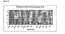
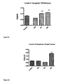
- Figure 13 shows the effect of monoclonal antibody HB10 on the growth of U87 cells in vitro.
- Tumour cells were seeded at 2000 cells in 96 well plates, AM at 2x10⁻⁷ and a dose range of antibody were added.
- As controls a non mammalian IgG antibody was added. This treatment regime was carried out every 2 days. At day 4 the cells were prepared for MTT assay ATTC.
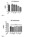
- Figure 14 shows a MTT assay assessing the rate of proliferation in the U-87 glioblastoma cell line in the presence of antibody JF2 at varying concentrations (after 8 days of culture).
- Figure 15 shows a MTT assay assessing the rate of proliferation in the U-87 glioblastoma cell line in the presence of antibody JB3 at varying concentrations (after 8 days of culture).
- Figure 16 shows a MTT assay assessing the rate of proliferation in the MDA-MB-436-GFP breast cancer cell line in the presence of antibody JB3 at varying concentrations (after 8 days of culture).
- Figure 17 shows a MTT assay assessing the rate of proliferation in the MDA-MB-436-GFP breast cancer cell line in the presence of antibody JF2 at varying concentrations (after 8 days of culture).
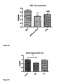
- Figure 18 shows a MTT assay assessing the rate of proliferation in the PC-3 prostate cancer cell line in the presence of antibody JB3 at a concentration of 10 ⁇ g (after 8 days of culture).
- Figure 19 shows a MTT assay assessing the rate of proliferation in the PC-3 prostate cancer cell line in the presence of antibody JF2 at a concentration of 10 ⁇ g (after 8 days of culture).
- Figure 20 shows a MTT assay assessing the rate of proliferation in the SAOS osteosarcoma cell line in the presence of antibody JB3 at a concentration of 10 ⁇ g (after 8 days of culture).
- Figure 21 shows a MTT assay assessing the rate of proliferation in the HCT116 colon cancer cell line in the presence of antibody JB3 and JF2 at a concentration of 10 ⁇ g (after 8 days of culture).
- Figure 22 shows an apoptosis assay assessing the levels of caspase-3 a marker in U-87 glioblastoma cancer cell line in the presence of antibody JB3 and JF2.
- Figure 23 shows an apoptosis assay assessing the levels of caspase-3 a marker in a MDA-MB-436-GFP breast cancer cell line in the presence of antibody JB3 and JF2.
- Figure 24 represents the tumour volume of MDA-MB-436-GFP injected into the right flank of CD1 nude mice, treated with JF2 antibody and controls over a period of 6 weeks.
- Figure 25 represents the tumour weights of MDA-MB-436-GFP injected into the right flank of CD1 nude mice, treated with JF2 antibody and controls over a period of 6 weeks.
- Figure 26 show fluorescence of the MDA-MB-436-GFP cancer cell lines injected into the right flank of nude mice.
- Figure 27 shows histological sections of tumours removed from nude mice following 6 weeks of treatment.
- the ECD regions of the RAMP were generated using a high fidelity PCR reaction using KOD Hot Start DNA Polymerase kit from Novagen Toyobo.
- the template DNA was obtained from a purchased sample of human brain cDNA (Ambion). This reaction was carried out twice the first reaction was carried out to isolate a region larger than the whole RAMP ECD using the following primers:
- the second PCR reaction uses the products from the reaction using the primers above. Using the primers below these primers have had EcoR1 and BamH1 restriction sites incorporated into them:
- the ECD peptides were expressed and purified.
- the protein was purified using the Glutathione S-transferase (GST) gene fusion system.
- GST Glutathione S-transferase
- Antibodies to the ECD peptides were generated using the following protocol.
- Pre-immune serum was taken from the mice prior to immunisation.
- Four mice were injected with a peptide corresponding to an extracellular domain of RAMP-3:
- Injections were boosted with 4 further injections, at approximately monthly intervals.
- Sample bleeds from the mice were taken to isolate serum containing polyclonal antibodies.
- the adjuvant used was Freunds (complete for the first injection, followed by incomplete for the rest of the course).
- the total volume that can be injected into rodents is 0.2ml (and preferably no more than 0.1 ml for mice). Half of this will be antigen and half adjuvant therefore the antigen should be of sufficient concentration to provide the required number of milligrams in a maximum of 0.1 ml or 0.05ml injected.
- Antibody blocking potential .
- the polyclonals were used to test their ability to regulate the effect of adrenomedullin to increase cyclic AMP in human MG63 osteosarcoma cells.
- Monoclonal antibodies were produced using the 3 rd mouse.
- the methods used to produce the monoclonal antibodies are disclosed by Kohler and Milstein in Nature 256, 495-497 (1975 ) and also by Donillard and Hoffman, "Basic Facts about Hybridomas" in Compendium of Immunology V.II ed. by Schwartz, 1981 .
- Clones were screened and selected on the basis of not binding to the GST tag on the peptide. Of these clones, ELISA data was obtained and the best 5 were selected for further work.
- the Isotypes of the monoclonal antibodies are as follows:
- the cell culture was carried out under aseptic conditions on Nunclon treated tissue culture plastics.
- the U-87 glioblastoma cells (ATCC noHTB-14) used for this project were cultured in standard EMEM (Earle's Minimum Essential Medium) media (Gibco) with 10% Fetal Calf Serum (Gibco) and 5% Penicillin/Streptomycin antibiotic (Sigma).
- the frozen cells were thawed at 37° C for 2 minutes.
- the cells were then immediately transferred to 75cm2 flask with 10ml of media.
- the culture was incubated at 37°C with 5% CO2 to attain confluency.
- the media was changed every three days and the cells were then subcultured after reaching 80-90% confluency.
- the cells were centrifuged at 1000rpm for 3 minutes and the supernatant was discarded, the resulting cell pellet was resuspended in 1 ml of media and subcultured into the appropriate number of flasks at a ratio of 1:6. (Refer to appendix 1a for cell culture materials).
- the cell concentration was adjusted to 2000 cells per 50 ⁇ l.
- 5-FluroUracil (Fluka) was used as a positive control (100mM) for the reduction in proliferation.
- PBS controls were also prepared . The plates were then incubated under standard conditions. Replacement and re-dosing of antibody and controls was carried out every 2 days. Mouse anti-GST antibody concentration responses were also calculated the similar way since it was the antibody control for the experiment
- MTT Cell Proliferation Assay ATC.
- 10 ⁇ l of MTT reagent was added to all the wells and left to incubate at 37°C for 2 hours.
- 100 ⁇ l of MTT detergent was added to the wells and incubated overnight and agitated in the dark.
- the plate was read for absorbance at 595nM (Spectramax M5e), using the software softmax Pro 5.2.X 100035
- the proliferation/survival of SW-13 and U87 (Glioblastoma cell line) cells was determined using the MTT assay (see www.lgcpromochem-atcc.com for details on the assay).
- the apoptosis assay was carried out using the Caspase-3 Assay kit (Sigma). (Refer to appendix 1cfor materials required).
- U-87 cells were prepared under standard conditions. A cell solution of 2x10⁷ cells wasmade up in 2ml of media were dispensed into polypropylene tubes.
- 2 x 10⁷ cells were taken for each treatment.
- the treatment groups were, antibody treated cells (for each antibody), anti-Fas antibody, antibody and adrenoemdullin treated cells, blank and adrenoemdullin treated cells.
- the positive control used was anti-Fas monoclonal antibody (MBL).
- the final concentration of antibodies used was 10g and for anti-Fas antibody was 500ng.
- the anti-Fas antibody is an IgM antibody and possesses cytolytic activity thereby inducing apoptosis to the cells.
- the treated cell suspension in 2ml media were incubated for 3 hours at 37°C in a 5% CO2 atmosphere.
- the assay buffer and lysis buffer given in the kit was prepared using the given 17megohm water as per the standard instructions given in the protocol. After 3 hours of incubation, the cells were centrifuged at 600g for 5 minutes at 4C. The supernatant was discarded and the cells were resuspended in 1 ml PBS. The cells were once again centrifuged using the above mentioned conditions and the supernatant was discarded. The cell pellet was resuspended in 200 ⁇ l 1x lysis buffer and incubated on ice for 20 minutes. The lysed cells were centrifuged at 20,000g for 15 minutes at 4°C. The cell lysate were then laid out in a 96 well flat bottomed plate as shown in figure 6 and incubated overnight. The absorbance was read at 405nm (Spectramax M5e), using the software softmax Pro 5.2.36.
- MDA-MB-436-GFP cells were cultured in RPMI media containing penicillin (50 U/ml), streptomycin (50 ⁇ g/ml), glutamine (1 mg/ml), and supplemented with 10% fetal bovine serum.
- Cells were cultured under a humid 5% CO 2 /95% air atmosphere, and fed with fresh medium every 2 days, being routinely monitored for mycoplasma contamination. Cells growing exponentially were harvested using Trypsin EDAT solution. All culture media components were purchased from Invitrogen Life Technologies
- mice Animal work was performed in the animal facility of the University of Sheffield. Male 4-to-5-week-old CD1 nude ( nu / nu ) mice were used. Mice were acclimated and housed in sterile cages in groups of four or less under laminar flow hoods in a temperature-controlled room with a 12-hour light/12-hour dark schedule, and fed autoclaved chow and water ad libitum .
- CD1 nude ( nu / nu ) nude mice were implanted with MDA-MB-436-GFP breast cancer cells.
- MDA-MB-436-GFP cells grown in culture, were washed with PBS, dispersed in a 0.05% solution of trypsin, and resuspended. After centrifugation (1000 rpm for 3 minutes at 8°C), the cell pellet was resuspended in PBS and the final concentration was adjusted to 3 x 10 7 cells/ml and the suspension was placed on ice. After the site was cleaned with ethanol, 0.1 ml (0.5 x 10 6 cells) of the suspension were subcutaneously injected in the right flanks of nude mice.
- Tumors were measured with a digital venier calliper (Site), and tumour volumes were determined using the formula width x length x height x 0.52 (for ellipsoid form), these measurement were taken twice weekly. Tumours were allowed to devlop for 21 days, animals were randomly divided into three groups.
- Schedule 1 was performed at the indicated time.
- Tumours were excised from mice following euthanasia and fixed in 10% formalin saline solution. Tumour sections were embedded in parafin (standard protocol). Sections were cut at 5 ⁇ m, through the tumour body. Sections of each specimen were stained using haematoxylin - and eosin (H&E) and mounted with glass cover slips.
- H&E haematoxylin - and eosin
- Figure 14 shows a MTT assay assessing the rate of proliferation in the U-87 glioblastoma cell line in the presence of antibody JF2 at varying concentrations (after 8 days of culture). Compared to controls a 10 ⁇ g dose of antibody JF2 produced a significant reduction in proliferation, this represents a 40% reduction in proliferation.
- Figure 15 shows a MTT assay assessing the rate of proliferation in the U-87 glioblastoma cell line in the presence of antibody JB3 at varying concentrations (after 8 days of culture).
- Figure 16 shows a MTT assay assessing the rate of proliferation in the MDA-MB-436-GFP breast cancer cell line in the presence of antibody JB3 at varying concentrations (after 8 days of culture). Compared to controls all dose of antibody JB3 produced a significant reduction in proliferation however no dose response was observed. 10ng produced the greatest reduction of 29% however this response does not significantly differ from the other treated groups
- Figure 17 shows a MTT assay assessing the rate of proliferation in the MDA-MB-436-GFP breast cancer cell line in the presence of antibody JF2 at varying concentrations (after 8 days of culture). Compared to controls doses 1 ⁇ g and 10 ⁇ g of antibody JF2 produced a significant reduction in proliferation. 10 ⁇ g produced the greatest reduction of 12%.
- Figure 18 shows a MTT assay assessing the rate of proliferation in the PC-3 prostate cancer cell line in the presence of antibody JB3 at a concentration of 10 ⁇ g (after 8 days of culture). Compared to controls doses 10 ⁇ g of antibody JB3 produced a significant reduction in proliferation both in the presence and absence of exogenously added adrenomedullin. 10 ⁇ g produced a 57% reduction in proliferation.
- Figure 19 shows a MTT assay assessing the rate of proliferation in the PC-3 prostate cancer cell line in the presence of antibody JF2 at a concentration of 10 ⁇ g (after 8 days of culture).
- 10 ⁇ g of antibody JF3 produced a significant reduction in proliferation both in the presence and absence of exogenously added adrenomedullin.
- 10 ⁇ g of JF2 in the presence of adrenomedullin produced a 37% reduction in proliferation and 22% reduction in the absence of exogenously added adrenomedullin.
- Figure 20 shows a MTT assay assessing the rate of proliferation in the SAOS osteosarcoma cell line in the presence of antibody JB3 at a concentration of 10 ⁇ g (after 8 days of culture). Compared to controls doses 10 ⁇ g of antibody JB3 produced a significant reduction in proliferation both in the presence, however no significant reduction was observed in the absence of exogenously added adrenomedullin. 10 ⁇ g of JB3 in the presence of adrenomedullin produced a 34% reduction in proliferation.
- Figure 21 shows a MTT assay assessing the rate of proliferation in the HCT116 colon cancer cell line in the presence of antibody JB3 and JF2 at a concentration of 10 ⁇ g (after 8 days of culture). Compared to controls doses 10 ⁇ g of antibody JB3 produced a significant reduction in proliferation; however JF2 produced no statistically significant reduction in proliferation. 10 ⁇ g of JB3 in the presence of adrenomedullin produced a 27% reduction in proliferation.
- Figure 22 represents levels of apoptosis, by measure levels of caspase-3 a marker apoptosis.
- Treated groups represent a 26% increase levels of caspase-3
- Figure 23 represents levels of apoptosis, by measure levels of caspase-3 a marker apoptosis.
- MDA-MB-436-GFP breast cancer cell line in the presence of antibody JB3 and JF2 at a concentration of 10 ⁇ g only antibody JB3 showed a significant increased in the levels of caspase-3 and their for apoptosis.
- the JB3 treated group represents a 59% increase in caspase-3.
- Figure 24 represents the tumour volume of MDA-MB-436-GFP injected into the right flank of CD1 nude mice, treated with JF2 antibody and controls over a period of 6 weeks. Both control groups show large tumour volumes increases after week 3, however rates of growth within the treatment groups are considerably slower. Error bars within the control groups are large throughout the experiment however within the treatment groups errors are consistently small.
- Figure 26 show fluorescence of the MDA-MB-436-GFP cancer cell lines injected into the right flank of nude mice.
- Figure 27 shows histological sections of tumours removed from nude mice following 6 weeks of treatment. Sections are stained with H&E the pictures are representative of the tumour. In the control groups larger numbers of blood vessels were visible (red arrows) and fewer areas of necrotic cells. Within the treatment groups there were fewer blood vessels and large areas of pre-necrotic cells represented by yellow circles above.
Abstract
Description
- The present invention relates to antibodies and fragments thereof which bind to a receptor activity modifying protein (RAMP) associated with the calcitonin receptor like receptor.
- The calcitonin family of peptides act through G-protein coupled membrane receptors (GPCRs). The gene for calcitonin receptors has been cloned. It is homologous to GPCRs in family "B" which typically recognize regulatory peptides (secretin, glucagons, VIP). A homolog of the calcitonin receptor, the Calcitonin Receptor Like Receptor (CRLR, also known as CL) has been identified (human 461 aa; rat/mouse 463 aa) and has 55% homology with calcitonin receptor (Njuki et al., Clin. Sci. 85, 385-388 (1993); Chang et al., Neuron 11, 1187-1195 (1993); Fluhmann et al., Biochem. Biophys. Res. Comun. 206, 341-347 (1995); Kapas et al., J. Biol. Chem. 270, 25344-25347 (1995)).
- Alone, the CRLR is unable to transduce a signal in response to adrenomedullin (AM), as the presence of a RAMP (calcitonin Receptor Activity Modifying Protein) is needed to induce ligand specificity, binding and activation of the CRLR. The RAMPs are a family of small intrinsic membrane proteins, with a predicted sizes of 14,000-17,0000 Kd. RAMPs consists of approximately 120 amino acids with a large extra-cellular domains of around 100 amino acids; a single membrane spanning domain and a short intra-cellular region of approximately 10 amino acids.
- It has been shown that CRLR can function as either a CGRP receptor or an AM receptor, depending upon which members of the RAMP family, RAMPs1-3, are expressed. The three members of the RAMP family, RAMP1, 2 and 3, engender different ligand specificities of the CRLR so that:


- The sequences of
RAMP - RAMP 1 - GenBank Accession No. NM_005855
- RAMP 2 - GenBank Accession No. NM_005854; UniGene ID Hs.155106
- RAMP 3 - GenBank Accession No. NM_005856; UniGene ID Hs.25691.
- Keleg,S. et. Al. (2007, Int.J.Cancer:121, 21-32) discloses that adrenomedulin which binds to CRLR is induced by hypoxia and enhances pancreatic cancer cell invasion.
- Polyclonal antibodies which bind to RAMP2 and are useful in the treatment of cancer are disclosed in
WO2004/060834 . The antibodies were raised to a region of the extracellular domain of RAMP2 believed to be critical for CRLR binding to RAMP2. -
WO 2007/045927 relates to agents which modulate the effect of a RAMP. - Because of the high potential utility such anti-RAMP antibodies could have in therapy and diagnosis there is a need for further anti-RAMP antibodies.
- The present Inventors have isolated and characterised further anti-RAMP antibodies. These antibodies have, Inter alia, been shown to inhibit cancer cell proliferation and are useful in the prevention of cancer.
- It will be appreciated that the present Invention is provided according to that defined in the claims.
- According to one aspect of present invention there is provided an isolated antibody capable of binding receptor activity modifying protein 3 (RAMP3) of CRLR which antibody comprises:
- i) a binding domain comprising an amino acid sequence substantially as represented by a sequence shown in
Figure 1a ; and - ii) a binding domain comprising an amino acid sequence substantially as represented by a sequence shown in
Figure 2a . - According to another aspect of the present invention there is provided an isolated antibody capable of binding receptor activity modifying protein 3 (RAMP3) of CRLR which antibody comprises:
- ii) a binding domain comprising an amino acid sequence substantially as represented by a sequence shown In
Figure 1b ; and - ii) a binding domain comprising an amino acid sequence substantially as represented by a sequence shown in
Figure 2b . - According to a first aspect of the Invention, there is provided an isolated antibody capable of binding a receptor activity modifying protein (RAMP) of CRLR receptor which antibody is of IgG. IgA or IgM isotype.
- In one embodiment the antibody of the first aspect of the Invention is of IgG isotype. The antibody may be of the subclass IgG1, IgG2, IgG3 or IgG4. Preferably the antibody is of IgG1 isotype.
- In another embodiment the antibody of the first aspect of the invention is of IgA isotype. The antibody may be of the subclass IgA1 or IgA2.
- In a further embodiment the antibody of the first aspect of the invention is of IgM isotype.
- The antibody of the first aspect of the invention may be a monomeric, dimeric, trimeric, tetrameric or pentameric polypeptide.
- The antibody may be capable of binding RAMP1, RAMP2 or RAMP3. Preferably the antibodies of the invention are capable of binding to RAMP3.
- The antibody of the invention may be a RAMP antagonist or agonist or potentiator of the actitivy of natural or artificial ligands to the receptor.
- The antibody of the invention may function as a RAMP antagonist. The data disclosed herein may indicate that the antibody may inhibit either the interaction between RAMP and CRLR on the one hand or inhibit the interaction between a RAMP/CRLR associated complex and a ligand such as adrenomedullin. In one embodiment the antibody is an anti-RAMP3 antibody of IgM isotype and is a RAMP antagonist. In further embodiment the antibody is an anti-RAMP3 antibody of IgG1 isotype and is a RAMP antagonist.
- In one embodiment, the antibody of the invention functions as a RAMP agonist. The data disclosed herein may indicate that the antibody may In one embodiment the antibody is an anti-RAMP3 antibody of IgG1 isotype and is a RAMP agonist or RAMP potentiator of AM action.
- A second aspect of the invention provides an isolated antibody capable of binding a receptor activity modifying protein (RAMP) of CRLR receptor which antibody comprises a binding domain selected from the group consisting of:
- i) a binding domain comprising an amino acid sequence encoded by a nucleic acid molecule consisting of a nucleic acid sequence as represented by a sequence shown in
Figure 1a ; - ii) a binding domain comprising an amino acid sequence encoded by a nucleic acid molecule consisting of a nucleic acid sequence as represented by a sequence shown in
Figure 1b ; - iii) a binding domain comprising an amino acid sequence encoded by a nucleic acid molecule consisting of a nucleic acid sequence as represented by a sequence shown in
Figure 1 c; - iv) a binding domain comprising an amino acid sequence encoded by a nucleic acid molecule consisting of a nucleic acid sequence as represented by a sequence shown in
Figure 1d ; - v) a binding domain comprising an amino acid sequence encoded by a nucleic acid molecule which hybridises to a nucleic acid molecule as defined in (i), (ii), (iii) or (iv) above;
- vi) a binding domain comprising an amino acid sequence encoded by a nucleic acid which is degenerate as a result of the genetic code to the nucleic acid sequence defined in (i), (ii), (iii), (iv) or (v).
- In a third aspect, the invention provides an isolated antibody capable of binding a receptor activity modifying protein (RAMP) of CRLR receptor which antibody comprises a binding domain selected from the group consisting of:
- i) a binding domain comprising an amino acid sequence encoded by a nucleic acid molecule consisting of a nucleic acid sequence as represented by a sequence shown in
Figure 2a ; - ii) a binding domain comprising an amino acid sequence encoded by a nucleic acid molecule consisting of a nucleic acid sequence as represented by a sequence shown in
Figure 2b ; - iii) a binding domain comprising an amino acid sequence encoded by a nucleic acid molecule consisting of a nucleic acid sequence as represented by a sequence shown in
Figure 2c ; - iv) a binding domain comprising an amino acid sequence encoded by a nucleic acid molecule consisting of a nucleic acid sequence as represented by a sequence shown in
Figure 2d ; - v) a binding domain comprising an amino acid sequence encoded by a nucleic acid molecule which hybridises to a nucleic acid molecule as defined in (i), (ii), (iii) or (iv) above; and
- vi) a binding domain comprising an amino acid sequence encoded by a nucleic acid .which is degenerate as a result of the genetic code to the nucleic acid sequence defined in (i), (ii), (iii), (iv) or (v).
- In a preferred aspect of the invention there is provided an isolated antibody capable of binding a receptor activity modifying protein of CRLR which antibody comprises one or both of the following binding domains:
- i) a binding domain comprising an amino acid sequence encoded by a nucleic acid molecule consisting of a nucleic acid sequence as represented by a sequence shown in
Figure 1a ; and/or - ii) a binding domain comprising an amino acid sequence encoded by a nucleic acid molecule consisting of a nucleic acid sequence as represented by a sequence shown in
Figure 2a . - In a further preferred aspect of the invention there is provided an isolated antibody capable of binding a receptor activity modifying protein of CRLR which antibody comprises one or both of the following binding domains:
- i) a binding domain comprising an amino acid sequence encoded by a nucleic acid molecule consisting of a nucleic acid sequence as represented by a sequence shown in
Figure 1b ; and/or - ii) a binding domain comprising an amino acid sequence encoded by a nucleic acid molecule consisting of a nucleic acid sequence as represented by a sequence shown in
Figure 2b . - The nucleic acid molecule may anneal under stringent hybridisation conditions to the nucleic acid sequence shown in
Figure 1 a to d or 2 a to d or to its complementary strand. Stringent hybridisation/washing conditions are well known in the art. For example, nucleic acid hybrids that are stable after washing in 0.1xSSC, 0.1% SDS at 60°C. It is well known in the art that optimal hybridisation conditions can be calculated if the sequences of the nucleic acid is known. For example, hybridisation conditions can be determined by the GC content of the nucleic acid subject to hybridisation. Please see Sambrook et al (1989) Molecular Cloning; A Laboratory Approach. A common formula for calculating the stringency conditions required to achieve hybridisation between nucleic acid molecules of a specified homology is:
- A further aspect of the invention provides an isolated antibody capable of binding a receptor activity modifying protein (RAMP) of CRLR receptor which antibody comprises a binding domain selected from the group consisting of:
- i) a binding domain comprising an amino acid sequence substantially as represented by a sequence shown in
Figure 1a ; - ii) a binding domain comprising an amino acid sequence substantially as represented by a sequence shown in
Figure 1b ; - iii) a binding domain comprising an amino acid sequence substantially as represented by a sequence shown in
Figure 1c ; and - iv) a binding domain comprising an amino acid sequence substantially as represented by a sequence shown in
Figure 1d . - In a further aspect, the invention provides an isolated antibody capable of binding a receptor activity modifying protein (RAMP) of CRLR receptor which antibody comprises a binding domain selected from the group consisting of:
- i) a binding domain comprising an amino acid sequence substantially as represented by a sequence shown in
Figure 2a ; - ii) a binding domain comprising an amino acid sequence substantially as represented by a sequence shown in
Figure 2b ; - iii) a binding domain comprising an amino acid sequence substantially as represented by a sequence shown in
Figure 2c ; and - iv) a binding domain comprising an amino acid sequence substantially as represented by a sequence shown in
Figure 2d . - In a preferred aspect of the invention there is provided an isolated antibody capable of binding a receptor activator modifying protein of CRLR which antibody comprises one or both of the following binding domains:
- i) a binding domain comprising an amino acid sequence substantially as represented by a sequence shown in
Figure 1a ; and/or - ii) a binding domain comprising an amino acid sequence substantially as represented by a sequence shown in
Figure 2a . - In a preferred aspect of the invention there is provided an isolated antibody capable of binding a receptor activator modifying protein of CRLR which antibody comprises one or both of the following binding domains:
- i) a binding domain comprising an amino acid sequence substantially as represented by a sequence shown in
Figure 1b ; and/or - ii) a binding domain comprising an amino acid sequence substantially as represented by a sequence shown in
Figure 2b . - In one embodiment the antibody of the invention comprises a binding domain comprising an amino acid sequence substantially as set out in an amino acid sequence selected from those represented in
Figure 3 . - In one embodiment the antibody of the invention comprises a binding domain comprising an amino acid sequence substantial as set out in an amino acid sequence selected from those represented in
Figure 4 . - In one embodiment the antibody of the invention comprises a binding domain comprising an amino acid sequence substantially as set out in an amino acid sequence selected from those represented in
Figure 5 . - In one embodiment the antibody of the invention comprises a binding domain comprising an amino acid sequence substantially as set out in an amino acid sequence selected from those represented in
Figure 6 . - In one embodiment the antibody of the invention comprises a binding domain comprising an amino acid sequence substantially as set out in the amino acid sequence selected from those represented in
Figure 7 . - In one embodiment the antibody of the invention comprises a binding domain comprising an amino acid sequence substantially as set out in the amino acid sequence selected from those represented in
Figure 8 . - In one embodiment the antibody of the invention comprises a binding domain comprising an amino acid sequence substantially as set out in the amino acid sequence selected from those represented in
Figure 9 . - In one embodiment the antibody of the invention comprises a binding domain comprising an amino acid sequence substantially as set out in the amino acid sequence selected from those represented in
Figure 10 . - Antibodies which comprise a plurality of binding domains of the same or different sequence, or combinations thereof, are included within the present invention. The or each polypeptide may be carried by a human antibody framework. For example, one or more binding regions may be substituted for the CDRs of a whole human antibody or of the variable region thereof.
- In a fourth aspect, the invention provides an antibody which comprises an antibody of the second aspect in combination or association with an antibody of the third aspect. Such an antibody may be in the form of a Fv, (Fab') 2, or scFV antibody fragment.
- Antibodies of the invention may carry a detectable or functional label.
- In further aspects, the invention provides an isolated nucleic acid which comprises a sequence encoding an antibody of the first, second, third or fourth aspects of the invention, and methods of preparing antibodies of the invention which comprise expressing said nucleic acids under conditions to bring about expression of said antibody, and recovering the antibody.
- Antibodies according to the invention may be used in a method of treatment, prevention or diagnosis of the human or animal body, such as a method of treatment of a proliferative disorder such as a tumour tumour in a patient (preferably human) which comprises administering to said patient an effective amount of an antibody of the invention. The invention also provides an antibody of the present invention for use in medicine, as well as the use of an antibody of the present invention in the manufacture of a medicament for the diagnosis or treatment of a proliferative disorder such as a tumour.
- Antibodies according to the invention may also be used in a method of treatment or prevention of an inflammatory disorder in a patient (preferably human) which comprises administering to said patient an effective amount of an antibody of the invention. The invention also provides the use of an antibody of the present invention in the manufacture of a medicament for the treatment of an inflammatory disorder.
- The term "antibody" as used herein refers to immunoglobulin molecules and immunologically active portions of immunoglobulin molecules, i.e. molecules that contain an antigen binding site that specifically binds an antigen, whether natural or partly or wholly synthetically produced. The term also covers any polypeptide or protein having a binding domain which is, or is homologous to, an antibody binding domain. These can be derived from natural sources, or they may be partly or wholly synthetically produced. Examples of antibodies are fragments which comprise an antigen binding domain such as Fab, scFv, Fv, dAb, Fd; and diabodies.
- Antibodies may be polyclonal or monoclonal. Preferably the antibody is a monoclonal antibody may be referred to herein as "mab".
- It is possible to take monoclonal and other antibodies and use techniques of recombinant DNA technology to produce other antibodies or chimeric molecules which retain the specificity of the original antibody. Such techniques may involve introducing DNA encoding the immunoglobulin variable region, or the complementary determining regions (CDRs), of an antibody to the constant regions, or constant regions plus framework regions, of a different immunoglobulin. See, for instance,
EP-A-184187 GB 2188638A EP-A-239400 - As antibodies can be modified in a number of ways, the term "antibody" should be construed as covering any antibody or substance having a binding domain with the required specificity. Thus, this term covers antibody fragments, derivatives, functional equivalents and homologues of antibodies, humanised antibodies, including any polypeptide comprising an immunoglobulin binding domain, whether natural or wholly or partially synthetic. Chimeric molecules comprising an immunoglobulin binding domain, or equivalent, fused to another polypeptide are therefore included. Cloning and expression of chimeric antibodies are described in
EP-A-0120694 andEP-A-0125023 . A humanised antibody may be a modified antibody having the variable regions of a non-human, e. g. murine, antibody and the constant region of a human antibody. Methods for making humanised antibodies are described in, for example,US Patent No. 5225539 It has been shown that fragments of a whole antibody can perform the function of binding antigens. Examples of binding fragments are (i) the Fab fragment consisting of VL, VH, CL and CHl domains; (ii) the Fd fragment consisting of the VH and CH1 domains; (iii) the Fv fragment consisting of the VL and VH domains of a single antibody; (iv) the dAb fragment (Ward,E.S. et al., Nature 341: 544-546 (1989)) which consists of a VH domain; (v) isolated CDR regions; (vi) F (ab')2 a bivalent fragment comprising two linked Fab fragments (vii) single chain Fv molecules (scFv), wherein a VH domain and a VL domain are linked by a peptide linker which allows the two domains to associate to form an antigen binding site (Bird et al., Science 242: 423-426 (1988) ; Huston et al., PNAS USA 85: 5879-5883 (1988) ); (viii) bispecific single chain Fv dimers(PCT/US92/09965 W094/13804 - Diabodies are multimers of polypeptides, each polypeptide comprising a first domain comprising a binding region of an immunoglobulin light chain and a second domain comprising a binding region of an immunoglobulin heavy chain, the two domains being linked (e. g. by a peptide linker) but unable to associated with each other to form an antigen binding site: antigen binding sites are formed by the association of the first domain of one polypeptide within the multimer with the second domain of another polypeptide within the multimer (
W094/13804 - The antibodies of the invention may be multispecific antibodies having specificity for at least two different antigens. While such a molecule is generally binds to two antigens (i.e., bispecific antibody), the term "multispecific antibody" in the present invention encompasses an antibody having specificity for two or more (such as three) antigens. The multispecific antibody can be a full length antibody or a fragment of such an antibody (e.g. F(ab')2 bispecific antibody). Where bispecific antibodies are to be used, these may be conventional bispecific antibodies, which can be manufactured in a variety of ways (Hollinger & Winter, Current OpinionBiotechnol. 4: 446-449 (1993) ), e. g. prepared chemically or from hybrid hybridomas, or may be any of the bispecific antibody fragments mentioned above. It may be preferable to use scFv dimers or diabodies rather than whole antibodies. Diabodies and scFv can be constructed without an Fc region, using only variable domains, potentially reducing the effects of anti-idiotypic reaction. Other forms of bispecific antibodies include the single chain "Janusins" described in Traunecker et al., EMBO Journal 10: 3655-3659 (1991). The bispecific antibody also include a heteroconjugate antibody in which one antibody is coupled to avidin and the other is coupled to biotin (
U.S. Pat. No. 4,676,980 ,WO 91/00360 WO 92/200373 EP 03089 U.S. Pat. No. 4,676,980 . - Bispecific diabodies, as opposed to bispecific whole antibodies, may also be useful because they can be readily constructed and expressed in E. coli. Diabodies (and many other polypeptides such as antibody fragments) of appropriate binding specificities can be readily selected using phage display (
W094/13804 - An "antigen binding domain" or "binding domain" is the part of an antibody which comprises the area which specifically binds to and is complementary to part or all of an antigen. Where an antigen is large, an antibody may only bind to a particular part of the antigen, which part is termed an epitope. An antigen binding domain may be provided by one or more antibody variable domains. An antigen binding domain may comprise an antibody light chain variable region (VL) and an antibody heavy chain variable region (VH).
- "Isolated" refers to the state in which antibodies of the invention or nucleic acid encoding such antibodies will preferably be, in accordance with the present invention. Antibodies and nucleic acid will generally be free or substantially free of material with which they are naturally associated such as other polypeptides or nucleic acids with which they are found in their natural environment, or the environment in which they are prepared (e. g. cell culture) when such preparation is by recombinant DNA technology practised in vitro or In vivo. Antibodies and nucleic acid may be formulated with diluents or adjuvants and still for practical purposes be isolated - for example, the antibodies will normally be mixed with gelatin or other carriers if used to coat microtitre plates for use in immunoassays, or will be mixed with pharmaceutically acceptable carriers or diluents when used in diagnosis or therapy. Antibodies may be glycosylated, either naturally or by systems of heterologous eukaryotic cells, or they may be (for example if produced by expression in a prokaryotic cell) unglycosylated.
By "substantially as represented" it is meant that the amino acid sequence of the binding domain will be either identical or highly homologous to the amino acid sequence represented in the amino acid sequence shown inFigure 1 a to d,Figure 2a to d orFigure 3 to 10 . - By "highly homologous" it is contemplated that the amino acid sequence have at least 70% identity to the amino acid sequence represented in
Figure 1 a to d,Figure 2a to d orFigure 3 to 10 . Preferably the amino acid sequence will have at least 80% identity, more preferably 80%identity to and more preferably at least 90% identity to and still more preferably at least 95% identity for example 98% identity to the amino acid sequence represented inFigure 1a to d,Figure 2a to d orFigure 3 to 10 . - As used herein, "treatment" includes any regime that can benefit a human or non- human animal, preferably mammal. Mammals, birds and other animals may be treated including dogs, cats and livestock, such as horses, cattle and sheep, The treatment may be in respect of an existing condition or may be prophylactic (preventative treatment).
- As used herein, a "tumour" is an abnormal growth of tissue. It may be localised (benign) or invade nearby tissues (malignant) or distant tissues (metastatic). Tumours include neoplastic growths which cause cancer and include oesophageal, colorectal, gastric, breast and endometrial tumours; as well as cancerous tissues or cell lines including, but not limited to, leukaemic cells. As used herein, "tumour" also includes within its scope endometriosis. Examples of tumours that may be treated in accordance with the invention include tumours of the skin, tung, mediastinum, pericardium, prostate, breast, colon & rectum, liver, pancreas, brain, intracranial structures, eye, testicle, ovary, uterus, cervix, kidney, thyroid, bladder, gastrointestinal tract, haematological tissue, bone, joints or colon.
- As used herein, an "inflammatory disorder" includes disorders selected from the group consisting of atherosclerosis, rheumatoid arthritis, osteoarthritis gout; lupus erythematosus, scleroderma, Sjorgen's syndrome, poly- and dermatomyositis, vasculitis, tendonitis, synovitis, bacterial endocarditis, osteomyelitis, psoriasis, pneumonia, fibrosing alveolitis, chronic bronchitis, bronchiectasis, emphysema, silicosis, pneumoconiosis, tuberculosis, ulcerative colitis, Crohn's disease, chronic inflammatory demyelinating polyradiculoneuropathy, chronic inflammatory demyelinating polyneuropathy, multiple sclerosis, Guillan-Barre Syndrome and myasthemia gravis, mastitis, laminitis, laryngitis, chronic chotecystitis, Hashimoto's thyroiditis, carpal tunnel syndrome and inflammatory breast disease. In an embodiment, the inflammatory disorder may be the result of tissue or organ rejection after transplantation. In particular embodiments the inflammatory disorder is selected from the group consisting of atherosclerosis, rheumatoid arthritis, osteoarthritis, sepsis and polyarthritis.
- The invention also includes within its scope polypeptides having the amino acid sequence as set out in Figure la-d or 2a-d, polynucleotides having the nucleic acid sequences as set out in Figure la-d or 2a-d and sequences having substantial identity thereto, for example, 70%, 80%, 85%, 90%, 95% or 99% identity thereto. The percent identity of two amino acid sequences or of two nucleic acid sequences is generally determined by aligning the sequences for optimal comparison purposes (e. g., gaps can be introduced in the first sequence for best alignment with the second sequence) and comparing the amino acid residues or nucleotides at corresponding positions. The "best alignment" is an alignment of two sequences that results in the highest percent identity. The percent identity is determined by comparing the number of identical amino acid residues or nucleotides within the sequences (i. e., % identity = number of identical positions/total number of positions x 100).
- The determination of percent identity between two sequences can be accomplished using a mathematical algorithm known to those of skill in the art. An example of a mathematical algorithm for comparing two sequences is the algorithm of Karlin and Altschul (1990) Proc. Natl. Acad. Sci. USA 87: 2264-2268, modified as in Karlin and Altschul (1993) Proc. Natl. Acad. Sci. USA 90: 5873-5877. The NBLAST and XBLAST programs of Altschul, et al. (1990) J. Mol. Biol. 215: 403-410 have incorporated such an algorithm. BLAST nucleotide searches can be performed with the NBLAST program, score = 100, wordlength = 12 to obtain nucleotide sequences homologous to a nucleic acid molecules of the invention. BLAST protein searches can be performed with the XBLAST program, score = 50, wordlength = 3 to obtain amino acid sequences homologous to a protein molecules of the invention. To obtain gapped alignments for comparison purposes, Gapped BLAST can be utilized as described in Altschul et al. (1997) Nucleic Acids Res. 25: 3389-3402. Alternatively, PSI-Blast can be used to perform an iterated search that detects distant relationships between molecules (Id. ). When utilizing BLAST, Gapped BLAST, and PSI-Blast programs, the default parameters of the respective programs (e. g., XBLAST and NBLAST) can be used. See http://www.ncbi.nim.nih.gov. Another example of a mathematical algorithm utilized for the comparison of sequences is the algorithm of Myers and Miller, CABIOS (1989). The ALIGN program (version 2.0) which is part of the GCG sequence alignment software package has incorporated such an algorithm.
- Other algorithms for sequence analysis known in the art include ADVANCE and ADAM as described in Torellis and Robotti (1994) Comput. Appl. Biosci. , 10 :3-5; and FASTA described in Pearson and Lipman (1988) Proc. Natl. Acad. Sci. 85: 2444-8. Within FASTA,ktup is a control option that sets the sensitivity and speed of the search.
- The invention also include within its scope an antibody comprising a binding domain which binding domain comprises an amino acid sequence selected from the amino acid sequences represented by
Figure 1a to d or2a to d , or a variant thereof wherein said variant sequence has been altered by addition, substitution or deletion of at least one amino acid residue without substantially affecting the biological function of the antibody. - "Variant(s)" of polypeptides as used herein include polypeptides that differ in amino acid sequence from a reference polypeptide. Generally, differences are limited so that the sequences of the reference and the variant are closely similar and, in many regions, identical. A variant polypeptide may differ in amino acid sequence by one or more substitutions, additions, deletions, truncations which may be present in any combination. Among preferred variants are those that vary from a reference polypeptide by conservative amino acid substitutions. Such substitutions are those that substitute a given amino acid by another amino acid of like characteristics. The following non-limiting list of amino acids are considered conservative replacements (similar): a) alanine, serine, and threonine; b) glutamic acid and asparatic acid; c) asparagine and glutamine d) arginine and lysine; e) isoleucine, leucine, methionine and valine and f) phenylalanine, tyrosine and tryptophan.
- One embodiment of the invention provides antibodies comprising a pair of binding domains based on the amino acid sequences for the VH and VL regions substantially as set out in Figures la-d and 2a-d respectively. Single binding domains based on either of these sequences form further aspects of the invention. In the case of the binding domains based on the amino acid sequence for the VH region substantially set out in
Figure 1a-d , such binding domains may be used as targeting agents since it is known that immunoglobulin VH domains are capable of binding target antigens in a specific manner. - In the case of either of the single chain specific binding domains, these domains may be used to screen for complementary domains capable of forming a two-domain antibody which has in vivo properties as good as or equal to the monoclonal antibodies disclosed herein.
- This may be achieved by phage display screening methods using the so-called hierarchical dual combinatorial approach as disclosed in
W092/01047 - Antibodies of the present invention may further comprise antibody constant regions or parts thereof. For example, antibodies based on the VL region shown in
Figure 2a-d may be attached at their C-terminal end to antibody light chain constant domains including human CK orC# chains. Similarly, antibodies based on VH region shown in Figure la-e may be attached at their C-terminal end to all or part of an immunoglobulin heavy chain derived from any antibody isotype, e. g. IgG, IgA, IgE and IgM and any of the isotype sub-classes. - Although antibodies of the invention have in themselves been shown to be effective in preventing cancer cell proliferation, they may additionally be labelled with a functional label. Functional labels include substances which are designed to be targeted to the site of cancer to cause destruction thereof. Such functional labels include toxins such as ricin and enzymes such as bacterial carboxypeptidase or nitroreductase, which are capable of converting prodrugs into active drugs. In addition, the antibodies may be attached or otherwise associated with chemotherapeutic or cytotoxic agents, such as calicheamicin, or radiolabels, such as 90 Y or 11.
- In an embodiment, the antibody of the invention is capable of inhibiting proliferation of a human adrenocarcinoma (SW-13) cell by at least 10%, wherein said inhibition is measured using a MTT Cell Proliferation assay. Preferably, the antibody is capable of modulating e.g. interfering with, interaction of RAMP-3 and CRLP.
- Typically, the antibody is capable of inhibiting proliferation by at least 12%. In some embodiments, the antibody may be capable of inhibiting proliferation by at least 20% and optionally at least 25%. In a further embodiment, the antibody may be capable of inhibiting proliferation by at least 30% and further optionally at least 40%.
- In one embodiment, the antibody is capable of reducing or inhibiting production of cAMP in a human MG63 osteosarcoma cell, when stimulated by adrenomedullin, by at least about 15%, e.g. at least 15%, 16%, 17%, 18% and 19%. In some embodiments, the antibody may be capable of inhibiting production of cAMP by at least about 20% e.g. 21%, 22% or 25%. Typically, the antibody is capable of modulating an interaction of RAMP-3 and CRLP.
- The antibodies of the present invention may be generated wholly or partly by chemical synthesis. The antibodies can be readily prepared according to well- established, standard liquid or, preferably, solid-phase peptide synthesis methods, general descriptions of which are broadly available (see, for example, in J. M. Stewart and J. D. Young, Solid Phase Peptide Synthesis, 2nd edition, Pierce Chemical Company, Rockford, Illinois (1984), in M. Bodanzsky and A. Bodanzsky, The Practice of Peptide Synthesis, Springer Verlag, New York (1984) ; and Applied Biosystems 430A Users Manual, ABI Inc., Foster City, California), or they may be prepared in solution, by the liquid phase method or by any combination of solid- phase, liquid phase and solution chemistry, e. g. by first completing the respective peptide portion and then, if desired and appropriate, after removal of any protecting groups being present, by introduction of the residue X by reaction of the respective carbonic or sulfonic acid or a reactive derivative thereof.
- Another convenient way of producing an antibody according to the present invention is to express the nucleic acid encoding it, by use of nucleic acid in an expression system.
- The present invention further provides an isolated nucleic acid encoding an antibody of the present invention. Nucleic acid includes DNA and RNA. In a preferred aspect, the present invention provides a nucleic acid which codes for an antibody of the invention as defined above. Examples of such nucleic acid are shown in
Figures 1a-d and2a-d . The skilled person will be able to determine substitutions, deletions and/or additions to such nucleic acids which will still provide an antibody of the present invention. - The present invention also provides constructs in the form of plasmids, vectors, transcription or expression cassettes which comprise at least one nucleic acid as described above. The present invention also provides a recombinant host cell which comprises one or more constructs as above. As mentioned, a nucleic acid encoding an antibody of the invention forms an aspect of the present invention, as does a method of production of the antibody which method comprises expression from encoding nucleic acid therefore. Expression may conveniently be achieved by culturing under appropriate conditions recombinant host cells containing the nucleic acid. Following production by expression, an antibody may be isolated and/or purified using any suitable technique, then used as appropriate.
- Systems for cloning and expression of a polypeptide in a variety of different host cells are well known. Suitable host cells include bacteria, mammalian cells, yeast and baculovirus systems. Mammalian cell lines available in the art for expression of a heterologous polypeptide include Chinese hamster ovary cells, HeLa cells, baby hamster kidney cells, NSO mouse melanoma cells and many others. A common, preferred bacterial host is E. coli. The expression of antibodies and antibody fragments in prokaryotic cells such as E. coli is well established in the art. For a review, see for example Pluckthun, BiolTechnology 9: 545-551 (1991). Expression in eukaryotic cells in culture is also available to those skilled in the art as an option for production of an antibody, see for recent review, for example Reff, Curr. Opinion Biotech. 4: 573-576 (1993); Trill et al., Curr. Opinion Biotech. 6: 553- 560 (1995).
- Suitable vectors can be chosen or constructed, containing appropriate regulatory sequences, including promoter sequences, terminator sequences, polyadenylation sequences, enhancer sequences, marker genes and other sequences as appropriate.
- Vectors may be plasmids, viral e. g. 'phage, or phagemid, as appropriate. For further details see, for example, Sambrook et al., Molecular Cloning: A Laboratory Manual: 2nd Edition, Cold Spring Harbor Laboratory Press (1989). Many known techniques and protocols for manipulation of nucleic acid, for example in preparation of nucleic acid constructs, mutagenesis, sequencing, introduction of DNA into cells and gene expression, and analysis of proteins, are described in detail in Ausubel et al. eds. , Short Protocols in Molecular Biology, 2nd Edition, John Wiley & Sons (1992).
- Thus, a further aspect of the present invention provides a host cell containing nucleic acid as disclosed herein. A still further aspect provides a method comprising introducing such nucleic acid into a host cell. The introduction may employ any available technique. For eukaryotic cells, suitable techniques may include calcium phosphate transfection, DEAE-Dextran, electroporation, liposome-mediated transfection and transduction using retrovirus or other virus, e. g. vaccinia or, for insect cells, baculovirus. For bacterial cells, suitable techniques may include calcium chloride transformation, electroporation and transfection using bacteriophage. The introduction may be followed by causing or allowing expression from the nucleic acid, e. g. by culturing host cells under conditions for expression of the gene.
- In one embodiment, the nucleic acid of the invention is integrated into the genome (e. g. chromosome) of the host cell. Integration may be promoted by infusion of sequences which promote recombination with the genome, in accordance with standard techniques.
- The present invention also provides a method which comprises using a construct as stated above in an expression system in order to express an antibody or polypeptide as above.
- Antibodies of the present invention can be used in methods of diagnosis of tumours in human or animal subjects.
- Genes encoding RAMPs show elevated expression in specific cancer tissue. The expression of RAMP can be detected in a tissue or cell using the antibody of the present invention in a method such as Western blotting, the ELISA method or histological staining. A sample (such as biopsy sample or blood sample) derived from tissue of a subject is brought into contact with an antibody of the present invention under conditions so as to form an immune complex. The presence or the amount of the RAMP in the sample can be determined by determining whether the sample binds to the antibody. In this way, diagnosis of cancer, monitoring of progress or cure of cancer, and prediction of prognosis may be carried out.
- In one embodiment the invention provides an in vitro method for detecting the presence of a tumour or inflammatory disorder in a biological sample, comprising the steps of contacting the biological sample with an antibody of the invention and detecting increased binding of the antibody relative to that detected in a negative control or in a biological sample from a normal healthy subject.
- The invention also provides the use of an antibody of the invention in a method of diagnosis in vivo. In one embodiment the invention provides a method for detecting the presence of a tumour or inflammatory disorder in a subject, comprising the steps of administering to said subject the antibody of the invention and detecting increased binding of the antibody relative to that detected in a negative control or in a normal healthy subject.
- The antibodies may be labelled or conjugated to other molecules to aid diagnoistic imaging or other detection. For example, 123-iodine-radiolabeled antibody may be produced for detection by scintigraphy with single photon emission tomography with computerized tomography (SPECT/CT). Such methods may be combined with other diagnostic and imaging techniques where for example, 18F-fluorodeoxyglucose (18FDG) positron emission tomography with computerized tomography (PET/CT) may be performed. These techniques are known to those skilled in the art, and are exemplified in Birchler et al., Otolaryngology - Head and Neck Surgery, Volume 136, Issue 4, Pages 543-548).
- In another embodiment the invention provides a method of monitoring the progression of a tumour or inflammatory disorder of a therapeutic regimen in a subject, comprising the steps of isolating a biological sample from the subject, contacting the biological sample with the antibody of the invention and detecting increased binding of the antibody relative to that detected in a negative control or in a biological sample from a normal healthy subject. In another embodiment the invention provides a method of monitoring the progression of a tumour or inflammatory disorder of a therapeutic regimen in a subject, comprising the steps of administering to said subject an antibody of the invention and detecting increased binding of the antibody relative to that detected in a negative control or in a normal healthy subject.
- The biological sample to be tested may include any tissue from a subject such as biopsy tissue (e.g. tissue from tumours such as described herein or body fluid (e.g.blood).
- When used in diagnosis, antibodies may be labelled with a detectable label, for example a radiolabel such as I or 99Tc, which may be attached to antibodies of the invention using conventional chemistry known in the art of antibody imaging. Labels also include enzyme labels such as horseradish peroxidase. Labels further include chemical moieties such as biotin which may be detected via binding to a specific cognate detectable moiety, e.g. labelled avidin.
- The antibodies of the present invention may be administered alone or in combination with other treatments, either simultaneously or sequentially, dependent upon the condition to be treated. Thus, the present invention further provides products containing an antibody of the present invention and an active agent as a combined preparation for simultaneous, separate or sequential use in the treatment of a tumour. Active agents may include chemotherapeutic or cytotoxic agents including, 5-Fluorouracil, cisplatin, Mitomycin C, oxaliplatin and tamoxifen, which may operate synergistically with the antibodies of the present invention. Other active agents may include suitable doses of pain relief drugs such as non-steroidal anti-inflammatory drugs (e. g. aspirin, paracetamol, ibuprofen or ketoprofen) or opitates such as morphine, or anti-emetics.
- Whilst not wishing to be bound by theory, the ability of the antibodies of the invention to synergise with an active agent to enhance tumour killing may not be due to immune effector mechanisms but rather may be a direct consequence of the antibody binding to cell surface RAMP.
- Antibodies of the present invention will usually be administered in the form of a pharmaceutical composition, which may comprise at least one component in addition to the antibody. The pharmaceutical composition may comprise, in addition to active ingredient, a pharmaceutically acceptable excipient, diluent, carrier, buffer, stabiliser or other materials well known to those skilled in the art. Such materials should be non-toxic and should not interfere with the efficacy of the active ingredient. The precise nature of the carrier or other material will depend on the route of administration, which may be oral, or by injection, e. g. intravenous.
- It is envisaged that systemic injections by normal routes (such as intravenous, intra-arterial, peritoneal, intramuscular or subcutaneous injection) will be the primary route for therapeutic administration of the compositions although local delivery by local injection or through a catheter or other surgical tubing may also used as may local injection or infusion by an indwelling reservoir minipump or other slow-release device. Local administration may be into the pathological tissue mass or into a body cavity that contains the target tissue. Examples of such cavities may include the cerebral ventricles, the synovial joints or the pleural cavity. Liquid formulations may be utilised after reconstitution from powder formulations.
- For intravenous injection, or injection at the site of affliction, the active ingredient will be in the form of a parenterally acceptable aqueous solution which is pyrogen- free and has suitable pH, isotonicity and stability. Those of relevant skill in the art are well able to prepare suitable solutions using, for example, isotonic vehicles such as Sodium Chloride Injection, Ringer's Injection, Lactated Ringer's Injection. Preservatives, stabilisers, buffers, antioxidants and/or other additives may be included, as required.
- Pharmaceutical compositions for oral administration may be in tablet, capsule, powder or liquid form. A tablet may comprise a solid carrier such as gelatin or an adjuvant. Liquid pharmaceutical compositions generally comprise a liquid carrier such as water, petroleum, animal or vegetable oils, mineral oil or synthetic oil.
- Physiological saline solution, dextrose or other saccharide solution or glycols such as ethylene glycol, propylene glycol or polyethylene glycol may be included. Where the formulation is a liquid it may be, for example, a physiologic salt solution containing non-phosphate buffer at pH 6.8-7.6, or a lyophilised powder.
- The composition may also be administered via microspheres, liposomes, other microparticulate delivery systems or sustained release formulations placed in certain tissues including blood. Suitable examples of sustained release carriers include semi- permeable polymer matrices in the form of shared articles, e. g. suppositories or microcapsules. Implantable or microcapsular sustained release matrices include polylactides (
US Patent No. 3, 773, 919 ;EP-A-0058481 ) copolymers of L-glutamic acid and gamma ethyl-L-glutamate (Sidman et al, Biopolymers 22 (1): 1985), poly (2-hydroxyethyl-methacrylate) or ethylene vinyl acetate (Langer et al, J. Biomed. Mater. Res. 15: 167-277, 1981, and Langer, Chem. Tech. 12:98-105, 1982). - liposomes containing the polypeptides are prepared by well-known methods:
DE3,218 ,121A ; Epstein et al, PNAS USA, 82: 3688-3692, 1985; Hwang et al, PNAS USA, 77: 4030-4034, 1980;EP-A-0052522 ;E-A-0036676 ;EP-A-0088046 ;EP-A- 0143949 ;EP-A-0142541 ;JP-A-83-11808 US Patent Nos 4,485,045 and4,544,545 . - Ordinarily, the liposomes are of the small (about 200-800 Angstroms) unilamellar type in which the lipid content is greater than about 30 mol.% cholesterol, the selected proportion being adjusted for the optimal rate of the polypeptide leakage.
- The composition may be administered in a localised manner to a tumour site or other desired site or may be delivered in a manner in which it targets tumour or other cells.
- The compositions are preferably administered to an individual in a "therapeutically effective amount", this being sufficient to show benefit to the individual. The actual amount administered, and rate and time-course of administration, will depend on the nature and severity of what is being treated. Prescription of treatment, e. g. decisions on dosage etc, is within the responsibility of general practitioners and other medical doctors, and typically takes account of the disorder to be treated, the condition of the individual patient, the site of delivery, the method of administration and other factors known to practitioners. The compositions of the invention are particularly relevant to the treatment of existing tumours, especially cancer, and in the prevention of the recurrence of such conditions after initial treatment or surgery. Examples of the techniques and protocols mentioned above can be found in Remington's Pharmaceutical Sciences, 16th edition, Oslo, A. (ed), 1980.
- The optimal dose can be determined by physicians based on a number of parameters including, for example, age, sex, weight, severity of the condition being treated, the active ingredient being administered and the route of administration. In general, a serum concentration of polypeptides and antibodies that permits saturation of receptors is desirable. A concentration in excess of approximately 0.1nm is normally sufficient. For example, a dose of 100mg/m of antibody provides a serum concentration of approximately 20nM for approximately eight days.
- The dose of the composition will be dependent upon the properties of the antibody, e. g. its binding activity and in vivo plasma half-life, the concentration of the polypeptide in the formulation, the administration route, the site and rate of dosage, the clinical tolerance of the patient involved, the pathological condition afflicting the patient, as Is well within the skill of the physician. This invention is also directed to optimise immunisation schedules for enhancing a protective immune response against cancer.
- An antibody or composition according to the invention may be useful In the treatment, retardation and/ or prevention of a proliferative disorder such as a tumour through, for example, the inhibition of angiogenesis or cancer cell proliferation.
- Specific examples of the tumours to be treated according to the invention include lung, intracranial (including brain) and skin tumours. Preferred are intracranial tumours for example brain tumours. Lung tumours or cancer may be classified into small cell carcinoma, adenocarcinoma, large cell carcinoma, bronchioalveolar, squamous and carcinoid. Any lung tumour may be treated according to the invention. Skin tumours or cancer may be classified into melanoma, oral squamous and teratocarcinoma. lntracranial and brain tumours may be classified into glioma, glioblastoma, neuroblastoma, pituitary adenoma, somatotropinomas, prolactinomas, meningiomas, astrocytomas and Choroid plexus carcinoma. Adrenal tumours may be classified into adrenocortical carcinoma, pheochromocytoma, aldosteronoma. The tumour may be an ocular tumour.
- The tumour to be treated may be selected from the group consisting of osteosarcoma, adrenocarcinoma, glioblastoma, prostate tumour, breast tumour and mesothelioma.
The tumour to be treated may be a brain tumour for example a glioblastoma. - The tumour to be treated may be an adrenal tumour for example an adrenocortical carcinoma.
- The tumour to be treated may be mesothelioma.
- The tumour to be treated may be a bone tumour for example an osteosarcoma.
The tumour to be treated may be a prostate tumour. - The tumour to be treated may be a tumour of the breast.
- The tumour to be treated may be a colon tumour.
- In one embodiment, the antibody of the invention useful in the treatment of a tumour is a RAMP (e.g. RAMP 3) antagonist.
- Other proliferative disorders to be treated may include hyperkeratosis.
- An antibody or composition according to the invention may be useful in the treatment, retardation and/or prevention of an inflammatory disorder such as defined herein. In one embodiment, the antibody of the invention useful in the treatment of an inflammatory disorder is a RAMP (e.g. RAMP 3) antagonist.
- An antibody or composition according to the invention may be useful in the treatment, retardation and/ or prevention of a cardiovascular condition through, for example, the promotion of angiogenesis and vasulogenesis. Specific examples of a cardiovascular condition may include heart failure, stroke (specifically re-vasculariation after stroke), coronary heart disease, vascular disease, myocardial infarction (specifically re-vasculariation after myocardial infarction) and diabetic angiopathy and specifically retinopathy. In one embodiment, the antibody of the invention useful in the treatment of a cardiovascular condition is a RAMP (e.g. RAMP 3) agonist.
- An antibody or composition according to the invention may also be useful in the treatment of a disorder selected from osteoporosis, obesity, sepsis and wounds. As used herein the term "wound" includes ulcers and lesions for example, cutaneous wounds such cuts or burns, and conditions associated therewith. The antibody or composition of the invention may be used to stimulate proliferation and tissue growth such as wound healing generally. This might include healing of skin wounds after surgery were an accelerator of healing could have benefits, healing of open skin wounds where there is skin or tissue loss, healing of ulcers whether due to diabetes, pressure sores in bed rest patients or other causes, healing of delayed tissue repair such as in delayed or non-union of bone fractures, repair of cartilage and joint tissues after injury with or without surgery, healing of tendon injuries such as chilles tendon ruptures and tendon injuries in horses. Other indications would include treatment of tissue loss such as occurs in osteoporosis. In one embodiment, the antibody of the invention useful in the treatment of hypertension, obesity, wounds and osteoporosis is a RAMP (e.g. RAMP 3) agonist. Alternatively the antibody useful in the treatment of osteoporosis may be a RAMP (e.g. RAMP 3) antagonist.
- Throughout the description and claims of this specification, the words "comprise" and "contain" and variations of the words, for example "comprising" and "comprises", means "including but not limited to", and is not intended to (and does not) exclude other moieties, additives, components, integers or steps.
- Throughout the description and claims of this specification, the singular encompasses the plural unless the context otherwise requires. In particular, where the indefinite article is used, the specification is to be understood as contemplating plurality as well as singularity, unless the context requires otherwise.
- Features, integers, characteristics, compounds, chemical moieties or groups described in conjunction with a particular aspect, embodiment or example of the invention are to be understood to be applicable to any other aspect, embodiment or example described herein unless incompatible therewith.
-
Figure 1 a - d shows the nucleic acid and amino acid sequences of the heavy chain variable region of monoclonal anti-RAMP 3 antibodies; -
Figure 2 a - d shows the nucleic acid and amino acid sequences of the light chain variable region of monoclonal anti-RAMP 3 antibodies; -
Figures 3 to 6 show the amino acid sequences of the heavy chain of a monoclonal anti-RAMP 3 antibody; -
Figure 7 to 10 show the amino acid sequences of the light chain of a monoclonal anti-RAMP 3 antibody; -
Figure 11 Polyclonal anti-RAMP-3 antibodies were tested for their ability to regulate the effect of adrenomedullin to increase cyclic AMP in human MG63 osteosarcoma cells. All antibodies reduced the effect of adrenomedullin; -
Figure 12 : shows the effect of the monoclonal antibodies on the growth of SW-13 cells in vitro. Monoclonal anti-RAMP-3 antibodies were tested for their ability to induce inhibition of proliferation of SW-13 (adrenocarcinoma cells) . The concentration of 1:50 equates to about 5ng per well final concentration; -
Figure 13 : shows the effect of monoclonal antibody HB10 on the growth of U87 cells in vitro. Tumour cells were seeded at 2000 cells in 96 well plates, AM at 2x10⁻⁷ and a dose range of antibody were added. As controls a non mammalian IgG antibody was added. This treatment regime was carried out every 2 days. At day 4 the cells were prepared for MTT assay ATTC. -
Figure 14 shows a MTT assay assessing the rate of proliferation in the U-87 glioblastoma cell line in the presence of antibody JF2 at varying concentrations (after 8 days of culture). -
Figure 15 shows a MTT assay assessing the rate of proliferation in the U-87 glioblastoma cell line in the presence of antibody JB3 at varying concentrations (after 8 days of culture). -
Figure 16 shows a MTT assay assessing the rate of proliferation in the MDA-MB-436-GFP breast cancer cell line in the presence of antibody JB3 at varying concentrations (after 8 days of culture). -
Figure 17 shows a MTT assay assessing the rate of proliferation in the MDA-MB-436-GFP breast cancer cell line in the presence of antibody JF2 at varying concentrations (after 8 days of culture). -
Figure 18 shows a MTT assay assessing the rate of proliferation in the PC-3 prostate cancer cell line in the presence of antibody JB3 at a concentration of 10µg (after 8 days of culture). -
Figure 19 shows a MTT assay assessing the rate of proliferation in the PC-3 prostate cancer cell line in the presence of antibody JF2 at a concentration of 10µg (after 8 days of culture). -
Figure 20 shows a MTT assay assessing the rate of proliferation in the SAOS osteosarcoma cell line in the presence of antibody JB3 at a concentration of 10µg (after 8 days of culture). -
Figure 21 shows a MTT assay assessing the rate of proliferation in the HCT116 colon cancer cell line in the presence of antibody JB3 and JF2 at a concentration of 10µg (after 8 days of culture). -
Figure 22 shows an apoptosis assay assessing the levels of caspase-3 a marker in U-87 glioblastoma cancer cell line in the presence of antibody JB3 and JF2. -
Figure 23 shows an apoptosis assay assessing the levels of caspase-3 a marker in a MDA-MB-436-GFP breast cancer cell line in the presence of antibody JB3 and JF2. -
Figure 24 represents the tumour volume of MDA-MB-436-GFP injected into the right flank of CD1 nude mice, treated with JF2 antibody and controls over a period of 6 weeks. -
Figure 25 represents the tumour weights of MDA-MB-436-GFP injected into the right flank of CD1 nude mice, treated with JF2 antibody and controls over a period of 6 weeks. -
Figure 26 show fluorescence of the MDA-MB-436-GFP cancer cell lines injected into the right flank of nude mice. -
Figure 27 shows histological sections of tumours removed from nude mice following 6 weeks of treatment. - EXAMPLES - The invention will now be described further in the following non-limiting example:
- The ECD regions of the RAMP were generated using a high fidelity PCR reaction using KOD Hot Start DNA Polymerase kit from Novagen Toyobo. The template DNA was obtained from a purchased sample of human brain cDNA (Ambion).
This reaction was carried out twice the first reaction was carried out to isolate a region larger than the whole RAMP ECD using the following primers: -
- Forward
CGAGCGGACTCGACTCGGCAC - Reverse
CTTCCTAGGGTGGCGGTGGCC -
- Forward
GTC CGC CTC CTC CTT CT GCT - Reverse
AAG TGG AGT AAC ATG GTT ATT GT -
- Forward
AGC CAT GGA GAC TGG AGC GCT GC - Reverse
GTG GCC CAG TAG CTG GAG ATT GGC - The second PCR reaction uses the products from the reaction using the primers above. Using the primers below these primers have had EcoR1 and BamH1 restriction sites incorporated into them:
-
- Forward
GCGAATTCCTGCCAGACCACCAG - Reverse
GTGGATCCTACCGGGCCCGGGACA -
- Forward
GCG AAT TCA ATC CCC ACG AGG CCC TGG CTC AGC C - Reverse
CAG GAT CCTACA AGA GTG ATG AGG AAG GGG ATG -
- Forward
CAG AATT TCC AGA GCA GGC CGC TGC AAC CAG ACA G - Reverse
GTG GAT CCC ACC ACC AGG CCA GCC ATG GCG ACA GT - Genomic sequencing of the product is performed to test the product conclusively. The ECD protein from this point onwards will be referred to as "the insert" unless stated other wise.
- The ECD peptides were expressed and purified. The protein was purified using the Glutathione S-transferase (GST) gene fusion system.
- Antibodies to the ECD peptides were generated using the following protocol.
- The following immunization protocol was followed to raise antibodies against the extracellular domain of RAMP-3:
- Pre-immune serum was taken from the mice prior to immunisation. Four mice were injected with a peptide corresponding to an extracellular domain of RAMP-3:


- Injections were boosted with 4 further injections, at approximately monthly intervals. Sample bleeds from the mice were taken to isolate serum containing polyclonal antibodies. The adjuvant used was Freunds (complete for the first injection, followed by incomplete for the rest of the course). The total volume that can be injected into rodents is 0.2ml (and preferably no more than 0.1 ml for mice). Half of this will be antigen and half adjuvant therefore the antigen should be of sufficient concentration to provide the required number of milligrams in a maximum of 0.1 ml or 0.05ml injected.
- Western blots of the antibodies were used to probe blots of the original ECD peptide run in duplicate lanes with a size marker.
Antibody Antibody 3 shows very strong binding at the same size, while AB4 was not detectable in this experiment (data not shown). - To test the ability of the antibodies to bind to RAMP, assays were carried out to determine the antibodies' blocking potential:
- Human MG63 osteosarcoma cells were treated with 10pmol of AM and the cAMP response measured (method as stated above e.g. using cAMP Fluorescence Polarization (FP) Blotrak Immunoassay (Amersham Biosciences)). (If RAMP-1 agents are being tested, this assay can also be carried out using CGRP as a ligand to test the agent's blocking ability)
- The cells were pre-treated with the antibody for 1 hr
- An EC60 dose of AM was applied (10pmol) was applied and cAMP response was measured.
- The polyclonals were used to test their ability to regulate the effect of adrenomedullin to increase cyclic AMP in human MG63 osteosarcoma cells.
- Monoclonal antibodies were produced using the 3rd mouse. The methods used to produce the monoclonal antibodies are disclosed by Kohler and Milstein in Nature 256, 495-497 (1975) and also by Donillard and Hoffman, "Basic Facts about Hybridomas" in Compendium of Immunology V.II ed. by Schwartz, 1981.
- Clones were screened and selected on the basis of not binding to the GST tag on the peptide. Of these clones, ELISA data was obtained and the best 5 were selected for further work.
- Characterisation of the monoclonal antibody's isotype was determined using the isotyping kit, IsoStrip (Roche Diagnostics GmbH).
- The Isotypes of the monoclonal antibodies are as follows:
- CD12 - IgG - IgG1 subtype
- HB10 - IgG - IgG1 subtype
- JF2 - IgG - IgG1 subtype
- JB3 - IgM
- CC2 - IgA
- The cell culture was carried out under aseptic conditions on Nunclon treated tissue culture plastics. The U-87 glioblastoma cells (ATCC noHTB-14) used for this project were cultured in standard EMEM (Earle's Minimum Essential Medium) media (Gibco) with 10% Fetal Calf Serum (Gibco) and 5% Penicillin/Streptomycin antibiotic (Sigma). The frozen cells were thawed at 37° C for 2 minutes. The cells were then immediately transferred to 75cm2 flask with 10ml of media. The culture was incubated at 37°C with 5% CO2 to attain confluency. The media was changed every three days and the cells were then subcultured after reaching 80-90% confluency. Cells were washed with PBS (Gibco) to remove serum present because it acts as an inhibitor for trypsin. 3ml of 10% Trypsin EDTA (Sigma) was added to remove the cell layer from the flask. The trypsinization was allowed to occur for 15 minutes at 37°c in order to facilitate dispersal. The flask was observed under a microscope to confirm that the cells have detached from the flask. 6-8ml of media was added to the flask and the cells were aspirated gently. The cells were centrifuged at 1000rpm for 3 minutes and the supernatant was discarded, the resulting cell pellet was resuspended in 1 ml of media and subcultured into the appropriate number of flasks at a ratio of 1:6. (Refer to appendix 1a for cell culture materials).
- The U87 glioblastoma cells were grown under standard conditions (as mentioned above). The total number of cells were counted using a haemocytometer (Hawksley) and the concentration was calculated using the following equation:

- The cell concentration was adjusted to 2000 cells per 50µl.
- 50 µl of the above cell preparation were seeded in clear 96 well plates (Costar, polystyrene, Flatbottom).
- Two groups of proliferation studies were carried out for each antibody. One group was used to study the effect on proliferation by the antibody in presence of endogenous adrenomedullin, while the other group was used to the study the effects of antibody in presence of exogenously added adrenomedullin. Varying concentrations of antibody were prepared in PBS to result in final well concentrations of 10µg, 1µg, 100ng, 10ng, 1ng, 100pg, 10pg and 1 pg. Six replicates of each concentration of each antibody was carried out. This dose range was used in both groups. Adrenomedullin was added to the appropriate group to a final well concentration of 200nM. 5-FluroUracil (Fluka) was used as a positive control (100mM) for the reduction in proliferation. PBS controls were also prepared . The plates were then incubated under standard conditions. Replacement and re-dosing of antibody and controls was carried out every 2 days. Mouse anti-GST antibody concentration responses were also calculated the similar way since it was the antibody control for the experiment
- In order to check for consistency of the cells seeded, a 12-24 hours study was performed. Further time points were carried out at 2, 4, 6 and 8 days. The kit used to perform the proliferation assay was MTT Cell Proliferation Assay (ATCC). At each
time point 10 µl of MTT reagent was added to all the wells and left to incubate at 37°C for 2 hours. After 2 hours, 100µl of MTT detergent was added to the wells and incubated overnight and agitated in the dark. The plate was read for absorbance at 595nM (Spectramax M5e), using the software softmax Pro 5.2.X 100035 - The proliferation/survival of SW-13 and U87 (Glioblastoma cell line) cells was determined using the MTT assay (see www.lgcpromochem-atcc.com for details on the assay).
- The apoptosis assay was carried out using the Caspase-3 Assay kit (Sigma). (Refer to appendix 1cfor materials required).
- U-87 cells were prepared under standard conditions. A cell solution of 2x10⁷ cells wasmade up in 2ml of media were dispensed into polypropylene tubes.
- 2 x 10⁷ cells were taken for each treatment. The treatment groups were, antibody treated cells (for each antibody), anti-Fas antibody, antibody and adrenoemdullin treated cells, blank and adrenoemdullin treated cells. The positive control used was anti-Fas monoclonal antibody (MBL). The final concentration of antibodies used was 10g and for anti-Fas antibody was 500ng. The anti-Fas antibody is an IgM antibody and possesses cytolytic activity thereby inducing apoptosis to the cells.
The treated cell suspension in 2ml media were incubated for 3 hours at 37°C in a 5% CO2 atmosphere. - The assay buffer and lysis buffer given in the kit was prepared using the given 17megohm water as per the standard instructions given in the protocol. After 3 hours of incubation, the cells were centrifuged at 600g for 5 minutes at 4C. The supernatant was discarded and the cells were resuspended in 1 ml PBS. The cells were once again centrifuged using the above mentioned conditions and the supernatant was discarded. The cell pellet was resuspended in 200µl 1x lysis buffer and incubated on ice for 20 minutes. The lysed cells were centrifuged at 20,000g for 15 minutes at 4°C. The cell lysate were then laid out in a 96 well flat bottomed plate as shown in
figure 6 and incubated overnight. The absorbance was read at 405nm (Spectramax M5e), using the software softmax Pro 5.2.36. - MDA-MB-436-GFP cells were cultured in RPMI media containing penicillin (50 U/ml), streptomycin (50 µg/ml), glutamine (1 mg/ml), and supplemented with 10% fetal bovine serum. Cells were cultured under a humid 5% CO2/95% air atmosphere, and fed with fresh medium every 2 days, being routinely monitored for mycoplasma contamination. Cells growing exponentially were harvested using Trypsin EDAT solution. All culture media components were purchased from Invitrogen Life Technologies
- Animal work was performed in the animal facility of the University of Sheffield. Male 4-to-5-week-old CD1 nude (nu/nu) mice were used. Mice were acclimated and housed in sterile cages in groups of four or less under laminar flow hoods in a temperature-controlled room with a 12-hour light/12-hour dark schedule, and fed autoclaved chow and water ad libitum.
- CD1 nude (nu/nu) nude mice were implanted with MDA-MB-436-GFP breast cancer cells. For the cell implantations, MDA-MB-436-GFP cells, grown in culture, were washed with PBS, dispersed in a 0.05% solution of trypsin, and resuspended. After centrifugation (1000 rpm for 3 minutes at 8°C), the cell pellet was resuspended in PBS and the final concentration was adjusted to 3 x 10⁷ cells/ml and the suspension was placed on ice. After the site was cleaned with ethanol, 0.1 ml (0.5 x 10⁶ cells) of the suspension were subcutaneously injected in the right flanks of nude mice. Tumors were measured with a digital venier calliper (Site), and tumour volumes were determined using the formula width x length x height x 0.52 (for ellipsoid form), these measurement were taken twice weekly. Tumours were allowed to devlop for 21 days, animals were randomly divided into three groups. One group (n = 8) received interperotanial injection of JB3 antibody (200 µg of purified IgG) as a suspension in PBS in a volume of 0.2 ml every Tuesday and Friday of each week. As control, one group (n = 8) received an irrelevant antibody (IgG of the same isotype, an inactivated form of the antibody) and the other group (n = 8) received comparable injections of the vehicle alone (PBS).
Schedule 1 was performed at the indicated time. - Tumours were excised from mice following euthanasia and fixed in 10% formalin saline solution. Tumour sections were embedded in parafin (standard protocol). Sections were cut at 5µm, through the tumour body. Sections of each specimen were stained using haematoxylin - and eosin (H&E) and mounted with glass cover slips.
- All polyclonal antibodies tested reduced the effect of adrenomedullin on cAMP production. The results shown in
Figure 11 indicate that the polyclonal antibodies raised against RAMP-3 inhibited cAMP production of the MG63 cells by, at least 15%. - Each monoclonal antibody produced induced inhibition of proliferation of SW-13 cells ranging from 12-45% see
Figure 12 (the concentration of 1:50 equates to about 5 nanogrammes per well final concentration). - One of the monoclonal antibodies tested, HB10, increased proliferation of U87 cells (see
Figure 13 ). As the dose of antibody increased so did the level of absorbance and therefore for the level of proliferation suggesting an agonist role for this antibody. -
Figure 14 shows a MTT assay assessing the rate of proliferation in the U-87 glioblastoma cell line in the presence of antibody JF2 at varying concentrations (after 8 days of culture). Compared to controls a 10µg dose of antibody JF2 produced a significant reduction in proliferation, this represents a 40% reduction in proliferation. -
Figure 15 shows a MTT assay assessing the rate of proliferation in the U-87 glioblastoma cell line in the presence of antibody JB3 at varying concentrations (after 8 days of culture). - Compared to controls all dose of antibody JB3 produced a significant reduction in proliferation, this represents a ~35% reduction in proliferation at all doses. This suggests an EC50 to be lower than 1 pg.
-
Figure 16 shows a MTT assay assessing the rate of proliferation in the MDA-MB-436-GFP breast cancer cell line in the presence of antibody JB3 at varying concentrations (after 8 days of culture). Compared to controls all dose of antibody JB3 produced a significant reduction in proliferation however no dose response was observed. 10ng produced the greatest reduction of 29% however this response does not significantly differ from the other treated groups -
Figure 17 shows a MTT assay assessing the rate of proliferation in the MDA-MB-436-GFP breast cancer cell line in the presence of antibody JF2 at varying concentrations (after 8 days of culture). Compared to controls doses 1µg and 10µg of antibody JF2 produced a significant reduction in proliferation. 10µg produced the greatest reduction of 12%. -
Figure 18 shows a MTT assay assessing the rate of proliferation in the PC-3 prostate cancer cell line in the presence of antibody JB3 at a concentration of 10µg (after 8 days of culture). Compared to controls doses 10µg of antibody JB3 produced a significant reduction in proliferation both in the presence and absence of exogenously added adrenomedullin. 10µg produced a 57% reduction in proliferation. -
Figure 19 shows a MTT assay assessing the rate of proliferation in the PC-3 prostate cancer cell line in the presence of antibody JF2 at a concentration of 10µg (after 8 days of culture). Compared to controls doses 10µg of antibody JF3 produced a significant reduction in proliferation both in the presence and absence of exogenously added adrenomedullin. 10µg of JF2 in the presence of adrenomedullin produced a 37% reduction in proliferation and 22% reduction in the absence of exogenously added adrenomedullin. -
Figure 20 shows a MTT assay assessing the rate of proliferation in the SAOS osteosarcoma cell line in the presence of antibody JB3 at a concentration of 10µg (after 8 days of culture). Compared to controls doses 10µg of antibody JB3 produced a significant reduction in proliferation both in the presence, however no significant reduction was observed in the absence of exogenously added adrenomedullin. 10µg of JB3 in the presence of adrenomedullin produced a 34% reduction in proliferation. -
Figure 21 shows a MTT assay assessing the rate of proliferation in the HCT116 colon cancer cell line in the presence of antibody JB3 and JF2 at a concentration of 10µg (after 8 days of culture). Compared to controls doses 10µg of antibody JB3 produced a significant reduction in proliferation; however JF2 produced no statistically significant reduction in proliferation. 10µg of JB3 in the presence of adrenomedullin produced a 27% reduction in proliferation. -
Figure 22 represents levels of apoptosis, by measure levels of caspase-3 a marker apoptosis. U-87 glioblastoma cancer cell line in the presence of antibody JB3 and JF2 at a concentration of 10µg both showed a significant increased in the levels of caspase-3 and their for apoptosis. Treated groups represent a 26% increase levels of caspase-3 -
Figure 23 represents levels of apoptosis, by measure levels of caspase-3 a marker apoptosis. MDA-MB-436-GFP breast cancer cell line in the presence of antibody JB3 and JF2 at a concentration of 10µg only antibody JB3 showed a significant increased in the levels of caspase-3 and their for apoptosis. The JB3 treated group represents a 59% increase in caspase-3. -
Figure 24 represents the tumour volume of MDA-MB-436-GFP injected into the right flank of CD1 nude mice, treated with JF2 antibody and controls over a period of 6 weeks. Both control groups show large tumour volumes increases afterweek 3, however rates of growth within the treatment groups are considerably slower. Error bars within the control groups are large throughout the experiment however within the treatment groups errors are consistently small. -
Figure 25 represents the tumour weights of MDA-MB-436=GFP injected into the right flank of CD1 nude mice, treated with JF2 antibody and controls over a period of 6 weeks. Tumour weights are taken following death and calculated back using the volumes of tumours. Control groups show increases in weight from early time points, however treated groups show slower growth until weeks 4 were weighs increase however still remain below control groups. -
Figure 26 show fluorescence of the MDA-MB-436-GFP cancer cell lines injected into the right flank of nude mice. -
Figure 27 shows histological sections of tumours removed from nude mice following 6 weeks of treatment. Sections are stained with H&E the pictures are representative of the tumour. In the control groups larger numbers of blood vessels were visible (red arrows) and fewer areas of necrotic cells. Within the treatment groups there were fewer blood vessels and large areas of pre-necrotic cells represented by yellow circles above.
Claims (17)
- An Isolated antibody capable of binding receptor activity modifying protein 3 named RAMP3 of named Calcitonin Receptor Like Receptor CRLR which antibody comprises:I) a binding domain comprising an amino acid sequence as represented by a sequence shown in Figure 1a; andII) a binding domain comprising an amino acid sequence as represented by a sequence shown in Figure 2a.
- An Isolated antibody capable of binding receptor activity modifying protein 3 named RAMP3 of named Calcitonin Receptor Like Receptor CRLR which antibody comprises:i) a binding domain comprising an amino acid sequence as represented by a sequence shown in Figure 1b; andii) a binding domain comprising an amino acid sequence as represented by a sequence shown In Figure 2b.
- An antibody according to claim 1 or 2 comprising a functional label.
- An antibody as claimed in claim 3 wherein the label is selected from toxins, enzymes and radiolabels.
- An antibody as claimed in any preceding claim for use in medicine.
- A pharmaceutical composition comprising an antibody as claimed in any one of claims 1 to 4 and a pharmaceutically acceptable excipient, diluent, carrier, buffer or stabiliser.
- A pharmaceutical composition as claimed in claim 6 additionally comprising an active agent.
- A pharmaceutical composition as claimed in claim 7 wherein the active agent is selected from one or more of chemotherapeutic agents, cytotoxic agents, 5-Fluorouracil, cisplatin, Mitomycin C, oxaliplatin, tamoxifen and pain relief drugs.
- The use of an antibody as claimed in any one of claims 1 to 4 in the manufacture of a medicament for the treatment of a tumour.
- The use of an antibody as claimed in claim 9 wherein the tumour is a tumour of the skin, lung, mediastinum, pericardium, prostate, breast, colon, rectum, liver, pancreas, brain, intracranial structures, eye, testicle, ovary, uterus, cervix, kidney, thyroid, bladder, gastrointestinal tract, haematological tissue, bone or joints.
- A nucleic acid encoding an antibody as claimed in any one of claims 1 or 2.
- A nucleic acid according to claim 11 as shown in Figures 1a, 1b, 2a or 2b.
- A construct comprising at least one nucleic acid according to claim 11 or 12.
- A construct as claimed in claim 13 wherein the construct is select from a plasmid, vector, transcription or expression cassettes.
- A recombinant host cell comprising a nucleic acid according to claim 11 or 12.
- A recombinant host cell comprising a construct according to claim 13 or 14.
- A polypeptide as represented by a sequence shown in Figure 1a, 1b, 2a or 2b.
Applications Claiming Priority (2)
| Application Number | Priority Date | Filing Date | Title |
|---|---|---|---|
| GBGB0708002.1A GB0708002D0 (en) | 2007-04-25 | 2007-04-25 | Antibodies |
| PCT/GB2008/001454 WO2008132453A1 (en) | 2007-04-25 | 2008-04-23 | Antibodies against ramp3 |
Publications (2)
| Publication Number | Publication Date |
|---|---|
| EP2152753A1 EP2152753A1 (en) | 2010-02-17 |
| EP2152753B1 true EP2152753B1 (en) | 2012-08-08 |
Family
ID=38170659
Family Applications (1)
| Application Number | Title | Priority Date | Filing Date |
|---|---|---|---|
| EP08750480A Not-in-force EP2152753B1 (en) | 2007-04-25 | 2008-04-23 | Antibodies against ramp3 |
Country Status (10)
| Country | Link |
|---|---|
| US (2) | US8389237B2 (en) |
| EP (1) | EP2152753B1 (en) |
| JP (2) | JP2010524489A (en) |
| KR (1) | KR20100015915A (en) |
| CN (1) | CN101842390A (en) |
| AU (1) | AU2008244074B2 (en) |
| CA (1) | CA2685099A1 (en) |
| ES (1) | ES2393938T3 (en) |
| GB (1) | GB0708002D0 (en) |
| WO (1) | WO2008132453A1 (en) |
Families Citing this family (48)
| Publication number | Priority date | Publication date | Assignee | Title |
|---|---|---|---|---|
| US20030114410A1 (en) | 2000-08-08 | 2003-06-19 | Technion Research And Development Foundation Ltd. | Pharmaceutical compositions and methods useful for modulating angiogenesis and inhibiting metastasis and tumor fibrosis |
| GB0521139D0 (en) * | 2005-10-18 | 2005-11-23 | Univ Sheffield | Therapeutic agent |
| IN2014DN10515A (en) | 2006-03-31 | 2015-08-21 | Chugai Pharmaceutical Co Ltd | |
| ES2429407T3 (en) | 2006-06-08 | 2013-11-14 | Chugai Seiyaku Kabushiki Kaisha | Preventive agent or remedy for inflammatory diseases |
| AU2008299784B9 (en) | 2007-08-02 | 2015-06-18 | Gilead Biologics, Inc. | LOX and LOXL2 inhibitors and uses thereof |
| CA2700701C (en) | 2007-09-26 | 2020-12-29 | Chugai Seiyaku Kabushiki Kaisha | Method of modifying isoelectric point of antibody via amino acid substitution in cdr |
| PL3059246T3 (en) | 2007-09-26 | 2018-11-30 | Chugai Seiyaku Kabushiki Kaisha | Modified antibody constant region |
| WO2009072598A1 (en) | 2007-12-05 | 2009-06-11 | Chugai Seiyaku Kabushiki Kaisha | Therapeutic agent for pruritus |
| ES2834741T3 (en) | 2007-12-05 | 2021-06-18 | Chugai Pharmaceutical Co Ltd | Anti-NR10 antibody and its use |
| SG189775A1 (en) | 2008-04-11 | 2013-05-31 | Chugai Pharmaceutical Co Ltd | Antigen-binding molecule capable of binding to two or more antigen molecules repeatedly |
| US9029508B2 (en) * | 2008-04-29 | 2015-05-12 | Abbvie Inc. | Dual variable domain immunoglobulins and uses thereof |
| CN102112494A (en) * | 2008-06-03 | 2011-06-29 | 雅培制药有限公司 | Dual variable domain immunoglobulins and uses thereof |
| EP3002299A1 (en) | 2008-06-03 | 2016-04-06 | AbbVie Inc. | Dual variable domain immunoglobulins and uses thereof |
| KR20110031369A (en) * | 2008-07-08 | 2011-03-25 | 아보트 러보러터리즈 | Prostaglandin e2 dual variable domain immunoglobulins and uses thereof |
| KR20160062207A (en) * | 2008-12-05 | 2016-06-01 | 추가이 세이야쿠 가부시키가이샤 | Anti-NR10 antibody and use thereof |
| JO3382B1 (en) | 2008-12-23 | 2019-03-13 | Amgen Inc | Human cgrp receptor binding antibodies |
| US9107935B2 (en) | 2009-01-06 | 2015-08-18 | Gilead Biologics, Inc. | Chemotherapeutic methods and compositions |
| EP2409991B1 (en) | 2009-03-19 | 2017-05-03 | Chugai Seiyaku Kabushiki Kaisha | Antibody constant region variant |
| TWI646193B (en) | 2009-03-19 | 2019-01-01 | 中外製藥股份有限公司 | Antibody constant region alteration |
| MX2011011670A (en) * | 2009-05-01 | 2011-11-18 | Abbott Lab | Dual variable domain immunoglobulins and uses thereof. |
| TW201109438A (en) * | 2009-07-29 | 2011-03-16 | Abbott Lab | Dual variable domain immunoglobulins and uses thereof |
| US8512990B2 (en) | 2009-08-21 | 2013-08-20 | Gilead Biologics, Inc. | Catalytic domains from lysyl oxidase and LOXL2 |
| AR078651A1 (en) * | 2009-10-15 | 2011-11-23 | Abbott Lab | IMMUNOGLOBULINS WITH DUAL VARIABLE DOMAIN AND USES OF THE SAME |
| UY32979A (en) * | 2009-10-28 | 2011-02-28 | Abbott Lab | IMMUNOGLOBULINS WITH DUAL VARIABLE DOMAIN AND USES OF THE SAME |
| NZ607480A (en) | 2010-08-03 | 2014-10-31 | Abbott Lab | Dual variable domain immunoglobulins and uses thereof |
| EP2608803A4 (en) | 2010-08-26 | 2014-01-15 | Abbvie Inc | Dual variable domain immunoglobulins and uses thereof |
| CN103328632A (en) | 2010-11-30 | 2013-09-25 | 中外制药株式会社 | Antigen-binding molecule capable of binding to plurality of antigen molecules repeatedly |
| BR112014010008A2 (en) * | 2011-10-26 | 2018-09-04 | Novartis Ag | monoclonal antibodies, their uses and nucleic acids |
| EP2797955A2 (en) | 2011-12-30 | 2014-11-05 | AbbVie Inc. | Dual variable domain immunoglobulins against il-13 and/or il-17 |
| SG11201503412RA (en) | 2012-11-01 | 2015-05-28 | Abbvie Inc | Anti-vegf/dll4 dual variable domain immunoglobulins and uses thereof |
| MX2015013166A (en) | 2013-03-15 | 2015-12-11 | Abbvie Inc | Dual specific binding proteins directed against il-1 beta and il-17. |
| US10379127B2 (en) | 2013-11-27 | 2019-08-13 | Welcome Receptor Antibodies Pty Ltd | Marker of cell death |
| AR101875A1 (en) | 2014-09-15 | 2017-01-18 | Amgen Inc | ANTIGENS, BI-SPECIFIC UNION PROTEIN OF THE ANTI-CGRP RECEIVER / PAC1 RECEIVER AND USES OF THE SAME |
| WO2016094881A2 (en) | 2014-12-11 | 2016-06-16 | Abbvie Inc. | Lrp-8 binding proteins |
| CN107614016B (en) | 2015-04-14 | 2022-06-17 | 中外制药株式会社 | Pharmaceutical composition for preventing and/or treating atopic dermatitis comprising an IL-31 antagonist as an active ingredient |
| JOP20200116A1 (en) | 2015-04-24 | 2017-06-16 | Amgen Inc | Methods for treating or preventing migraine headache |
| JP6854246B2 (en) | 2015-06-08 | 2021-04-07 | アーケア ダイアグノスティクス リミテッド | How to analyze urine sample |
| WO2016198835A1 (en) | 2015-06-08 | 2016-12-15 | Arquer Diagnostics Limited | Methods and kits |
| GB201509907D0 (en) * | 2015-06-08 | 2015-07-22 | Urosens Ltd | Antibodies |
| TW201710286A (en) | 2015-06-15 | 2017-03-16 | 艾伯維有限公司 | Binding proteins against VEGF, PDGF, and/or their receptors |
| AU2017254674A1 (en) * | 2016-04-21 | 2018-11-01 | Abbvie Stemcentrx Llc | Novel anti-BMPR1B antibodies and methods of use |
| GB201707938D0 (en) | 2017-05-17 | 2017-06-28 | Univ Sheffield | Compounds |
| EP3774881A1 (en) | 2018-04-02 | 2021-02-17 | Amgen Inc. | Erenumab compositions and uses thereof |
| WO2020076791A2 (en) * | 2018-10-08 | 2020-04-16 | The Uab Research Foundation | Neuroendocrine cancer targeted therapy |
| GB201818651D0 (en) | 2018-11-15 | 2019-01-02 | Univ Sheffield | Compounds |
| GB201818649D0 (en) | 2018-11-15 | 2019-01-02 | Univ Sheffield | Compounds |
| EP4062933A4 (en) | 2019-11-20 | 2023-12-13 | Chugai Seiyaku Kabushiki Kaisha | Antibody-containing preparation |
| CN113512109B (en) * | 2021-03-09 | 2022-03-25 | 北京康乐卫士生物技术股份有限公司 | Monoclonal neutralizing antibody against human papillomavirus 31 and application thereof |
Family Cites Families (3)
| Publication number | Priority date | Publication date | Assignee | Title |
|---|---|---|---|---|
| US20030055231A1 (en) * | 1998-10-28 | 2003-03-20 | Jian Ni | 12 human secreted proteins |
| WO2004050834A2 (en) | 2002-11-27 | 2004-06-17 | Genpath Pharmaceuticals, Incorporated | GPC99 AND GPC99a: METHODS AND COMPOSITIONS FOR TREATING CANCER |
| GB0521139D0 (en) * | 2005-10-18 | 2005-11-23 | Univ Sheffield | Therapeutic agent |
-
2007
- 2007-04-25 GB GBGB0708002.1A patent/GB0708002D0/en not_active Ceased
-
2008
- 2008-04-23 KR KR1020097022348A patent/KR20100015915A/en not_active Application Discontinuation
- 2008-04-23 US US12/597,269 patent/US8389237B2/en not_active Expired - Fee Related
- 2008-04-23 ES ES08750480T patent/ES2393938T3/en active Active
- 2008-04-23 EP EP08750480A patent/EP2152753B1/en not_active Not-in-force
- 2008-04-23 AU AU2008244074A patent/AU2008244074B2/en not_active Ceased
- 2008-04-23 JP JP2010504835A patent/JP2010524489A/en active Pending
- 2008-04-23 CA CA002685099A patent/CA2685099A1/en not_active Abandoned
- 2008-04-23 WO PCT/GB2008/001454 patent/WO2008132453A1/en active Application Filing
- 2008-04-23 CN CN200880013626A patent/CN101842390A/en active Pending
-
2013
- 2013-02-07 US US13/762,266 patent/US20130189285A1/en not_active Abandoned
-
2014
- 2014-05-16 JP JP2014102881A patent/JP6086495B2/en not_active Expired - Fee Related
Also Published As
| Publication number | Publication date |
|---|---|
| ES2393938T3 (en) | 2013-01-02 |
| JP2010524489A (en) | 2010-07-22 |
| AU2008244074B2 (en) | 2014-06-26 |
| CA2685099A1 (en) | 2008-11-06 |
| AU2008244074A1 (en) | 2008-11-06 |
| US20130189285A1 (en) | 2013-07-25 |
| GB0708002D0 (en) | 2007-06-06 |
| WO2008132453A1 (en) | 2008-11-06 |
| CN101842390A (en) | 2010-09-22 |
| US20100111932A1 (en) | 2010-05-06 |
| EP2152753A1 (en) | 2010-02-17 |
| US8389237B2 (en) | 2013-03-05 |
| JP6086495B2 (en) | 2017-03-01 |
| JP2014204724A (en) | 2014-10-30 |
| KR20100015915A (en) | 2010-02-12 |
Similar Documents
| Publication | Publication Date | Title |
|---|---|---|
| EP2152753B1 (en) | Antibodies against ramp3 | |
| KR101699432B1 (en) | Monoclonal antibodies to fibroblast growth factor receptor 2 | |
| JP2021531826A (en) | Specific antibody against trophoblast cell surface antigen 2 (TROP2) | |
| CA2794407C (en) | Humanized anti cxcr4 antibodies for the treatment of cancer | |
| US20170121418A1 (en) | Cd55-interaction partners and the uses thereof | |
| US7879983B2 (en) | Binding member which binds to both Lewis-y and Lewis-b haptens, and its use for treating cancer | |
| JP2022523710A (en) | CD44-specific antibody | |
| JP2022514693A (en) | MUC18-specific antibody | |
| JP2015214569A (en) | Fdf03 antibodies and uses thereof | |
| JP2022514786A (en) | MUC18-specific antibody | |
| US20060083738A1 (en) | Treatment of cancer by the use of anti fas antibody | |
| CA2929542A1 (en) | Treatment with anti-efna4 antibody-drug conjugates |
Legal Events
| Date | Code | Title | Description |
|---|---|---|---|
| PUAI | Public reference made under article 153(3) epc to a published international application that has entered the european phase |
Free format text: ORIGINAL CODE: 0009012 |
|
| 17P | Request for examination filed |
Effective date: 20091125 |
|
| AK | Designated contracting states |
Kind code of ref document: A1 Designated state(s): AT BE BG CH CY CZ DE DK EE ES FI FR GB GR HR HU IE IS IT LI LT LU LV MC MT NL NO PL PT RO SE SI SK TR |
|
| AX | Request for extension of the european patent |
Extension state: AL BA MK RS |
|
| 17Q | First examination report despatched |
Effective date: 20100326 |
|
| DAX | Request for extension of the european patent (deleted) | ||
| GRAP | Despatch of communication of intention to grant a patent |
Free format text: ORIGINAL CODE: EPIDOSNIGR1 |
|
| RIN1 | Information on inventor provided before grant (corrected) |
Inventor name: SKERRY, TIMOTHY MICHAEL Inventor name: RICHARDS, GARETH OWEN |
|
| RIN1 | Information on inventor provided before grant (corrected) |
Inventor name: RICHARDS, GARETH OWEN Inventor name: SKERRY, TIMOTHY MICHAEL |
|
| GRAS | Grant fee paid |
Free format text: ORIGINAL CODE: EPIDOSNIGR3 |
|
| GRAA | (expected) grant |
Free format text: ORIGINAL CODE: 0009210 |
|
| AK | Designated contracting states |
Kind code of ref document: B1 Designated state(s): AT BE BG CH CY CZ DE DK EE ES FI FR GB GR HR HU IE IS IT LI LT LU LV MC MT NL NO PL PT RO SE SI SK TR |
|
| REG | Reference to a national code |
Ref country code: GB Ref legal event code: FG4D |
|
| REG | Reference to a national code |
Ref country code: CH Ref legal event code: EP Ref country code: AT Ref legal event code: REF Ref document number: 569744 Country of ref document: AT Kind code of ref document: T Effective date: 20120815 |
|
| REG | Reference to a national code |
Ref country code: IE Ref legal event code: FG4D |
|
| REG | Reference to a national code |
Ref country code: DE Ref legal event code: R096 Ref document number: 602008017822 Country of ref document: DE Effective date: 20121004 |
|
| REG | Reference to a national code |
Ref country code: CH Ref legal event code: NV Representative=s name: MEYER AND KOLLEGEN, CH |
|
| REG | Reference to a national code |
Ref country code: NL Ref legal event code: T3 |
|
| REG | Reference to a national code |
Ref country code: SE Ref legal event code: TRGR |
|
| REG | Reference to a national code |
Ref country code: AT Ref legal event code: MK05 Ref document number: 569744 Country of ref document: AT Kind code of ref document: T Effective date: 20120808 |
|
| REG | Reference to a national code |
Ref country code: ES Ref legal event code: FG2A Ref document number: 2393938 Country of ref document: ES Kind code of ref document: T3 Effective date: 20130102 |
|
| REG | Reference to a national code |
Ref country code: LT Ref legal event code: MG4D Effective date: 20120808 |
|
| PG25 | Lapsed in a contracting state [announced via postgrant information from national office to epo] |
Ref country code: FI Free format text: LAPSE BECAUSE OF FAILURE TO SUBMIT A TRANSLATION OF THE DESCRIPTION OR TO PAY THE FEE WITHIN THE PRESCRIBED TIME-LIMIT Effective date: 20120808 Ref country code: NO Free format text: LAPSE BECAUSE OF FAILURE TO SUBMIT A TRANSLATION OF THE DESCRIPTION OR TO PAY THE FEE WITHIN THE PRESCRIBED TIME-LIMIT Effective date: 20121108 Ref country code: HR Free format text: LAPSE BECAUSE OF FAILURE TO SUBMIT A TRANSLATION OF THE DESCRIPTION OR TO PAY THE FEE WITHIN THE PRESCRIBED TIME-LIMIT Effective date: 20120808 Ref country code: LT Free format text: LAPSE BECAUSE OF FAILURE TO SUBMIT A TRANSLATION OF THE DESCRIPTION OR TO PAY THE FEE WITHIN THE PRESCRIBED TIME-LIMIT Effective date: 20120808 Ref country code: CY Free format text: LAPSE BECAUSE OF FAILURE TO SUBMIT A TRANSLATION OF THE DESCRIPTION OR TO PAY THE FEE WITHIN THE PRESCRIBED TIME-LIMIT Effective date: 20120808 Ref country code: IS Free format text: LAPSE BECAUSE OF FAILURE TO SUBMIT A TRANSLATION OF THE DESCRIPTION OR TO PAY THE FEE WITHIN THE PRESCRIBED TIME-LIMIT Effective date: 20121208 Ref country code: AT Free format text: LAPSE BECAUSE OF FAILURE TO SUBMIT A TRANSLATION OF THE DESCRIPTION OR TO PAY THE FEE WITHIN THE PRESCRIBED TIME-LIMIT Effective date: 20120808 |
|
| PG25 | Lapsed in a contracting state [announced via postgrant information from national office to epo] |
Ref country code: LV Free format text: LAPSE BECAUSE OF FAILURE TO SUBMIT A TRANSLATION OF THE DESCRIPTION OR TO PAY THE FEE WITHIN THE PRESCRIBED TIME-LIMIT Effective date: 20120808 Ref country code: SI Free format text: LAPSE BECAUSE OF FAILURE TO SUBMIT A TRANSLATION OF THE DESCRIPTION OR TO PAY THE FEE WITHIN THE PRESCRIBED TIME-LIMIT Effective date: 20120808 Ref country code: PT Free format text: LAPSE BECAUSE OF FAILURE TO SUBMIT A TRANSLATION OF THE DESCRIPTION OR TO PAY THE FEE WITHIN THE PRESCRIBED TIME-LIMIT Effective date: 20121210 Ref country code: PL Free format text: LAPSE BECAUSE OF FAILURE TO SUBMIT A TRANSLATION OF THE DESCRIPTION OR TO PAY THE FEE WITHIN THE PRESCRIBED TIME-LIMIT Effective date: 20120808 Ref country code: BE Free format text: LAPSE BECAUSE OF FAILURE TO SUBMIT A TRANSLATION OF THE DESCRIPTION OR TO PAY THE FEE WITHIN THE PRESCRIBED TIME-LIMIT Effective date: 20120808 Ref country code: GR Free format text: LAPSE BECAUSE OF FAILURE TO SUBMIT A TRANSLATION OF THE DESCRIPTION OR TO PAY THE FEE WITHIN THE PRESCRIBED TIME-LIMIT Effective date: 20121109 |
|
| PG25 | Lapsed in a contracting state [announced via postgrant information from national office to epo] |
Ref country code: EE Free format text: LAPSE BECAUSE OF FAILURE TO SUBMIT A TRANSLATION OF THE DESCRIPTION OR TO PAY THE FEE WITHIN THE PRESCRIBED TIME-LIMIT Effective date: 20120808 Ref country code: CZ Free format text: LAPSE BECAUSE OF FAILURE TO SUBMIT A TRANSLATION OF THE DESCRIPTION OR TO PAY THE FEE WITHIN THE PRESCRIBED TIME-LIMIT Effective date: 20120808 Ref country code: DK Free format text: LAPSE BECAUSE OF FAILURE TO SUBMIT A TRANSLATION OF THE DESCRIPTION OR TO PAY THE FEE WITHIN THE PRESCRIBED TIME-LIMIT Effective date: 20120808 Ref country code: RO Free format text: LAPSE BECAUSE OF FAILURE TO SUBMIT A TRANSLATION OF THE DESCRIPTION OR TO PAY THE FEE WITHIN THE PRESCRIBED TIME-LIMIT Effective date: 20120808 |
|
| PG25 | Lapsed in a contracting state [announced via postgrant information from national office to epo] |
Ref country code: SK Free format text: LAPSE BECAUSE OF FAILURE TO SUBMIT A TRANSLATION OF THE DESCRIPTION OR TO PAY THE FEE WITHIN THE PRESCRIBED TIME-LIMIT Effective date: 20120808 |
|
| PLBE | No opposition filed within time limit |
Free format text: ORIGINAL CODE: 0009261 |
|
| STAA | Information on the status of an ep patent application or granted ep patent |
Free format text: STATUS: NO OPPOSITION FILED WITHIN TIME LIMIT |
|
| 26N | No opposition filed |
Effective date: 20130510 |
|
| PG25 | Lapsed in a contracting state [announced via postgrant information from national office to epo] |
Ref country code: BG Free format text: LAPSE BECAUSE OF FAILURE TO SUBMIT A TRANSLATION OF THE DESCRIPTION OR TO PAY THE FEE WITHIN THE PRESCRIBED TIME-LIMIT Effective date: 20121108 |
|
| REG | Reference to a national code |
Ref country code: DE Ref legal event code: R097 Ref document number: 602008017822 Country of ref document: DE Effective date: 20130510 |
|
| PG25 | Lapsed in a contracting state [announced via postgrant information from national office to epo] |
Ref country code: MC Free format text: LAPSE BECAUSE OF FAILURE TO SUBMIT A TRANSLATION OF THE DESCRIPTION OR TO PAY THE FEE WITHIN THE PRESCRIBED TIME-LIMIT Effective date: 20120808 |
|
| PGFP | Annual fee paid to national office [announced via postgrant information from national office to epo] |
Ref country code: IE Payment date: 20140428 Year of fee payment: 7 |
|
| PGFP | Annual fee paid to national office [announced via postgrant information from national office to epo] |
Ref country code: NL Payment date: 20140418 Year of fee payment: 7 Ref country code: CH Payment date: 20140418 Year of fee payment: 7 Ref country code: SE Payment date: 20140418 Year of fee payment: 7 |
|
| PG25 | Lapsed in a contracting state [announced via postgrant information from national office to epo] |
Ref country code: MT Free format text: LAPSE BECAUSE OF FAILURE TO SUBMIT A TRANSLATION OF THE DESCRIPTION OR TO PAY THE FEE WITHIN THE PRESCRIBED TIME-LIMIT Effective date: 20120808 |
|
| PG25 | Lapsed in a contracting state [announced via postgrant information from national office to epo] |
Ref country code: TR Free format text: LAPSE BECAUSE OF FAILURE TO SUBMIT A TRANSLATION OF THE DESCRIPTION OR TO PAY THE FEE WITHIN THE PRESCRIBED TIME-LIMIT Effective date: 20120808 |
|
| PG25 | Lapsed in a contracting state [announced via postgrant information from national office to epo] |
Ref country code: LU Free format text: LAPSE BECAUSE OF NON-PAYMENT OF DUE FEES Effective date: 20130423 Ref country code: HU Free format text: LAPSE BECAUSE OF FAILURE TO SUBMIT A TRANSLATION OF THE DESCRIPTION OR TO PAY THE FEE WITHIN THE PRESCRIBED TIME-LIMIT; INVALID AB INITIO Effective date: 20080423 |
|
| REG | Reference to a national code |
Ref country code: FR Ref legal event code: PLFP Year of fee payment: 8 |
|
| REG | Reference to a national code |
Ref country code: CH Ref legal event code: PL |
|
| REG | Reference to a national code |
Ref country code: SE Ref legal event code: EUG |
|
| REG | Reference to a national code |
Ref country code: NL Ref legal event code: MM Effective date: 20150501 |
|
| REG | Reference to a national code |
Ref country code: IE Ref legal event code: MM4A |
|
| PG25 | Lapsed in a contracting state [announced via postgrant information from national office to epo] |
Ref country code: LI Free format text: LAPSE BECAUSE OF NON-PAYMENT OF DUE FEES Effective date: 20150430 Ref country code: CH Free format text: LAPSE BECAUSE OF NON-PAYMENT OF DUE FEES Effective date: 20150430 |
|
| PG25 | Lapsed in a contracting state [announced via postgrant information from national office to epo] |
Ref country code: SE Free format text: LAPSE BECAUSE OF NON-PAYMENT OF DUE FEES Effective date: 20150424 |
|
| PG25 | Lapsed in a contracting state [announced via postgrant information from national office to epo] |
Ref country code: NL Free format text: LAPSE BECAUSE OF NON-PAYMENT OF DUE FEES Effective date: 20150501 |
|
| REG | Reference to a national code |
Ref country code: FR Ref legal event code: PLFP Year of fee payment: 9 |
|
| PG25 | Lapsed in a contracting state [announced via postgrant information from national office to epo] |
Ref country code: IE Free format text: LAPSE BECAUSE OF NON-PAYMENT OF DUE FEES Effective date: 20150423 |
|
| PGFP | Annual fee paid to national office [announced via postgrant information from national office to epo] |
Ref country code: DE Payment date: 20160421 Year of fee payment: 9 Ref country code: GB Payment date: 20160421 Year of fee payment: 9 Ref country code: ES Payment date: 20160414 Year of fee payment: 9 |
|
| PGFP | Annual fee paid to national office [announced via postgrant information from national office to epo] |
Ref country code: IT Payment date: 20160426 Year of fee payment: 9 Ref country code: FR Payment date: 20160421 Year of fee payment: 9 |
|
| REG | Reference to a national code |
Ref country code: DE Ref legal event code: R119 Ref document number: 602008017822 Country of ref document: DE |
|
| GBPC | Gb: european patent ceased through non-payment of renewal fee |
Effective date: 20170423 |
|
| REG | Reference to a national code |
Ref country code: FR Ref legal event code: ST Effective date: 20171229 |
|
| PG25 | Lapsed in a contracting state [announced via postgrant information from national office to epo] |
Ref country code: FR Free format text: LAPSE BECAUSE OF NON-PAYMENT OF DUE FEES Effective date: 20170502 Ref country code: DE Free format text: LAPSE BECAUSE OF NON-PAYMENT OF DUE FEES Effective date: 20171103 |
|
| PG25 | Lapsed in a contracting state [announced via postgrant information from national office to epo] |
Ref country code: GB Free format text: LAPSE BECAUSE OF NON-PAYMENT OF DUE FEES Effective date: 20170423 |
|
| PG25 | Lapsed in a contracting state [announced via postgrant information from national office to epo] |
Ref country code: IT Free format text: LAPSE BECAUSE OF NON-PAYMENT OF DUE FEES Effective date: 20170423 |
|
| REG | Reference to a national code |
Ref country code: ES Ref legal event code: FD2A Effective date: 20180709 |
|
| REG | Reference to a national code |
Ref country code: ES Ref legal event code: FD2A Effective date: 20180710 |
|
| PG25 | Lapsed in a contracting state [announced via postgrant information from national office to epo] |
Ref country code: ES Free format text: LAPSE BECAUSE OF NON-PAYMENT OF DUE FEES Effective date: 20170424 |