EP2133418A1 - Zellisolierungsverfahren, zelltestverfahren und reagenskit dafür - Google Patents
Zellisolierungsverfahren, zelltestverfahren und reagenskit dafür Download PDFInfo
- Publication number
- EP2133418A1 EP2133418A1 EP07738479A EP07738479A EP2133418A1 EP 2133418 A1 EP2133418 A1 EP 2133418A1 EP 07738479 A EP07738479 A EP 07738479A EP 07738479 A EP07738479 A EP 07738479A EP 2133418 A1 EP2133418 A1 EP 2133418A1
- Authority
- EP
- European Patent Office
- Prior art keywords
- cell
- mentioned
- polynucleotide
- cells
- paramagnetic
- Prior art date
- Legal status (The legal status is an assumption and is not a legal conclusion. Google has not performed a legal analysis and makes no representation as to the accuracy of the status listed.)
- Withdrawn
Links
Images

Classifications
-
- G—PHYSICS
- G01—MEASURING; TESTING
- G01N—INVESTIGATING OR ANALYSING MATERIALS BY DETERMINING THEIR CHEMICAL OR PHYSICAL PROPERTIES
- G01N33/00—Investigating or analysing materials by specific methods not covered by groups G01N1/00 - G01N31/00
- G01N33/48—Biological material, e.g. blood, urine; Haemocytometers
- G01N33/50—Chemical analysis of biological material, e.g. blood, urine; Testing involving biospecific ligand binding methods; Immunological testing
- G01N33/53—Immunoassay; Biospecific binding assay; Materials therefor
- G01N33/569—Immunoassay; Biospecific binding assay; Materials therefor for microorganisms, e.g. protozoa, bacteria, viruses
- G01N33/56966—Animal cells
-
- C—CHEMISTRY; METALLURGY
- C12—BIOCHEMISTRY; BEER; SPIRITS; WINE; VINEGAR; MICROBIOLOGY; ENZYMOLOGY; MUTATION OR GENETIC ENGINEERING
- C12N—MICROORGANISMS OR ENZYMES; COMPOSITIONS THEREOF; PROPAGATING, PRESERVING, OR MAINTAINING MICROORGANISMS; MUTATION OR GENETIC ENGINEERING; CULTURE MEDIA
- C12N13/00—Treatment of microorganisms or enzymes with electrical or wave energy, e.g. magnetism, sonic waves
-
- G—PHYSICS
- G01—MEASURING; TESTING
- G01N—INVESTIGATING OR ANALYSING MATERIALS BY DETERMINING THEIR CHEMICAL OR PHYSICAL PROPERTIES
- G01N33/00—Investigating or analysing materials by specific methods not covered by groups G01N1/00 - G01N31/00
- G01N33/48—Biological material, e.g. blood, urine; Haemocytometers
- G01N33/50—Chemical analysis of biological material, e.g. blood, urine; Testing involving biospecific ligand binding methods; Immunological testing
- G01N33/53—Immunoassay; Biospecific binding assay; Materials therefor
- G01N33/543—Immunoassay; Biospecific binding assay; Materials therefor with an insoluble carrier for immobilising immunochemicals
- G01N33/54313—Immunoassay; Biospecific binding assay; Materials therefor with an insoluble carrier for immobilising immunochemicals the carrier being characterised by its particulate form
- G01N33/54326—Magnetic particles
-
- G—PHYSICS
- G01—MEASURING; TESTING
- G01N—INVESTIGATING OR ANALYSING MATERIALS BY DETERMINING THEIR CHEMICAL OR PHYSICAL PROPERTIES
- G01N33/00—Investigating or analysing materials by specific methods not covered by groups G01N1/00 - G01N31/00
- G01N33/48—Biological material, e.g. blood, urine; Haemocytometers
- G01N33/50—Chemical analysis of biological material, e.g. blood, urine; Testing involving biospecific ligand binding methods; Immunological testing
- G01N33/53—Immunoassay; Biospecific binding assay; Materials therefor
- G01N33/575—Immunoassay; Biospecific binding assay; Materials therefor for cancer
- G01N33/5758—Immunoassay; Biospecific binding assay; Materials therefor for cancer involving compounds serving as markers for tumours, cancers or neoplasias, e.g. cellular determinants, receptors, heat shock/stress proteins, A-protein, oligosaccharides or metabolites
- G01N33/5759—Immunoassay; Biospecific binding assay; Materials therefor for cancer involving compounds serving as markers for tumours, cancers or neoplasias, e.g. cellular determinants, receptors, heat shock/stress proteins, A-protein, oligosaccharides or metabolites involving compounds localised on the membrane of tumour or cancer cells
Definitions
- the present invention relates to a method for identification and isolation of cells.
- the present invention relates to cell identification/isolation for cell isolation used in regenerative medicine and cell implantation.
- Differentiation must be made in accordance with some sort of index when an attempt is made to identify or isolate cells.
- the following are examples of cell differentiation methods.
- Cell permeabilization dye can be used to classify each cell by the shape of the entire cell, the ratio of the nucleus to the entire cell, and the type and quantity of granules.
- nuclei and intracellular granules are the targets of classification, they are often colored through the use of stain and detected. Examples of dyes used when the experimenter wants to see the nuclei are acetic orcein or acetocarmine, Papanicolaou stain, and DAPI stain.
- fluorescence staining can also be used to observe fluorescent images.
- Both a detection method wherein identification is made by visual observation under a microscope and a detection method wherein images are identified using a CCD camera or the like are in practical use. Examples include screening for bladder cancer or urethral cancer by screening for atypical cells that are expressed in urine, classification of atypical cells in the blood, and cancer screening through cytodiagnosis in tissue.
- Cell surface antigens are stained with fluorescence-labeled antibodies specific thereto and used in cell isolation by cell sorter, cancer screening by flow site meter and histological stain, etc. These cell surface antigens are, of course, heavily used not only in medical treatment, but also in cytophysiological research and in industrial cell usage.
- method 2 which is based on surface antigen classification, can be used in minute classification, and has become indispensible in cell isolation by cell sorter.
- isolating cells and then attempting to culture and use the isolated cells presents a problem. Namely, modification by fluorescence-labeled antibodies of the surface antigens is irreversible, so cell function may be lost due to the fluorescence-labeled antibodies of the surface antigens that remain after isolation.
- the labeled substance that is the isolation key is necessary to isolate cells.
- This labeled substance must identify a specific cell surface antigen and strongly bond thereto.
- This labeled substance must be bound to an identification substance comprising a microparticle and a phosphor for detecting that the labeled substance is bound to the cell.
- an antibody for a surface antigen of the desired cell is used as the labeled substance that binds to the cell surface antigen.
- Monoclonal antibodies are often used as the antibodies.
- Polyclonal antibodies may, of course, also be used.
- F(ab), F(ab') 2 , or F(ab') that removed the Fc section of the antibody are often used as antibodies that suppress nonspecific bonds or to prevent inadvertent activation of the complement system.
- An identification substance is covalently bound to these antibodies and F(ab), F(ab') 2 , or F(ab').
- Phosphors used as identification substances can be fluorescentally detected, and particles can also be optically detected. A method wherein a specific cell can be simultaneously detected and isolated by a magnet through the use of paramagnetic particles as identification particles is also in development.
- the bond between an antibody, F(ab), F(ab') 2 , or F(ab'), which is a labeling substance, and the surface antigen is strong as the binding constant being 10 8 M -1 or larger. Accordingly, in equilibrium processing methods, it is extremely difficult to detach the labeled substance after cell isolation. Regardless, a method wherein the epitope of a surface antigen that is highly concentrated in terms of equilibrium is added as a free antigen is generally used as a method for dissolution of antigen/antibody bonds.
- the antibody, which is the labeling substance can be disassociated from the cell by causing an interchange reaction between the free antigen and the cell surface antigen.
- the antigen/antibody bond dissociates due to denaturation induced by a chaotropic ion such as 4M guanidine hydrochloride or hydrochloric acid acidification-induced acid denaturation.
- a chaotropic ion such as 4M guanidine hydrochloride or hydrochloric acid acidification-induced acid denaturation.
- cells can not exist under such extreme conditions.
- the present inventors proposed, as Japanese Patent Application No. 2004-226359 , the use of a transporter that exists in specific cells as a new isolation method in order to avoid loss of cell function during the cell classification process.
- the labeling substance is integrated into the cell through the transporter on the cell surface, the labeling substance is caused to bond with a fluorescent dye as a labeling substance, and then cell classification is performed using the fluorescent dye that passed through the transporter and bound to the labeling substance.
- the integrated labeling substance can be reversibly drained from the transporter after isolation and collection, so post-isolation effect on the cell is minimal.
- Conventional cell isolation methods are: a method wherein a sheath flow cell is irradiated with a laser and the light that is scattered when the cell moves across the laser light is detected and a method wherein a cell is fluorescently labeled in advance and the fluorescent light that is produced when the laser moves across the cell is detected.
- the present invention offers the following cell isolation method, cell isolation device, and cell isolation kit.
- the identification substance of the surface antigen is degraded and then removed under physiological conditions so that the cells are not affected.
- polynucleotides that can form various types of steric structure are used as the identification substance.
- This polynucleotide is generally called an aptamer.
- An evolutionary engineering method wherein fidelity is reduced on purpose so that the sequences change and affinity purification is repeated during PCR amplification can be used simultaneously in order to obtain a polynucleotide (aptamer) with greater specificity and stronger bond strength.
- a polynucleotide aptamer
- the base section that binds to the surface antigen is modified and charged, thereby increasing its bond strength.
- a nucleotide with a base whose sugar chain is modified may be used to increase bond strength.
- the backbone structure of the obtained structure recognition-type polynucleotide may be either ribonucleotide-type or deoxyribonucleotide-type.
- ribonucleotide-type is generally advantageous because it can easily assume a wide variety of structures, when ribonucleotide-type is used there are also difficult cases in which there is inadvertent digestion due to RNase in the vicinity. This is because DNase does not exist in large quantities outside of cells and is easy to inactivate. Therefore, deoxyribonucleotide-type backbone structures are easier to work in.
- the structure recognition-type polynucleotide (aptamer) obtained above is used as the recognition substance, paramagnetic particles are modified therewith, and recognition elements are thereby configured. If paramagnetic particles are too small in comparison with the size of the target cells, the paramagnetic particles will be incorporated into the cell when bonded, and will not only affect the condition of the cells, but will also make recovery by magnetic field difficult. However, as paramagnetic particle size increases, the probability of collision with cells in solution decreases, and the efficiency of cell recovery declines.
- the characteristics of the target cell and the collection purpose determine the size of the paramagnetic particle used as the recognition element carrier. If only the recovery amount of the target cell is of concern, a paramagnetic particle size at or below half the (estimated) average diameter of the target cell is used. Paramagnetic particles with a diameter of 4 ⁇ m, which is larger than a half of the average (estimated) diameter of the target cells, cannot be incorporated into a cell and the recovery rate is satisfactory. Paramagnetic particles with a diameter that exceeds 4 ⁇ m can be recovered quickly with a magnetic field and are therefore used when there are adequate target cells in a sample.
- the target cell that is bound to the recognition element can be recovered through placement in a magnetic field.
- Target cells bound to the recovered recognition elements are processed with a nuclease to digest and remove aptamer, which is a recognition substance that bonds to surface antigen.
- recognition polynucleotides are ribonucleotide-type, they are digested with RNase. If recognition polynucleotides are deoxyribonucleotide-type, they are digested with DNase.
- modified nucleotides it is extremely important to take care so that digestion with these nucleases is not completely obstructed. Nucleotide structures it is feared could obstruct nuclease effects should only be partially inserted into aptamer molecules.
- the aptamer when viewed as a whole, will be adequately digested into low molecules. Subsequently, pure target cells can be isolated by using a magnetic field to recover paramagnetic particles isolated from target cells.
- the cell isolation method wherein the recognition elements are a combination of aptamer as a recognition substance and paramagnetic particles makes possible extremely easy cell isolation in which cells are reversibly labeled, and samples are supplied in a magnetic field.
- Structure recognition-type polynucleotide which is the labeling substance for cell surface antigens, can easily be removed with a nuclease.
- RNase and DNase cannot permeate the cell membrane, intracellular RNA and DNA do not suffer any damage.
- RNA and DNA are exposed on the cell surface, it is not believed that the structure recognition-type polynucleotide (aptamer) bound to the cell surface antigens is affecting the cells themselves.
- an isolation method in which paramagnetic particles used as a recognition element carrier are used makes an easy isolation method possible. Accordingly, this does not require heavy equipment, and the alteration of cells due to isolation processing can be prevented.
- a method in which cells having a particular antigen on their surface are isolated comprises the step of mixing a modified polynucleotide wherein a carrier having paramagnetic properties is bound to a polynucleotide that specifically binds to the above-mentioned antigen with a sample solution containing cells; the step of applying magnetic force to the above-mentioned mixed sample solution to isolate cells to which the above-mentioned modified polynucleotide is bound; the step of adding a nuclease to the sample solution containing the above-mentioned isolated cells and degrading the above-mentioned polynucleotide; and the step of applying magnetic force to the sample solution after the above-mentioned polynucleotides have degraded and removing the freed paramagnetic carrier.
- the above-mentioned polynucleotide is an RNA aptamer.
- the above-mentioned cell surface antigen is CD4.
- the above-mentioned cells are cancer-derived cells.
- This specific embodiment is used in a test to determine the existence of cancer (in other words, a cancer diagnosis method).
- a cell isolation device for use in this type of cell isolation method.
- the cell isolation device of the present invention comprises the following means: a means for retaining a sample solution containing a cell; a means for adding a modified polynucleotide to which a carrier having paramagnetic properties is bound in the above-mentioned sample solution wherein the polynucleotides specifically bind to the above-mentioned antigen; a means for applying a magnetic force to the above-mentioned sample solution; a means for adding nuclease to the above-mentioned sample solution; and a means for recovering the above-mentioned isolated cells.
- a cell isolation kit that can be used in the practice of the cell isolation method of the present invention.
- the kit of the present invention typically contains a polynucleotide that binds to a specific antigen of the cell surface, a paramagnetic carrier for binding with the above-mentioned polynucleotide, a magnet, a nuclease for degradation of the above-mentioned polynucleotide; and a retaining member for the retention of the sample solution.
- cell surface antigen means a molecule expressed (bound) at the surface of a cell membrane of a cell including a lymph cell such as a T cell or B cell, or antigen presenting cells, etc. (for example, dendritic cells, macrophages and B cells) that present T cells to antigens expressed in the form of a peptide on a cell surface into which antigens have been taken up. These are classified by CD (cluster of differentiation). Examples of cell surface antigens related to the present invention include but are not limited to CD4, CD8, CD34, CD44 and the like.
- polynucleotide that specifically binds to an antigen means a nucleic acid ligand that specifically binds to a cell surface antigen, and typically includes an RNA aptamer or a DNA aptamer.
- nuclease means an enzyme that digests nucleic acid.
- Representative examples include ribonuclease, which digests RNA, and deoxyribonuclease, which digests DNA.
- magnetomagnetic carrier includes, for example, a ferromagnetic body broken into pieces smaller than the magnetic domain structure and the like, and means a carrier comprising a substance that produces no magnetization when there is no external magnetic field, and, when a magnetic field is applied, exhibits magnetism that magnetizes in the direction of said application.
- paramagnetic carriers used in the present invention include but are not limited to, for example, the above-mentioned ferromagnetic body broken into pieces smaller than the magnetic domain structure, dispersed so that they do not bind with each other within a bead and hardened as polymer beads which are paramagnetic particles (beads) having a tosyl group and epoxy group surface and sold by Dynal Biotech, etc.
- modified polynucleotide means a polynucleotide to which a paramagnetic carrier is bonded, and that binds specifically to a cell surface antigen.
- paramagnetic carriers bond to a polynucleotide include but are not limited to the examples below.
- an aptamer for cell surface antigen CD4 described in the article " Staining of cell surface human CD4 with 3'-F-pyrimidine-containing RNA aptamers for flow cytometry" published in Nucleic Acids Research 26,3915-3924 (1998 ) is used as an aptamer, which is a recognition substance.
- This aptamer is ribonucleotide-type, in other words, an RNA aptamer.
- GDP- ⁇ -S is introduced at the 5' terminal of an RNA aptamer through in vitro transcription so that the aptamer can be identified by fluorescent light.
- a thiophosphoric acid group is inserted at the 5' terminal of the aptamer at this point in time.
- Biotin into which an acetyl iodide group has been introduced is reacted with this thiophosphoric acid group to obtain 5' biotinylated RNA aptamer.
- 5' terminal biotinylated RNA aptamer is added to streptavidin-coated paramagnetic particles.
- Streptavidin-coated paramagnetic particles are already on the market. For example, 2.8 ⁇ m diameter paramagnetic particles that have the product name Dynabeads M-28 Streptavidin can be obtained from Dynal Biotech.
- 5' biotinylated RNA aptamer is modified by paramagnetic particles due to avidin-biotin interaction. In this instance, the modification quantity of the aptamer can be controlled by controlling the amount of 5' biotinylated RNA aptamer added and the salt strength of the mixture solution.
- RNA aptamer the method described in the above-mentioned paper article may be used.
- an RNA aptamer prepared through chemical synthesis is used here. Even in the below methods, the mixing ratio and the salt strength of the mixture solution are controlled when an RNA aptamer is modified by paramagnetic particles.
- RNA aptamer is modified by paramagnetic particles. Even in the case of a DNA aptamer modified by deoxyribonucleotide, since an SH group or an amino group can be introduced at the 5' terminal in the same way as the above-mentioned RNA aptamer when a DNA aptamer is synthesized by a synthesizer, a DNA aptamer can be prepared as a recognition element in the same way.
- an RNA aptamer can be synthesized as a single-strand DNA having a T7 promoter at the 5' terminal and an RNA polymerase may be used to perform RNA transcription thereafter in accordance with a conventional method.
- RNA aptamer is used as a substance for the recognition of cell surface antigen CD4 and in which paramagnetic particles with a particle diameter of 3 ⁇ m are used in the isolation and recovery of cells bound to the RNA aptamer is explained hereinbelow.
- recognition elements composed of the above-described RNA aptamer and paramagnetic particles are used, and a magnetic field is utilized, to isolate and recover cell surface antigen CD4 presenting cells.
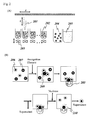
- Fig. 1 is a diagram explaining the isolation system and flow thereof.
- 101 is a first mixing tank in which the sample cell groups which comprise the sample, a recognition element, and buffers for cleaning and dilution are introduced and mixed.
- a target cell 111 cell group which has a CD4 cell surface antigen 113 and is indicated by a ⁇
- a cell 112 cell group which has a cell surface antigen 114 other than CD4 and is indicated by •
- a cell surface antigen CD4 recognition RNA aptamer 116, and recognition elements 117 modified by paramagnetic particles 115 are mixed.
- ingredients are mixed so that the concentration of recognition elements 117 is 110 nM.
- CD4 antigen 113 bonds with RNA aptamer 116 and target cells 111 and recognition elements 117 form an aggregate as a result. Unneeded drainage is controlled by valve 102 and flows out from a rear section of the first tank 101.
- first magnetic field isolation tank 104 A magnet 105 is provided in first magnetic field isolation tank 104 in order to generate a magnetic field. Rare earth neodymium should be used for this magnet 105. Furthermore, this magnet is movable so that the strength of the magnetic field of first magnetic field isolation tank 104 can be adjusted.
- target cells 111 which are bound to recognition element 117 having paramagnetic particles 115 as a carrier, are trapped in the magnetic field. Unbonded cells are controlled by valve 102 and run into the drain. As a result, only target cells 111 are recovered from the sample cell group.
- Magnet 105 is released from the first magnetic field isolation tank 104, the magnetic field is now unapplied, and buffer is introduced from the upstream section.
- the buffer used here has a composition of 10 mM HEPES (pH 7.4), 0.15M NaCl, 2 mM MgCl 2 , and 1 mg/ml BSA.
- the aggregate of target cells 111 and recognition elements 117 is mixed with 50 ⁇ /ml of Benzonase (registered trademark) nuclease in second mixture tank 106.
- Benzonase registered trademark
- the RNA aptamer forms a steric structure, there are instances in which a type of nuclease such as ribonuclease A that only digests single-strand RNA does not provide adequate digestion. It is effective to use an enzyme that digests both single-strand and double-strand RNA.
- the genetically-engineered, mass-produced, serratia marcescens-derived nuclease described in The Journal of Biological Chemistry 244, 5219-5225 (1969 ) is used.
- the product name is Benzonase (registered trademark) ( EP Patent No. 0229866 , U.S. Patent No. 5,173,418 ).
- This enzyme is easy to use on cells because it works at 37°C and its pH use range, 6 to 9, is in the neutral region. Highly concentrated phosphoric acid and univalent metallic ions cause a decline in enzymatic activity, so non phosphate buffer is used here. If phosphoric acid must be utilized, conditions are used wherein the potassium/sodium phosphate concentration is limited to 5 mM and 0.15 M NaCl, and 2 mM MgCl 2 , 1 mg/ml BSA are included.
- Benzonaze (registered trademark) nuclease is used in a quantity of 10 to 100 u/ml. Although a mixture of ribonuclease A and ribonuclease T1 can also be used, Serratia marcescens-derived nuclease is more versatile.
- the use of plasma can be considered instead of buffer.
- the nuclease inhibitor in plasma may have an effect. Consequently, it may be necessary to decrease the amount of Benzonase (registered trademark) nuclease additive in each plasma lot.
- Benzonase registered trademark
- good results are obtained with the use of 100 to 400 u/ml.
- RNA aptamer will decompose as indicated by the magnified image on the right side (here, indicated by the broken line leading to reference mark 119) and target cells 111 will isolate from recognition elements 117.
- the mixed sample After nuclease processing, the mixed sample, with target cells 111 and recognition elements 117 still isolated, is allowed to flow into second magnetic field isolation tank 107.
- a magnet 105 is also provided in second magnetic field isolation tank 107 in order to generate a magnetic field.
- paramagnetic particles 115 are trapped by the magnetic field in the second magnetic field isolation tank 107, and then only the isolated target cells 111 flow away.
- a light source 108 and a photon counter 109 are arranged on either side of a cell flow channel and the quantity of isolated target cells 111 is counted in real time.
- Scattered light detection may also be used as a method that can be used for measuring the quantity of cells.
- Pure cell surface antigen CD4 presenting cells 111 can be collected by recovering the solution containing the recovered target cells, reciprocating the solution, and removing degradation product 119 of the recognition substance RNA aptamer. Centrifugation is used in the reciprocation of the cell supernatant. Centrifugation is performed at 3000 rpm for 15 minutes to precipitate the cells, and then the supernatant is disposed of.
- RNA aptamers are bound to surface antigens, and the aptamers are removed by degradation with the ribonuclease that ordinarily exists in culture medium after cell recovery or excessively added ribonuclease, the aptamers will dissociate and the state of the proteins, etc. on the cell surface can be completely restored to its pre cell-labeled state without damage to the cell surface proteins, etc. that were used as aptamer bond targets.
- Figs 2(A) and 2(B) are figures that show a device example and a method in which cell surface antigen CD4 presenting cells are isolated and recovered with a magnetic field using recognition elements comprising paramagnetic particles modified with RNA aptamer that recognizes cell surface antigen CD4.
- Fig. 2(A) is a conceptual diagram showing a screening device.
- Sample tube 202, dispensing mechanism 201, and magnetic field generation mechanism 203 is the smallest constituent unit.
- a tube retaining recognition element solution 204 (3 ⁇ m paramagnetic particles 208 modified by an RNA aptamer that recognizes cell surface antigen CD4 is intermixed), and a tube 205 that retains a nuclease solution are prepared.
- the movement of dispensing mechanism 201 is controlled in the x direction, y direction (perpendicular to sheet surface), and z direction.
- magnetic field generation mechanism 203 is movable, and the strength of the magnetic field within sample tube 202 can be controlled.
- a rare earth magnet is used as a magnet for generating a magnetic field.
- the magnetic field generation mechanism may be located to the side of the sample tube.
- Fig. 2(B) shows a method in which this device is used to isolate and recover cell surface antigen CD4 presenting cells.
- Sample cell groups are inserted into sample tube 202.
- Cell surface antigen CD4 presenting cells 206 and cells 207, which have another surface antigen, are mixed together in sample cell groups.
- Dispensing mechanism 201 is used to dispense and stir recognition element solution 204. After a reaction is performed for an adequate amount of time while mixing by means of the dispensing mechanism, magnetic field generating mechanism 203 generates a magnetic field in sample tube 202. Cells that bound to recognition elements 208 are immobilized to the inner walls of the sample tube by magnetic force.
- dispensing mechanism 201 With a magnetic field applied, the supernatant is recovered with dispensing mechanism 201 and sample tube 202 is washed twice with a buffer or a culture solution. Subsequently, dispensing mechanism 201 adds nuclease solution 205 to sample tube 202. Adding nuclease digests the aptamer of recognition element 208 (indicated here by reference symbol 210). As a result, the cells 206 trapped on the walls of sample tube 202 and paramagnetic particles modified with aptamer and bound to aptamer are disassociated. By recovering the solution in which cells 206 were dissociated, cell surface antigen CD4 presenting cells are detached.
- a testing kit can be realized through the application of the present invention.
- cell surface antigen CD44 which binds to an RNA aptamer, is the recognition substance and paramagnetic particles (particle diameter 1 ⁇ m) are used as the recognition element carrier.
- paramagnetic particles particle diameter 1 ⁇ m
- fecal matter which is the test specimen, is suspended in a buffer of 1 mL of 10 mM PBS (pH 7.5), 0.15 M NaCL, 1% BSA and used as a sample.
- RNA aptamer-modified paramagnetic particles (recognition element) are added to the sample solution, which is gently stirred for 30 minutes.
- the suspension is fed into a tube with an internal diameter of 2mm, and magnetic particles moving inside the tube, which has a neodymium magnet array arranged at 1 cm intervals, are trapped.
- the collected paramagnetic particles are washed in culture fluid and Benzonase (registered trademark) nuclease is added.
- Isolated live cells are cultured in a cell culture microchamber, for example, the cell culture microchamber proposed by the inventors of the present application and described in Japanese Patent Application No. 2004-305258 . If the cells are cancerous, they will be able to withstand prolonged incubation, and dividing cells will soon start to appear.
- cancer-derived cells are isolated from cells in fecal matter.
- cancerous sections cannot be determined by isolation of a cancer-derived cell from the fecal matter and incubation for a given period of time, the fact that there is a lesion in the body can be ascertained. Further, the effectiveness of anticancer drugs, etc. can be tested simultaneously.
- the present invention With the present invention, loss of cell function can be avoided during the classification process when it is necessary to identify or isolate cells.
- the present invention is especially useful as a cell identification/isolation method, a cell screening method and a reagent kit for cell isolation used in regenerative medicine and cell implants.
Landscapes
- Health & Medical Sciences (AREA)
- Life Sciences & Earth Sciences (AREA)
- Engineering & Computer Science (AREA)
- Immunology (AREA)
- Chemical & Material Sciences (AREA)
- Biomedical Technology (AREA)
- Molecular Biology (AREA)
- Hematology (AREA)
- Urology & Nephrology (AREA)
- Microbiology (AREA)
- Cell Biology (AREA)
- Biotechnology (AREA)
- General Health & Medical Sciences (AREA)
- Biochemistry (AREA)
- Physics & Mathematics (AREA)
- Analytical Chemistry (AREA)
- Medicinal Chemistry (AREA)
- Food Science & Technology (AREA)
- General Physics & Mathematics (AREA)
- Pathology (AREA)
- Zoology (AREA)
- Genetics & Genomics (AREA)
- Wood Science & Technology (AREA)
- Bioinformatics & Cheminformatics (AREA)
- Organic Chemistry (AREA)
- General Engineering & Computer Science (AREA)
- Tropical Medicine & Parasitology (AREA)
- Virology (AREA)
- Measuring Or Testing Involving Enzymes Or Micro-Organisms (AREA)
- Micro-Organisms Or Cultivation Processes Thereof (AREA)
Applications Claiming Priority (1)
| Application Number | Priority Date | Filing Date | Title |
|---|---|---|---|
| PCT/JP2007/055008 WO2008108006A1 (ja) | 2007-03-07 | 2007-03-07 | 細胞分離方法ならびに細胞検査方法とその試薬キット |
Publications (2)
| Publication Number | Publication Date |
|---|---|
| EP2133418A1 true EP2133418A1 (de) | 2009-12-16 |
| EP2133418A4 EP2133418A4 (de) | 2010-04-07 |
Family
ID=39737910
Family Applications (1)
| Application Number | Title | Priority Date | Filing Date |
|---|---|---|---|
| EP07738479A Withdrawn EP2133418A4 (de) | 2007-03-07 | 2007-03-07 | Zellisolierungsverfahren, zelltestverfahren und reagenskit dafür |
Country Status (3)
| Country | Link |
|---|---|
| US (1) | US20100092969A1 (de) |
| EP (1) | EP2133418A4 (de) |
| WO (1) | WO2008108006A1 (de) |
Families Citing this family (4)
| Publication number | Priority date | Publication date | Assignee | Title |
|---|---|---|---|---|
| US10155988B2 (en) * | 2010-04-07 | 2018-12-18 | Board Of Regents, The University Of Texas System | Methods of detecting tumor cells |
| JP2013202018A (ja) * | 2012-03-29 | 2013-10-07 | Kanagawa Academy Of Science & Technology | 細胞機能制御方法 |
| CN106269223B (zh) * | 2016-08-11 | 2017-11-10 | 冷彦宁 | 磁分离方法以及磁分离装置 |
| JP7231229B2 (ja) * | 2017-03-23 | 2023-03-01 | デューク ユニバーシティ | 細胞外アプタマー染色のアンチドート媒介性解除 |
Family Cites Families (8)
| Publication number | Priority date | Publication date | Assignee | Title |
|---|---|---|---|---|
| US5173418A (en) | 1985-05-10 | 1992-12-22 | Benzon Pharma, A/S | Production in Escherichia coli of extracellular Serratia spp. hydrolases |
| IL78703A (en) | 1985-05-10 | 1993-02-21 | Benzon Alfred | Method of producing extracellular enzymes, the enzymes produced thereby and compositions containing them; a hybrid plasmid and a dna fragment comprising dna encoding the enzymes; a microorganism harbouring the plasmid; and a method for removing nucleic acids from biological materials |
| US20010008760A1 (en) * | 1998-08-25 | 2001-07-19 | Chester F. King | Reagent system and kit for detecting hiv infected cells |
| US6821790B2 (en) * | 1999-03-02 | 2004-11-23 | Vijay Mahant | Methods and apparatus for separation of biological fluids |
| US20040018611A1 (en) * | 2002-07-23 | 2004-01-29 | Ward Michael Dennis | Microfluidic devices for high gradient magnetic separation |
| JP2004226359A (ja) | 2003-01-27 | 2004-08-12 | Seiko Epson Corp | 半導体装置の検査方法 |
| JP4205470B2 (ja) | 2003-04-02 | 2009-01-07 | 帝人株式会社 | 検査装置、治療システム |
| JP4677254B2 (ja) * | 2005-03-07 | 2011-04-27 | 一般社団法人オンチップ・セロミクス・コンソーシアム | 細胞分離方法、細胞識別方法ならびに細胞検査方法 |
-
2007
- 2007-03-07 EP EP07738479A patent/EP2133418A4/de not_active Withdrawn
- 2007-03-07 US US12/530,154 patent/US20100092969A1/en not_active Abandoned
- 2007-03-07 WO PCT/JP2007/055008 patent/WO2008108006A1/ja not_active Ceased
Also Published As
| Publication number | Publication date |
|---|---|
| EP2133418A4 (de) | 2010-04-07 |
| US20100092969A1 (en) | 2010-04-15 |
| WO2008108006A1 (ja) | 2008-09-12 |
Similar Documents
| Publication | Publication Date | Title |
|---|---|---|
| TWI577389B (zh) | 使用多專一性捕捉及雞尾酒檢測試劑檢測胰臟病患之循環腫瘤細胞的方法及套組 | |
| JP5766168B2 (ja) | 循環ガン細胞の迅速かつ効率的な単離のための方法および試薬 | |
| JP5814344B2 (ja) | 体液からの標的分析物の単離 | |
| US8697435B2 (en) | Integrated sample preparation and analyte detection | |
| EP2725359B1 (de) | Zelltrennungsverfahren mit einem Freisetzungssystem für Zell-Antikörper-Substrat-Konjugate mit einer Polyethylenglycol-Distanzeinheit | |
| EP3336546B1 (de) | Umkehrbare zellmarkierung mit konjugaten mit zwei lösbaren bindungsstellen | |
| WO2007095279A2 (en) | Dual nanoparticle assay for detection and separation of biological species | |
| JP2004504129A (ja) | 磁性ナノ粒子の制御された凝集による増大した分離効率 | |
| KR102084688B1 (ko) | 다중 프로브 혼성화를 이용한 미생물 검출 방법 | |
| KR102190922B1 (ko) | 핵산 분리 방법 | |
| WO2020219557A1 (en) | Magnetic platelet probes to detect circulating tumor cells | |
| JP2024023284A (ja) | がんのスクリーニング、診断、治療、及び再発における巨細胞の核酸の特徴付けの使用方法 | |
| EP2133418A1 (de) | Zellisolierungsverfahren, zelltestverfahren und reagenskit dafür | |
| EP3480320A1 (de) | Signalsonde einer doppelsträngigen nukleinsäure und verfahren zum nachweis von zielmolekülen unter verwendung davon | |
| JP4542502B2 (ja) | 細胞分離方法ならびに細胞検査方法とその試薬キット | |
| JP4677254B2 (ja) | 細胞分離方法、細胞識別方法ならびに細胞検査方法 | |
| Elaissari et al. | Biomedical Application | |
| US9347863B2 (en) | Phosphorothioate oligonucleotide-labeling of white blood cells | |
| EP4424842A1 (de) | Verfahren zum nachweis von nukleinsäureenden | |
| JP5080434B2 (ja) | 生物学的試料から標的生体部分を単離する方法およびそのためのキット | |
| Sathe et al. | Integrating magnetic and optical nanotechnology for selective capture and multiplexed analysis of rare tumor cells | |
| Elaissari et al. | Latexes: Magnetic | |
| WO2026073060A1 (en) | Ultra sensitive probes for detecting biomarkers | |
| JP2009247320A (ja) | 細胞分離方法 | |
| Bradbury et al. | Magnetic and fluorescence-encoded polystyrene microparticles for cell separation |
Legal Events
| Date | Code | Title | Description |
|---|---|---|---|
| PUAI | Public reference made under article 153(3) epc to a published international application that has entered the european phase |
Free format text: ORIGINAL CODE: 0009012 |
|
| 17P | Request for examination filed |
Effective date: 20091005 |
|
| AK | Designated contracting states |
Kind code of ref document: A1 Designated state(s): AT BE BG CH CY CZ DE DK EE ES FI FR GB GR HU IE IS IT LI LT LU LV MC MT NL PL PT RO SE SI SK TR |
|
| A4 | Supplementary search report drawn up and despatched |
Effective date: 20100304 |
|
| DAX | Request for extension of the european patent (deleted) | ||
| STAA | Information on the status of an ep patent application or granted ep patent |
Free format text: STATUS: THE APPLICATION IS DEEMED TO BE WITHDRAWN |
|
| 18D | Application deemed to be withdrawn |
Effective date: 20100605 |