EP2130497A1 - Extraction de fonction anatomique à partir d'une image de foie à ultrasons - Google Patents
Extraction de fonction anatomique à partir d'une image de foie à ultrasons Download PDFInfo
- Publication number
- EP2130497A1 EP2130497A1 EP09161527A EP09161527A EP2130497A1 EP 2130497 A1 EP2130497 A1 EP 2130497A1 EP 09161527 A EP09161527 A EP 09161527A EP 09161527 A EP09161527 A EP 09161527A EP 2130497 A1 EP2130497 A1 EP 2130497A1
- Authority
- EP
- European Patent Office
- Prior art keywords
- image
- dimensional
- vessel
- ultrasound
- diaphragm
- Prior art date
- Legal status (The legal status is an assumption and is not a legal conclusion. Google has not performed a legal analysis and makes no representation as to the accuracy of the status listed.)
- Withdrawn
Links
- 238000002604 ultrasonography Methods 0.000 title claims abstract description 82
- 210000004185 liver Anatomy 0.000 title claims abstract description 57
- 238000000605 extraction Methods 0.000 title description 13
- 239000000284 extract Substances 0.000 claims abstract description 8
- 238000007670 refining Methods 0.000 claims abstract description 4
- 238000001914 filtration Methods 0.000 claims description 15
- 230000000873 masking effect Effects 0.000 claims description 9
- 238000000034 method Methods 0.000 claims description 9
- 230000002708 enhancing effect Effects 0.000 claims description 8
- 238000009792 diffusion process Methods 0.000 claims description 6
- 230000000877 morphologic effect Effects 0.000 description 6
- 239000011159 matrix material Substances 0.000 description 5
- 238000010586 diagram Methods 0.000 description 3
- 238000012986 modification Methods 0.000 description 3
- 230000004048 modification Effects 0.000 description 3
- 230000011218 segmentation Effects 0.000 description 3
- 230000015556 catabolic process Effects 0.000 description 2
- 238000006731 degradation reaction Methods 0.000 description 2
- 230000003628 erosive effect Effects 0.000 description 2
- 210000001631 vena cava inferior Anatomy 0.000 description 2
- 230000003044 adaptive effect Effects 0.000 description 1
- 230000007812 deficiency Effects 0.000 description 1
- 230000001066 destructive effect Effects 0.000 description 1
- 230000010339 dilation Effects 0.000 description 1
- 230000000694 effects Effects 0.000 description 1
- 210000000056 organ Anatomy 0.000 description 1
- 238000003325 tomography Methods 0.000 description 1
Images


Classifications
-
- G—PHYSICS
- G06—COMPUTING; CALCULATING OR COUNTING
- G06T—IMAGE DATA PROCESSING OR GENERATION, IN GENERAL
- G06T5/00—Image enhancement or restoration
- G06T5/20—Image enhancement or restoration by the use of local operators
- G06T5/30—Erosion or dilatation, e.g. thinning
-
- A—HUMAN NECESSITIES
- A61—MEDICAL OR VETERINARY SCIENCE; HYGIENE
- A61B—DIAGNOSIS; SURGERY; IDENTIFICATION
- A61B8/00—Diagnosis using ultrasonic, sonic or infrasonic waves
- A61B8/08—Detecting organic movements or changes, e.g. tumours, cysts, swellings
-
- A—HUMAN NECESSITIES
- A61—MEDICAL OR VETERINARY SCIENCE; HYGIENE
- A61B—DIAGNOSIS; SURGERY; IDENTIFICATION
- A61B8/00—Diagnosis using ultrasonic, sonic or infrasonic waves
- A61B8/44—Constructional features of the ultrasonic, sonic or infrasonic diagnostic device
- A61B8/4416—Constructional features of the ultrasonic, sonic or infrasonic diagnostic device related to combined acquisition of different diagnostic modalities, e.g. combination of ultrasound and X-ray acquisitions
-
- A—HUMAN NECESSITIES
- A61—MEDICAL OR VETERINARY SCIENCE; HYGIENE
- A61B—DIAGNOSIS; SURGERY; IDENTIFICATION
- A61B8/00—Diagnosis using ultrasonic, sonic or infrasonic waves
- A61B8/48—Diagnostic techniques
- A61B8/483—Diagnostic techniques involving the acquisition of a 3D volume of data
-
- G—PHYSICS
- G06—COMPUTING; CALCULATING OR COUNTING
- G06T—IMAGE DATA PROCESSING OR GENERATION, IN GENERAL
- G06T17/00—Three dimensional [3D] modelling, e.g. data description of 3D objects
-
- G06T5/70—
-
- G06T5/94—
-
- G—PHYSICS
- G06—COMPUTING; CALCULATING OR COUNTING
- G06T—IMAGE DATA PROCESSING OR GENERATION, IN GENERAL
- G06T7/00—Image analysis
- G06T7/10—Segmentation; Edge detection
- G06T7/11—Region-based segmentation
-
- G—PHYSICS
- G06—COMPUTING; CALCULATING OR COUNTING
- G06T—IMAGE DATA PROCESSING OR GENERATION, IN GENERAL
- G06T7/00—Image analysis
- G06T7/10—Segmentation; Edge detection
- G06T7/187—Segmentation; Edge detection involving region growing; involving region merging; involving connected component labelling
-
- G—PHYSICS
- G06—COMPUTING; CALCULATING OR COUNTING
- G06T—IMAGE DATA PROCESSING OR GENERATION, IN GENERAL
- G06T7/00—Image analysis
- G06T7/30—Determination of transform parameters for the alignment of images, i.e. image registration
-
- G—PHYSICS
- G06—COMPUTING; CALCULATING OR COUNTING
- G06T—IMAGE DATA PROCESSING OR GENERATION, IN GENERAL
- G06T2207/00—Indexing scheme for image analysis or image enhancement
- G06T2207/10—Image acquisition modality
- G06T2207/10132—Ultrasound image
- G06T2207/10136—3D ultrasound image
-
- G—PHYSICS
- G06—COMPUTING; CALCULATING OR COUNTING
- G06T—IMAGE DATA PROCESSING OR GENERATION, IN GENERAL
- G06T2207/00—Indexing scheme for image analysis or image enhancement
- G06T2207/20—Special algorithmic details
- G06T2207/20172—Image enhancement details
- G06T2207/20192—Edge enhancement; Edge preservation
-
- G—PHYSICS
- G06—COMPUTING; CALCULATING OR COUNTING
- G06T—IMAGE DATA PROCESSING OR GENERATION, IN GENERAL
- G06T2207/00—Indexing scheme for image analysis or image enhancement
- G06T2207/30—Subject of image; Context of image processing
- G06T2207/30004—Biomedical image processing
- G06T2207/30056—Liver; Hepatic
Definitions
- the present disclosure relates to anatomical feature extractions, and more particularly to an anatomical feature extraction from a 3-dimensional B-mode ultrasound liver image.
- An ultrasound diagnostic system has been extensively used in the medical field due to its non-invasive and non-destructive nature.
- the ultrasound diagnostic system is highly safe and has no dangerous side effects such as exposure to X-rays, etc.
- the ultrasound diagnostic system suffers from inherent shortcomings of an ultrasound image such as a low signal-to-noise ratio and a limited field of view.
- the image registration of a CT (or MR) image onto the ultrasound image has been introduced in order to compensate for deficiencies of the ultrasound image.
- a 3-dimensional ultrasound liver image may include obstacles to deter anatomical feature extractions such as speckle noises, mirroring image artifacts, shading artifacts or appearance of other organs in a region of interest. These obstacles may cause a registration error during the ultrasound-CT image registration.
- a system for extracting anatomical features from an ultrasound image comprises: an image forming unit configured to form a 3-dimensional ultrasound liver image based on ultrasound signals reflected from a liver; a diaphragm extracting unit configured to extract a diaphragm region from the 3-dimensional ultrasound liver image; a vessel extracting unit configured to extract vessel regions from the 3-dimensional ultrasound liver image; and a diaphragm refining unit configured to remove clutters from the diaphragm region based on the extracted vessel regions to thereby refine the extracted diaphragm region.
- a method of extracting anatomical features from an ultrasound image comprises: a) forming a 3-dimensional ultrasound liver image based on ultrasound signals reflected from a liver; b) extracting a diaphragm region from the 3-dimensional ultrasound liver image; c) extracting vessel regions from the 3-dimensional ultrasound liver image; d) removing clutters from the diaphragm region based on the extracted vessel regions to thereby refine the extracted diaphragm region; and e) extracting sample points from the diaphragm region with the clutters removed and the vessel regions.
- an anatomical feature extraction from an ultrasound image of a target object for image registration e.g., image registration between an ultrasound image and a computerized tomography (CT) image
- CT computerized tomography
- the target object may be a liver, although it is certainly not limited thereto.
- the ultrasound image of the target object may be a 3-dimensional brightness (B)-mode liver image.
- a system 100 may include an image forming unit 110.
- the image forming unit may be configured to transmit/receive ultrasound signals to/from the target object (e.g., liver) to thereby output electrical receive signals.
- the image forming unit 110 may be further configured to form a 3-dimensional ultrasound image of the target object.
- the 3-dimensional ultrasound image of the target object will be referred to as a 3-dimensional B-mode ultrasound liver image.
- the system 100 may further include an image enhancing unit 120.
- the image enhancing unit 120 may be configured to receive observed signals for forming the 3-dimensional B-mode ultrasound liver image from the image forming unit 110.
- the image enhancing unit 120 may be further configured to perform speckle noise filtering to remove speckle noises contained in the observed signals.
- the image enhancing unit 120 may be also configured to increase an edge contrast to thereby enhance anatomical feature definition in the 3-dimensional B-mode ultrasound liver image with the speckle noise removed.
- the speckle noise filtering may be carried out by applying the total variation based speckle constraint filtering algorithm in combination with an anisotropic diffusion to the 3-dimensional B-mode ultrasound liver image. This speckle noise filtering may be performed such as the following equations (1) to (5).
- the observed signals u 0 containing the speckle noises may be modeled as the following equation (1).
- u 0 u + u ⁇ n
- n represents a zero-mean Gaussian variable having standard deviation
- u represents original signals not containing the speckle noises.
- n represents a zero-mean Gaussian variable having standard deviation
- u represents original signals not containing the speckle noises.
- u ⁇ d ⁇ ⁇ ⁇ n 2
- ⁇ represents a dimension of the image and ⁇ n represents the standard deviation.
- the following equation (3) represents the total variation of the original signals u. ⁇ ⁇ ⁇ ⁇ u ⁇ d ⁇ ⁇
- the speckle noises may be removed to thereby obtain image signals close to the original signals u.
- the following equation (4) may be derived from the equations (2) and (3) by the constraint Euler-Lagrange equation.
- ⁇ u ⁇ t div F - ⁇ t ⁇ u 2 - u 0 2 u , in ⁇ ⁇
- ⁇ (t) represents an attachment coefficient for preventing that the image signals with the speckle noises removed steeply differ from the observed signals according to iteration.
- the equation (4) may be solved by applying these values, iteratively, under the assumption that ⁇ ul ⁇ t is 0 . This is so that image signals with the speckle noises removed, i.e., image signals u close to the original signals, may be obtained, iteratively.
- the solution of the equation (4) may be iteratively carried out about 40 times so that desirable image signals with the speckle noises removed may be obtained.
- the speckle noise filtering may be carried out in consideration with the orientations of the edges for preventing the degradation of the edge sharpness, i.e., while the sharpness of edges neighboring to feature points is maintained.
- FIG. 2A shows an example of an ultrasound liver image before removing the speckle noises
- FIG. 2B shows an example of an ultrasound liver image with the speckle noises removed.
- the system 100 may further include a diaphragm extracting unit 130.
- the diaphragm extracting unit may be configured to extract a diaphragm from the 3-dimensional B-mode ultrasound liver image with the edge contrast enhanced.
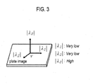
- the diaphragm may perform a Hessian matrix based flatness test upon the 3-dimensional B-mode ultrasound liver image to extract the diaphragm.
- the diaphragm may be considered as a curved surface in the 3-dimensional ultrasound liver image.
- regions in which a voxel intensity change in a normal direction at a surface is greater than a voxel intensity change in a horizontal direction at the surface may be extracted as the diaphragm.
- FIG. 3 is a schematic diagram showing an example of eigenvalues ⁇ 1 , ⁇ 2 and ⁇ 3 in the Hessian matrix.
- the diaphragm extracting unit 130 may be configured to select voxels having a relatively high flatness value.
- the voxles may be represented with pixels.
- the flatness ⁇ (v) may be defined as the following equation (7).
- ⁇ 1 v 1 - ⁇ 1 v ⁇ 3 v 2
- ⁇ 2 v 1 - ⁇ 2 v ⁇ 3 v 2
- ⁇ 3 v ⁇ i ⁇ i v 2
- ⁇ 1 , ⁇ 2 and ⁇ 3 denote eigenvalues of the Hessian matrix at voxel v .
- the flatness ⁇ (v) may be normalized to have values of ranging 0-1.
- a flatness map may be formed based on the flatness values obtained from all of the voxels according to the equations (6) and (7). Thereafter, the voxles having a relatively high flatness value are selected. In one embodiment, the voxels having the flatness over 0.1 may be selected.
- the diaphragm extracting unit 130 may be further configured to perform the morphological opening upon the selected voxels to remove small clutters therefrom.
- the morphological opening may be carried out by sequentially performing erosion and dilation. That is, morphological boundaries in which the voxel values exist are removed as many as the predetermined number of the voxels and then contracted (erosion). Thereafter, the boundaries are expanded as many as the predetermined number of the voxels. In one embodiment, the boundaries may be contracted and expanded by 1 voxel.
- the diaphragm is the largest surface in the 3-dimensional B-mode ultrasound liver image.
- the largest surface may be selected among candidate surfaces obtained by the intensity-based connected component analysis (CCA) for the voxles and the selected surface may be regarded as the diaphragm in the ultrasound liver image.
- CCA intensity-based connected component analysis
- the voxel-based CCA is one of the methods of grouping regions in which voxel values exist. For example, the number of voxels connected to each of the voxels through a connectivity test by referring to values of voxels neighboring to the corresponding voxel (e.g., 26 voxels) may be computed. The voxels, of which connected voxels are greater than the predetermined number, are selected as candidate groups.
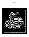
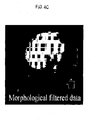
- FIGS. 4A to 4D show examples of images formed through diaphragm extraction.
- FIG. 4A shows an example of an ultrasound image with the diaphragm indicated
- FIG. 4B shows an example of an image obtained through the flatness test.
- FIG. 4C shows an example of an image obtained through morphological filtering
- FIG. 4D shows an example of a final selected diaphragm image.
- the system 100 may further include a vessel extracting unit 140 that may operable to perform vessel extraction upon the 3-dimensional B-mode ultrasound liver image with the edge contrast enhanced.
- the vessel extracting unit 140 may be configured to perform the vessel extraction through ROI masking, vessel segmentation and classification.
- the ROI masking may be applied to the 3-dimensional B-mode ultrasound liver image with the edge contrast enhanced by modeling the diaphragm as a polynomial curved surface.
- the ROI masking which models the diaphragm as the polynomial curved surface by using the least means square, may be used.
- the lower portion of the modeled polynomial curved surface may be eliminated with a marginal distance.
- the marginal distance may be set to about 10 vessels at a lower portion of the ROI mask.
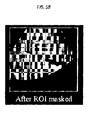
- FIG. 5A shows an example of an image before ROI masking
- FIG. 5B shows an example of an image with ROI masked.
- the vessel extracting unit 140 may be further configured to segment a vessel region and a non-vessel region.
- a low intensity bound having a less reference bound value in the ROI masked image may be estimated.
- voxels with a higher intensity value than the reference bound value may be removed.
- An adaptive threshold scheme may be applied to the remaining regions for binarization thereof.
- the binary segments may be labeled as vessel candidates.
- the vessel extracting unit 140 may be further configured to remove non-vessel-type clutters from the binarization image to classify real vessels from vessel candidates.
- the vessel classification may include a size test, which evaluates the goodness of fit to a cylindrical tube, for filtering out tiny background clutters, a structure-based vessel test for removing non-vessel type clutters, i.e., an initial vessel test, gradient magnitude analysis, and a final vessel test for perfectly removing the clutters.
- an initial threshold may be marginally set so that all vessels may be included. For example, the initial threshold may be set to 0.6.
- a threshold of the final vessel test may be set to 0.4.
- the system 100 may further include a diaphragm refining unit 150 that may be operable to refine the diaphragm region by removing the clutters with the extracted vessel data.
- the clutters are mainly placed near the vessel walls.
- the vessel wall of inferior vena cava (IVC) is more likely to be connected to the diaphragm and cause clutters. These clutters may degrade the accuracy of feature based registration so that the diaphragm should be refined.
- the vessel region is extracted according to the vessel extraction mentioned above, the extracted vessel region may be dilated, and then the dilated vessel region may be subtracted from the initially extracted diaphragm region to estimate vessel walls.
- the estimated vessel walls may be removed from the diaphragm region.
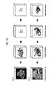
- FIG. 6 shows an example of images obtained through the vessel extraction. As shown in FIG. 6 , the vessel regions are extracted at arrow 61 and the extracted vessel regions are dilated at arrow 62. Then, the vessel walls are extracted from the dilated image at arrow 63. Finally, the diaphragm region may be extracted by applying CCA and the size test.
- the system 100 may include a registration unit 160 that may be operable to perform the image registration between the 3-dimensional ultrasound and CT liver images.
- the registration unit 160 may extract sample points from the vessel region and the diaphragm region, respectively, among the features extracted from the 3-dimensional B-mode ultrasound image. Also, after the vessel region and the diaphragm region are extracted from the CT image, the registration unit 160 may be operable to extract sample points from the vessel and the diaphragm region, respectively.
- the ICP based registration may be performed with the extracted sample points from the respective ultrasound and CT images for registration.
- FIG. 7A shows an example of images resulting from flatness test, morphological filtering, size test and refinement for the 3-dimensional B-mode ultrasound images A and B (Data A and B) with speckle noises removed.
- Data A having a size of 200*200*200 and isotropic voxel resolution of 0.8
- Data B having a size of 200*188*168 and isotropic voxel resolution of 0.6
- FIG. 7B shows an example of images resulting from segmentation of vessel candidates, initial vesselness test and final vesselness test for the 3-dimensional B-mode ultrasound images A and B (Data A and B) with speckle noises removed.
- the image registration error between the ultrasound-CT images may be reduced.
Applications Claiming Priority (1)
| Application Number | Priority Date | Filing Date | Title |
|---|---|---|---|
| KR20080053231 | 2008-06-05 |
Publications (1)
| Publication Number | Publication Date |
|---|---|
| EP2130497A1 true EP2130497A1 (fr) | 2009-12-09 |
Family
ID=41138991
Family Applications (1)
| Application Number | Title | Priority Date | Filing Date |
|---|---|---|---|
| EP09161527A Withdrawn EP2130497A1 (fr) | 2008-06-05 | 2009-05-29 | Extraction de fonction anatomique à partir d'une image de foie à ultrasons |
Country Status (4)
| Country | Link |
|---|---|
| US (1) | US20090306507A1 (fr) |
| EP (1) | EP2130497A1 (fr) |
| JP (1) | JP2009291618A (fr) |
| KR (1) | KR101017611B1 (fr) |
Cited By (4)
| Publication number | Priority date | Publication date | Assignee | Title |
|---|---|---|---|---|
| EP2317472A1 (fr) * | 2009-10-13 | 2011-05-04 | Medison Co., Ltd. | Système d'ultrasons générant une image basée sur une valeur de données de luminosité |
| CN108694705A (zh) * | 2018-07-05 | 2018-10-23 | 浙江大学 | 一种多帧图像配准与融合去噪的方法 |
| CN110136138A (zh) * | 2019-04-02 | 2019-08-16 | 中国人民解放军61540部队 | 基于点云区域分割的自适应滤波方法 |
| CN114224388A (zh) * | 2021-12-31 | 2022-03-25 | 山东大学 | 基于多频率超声的颈动脉三维重建方法及系统 |
Families Citing this family (29)
| Publication number | Priority date | Publication date | Assignee | Title |
|---|---|---|---|---|
| KR101121353B1 (ko) | 2009-08-03 | 2012-03-09 | 한국과학기술원 | 2차원 초음파 영상에 대응하는 2차원 ct 영상을 제공하는 시스템 및 방법 |
| WO2011025943A2 (fr) | 2009-08-28 | 2011-03-03 | Dartmouth College | Système et procédé de repérage d'un patient sans repères de cadre |
| CN102081697B (zh) | 2009-11-27 | 2013-12-11 | 深圳迈瑞生物医疗电子股份有限公司 | 一种在超声成像空间中定义感兴趣容积的方法及其装置 |
| US8483432B2 (en) | 2009-12-23 | 2013-07-09 | General Electric Company | Methods for automatic segmentation and temporal tracking |
| US9226729B2 (en) * | 2010-09-28 | 2016-01-05 | Fujifilm Corporation | Ultrasound diagnostic system, ultrasound image generation apparatus, and ultrasound image generation method |
| US9098904B2 (en) | 2010-11-15 | 2015-08-04 | Dartmouth College | System and method for registering ultrasound and magnetic resonance images |
| CN103338707B (zh) * | 2011-01-26 | 2015-09-30 | 株式会社日立医疗器械 | 超声波诊断装置以及图像处理方法 |
| WO2012175010A1 (fr) * | 2011-06-24 | 2012-12-27 | Technicolor (China) Technology Co., Ltd. | Procédé et dispositif de traitement d'une image |
| TWI446897B (zh) | 2011-08-19 | 2014-08-01 | Ind Tech Res Inst | 超音波影像對齊裝置及其方法 |
| US20130303915A1 (en) * | 2012-04-26 | 2013-11-14 | dBMEDx INC | Ultrasound apparatus and methods to monitor bodily vessels |
| KR101768526B1 (ko) | 2012-07-27 | 2017-08-17 | 삼성전자주식회사 | 혈관 구조에 기초하여 환자에 특화된 대상 장기의 모델을 생성하는 방법 및 장치 |
| KR101932721B1 (ko) * | 2012-09-07 | 2018-12-26 | 삼성전자주식회사 | 의료 영상들의 정합 방법 및 장치 |
| KR101989156B1 (ko) | 2012-11-01 | 2019-06-13 | 삼성전자주식회사 | 장기의 영상에서 장기에 포함된 객체의 영상을 분리하는 방법, 장치 및 의료 영상 시스템 |
| KR102001219B1 (ko) | 2012-11-26 | 2019-07-17 | 삼성전자주식회사 | 의료 영상들의 정합 방법 및 장치 |
| KR102025756B1 (ko) * | 2013-01-04 | 2019-09-27 | 삼성전자주식회사 | 영상에서 스펙클을 제거하는 방법, 장치 및 시스템. |
| CN103961135B (zh) | 2013-02-04 | 2017-04-12 | 通用电气公司 | 用于侦测三维超声图像中导管位置的系统及方法 |
| KR102042202B1 (ko) * | 2013-02-25 | 2019-11-08 | 삼성전자주식회사 | 의료영상에서 병변을 분할하는 장치 및 방법 |
| KR102205898B1 (ko) * | 2013-09-04 | 2021-01-21 | 삼성전자주식회사 | 의료영상들을 정합하는 방법 및 장치 |
| KR20150058672A (ko) | 2013-11-19 | 2015-05-29 | 삼성전자주식회사 | 엑스선 영상 장치 및 그 제어 방법 |
| CN106030657B (zh) * | 2014-02-19 | 2019-06-28 | 皇家飞利浦有限公司 | 医学4d成像中的运动自适应可视化 |
| KR20150108701A (ko) | 2014-03-18 | 2015-09-30 | 삼성전자주식회사 | 의료 영상 내 해부학적 요소 시각화 시스템 및 방법 |
| CN105917389B (zh) * | 2014-03-21 | 2018-10-30 | 圣犹达医疗用品心脏病学部门有限公司 | 用于生成几何结构的多维表面模型的方法和系统 |
| US8958623B1 (en) | 2014-04-29 | 2015-02-17 | Heartflow, Inc. | Systems and methods for correction of artificial deformation in anatomic modeling |
| KR102250086B1 (ko) | 2014-05-16 | 2021-05-10 | 삼성전자주식회사 | 의료 영상 정합 방법, 이를 포함하는 장치 및 컴퓨터 기록 매체 |
| JP6560597B2 (ja) * | 2014-11-28 | 2019-08-14 | ジーイー・メディカル・システムズ・グローバル・テクノロジー・カンパニー・エルエルシー | 超音波画像の処理方法及び超音波画像の処理装置 |
| KR101876643B1 (ko) * | 2016-11-23 | 2018-07-13 | 한국과학기술원 | 2d 형광 투시 영상과 3d ct 영상 정합 기반의 치료 가이딩 시스템 및 방법 |
| KR102375910B1 (ko) * | 2020-03-02 | 2022-03-16 | 연세대학교 산학협력단 | 초음파 영상유도 방법 및 장치 |
| CN111369627B (zh) * | 2020-03-05 | 2023-04-07 | 电子科技大学 | 一种非侵入式散斑定向成像方法 |
| CN117557785B (zh) * | 2024-01-11 | 2024-04-02 | 宁波海上鲜信息技术股份有限公司 | 一种基于图像处理的远距离渔船船牌识别方法 |
Citations (3)
| Publication number | Priority date | Publication date | Assignee | Title |
|---|---|---|---|---|
| EP0991015A1 (fr) * | 1998-09-29 | 2000-04-05 | Koninklijke Philips Electronics N.V. | Procédé de traitement d'image médicale d'ultrasons de structure osseuse et dispositif pour chirurgie assistée par ordinateur |
| US20030236460A1 (en) * | 2002-06-25 | 2003-12-25 | Siemens Medical Solutions Usa, Inc. | Adaptive ultrasound image fusion |
| US20070167784A1 (en) * | 2005-12-13 | 2007-07-19 | Raj Shekhar | Real-time Elastic Registration to Determine Temporal Evolution of Internal Tissues for Image-Guided Interventions |
Family Cites Families (8)
| Publication number | Priority date | Publication date | Assignee | Title |
|---|---|---|---|---|
| US4887306A (en) * | 1987-11-04 | 1989-12-12 | Advanced Technology Laboratories, Inc. | Adaptive temporal filter for ultrasound imaging system |
| JP4907798B2 (ja) * | 2001-08-24 | 2012-04-04 | 株式会社東芝 | 超音波診断装置 |
| JP4373698B2 (ja) | 2003-04-25 | 2009-11-25 | 株式会社東芝 | 超音波診断装置及び超音波診断支援プログラム |
| CA2558650A1 (fr) | 2004-03-08 | 2005-09-22 | The Johns Hopkins University | Dispositif et procede de formation et d'evaluation medicaux |
| KR101411639B1 (ko) * | 2007-09-11 | 2014-06-24 | 삼성전기주식회사 | 영상 정합 방법 및 장치 |
| KR101121396B1 (ko) * | 2009-07-31 | 2012-03-05 | 한국과학기술원 | 2차원 초음파 영상에 대응하는 2차원 ct 영상을 제공하는 시스템 및 방법 |
| KR101121286B1 (ko) * | 2009-07-31 | 2012-03-23 | 한국과학기술원 | 센서의 교정을 수행하는 초음파 시스템 및 방법 |
| KR101121353B1 (ko) * | 2009-08-03 | 2012-03-09 | 한국과학기술원 | 2차원 초음파 영상에 대응하는 2차원 ct 영상을 제공하는 시스템 및 방법 |
-
2009
- 2009-05-29 EP EP09161527A patent/EP2130497A1/fr not_active Withdrawn
- 2009-06-02 US US12/477,072 patent/US20090306507A1/en not_active Abandoned
- 2009-06-05 JP JP2009136263A patent/JP2009291618A/ja active Pending
- 2009-06-05 KR KR1020090050032A patent/KR101017611B1/ko active IP Right Grant
Patent Citations (3)
| Publication number | Priority date | Publication date | Assignee | Title |
|---|---|---|---|---|
| EP0991015A1 (fr) * | 1998-09-29 | 2000-04-05 | Koninklijke Philips Electronics N.V. | Procédé de traitement d'image médicale d'ultrasons de structure osseuse et dispositif pour chirurgie assistée par ordinateur |
| US20030236460A1 (en) * | 2002-06-25 | 2003-12-25 | Siemens Medical Solutions Usa, Inc. | Adaptive ultrasound image fusion |
| US20070167784A1 (en) * | 2005-12-13 | 2007-07-19 | Raj Shekhar | Real-time Elastic Registration to Determine Temporal Evolution of Internal Tissues for Image-Guided Interventions |
Non-Patent Citations (2)
| Title |
|---|
| HEINZ U. LEMKE: "Preface", PROCEEDINGS CARS 2008,, vol. 3, no. SUPPL. 1, 27 May 2008 (2008-05-27), pages 1 - 2 * |
| NAM W H ET AL: "Anatomical feature extraction in 3D B-mode ultrasound liver images for CT-ultrasound image registration", PROCEEDINGS CARS 2008,, vol. 3, no. SUPPL. 1, 27 May 2008 (2008-05-27), pages 401 - 402, XP002602575, Retrieved from the Internet <URL:http://www-isl.kaist.ac.kr/Papers/IC/ic120.pdf> * |
Cited By (5)
| Publication number | Priority date | Publication date | Assignee | Title |
|---|---|---|---|---|
| EP2317472A1 (fr) * | 2009-10-13 | 2011-05-04 | Medison Co., Ltd. | Système d'ultrasons générant une image basée sur une valeur de données de luminosité |
| CN108694705A (zh) * | 2018-07-05 | 2018-10-23 | 浙江大学 | 一种多帧图像配准与融合去噪的方法 |
| CN110136138A (zh) * | 2019-04-02 | 2019-08-16 | 中国人民解放军61540部队 | 基于点云区域分割的自适应滤波方法 |
| CN114224388A (zh) * | 2021-12-31 | 2022-03-25 | 山东大学 | 基于多频率超声的颈动脉三维重建方法及系统 |
| CN114224388B (zh) * | 2021-12-31 | 2023-07-28 | 山东大学 | 基于多频率超声的颈动脉三维重建方法及系统 |
Also Published As
| Publication number | Publication date |
|---|---|
| KR20090127100A (ko) | 2009-12-09 |
| KR101017611B1 (ko) | 2011-02-28 |
| US20090306507A1 (en) | 2009-12-10 |
| JP2009291618A (ja) | 2009-12-17 |
Similar Documents
| Publication | Publication Date | Title |
|---|---|---|
| EP2130497A1 (fr) | Extraction de fonction anatomique à partir d'une image de foie à ultrasons | |
| KR101121286B1 (ko) | 센서의 교정을 수행하는 초음파 시스템 및 방법 | |
| Rajinikanth et al. | An approach to examine magnetic resonance angiography based on Tsallis entropy and deformable snake model | |
| KR101121396B1 (ko) | 2차원 초음파 영상에 대응하는 2차원 ct 영상을 제공하는 시스템 및 방법 | |
| EP1851722B1 (fr) | Dispositif et procede de traitement d'image | |
| US7711167B2 (en) | Fissure detection methods for lung lobe segmentation | |
| US8447383B2 (en) | System and method for providing 2-dimensional computerized-tomography image corresponding to 2-dimensional ultrasound image | |
| Zheng et al. | Machine learning based vesselness measurement for coronary artery segmentation in cardiac CT volumes | |
| US8068655B2 (en) | Method and system for vessel enhancement and artifact reduction in TOF MR angiography of brain | |
| CN107527341B (zh) | 血管造影图像的处理方法和系统 | |
| EP2869261B1 (fr) | Procédé de traitement de données d'image représentant un volume tridimensionnel | |
| Athanasiou et al. | Three-dimensional reconstruction of coronary arteries and plaque morphology using CT angiography–comparison and registration with IVUS | |
| WO2012040410A2 (fr) | Procédé et système pour la détection des lésions du foie | |
| EP2372646B1 (fr) | Dispositif de traitement d'image, procédé et programme | |
| KR101514003B1 (ko) | 폐엽 추출 방법 및 그 장치 | |
| EP2446418A1 (fr) | Procédé et système de segmentation d'une image du cerveau | |
| Metz et al. | Semi-automatic coronary artery centerline extraction in computed tomography angiography data | |
| Shamonin et al. | Automatic lung lobe segmentation of COPD patients using iterative B-spline fitting | |
| KR101494975B1 (ko) | 3차원 자동 유방 초음파 영상의 유두 자동 검출 시스템 및 그 검출 방법 | |
| You et al. | Extraction of samples from airway and vessel trees in 3D lung CT based on a multi-scale principal curve tracing algorithm | |
| Yoshida et al. | Bayesian wavelet snake for computer-aided diagnosis of lung nodules | |
| Abdallah | Segmentation of salivary glands in nuclear medicine images using edge detection tools | |
| Renard et al. | Coronary artery extraction and analysis for detection of soft plaques in MDCT images | |
| Farahani et al. | Carotid modeling and stenosis detection | |
| Rauf et al. | Brain Aneurysm Extraction in MRI Images |
Legal Events
| Date | Code | Title | Description |
|---|---|---|---|
| PUAI | Public reference made under article 153(3) epc to a published international application that has entered the european phase |
Free format text: ORIGINAL CODE: 0009012 |
|
| AK | Designated contracting states |
Kind code of ref document: A1 Designated state(s): AT BE BG CH CY CZ DE DK EE ES FI FR GB GR HR HU IE IS IT LI LT LU LV MC MK MT NL NO PL PT RO SE SI SK TR |
|
| 17P | Request for examination filed |
Effective date: 20100512 |
|
| 17Q | First examination report despatched |
Effective date: 20100610 |
|
| STAA | Information on the status of an ep patent application or granted ep patent |
Free format text: STATUS: THE APPLICATION HAS BEEN WITHDRAWN |
|
| 18W | Application withdrawn |
Effective date: 20160715 |