EP2113561A1 - Procédés rapides et kits pour la purification de lymphocytes T régulateurs Foxp3+ - Google Patents
Procédés rapides et kits pour la purification de lymphocytes T régulateurs Foxp3+ Download PDFInfo
- Publication number
- EP2113561A1 EP2113561A1 EP08008255A EP08008255A EP2113561A1 EP 2113561 A1 EP2113561 A1 EP 2113561A1 EP 08008255 A EP08008255 A EP 08008255A EP 08008255 A EP08008255 A EP 08008255A EP 2113561 A1 EP2113561 A1 EP 2113561A1
- Authority
- EP
- European Patent Office
- Prior art keywords
- cells
- antibody
- foxp3
- cd49d
- treg
- Prior art date
- Legal status (The legal status is an assumption and is not a legal conclusion. Google has not performed a legal analysis and makes no representation as to the accuracy of the status listed.)
- Withdrawn
Links
- 210000003289 regulatory T cell Anatomy 0.000 title claims abstract description 150
- 238000000034 method Methods 0.000 title claims abstract description 72
- 238000002955 isolation Methods 0.000 title claims abstract description 43
- 210000003819 peripheral blood mononuclear cell Anatomy 0.000 claims abstract description 68
- 210000004698 lymphocyte Anatomy 0.000 claims abstract description 15
- 239000012530 fluid Substances 0.000 claims abstract description 7
- 210000004027 cell Anatomy 0.000 claims description 200
- 101000994375 Homo sapiens Integrin alpha-4 Proteins 0.000 claims description 69
- 102100032818 Integrin alpha-4 Human genes 0.000 claims description 69
- 210000001744 T-lymphocyte Anatomy 0.000 claims description 55
- 238000000926 separation method Methods 0.000 claims description 32
- 238000001943 fluorescence-activated cell sorting Methods 0.000 claims description 23
- 230000001105 regulatory effect Effects 0.000 claims description 12
- 230000000295 complement effect Effects 0.000 claims description 7
- 230000001900 immune effect Effects 0.000 claims description 7
- 238000000684 flow cytometry Methods 0.000 claims description 6
- 238000005119 centrifugation Methods 0.000 claims description 5
- 230000009089 cytolysis Effects 0.000 claims description 4
- 238000007885 magnetic separation Methods 0.000 claims description 4
- 101000861452 Homo sapiens Forkhead box protein P3 Proteins 0.000 abstract description 2
- 102000053917 human FOXP3 Human genes 0.000 abstract description 2
- 102100036011 T-cell surface glycoprotein CD4 Human genes 0.000 description 98
- 101001057504 Homo sapiens Interferon-stimulated gene 20 kDa protein Proteins 0.000 description 48
- 101001055144 Homo sapiens Interleukin-2 receptor subunit alpha Proteins 0.000 description 48
- 102100027268 Interferon-stimulated gene 20 kDa protein Human genes 0.000 description 48
- 238000002826 magnetic-activated cell sorting Methods 0.000 description 31
- 239000000523 sample Substances 0.000 description 24
- 239000012636 effector Substances 0.000 description 19
- 102000004127 Cytokines Human genes 0.000 description 17
- 108090000695 Cytokines Proteins 0.000 description 17
- 241000699670 Mus sp. Species 0.000 description 13
- 238000000338 in vitro Methods 0.000 description 13
- 230000008901 benefit Effects 0.000 description 12
- 238000002474 experimental method Methods 0.000 description 12
- 208000009329 Graft vs Host Disease Diseases 0.000 description 11
- 208000024908 graft versus host disease Diseases 0.000 description 11
- 238000002360 preparation method Methods 0.000 description 11
- YBJHBAHKTGYVGT-ZKWXMUAHSA-N (+)-Biotin Chemical compound N1C(=O)N[C@@H]2[C@H](CCCCC(=O)O)SC[C@@H]21 YBJHBAHKTGYVGT-ZKWXMUAHSA-N 0.000 description 10
- 238000010186 staining Methods 0.000 description 10
- 238000005406 washing Methods 0.000 description 10
- 108010004729 Phycoerythrin Proteins 0.000 description 9
- 210000003162 effector t lymphocyte Anatomy 0.000 description 9
- 238000011194 good manufacturing practice Methods 0.000 description 9
- 239000003550 marker Substances 0.000 description 9
- 239000011324 bead Substances 0.000 description 8
- 229960002685 biotin Drugs 0.000 description 8
- 239000011616 biotin Substances 0.000 description 8
- 238000001727 in vivo Methods 0.000 description 8
- 230000035755 proliferation Effects 0.000 description 8
- 101100005713 Homo sapiens CD4 gene Proteins 0.000 description 7
- 239000000203 mixture Substances 0.000 description 7
- 230000001629 suppression Effects 0.000 description 7
- 102000004190 Enzymes Human genes 0.000 description 6
- 108090000790 Enzymes Proteins 0.000 description 6
- 230000004913 activation Effects 0.000 description 6
- 210000004369 blood Anatomy 0.000 description 6
- 239000008280 blood Substances 0.000 description 6
- 230000028993 immune response Effects 0.000 description 6
- 230000005764 inhibitory process Effects 0.000 description 6
- 239000011159 matrix material Substances 0.000 description 6
- 210000001519 tissue Anatomy 0.000 description 6
- 239000004677 Nylon Substances 0.000 description 5
- 238000013459 approach Methods 0.000 description 5
- 235000020958 biotin Nutrition 0.000 description 5
- 238000006243 chemical reaction Methods 0.000 description 5
- MHMNJMPURVTYEJ-UHFFFAOYSA-N fluorescein-5-isothiocyanate Chemical compound O1C(=O)C2=CC(N=C=S)=CC=C2C21C1=CC=C(O)C=C1OC1=CC(O)=CC=C21 MHMNJMPURVTYEJ-UHFFFAOYSA-N 0.000 description 5
- 230000006870 function Effects 0.000 description 5
- 229920001778 nylon Polymers 0.000 description 5
- 238000000746 purification Methods 0.000 description 5
- 230000003248 secreting effect Effects 0.000 description 5
- 238000005204 segregation Methods 0.000 description 5
- 238000012546 transfer Methods 0.000 description 5
- 238000012413 Fluorescence activated cell sorting analysis Methods 0.000 description 4
- 102100037850 Interferon gamma Human genes 0.000 description 4
- 108010074328 Interferon-gamma Proteins 0.000 description 4
- 230000001154 acute effect Effects 0.000 description 4
- 238000011109 contamination Methods 0.000 description 4
- 201000010099 disease Diseases 0.000 description 4
- 208000037265 diseases, disorders, signs and symptoms Diseases 0.000 description 4
- 230000000694 effects Effects 0.000 description 4
- GNBHRKFJIUUOQI-UHFFFAOYSA-N fluorescein Chemical compound O1C(=O)C2=CC=CC=C2C21C1=CC=C(O)C=C1OC1=CC(O)=CC=C21 GNBHRKFJIUUOQI-UHFFFAOYSA-N 0.000 description 4
- PGHMRUGBZOYCAA-ADZNBVRBSA-N ionomycin Chemical compound O1[C@H](C[C@H](O)[C@H](C)[C@H](O)[C@H](C)/C=C/C[C@@H](C)C[C@@H](C)C(/O)=C/C(=O)[C@@H](C)C[C@@H](C)C[C@@H](CCC(O)=O)C)CC[C@@]1(C)[C@@H]1O[C@](C)([C@@H](C)O)CC1 PGHMRUGBZOYCAA-ADZNBVRBSA-N 0.000 description 4
- PGHMRUGBZOYCAA-UHFFFAOYSA-N ionomycin Natural products O1C(CC(O)C(C)C(O)C(C)C=CCC(C)CC(C)C(O)=CC(=O)C(C)CC(C)CC(CCC(O)=O)C)CCC1(C)C1OC(C)(C(C)O)CC1 PGHMRUGBZOYCAA-UHFFFAOYSA-N 0.000 description 4
- 238000007799 mixed lymphocyte reaction assay Methods 0.000 description 4
- 230000004580 weight loss Effects 0.000 description 4
- 102000008394 Immunoglobulin Fragments Human genes 0.000 description 3
- 108010021625 Immunoglobulin Fragments Proteins 0.000 description 3
- 108010038453 Interleukin-2 Receptors Proteins 0.000 description 3
- 102000010789 Interleukin-2 Receptors Human genes 0.000 description 3
- 239000007801 affinity label Substances 0.000 description 3
- 238000003556 assay Methods 0.000 description 3
- 230000002596 correlated effect Effects 0.000 description 3
- 238000012137 double-staining Methods 0.000 description 3
- 238000009169 immunotherapy Methods 0.000 description 3
- 238000004020 luminiscence type Methods 0.000 description 3
- 230000001404 mediated effect Effects 0.000 description 3
- 230000000770 proinflammatory effect Effects 0.000 description 3
- 230000004044 response Effects 0.000 description 3
- 230000028327 secretion Effects 0.000 description 3
- 230000000638 stimulation Effects 0.000 description 3
- 239000000758 substrate Substances 0.000 description 3
- 208000024891 symptom Diseases 0.000 description 3
- 230000001225 therapeutic effect Effects 0.000 description 3
- 230000035899 viability Effects 0.000 description 3
- 238000010600 3H thymidine incorporation assay Methods 0.000 description 2
- NJYVEMPWNAYQQN-UHFFFAOYSA-N 5-carboxyfluorescein Chemical compound C12=CC=C(O)C=C2OC2=CC(O)=CC=C2C21OC(=O)C1=CC(C(=O)O)=CC=C21 NJYVEMPWNAYQQN-UHFFFAOYSA-N 0.000 description 2
- BZTDTCNHAFUJOG-UHFFFAOYSA-N 6-carboxyfluorescein Chemical compound C12=CC=C(O)C=C2OC2=CC(O)=CC=C2C11OC(=O)C2=CC=C(C(=O)O)C=C21 BZTDTCNHAFUJOG-UHFFFAOYSA-N 0.000 description 2
- 229920000936 Agarose Polymers 0.000 description 2
- 102000002260 Alkaline Phosphatase Human genes 0.000 description 2
- 108020004774 Alkaline Phosphatase Proteins 0.000 description 2
- 208000023275 Autoimmune disease Diseases 0.000 description 2
- 102000007350 Bone Morphogenetic Proteins Human genes 0.000 description 2
- 108010007726 Bone Morphogenetic Proteins Proteins 0.000 description 2
- 102000000844 Cell Surface Receptors Human genes 0.000 description 2
- 108010001857 Cell Surface Receptors Proteins 0.000 description 2
- 101100193633 Danio rerio rag2 gene Proteins 0.000 description 2
- 108010001336 Horseradish Peroxidase Proteins 0.000 description 2
- 101100193635 Mus musculus Rag2 gene Proteins 0.000 description 2
- 206010028980 Neoplasm Diseases 0.000 description 2
- 210000000662 T-lymphocyte subset Anatomy 0.000 description 2
- 206010067584 Type 1 diabetes mellitus Diseases 0.000 description 2
- 239000000427 antigen Substances 0.000 description 2
- 102000036639 antigens Human genes 0.000 description 2
- 108091007433 antigens Proteins 0.000 description 2
- 229940112869 bone morphogenetic protein Drugs 0.000 description 2
- 201000011510 cancer Diseases 0.000 description 2
- 210000004970 cd4 cell Anatomy 0.000 description 2
- 238000002659 cell therapy Methods 0.000 description 2
- 239000003153 chemical reaction reagent Substances 0.000 description 2
- 230000016396 cytokine production Effects 0.000 description 2
- 230000034994 death Effects 0.000 description 2
- 230000001066 destructive effect Effects 0.000 description 2
- 230000001771 impaired effect Effects 0.000 description 2
- 238000011534 incubation Methods 0.000 description 2
- 230000003834 intracellular effect Effects 0.000 description 2
- 238000010212 intracellular staining Methods 0.000 description 2
- 238000002372 labelling Methods 0.000 description 2
- 239000011325 microbead Substances 0.000 description 2
- 239000003068 molecular probe Substances 0.000 description 2
- 201000006417 multiple sclerosis Diseases 0.000 description 2
- 206010028417 myasthenia gravis Diseases 0.000 description 2
- 230000003389 potentiating effect Effects 0.000 description 2
- 230000002265 prevention Effects 0.000 description 2
- 230000009696 proliferative response Effects 0.000 description 2
- WPUZGNPQMIWOHE-UHFFFAOYSA-N 3',6'-diacetyloxy-3-oxospiro[2-benzofuran-1,9'-xanthene]-5-carboxylic acid Chemical compound O1C(=O)C2=CC(C(O)=O)=CC=C2C21C1=CC=C(OC(C)=O)C=C1OC1=CC(OC(=O)C)=CC=C21 WPUZGNPQMIWOHE-UHFFFAOYSA-N 0.000 description 1
- WQZIDRAQTRIQDX-UHFFFAOYSA-N 6-carboxy-x-rhodamine Chemical compound OC(=O)C1=CC=C(C([O-])=O)C=C1C(C1=CC=2CCCN3CCCC(C=23)=C1O1)=C2C1=C(CCC1)C3=[N+]1CCCC3=C2 WQZIDRAQTRIQDX-UHFFFAOYSA-N 0.000 description 1
- 206010067484 Adverse reaction Diseases 0.000 description 1
- 206010003645 Atopy Diseases 0.000 description 1
- 108090001008 Avidin Proteins 0.000 description 1
- 102000009109 Fc receptors Human genes 0.000 description 1
- 108010087819 Fc receptors Proteins 0.000 description 1
- 229920001917 Ficoll Polymers 0.000 description 1
- 108090000852 Forkhead Transcription Factors Proteins 0.000 description 1
- 102000004315 Forkhead Transcription Factors Human genes 0.000 description 1
- 241000282412 Homo Species 0.000 description 1
- 102000008100 Human Serum Albumin Human genes 0.000 description 1
- 108091006905 Human Serum Albumin Proteins 0.000 description 1
- 206010020751 Hypersensitivity Diseases 0.000 description 1
- 108010008212 Integrin alpha4beta1 Proteins 0.000 description 1
- 102000000588 Interleukin-2 Human genes 0.000 description 1
- 108010002350 Interleukin-2 Proteins 0.000 description 1
- 102000010782 Interleukin-7 Receptors Human genes 0.000 description 1
- 108010038498 Interleukin-7 Receptors Proteins 0.000 description 1
- 241001465754 Metazoa Species 0.000 description 1
- KWYHDKDOAIKMQN-UHFFFAOYSA-N N,N,N',N'-tetramethylethylenediamine Chemical compound CN(C)CCN(C)C KWYHDKDOAIKMQN-UHFFFAOYSA-N 0.000 description 1
- 208000037581 Persistent Infection Diseases 0.000 description 1
- 201000004681 Psoriasis Diseases 0.000 description 1
- 239000006146 Roswell Park Memorial Institute medium Substances 0.000 description 1
- 230000006052 T cell proliferation Effects 0.000 description 1
- IQFYYKKMVGJFEH-XLPZGREQSA-N Thymidine Chemical compound O=C1NC(=O)C(C)=CN1[C@@H]1O[C@H](CO)[C@@H](O)C1 IQFYYKKMVGJFEH-XLPZGREQSA-N 0.000 description 1
- 108091023040 Transcription factor Proteins 0.000 description 1
- 102000040945 Transcription factor Human genes 0.000 description 1
- 206010052779 Transplant rejections Diseases 0.000 description 1
- 230000006978 adaptation Effects 0.000 description 1
- 230000002411 adverse Effects 0.000 description 1
- 230000006838 adverse reaction Effects 0.000 description 1
- 208000026935 allergic disease Diseases 0.000 description 1
- 230000007815 allergy Effects 0.000 description 1
- 230000000735 allogeneic effect Effects 0.000 description 1
- 108010004469 allophycocyanin Proteins 0.000 description 1
- 238000004458 analytical method Methods 0.000 description 1
- 230000010056 antibody-dependent cellular cytotoxicity Effects 0.000 description 1
- 230000003190 augmentative effect Effects 0.000 description 1
- 230000009286 beneficial effect Effects 0.000 description 1
- 230000033228 biological regulation Effects 0.000 description 1
- 230000015572 biosynthetic process Effects 0.000 description 1
- 230000000903 blocking effect Effects 0.000 description 1
- 210000001124 body fluid Anatomy 0.000 description 1
- 239000010839 body fluid Substances 0.000 description 1
- 210000001185 bone marrow Anatomy 0.000 description 1
- KQNZDYYTLMIZCT-KQPMLPITSA-N brefeldin A Chemical compound O[C@@H]1\C=C\C(=O)O[C@@H](C)CCC\C=C\[C@@H]2C[C@H](O)C[C@H]21 KQNZDYYTLMIZCT-KQPMLPITSA-N 0.000 description 1
- JUMGSHROWPPKFX-UHFFFAOYSA-N brefeldin-A Natural products CC1CCCC=CC2(C)CC(O)CC2(C)C(O)C=CC(=O)O1 JUMGSHROWPPKFX-UHFFFAOYSA-N 0.000 description 1
- 230000030833 cell death Effects 0.000 description 1
- 230000011712 cell development Effects 0.000 description 1
- 238000001516 cell proliferation assay Methods 0.000 description 1
- 239000002458 cell surface marker Substances 0.000 description 1
- 238000012512 characterization method Methods 0.000 description 1
- 238000004587 chromatography analysis Methods 0.000 description 1
- ACSIXWWBWUQEHA-UHFFFAOYSA-N clodronic acid Chemical compound OP(O)(=O)C(Cl)(Cl)P(O)(O)=O ACSIXWWBWUQEHA-UHFFFAOYSA-N 0.000 description 1
- 229960002286 clodronic acid Drugs 0.000 description 1
- 150000001875 compounds Chemical class 0.000 description 1
- 230000001276 controlling effect Effects 0.000 description 1
- 238000004132 cross linking Methods 0.000 description 1
- 230000007547 defect Effects 0.000 description 1
- 230000002950 deficient Effects 0.000 description 1
- 238000000432 density-gradient centrifugation Methods 0.000 description 1
- 230000001419 dependent effect Effects 0.000 description 1
- 230000000779 depleting effect Effects 0.000 description 1
- 238000001514 detection method Methods 0.000 description 1
- 238000011161 development Methods 0.000 description 1
- 230000018109 developmental process Effects 0.000 description 1
- 238000003745 diagnosis Methods 0.000 description 1
- 230000003292 diminished effect Effects 0.000 description 1
- 230000002222 downregulating effect Effects 0.000 description 1
- 239000003814 drug Substances 0.000 description 1
- 230000008030 elimination Effects 0.000 description 1
- 238000003379 elimination reaction Methods 0.000 description 1
- 238000005516 engineering process Methods 0.000 description 1
- 239000012634 fragment Substances 0.000 description 1
- 230000003100 immobilizing effect Effects 0.000 description 1
- 210000002865 immune cell Anatomy 0.000 description 1
- 230000008105 immune reaction Effects 0.000 description 1
- 230000008629 immune suppression Effects 0.000 description 1
- 210000000987 immune system Anatomy 0.000 description 1
- 238000000099 in vitro assay Methods 0.000 description 1
- 230000003993 interaction Effects 0.000 description 1
- 238000011005 laboratory method Methods 0.000 description 1
- 231100000518 lethal Toxicity 0.000 description 1
- 230000001665 lethal effect Effects 0.000 description 1
- 239000002502 liposome Substances 0.000 description 1
- 230000033001 locomotion Effects 0.000 description 1
- 210000001165 lymph node Anatomy 0.000 description 1
- 210000002540 macrophage Anatomy 0.000 description 1
- 238000007898 magnetic cell sorting Methods 0.000 description 1
- 238000004519 manufacturing process Methods 0.000 description 1
- 239000000463 material Substances 0.000 description 1
- 230000007246 mechanism Effects 0.000 description 1
- 238000013508 migration Methods 0.000 description 1
- 230000005012 migration Effects 0.000 description 1
- 238000002156 mixing Methods 0.000 description 1
- 210000005087 mononuclear cell Anatomy 0.000 description 1
- 210000000822 natural killer cell Anatomy 0.000 description 1
- 238000010915 one-step procedure Methods 0.000 description 1
- 210000002741 palatine tonsil Anatomy 0.000 description 1
- 210000001986 peyer's patch Anatomy 0.000 description 1
- 239000004033 plastic Substances 0.000 description 1
- 230000002028 premature Effects 0.000 description 1
- XJMOSONTPMZWPB-UHFFFAOYSA-M propidium iodide Chemical compound [I-].[I-].C12=CC(N)=CC=C2C2=CC=C(N)C=C2[N+](CCC[N+](C)(CC)CC)=C1C1=CC=CC=C1 XJMOSONTPMZWPB-UHFFFAOYSA-M 0.000 description 1
- 108090000623 proteins and genes Proteins 0.000 description 1
- 230000009711 regulatory function Effects 0.000 description 1
- 230000010076 replication Effects 0.000 description 1
- 238000011160 research Methods 0.000 description 1
- 229920005989 resin Polymers 0.000 description 1
- 239000011347 resin Substances 0.000 description 1
- 238000012552 review Methods 0.000 description 1
- PYWVYCXTNDRMGF-UHFFFAOYSA-N rhodamine B Chemical compound [Cl-].C=12C=CC(=[N+](CC)CC)C=C2OC2=CC(N(CC)CC)=CC=C2C=1C1=CC=CC=C1C(O)=O PYWVYCXTNDRMGF-UHFFFAOYSA-N 0.000 description 1
- 238000010187 selection method Methods 0.000 description 1
- 239000007787 solid Substances 0.000 description 1
- 210000000952 spleen Anatomy 0.000 description 1
- 238000010561 standard procedure Methods 0.000 description 1
- 230000002483 superagonistic effect Effects 0.000 description 1
- 230000004083 survival effect Effects 0.000 description 1
- 210000001179 synovial fluid Anatomy 0.000 description 1
- ABZLKHKQJHEPAX-UHFFFAOYSA-N tetramethylrhodamine Chemical compound C=12C=CC(N(C)C)=CC2=[O+]C2=CC(N(C)C)=CC=C2C=1C1=CC=CC=C1C([O-])=O ABZLKHKQJHEPAX-UHFFFAOYSA-N 0.000 description 1
- MPLHNVLQVRSVEE-UHFFFAOYSA-N texas red Chemical compound [O-]S(=O)(=O)C1=CC(S(Cl)(=O)=O)=CC=C1C(C1=CC=2CCCN3CCCC(C=23)=C1O1)=C2C1=C(CCC1)C3=[N+]1CCCC3=C2 MPLHNVLQVRSVEE-UHFFFAOYSA-N 0.000 description 1
- 238000002560 therapeutic procedure Methods 0.000 description 1
- 210000001541 thymus gland Anatomy 0.000 description 1
- 238000002054 transplantation Methods 0.000 description 1
- 230000001960 triggered effect Effects 0.000 description 1
Images


Classifications
-
- A—HUMAN NECESSITIES
- A61—MEDICAL OR VETERINARY SCIENCE; HYGIENE
- A61K—PREPARATIONS FOR MEDICAL, DENTAL OR TOILETRY PURPOSES
- A61K39/00—Medicinal preparations containing antigens or antibodies
- A61K39/46—Cellular immunotherapy
- A61K39/461—Cellular immunotherapy characterised by the cell type used
- A61K39/4611—T-cells, e.g. tumor infiltrating lymphocytes [TIL], lymphokine-activated killer cells [LAK] or regulatory T cells [Treg]
-
- A—HUMAN NECESSITIES
- A61—MEDICAL OR VETERINARY SCIENCE; HYGIENE
- A61K—PREPARATIONS FOR MEDICAL, DENTAL OR TOILETRY PURPOSES
- A61K39/00—Medicinal preparations containing antigens or antibodies
- A61K39/46—Cellular immunotherapy
- A61K39/462—Cellular immunotherapy characterized by the effect or the function of the cells
- A61K39/4621—Cellular immunotherapy characterized by the effect or the function of the cells immunosuppressive or immunotolerising
-
- A—HUMAN NECESSITIES
- A61—MEDICAL OR VETERINARY SCIENCE; HYGIENE
- A61K—PREPARATIONS FOR MEDICAL, DENTAL OR TOILETRY PURPOSES
- A61K39/00—Medicinal preparations containing antigens or antibodies
- A61K39/46—Cellular immunotherapy
- A61K39/464—Cellular immunotherapy characterised by the antigen targeted or presented
- A61K39/4643—Vertebrate antigens
- A61K39/46434—Antigens related to induction of tolerance to non-self
-
- C—CHEMISTRY; METALLURGY
- C12—BIOCHEMISTRY; BEER; SPIRITS; WINE; VINEGAR; MICROBIOLOGY; ENZYMOLOGY; MUTATION OR GENETIC ENGINEERING
- C12N—MICROORGANISMS OR ENZYMES; COMPOSITIONS THEREOF; PROPAGATING, PRESERVING, OR MAINTAINING MICROORGANISMS; MUTATION OR GENETIC ENGINEERING; CULTURE MEDIA
- C12N5/00—Undifferentiated human, animal or plant cells, e.g. cell lines; Tissues; Cultivation or maintenance thereof; Culture media therefor
- C12N5/06—Animal cells or tissues; Human cells or tissues
- C12N5/0602—Vertebrate cells
- C12N5/0634—Cells from the blood or the immune system
- C12N5/0636—T lymphocytes
-
- A—HUMAN NECESSITIES
- A61—MEDICAL OR VETERINARY SCIENCE; HYGIENE
- A61K—PREPARATIONS FOR MEDICAL, DENTAL OR TOILETRY PURPOSES
- A61K35/00—Medicinal preparations containing materials or reaction products thereof with undetermined constitution
- A61K35/12—Materials from mammals; Compositions comprising non-specified tissues or cells; Compositions comprising non-embryonic stem cells; Genetically modified cells
- A61K2035/122—Materials from mammals; Compositions comprising non-specified tissues or cells; Compositions comprising non-embryonic stem cells; Genetically modified cells for inducing tolerance or supression of immune responses
Definitions
- the present invention relates to methods for isolating human forkhead box P3 (Foxp3+) CD4+ regulatory T cells (herein referred to as Foxp3+ Treg cells) from a sample containing (i) peripheral blood mononuclear cells (PBMCs), (ii) a lymphocyte containing fluid, or (iii) a lymphocyte containing tissue, a kit for isolating human Foxp3+ Treg cells, and the use of anti-CD49d antibody for the isolation of human Foxp3+ Treg cells.
- PBMCs peripheral blood mononuclear cells
- a lymphocyte containing fluid a lymphocyte containing fluid
- a lymphocyte containing tissue a kit for isolating human Foxp3+ Treg cells
- anti-CD49d antibody for the isolation of human Foxp3+ Treg cells.
- Tregs are fundamental in controlling various immune responses in that Tregs can rapidly suppress the activity of other immune cells.
- Tregs are crucial for maintaining tolerance by downregulating undesired immune responses to self and non-self antigens (see, e.g. Fontenot, J. D. & Rudensky, A. Y. Nat Immunol 6, 331-7 (2005 ); Sakaguchi, S., Annu Rev Immunol 22, 531-62 (2004 )).
- Treg defects have been discovered in patients with multiple sclerosis (MS), type I diabetes (T1D), psoriasis, myasthenia gravis (MG) and other autoimmune diseases ( Baecher-Allan, C.
- the characteristic marker of Tregs is Foxp3.
- Methods for the isolation of human Foxp3+ Treg cells are known. For instance, Hoffmann, P. et al. Biol Blood Marrow Transplant 12, 267-74 (2006 ) describe the isolation of CD4+CD25+ T cells with regulatory function from standard leukapheresis products by using a 2-step magnetic cell-separation protocol under good manufacturing practice (GMP) conditions). The generated cell products contained on average 49.5% Foxp3+ Treg cells.
- commercial kits e.g. CD4+CD25+ Regulatory T Cell Isolation Kit from Miltenyi Biotec or Dynal ® CD4+CD25+ Treg Kit from Invitrogen are available.
- All of the hitherto described methods for isolation of human Foxp3+ Treg cells employ positive selection of Foxp3+ Treg cells based on cell surface markers of Tregs (see, e.g. Seddiki, N. et al., J Exp Med 203, 1693-700 (2006 )). That is, the Foxp3+ Treg cells are isolated by using antibodies for Treg associated cell surface markers, mostly CD25. Yet most cell surface markers of Tregs, such as CD4 and CD25, are not restricted to Tregs. For instance, the commonly employed CD25 is not present on all Foxp3+ Treg cells and is also expressed by effector and memory CD4+ T cells (see, e.g. Baecher-Allan, C., Brown, J.
- Another disadvantage of current methods is the contamination of the isolated Treg subsets with effector T cells.
- the latter represent an inherent risk of adverse reactions as they drive pro-inflammatory immune reactions by secreting cytokines such as IFN- ⁇ or IL-17.
- these contaminations can be significant as up to half of the isolated CD4+ cell population can be comprised of effector T cells ( Hoffmann, P. et al. Biol Blood Marrow Transplant 12, 267-74 (2006 )).
- the methods and kits described above show major disadvantages with respect to isolating Foxp3+ Treg cells. Additionally, so far no method exists that allows to access Foxp3+ Treg cells by negative selection, i.e. leaving the Foxp3+ Treg cells label/antibody-free. It is therefore an object of the present invention to provide a method which avoids the above mentioned disadvantages of the prior art. Additionally, it is a further object of the present invention to provide a kit for the isolation of Foxp3+ Treg cells.
- the method and kit of the present invention offer a new approach to improve the quality of Treg preparations over those obtained by other methods, especially due to the removal of cytokine-producing effector T cells.
- the present invention is based on results about the correlation of specific cell surface markers of Foxp3+ Treg cells and cell surface markers of non-regulatory CD4+ T cells.
- the cell surface marker CD49d was examined. During the experiments in the context of the present invention, it could be shown that the surface marker CD49d is absent in most Foxp3+ Treg cells. In the context of the present invention, it could be shown that human Foxp3+ Treg cells can be isolated by the use of an antibody against CD49d. Additionally, the suppressor activity of the isolated Foxp3+ Treg cells was confirmed by experiments in the context of the present invention.
- the isolated Foxp3+ Treg cells were able to suppress effector T cell proliferation in a suppression assay, strongly inhibited mixed lypmphocyte reactions (MLR) in vitro and prevented the fatal attack of transferred human PBMC in vivo in a GvHD model based on Rag2-/- ⁇ c-/- mice.
- MLR mixed lypmphocyte reactions
- one of its objects is solved by a method for isolating human Foxp3+ Treg cells from a sample containing (i) peripheral blood mononuclear cells, or 'PBMCs', (ii) a lymphocyte containing fluid, or (iii) a lymphocyte containing tissue, the method comprising the steps of:
- treating the sample shall especially imply that the cells contained in the sample are brought into direct physical contact with the antibodies in a way that the antibodies can interact with the targeted cells.
- the peripheral blood mononuclear cells (PBMCs), the lymphocyte containing fluid, or the lymphocyte containing tissue are contacted with an anti-CD49d antibody and Foxp3+ Treg cells are separated.
- separating refers to the removal by physical means either of one cell type, e.g. Foxp3+ Treg cells, from other cell types, e.g. CD4+ effector cells, or of all non-regulatory CD4+ T cells from Foxp3+ Treg cells, thereby retaining an enriched population of Foxp3+ Treg cells.
- the separation can be carried out in one step or in more steps, which steps can also be performed consecutively, i.e. a first separation can be carried out after a first treatment of the sample with one or more antibody/antibodies and then another treatment of the sample isolated from the first separation with one or more antibody/antibodies can be carried out followed by another separation, and so on.
- sample refers to a body fluid or tissue that contains peripheral blood mononuclear cells, or 'PBMCs', or lymphocytes.
- lymphocyte containing fluid refers to any fluid that contains lymphocytes, such as synovial fluid.
- lymphocyte containing tissue refers to any tissue that contains lymphocytes, such as spleen, thymus, lymph nodes, bone marrow, Peyer's patches, and tonsils.
- the invention solves the recited technical problem by a simple, highly reliable and reproducible method for the isolation of Foxp3+ Treg cells.
- prior art methods rely on cell surface markers of Foxp3+ Treg cells.
- said methods only allowed the isolation of Foxp3+ Treg cells that were labelled, namely tagged by an antibody against one of the cell surface markers of the Foxp3+ Treg cells.
- said methods required at least two isolation steps: (i) depletion of non-CD4 cells; (ii) isolation of CD25+ cells by positive sorting.
- the method of the present invention first of all allows the isolation of Foxp3+ Treg in a single step.
- CD49d is a marker that allows to discriminate between effector T cells and Foxp3+ Treg cells. Accordingly, it can be employed to remove contaminating cells, e.g. cytokine-producing effector T cells, from Foxp3+ Treg preparations. This applies for total CD4+ cells and, most strikingly, for conventional preparations of CD25+ cells.
- isolation of Foxp3+ Treg cells using the method according to the present invention is faster, easier and above all more effective with regard to the isolation of a uniform population that accounts for most of the Foxp3+ Treg cells contained in the sample.
- the method of the present invention can be automated, therefore further augmenting easy applicability of the method.
- the most encouraging result obtained in accordance with the present invention was the finding that untouched Foxp3+ Treg cells can be obtained with high purity by the combined, i.e. sequential or simultaneous, use, of anti-CD49d antibody and anti-CD127 antibody, which target two cell surface markers, namely CD127 and CD49d, inversely correlated with Foxp3 expression, in the method according to the present invention.
- the isolated Foxp3+ Treg cells obtained by the method according to the present invention are fully functional, demonstrated by their capacity to inhibit mixed lymphocyte reactions in vitro and to prevent lethal xeno-GvHD responses in vivo . So far, contamination of isolated Foxp3+ Treg cells with non-regulatory CD4+ cells, e.g.
- steps (a) and (b) can be carried out simultaneously. Additionally, steps (a) and (b) can also be carried out repeatedly, i.e. a first step (a) and a first step (b) are carried out, followed by a second step (a) and a second step (b), optionally followed by a third, fourth, etc. step (a) and (b), respectively.
- steps (a) and (b) are carried out repeatedly, the respective sample of each subsequent step (i.e. second step (a) or (b), third step (a) or (b), etc.) is treated and separated individually and independently of any other sample.
- a first step (a) if the sample is treated with anti-CD49d in a first step (a), followed by a first separation step (b) for Foxp3+ Treg cells, e.g. by using fluorescence activated cell sorting, than these Foxp3+ Treg cells of the first step (b) can be treated with, e.g. anti-CD25 antibody, in a second step (a), followed by a second separation step (b), e.g. using magnetic cell separation, for the isolation of CD25+Foxp3+ Treg cells.
- the advantage of carrying out steps (a) and (b) simultaneously lies in that the isolation of Foxp3+ Treg cells is less laborious, simple, fast and also more cost effective.
- step (a) and (b) repeatedly lies in that different cell markers, e.g. positive markers, such as CD4, CD25, or negative markers, such as CD127, can be used for the separation and cells carrying these markers can isolated or removed in subsequent separation steps.
- different cell markers e.g. positive markers, such as CD4, CD25, or negative markers, such as CD127
- CD4 positive markers
- CD25 positive markers
- CD127 negative markers
- a unique cell population having specific properties e.g. particular cell surface receptors, can be isolated.
- step (a) can additionally comprise treatment of the sample with an anti-CD25 antibody.
- an anti-CD25 antibody Preferably, Foxp3+ Treg cells that have been isolated using anti-CD49d antibody are treated with an anti-CD25 antibody followed by a separation of CD25+ Foxp3+ Treg cells.
- step (a) can additionally comprise treatment of the sample with an anti-CD127 antibody.
- step (a) can additionally comprise separation of non-CD4+ T cells from the sample.
- the advantage of additionally separating non-CD4+ T cells from the sample lies in that non-regulatory CD4+ T cells are removed with higher efficiency and, as a result, the purity of the obtained cell product is higher.
- non-CD4+ T cells can be separated from the sample by negative selection using an anti-CD4 antibody.
- negative selection means that unwanted cells are removed from the repertoire of cells by labelling/capturing said unwanted cells, while leaving the cells of interest (label-)free.
- one or more antibody/antibodies that allow for the specific depletion of non-CD4+ T cells from the sample can be used for the separation of non-CD4+ T cells from the sample.
- Preferred according to the present invention is a method, wherein the antibody/antibodies used for the specific depletion of non-CD4+ T cells from the sample can be selected from the group comprising anti-CD8 antibody, anti-CD10 antibody, anti-CD14 antibody, anti-CD15 antibody, anti-CD16 antibody, anti-CD19 antibody, anti-CD35 antibody, anti-CD36 antibody, anti-CD49b antibody, anti-CD56 antibody, anti-CD66a antibody, anti-CD66b antibody, anti-CD66c antibody, anti-CD66d antibody, anti-CD89 antibody, anti-CDw92 antibody, anti-CD93 antibody, anti-CD111 antibody, anti-CD112 antibody, anti-CD123 antibody, anti-CD141 antibody, anti-CD156a antibody, anti-CD170 antibody, anti-TCRg/d antibody, anti-CD235a antibody, anti-CD282 antibody, and anti-CDw329 antibody, or mixtures.
- anti-CD14 antibody, anti-CD15 antibody, anti-CD16 antibody, and/or anti-CD66b antibody can be used for the specific depletion of non-CD4+ T cells from the sample.
- anti-CD14 antibody, anti-CD15 antibody, anti-CD16 antibody, and/or anti-CD66b antibody for the specific depletion of non-CD4+ T cells is that this subset of antibodies is small compared to some commercially available subsets.
- anti-CD14 antibody and/or anti-CD16 antibody and/or anti-CD66b antibody can be used for the specific depletion of non-CD4+ T cells from the sample.
- Preferred according to the present invention is further a method, wherein at least one of the antibodies used in step (a) is labelled or immobilized.
- labelled means that a molecule, e.g. an antibody, is conjugated to a label.
- a label e.g. an antibody
- many different labels that can be conjugated to an antibody are known to the skilled artisan.
- radioisotopes e.g. 32 P, 35 S or 3 H
- fluorescence or luminescence markers e.g.
- FITC fluorescein
- PE phycoerythrin
- TAM tumor necrosin-associated amine
- FITC fluorescein
- PE rhodamine
- PE texas red
- PE phycoerythrin
- allophycocyanin 6-carboxyfluorescein
- 6-FAM 2',7'-dimethoxy-4',5'-dichloro-6-carboxyfluorescein
- ROX 6-carboxy-X-rhodamine
- HEX 6-carboxy-2',4',7',4,7-hexachlorofluorescein
- HEX 6-carboxy-2',4',7',4,7-hexachlorofluorescein
- 5-FAM 5-carboxyfluorescein
- TAMRA N,N,N',N'-tetramethyl-6-carboxyrhodamine
- antibodies or antibody fragments e.g.
- F(ab)2 fragment F(ab)2 fragment; affinity labels, e.g. biotin, avidin, agarose, bone morphogenetic protein (BMP), matrix bound, haptens; and enzymes or enzyme substrates, e.g. alkaline phosphatase (AP) and horseradish peroxidase (HRP).
- affinity labels e.g. biotin, avidin, agarose, bone morphogenetic protein (BMP), matrix bound, haptens
- enzymes or enzyme substrates e.g. alkaline phosphatase (AP) and horseradish peroxidase (HRP).
- immobilized refers to any support to which an antibody can be linked to while retaining its activity.
- the support may be the surface of a matrix, e.g. a nylon matrix; a microtiter plate or a similar solid plastic support; beads, e.g. agarose or magentic beads.
- Immobilized antibodies are for example described in US 4,615,985 and in references cited therein.
- labelled or immobilized antibodies can be easier used with standard equipment and also an adaptation to standard isolation techniques is facilitated.
- At least one antibody used in step (a) is immobilized.
- At least one antibody used in step (a) is immobilized on a nylon matrix.
- the advantage of immobilizing an antibody on a nylon matrix lies in that immobilization on nylon matrices is very efficient and allows a flexible, easy, fast, simple and inexpensive column-based isolation of cells. Immobilisation on a nylon matrix is, for example, described in US 4,615,985 .
- the antibodies used in step (a) can be uniformly labelled.
- the advantage of uniformly labelling the antibodies lies in that the antibodies can be detected all at once.
- the label can be selected from the group comprising isotopes, fluorescence or luminescence markers, antibodies or antibody fragments, affinity labels, and enzymes or enzyme substrates.
- the anti-CD25 antibody can be labelled with biotin, fluorescein (FITC) or phycoerythrin (PE).
- FITC fluorescein
- PE phycoerythrin
- the anti-CD127 antibody can be labelled with biotin, fluorescein (FITC) or phycoerythrin (PE).
- FITC fluorescein
- PE phycoerythrin
- the anti-CD49d antibody can be labelled with biotin, fluorescein (FITC) or phycoerythrin (PE).
- FITC fluorescein
- PE phycoerythrin
- step (b) can be carried out using centrifugation, particularly, density gradient centrifugation, cell elutriation, magnetic separation, fluorescence activated cell sorting, immunological separation, adhesion, complement lysis or flow cytometry.
- centrifugation particularly, density gradient centrifugation, cell elutriation, magnetic separation, fluorescence activated cell sorting, immunological separation, adhesion, complement lysis or flow cytometry.
- step (b) can be carried out using magnetic cell separation, fluorescence activated cell sorting, or a column-based immunological separation.
- column-based immunological separation refers to a way of sorting cells, where antibodies employed in the method according to the invention can be attached to resins of chromatography columns and used to bind a cell that possesses an antigen recognized by the specific antibody.
- an anti-CD45RO antibody can be used as an additional antibody in step (a), and the isolated Foxp3+ Treg cells are CD45RA+ T cells.
- step (a) lies in that a specific subset of Foxp3+ Treg cells, namely CD45RA+ T cells can be isolated in a highly efficient manner and a very high purity.
- an anti-CD45RA antibody can be used as an additional antibody in step (a), and the isolated Foxp3+ Treg cells are CD45RO+ T cells.
- step (a) lies in that a specific subset of Foxp3+ Treg cells, namely CD45RO+ T cells can be isolated in a highly efficient manner and a very high purity.
- kits for isolating human Foxp3+ Treg cells comprising an anti-CD49d antibody and an anti-CD25 antibody or an anti-CD49d antibody and an anti-CD127 antibody.
- kit according to the present invention can comprise an anti-CD49d antibody, anti-CD25 antibody and an anti-CD127 antibody.
- the kit according to the present invention can additionally comprise one or more antibody/antibodies selected from the group comprising anti-CD8 antibody, anti-CD10 antibody, anti-CD14 antibody, anti-CD15 antibody, anti-CD16 antibody, anti-CD19 antibody, anti-CD35 antibody, anti-CD36 antibody, anti-CD49b antibody, anti-CD56 antibody, anti-CD66a antibody, anti-CD66b antibody, anti-CD66c antibody, anti-CD66d antibody, anti-CD89 antibody, anti-CDw92 antibody, anti-CD93 antibody, anti-CD111 antibody, anti-CD112 antibody, anti-CD123 antibody, anti-CD141 antibody, anti-CD156a antibody, anti-CD170 antibody, anti-TCRg/d antibody, anti-CD235a antibody, anti-CD282 antibody, and anti-CDw329 antibody, or mixtures.
- antibody/antibodies selected from the group comprising anti-CD8 antibody, anti-CD10 antibody, anti-CD14 antibody, anti-CD15 antibody, anti-CD16 antibody, anti-CD19
- kit of the present invention can contain at least one antibody that is immobilized.
- kit according to the invention can contain at least one antibody that is labelled.
- the kit of the present invention can contain antibodies that are uniformly labelled.
- kits according to the invention can contain labelled antibodies, wherein the label can be selected from the group comprising isotopes, fluorescence or luminescence markers, antibodies or antibody fragments, affinity labels, and enzymes or enzyme substrates.
- the kit can contain an anti-CD127 antibody, an anti-CD25 antibody, and/or an anti-CD49d antibody which can be labelled with biotin, FITC, or PE.
- one of its objects is solved by a use of (i) an anti-CD49d antibody, (ii) anti-CD49d antibody in combination with anti-CD25 antibody and/or anti-CD127 antibody, or (iii) of a kit according to the invention for the isolation of human Foxp3+ Treg cells.
- the use according to the invention can be characterized in that separation of the human Foxp3+ Treg cells can be achieved by separating CD49d+ PBMCs including non-regulatory CD4+ T cells from Foxp3+ Treg cells via centrifugation, cell elutriation, magnetic separation, fluorescence activated cell sorting, immunological separation, adhesion, complement lysis or flow cytometry.
- the use according to the invention can be characterized in that depletion of non-CD4+ T cells from the sample can be carried out using at least one antibody selected from the group comprising anti-CD8 antibody, anti-CD10 antibody, anti-CD14 antibody, anti-CD15 antibody, anti-CD16 antibody, anti-CD19 antibody, anti-CD35 antibody, anti-CD36 antibody, anti-CD49b antibody, anti-CD56 antibody, anti-CD66a antibody, anti-CD66b antibody, anti-CD66c antibody, anti-CD66d antibody, anti-CD89 antibody, anti-CDw92 antibody, anti-CD93 antibody, anti-CD111 antibody, anti-CD112 antibody, anti-CD123 antibody, anti-CD141 antibody, anti-CD156a antibody, anti-CD170 antibody, anti-TCRg/d antibody, anti-CD235a antibody, anti-CD282 antibody, and anti-CDw329 antibody, or mixtures.
- Antibodies specific for CD4 (RPA-T4), CD25 (MA251), and CD127 (hIL-7R-M21) were purchased from BD Bioscience.
- Anti-IFN- ⁇ was purchased from BD (B27) or Miltenyi Biotech (45-15).
- Anti-CD49d (BU49) was obtained from ImmunoTools.
- ⁇ -CD3 (UCHT-1) was produced at the MDC.
- ⁇ Foxp3 (PCH101) and ⁇ -IL-17 (eBio64CAP17) were was purchased from eBioscience, intracellular staining was carried out according to manufacturer's recommendation.
- CFDA was obtained from Molecular Probes, 3 H-thymidine from GE Healthcare.
- PBMC Human PBMC were obtained from healthy volunteers. Mononuclear cells were isolated by Ficoll gradient centrifugation (GE Healthcare). FACS analysis was carried out on a FACSCalibur or LSR II instrument (BD Bioscience). Data were analysed using FACSDiva software (BD Bioscience), CellQuest (BD Bioscience) or Flowjo software (Treestar).
- cytokine secretion was determined by intracellular staining with ⁇ -IL-17 and ⁇ -IFN- ⁇ using MACS- or FACS-sorted CD4+ T cell subsets.
- MACS sorted hCD4-isolation kit II/Miltenyi Biotech
- ⁇ -CD49d-FITC ⁇ -CD49d-FITC
- MACS buffer ⁇ -CD49d-FITC
- Cells were stained with ⁇ -CD49d-FITC and ⁇ -CD127-PE for 10' at 4-8°C. After washing with MACS buffer, cells were resuspended in MACS buffer and sorted on the cell sorter. Untouched Treg cells were obtained using a sorting gate for CD49d-CD127- cells.
- FACS-sortings of CD25+ cells human PBMC were stained with ⁇ -CD49d, ⁇ -CD4 and ⁇ -CD25 for 10' at 4-8°C. After washing with MACS buffer, cells were resuspended in MACS buffer and sorted on the cell sorter using a sorting gate for CD4+CD25 high CD49d+ or CD4+CD25 high CD49d- cells. Dead cells were excluded by propidium iodide (Sigma).
- CD4+ effector cells were labelled with 0.5 ⁇ M 5-carboxyfluorescein diacetate (CFDA; Molecular Probes) as described before ( Kleinewietfeld, M. et al. Blood 105, 2877-86 (2005 )).
- CD4+ effector T cells (25.000 cells/well) were incubated with irradiated CD4-depleted PBMC (50.000 cells/well; 3000 rad) and 10 ⁇ g/ml anti-CD3 (UCHT-1) for 3-4 days in 96 well V-bottom plates (Costar). For T cell suppression Treg cells were added at the indicated ratio. Proliferation of CD4+ T cells was analyzed by FACS.
- MLR Mixed lymphocyte reaction
- x-GvHD Xenogeneic-Graft versus Host disease
- mice purchased from Taconic.
- PBMC peripheral blood mononuclear cells
- Treg cells were obtained from PBMC of healthy donors using the one step procedure.
- mice received i.v. 0.2 ml clodronate-containing liposomes. 4h prior to the transfer of cells, mice were irradiated (350rad).
- mice 30 x 10 6 CD25 depleted PBMC were injected i.v., either alone or as mixture with 0.5 x 10 5 Treg cells in PBS/0.1% human serum albumin.
- the weight of the mice and the clinical symptoms were determined over the entire period of the experiment; clinical signs of the disease were ruffled fur, hunched posture and impaired movement.
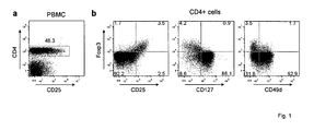
- CD127 and CD49d are inversely correlated with Foxp3.
- CD4+ T cells Fig.1a
- CD25 high the level of the IL-2 receptor ⁇ -chain CD25
- CD25 low the amount of CD25 (CD25 low ), so that their population partly overlaps with the CD25 high Treg subset ( Baecher-Allan, C., Brown, J. A., Freeman, G. J. & Hafler, D.
- Treg separations based on this marker therefore have an inherent risk of being contaminated by these cells.
- CD25 expression on Treg cells is driven directly by Foxp3 ( Hori, S., Nomura, T. & Sakaguchi, S., Science 299, 1057-61 (2003 )).
- Counterstaining with Foxp3 therefore indicates a near linear correlation for the CD25 high cells, in which cells expressing the highest amounts of Foxp3 also stain brightest for CD25 ( Fig.1b , left panel).
- the ⁇ -chain of the IL-7 receptor (CD127) is inversely correlated with Foxp3 expression ( Liu, W. et al., J Exp Med 203, 1701-11 (2006 ); Seddiki, N. et al., J Exp Med 203, 1693-700 (2006 )) ( Fig. 1b , middle panel). Also here the correlation is nearly linear, but cells with highest level of Foxp3 have the lowest expression level of CD127. The segregation, however, is not complete. In the example shown in Fig. 1b (middle panel), about 2/3 of the CD127- cells were also Foxp3-. For the characterization of Treg cells CD127 is therefore always used in combination with CD25 ( Liu, W. et al., J Exp Med 203, 1701-11 (2006 ); Seddiki, N. et al., J Exp Med 203, 1693-700 (2006 )).
- CD49d is the ⁇ -chain of the integrin VLA-4 ( ⁇ 4 ⁇ 1 ). Also here the co-segregation is incomplete ( Fig. 1b , right panel). An inverse linear correlation with Foxp3, as observed for CD127, however, does apparently not exist. Double-staining instead revealed absence of CD49d in Foxp3+ cells independent of the level of Foxp3 expression.
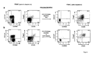
- CD49d discriminates Foxp3+ Treg cells from Foxp3- CD127-cells. While the segregation of both markers with Treg cells was incomplete, the combined use of CD127 and CD49d may complement each other. Double-staining with ⁇ -CD127 and ⁇ -CD49d allowed dividing the population of CD4+ cells into the three major populations: CD127+, CD49d+CD127- and CD49d-CD127- cells ( Fig. 2a , upper panel). Staining with ⁇ -CD25 and ⁇ -Foxp3 confirmed that the vast majority of the CD127+ cells were non-regulatory CD25-Foxp3- cells ( Fig. 2a , lower left panel).
- CD49d divided the CD127- subset into two nearly equally large subpopulations.
- the majority of CD49d+CD127-cells was Foxp3-, only less than 18% were CD25+Foxp3+ ( Fig. 2a , lower right panel).
- the CD49d-CD127- population in contrast, consisted almost exclusively of Foxp3+ cells. More than 83% of the cells were CD25+Foxp3+ Treg which express high levels of Foxp3 ( Fig. 2a , lower middle panel).
- CD49d+CD127- and CD49d-CD127- cells were isolated by FACS sorting from CD4+ PBMC ( Fig. 2b ). Almost no inhibition was observed with the CD49d+CD127- subset ( Fig. 2c ). In contrast, CD4+ cells sorted only based on the absence of CD49d and CD127 effectively prevented the expansion of activated CD25-CD4+ cells. Thus, the use of CD49d allows discriminating the non-suppressive CD127- cells from functional Foxp3+ Treg cells.
- untouched Foxp3+ Treg cells Purification of untouched Foxp3+ Treg cells by MACS.
- the result of the FACS sorting experiment indicated that the combined depletion of CD4+ T cells with ⁇ -CD127 and ⁇ -CD49d by MACS should produce a clean population of untouched Treg cells.
- untouched CD4+ cells isolated before from human PBMC were labelled with ⁇ -CD127 and ⁇ -CD49d and depleted by MACS using conventional magnetic bead-labelled antibodies ( Fig. 3 ).
- the analysis by FACS revealed that the depletion produced a population of CD49d-CD127- cells with a purity >95% ( Fig. 3a upper panel).
- Treg cells inhibited the proliferation to 14%, which mostly stopped the expansion already after a single replication cycle ( Fig. 3b , lower panel).
- untouched Treg cells isolated by MACS-depletion with ⁇ -CD127 and ⁇ -CD49d are fully able to suppress autologous CD4+ effector cells in vitro.
- CD49d Purification of untouched Foxp3+ Treg cells from PBMC by MACS in a single step.
- the selectivity of CD49d is particularly striking when using total PBMC instead of pre-purified CD4+ cells ( Fig. 4 ).
- CD127 which is absent on almost half of the PBMC
- the vast majority of non-CD4+ T cells still expresses CD49d ( Fig. 4 left panels).
- MLR mixed lymphocyte reaction
- Treg cells 4b proved to be potent suppressors of the allogenic response.
- the presence of Treg cells strongly suppressed the proliferation of autologous PBMC.
- untouched PBMC-derived Foxp3+ cells are fully functional Treg cells able to control allogenic immune responses in vitro.
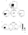
- the MLR experiment clearly demonstrated the suppressive capacity of the isolated Treg cells in vitro. With regard to future therapeutic applications it is crucial, however, to demonstrate that untouched Treg cells can also suppress destructive immune responses in vivo.
- an acute GvHD in vivo model was used, which is based on the transfer of CD25-depleted human PBMC into Rag2 -/- ⁇ c -/- mice ( Mutis, T. et al., Clin Cancer Res 12, 5520-5 (2006 )).
- the elimination of Treg cells from the transferred cell population allows simulating a particularly aggressive form of the disease which frequently leads to the death of the animals.
- Treg cells from PBMC were depleted by MACS using ⁇ -CD25 microbeads, untouched Treg cells were isolated again in a single step by combined use of ⁇ -CD127/ ⁇ -CD49d together with a commercial CD4+ isolation kit (according to example 4, Fig. 4b ). All mice that received 30 x 10 6 CD25-depleted PBMC exhibited a more or less pronounced weight loss within the first days of the experiment ( Fig. 5b ). 4 of the 6 mice developed clinical symptoms and of these 2 mice died during the experiment. In line with a previous publication the severity of PBMC-induced GvHD was diminished when autologous Treg cells were co-transferred ( Mutis, T.
- untouched Treg cells were sufficient to completely abrogate the weight loss and all mice of the treated group remained without symptoms for the entire course of the experiment.
- untouched Treg cells isolated by CD127/CD49d-depletion are potent suppressor cells capable to control destructive immune responses both in vitro and in vivo.
- CD25 is expressed in high amounts by Treg cells (CD25 high ). In humans, however, the marker is also expressed in lower amounts also by proinflammatory cells including effector and memory CD4+ T cells ( Baecher-Allan et al., J Immunol. 167:1245-53 (2001 )) and activated CD4+ T cells transiently expressing Foxp3 ( Allan et al., Int Immunol. 19:345-54(2007 )). In contrast to Treg cells these effector cells are able to secrete proinflammatory cytokines such as IFN- ⁇ or IL-17. As shown in Fig.
- CD49d expression segregates with the ability to secrete cytokines. This applies both for total CD4+ cells as well as for the subset of Foxp3+ cells.
- CD49d can be used to remove contaminating cytokine secreting effector cells from Treg preparations based on CD25-isolation.
- FACS-sorting Fig. 6b
- pure Treg cells can be obtained by gating on the CD49d-CD25 high CD4+ subset, for MACS-sorting the cells will be depleted with ⁇ -CD49d prior to the sorting procedures involving positive isolation with ⁇ -CD25.
- CD49d is absent on the majority of Foxp3+ Treg cells, they can be significantly enriched by the depletion of CD49d+ cells from total CD4+ T cells.
- Human CD4+ T cells were depleted of all CD49d+ cells by MACS and analysed for Foxp3 expression and compared to total CD4+ T cells.
- Foxp3+ Treg can already be enriched up to 4-5 fold by the depletion of CD49d+ cells from human CD4+ T cells.
- most of the cytokine producing cells are removed from this subset as cytokine secretion segregates with this marker ( Fig. 6a ).
- CD49d+ cells removes cytokine producing CD4+ effector cells contaminating CD127- Treg preparations.
- CD49d allows the isolation of untouched Treg cells ( Fig. 2 ).
- the segregation of CD49d with cytokine production also allows not only to remove Foxp3- cells but also the Th1- or Th17-like cells contaminating the CD127- Treg preparations.
- activation of CD4+ cell subsets sorted according to their CD49d and CD127 expression clearly reveals that IFN-g or IL-17 producing cells are absent from the CD49d-CD127- subset but present among the CD49d+CD127- subset.
- the method of the present invention offers a simple and cost effective way to isolate human Foxp3+ Treg cells. Additionally, the 'untouched' status of the cells, high purity and GMP compatibility of the method make the method widely applicable in different settings. The new method can therefore be employed to access 'untouched' human Treg cells for research & development, diagnosis and also offers a new perspective for their routine use for the manufacture of medicaments for immunotherapies.
Landscapes
- Health & Medical Sciences (AREA)
- Life Sciences & Earth Sciences (AREA)
- Cell Biology (AREA)
- Chemical & Material Sciences (AREA)
- Immunology (AREA)
- General Health & Medical Sciences (AREA)
- Engineering & Computer Science (AREA)
- Microbiology (AREA)
- Public Health (AREA)
- Biomedical Technology (AREA)
- Animal Behavior & Ethology (AREA)
- Pharmacology & Pharmacy (AREA)
- Mycology (AREA)
- Veterinary Medicine (AREA)
- Epidemiology (AREA)
- Medicinal Chemistry (AREA)
- Biotechnology (AREA)
- Bioinformatics & Cheminformatics (AREA)
- Genetics & Genomics (AREA)
- Wood Science & Technology (AREA)
- Zoology (AREA)
- Organic Chemistry (AREA)
- Hematology (AREA)
- Biochemistry (AREA)
- General Engineering & Computer Science (AREA)
- Micro-Organisms Or Cultivation Processes Thereof (AREA)
- Measuring Or Testing Involving Enzymes Or Micro-Organisms (AREA)
Priority Applications (13)
| Application Number | Priority Date | Filing Date | Title |
|---|---|---|---|
| EP08008255A EP2113561A1 (fr) | 2008-04-30 | 2008-04-30 | Procédés rapides et kits pour la purification de lymphocytes T régulateurs Foxp3+ |
| PCT/EP2008/008599 WO2009047003A1 (fr) | 2007-10-12 | 2008-10-10 | Procédé et trousse pour l'isolement rapide de cellules treg foxp3+ humaines |
| CN200880118525.8A CN101883844A (zh) | 2007-10-12 | 2008-10-10 | 快速分离人Foxp3+Treg细胞的方法和试剂盒 |
| AU2008309877A AU2008309877A1 (en) | 2007-10-12 | 2008-10-10 | Method and kit for rapid isolation of human Foxp3+ Treg cells |
| KR1020107008794A KR20100087290A (ko) | 2007-10-12 | 2008-10-10 | 인간 Foxp3+ 조절 Treg 세포의 신속한 분리 방법 및 키트 |
| DE08838013T DE08838013T8 (de) | 2007-10-12 | 2008-10-10 | Verfahren und kit zur schnellen isolierung humaner foxp3+-treg-zellen |
| US12/682,674 US20110014697A1 (en) | 2007-10-12 | 2008-10-10 | Method and Kit for Rapid Isolation of Human Foxp3+ Treg Cells |
| EP08838013.4A EP2198008B1 (fr) | 2007-10-12 | 2008-10-10 | Procédé et trousse pour l'isolement rapide de cellules treg foxp3+ humaines |
| CA2702230A CA2702230A1 (fr) | 2007-10-12 | 2008-10-10 | Procede et trousse pour l'isolement rapide de cellules treg foxp3+ humaines |
| JP2010528324A JP5414678B2 (ja) | 2007-10-12 | 2008-10-10 | ヒトのFoxp3+Treg細胞を迅速に分離するための方法およびキット |
| CN201110206170.1A CN102277331B (zh) | 2007-10-12 | 2008-10-10 | 快速分离人Foxp3+ Treg细胞的方法和试剂盒 |
| HK12105268.0A HK1164919A1 (en) | 2007-10-12 | 2012-05-30 | Method and kit for rapid isolation of human foxp3 treg cells foxp3 treg |
| US14/089,179 US9213028B2 (en) | 2007-10-12 | 2013-11-25 | Method and kit for rapid isolation of human Foxp3+ Treg cells |
Applications Claiming Priority (1)
| Application Number | Priority Date | Filing Date | Title |
|---|---|---|---|
| EP08008255A EP2113561A1 (fr) | 2008-04-30 | 2008-04-30 | Procédés rapides et kits pour la purification de lymphocytes T régulateurs Foxp3+ |
Publications (1)
| Publication Number | Publication Date |
|---|---|
| EP2113561A1 true EP2113561A1 (fr) | 2009-11-04 |
Family
ID=39495661
Family Applications (1)
| Application Number | Title | Priority Date | Filing Date |
|---|---|---|---|
| EP08008255A Withdrawn EP2113561A1 (fr) | 2007-10-12 | 2008-04-30 | Procédés rapides et kits pour la purification de lymphocytes T régulateurs Foxp3+ |
Country Status (1)
| Country | Link |
|---|---|
| EP (1) | EP2113561A1 (fr) |
Cited By (3)
| Publication number | Priority date | Publication date | Assignee | Title |
|---|---|---|---|---|
| WO2010022341A1 (fr) * | 2008-08-21 | 2010-02-25 | The United State Of America, As Represented By The Secretary, Department Of Health And Human Services | Procédés pour enrichir et utiliser des cellules t régulatrices |
| CN110187107A (zh) * | 2019-04-26 | 2019-08-30 | 温州医科大学 | 一种基于肿瘤组织浸润免疫细胞特征建立的结直肠癌预后评估装置及方法 |
| US11384336B2 (en) | 2016-12-07 | 2022-07-12 | East Carolina University | Compositions and methods for in vitro cultivation and/or expansion of regulatory T cells |
Citations (2)
| Publication number | Priority date | Publication date | Assignee | Title |
|---|---|---|---|---|
| US4615985A (en) | 1984-04-16 | 1986-10-07 | Genetic Diagnostics Corporation | Immobilized protein on nylon for immunoassay |
| WO2007014420A1 (fr) * | 2005-08-02 | 2007-02-08 | Centenary Institute Of Cancer Medicine And Cell Biology | Procédé destiné à identifier des lymphocytes t régulateurs |
-
2008
- 2008-04-30 EP EP08008255A patent/EP2113561A1/fr not_active Withdrawn
Patent Citations (2)
| Publication number | Priority date | Publication date | Assignee | Title |
|---|---|---|---|---|
| US4615985A (en) | 1984-04-16 | 1986-10-07 | Genetic Diagnostics Corporation | Immobilized protein on nylon for immunoassay |
| WO2007014420A1 (fr) * | 2005-08-02 | 2007-02-08 | Centenary Institute Of Cancer Medicine And Cell Biology | Procédé destiné à identifier des lymphocytes t régulateurs |
Non-Patent Citations (58)
| Title |
|---|
| "Cell Separation: A Practical Approach", 1999, OXFORD UNIVERSITY PRESS |
| ALLAN ET AL., INT IMMUNOL., vol. 19, 2007, pages 345 - 54 |
| ALLAN SE; CROME SQ; CRELLIN NK ET AL.: "Activation-induced FOXP3 in human T effector cells does not suppress proliferation or cytokine production", INT IMMUNOL, vol. 19, 2007, pages 345 - 354 |
| BAECHER-ALLAN ET AL., J IMMUNOL., vol. 167, 2001, pages 1245 - 53 |
| BAECHER-ALLAN, C. ET AL., J IMMUNOL, vol. 167, 2001, pages 1245 - 53 |
| BAECHER-ALLAN, C. ET AL.: "CD4+CD25high regulatory cells in human peripheral blood", J IMMUNOL, vol. 167, 2001, pages 1245 - 53 |
| BAECHER-ALLAN, C.; HAFLER, D. A., IMMUNOL REV, vol. 212, 2006, pages 203 - 16 |
| BAECHER-ALLAN, C.; HAFLER, D. A.: "Human regulatory T cells and their role in autoimmune disease", IMMUNOL REV, vol. 212, 2006, pages 203 - 16 |
| BARNARD, A. L. ET AL., BLOOD, vol. 106, 2005, pages 988 - 95 |
| BARNARD, A. L. ET AL.: "Engagement of specific T-cell surface molecules regulates cytoskeletal polarization in HTLV-1-infected lymphocytes", BLOOD, vol. 106, 2005, pages 988 - 95 |
| BORSELLINO ET AL: "F.94. Differential Expression of VLA-4 By Functional Treg and Effector CD4+ T-Cells", CLINICAL IMMUNOLOGY, ACADEMIC PRESS, US, vol. 119, 1 January 2006 (2006-01-01), pages S84, XP005438416, ISSN: 1521-6616 * |
| EBERT, L. M.; MCCOLL, S. R., J IMMUNOL, vol. 166, 2001, pages 4870 - 8 |
| EBERT, L. M.; MCCOLL, S. R.: "Coregulation of CXC chemokine receptor and CD4 expression on T lymphocytes during allogeneic activation", J IMMUNOL, vol. 166, 2001, pages 4870 - 8 |
| FONTENOT, J. D. ET AL., NAT IMMUNOL, vol. 6, 2005, pages 1142 - 51 |
| FONTENOT, J. D. ET AL.: "A function for interleukin 2 in Foxp3-expressing regulatory T cells", NAT IMMUNOL, vol. 6, 2005, pages 1142 - 51 |
| FONTENOT, J. D. ET AL.: "Regulatory T cell lineage specification by the forkhead transcription factor foxp3", IMMUNITY, vol. 22, 2005, pages 329 - 41 |
| FONTENOT, J. D.; RUDENSKY, A. Y., NAT IMMUNOL, vol. 6, 2005, pages 331 - 7 |
| FONTENOT, J. D.; RUDENSKY, A. Y.: "A well adapted regulatory contrivance: regulatory T cell development and the forkhead family transcription factor Foxp3", NAT IMMUNOL, vol. 6, 2005, pages 331 - 7 |
| HOFFMANN, P. ET AL., BIOL BLOOD MARROW TRANSPLANT, vol. 12, 2006, pages 267 - 74 |
| HOFFMANN, P. ET AL.: "Isolation of CD4+CD25+ regulatory T cells for clinical trials", BIOL BLOOD MARROW TRANSPLANT, vol. 12, 2006, pages 267 - 74 |
| HORI, S.; NOMURA, T.; SAKAGUCHI, S., SCIENCE, vol. 299, 2003, pages 1057 - 61 |
| HORI, S.; NOMURA, T.; SAKAGUCHI, S.: "Control of regulatory T cell development by the transcription factor Foxp3", SCIENCE, vol. 299, 2003, pages 1057 - 61 |
| HUEHN JOCHEN ET AL: "Developmental stage, phenotype, and migration distinguish naive- and effector/memory-like CD4+ regulatory T cells.", JOURNAL OF EXPERIMENTAL MEDICINE, vol. 199, no. 3, 2 February 2004 (2004-02-02), pages 303 - 313, XP002484932, ISSN: 0022-1007 * |
| KLEINEWIETFELD M ET AL: "OR.34. Rapid Isolation of 'Untouched' Human Foxp3+ Treg Cells", CLINICAL IMMUNOLOGY, ACADEMIC PRESS, US, vol. 127, 1 January 2008 (2008-01-01), pages S15, XP022615834, ISSN: 1521-6616, [retrieved on 20080101] * |
| KLEINEWIETFELD M.: "Charakterisierung von CD25+ regulatorischen T Zellen", DISSERTATION, FREIEN UNIVERSITÄT BERLIN, PHARMAZIE, 2006, Berlin, XP002484933, Retrieved from the Internet <URL:http://www.diss.fu-berlin.de/diss/receive/FUDISS_thesis_000000003036> [retrieved on 20080619] * |
| KLEINEWIETFELD, M. ET AL., BLOOD, vol. 105, 2005, pages 2877 - 86 |
| KLEINEWIETFELD, M. ET AL.: "CCR6 expression defines regulatory effector/memory-like cells within the CD25(+)CD4+ T-cell subset", BLOOD, vol. 105, 2005, pages 2877 - 86 |
| KOHM, A. P. ET AL., J IMMUNOL, vol. 176, 2006, pages 3301 - 5 |
| KOHM, A. P. ET AL.: "Cutting Edge: Anti-CD25 monoclonal antibody injection results in the functional inactivation, not depletion, of CD4+CD25+ T regulatory cells", J IMMUNOL, vol. 176, 2006, pages 3301 - 5 |
| LIU WEIHONG ET AL: "CD127 expression inversely correlates with FoxP3 and suppressive function of human CD4(+) T reg cells", JOURNAL OF EXPERIMENTAL MEDICINE, TOKYO, JP, vol. 203, no. 7, 1 July 2006 (2006-07-01), pages 1701 - 1711, XP002469608, ISSN: 0022-1007 * |
| LIU, W. ET AL., J EXP MED, vol. 203, 2006, pages 1701 - 11 |
| LIU, W. ET AL.: "CD127 expression inversely correlates with FoxP3 and suppressive function of human CD4+ T reg cells", J EXP MED, vol. 203, 2006, pages 1701 - 11 |
| MASTELLER, E. L. ET AL.: "Expansion of functional endogenous antigen-specific CD4+CD25+ regulatory T cells from nonobese diabetic mice", J IMMUNOL, vol. 175, 2005, pages 3053 - 9 |
| MCNEILL, A.; SPITTLE, E.; BACKSTROM, B. T., SCAND J IMMUNOL, vol. 65, 2007, pages 63 - 9 |
| MCNEILL, A.; SPITTLE, E.; BACKSTROM, B. T.: "Partial depletion of CD691ow-expressing natural regulatory T cells with the anti-CD25 monoclonal antibody PC61", SCAND J IMMUNOL, vol. 65, 2007, pages 63 - 9 |
| MUTIS, T. ET AL., CLIN CANCER RES, vol. 12, 2006, pages 5520 - 5 |
| MUTIS, T. ET AL.: "Human regulatory T cells control xenogeneic graft-versus-host disease induced by autologous T cells in RAG2-/-gammac-/- immunodeficient mice", CLIN CANCER RES, vol. 12, 2006, pages 5520 - 5 |
| P.T. SHARPE: "Methods of Cell Separation, Laboratory Techniques in Biochemistry and Molecular Biology", vol. 18, 1988, ELSEVIER |
| RANDOLPH, D. A.; FATHMAN, C. G.: "Cd4+Cd25+ regulatory T cells and their therapeutic potential", ANNU REV MED, vol. 57, 2006, pages 381 - 402 |
| ROBINSON, D. S.; LARCHE, M.; DURHAM, S. R., J CLIN INVEST, vol. 114, 2004, pages 1389 - 97 |
| ROBINSON, D. S.; LARCHE, M.; DURHAM, S. R.: "Tregs and allergic disease", J CLIN INVEST, vol. 114, 2004, pages 1389 - 97 |
| RONCAROLO, M. G.; BATTAGLIA, M., NAT REV IMMUNOL, vol. 7, 2007, pages 585 - 98 |
| RONCAROLO, M. G.; BATTAGLIA, M.: "Regulatory T-cell immunotherapy for tolerance to self antigens and alloantigens in humans", NAT REV IMMUNOL, vol. 7, 2007, pages 585 - 98 |
| SAKAGUCHI, S., ANNU REV IMMUNOL, vol. 22, 2004, pages 531 - 62 |
| SAKAGUCHI, S.: "Naturally arising CD4+ regulatory t cells for immunologic self-tolerance and negative control of immune responses", ANNU REV IMMUNOL, vol. 22, 2004, pages 531 - 62 |
| SEDDIKI, N. ET AL., J EXP MED, vol. 203, 2006, pages 1693 - 700 |
| SEDDIKI, N. ET AL.: "Expression of interleukin (IL)-2 and IL-7 receptors discriminates between human regulatory and activated T cells", J EXP MED, vol. 203, 2006, pages 1693 - 700 |
| SHARPE, A. H.; ABBAS, A. K., N ENGL J MED, vol. 355, 2006, pages 973 - 5 |
| SHARPE, A. H.; ABBAS, A. K.: "T-cell costimulation--biology, therapeutic potential, and challenges", N ENGL J MED, vol. 355, 2006, pages 973 - 5 |
| STASSEN M ET AL: "Human CD25+ regulatory T cells: two subsets defined by the integrins alpha4beta7 or alpha4beta1 confer distinct suppressive properties upon CD4+ T helper cells", EUROPEAN JOURNAL OF IMMUNOLOGY, WEINHEIM, vol. 34, no. 5, 1 May 2004 (2004-05-01), pages 1303 - 1311, XP002377231, ISSN: 0014-2980 * |
| SUNTHARALINGAM, G. ET AL., N ENGL J MED, vol. 355, 2006, pages 1018 - 28 |
| SUNTHARALINGAM, G. ET AL.: "Cytokine storm in a phase 1 trial of the anti-CD28 monoclonal antibody TGN1412", N ENGL J MED, vol. 355, 2006, pages 1018 - 28 |
| TANG, Q.; BLUESTONE, J. A.: "Regulatory T-cell physiology and application to treat autoimmunity", IMMUNOL REV, vol. 212, 2006, pages 217 - 37 |
| THORNTON, A. M. ET AL., J IMMUNOL, vol. 172, 2004, pages 6519 - 23 |
| THORNTON, A. M. ET AL.: "Cutting edge: IL-2 is critically required for the in vitro activation of CD4+CD25+ T cell suppressor function", J IMMUNOL, vol. 172, 2004, pages 6519 - 23 |
| VON BONIN, A.; HUHN, J.; FLEISCHER, B., IMMUNOL REV, vol. 161, 1998, pages 43 - 53 |
| VON BONIN, A.; HUHN, J.; FLEISCHER, B.: "Dipeptidyl-peptidase IV/CD26 on T cells: analysis of an alternative T-cell activation pathway", IMMUNOL REV, vol. 161, 1998, pages 43 - 53 |
| WALDMANN, H. ET AL.: "Regulatory T cells and organ transplantation", SEMIN IMMUNOL, vol. 16, 2004, pages 119 - 26 |
Cited By (4)
| Publication number | Priority date | Publication date | Assignee | Title |
|---|---|---|---|---|
| WO2010022341A1 (fr) * | 2008-08-21 | 2010-02-25 | The United State Of America, As Represented By The Secretary, Department Of Health And Human Services | Procédés pour enrichir et utiliser des cellules t régulatrices |
| US8951793B2 (en) | 2008-08-21 | 2015-02-10 | The United States Of America, As Represented By The Secretary, Department Of Health And Human Services | Method of making an isolated population of FOXP3+ regulatory T cells |
| US11384336B2 (en) | 2016-12-07 | 2022-07-12 | East Carolina University | Compositions and methods for in vitro cultivation and/or expansion of regulatory T cells |
| CN110187107A (zh) * | 2019-04-26 | 2019-08-30 | 温州医科大学 | 一种基于肿瘤组织浸润免疫细胞特征建立的结直肠癌预后评估装置及方法 |
Similar Documents
| Publication | Publication Date | Title |
|---|---|---|
| EP2198008B1 (fr) | Procédé et trousse pour l'isolement rapide de cellules treg foxp3+ humaines | |
| US20230280341A1 (en) | Cd127 expression inversely correlates with foxp3 and suppressive function of cd4+ tregs | |
| JP4714767B2 (ja) | ヒト血液由来のcd4+cd25+調節t細胞 | |
| JP4601166B2 (ja) | 抗原特異的t細胞の直接的選択方法 | |
| US20150210982A1 (en) | Isolation and Use of Human Regulatory T Cells | |
| Portevin et al. | Regulatory activity of azabisphosphonate-capped dendrimers on human CD4+ T cell proliferation enhances ex-vivo expansion of NK cells from PBMCs for immunotherapy | |
| KR20140061374A (ko) | 조절 t 세포, 및 cd6-발현 또는 cd4, cd25 및 cd127의 조합을 사용하여 이를 확인 및 단리하는 방법 | |
| EP2378287A1 (fr) | Nouveau procédé pour l'isolement de cellules T1 | |
| EP2113561A1 (fr) | Procédés rapides et kits pour la purification de lymphocytes T régulateurs Foxp3+ | |
| Wichlan et al. | Efficient and reproducible large-scale isolation of human CD4+ CD25+ regulatory T cells with potent suppressor activity | |
| EP2048225B1 (fr) | Procédé et kit pour l'isolement rapide de cellules Foxp3+treg humaines | |
| Patel et al. | Clinical grade isolation of regulatory T cells from G-CSF mobilized peripheral blood improves with initial depletion of monocytes | |
| WO2008028229A1 (fr) | Techniques d'identification de marqueurs | |
| US20090075296A1 (en) | Methods and compositions for assaying regulatory t cells | |
| Moore | Investigation of regulatory B-cell responses during Mycobacterium tuberculosis exposure |
Legal Events
| Date | Code | Title | Description |
|---|---|---|---|
| PUAI | Public reference made under article 153(3) epc to a published international application that has entered the european phase |
Free format text: ORIGINAL CODE: 0009012 |
|
| AK | Designated contracting states |
Kind code of ref document: A1 Designated state(s): AT BE BG CH CY CZ DE DK EE ES FI FR GB GR HR HU IE IS IT LI LT LU LV MC MT NL NO PL PT RO SE SI SK TR |
|
| AX | Request for extension of the european patent |
Extension state: AL BA MK RS |
|
| STAA | Information on the status of an ep patent application or granted ep patent |
Free format text: STATUS: THE APPLICATION HAS BEEN WITHDRAWN |
|
| 17P | Request for examination filed |
Effective date: 20100504 |
|
| 18W | Application withdrawn |
Effective date: 20100518 |