EP2102361B1 - Marqueurs de méthylation de l'adn et leurs méthodes d'utilisation - Google Patents
Marqueurs de méthylation de l'adn et leurs méthodes d'utilisation Download PDFInfo
- Publication number
- EP2102361B1 EP2102361B1 EP07862181.0A EP07862181A EP2102361B1 EP 2102361 B1 EP2102361 B1 EP 2102361B1 EP 07862181 A EP07862181 A EP 07862181A EP 2102361 B1 EP2102361 B1 EP 2102361B1
- Authority
- EP
- European Patent Office
- Prior art keywords
- nucleic acid
- genes
- methylation
- cancer
- hypermethylation
- Prior art date
- Legal status (The legal status is an assumption and is not a legal conclusion. Google has not performed a legal analysis and makes no representation as to the accuracy of the status listed.)
- Not-in-force
Links
Images


Classifications
-
- C—CHEMISTRY; METALLURGY
- C12—BIOCHEMISTRY; BEER; SPIRITS; WINE; VINEGAR; MICROBIOLOGY; ENZYMOLOGY; MUTATION OR GENETIC ENGINEERING
- C12Q—MEASURING OR TESTING PROCESSES INVOLVING ENZYMES, NUCLEIC ACIDS OR MICROORGANISMS; COMPOSITIONS OR TEST PAPERS THEREFOR; PROCESSES OF PREPARING SUCH COMPOSITIONS; CONDITION-RESPONSIVE CONTROL IN MICROBIOLOGICAL OR ENZYMOLOGICAL PROCESSES
- C12Q1/00—Measuring or testing processes involving enzymes, nucleic acids or microorganisms; Compositions therefor; Processes of preparing such compositions
- C12Q1/68—Measuring or testing processes involving enzymes, nucleic acids or microorganisms; Compositions therefor; Processes of preparing such compositions involving nucleic acids
- C12Q1/6876—Nucleic acid products used in the analysis of nucleic acids, e.g. primers or probes
- C12Q1/6883—Nucleic acid products used in the analysis of nucleic acids, e.g. primers or probes for diseases caused by alterations of genetic material
- C12Q1/6886—Nucleic acid products used in the analysis of nucleic acids, e.g. primers or probes for diseases caused by alterations of genetic material for cancer
-
- A—HUMAN NECESSITIES
- A61—MEDICAL OR VETERINARY SCIENCE; HYGIENE
- A61P—SPECIFIC THERAPEUTIC ACTIVITY OF CHEMICAL COMPOUNDS OR MEDICINAL PREPARATIONS
- A61P35/00—Antineoplastic agents
-
- C—CHEMISTRY; METALLURGY
- C12—BIOCHEMISTRY; BEER; SPIRITS; WINE; VINEGAR; MICROBIOLOGY; ENZYMOLOGY; MUTATION OR GENETIC ENGINEERING
- C12Q—MEASURING OR TESTING PROCESSES INVOLVING ENZYMES, NUCLEIC ACIDS OR MICROORGANISMS; COMPOSITIONS OR TEST PAPERS THEREFOR; PROCESSES OF PREPARING SUCH COMPOSITIONS; CONDITION-RESPONSIVE CONTROL IN MICROBIOLOGICAL OR ENZYMOLOGICAL PROCESSES
- C12Q2600/00—Oligonucleotides characterized by their use
- C12Q2600/112—Disease subtyping, staging or classification
-
- C—CHEMISTRY; METALLURGY
- C12—BIOCHEMISTRY; BEER; SPIRITS; WINE; VINEGAR; MICROBIOLOGY; ENZYMOLOGY; MUTATION OR GENETIC ENGINEERING
- C12Q—MEASURING OR TESTING PROCESSES INVOLVING ENZYMES, NUCLEIC ACIDS OR MICROORGANISMS; COMPOSITIONS OR TEST PAPERS THEREFOR; PROCESSES OF PREPARING SUCH COMPOSITIONS; CONDITION-RESPONSIVE CONTROL IN MICROBIOLOGICAL OR ENZYMOLOGICAL PROCESSES
- C12Q2600/00—Oligonucleotides characterized by their use
- C12Q2600/118—Prognosis of disease development
-
- C—CHEMISTRY; METALLURGY
- C12—BIOCHEMISTRY; BEER; SPIRITS; WINE; VINEGAR; MICROBIOLOGY; ENZYMOLOGY; MUTATION OR GENETIC ENGINEERING
- C12Q—MEASURING OR TESTING PROCESSES INVOLVING ENZYMES, NUCLEIC ACIDS OR MICROORGANISMS; COMPOSITIONS OR TEST PAPERS THEREFOR; PROCESSES OF PREPARING SUCH COMPOSITIONS; CONDITION-RESPONSIVE CONTROL IN MICROBIOLOGICAL OR ENZYMOLOGICAL PROCESSES
- C12Q2600/00—Oligonucleotides characterized by their use
- C12Q2600/154—Methylation markers
-
- C—CHEMISTRY; METALLURGY
- C12—BIOCHEMISTRY; BEER; SPIRITS; WINE; VINEGAR; MICROBIOLOGY; ENZYMOLOGY; MUTATION OR GENETIC ENGINEERING
- C12Q—MEASURING OR TESTING PROCESSES INVOLVING ENZYMES, NUCLEIC ACIDS OR MICROORGANISMS; COMPOSITIONS OR TEST PAPERS THEREFOR; PROCESSES OF PREPARING SUCH COMPOSITIONS; CONDITION-RESPONSIVE CONTROL IN MICROBIOLOGICAL OR ENZYMOLOGICAL PROCESSES
- C12Q2600/00—Oligonucleotides characterized by their use
- C12Q2600/16—Primer sets for multiplex assays
Definitions
- the present invention relates to the use of nucleic acid methylation and methylation profiles to detect metastatic disease.
- the invention relates to methods for identifying metastases by detecting nucleic acid hypermethylation of one or more genes in one or more samples and, in particular, in tumor tissue and lymph nodes.
- the invention further relates to DNA hypermethylation as a predictor of disease recurrence and patient prognosis, specifically in patients suffering from cancer.
- Cancer remains one of the leading causes of death in the United States.
- Clinically a broad variety of medical approaches, including surgery, radiation therapy and chemotherapeutic drug therapy are currently being used in the treatment of human cancer (see the textbook CANCER: Principles & Practice of Oncology, 2d Edition, De Vita et al., eds., J. B. Lippincott Company, Philadelphia, Pa., 1985 ).
- CANCER Principles & Practice of Oncology, 2d Edition, De Vita et al., eds., J. B. Lippincott Company, Philadelphia, Pa., 1985 .
- it is recognized that such approaches continue to be limited by an inability to predict the likelihood of metastasis and tumor recurrence or the most efficacious treatment regime for minimizing the occurrence of these negative outcomes.
- Human cancer cells typically contain somatically altered nucleic acids, characterized by mutation, amplification, or deletion of critical genes.
- nucleic acids from human cancer cells often display somatic changes in DNA methylation (36, 37, 38).
- DNA methylation 36, 37, 38.
- a precise role for, and the significance of, abnormal DNA methylation in human tumorigenesis has not been well established.
- Loss of gene function is cancer can occur by both genetic and epigenetic mechanisms.
- the best-defined epigenetic alteration of cancer genes involves DNA methylation of clustered CpG dinucleotides, or CpG islands, in promoter regions associated with the transcriptional inactivation of the affected genes.
- CpG islands are short sequences rich in the CpG dinucleotide, and can be found in the 5' region of about half of all human genes.
- Methylation of cytosine within 5' CGIs is associated with loss of gene expression and has been seen in a number of physiological conditions, including X chromosome inactivation and genomic imprinting.
- CpG island methylation Aberrant methylation of CpG islands has been detected in genetic diseases such as the fragile-X syndrome, in aging cells and in neoplasia. About half of the tumor suppressor genes which have been shown to be mutated in the germline of patients with familial cancer syndromes have also been shown to be aberrantly methylated in some proportion of sporadic cancers, including Rb, VHL, p16, hMLH1, and BRCA1 (reviewed in Baylin, et al, Adv. Cancer Res. 72:141-196 1998 ). Methylation of tumor suppressor genes in cancer is usually associated with (1) lack of gene transcription and (2) absence of coding region mutation. Thus CpG island methylation can serve as an alternative mechanism of gene inactivation in cancer.
- Cancer treatments in general, have a higher rate of success if the cancer is diagnosed early, and treatment is started earlier in the disease process. A relationship between improved prognosis and stage of disease at diagnosis can be seen across a majority of cancers. Identification of the earliest changes in cells associated with cancer is thus a major focus in molecular cancer research. Diagnostic approaches based on identification of these changes in specific genes may allow implementation of early detection strategies and novel therapeutic approaches. Targeting these early changes will lead to more effective cancer treatment.
- the invention features methods for identifying metastases by detecting nucleic acid hypermethylation of one or more genes in one or more samples, and in particular in tumor tissue and lymph nodes.
- the invention features methods for identifying metastases in a subject comprising detecting nucleic acid hypermethylation of one or more genes in one or more samples, wherein detecting nucleic acid hypermethylation identifies metastases.
- the sample comprises cells or tissues selected from the group consisting of tumor, lymph nodes, bone marrow and blood.
- the sample is from a tumor.
- the sample is from a lymph node.
- the lymph node is a N1 lymph node or a mediastinal lymph node.
- the invention features methods for identifying metastases in a subject comprising detecting nucleic acid hypermethylation of one or more genes in tumor tissue or lymph node, wherein the genes are selected from the group consisting of genes involved in tumor suppression, DNA repair, apoptosis, anti-proliferation, ras signaling, adhesion, differentiation, development, and cell cycle regulation, wherein detecting nucleic acid hypermethylation identifies metastases.
- the metastases are micrometastases.
- the one or more genes comprise one or more CpG islands.
- the one or more genes is selected from the group consisting of H-cadherin, p16, APC, RASSF1A, MGMT, DAPK, and ASC.
- H-cadherin in certain exemplary embodiments is encoded by NCBI accession No. AAB18912 and is shown in (SEQ ID NO:1) below:
- p-16 in certain exemplary embodiments is encoded by NCBI accession No. CAB58124 and is shown in (SEQ ID NO:2) below:
- APC in certain exemplary embodiments is encoded by NCBI accession No. NP_000029 and is shown in (SEQ ID NO:3) below:
- RASSF1A in certain exemplary embodiments is encoded by NCBI accession No. NP_009113 and is shown in (SEQ ID NO:4) below:
- MGMT in certain exemplary embodiments is encoded by NCBI accession No. AAH00824 and is shown in (SEQ ID NO:5) below:
- DAPK in certain exemplary embodiments is encoded by NCBI accession No. NP_004929 and is shown in (SEQ ID NO:6) below:
- ASC in certain exemplary embodiments is encoded by NCBI accession No. NP_037390 and is shown in (SEQ ID NO:7) below:
- hypermethylation of at least one of the genes is detected. In still other embodiments of the above aspects, hypermethylation of at least two of the genes is detected.
- the invention features methods for identifying micrometastases in a subject comprising detecting nucleic acid hypermethylation of at least one or more genes in a sample comprising tumor and lymph nodes, wherein the sample genes are selected from the group consisting of H-cadherin, p16, APC, RASSF1A, MGMT, DAPK, and ASC, and wherein detecting nucleic acid methylation identifies micrometastases.
- hypermethylation of at least two of the genes is detected.
- at least two of the genes are selected from p-16 and H-cadherin, H-cadherin and APC, APC and p16, or RASSf1A and p16.
- the detection of metastases is used to detect or diagnose a proliferative disease.
- the detection or diagnosis is performed after surgery or therapy to treat a proliferative disease. In other certain embodiments, the detection is used to predict the recurrence of a proliferative disease. In other certain embodiments, the detection is used to stage a proliferative disease. In still other certain embodiments, the detection is further used to determine a course of treatment for a subject.
- the invention features a method for detecting or diagnosing a proliferative disease in a subject comprising detecting nucleic acid hypermethylation of one or more genes in one or more samples, wherein detecting nucleic acid hypermethylation is used to detect or diagnose a proliferative disease.
- the invention features a method for predicting the recurrence of a proliferative disease in a subject comprising detecting nucleic acid hypermethylation of one or more genes wherein detecting nucleic acid hypermethylation of one or more genes is a predictor of the recurrence of a proliferative disease.
- hypermethylation of one or more genes is detected in tumor or lymph nodes.
- detection of hypermethylation of one or more genes in lymph nodes is predictive of aggressive disease recurrence.
- the invention features a method for staging or re-staging a proliferative disease in a subject comprising detecting nucleic acid hypermethylation of one or more genes wherein detecting nucleic acid hypermethylation is used for staging or re-staging a proliferative disease.
- the stage of proliferative disease is predictive of disease recurrence. In a further embodiment, the stage of proliferative disease determines course of treatment.
- the invention features a method for determining the prognosis of a subject suffering from a proliferative disease comprising detecting nucleic acid hypermethylation of one or more genes wherein the detection of nucleic acid hypermethylation is used for determining the prognosis of a subject suffering from a proliferative disease.
- the prognosis determines course of treatment.
- the subject is a human.
- the method is performed prior to therapeutic intervention for the disease.
- the method is performed after therapeutic intervention for the disease.
- the therapeutic intervention is selected from treatment with an agent or surgery.
- hypermethylation is detected in CpG islands of the one or more genes.
- hypermethylation is detected in CpG islands.
- the invention features methods for detecting or diagnosing a proliferative disease in a subject comprising extracting nucleic acid from one or more cell or tissue samples, detecting nucleic acid hypermethylation of one or more genes in the sample; and identifying the nucleic acid hypermethylation state of one or more genes, wherein nucleic acid hypermethylation of genes indicates a proliferative disease.
- the invention features methods for predicting the recurrence of a proliferative disease in a subject comprising extracting nucleic acid from one or more cell or tissue samples, detecting nucleic acid hypermethylation of one or more genes in the sample; and identifying the nucleic acid hypermethylation state of one or more genes, wherein nucleic acid hypermethylation of genes is indicative of the recurrence of a proliferative disease.
- the invention features methods for staging or re-staging a proliferative disease in a subject comprising extracting nucleic acid from one or more cell or tissue samples, detecting nucleic acid hypermethylation of one or more genes in the sample; and identifying the nucleic acid hypermethylation state of one or more genes, wherein nucleic acid hypermethylation of genes is used for staging or restaging of a proliferative disease.
- the tissue samples are selected from tumor, lymph node, bone marrow or blood or a combination thereof.
- the method determines the course of disease treatment.
- the method is performed prior to therapeutic intervention for the disease.
- the method is performed after therapeutic intervention for the disease.
- the therapeutic intervention is selected from treatment with an agent or surgery.
- the invention features methods of treating a subject having or at risk for having a proliferative disease comprising identifying nucleic acid hypermethylation of one or more genes, where nucleic acid hypermethylation indicates having or a risk for having a proliferative disease, and administering to the subject a therapeutically effective amount of a demethylating agent, thereby treating a subject having or at risk for having a proliferative disease.
- the method is used in combination with one or more chemotherapeutic agents.
- the method further comprises comparing the nucleic acid hypermethylation of one or more genes in the sample with comparable samples obtained from a normal subject.
- detecting nucleic acid hypermethylation of one or more genes indicates the presence of metastases.
- the metastases are micrometastases.
- the proliferative disease is a neoplasia.
- the neoplasia is cancer.
- the cancer is a solid tumor.
- the cancer is selected from the group consisting of lung cancer, pancreatic cancer, esophageal cancer, head and neck cancer, stomach cancer, liver cancer, prostate cancer, gastrointestinal cancer, ovarian cancer, and uterine cancer.
- the cells or tissues are selected from the group consisting of tumor, lymph nodes, bone marrow or blood.
- the cells or tissues are from a tumor or the lymph nodes.
- the lymph node is a N1 lymph node or a mediastinal lymph node.
- the invention features a method of identifying an agent that de-methylates hypermethylated nucleic acid comprising identifying one or more cell or tissue samples with hypermethylated nucleic acid, extracting the hypermethylated nucleic acid, contacting the nucleic acid with one or more nucleic acid de-methylating candidate agents and a control agent, identifying the nucleic acid hypermethylation state, wherein nucleic acid de-methylation of genes in the sample by the candidate agent compared to the control indicates a demethylating agent, and thereby identifying an agent that de-methylates hypermethylated nucleic acid.
- the one or more genes are selected from the group consisting of genes involved in tumor suppression, DNA repair, anti-proliferation, apoptosis, ras signaling, adhesion, differentiation, development, and cell cycle regulation.
- the one or more genes are selected from a panel consisting of (1) genes involved in tumor suppression and cell adhesion, (2) genes involved in cell cycle regulation and adhesion, (3) genes involved in tumor suppression and cell cycle regulation, and (4) genes involved in ras signaling and cell cycle control.
- the one or more genes comprise one or more CpG islands.
- the genes are selected from the group consisting of p-16, H-cadherin, APC, RASSF1A, MGMT, DAPK, and ASC.
- the hypermethylation of at least one of the genes is detected. In a further related embodiment, the hypermethylation of at least two of the genes is detected. In still another related embodiment, the two genes are selected from p-16 and H-cadherin, H-cadherin and APC, APC and p16, or RASSf1A and p16:
- the detection of nucleic acid methylation is by a quantitative method.
- the detection of nucleic acid methylation is carried out by polymerase chain reaction (PCR) analysis.
- PCR polymerase chain reaction
- MSP methylation specific PCR
- the method of detecting nucleic acid methylation is performed as a high-throughput method.
- the method is used in combination with the detection of other epigenetic markers.
- the other epigenetic markers are plasma or tumor epigenetic markers.
- hypermethylation is detected in CpG islands of the one or more genes. In a further embodiment of the above-described aspects, hypermethylation is detected in CpG islands of the promoter region.
- kits for identifying the nucleic acid hypermethylation state of one or more genes comprising gene specific primers for use in polymerase chain reaction (PCR), and instructions for use.
- PCR polymerase chain reaction
- kits for detecting metastases by detecting nucleic acid hypermethylation of one or more genes comprising gene specific primers for use in polymerase chain reaction (PCR), and instructions for use.
- PCR polymerase chain reaction
- the metastases are micrometastases.
- the PCR is methylation specific PCR (MSP).
- the one or more genes are selected from the group consisting of genes involved in tumor suppression, DNA repair, anti-proliferation, apotosis, ras signaling, adhesion, differentiation, development, and cell cycle regulation.
- the one or more genes are selected from a panel consisting of (1) genes involved in tumor suppression and cell adhesion, (2) genes involved in cell cycle regulation and adhesion, (3) genes involved in tumor suppression and cell cycle regulation, and (4) genes involved in ras signaling and cell cycle control.
- the one or more genes comprise one or more CpG islands.
- the CpG islands are in the promoter region.
- the genes are selected from the group consisting of p-16, H-cadherin, APC, RASSF1A, MGMT, DAPK, and ASC
- the hypermethylation of at least one of the genes is detected. In still another embodiment, the hypermethylation of at least two of the genes is detected. In still another further embodiment, the two genes are selected from p-16 and H-cadherin, H-cadherin and APC, APC and p16, or RASSf1A and p16.
- control is meant a standard or reference condition.
- a de-methylating agent or the methods of the instant invention (e.g. methods of detection of hypermethylation) together with a second agent, such as a chemotherapeutic agent, or a de-methylating agent, where the two are administered concurrently or sequentially in any order.
- a second agent such as a chemotherapeutic agent, or a de-methylating agent
- agent as used herein is meant to refer to a polypeptide, polynucleotide, or fragment, or analog thereof, small molecule; or other biologically active molecule.
- CpG island refers to a sequence of nucleic acid with an increased density relative to other nucleic acid regions of the.dinucleotide CpG.
- epigenetic marker or "epigenetic change” as used herein is meant to refer to a change in the DNA sequences or gene expression by a process or processes that do not change the DNA coding sequence itself.
- methylation is an epigenetic marker.
- hypermethylation refers to the presence of methylated alleles in one or more nucleic acids.
- hypermethylation is detected using methylation specific polymerase chain reaction (MSP).
- MSP methylation specific polymerase chain reaction
- metastatic tumor is meant to refer to the spread of a malignant tumor from its sight of origin. Cancer cells may metastasize through the bloodstream, through the lymphatic system, across body cavities, or any combination thereof.
- a metastatic tumor can arise from a multitude of primary tumor types, including but not limited to lung, breast, thyroid, head and neck, brain, lymphoid, gastrointestinal (mouth, esophagus, stomach, small intestine, colon, rectum), genito-urinary tract (uterus, ovary, cervix, bladder, testicle, penis, prostate), kidney, pancreas, liver, bone, muscle or skin.
- micrometastases is meant to refer to a metastasis that cannot be detected by routine histological evaluation, for example by Hematoxylin and Eosin (H & E) staining and microscopic assessment.
- neoplasm or “neoplasia” as used herein refers to inappropriately high levels of cell division, inappropriately low levels of apoptosis, or both.
- a neoplasm creates an unstructured mass (a tumor), which can be either benign or malignant.
- cancer is a neoplasia.
- cancers include, without limitation, leukemias (e.g., acute leukemia, acute lymphocytic leukemia, acute myelocytic leukemia, acute myeloblastic leukemia, acute promyelocytic leukemia, acute myelomonocytic leukemia, acute monocytic leukemia, acute erythroleukemia, chronic leukemia, chronic myelocytic leukemia, chronic lymphocytic leukemia), polycythemia vera, lymphoma (Hodgkin's disease, non-Hodgkin's disease), Waldenstrom's macroglobulinemia, heavy chain disease, and solid tumors such as sarcomas and carcinomas (e.g., fibrosarcoma, myxosarcoma, liposarcoma, chondrosarcoma, osteogenic sarcoma, chordoma, angiosarcoma, endotheliosarcoma, lymphangiosarcoma
- nucleic acid refers to an oligonucleotide, nucleotide, polynucleotide, or to a fragment of any of these, to DNA or RNA of genomic or synthetic origin which may be single-stranded or double-stranded and may represent a sense or antisense strand, peptide nucleic acid (PNA), or to any DNA-like or RNA-like material, natural or synthetic in origin.
- PNA peptide nucleic acid
- proliferative disorder refers to an abnormal growth of cells.
- a cell proliferative disorder as described herein may be a neoplasm.
- gene refers to a segment of deoxyribonucleic acid that encodes a polypeptide including the upstream and downstream regulatory sequences. Specifically, the term gene includes the promoter region upstream of the gene.
- promoter refers to a minimal sequence sufficient to direct transcription or to render promoter-dependent gene expression that is controllable for cell-type specific, tissue-specific, or is inducible by external signals or agents. Promoters may be located in the 5' or 3' regions of the gene. Promoter regions, in whole or in part, of a number of nucleic acids can be examined for sites of CpG-island methylation.
- sample refers to any biological or chemical mixture for use in the method of the invention.
- the sample can be a biological sample.
- the biological samples are generally derived from a patient, preferably as a bodily fluid (such as tumor tissue, lymph node, sputum, blood, bone marrow, cerebrospinal fluid, phlegm, saliva, or urine) or cell lysate.
- the cell lysate can be prepared from a tissue sample (e.g. a tissue sample obtained by biopsy), for example, a tissue sample (e.g. a tissue sample obtained by biopsy), blood, cerebrospinal fluid, phlegm, saliva, urine, or the sample can be cell lysate.
- stage or “staging” as used herein is meant to refer to the extent or progression of proliferative disease, e.g. cancer, in a subject.
- Staging can be “clinical” and is according to the "stage classification” corresponding to the TNM classification (" Rinsho, Byori, Genpatsusei Kangan Toriatsukaikiyaku (Clinical and Pathological Codes for Handling Primary Liver Cancer)”: 22p. Nihon Kangangaku Kenkyukai (Liver Cancer Study Group of Japan) edition (3rd revised edition), Kanehara Shuppan, 1992 ).
- Staging in certain embodiments can refer to "molecular staging” as defined by nucleic acid hypermethylation of one or more genes in one or more samples.
- the "molecular stage” stage of a cancer is determined by detection of nucleic acid hypermethylation of one or more genes in a sample from the lymph nodes.
- subject as used herein is meant to include vertebrates, preferably a mammal. Mammals include, but are not limited to, humans.
- tumor as used herein is intended to include an abnormal mass or growth of cells or tissue.
- a tumor can be benign or malignant.
- the invention is based upon the discovery that the hypermethylation of certain genes, including promoter regions, can serve as prognostic and diagnostic markers for cellular proliferative disorders. This is the first time that promoter hypermethylation of certain genes, such as p16, H-cadherin, RASSf1A and APC, in the lymph nodes has been associated with the ability to predict recurrence and aggressiveness of certain cancers, such as lung cancer.
- DNA methylases transfer methyl groups from the universal methyl donor S-adenosyl methionine to specific sites on the DNA.
- Several biological functions have been attributed to the methylated bases in DNA.
- the most established biological function for methylated DNA is the protection of DNA from digestion by cognate restriction enzymes.
- the restriction modification phenomenon has, so far, been observed only in bacteria.
- Mammalian cells possess a different methylase that exclusively methylates cytosine residues that are 5' neighbors of guanine (CpG). This modification of cytosine residues has important regulatory effects on gene expression, especially when involving CpG rich areas, known as CpG islands, located in the promoter regions of many genes.
- Methylation has been shown by several lines of evidence to play a role in gene activity, cell differentiation, tumorigenesis, X-chromosome inactivation, genomic imprinting and other major biological processes ( Razin, A., H., and Riggs, R. D. eds. in DNA Methylation Biochemistry and Biological Significance, Springer-Verlag, New York, 1984 ).
- Razin, A., H., and Riggs, R. D. eds. in DNA Methylation Biochemistry and Biological Significance, Springer-Verlag, New York, 1984 In eukaryotic cells, methylation of cytosine residues that are immediately 5' to a guanosine, occurs predominantly in CG poor regions ( Bird, A., Nature, 321:209, 1986 ).
- CpG islands remain unmethylated in normal cells, except during X-chromosome inactivation and parental specific imprinting ( Li, et al., Nature, 366:362, 1993 ) where methylation of 5' regulatory regions can lead to transcriptional repression.
- De novo methylation of the Rb gene has been demonstrated in a small fraction of retinoblastomas ( Sakai, et al., Am. J. Hum. Genet., 48:880, 1991 ), and recently, a more detailed analysis of the VHL gene showed aberrant methylation in a subset of sporadic renal cell carcinomas ( Herman, et al., Proc. Natl. Acad.
- DNA is methylated only at cytosines located 5' to guanosine in the CpG dinucleotide. This modification has important regulatory effects on gene expression, especially when involving CpG rich areas, known as CpG islands, located in the promoter regions of many genes. While almost all gene-associated islands are protected from methylation on autosomal chromosomes, extensive methylation of CpG islands has been associated with transcriptional inactivation of selected imprinted genes and genes on the inactive X-chromosome of females. Aberrant methylation of normally unmethylated CpG islands has been described as a frequent event in immortalized and transformed cells, and has been associated with transcriptional inactivation of defined tumor suppressor genes in human cancers.
- Any method that is sufficient to detect hypermethylation e.g. a method that can detect methylation of nucleotides at levels as low as 0.1%, is a suitable for use in the methods of the invention.
- a number of different methods can be used to detect hypermethylation.
- the ability to monitor the real-time progress of the PCR changes the way one approaches PCR-based quantification of DNA and RNA. Reactions are characterized by the point in time during cycling when amplification of a PCR product is first detected rather than the amount of PCR product accumulated after a fixed number of cycles. The higher the starting copy number of the nucleic acid target, the sooner a significant increase in fluorescence is observed.
- An amplification plot is the plot of fluorescence signal versus cycle number. In the initial cycles of PCR, there is little change in fluorescence signal. This defines the baseline for the amplification plot. An increase in fluorescence above the baseline indicates the detection of accumulated PCR product. A fixed fluorescence threshold can be set above the baseline.
- the parameter C T is defined as the fractional cycle number at which the fluorescence passes the fixed threshold.
- the PCR cycle number at which fluorescence reaches a threshold value of 10 times the standard deviation of baseline emission may be used as C T and it is inversely proportional to the starting amount of target cDNA.
- a plot of the log of initial target copy number for a set of standards versus C T is a straight line. Quantification of the amount of target in unknown samples is accomplished by measuring C T and using the standard curve to determine starting copy number.
- Real-time PCR requires an instrumentation platform that consists of a thermal cycler, computer, optics for fluorescence excitation and emission collection, and data acquisition and analysis software. These machines, available from several manufacturers, differ in sample capacity (some are 96-well standard format, others process fewer samples or require specialized glass capillary tubes), method of excitation (some use lasers, others broad spectrum light sources with tunable filters), and overall sensitivity. There are also platform-specific differences in how the software processes data. Real-time PCR machines are available at core facilities or labs that have the need for high throughput quantitative analysis.
- the number of target gene copies can be extrapolated from a standard curve equation using the absolute quantitation method.
- cDNA from a positive control is first generated from RNA by the reverse transcription reaction.
- the gene under investigation is amplified using the primers by means of a standard PCR reaction.
- the amount of amplicon obtained is then quantified by spectrophotometry and the number of copies calculated on the basis of the molecular weight of each individual gene amplicon. Serial dilutions of this amplicon are tested with the Q-PCR assay to generate the gene specific standard curve.
- Optimal standard curves are based on PCR amplification efficiency from 90 to 100% (100% meaning that the amount of template is doubled after each cycle), as demonstrated by the slope of the standard curve equation. Linear regression analysis of all standard curves should show a high correlation (R 2 coefficient .gtoreq.0.98). Genomic DNA can be similarly quantified.
- transcripts of a target gene When measuring transcripts of a target gene, the starting material, transcripts of a housekeeping gene are quantified as an endogenous control. Beta-actin is one of the most used nonspecific housekeeping genes. For each experimental sample, the value of both the target and the housekeeping gene are extrapolated from the respective standard curve. The target value is then divided by the endogenous reference value to obtain a normalized target value independent of the amount of starting material.
- the above-described quantitative real-time PCR methodology has been adapted to perform quantitative methylation-specific PCR (QM-MSP) by utilizing the external primers pairs in round one (multiplex) PCR and internal primer pairs in round two (real time MSP) PCR.
- QM-MSP quantitative methylation-specific PCR
- each set of genes has one pair of external primers and two sets of three internal primers/probe (internal sets are specific for unmethylated or methylated DNA).
- the external primer pairs can co-amplify a cocktail of genes, each pair selectively hybridizing to a member of the panel of genes being investigated using the invention method.
- the method of methylation-specific PCR (QM-MSP) has been described in US Patent Application 20050239101 .
- Hypermethylation can be detected using two-stage, or "nested” PCR, for example as described in US Patent 7,214,485 .
- two-stage, or “nested” polymerase chain reaction method is disclosed for detecting methylated DNA sequences at sufficiently high levels of sensitivity to permit cancer screening in biological fluid samples, such as sputum, obtained non-invasively.
- a method for assessment of the methylation status of any group of CpG sites within a CpG island, independent of the use of methylation-sensitive restriction enzymes, is described in U.S. Pat. No. 6,017,704 .
- This method employs primers that specific for the bisulfite reaction such that the PCR reaction itself is used to distinguish between the chemically modified methylated and unmethylated DNA, which adds an improved sensitivity of methylation detection.
- MSP primers themselves are specifically designed to recognize CpG sites to take advantage of the differences in methylation to amplify specific products to be identified by the invention assay.
- MSP modified DNA by sodium bisulfite or a comparable agent that converts all unmethylated but not methylated cytosines to uracil, and subsequent amplification with primers specific for methylated versus unmethylated DNA.
- This method of "methylation specific PCR" or MSP requires only small amounts of DNA, is sensitive to 0.1% of methylated alleles of a given CpG island locus, and can be performed on DNA extracted from paraffin-embedded samples, for example.
- MSP eliminates the false positive results inherent to previous PCR-based approaches which relied on differential restriction enzyme cleavage to distinguish methylated from unmethylated DNA.
- MSP provides significant advantages over previous PCR and other methods used for assaying methylation. MSP is markedly more sensitive than Southern analyses, facilitating detection of low numbers of methylated alleles and the study of DNA from small samples. MSP allows the study of paraffin-embedded materials, which could not previously be analyzed by Southern analysis. MSP also allows examination of all CpG sites, not just those within sequences recognized by methylation-sensitive restriction enzymes. This markedly increases the number of such sites which can be assessed and will allow rapid, fine mapping of methylation patterns throughout CpG rich regions. MSP also eliminates the frequent false positive results due to partial digestion of methylation-sensitive enzymes inherent in previous PCR methods for detecting methylation.
- MSP can provide similar information as genomic sequencing, but can be performed with some advantages as follows. MSP is simpler and requires less time than genomic sequencing, with a typical PCR and gel analysis taking 4-6 hours. In contrast, genomic sequencing, amplification, cloning, and subsequent sequencing may take days. MSP also avoids the use of expensive sequencing reagents and the use of radioactivity. Both of these factors make MSP better suited for the analysis of large numbers of samples.
- the use of PCR as the step to distinguish methylated from unmethylated DNA in MSP allows for significant increase in the sensitivity of methylation detection.
- Multiplex methylation-specific PCR is a unique version of methylation-specific PCR. Methylation-specific PCR is described in U.S. Pat. Nos. 5,786,146 ; 6,200,756 ; 6,017,704 and 6,265,171 . Multiplex methylation-specific PCR utilizes MSP primers for a multiplicity of markers, for example three or more different markers, in a two-stage nested PCR amplification reaction. The primers used in the first PCR reaction are selected to amplify a larger portion of the target sequence than the primers of the second PCR reaction.
- the primers used in the first PCR reaction are referred to herein as “external primers” or DNA primers” and the primers used in the second PCR reaction are referred to herein as "MSP primers.”
- Two sets of primers i.e., methylated and unmethylated for each of the markers targeted in the reaction
- a small amount i.e., ⁇ l
- a small amount i.e., ⁇ l
- a 1:10 to about 10 6 dilution of the reaction product of the first "external" PCR reaction is used in the second "internal" MSP PCR reaction.
- primer refers to a sequence comprising two or more deoxyribonucleotides or ribonucleotides, preferably more than three, and most preferably more than 8, which sequence is capable of initiating synthesis of a primer extension product, which is substantially complementary to a polymorphic locus strand.
- Environmental conditions conducive to synthesis include the presence of nucleoside triphosphates and an agent for polymerization, such as DNA polymerase, and a suitable temperature and pH.
- the primer is preferably single stranded for maximum efficiency in amplification, but may be double stranded. If double stranded, the primer is first treated to separate its strands before being used to prepare extension products.
- the primer is an oligodeoxy ribonucleotide.
- the primer must be sufficiently long to prime the synthesis of extension products in the presence of the inducing agent for polymerization.
- the exact length of primer will depend on many factors, including temperature, buffer, and nucleotide composition.
- the oligonucleotide primer typically contains 12-20 or more nucleotides, although it may contain fewer nucleotides.
- Primers of the invention are designed to be “substantially" complementary to each strand of the oligonucleotide to be amplified and include the appropriate G or C nucleotides as discussed above. This means that the primers must be sufficiently complementary to hybridize with their respective strands under conditions that allow the agent for polymerization to perform. In other words, the primers should have sufficient complementarity with a 5' and 3' oligonucleotide to hybridize therewith and permit amplification of CpG containing nucleic acid sequence.
- Primers of the invention are employed in the amplification process, which is an enzymatic chain reaction that produces exponentially increasing quantities of target locus relative to the number of reaction steps involved (e.g., polymerase chain reaction or PCR).
- one primer is complementary to the negative (-) strand of the locus (antisense primer) and the other is complementary to the positive (+) strand (sense primer).
- the product of the chain reaction is a discrete nucleic acid duplex with termini corresponding to the ends of the specific primers employed.
- oligonucleotide primers used in invention methods may be prepared using any suitable method, such as conventional phosphotriester and phosphodiester methods or automated embodiments thereof.
- diethylphos-phoramidites are used as starting materials and may be synthesized as described by Beaucage, et al. (Tetrahedron Letters, 22:1859-1862, 1981 ).
- Beaucage, et al. Tetrahedron Letters, 22:1859-1862, 1981
- One method for synthesizing oligonucleotides on a modified solid support is described in U.S. Pat. No. 4,458,066 .
- MSP primers for the non-methylated DNA preferably have a T in the 3' CG pair to distinguish it from the C retained in methylated DNA, and the complement is designed for the antisense primer.
- MSP primers usually contain relatively few Cs or Gs in the sequence since the Cs will be absent in the sense primer and the Gs absent in the antisense primer (C becomes modified to U (uracil) which is amplified as T (thymidine) in the amplification product).
- the primers of the invention embrace oligonucleotides of sufficient length and appropriate sequence so as to provide specific initiation of polymerization on a significant number of nucleic acids in the polymorphic locus.
- the nucleic acid sequence of interest contains two strands, it is necessary to separate the strands of the nucleic acid before it can be used as a template for the amplification process.
- Strand separation can be effected either as a separate step or simultaneously with the synthesis of the primer extension products. This strand separation can be accomplished using various suitable denaturing conditions, including physical, chemical, or enzymatic means, the word "denaturing" includes all such means.
- One physical method of separating nucleic acid strands involves heating the nucleic acid until it is denatured.
- Typical heat denaturation may involve temperatures ranging from about 80.degree. to 105.degree C for times ranging from about 1 to 10 minutes.
- Strand separation may also be induced by an enzyme from the class of enzymes known as helicases or by the enzyme RecA, which has helicase activity, and in the presence of riboATP, is known to denature DNA.
- the reaction conditions suitable for strand separation of nucleic acids with helicases are described by Kuhn Hoffmann-Berling (CSH-Quantitative Biology, 43:63, 1978 ) and techniques for using RecA are reviewed in C. Radding (Ann. Rev. Genetics, 16:405-437, 1982 ).
- any nucleic acid specimen in purified or nonpurified form, can be utilized as the starting nucleic acid or acids, provided it contains, or is suspected of containing, the specific nucleic acid sequence containing the target locus (e.g., CpG).
- the target locus e.g., CpG
- the separated strands are ready to be used as a template for the synthesis of additional nucleic acid strands.
- This synthesis is performed under conditions allowing hybridization of primers to templates to occur. Generally synthesis occurs in a buffered aqueous solution, preferably at a pH of 7-9, most preferably about 8.
- a molar excess for genomic nucleic acid, usually about 10 8 :1 primer:template
- a molar excess for genomic nucleic acid, usually about 10 8 :1 primer:template
- the amount of complementary strand may not be known if the process of the invention is used for diagnostic applications, so that the amount of primer relative to the amount of complementary strand cannot be determined with certainty.
- the amount of primer added will generally be in molar excess over the amount of complementary strand (template) when the sequence to be amplified is contained in a mixture of complicated lona-chain nucleic acid strands. A large molar excess is preferred to improve the efficiency of the process.
- the deoxyribonucleoside triphosphates dATP, dCTP, dGTP, and dTTP are added to the synthesis mixture, either separately or together with the primers, in adequate amounts and the resulting solution is heated to about 90C - 100C. from about 1 to 10 minutes, preferably from 1 to 4 minutes. After this heating period, the solution is allowed to cool to room temperature, which is preferable for the primer hybridization. To the cooled mixture is added an appropriate agent for effecting the primer extension reaction (called herein "agent for polymerization”), and the reaction is allowed to occur under conditions known in the art.
- agent for polymerization may also be added together with the other reagents if it is heat stable.
- This synthesis (or amplification) reaction may occur at room temperature up to a temperature above which the agent for polymerization no longer functions.
- the temperature is generally no greater than about 40 C. Most conveniently the reaction occurs at room temperature.
- the agent for polymerization may be any compound or system which will function to accomplish the synthesis of primer extension products, including enzymes.
- Suitable enzymes for this purpose include, for example, E. coli DNA polymerase I, Klenow fragment of E.coli DNA polymerase I, T4 DNA polymerase, other available DNA polymerases, polymerase muteins, reverse transcriptase, and other enzymes, including heat-stable enzymes (i.e., those enzymes which perform primer extension after being subjected to temperatures sufficiently elevated to cause denaturation).
- Suitable enzymes will facilitate combination of the nucleotides in the proper manner to form the primer extension products which are complementary to each locus nucleic acid strand.
- the synthesis will be initiated at the 3' end of each primer and proceed in the 5' direction along the template strand, until synthesis terminates, producing molecules of different lengths.
- agents for polymerization may be agents for polymerization, however, which initiate synthesis at the 5' end and proceed in the other direction, using the same process as described above.
- nucleic acid hybridization reactions the conditions used to achieve a particular level of stringency will vary, depending on the nature of the nucleic acids being hybridized. For example, the length, degree of complementarity, nucleotide sequence composition (e.g., GC v. AT content), and nucleic acid type (e.g., RNA v. DNA) of the hybridizing regions of the nucleic acids can be considered in selecting hybridization conditions. An additional consideration is whether one of the nucleic acids is immobilized, for example, on a filter.
- progressively higher stringency conditions is as follows: 2.times.SSC/0.1% SDS at about room temperature (hybridization conditions); 0.2.times.SSC/0.1% SDS at about room temperature (low stringency conditions); 0.2.times.SSC/0.1% SDS at about 42.degree. C. (moderate stringency conditions); and 0.1.times.SSC at about 68.degree. C. (high stringency conditions). Washing can be carried out using only one of these conditions, e.g., high stringency conditions, or each of the conditions can be used, e.g., for 10-15 minutes each, in the order listed above, repeating any or all of the steps listed. However, as mentioned above, optimal conditions will vary, depending on the particular hybridization reaction involved, and can be determined empirically.
- the method of amplifying is by PCR, as described herein and as is commonly used by those of ordinary skill in the art.
- Alternative methods of amplification have been described and can also be employed as long as the methylated and non-methylated loci amplified by PCR using the primers of the invention is similarly amplified by the alternative means.
- the amplified products are preferably identified as methylated or non-methylated by sequencing. Sequences amplified by the methods of the invention can be further evaluated, detected, cloned, sequenced, and the like, either in solution or after binding to a solid support, by any method usually applied to the detection of a specific DNA sequence such as PCR, oligomer restriction (39), allele-specific oligonucleotide (ASO) probe analysis (40), oligonucleotide ligation assays (OLAs) (41), and the like. Molecular techniques for DNA analysis have been reviewed (42).
- the methylation pattern of the nucleic acid can be confirmed by restriction enzyme digestion and Southern blot analysis.
- methylation sensitive restriction endonucleases which can be used to detect 5'CpG methylation include SmaI, SacII, EagI, MspI, HpaII, BstUI and BssHII, for example.
- the invention provides a method for detecting a cell having a hypermethylated CpG island or a cell proliferative disorder associated with hypermethylated CpG in a tissue or biological fluid of a subject, comprising contacting a target cellular component suspected of expressing a gene having a methylated CpG or having a CpG-associated disorder, with an agent which binds to the component.
- the target cell component can be nucleic acid, such as DNA or RNA, or protein.
- the reagent is a nucleic acid probe or PCR primer.
- the reagent is an antibody probe.
- the probes can be detectably labeled, for example, with a radioisotope, a fluorescent compound, a bioluminescent compound, a chemiluminescent compound, a metal chelator, or an enzyme.
- a radioisotope for example, with a fluorescent compound, a bioluminescent compound, a chemiluminescent compound, a metal chelator, or an enzyme.
- Those of ordinary skill in the art will know of other suitable labels for binding to the antibody, or will be able to ascertain such, using routine experimentation.
- Actively transcribed genes generally contain fewer methylated CGs than the average number in DNA. Hypermethylation can also be detected by restriction endonuclease treatment and Southern blot analysis. Therefore, in certain preferred embodiments, when the cellular component detected is DNA, restriction endonuclease analysis is preferable to detect hypermethylation of the promoter for example. Any restriction endonuclease that includes CG as part of its recognition site and that is inhibited when the C is methylated can be utilized. In certain preferred examples, the methylation sensitive restriction endonuclease is BssHII, MspI, or HpaII, used alone or in combination. Other methylation sensitive restriction endonucleases will be known to those of skill in the art.
- an antibody or nucleic acid probe specific for a gene or gene product may be used to detect the presence of methylation either by detecting the level of polypeptide (using antibody) or methylation of the polynucleotide (using nucleic acid probe) in biological fluids or tissues. For antibody-based detection, the level of the polypeptide is compared with the level of polypeptide found in a corresponding "normal" tissue.
- Oligonucleotide primers based on any coding sequence region of the promoter in gene selected from genes involved in tumor suppression, nucleic acid repair, apoptosis, anti-proliferation, ras signaling, adhesion, differentiation, development, and cell cycle regulation. In particular, oligonucleotide primers are based on coding sequence region of the promoter in the gene selected from the following are useful for amplifying DNA, for example by PCR:
- Any specimen containing a detectable amount of polynucleotide or antigen can be used.
- the subject is human.
- the present invention provides the finding that gene hypermethylation of not only the primary malignancy, but also lymph nodes, may be used to restage and assess prognosis of patients with stage I tumors, in particular examples patients with stage I non small cell lung carcinoma (NSCLC). These markers are shown to also be potential targets for reversal of gene silencing and may be important in adjuvant approaches to reduce disease recurrence.
- NSCLC non small cell lung carcinoma
- any gene such as genes involved in tumor suppression, nucleic acid repair, apoptosis, anti-proliferation, ras signaling, adhesion, differentiation, development, and cell cycle regulation
- the appropriate course of treatment can be employed (e.g., sense gene therapy or drug therapy).
- the expression pattern of the gene may vary with the stage of malignancy of a cell, therefore, a sample such as NSCLC or breast tissue can be screened with a panel of gene or gene product specific reagents (i.e., nucleic acid probes or antibodies) to detect gene expression and then diagnose the stage of malignancy of the cell.
- the methods of the invention as described herein are used in certain exemplary embodiments to identify metastases by detecting hypermethylation of one or more genes in one or more samples. In this way, the detection of nucleic acid hypermethylation identifies metastases.
- conditions associated with aberrant methylation of genes that can be detected or monitored include, but are not limited to, metastases associated with carcinomas and sarcomas of all kinds, including one or more specific types of cancer, e.g., a lung cancer, breast cancer, an alimentary or gastrointestinal tract cancer such as colon, esophageal and pancreatic cancer, a liver cancer, a skin cancer, an ovarian cancer, an endometrial cancer, a prostate cancer, a lymphoma, hematopoietic tumors, such as a leukemia, a kidney cancer, a bronchial cancer, a muscle cancer, a bone cancer, a bladder cancer or a brain cancer, such as astrocytoma, anaplastic astrocytoma, glioblastoma, medulloblastoma, and neuroblastoma and their metastases.
- Suitable pre-malignant lesions to be detected or monitored using the invention include, but are not limited to, lobular carcinoma in
- the invention methods can be used to assay the DNA of any mammalian subject, including, but not limited to, humans, pet (e.g., dogs, cats, ferrets) and farm animals (meat and dairy).
- the invention features in certain aspects a method for identifying metastases in a subject comprising detecting nucleic acid hypermethylation of one or more genes in one or more samples, wherein detecting nucleic acid hypermethylation identifies metastases.
- hypermethylation refers to the presence of methylated alleles in one or more nucleic acids.
- hypermethylation is detected using methylation specific polymerase chain reaction (MSP).
- the samples can be from tumor tissue, lymph nodes, bone marrow or blood.
- the invention can be used to identify metastases in a subject comprising detecting nucleic acid hypermethylation of one or more genes in tumor tissues or in lymph nodes, wherein detecting nucleic acid hypermethylation identifies metastases.
- Hypermethylation can be detected in tumor tissue alone, e.g. primary tumor tissue, or tumor tissue and lymph nodes.
- detection of hypermethylation in the lymph nodes indicates an early recurring disease.
- detection of hypermethylation in the lymph nodes indicates a more aggressive disease. Often, an early recurring disease is a more aggressive disease although the two are not mutually exclusive.
- the invention features a method for identifying micrometastases in a subject comprising detecting nucleic acid hypermethylation of one or more genes in tumor tissue or lymph node, wherein the genes are selected from the group consisting of: genes involved in tumor suppression, DNA repair, apoptosis, anti-proliferation, ras signaling, adhesion, differentiation, development, and cell cycle regulation, in one or more cells or tissues, wherein detecting nucleic acid hypermethylation identifies micrometastases.
- the invention as described herein features a method for identifying micrometastases in a subject comprising detecting nucleic acid hypermethylation of at least one or more genes in a sample comprising tumor and lymph nodes, where the sample genes are selected from genes involved in tumor suppression, nucleic acid repair, apoptosis, anti-proliferation, ras signaling, adhesion, differentiation, development, and cell cycle regulation, in one or more cells or tissues, and where detecting nucleic acid methylation identifies micrometastases.
- the method for detecting or diagnosing a proliferative disease in a subject comprises, in certain embodiments, extracting nucleic acid from one or more cell or tissue samples, detecting nucleic acid hypermethylation of one or more genes in the sample; and identifying the nucleic acid hypermethylation state of one or more genes, wherein nucleic acid hypermethylation of genes indicates a proliferative disease.
- the proliferative disease is cancer.
- the one or more genes comprise one or more CpG islands in the promoter regions. Accordingly, any gene that contains one or more CpG island in the promoter region is suitable for use in the methods of the invention; however in certain preferred examples, the one or more genes may be selected from any of p-16, H-cadherin, APC, RASSF1A, MGMT, DAPK, or ASC, and as described in SEQ ID NOs 1 - 7.
- hypermethylation of at least one of the genes is detected. In other certain embodiments, hypermethylation of at least two of the genes is detected. In other certain embodiments, hypermethylation of at least three of the genes is detected.
- the detection of metastases as described in these methods can be used to detect or diagnose a proliferative disease.
- the detection of metastases as described in these methods can be used after surgery or therapy to treat a proliferative disease.
- the detection of metastases as described in these methods can be used to predict the recurrence of a proliferative disease.
- the detection of metastases as described in these methods can be used to stage a proliferative disease.
- the detection of metastases as described in these methods can be used to determine a course of treatment for a subject.
- the invention as described herein can be used to treat a subject having or at risk for having a proliferative disease, such as cancer. Accordingly, the method comprises identifying nucleic acid hypermethylation of one or more genes, where nucleic acid hypermethylation indicates having or a risk for having a proliferative disease, and administering to the subject a therapeutically effective amount of a demethylating agent, thereby treating a subject having or at risk for having a proliferative disease.
- Anti-cancer drugs that may be used in the various embodiments of the invention, including pharmaceutical compositions and dosage forms and kits of the invention, include, but are not limited to: acivicin; aclarubicin; acodazole hydrochloride; acronine; adozelesin; aldesleukin; altretamine; ambomycin; ametantrone acetate; aminoglutethimide; amsacrine; anastrozole; anthramycin; asparaginase; asperlin; azacitidine; azetepa; azotomycin; batimastat; benzodepa; bicalutamide; bisantrene hydrochloride; bisnafide dimesylate; bizelesin; bleomycin sulfate; brequinar sodium; bropirimine; busulfan; cactinomycin; calusterone; caracemid
- the invention features methods of identifying an agent that de-methylates hypermethylated nucleic acids comprising identifying one or more cell or tissue samples with hypermethylated nucleic acid, extracting the hypermethylated nucleic acid, contacting the nucleic acid with one or more nucleic acid de-methylating candidate agents and a control agent, and identifying the nucleic acid hypermethylation state, wherein nucleic acid de-methylation of genes in the sample by the candidate agent compared to the control indicates a demethylating agent, thereby identifying an agent that de-methylates hypermethylated nucleic acid.
- the invention features methods for predicting the recurrence of proliferative diseases, e.g. cancer.
- the invention features methods for predicting the recurrence of a proliferative disease in a subject comprising detecting nucleic acid hypermethylation of one or more genes wherein detecting nucleic acid hypermethylation of one or more genes is a predictor of the recurrence of a proliferative disease.
- the method comprises extracting nucleic acid from one or more cell or tissue samples, detecting nucleic acid hypermethylation of one or more genes in the sample, and identifying the nucleic acid hypermethylation state of one or more genes, wherein nucleic acid hypermethylation of genes is indicative of the recurrence of a proliferative disease.
- the rate of recurrence of a proliferative disease can be correlated with the detection of hypermethylation in a cell or tissue sample.
- the cell or tissue sample is tumor tissue or lymph node.
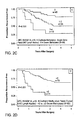
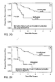
- the rate of recurrence of a proliferative disease is more rapid when gene hypermethylation is detected in lymph node.
- gene hypermethylation e.g. p16 or H-cadherin
- the odds of recurrence may be less than when the same genes are hypermethylated in N1 lymph nodes.
- the odds of recurrence may be the greatest compared to the former tissues (primary tumor and N1 lymph nodes).
- Adjuvant and neoadjuvant therapy are promising treatment modalities, however although adjuvant chemotherapy has been demonstrated to improve survival, for example in node negative breast cancer patients (43), problems remain, for example in the uncertainty as to how to best identify patients whose risk of disease recurrence exceeds their risk of significant therapeutic toxicity.
- adjuvant chemotherapy has been demonstrated to improve survival, for example in node negative breast cancer patients (43)
- problems remain, for example in the uncertainty as to how to best identify patients whose risk of disease recurrence exceeds their risk of significant therapeutic toxicity.
- a need remains for that enable clinical decisions on adjuvant and neoadjuvant therapy, tumor surveillance and the likelihood of disease progression based on validated tumor markers correlated with metastasis and recurrence.
- the invention features a method for determining the prognosis of a subject suffering from a proliferative disease comprising: detecting nucleic acid hypermethylation of one or more genes wherein the detection of nucleic acid hypermethylation is used for determining the prognosis of a subject suffering from a proliferative disease.
- prognosis can be used by the clinician to determine the course of treatment, and to monitor the course of treatment. As is understood by the skilled practicioner, prognosis is a prediction and can change during the course of treatment.
- a proliferative disease e.g. a neoplasia or cancer.
- Staging can refer to "clinical” staging or "molecular” staging.
- Clinical staging describes the extent or severity of an individual's cancer based on the extent of the original (primary) tumor and the extent of spread in the body.
- clinical staging is used in determining a subject's course of treatment and to estimate the subject's prognosis.
- the TNM system is one of the most commonly used staging systems.
- the formal TNM staging system promulgated by the American Joint Committee on Cancer (AJCC), is based almost exclusively on the anatomical extent of disease, which is assessed using a combination of tumor size or depth (T), lymph node spread (N), and presence or absence of metastases (M). Since its inception in 1958, the TNM system has provided a standardized, anatomical basis for staging with several important functions. It provides a basis for prediction of survival, choice of initial treatment, stratification of patients in clinical trials, accurate communication among healthcare providers, and uniform reporting of outcomes. For most tumor types, disease burden and spread have been considered the most reliable predictors of survival and determinants of the type and intensity of therapy to be used.
- TMJ staging Less often, tumor grade, histological subtype or patient age has been added to TNM staging when the AJCC became convinced that such information would significantly improve the prediction of survival or response to therapy.
- TMJ system a number is added to each letter to indicate the size or extent of the tumor and the extent of spread.
- TX indicates that the primary tumor cannot be evaluated
- T0 indicates no evidence of primary tumor
- Tis indicates carcinoma in situ (early cancer that has not spread to neighboring tissue)
- T1, T2, T3, T4 indicates size and/or extent of the primary tumor.
- NX indicates the regional lymph nodes cannot be evaluated
- N0 indicates there is no regional lymph node involvement (no cancer found in the lymph nodes)
- N1, N2, N3 indicates the involvement of regional lymph nodes (number and/or extent of spread).
- MX indicates that distant metastasis cannot be evaluated
- M0 indicates no distant metastasis (cancer has not spread to other parts of the body)
- M1 indicates distant metastasis (cancer has spread to distant parts of the body).
- Criteria for stages differ for different types of cancer. More information on clinical staging can be found on the world wide web, for example at http://www.cancer.gov/cancertopics/factsheet/Detection/staging.
- the instant invention provides the incorporation of biomarkers into TNM staging.
- the instant invention provides a method of molecular staging and re-staging by determining the nucleic acid hypermethylation of one or more certain genes.
- the invention provides methods of molecular restaging that can be used to re-stage any cancer with metastatic capability.
- hypermethylated nucleic acids are detected in the lymph nodes.
- the invention provides methods of detection of early recurrence of proliferative disease, e.g. cancer, that are unable to be detected by methods of clinical staging.
- molecular re-staging as described herein can predict early recurrence of cancer, and thereby detect aggressive cancers at an earlier stage in their progression.
- the invention features a method for staging or re-staging a proliferative disease in a subject comprising detecting nucleic acid hypermethylation of one or more genes wherein detecting nucleic acid hypermethylation is used for staging a proliferative disease.
- the stage of proliferative disease is predictive of disease recurrence.
- Determining the stage of a proliferative disease can be used by the clinician to determine the course of treatment.
- the terms "treat,” treating,” “treatment,” and the like are meant to refer to reducing or ameliorating a disorder and/or symptoms associated therewith. It will be appreciated that, although not precluded, treating a disorder or condition does not require that the disorder, condition or symptoms associated therewith be completely eliminated. In certain cases, an early recurring cancer may be treated with more aggressive therapy.
- the term "aggressive treatment regimen” is intended to mean reducing or ameliorating a disorder and/or symptoms associated therewith with a method of treatment (e.g. combination of chemotherapeutic agents) more intensive or comprehensive than usual, for instance in dosage or extent. It will be appreciated that, although not precluded, aggressively treating a disorder or condition does not require that the disorder, condition or symptoms associated therewith be completely eliminated.
- the invention also features a method for staging or re-staging a proliferative disease in a subject comprising extracting nucleic acid from one or more cell or tissue samples, detecting nucleic acid hypermethylation of one or more genes in the sample; and identifying the nucleic acid hypermethylation state of one or more genes, wherein nucleic acid hypermethylation of genes is used for staging or re-staging of a proliferative disease.
- tissue sample is suitable for use in the methods of staging or re-staging.
- the tissue samples are selected from tumor, lymph node, bone marrow or blood or a combination thereof.
- the samples are from the lymph nodes.
- the molecular grading methods as described herein cab be performed prior to or after therapeutic intervention for the proliferative disease, e.g. cancer.
- the therapeutic intervention can be selected from treatment with an agent or can be a surgical procedure.
- the methods for staging or re-staging a proliferative disease in a subject comprising detecting nucleic acid hypermethylation of one or more genes as described herein can be used as adjuvant or neoadjuvant therapy.
- Samples for use in the methods of the invention include cells or tissues obtained from any solid tumor, samples taken from lymph nodes, from bone marrow or from blood. Additionally, the sample may be a sample that is taken from plasma, serum, sputum, or other fluid. Tumor DNA can be found in various body fluids and these fluids can potentially serve as diagnostic material.
- nucleic acid specimen in purified or nonpurified form, can be utilized as the starting nucleic acid or acids, provided it contains, or is suspected of containing, the specific nucleic acid sequence containing the target locus (e.g., CpG).
- the process may employ, for example, DNA or RNA, including messenger RNA, wherein DNA or RNA may be single stranded or double stranded.
- RNA is to be used as a template
- enzymes, and/or conditions optimal for reverse transcribing the template to DNA would be utilized.
- a DNA-RNA hybrid which contains one strand of each may be utilized.
- a mixture of nucleic acids may also be employed, or the nucleic acids produced in a previous amplification reaction herein, using the same or different primers may be so utilized.
- the specific nucleic acid sequence to be amplified i.e., the target locus, may be a fraction of a larger molecule or can be present initially as a discrete molecule, so that the specific sequence constitutes the entire nucleic acid: It is not necessary that the sequence to be amplified be present initially in a pure form; it may be a minor fraction of a complex mixture, such as contained in whole human DNA.
- the nucleic acid-containing sample or specimen used for detection of methylated CpG may be from any solid tumor or any source including brain, colon, urogenital, hematopoietic, thymus, testis, ovarian, uterine, prostate, breast, colon, lung and renal tissue and may be extracted by a variety of techniques such as that described by Maniatis, et al. (Molecular Cloning: A Laboratory Manual, Cold Spring Harbor, N.Y., pp 280, 281, 1982 ).
- the extracted sample is impure (e.g., plasma, serum, stool, ejaculate, sputum, saliva, ductal cells, nipple aspiration fluid, ductal lavage fluid, cerebrospinal fluid or blood or a sample embedded in parrafin), it may be treated before amplification with an amount of a reagent effective to open the cells, fluids, tissues, or animal cell membranes of the sample, and to expose and/or separate the strand(s) of the nucleic acid(s). This lysing and nucleic acid denaturing step to expose and separate the strands will allow amplification to occur much more readily
- the method of amplifying is by PCR, as described herein and as is commonly used by those of ordinary skill in the art.
- alternative methods of amplification have been described and can also be employed.
- PCR techniques and many variations of PCR are known. Basic PCR techniques are described by Saiki et al. (1988 Science 239:487-491 ) and by U.S. Pat. Nos. 4,683,195 , 4,683,202 and 4,800,159 .
- the conditions generally required for PCR include temperature, salt, cation, pH and related conditions needed for efficient copying of the master-cut fragment.
- PCR conditions include repeated cycles of heat denaturation (i.e. heating to at least about 95° C) and incubation at a temperature permitting primer: adaptor hybridization and copying of the master-cut DNA fragment by the amplification enzyme.
- Heat stable amplification enzymes like the pwo, Thermus aquaticus or Thermococcus litoralis DNA polymerases which eliminate the need to add enzyme after each denaturation cycle, are commercially available.
- the salt, cation, pH and related factors needed for enzymatic amplification activity are available from commercial manufacturers of amplification enzymes.
- an amplification enzyme is any enzyme which can be used for in vitro nucleic acid amplification, e.g. by the above-described procedures.
- amplification enzymes include pwo, Escherichia coli DNA polymerase I, Klenow fragment of E.
- coli polymerase I T4 DNA polymerase, T7 DNA polymerase, Thermus aquaticus (Taq) DNA polymerase, Thermococcus litoralis DNA polymerase, SP6 RNA polymerase, T7 RNA polymerase, T3 RNA polymerase, T4 polynucleotide kinase, Avian Myeloblastosis Virus reverse transcriptase, Moloney Murine Leukemia Virus reverse transcriptase, T4 DNA ligase, E. coli DNA ligase or Q.beta. replicase.
- Preferred amplification enzymes are the pwo and Taq polymerases. The pwo enzyme is especially preferred because of its fidelity in replicating DNA.
- the nucleic acid can be attached to a solid support, such as a membrane, and can be hybridized with any probe of interest, to detect any nucleic acid sequence.
- a solid support such as a membrane
- membranes are known to one of skill in the art for the adhesion of nucleic acid sequences. Specific non-limiting examples of these membranes include nitrocellulose (NITROPURE®) or other membranes used in for detection of gene expression such as polyvinylchloride, diazotized paper and other commercially available membranes such as GENESCREEN®, ZETAPROBE.RTM. (Biorad), and NYTRAN®. Methods for attaching nucleic acids to these membranes are well known to one of skill in the art. Alternatively, screening can be done in a liquid phase.
- nucleic acid hybridization reactions the conditions used to achieve a particular level of stringency will vary, depending on the nature of the nucleic acids being hybridized. For example, the length, degree of complementarity, nucleotide sequence composition (e.g., GC v. AT content), and nucleic acid type (e.g., RNA v. DNA) of the hybridizing regions of the nucleic acids can be considered in selecting hybridization conditions. An additional consideration is whether one of the nucleic acids is immobilized, for example, on a filter.
- An example of progressively higher stringency conditions is as follows: 2xSSC/0.1% SDS at about room temperature (hybridization conditions); 0.2xSSC/0.1% SDS at about room temperature (low stringency conditions); 0.2xSSC/0.1% SDS at about 42°C. (moderate stringency conditions); and 0.1 xSSC at about 68°C. (high stringency conditions). Washing can be carried out using only one of these conditions, e.g., high stringency conditions, or each of the conditions can be used, e.g., for 10-15 minutes each, in the order listed above, repeating any or all of the steps listed. However, as mentioned above, optimal conditions will vary, depending on the particular hybridization reaction involved, and can be determined empirically. In general, conditions of high stringency are used for the hybridization of the probe of interest.
- the probe of interest can be detectably labeled, for example, with a radioisotope, a fluorescent compound, a bioluminescent compound, a chemiluminescent compound, a metal chelator, or an enzyme.
- a radioisotope for example, with a fluorescent compound, a bioluminescent compound, a chemiluminescent compound, a metal chelator, or an enzyme.
- Those of ordinary skill in the art will know of other suitable labels for binding to the probe, or will be able to ascertain such, using routine experimentation.
- kits The methods of the invention are ideally suited for the preparation of kits.
- kits for identifying the nucleic acid hypermethylation state of one or more genes comprising gene specific primers for use in polymerase chain reaction (PCR), and instructions for use.
- PCR polymerase chain reaction
- kits for detecting metastases by detecting nucleic acid hypermethylation of one or more genes comprising gene specific primers for use in polymerase chain reaction (PCR), and instructions for use.
- the metastases are micrometastases.
- the PCR in particularly preferred examples, is methylation specific PCR (MSP).
- MSP methylation specific PCR
- any gene comprising one or more CpG islands in the promoter region can be detected using the kits of the invention.
- the one or more genes are selected from the group consisting of genes involved in tumor suppression, nucleic acid repair, anti-proliferation, apoptosis, ras signaling, adhesion, differentiation, development, and cell cycle regulation.
- the genes are selected from the group consisting of: p-16, H-cadherin, APC, RASSF1A, MGMT, DAPK, and ASC
- kits can be used to detect hypermethylation of at least one of the genes as described herein. In some examples, can be used to detect hypermethylation of at least two of the genes as described herein. In other examples, the kits can be used to detect hypermethylation of at least three of the genes as described herein.
- the two genes can be selected from the following: p-16 and H-cadherin, H-cadherin and APC, APC and p16, or RASSf1A and p16.
- Carrier means are suited for containing one or more container means such as vials, tubes, and the like, each of the container means comprising one of the separate elements to be used in the method.
- container means such as vials, tubes, and the like
- each of the container means comprising one of the separate elements to be used in the method.
- the container means can comprise a container containing gene specific primers for use in polymerase chain reaction methods of the invention.
- container means can also be included which comprise a methylation sensitive restriction endonuclease.
- gene hypermethylation versus traditional histopathology was tested to predict disease recurrence in solid tumors.
- the examples presented herein show that gene hypermethylation of not only the primary malignancy, but also lymph nodes, may be used to restage and assess prognosis of patients with stage I tumors, in particular examples patients with stage I NSCLC. These markers are shown to also be potential targets for reversal of gene silencing and may be important in adjuvant approaches to reduce disease recurrence.
- Pathologic tumor stage showed the strongest risk for predicting disease recurrence independent of other covariates, with patients with stage 1B disease (T2 malignancies ⁇ 3cm and/or visceral pleural invasion) having a 1.71 (95%CI, 0.86-3.41) fold risk for disease recurrence compared to patients with smaller sized tumors and no pleural invasion (stage 1A).
- Methylation profiles using 7 genes were obtained on 727 of 731 paraffin blocks corresponding to 167 patients.
- Figure 1 shows multivariate logistic regression analysis that was performed using the four genes that exhibited the largest univariate distribution differences in methylation: p16, H-cadherin, APC, and RASSF1A.
- the prognostic value of each molecular variable was assessed in a model that adjusted for stage (1A/1B), age (continuous), sex, race (Caucasian/African American), histology (adenocarcinoma, squamous cell, other), smoking status (ever/never) and then graphed as a Forest Plot. On the whole, regardless of the genes considered, hypermethylation of the mediastinal lymph node tissue had the highest prognostic value for estimating lung cancer recurrence versus no methylation.
- methylation of both p16 and H-cadherin had robust odds ratios for recurrence for primary tumor, regional, and mediastinal lymph nodes. Methylation of either RASSF1A or APC in primary tumor or N2 nodes was also associated with a modest elevation in odds of recurrence but this was not statistically significant. If a patient with pathologic stage 1 disease (T1-2N0) had concomitant methylation of p16 as well as H-cadherin in both tumor and mediastinal lymph nodes, the estimated odds of lung cancer recurrence was over 15 as compared to those patients without concomitant methylation in these tissues.
- pathologic stage 1 disease T1-2N0
- the Kaplan-Meier estimates for time to recurrence indicate that for both tumor as well as for regional (N1) and mediastinal (N2) lymph nodes, as the number of methylated genes in the panel increases, there is a significant reduction in the recurrence-free survival (Panels A-C, E-G).
- the Kaplan-Meier estimates reflect the recurrence-free survivals of patients with a clinically useful risk classification system as determined by our epigenetic marker panel.
- Patients without methylation of the gene combination p16 and H-cadherin in both primary tumor and mediastinal lymph nodes have a significantly improved 5 year recurrence-free period than those with these genes methylated (64% vs. 14%, p ⁇ 0.001, respectively, Panel H).
- the results presented herein demonstrate two related features for the detection of promoter region methylation in cancer.
- the detection of promoter methylation within the resected primary tumor for key genes can be associated with a more aggressive, recurrent phenotype as has been shown in other settings (28,29).
- the loss of function in key regulatory genes for cell cycle control (p16), invasion and metastasis (H-Cadherin, APC), as well as Ras signaling (RASSFIa) might be expected to result in a more aggressive primary tumor.
- the ability to detect methylation within local or regional nodes provides a second layer of information to this subset identification, and demonstrates an additional approach for detecting micrometastatic disease.
- stage 1 lung cancer patients who were not staged optimally by routine pathological analysis.
- the current staging system for NSCLC is imprecise, and in stage 1 (T1-2N0) disease, in particular, the clinicopathologic criteria understages patients.
- the results presented herein show that gene promoter methylation detection within NSCLC primary tumors can be used to identify cells with high potential for metastatic spread, and also to detect histologically occult micrometastases in lymph nodes used to stage NSCLC.
- the methylation detected could represent free tumor DNA that has drained from the primary tumor via lymphatics, this is unlikely, particularly for N2 nodes, since these are located distant from the lung in a separate body compartment, the mediastinum. While the detection of methylation of these genes in tumor or N1 nodes was often associated with increased risk of recurrence, in the N2 lymph nodes, especially, the markers were very strongly prognostic, strengthening the contention that the DNA methylation is detecting micrometastatic disease.
- This molecular distribution of markers in patients with rapid recurrence mirrors the biological basis of current histological staging systems that identify intact tumor cells that have traversed the mediastinal pleura to the N2 nodes and are associated with increased risk of recurrence.
- the methods described herein present a molecular tool that parallels the use of histologic examination in accepted clinical pathology practice, but is more sensitive. This differs from previous studies that have relied solely on molecular characteristics of the primary malignancy (30-33). This ability to utilize the predictive power inherent in identifying micrometastases to lymph nodes may allow a more robust and reliable molecular staging built upon tumor characteristics and the detection of micrometastatic disease. Furthermore, recent promising results from examining methylation changes in sputum for predicting risk of lung cancer (34), or its recurrence (18), means the detection of these changes could provide valuable pre-surgical information as to disease stage and the metastatic potential of a patient's tumor.
- the invention was carried out using methods that include the following.
- stage 1 pathologically proven stage 1 (T1-2N0) patients diagnosed with non-small cell lung carcinoma (NSCLC) (International Classification of Diseases-ninth revision-Clinical Modification [ICD-9-CM] code 162.3-162.9) who underwent lobectomy or greater resections at the Johns Hopkins Hospital between January 1, 1986 and July 31, 2002. Only patients followed for recurrent disease at the institution were eligible for the analysis.
- the study cohort consisted of 71 patients (cases) who despite receiving surgery with curative intent for pathological stage 1 (T1-2N0) primary NSCLC, recurred at the institution within 40 months of surgery and died of their cancer.
- stage 1 lung cancer It was estimated that by 40 months approximately 80% of patients with resected stage 1 lung cancer would recur. Using patient age, stage, date of surgery (within 5 years), and sex, the cases were matched to 158 stage 1 patients (controls) from the remainder of the study population. These 71 cases and 158 controls formed the basic case-control population. From the above patients, samples were gathered for methylation analysis for 51 cases and 116 matched controls. Neither cases nor controls received adjuvant chemotherapy since surgery was performed between 1986-2002 when guidelines did not recommend adjuvant therapy for stage 1B patients (20, 21).
- TNM classification criteria (5) which include histological status of mediastinal lymph nodes sampled from levels 2, 4, 7, 8, 9, and 10 on the right, and 5, 6, 7, 8, 9 on the left side.
- Regional lymph nodes confined to the pleural space, were resected en bloc with the tumor. Patients were excluded as cases if they had surgery involving less than a lobectomy because there is strong evidence that patients with such resections are at significantly increased risk of local recurrence (22). Patients were also excluded as cases if they had any macroscopic or microscopically positive surgical margins, or underwent incomplete resection. In accord with this nested design, seven individuals with recurrences after 40 months postoperatively were considered as controls.
- the 20 patients in the validation set consisting of 11 cases and 9 matched controls, had 162 paraffin blocks evaluated. All cases in the independent validation cohort, except for two, underwent resection at our institution after August 2002. The study was approved by the Institutional Review Board of the Johns Hopkins Medical Institutions.
- Wizard DNA purification resin Promega, Madison, Wis.
- MSP Methylation Specific PCR
- DNA methylation for all lung cancer and lymph node DNA, was determined by MSP performed by 3 individuals blinded to the results of other investigators. Each individual extracted DNA and performed all steps of the MSP reaction separately. There were 889 total samples of tumor and lymph nodes examined. A multiplex-nested MSP assay as previously described was used for all samples24. The nested approach amplifies bisulfite-modified DNA initially with flanking PCR primers without preferentially amplifying methylated or unmethylated DNA. The resulting fragment is then used as the template for MSP.
- Primer sequences and conditions of p16, MGMT, DAPK, RASSF1A, H-cadherin, ASC and APC have all been previously describedl6, 18, 24-26 including conditions optimized to achieve specific detection of methylation in tumor but not in normal lymphocytes, and are shown in Table 5, below (16, 18, 24-26).
- Figure 3 shows methylation specific PCR for the H-cadherin gene in 36 samples of tumors and lymph nodes, numbered at top of each panel. * represents the molecular weight marker.
- the presence of a visible PCR product in Lanes U indicates the presence of an unmethylated promoter region amplified and serves as a control for sample preparation; the presence of product in Lanes M indicates a methylated gene promoter and was scored as positive for methylation.
- Samples 7 and 16 did not amplify for this gene. Samples 1, 2, 3, 4, 8, 9, 11, 13, 14, 20, 21, 22, 23, 26, 30, 32, 33, 36 are scored as unmethylated.
- Samples 5, 10, 12, 15, 17, 18, 19, 25, 27, 28, 31, 34, and 35 are methylated and samples 24 and 29 had very minimal M amplification that was scored as negative.
- Placental DNA treated in vitro with SssI methyltransferase (New England Biolabs, Beverly, MA) was used as a positive control.
- DNA from normal lymphocytes and water (bisulfite-modified and unmodified water) were used as negative controls.
- PCR products were visualized using 2% agarose or 6% nondenaturing polyacrylamide gels, and visually scored as methylated or unmethylated according to the presence or absence of a PCR product ( Figure 3 ), blinded to both the clinical outcomes and sources of DNA. (9, 25, 27).
Landscapes
- Chemical & Material Sciences (AREA)
- Health & Medical Sciences (AREA)
- Life Sciences & Earth Sciences (AREA)
- Organic Chemistry (AREA)
- Proteomics, Peptides & Aminoacids (AREA)
- Analytical Chemistry (AREA)
- Zoology (AREA)
- Pathology (AREA)
- Genetics & Genomics (AREA)
- Immunology (AREA)
- Wood Science & Technology (AREA)
- Engineering & Computer Science (AREA)
- General Health & Medical Sciences (AREA)
- Physics & Mathematics (AREA)
- Microbiology (AREA)
- General Engineering & Computer Science (AREA)
- Hospice & Palliative Care (AREA)
- Biophysics (AREA)
- Oncology (AREA)
- Biochemistry (AREA)
- Bioinformatics & Cheminformatics (AREA)
- Molecular Biology (AREA)
- Biotechnology (AREA)
- Nuclear Medicine, Radiotherapy & Molecular Imaging (AREA)
- Chemical Kinetics & Catalysis (AREA)
- Medicinal Chemistry (AREA)
- General Chemical & Material Sciences (AREA)
- Pharmacology & Pharmacy (AREA)
- Animal Behavior & Ethology (AREA)
- Public Health (AREA)
- Veterinary Medicine (AREA)
- Measuring Or Testing Involving Enzymes Or Micro-Organisms (AREA)
Claims (6)
- Procédé in vitro pour prédire le prognostic du cancer du poumon non à petites cellules chez un sujet comprenant l'étape consistant à détecter une hyperméthylation d'acide nucléique de l'H-cadhérine ou d'un ou plusieurs gène(s) additionnel(s) choisi(s) dans le groupe consistant en APC et RASSF1A dans un ou plusieurs échantillon(s) de tissu tumoral ou de ganglion lymphatique provenant du sujet, dans lequel la détection d'une hyperméthylation d'acide nucléique dans la tumeur primaire, et éventuellement dans le ganglion lymphatique, identifie une durée de survie médiane sensiblement réduite.
- Procédé selon la revendication comprenant l'étape consistant à détecter une hyperméthylation d'acide nucléique de l'H-cadhérine et d'APC ou de l'H-cadhérine et de RASSF1A.
- Procédé selon la revendication 1 ou 2, dans lequel les cellules ou les tissus comprennent des métastases ou des micrométastases.
- Procédé in vitro pour prédire la récurrence du cancer du poumon non à petites cellules chez un sujet comprenant l'étape consistant à détecter une hyperméthylation d'acide nucléique de l'H-cadhérine ou d'un ou plusieurs gène(s) additionnel(s) choisi(s) dans le groupe consistant en APC et RASSF1A dans un ou plusieurs échantillon(s) de tissu tumoral ou de ganglion lymphatique provenant du sujet, dans lequel la détection d'une hyperméthylation d'acide nucléique dans la tumeur primaire, et éventuellement dans le ganglion lymphatique, est utilisé pour prédire la récurrence du cancer du poumon.
- Procédé selon la revendication 1 ou 4 comprenant en outre une étape pour comparer l'hyperméthylation d'acide nucléique de l'H-cadhérine dans l'échantillon avec un échantillon comparative provenant d'un sujet normal.
- Procédé in vitro pour prédire la durée de survie chez un sujet ayant un cancer du poumon non à petites cellules comprenant l'étape consistant à détecter une hyperméthylation d'acide nucléique de l'H-cadhérine ou d'un ou plusieurs gène(s) additionnel(s) choisi(s) dans le groupe consistant en APC et RASSF1A dans un ou plusieurs échantillon(s) de tissu tumoral ou de ganglion lymphatique provenant du sujet, dans lequel la détection d'une hyperméthylation d'acide nucléique dans la tumeur primaire, et éventuellement dans le ganglion lymphatique, prédit une durée de survie réduite due au cancer du poumon non à petites cellules.
Priority Applications (1)
| Application Number | Priority Date | Filing Date | Title |
|---|---|---|---|
| EP12163883.7A EP2508623B1 (fr) | 2006-11-20 | 2007-11-20 | Marqueurs de méthylation de l'ADN et leurs méthodes d'utilisation |
Applications Claiming Priority (2)
| Application Number | Priority Date | Filing Date | Title |
|---|---|---|---|
| US86019606P | 2006-11-20 | 2006-11-20 | |
| PCT/US2007/024308 WO2008063655A2 (fr) | 2006-11-20 | 2007-11-20 | Marqueurs de méthylation de l'adn et leurs méthodes d'utilisation |
Related Child Applications (2)
| Application Number | Title | Priority Date | Filing Date |
|---|---|---|---|
| EP12163883.7A Division EP2508623B1 (fr) | 2006-11-20 | 2007-11-20 | Marqueurs de méthylation de l'ADN et leurs méthodes d'utilisation |
| EP12163883.7A Division-Into EP2508623B1 (fr) | 2006-11-20 | 2007-11-20 | Marqueurs de méthylation de l'ADN et leurs méthodes d'utilisation |
Publications (3)
| Publication Number | Publication Date |
|---|---|
| EP2102361A2 EP2102361A2 (fr) | 2009-09-23 |
| EP2102361A4 EP2102361A4 (fr) | 2010-02-24 |
| EP2102361B1 true EP2102361B1 (fr) | 2015-07-08 |
Family
ID=39430374
Family Applications (2)
| Application Number | Title | Priority Date | Filing Date |
|---|---|---|---|
| EP07862181.0A Not-in-force EP2102361B1 (fr) | 2006-11-20 | 2007-11-20 | Marqueurs de méthylation de l'adn et leurs méthodes d'utilisation |
| EP12163883.7A Not-in-force EP2508623B1 (fr) | 2006-11-20 | 2007-11-20 | Marqueurs de méthylation de l'ADN et leurs méthodes d'utilisation |
Family Applications After (1)
| Application Number | Title | Priority Date | Filing Date |
|---|---|---|---|
| EP12163883.7A Not-in-force EP2508623B1 (fr) | 2006-11-20 | 2007-11-20 | Marqueurs de méthylation de l'ADN et leurs méthodes d'utilisation |
Country Status (4)
| Country | Link |
|---|---|
| US (2) | US20100278772A1 (fr) |
| EP (2) | EP2102361B1 (fr) |
| CA (1) | CA2670082A1 (fr) |
| WO (1) | WO2008063655A2 (fr) |
Families Citing this family (9)
| Publication number | Priority date | Publication date | Assignee | Title |
|---|---|---|---|---|
| JP2010517582A (ja) * | 2007-02-12 | 2010-05-27 | ザ・ジョンズ・ホプキンス・ユニバーシティー | 結腸癌の早期検出および予後診断の方法 |
| WO2010000467A1 (fr) | 2008-07-03 | 2010-01-07 | F. Hoffmann-La Roche Ag | Asc en tant que marqueur du cancer du poumon |
| CN102787174B (zh) * | 2012-09-06 | 2013-08-07 | 南京大学 | 胃肠肿瘤发病相关抑癌基因表观突变检测试剂盒及其用途 |
| WO2014173905A2 (fr) * | 2013-04-23 | 2014-10-30 | Institut D'investigació Biomèdica De Bellvitge (Idibell) | Méthodes et trousses pour le pronostic de carcinome du poumon non à petites molécules (nsclc) de stade i par la détermination du motif de méthylation de dinucléotides cpg |
| WO2017158158A1 (fr) * | 2016-03-18 | 2017-09-21 | Region Nordjylland, Aalborg University Hospital | Marqueurs de méthylation pour le cancer du pancréas |
| US12009061B2 (en) | 2019-04-10 | 2024-06-11 | University of Pittsburgh—of the Commonwealth System of Higher Education | Computational filtering of methylated sequence data for predictive modeling |
| WO2020243722A1 (fr) * | 2019-05-31 | 2020-12-03 | Guardant Health, Inc. | Procédés et systèmes pour améliorer une surveillance de patient après une intervention chirurgicale |
| WO2020257404A1 (fr) * | 2019-06-20 | 2020-12-24 | The Johns Hopkins University | Marqueurs d'adn pour la différenciation d'échantillons de biopsie |
| WO2024117791A1 (fr) * | 2022-11-29 | 2024-06-06 | 주식회사 지씨지놈 | Marqueurs de méthylation de l'adn pour le diagnostic du cancer de l'oesophage et du cancer de l'estomac et leur utilisation |
Family Cites Families (15)
| Publication number | Priority date | Publication date | Assignee | Title |
|---|---|---|---|---|
| US4458066A (en) | 1980-02-29 | 1984-07-03 | University Patents, Inc. | Process for preparing polynucleotides |
| US4683202A (en) | 1985-03-28 | 1987-07-28 | Cetus Corporation | Process for amplifying nucleic acid sequences |
| US4683195A (en) | 1986-01-30 | 1987-07-28 | Cetus Corporation | Process for amplifying, detecting, and/or-cloning nucleic acid sequences |
| US4800159A (en) | 1986-02-07 | 1989-01-24 | Cetus Corporation | Process for amplifying, detecting, and/or cloning nucleic acid sequences |
| US5856094A (en) * | 1995-05-12 | 1999-01-05 | The Johns Hopkins University School Of Medicine | Method of detection of neoplastic cells |
| US5786146A (en) | 1996-06-03 | 1998-07-28 | The Johns Hopkins University School Of Medicine | Method of detection of methylated nucleic acid using agents which modify unmethylated cytosine and distinguishing modified methylated and non-methylated nucleic acids |
| US6017704A (en) | 1996-06-03 | 2000-01-25 | The Johns Hopkins University School Of Medicine | Method of detection of methylated nucleic acid using agents which modify unmethylated cytosine and distinguishing modified methylated and non-methylated nucleic acids |
| US6605432B1 (en) | 1999-02-05 | 2003-08-12 | Curators Of The University Of Missouri | High-throughput methods for detecting DNA methylation |
| EP1352089B1 (fr) | 2000-08-25 | 2006-12-06 | Lovelace Respiratory Research Institute | Methode de detection de cancer par reaction de polymerisation en chaine specifique de la methylation imbriquee |
| AU2002236507A1 (en) * | 2000-11-29 | 2002-06-11 | Cangen International | Dap-kinase and hoxa9, two human genes associated with genesis, progression, and aggressiveness of non-small cell lung cancer |
| US6756200B2 (en) * | 2001-01-26 | 2004-06-29 | The Johns Hopkins University School Of Medicine | Aberrantly methylated genes as markers of breast malignancy |
| CA2478592A1 (fr) * | 2002-03-07 | 2003-09-18 | The Johns Hopkins University School Of Medicine | Depistage genomique pour genes lies au cancer rendus epigenetiquement silencieux |
| AU2003224978A1 (en) * | 2002-04-12 | 2003-10-27 | Curators Of The University Of Missouri | ECIST microarrays for dual screening of DNA hypermethylation and gene silencing |
| WO2004086949A2 (fr) * | 2003-03-25 | 2004-10-14 | John Wayne Cancer Institute | Marqueurs d'adn utilises dans la gestion du cancer |
| EP1692264A2 (fr) | 2003-10-28 | 2006-08-23 | The Johns Hopkins University | Pcr quantitative multiplex specifique de la methylation |
-
2007
- 2007-11-20 WO PCT/US2007/024308 patent/WO2008063655A2/fr active Application Filing
- 2007-11-20 US US12/515,735 patent/US20100278772A1/en not_active Abandoned
- 2007-11-20 CA CA002670082A patent/CA2670082A1/fr not_active Abandoned
- 2007-11-20 EP EP07862181.0A patent/EP2102361B1/fr not_active Not-in-force
- 2007-11-20 EP EP12163883.7A patent/EP2508623B1/fr not_active Not-in-force
-
2014
- 2014-02-14 US US14/181,121 patent/US10329621B2/en active Active
Non-Patent Citations (2)
| Title |
|---|
| CHANG HSIAO WEN ET AL: "Loss of E-cadherin expression resulting from promoter hypermethylation in oral tongue carcinoma and its prognostic significance", CANCER, vol. 94, no. 2, 15 January 2002 (2002-01-15), pages 386 - 392, ISSN: 0008-543X * |
| HOJOONG KIM ET AL: "Tumor-specific methylation in bronchial lavage for the early detection of non-small-cell lung cancer", JOURNAL OF CLINICAL ONCOLOGY, AMERICAN SOCIETY OF CLINICAL ONCOLOGY, US, vol. 22, no. 12, 15 June 2004 (2004-06-15), pages 2363 - 2370, XP002641112, ISSN: 0732-183X, DOI: 10.1200/JCO.2004.10.077 * |
Also Published As
| Publication number | Publication date |
|---|---|
| CA2670082A1 (fr) | 2008-05-29 |
| EP2508623A1 (fr) | 2012-10-10 |
| EP2102361A2 (fr) | 2009-09-23 |
| US20150031022A1 (en) | 2015-01-29 |
| WO2008063655A2 (fr) | 2008-05-29 |
| US20100278772A1 (en) | 2010-11-04 |
| US10329621B2 (en) | 2019-06-25 |
| EP2102361A4 (fr) | 2010-02-24 |
| WO2008063655A9 (fr) | 2008-09-18 |
| EP2508623B1 (fr) | 2015-08-12 |
| WO2008063655A3 (fr) | 2008-11-27 |
Similar Documents
| Publication | Publication Date | Title |
|---|---|---|
| US10329621B2 (en) | DNA methylation markers and methods of use | |
| US20190048422A1 (en) | Compositions and methods for detecting and diagnosing neoplasia | |
| CN108350504B (zh) | 膀胱癌的诊断方法 | |
| EP3334834B1 (fr) | Procédé de préparation de molécules d'acide nucléique sans cellule par amplification in situ | |
| EP2670861B1 (fr) | Marqueurs de mélanome et leurs utilisations | |
| EP2748333B1 (fr) | Méthylation d'adn dans des procédés de diagnostic du cancer colorectal | |
| EP2891720B1 (fr) | Méthode de dépistage du cancer | |
| Aparicio et al. | LINE-1 methylation in plasma DNA as a biomarker of activity of DNA methylation inhibitors in patients with solid tumors | |
| EP2625288B1 (fr) | Méthode de détermination du pronostic de survie d'un patient atteint de cancer basée sur l'analyse des lymphocytes t globaux infiltrant la tumeur | |
| US20080311574A1 (en) | Novel Missense Mutations and Single Nucleotide Polymorphisms in the Rabphillin-3A-Like Gene and Uses Thereof | |
| CA2600337A1 (fr) | Marqueurs de melanome | |
| US20100297067A1 (en) | Methylation markers for prostate cancer and methods of use | |
| US10597730B2 (en) | Method of diagnosing cancer | |
| EP2855705B1 (fr) | Méthode de diagnostic du cancer comprenant la détection de la signature de méthylation dans le promoteur htert | |
| US20220356532A1 (en) | Compositions and methods for detecting and treating esophageal cancer | |
| US20080026481A1 (en) | Molecular based two-marker assays that predict outcome of adenocarcinoma patients | |
| US20110159504A1 (en) | Methylation markers for sensitivity to microtube based therapies and methods of use | |
| US20220195535A1 (en) | Methods for Detecting and Treating Esophageal Cancer | |
| JP2010088406A (ja) | 癌患者の外科的手術後の治療選択方法及び予後診断 | |
| EP2406393B1 (fr) | Nouveaux marqueurs pour la détection du cancer de la vessie | |
| WO2015021263A2 (fr) | Biomarqueurs de méthylation pour le cancer colorectal | |
| EP3006572B1 (fr) | Méthode de prédiction de la réponse au traitement par radiothérapie combinée à la chimiothérapie au cisplatine | |
| US20090298068A1 (en) | Method and test kit for the diagnosis and/or making predictions about and/or for the assessment of the efficacy of therapeutic agents for the treatment of ovarian cancer and method of planning a regimen for the treatment of ovarian cancer | |
| Cee | Detection of methylated tumor DNA in serum as a marker for the colorectal carcinoma | |
| Fiano | DNA METHYLATION IN PROSTATE CANCER: POSSIBLE PROGNOSIS MARKERS AND HYPOTHESIS ON CAUSAL RELATIONSHIP BETWEEN EPIGENETICS AND TUMOR PROGRESSION |
Legal Events
| Date | Code | Title | Description |
|---|---|---|---|
| PUAI | Public reference made under article 153(3) epc to a published international application that has entered the european phase |
Free format text: ORIGINAL CODE: 0009012 |
|
| 17P | Request for examination filed |
Effective date: 20090622 |
|
| AK | Designated contracting states |
Kind code of ref document: A2 Designated state(s): AT BE BG CH CY CZ DE DK EE ES FI FR GB GR HU IE IS IT LI LT LU LV MC MT NL PL PT RO SE SI SK TR |
|
| A4 | Supplementary search report drawn up and despatched |
Effective date: 20100121 |
|
| DAX | Request for extension of the european patent (deleted) | ||
| 17Q | First examination report despatched |
Effective date: 20100412 |
|
| GRAP | Despatch of communication of intention to grant a patent |
Free format text: ORIGINAL CODE: EPIDOSNIGR1 |
|
| INTG | Intention to grant announced |
Effective date: 20141022 |
|
| GRAS | Grant fee paid |
Free format text: ORIGINAL CODE: EPIDOSNIGR3 |
|
| GRAA | (expected) grant |
Free format text: ORIGINAL CODE: 0009210 |
|
| AK | Designated contracting states |
Kind code of ref document: B1 Designated state(s): AT BE BG CH CY CZ DE DK EE ES FI FR GB GR HU IE IS IT LI LT LU LV MC MT NL PL PT RO SE SI SK TR |
|
| REG | Reference to a national code |
Ref country code: GB Ref legal event code: FG4D |
|
| REG | Reference to a national code |
Ref country code: AT Ref legal event code: REF Ref document number: 735475 Country of ref document: AT Kind code of ref document: T Effective date: 20150715 Ref country code: CH Ref legal event code: EP |
|
| REG | Reference to a national code |
Ref country code: IE Ref legal event code: FG4D |
|
| REG | Reference to a national code |
Ref country code: DE Ref legal event code: R096 Ref document number: 602007042099 Country of ref document: DE |
|
| REG | Reference to a national code |
Ref country code: AT Ref legal event code: MK05 Ref document number: 735475 Country of ref document: AT Kind code of ref document: T Effective date: 20150708 |
|
| REG | Reference to a national code |
Ref country code: NL Ref legal event code: MP Effective date: 20150708 |
|
| REG | Reference to a national code |
Ref country code: LT Ref legal event code: MG4D |
|
| REG | Reference to a national code |
Ref country code: FR Ref legal event code: PLFP Year of fee payment: 9 |
|
| PG25 | Lapsed in a contracting state [announced via postgrant information from national office to epo] |
Ref country code: FI Free format text: LAPSE BECAUSE OF FAILURE TO SUBMIT A TRANSLATION OF THE DESCRIPTION OR TO PAY THE FEE WITHIN THE PRESCRIBED TIME-LIMIT Effective date: 20150708 Ref country code: GR Free format text: LAPSE BECAUSE OF FAILURE TO SUBMIT A TRANSLATION OF THE DESCRIPTION OR TO PAY THE FEE WITHIN THE PRESCRIBED TIME-LIMIT Effective date: 20151009 Ref country code: LV Free format text: LAPSE BECAUSE OF FAILURE TO SUBMIT A TRANSLATION OF THE DESCRIPTION OR TO PAY THE FEE WITHIN THE PRESCRIBED TIME-LIMIT Effective date: 20150708 Ref country code: LT Free format text: LAPSE BECAUSE OF FAILURE TO SUBMIT A TRANSLATION OF THE DESCRIPTION OR TO PAY THE FEE WITHIN THE PRESCRIBED TIME-LIMIT Effective date: 20150708 |
|
| PG25 | Lapsed in a contracting state [announced via postgrant information from national office to epo] |
Ref country code: PL Free format text: LAPSE BECAUSE OF FAILURE TO SUBMIT A TRANSLATION OF THE DESCRIPTION OR TO PAY THE FEE WITHIN THE PRESCRIBED TIME-LIMIT Effective date: 20150708 Ref country code: PT Free format text: LAPSE BECAUSE OF FAILURE TO SUBMIT A TRANSLATION OF THE DESCRIPTION OR TO PAY THE FEE WITHIN THE PRESCRIBED TIME-LIMIT Effective date: 20151109 Ref country code: IS Free format text: LAPSE BECAUSE OF FAILURE TO SUBMIT A TRANSLATION OF THE DESCRIPTION OR TO PAY THE FEE WITHIN THE PRESCRIBED TIME-LIMIT Effective date: 20151108 Ref country code: ES Free format text: LAPSE BECAUSE OF FAILURE TO SUBMIT A TRANSLATION OF THE DESCRIPTION OR TO PAY THE FEE WITHIN THE PRESCRIBED TIME-LIMIT Effective date: 20150708 Ref country code: SE Free format text: LAPSE BECAUSE OF FAILURE TO SUBMIT A TRANSLATION OF THE DESCRIPTION OR TO PAY THE FEE WITHIN THE PRESCRIBED TIME-LIMIT Effective date: 20150708 Ref country code: AT Free format text: LAPSE BECAUSE OF FAILURE TO SUBMIT A TRANSLATION OF THE DESCRIPTION OR TO PAY THE FEE WITHIN THE PRESCRIBED TIME-LIMIT Effective date: 20150708 |
|
| REG | Reference to a national code |
Ref country code: DE Ref legal event code: R097 Ref document number: 602007042099 Country of ref document: DE |
|
| PG25 | Lapsed in a contracting state [announced via postgrant information from national office to epo] |
Ref country code: CZ Free format text: LAPSE BECAUSE OF FAILURE TO SUBMIT A TRANSLATION OF THE DESCRIPTION OR TO PAY THE FEE WITHIN THE PRESCRIBED TIME-LIMIT Effective date: 20150708 Ref country code: SK Free format text: LAPSE BECAUSE OF FAILURE TO SUBMIT A TRANSLATION OF THE DESCRIPTION OR TO PAY THE FEE WITHIN THE PRESCRIBED TIME-LIMIT Effective date: 20150708 Ref country code: EE Free format text: LAPSE BECAUSE OF FAILURE TO SUBMIT A TRANSLATION OF THE DESCRIPTION OR TO PAY THE FEE WITHIN THE PRESCRIBED TIME-LIMIT Effective date: 20150708 Ref country code: DK Free format text: LAPSE BECAUSE OF FAILURE TO SUBMIT A TRANSLATION OF THE DESCRIPTION OR TO PAY THE FEE WITHIN THE PRESCRIBED TIME-LIMIT Effective date: 20150708 Ref country code: IT Free format text: LAPSE BECAUSE OF FAILURE TO SUBMIT A TRANSLATION OF THE DESCRIPTION OR TO PAY THE FEE WITHIN THE PRESCRIBED TIME-LIMIT Effective date: 20150708 |
|
| PLBE | No opposition filed within time limit |
Free format text: ORIGINAL CODE: 0009261 |
|
| STAA | Information on the status of an ep patent application or granted ep patent |
Free format text: STATUS: NO OPPOSITION FILED WITHIN TIME LIMIT |
|
| PG25 | Lapsed in a contracting state [announced via postgrant information from national office to epo] |
Ref country code: RO Free format text: LAPSE BECAUSE OF FAILURE TO SUBMIT A TRANSLATION OF THE DESCRIPTION OR TO PAY THE FEE WITHIN THE PRESCRIBED TIME-LIMIT Effective date: 20150708 |
|
| 26N | No opposition filed |
Effective date: 20160411 |
|
| PG25 | Lapsed in a contracting state [announced via postgrant information from national office to epo] |
Ref country code: MC Free format text: LAPSE BECAUSE OF FAILURE TO SUBMIT A TRANSLATION OF THE DESCRIPTION OR TO PAY THE FEE WITHIN THE PRESCRIBED TIME-LIMIT Effective date: 20150708 Ref country code: LU Free format text: LAPSE BECAUSE OF FAILURE TO SUBMIT A TRANSLATION OF THE DESCRIPTION OR TO PAY THE FEE WITHIN THE PRESCRIBED TIME-LIMIT Effective date: 20151120 |
|
| REG | Reference to a national code |
Ref country code: CH Ref legal event code: PL |
|
| PG25 | Lapsed in a contracting state [announced via postgrant information from national office to epo] |
Ref country code: LI Free format text: LAPSE BECAUSE OF NON-PAYMENT OF DUE FEES Effective date: 20151130 Ref country code: CH Free format text: LAPSE BECAUSE OF NON-PAYMENT OF DUE FEES Effective date: 20151130 |
|
| REG | Reference to a national code |
Ref country code: IE Ref legal event code: MM4A |
|
| PG25 | Lapsed in a contracting state [announced via postgrant information from national office to epo] |
Ref country code: SI Free format text: LAPSE BECAUSE OF FAILURE TO SUBMIT A TRANSLATION OF THE DESCRIPTION OR TO PAY THE FEE WITHIN THE PRESCRIBED TIME-LIMIT Effective date: 20150708 |
|
| PG25 | Lapsed in a contracting state [announced via postgrant information from national office to epo] |
Ref country code: IE Free format text: LAPSE BECAUSE OF NON-PAYMENT OF DUE FEES Effective date: 20151120 |
|
| REG | Reference to a national code |
Ref country code: FR Ref legal event code: PLFP Year of fee payment: 10 |
|
| PG25 | Lapsed in a contracting state [announced via postgrant information from national office to epo] |
Ref country code: BE Free format text: LAPSE BECAUSE OF FAILURE TO SUBMIT A TRANSLATION OF THE DESCRIPTION OR TO PAY THE FEE WITHIN THE PRESCRIBED TIME-LIMIT Effective date: 20150708 |
|
| PGFP | Annual fee paid to national office [announced via postgrant information from national office to epo] |
Ref country code: FR Payment date: 20161123 Year of fee payment: 10 Ref country code: GB Payment date: 20161128 Year of fee payment: 10 Ref country code: DE Payment date: 20161123 Year of fee payment: 10 |
|
| PG25 | Lapsed in a contracting state [announced via postgrant information from national office to epo] |
Ref country code: BG Free format text: LAPSE BECAUSE OF FAILURE TO SUBMIT A TRANSLATION OF THE DESCRIPTION OR TO PAY THE FEE WITHIN THE PRESCRIBED TIME-LIMIT Effective date: 20150708 Ref country code: HU Free format text: LAPSE BECAUSE OF FAILURE TO SUBMIT A TRANSLATION OF THE DESCRIPTION OR TO PAY THE FEE WITHIN THE PRESCRIBED TIME-LIMIT; INVALID AB INITIO Effective date: 20071120 |
|
| PG25 | Lapsed in a contracting state [announced via postgrant information from national office to epo] |
Ref country code: CY Free format text: LAPSE BECAUSE OF FAILURE TO SUBMIT A TRANSLATION OF THE DESCRIPTION OR TO PAY THE FEE WITHIN THE PRESCRIBED TIME-LIMIT Effective date: 20150708 Ref country code: NL Free format text: LAPSE BECAUSE OF FAILURE TO SUBMIT A TRANSLATION OF THE DESCRIPTION OR TO PAY THE FEE WITHIN THE PRESCRIBED TIME-LIMIT Effective date: 20150708 |
|
| PG25 | Lapsed in a contracting state [announced via postgrant information from national office to epo] |
Ref country code: TR Free format text: LAPSE BECAUSE OF FAILURE TO SUBMIT A TRANSLATION OF THE DESCRIPTION OR TO PAY THE FEE WITHIN THE PRESCRIBED TIME-LIMIT Effective date: 20150708 Ref country code: MT Free format text: LAPSE BECAUSE OF FAILURE TO SUBMIT A TRANSLATION OF THE DESCRIPTION OR TO PAY THE FEE WITHIN THE PRESCRIBED TIME-LIMIT Effective date: 20150708 |
|
| REG | Reference to a national code |
Ref country code: DE Ref legal event code: R119 Ref document number: 602007042099 Country of ref document: DE |
|
| GBPC | Gb: european patent ceased through non-payment of renewal fee |
Effective date: 20171120 |
|
| REG | Reference to a national code |
Ref country code: FR Ref legal event code: ST Effective date: 20180731 |
|
| PG25 | Lapsed in a contracting state [announced via postgrant information from national office to epo] |
Ref country code: DE Free format text: LAPSE BECAUSE OF NON-PAYMENT OF DUE FEES Effective date: 20180602 Ref country code: FR Free format text: LAPSE BECAUSE OF NON-PAYMENT OF DUE FEES Effective date: 20171130 |
|
| PG25 | Lapsed in a contracting state [announced via postgrant information from national office to epo] |
Ref country code: GB Free format text: LAPSE BECAUSE OF NON-PAYMENT OF DUE FEES Effective date: 20171120 |