EP2095762A1 - Vorrichtung zur mikroinvasiven In-vivo-Untersuchung, die einen metallischen Leiter umfasst - Google Patents
Vorrichtung zur mikroinvasiven In-vivo-Untersuchung, die einen metallischen Leiter umfasst Download PDFInfo
- Publication number
- EP2095762A1 EP2095762A1 EP08290179A EP08290179A EP2095762A1 EP 2095762 A1 EP2095762 A1 EP 2095762A1 EP 08290179 A EP08290179 A EP 08290179A EP 08290179 A EP08290179 A EP 08290179A EP 2095762 A1 EP2095762 A1 EP 2095762A1
- Authority
- EP
- European Patent Office
- Prior art keywords
- metal guide
- substrate
- specific
- antibody
- metal
- Prior art date
- Legal status (The legal status is an assumption and is not a legal conclusion. Google has not performed a legal analysis and makes no representation as to the accuracy of the status listed.)
- Granted
Links
- 229910052751 metal Inorganic materials 0.000 title claims description 69
- 239000002184 metal Substances 0.000 title claims description 69
- 238000011835 investigation Methods 0.000 title claims description 9
- 238000001727 in vivo Methods 0.000 title description 10
- 238000000034 method Methods 0.000 claims abstract description 45
- 239000000758 substrate Substances 0.000 claims abstract description 38
- 210000000056 organ Anatomy 0.000 claims abstract description 32
- 238000005070 sampling Methods 0.000 claims abstract description 14
- 239000000427 antigen Substances 0.000 claims description 58
- 102000036639 antigens Human genes 0.000 claims description 58
- 108091007433 antigens Proteins 0.000 claims description 58
- 238000001514 detection method Methods 0.000 claims description 23
- 206010028980 Neoplasm Diseases 0.000 claims description 22
- 102000008394 Immunoglobulin Fragments Human genes 0.000 claims description 14
- 108010021625 Immunoglobulin Fragments Proteins 0.000 claims description 14
- 239000003795 chemical substances by application Substances 0.000 claims description 14
- 201000011510 cancer Diseases 0.000 claims description 13
- 208000015181 infectious disease Diseases 0.000 claims description 12
- 206010061218 Inflammation Diseases 0.000 claims description 10
- 230000004054 inflammatory process Effects 0.000 claims description 10
- 239000013307 optical fiber Substances 0.000 claims description 10
- 238000004452 microanalysis Methods 0.000 claims description 9
- 238000005406 washing Methods 0.000 claims description 9
- PXHVJJICTQNCMI-UHFFFAOYSA-N Nickel Chemical compound [Ni] PXHVJJICTQNCMI-UHFFFAOYSA-N 0.000 claims description 8
- 238000003745 diagnosis Methods 0.000 claims description 8
- 229920000642 polymer Polymers 0.000 claims description 8
- 230000008569 process Effects 0.000 claims description 8
- 239000007787 solid Substances 0.000 claims description 8
- 206010052779 Transplant rejections Diseases 0.000 claims description 6
- 238000004458 analytical method Methods 0.000 claims description 6
- 239000012634 fragment Substances 0.000 claims description 6
- 229910045601 alloy Inorganic materials 0.000 claims description 4
- 239000000956 alloy Substances 0.000 claims description 4
- 238000004519 manufacturing process Methods 0.000 claims description 4
- 239000000203 mixture Substances 0.000 claims description 4
- 230000037361 pathway Effects 0.000 claims description 4
- 230000001681 protective effect Effects 0.000 claims description 4
- 229910000990 Ni alloy Inorganic materials 0.000 claims description 3
- 229910001069 Ti alloy Inorganic materials 0.000 claims description 3
- RTAQQCXQSZGOHL-UHFFFAOYSA-N Titanium Chemical compound [Ti] RTAQQCXQSZGOHL-UHFFFAOYSA-N 0.000 claims description 3
- 239000010936 titanium Substances 0.000 claims description 3
- 239000010941 cobalt Substances 0.000 claims description 2
- 229910017052 cobalt Inorganic materials 0.000 claims description 2
- GUTLYIVDDKVIGB-UHFFFAOYSA-N cobalt atom Chemical compound [Co] GUTLYIVDDKVIGB-UHFFFAOYSA-N 0.000 claims description 2
- 229910001092 metal group alloy Inorganic materials 0.000 claims description 2
- 229910052759 nickel Inorganic materials 0.000 claims description 2
- 229910001220 stainless steel Inorganic materials 0.000 claims description 2
- 229910052719 titanium Inorganic materials 0.000 claims description 2
- UUUHXMGGBIUAPW-UHFFFAOYSA-N 1-[1-[2-[[5-amino-2-[[1-[5-(diaminomethylideneamino)-2-[[1-[3-(1h-indol-3-yl)-2-[(5-oxopyrrolidine-2-carbonyl)amino]propanoyl]pyrrolidine-2-carbonyl]amino]pentanoyl]pyrrolidine-2-carbonyl]amino]-5-oxopentanoyl]amino]-3-methylpentanoyl]pyrrolidine-2-carbon Chemical compound C1CCC(C(=O)N2C(CCC2)C(O)=O)N1C(=O)C(C(C)CC)NC(=O)C(CCC(N)=O)NC(=O)C1CCCN1C(=O)C(CCCN=C(N)N)NC(=O)C1CCCN1C(=O)C(CC=1C2=CC=CC=C2NC=1)NC(=O)C1CCC(=O)N1 UUUHXMGGBIUAPW-UHFFFAOYSA-N 0.000 description 49
- 102000004270 Peptidyl-Dipeptidase A Human genes 0.000 description 49
- 108090000882 Peptidyl-Dipeptidase A Proteins 0.000 description 49
- 210000001519 tissue Anatomy 0.000 description 28
- 239000003550 marker Substances 0.000 description 26
- 210000002966 serum Anatomy 0.000 description 24
- 238000010790 dilution Methods 0.000 description 21
- 239000012895 dilution Substances 0.000 description 21
- 210000003734 kidney Anatomy 0.000 description 15
- 210000004185 liver Anatomy 0.000 description 15
- YBJHBAHKTGYVGT-ZKWXMUAHSA-N (+)-Biotin Chemical compound N1C(=O)N[C@@H]2[C@H](CCCCC(=O)O)SC[C@@H]21 YBJHBAHKTGYVGT-ZKWXMUAHSA-N 0.000 description 14
- 239000000243 solution Substances 0.000 description 14
- 238000002965 ELISA Methods 0.000 description 12
- 238000002835 absorbance Methods 0.000 description 12
- 239000010410 layer Substances 0.000 description 12
- 229910001000 nickel titanium Inorganic materials 0.000 description 10
- 238000001574 biopsy Methods 0.000 description 9
- 238000003312 immunocapture Methods 0.000 description 9
- HLXZNVUGXRDIFK-UHFFFAOYSA-N nickel titanium Chemical compound [Ti].[Ti].[Ti].[Ti].[Ti].[Ti].[Ti].[Ti].[Ti].[Ti].[Ti].[Ni].[Ni].[Ni].[Ni].[Ni].[Ni].[Ni].[Ni].[Ni].[Ni].[Ni].[Ni].[Ni].[Ni] HLXZNVUGXRDIFK-UHFFFAOYSA-N 0.000 description 9
- 229960002685 biotin Drugs 0.000 description 8
- 239000011616 biotin Substances 0.000 description 8
- 210000004027 cell Anatomy 0.000 description 8
- 239000003153 chemical reaction reagent Substances 0.000 description 8
- 238000011534 incubation Methods 0.000 description 8
- 210000004072 lung Anatomy 0.000 description 8
- 229920000136 polysorbate Polymers 0.000 description 8
- 235000020958 biotin Nutrition 0.000 description 7
- 238000005530 etching Methods 0.000 description 7
- 208000014674 injury Diseases 0.000 description 7
- 210000005075 mammary gland Anatomy 0.000 description 7
- 210000000496 pancreas Anatomy 0.000 description 7
- 210000002307 prostate Anatomy 0.000 description 7
- 230000008733 trauma Effects 0.000 description 7
- 206010006187 Breast cancer Diseases 0.000 description 6
- 102100025475 Carcinoembryonic antigen-related cell adhesion molecule 5 Human genes 0.000 description 6
- 210000004556 brain Anatomy 0.000 description 6
- 238000012360 testing method Methods 0.000 description 6
- 208000026310 Breast neoplasm Diseases 0.000 description 5
- 230000008878 coupling Effects 0.000 description 5
- 238000010168 coupling process Methods 0.000 description 5
- 238000005859 coupling reaction Methods 0.000 description 5
- 230000000694 effects Effects 0.000 description 5
- 230000006870 function Effects 0.000 description 5
- 238000003384 imaging method Methods 0.000 description 5
- 238000011065 in-situ storage Methods 0.000 description 5
- 239000013642 negative control Substances 0.000 description 5
- 210000001672 ovary Anatomy 0.000 description 5
- 102000013415 peroxidase activity proteins Human genes 0.000 description 5
- 108040007629 peroxidase activity proteins Proteins 0.000 description 5
- 108090000623 proteins and genes Proteins 0.000 description 5
- 210000001550 testis Anatomy 0.000 description 5
- 241001465754 Metazoa Species 0.000 description 4
- KAESVJOAVNADME-UHFFFAOYSA-N Pyrrole Chemical compound C=1C=CNC=1 KAESVJOAVNADME-UHFFFAOYSA-N 0.000 description 4
- 108010090804 Streptavidin Proteins 0.000 description 4
- QAOWNCQODCNURD-UHFFFAOYSA-N Sulfuric acid Chemical compound OS(O)(=O)=O QAOWNCQODCNURD-UHFFFAOYSA-N 0.000 description 4
- 238000003556 assay Methods 0.000 description 4
- 239000011545 carbonate/bicarbonate buffer Substances 0.000 description 4
- 238000002474 experimental method Methods 0.000 description 4
- 210000001105 femoral artery Anatomy 0.000 description 4
- 238000003780 insertion Methods 0.000 description 4
- 230000037431 insertion Effects 0.000 description 4
- 230000003993 interaction Effects 0.000 description 4
- 238000010884 ion-beam technique Methods 0.000 description 4
- 238000000608 laser ablation Methods 0.000 description 4
- 230000003902 lesion Effects 0.000 description 4
- 239000000463 material Substances 0.000 description 4
- 102000004169 proteins and genes Human genes 0.000 description 4
- 238000011160 research Methods 0.000 description 4
- 125000003396 thiol group Chemical group [H]S* 0.000 description 4
- 238000012800 visualization Methods 0.000 description 4
- IAZDPXIOMUYVGZ-UHFFFAOYSA-N Dimethylsulphoxide Chemical compound CS(C)=O IAZDPXIOMUYVGZ-UHFFFAOYSA-N 0.000 description 3
- 102000004190 Enzymes Human genes 0.000 description 3
- 108090000790 Enzymes Proteins 0.000 description 3
- -1 IL -5 Proteins 0.000 description 3
- 108060003951 Immunoglobulin Proteins 0.000 description 3
- 241000699666 Mus <mouse, genus> Species 0.000 description 3
- 241000699670 Mus sp. Species 0.000 description 3
- 238000002583 angiography Methods 0.000 description 3
- 210000000481 breast Anatomy 0.000 description 3
- 238000006243 chemical reaction Methods 0.000 description 3
- 210000002249 digestive system Anatomy 0.000 description 3
- 230000002255 enzymatic effect Effects 0.000 description 3
- 229940088598 enzyme Drugs 0.000 description 3
- 210000002216 heart Anatomy 0.000 description 3
- 102000018358 immunoglobulin Human genes 0.000 description 3
- 230000036512 infertility Effects 0.000 description 3
- 238000001459 lithography Methods 0.000 description 3
- 210000001989 nasopharynx Anatomy 0.000 description 3
- 150000007523 nucleic acids Chemical group 0.000 description 3
- 230000035515 penetration Effects 0.000 description 3
- 210000000664 rectum Anatomy 0.000 description 3
- 238000004626 scanning electron microscopy Methods 0.000 description 3
- 239000013545 self-assembled monolayer Substances 0.000 description 3
- 239000000126 substance Substances 0.000 description 3
- 230000003746 surface roughness Effects 0.000 description 3
- 210000001685 thyroid gland Anatomy 0.000 description 3
- 239000000439 tumor marker Substances 0.000 description 3
- 210000003932 urinary bladder Anatomy 0.000 description 3
- 210000002229 urogenital system Anatomy 0.000 description 3
- 210000003462 vein Anatomy 0.000 description 3
- GHCZTIFQWKKGSB-UHFFFAOYSA-N 2-hydroxypropane-1,2,3-tricarboxylic acid;phosphoric acid Chemical compound OP(O)(O)=O.OC(=O)CC(O)(C(O)=O)CC(O)=O GHCZTIFQWKKGSB-UHFFFAOYSA-N 0.000 description 2
- 102000002260 Alkaline Phosphatase Human genes 0.000 description 2
- 108020004774 Alkaline Phosphatase Proteins 0.000 description 2
- NLXLAEXVIDQMFP-UHFFFAOYSA-N Ammonia chloride Chemical compound [NH4+].[Cl-] NLXLAEXVIDQMFP-UHFFFAOYSA-N 0.000 description 2
- 201000009030 Carcinoma Diseases 0.000 description 2
- 102100041003 Glutamate carboxypeptidase 2 Human genes 0.000 description 2
- 208000032843 Hemorrhage Diseases 0.000 description 2
- 241000282412 Homo Species 0.000 description 2
- 241000282414 Homo sapiens Species 0.000 description 2
- 101000892862 Homo sapiens Glutamate carboxypeptidase 2 Proteins 0.000 description 2
- 108010076876 Keratins Proteins 0.000 description 2
- 102000011782 Keratins Human genes 0.000 description 2
- 108091028043 Nucleic acid sequence Proteins 0.000 description 2
- 102000012288 Phosphopyruvate Hydratase Human genes 0.000 description 2
- 108010022181 Phosphopyruvate Hydratase Proteins 0.000 description 2
- 108010072866 Prostate-Specific Antigen Proteins 0.000 description 2
- 102100038358 Prostate-specific antigen Human genes 0.000 description 2
- 241000282887 Suidae Species 0.000 description 2
- 229910010413 TiO 2 Inorganic materials 0.000 description 2
- 230000004913 activation Effects 0.000 description 2
- 230000015572 biosynthetic process Effects 0.000 description 2
- 210000004204 blood vessel Anatomy 0.000 description 2
- WUKWITHWXAAZEY-UHFFFAOYSA-L calcium difluoride Chemical compound [F-].[F-].[Ca+2] WUKWITHWXAAZEY-UHFFFAOYSA-L 0.000 description 2
- 210000001072 colon Anatomy 0.000 description 2
- 230000005218 dilaceration Effects 0.000 description 2
- 210000003372 endocrine gland Anatomy 0.000 description 2
- 210000000750 endocrine system Anatomy 0.000 description 2
- 238000011156 evaluation Methods 0.000 description 2
- 239000010436 fluorite Substances 0.000 description 2
- 210000001035 gastrointestinal tract Anatomy 0.000 description 2
- 238000002695 general anesthesia Methods 0.000 description 2
- 230000002008 hemorrhagic effect Effects 0.000 description 2
- 238000010166 immunofluorescence Methods 0.000 description 2
- 229940072221 immunoglobulins Drugs 0.000 description 2
- 238000000338 in vitro Methods 0.000 description 2
- 238000002164 ion-beam lithography Methods 0.000 description 2
- 230000000670 limiting effect Effects 0.000 description 2
- 238000003801 milling Methods 0.000 description 2
- 230000003287 optical effect Effects 0.000 description 2
- 230000003647 oxidation Effects 0.000 description 2
- 238000007254 oxidation reaction Methods 0.000 description 2
- 230000007170 pathology Effects 0.000 description 2
- 210000003800 pharynx Anatomy 0.000 description 2
- 239000008363 phosphate buffer Substances 0.000 description 2
- 238000005498 polishing Methods 0.000 description 2
- 229920000052 poly(p-xylylene) Polymers 0.000 description 2
- 206010039073 rheumatoid arthritis Diseases 0.000 description 2
- 230000035945 sensitivity Effects 0.000 description 2
- 229910052709 silver Inorganic materials 0.000 description 2
- 239000004332 silver Substances 0.000 description 2
- 108010088201 squamous cell carcinoma-related antigen Proteins 0.000 description 2
- 230000001954 sterilising effect Effects 0.000 description 2
- 238000004659 sterilization and disinfection Methods 0.000 description 2
- 238000002560 therapeutic procedure Methods 0.000 description 2
- 238000002604 ultrasonography Methods 0.000 description 2
- UDIODXADSSQKTM-GUHNCMMLSA-N (5ar,8ar,9r)-5-[[(2r,4ar,6r,7r,8r,8as)-7,8-dihydroxy-2-methyl-4,4a,6,7,8,8a-hexahydropyrano[3,2-d][1,3]dioxin-6-yl]oxy]-9-(4-hydroxy-3,5-dimethoxyphenyl)-5a,6,8a,9-tetrahydro-5h-[2]benzofuro[6,5-f][1,3]benzodioxol-8-one;(7s,9s)-7-[(2r,4s,5s,6s)-4-amino-5- Chemical compound ClCCN(CCCl)P1(=O)NCCCO1.O([C@H]1C[C@@](O)(CC=2C(O)=C3C(=O)C=4C=CC=C(C=4C(=O)C3=C(O)C=21)OC)C(=O)CO)[C@H]1C[C@H](N)[C@H](O)[C@H](C)O1.COC1=C(O)C(OC)=CC([C@@H]2C3=CC=4OCOC=4C=C3C(O[C@H]3[C@@H]([C@@H](O)[C@@H]4O[C@H](C)OC[C@H]4O3)O)[C@@H]3[C@@H]2C(OC3)=O)=C1 UDIODXADSSQKTM-GUHNCMMLSA-N 0.000 description 1
- 101150111329 ACE-1 gene Proteins 0.000 description 1
- 102100023635 Alpha-fetoprotein Human genes 0.000 description 1
- 102100032187 Androgen receptor Human genes 0.000 description 1
- 208000035143 Bacterial infection Diseases 0.000 description 1
- BTBUEUYNUDRHOZ-UHFFFAOYSA-N Borate Chemical compound [O-]B([O-])[O-] BTBUEUYNUDRHOZ-UHFFFAOYSA-N 0.000 description 1
- 108091003079 Bovine Serum Albumin Proteins 0.000 description 1
- 102100021943 C-C motif chemokine 2 Human genes 0.000 description 1
- 101710155857 C-C motif chemokine 2 Proteins 0.000 description 1
- 102100032367 C-C motif chemokine 5 Human genes 0.000 description 1
- 101710098275 C-X-C motif chemokine 10 Proteins 0.000 description 1
- 101710085500 C-X-C motif chemokine 9 Proteins 0.000 description 1
- 108010022366 Carcinoembryonic Antigen Proteins 0.000 description 1
- 102000001326 Chemokine CCL4 Human genes 0.000 description 1
- 108010055165 Chemokine CCL4 Proteins 0.000 description 1
- 108010055166 Chemokine CCL5 Proteins 0.000 description 1
- 102000011022 Chorionic Gonadotropin Human genes 0.000 description 1
- 108010062540 Chorionic Gonadotropin Proteins 0.000 description 1
- 102000010792 Chromogranin A Human genes 0.000 description 1
- 108010038447 Chromogranin A Proteins 0.000 description 1
- 206010009944 Colon cancer Diseases 0.000 description 1
- 244000258136 Costus speciosus Species 0.000 description 1
- 235000000385 Costus speciosus Nutrition 0.000 description 1
- 108020004414 DNA Proteins 0.000 description 1
- 238000000018 DNA microarray Methods 0.000 description 1
- 238000012286 ELISA Assay Methods 0.000 description 1
- 102100038595 Estrogen receptor Human genes 0.000 description 1
- IAYPIBMASNFSPL-UHFFFAOYSA-N Ethylene oxide Chemical compound C1CO1 IAYPIBMASNFSPL-UHFFFAOYSA-N 0.000 description 1
- 108010017213 Granulocyte-Macrophage Colony-Stimulating Factor Proteins 0.000 description 1
- 102100039620 Granulocyte-macrophage colony-stimulating factor Human genes 0.000 description 1
- 206010018910 Haemolysis Diseases 0.000 description 1
- 240000008669 Hedera helix Species 0.000 description 1
- 101001012157 Homo sapiens Receptor tyrosine-protein kinase erbB-2 Proteins 0.000 description 1
- 102000015872 Human beta Subunit Chorionic Gonadotropin Human genes 0.000 description 1
- 108010010590 Human beta Subunit Chorionic Gonadotropin Proteins 0.000 description 1
- 108091058560 IL8 Proteins 0.000 description 1
- 102000017727 Immunoglobulin Variable Region Human genes 0.000 description 1
- 108010067060 Immunoglobulin Variable Region Proteins 0.000 description 1
- 108090000174 Interleukin-10 Proteins 0.000 description 1
- 102000003814 Interleukin-10 Human genes 0.000 description 1
- 108010002350 Interleukin-2 Proteins 0.000 description 1
- 108090000978 Interleukin-4 Proteins 0.000 description 1
- 108090001005 Interleukin-6 Proteins 0.000 description 1
- 108010002586 Interleukin-7 Proteins 0.000 description 1
- 108090001007 Interleukin-8 Proteins 0.000 description 1
- 206010058467 Lung neoplasm malignant Diseases 0.000 description 1
- 101710151805 Mitochondrial intermediate peptidase 1 Proteins 0.000 description 1
- NQTADLQHYWFPDB-UHFFFAOYSA-N N-Hydroxysuccinimide Chemical compound ON1C(=O)CCC1=O NQTADLQHYWFPDB-UHFFFAOYSA-N 0.000 description 1
- 206010033128 Ovarian cancer Diseases 0.000 description 1
- 206010061535 Ovarian neoplasm Diseases 0.000 description 1
- 206010061902 Pancreatic neoplasm Diseases 0.000 description 1
- 208000030852 Parasitic disease Diseases 0.000 description 1
- 229920001213 Polysorbate 20 Polymers 0.000 description 1
- 102100025803 Progesterone receptor Human genes 0.000 description 1
- 206010060862 Prostate cancer Diseases 0.000 description 1
- 208000000236 Prostatic Neoplasms Diseases 0.000 description 1
- 102100030086 Receptor tyrosine-protein kinase erbB-2 Human genes 0.000 description 1
- 208000000453 Skin Neoplasms Diseases 0.000 description 1
- 208000007536 Thrombosis Diseases 0.000 description 1
- 108060008682 Tumor Necrosis Factor Proteins 0.000 description 1
- 102000000852 Tumor Necrosis Factor-alpha Human genes 0.000 description 1
- 102000003990 Urokinase-type plasminogen activator Human genes 0.000 description 1
- 108090000435 Urokinase-type plasminogen activator Proteins 0.000 description 1
- 102000008790 VE-cadherin Human genes 0.000 description 1
- 208000036142 Viral infection Diseases 0.000 description 1
- 230000005856 abnormality Effects 0.000 description 1
- 230000009471 action Effects 0.000 description 1
- 239000012190 activator Substances 0.000 description 1
- 230000001919 adrenal effect Effects 0.000 description 1
- 230000002411 adverse Effects 0.000 description 1
- 108010026331 alpha-Fetoproteins Proteins 0.000 description 1
- 150000001412 amines Chemical class 0.000 description 1
- 150000001413 amino acids Chemical class 0.000 description 1
- 235000019270 ammonium chloride Nutrition 0.000 description 1
- 230000003321 amplification Effects 0.000 description 1
- 239000012491 analyte Substances 0.000 description 1
- 108010080146 androgen receptors Proteins 0.000 description 1
- 230000001093 anti-cancer Effects 0.000 description 1
- 108010036226 antigen CYFRA21.1 Proteins 0.000 description 1
- 210000001367 artery Anatomy 0.000 description 1
- 230000001580 bacterial effect Effects 0.000 description 1
- 208000022362 bacterial infectious disease Diseases 0.000 description 1
- 230000008901 benefit Effects 0.000 description 1
- 238000005842 biochemical reaction Methods 0.000 description 1
- 230000000903 blocking effect Effects 0.000 description 1
- 229940098773 bovine serum albumin Drugs 0.000 description 1
- 239000000872 buffer Substances 0.000 description 1
- 108010018828 cadherin 5 Proteins 0.000 description 1
- 150000001735 carboxylic acids Chemical class 0.000 description 1
- 230000004663 cell proliferation Effects 0.000 description 1
- 210000003169 central nervous system Anatomy 0.000 description 1
- 208000029742 colonic neoplasm Diseases 0.000 description 1
- 230000000295 complement effect Effects 0.000 description 1
- 238000007796 conventional method Methods 0.000 description 1
- 230000002596 correlated effect Effects 0.000 description 1
- 230000006378 damage Effects 0.000 description 1
- 230000003247 decreasing effect Effects 0.000 description 1
- 238000013461 design Methods 0.000 description 1
- 239000003599 detergent Substances 0.000 description 1
- 238000010586 diagram Methods 0.000 description 1
- 201000010099 disease Diseases 0.000 description 1
- 208000037265 diseases, disorders, signs and symptoms Diseases 0.000 description 1
- 230000008030 elimination Effects 0.000 description 1
- 238000003379 elimination reaction Methods 0.000 description 1
- 150000002148 esters Chemical class 0.000 description 1
- 108010038795 estrogen receptors Proteins 0.000 description 1
- 238000001704 evaporation Methods 0.000 description 1
- 230000008020 evaporation Effects 0.000 description 1
- 239000004744 fabric Substances 0.000 description 1
- 238000007306 functionalization reaction Methods 0.000 description 1
- 230000035876 healing Effects 0.000 description 1
- 230000008588 hemolysis Effects 0.000 description 1
- 230000002440 hepatic effect Effects 0.000 description 1
- 229940084986 human chorionic gonadotropin Drugs 0.000 description 1
- 239000000017 hydrogel Substances 0.000 description 1
- 229920001477 hydrophilic polymer Polymers 0.000 description 1
- 238000002649 immunization Methods 0.000 description 1
- 230000003053 immunization Effects 0.000 description 1
- 238000003018 immunoassay Methods 0.000 description 1
- 230000005847 immunogenicity Effects 0.000 description 1
- 230000002458 infectious effect Effects 0.000 description 1
- 238000002347 injection Methods 0.000 description 1
- 239000007924 injection Substances 0.000 description 1
- 210000001613 integumentary system Anatomy 0.000 description 1
- 108040006849 interleukin-2 receptor activity proteins Proteins 0.000 description 1
- 201000007270 liver cancer Diseases 0.000 description 1
- 208000014018 liver neoplasm Diseases 0.000 description 1
- 238000002690 local anesthesia Methods 0.000 description 1
- 201000005202 lung cancer Diseases 0.000 description 1
- 208000020816 lung neoplasm Diseases 0.000 description 1
- 208000015486 malignant pancreatic neoplasm Diseases 0.000 description 1
- 230000007246 mechanism Effects 0.000 description 1
- 238000001531 micro-dissection Methods 0.000 description 1
- 238000012806 monitoring device Methods 0.000 description 1
- 210000003205 muscle Anatomy 0.000 description 1
- 210000004165 myocardium Anatomy 0.000 description 1
- 210000000653 nervous system Anatomy 0.000 description 1
- 238000003199 nucleic acid amplification method Methods 0.000 description 1
- 108020004707 nucleic acids Proteins 0.000 description 1
- 102000039446 nucleic acids Human genes 0.000 description 1
- 210000003300 oropharynx Anatomy 0.000 description 1
- 230000002611 ovarian Effects 0.000 description 1
- 201000002528 pancreatic cancer Diseases 0.000 description 1
- 208000008443 pancreatic carcinoma Diseases 0.000 description 1
- 230000000849 parathyroid Effects 0.000 description 1
- 210000004738 parenchymal cell Anatomy 0.000 description 1
- 230000001575 pathological effect Effects 0.000 description 1
- 238000006116 polymerization reaction Methods 0.000 description 1
- 235000010486 polyoxyethylene sorbitan monolaurate Nutrition 0.000 description 1
- 239000000256 polyoxyethylene sorbitan monolaurate Substances 0.000 description 1
- 229920001184 polypeptide Polymers 0.000 description 1
- 238000002360 preparation method Methods 0.000 description 1
- 102000004196 processed proteins & peptides Human genes 0.000 description 1
- 108090000765 processed proteins & peptides Proteins 0.000 description 1
- 108090000468 progesterone receptors Proteins 0.000 description 1
- 230000005855 radiation Effects 0.000 description 1
- 230000009257 reactivity Effects 0.000 description 1
- 230000002829 reductive effect Effects 0.000 description 1
- 230000000241 respiratory effect Effects 0.000 description 1
- 229920006395 saturated elastomer Polymers 0.000 description 1
- 210000004761 scalp Anatomy 0.000 description 1
- 238000000682 scanning probe acoustic microscopy Methods 0.000 description 1
- 239000002094 self assembled monolayer Substances 0.000 description 1
- 210000002265 sensory receptor cell Anatomy 0.000 description 1
- 102000027509 sensory receptors Human genes 0.000 description 1
- 108091008691 sensory receptors Proteins 0.000 description 1
- 238000007493 shaping process Methods 0.000 description 1
- 238000001179 sorption measurement Methods 0.000 description 1
- 230000009870 specific binding Effects 0.000 description 1
- 238000011895 specific detection Methods 0.000 description 1
- 238000003756 stirring Methods 0.000 description 1
- 108010030690 tissue polypeptide specific antigen Proteins 0.000 description 1
- 238000002054 transplantation Methods 0.000 description 1
- GPRLSGONYQIRFK-MNYXATJNSA-N triton Chemical compound [3H+] GPRLSGONYQIRFK-MNYXATJNSA-N 0.000 description 1
- 210000003708 urethra Anatomy 0.000 description 1
- 229960005356 urokinase Drugs 0.000 description 1
- 210000000264 venule Anatomy 0.000 description 1
- 230000003612 virological effect Effects 0.000 description 1
Images


Classifications
-
- A—HUMAN NECESSITIES
- A61—MEDICAL OR VETERINARY SCIENCE; HYGIENE
- A61B—DIAGNOSIS; SURGERY; IDENTIFICATION
- A61B5/00—Measuring for diagnostic purposes; Identification of persons
-
- A—HUMAN NECESSITIES
- A61—MEDICAL OR VETERINARY SCIENCE; HYGIENE
- A61B—DIAGNOSIS; SURGERY; IDENTIFICATION
- A61B5/00—Measuring for diagnostic purposes; Identification of persons
- A61B5/145—Measuring characteristics of blood in vivo, e.g. gas concentration, pH value; Measuring characteristics of body fluids or tissues, e.g. interstitial fluid, cerebral tissue
- A61B5/14542—Measuring characteristics of blood in vivo, e.g. gas concentration, pH value; Measuring characteristics of body fluids or tissues, e.g. interstitial fluid, cerebral tissue for measuring blood gases
-
- A—HUMAN NECESSITIES
- A61—MEDICAL OR VETERINARY SCIENCE; HYGIENE
- A61B—DIAGNOSIS; SURGERY; IDENTIFICATION
- A61B5/00—Measuring for diagnostic purposes; Identification of persons
- A61B5/145—Measuring characteristics of blood in vivo, e.g. gas concentration, pH value; Measuring characteristics of body fluids or tissues, e.g. interstitial fluid, cerebral tissue
- A61B5/1468—Measuring characteristics of blood in vivo, e.g. gas concentration, pH value; Measuring characteristics of body fluids or tissues, e.g. interstitial fluid, cerebral tissue using chemical or electrochemical methods, e.g. by polarographic means
- A61B5/1473—Measuring characteristics of blood in vivo, e.g. gas concentration, pH value; Measuring characteristics of body fluids or tissues, e.g. interstitial fluid, cerebral tissue using chemical or electrochemical methods, e.g. by polarographic means invasive, e.g. introduced into the body by a catheter
-
- A—HUMAN NECESSITIES
- A61—MEDICAL OR VETERINARY SCIENCE; HYGIENE
- A61B—DIAGNOSIS; SURGERY; IDENTIFICATION
- A61B5/00—Measuring for diagnostic purposes; Identification of persons
- A61B5/40—Detecting, measuring or recording for evaluating the nervous system
- A61B5/4076—Diagnosing or monitoring particular conditions of the nervous system
-
- A—HUMAN NECESSITIES
- A61—MEDICAL OR VETERINARY SCIENCE; HYGIENE
- A61B—DIAGNOSIS; SURGERY; IDENTIFICATION
- A61B5/00—Measuring for diagnostic purposes; Identification of persons
- A61B5/68—Arrangements of detecting, measuring or recording means, e.g. sensors, in relation to patient
- A61B5/6846—Arrangements of detecting, measuring or recording means, e.g. sensors, in relation to patient specially adapted to be brought in contact with an internal body part, i.e. invasive
- A61B5/6847—Arrangements of detecting, measuring or recording means, e.g. sensors, in relation to patient specially adapted to be brought in contact with an internal body part, i.e. invasive mounted on an invasive device
- A61B5/6851—Guide wires
-
- A—HUMAN NECESSITIES
- A61—MEDICAL OR VETERINARY SCIENCE; HYGIENE
- A61B—DIAGNOSIS; SURGERY; IDENTIFICATION
- A61B2562/00—Details of sensors; Constructional details of sensor housings or probes; Accessories for sensors
- A61B2562/02—Details of sensors specially adapted for in-vivo measurements
-
- A—HUMAN NECESSITIES
- A61—MEDICAL OR VETERINARY SCIENCE; HYGIENE
- A61B—DIAGNOSIS; SURGERY; IDENTIFICATION
- A61B2562/00—Details of sensors; Constructional details of sensor housings or probes; Accessories for sensors
- A61B2562/04—Arrangements of multiple sensors of the same type
- A61B2562/043—Arrangements of multiple sensors of the same type in a linear array
Definitions
- the present invention relates to the functionalization of a transparietal investigation device, a method of ex vivo analysis of a substrate using a functionalized device of the invention, and the use of such a device for the manufacture of a device. tool for diagnosing cancer, infection, inflammation, or transplant rejection in a patient.
- Such devices take the form of a rigid tube endoscope type or a catheter consisting of a flexible tube which is inserted into the body, including the natural pathways or vessels, and which makes it possible to reach an organ or a specific tissue.
- These devices allow in particular the elimination of blood clots or, when they are associated with optical fibers, the visualization and the in vivo control of the state of a system, such as the digestive tract, or of an organ, like the colon.
- the trauma to the patient resulting from the use of such devices is then minimized but still needs to be improved.
- the patent EP 1 358 481 describes an in vivo analysis or treatment device comprising (i) a micro-system for investigating a substrate other than by analyzing a fluorescent signal, (ii) a flexible rod at one end of which is fixed said microsystem and whose other end is intended for the operation of said micro-system, (iii) a medical instrument having an internal lumen in which said flexible rod can slide, (iv) a sliding system for protecting the removable micro-system at of the substrate, and (v) at the level of said micro-system, a tissue or cell dilaceration system, optionally associated with one or more devices selected from a remote monitoring device by sensory receptors (tactile, optical, physical). chemical and especially electronic or computerized digitized), performing biopsy, treatment, local injection of biological or chemical products.
- the micro-system is then associated with another more rigid system providing this function, preferably in a distal position.
- the investigational device is most often directed to the target organ or tissue using the endovascular or endocavitary route.
- the coupling of the micro-system with a dilaceration system then increases significantly the diameter at the end of the device described. The latter then proves to be of a complex use to be properly directed to its target site without simultaneously altering the bloodstream.
- the coupling of multiple elements adversely affects the overall flexibility of the device, and therefore its correct guidance, as well as obtaining the rigidity necessary to allow the perforation of an organ or tissue.
- a device comprising a metal guide at a perforating end which is directly coupled reactive groups, including antibodies or antibody fragments, specific for a substrate to test, and whose other end is intended for the operation of said insertion site guide to the site of micro-analysis and / or micro-sampling of said substrate.
- Said guide may be inserted into a removable protection system, for example a flexible catheter, thus making it possible to protect the functionalized end of said guide to the site of micro-analysis and / or micro-sampling of the test substrate constituting the tissue , organ, or cells thereof.
- Said “wells” can be produced by various methods, for example by focussed ion beam lithography (FIB or "Focused Ion beam”: Xie et al., Nuclear Instruments & Methods in Physics Research B-beam Section Interactions with Materials and Atoms, 211 (3): 363-368, 2003 ), by laser lithography followed by electrochemical etching and laser ablation.
- FIB focussed ion beam lithography
- the structuring of the metal guide may be carried out in order to define locations on said guide, for example at least one groove in which is associated at least one linear, circular or ribbon miniaturized biochip where the reactive groups will be deposited. and where the biochemical reactions will take place.
- Said device may also be inserted in a medical instrument having an internal lumen in which said metal guide can slide, and in particular in a transparietal puncture needle, in particular transcutaneous or transmucosal, or an endoscope, including an endovascular navigation system.
- Said device may also be associated with an optical fiber, or the metal guide of said device may be replaced by an optical fiber at a piercing end of which is associated a metal ring on which reactive groups, in particular antibodies or antibody fragments, specific for a substrate to be tested, thus allowing a fine in situ visualization, with a view to the in vivo capture of the elements necessary for diagnosis and possibly for prognostic evaluation.
- the properties of the optical fiber are used for imaging and device placement purposes from in situ visualization.
- the device according to the invention due to its simplicity has, while remaining micro-invasive, improved flexibility or elasticity compared to the devices of the prior art. The latter can then be used much more effectively for an endocavitary investigation, especially endovascular, or transparietal, including not transcutaneous or transmutaneous.
- the device according to the invention due to the use of a metal guide, one end of which is coupled to specific reagents, has sufficient rigidity at its functionalized end to effectively perforate a tissue or an organ.
- the diameter of the device according to the invention at its end is sufficiently small to allow simplified navigation in the bloodstream or in the natural cavities, and especially minimize the trauma in the tissue or organ that it must perforated.
- the device has an improved signal in imaging (arteriography, ultrasound, scanner, MRI, etc ). This property makes it possible to considerably simplify the radio guidance of the device according to the invention during the intervention to the target tissue or organ. It is the same for the coupling of said device with an optical fiber for identification imaging purposes, and which allows fine visualization in situ .
- a first object of the invention is a device for the analysis of a substrate, characterized in that it comprises a micro-invasive investigation system and / or micro-sampling said substrate, said system being consisting of at least one metal guide at one end Ea of which is provided at least one series of wells to which is directly coupled at least one specific reactive group of said substrate, said functionalized end Ea being perforating, and the other end Em is intended at the operation of said metal guide and possibly associated with a suction system.
- the device according to the invention because of the low trauma that it generates in the patient, makes it possible to perform several micro-analyzes or micro-samples in a patient at regular intervals (for example, analysis and / or staged samples in the context of the prostate). Said analyzes and / or successive samples thus make it possible, in addition to the diagnosis, to follow the evolution of a cancer, an inflammation, an infection, or the proper taking of an organ transplant in a patient.
- the functionalized end Ea may have, over a length of approximately 0.5 to 2 cm, at least a series of 1 to 25 wells, preferably 2 ⁇ 25 wells, having an average diameter of approximately 30 to 80 ⁇ m, preferably about 40 to 60 ⁇ m, and most preferably about 50 ⁇ m, a depth of about 20 to 30 ⁇ m, preferably 25 ⁇ m, said wells being spaced from each other by about 60 ⁇ m. at 120 ⁇ m.
- the wells have a smooth or rough wall, an oval or round shape, with a flat or concave bottom.
- said device further comprises a removable protection system at the functionalized end Ea.
- said device further comprises a medical instrument having an internal lumen in which said at least one metal guide can slide.
- metal guide is meant, for example, a flexible solid metal rod or a rigid hollow metal rod, having a diameter ranging from 0.2 to 3.5 mm and a length ranging from 5 ⁇ 10 -2 to 2 m, and which may be inserted into a blood vessel, a small cavity or through an organ or tissue, so that it can be directed from the insertion site to the site of microanalysis and / or microdissection in situ .
- a flexible solid metal rod may consist of an optical fiber at one end Ea of which is associated a metal ring provided with at least one series of wells to which is directly coupled at least one specific reactive group of said substrate, said functional end Ea being perforating.
- said metal ring has a width of approximately 0.5 to 2 cm, and may have at least a series of 1 to 25 wells, preferably 2 ⁇ 25 wells, having an average diameter of approximately 30 to 80 ⁇ m, preferably from about 40 to 60 ⁇ m, and most preferably about 50 ⁇ m, a depth of about 20 to 30 ⁇ m, preferably 25 ⁇ m, said wells being spaced from each other by about 60 to 120 ⁇ m.
- the wells have a smooth wall or rough, oval or round, with a flat or concave bottom.
- Metal guide means, in addition, a flexible or hollow, rigid or flexible solid rod made in whole or in part of a metal alloy whose characteristics of flexibility, stiffness, oxidation and immunogenicity are compatible with a such use in the living being and in particular in animals, and especially in humans.
- biocompatible alloys can be identified simply by one skilled in the art in view of his general knowledge and include stainless steels, alloys based on titanium, nickel, cobalt, or mixtures thereof.
- the inventor has been able to demonstrate that a guide based on an alloy of titanium and nickel (Nitinol alloy) has particularly interesting properties in terms of overall flexibility and rigidity at its end to be used effectively for endovascular or endocavitary and also to effectively perforate a tissue or an organ while minimizing trauma (the size of the perforation at said tissue or organ is for example of the order of 0.05 to 0.5 mm 2 , and preferably of the order of 0.07 mm 2 ).
- the metal guide is based on an alloy of nickel and titanium, preferably based on Nitinol (metal guides marketed by Euroflex).
- Said metal guide may be covered, with the exception of the functionalized end Ea, of a hydrophilic polymer, preferably a hydrogel, or a diaper porous protective polymer having a thickness of from about 0.1 to 51 ⁇ m.
- the protective polymer consists of a film of parylene, TiO 2 or OptoDex® (Arrayon Biotechnology, Switzerland), and more preferably a parylene film.
- the removable protective system into which the metal guide is inserted can take many forms, and in particular that of a flexible catheter, which can be determined simply by those skilled in the art, for example forms that can be used for the endovascular, endocavitary route. , transparietal, and especially transcutaneous.
- the removable shielding system can be inserted endovascularly to reach the vessels of the heart, brain, lung, pancreas, kidney, and liver.
- Said removable protection system can be inserted by endocavitary access, in particular through an endoscope, orally, anal, urogenital and respiratory, or transmucosal ORL, or by transcutaneous means through a puncture at the level of the skin, to reach for example the mammary gland, and in particular to the kidney or in a joint or transparietal in the liver, lung or kidney, but also transmucosal, especially for the digestive tract.
- the device according to the invention then makes it possible to reach tissues or organs, normally difficult to access, by the endovascular, endocavitary or transparietal, and in particular transmucosal or transcutaneous, routes conventionally used.
- the device according to the invention makes it possible, because of its specific characteristics of overall flexibility and of rigidity at its end, to reach and perforate by the transparietal (transcutaneous, transmucosal) route.
- said removable protection system into which the metal guide is inserted takes the form of a flexible catheter adapted for the endovascular or endocavitary route.
- the device according to the invention is then particularly suitable for carrying out an investigation, for example, in the arteries and veins, the vessels of the heart, the prostate, the mammary gland, the pancreas, the kidney, the cardiac muscle, central nervous system and its cavities or ducts, brain or liver.
- the removable protection system in which is inserted the metal guide is itself inserted into an endoscope.
- the device according to the invention is then particularly suitable for administration by the endocavitary route.
- the device according to the invention is then particularly suitable for investigating, for example, the tracheobronchial system (including lung), the digestive system of the pharynx in the rectum (including liver and pancreas), the urogenital system (including bladder, kidney, prostate, testicle, ovary and mammary gland), the ophthalmic system (lacrimal ducts), the ENT system (including the ear and nasopharynx), the osteo-articular system, or the brain or mammary gland by the endogalactophoric pathway.
- the tracheobronchial system including lung
- the digestive system of the pharynx in the rectum including liver and pancreas
- the urogenital system including bladder, kidney, prostate, testicle, ovary and mammary gland
- the ophthalmic system lacrimal ducts
- the ENT system including the ear and nasopharynx
- the osteo-articular system or the brain or mammary gland by the end
- the metal guide consisting of a thin needle for transparietal puncture, and in particular for transcutaneous or transmucosal puncture, can be inserted into a removable protection system, for example a flexible catheter.
- a removable protection system for example a flexible catheter.
- the device according to the invention is then particularly suitable for administration specifically by the transcutaneous or transmucosal route.
- the device according to the invention is then particularly suitable for carrying out an investigation, for example, at the level of the integuments (skin, scalp, etc.), of the breast, of the kidney, of the lung, of the liver, of the muscle, of the osteo-muscular or osteo-articular apparatus, or endocrine glands (in particular thyroid, parathyroid, adrenal, testes, mammary glands or ovaries).
- integuments skin, scalp, etc.
- endocrine glands in particular thyroid, parathyroid, adrenal, testes, mammary glands or ovaries.
- the functionalized end Ea of the device is provided with at least a series of wells to which specific reactive groups of a substrate to be tested are directly coupled.
- telomere sequence a nucleic acid sequence complementary to a nucleic acid sequence to be detected, a specific antigen of an antibody to be detected or an antibody or an antibody fragment specific for an antigen to be detected, preferably an antibody or an antibody fragment.
- said specific reactive groups are arranged in the microwells of the functionalized end of the device according to the invention in an increasing or decreasing range.
- said range is of the order of 50 to 500 ⁇ g / ml, preferably of 10 to 100 ⁇ g / ml, for a reagent, in particular an antibody, having an affinity for its substrate, in particular an antigen, of the order of 10 -9 .
- antibody is preferably meant a mammalian immunoglobulin, especially human, and particularly preferably an IgG.
- antibody fragments fragments of antibodies capable of maintaining a specific binding of their antigen.
- antibody fragments mention may be made of Fab, Fab ', F (ab') 2 or Fv fragments.
- Coupling of the antibody or antibody fragment to this layer of functional molecules means a covalent bond (such as disulfide bridges between the free thiol groups of the alkanethiols) or non-covalent bond (such as a streptavidin-biotin bond between the biotin of a pyroll / biotin complex polymer layer and streptavidin of a streptavidin / antibody or antibody fragment) streptavidin).
- a covalent bond such as disulfide bridges between the free thiol groups of the alkanethiols
- non-covalent bond such as a streptavidin-biotin bond between the biotin of a pyroll / biotin complex polymer layer and streptavidin of a streptavidin / antibody or antibody fragment
- the device according to the invention comprises at least one metal guide whose one end Ea is coupled with at least one reactive group, preferably an antibody or an antibody fragment, specific for a marker ( antigen) of cancer, including breast cancer, ovarian, prostate, colon, intra-abdominal, kidney, brain, liver, lung, pancreas or endocrine gland (including thyroid, testes or ovaries).
- a marker antigen of cancer
- CA 15-3 marker Carcinoma-Associated Antigen 15-3; Duffy MJ, Shering S, Sherry F, McDermott E, O'Higgins N, Int J Biol Markers, 2000 Oct-Dec; 15 (4): 330-3
- CA 27-29 Carcinoma-Associated Antigen 27-29; Kaohsiung J, J Med Sci, 1999 Sep; 15 (9): 520-8
- CEA Carcinoembryonic antigen; Soletormos G, Nielsen D, Schioler V, Mouridsen H, Dombernowsky P, Eur J Cancer, 2004 Mar; 40 (4): 481-6
- TPA Antigen Tissue Polypeptide
- TPS Tissue Polypeptide Specific Antigen; Given M, Scott M, Mc Grath JP, Given HF, Breast, 2000 Oct; 9 (5): 277-80
- HER2 Fehm T, Jager W, Kramer S, Sohn C, Solomay
- Ki-67 cell proliferation-associated antigen of Ki-67 antibody; Schlüter C, Duchrow M, Wohlenberg C, Becker MH, Key G, Flad HD, Gerda J, J Cell Biol, 1993 Nov; 123 (3): 513-22
- UPA Ultrakinase Plasmogen Activator; Duffy MJ, Crit Rev Sci Lab Lab, 2001 Jun; 38 (3): 225-62 ).
- CA125 marker Carcinom Antigen 125; Moss EL, Hollingworth J, Reynolds TM, Pathol J, 2005 Mar; 58 (3): 308-12
- CA 15-3 and CEA Valenzuela P, Mateos S, Tello E, Lopez-Bueno MJ, Garrido N, Gaspar MJ, Eur J Gyn Oncol, 2003; 24 (1): 60-2 ).
- prostate cancer marker As an example of prostate cancer marker, mention may be made of the PSA marker (Prostate-Specific Antigen; Gray MA, Clin Lab., 2005; 51 (3-4): 127-33 ); PMSA (Prostate-Specific Membrane Antigen) and AR (Androgen Receptor; Birtle AJ, Freeman A, JR Masters, Payne HA, SJ Harland, BJU Int, 2005 Aug; 96 (3): 303-7 ).
- a marker of colon cancer mention may be made of the CEA marker ( Duffy MJ, Clin Chem, 2001 Apr; 47 (4): 624-30 ), CA 19-9 (Carcinom Antigen 19-9), CA242 (Carcinom Antigen 242), CA 72-4 (Carcinom Antigen 72-4), TPA, TPS ( Duffy MJ, van Dalen A, Haglund C, Hansson L, Klapdor R, Lamerz R, Nilsson O, Sturgeon C, Topolcan O, Eur J Cancer, 2003 Apr; 39 (6): 718-27 ).
- an intraabdominal cancer marker mention may be made of the CEA or CA 19-9 marker ( Coban E, Samur M, Bozcuk H, Ozdogan M, Int J Biol Markers, 2003 Jul-Sep; 18 (3): 177-81 ).
- TA90-IC a 90-kDa immunogens Tumor-associated Antigen
- CA-19-9 Chung MH, Gupta RK, AJ Bilchik, Ye W, Yee R, Morton DL, Curr Surg, 2002 March-April; 59 (2): 194-198
- GST HCG beta (hCG beta, Human Chorionic Gonadotropin beta)
- CA 72-4 CEA, CA 19-9, CA 242 ( Louhimo J, Alfthan H, Stenman UH, Haglund C, Oncology, 2004; 66 (2): 126-31 ).
- liver cancer marker there may be mentioned alpha-fetoprotein marker.
- lung cancer markers include Cyfra A41 (Cytokeratin fragment 41), SCC (Squamous Cell Carcinoma antigen), ACE (Angiotensin Converting Enzyme), CA 19-9, CA 125, NSE ( Neuron Specific Enolase), Chromogranin A, CYFRA 21-1 (Cytokeratin fragment 21-1) CA 15-3.
- the device according to the invention comprises at least one metal guide whose end Ea is coupled with at least one reagent, preferably an antibody or an antibody fragment, specific for a specific marker of inflammation, including rheumatoid arthritis.
- IL-1 ⁇ As an example of a marker for rheumatoid arthritis, there may be mentioned in particular IL-1 ⁇ , IL-1R ⁇ , IL-2, IL-2R, IL-4, IL -5, IL-6, IL7, IL8, IL10, IL12p40P70, IL-13, IL-15, IL-17, TNF ⁇ , IFN ⁇ , IFN ⁇ , GM-CSF, MIP-1, IP-10, MIG, Eotaxine, RANTES and MCP-1 ( COCKRUM et al., Lab Automation, BTi, October 2005, p: 19-21 ).
- the device according to the invention comprises at least one metal guide whose end Ea is coupled with at least one reagent, preferably an antibody or an antibody fragment, specific for a specific marker of an infection, including a viral, bacterial or parasitic infection.
- at least one reagent preferably an antibody or an antibody fragment, specific for a specific marker of an infection, including a viral, bacterial or parasitic infection.
- the device according to the invention comprises at least one metal guide whose end Ea is coupled with at least one reagent, preferably an antibody or an antibody fragment, specific for a specific marker of the transplant rejection.
- antibodies or antibody fragments that can be used in the device according to the invention.
- such antibodies include the antibodies available from TEBU or AXXORA. Those skilled in the art may also obtain such antibodies by well known methods of immunization.
- the device according to the invention at least in its terminal portion functionalized in contact with the substrate to be analyzed, has a level of assurance of sterility (SAL for Sterility Assurance Level) of the order of 10 -6 .
- SAL Sterility Assurance Level
- Different alternatives are possible to reach this level of sterility.
- One possibility is to sterilize the device in the absence of the specific reactive groups of the substrate to be detected and then to add the latter under sterile conditions.
- Another possibility is to sterilize the device after the addition of the reactive groups, which requires the use of sterilization techniques that do not significantly reduce the activity of said reactive groups (for example sterilization by ethylene oxide or radiation) .
- the incubation step is carried out for a time sufficient for the detection agent in solution, in particular an antibody, to bind specifically to the substrate (marker, antigen, antibody, etc.), in particular antigen, optionally present at the end of the device.
- the person skilled in the art can simply determine, using his general knowledge and routine experiments, this incubation time as a function of the affinity of the detection agent in solution, in particular an antibody, for its substrate. , especially an antigen.
- This incubation time is also a function of the temperature of the solution during this incubation.
- the incubation time is of the order of 1 minute to 2 hours, preferably of 5 minutes to 1 hour, and particularly preferably of 10 to 30 minutes, for a temperature of between 20 ° C. C (room temperature) and 37 ° C.
- the detection agent in solution is different from the specific reagent coupled to the functionalized end of the device according to the invention.
- the detection agent is an antibody.
- the antibody in solution and the antibody coupled to the functionalized end of the device according to the invention are each a polyclonal antibody, preferably said antibodies are identical.
- the antibody in solution and the antibody coupled to the functionalized end of the device according to the invention are each a monoclonal antibody, preferably said antibodies are different.
- the antibody in solution is labeled, and in particular is coupled to an enzyme, for example to peroxidase or alkaline phosphatase.
- the method further comprises a step a ') of incubating said end in a solution comprising at least one detection agent specific for the detection agent of the step a), interspersed between steps a) and b).
- the antibodies adapted to the process according to the invention By way of example, it is possible to use in this second step an antibody specifically recognizing mouse immunoglobulins, if such mouse immunoglobulins directed specifically against the substrate (marker, antigen, antibody, etc.) to be identified. are used in step a).
- the method according to the invention comprises a washing step following incubation step a), and optionally step a '), which step washing allows the removal of antibodies that have not specifically attached to the marker (the antigen).
- Such a washing step is again part of the general knowledge or can be determined simply by routine experiments.
- a solution comprising a higher or lower concentration of detergent (from 0.05 to 1%), such as TRITON X100® or TWEEN 20®, and this depending on the affinity of the antibody in solution for its specific antigen.
- the detection step is carried out by demonstrating an activity, in particular enzymatic, coupled to the antibody used in step a) or possibly in step a ').
- the protocol used for this detection step is a function of the label used, and in particular of the enzyme used, for example peroxidase and alkaline phosphatase, and is part of the general knowledge of those skilled in the art.
- This detection step makes it possible to deduce the amount of specific substrate (for example of antigen) attached to the functionalized end of the device and finally the amount of specific substrate present at the organ or tissue where has been performed. ) micro-analysis and / or micro-sampling.
- specific substrate for example of antigen
- the various reactive groups that can be used to carry out the process according to the invention are well known to those skilled in the art and include, in particular, the reagents used for the enzyme immunoassay or immunofluorescence assay technique, for example on a solid support [ELISA technique]. protein chip (ESPINA et al., cited above, 2004)].
- a third object of the invention is the use of a device according to the invention, for the manufacture of a tool for the diagnosis of cancer, inflammation, infection or rejection. of a transplant in a patient.
- said diagnostic tool may comprise at least one metal guide inserted into a flexible catheter inserted into an endoscope.
- said diagnostic tool may comprise at least one metal guide consisting of a transparietal puncture needle, and in particular a transcutaneous or transmucosal puncture, which can be inserted in a removable protection system, by example a flexible catheter.
- a removable protection system by example a flexible catheter.
- the removable protection system and the metal guide cooperate to allow contacting the functionalized end Ea of said guide with the site of micro-analysis and / or micro-sampling.
- said at least one metal guide may be associated over at least a portion of its length with an optical fiber for identification and positioning.
- said diagnostic tool is administered endocavity.
- the tool thus makes it possible to carry out a micro-analysis and / or micro-sampling at the level of the digestive system of the pharynx in the rectum (including liver and pancreas), of the urogenital system (including bladder, urethra, kidney, prostate), tracheobronchial system (including lung), of the ENT system (of which ear and rhinopharynx), of the osteo-articular system (including synovial cavities).
- said diagnostic tool is administered by the transparietal route, in particular transmucosally or transcutaneously.
- Such a diagnostic tool thus makes it possible to analyze tissues or organs difficult to reach by the endocavitary or endovascular route usually used.
- Such a diagnostic tool thus makes it possible to carry out a micro-analysis and / or a micro-sampling by transparietal way in the skin, the testes, the prostate, the ovary or the mammary glands, but also the kidney or the liver, the nervous system (especially the brain) and the endocrine system (eg the thyroid).
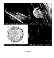
- Nitinol-based metal guide (Euroflex) is structured in order to define locations, for example wells, where the reactive groups will be deposited and where the biochemical interactions will take place ( Figure 1 ).
- Said "wells” can be produced by various methods, for example by focussed ion beam lithography (FIB or "Focused Ion beam”: Xie et al., Nuclear Instruments & Methods in Physics Research B-beam Section Interactions with Materials and Atoms, 211 (3): 363-368, 2003 ), by laser lithography followed by electrochemical etching and laser ablation.
- FIB focussed ion beam lithography
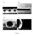
- the machine creates an ion beam, which is focused on the surface to be structured. Under the mechanical action of the ion beam, the atoms of the surface material are removed from the surface. Holes with a diameter of 20 ⁇ m can be formed with the FIB technique in a reasonable time with a bite factor of 8 ⁇ m 3 s -1 under a beam current of 20 nA.
- the Figure 2 shows holes with diameters of 5, 20 and 40 ⁇ m with a depth of 10 and 20 ⁇ m. The bottom surface of the hole is rough due to the re-deposit of the material sprayed during the attack.
- the bite factor was measured at 200 nm min -1 over a circular area of 40 ⁇ m in diameter and with a beam current of 20 nA. This gives a bite factor of 0.2 ⁇ m 3 nC -1 (approximately 5 ⁇ m 3 s -1 ), which corresponds to a procedure time of 20 min to make a hole 20 ⁇ m in diameter and 20 ⁇ m deep. .
- a fluorite-assisted milling technique XeF 2
- XeF 2 fluorite-assisted milling technique
- the technique of laser lithography and electrochemical etching consists in a first step in covering the surface with a polymer layer.
- the polymer layer is shaped using laser ablation.
- the surface is etched using isotropic electrochemical etching through the opening made in the polymer layer ( Figure 4 ).
- the Figure 5 presents the results of different structuring tests on metal guides based on Nitinol.
- Nitinol-based metal guides that are used in vivo are usually treated by electrochemical polishing, which replaces the native NiTi oxide layer with a biocompatible TiO 2 layer. Shaped guides with holes are subjected to this process to evaluate the influence of the process on the hole structure ( Figure 6 ).
- Another way of preparing the cavities on the surfaces of the Nitinol-based guides is laser ablation.
- the use of short laser pulses allows local evaporation of the metal without affecting the surrounding metal due to the heat generated.
- the smallest reported dimensions are of the order of 20 microns.
- metal micro-guides (MTI 0.012 "Silver speed) were used, which metal guides were inserted into micro-catheters.
- the device was introduced in pigs, under general anesthesia, at the level of a puncture and then at the scarpa to the kidney by the endovascular route (via the femoral artery). This guidance was ensured by the follow-up of said device in the femoral artery by arteriography.
- the device was introduced into the kidney by endoarterial break-in. This penetration into the tissue was made to a depth of a few millimeters and said device was held there for about ten minutes.
- the animals were then euthanized and the kidneys of the latter were removed to assess the state of the latter after penetration of the device according to the invention.
- the device according to the invention thus allows access to an organ while being very weakly invasive.
- Metal micro-guides (MTI 0.012 "Silver speed) were used, which metal micro-guides were placed in a fiberscope in contrast to Example 1.
- the device was introduced in pigs, under general anesthesia, at the level of a puncture in the scarpa and then to the liver by endoarterial navigation (via the femoral artery). This guidance was ensured by the follow-up of said device in the femoral artery by arteriography.
- the device was introduced into it. This penetration into the fabric was made to a depth of a few millimeters and said device was held there for a further ten minutes.
- the removal of the liver after the operation made it possible to judge the aggressiveness of the intervention on the organ.
- hemorrhagic lesions were minimal: two minor macroscopic lesions could be observed without any destruction of the parenchymal cells and with a simple congestion of the capillaries and centrilobular veins.
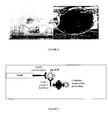
- the device uses the principle of the ELISA technique to highlight the ACE antigen.
- Two monoclonal antibodies recognizing different epitopes on this antigen were used for capture (AcM1) and revelation of ACE antigen (AcM2).
- These monoclonal antibodies having the same isotype (IgG1), the revelation of the ACE antigen was carried out using a monoclonal antibody coupled to biotin and a streptavidin-peroxidase complex ( Figure 7 ).
- PBS-BSA 3% bovine serum albumin
- the reaction was then stopped by adding 50 ⁇ l of 1M sulfuric acid per well.
- the absorbance was read at 492 nm on a plate reader (ref: ELX.800 UV).
- rigid plastic supports in the form of rods 2 to 3 cm in length and 0.5 to 1 mm in diameter were activated.
- the supports thus activated were placed in 1 ml hemolysis microtubes (Fisher) and were functionalized with a monoclonal antibody directed against the ACE antigen (clone 5910 produced in mice and marketed by Medix Biochemical). and diluted 1/50, 1/100, 1/250, 1/500 in carbonate / bicarbonate buffer (250 ⁇ l / tube) for 1 hour at 37 ° C.
- a negative control was made by replacing the monoclonal antibody with carbonate / bicarbonate buffer.
- the saturation was performed with 500 ⁇ l of 3% PBS-BSA overnight at + 4 ° C.
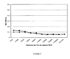
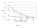
- the supports were then incubated with 250 ⁇ l of ACE antigen serum diluted 1/10, 1/100 in PBS or with the serum of a "healthy" subject (ACE antigen negative) at the same dilution for 1 hour. hour at 37 ° C.
- a monoclonal antibody against the ACE antigen (clone 5909 produced in mice and marketed by Medix Biochemical, which differs from clone 5910 by its affinity constant and recognized epitopes) purified at 1 mg / ml, was dialyzed overnight at 4 ° C against 0.1 M borate buffer pH 8.8. A solution of biotin at 10 mg / ml in DMSO was then added at a rate of 50 ⁇ g / mg of antibody. After incubation for 4 hours at room temperature and with stirring, 1 M ammonium chloride was added, at a rate of 20 ⁇ l / 250 ⁇ g of biotin, and the solution obtained was incubated again for 1 minute at room temperature. After blocking the reaction, the labeled antibody was dialyzed for 24 hours at + 4 ° C against PBS and this labeled antibody was stored as aliquots at -20 ° C.
- the supports were incubated with 250 ⁇ l of the biotinylated antibody 5909 and diluted 1/500 in PBS-Tween for 1 hour at 37 ° C.
- biotin ester of 6-biotinamidocaproylamido-caproic acid and N-hydroxysuccinimide, Sigma
- streptavidin-peroxidase complex Amersham Biosciences
- the revelation of the enzymatic activity was carried out by adding 750 ⁇ l per tube of the substrate mixture (H 2 O 2 ) and chromogenic (OPD, Sigma) in citrate-phosphate buffer (pH 5).
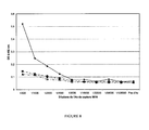
- the results show that the ODs are 7 to 10 times higher with ACE antigen-positive serum than those obtained with ACE antigen negative serum.
- the ACE antigen concentration having been the best detected corresponds to the serum of the patient diluted 1/100 to 6 IU / ml (close to the level considered "normal”: ⁇ 5 IU / ml) and when the dilution of the antibody Detection monoclonal 5909 is at 1/500.
- plastic supports makes it possible to validate the specificity and the sensitivity of the immunocapture processes on a metal rod functionalized according to the protocol described previously.
- an alkanethiol layer is adsorbed on one of the ends of Nitinol-based metal guides (Euroflex) in a first step.
- the free thiol functions of this layer allow the formation of disulfide bridges with a monoclonal antibody directed against the ACE antigen.
- the metal guide obtained is then introduced into a biopsy needle adapted for use in animals or humans.
- an extemporaneous anatomopathological examination is performed using this device on an operative part (mammary tumor), after its removal in a patient suffering from breast cancer.
- a micro-incision in the breast is performed under local anesthesia or general in a patient with breast cancer.
- the needle in which is inserted the metal guide coupled to the antibody directed against the ACE antigen, is introduced into the tumor or by the micro-incision and then directed to the tumor following its progression by imaging, including by ultrasound.
- Said micro-invasive guidance system then makes it possible to remove the end of the metal guide coupled to the antibody directed against the ACE antigen.
- the end of the metal guide is then introduced into the tumor (by perforation) to a depth of the order of a few millimeters. After a short waiting time, of the order of ten minutes, which allows the immunocapture of the ACE antigen possibly expressed by the tumor, the device is removed.
- Micro-sampling is limited to immunocapture of the analyte in vivo and does not require biopsy.
- ACE marker ELISA assay is performed on the end of the device with a monoclonal antibody directed against the ACE antigen which differentiates from the capture antibody by its affinity constant vis-a-vis antigen ACE and recognized epitopes, which is coupled with biotin.
- an alkanethiol layer was adsorbed on the end of a Nitinol-based metal guide (Euroflex) in a first step.
- the free thiol functions of this layer allowed the formation of disulfide bridges with a monoclonal antibody directed against the FAP (Fibroblast-activation protein; RETTIG et al., Proc. Natl. Acad. Sci. USA, vol.85, p: 3110, 1988 ).
- the metallic guide obtained is then introduced at the level of a cutaneous tumor in the animal or in the man when the medical ethical conditions are met, or at the level of a cutaneous tumor in a patient suffering from a cancer of the skin. skin after it has been removed for classical or extemporaneous pathological examination.
- micro-sampling is limited to immunocapture in vivo and again does not require a specific biopsy.
- the device is removed and an ELISA FAP marker assay is performed on the end of the device with a monoclonal antibody directed against the peroxidase-coupled FAP marker.
- the revelation of the peroxidase activity makes it possible to conclude the expression of the FAP marker by the tumor and to modulate accordingly the therapy to be used to treat the patient as best as possible.
Landscapes
- Health & Medical Sciences (AREA)
- Life Sciences & Earth Sciences (AREA)
- Physics & Mathematics (AREA)
- General Health & Medical Sciences (AREA)
- Veterinary Medicine (AREA)
- Engineering & Computer Science (AREA)
- Biomedical Technology (AREA)
- Heart & Thoracic Surgery (AREA)
- Medical Informatics (AREA)
- Molecular Biology (AREA)
- Surgery (AREA)
- Animal Behavior & Ethology (AREA)
- Biophysics (AREA)
- Public Health (AREA)
- Pathology (AREA)
- Optics & Photonics (AREA)
- Neurology (AREA)
- Chemical & Material Sciences (AREA)
- Chemical Kinetics & Catalysis (AREA)
- General Chemical & Material Sciences (AREA)
- Neurosurgery (AREA)
- Physiology (AREA)
- Investigating Or Analysing Biological Materials (AREA)
- Measurement Of The Respiration, Hearing Ability, Form, And Blood Characteristics Of Living Organisms (AREA)
- Apparatus Associated With Microorganisms And Enzymes (AREA)
Priority Applications (11)
| Application Number | Priority Date | Filing Date | Title |
|---|---|---|---|
| AT08290179T ATE508679T1 (de) | 2008-02-26 | 2008-02-26 | Vorrichtung zur mikroinvasiven in-vivo- untersuchung, die einen metallischen leiter umfasst |
| EP08290179A EP2095762B1 (de) | 2008-02-26 | 2008-02-26 | Vorrichtung zur mikroinvasiven In-vivo-Untersuchung, die einen metallischen Leiter umfasst |
| ES08290179T ES2369472T3 (es) | 2008-02-26 | 2008-02-26 | Dispositivo de investigación micro-invasiva en vivo que comprende una guía metálica. |
| CN200980112079.4A CN102014734B (zh) | 2008-02-26 | 2009-02-24 | 包括金属导向装置的体内微创检查设备 |
| CA2716751A CA2716751C (fr) | 2008-02-26 | 2009-02-24 | Dispositif d'investigation micro-invasif in vivo comprenant un guide metallique |
| KR1020107021340A KR101624755B1 (ko) | 2008-02-26 | 2009-02-24 | 금속 가이드를 포함하는 체내 미세-침습성 조사 장치 |
| US12/919,655 US8969010B2 (en) | 2008-02-26 | 2009-02-24 | In vivo micro-invasive investigation device including a metal guide |
| CN201410136896.6A CN103932678B (zh) | 2008-02-26 | 2009-02-24 | 包括金属导向装置的体内微创检查设备 |
| PCT/EP2009/001308 WO2009106295A1 (fr) | 2008-02-26 | 2009-02-24 | Dispositif d'investigation micro-invasif in vivo comprenant un guide metallique |
| JP2010547125A JP5620277B2 (ja) | 2008-02-26 | 2009-02-24 | 金属ガイドを含む生体内低侵襲性検査デバイス |
| HK11110420.6A HK1155928A1 (en) | 2008-02-26 | 2011-10-03 | In vivo micro-invasive investigation device including a metal guide |
Applications Claiming Priority (1)
| Application Number | Priority Date | Filing Date | Title |
|---|---|---|---|
| EP08290179A EP2095762B1 (de) | 2008-02-26 | 2008-02-26 | Vorrichtung zur mikroinvasiven In-vivo-Untersuchung, die einen metallischen Leiter umfasst |
Publications (2)
| Publication Number | Publication Date |
|---|---|
| EP2095762A1 true EP2095762A1 (de) | 2009-09-02 |
| EP2095762B1 EP2095762B1 (de) | 2011-05-11 |
Family
ID=39830196
Family Applications (1)
| Application Number | Title | Priority Date | Filing Date |
|---|---|---|---|
| EP08290179A Active EP2095762B1 (de) | 2008-02-26 | 2008-02-26 | Vorrichtung zur mikroinvasiven In-vivo-Untersuchung, die einen metallischen Leiter umfasst |
Country Status (10)
| Country | Link |
|---|---|
| US (1) | US8969010B2 (de) |
| EP (1) | EP2095762B1 (de) |
| JP (1) | JP5620277B2 (de) |
| KR (1) | KR101624755B1 (de) |
| CN (2) | CN103932678B (de) |
| AT (1) | ATE508679T1 (de) |
| CA (1) | CA2716751C (de) |
| ES (1) | ES2369472T3 (de) |
| HK (1) | HK1155928A1 (de) |
| WO (1) | WO2009106295A1 (de) |
Families Citing this family (5)
| Publication number | Priority date | Publication date | Assignee | Title |
|---|---|---|---|---|
| ES2369472T3 (es) * | 2008-02-26 | 2011-12-01 | Biostems Ltd. | Dispositivo de investigación micro-invasiva en vivo que comprende una guía metálica. |
| DE102013211837A1 (de) * | 2013-06-21 | 2014-12-24 | Gilupi Gmbh | Katheter mit Detektionsvorrichtung zum Echtzeitnachweis eines Probenmaterials |
| CN104970806B (zh) * | 2015-07-12 | 2017-08-04 | 北京泱深生物信息技术有限公司 | 一种用于癌症复发实时动态监测的智能设备 |
| ES2722802B2 (es) * | 2018-02-14 | 2019-12-18 | Fund De Neurociencias | Dispositivo para la eliminacion selectiva de moleculas de tejidos o fluidos |
| CN111956269B (zh) * | 2020-08-20 | 2023-11-28 | 上海蓓蕊医疗科技有限公司 | 一种皮肤保护器 |
Citations (3)
| Publication number | Priority date | Publication date | Assignee | Title |
|---|---|---|---|---|
| US5938595A (en) * | 1996-05-24 | 1999-08-17 | The Regents Of The University Of California | Fiber optic D dimer biosensor |
| EP1358481A2 (de) | 2000-03-17 | 2003-11-05 | Alain Pompidou | Vorrichtung zur analyse und/oder zur behandlung mit einem flexiblen schaft |
| US7291497B2 (en) * | 2003-09-11 | 2007-11-06 | Theranos, Inc. | Medical device for analyte monitoring and drug delivery |
Family Cites Families (16)
| Publication number | Priority date | Publication date | Assignee | Title |
|---|---|---|---|---|
| DE3332924A1 (de) | 1983-09-13 | 1985-03-21 | Bayer Ag, 5090 Leverkusen | Kerbschlagzaehe, in der schmelze leichtfliessende polyamidformmassen |
| US4710623A (en) * | 1986-02-27 | 1987-12-01 | Eli Lilly And Company | Optical fiber catheter with fiber-contained reactive element |
| US7622294B2 (en) * | 1997-03-14 | 2009-11-24 | Trustees Of Tufts College | Methods for detecting target analytes and enzymatic reactions |
| US6689604B1 (en) * | 1998-03-20 | 2004-02-10 | National Research Council Of Canada | Lipopolysaccharide α-2,3 sialyltransferase of Campylobacter jejuni and its uses |
| ATE441110T1 (de) * | 2000-11-09 | 2009-09-15 | Sicel Technologies Inc | In-vivo detektion von biomolekülekonzentrationen mittels fluoreszenzmarker |
| FR2817067B1 (fr) * | 2000-11-21 | 2003-02-21 | Cyber Comm | Procede et dispositif d'authentification de documents electroniques au moyen d'une signature numerique |
| CA2453141A1 (en) * | 2001-07-09 | 2003-01-23 | Arizona Board Of Regents A Body Corporate Acting On Behalf Of Arizona State University | Afinity biosensor for monitoring of biological process |
| US7025734B1 (en) * | 2001-09-28 | 2006-04-11 | Advanced Cardiovascular Systmes, Inc. | Guidewire with chemical sensing capabilities |
| US7259019B2 (en) * | 2002-03-11 | 2007-08-21 | Pawliszyn Janusz B | Multiple sampling device and method for investigating biological systems |
| US7740595B2 (en) * | 2002-07-26 | 2010-06-22 | Stuart Brown | Tissue and fluid sampling device |
| US20050153309A1 (en) * | 2003-12-22 | 2005-07-14 | David Hoon | Method and apparatus for in vivo surveillance of circulating biological components |
| JP4493009B2 (ja) * | 2004-04-30 | 2010-06-30 | 尚 吉武 | 脳内高分子のサンプリング方法 |
| FR2881339B1 (fr) * | 2005-02-02 | 2009-07-10 | Commissariat Energie Atomique | Dispositif de prelevement moleculaire par contact |
| DE102005005007B3 (de) * | 2005-02-03 | 2006-09-07 | Lts Lohmann Therapie-Systeme Ag | Biopsienadel zur histologischen Untersuchung von Körpergewebe |
| US20070287991A1 (en) * | 2006-06-08 | 2007-12-13 | Mckay William F | Devices and methods for detection of markers of axial pain with or without radiculopathy |
| ES2369472T3 (es) * | 2008-02-26 | 2011-12-01 | Biostems Ltd. | Dispositivo de investigación micro-invasiva en vivo que comprende una guía metálica. |
-
2008
- 2008-02-26 ES ES08290179T patent/ES2369472T3/es active Active
- 2008-02-26 AT AT08290179T patent/ATE508679T1/de not_active IP Right Cessation
- 2008-02-26 EP EP08290179A patent/EP2095762B1/de active Active
-
2009
- 2009-02-24 CN CN201410136896.6A patent/CN103932678B/zh active Active
- 2009-02-24 CA CA2716751A patent/CA2716751C/fr active Active
- 2009-02-24 WO PCT/EP2009/001308 patent/WO2009106295A1/fr active Application Filing
- 2009-02-24 CN CN200980112079.4A patent/CN102014734B/zh active Active
- 2009-02-24 US US12/919,655 patent/US8969010B2/en active Active
- 2009-02-24 JP JP2010547125A patent/JP5620277B2/ja active Active
- 2009-02-24 KR KR1020107021340A patent/KR101624755B1/ko active IP Right Grant
-
2011
- 2011-10-03 HK HK11110420.6A patent/HK1155928A1/xx not_active IP Right Cessation
Patent Citations (4)
| Publication number | Priority date | Publication date | Assignee | Title |
|---|---|---|---|---|
| US5938595A (en) * | 1996-05-24 | 1999-08-17 | The Regents Of The University Of California | Fiber optic D dimer biosensor |
| EP1358481A2 (de) | 2000-03-17 | 2003-11-05 | Alain Pompidou | Vorrichtung zur analyse und/oder zur behandlung mit einem flexiblen schaft |
| EP1358481B1 (de) * | 2000-03-17 | 2004-06-30 | Alain Pompidou | Vorrichtung zur analyse und/oder zur behandlung mit einem flexiblen schaft |
| US7291497B2 (en) * | 2003-09-11 | 2007-11-06 | Theranos, Inc. | Medical device for analyte monitoring and drug delivery |
Non-Patent Citations (4)
| Title |
|---|
| ARIGA ET AL., AM. J. SURG., vol. 184, 2002, pages 410 - 413 |
| FERNANDEZ-ESPARRACH ET AL., ARCH. BRONCONEOMOL., vol. 43, 2007, pages 219 - 224 |
| PÉREZ-GUILLERMO ET AL., DIAGN. CYTOPATHOL., vol. 32, 2005, pages 315 - 320 |
| RODRIGUES ET AL., J. AM. ACAD. DERMATOL., vol. 42, 2000, pages 735 - 740 |
Also Published As
| Publication number | Publication date |
|---|---|
| CN103932678B (zh) | 2017-10-24 |
| CN102014734B (zh) | 2014-05-07 |
| ATE508679T1 (de) | 2011-05-15 |
| KR20110005685A (ko) | 2011-01-18 |
| CN103932678A (zh) | 2014-07-23 |
| HK1155928A1 (en) | 2012-06-01 |
| WO2009106295A1 (fr) | 2009-09-03 |
| JP5620277B2 (ja) | 2014-11-05 |
| CN102014734A (zh) | 2011-04-13 |
| US8969010B2 (en) | 2015-03-03 |
| CA2716751C (fr) | 2016-03-08 |
| US20110092783A1 (en) | 2011-04-21 |
| ES2369472T3 (es) | 2011-12-01 |
| KR101624755B1 (ko) | 2016-05-26 |
| CA2716751A1 (fr) | 2009-09-03 |
| EP2095762B1 (de) | 2011-05-11 |
| JP2011514815A (ja) | 2011-05-12 |
Similar Documents
| Publication | Publication Date | Title |
|---|---|---|
| Dervisevic et al. | Electrochemical immunosensor for breast cancer biomarker detection using high-density silicon microneedle array | |
| US10987037B2 (en) | Method and apparatus for in vivo surveillance of circulating biological components | |
| US7553625B2 (en) | Method and apparatus for in vivo collection of circulating biological components | |
| EP2095762B1 (de) | Vorrichtung zur mikroinvasiven In-vivo-Untersuchung, die einen metallischen Leiter umfasst | |
| Dardano et al. | Microneedles-based electrochemical sensors: New tools for advanced biosensing | |
| US20100200538A1 (en) | Analyte Sensor and Fabrication Methods | |
| WO2021232109A1 (en) | Bioanalyte detection and monitoring | |
| JP2011511665A (ja) | 半導体を素材とする分析対象物センサー及び方法 | |
| US9921163B2 (en) | Method and device for detecting analytes | |
| JP2020504749A (ja) | アプタマーでコーティングされたマイクロニードルベースの診断用皮膚パッチの製造方法及びそのパッチ | |
| WO2001069257A9 (fr) | Dispositif d'analyse et/ou de traitement avec une tige souple | |
| Córcoles et al. | Biosensors and invasive monitoring in clinical applications | |
| WO2006024788A1 (fr) | Procede et dispositif de mesure d'une activite enzymatique dans un fluide biologique | |
| Lu et al. | Microneedle-based device for biological analysis | |
| Garg et al. | Recent advancements in the expedition of microneedles: from lab worktops to diagnostic care centers | |
| JPWO2014196169A1 (ja) | センサーチップ、検出方法および検出装置 | |
| WO2005062940A2 (en) | Method and apparatus for in vivo collection and surveillance of circulating biological components | |
| Mason-King | Surface Modification Techniques for Improving Longevity of EAB Sensors | |
| Li | Brief Description and Application of Microneedle Biosensors | |
| Córcoles et al. | Implantable biosensors | |
| Omidian et al. | Hydrogels in biosensing and medical diagnostics | |
| Deshpande | Development of a nanowire based titanium needle probe sensor for glucose monitoring | |
| Baldini | Advanced biomedical optical fibre sensors |
Legal Events
| Date | Code | Title | Description |
|---|---|---|---|
| PUAI | Public reference made under article 153(3) epc to a published international application that has entered the european phase |
Free format text: ORIGINAL CODE: 0009012 |
|
| 17P | Request for examination filed |
Effective date: 20080307 |
|
| AK | Designated contracting states |
Kind code of ref document: A1 Designated state(s): AT BE BG CH CY CZ DE DK EE ES FI FR GB GR HR HU IE IS IT LI LT LU LV MC MT NL NO PL PT RO SE SI SK TR |
|
| AX | Request for extension of the european patent |
Extension state: AL BA MK RS |
|
| GRAP | Despatch of communication of intention to grant a patent |
Free format text: ORIGINAL CODE: EPIDOSNIGR1 |
|
| AKX | Designation fees paid |
Designated state(s): AT BE BG CH CY CZ DE DK EE ES FI FR GB GR HR HU IE IS IT LI LT LU LV MC MT NL NO PL PT RO SE SI SK TR |
|
| GRAS | Grant fee paid |
Free format text: ORIGINAL CODE: EPIDOSNIGR3 |
|
| GRAA | (expected) grant |
Free format text: ORIGINAL CODE: 0009210 |
|
| AK | Designated contracting states |
Kind code of ref document: B1 Designated state(s): AT BE BG CH CY CZ DE DK EE ES FI FR GB GR HR HU IE IS IT LI LT LU LV MC MT NL NO PL PT RO SE SI SK TR |
|
| REG | Reference to a national code |
Ref country code: GB Ref legal event code: FG4D Free format text: NOT ENGLISH |
|
| REG | Reference to a national code |
Ref country code: CH Ref legal event code: EP |
|
| REG | Reference to a national code |
Ref country code: IE Ref legal event code: FG4D |
|
| REG | Reference to a national code |
Ref country code: DE Ref legal event code: R096 Ref document number: 602008006841 Country of ref document: DE Effective date: 20110622 |
|
| REG | Reference to a national code |
Ref country code: NL Ref legal event code: T3 |
|
| REG | Reference to a national code |
Ref country code: SE Ref legal event code: TRGR |
|
| PG25 | Lapsed in a contracting state [announced via postgrant information from national office to epo] |
Ref country code: NO Free format text: LAPSE BECAUSE OF FAILURE TO SUBMIT A TRANSLATION OF THE DESCRIPTION OR TO PAY THE FEE WITHIN THE PRESCRIBED TIME-LIMIT Effective date: 20110811 Ref country code: LT Free format text: LAPSE BECAUSE OF FAILURE TO SUBMIT A TRANSLATION OF THE DESCRIPTION OR TO PAY THE FEE WITHIN THE PRESCRIBED TIME-LIMIT Effective date: 20110511 |
|
| PG25 | Lapsed in a contracting state [announced via postgrant information from national office to epo] |
Ref country code: GR Free format text: LAPSE BECAUSE OF FAILURE TO SUBMIT A TRANSLATION OF THE DESCRIPTION OR TO PAY THE FEE WITHIN THE PRESCRIBED TIME-LIMIT Effective date: 20110812 Ref country code: AT Free format text: LAPSE BECAUSE OF FAILURE TO SUBMIT A TRANSLATION OF THE DESCRIPTION OR TO PAY THE FEE WITHIN THE PRESCRIBED TIME-LIMIT Effective date: 20110511 Ref country code: IS Free format text: LAPSE BECAUSE OF FAILURE TO SUBMIT A TRANSLATION OF THE DESCRIPTION OR TO PAY THE FEE WITHIN THE PRESCRIBED TIME-LIMIT Effective date: 20110911 Ref country code: LV Free format text: LAPSE BECAUSE OF FAILURE TO SUBMIT A TRANSLATION OF THE DESCRIPTION OR TO PAY THE FEE WITHIN THE PRESCRIBED TIME-LIMIT Effective date: 20110511 Ref country code: CY Free format text: LAPSE BECAUSE OF FAILURE TO SUBMIT A TRANSLATION OF THE DESCRIPTION OR TO PAY THE FEE WITHIN THE PRESCRIBED TIME-LIMIT Effective date: 20110511 Ref country code: FI Free format text: LAPSE BECAUSE OF FAILURE TO SUBMIT A TRANSLATION OF THE DESCRIPTION OR TO PAY THE FEE WITHIN THE PRESCRIBED TIME-LIMIT Effective date: 20110511 Ref country code: SI Free format text: LAPSE BECAUSE OF FAILURE TO SUBMIT A TRANSLATION OF THE DESCRIPTION OR TO PAY THE FEE WITHIN THE PRESCRIBED TIME-LIMIT Effective date: 20110511 |
|
| REG | Reference to a national code |
Ref country code: ES Ref legal event code: FG2A Ref document number: 2369472 Country of ref document: ES Kind code of ref document: T3 Effective date: 20111201 |
|
| REG | Reference to a national code |
Ref country code: IE Ref legal event code: FD4D |
|
| REG | Reference to a national code |
Ref country code: CH Ref legal event code: NV Representative=s name: NOVAGRAAF SWITZERLAND SA |
|
| PG25 | Lapsed in a contracting state [announced via postgrant information from national office to epo] |
Ref country code: EE Free format text: LAPSE BECAUSE OF FAILURE TO SUBMIT A TRANSLATION OF THE DESCRIPTION OR TO PAY THE FEE WITHIN THE PRESCRIBED TIME-LIMIT Effective date: 20110511 Ref country code: IE Free format text: LAPSE BECAUSE OF FAILURE TO SUBMIT A TRANSLATION OF THE DESCRIPTION OR TO PAY THE FEE WITHIN THE PRESCRIBED TIME-LIMIT Effective date: 20110511 Ref country code: CZ Free format text: LAPSE BECAUSE OF FAILURE TO SUBMIT A TRANSLATION OF THE DESCRIPTION OR TO PAY THE FEE WITHIN THE PRESCRIBED TIME-LIMIT Effective date: 20110511 |
|
| PG25 | Lapsed in a contracting state [announced via postgrant information from national office to epo] |
Ref country code: RO Free format text: LAPSE BECAUSE OF FAILURE TO SUBMIT A TRANSLATION OF THE DESCRIPTION OR TO PAY THE FEE WITHIN THE PRESCRIBED TIME-LIMIT Effective date: 20110511 Ref country code: SK Free format text: LAPSE BECAUSE OF FAILURE TO SUBMIT A TRANSLATION OF THE DESCRIPTION OR TO PAY THE FEE WITHIN THE PRESCRIBED TIME-LIMIT Effective date: 20110511 Ref country code: DK Free format text: LAPSE BECAUSE OF FAILURE TO SUBMIT A TRANSLATION OF THE DESCRIPTION OR TO PAY THE FEE WITHIN THE PRESCRIBED TIME-LIMIT Effective date: 20110511 Ref country code: PL Free format text: LAPSE BECAUSE OF FAILURE TO SUBMIT A TRANSLATION OF THE DESCRIPTION OR TO PAY THE FEE WITHIN THE PRESCRIBED TIME-LIMIT Effective date: 20110511 |
|
| PLBE | No opposition filed within time limit |
Free format text: ORIGINAL CODE: 0009261 |
|
| STAA | Information on the status of an ep patent application or granted ep patent |
Free format text: STATUS: NO OPPOSITION FILED WITHIN TIME LIMIT |
|
| 26N | No opposition filed |
Effective date: 20120214 |
|
| PG25 | Lapsed in a contracting state [announced via postgrant information from national office to epo] |
Ref country code: HR Free format text: LAPSE BECAUSE OF FAILURE TO SUBMIT A TRANSLATION OF THE DESCRIPTION OR TO PAY THE FEE WITHIN THE PRESCRIBED TIME-LIMIT Effective date: 20111123 |
|
| REG | Reference to a national code |
Ref country code: DE Ref legal event code: R097 Ref document number: 602008006841 Country of ref document: DE Effective date: 20120214 |
|
| BERE | Be: lapsed |
Owner name: BIOSTEMS LTD. Effective date: 20120228 |
|
| PG25 | Lapsed in a contracting state [announced via postgrant information from national office to epo] |
Ref country code: MC Free format text: LAPSE BECAUSE OF NON-PAYMENT OF DUE FEES Effective date: 20120229 |
|
| PG25 | Lapsed in a contracting state [announced via postgrant information from national office to epo] |
Ref country code: BE Free format text: LAPSE BECAUSE OF NON-PAYMENT OF DUE FEES Effective date: 20120228 |
|
| PG25 | Lapsed in a contracting state [announced via postgrant information from national office to epo] |
Ref country code: BG Free format text: LAPSE BECAUSE OF FAILURE TO SUBMIT A TRANSLATION OF THE DESCRIPTION OR TO PAY THE FEE WITHIN THE PRESCRIBED TIME-LIMIT Effective date: 20110811 |
|
| PG25 | Lapsed in a contracting state [announced via postgrant information from national office to epo] |
Ref country code: MT Free format text: LAPSE BECAUSE OF FAILURE TO SUBMIT A TRANSLATION OF THE DESCRIPTION OR TO PAY THE FEE WITHIN THE PRESCRIBED TIME-LIMIT Effective date: 20110511 |
|
| PG25 | Lapsed in a contracting state [announced via postgrant information from national office to epo] |
Ref country code: HR Free format text: LAPSE BECAUSE OF FAILURE TO SUBMIT A TRANSLATION OF THE DESCRIPTION OR TO PAY THE FEE WITHIN THE PRESCRIBED TIME-LIMIT Effective date: 20110511 |
|
| PG25 | Lapsed in a contracting state [announced via postgrant information from national office to epo] |
Ref country code: LU Free format text: LAPSE BECAUSE OF NON-PAYMENT OF DUE FEES Effective date: 20120226 |
|
| PG25 | Lapsed in a contracting state [announced via postgrant information from national office to epo] |
Ref country code: HU Free format text: LAPSE BECAUSE OF FAILURE TO SUBMIT A TRANSLATION OF THE DESCRIPTION OR TO PAY THE FEE WITHIN THE PRESCRIBED TIME-LIMIT Effective date: 20080226 |
|
| REG | Reference to a national code |
Ref country code: FR Ref legal event code: PLFP Year of fee payment: 9 |
|
| REG | Reference to a national code |
Ref country code: FR Ref legal event code: PLFP Year of fee payment: 10 |
|
| REG | Reference to a national code |
Ref country code: FR Ref legal event code: PLFP Year of fee payment: 11 |
|
| PGFP | Annual fee paid to national office [announced via postgrant information from national office to epo] |
Ref country code: TR Payment date: 20230824 Year of fee payment: 16 Ref country code: NL Payment date: 20230830 Year of fee payment: 16 Ref country code: IT Payment date: 20230829 Year of fee payment: 16 Ref country code: GB Payment date: 20230830 Year of fee payment: 16 Ref country code: ES Payment date: 20230829 Year of fee payment: 16 Ref country code: CH Payment date: 20230831 Year of fee payment: 16 |
|
| PGFP | Annual fee paid to national office [announced via postgrant information from national office to epo] |
Ref country code: SE Payment date: 20230830 Year of fee payment: 16 Ref country code: PT Payment date: 20230824 Year of fee payment: 16 Ref country code: FR Payment date: 20230830 Year of fee payment: 16 Ref country code: DE Payment date: 20230829 Year of fee payment: 16 |
|
| PGFP | Annual fee paid to national office [announced via postgrant information from national office to epo] |
Ref country code: SE Payment date: 20230830 Year of fee payment: 16 |