EP1991303B1 - Procédé et dispositif de traitement de tumeurs résiduelles microscopiques restant dans des tissus après résection chirurgicale - Google Patents
Procédé et dispositif de traitement de tumeurs résiduelles microscopiques restant dans des tissus après résection chirurgicale Download PDFInfo
- Publication number
- EP1991303B1 EP1991303B1 EP07751865.2A EP07751865A EP1991303B1 EP 1991303 B1 EP1991303 B1 EP 1991303B1 EP 07751865 A EP07751865 A EP 07751865A EP 1991303 B1 EP1991303 B1 EP 1991303B1
- Authority
- EP
- European Patent Office
- Prior art keywords
- tumor
- tissue
- bleomycin
- agent
- electroporation
- Prior art date
- Legal status (The legal status is an assumption and is not a legal conclusion. Google has not performed a legal analysis and makes no representation as to the accuracy of the status listed.)
- Active
Links
- 238000002271 resection Methods 0.000 title claims description 19
- 208000007660 Residual Neoplasm Diseases 0.000 title claims 4
- 238000000034 method Methods 0.000 title description 33
- 206010028980 Neoplasm Diseases 0.000 claims description 209
- OYVAGSVQBOHSSS-UAPAGMARSA-O bleomycin A2 Chemical compound N([C@H](C(=O)N[C@H](C)[C@@H](O)[C@H](C)C(=O)N[C@@H]([C@H](O)C)C(=O)NCCC=1SC=C(N=1)C=1SC=C(N=1)C(=O)NCCC[S+](C)C)[C@@H](O[C@H]1[C@H]([C@@H](O)[C@H](O)[C@H](CO)O1)O[C@@H]1[C@H]([C@@H](OC(N)=O)[C@H](O)[C@@H](CO)O1)O)C=1N=CNC=1)C(=O)C1=NC([C@H](CC(N)=O)NC[C@H](N)C(N)=O)=NC(N)=C1C OYVAGSVQBOHSSS-UAPAGMARSA-O 0.000 claims description 78
- 108010006654 Bleomycin Proteins 0.000 claims description 74
- 229960001561 bleomycin Drugs 0.000 claims description 74
- 238000011282 treatment Methods 0.000 claims description 67
- 210000004027 cell Anatomy 0.000 claims description 39
- 239000003795 chemical substances by application Substances 0.000 claims description 23
- 239000002246 antineoplastic agent Substances 0.000 claims description 22
- 239000003814 drug Substances 0.000 claims description 19
- 108020004707 nucleic acids Proteins 0.000 claims description 8
- 102000039446 nucleic acids Human genes 0.000 claims description 8
- 150000007523 nucleic acids Chemical class 0.000 claims description 8
- 108090000623 proteins and genes Proteins 0.000 claims description 8
- 230000009467 reduction Effects 0.000 claims description 7
- 230000004614 tumor growth Effects 0.000 claims description 6
- 102000019034 Chemokines Human genes 0.000 claims description 5
- 108010012236 Chemokines Proteins 0.000 claims description 5
- 102000004127 Cytokines Human genes 0.000 claims description 5
- 108090000695 Cytokines Proteins 0.000 claims description 5
- 229920001184 polypeptide Polymers 0.000 claims description 5
- 102000004196 processed proteins & peptides Human genes 0.000 claims description 5
- 108090000765 processed proteins & peptides Proteins 0.000 claims description 5
- 241000124008 Mammalia Species 0.000 claims description 4
- 230000000692 anti-sense effect Effects 0.000 claims description 4
- 238000010253 intravenous injection Methods 0.000 claims description 4
- 239000000243 solution Substances 0.000 claims description 4
- 108091030071 RNAI Proteins 0.000 claims description 3
- DQLATGHUWYMOKM-UHFFFAOYSA-L cisplatin Chemical compound N[Pt](N)(Cl)Cl DQLATGHUWYMOKM-UHFFFAOYSA-L 0.000 claims description 3
- 229960004316 cisplatin Drugs 0.000 claims description 3
- 230000008030 elimination Effects 0.000 claims description 3
- 238000003379 elimination reaction Methods 0.000 claims description 3
- 230000009368 gene silencing by RNA Effects 0.000 claims description 3
- 229920002643 polyglutamic acid Polymers 0.000 claims description 3
- 102000004169 proteins and genes Human genes 0.000 claims description 3
- 230000004565 tumor cell growth Effects 0.000 claims description 2
- 210000001519 tissue Anatomy 0.000 description 127
- 238000004520 electroporation Methods 0.000 description 99
- 241001465754 Metazoa Species 0.000 description 37
- FAPWRFPIFSIZLT-UHFFFAOYSA-M Sodium chloride Chemical compound [Na+].[Cl-] FAPWRFPIFSIZLT-UHFFFAOYSA-M 0.000 description 37
- 239000011780 sodium chloride Substances 0.000 description 36
- 238000001356 surgical procedure Methods 0.000 description 25
- 210000003491 skin Anatomy 0.000 description 24
- 239000000758 substrate Substances 0.000 description 23
- 210000003205 muscle Anatomy 0.000 description 21
- 206010052428 Wound Diseases 0.000 description 16
- 230000036961 partial effect Effects 0.000 description 16
- 230000000694 effects Effects 0.000 description 15
- 238000002474 experimental method Methods 0.000 description 14
- 238000012360 testing method Methods 0.000 description 14
- 230000035876 healing Effects 0.000 description 13
- 230000005855 radiation Effects 0.000 description 12
- 238000002347 injection Methods 0.000 description 11
- 239000007924 injection Substances 0.000 description 11
- 208000027418 Wounds and injury Diseases 0.000 description 10
- 238000001959 radiotherapy Methods 0.000 description 10
- 239000002671 adjuvant Substances 0.000 description 9
- 238000002512 chemotherapy Methods 0.000 description 9
- 230000005684 electric field Effects 0.000 description 8
- 229940090044 injection Drugs 0.000 description 8
- 210000003813 thumb Anatomy 0.000 description 8
- 102000008186 Collagen Human genes 0.000 description 6
- 108010035532 Collagen Proteins 0.000 description 6
- WSFSSNUMVMOOMR-UHFFFAOYSA-N Formaldehyde Chemical compound O=C WSFSSNUMVMOOMR-UHFFFAOYSA-N 0.000 description 6
- 241000282414 Homo sapiens Species 0.000 description 6
- 241000699666 Mus <mouse, genus> Species 0.000 description 6
- 238000002679 ablation Methods 0.000 description 6
- 229920001436 collagen Polymers 0.000 description 6
- 229940079593 drug Drugs 0.000 description 6
- 206010033675 panniculitis Diseases 0.000 description 6
- 210000004304 subcutaneous tissue Anatomy 0.000 description 6
- 206010016654 Fibrosis Diseases 0.000 description 5
- 208000035346 Margins of Excision Diseases 0.000 description 5
- 241000699670 Mus sp. Species 0.000 description 5
- 208000003788 Neoplasm Micrometastasis Diseases 0.000 description 5
- 238000002725 brachytherapy Methods 0.000 description 5
- 230000006378 damage Effects 0.000 description 5
- 230000004761 fibrosis Effects 0.000 description 5
- 230000002601 intratumoral effect Effects 0.000 description 5
- 230000007246 mechanism Effects 0.000 description 5
- 238000002560 therapeutic procedure Methods 0.000 description 5
- 206010028851 Necrosis Diseases 0.000 description 4
- 201000011510 cancer Diseases 0.000 description 4
- 238000000315 cryotherapy Methods 0.000 description 4
- 201000010099 disease Diseases 0.000 description 4
- 208000037265 diseases, disorders, signs and symptoms Diseases 0.000 description 4
- 230000012010 growth Effects 0.000 description 4
- 238000001000 micrograph Methods 0.000 description 4
- 239000000203 mixture Substances 0.000 description 4
- 238000002428 photodynamic therapy Methods 0.000 description 4
- 238000004321 preservation Methods 0.000 description 4
- 238000011127 radiochemotherapy Methods 0.000 description 4
- 239000000126 substance Substances 0.000 description 4
- 230000001225 therapeutic effect Effects 0.000 description 4
- 210000004881 tumor cell Anatomy 0.000 description 4
- 230000029663 wound healing Effects 0.000 description 4
- 206010063560 Excessive granulation tissue Diseases 0.000 description 3
- 108010065805 Interleukin-12 Proteins 0.000 description 3
- 241000282898 Sus scrofa Species 0.000 description 3
- 230000008901 benefit Effects 0.000 description 3
- 210000004207 dermis Anatomy 0.000 description 3
- 239000000835 fiber Substances 0.000 description 3
- 239000012530 fluid Substances 0.000 description 3
- 238000009472 formulation Methods 0.000 description 3
- 230000006870 function Effects 0.000 description 3
- 210000001126 granulation tissue Anatomy 0.000 description 3
- 210000003701 histiocyte Anatomy 0.000 description 3
- 238000001727 in vivo Methods 0.000 description 3
- 230000003211 malignant effect Effects 0.000 description 3
- 230000017074 necrotic cell death Effects 0.000 description 3
- 230000001338 necrotic effect Effects 0.000 description 3
- 230000002829 reductive effect Effects 0.000 description 3
- 230000037390 scarring Effects 0.000 description 3
- 229940124597 therapeutic agent Drugs 0.000 description 3
- 238000011269 treatment regimen Methods 0.000 description 3
- 206010009944 Colon cancer Diseases 0.000 description 2
- 108020004414 DNA Proteins 0.000 description 2
- 206010061619 Deformity Diseases 0.000 description 2
- 241000282412 Homo Species 0.000 description 2
- 102000013462 Interleukin-12 Human genes 0.000 description 2
- 241001313288 Labia Species 0.000 description 2
- 208000000102 Squamous Cell Carcinoma of Head and Neck Diseases 0.000 description 2
- 208000002847 Surgical Wound Diseases 0.000 description 2
- 230000002159 abnormal effect Effects 0.000 description 2
- 230000002411 adverse Effects 0.000 description 2
- 230000001093 anti-cancer Effects 0.000 description 2
- 230000000259 anti-tumor effect Effects 0.000 description 2
- 229940041181 antineoplastic drug Drugs 0.000 description 2
- 230000001640 apoptogenic effect Effects 0.000 description 2
- 210000001142 back Anatomy 0.000 description 2
- 230000001627 detrimental effect Effects 0.000 description 2
- 238000001647 drug administration Methods 0.000 description 2
- 230000002500 effect on skin Effects 0.000 description 2
- 238000011156 evaluation Methods 0.000 description 2
- 238000001415 gene therapy Methods 0.000 description 2
- 201000000459 head and neck squamous cell carcinoma Diseases 0.000 description 2
- 238000011081 inoculation Methods 0.000 description 2
- 238000001990 intravenous administration Methods 0.000 description 2
- 230000003902 lesion Effects 0.000 description 2
- 210000004698 lymphocyte Anatomy 0.000 description 2
- 239000000463 material Substances 0.000 description 2
- 230000007935 neutral effect Effects 0.000 description 2
- 238000011275 oncology therapy Methods 0.000 description 2
- 210000000056 organ Anatomy 0.000 description 2
- 230000000149 penetrating effect Effects 0.000 description 2
- 210000003899 penis Anatomy 0.000 description 2
- 210000000664 rectum Anatomy 0.000 description 2
- 206010041823 squamous cell carcinoma Diseases 0.000 description 2
- 238000007920 subcutaneous administration Methods 0.000 description 2
- 210000002105 tongue Anatomy 0.000 description 2
- 230000001755 vocal effect Effects 0.000 description 2
- LCSKNASZPVZHEG-UHFFFAOYSA-N 3,6-dimethyl-1,4-dioxane-2,5-dione;1,4-dioxane-2,5-dione Chemical group O=C1COC(=O)CO1.CC1OC(=O)C(C)OC1=O LCSKNASZPVZHEG-UHFFFAOYSA-N 0.000 description 1
- 108020004491 Antisense DNA Proteins 0.000 description 1
- 108020005544 Antisense RNA Proteins 0.000 description 1
- 108091003079 Bovine Serum Albumin Proteins 0.000 description 1
- 208000035473 Communicable disease Diseases 0.000 description 1
- 102000004190 Enzymes Human genes 0.000 description 1
- 108090000790 Enzymes Proteins 0.000 description 1
- 208000032843 Hemorrhage Diseases 0.000 description 1
- 206010061218 Inflammation Diseases 0.000 description 1
- ZDXPYRJPNDTMRX-VKHMYHEASA-N L-glutamine Chemical compound OC(=O)[C@@H](N)CCC(N)=O ZDXPYRJPNDTMRX-VKHMYHEASA-N 0.000 description 1
- 229930182816 L-glutamine Natural products 0.000 description 1
- 206010027476 Metastases Diseases 0.000 description 1
- 241001529936 Murinae Species 0.000 description 1
- 241000699660 Mus musculus Species 0.000 description 1
- 206010067482 No adverse event Diseases 0.000 description 1
- 239000004677 Nylon Substances 0.000 description 1
- 108010020346 Polyglutamic Acid Proteins 0.000 description 1
- UIIMBOGNXHQVGW-DEQYMQKBSA-M Sodium bicarbonate-14C Chemical compound [Na+].O[14C]([O-])=O UIIMBOGNXHQVGW-DEQYMQKBSA-M 0.000 description 1
- 238000010317 ablation therapy Methods 0.000 description 1
- 230000004913 activation Effects 0.000 description 1
- 230000004075 alteration Effects 0.000 description 1
- 230000001772 anti-angiogenic effect Effects 0.000 description 1
- -1 antibody Proteins 0.000 description 1
- 239000003816 antisense DNA Substances 0.000 description 1
- 238000003491 array Methods 0.000 description 1
- 230000002457 bidirectional effect Effects 0.000 description 1
- 230000015572 biosynthetic process Effects 0.000 description 1
- 229940040609 bleomycin injection Drugs 0.000 description 1
- 210000004369 blood Anatomy 0.000 description 1
- 239000008280 blood Substances 0.000 description 1
- 210000000481 breast Anatomy 0.000 description 1
- 239000000872 buffer Substances 0.000 description 1
- 230000030833 cell death Effects 0.000 description 1
- 230000010261 cell growth Effects 0.000 description 1
- 210000000170 cell membrane Anatomy 0.000 description 1
- 230000001413 cellular effect Effects 0.000 description 1
- 230000007248 cellular mechanism Effects 0.000 description 1
- 230000000973 chemotherapeutic effect Effects 0.000 description 1
- 229940044683 chemotherapy drug Drugs 0.000 description 1
- 239000003184 complementary RNA Substances 0.000 description 1
- 150000001875 compounds Chemical class 0.000 description 1
- 238000010276 construction Methods 0.000 description 1
- 238000011443 conventional therapy Methods 0.000 description 1
- 230000003013 cytotoxicity Effects 0.000 description 1
- 231100000135 cytotoxicity Toxicity 0.000 description 1
- 230000034994 death Effects 0.000 description 1
- 230000003247 decreasing effect Effects 0.000 description 1
- 230000003111 delayed effect Effects 0.000 description 1
- 230000001419 dependent effect Effects 0.000 description 1
- 230000008021 deposition Effects 0.000 description 1
- 230000006866 deterioration Effects 0.000 description 1
- 238000013401 experimental design Methods 0.000 description 1
- 239000013604 expression vector Substances 0.000 description 1
- 239000012091 fetal bovine serum Substances 0.000 description 1
- ZZUFCTLCJUWOSV-UHFFFAOYSA-N furosemide Chemical compound C1=C(Cl)C(S(=O)(=O)N)=CC(C(O)=O)=C1NCC1=CC=CO1 ZZUFCTLCJUWOSV-UHFFFAOYSA-N 0.000 description 1
- 238000005469 granulation Methods 0.000 description 1
- 230000003179 granulation Effects 0.000 description 1
- 230000028993 immune response Effects 0.000 description 1
- 238000000338 in vitro Methods 0.000 description 1
- 230000006698 induction Effects 0.000 description 1
- 230000004054 inflammatory process Effects 0.000 description 1
- 238000003780 insertion Methods 0.000 description 1
- 230000037431 insertion Effects 0.000 description 1
- 230000002452 interceptive effect Effects 0.000 description 1
- 229940117681 interleukin-12 Drugs 0.000 description 1
- 230000002427 irreversible effect Effects 0.000 description 1
- 210000004185 liver Anatomy 0.000 description 1
- 230000007774 longterm Effects 0.000 description 1
- 230000014759 maintenance of location Effects 0.000 description 1
- 230000005226 mechanical processes and functions Effects 0.000 description 1
- 239000012528 membrane Substances 0.000 description 1
- 238000002324 minimally invasive surgery Methods 0.000 description 1
- 230000009456 molecular mechanism Effects 0.000 description 1
- 206010028320 muscle necrosis Diseases 0.000 description 1
- 210000001087 myotubule Anatomy 0.000 description 1
- 230000001537 neural effect Effects 0.000 description 1
- 238000010899 nucleation Methods 0.000 description 1
- 238000011580 nude mouse model Methods 0.000 description 1
- 229920001778 nylon Polymers 0.000 description 1
- 229940127084 other anti-cancer agent Drugs 0.000 description 1
- 230000008823 permeabilization Effects 0.000 description 1
- 239000000546 pharmaceutical excipient Substances 0.000 description 1
- 230000035790 physiological processes and functions Effects 0.000 description 1
- 230000002265 prevention Effects 0.000 description 1
- 230000008569 process Effects 0.000 description 1
- 230000001737 promoting effect Effects 0.000 description 1
- 210000002307 prostate Anatomy 0.000 description 1
- 238000011084 recovery Methods 0.000 description 1
- 230000000306 recurrent effect Effects 0.000 description 1
- 230000004044 response Effects 0.000 description 1
- 150000003839 salts Chemical class 0.000 description 1
- 230000007480 spreading Effects 0.000 description 1
- 238000010186 staining Methods 0.000 description 1
- 238000010561 standard procedure Methods 0.000 description 1
- 150000003431 steroids Chemical class 0.000 description 1
- 230000003319 supportive effect Effects 0.000 description 1
- 230000002459 sustained effect Effects 0.000 description 1
- 230000009885 systemic effect Effects 0.000 description 1
- 230000008685 targeting Effects 0.000 description 1
- 239000003106 tissue adhesive Substances 0.000 description 1
- 230000002792 vascular Effects 0.000 description 1
- 208000019553 vascular disease Diseases 0.000 description 1
- 210000003462 vein Anatomy 0.000 description 1
- 230000000007 visual effect Effects 0.000 description 1
Images


Classifications
-
- A—HUMAN NECESSITIES
- A61—MEDICAL OR VETERINARY SCIENCE; HYGIENE
- A61N—ELECTROTHERAPY; MAGNETOTHERAPY; RADIATION THERAPY; ULTRASOUND THERAPY
- A61N1/00—Electrotherapy; Circuits therefor
- A61N1/18—Applying electric currents by contact electrodes
- A61N1/32—Applying electric currents by contact electrodes alternating or intermittent currents
- A61N1/327—Applying electric currents by contact electrodes alternating or intermittent currents for enhancing the absorption properties of tissue, e.g. by electroporation
-
- A—HUMAN NECESSITIES
- A61—MEDICAL OR VETERINARY SCIENCE; HYGIENE
- A61N—ELECTROTHERAPY; MAGNETOTHERAPY; RADIATION THERAPY; ULTRASOUND THERAPY
- A61N1/00—Electrotherapy; Circuits therefor
- A61N1/18—Applying electric currents by contact electrodes
- A61N1/20—Applying electric currents by contact electrodes continuous direct currents
- A61N1/30—Apparatus for iontophoresis, i.e. transfer of media in ionic state by an electromotoric force into the body, or cataphoresis
-
- A—HUMAN NECESSITIES
- A61—MEDICAL OR VETERINARY SCIENCE; HYGIENE
- A61K—PREPARATIONS FOR MEDICAL, DENTAL OR TOILETRY PURPOSES
- A61K38/00—Medicinal preparations containing peptides
- A61K38/04—Peptides having up to 20 amino acids in a fully defined sequence; Derivatives thereof
- A61K38/14—Peptides containing saccharide radicals; Derivatives thereof, e.g. bleomycin, phleomycin, muramylpeptides or vancomycin
-
- A—HUMAN NECESSITIES
- A61—MEDICAL OR VETERINARY SCIENCE; HYGIENE
- A61K—PREPARATIONS FOR MEDICAL, DENTAL OR TOILETRY PURPOSES
- A61K9/00—Medicinal preparations characterised by special physical form
- A61K9/0002—Galenical forms characterised by the drug release technique; Application systems commanded by energy
- A61K9/0009—Galenical forms characterised by the drug release technique; Application systems commanded by energy involving or responsive to electricity, magnetism or acoustic waves; Galenical aspects of sonophoresis, iontophoresis, electroporation or electroosmosis
-
- A—HUMAN NECESSITIES
- A61—MEDICAL OR VETERINARY SCIENCE; HYGIENE
- A61M—DEVICES FOR INTRODUCING MEDIA INTO, OR ONTO, THE BODY; DEVICES FOR TRANSDUCING BODY MEDIA OR FOR TAKING MEDIA FROM THE BODY; DEVICES FOR PRODUCING OR ENDING SLEEP OR STUPOR
- A61M5/00—Devices for bringing media into the body in a subcutaneous, intra-vascular or intramuscular way; Accessories therefor, e.g. filling or cleaning devices, arm-rests
- A61M5/178—Syringes
- A61M5/19—Syringes having more than one chamber, e.g. including a manifold coupling two parallelly aligned syringes through separate channels to a common discharge assembly
-
- A—HUMAN NECESSITIES
- A61—MEDICAL OR VETERINARY SCIENCE; HYGIENE
- A61M—DEVICES FOR INTRODUCING MEDIA INTO, OR ONTO, THE BODY; DEVICES FOR TRANSDUCING BODY MEDIA OR FOR TAKING MEDIA FROM THE BODY; DEVICES FOR PRODUCING OR ENDING SLEEP OR STUPOR
- A61M5/00—Devices for bringing media into the body in a subcutaneous, intra-vascular or intramuscular way; Accessories therefor, e.g. filling or cleaning devices, arm-rests
- A61M5/178—Syringes
- A61M5/20—Automatic syringes, e.g. with automatically actuated piston rod, with automatic needle injection, filling automatically
-
- A—HUMAN NECESSITIES
- A61—MEDICAL OR VETERINARY SCIENCE; HYGIENE
- A61P—SPECIFIC THERAPEUTIC ACTIVITY OF CHEMICAL COMPOUNDS OR MEDICINAL PREPARATIONS
- A61P35/00—Antineoplastic agents
-
- A—HUMAN NECESSITIES
- A61—MEDICAL OR VETERINARY SCIENCE; HYGIENE
- A61P—SPECIFIC THERAPEUTIC ACTIVITY OF CHEMICAL COMPOUNDS OR MEDICINAL PREPARATIONS
- A61P43/00—Drugs for specific purposes, not provided for in groups A61P1/00-A61P41/00
Definitions
- This invention relates to electroporation systems, devices and methods of using such devices for treating tissues surrounding sites of tumors. More specifically, this invention relates to sparing of tissue surrounding tumors and reducing tumor recurrence rates by treating apparently non-cancerous tissues with electroporative pulses and anticancer agents.
- cancer is the most frequent cause of death in industrialized countries.
- the traditionally accepted paradigm for treating cancers and tumors that comprise a single relatively well defined tissue mass has included the use of radiation and chemotherapy typically in association with surgical removal.
- surgery is usually the most effective form of treatment.
- the effectiveness of surgical treatment depends on the complete removal of malignant tissue, encompassing the main tumor mass as well as branches and micrometastases which are frequently present in the vicinity of the main tumor.
- margin tissue When removing the main tumor, the surgeon also attempts to eliminate such local or regional micrometastases by resecting apparently normal tissue surroundings the tumor. That tissue is referred to as “margin tissue” or simply as “margin.” To what extent margin tissue is removed is subject to the surgeon's judgment. Typically, a margin of 0.5-2 cm around the entire tumor is acceptable. However, more extensive resections are not uncommon for invasive tumors. Even after careful resection of the tumor and margin, most types of tumors recur with a frequency of 10-40%. The recurrence rate depends on multiple factors including tumor size, type and location of the tumor, condition of the patient, etc. In order to reduce the rate of recurrence, surgery is usually followed by radiation and/or chemotherapy. Despite such secondary treatments recurrence rates are still uncomfortably high.
- RF Radiofrequency
- PDT Photodynamic Therapy
- Cryotherapy Cryotherapy
- CR Chemo-Radiation
- BT Brachytherapy
- GT Galvanotherapy
- RF, PDT, CRYO and CR rely on the complete removal or destruction of the tumor and margin tissues, whereas radiation, BT, and GT leave treated normal tissue more or less intact, although radiation and BT may cause severe scarring, fibrosis and vascular and neural damage.
- removal, scarring, and physical damage of healthy tissue can result in substantial disfigurement and substantial loss of physical use of body parts and/or functionality thereof.
- most of the above listed current adjuvants to surgery for destroying or removing tumor masses cause nonspecific damage to normal tissues surrounding the tumor. Excision of a tumor mass can be completely debilitating to function where tumors must be removed from organs including the tongue, vocal chords, rectum, labia, penis, or to fine muscle and visual structures of the face tissue.
- the present invention is provided as an adjuvant or neo-adjuvant to surgery as there is still a need in the oncology arts for a method of sparing apparently healthy tissues that lie adjacent to tumors and to reduce tumor recurrence rates.
- the invention comprises a method of reducing the probability of recurrence of cancer cell growth in a tissue.
- an anticancer agent for use as a medicament for reducing recurrence of tumor growth in a mammalian tissue in a mammal at and around a location in said mammal of an excised tumor, wherein said agent is selected from the group consisting of Bleomycin, cisplatin, a protein, an antibody, RNAi, an antisense nucleic acid, an expressible gene encoding a therapeutically active polypeptide, a chemokine, and a cytokine; and wherein said agent is for administration to said tissue by means of intravenous injection or injected directly at and around said tissue either prior to or simultaneously with administration of an electroporative electric pulse to said tissue, thereby delivering said agent into cells of said tissue resulting in a reduction or elimination of tumor cell growth in said tissue.
- the invention comprises an anticancer agent for use intreating residual cancerous cells remaining in tissues following surgical resection.
- the invention provides for controlling further spreading of cancer by subjecting microscopic nodules or other forms of cancerous tissue to the medicament in an electroporating electric field.
- such treatment is an adjuvant to surgery in that it is applied after tumor removal.
- no surgical procedure may be employed.
- the EP treatment provides a method to reduce tumor mass and terminate or delay further growth of cancerous cells in the tissue.
- the described methods provide for debulking of larger tumor masses by causing through the effect of an anticancer agent, such as Bleomycin, a 'softening' of the tumor tissue such that the tumor mass will be easier to remove from surrounding healthy or normal tissue.
- the invention provides for decreasing the amount of normal tissue surrounding a tumor site, i.e. the "margin" tissue, that must be removed at the time of tumor excision and thereby spare normal tissue and consequently provide for greater retention of tissue function, and appearance.
- Described, but not claimed, is an instrument that is capable of providing said electroporating energy pulses to the tissue surrounding an excised tumor on demand and in an easily operable manner.
- the device may provide for both administration of an anti-cancer agent and administration of electric fields sufficient to cause electroporation of the tissues in the margin region. Further examples of the device are provided below.
- Figure 1 is a graph showing wound strength as measured by the breaking point of healing skin samples treated in vivo with saline alone, saline plus electroporation, Bleomycin alone, and Bleomycin plus electroporation. Underlying fat and muscle tissue were also treated in these experiments.
- Figures 2A-F are micrographs of trichrome stained tissues showing comparisons between (A) untreated tissue at day 2 post incision, and (B) tissue treated with saline plus electroporation at day 2 post incision, (C) saline plus electroporation at week 2, (D) Bleomycin plus electroporation at 2 weeks, (E) saline plus electroporation at 3 weeks, and (F) Bleomycin plus electroporation at 3 weeks.
- Figures 3A-D are stained (Trichrome) micrographs of collagen deposition in skin incisions after three weeks.
- Figures 3A and C are bright field images for Bleomycin and Saline treated skin tissues, respectively, while Figures 3B and C are polarized light images showing Bleomycin and Saline treated skin tissues, respectively.
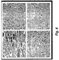
- Figures 4A-D are micrographs showing samples of muscle tissue at 3 weeks post incision; (A) saline alone, (B) saline plus electroporation, (C) Bleomycin alone, and (D) Bleomycin plus electroporation.
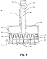
- Figure 5 is a cross sectional figure of one example of the device described herein showing an example comprising a handle comprising a thumb wheel for raising and lowering an array of plungers which actuate loading or dispensing of a substance through the electrode needles.
- Figure 6 is a drawing depicting an exploded view of one example of the device.
- Figure 7 is a cross sectional drawing of one example of the device wherein the plungers are actuated by a screw driven by clockwise (raising plungers) and counter clockwise (lowering plungers) rotation of the housing 50.
- Figures 8A and B are perspective drawings of one example of the device wherein rotation of the housing 50 will drive the array of plungers back and forth.
- Figure A shows the example with the tray 20 while Figure B shows the main body of the example without the tray 20.
- Figure 9 is a perspective drawing showing one example of the device wherein the array of plungers is actuated via a wing nut.
- Figure 10 shows an example of the array of the plurality of electrode needles from the underside of the substrate 22.
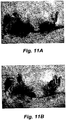
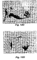
- Figures 11 A, B, C, and D are photographs of Group 2 cohort animals showing the test animal with tumor (A), the tumor surgically exposed prior to complete tumor removal (B), the open wound bed after tumor removal but prior to sham EP (C), and the surgical/treatment site after 3 weeks post treatment (D). With this Group 2, no electroporation was performed and the tumor recurred even though the tumor had been completely removed.
- Figures 12 A, B, C, and D are photographs of Group 1 cohort animals showing the test animal with tumor (A), the tumor surgically exposed prior to complete tumor removal (B), the open wound bed after tumor removal but prior to treatment with EP (C), and the surgical/treatment site after 3 weeks post treatment (D). As observed with Bleomycin-EP treatment no tumor recurred at the site of treatment.
- Figures 13 A, B, and C are photographs of Group 7 cohort animals showing the test animal with tumor (A), after partial tumor removal (B), and the surgical/treatment site after 3 weeks post treatment (C). The mouse at 3 weeks showed no tumor recurrence even though the tumor was only partially removed.
- Figures 14 A, B, and C are photographs of Group 4 cohort animals showing the test animal with tumor (A), after partial tumor removal (B), and the surgical/treatment site after 3 weeks post treatment (C). Without EPT the tumor continued to grow.
- Figure 15 shows a bar graph of the data presented in Table V.
- PTE refers to partial tumor excision
- CTE refers to complete tumor excision
- Bi.v. means Bleomycin administered to test animals intraveneously
- EP means electroporation
- i.t.B refers to Bleomycin administered intratumorally
- PEP means partial electroporation.
- Electroporation Therapy also known as Electrochemotherapy (ECT) is a method to treat localized cancerous lesions and tumor masses.
- the method comprises administering certain chemotherapeutic drugs, most commonly Bleomycin, either intratumorally or intravenously.
- electroporation is performed by inserting arrays of needle electrodes and delivering pulsed electrical fields emanating from these electrodes directly to the cancerous cell mass.
- Pulse parameters are generally within the following ranges: field strength 200-2000 V/cm; pulse length 0.1-10.0 ms; pulse number 2-20; and pulse frequency 1-5 Hz.
- EPT has been shown to be effective against many types of solid tumors in animals and several types of tumors in humans.
- several clinical studies are presently ongoing, including a Phase III study evaluating the safety and efficacy of EPT for the treatment of squamous cell carcinomas of the head and neck.
- Phase III study evaluating the safety and efficacy of EPT for the treatment of squamous cell carcinomas of the head and neck.
- such treatment studies are not designed or intended to treat or test the effect of EPT on noncancerous margin tissue coincident to cancerous cells.
- EPT is somewhat unique among the many methods employed for local and regional tumor control in that it is far less damaging to normal tissue than to malignant tissue. Results from in vitro as well as in vivo animal and human studies indicate that EPT using a drug, such as Bleomycin, effectively destroys most tumors via apoptotic and necrotic mechanisms while causing only a minor effect (inflammation, minor necrosis) on normal tissues surrounding the tumor, particularly when Bleomycin concentration, doses, and pulse parameters are within appropriate ranges.
- a drug such as Bleomycin
- the margin tissue surrounding tumors that is typically amenable to treatment include, without limitation, those of organs including breast, prostate, tongue, penis, labia, rectum, vocal chords, liver, squamous cell carcinoma of the head and neck (SCCHN), cutaneous and other tumors.
- EPT can be performed on such tissues whether the tumor mass has been removed or not. In such cancers the patient would be helped substantially by treating the tissues in a manner wherein healing can proceed largely unimpaired and there is a possibility of eliminating microscopic metastases and tumor branches trending into the tissues that give rise to local recurrences.
- electroporation treatments with agents such as Bleomycin provide for a measurable degree of selectivity in targeting cancerous cells.
- This methodology is also pertinent to treatments with other agents that have specific activities against particular disease states, such as highly localized infectious diseases and other disease states where there are foci of infected or diseased tissues wherein a cell, or group of cells that are infected or diseased, can be targeted by the agent to destroy the affected cells.
- electroporation of such cells and surrounding tissue will allow the agent to reach its intended target in the infected or diseased cells, yet not have a detrimental effect on uninfected or normal cells as empirically observed.
- the molecular and cellular mechanisms underlying the different response of abnormal and normal cells, respectively, is presently unknown. Additionally, use of the described methods and device is applicable to disease states where sites of cancerous cells are distributed in areas not easily amenable to surgery.
- skin tissues of the face containing microscopic or focal disease sites can be treated with the device in the same manner as used for treating a margin tissue bed resulting in substantially less scarring than would be caused by surgery.
- the described methods can be used as an adjuvant to surgery or even a neo-adjuvant.
- cells of tumor mass can be electroporated along with surrounding normal tissue and thereby provide for a method of debulking the tumor mass in providing for a mechanism to hinder, terminate or otherwise reduce the growth and/or recurrence rates of the diseased cells.
- tumor tissue can be removed either before or after EPT, or can even be allowed to remain unexcised following EPT, in which cases the effect of an agent such as Bleomycin will provide for cell death by apoptotic and /or necrotic mechanisms and softening of the tumor cell mass followed by further necrosis. Normal tissue in the vicinity will remain largely unaffected.
- the tumor is excised prior to EPT.
- treatment of a tumor with EPT includes a Bleomycin dose of 1 Unit/ccm tissue to be treated, injected intratumorally at a concentration of 4 Units/ml.
- the pulsed fields are generated such that the field is usually applied in six separate pulses at 4 Hz, one each in 6 different field orientations, each of 100usec duration, at a nominal field strength of anywhere between 200 and 2000V/cm but, generally between 600 and 1500 V/cm, more usually between 600 and 1400 V/cm and even more usually between 1200-1300 V/cm across the tumor mass.
- Increasing Bleomycin dose or concentration and increasing the intensity of the electroporating pulse beyond the standard parameters can result in more severe effects on normal tissue.
- a device for operating the invention can itself be operated using pulsed field strengths of between 1-600 V/cm when injecting DNA as a therapeutic agent as set forth in US patent 6,528,315 .
- the DNA ideally codes for a polypeptide that provides for a therapeutic anticancer effect on the tumor-containing tissue being treated.
- the same high pulsing parameters can be used (i.e., for example, 800- 1500 V/cm or, alternatively, 1-600 V/cm, more preferably 200 to 600 V/cm, and still more preferred 400-600 V/cm. It is further contemplated that these and any other agents can be used where such agents promote wound healing without promoting growth of malignant cells.
- lower-end field strengths as low as 50V/cm can be used to electroporate cells within tissue containing microscopic lesions and diseased cells where the tissues have not formed an abnormal tissue mass.
- the present rationale for cancer therapy involves in one example surgical removal of the tumor mass ('debulking') to reduce biological stress on the patient, surgical elimination of margin tissue to eradicate micrometastases and invading tumor tissue, and radiation and/or chemotherapy to control local, regional and systemic spread of the tumor. While surgical removal of the tumor itself generally provides relief for the patient, without severe sequelae, the subsequent steps of margin resection, radiation and chemotherapy can be very detrimental to the patient. Physiological and mechanical function, look, quality of life and eventual outcome can be severely affected by these treatments.
- the current invention is intended to provide a novel method for lowering the probability of recurrence of tumor growth in otherwise normal tissues surrounding a site of excised cancerous cells.
- the invention comprises treating "margins" of tissue surrounding a site of cancer cells, such cancerous cells typically formed at a distinct tissue site.
- the invention provides for reducing the amount of tissue that must be excised along with the tumor and its cancerous cells and making radiation or chemotherapy superfluous.
- the method comprises applying an electroporative pulse of electric energy to the tissues surrounding the tumor site.
- a formulation comprising an anticancer medicament is provided either prior to or simultaneously with the electric pulse.
- Such medicament comprises a biologically active molecule.
- the medicament can comprise a nucleic acid encoding an expressible polypeptide, such as, for example, a nucleic acid in an expression vector and comprising a gene sequence for a cytokine or chemokine, antibody, or enzyme.
- an expressible polypeptide such as, for example, a nucleic acid in an expression vector and comprising a gene sequence for a cytokine or chemokine, antibody, or enzyme.
- Another example comprises antisense DNA or RNA, or interfering RNA (RNAi).
- the medicament can comprise a polypeptide or an organic molecule such as, for example, a cytokine, chemokine, antibody, Cisplatin or Bleomycin.
- any of such compounds disclosed above can be administered in a formulation that can comprise any combination of pharmaceutically acceptable salts, buffers and other excipients as are well known in the art.
- formulations for nucleic acids can comprise, for example, said nucleic acid and poly-glutamic acid (poly-L-
- the electroporative pulse is applied using a device capable of providing such electroporative pulses to localized areas at the tumor site.
- a device capable of providing such electroporative pulses to localized areas at the tumor site.
- such a device can include an array of elongate needle-like electrodes.
- Alternate embodiments can use short non-penetrating electrodes or semi-penetrating microneedle electrodes depending upon the strength of the electric pulse required and upon the delivery mode of anticancer agent.
- the device would be designed without a need for a plunger and delivery needles.
- the treatment methods can be applied using an electroporation device comprising an electrode array only, such array comprising the non-penetrating or semi-penetrating electrodes.
- the electrodes are positionable with respect to the tissues to be treated to create an electric field having a field strength and energy sufficient to electroporate cells in said tissue within a specified area and depth.
- the device will be positioned to impart an electroporative pulse to all tissues within a preselected margin of tissue around (in three dimensions) the tumor excision site.
- the surrounding margin tissue bed can be electroporated either entirely in one electroporative pulse application or may require a multiplicity of electroporative pulse applications by the positioning and repositioning of the electrode array so as to completely encompass the tumor site margin tissue in electroporating energy fields.
- the electrodes can serve as delivery needles for the agent intended to be electroporated into the cells, while in other embodiments, the agent can be administered to the tissue independently from the administration/positioning of the electrodes in the tissue.
- One embodiment of the invention is to treat surgical wound margins or, more generally, tumor margins and in the process (a) achieve a superior recurrence rate (i.e., less frequent recurrence) compared with conventional surgical tumor and margin resection with or without secondary treatment; (b) reduce or eliminate the need for surgical resection of margin tissue while maintaining equal or better recurrence rates as with traditional surgical margin removal, and thus preserve functional tissue; and (c) reduce or eliminate the need for radiation or chemotherapy subsequent to surgical tumor resection (with or without margin resection) while maintaining equal or superior recurrence rates as with traditional surgical margin resection and secondary treatments such as, e.g., radiation, and chemotherapy.
- a superior recurrence rate i.e., less frequent recurrence
- transcutaneous incisions were made into the dorsal muscles in each of eight pig test animals grouped 1-6.
- longitudinal incisions were made while in group 6 the incisions were transverse to the longitudinal orientation of the muscle fiber.
- Bleomycin an anti-cancer agent, was used wherein each injection consisted of 0.125 ml (4 units/ml saline).
- the incisions were then treated with bipolar pulses of 530 V, 100 ⁇ sec each, from a pulse generator using a 6-needle electrode array of 0.5 cm diameter (nominal field strength for each pulse was 1,233 V/cm).
- Injections of a total volume of 1.5 ml of saline or Bleomycin (6 U) were administered at 12 sites per incision, 6 in the muscle and 6 in the skin. Incisions treated with electroporation were electroporated using a 0.5 cm diameter, 1.5 cm long needle array. On each side of the muscle incision, electroporation was given 3 times in the central 3 cm of the incision, each electroporation site being 1 cm apart from the other. Skin was not electroporated. The epimysium was closed with 4-0 Vicryl absorbable interrupted sutures. The skin was closed with 4-0 nylon interrupted sutures. Swine were sacrificed at 2d, 1 wk, 2wks, or 3wks following surgery.
- Histology specimens containing skin and at least 5 mm depth of underlying muscle were taken from the center of the incision and immediately placed in 10% neutral buffered formalin. An additional control specimen was taken from non-incised skin on the dorsum. After staining and sectioning, skin, subcutaneous tissue, and muscle were graded histologically on a 0-3 scale using the following parameters: healing, Granulations/Fibrosis, PMN's, Lymphocytes, Histiocytes, Necrosis, Hemorrhage and Atypical Cells.
- Group 3 (saline injection with electroporation) was not significantly different from Group 2 at any time indicating that electroporation did not cause adverse effects in breaking strength.
- all three Bleomycin treated groups had significantly (i.e., 90%) reduced breaking strengths compared to all three non-Bleomycin groups, indicating that Bleomycin had delayed dermal wound healing.
- Electroporation had no adverse effects on breaking strength as Groups 4 and 5 were not significantly different from each other.
- week 2 there were no significant differences between Groups 1-5. Only the transverse incisions with Bleomycin and electroporation exhibited lower breaking strengths. This effect carried through to week 3 where Group 6 had significantly lower breaking strength than Groups 1-5.
- Histology specimens at four different time points namely, Days 2, 7, 14, and 21 containing skin and at least 5 mm of muscle were taken from the center of the incisions and immediately placed in 10% neutral buffered formalin. Histological studies showed statistically significant changes (Bleomycin groups only) present at day 7. All non-treated tissue and saline injected tissue remained normal in appearance. By day 21, all Bleomycin injected samples were comparable with saline-injected controls. For example, when comparing saline-injected v.s. saline-injected plus electroporated tissue samples, the only effects of electroporation were slight increases in the number of skin histiocytes at 1 week and a reduction in skin lymphocytes at 2 weeks.
- wound healing as compared between untreated tissue, saline plus electroporation treated tissue, and Bleomycin plus electroporation treated tissue, becomes histologically equivalent by week three.
- week three saline plus electroporation is equivalent to Bleomycin plus electroporation (compare E and F ).
- week 2 saline plus electroporation shows more advanced healing than Bleomycin and electroporation (compare C and A ).
- week 3 saline plus electroporation appears equivalent to Bleomycin plus electroporation at week 2 (compare E and D ).
- Electroporation of the muscle caused no significant alteration in the breaking strength of porcine dermis in the presence or absence of Bleomycin at weeks 2 and 3 and arguably even within week 1.
- the presence of Bleomycin regardless of whether EP was used, was associated with a reduction in the breaking strength of porcine dermis up to 1-2 weeks post treatment when compared to incision only, or to incision injected with saline, with or without EP.
- electroporation of the tissues resulted in no significant differences in healing of the skin, subcutaneous tissue or muscle.
- electroporation therapy does not significantly adversely affect healing of muscle or skin. This appears to apply also where EPT is used for treatment of tissue surrounding tumors, and thus the invention provides an alternative to surgical resection or other therapeutic interventions in the treatment of wound margins aimed at reducing tumor recurrence.
- steps 1 and 2 above The outcome of performing steps 1 and 2 above is intended to comprise improved cure rate or time to recurrence with some degree of preservation of tissue over conventional therapies but margin tissue would have been removed nonetheless.
- Variation B Protocol for Resecting a tumor with no margin resection
- the device is intended to provide the capability to deliver to the margin tissue an anticancer agent, e.g., for example, Bleomycin, evenly distributed throughout the margin bed.
- an anticancer agent e.g., for example, Bleomycin
- the device provides for the capability of delivering to the margin tissue a plurality of pulses of electric energy sufficient to cause electroporation of cells throughout the margin bed to a depth of between 1 and 1.5 cm.
- the device can provide for delivery of the electroporation pulses to a substantial portion of the margin bed, if not all of the margin bed (depending upon the relative dimensions of the device and the tumor), by a single placement of the device such that in order to provide electroporating pulses to the entire margin bed the device should preferably only need to be placed once.
- the device includes a plurality of electrodes positioned in a geometric array to provide for delivery of a series of electric pulses between selected electrodes of the array.
- the device can comprise a plurality of shapes to accommodate various tumor shapes and sizes and further provide an array of electrodes in a preferred geometry.
- the electrodes of the device can comprise elongate hollow needles such that the electrodes can act as both electrodes and anticancer agent delivery needles.
- the device can include a sharps cover to cover the electrodes and to provide a mechanism for delivery of anticancer agent to the hollow needles and a means for keeping the electrodes sterile prior to surgery.
- FIG. 5 shows device 10 comprising a substantially rigid sharps cover 20 which doubles as a therapeutic agent filling tray.
- the cover/tray 20 further comprises a fill port 21 which can be designed with a fitting capable of connecting with any desired type fitting for attaching to a source of fluid for filling the tray to a desired level of therapeutic substance.
- the cover/tray 20 snuggly fits the main body of the device which itself comprises a substantially rigid substrate 22 through which an array of a plurality of elongate electrodes 23 pass and which are each individually connected to electric leads 24 which terminate on a lateral portion of the device in a plug or connector 25 for attachment to a source of electrical energy.
- the substrate 22 and the plug 25 are connected to main body substrate 30 which itself comprises an array of a plurality of wells 31 (see Figure 6 ).
- Each well comprises a hollow electrode/needle through which a substance such as a therapeutic substance can be transmitted.
- substrate 40 which comprises an array of a plurality of plungers 41.
- the substrate 40 is connected to a mechanism which can actuate the movement of the substrate 40 towards and away from substrate 30 so as to drive into or extract away from substrate 30 the array of plungers 41 of substrate 40.
- the substrate 40 is actuated by a central rod 60 connected to substrate 40 on one end and at the other configured into a ratchet and a thumb activated wheel 61.
- substrate 50 further forms an enclosing support structure 63 to surround and enclose central rod 60.
- the support structure 63 can further comprise user friendly handgrip 64.
- the operator would use port 21 to fill the cover/tray 20 to a desired level with a fluid therapeutic agent, such as for example Bleomycin, then use the thumb wheel 61 to draw into the wells 31 the agent ( Fig. 6 ).
- a fluid therapeutic agent such as for example Bleomycin
- the operator would then remove the device from the tray 20 using the handle formed by the support structure 63 and place the device at an appropriate position on the tissue or margin bed.
- the operator would then insert the electrodes, activate the thumb wheel 61 to administer the agent into the tissue followed by activation of the electrodes, the device having previously been attached to a source of electrical energy via the plug 25.
- the device further can comprises other various arrangements of components to provide the same result.
- the device can comprise attached to the substrate 30, an optionally dynamic housing 50.
- Housing 50 is considered dynamic on an optional basis since depending upon the style of the device chosen, the housing can rotate clockwise and counter clockwise to raise and lower the plunger instead of using a thumb wheel.
- the housing 50 is not dynamic in that although the housing 50 may be capable of rotation, but for the handle and thumb wheel, such rotation in this example servers no utility.
- the housing is dynamic in that substrate 40 is actuated toward or away from substrate 30 by screw 70 which is attached at one end to substrate 40 and to housing 50 on the other end by engagement of said screw 70.
- the housing 50 portion which interacts with threads of screw 70 is itself formed into screw threads 71 which upon rotation of housing 50 will draw substrate 40 away from or towards substrate 30.
- the operator would fill the cover/tray 20 with agent as previously described, then place the discoid device appropriately on the tissue to be treated. After inserting the electrodes into the tissue, the user would, while pressing down on the device, rotate the housing 50 in the direction that will move the plungers toward substrate 30 and thereby expel the agent into the tissues. The operator would then active the electrodes, assuming the device had been previously connected by plug 25 to a source of electrical energy.
- the device such as for example use of a wing nut 80 attached to screw 70 for actuating the plungers 41 (see Fig. 9 ).
- the housing 50 would not comprise screw threads 71 but instead only a smooth bore past which the threaded central rod 60 can pass. In such example, the operator would not need to rotate the housing but only the wing nut.
- the plungers can be actuated by a motor driven plunger.
- a motor can be placed in arrangement with the housing 50 to, for example, cause bidirectional rotation of the housing with respect to the example disclosed in Figures 6-8 .
- a motor can be affixed to gears as in the thumb wheel example to actuate raising and lowering the plungers.
- the device can include indicators to provide the operator notice when the plungers have reached their respective maximum of travel into or away from the wells 31.
- Still further examples include use of any variety of energizing parameters including use of monopolar and/or bipolar pulses in any form, including without limitation, trains of pulses, exponential decaying pluses, square pulses, etc.
- the invention can use pulses having a field strength of between 10 and 1500 V/cm.
- the invention contemplates the capacity to energize each electrode of the array individually or together with any combination of the remaining electrodes. For example, in one example, any 4 adjacent electrodes can be pulsed such that two are positive and an opposing pair of electrodes are both negatively charged. Such pulsing provides "opposed pair" pulsing as disclosed in US patents numbers 5,993,434 and 5,702,359 .
- single pairs of electrodes can be energized to impart an electroporating pulse of energy to the tissue until all adjacent pairs of electrodes are energized at least once.
- the device as disclosed in its various examples includes individually addressed electrode needles. This is accomplished by incorporating within substrate 22 such as by sandwich technique (i.e., substrate 22 can comprise a two layer construction with electric leads placed on a grid in between the two layers) electric lead connections running from the electrodes to the side of the device (as depicted as leads 24) terminating in plug 25 having multiple pin connector capability.
- this experiment comprises an in vivo study to establish the efficacy of EPT on surgical margin treatment in nude mice after subcutaneous introduction of HT-29 human colon carcinoma cells and resection of the tumors that arise from such introduction. The experiment was conducted under humane conditions with respect to treatment of the test animals.
- the experimental design was as follows. Normal Athymic Nude female mice 4-6 weeks of age were placed in 8 groups of six mice each, except for Group 1 which comprised 10 animals and Group 3 comprising 7 animals.
- the HT-29 cell line is derived from a human colon carcinoma and is an aggressive fast-growing tumor in mice. In each group the animals received HT-29 human tumor cells in the form of two adjacent inocula of 5X10 6 cells using a 21 gauge needle and syringe.
- the HT-29 cell line was obtained from ECACC (#HTB-38), grown in McCoy's 5a medium (modified) with 1.5 mM L-glutamine adjusted to contain 2.2 g/L sodium bicarbonate, 90%, and fetal bovine serum, 10%. Cells were divided 1:2 to 1:4 on growth cycles until sufficient cells were available to use in this experiment.
- Tumor volume (a 2 x b/2) where 'a' is the smallest diameter and 'b' is the largest diameter perpendicular to 'a'. Tumors were measured three times per week starting from the day the tumors were palpable by hand. The tumors were allowed to progress until the size was between 500 and 1500 cubic millimeters. Surgical tumor removal, partial removal, or no removal was then performed in association with either EPT or no EPT. In these experiments EPT means treatment with Bleomycin and electroporation (EP).
- the EP where performed, was conducted with one pulse cycle (i.e., 6 pulses at 4 Hz) per electrode insertion using a standard MedPulser generator (Genetronics, Inc. San Diego, CA) and an applicator with a disposable six needle array of 1 cm diameter, a distance between electrodes of 0.86 cm, and 1 cm needle length. Each square pulse had a duration of 100 usec and an applied voltage of 1500 V. For Groups 4-8 and 10 and 11, after intratumoral injection of Bleomycin the needle electrode array was inserted in such a way as to encompass the tumor within the needle array or, in the case of the largest tumors, such that the needles penetrated the edge of the tumor.
- the needle array was inserted percutaneously and essentially perpendicular to the surface of the flank of the mouse carrying the tumor.
- the needle electrode array was inserted percutaneously encompassing the surgical wound area (Groups 5-8).
- the experiment used 8 cohorts of at least 6 mice each. Each cohort was subjected to a treatment regimen of a combination of any of EP, Bleomycin, saline instead of Bleomycin, complete tumor excision, partial tumor excision, and no tumor excision. Tables II and III disclose the treatment regimens.
- the animals were anaesthetized, followed by removal or partial removal of the tumor as indicated, followed in turn by intraveneous (i.v.) injection of Bleomycin or saline.
- the Bleomycin dose was dependent on tumor size as described above.
- the animals indicated were treated with EP followed by closure of the wound with clips and surgical adhesive.
- cohort 1 the entire tumor was surgically removed (see, for example Figure 12B and C ) and the animals treated with Bleomycin and EP.
- the tumors were removed from the animals but only saline was injected instead of Bleomycin and no EP was performed. Only a "sham" EP procedure was performed in that the electrode array was inserted into the tissue but the electrodes were not pulsed.
- the animals of Group 9 were anaesthetized followed by complete excision of the tumors.
- the tumors were examined for consistency and preserved in formalin for later histological evaluation, as were the tumor tissues of cohorts 10 and 11.
- Anaesthetized animals of cohort 10 were subjected to intratumoral administration of Bleomycin solution at a dose and volume as described above.
- EP was performed'10 minutes after drug administration. Tumors were excised 2 hrs after EP. Animals of cohort 11 were treated as those in cohort 10 except that their tumors were completely removed at 24 hrs after EP. All wounds were closed as described above.
- Groups 1 to 8 provide results indicating that treatment of tumor bed tissue provides an unexpected and surprising benefit in protecting the animal from recurrent tumors at the site of the treatment.
- excision alone is not effective, whether the tumors were completely or partially removed. This is likely due to the aggressive nature of the tumor type used which left invasive tumor segments and/or micrometastases at or close to the tumor site despite careful surgical removal of the tumor correlating to similar aggressive tumors in man. This mimics situations encountered in human surgical therapy although recurrence rates in humans are generally in the 10 to 40% range and not as high as observed in this mouse experiment.
- FIGS 11A , 12A , 13A and 14A which represent tumors in test animals prior to treatment.
- Figures 11B and 12B represent examples of surgical tumor removal at the stage of having the tumor exposed before the final excision.
- Figures 11C , 12C , 13B , and 14B represent examples of the wound after complete tumor removal prior to Bleomycin-EPT ( Fig. 12C ) or saline-sham EP treatment ( Fig. 11C ), or after partial tumor removal prior to saline-sham treatment ( Fig. 14B ), or 15 minutes after i.t. Bleomycin-EPT ( Fig. 13B ).
- Figure 11D shows recurring tumor after complete tumor excision and saline-sham EP treatment
- Fig. 14C shows recurring tumor after partial tumor excision and saline-sham EP treatment
- Figures 12D and 13C show tumor sites free of tumor after tumor excision and treatment with i.v. Bleomycin and EP, or treatment with i.t. Bleomycin and EP with subsequent partial tumor excision, respectively.
- These procedures have advantages over present standard procedures that treat cancers with anticancer drug i.t. or i.v. Excision of the tumor, either prior to or after drug-EPT treatment prevents formation of a large necrotic mass at the tumor site which the body has to resorb or otherwise eliminate, which empirically takes at least several weeks and enhances the probability of complications. Removal of the tumor prior to or after drug-EPT is likely to expedite wound healing and reduce potential complications. Further with this methodology, both intratumoral and intravenous injection of drug can be used.
Claims (4)
- Un agent anticancéreux pour une utilisation en tant que médicament destiné à réduire la récurrence d'une croissance tumorale dans un tissu mammalien chez un mammifère au niveau et autour d'un emplacement chez ledit mammifère où une tumeur a été excisée :où ledit agent est sélectionné dans le groupe constitué de la bléomycine, du cisplatine,d'une protéine, d'un anticorps, d'un ARNi, d'un acide nucléique antisens, d'un gène pouvant s'exprimer codant pour un polypeptide thérapeutiquement actif, d'une chimiokine, et d'une cytokine ;où ledit agent est destiné à être administré audit tissu au moyen d'une injection intraveineuse ou est injecté directement au niveau et autour dudit tissu soit avant soit en même temps que l'administration d'une impulsion électrique d'électroporation audit tissu, apportant ainsi ledit agent jusque dans des cellules dudit tissu, ce qui résulte en une réduction ou une élimination d'une croissance de cellules tumorales dans ledit tissu.
- L'agent anticancéreux pour une utilisation en tant que médicament selon la revendication 1 où lesdits ARNi, acide nucléique antisens, et gène pouvant s'exprimer sont formulés dans une solution de poly-L-glutamate.
- L'agent anticancéreux pour une utilisation en tant que médicament selon la revendication 1, et pour une utilisation dans le traitement de tumeurs résiduelles microscopiques restant dans ledit tissu après une résection chirurgicale de ladite tumeur :où ledit agent est destiné à être administré audit tissu précédemment adjacent à etautour de ladite tumeur et contenant lesdites tumeurs résiduelles, soit avant soit en même temps que l'administration audit tissu d'au moins une impulsion électrique d'électroporation,où la présence de l'agent et l'impulsion électrique d'électroporation apportent un traitement sélectif dudit tissu afin de tuer lesdites tumeurs résiduelles.
- L'agent anticancéreux pour une utilisation en tant que médicament selon la revendication 1, où ladite impulsion électrique d'électroporation a une intensité de champ de 200 à 2 000 V/cm ; une durée d'impulsion de 0,1 à 10,0 ms ; un nombre d'impulsions de 2 à 20 ; et une fréquence d'impulsion de 1 à 5 Hz.
Priority Applications (2)
| Application Number | Priority Date | Filing Date | Title |
|---|---|---|---|
| PL07751865T PL1991303T3 (pl) | 2006-03-03 | 2007-03-02 | Sposób i urządzenie do leczenia mikroskopowych nowotworów pozostałych w tkankach po resekcji chirurgicznej |
| EP21156169.1A EP3845269A1 (fr) | 2006-03-03 | 2007-03-02 | Procédé et dispositif de traitement de tumeurs résiduelles microscopiques restant dans des tissus après résection chirurgicale |
Applications Claiming Priority (2)
| Application Number | Priority Date | Filing Date | Title |
|---|---|---|---|
| US77874006P | 2006-03-03 | 2006-03-03 | |
| PCT/US2007/005133 WO2007103070A2 (fr) | 2006-03-03 | 2007-03-02 | Procédé et dispositif de traitement de tumeurs résiduelles microscopiques restant dans des tissus après résection chirurgicale |
Related Child Applications (2)
| Application Number | Title | Priority Date | Filing Date |
|---|---|---|---|
| EP21156169.1A Division EP3845269A1 (fr) | 2006-03-03 | 2007-03-02 | Procédé et dispositif de traitement de tumeurs résiduelles microscopiques restant dans des tissus après résection chirurgicale |
| EP21156169.1A Division-Into EP3845269A1 (fr) | 2006-03-03 | 2007-03-02 | Procédé et dispositif de traitement de tumeurs résiduelles microscopiques restant dans des tissus après résection chirurgicale |
Publications (3)
| Publication Number | Publication Date |
|---|---|
| EP1991303A2 EP1991303A2 (fr) | 2008-11-19 |
| EP1991303A4 EP1991303A4 (fr) | 2012-01-18 |
| EP1991303B1 true EP1991303B1 (fr) | 2021-05-05 |
Family
ID=38475369
Family Applications (2)
| Application Number | Title | Priority Date | Filing Date |
|---|---|---|---|
| EP21156169.1A Pending EP3845269A1 (fr) | 2006-03-03 | 2007-03-02 | Procédé et dispositif de traitement de tumeurs résiduelles microscopiques restant dans des tissus après résection chirurgicale |
| EP07751865.2A Active EP1991303B1 (fr) | 2006-03-03 | 2007-03-02 | Procédé et dispositif de traitement de tumeurs résiduelles microscopiques restant dans des tissus après résection chirurgicale |
Family Applications Before (1)
| Application Number | Title | Priority Date | Filing Date |
|---|---|---|---|
| EP21156169.1A Pending EP3845269A1 (fr) | 2006-03-03 | 2007-03-02 | Procédé et dispositif de traitement de tumeurs résiduelles microscopiques restant dans des tissus après résection chirurgicale |
Country Status (12)
| Country | Link |
|---|---|
| US (4) | US9037230B2 (fr) |
| EP (2) | EP3845269A1 (fr) |
| JP (3) | JP2009528131A (fr) |
| KR (2) | KR101529448B1 (fr) |
| CN (2) | CN101426550B (fr) |
| AU (1) | AU2007224275B2 (fr) |
| CA (1) | CA2644163C (fr) |
| DK (1) | DK1991303T3 (fr) |
| ES (1) | ES2874482T3 (fr) |
| NO (2) | NO344476B1 (fr) |
| PL (1) | PL1991303T3 (fr) |
| WO (1) | WO2007103070A2 (fr) |
Families Citing this family (40)
| Publication number | Priority date | Publication date | Assignee | Title |
|---|---|---|---|---|
| KR101529448B1 (ko) | 2006-03-03 | 2015-06-18 | 온코섹 메디컬 인코포레이티드 | 외과적 절제술 이후에 조직내 남아 있는 미세 잔류 종양을 치료하는 장치 |
| GB2464902B (en) | 2007-08-14 | 2011-10-19 | Hutchinson Fred Cancer Res | Methods for evaluating candidate therapeutic agents |
| US11272979B2 (en) | 2008-04-29 | 2022-03-15 | Virginia Tech Intellectual Properties, Inc. | System and method for estimating tissue heating of a target ablation zone for electrical-energy based therapies |
| US9283051B2 (en) | 2008-04-29 | 2016-03-15 | Virginia Tech Intellectual Properties, Inc. | System and method for estimating a treatment volume for administering electrical-energy based therapies |
| US10245098B2 (en) | 2008-04-29 | 2019-04-02 | Virginia Tech Intellectual Properties, Inc. | Acute blood-brain barrier disruption using electrical energy based therapy |
| US9598691B2 (en) | 2008-04-29 | 2017-03-21 | Virginia Tech Intellectual Properties, Inc. | Irreversible electroporation to create tissue scaffolds |
| US10238447B2 (en) | 2008-04-29 | 2019-03-26 | Virginia Tech Intellectual Properties, Inc. | System and method for ablating a tissue site by electroporation with real-time monitoring of treatment progress |
| US8992517B2 (en) | 2008-04-29 | 2015-03-31 | Virginia Tech Intellectual Properties Inc. | Irreversible electroporation to treat aberrant cell masses |
| US9198733B2 (en) | 2008-04-29 | 2015-12-01 | Virginia Tech Intellectual Properties, Inc. | Treatment planning for electroporation-based therapies |
| US9867652B2 (en) | 2008-04-29 | 2018-01-16 | Virginia Tech Intellectual Properties, Inc. | Irreversible electroporation using tissue vasculature to treat aberrant cell masses or create tissue scaffolds |
| US10272178B2 (en) | 2008-04-29 | 2019-04-30 | Virginia Tech Intellectual Properties Inc. | Methods for blood-brain barrier disruption using electrical energy |
| US11254926B2 (en) | 2008-04-29 | 2022-02-22 | Virginia Tech Intellectual Properties, Inc. | Devices and methods for high frequency electroporation |
| US10117707B2 (en) | 2008-04-29 | 2018-11-06 | Virginia Tech Intellectual Properties, Inc. | System and method for estimating tissue heating of a target ablation zone for electrical-energy based therapies |
| US10702326B2 (en) | 2011-07-15 | 2020-07-07 | Virginia Tech Intellectual Properties, Inc. | Device and method for electroporation based treatment of stenosis of a tubular body part |
| WO2009155526A2 (fr) * | 2008-06-20 | 2009-12-23 | Angiodynamics, Inc. | Dispositif et méthodes permettant de supprimer une gaine de fibrine formée sur un cathéter veineux |
| US11382681B2 (en) | 2009-04-09 | 2022-07-12 | Virginia Tech Intellectual Properties, Inc. | Device and methods for delivery of high frequency electrical pulses for non-thermal ablation |
| US11638603B2 (en) | 2009-04-09 | 2023-05-02 | Virginia Tech Intellectual Properties, Inc. | Selective modulation of intracellular effects of cells using pulsed electric fields |
| WO2010138919A2 (fr) | 2009-05-28 | 2010-12-02 | Angiodynamics, Inc. | Système et méthode de synchronisation de l'apport d'énergie et du rythme cardiaque |
| US9895189B2 (en) | 2009-06-19 | 2018-02-20 | Angiodynamics, Inc. | Methods of sterilization and treating infection using irreversible electroporation |
| CA2689400C (fr) * | 2009-12-30 | 2023-01-10 | Kenneth W. Adams | Appareil et procede d'administration d'agent therapeutique |
| WO2012051433A2 (fr) | 2010-10-13 | 2012-04-19 | Angiodynamics, Inc. | Système et procédé d'ablation électrique des tissus chez un patient |
| WO2012088149A2 (fr) | 2010-12-20 | 2012-06-28 | Virginia Tech Intellectual Properties, Inc. | Électroporation à haute fréquence pour thérapie anticancéreuse |
| CA2839196A1 (fr) | 2011-06-15 | 2012-12-20 | Chrontech Pharma Ab | Aiguille et dispositif d'injection |
| US9078665B2 (en) | 2011-09-28 | 2015-07-14 | Angiodynamics, Inc. | Multiple treatment zone ablation probe |
| WO2013063530A2 (fr) | 2011-10-28 | 2013-05-02 | Presage Biosciences, Inc. | Procédés d'administration de médicament |
| US10166321B2 (en) | 2014-01-09 | 2019-01-01 | Angiodynamics, Inc. | High-flow port and infusion needle systems |
| AU2015259303B2 (en) | 2014-05-12 | 2021-10-28 | Arena, Christopher B. | Selective modulation of intracellular effects of cells using pulsed electric fields |
| US10694972B2 (en) | 2014-12-15 | 2020-06-30 | Virginia Tech Intellectual Properties, Inc. | Devices, systems, and methods for real-time monitoring of electrophysical effects during tissue treatment |
| US10287543B2 (en) * | 2015-11-19 | 2019-05-14 | Miltenyi Biotec, Gmbh | Process and device for isolating cells from biological tissue |
| US20170232253A1 (en) * | 2016-02-16 | 2017-08-17 | Marcy C. Purnell | Bioelectrodynamics Modulation Method |
| US10233419B2 (en) | 2016-06-30 | 2019-03-19 | Zymergen Inc. | Apparatuses and methods for electroporation |
| US10905492B2 (en) | 2016-11-17 | 2021-02-02 | Angiodynamics, Inc. | Techniques for irreversible electroporation using a single-pole tine-style internal device communicating with an external surface electrode |
| JP2021502882A (ja) | 2017-11-15 | 2021-02-04 | アルキオーネ・ライフサイエンシズ・インコーポレイテッドAlcyone Lifesciences, Inc. | 治療法別の事前プログラムされた自動注射デバイス |
| EP3710102A4 (fr) * | 2017-11-17 | 2021-08-04 | AbbVie Inc. | Méthodes de traitement d'un glioblastome |
| US11607537B2 (en) | 2017-12-05 | 2023-03-21 | Virginia Tech Intellectual Properties, Inc. | Method for treating neurological disorders, including tumors, with electroporation |
| JP2021508533A (ja) | 2017-12-26 | 2021-03-11 | ギャラリー,インコーポレイテッド | 様々な用途のためのエネルギー送達の最適化 |
| US11311329B2 (en) | 2018-03-13 | 2022-04-26 | Virginia Tech Intellectual Properties, Inc. | Treatment planning for immunotherapy based treatments using non-thermal ablation techniques |
| US11925405B2 (en) | 2018-03-13 | 2024-03-12 | Virginia Tech Intellectual Properties, Inc. | Treatment planning system for immunotherapy enhancement via non-thermal ablation |
| US11950835B2 (en) | 2019-06-28 | 2024-04-09 | Virginia Tech Intellectual Properties, Inc. | Cycled pulsing to mitigate thermal damage for multi-electrode irreversible electroporation therapy |
| CN116509535B (zh) * | 2023-03-14 | 2024-01-23 | 武汉金柏威光电技术有限公司 | 一种医用高频电刀 |
Family Cites Families (24)
| Publication number | Priority date | Publication date | Assignee | Title |
|---|---|---|---|---|
| US5747469A (en) * | 1991-03-06 | 1998-05-05 | Board Of Regents, The University Of Texas System | Methods and compositions comprising DNA damaging agents and p53 |
| US5993434A (en) | 1993-04-01 | 1999-11-30 | Genetronics, Inc. | Method of treatment using electroporation mediated delivery of drugs and genes |
| US5702359A (en) | 1995-06-06 | 1997-12-30 | Genetronics, Inc. | Needle electrodes for mediated delivery of drugs and genes |
| US20030060434A1 (en) * | 1997-02-18 | 2003-03-27 | Loretta Nielsen | Combined tumor suppressor gene therapy and chemotherapy in the treatment of neoplasms |
| AU8444698A (en) * | 1997-06-30 | 1999-01-25 | Centre National De La Recherche Scientifique | Improved method for transferring nucleic acid into multicelled eukaryo tic organism cells and combination therefor |
| US6055453A (en) | 1997-08-01 | 2000-04-25 | Genetronics, Inc. | Apparatus for addressing needle array electrodes for electroporation therapy |
| US6224019B1 (en) | 1998-01-26 | 2001-05-01 | Simula, Inc. | Parachute landing velocity attenuator |
| US6208893B1 (en) * | 1998-01-27 | 2001-03-27 | Genetronics, Inc. | Electroporation apparatus with connective electrode template |
| US6104584A (en) * | 1999-02-18 | 2000-08-15 | Lucent Technologies, Inc. | Voltage feedback inrush current limit circuit having increased tolerance for component value variation |
| AU2001245427A1 (en) | 2000-03-03 | 2001-09-17 | Valentis, Inc. | Nucleic acid formulations for gene delivery and methods of use |
| US6428504B1 (en) * | 2000-04-06 | 2002-08-06 | Varian Medical Systems, Inc. | Multipurpose template and needles for the delivery and monitoring of multiple minimally invasive therapies |
| US20040092860A1 (en) * | 2000-07-26 | 2004-05-13 | Dev Nagendu B. | Skin and muscle-targeted gene therapy by pulsed electrical field |
| WO2002072781A2 (fr) * | 2001-03-13 | 2002-09-19 | University Of South Florida | Dispositif et procédé d'électromanipulation |
| CA2470322A1 (fr) * | 2001-12-14 | 2003-06-26 | Genetronics, Inc. | Procedes d'immunisation assistee par particules reposant sur l'utilisation d'un champ electrique pulse |
| US8209006B2 (en) * | 2002-03-07 | 2012-06-26 | Vgx Pharmaceuticals, Inc. | Constant current electroporation device and methods of use |
| EP1480692A2 (fr) * | 2002-03-07 | 2004-12-01 | Merck & Co., Inc. | Seringue clinique avec des aspects de stimulation electrique |
| US20040014645A1 (en) * | 2002-05-28 | 2004-01-22 | Advisys, Inc. | Increased delivery of a nucleic acid construct in vivo by the poly-L-glutamate ("PLG") system |
| JP4409239B2 (ja) * | 2003-09-18 | 2010-02-03 | テルモ株式会社 | 薬液注入装置 |
| US20050209625A1 (en) * | 2004-03-02 | 2005-09-22 | Chan Frank A | Method and apparatus for electrical stimulation to enhance lancing device performance |
| US7537590B2 (en) * | 2004-07-30 | 2009-05-26 | Microchips, Inc. | Multi-reservoir device for transdermal drug delivery and sensing |
| US20070088345A1 (en) | 2005-10-13 | 2007-04-19 | Ust Inc. | Applications of HIFU and chemotherapy |
| KR101529448B1 (ko) | 2006-03-03 | 2015-06-18 | 온코섹 메디컬 인코포레이티드 | 외과적 절제술 이후에 조직내 남아 있는 미세 잔류 종양을 치료하는 장치 |
| US7671095B2 (en) * | 2006-05-31 | 2010-03-02 | The Trustees Of The Boston University | Films and particles |
| CN105457157B (zh) * | 2006-10-17 | 2020-04-14 | 因诺维奥制药公司 | 电穿孔装置及用其进行哺乳动物细胞电穿孔的方法 |
-
2007
- 2007-03-02 KR KR1020147014055A patent/KR101529448B1/ko active IP Right Grant
- 2007-03-02 US US11/713,181 patent/US9037230B2/en active Active
- 2007-03-02 CN CN2007800143130A patent/CN101426550B/zh active Active
- 2007-03-02 PL PL07751865T patent/PL1991303T3/pl unknown
- 2007-03-02 WO PCT/US2007/005133 patent/WO2007103070A2/fr active Application Filing
- 2007-03-02 JP JP2008557341A patent/JP2009528131A/ja not_active Withdrawn
- 2007-03-02 EP EP21156169.1A patent/EP3845269A1/fr active Pending
- 2007-03-02 CA CA2644163A patent/CA2644163C/fr active Active
- 2007-03-02 KR KR1020087023591A patent/KR101444471B1/ko active IP Right Grant
- 2007-03-02 ES ES07751865T patent/ES2874482T3/es active Active
- 2007-03-02 CN CN201210397694.8A patent/CN102921102B/zh active Active
- 2007-03-02 AU AU2007224275A patent/AU2007224275B2/en active Active
- 2007-03-02 DK DK07751865.2T patent/DK1991303T3/da active
- 2007-03-02 EP EP07751865.2A patent/EP1991303B1/fr active Active
-
2008
- 2008-10-01 NO NO20084148A patent/NO344476B1/no unknown
-
2011
- 2011-10-21 US US13/278,721 patent/US20120143120A1/en not_active Abandoned
-
2013
- 2013-03-06 JP JP2013043715A patent/JP2013127000A/ja not_active Withdrawn
-
2015
- 2015-07-07 JP JP2015135937A patent/JP6174632B2/ja active Active
-
2017
- 2017-04-14 US US15/488,288 patent/US11007365B2/en active Active
-
2019
- 2019-09-13 NO NO20191102A patent/NO20191102A1/no not_active Application Discontinuation
-
2021
- 2021-04-20 US US17/235,649 patent/US20210308451A1/en active Pending
Non-Patent Citations (7)
| Title |
|---|
| G SERS ÏA ET AL: "Original Paper Electrochemotherapy with Cisplatin: Potentiation of Local Cisplatin Antitumour EVectiveness by Application of Electric Pulses in Cancer Patients", EUR J CANCER, 1 January 1998 (1998-01-01), pages 1213 - 1218, XP055303697, Retrieved from the Internet <URL:http://lbk.fe.uni-lj.si/pdfs/ejc1998.pdf> * |
| HISAHIRO MATSUBARA ET AL: "Combinatory anti-tumor effects of electroporation-mediated chemotherapy and wild-type p53 gene transfer to human esophageal cancer cells", INTERNATIONAL JOURNAL OF ONCOLOGY, vol. 18, no. 4, 1 April 2001 (2001-04-01), GR, pages 825 - 829, XP055240372, ISSN: 1019-6439, DOI: 10.3892/ijo.18.4.825 * |
| JAVOROVIC ET AL: "RNA Transfer by Electroporation into Mature Dendritic Cells Leading to Reactivation of Effector-Memory Cytotoxic T Lymphocytes: A Quantitative Analysis", MOLECULAR THERAPY, NATURE PUBLISHING GROUP, GB, vol. 12, no. 4, 1 October 2005 (2005-10-01), pages 734 - 743, XP005078453, ISSN: 1525-0016, DOI: 10.1016/J.YMTHE.2005.03.034 * |
| LI S ET AL: "Regression of tumors by IFN-a electroporation gene therapy and analysis of the responsible genes by cDNA array", GENE THERAPY, NATURE PUBLISHING GROUP, GB, vol. 9, 1 March 2002 (2002-03-01), pages 390 - 397, XP002904771, ISSN: 0969-7128, DOI: 10.1038/SJ.GT.3301645 * |
| MARTIN BURIAN ET AL: "Electroporation therapy in head and neck cancer", ACTA OTOLARYNGOL, 1 January 2003 (2003-01-01), pages 264 - 268, XP055304380, Retrieved from the Internet <URL:http://pulsedenergytech.com/human/pdf_files/electroporation-therapy.pdf> [retrieved on 20160921] * |
| S. LI ET AL: "Regression of Tumor Growth and Induction of Long-Term Antitumor Memory by Interleukin 12 Electro-Gene Therapy", CANCERSPECTRUM KNOWLEDGE ENVIRONMENT, vol. 94, no. 10, 15 May 2002 (2002-05-15), pages 762 - 768, XP055303676, DOI: 10.1093/jnci/94.10.762 * |
| TSURUO T ET AL: "Inhibition of multidrug-resistant human tumor growth in athymic mice by anti-P-glycoprotein monoclonal antibodies", JAPANESE JOURNAL OF CANCER RESEARCH, JAPANESE CANCER ASSOCIATION, TOKYO, JP, vol. 80, no. 7, 1 July 1989 (1989-07-01), pages 627 - 631, XP009136974, ISSN: 0910-5050 * |
Also Published As
Similar Documents
| Publication | Publication Date | Title |
|---|---|---|
| US20210308451A1 (en) | Method and device for treating microscopic residual tumors remaining in tissues following surgical resection | |
| Panje et al. | Electroporation therapy of head and neck cancer | |
| US10252004B2 (en) | Method and apparatus for delivery of therapeutic agents | |
| US6678558B1 (en) | Method and apparatus for reducing electroporation-mediated muscle reaction and pain response | |
| Escoffre et al. | Electrochemotherapy: progress and prospects | |
| AU4177100A (en) | Method and apparatus for reducing electroporation-mediated muscle reaction and pain response | |
| AU2012201850A1 (en) | Method and device for treating microscopic residual tumors remaining in tissues following surgical resection | |
| Deore et al. | Elecrochemotherapy: An enhancement of cytotoxicity of anticancer drugs | |
| Heller et al. | Medical Applications | |
| Tijink et al. | Bleomycin-electroporation therapy in the head and neck area | |
| Möller et al. | The Role of Electrochemotherapy in the Treatment of Malignant Melanoma |
Legal Events
| Date | Code | Title | Description |
|---|---|---|---|
| PUAI | Public reference made under article 153(3) epc to a published international application that has entered the european phase |
Free format text: ORIGINAL CODE: 0009012 |
|
| 17P | Request for examination filed |
Effective date: 20080924 |
|
| AK | Designated contracting states |
Kind code of ref document: A2 Designated state(s): AT BE BG CH CY CZ DE DK EE ES FI FR GB GR HU IE IS IT LI LT LU LV MC MT NL PL PT RO SE SI SK TR |
|
| AX | Request for extension of the european patent |
Extension state: AL BA HR MK RS |
|
| A4 | Supplementary search report drawn up and despatched |
Effective date: 20111220 |
|
| RIC1 | Information provided on ipc code assigned before grant |
Ipc: A61N 1/32 20060101AFI20111214BHEP |
|
| DAX | Request for extension of the european patent (deleted) | ||
| RAP1 | Party data changed (applicant data changed or rights of an application transferred) |
Owner name: INOVIO PHARMACEUTICALS, INC. |
|
| RAP1 | Party data changed (applicant data changed or rights of an application transferred) |
Owner name: ONCOSEC MEDICAL INCORPORATED |
|
| 17Q | First examination report despatched |
Effective date: 20150417 |
|
| STAA | Information on the status of an ep patent application or granted ep patent |
Free format text: STATUS: EXAMINATION IS IN PROGRESS |
|
| GRAP | Despatch of communication of intention to grant a patent |
Free format text: ORIGINAL CODE: EPIDOSNIGR1 |
|
| STAA | Information on the status of an ep patent application or granted ep patent |
Free format text: STATUS: GRANT OF PATENT IS INTENDED |
|
| INTG | Intention to grant announced |
Effective date: 20200929 |
|
| GRAS | Grant fee paid |
Free format text: ORIGINAL CODE: EPIDOSNIGR3 |
|
| STAA | Information on the status of an ep patent application or granted ep patent |
Free format text: STATUS: GRANT OF PATENT IS INTENDED |
|
| RAP1 | Party data changed (applicant data changed or rights of an application transferred) |
Owner name: ONCOSEC MEDICAL INCORPORATED |
|
| GRAA | (expected) grant |
Free format text: ORIGINAL CODE: 0009210 |
|
| STAA | Information on the status of an ep patent application or granted ep patent |
Free format text: STATUS: THE PATENT HAS BEEN GRANTED |
|
| AK | Designated contracting states |
Kind code of ref document: B1 Designated state(s): AT BE BG CH CY CZ DE DK EE ES FI FR GB GR HU IE IS IT LI LT LU LV MC MT NL PL PT RO SE SI SK TR |
|
| REG | Reference to a national code |
Ref country code: GB Ref legal event code: FG4D |
|
| REG | Reference to a national code |
Ref country code: CH Ref legal event code: NV Representative=s name: NOVAGRAAF INTERNATIONAL SA, CH Ref country code: CH Ref legal event code: EP |
|
| REG | Reference to a national code |
Ref country code: AT Ref legal event code: REF Ref document number: 1389036 Country of ref document: AT Kind code of ref document: T Effective date: 20210515 |
|
| REG | Reference to a national code |
Ref country code: DE Ref legal event code: R096 Ref document number: 602007061104 Country of ref document: DE |
|
| REG | Reference to a national code |
Ref country code: IE Ref legal event code: FG4D |
|
| REG | Reference to a national code |
Ref country code: DK Ref legal event code: T3 Effective date: 20210603 |
|
| REG | Reference to a national code |
Ref country code: SE Ref legal event code: TRGR |
|
| REG | Reference to a national code |
Ref country code: NL Ref legal event code: FP |
|
| REG | Reference to a national code |
Ref country code: LT Ref legal event code: MG9D |
|
| REG | Reference to a national code |
Ref country code: AT Ref legal event code: MK05 Ref document number: 1389036 Country of ref document: AT Kind code of ref document: T Effective date: 20210505 |
|
| PG25 | Lapsed in a contracting state [announced via postgrant information from national office to epo] |
Ref country code: FI Free format text: LAPSE BECAUSE OF FAILURE TO SUBMIT A TRANSLATION OF THE DESCRIPTION OR TO PAY THE FEE WITHIN THE PRESCRIBED TIME-LIMIT Effective date: 20210505 Ref country code: LT Free format text: LAPSE BECAUSE OF FAILURE TO SUBMIT A TRANSLATION OF THE DESCRIPTION OR TO PAY THE FEE WITHIN THE PRESCRIBED TIME-LIMIT Effective date: 20210505 Ref country code: AT Free format text: LAPSE BECAUSE OF FAILURE TO SUBMIT A TRANSLATION OF THE DESCRIPTION OR TO PAY THE FEE WITHIN THE PRESCRIBED TIME-LIMIT Effective date: 20210505 Ref country code: BG Free format text: LAPSE BECAUSE OF FAILURE TO SUBMIT A TRANSLATION OF THE DESCRIPTION OR TO PAY THE FEE WITHIN THE PRESCRIBED TIME-LIMIT Effective date: 20210805 |
|
| REG | Reference to a national code |
Ref country code: ES Ref legal event code: FG2A Ref document number: 2874482 Country of ref document: ES Kind code of ref document: T3 Effective date: 20211105 |
|
| PG25 | Lapsed in a contracting state [announced via postgrant information from national office to epo] |
Ref country code: PT Free format text: LAPSE BECAUSE OF FAILURE TO SUBMIT A TRANSLATION OF THE DESCRIPTION OR TO PAY THE FEE WITHIN THE PRESCRIBED TIME-LIMIT Effective date: 20210906 Ref country code: GR Free format text: LAPSE BECAUSE OF FAILURE TO SUBMIT A TRANSLATION OF THE DESCRIPTION OR TO PAY THE FEE WITHIN THE PRESCRIBED TIME-LIMIT Effective date: 20210806 Ref country code: IS Free format text: LAPSE BECAUSE OF FAILURE TO SUBMIT A TRANSLATION OF THE DESCRIPTION OR TO PAY THE FEE WITHIN THE PRESCRIBED TIME-LIMIT Effective date: 20210905 Ref country code: LV Free format text: LAPSE BECAUSE OF FAILURE TO SUBMIT A TRANSLATION OF THE DESCRIPTION OR TO PAY THE FEE WITHIN THE PRESCRIBED TIME-LIMIT Effective date: 20210505 |
|
| PG25 | Lapsed in a contracting state [announced via postgrant information from national office to epo] |
Ref country code: RO Free format text: LAPSE BECAUSE OF FAILURE TO SUBMIT A TRANSLATION OF THE DESCRIPTION OR TO PAY THE FEE WITHIN THE PRESCRIBED TIME-LIMIT Effective date: 20210505 Ref country code: SK Free format text: LAPSE BECAUSE OF FAILURE TO SUBMIT A TRANSLATION OF THE DESCRIPTION OR TO PAY THE FEE WITHIN THE PRESCRIBED TIME-LIMIT Effective date: 20210505 Ref country code: EE Free format text: LAPSE BECAUSE OF FAILURE TO SUBMIT A TRANSLATION OF THE DESCRIPTION OR TO PAY THE FEE WITHIN THE PRESCRIBED TIME-LIMIT Effective date: 20210505 Ref country code: CZ Free format text: LAPSE BECAUSE OF FAILURE TO SUBMIT A TRANSLATION OF THE DESCRIPTION OR TO PAY THE FEE WITHIN THE PRESCRIBED TIME-LIMIT Effective date: 20210505 |
|
| REG | Reference to a national code |
Ref country code: DE Ref legal event code: R097 Ref document number: 602007061104 Country of ref document: DE |
|
| PLBE | No opposition filed within time limit |
Free format text: ORIGINAL CODE: 0009261 |
|
| STAA | Information on the status of an ep patent application or granted ep patent |
Free format text: STATUS: NO OPPOSITION FILED WITHIN TIME LIMIT |
|
| 26N | No opposition filed |
Effective date: 20220208 |
|
| PG25 | Lapsed in a contracting state [announced via postgrant information from national office to epo] |
Ref country code: IS Free format text: LAPSE BECAUSE OF FAILURE TO SUBMIT A TRANSLATION OF THE DESCRIPTION OR TO PAY THE FEE WITHIN THE PRESCRIBED TIME-LIMIT Effective date: 20210905 |
|
| PG25 | Lapsed in a contracting state [announced via postgrant information from national office to epo] |
Ref country code: MC Free format text: LAPSE BECAUSE OF FAILURE TO SUBMIT A TRANSLATION OF THE DESCRIPTION OR TO PAY THE FEE WITHIN THE PRESCRIBED TIME-LIMIT Effective date: 20210505 |
|
| PG25 | Lapsed in a contracting state [announced via postgrant information from national office to epo] |
Ref country code: LU Free format text: LAPSE BECAUSE OF NON-PAYMENT OF DUE FEES Effective date: 20220302 |
|
| PGFP | Annual fee paid to national office [announced via postgrant information from national office to epo] |
Ref country code: IE Payment date: 20230310 Year of fee payment: 17 Ref country code: FR Payment date: 20230309 Year of fee payment: 17 Ref country code: DK Payment date: 20230314 Year of fee payment: 17 |
|
| PGFP | Annual fee paid to national office [announced via postgrant information from national office to epo] |
Ref country code: SE Payment date: 20230310 Year of fee payment: 17 Ref country code: PL Payment date: 20230306 Year of fee payment: 17 Ref country code: IT Payment date: 20230310 Year of fee payment: 17 Ref country code: GB Payment date: 20230302 Year of fee payment: 17 Ref country code: DE Payment date: 20230307 Year of fee payment: 17 Ref country code: BE Payment date: 20230315 Year of fee payment: 17 |
|
| P01 | Opt-out of the competence of the unified patent court (upc) registered |
Effective date: 20230510 |
|
| PGFP | Annual fee paid to national office [announced via postgrant information from national office to epo] |
Ref country code: NL Payment date: 20230314 Year of fee payment: 17 |
|
| PGFP | Annual fee paid to national office [announced via postgrant information from national office to epo] |
Ref country code: ES Payment date: 20230407 Year of fee payment: 17 Ref country code: CH Payment date: 20230401 Year of fee payment: 17 |
|
| PG25 | Lapsed in a contracting state [announced via postgrant information from national office to epo] |
Ref country code: HU Free format text: LAPSE BECAUSE OF FAILURE TO SUBMIT A TRANSLATION OF THE DESCRIPTION OR TO PAY THE FEE WITHIN THE PRESCRIBED TIME-LIMIT; INVALID AB INITIO Effective date: 20070302 |