EP1983898B1 - Ultrasonic method and apparatus for measuring or detecting flow behavior of a non-sinusoidal periodicity - Google Patents
Ultrasonic method and apparatus for measuring or detecting flow behavior of a non-sinusoidal periodicity Download PDFInfo
- Publication number
- EP1983898B1 EP1983898B1 EP07705730.5A EP07705730A EP1983898B1 EP 1983898 B1 EP1983898 B1 EP 1983898B1 EP 07705730 A EP07705730 A EP 07705730A EP 1983898 B1 EP1983898 B1 EP 1983898B1
- Authority
- EP
- European Patent Office
- Prior art keywords
- power
- doppler
- fundamental
- spectrum
- peaks
- Prior art date
- Legal status (The legal status is an assumption and is not a legal conclusion. Google has not performed a legal analysis and makes no representation as to the accuracy of the status listed.)
- Active
Links
Images

Classifications
-
- A—HUMAN NECESSITIES
- A61—MEDICAL OR VETERINARY SCIENCE; HYGIENE
- A61B—DIAGNOSIS; SURGERY; IDENTIFICATION
- A61B8/00—Diagnosis using ultrasonic, sonic or infrasonic waves
- A61B8/06—Measuring blood flow
-
- A—HUMAN NECESSITIES
- A61—MEDICAL OR VETERINARY SCIENCE; HYGIENE
- A61B—DIAGNOSIS; SURGERY; IDENTIFICATION
- A61B5/00—Measuring for diagnostic purposes; Identification of persons
- A61B5/24—Detecting, measuring or recording bioelectric or biomagnetic signals of the body or parts thereof
- A61B5/316—Modalities, i.e. specific diagnostic methods
- A61B5/318—Heart-related electrical modalities, e.g. electrocardiography [ECG]
-
- A—HUMAN NECESSITIES
- A61—MEDICAL OR VETERINARY SCIENCE; HYGIENE
- A61B—DIAGNOSIS; SURGERY; IDENTIFICATION
- A61B5/00—Measuring for diagnostic purposes; Identification of persons
- A61B5/72—Signal processing specially adapted for physiological signals or for diagnostic purposes
- A61B5/7203—Signal processing specially adapted for physiological signals or for diagnostic purposes for noise prevention, reduction or removal
- A61B5/7207—Signal processing specially adapted for physiological signals or for diagnostic purposes for noise prevention, reduction or removal of noise induced by motion artifacts
-
- A—HUMAN NECESSITIES
- A61—MEDICAL OR VETERINARY SCIENCE; HYGIENE
- A61B—DIAGNOSIS; SURGERY; IDENTIFICATION
- A61B8/00—Diagnosis using ultrasonic, sonic or infrasonic waves
- A61B8/48—Diagnostic techniques
- A61B8/488—Diagnostic techniques involving Doppler signals
-
- A—HUMAN NECESSITIES
- A61—MEDICAL OR VETERINARY SCIENCE; HYGIENE
- A61B—DIAGNOSIS; SURGERY; IDENTIFICATION
- A61B8/00—Diagnosis using ultrasonic, sonic or infrasonic waves
- A61B8/52—Devices using data or image processing specially adapted for diagnosis using ultrasonic, sonic or infrasonic waves
- A61B8/5269—Devices using data or image processing specially adapted for diagnosis using ultrasonic, sonic or infrasonic waves involving detection or reduction of artifacts
-
- A—HUMAN NECESSITIES
- A61—MEDICAL OR VETERINARY SCIENCE; HYGIENE
- A61N—ELECTROTHERAPY; MAGNETOTHERAPY; RADIATION THERAPY; ULTRASOUND THERAPY
- A61N1/00—Electrotherapy; Circuits therefor
- A61N1/18—Applying electric currents by contact electrodes
- A61N1/32—Applying electric currents by contact electrodes alternating or intermittent currents
- A61N1/38—Applying electric currents by contact electrodes alternating or intermittent currents for producing shock effects
- A61N1/39—Heart defibrillators
- A61N1/3925—Monitoring; Protecting
-
- A—HUMAN NECESSITIES
- A61—MEDICAL OR VETERINARY SCIENCE; HYGIENE
- A61B—DIAGNOSIS; SURGERY; IDENTIFICATION
- A61B5/00—Measuring for diagnostic purposes; Identification of persons
- A61B5/02—Detecting, measuring or recording for evaluating the cardiovascular system, e.g. pulse, heart rate, blood pressure or blood flow
- A61B5/021—Measuring pressure in heart or blood vessels
-
- A—HUMAN NECESSITIES
- A61—MEDICAL OR VETERINARY SCIENCE; HYGIENE
- A61B—DIAGNOSIS; SURGERY; IDENTIFICATION
- A61B8/00—Diagnosis using ultrasonic, sonic or infrasonic waves
- A61B8/08—Clinical applications
-
- A—HUMAN NECESSITIES
- A61—MEDICAL OR VETERINARY SCIENCE; HYGIENE
- A61B—DIAGNOSIS; SURGERY; IDENTIFICATION
- A61B8/00—Diagnosis using ultrasonic, sonic or infrasonic waves
- A61B8/54—Control of the diagnostic device
- A61B8/543—Control of the diagnostic device involving acquisition triggered by a physiological signal
Definitions
- the present invention relates generally to the field of medical ultrasound diagnostics and, more specifically, to a method and apparatus for ultrasonically measuring and/or detecting flow of a non-sinusoidal periodicity.
- Defibrillation therapy is applied in cardiac arrest situations in order to convert non-perfusing electrical activity such as ventricular fibrillation to a normal rhythm. In these situations, it insufficient to monitor the patient's ECG alone, since the heart's electrical rhythm could appear normal, but be of a non-perfusing nature, such as in the case of pulseless electrical activity (PEA). It is important to determine whether the patient has regular and pulsatile blood circulation, the absence of which would indicate the need for cardiopulmonary resuscitation (CPR) and/or appropriate medications. Moreover such an assessment is preferably done in a continuous manner without user intervention, from the scene of a cardiac arrest event all the way to the ER and ICU.
- CPR cardiopulmonary resuscitation
- Doppler ultrasound is a known tool for assessing blood flow and has been proposed for assessment of the state of the pulse of a cardiac victim.
- the use of Doppler ultrasound for automated assessment without an image display and a trained person for interpretation is quite challenging.
- Making an assessment purely based on the total Doppler power is difficult since there is a need to select a suitable threshold.
- the total Doppler power measure is dependent on the strength of the backscattered echo, which depends on level of attenuation in the intervening tissues, as well as the scattering properties of the patient's blood. These parameters are highly variable from one patient to another, making a predetermined, fixed threshold impractical.
- the Doppler power within a specific frequency band is referred to as the banded Doppler signal.
- this signal when plotted as a function of time, fluctuates periodically from a high value during systole to a low value during diastole.
- the method of the parent application uses auto-correlations and Fourier transforms of the banded Doppler signal in order to determine whether there is periodicity in the banded Doppler signal.
- the fundamental peak frequency in the spectrum of the banded Doppler signal is first located. Then the ratio of the power within a narrow band around this fundamental frequency to the total power, termed pulsation index, is computed. This pulsation index is high when there is a periodic flow, and is low when a periodic flow is absent.
- This technique is premised upon a model of pulsatile cardiac activity as sinusoidal.
- cardiac blood flow pattern is not periodic in a purely sinusoidal manner since the time spent in systole is typically less than the time spent in diastole, and the shape is usually not a sinusoidal one. This usually leads to the presence of a few harmonics in the frequency spectrum.
- a sinusoidal periodicity detection scheme may not be adequate in such cases, and the pulsation index would be lower than what it would be when the non-sinusoidal character of the periodicity is taken into account. Accordingly it is desirable that the measure or detection of pulsatile cardiac activity consider this non-sinusoidal character of the physiology.
- an ultrasonic method and apparatus are described as defined in claims 1 and 10, which take into account the non-sinusoidal behavior of pulsatile flow. This is done by the inclusion of several harmonic peaks in the computation of the pulsation index, while taking care to avoid peaks due to noise (when no flow exists).
- the noise level is computed by considering that, for pulsatile flow, the regions between the peaks in the spectrum must be due to noise. Comparison with a noise threshold derived from these regions ensures that valid pulsatile activity has been detected, aided by the frequency resolution in the spectrum being sufficiently high enough to individually resolve the fundamental and harmonic peaks of the flow spectrum, with the bandwidth being sufficiently small to ensure adequate separation of the main lobes of each of the peaks.
- This inventive method has the advantage that a better separation of the pulse and pulselessness states is possible.
- the calculated pulsation index would be much closer to unity for the pulse case and would still remain close to zero for the pulselessness case. This in turn leads to better sensitivity and specificity for pulse assessment.
- a pure sinusoidal function exhibits no harmonics and, when analyzed in the frequency domain, is seen to exhibit only a fundamental frequency component.
- the usual cardiac blood flow pattern is not a pure sinusoid. This is because the cardiac activity spends more time in diastole than systole, giving the flow pattern a non-sinusoidal character.
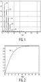
- FIG. 1 is a spectrum of a banded Doppler signal having a fundamental frequency component 10 at 1.64 Hz.
- the non-sinusoidal nature of the pulsatile activity gives the spectrum several harmonics of the fundamental. In this example higher harmonics 12, 14, 16, and 18 can be seen ranging up to about 8 Hz.
- the power within a small band around each fundamental and harmonic peak is compared to the total power in the banded Doppler signal in the assessment of pulsatility.
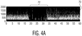
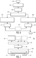
- FIG. 3 A flowchart illustrating a method of the present invention is shown in FIG. 3 .
- the banded Doppler signal within a certain frequency band e.g., 1-1.2 kHz
- a median filter is applied to the banded Doppler signal to improve assessment under noisy conditions, as supported by empirical studies.
- a non-linear filter like a median filter is desirable because of its ability to preserve the signal structure compared to a linear filter.
- the frequency spectrum of the filtered banded Doppler signal is obtained in step 24, which in this example is taken over a span of 5-second windows that are progressively marched in time in 1-second intervals.
- the calculation of the frequency spectrum can be done by taking the Fourier transform of the auto-correlation of the banded Doppler signal, or directly by any means commonly employed for power spectrum estimation (e . g ., averaged periodogram, etc.)
- the fundamental peak frequency (fpeak) in the spectrum is located through peak searching (using second derivative tests) within a frequency span that is physiologically meaningful such as 0.5 to 4 Hz. If no peak is located, then the method concludes at 28 with a determination of the absence of pulsatile flow and no further processing is done.
- a peak-searching algorithm is conducted in step 32 in regions around the locations where harmonics are expected (2fpeak, 3fpeak, etc.) in order to determine if harmonics are present. Typically up to four harmonics may be searched, although a greater or lesser number of harmonic regions may be chosen in a particular implementation or for a particular patient signal.
- a normalized power ratio is computed in step 42 as the ratio of the power within a small bandwidth around the peak to the total power in the banded Doppler signal.
- Each peak is separately processed in the method of FIG. 3 by initializing the peak count to one and setting the pulsatile index to zero in step 36.
- the normalized power ratios are expected to be high, with the highest value usually for the fundamental frequency 10, and progressively decreasing for each harmonic 12-18. If the sum total of these power ratios is taken to be the pulsation index, a normalized sum will have a value close to unity for the pulsatile flow case. However, when pulsatile flow is absent, then these power ratios (if peaks existed for the fundamental and harmonics) are simply based on noise. Summing these values would inadvertently raise the pulsation index leading to false positives in the pulse assessment.
- a noise level in the banded Doppler signal is determined at step 34. If periodic flow is present, then the spectrum can only have peaks at fundamental and harmonic locations, and regions in the spectrum between these peaks would constitute noise. Hence, once fundamental and harmonic locations 10-18 are determined, the method also looks at regions in between these peaks in step 34 and calculates a mean noise level. Alternatively, the noise level could be computed by looking at frequencies that are sufficiently high where no effects of pulsatile activity are expected.
- each peak in the spectrum is truly significant (the flow case)
- the SNR defined as the ratio of the power around the peak to the noise level
- the SNR would be high.
- the SNR would be low.
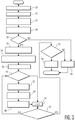
- a suitable weighting function is applied in step 44 to the normalized power ratio contributed by each peak frequency (step 42) as illustrated by the sigmoid-type weighting function 60 shown in FIG 2 .
- the weighting function 60 suppresses contributions from the peaks if the SNR is low while still maintaining contributions from true peaks that have a high SNR.
- the pulsation index is incremented by this weighted value in step 46. This is done for each of the peaks by incrementing the peak count in step 48, comparing the count to N, the number of harmonics used in the computation, in step 50, and repeating the computation for each peak. When all of the peaks have been included in the pulsation index the index is compared to a threshold in step 52. If the pulsation index is high enough there is pulsatile flow (54) and if not, then it is concluded that pulsatile flow is absent (28).
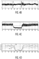
- FIGS. 4a-4d compare the performance of an embodiment of the present invention with that of the technique of the parent application.
- FIG. 4a illustrates a Doppler spectrogram 70 of for a subject experiencing ventricular fibrillation during the interval 72. This event is also reflected in the ECG waveform of FIG. 4b and in the blood pressure graph of FIG. 4c.
- FIG. 4d shows a pulsation index 74 determined in accordance with the sinusoidal-based technique of the parent application, and a pulsation index 76 determined in accordance with a non-sinusoidal-based technique of the present invention.
- a comparison of the lines 74 and 76 show that the pulsation index 76 is higher during the pulsatile blood flow before and after the VF interval 72, and remains low during the non-pulsatile flow condition of interval 72.
- FIG. 5 depicts a block diagram of an exemplary apparatus 100 of the kind that may be used for ultrasound diagnostics in accordance with one embodiment of the present invention.
- the apparatus 100 can perform assessment (e.g., detection and/or measurements) of perfusion and/or the pulse state of a patient.
- perfusion refers to blood flow in a blood vessel (e.g., carotid artery) or a tissue.
- the apparatus 100 may be used as a component in resuscitation systems and defibrillators, monitors and detectors of weak heart beat (e.g., fetal heart beat), among other medical diagnostic and clinical systems.
- the apparatus 100 may also be used in non-medical systems for measuring, for example, flow or pulsatile activity of colloidal and emulsion solutions.
- the apparatus 100 comprises a generator 102, at least one ultrasonic transducer 104 (one transducer 104 is shown), and a data processor 110.
- the transducers 104 together, form an array that typically is disposed upon an application pad (not shown), and the transducers may additionally be time multiplexed.
- Such arrays are disclosed, for example, in US patent 6,565,914 to Rock et al.
- the transducer 104 comprises a transmitter 106 and a receiver 108.
- the generator 102 is generally a source of a continuous wave (CW) radio frequency (RF) signal (e.g., 1-10 MHz).
- CW continuous wave
- RF radio frequency
- the generator 102 via interface 134 activates (or excites) the transmitter 106 to emit ultrasound (illustratively shown as a beam 132) propagating in a portion 124 of the body of a patient located beneath the transducer.
- the receiver 108 collects, within an aperture 130, an acoustic echo signal (i.e., scattered ultrasound), transforms the echo signal into an electrical signal and transmits, via interface 136, to the data processor 110.
- the transmitter 106 and receiver 108 are positioned such that the beam 132 and aperture 130 overlap in a region 128 of a large blood vessel 126, such as a carotid artery, and the like.
- the apparatus 100 may comprise the transducer 104 capable of operating as a transmitter when RF power is ON, or a receiver when the RF power is OFF, respectively.
- the generator 102 produces pulsed RF power (PW) having duration of an ON time interval of about 0.2 to 20 microseconds and a duty cycle in a range of about 0.2 to 20%.
- PW pulsed RF power
- the data processor 110 comprises a signal acquisition module 112, a frequency band discriminator 114, and a signal analyzer 118 including a processing module 120 which performs the non-sinusoidal pulsatility calculations described above, a perfusion detector 122, and a pulse state detector 123.
- Components of the data processor 110 may be reduced to practice in a form of electronic hardware, a computer program (i.e., software), or both. Alternatively, portions of signal processing performed by the module 110 may also be accomplished using a remote processor (not shown).
- the analysis may be performed in the analog, rather than the digital, domain, e.g., frequency band discriminator 114 could be replaced with an analog filter bank, data processor 110 could comprise a correlator, etc., as would be known to one of ordinary skill in the art.
- the signal acquisition module 112 acquires the echo signal and defines a Doppler signal.
- Doppler signal relates to a signal that is proportional to a frequency shift between the incident ultrasound and the echo signal.
- the module 112 includes frequency converters of the echo signal, analog and digital filters, memory devices, computer processors, and other means conventionally used for data acquisition and digital signal processing.
- One filter may be a high frequency pass filter that suppresses the echo originated in the region 128 by stationary or slowly moving objects, such as tissues, walls of the blood vessel 126, the like.
- the module 112 stores in a memory 113 in a digital format the Doppler signal that has been acquired during at least one time interval ⁇ T 1 having duration of about 2 to 20 sec (preferably 5-10 sec).
- the stored digitized Doppler signal may be provided for further processing to the frequency band discriminator 114 in a form of consecutive data banks each relating to a time segment ⁇ T 2 having duration of about 10 to 100 msec (e.g., 40 msec).
- the frequency band discriminator 114 comprises a plurality (e.g., 4 to 10) of band pass filters 115 (six filters 115 are shown), which selectively decompose the Doppler signal in a plurality of sampling signals 140.
- Each sampling signal 140 has a frequency range that represents a portion of a pre-selected frequency range of the Doppler signal, wherein such ranges do not overlap.
- the terms "frequency range” and "frequency band” are used interchangeably. Together, frequency ranges of the sampling signals 140 comprise the frequency range of the decomposed Doppler signal or a portion of it.
- the band pass filters are selectively calibrated to have the same coefficient of amplification that may be either greater or smaller than 1.
- the sampling signals 140 preserve instant spectral power distribution of the Doppler signal as provided by the signal acquisition module 112 and, therefore, power of each sampling signal is proportional to the power of the Doppler signal in the frequency range of the respective sampling signal 140.
- an output of each band pass filter 115 is illustratively coupled to a respective input of the power metering unit 116. In an alternate embodiment (not shown), such outputs may be multiplexed (e.g., time multiplexed) and be coupled to the power metering unit 116 using a single transmission line.
- the power metering unit 116 selectively calculates the power of each of the sampling signals 140 and outputs to the processing module 120 a plurality of signals 142 each representing the power of the respective sampling signal as averaged for duration of the time segment ⁇ T 2 .
- the signals 142 may also be multiplexed (e.g., time multiplexed) and coupled to the processing module 120 using a single transmission line.
- the processing module 120 selectively computes a measure of periodicity of the Doppler signal selectively in each frequency band of the signal using, e.g., a ratio of the power of the Doppler signal to baseline noise.
- a peak value of the ratio and the data identifying the frequency band having such a ratio are transmitted to the perfusion detector 122.
- the computed peak ratio is compared with pre-determined settings to assess a velocity of the blood flow in the examined blood vessel (e.g., carotid artery).
- Data relating to a specific pattern of the spectral power distribution of the Doppler signal may also carry additional diagnostic information regarding mechanical activity of the patient's heart and, as such, be preserved, e.g., in a memory of the signal analyzer 118 or, alternatively, data processor 110.
- the processing module 120 selectively computes a measure of periodicity of the Doppler signal selectively in each fundamental and harmonic frequency band as described above.
- the aforementioned noise analysis is performed, and the contribution of each fundamental and harmonic signal is included in the pulsatility index.
- the pulsatility index so determined is validated against a threshold and presented to the user.
- One computational technique includes auto-correlation analysis of the power of the Doppler signal over a pre-determined time interval to determine if an auto-correlation function has periodically spaced peaks identifying a pulsatile activity of the heart. Results of the auto-correlation analysis are transmitted to the pulse state detector 123.
- the intensity of blood pulsing may be assessed using, for example, a pulsation index as previously described and like measures of the periodicity.
- the computed value of the selected measure of periodicity may be compared with other pre-determined settings and/or thresholds to define and assess the state of the pulse in the blood vessel 126.
- the processing module 120 collects output signals 142 during a period of time that encompasses several cardiac cycles.
- the processing module 120 may acquire the signals 142, in a form of blocks of data each relating to the segment ⁇ T 2 , for duration of the time interval ⁇ T 1 extending over several cardiac cycles and selectively process each such a block of data.
- the processing module 120 may utilize computational techniques known to those skilled in the art, such as algebraic and Boolean logic operations, spectral analysis, Fourier analysis (e.g., Fast Fourier transform (FFT) analysis), correlation analysis, and other signal processing techniques.
- FFT Fast Fourier transform
- FIG. 6 depicts a flow diagram of one exemplary embodiment of the inventive method for ultrasound diagnostics. The method may be reduced to practice, e.g., using the apparatus of FIG. 5 for performing an illustrative procedure of detecting blood perfusion and/or the pulse state of a patient.
- the method starts at step 601 and proceeds to step 602.
- at least one ultrasonic transducer 104 is activated to emit ultrasound towards the blood vessel 126 (e.g., carotid artery) and collect the echo signal scattered in the region 128 of the body of a patient.
- the ultrasonic echo signal is converted to the electrical format and transmitted to the data processor 110.
- the echo signal is acquired for duration of the time interval ⁇ T 1 , digitized, and stored in a memory, as discussed above in reference to FIG. 5 .
- the time interval ⁇ T 1 typically encompasses several (e.g., 3-6) cardiac cycles. Alternatively, the time interval ⁇ T 1 may have a pre-determined duration.
- spectral power distribution of the Doppler signal is defined in a plurality of discrete frequency bands and averaged within time segments ⁇ T 2 of the time interval ⁇ T 1 .
- a frequency band having, during a cardiac cycle, maximal periodic variations of the Doppler power is defined and, at step 610, the pulse state of the patient is calculated, as discussed in detail in reference to FIG. 3 .
- a frequency band having, during a cardiac cycle, a peak ratio of the Doppler power to baseline noise is defined and, at step 614, the perfusion is calculated as discussed above in reference to FIG. 5 .
- step 616 data collected using simultaneously operating electrocardiograph (ECG system) may be used when, e.g., the method is reduced to practice in a defibrillating system, as discussed in reference to FIG. 7 below.
- ECG system electrocardiograph
- steps 608, 610, 612, 614, and 616 may be performed substantially simultaneously.
- the method proceeds to step 618 where the method ends.
- FIG. 7 depicts a block diagram of an exemplary programmable defibrillating system 700 in accordance with one embodiment of the present invention.
- the defibrillating system 700 comprises the ultrasound diagnostic apparatus 100 of FIG. 5 , an optional ECG system 702, an optional blood pressure monitor 703, an analyzer 704 of diagnostic information, a defibrillating unit 708, and a programmable controller 706 of the defibrillating unit.
- the apparatus 100 provides to the analyzer 704 diagnostic information relating to the mechanical activity of the heart and including at least one of the perfusion and the pulse state of a patient (e.g., the pulsation index PI).
- Ultrasonic diagnostic information may be obtained using the measurements performed on the patient's carotid artery. Such information may additionally be used in diagnosing, in real time, the state of blood supply to the brain of the patient.
- the ECG system 702 and the apparatus 100 acquire the diagnostic data simultaneously.
- the signal related to the spectral distribution of the power of the Doppler signal may further be cross-correlated with an ECG signal. Such correlation may further increase accuracy and reliability of interpreting the diagnostic information by the analyzer 704.
- each of the signals 142 may be coupled to the analyzer 704 where the signals 142 are selectively cross-correlated with the ECG signal to provide most accurate assessment of the perfusion, whereas the ABP monitor may be used as a source of data characterizing an overall state of mechanical activity of the heart.
- the analyzer 704 may use only the diagnostic information provided by the apparatus 100.
- the ECG signal corresponds to the electrical activity of the heart.
- Exclusive use of the ECG diagnostics in the system 700 may result in masking the lack of the mechanical activity (i.e., blood pumping functionality) of the patient's heart by the pulseless electrical activity (PEA) of the heart and, as such, cause erroneous clinical decisions.
- PDA pulseless electrical activity
- the analyzer 704 performs analysis of collected information to determine whether to defibrillate the patient and define parameters of a defibrillation procedure. In operation, the analyzer 704 outputs the results of the analysis to the programmable controller 706 that configures the defibrillating unit 708 comprising a controlled source 710 of high voltage and application electrodes 712 (two electrodes 712 are shown) for executing the procedure.
- apparatus 100 and system 700 are available in medical ultrasound and defibrillation systems and application specific integrated circuits (ASICs) available from Koninklijke Philips Electronics N.V. of Eindhoven, Netherlands.
- ASICs application specific integrated circuits
Landscapes
- Health & Medical Sciences (AREA)
- Life Sciences & Earth Sciences (AREA)
- Engineering & Computer Science (AREA)
- Biomedical Technology (AREA)
- Veterinary Medicine (AREA)
- Public Health (AREA)
- General Health & Medical Sciences (AREA)
- Animal Behavior & Ethology (AREA)
- Heart & Thoracic Surgery (AREA)
- Molecular Biology (AREA)
- Medical Informatics (AREA)
- Pathology (AREA)
- Surgery (AREA)
- Biophysics (AREA)
- Physics & Mathematics (AREA)
- Radiology & Medical Imaging (AREA)
- Nuclear Medicine, Radiotherapy & Molecular Imaging (AREA)
- Cardiology (AREA)
- Signal Processing (AREA)
- Computer Vision & Pattern Recognition (AREA)
- Hematology (AREA)
- Artificial Intelligence (AREA)
- Physiology (AREA)
- Psychiatry (AREA)
- Ultra Sonic Daignosis Equipment (AREA)
- Measuring Pulse, Heart Rate, Blood Pressure Or Blood Flow (AREA)
- Electrotherapy Devices (AREA)
Applications Claiming Priority (2)
| Application Number | Priority Date | Filing Date | Title |
|---|---|---|---|
| US76483806P | 2006-02-03 | 2006-02-03 | |
| PCT/IB2007/050296 WO2007088508A2 (en) | 2006-02-03 | 2007-01-29 | Ultrasonic method and apparatus for measuring or detecting flow behavior of a non-sinusoidal periodicity |
Publications (2)
| Publication Number | Publication Date |
|---|---|
| EP1983898A2 EP1983898A2 (en) | 2008-10-29 |
| EP1983898B1 true EP1983898B1 (en) | 2017-01-18 |
Family
ID=38327766
Family Applications (1)
| Application Number | Title | Priority Date | Filing Date |
|---|---|---|---|
| EP07705730.5A Active EP1983898B1 (en) | 2006-02-03 | 2007-01-29 | Ultrasonic method and apparatus for measuring or detecting flow behavior of a non-sinusoidal periodicity |
Country Status (6)
| Country | Link |
|---|---|
| US (2) | US20090018449A1 (enExample) |
| EP (1) | EP1983898B1 (enExample) |
| JP (1) | JP5534674B2 (enExample) |
| KR (1) | KR20080094014A (enExample) |
| CN (1) | CN101378699B (enExample) |
| WO (1) | WO2007088508A2 (enExample) |
Families Citing this family (7)
| Publication number | Priority date | Publication date | Assignee | Title |
|---|---|---|---|---|
| CN101167656B (zh) * | 2006-10-23 | 2012-10-03 | 深圳迈瑞生物医疗电子股份有限公司 | 组织多普勒成像方法和装置 |
| JP5944916B2 (ja) * | 2010-11-11 | 2016-07-05 | ゾール メディカル コーポレイションZOLL Medical Corporation | 急性期介護処置システムの計器盤 |
| JP6114757B2 (ja) * | 2011-12-16 | 2017-04-12 | コーニンクレッカ フィリップス エヌ ヴェKoninklijke Philips N.V. | 処理装置及び記憶媒体 |
| WO2013155537A1 (en) * | 2012-04-13 | 2013-10-17 | Tessonics Corp. | A method to obtain 3d images of a flowing region beneath an object using speckle reflections |
| US11471063B2 (en) | 2016-11-10 | 2022-10-18 | Auburn University | Information processing method, device, and system for evaluating blood vessels |
| EP3537962A4 (en) | 2016-11-10 | 2019-11-27 | Auburn University | METHOD AND SYSTEM FOR ASSESSING BLOOD VESSELS |
| US11331078B2 (en) * | 2019-03-20 | 2022-05-17 | Siemens Medical Solutions Usa, Inc. | Quantitative ultrasound using fundamental and harmonic signals |
Family Cites Families (36)
| Publication number | Priority date | Publication date | Assignee | Title |
|---|---|---|---|---|
| US4513249A (en) * | 1979-04-23 | 1985-04-23 | Baghdady Elie J | Method and apparatus for signal detection, separation and suppression |
| JP3040466B2 (ja) * | 1990-07-17 | 2000-05-15 | ブリテイッシュ・テレコミュニケーションズ・パブリック・リミテッド・カンパニー | 画像処理方法 |
| US6185457B1 (en) * | 1994-05-31 | 2001-02-06 | Galvani, Ltd. | Method and apparatus for electrically forcing cardiac output in an arrhythmia patient |
| US5741980A (en) * | 1994-11-02 | 1998-04-21 | Foster-Miller, Inc. | Flow analysis system and method |
| US6122482A (en) * | 1995-02-22 | 2000-09-19 | Global Communications, Inc. | Satellite broadcast receiving and distribution system |
| EP0816986B1 (en) * | 1996-07-03 | 2006-09-06 | Hitachi, Ltd. | System for recognizing motions |
| JP3857788B2 (ja) * | 1997-09-01 | 2006-12-13 | テルモ株式会社 | 循環器情報計測システム |
| GB9726664D0 (en) * | 1997-12-17 | 1998-02-18 | Nycomed Imaging As | Improvements in or relating to ultrasonography |
| DE59914044D1 (de) * | 1998-02-26 | 2007-01-25 | Alfred Schiller | Vorrichtung zum Erzeugen eines Hilfssignals zum Bestimmen des Zeitpunkts einer Herzdefibrillation |
| JPH11287853A (ja) * | 1998-04-03 | 1999-10-19 | Denso Corp | レーダ装置 |
| EP1082050B1 (en) * | 1998-06-03 | 2011-08-24 | Masimo Corporation | Stereo pulse oximeter |
| US6080107A (en) * | 1999-01-26 | 2000-06-27 | Hewlett-Packard Company | Methods for the use of contrast agents in ultrasonic imaging |
| US6213951B1 (en) * | 1999-02-19 | 2001-04-10 | Acuson Corporation | Medical diagnostic ultrasound method and system for contrast specific frequency imaging |
| US6213947B1 (en) * | 1999-03-31 | 2001-04-10 | Acuson Corporation | Medical diagnostic ultrasonic imaging system using coded transmit pulses |
| US6179781B1 (en) * | 1999-03-31 | 2001-01-30 | Acuson Corporation | Medical diagnostic ultrasound method and apparatus for improving doppler processing |
| US6440082B1 (en) * | 1999-09-30 | 2002-08-27 | Medtronic Physio-Control Manufacturing Corp. | Method and apparatus for using heart sounds to determine the presence of a pulse |
| US6970742B2 (en) * | 2000-01-11 | 2005-11-29 | Savacor, Inc. | Method for detecting, diagnosing, and treating cardiovascular disease |
| US6461303B2 (en) * | 2000-01-19 | 2002-10-08 | Bjorn Angelsen | Method of detecting ultrasound contrast agent in soft tissue, and quantitating blood perfusion through regions of tissue |
| US6375618B1 (en) * | 2000-01-31 | 2002-04-23 | General Electric Company | Enhanced tissue-generated harmonic imaging using coded excitation |
| DE10008886A1 (de) | 2000-02-25 | 2001-09-13 | Ulrich Kreutzer | Defibrillator |
| DE20003455U1 (de) | 2000-02-25 | 2000-05-25 | Kreutzer, Ulrich, Dr.med., 03048 Cottbus | Defibrillator |
| JP4567842B2 (ja) * | 2000-04-10 | 2010-10-20 | 株式会社東芝 | 超音波診断装置 |
| US6398733B1 (en) * | 2000-04-24 | 2002-06-04 | Acuson Corporation | Medical ultrasonic imaging system with adaptive multi-dimensional back-end mapping |
| JP2002017695A (ja) * | 2000-07-03 | 2002-01-22 | Seiko Instruments Inc | 脈波検出装置 |
| US6623435B2 (en) * | 2000-07-03 | 2003-09-23 | Seiko Instruments Inc. | Pulse wave detecting apparatus |
| US6626836B2 (en) * | 2001-04-04 | 2003-09-30 | Siemens Medical Solutions Usa, Inc. | Adaptive signal processing scheme for contrast agent imaging |
| US6575914B2 (en) * | 2001-05-18 | 2003-06-10 | Koninklijke Philips Electronics N.V. | Integrated cardiac resuscitation system with ability to detect perfusion |
| US6565914B2 (en) | 2001-07-11 | 2003-05-20 | The Goodyear Tire & Rubber Company | Method for controlling deposited polymer on a substrate |
| US6679847B1 (en) * | 2002-04-30 | 2004-01-20 | Koninklijke Philips Electronics N.V. | Synthetically focused ultrasonic diagnostic imaging system for tissue and flow imaging |
| US6632177B1 (en) * | 2002-05-01 | 2003-10-14 | Acuson Corporation | Dual process ultrasound contrast agent imaging |
| US20040116969A1 (en) * | 2002-08-26 | 2004-06-17 | Owen James M. | Pulse detection using patient physiological signals |
| US6966878B2 (en) * | 2003-08-28 | 2005-11-22 | Ge Medical Systems Global Technology Company, Llc | Method and apparatus for obtaining a volumetric scan of a periodically moving object |
| US8469887B2 (en) * | 2003-12-19 | 2013-06-25 | General Electric Company | Method and apparatus for flow parameter imaging |
| CN100581483C (zh) * | 2004-09-28 | 2010-01-20 | 皇家飞利浦电子股份有限公司 | 用于呈现关于由超声外部测量的体液的流动特性的信息的方法和装置 |
| US8731658B2 (en) * | 2005-01-31 | 2014-05-20 | Physio-Control, Inc | System and method for using diagnostic pulses in connection with defibrillation therapy |
| US20090306525A1 (en) * | 2005-07-15 | 2009-12-10 | Koninklijke Philips Electronics N.V. | Apparatus and Method for Defibrillation Pulse Detection Using Electromagnetic Waves |
-
2007
- 2007-01-29 KR KR1020087018648A patent/KR20080094014A/ko not_active Withdrawn
- 2007-01-29 CN CN200780004132XA patent/CN101378699B/zh active Active
- 2007-01-29 US US12/161,983 patent/US20090018449A1/en not_active Abandoned
- 2007-01-29 JP JP2008552934A patent/JP5534674B2/ja active Active
- 2007-01-29 WO PCT/IB2007/050296 patent/WO2007088508A2/en not_active Ceased
- 2007-01-29 EP EP07705730.5A patent/EP1983898B1/en active Active
-
2015
- 2015-06-08 US US14/733,093 patent/US9955942B2/en active Active
Non-Patent Citations (1)
| Title |
|---|
| None * |
Also Published As
| Publication number | Publication date |
|---|---|
| US20090018449A1 (en) | 2009-01-15 |
| JP5534674B2 (ja) | 2014-07-02 |
| WO2007088508A3 (en) | 2008-03-06 |
| CN101378699B (zh) | 2012-06-20 |
| WO2007088508A2 (en) | 2007-08-09 |
| US9955942B2 (en) | 2018-05-01 |
| US20150265240A1 (en) | 2015-09-24 |
| CN101378699A (zh) | 2009-03-04 |
| JP2009525117A (ja) | 2009-07-09 |
| EP1983898A2 (en) | 2008-10-29 |
| KR20080094014A (ko) | 2008-10-22 |
Similar Documents
| Publication | Publication Date | Title |
|---|---|---|
| US9955942B2 (en) | Ultrasonic method and apparatus for measuring or detecting flow behavior of a non-sinusoidal periodicity | |
| US20080015439A1 (en) | Method and Apparatus for Measuring and/or Detecting Flow Behavior of a Body Fluid Using Ultrasound | |
| RU2571333C2 (ru) | Система, стетоскоп и способ для индикации риска ишемической болезни сердца | |
| EP3675717B1 (en) | Multisensor cardiac function monitoring and analytics systems | |
| US20080077010A1 (en) | Method and Aparatus for Presenting Information Concerning Flow Behavior of a Bodyfluid Externally Measured by Ultrasound | |
| US8380305B2 (en) | System and method for predicting successful defibrillation for ventricular fibrillation cardiac arrest | |
| JP2009525117A5 (enExample) | ||
| Alfred et al. | An automated carotid pulse assessment approach using Doppler ultrasound | |
| EP1480561A1 (en) | Ultrasonic imaging to detect coronary artery stenosis at rest | |
| EP4649892A1 (en) | Method for hemodynamic monitoring | |
| Gorodetska et al. | Differentiation of Heart Rhythms from ECG by Joint Use of Quantile Peak Localization and Windowed Cyclostationarity Analysis | |
| JP2003190109A (ja) | 自律神経系機能評価方法およびそのシステム | |
| JP2025180796A (ja) | 心拍数特定装置、心拍数特定方法及びプログラム | |
| JP2025092800A (ja) | 携帯用心電図測定装置の異常波形抽出方法 | |
| Raju et al. | A novel ultrasound based automated pulsatile flow detection method for resuscitation | |
| Hantono | Blood-pressure measurement | |
| HK40023935B (en) | Multisensor cardiac function monitoring and analytics systems | |
| HK40023935A (en) | Multisensor cardiac function monitoring and analytics systems |
Legal Events
| Date | Code | Title | Description |
|---|---|---|---|
| PUAI | Public reference made under article 153(3) epc to a published international application that has entered the european phase |
Free format text: ORIGINAL CODE: 0009012 |
|
| 17P | Request for examination filed |
Effective date: 20080908 |
|
| AK | Designated contracting states |
Kind code of ref document: A2 Designated state(s): AT BE BG CH CY CZ DE DK EE ES FI FR GB GR HU IE IS IT LI LT LU LV MC NL PL PT RO SE SI SK TR |
|
| DAX | Request for extension of the european patent (deleted) | ||
| RAP1 | Party data changed (applicant data changed or rights of an application transferred) |
Owner name: KONINKLIJKE PHILIPS N.V. |
|
| 17Q | First examination report despatched |
Effective date: 20150617 |
|
| REG | Reference to a national code |
Ref country code: DE Ref legal event code: R079 Ref document number: 602007049566 Country of ref document: DE Free format text: PREVIOUS MAIN CLASS: A61B0008060000 Ipc: A61B0005000000 |
|
| GRAP | Despatch of communication of intention to grant a patent |
Free format text: ORIGINAL CODE: EPIDOSNIGR1 |
|
| RIC1 | Information provided on ipc code assigned before grant |
Ipc: A61B 5/0402 20060101ALI20160704BHEP Ipc: A61B 5/00 20060101AFI20160704BHEP Ipc: A61N 1/39 20060101ALI20160704BHEP Ipc: A61B 8/08 20060101ALI20160704BHEP Ipc: A61B 5/021 20060101ALI20160704BHEP Ipc: A61B 8/00 20060101ALI20160704BHEP Ipc: A61B 8/06 20060101ALI20160704BHEP |
|
| INTG | Intention to grant announced |
Effective date: 20160805 |
|
| GRAS | Grant fee paid |
Free format text: ORIGINAL CODE: EPIDOSNIGR3 |
|
| GRAA | (expected) grant |
Free format text: ORIGINAL CODE: 0009210 |
|
| AK | Designated contracting states |
Kind code of ref document: B1 Designated state(s): AT BE BG CH CY CZ DE DK EE ES FI FR GB GR HU IE IS IT LI LT LU LV MC NL PL PT RO SE SI SK TR |
|
| REG | Reference to a national code |
Ref country code: GB Ref legal event code: FG4D |
|
| REG | Reference to a national code |
Ref country code: FR Ref legal event code: PLFP Year of fee payment: 11 |
|
| REG | Reference to a national code |
Ref country code: CH Ref legal event code: EP |
|
| REG | Reference to a national code |
Ref country code: AT Ref legal event code: REF Ref document number: 862400 Country of ref document: AT Kind code of ref document: T Effective date: 20170215 |
|
| REG | Reference to a national code |
Ref country code: IE Ref legal event code: FG4D |
|
| REG | Reference to a national code |
Ref country code: DE Ref legal event code: R096 Ref document number: 602007049566 Country of ref document: DE |
|
| REG | Reference to a national code |
Ref country code: NL Ref legal event code: MP Effective date: 20170118 |
|
| REG | Reference to a national code |
Ref country code: LT Ref legal event code: MG4D |
|
| PG25 | Lapsed in a contracting state [announced via postgrant information from national office to epo] |
Ref country code: BE Free format text: LAPSE BECAUSE OF NON-PAYMENT OF DUE FEES Effective date: 20170131 |
|
| REG | Reference to a national code |
Ref country code: AT Ref legal event code: MK05 Ref document number: 862400 Country of ref document: AT Kind code of ref document: T Effective date: 20170118 |
|
| PG25 | Lapsed in a contracting state [announced via postgrant information from national office to epo] |
Ref country code: NL Free format text: LAPSE BECAUSE OF FAILURE TO SUBMIT A TRANSLATION OF THE DESCRIPTION OR TO PAY THE FEE WITHIN THE PRESCRIBED TIME-LIMIT Effective date: 20170118 |
|
| PG25 | Lapsed in a contracting state [announced via postgrant information from national office to epo] |
Ref country code: GR Free format text: LAPSE BECAUSE OF FAILURE TO SUBMIT A TRANSLATION OF THE DESCRIPTION OR TO PAY THE FEE WITHIN THE PRESCRIBED TIME-LIMIT Effective date: 20170419 Ref country code: IS Free format text: LAPSE BECAUSE OF FAILURE TO SUBMIT A TRANSLATION OF THE DESCRIPTION OR TO PAY THE FEE WITHIN THE PRESCRIBED TIME-LIMIT Effective date: 20170518 Ref country code: FI Free format text: LAPSE BECAUSE OF FAILURE TO SUBMIT A TRANSLATION OF THE DESCRIPTION OR TO PAY THE FEE WITHIN THE PRESCRIBED TIME-LIMIT Effective date: 20170118 Ref country code: LT Free format text: LAPSE BECAUSE OF FAILURE TO SUBMIT A TRANSLATION OF THE DESCRIPTION OR TO PAY THE FEE WITHIN THE PRESCRIBED TIME-LIMIT Effective date: 20170118 |
|
| PG25 | Lapsed in a contracting state [announced via postgrant information from national office to epo] |
Ref country code: PT Free format text: LAPSE BECAUSE OF FAILURE TO SUBMIT A TRANSLATION OF THE DESCRIPTION OR TO PAY THE FEE WITHIN THE PRESCRIBED TIME-LIMIT Effective date: 20170518 Ref country code: AT Free format text: LAPSE BECAUSE OF FAILURE TO SUBMIT A TRANSLATION OF THE DESCRIPTION OR TO PAY THE FEE WITHIN THE PRESCRIBED TIME-LIMIT Effective date: 20170118 Ref country code: SE Free format text: LAPSE BECAUSE OF FAILURE TO SUBMIT A TRANSLATION OF THE DESCRIPTION OR TO PAY THE FEE WITHIN THE PRESCRIBED TIME-LIMIT Effective date: 20170118 Ref country code: ES Free format text: LAPSE BECAUSE OF FAILURE TO SUBMIT A TRANSLATION OF THE DESCRIPTION OR TO PAY THE FEE WITHIN THE PRESCRIBED TIME-LIMIT Effective date: 20170118 Ref country code: PL Free format text: LAPSE BECAUSE OF FAILURE TO SUBMIT A TRANSLATION OF THE DESCRIPTION OR TO PAY THE FEE WITHIN THE PRESCRIBED TIME-LIMIT Effective date: 20170118 Ref country code: LV Free format text: LAPSE BECAUSE OF FAILURE TO SUBMIT A TRANSLATION OF THE DESCRIPTION OR TO PAY THE FEE WITHIN THE PRESCRIBED TIME-LIMIT Effective date: 20170118 Ref country code: BG Free format text: LAPSE BECAUSE OF FAILURE TO SUBMIT A TRANSLATION OF THE DESCRIPTION OR TO PAY THE FEE WITHIN THE PRESCRIBED TIME-LIMIT Effective date: 20170418 |
|
| REG | Reference to a national code |
Ref country code: CH Ref legal event code: PL |
|
| REG | Reference to a national code |
Ref country code: DE Ref legal event code: R097 Ref document number: 602007049566 Country of ref document: DE |
|
| PG25 | Lapsed in a contracting state [announced via postgrant information from national office to epo] |
Ref country code: RO Free format text: LAPSE BECAUSE OF FAILURE TO SUBMIT A TRANSLATION OF THE DESCRIPTION OR TO PAY THE FEE WITHIN THE PRESCRIBED TIME-LIMIT Effective date: 20170118 Ref country code: LI Free format text: LAPSE BECAUSE OF NON-PAYMENT OF DUE FEES Effective date: 20170131 Ref country code: CZ Free format text: LAPSE BECAUSE OF FAILURE TO SUBMIT A TRANSLATION OF THE DESCRIPTION OR TO PAY THE FEE WITHIN THE PRESCRIBED TIME-LIMIT Effective date: 20170118 Ref country code: CH Free format text: LAPSE BECAUSE OF NON-PAYMENT OF DUE FEES Effective date: 20170131 Ref country code: IT Free format text: LAPSE BECAUSE OF FAILURE TO SUBMIT A TRANSLATION OF THE DESCRIPTION OR TO PAY THE FEE WITHIN THE PRESCRIBED TIME-LIMIT Effective date: 20170118 Ref country code: EE Free format text: LAPSE BECAUSE OF FAILURE TO SUBMIT A TRANSLATION OF THE DESCRIPTION OR TO PAY THE FEE WITHIN THE PRESCRIBED TIME-LIMIT Effective date: 20170118 Ref country code: SK Free format text: LAPSE BECAUSE OF FAILURE TO SUBMIT A TRANSLATION OF THE DESCRIPTION OR TO PAY THE FEE WITHIN THE PRESCRIBED TIME-LIMIT Effective date: 20170118 |
|
| REG | Reference to a national code |
Ref country code: IE Ref legal event code: MM4A |
|
| PLBE | No opposition filed within time limit |
Free format text: ORIGINAL CODE: 0009261 |
|
| STAA | Information on the status of an ep patent application or granted ep patent |
Free format text: STATUS: NO OPPOSITION FILED WITHIN TIME LIMIT |
|
| PG25 | Lapsed in a contracting state [announced via postgrant information from national office to epo] |
Ref country code: MC Free format text: LAPSE BECAUSE OF FAILURE TO SUBMIT A TRANSLATION OF THE DESCRIPTION OR TO PAY THE FEE WITHIN THE PRESCRIBED TIME-LIMIT Effective date: 20170118 Ref country code: LU Free format text: LAPSE BECAUSE OF NON-PAYMENT OF DUE FEES Effective date: 20170129 Ref country code: DK Free format text: LAPSE BECAUSE OF FAILURE TO SUBMIT A TRANSLATION OF THE DESCRIPTION OR TO PAY THE FEE WITHIN THE PRESCRIBED TIME-LIMIT Effective date: 20170118 |
|
| 26N | No opposition filed |
Effective date: 20171019 |
|
| REG | Reference to a national code |
Ref country code: FR Ref legal event code: PLFP Year of fee payment: 12 |
|
| REG | Reference to a national code |
Ref country code: BE Ref legal event code: MM Effective date: 20170131 |
|
| PG25 | Lapsed in a contracting state [announced via postgrant information from national office to epo] |
Ref country code: SI Free format text: LAPSE BECAUSE OF FAILURE TO SUBMIT A TRANSLATION OF THE DESCRIPTION OR TO PAY THE FEE WITHIN THE PRESCRIBED TIME-LIMIT Effective date: 20170118 Ref country code: IE Free format text: LAPSE BECAUSE OF NON-PAYMENT OF DUE FEES Effective date: 20170129 |
|
| PG25 | Lapsed in a contracting state [announced via postgrant information from national office to epo] |
Ref country code: HU Free format text: LAPSE BECAUSE OF FAILURE TO SUBMIT A TRANSLATION OF THE DESCRIPTION OR TO PAY THE FEE WITHIN THE PRESCRIBED TIME-LIMIT; INVALID AB INITIO Effective date: 20070129 |
|
| PG25 | Lapsed in a contracting state [announced via postgrant information from national office to epo] |
Ref country code: CY Free format text: LAPSE BECAUSE OF NON-PAYMENT OF DUE FEES Effective date: 20170118 |
|
| PG25 | Lapsed in a contracting state [announced via postgrant information from national office to epo] |
Ref country code: TR Free format text: LAPSE BECAUSE OF FAILURE TO SUBMIT A TRANSLATION OF THE DESCRIPTION OR TO PAY THE FEE WITHIN THE PRESCRIBED TIME-LIMIT Effective date: 20170118 |
|
| PGFP | Annual fee paid to national office [announced via postgrant information from national office to epo] |
Ref country code: GB Payment date: 20200129 Year of fee payment: 14 |
|
| PGFP | Annual fee paid to national office [announced via postgrant information from national office to epo] |
Ref country code: FR Payment date: 20200128 Year of fee payment: 14 |
|
| GBPC | Gb: european patent ceased through non-payment of renewal fee |
Effective date: 20210129 |
|
| PG25 | Lapsed in a contracting state [announced via postgrant information from national office to epo] |
Ref country code: FR Free format text: LAPSE BECAUSE OF NON-PAYMENT OF DUE FEES Effective date: 20210131 |
|
| PG25 | Lapsed in a contracting state [announced via postgrant information from national office to epo] |
Ref country code: GB Free format text: LAPSE BECAUSE OF NON-PAYMENT OF DUE FEES Effective date: 20210129 |
|
| REG | Reference to a national code |
Ref country code: DE Ref legal event code: R084 Ref document number: 602007049566 Country of ref document: DE |
|
| PGFP | Annual fee paid to national office [announced via postgrant information from national office to epo] |
Ref country code: DE Payment date: 20260127 Year of fee payment: 20 |