EP1977269B1 - Improved indication of patient skin dose in radiology - Google Patents
Improved indication of patient skin dose in radiology Download PDFInfo
- Publication number
- EP1977269B1 EP1977269B1 EP07700038.8A EP07700038A EP1977269B1 EP 1977269 B1 EP1977269 B1 EP 1977269B1 EP 07700038 A EP07700038 A EP 07700038A EP 1977269 B1 EP1977269 B1 EP 1977269B1
- Authority
- EP
- European Patent Office
- Prior art keywords
- dose
- skin
- display
- area
- patient
- Prior art date
- Legal status (The legal status is an assumption and is not a legal conclusion. Google has not performed a legal analysis and makes no representation as to the accuracy of the status listed.)
- Not-in-force
Links
- 238000000034 method Methods 0.000 claims abstract description 46
- 230000005855 radiation Effects 0.000 claims abstract description 28
- 230000005865 ionizing radiation Effects 0.000 claims abstract description 8
- 230000000007 visual effect Effects 0.000 claims description 8
- 238000003384 imaging method Methods 0.000 claims description 3
- 238000002697 interventional radiology Methods 0.000 abstract description 14
- 238000013152 interventional procedure Methods 0.000 description 10
- 230000000694 effects Effects 0.000 description 8
- 230000008901 benefit Effects 0.000 description 5
- 238000005259 measurement Methods 0.000 description 5
- 210000004351 coronary vessel Anatomy 0.000 description 4
- 230000008859 change Effects 0.000 description 3
- 230000006378 damage Effects 0.000 description 3
- 208000028990 Skin injury Diseases 0.000 description 2
- 231100000987 absorbed dose Toxicity 0.000 description 2
- 238000010521 absorption reaction Methods 0.000 description 2
- 238000002583 angiography Methods 0.000 description 2
- 238000004364 calculation method Methods 0.000 description 2
- 230000000747 cardiac effect Effects 0.000 description 2
- 238000003745 diagnosis Methods 0.000 description 2
- 230000002500 effect on skin Effects 0.000 description 2
- 230000008569 process Effects 0.000 description 2
- 230000009467 reduction Effects 0.000 description 2
- 230000008080 stochastic effect Effects 0.000 description 2
- 206010060965 Arterial stenosis Diseases 0.000 description 1
- 206010015150 Erythema Diseases 0.000 description 1
- 101000583616 Homo sapiens Polyhomeotic-like protein 2 Proteins 0.000 description 1
- 206010028980 Neoplasm Diseases 0.000 description 1
- 102100030903 Polyhomeotic-like protein 2 Human genes 0.000 description 1
- 206010040844 Skin exfoliation Diseases 0.000 description 1
- 208000027418 Wounds and injury Diseases 0.000 description 1
- 230000004075 alteration Effects 0.000 description 1
- 210000003484 anatomy Anatomy 0.000 description 1
- 210000001367 artery Anatomy 0.000 description 1
- 201000011510 cancer Diseases 0.000 description 1
- 238000011976 chest X-ray Methods 0.000 description 1
- 230000001149 cognitive effect Effects 0.000 description 1
- 238000002586 coronary angiography Methods 0.000 description 1
- 230000003247 decreasing effect Effects 0.000 description 1
- 230000035618 desquamation Effects 0.000 description 1
- 238000002059 diagnostic imaging Methods 0.000 description 1
- 238000002405 diagnostic procedure Methods 0.000 description 1
- 230000004069 differentiation Effects 0.000 description 1
- 239000003814 drug Substances 0.000 description 1
- 231100000321 erythema Toxicity 0.000 description 1
- 238000011156 evaluation Methods 0.000 description 1
- 238000013213 extrapolation Methods 0.000 description 1
- 238000002594 fluoroscopy Methods 0.000 description 1
- 230000036541 health Effects 0.000 description 1
- 230000006698 induction Effects 0.000 description 1
- 208000014674 injury Diseases 0.000 description 1
- 230000003902 lesion Effects 0.000 description 1
- 230000004807 localization Effects 0.000 description 1
- 206010025482 malaise Diseases 0.000 description 1
- 238000011002 quantification Methods 0.000 description 1
- 238000009877 rendering Methods 0.000 description 1
- 238000011477 surgical intervention Methods 0.000 description 1
- 230000000451 tissue damage Effects 0.000 description 1
- 231100000827 tissue damage Toxicity 0.000 description 1
- 230000001052 transient effect Effects 0.000 description 1
- 238000012800 visualization Methods 0.000 description 1
Images


Classifications
-
- A—HUMAN NECESSITIES
- A61—MEDICAL OR VETERINARY SCIENCE; HYGIENE
- A61N—ELECTROTHERAPY; MAGNETOTHERAPY; RADIATION THERAPY; ULTRASOUND THERAPY
- A61N5/00—Radiation therapy
- A61N5/10—X-ray therapy; Gamma-ray therapy; Particle-irradiation therapy
- A61N5/1048—Monitoring, verifying, controlling systems and methods
-
- A—HUMAN NECESSITIES
- A61—MEDICAL OR VETERINARY SCIENCE; HYGIENE
- A61N—ELECTROTHERAPY; MAGNETOTHERAPY; RADIATION THERAPY; ULTRASOUND THERAPY
- A61N5/00—Radiation therapy
- A61N5/10—X-ray therapy; Gamma-ray therapy; Particle-irradiation therapy
- A61N5/103—Treatment planning systems
- A61N5/1031—Treatment planning systems using a specific method of dose optimization
Definitions
- the invention relates to a dose indicator for use in a system for performing radiological procedures using ionizing radiation, for indicating a dose of radiation received in an area of skin by a subject during the radiological procedure.
- Ionizing radiation is used in medicine for the acquisition of images of the body both for the purposes of diagnosis and, under special circumstances, for the guiding of interventional equipment during radiological and surgical interventions. Radiological images are performed currently on both radiographic equipment, by which the image is stored, and fluoroscopic equipment, in which the image is not stored.
- Interventional radiology uses imaging technology to form clinically relevant images of a patient while an interventional procedure, usually surgical, is underway. Prior to this the same equipment is often used to form a series of diagnostic images of the patient for the purposes of ascertaining if full intervention is warranted. Such diagnostic images as are acquired under these circumstances usually involve the use of radiographic dye and guide wires. The images acquired during both the diagnostic acquisition and the interventional acquisition allow accurate localization of any guidewires or interventional equipment and any surgical instruments used in the procedure.
- the overwhelming majority of IVR procedures are undertaken using ionizing radiation which provides real time images, displayed quickly for the user, and which contain sufficient contrast to allow visual differentiation of surgical equipment from human tissue. An example is the use of fluoroscopy while performing balloon catheterization of an arterial stenosis or a stent placement.
- the absorption in matter, including living tissue, of both X-rays and gamma radiation is essentially stochastic in nature and with this is concomitant stochastic damage to any living tissue exposed to ionizing radiation.
- the stochastic effects of radiation are those whose occurrence is a statistical function of the absorption of radiation and include the induction of cancer through radiation exposure, a process known as radiocarcinogenesis.
- terms associated with stochastic effects are absorbed dose, equivalent dose and effective dose.
- Radiologically are the deterministic effects of radiation to the surface of the human body during radiological.procedures. Deterministic effects on the skin include early transient erythema, occurring at 2 Gy, temporary epilation, occurring at 3 Gy, permanent epilation, occurring at 7 Gy and dry desquamation, occurring at 14 Gy. The values quoted are known to be approximate. As is known in the art, terms associated with deterministic effects are entrance skin dose and Air Kerma.
- a survey of literature e.g. Minimizing radiation - induced skin injury in interventional radiology procedures, Radiology 2002; 225; 329-336 , and Calibration of Kodak EDR2 film for patient skin dose assessment in cardiac catheterization procedures, Morrell et al., Phys. Med. Biol. 2004; 49; 5559-5570 , reveals that reduction of patient dose during interventional radiology procedures is important for overall patient health and that an important step in the reduction of patient dose is the provision and display to the user of relevant information concerning the dose to the patient.
- skin injury as a deterministic effect of radiation, can be reduced if information regarding the skin dose is available to the surgeon.
- the Siemens Caregraph system is a product designed to indicate skin dose to the patient during an IVR procedure and is described in several publications, e.g. Techniques to estimate radiation dose to skin during fluoroscopically guided procedures, Balter et al., Skin Dose Measurements AAPM July 2002 ; Comparison of Four Techniques to Estimate Radiation Dose to Skin During Angiographic and Interventional Radiology Procedures, Fletcher et al. SCVIR, 2002, pages 391 to 397 . It consists of a screen display showing an area of patient skin surface folded out into a flat two dimensional image on which is displayed the dose absorbed by each 0.5 cm 2 patch of skin exposed.
- Each patch is color coded to indicate the dose absorbed to the nearest interval and a key is shown indicating the range of dose intervals plotted and the color with which each is depicted.
- the Siemens Caregraph is currently considered the most accurate product which indicates dose to the patient. However, as an indicator of dose it is difficult to understand quickly and far too difficult to understand by a user in the process of performing an interventional procedure.
- the dose indicator is arranged to display a zone representative of the extent of the area of skin exposed, and further arranged to display a value of dose associated with the zone, which value of dose is representative of the dose of radiation received in the area of skin exposed during the procedure.
- the value of dose displayed may be measured or may be calculated, as is known in the art, and provides the user with a simple and easily understood indication of the dose received in the area of skin represented by the zone.
- the dose indicator therefore shows the user a significantly reduced amount of information and further, displays the information in such a way that it is associated with a representative image of patient skin which is instantly clinically recognizable.
- the information is therefore easily absorbed on a cognitive level by the user.
- the user of the system the surgeon or radiologist, has very little time, in fact typically only split seconds, in which to absorb any offered information concerning the absorbed dose to the patient while trying simultaneously to absorb the visual information depicted in the image of the procedure.
- the user By showing in effect only a single value of dose the user is provided with salient information without being overloaded with information.
- By associating the dose shown with a zone representative of the area being exposed the user can instantly see and comprehend the relevance of the dose information displayed.
- zone can be depicted as the commonly sized area of skin on the patient exposed during the IVR procedure in the commonly used rotation and angulation applied within the system for the procedure. This feature allows the user to see the information presented in a way which is instantly clinically relevant and recognizable and further enhances the comprehensibility of the information displayed.
- the value dose can be displayed graphically.
- a particularly useful measure of dose which can be displayed using the invention is the Air Kerma value of dose.
- the size of the zone being representative of the area of skin exposed by radiation, represents a area of skin which is typically exposed in the procedure undertaken. It is found that using a zone which represents an area of skin whih is at least 10 cm by 10 cm is particularly useful and covers a size of skin commonly exposed by interventional procedures.
- the dose indicator can be arranged to indicate when the value of dose passes a threshold. Commonly this will be a threshold for deterministic skin effects and will serve as a warning to the surgeon that any further exposure to the same area of skin, even if clinically necessary for the completion of the procedure, is likely to indice deterministic skin effects in that particular area of skin.
- the invention also relates to a system for the performance of radiological procedures using ionizing radiation on a subject, the system comprising imaging apparatus for the performance of a radiological procedure on a subject in which an area of skin of the subject is exposed to a dose of radiation, and also comprising display apparatus arranged to display a zone representative of the extent of the area of skin exposed, and further arranged to display a value of dose associated with the zone, which value of dose is representative of the dose of radiation received in the area of skin exposed during the procedure.
- a system has the advantage that it incorporates a dose indicator according to the invention and is therefore suitable for use in interventional radiology while providing the user with information which is quickly understood concerning the dose to the patient.
- the invention also relates to a user interface for a system for performing radiological procedures comprising a dose indicator according to the invention.
- a user interface allows the dose indicator to be applied to any user interface used for driving and operating radiological apparatus that uses ionising radiation.
- the invention also relates to a workstation for a system for performing radiological procedures comprising a dose indicator according to the invention. Such a workstation allows the dose indicator to be applied to any user interface used for driving and operating radiological apparatus that uses ionising radiation.
- the system may be arranged to display the dose indicator on the same screen used to display the images from the interventional procedure. This has the advantage that the surgeon using the images to guide his or her procedure can see and quickly understand the dose to the patient in the area of skin under exposure without taking his or her eyes off the screen.
- the invention while explained chiefly in terms of its application to radiographic and fluoroscopic equipment suitable for interventional procedures, can in fact be applied to any radiographic equipment which uses ionizing radiation. It could indeed be applied to an X-ray unit used for taking standard chest X-rays, for instance.
- the power of the invention lies in its ability to communicate information about patient skin dose in a manner which is quickly and easily understood and which is intuitive and can therefore be applied to any radiological equipment used for patient exposure under circumstances when the user or clinician wishes to quickly and easily understand the accrued patient skin dose.
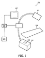
- Figure 1 shows a typical X-ray unit 101 suitable for the performance of interventional radiology.
- the unit consists of an X-ray source 102 and X-ray detector 103, a patient bed 104 for placement of the patient, a control unit 105 for controlling the rotation and angulation of the X-ray source, and correspondingly the X-ray detector, an input point 106 by which the user or users can input instructions to control the rotation and angulation of the source and control the operation of the source, and display apparatus 107 by which the user can view the image acquired by the X-ray unit.
- the patient is placed on the bed 104 and the user acquires images of the patient while performing an interventional procedure.
- the display of the images on the display apparatus 107 during the interventional procedure allows the user to perceive the progress of the interventional procedure.
- the user controls the operation of the X-ray source, in other words, switches it on and off, controls the KV p of the source and the size of the collimator jaws, using the input point 106.
- the input point 106 can also be used to control the position of the X-ray source, which is for example the rotation of the X-ray source around the central axis of the patient and the angulation of the X-ray source around an axis perpendicular to a saggital plane of the patient.
- individual manufacturers of equipment vary in their exact definitions of rotation and angulation.
- the input point may be any device suitable for the input of information to the control unit 105 and therefore may be for example a handset, a keyboard or a foot operated device.
- a voice operated input point is also possible, as is known in the art.
- the X-ray unit 101 typically includes a dose measurement device 108 for the measurement of radiation exposure from the source.
- the calculation of patient skin dose is known in the art and, for example, may typically be undertaken by the measurement of radiation exposure at a known point in the X-ray beam while the beam is switched on, using further extrapolation incorporating a single or series of known or assumed source to skin distances to arrive at a final measure of skin dose. Various methods to achieve this are known.
- the application of the invention requires that some measurement or calculation of dose is made which is representative of the dose received by the patient in the area of skin exposed during exposure.
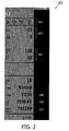
- FIG. 2 shows a state of the art graphical display incorporating a dose indicator as has been commonly used.
- the display 201 includes details of the energy of the radiation beam 202, the geometrical orientation of the source relative to the patient 203, the distance between the source and the patient 204, the exposure time elapsed 205, the Air Kerma 206 and the Dose Area Product 207.
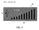
- FIG 3 shows a dose indicator 301 comprising an indication of a zone 302 and a graphical display of the value of dose 303.
- This dose indicator can be clearly understood by the user and indicates the accrued dose in Air Kerma, shown by the bars of the graphical display 303, and also the skin area on the patient over which this dose has been absorbed, 302.
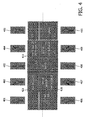
- FIG. 4 shows an array of possible zones, 401 to 410, according to the invention.
- these are zones representing the areas of skin on a patient commonly exposed in a cardiac procedure.
- the cardiologist or radiologist typically wishes to view the three main coronary arteries, the circumflex, the right coronary artery and the left coronary artery.
- the three coronary arteries can usually be viewed in a series of standard image projections, as is known in the art, e.g. in 'Coronary Arteriography, Nomenclature of the Arteries, Codification of Lesions', by the Working Group "Functional Evaluation and Angiography of the French Society of Cardiology", Coordinated by M.E. Bertrand, 1979.
- the zones represent the commonly used projections, 411 to 421, and are the right anterior oblique projection at 30° (RAO 30°), item 420; the anterior-posterior projection (AP), item 416; the left anterior oblique projection at 55/60° (LAO 55/60°), shown here as LAO 55, item 413; left anterior oblique projection at 55/60° combined with cranial angulation of 20°, shown here as LAO 55 Caud 20, item 411; left lateral projection, item 414; left anterior oblique projection at 45° combined with a caudal angulation of 15°, item 412; right anterior oblique projection at 45° (RAO 45°), item 419. Also possible is right anterior oblique projection at 120° combined with a cranial angulation of 10°, unshown.
- Each hospital or medical facility commonly uses a specific set of preferred orientations, often based on the clinical expertise of the individual users and also the specifics of the particular patient morphology with which they are presented. As is known in the art, these are standard projections with standard degrees of angulation and rotation used with radiographic and fluoroscopic equipment, but which often require small angular alterations on a patient by patient basis.
- the zones shown are indicators of the particular clinical orientations which may be used. In this way Figure 4 shows 10 zones which can be displayed and which in each case can be used to depict the orientation used by the hospital in question.
- a different set of zones are used for applications of the invention to other diagnostic and interventional procedures.
- the possible zones shown depict the commonly used orientations of the X-ray equipment which acquire images of the correct anatomy.
- zones has the further advantage that the same standardized zones are displayed regardless of any small deviations from the standard projections that may be made during actual exposure of the patient. This simplifies the display greatly, from the point of view of the user of the system, and allows an understanding of the skin dose to the patient regardless of the actual angulation and rotation combination applied for any specific patient.
- FIG. 5 shows an embodiment of a graphical display 501 for the value of dose which has been found to be particularly effective in indicating the value of dose clearly, quickly and effectively to the user of the interventional radiology system.
- the display 501 comprises a series of bars 502, graduated to show increasingly accrued skin dose up to a pre-determined threshold.
- the pre-determined threshold will normally be 2 Gy but may be decreased in the case of a patient who has a pre-accrued existing dose to the area of skin under exposure, and may also theoretically be increased in the case of a patient known to be radioresistant.
- the bars 502 appear on the display 501 as the skin dose to the area under exposure increases to the levels represented by each bar 502.
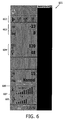
- Figure 6 shows how the invention, and in particular the embodiment of Figure 5 , might be incorporated into a visual display.
- Figure 6 shows a display 601 comprising details of the energy of the radiation beam 602, the geometrical orientation of the source relative to the patient 603, the distance between the source and the patient 604, and in this case the zone representative of the extent of the area of skin exposed 605 and a value of dose associated with that zone 606.
- the skin dose rate is shown on the display as a numerical value 609, but also using substantially the same form of graphical display 608 as the value of dose 606.
- Taking together the skin dose rate and the skin dose accrued allows a predication to be made of the time remaining before the threshold is reached and this time can also be displayed 607, providing further useful information for the user. It is also possible to predict the dose rate for specific operational set-ups and the predicated time to threshold may be shown for these set-ups may be displayed, allowing the user to choose the optimal set-up for the specific requirements of the remaining parts of the procedure.
- Figure 7 shows how the invention, and in particular the embodiment of Figure 5 , might be incorporated into a visual display, incorporating the change in display after the passing of a predefined threshold.
- Figure 7 shows a display 701 comprising details of the energy of the radiation beam 702, the geometrical orientation of the source relative to the patient 703, the distance between the source and the patient 704, the zone representative of the extent of the area of skin exposed 705, and a value of dose 707, shown in a visually alerting color, indicating that a pre-defined threshold has been exceeded. This would occur when the bars shown in 606 of Figure 6 have reached their maximum value. At this point the bars are shown in an equally visually alerting color 706 and remain at their maximum value, so showing the user that the threshold for that area of skin has been reached.
- the form of the display may change, so that the bars do not remain on the display but are replaced by a warning symbol, for example.
- the bars are redisplayed from a value of zero but displayed now upside down. This has the advantage of being visually alerting, by being different from the previous display, while continuing to count up dose to a new, second, threshold. This has the advantage that the user can continue to receive information about the steadily accruing patient skin dose.
- the new bars can still be displayed in a visually alerting colour.
- the rate at which skin dose is accruing, 709, may also be shown.
- the invention allows the clinical user of interventional radiological equipment to monitor easily, intuitively and understandibly the dose to the patient and therefore reduces the risk of overexposure and concomitant injury to the patient.
Landscapes
- Health & Medical Sciences (AREA)
- Engineering & Computer Science (AREA)
- Biomedical Technology (AREA)
- Life Sciences & Earth Sciences (AREA)
- Nuclear Medicine, Radiotherapy & Molecular Imaging (AREA)
- Radiology & Medical Imaging (AREA)
- Pathology (AREA)
- Animal Behavior & Ethology (AREA)
- General Health & Medical Sciences (AREA)
- Public Health (AREA)
- Veterinary Medicine (AREA)
- Apparatus For Radiation Diagnosis (AREA)
- Radiation-Therapy Devices (AREA)
- Measurement Of Radiation (AREA)
Priority Applications (1)
| Application Number | Priority Date | Filing Date | Title |
|---|---|---|---|
| EP07700038.8A EP1977269B1 (en) | 2006-01-12 | 2007-01-04 | Improved indication of patient skin dose in radiology |
Applications Claiming Priority (3)
| Application Number | Priority Date | Filing Date | Title |
|---|---|---|---|
| EP06100285 | 2006-01-12 | ||
| PCT/IB2007/050019 WO2007080522A1 (en) | 2006-01-12 | 2007-01-04 | Improved indication of patient skin dose in radiology |
| EP07700038.8A EP1977269B1 (en) | 2006-01-12 | 2007-01-04 | Improved indication of patient skin dose in radiology |
Publications (2)
| Publication Number | Publication Date |
|---|---|
| EP1977269A1 EP1977269A1 (en) | 2008-10-08 |
| EP1977269B1 true EP1977269B1 (en) | 2013-11-20 |
Family
ID=37903803
Family Applications (1)
| Application Number | Title | Priority Date | Filing Date |
|---|---|---|---|
| EP07700038.8A Not-in-force EP1977269B1 (en) | 2006-01-12 | 2007-01-04 | Improved indication of patient skin dose in radiology |
Country Status (5)
| Country | Link |
|---|---|
| US (1) | US7801273B2 (enExample) |
| EP (1) | EP1977269B1 (enExample) |
| JP (1) | JP5596290B2 (enExample) |
| CN (1) | CN101371161A (enExample) |
| WO (1) | WO2007080522A1 (enExample) |
Cited By (1)
| Publication number | Priority date | Publication date | Assignee | Title |
|---|---|---|---|---|
| US11974873B2 (en) | 2019-11-12 | 2024-05-07 | Siemens Healthineers Ag | Method for the automatic regulation of radiation doses of medical X-ray devices |
Families Citing this family (17)
| Publication number | Priority date | Publication date | Assignee | Title |
|---|---|---|---|---|
| CN101680952B (zh) * | 2007-05-07 | 2012-10-10 | 皇家飞利浦电子股份有限公司 | 工作人员剂量知晓指示 |
| JP5065822B2 (ja) * | 2007-09-14 | 2012-11-07 | 株式会社東芝 | X線ct装置、撮影計画支援装置および撮影計画支援プログラム |
| US8489431B2 (en) | 2009-03-20 | 2013-07-16 | General Electric Company | System and method of remote reporting of radiation dose usage in image acquisition |
| CN104324449B (zh) * | 2009-08-27 | 2017-04-12 | 三菱电机株式会社 | 粒子射线照射装置及粒子射线治疗装置 |
| DE102010025512B4 (de) * | 2010-06-29 | 2016-02-25 | Siemens Aktiengesellschaft | Verfahren zum Darstellen einer Patientendosis und Röntgengerät |
| JP5274526B2 (ja) * | 2010-09-09 | 2013-08-28 | 三菱電機株式会社 | 皮膚線量表示装置及び皮膚線量表示方法 |
| US8503613B2 (en) | 2010-11-24 | 2013-08-06 | General Electric Company | Dose level indication |
| FR2968187B1 (fr) | 2010-12-03 | 2013-11-29 | Gen Electric | Procede de suivi d'une dose de rayonnement |
| EP2465435B1 (en) | 2010-12-14 | 2019-12-04 | General Electric Company | Selection of optimal viewing angle to optimize anatomy visibility and patient skin dose |
| CN102090898A (zh) * | 2010-12-24 | 2011-06-15 | 苏州雷泰医疗科技有限公司 | 用于快速实时测量点剂量的探测验证的数字图像探测装置 |
| WO2013024534A1 (ja) * | 2011-08-17 | 2013-02-21 | 三菱電機株式会社 | 皮膚線量評価支援装置及び治療計画装置 |
| JP6058451B2 (ja) * | 2013-03-29 | 2017-01-11 | 東芝メディカルシステムズ株式会社 | 磁気共鳴撮像装置 |
| WO2015146164A1 (ja) * | 2014-03-28 | 2015-10-01 | 独立行政法人放射線医学総合研究所 | 放射線照射による皮膚変化予測装置と検証装置 |
| US9480448B2 (en) | 2014-07-23 | 2016-11-01 | General Electric Company | System and method for use in mapping a radiation dose applied in an angiography imaging procedure of a patient |
| USD771089S1 (en) * | 2014-07-23 | 2016-11-08 | General Electric Company | Display screen or portion thereof with graphical user interface for a radiation dose mapping system |
| US9649079B1 (en) | 2014-10-09 | 2017-05-16 | General Electric Company | System and method to illustrate a radiation dose applied to different anatomical stages of an exposed subject |
| CN111840828A (zh) * | 2019-04-29 | 2020-10-30 | 罗俭富 | 美容实时图像显示方法、装置以及美容仪 |
Family Cites Families (8)
| Publication number | Priority date | Publication date | Assignee | Title |
|---|---|---|---|---|
| JPH10225525A (ja) * | 1997-02-13 | 1998-08-25 | Ge Yokogawa Medical Syst Ltd | 放射線治療計画方法および装置 |
| US6501818B1 (en) * | 1997-11-26 | 2002-12-31 | Ge Medical Systems Global Technology Company, Llc | Apparatus and methods for displaying computed tomography fluoroscopy images including data transfer provided over a network |
| ATE217989T1 (de) * | 1998-02-09 | 2002-06-15 | Univ Southampton | Behandlungsplanungsverfahren und gerät für bestrahlungstherapie |
| JP4537506B2 (ja) * | 1998-11-20 | 2010-09-01 | 株式会社東芝 | X線診断装置 |
| JP4309103B2 (ja) * | 2002-08-09 | 2009-08-05 | 東芝医用システムエンジニアリング株式会社 | 放射線量推定装置および放射線診断装置 |
| AU2004279424A1 (en) * | 2003-10-07 | 2005-04-21 | Nomos Corporation | Planning system, method and apparatus for conformal radiation therapy |
| JP3891442B2 (ja) * | 2005-05-25 | 2007-03-14 | 株式会社日立メディコ | 3次元画像処理方法 |
| US7379531B2 (en) * | 2005-06-13 | 2008-05-27 | Siemens Medical Solutions Health Services Corporation | Beam therapy treatment user interface monitoring and recording system |
-
2007
- 2007-01-04 CN CNA2007800022545A patent/CN101371161A/zh active Pending
- 2007-01-04 US US12/160,223 patent/US7801273B2/en not_active Expired - Fee Related
- 2007-01-04 EP EP07700038.8A patent/EP1977269B1/en not_active Not-in-force
- 2007-01-04 JP JP2008549954A patent/JP5596290B2/ja not_active Expired - Fee Related
- 2007-01-04 WO PCT/IB2007/050019 patent/WO2007080522A1/en not_active Ceased
Cited By (1)
| Publication number | Priority date | Publication date | Assignee | Title |
|---|---|---|---|---|
| US11974873B2 (en) | 2019-11-12 | 2024-05-07 | Siemens Healthineers Ag | Method for the automatic regulation of radiation doses of medical X-ray devices |
Also Published As
| Publication number | Publication date |
|---|---|
| US7801273B2 (en) | 2010-09-21 |
| JP5596290B2 (ja) | 2014-09-24 |
| WO2007080522A1 (en) | 2007-07-19 |
| EP1977269A1 (en) | 2008-10-08 |
| US20090003527A1 (en) | 2009-01-01 |
| JP2009523049A (ja) | 2009-06-18 |
| CN101371161A (zh) | 2009-02-18 |
Similar Documents
| Publication | Publication Date | Title |
|---|---|---|
| EP1977269B1 (en) | Improved indication of patient skin dose in radiology | |
| Chida | What are useful methods to reduce occupational radiation exposure among radiological medical workers, especially for interventional radiology personnel? | |
| Vano et al. | Eye lens exposure to radiation in interventional suites: caution is warranted | |
| Stecker et al. | Guidelines for patient radiation dose management | |
| Heidbuchel et al. | Practical ways to reduce radiation dose for patients and staff during device implantations and electrophysiological procedures | |
| Johnson et al. | Radiation protection in interventional radiology | |
| Miller et al. | Minimizing radiation-induced skin injury in interventional radiology procedures | |
| Shope | Radiation-induced skin injuries from fluoroscopy. | |
| Theodorakou et al. | A study on radiation doses and irradiated areas in cerebral embolisation | |
| US20200023196A1 (en) | Radiation irradiating apparatus and radiation dose management system | |
| Bhar et al. | Monte Carlo study of patient and medical staff radiation exposures during interventional cardiology | |
| Bridcut et al. | Patient dose from 3D rotational neurovascular studies | |
| Norris | Radiation safety in fluoroscopy.(Directed Reading) | |
| Stacey et al. | Personnel protection during cardiac catheterization with a comparison of the hazards of undercouch and overcouch x-ray tube mountings | |
| US20180368800A1 (en) | Medical apparatus and x-ray system | |
| Ji et al. | Radiation-shielding devices: the best combination for spine interventional procedures | |
| Nair et al. | Radiation safety and ergonomics in the electrophysiology laboratory: update on recent advances | |
| Sulieman et al. | Patient effective doses and radiation risks in cardiac catheterization procedures | |
| Kara et al. | A study on radiation in operating room in Suleyman Demirel University | |
| Lai et al. | Effective doses in children: association with common complex imaging techniques used during interventional radiology procedures | |
| Rodas | Context-aware radiation protection for the hybrid operating room | |
| Zhao et al. | Deciphering the Radiation Dose Summary Page in Interventional Fluoroscopy | |
| Damanik et al. | Monitoring skin dose resulting from fluoroscopically guided interventions at Adam Malik Hospital Medan | |
| Sedlmair | Radiation Protection in CT-Guided Interventions | |
| Lynch et al. | Optimization and Dose Reduction in Fluoroscopy and Interventional Imaging |
Legal Events
| Date | Code | Title | Description |
|---|---|---|---|
| PUAI | Public reference made under article 153(3) epc to a published international application that has entered the european phase |
Free format text: ORIGINAL CODE: 0009012 |
|
| 17P | Request for examination filed |
Effective date: 20080812 |
|
| AK | Designated contracting states |
Kind code of ref document: A1 Designated state(s): AT BE BG CH CY CZ DE DK EE ES FI FR GB GR HU IE IS IT LI LT LU LV MC NL PL PT RO SE SI SK TR |
|
| 17Q | First examination report despatched |
Effective date: 20120208 |
|
| DAX | Request for extension of the european patent (deleted) | ||
| GRAP | Despatch of communication of intention to grant a patent |
Free format text: ORIGINAL CODE: EPIDOSNIGR1 |
|
| INTG | Intention to grant announced |
Effective date: 20130606 |
|
| RAP1 | Party data changed (applicant data changed or rights of an application transferred) |
Owner name: KONINKLIJKE PHILIPS N.V. |
|
| GRAS | Grant fee paid |
Free format text: ORIGINAL CODE: EPIDOSNIGR3 |
|
| GRAA | (expected) grant |
Free format text: ORIGINAL CODE: 0009210 |
|
| AK | Designated contracting states |
Kind code of ref document: B1 Designated state(s): AT BE BG CH CY CZ DE DK EE ES FI FR GB GR HU IE IS IT LI LT LU LV MC NL PL PT RO SE SI SK TR |
|
| REG | Reference to a national code |
Ref country code: GB Ref legal event code: FG4D |
|
| REG | Reference to a national code |
Ref country code: CH Ref legal event code: EP |
|
| REG | Reference to a national code |
Ref country code: AT Ref legal event code: REF Ref document number: 641956 Country of ref document: AT Kind code of ref document: T Effective date: 20131215 |
|
| REG | Reference to a national code |
Ref country code: IE Ref legal event code: FG4D |
|
| REG | Reference to a national code |
Ref country code: DE Ref legal event code: R096 Ref document number: 602007033867 Country of ref document: DE Effective date: 20140116 |
|
| REG | Reference to a national code |
Ref country code: NL Ref legal event code: VDEP Effective date: 20131120 |
|
| REG | Reference to a national code |
Ref country code: GB Ref legal event code: 746 Effective date: 20140304 |
|
| REG | Reference to a national code |
Ref country code: AT Ref legal event code: MK05 Ref document number: 641956 Country of ref document: AT Kind code of ref document: T Effective date: 20131120 |
|
| REG | Reference to a national code |
Ref country code: LT Ref legal event code: MG4D |
|
| PG25 | Lapsed in a contracting state [announced via postgrant information from national office to epo] |
Ref country code: NL Free format text: LAPSE BECAUSE OF FAILURE TO SUBMIT A TRANSLATION OF THE DESCRIPTION OR TO PAY THE FEE WITHIN THE PRESCRIBED TIME-LIMIT Effective date: 20131120 Ref country code: SE Free format text: LAPSE BECAUSE OF FAILURE TO SUBMIT A TRANSLATION OF THE DESCRIPTION OR TO PAY THE FEE WITHIN THE PRESCRIBED TIME-LIMIT Effective date: 20131120 Ref country code: IS Free format text: LAPSE BECAUSE OF FAILURE TO SUBMIT A TRANSLATION OF THE DESCRIPTION OR TO PAY THE FEE WITHIN THE PRESCRIBED TIME-LIMIT Effective date: 20140320 Ref country code: LT Free format text: LAPSE BECAUSE OF FAILURE TO SUBMIT A TRANSLATION OF THE DESCRIPTION OR TO PAY THE FEE WITHIN THE PRESCRIBED TIME-LIMIT Effective date: 20131120 Ref country code: FI Free format text: LAPSE BECAUSE OF FAILURE TO SUBMIT A TRANSLATION OF THE DESCRIPTION OR TO PAY THE FEE WITHIN THE PRESCRIBED TIME-LIMIT Effective date: 20131120 |
|
| PG25 | Lapsed in a contracting state [announced via postgrant information from national office to epo] |
Ref country code: BE Free format text: LAPSE BECAUSE OF FAILURE TO SUBMIT A TRANSLATION OF THE DESCRIPTION OR TO PAY THE FEE WITHIN THE PRESCRIBED TIME-LIMIT Effective date: 20131120 Ref country code: AT Free format text: LAPSE BECAUSE OF FAILURE TO SUBMIT A TRANSLATION OF THE DESCRIPTION OR TO PAY THE FEE WITHIN THE PRESCRIBED TIME-LIMIT Effective date: 20131120 Ref country code: ES Free format text: LAPSE BECAUSE OF FAILURE TO SUBMIT A TRANSLATION OF THE DESCRIPTION OR TO PAY THE FEE WITHIN THE PRESCRIBED TIME-LIMIT Effective date: 20131120 Ref country code: LV Free format text: LAPSE BECAUSE OF FAILURE TO SUBMIT A TRANSLATION OF THE DESCRIPTION OR TO PAY THE FEE WITHIN THE PRESCRIBED TIME-LIMIT Effective date: 20131120 |
|
| PG25 | Lapsed in a contracting state [announced via postgrant information from national office to epo] |
Ref country code: PT Free format text: LAPSE BECAUSE OF FAILURE TO SUBMIT A TRANSLATION OF THE DESCRIPTION OR TO PAY THE FEE WITHIN THE PRESCRIBED TIME-LIMIT Effective date: 20140320 |
|
| PG25 | Lapsed in a contracting state [announced via postgrant information from national office to epo] |
Ref country code: EE Free format text: LAPSE BECAUSE OF FAILURE TO SUBMIT A TRANSLATION OF THE DESCRIPTION OR TO PAY THE FEE WITHIN THE PRESCRIBED TIME-LIMIT Effective date: 20131120 |
|
| REG | Reference to a national code |
Ref country code: DE Ref legal event code: R097 Ref document number: 602007033867 Country of ref document: DE |
|
| PG25 | Lapsed in a contracting state [announced via postgrant information from national office to epo] |
Ref country code: LU Free format text: LAPSE BECAUSE OF FAILURE TO SUBMIT A TRANSLATION OF THE DESCRIPTION OR TO PAY THE FEE WITHIN THE PRESCRIBED TIME-LIMIT Effective date: 20140104 Ref country code: SK Free format text: LAPSE BECAUSE OF FAILURE TO SUBMIT A TRANSLATION OF THE DESCRIPTION OR TO PAY THE FEE WITHIN THE PRESCRIBED TIME-LIMIT Effective date: 20131120 Ref country code: CZ Free format text: LAPSE BECAUSE OF FAILURE TO SUBMIT A TRANSLATION OF THE DESCRIPTION OR TO PAY THE FEE WITHIN THE PRESCRIBED TIME-LIMIT Effective date: 20131120 Ref country code: RO Free format text: LAPSE BECAUSE OF FAILURE TO SUBMIT A TRANSLATION OF THE DESCRIPTION OR TO PAY THE FEE WITHIN THE PRESCRIBED TIME-LIMIT Effective date: 20131120 Ref country code: MC Free format text: LAPSE BECAUSE OF FAILURE TO SUBMIT A TRANSLATION OF THE DESCRIPTION OR TO PAY THE FEE WITHIN THE PRESCRIBED TIME-LIMIT Effective date: 20131120 Ref country code: PL Free format text: LAPSE BECAUSE OF FAILURE TO SUBMIT A TRANSLATION OF THE DESCRIPTION OR TO PAY THE FEE WITHIN THE PRESCRIBED TIME-LIMIT Effective date: 20131120 |
|
| REG | Reference to a national code |
Ref country code: CH Ref legal event code: PL |
|
| PLBE | No opposition filed within time limit |
Free format text: ORIGINAL CODE: 0009261 |
|
| STAA | Information on the status of an ep patent application or granted ep patent |
Free format text: STATUS: NO OPPOSITION FILED WITHIN TIME LIMIT |
|
| PG25 | Lapsed in a contracting state [announced via postgrant information from national office to epo] |
Ref country code: DK Free format text: LAPSE BECAUSE OF FAILURE TO SUBMIT A TRANSLATION OF THE DESCRIPTION OR TO PAY THE FEE WITHIN THE PRESCRIBED TIME-LIMIT Effective date: 20131120 |
|
| 26N | No opposition filed |
Effective date: 20140821 |
|
| PG25 | Lapsed in a contracting state [announced via postgrant information from national office to epo] |
Ref country code: LI Free format text: LAPSE BECAUSE OF NON-PAYMENT OF DUE FEES Effective date: 20140131 Ref country code: CH Free format text: LAPSE BECAUSE OF NON-PAYMENT OF DUE FEES Effective date: 20140131 |
|
| REG | Reference to a national code |
Ref country code: IE Ref legal event code: MM4A |
|
| REG | Reference to a national code |
Ref country code: DE Ref legal event code: R097 Ref document number: 602007033867 Country of ref document: DE Effective date: 20140821 |
|
| REG | Reference to a national code |
Ref country code: FR Ref legal event code: PLFP Year of fee payment: 9 |
|
| PG25 | Lapsed in a contracting state [announced via postgrant information from national office to epo] |
Ref country code: IE Free format text: LAPSE BECAUSE OF NON-PAYMENT OF DUE FEES Effective date: 20140104 |
|
| PG25 | Lapsed in a contracting state [announced via postgrant information from national office to epo] |
Ref country code: SI Free format text: LAPSE BECAUSE OF FAILURE TO SUBMIT A TRANSLATION OF THE DESCRIPTION OR TO PAY THE FEE WITHIN THE PRESCRIBED TIME-LIMIT Effective date: 20131120 |
|
| PGFP | Annual fee paid to national office [announced via postgrant information from national office to epo] |
Ref country code: FR Payment date: 20150128 Year of fee payment: 9 Ref country code: GB Payment date: 20150202 Year of fee payment: 9 Ref country code: TR Payment date: 20150102 Year of fee payment: 9 |
|
| PGFP | Annual fee paid to national office [announced via postgrant information from national office to epo] |
Ref country code: DE Payment date: 20150331 Year of fee payment: 9 |
|
| PG25 | Lapsed in a contracting state [announced via postgrant information from national office to epo] |
Ref country code: BG Free format text: LAPSE BECAUSE OF FAILURE TO SUBMIT A TRANSLATION OF THE DESCRIPTION OR TO PAY THE FEE WITHIN THE PRESCRIBED TIME-LIMIT Effective date: 20131120 |
|
| PG25 | Lapsed in a contracting state [announced via postgrant information from national office to epo] |
Ref country code: IT Free format text: LAPSE BECAUSE OF FAILURE TO SUBMIT A TRANSLATION OF THE DESCRIPTION OR TO PAY THE FEE WITHIN THE PRESCRIBED TIME-LIMIT Effective date: 20131120 Ref country code: GR Free format text: LAPSE BECAUSE OF FAILURE TO SUBMIT A TRANSLATION OF THE DESCRIPTION OR TO PAY THE FEE WITHIN THE PRESCRIBED TIME-LIMIT Effective date: 20140221 Ref country code: CY Free format text: LAPSE BECAUSE OF FAILURE TO SUBMIT A TRANSLATION OF THE DESCRIPTION OR TO PAY THE FEE WITHIN THE PRESCRIBED TIME-LIMIT Effective date: 20131120 |
|
| PG25 | Lapsed in a contracting state [announced via postgrant information from national office to epo] |
Ref country code: HU Free format text: LAPSE BECAUSE OF FAILURE TO SUBMIT A TRANSLATION OF THE DESCRIPTION OR TO PAY THE FEE WITHIN THE PRESCRIBED TIME-LIMIT; INVALID AB INITIO Effective date: 20070104 |
|
| REG | Reference to a national code |
Ref country code: DE Ref legal event code: R119 Ref document number: 602007033867 Country of ref document: DE |
|
| GBPC | Gb: european patent ceased through non-payment of renewal fee |
Effective date: 20160104 |
|
| REG | Reference to a national code |
Ref country code: FR Ref legal event code: ST Effective date: 20160930 |
|
| PG25 | Lapsed in a contracting state [announced via postgrant information from national office to epo] |
Ref country code: GB Free format text: LAPSE BECAUSE OF NON-PAYMENT OF DUE FEES Effective date: 20160104 Ref country code: DE Free format text: LAPSE BECAUSE OF NON-PAYMENT OF DUE FEES Effective date: 20160802 |
|
| PG25 | Lapsed in a contracting state [announced via postgrant information from national office to epo] |
Ref country code: FR Free format text: LAPSE BECAUSE OF NON-PAYMENT OF DUE FEES Effective date: 20160201 |
|
| PG25 | Lapsed in a contracting state [announced via postgrant information from national office to epo] |
Ref country code: TR Free format text: LAPSE BECAUSE OF NON-PAYMENT OF DUE FEES Effective date: 20160104 |