EP1956080A2 - Utilisation de cytokines de chaîne gamma communes pour la modification génétique des lymphocytes T à mémoire - Google Patents
Utilisation de cytokines de chaîne gamma communes pour la modification génétique des lymphocytes T à mémoire Download PDFInfo
- Publication number
- EP1956080A2 EP1956080A2 EP08102851A EP08102851A EP1956080A2 EP 1956080 A2 EP1956080 A2 EP 1956080A2 EP 08102851 A EP08102851 A EP 08102851A EP 08102851 A EP08102851 A EP 08102851A EP 1956080 A2 EP1956080 A2 EP 1956080A2
- Authority
- EP
- European Patent Office
- Prior art keywords
- cells
- cell
- specific
- memory
- lymphocytes
- Prior art date
- Legal status (The legal status is an assumption and is not a legal conclusion. Google has not performed a legal analysis and makes no representation as to the accuracy of the status listed.)
- Granted
Links
- 210000003071 memory t lymphocyte Anatomy 0.000 title claims abstract description 81
- 102000004127 Cytokines Human genes 0.000 title description 67
- 108090000695 Cytokines Proteins 0.000 title description 67
- 238000012239 gene modification Methods 0.000 title description 7
- 230000005017 genetic modification Effects 0.000 title description 6
- 235000013617 genetically modified food Nutrition 0.000 title description 6
- 108010066719 Interleukin Receptor Common gamma Subunit Proteins 0.000 title description 5
- 102000018682 Interleukin Receptor Common gamma Subunit Human genes 0.000 title description 5
- 210000004027 cell Anatomy 0.000 claims abstract description 354
- 238000000338 in vitro Methods 0.000 claims abstract description 54
- 238000000034 method Methods 0.000 claims abstract description 43
- 210000001744 T-lymphocyte Anatomy 0.000 claims description 306
- 210000004698 lymphocyte Anatomy 0.000 claims description 95
- 206010028980 Neoplasm Diseases 0.000 claims description 72
- 239000011324 bead Substances 0.000 claims description 70
- 101000914514 Homo sapiens T-cell-specific surface glycoprotein CD28 Proteins 0.000 claims description 59
- 102100027213 T-cell-specific surface glycoprotein CD28 Human genes 0.000 claims description 59
- 230000004913 activation Effects 0.000 claims description 36
- 239000000018 receptor agonist Substances 0.000 claims description 20
- 229940044601 receptor agonist Drugs 0.000 claims description 20
- 108090000765 processed proteins & peptides Proteins 0.000 claims description 18
- 108090000623 proteins and genes Proteins 0.000 claims description 18
- 239000013598 vector Substances 0.000 claims description 13
- 108010038498 Interleukin-7 Receptors Proteins 0.000 claims description 12
- 102000010782 Interleukin-7 Receptors Human genes 0.000 claims description 12
- 239000003446 ligand Substances 0.000 claims description 12
- 239000012472 biological sample Substances 0.000 claims description 11
- 230000003213 activating effect Effects 0.000 claims description 10
- 239000000409 cytokine receptor agonist Substances 0.000 claims description 10
- 102000005962 receptors Human genes 0.000 claims description 10
- 108020003175 receptors Proteins 0.000 claims description 10
- 208000015181 infectious disease Diseases 0.000 claims description 8
- 108010017535 Interleukin-15 Receptors Proteins 0.000 claims description 7
- 102000004556 Interleukin-15 Receptors Human genes 0.000 claims description 7
- 210000004369 blood Anatomy 0.000 claims description 7
- 239000008280 blood Substances 0.000 claims description 7
- 239000003550 marker Substances 0.000 claims description 7
- 230000000717 retained effect Effects 0.000 claims description 7
- 238000002054 transplantation Methods 0.000 claims description 7
- 201000011510 cancer Diseases 0.000 claims description 6
- 102000004196 processed proteins & peptides Human genes 0.000 claims description 5
- 239000007787 solid Substances 0.000 claims description 5
- 239000013603 viral vector Substances 0.000 claims description 5
- 206010061598 Immunodeficiency Diseases 0.000 claims description 4
- 208000029462 Immunodeficiency disease Diseases 0.000 claims description 4
- 230000007813 immunodeficiency Effects 0.000 claims description 4
- 239000002243 precursor Substances 0.000 claims description 4
- 230000005784 autoimmunity Effects 0.000 claims description 3
- 230000003394 haemopoietic effect Effects 0.000 claims description 3
- 239000007788 liquid Substances 0.000 claims description 3
- 230000005298 paramagnetic effect Effects 0.000 claims description 3
- 230000002265 prevention Effects 0.000 claims description 3
- 229940002612 prodrug Drugs 0.000 claims description 3
- 239000000651 prodrug Substances 0.000 claims description 3
- 238000011282 treatment Methods 0.000 claims description 3
- 239000000556 agonist Substances 0.000 claims description 2
- 108091008034 costimulatory receptors Proteins 0.000 claims description 2
- 210000000056 organ Anatomy 0.000 claims description 2
- 229920001184 polypeptide Polymers 0.000 claims description 2
- 239000000427 antigen Substances 0.000 abstract description 68
- 108091007433 antigens Proteins 0.000 abstract description 66
- 102000036639 antigens Human genes 0.000 abstract description 66
- 102000000704 Interleukin-7 Human genes 0.000 description 230
- 108010002586 Interleukin-7 Proteins 0.000 description 230
- 101000716102 Homo sapiens T-cell surface glycoprotein CD4 Proteins 0.000 description 179
- 102100036011 T-cell surface glycoprotein CD4 Human genes 0.000 description 179
- 102000000588 Interleukin-2 Human genes 0.000 description 147
- 108010002350 Interleukin-2 Proteins 0.000 description 146
- 230000015654 memory Effects 0.000 description 97
- 108090000172 Interleukin-15 Proteins 0.000 description 80
- 102000003812 Interleukin-15 Human genes 0.000 description 80
- 241000699670 Mus sp. Species 0.000 description 80
- 108010074328 Interferon-gamma Proteins 0.000 description 62
- 102100037850 Interferon gamma Human genes 0.000 description 61
- 210000001165 lymph node Anatomy 0.000 description 56
- 230000014509 gene expression Effects 0.000 description 52
- 102000017420 CD3 protein, epsilon/gamma/delta subunit Human genes 0.000 description 49
- 230000000638 stimulation Effects 0.000 description 47
- 210000003819 peripheral blood mononuclear cell Anatomy 0.000 description 41
- 238000001727 in vivo Methods 0.000 description 34
- 239000012636 effector Substances 0.000 description 32
- 238000000684 flow cytometry Methods 0.000 description 31
- 238000009825 accumulation Methods 0.000 description 29
- 208000009329 Graft vs Host Disease Diseases 0.000 description 24
- 230000000735 allogeneic effect Effects 0.000 description 24
- 208000024908 graft versus host disease Diseases 0.000 description 24
- 230000035755 proliferation Effects 0.000 description 23
- 108090001005 Interleukin-6 Proteins 0.000 description 22
- 102000004889 Interleukin-6 Human genes 0.000 description 22
- 101710122864 Major tegument protein Proteins 0.000 description 22
- 102100031545 Microsomal triglyceride transfer protein large subunit Human genes 0.000 description 22
- 101710199973 Tail tube protein Proteins 0.000 description 22
- 230000004083 survival effect Effects 0.000 description 21
- 101001018097 Homo sapiens L-selectin Proteins 0.000 description 19
- 102100033467 L-selectin Human genes 0.000 description 19
- 230000002062 proliferating effect Effects 0.000 description 19
- 229930105110 Cyclosporin A Natural products 0.000 description 18
- PMATZTZNYRCHOR-CGLBZJNRSA-N Cyclosporin A Chemical compound CC[C@@H]1NC(=O)[C@H]([C@H](O)[C@H](C)C\C=C\C)N(C)C(=O)[C@H](C(C)C)N(C)C(=O)[C@H](CC(C)C)N(C)C(=O)[C@H](CC(C)C)N(C)C(=O)[C@@H](C)NC(=O)[C@H](C)NC(=O)[C@H](CC(C)C)N(C)C(=O)[C@H](C(C)C)NC(=O)[C@H](CC(C)C)N(C)C(=O)CN(C)C1=O PMATZTZNYRCHOR-CGLBZJNRSA-N 0.000 description 18
- 210000003491 skin Anatomy 0.000 description 18
- 238000010186 staining Methods 0.000 description 18
- 239000000975 dye Substances 0.000 description 17
- 238000002474 experimental method Methods 0.000 description 17
- 238000010361 transduction Methods 0.000 description 17
- 230000026683 transduction Effects 0.000 description 17
- 210000004443 dendritic cell Anatomy 0.000 description 16
- 230000003834 intracellular effect Effects 0.000 description 16
- 239000002609 medium Substances 0.000 description 16
- 239000000523 sample Substances 0.000 description 16
- 101001057504 Homo sapiens Interferon-stimulated gene 20 kDa protein Proteins 0.000 description 15
- 101001055144 Homo sapiens Interleukin-2 receptor subunit alpha Proteins 0.000 description 15
- 102100027268 Interferon-stimulated gene 20 kDa protein Human genes 0.000 description 15
- 210000000612 antigen-presenting cell Anatomy 0.000 description 15
- 230000032823 cell division Effects 0.000 description 15
- 230000010261 cell growth Effects 0.000 description 15
- 230000001177 retroviral effect Effects 0.000 description 15
- 102100021569 Apoptosis regulator Bcl-2 Human genes 0.000 description 14
- 101000971171 Homo sapiens Apoptosis regulator Bcl-2 Proteins 0.000 description 14
- 238000001802 infusion Methods 0.000 description 14
- 238000011725 BALB/c mouse Methods 0.000 description 12
- -1 IFN-g Proteins 0.000 description 12
- 230000028993 immune response Effects 0.000 description 12
- 230000001965 increasing effect Effects 0.000 description 12
- 102000004388 Interleukin-4 Human genes 0.000 description 11
- 108090000978 Interleukin-4 Proteins 0.000 description 11
- 238000004458 analytical method Methods 0.000 description 11
- 238000001514 detection method Methods 0.000 description 11
- 238000003114 enzyme-linked immunosorbent spot assay Methods 0.000 description 11
- 238000004519 manufacturing process Methods 0.000 description 11
- 238000011740 C57BL/6 mouse Methods 0.000 description 10
- 230000005867 T cell response Effects 0.000 description 10
- QHNORJFCVHUPNH-UHFFFAOYSA-L To-Pro-3 Chemical compound [I-].[I-].S1C2=CC=CC=C2[N+](C)=C1C=CC=C1C2=CC=CC=C2N(CCC[N+](C)(C)C)C=C1 QHNORJFCVHUPNH-UHFFFAOYSA-L 0.000 description 10
- 201000008827 tuberculosis Diseases 0.000 description 10
- 206010068051 Chimerism Diseases 0.000 description 9
- 241000699666 Mus <mouse, genus> Species 0.000 description 9
- 238000012360 testing method Methods 0.000 description 9
- 210000004881 tumor cell Anatomy 0.000 description 9
- 102100036301 C-C chemokine receptor type 7 Human genes 0.000 description 8
- 102100032912 CD44 antigen Human genes 0.000 description 8
- 101000716065 Homo sapiens C-C chemokine receptor type 7 Proteins 0.000 description 8
- 101000868273 Homo sapiens CD44 antigen Proteins 0.000 description 8
- 108700018351 Major Histocompatibility Complex Proteins 0.000 description 8
- 241001465754 Metazoa Species 0.000 description 8
- 238000001943 fluorescence-activated cell sorting Methods 0.000 description 8
- 210000005259 peripheral blood Anatomy 0.000 description 8
- 239000011886 peripheral blood Substances 0.000 description 8
- 230000001105 regulatory effect Effects 0.000 description 8
- 230000004044 response Effects 0.000 description 8
- 230000020382 suppression by virus of host antigen processing and presentation of peptide antigen via MHC class I Effects 0.000 description 8
- 108010038453 Interleukin-2 Receptors Proteins 0.000 description 7
- 241000187479 Mycobacterium tuberculosis Species 0.000 description 7
- 230000006044 T cell activation Effects 0.000 description 7
- 230000008901 benefit Effects 0.000 description 7
- 230000000694 effects Effects 0.000 description 7
- 244000052769 pathogen Species 0.000 description 7
- 230000004614 tumor growth Effects 0.000 description 7
- 102100027207 CD27 antigen Human genes 0.000 description 6
- 238000011510 Elispot assay Methods 0.000 description 6
- 101000914511 Homo sapiens CD27 antigen Proteins 0.000 description 6
- 101001043807 Homo sapiens Interleukin-7 Proteins 0.000 description 6
- 102000010789 Interleukin-2 Receptors Human genes 0.000 description 6
- 241000699660 Mus musculus Species 0.000 description 6
- 238000003556 assay Methods 0.000 description 6
- 210000001185 bone marrow Anatomy 0.000 description 6
- 238000004113 cell culture Methods 0.000 description 6
- 230000004186 co-expression Effects 0.000 description 6
- 230000001143 conditioned effect Effects 0.000 description 6
- 210000004748 cultured cell Anatomy 0.000 description 6
- 230000016396 cytokine production Effects 0.000 description 6
- 238000009826 distribution Methods 0.000 description 6
- 230000003284 homeostatic effect Effects 0.000 description 6
- 102000052622 human IL7 Human genes 0.000 description 6
- 239000003112 inhibitor Substances 0.000 description 6
- 230000007774 longterm Effects 0.000 description 6
- 239000000463 material Substances 0.000 description 6
- 210000005105 peripheral blood lymphocyte Anatomy 0.000 description 6
- 230000009257 reactivity Effects 0.000 description 6
- 210000004988 splenocyte Anatomy 0.000 description 6
- 210000001519 tissue Anatomy 0.000 description 6
- 238000012546 transfer Methods 0.000 description 6
- 238000011830 transgenic mouse model Methods 0.000 description 6
- 230000004580 weight loss Effects 0.000 description 6
- 241000222122 Candida albicans Species 0.000 description 5
- 230000006786 activation induced cell death Effects 0.000 description 5
- 230000030833 cell death Effects 0.000 description 5
- 230000001419 dependent effect Effects 0.000 description 5
- 238000010790 dilution Methods 0.000 description 5
- 239000012895 dilution Substances 0.000 description 5
- 208000037265 diseases, disorders, signs and symptoms Diseases 0.000 description 5
- 239000012091 fetal bovine serum Substances 0.000 description 5
- 210000002966 serum Anatomy 0.000 description 5
- 210000001266 CD8-positive T-lymphocyte Anatomy 0.000 description 4
- 108010036949 Cyclosporine Proteins 0.000 description 4
- WZUVPPKBWHMQCE-UHFFFAOYSA-N Haematoxylin Chemical compound C12=CC(O)=C(O)C=C2CC2(O)C1C1=CC=C(O)C(O)=C1OC2 WZUVPPKBWHMQCE-UHFFFAOYSA-N 0.000 description 4
- 101100005713 Homo sapiens CD4 gene Proteins 0.000 description 4
- 102000003814 Interleukin-10 Human genes 0.000 description 4
- 108090000174 Interleukin-10 Proteins 0.000 description 4
- 241001529936 Murinae Species 0.000 description 4
- 230000006052 T cell proliferation Effects 0.000 description 4
- GLNADSQYFUSGOU-GPTZEZBUSA-J Trypan blue Chemical compound [Na+].[Na+].[Na+].[Na+].C1=C(S([O-])(=O)=O)C=C2C=C(S([O-])(=O)=O)C(/N=N/C3=CC=C(C=C3C)C=3C=C(C(=CC=3)\N=N\C=3C(=CC4=CC(=CC(N)=C4C=3O)S([O-])(=O)=O)S([O-])(=O)=O)C)=C(O)C2=C1N GLNADSQYFUSGOU-GPTZEZBUSA-J 0.000 description 4
- 239000013566 allergen Substances 0.000 description 4
- 230000000903 blocking effect Effects 0.000 description 4
- 230000022131 cell cycle Effects 0.000 description 4
- 238000006243 chemical reaction Methods 0.000 description 4
- 229960001265 ciclosporin Drugs 0.000 description 4
- 230000034994 death Effects 0.000 description 4
- 201000010099 disease Diseases 0.000 description 4
- BFMYDTVEBKDAKJ-UHFFFAOYSA-L disodium;(2',7'-dibromo-3',6'-dioxido-3-oxospiro[2-benzofuran-1,9'-xanthene]-4'-yl)mercury;hydrate Chemical compound O.[Na+].[Na+].O1C(=O)C2=CC=CC=C2C21C1=CC(Br)=C([O-])C([Hg])=C1OC1=C2C=C(Br)C([O-])=C1 BFMYDTVEBKDAKJ-UHFFFAOYSA-L 0.000 description 4
- 238000001415 gene therapy Methods 0.000 description 4
- PGHMRUGBZOYCAA-UHFFFAOYSA-N ionomycin Natural products O1C(CC(O)C(C)C(O)C(C)C=CCC(C)CC(C)C(O)=CC(=O)C(C)CC(C)CC(CCC(O)=O)C)CCC1(C)C1OC(C)(C(C)O)CC1 PGHMRUGBZOYCAA-UHFFFAOYSA-N 0.000 description 4
- PGHMRUGBZOYCAA-ADZNBVRBSA-N ionomycin Chemical compound O1[C@H](C[C@H](O)[C@H](C)[C@H](O)[C@H](C)/C=C/C[C@@H](C)C[C@@H](C)C(/O)=C/C(=O)[C@@H](C)C[C@@H](C)C[C@@H](CCC(O)=O)C)CC[C@@]1(C)[C@@H]1O[C@](C)([C@@H](C)O)CC1 PGHMRUGBZOYCAA-ADZNBVRBSA-N 0.000 description 4
- 230000005291 magnetic effect Effects 0.000 description 4
- 238000012423 maintenance Methods 0.000 description 4
- 238000007799 mixed lymphocyte reaction assay Methods 0.000 description 4
- 239000013642 negative control Substances 0.000 description 4
- 230000001717 pathogenic effect Effects 0.000 description 4
- 230000002093 peripheral effect Effects 0.000 description 4
- 102000004169 proteins and genes Human genes 0.000 description 4
- 230000028327 secretion Effects 0.000 description 4
- 230000035945 sensitivity Effects 0.000 description 4
- 230000001225 therapeutic effect Effects 0.000 description 4
- 238000002255 vaccination Methods 0.000 description 4
- 229960005486 vaccine Drugs 0.000 description 4
- MZOFCQQQCNRIBI-VMXHOPILSA-N (3s)-4-[[(2s)-1-[[(2s)-1-[[(1s)-1-carboxy-2-hydroxyethyl]amino]-4-methyl-1-oxopentan-2-yl]amino]-5-(diaminomethylideneamino)-1-oxopentan-2-yl]amino]-3-[[2-[[(2s)-2,6-diaminohexanoyl]amino]acetyl]amino]-4-oxobutanoic acid Chemical compound OC[C@@H](C(O)=O)NC(=O)[C@H](CC(C)C)NC(=O)[C@H](CCCN=C(N)N)NC(=O)[C@H](CC(O)=O)NC(=O)CNC(=O)[C@@H](N)CCCCN MZOFCQQQCNRIBI-VMXHOPILSA-N 0.000 description 3
- NHBKXEKEPDILRR-UHFFFAOYSA-N 2,3-bis(butanoylsulfanyl)propyl butanoate Chemical compound CCCC(=O)OCC(SC(=O)CCC)CSC(=O)CCC NHBKXEKEPDILRR-UHFFFAOYSA-N 0.000 description 3
- 241000222120 Candida <Saccharomycetales> Species 0.000 description 3
- 101000738771 Homo sapiens Receptor-type tyrosine-protein phosphatase C Proteins 0.000 description 3
- 102000043131 MHC class II family Human genes 0.000 description 3
- 108091054438 MHC class II family Proteins 0.000 description 3
- 102100037422 Receptor-type tyrosine-protein phosphatase C Human genes 0.000 description 3
- 210000000662 T-lymphocyte subset Anatomy 0.000 description 3
- 230000006907 apoptotic process Effects 0.000 description 3
- KQNZDYYTLMIZCT-KQPMLPITSA-N brefeldin A Chemical compound O[C@@H]1\C=C\C(=O)O[C@@H](C)CCC\C=C\[C@@H]2C[C@H](O)C[C@H]21 KQNZDYYTLMIZCT-KQPMLPITSA-N 0.000 description 3
- JUMGSHROWPPKFX-UHFFFAOYSA-N brefeldin-A Natural products CC1CCCC=CC2(C)CC(O)CC2(C)C(O)C=CC(=O)O1 JUMGSHROWPPKFX-UHFFFAOYSA-N 0.000 description 3
- 238000012258 culturing Methods 0.000 description 3
- 238000003568 cytokine secretion assay Methods 0.000 description 3
- 230000007423 decrease Effects 0.000 description 3
- 239000007850 fluorescent dye Substances 0.000 description 3
- 230000006870 function Effects 0.000 description 3
- IRSCQMHQWWYFCW-UHFFFAOYSA-N ganciclovir Chemical compound O=C1NC(N)=NC2=C1N=CN2COC(CO)CO IRSCQMHQWWYFCW-UHFFFAOYSA-N 0.000 description 3
- 229960002963 ganciclovir Drugs 0.000 description 3
- 210000004408 hybridoma Anatomy 0.000 description 3
- 230000001939 inductive effect Effects 0.000 description 3
- 230000008595 infiltration Effects 0.000 description 3
- 238000001764 infiltration Methods 0.000 description 3
- 230000007170 pathology Effects 0.000 description 3
- 230000003389 potentiating effect Effects 0.000 description 3
- 230000002035 prolonged effect Effects 0.000 description 3
- 230000002829 reductive effect Effects 0.000 description 3
- 230000003248 secreting effect Effects 0.000 description 3
- 230000011664 signaling Effects 0.000 description 3
- QFJCIRLUMZQUOT-HPLJOQBZSA-N sirolimus Chemical compound C1C[C@@H](O)[C@H](OC)C[C@@H]1C[C@@H](C)[C@H]1OC(=O)[C@@H]2CCCCN2C(=O)C(=O)[C@](O)(O2)[C@H](C)CC[C@H]2C[C@H](OC)/C(C)=C/C=C/C=C/[C@@H](C)C[C@@H](C)C(=O)[C@H](OC)[C@H](O)/C(C)=C/[C@@H](C)C(=O)C1 QFJCIRLUMZQUOT-HPLJOQBZSA-N 0.000 description 3
- 230000002269 spontaneous effect Effects 0.000 description 3
- 239000006228 supernatant Substances 0.000 description 3
- 238000010257 thawing Methods 0.000 description 3
- 238000002560 therapeutic procedure Methods 0.000 description 3
- 229960001005 tuberculin Drugs 0.000 description 3
- YBJHBAHKTGYVGT-ZKWXMUAHSA-N (+)-Biotin Chemical compound N1C(=O)N[C@@H]2[C@H](CCCCC(=O)O)SC[C@@H]21 YBJHBAHKTGYVGT-ZKWXMUAHSA-N 0.000 description 2
- QTBSBXVTEAMEQO-UHFFFAOYSA-N Acetic acid Chemical group CC(O)=O QTBSBXVTEAMEQO-UHFFFAOYSA-N 0.000 description 2
- 208000023275 Autoimmune disease Diseases 0.000 description 2
- 101800001467 Envelope glycoprotein E2 Proteins 0.000 description 2
- 101710091045 Envelope protein Proteins 0.000 description 2
- 206010020751 Hypersensitivity Diseases 0.000 description 2
- 102100034349 Integrase Human genes 0.000 description 2
- 102100022339 Integrin alpha-L Human genes 0.000 description 2
- 241000222732 Leishmania major Species 0.000 description 2
- 108010064548 Lymphocyte Function-Associated Antigen-1 Proteins 0.000 description 2
- 241000186359 Mycobacterium Species 0.000 description 2
- 101710188315 Protein X Proteins 0.000 description 2
- 101800001271 Surface protein Proteins 0.000 description 2
- 102100034922 T-cell surface glycoprotein CD8 alpha chain Human genes 0.000 description 2
- 241000223996 Toxoplasma Species 0.000 description 2
- 108060008682 Tumor Necrosis Factor Proteins 0.000 description 2
- 102000000852 Tumor Necrosis Factor-alpha Human genes 0.000 description 2
- 230000003187 abdominal effect Effects 0.000 description 2
- 208000026935 allergic disease Diseases 0.000 description 2
- 238000000540 analysis of variance Methods 0.000 description 2
- 238000010171 animal model Methods 0.000 description 2
- 230000002424 anti-apoptotic effect Effects 0.000 description 2
- 230000000919 anti-host Effects 0.000 description 2
- 230000000890 antigenic effect Effects 0.000 description 2
- 230000007503 antigenic stimulation Effects 0.000 description 2
- 238000013459 approach Methods 0.000 description 2
- 230000001363 autoimmune Effects 0.000 description 2
- 238000001574 biopsy Methods 0.000 description 2
- 230000037396 body weight Effects 0.000 description 2
- 229940095731 candida albicans Drugs 0.000 description 2
- 230000011712 cell development Effects 0.000 description 2
- 230000003915 cell function Effects 0.000 description 2
- 230000004663 cell proliferation Effects 0.000 description 2
- 239000006285 cell suspension Substances 0.000 description 2
- 238000002659 cell therapy Methods 0.000 description 2
- 238000012512 characterization method Methods 0.000 description 2
- 238000007796 conventional method Methods 0.000 description 2
- 102000003675 cytokine receptors Human genes 0.000 description 2
- 108010057085 cytokine receptors Proteins 0.000 description 2
- 230000001934 delay Effects 0.000 description 2
- 230000003111 delayed effect Effects 0.000 description 2
- 230000004069 differentiation Effects 0.000 description 2
- 239000000539 dimer Substances 0.000 description 2
- 231100000673 dose–response relationship Toxicity 0.000 description 2
- 230000003828 downregulation Effects 0.000 description 2
- 210000003162 effector t lymphocyte Anatomy 0.000 description 2
- 210000002615 epidermis Anatomy 0.000 description 2
- 150000002148 esters Chemical class 0.000 description 2
- 230000007717 exclusion Effects 0.000 description 2
- MHMNJMPURVTYEJ-UHFFFAOYSA-N fluorescein-5-isothiocyanate Chemical compound O1C(=O)C2=CC(N=C=S)=CC=C2C21C1=CC=C(O)C=C1OC1=CC(O)=CC=C21 MHMNJMPURVTYEJ-UHFFFAOYSA-N 0.000 description 2
- 230000013632 homeostatic process Effects 0.000 description 2
- 230000006054 immunological memory Effects 0.000 description 2
- 238000011293 immunotherapeutic strategy Methods 0.000 description 2
- 238000009169 immunotherapy Methods 0.000 description 2
- 238000002347 injection Methods 0.000 description 2
- 239000007924 injection Substances 0.000 description 2
- 230000003993 interaction Effects 0.000 description 2
- 238000010212 intracellular staining Methods 0.000 description 2
- 238000011835 investigation Methods 0.000 description 2
- 230000000670 limiting effect Effects 0.000 description 2
- 230000005923 long-lasting effect Effects 0.000 description 2
- 210000005210 lymphoid organ Anatomy 0.000 description 2
- 230000007246 mechanism Effects 0.000 description 2
- 230000001404 mediated effect Effects 0.000 description 2
- 244000000010 microbial pathogen Species 0.000 description 2
- 239000003068 molecular probe Substances 0.000 description 2
- 238000010172 mouse model Methods 0.000 description 2
- 210000000822 natural killer cell Anatomy 0.000 description 2
- 102000002574 p38 Mitogen-Activated Protein Kinases Human genes 0.000 description 2
- 239000008188 pellet Substances 0.000 description 2
- 230000002688 persistence Effects 0.000 description 2
- 230000002085 persistent effect Effects 0.000 description 2
- 230000004962 physiological condition Effects 0.000 description 2
- 239000013641 positive control Substances 0.000 description 2
- 230000001686 pro-survival effect Effects 0.000 description 2
- 230000001737 promoting effect Effects 0.000 description 2
- ZAHRKKWIAAJSAO-UHFFFAOYSA-N rapamycin Natural products COCC(O)C(=C/C(C)C(=O)CC(OC(=O)C1CCCCN1C(=O)C(=O)C2(O)OC(CC(OC)C(=CC=CC=CC(C)CC(C)C(=O)C)C)CCC2C)C(C)CC3CCC(O)C(C3)OC)C ZAHRKKWIAAJSAO-UHFFFAOYSA-N 0.000 description 2
- 230000000284 resting effect Effects 0.000 description 2
- 210000005212 secondary lymphoid organ Anatomy 0.000 description 2
- 230000019491 signal transduction Effects 0.000 description 2
- 229960002930 sirolimus Drugs 0.000 description 2
- 239000000243 solution Substances 0.000 description 2
- 238000007619 statistical method Methods 0.000 description 2
- 239000008223 sterile water Substances 0.000 description 2
- 230000004936 stimulating effect Effects 0.000 description 2
- 238000007920 subcutaneous administration Methods 0.000 description 2
- 230000009261 transgenic effect Effects 0.000 description 2
- 230000007704 transition Effects 0.000 description 2
- 230000003827 upregulation Effects 0.000 description 2
- 210000003462 vein Anatomy 0.000 description 2
- XLYOFNOQVPJJNP-UHFFFAOYSA-N water Chemical compound O XLYOFNOQVPJJNP-UHFFFAOYSA-N 0.000 description 2
- 230000003442 weekly effect Effects 0.000 description 2
- QRXMUCSWCMTJGU-UHFFFAOYSA-L (5-bromo-4-chloro-1h-indol-3-yl) phosphate Chemical compound C1=C(Br)C(Cl)=C2C(OP([O-])(=O)[O-])=CNC2=C1 QRXMUCSWCMTJGU-UHFFFAOYSA-L 0.000 description 1
- BPVHBBXCESDRKW-UHFFFAOYSA-N 5(6)-carboxyfluorescein Chemical compound C12=CC=C(O)C=C2OC2=CC(O)=CC=C2C21OC(=O)C1=CC(C(=O)O)=CC=C21.C12=CC=C(O)C=C2OC2=CC(O)=CC=C2C11OC(=O)C2=CC=C(C(=O)O)C=C21 BPVHBBXCESDRKW-UHFFFAOYSA-N 0.000 description 1
- 101710166488 6 kDa early secretory antigenic target Proteins 0.000 description 1
- 108090001008 Avidin Proteins 0.000 description 1
- 102100023995 Beta-nerve growth factor Human genes 0.000 description 1
- 108091003079 Bovine Serum Albumin Proteins 0.000 description 1
- 101150013553 CD40 gene Proteins 0.000 description 1
- 241001227713 Chiron Species 0.000 description 1
- 208000015943 Coeliac disease Diseases 0.000 description 1
- 208000035473 Communicable disease Diseases 0.000 description 1
- 101710112752 Cytotoxin Proteins 0.000 description 1
- 108020004414 DNA Proteins 0.000 description 1
- 238000012286 ELISA Assay Methods 0.000 description 1
- 108090000371 Esterases Proteins 0.000 description 1
- 208000020545 Exposure to communicable disease Diseases 0.000 description 1
- WSFSSNUMVMOOMR-UHFFFAOYSA-N Formaldehyde Chemical compound O=C WSFSSNUMVMOOMR-UHFFFAOYSA-N 0.000 description 1
- 108010017213 Granulocyte-Macrophage Colony-Stimulating Factor Proteins 0.000 description 1
- 102100039620 Granulocyte-macrophage colony-stimulating factor Human genes 0.000 description 1
- 102000001398 Granzyme Human genes 0.000 description 1
- 108060005986 Granzyme Proteins 0.000 description 1
- HTTJABKRGRZYRN-UHFFFAOYSA-N Heparin Chemical compound OC1C(NC(=O)C)C(O)OC(COS(O)(=O)=O)C1OC1C(OS(O)(=O)=O)C(O)C(OC2C(C(OS(O)(=O)=O)C(OC3C(C(O)C(O)C(O3)C(O)=O)OS(O)(=O)=O)C(CO)O2)NS(O)(=O)=O)C(C(O)=O)O1 HTTJABKRGRZYRN-UHFFFAOYSA-N 0.000 description 1
- 208000009889 Herpes Simplex Diseases 0.000 description 1
- 229920000209 Hexadimethrine bromide Polymers 0.000 description 1
- 102000018713 Histocompatibility Antigens Class II Human genes 0.000 description 1
- 108010027412 Histocompatibility Antigens Class II Proteins 0.000 description 1
- 241000282412 Homo Species 0.000 description 1
- 101001002657 Homo sapiens Interleukin-2 Proteins 0.000 description 1
- 101000581981 Homo sapiens Neural cell adhesion molecule 1 Proteins 0.000 description 1
- 101000634900 Homo sapiens Transcriptional-regulating factor 1 Proteins 0.000 description 1
- 208000022361 Human papillomavirus infectious disease Diseases 0.000 description 1
- 102100022297 Integrin alpha-X Human genes 0.000 description 1
- 102100030703 Interleukin-22 Human genes 0.000 description 1
- 108010002616 Interleukin-5 Proteins 0.000 description 1
- 108010002335 Interleukin-9 Proteins 0.000 description 1
- 102000000585 Interleukin-9 Human genes 0.000 description 1
- 238000012313 Kruskal-Wallis test Methods 0.000 description 1
- 101710128836 Large T antigen Proteins 0.000 description 1
- 208000016604 Lyme disease Diseases 0.000 description 1
- 102000043129 MHC class I family Human genes 0.000 description 1
- 108091054437 MHC class I family Proteins 0.000 description 1
- 102100024193 Mitogen-activated protein kinase 1 Human genes 0.000 description 1
- 101001057048 Mycobacterium tuberculosis (strain ATCC 25618 / H37Rv) ESAT-6-like protein EsxB Proteins 0.000 description 1
- 108010025020 Nerve Growth Factor Proteins 0.000 description 1
- 102100027347 Neural cell adhesion molecule 1 Human genes 0.000 description 1
- 239000012826 P38 inhibitor Substances 0.000 description 1
- 229930040373 Paraformaldehyde Natural products 0.000 description 1
- 241001494479 Pecora Species 0.000 description 1
- 102000003992 Peroxidases Human genes 0.000 description 1
- 108090000608 Phosphoric Monoester Hydrolases Proteins 0.000 description 1
- 102000004160 Phosphoric Monoester Hydrolases Human genes 0.000 description 1
- 108091000080 Phosphotransferase Proteins 0.000 description 1
- 108010047620 Phytohemagglutinins Proteins 0.000 description 1
- 241000223960 Plasmodium falciparum Species 0.000 description 1
- 239000004793 Polystyrene Substances 0.000 description 1
- 239000012980 RPMI-1640 medium Substances 0.000 description 1
- 241001506137 Rapa Species 0.000 description 1
- 208000037323 Rare tumor Diseases 0.000 description 1
- 108091058545 Secretory proteins Proteins 0.000 description 1
- 102000040739 Secretory proteins Human genes 0.000 description 1
- FAPWRFPIFSIZLT-UHFFFAOYSA-M Sodium chloride Chemical compound [Na+].[Cl-] FAPWRFPIFSIZLT-UHFFFAOYSA-M 0.000 description 1
- 101100289792 Squirrel monkey polyomavirus large T gene Proteins 0.000 description 1
- QAOWNCQODCNURD-UHFFFAOYSA-L Sulfate Chemical compound [O-]S([O-])(=O)=O QAOWNCQODCNURD-UHFFFAOYSA-L 0.000 description 1
- 230000033540 T cell apoptotic process Effects 0.000 description 1
- 102100027222 T-lymphocyte activation antigen CD80 Human genes 0.000 description 1
- 102100029446 Transcriptional-regulating factor 1 Human genes 0.000 description 1
- 102000004887 Transforming Growth Factor beta Human genes 0.000 description 1
- 108090001012 Transforming Growth Factor beta Proteins 0.000 description 1
- 102100040245 Tumor necrosis factor receptor superfamily member 5 Human genes 0.000 description 1
- 208000036142 Viral infection Diseases 0.000 description 1
- UYRDHEJRPVSJFM-VSWVFQEASA-N [(1s,3r)-3-hydroxy-4-[(3e,5e,7e,9e,11z)-11-[4-[(e)-2-[(1r,3s,6s)-3-hydroxy-1,5,5-trimethyl-7-oxabicyclo[4.1.0]heptan-6-yl]ethenyl]-5-oxofuran-2-ylidene]-3,10-dimethylundeca-1,3,5,7,9-pentaenylidene]-3,5,5-trimethylcyclohexyl] acetate Chemical compound C[C@@]1(O)C[C@@H](OC(=O)C)CC(C)(C)C1=C=C\C(C)=C\C=C\C=C\C=C(/C)\C=C/1C=C(\C=C\[C@]23[C@@](O2)(C)C[C@@H](O)CC3(C)C)C(=O)O\1 UYRDHEJRPVSJFM-VSWVFQEASA-N 0.000 description 1
- 208000036981 active tuberculosis Diseases 0.000 description 1
- 230000001154 acute effect Effects 0.000 description 1
- 230000003044 adaptive effect Effects 0.000 description 1
- 208000009956 adenocarcinoma Diseases 0.000 description 1
- 230000000240 adjuvant effect Effects 0.000 description 1
- 238000011467 adoptive cell therapy Methods 0.000 description 1
- 230000002411 adverse Effects 0.000 description 1
- 108010004469 allophycocyanin Proteins 0.000 description 1
- 150000001413 amino acids Chemical class 0.000 description 1
- 239000003242 anti bacterial agent Substances 0.000 description 1
- 230000003092 anti-cytokine Effects 0.000 description 1
- 230000000259 anti-tumor effect Effects 0.000 description 1
- 229940088710 antibiotic agent Drugs 0.000 description 1
- 230000005975 antitumor immune response Effects 0.000 description 1
- 230000001640 apoptogenic effect Effects 0.000 description 1
- 230000009286 beneficial effect Effects 0.000 description 1
- 229960002685 biotin Drugs 0.000 description 1
- 235000020958 biotin Nutrition 0.000 description 1
- 239000011616 biotin Substances 0.000 description 1
- 230000000740 bleeding effect Effects 0.000 description 1
- 201000008274 breast adenocarcinoma Diseases 0.000 description 1
- 238000001516 cell proliferation assay Methods 0.000 description 1
- 238000005119 centrifugation Methods 0.000 description 1
- 230000008859 change Effects 0.000 description 1
- 239000003795 chemical substances by application Substances 0.000 description 1
- 108091000085 chlorophyll binding Proteins 0.000 description 1
- 239000003593 chromogenic compound Substances 0.000 description 1
- 230000001684 chronic effect Effects 0.000 description 1
- 238000010367 cloning Methods 0.000 description 1
- 239000011248 coating agent Substances 0.000 description 1
- 238000000576 coating method Methods 0.000 description 1
- 210000002808 connective tissue Anatomy 0.000 description 1
- 230000002596 correlated effect Effects 0.000 description 1
- 230000008878 coupling Effects 0.000 description 1
- 238000010168 coupling process Methods 0.000 description 1
- 238000005859 coupling reaction Methods 0.000 description 1
- 239000012228 culture supernatant Substances 0.000 description 1
- 210000000805 cytoplasm Anatomy 0.000 description 1
- 231100000599 cytotoxic agent Toxicity 0.000 description 1
- 239000002619 cytotoxin Substances 0.000 description 1
- 230000003247 decreasing effect Effects 0.000 description 1
- 230000007547 defect Effects 0.000 description 1
- 238000011161 development Methods 0.000 description 1
- 230000018109 developmental process Effects 0.000 description 1
- 206010012601 diabetes mellitus Diseases 0.000 description 1
- 238000003745 diagnosis Methods 0.000 description 1
- 208000035475 disorder Diseases 0.000 description 1
- 230000002500 effect on skin Effects 0.000 description 1
- 230000008030 elimination Effects 0.000 description 1
- 238000003379 elimination reaction Methods 0.000 description 1
- YQGOJNYOYNNSMM-UHFFFAOYSA-N eosin Chemical compound [Na+].OC(=O)C1=CC=CC=C1C1=C2C=C(Br)C(=O)C(Br)=C2OC2=C(Br)C(O)=C(Br)C=C21 YQGOJNYOYNNSMM-UHFFFAOYSA-N 0.000 description 1
- 210000001339 epidermal cell Anatomy 0.000 description 1
- 210000003743 erythrocyte Anatomy 0.000 description 1
- 238000011156 evaluation Methods 0.000 description 1
- GNBHRKFJIUUOQI-UHFFFAOYSA-N fluorescein Chemical compound O1C(=O)C2=CC=CC=C2C21C1=CC=C(O)C=C1OC1=CC(O)=CC=C21 GNBHRKFJIUUOQI-UHFFFAOYSA-N 0.000 description 1
- 239000012737 fresh medium Substances 0.000 description 1
- 230000005714 functional activity Effects 0.000 description 1
- 238000002825 functional assay Methods 0.000 description 1
- 238000010353 genetic engineering Methods 0.000 description 1
- ZDXPYRJPNDTMRX-UHFFFAOYSA-N glutamine Natural products OC(=O)C(N)CCC(N)=O ZDXPYRJPNDTMRX-UHFFFAOYSA-N 0.000 description 1
- 210000003958 hematopoietic stem cell Anatomy 0.000 description 1
- 229960002897 heparin Drugs 0.000 description 1
- 229920000669 heparin Polymers 0.000 description 1
- 230000009097 homeostatic mechanism Effects 0.000 description 1
- 230000009675 homeostatic proliferation Effects 0.000 description 1
- 210000005260 human cell Anatomy 0.000 description 1
- 238000010191 image analysis Methods 0.000 description 1
- 230000001900 immune effect Effects 0.000 description 1
- 230000008073 immune recognition Effects 0.000 description 1
- 210000000987 immune system Anatomy 0.000 description 1
- 230000007235 immunity generation Effects 0.000 description 1
- 230000002055 immunohistochemical effect Effects 0.000 description 1
- 230000001976 improved effect Effects 0.000 description 1
- 230000002779 inactivation Effects 0.000 description 1
- 238000011534 incubation Methods 0.000 description 1
- 230000002458 infectious effect Effects 0.000 description 1
- 230000002401 inhibitory effect Effects 0.000 description 1
- 230000005764 inhibitory process Effects 0.000 description 1
- 238000003780 insertion Methods 0.000 description 1
- 230000037431 insertion Effects 0.000 description 1
- 238000009830 intercalation Methods 0.000 description 1
- 108010074108 interleukin-21 Proteins 0.000 description 1
- 230000017307 interleukin-4 production Effects 0.000 description 1
- 229940100994 interleukin-7 Drugs 0.000 description 1
- 238000002955 isolation Methods 0.000 description 1
- 239000004816 latex Substances 0.000 description 1
- 229920000126 latex Polymers 0.000 description 1
- 208000032839 leukemia Diseases 0.000 description 1
- 210000000207 lymphocyte subset Anatomy 0.000 description 1
- 210000003563 lymphoid tissue Anatomy 0.000 description 1
- 229940124302 mTOR inhibitor Drugs 0.000 description 1
- 239000003628 mammalian target of rapamycin inhibitor Substances 0.000 description 1
- 230000035800 maturation Effects 0.000 description 1
- 239000012528 membrane Substances 0.000 description 1
- 210000001616 monocyte Anatomy 0.000 description 1
- 230000000877 morphologic effect Effects 0.000 description 1
- 201000006417 multiple sclerosis Diseases 0.000 description 1
- 229940053128 nerve growth factor Drugs 0.000 description 1
- 231100001160 nonlethal Toxicity 0.000 description 1
- UTIQDNPUHSAVDN-UHFFFAOYSA-N peridinin Natural products CC(=O)OC1CC(C)(C)C(=C=CC(=CC=CC=CC=C2/OC(=O)C(=C2)C=CC34OC3(C)CC(O)CC4(C)C)C)C(C)(O)C1 UTIQDNPUHSAVDN-UHFFFAOYSA-N 0.000 description 1
- 108040007629 peroxidase activity proteins Proteins 0.000 description 1
- 102000020233 phosphotransferase Human genes 0.000 description 1
- 230000035479 physiological effects, processes and functions Effects 0.000 description 1
- 230000001885 phytohemagglutinin Effects 0.000 description 1
- 108010086652 phytohemagglutinin-P Proteins 0.000 description 1
- 229920002223 polystyrene Polymers 0.000 description 1
- 238000002360 preparation method Methods 0.000 description 1
- 238000004321 preservation Methods 0.000 description 1
- 230000008569 process Effects 0.000 description 1
- 239000000047 product Substances 0.000 description 1
- 230000000644 propagated effect Effects 0.000 description 1
- 238000000159 protein binding assay Methods 0.000 description 1
- 239000001397 quillaja saponaria molina bark Substances 0.000 description 1
- 206010039073 rheumatoid arthritis Diseases 0.000 description 1
- 229930182490 saponin Natural products 0.000 description 1
- 150000007949 saponins Chemical class 0.000 description 1
- 229920006395 saturated elastomer Polymers 0.000 description 1
- 238000000926 separation method Methods 0.000 description 1
- YEENEYXBHNNNGV-XEHWZWQGSA-M sodium;3-acetamido-5-[acetyl(methyl)amino]-2,4,6-triiodobenzoate;(2r,3r,4s,5s,6r)-2-[(2r,3s,4s,5r)-3,4-dihydroxy-2,5-bis(hydroxymethyl)oxolan-2-yl]oxy-6-(hydroxymethyl)oxane-3,4,5-triol Chemical compound [Na+].CC(=O)N(C)C1=C(I)C(NC(C)=O)=C(I)C(C([O-])=O)=C1I.O[C@H]1[C@H](O)[C@@H](CO)O[C@]1(CO)O[C@@H]1[C@H](O)[C@@H](O)[C@H](O)[C@@H](CO)O1 YEENEYXBHNNNGV-XEHWZWQGSA-M 0.000 description 1
- 210000000952 spleen Anatomy 0.000 description 1
- 230000007480 spreading Effects 0.000 description 1
- 238000003892 spreading Methods 0.000 description 1
- 210000002536 stromal cell Anatomy 0.000 description 1
- 230000008093 supporting effect Effects 0.000 description 1
- 230000002459 sustained effect Effects 0.000 description 1
- ZRKFYGHZFMAOKI-QMGMOQQFSA-N tgfbeta Chemical compound C([C@H](NC(=O)[C@H](C(C)C)NC(=O)CNC(=O)[C@H](CCC(O)=O)NC(=O)[C@H](CCCNC(N)=N)NC(=O)[C@H](CC(N)=O)NC(=O)[C@H](CC(C)C)NC(=O)[C@H]([C@@H](C)O)NC(=O)[C@H](CCC(O)=O)NC(=O)[C@H]([C@@H](C)O)NC(=O)[C@H](CC(C)C)NC(=O)CNC(=O)[C@H](C)NC(=O)[C@H](CO)NC(=O)[C@H](CCC(N)=O)NC(=O)[C@@H](NC(=O)[C@H](C)NC(=O)[C@H](C)NC(=O)[C@@H](NC(=O)[C@H](CC(C)C)NC(=O)[C@@H](N)CCSC)C(C)C)[C@@H](C)CC)C(=O)N[C@@H]([C@@H](C)O)C(=O)N[C@@H](C(C)C)C(=O)N[C@@H](CC=1C=CC=CC=1)C(=O)N[C@@H](C)C(=O)N1[C@@H](CCC1)C(=O)N[C@@H]([C@@H](C)O)C(=O)N[C@@H](CC(N)=O)C(=O)N[C@@H](CCC(O)=O)C(=O)N[C@@H](C)C(=O)N[C@@H](CC=1C=CC=CC=1)C(=O)N[C@@H](CCCNC(N)=N)C(=O)N[C@@H](C)C(=O)N[C@@H](CC(C)C)C(=O)N1[C@@H](CCC1)C(=O)N1[C@@H](CCC1)C(=O)N[C@@H](CCCNC(N)=N)C(=O)N[C@@H](CCC(O)=O)C(=O)N[C@@H](CCCNC(N)=N)C(=O)N[C@@H](CO)C(=O)N[C@@H](CCCNC(N)=N)C(=O)N[C@@H](CC(C)C)C(=O)N[C@@H](CC(C)C)C(O)=O)C1=CC=C(O)C=C1 ZRKFYGHZFMAOKI-QMGMOQQFSA-N 0.000 description 1
- 229940104230 thymidine Drugs 0.000 description 1
- 231100000331 toxic Toxicity 0.000 description 1
- 230000002588 toxic effect Effects 0.000 description 1
- 231100000419 toxicity Toxicity 0.000 description 1
- 230000001988 toxicity Effects 0.000 description 1
- 238000013519 translation Methods 0.000 description 1
- YFDSDPIBEUFTMI-UHFFFAOYSA-N tribromoethanol Chemical compound OCC(Br)(Br)Br YFDSDPIBEUFTMI-UHFFFAOYSA-N 0.000 description 1
- 229950004616 tribromoethanol Drugs 0.000 description 1
- 238000010200 validation analysis Methods 0.000 description 1
- 230000035899 viability Effects 0.000 description 1
- 230000009385 viral infection Effects 0.000 description 1
- 230000003612 virological effect Effects 0.000 description 1
- 238000005406 washing Methods 0.000 description 1
Images


Classifications
-
- A—HUMAN NECESSITIES
- A61—MEDICAL OR VETERINARY SCIENCE; HYGIENE
- A61K—PREPARATIONS FOR MEDICAL, DENTAL OR TOILETRY PURPOSES
- A61K35/00—Medicinal preparations containing materials or reaction products thereof with undetermined constitution
- A61K35/12—Materials from mammals; Compositions comprising non-specified tissues or cells; Compositions comprising non-embryonic stem cells; Genetically modified cells
- A61K35/14—Blood; Artificial blood
- A61K35/17—Lymphocytes; B-cells; T-cells; Natural killer cells; Interferon-activated or cytokine-activated lymphocytes
-
- A—HUMAN NECESSITIES
- A61—MEDICAL OR VETERINARY SCIENCE; HYGIENE
- A61K—PREPARATIONS FOR MEDICAL, DENTAL OR TOILETRY PURPOSES
- A61K39/00—Medicinal preparations containing antigens or antibodies
- A61K39/46—Cellular immunotherapy
- A61K39/461—Cellular immunotherapy characterised by the cell type used
- A61K39/4611—T-cells, e.g. tumor infiltrating lymphocytes [TIL], lymphokine-activated killer cells [LAK] or regulatory T cells [Treg]
-
- A—HUMAN NECESSITIES
- A61—MEDICAL OR VETERINARY SCIENCE; HYGIENE
- A61K—PREPARATIONS FOR MEDICAL, DENTAL OR TOILETRY PURPOSES
- A61K39/00—Medicinal preparations containing antigens or antibodies
- A61K39/46—Cellular immunotherapy
- A61K39/464—Cellular immunotherapy characterised by the antigen targeted or presented
- A61K39/4643—Vertebrate antigens
- A61K39/4644—Cancer antigens
-
- A—HUMAN NECESSITIES
- A61—MEDICAL OR VETERINARY SCIENCE; HYGIENE
- A61K—PREPARATIONS FOR MEDICAL, DENTAL OR TOILETRY PURPOSES
- A61K48/00—Medicinal preparations containing genetic material which is inserted into cells of the living body to treat genetic diseases; Gene therapy
-
- A—HUMAN NECESSITIES
- A61—MEDICAL OR VETERINARY SCIENCE; HYGIENE
- A61K—PREPARATIONS FOR MEDICAL, DENTAL OR TOILETRY PURPOSES
- A61K48/00—Medicinal preparations containing genetic material which is inserted into cells of the living body to treat genetic diseases; Gene therapy
- A61K48/0091—Purification or manufacturing processes for gene therapy compositions
-
- A—HUMAN NECESSITIES
- A61—MEDICAL OR VETERINARY SCIENCE; HYGIENE
- A61P—SPECIFIC THERAPEUTIC ACTIVITY OF CHEMICAL COMPOUNDS OR MEDICINAL PREPARATIONS
- A61P31/00—Antiinfectives, i.e. antibiotics, antiseptics, chemotherapeutics
-
- A—HUMAN NECESSITIES
- A61—MEDICAL OR VETERINARY SCIENCE; HYGIENE
- A61P—SPECIFIC THERAPEUTIC ACTIVITY OF CHEMICAL COMPOUNDS OR MEDICINAL PREPARATIONS
- A61P31/00—Antiinfectives, i.e. antibiotics, antiseptics, chemotherapeutics
- A61P31/04—Antibacterial agents
-
- A—HUMAN NECESSITIES
- A61—MEDICAL OR VETERINARY SCIENCE; HYGIENE
- A61P—SPECIFIC THERAPEUTIC ACTIVITY OF CHEMICAL COMPOUNDS OR MEDICINAL PREPARATIONS
- A61P31/00—Antiinfectives, i.e. antibiotics, antiseptics, chemotherapeutics
- A61P31/12—Antivirals
- A61P31/14—Antivirals for RNA viruses
- A61P31/18—Antivirals for RNA viruses for HIV
-
- A—HUMAN NECESSITIES
- A61—MEDICAL OR VETERINARY SCIENCE; HYGIENE
- A61P—SPECIFIC THERAPEUTIC ACTIVITY OF CHEMICAL COMPOUNDS OR MEDICINAL PREPARATIONS
- A61P35/00—Antineoplastic agents
-
- A—HUMAN NECESSITIES
- A61—MEDICAL OR VETERINARY SCIENCE; HYGIENE
- A61P—SPECIFIC THERAPEUTIC ACTIVITY OF CHEMICAL COMPOUNDS OR MEDICINAL PREPARATIONS
- A61P37/00—Drugs for immunological or allergic disorders
- A61P37/02—Immunomodulators
- A61P37/04—Immunostimulants
-
- A—HUMAN NECESSITIES
- A61—MEDICAL OR VETERINARY SCIENCE; HYGIENE
- A61P—SPECIFIC THERAPEUTIC ACTIVITY OF CHEMICAL COMPOUNDS OR MEDICINAL PREPARATIONS
- A61P37/00—Drugs for immunological or allergic disorders
- A61P37/02—Immunomodulators
- A61P37/06—Immunosuppressants, e.g. drugs for graft rejection
-
- C—CHEMISTRY; METALLURGY
- C12—BIOCHEMISTRY; BEER; SPIRITS; WINE; VINEGAR; MICROBIOLOGY; ENZYMOLOGY; MUTATION OR GENETIC ENGINEERING
- C12N—MICROORGANISMS OR ENZYMES; COMPOSITIONS THEREOF; PROPAGATING, PRESERVING, OR MAINTAINING MICROORGANISMS; MUTATION OR GENETIC ENGINEERING; CULTURE MEDIA
- C12N5/00—Undifferentiated human, animal or plant cells, e.g. cell lines; Tissues; Cultivation or maintenance thereof; Culture media therefor
- C12N5/06—Animal cells or tissues; Human cells or tissues
- C12N5/0602—Vertebrate cells
- C12N5/0634—Cells from the blood or the immune system
- C12N5/0636—T lymphocytes
-
- G—PHYSICS
- G01—MEASURING; TESTING
- G01N—INVESTIGATING OR ANALYSING MATERIALS BY DETERMINING THEIR CHEMICAL OR PHYSICAL PROPERTIES
- G01N33/00—Investigating or analysing materials by specific methods not covered by groups G01N1/00 - G01N31/00
- G01N33/48—Biological material, e.g. blood, urine; Haemocytometers
- G01N33/50—Chemical analysis of biological material, e.g. blood, urine; Testing involving biospecific ligand binding methods; Immunological testing
- G01N33/53—Immunoassay; Biospecific binding assay; Materials therefor
- G01N33/569—Immunoassay; Biospecific binding assay; Materials therefor for microorganisms, e.g. protozoa, bacteria, viruses
- G01N33/56966—Animal cells
- G01N33/56972—White blood cells
-
- G—PHYSICS
- G01—MEASURING; TESTING
- G01N—INVESTIGATING OR ANALYSING MATERIALS BY DETERMINING THEIR CHEMICAL OR PHYSICAL PROPERTIES
- G01N33/00—Investigating or analysing materials by specific methods not covered by groups G01N1/00 - G01N31/00
- G01N33/48—Biological material, e.g. blood, urine; Haemocytometers
- G01N33/50—Chemical analysis of biological material, e.g. blood, urine; Testing involving biospecific ligand binding methods; Immunological testing
- G01N33/68—Chemical analysis of biological material, e.g. blood, urine; Testing involving biospecific ligand binding methods; Immunological testing involving proteins, peptides or amino acids
- G01N33/6863—Cytokines, i.e. immune system proteins modifying a biological response such as cell growth proliferation or differentiation, e.g. TNF, CNF, GM-CSF, lymphotoxin, MIF or their receptors
-
- A—HUMAN NECESSITIES
- A61—MEDICAL OR VETERINARY SCIENCE; HYGIENE
- A61K—PREPARATIONS FOR MEDICAL, DENTAL OR TOILETRY PURPOSES
- A61K39/00—Medicinal preparations containing antigens or antibodies
- A61K2039/51—Medicinal preparations containing antigens or antibodies comprising whole cells, viruses or DNA/RNA
- A61K2039/515—Animal cells
- A61K2039/5156—Animal cells expressing foreign proteins
-
- C—CHEMISTRY; METALLURGY
- C12—BIOCHEMISTRY; BEER; SPIRITS; WINE; VINEGAR; MICROBIOLOGY; ENZYMOLOGY; MUTATION OR GENETIC ENGINEERING
- C12N—MICROORGANISMS OR ENZYMES; COMPOSITIONS THEREOF; PROPAGATING, PRESERVING, OR MAINTAINING MICROORGANISMS; MUTATION OR GENETIC ENGINEERING; CULTURE MEDIA
- C12N2501/00—Active agents used in cell culture processes, e.g. differentation
- C12N2501/20—Cytokines; Chemokines
- C12N2501/23—Interleukins [IL]
- C12N2501/2302—Interleukin-2 (IL-2)
-
- C—CHEMISTRY; METALLURGY
- C12—BIOCHEMISTRY; BEER; SPIRITS; WINE; VINEGAR; MICROBIOLOGY; ENZYMOLOGY; MUTATION OR GENETIC ENGINEERING
- C12N—MICROORGANISMS OR ENZYMES; COMPOSITIONS THEREOF; PROPAGATING, PRESERVING, OR MAINTAINING MICROORGANISMS; MUTATION OR GENETIC ENGINEERING; CULTURE MEDIA
- C12N2501/00—Active agents used in cell culture processes, e.g. differentation
- C12N2501/20—Cytokines; Chemokines
- C12N2501/23—Interleukins [IL]
- C12N2501/2306—Interleukin-6 (IL-6)
-
- C—CHEMISTRY; METALLURGY
- C12—BIOCHEMISTRY; BEER; SPIRITS; WINE; VINEGAR; MICROBIOLOGY; ENZYMOLOGY; MUTATION OR GENETIC ENGINEERING
- C12N—MICROORGANISMS OR ENZYMES; COMPOSITIONS THEREOF; PROPAGATING, PRESERVING, OR MAINTAINING MICROORGANISMS; MUTATION OR GENETIC ENGINEERING; CULTURE MEDIA
- C12N2501/00—Active agents used in cell culture processes, e.g. differentation
- C12N2501/20—Cytokines; Chemokines
- C12N2501/23—Interleukins [IL]
- C12N2501/2307—Interleukin-7 (IL-7)
-
- C—CHEMISTRY; METALLURGY
- C12—BIOCHEMISTRY; BEER; SPIRITS; WINE; VINEGAR; MICROBIOLOGY; ENZYMOLOGY; MUTATION OR GENETIC ENGINEERING
- C12N—MICROORGANISMS OR ENZYMES; COMPOSITIONS THEREOF; PROPAGATING, PRESERVING, OR MAINTAINING MICROORGANISMS; MUTATION OR GENETIC ENGINEERING; CULTURE MEDIA
- C12N2501/00—Active agents used in cell culture processes, e.g. differentation
- C12N2501/20—Cytokines; Chemokines
- C12N2501/23—Interleukins [IL]
- C12N2501/2315—Interleukin-15 (IL-15)
-
- C—CHEMISTRY; METALLURGY
- C12—BIOCHEMISTRY; BEER; SPIRITS; WINE; VINEGAR; MICROBIOLOGY; ENZYMOLOGY; MUTATION OR GENETIC ENGINEERING
- C12N—MICROORGANISMS OR ENZYMES; COMPOSITIONS THEREOF; PROPAGATING, PRESERVING, OR MAINTAINING MICROORGANISMS; MUTATION OR GENETIC ENGINEERING; CULTURE MEDIA
- C12N2501/00—Active agents used in cell culture processes, e.g. differentation
- C12N2501/50—Cell markers; Cell surface determinants
- C12N2501/51—B7 molecules, e.g. CD80, CD86, CD28 (ligand), CD152 (ligand)
-
- C—CHEMISTRY; METALLURGY
- C12—BIOCHEMISTRY; BEER; SPIRITS; WINE; VINEGAR; MICROBIOLOGY; ENZYMOLOGY; MUTATION OR GENETIC ENGINEERING
- C12N—MICROORGANISMS OR ENZYMES; COMPOSITIONS THEREOF; PROPAGATING, PRESERVING, OR MAINTAINING MICROORGANISMS; MUTATION OR GENETIC ENGINEERING; CULTURE MEDIA
- C12N2501/00—Active agents used in cell culture processes, e.g. differentation
- C12N2501/50—Cell markers; Cell surface determinants
- C12N2501/515—CD3, T-cell receptor complex
-
- C—CHEMISTRY; METALLURGY
- C12—BIOCHEMISTRY; BEER; SPIRITS; WINE; VINEGAR; MICROBIOLOGY; ENZYMOLOGY; MUTATION OR GENETIC ENGINEERING
- C12N—MICROORGANISMS OR ENZYMES; COMPOSITIONS THEREOF; PROPAGATING, PRESERVING, OR MAINTAINING MICROORGANISMS; MUTATION OR GENETIC ENGINEERING; CULTURE MEDIA
- C12N2506/00—Differentiation of animal cells from one lineage to another; Differentiation of pluripotent cells
- C12N2506/11—Differentiation of animal cells from one lineage to another; Differentiation of pluripotent cells from blood or immune system cells
-
- C—CHEMISTRY; METALLURGY
- C12—BIOCHEMISTRY; BEER; SPIRITS; WINE; VINEGAR; MICROBIOLOGY; ENZYMOLOGY; MUTATION OR GENETIC ENGINEERING
- C12N—MICROORGANISMS OR ENZYMES; COMPOSITIONS THEREOF; PROPAGATING, PRESERVING, OR MAINTAINING MICROORGANISMS; MUTATION OR GENETIC ENGINEERING; CULTURE MEDIA
- C12N2510/00—Genetically modified cells
-
- Y—GENERAL TAGGING OF NEW TECHNOLOGICAL DEVELOPMENTS; GENERAL TAGGING OF CROSS-SECTIONAL TECHNOLOGIES SPANNING OVER SEVERAL SECTIONS OF THE IPC; TECHNICAL SUBJECTS COVERED BY FORMER USPC CROSS-REFERENCE ART COLLECTIONS [XRACs] AND DIGESTS
- Y02—TECHNOLOGIES OR APPLICATIONS FOR MITIGATION OR ADAPTATION AGAINST CLIMATE CHANGE
- Y02A—TECHNOLOGIES FOR ADAPTATION TO CLIMATE CHANGE
- Y02A50/00—TECHNOLOGIES FOR ADAPTATION TO CLIMATE CHANGE in human health protection, e.g. against extreme weather
- Y02A50/30—Against vector-borne diseases, e.g. mosquito-borne, fly-borne, tick-borne or waterborne diseases whose impact is exacerbated by climate change
Definitions
- the repertoire of antigen (Ag)-specific T-cells is tightly regulated by homeostatic mechanisms that ensure their persistence and functionality even in the absence of the antigen.
- naive T cells undergo rapid clonal expansion and differentiate into effector T-cells (1, 2).
- effector T-cells (1, 2).
- the life-span of effector T-cells is limited by cell death which can occur upon further Ag encounter (activation induced cell death) or due to the lack of survival factors.
- memory T-cells are also generated. Memory T-cells can survive throughout life, thus providing long-lasting protection against re-call pathogens (3).
- T cell memory is controlled by cell associated (Ag/MHC complex) and soluble (cytokines) driven signals (3, 6, 7). Triggering of the TCR by self and non self Ag/MHC complexes regulates the transition from naive to memory cells, the survival and the proliferation of memory cells.
- the pool of memory lymphocytes is possibly highly heterogeneous.
- effector memory T-cells CD45RA- CCR7-, CD62L-
- central memory T-cells that are CD45RA negative cells characterized by the expression of CCR7 and CD62L, two molecules required for homing in T-cell areas of secondary lymphoid organs.
- central memory T-cells Upon antigenic stimulation, central memory T-cells produce low levels of effector cytokines such as IL-4 and IFN- ⁇ , but high levels of IL-2, which is able to sustain their rapid and consistent proliferation.
- central memory T-cells undergo: 1) Proliferation, resulting in an auto-regenerative process, aimed at increasing their pool, and 2) differentiation, resulting in the generation of effector memory T-cells, which are characterized by a low proliferative potential but are able to migrate to inflamed non-lymphoid tissues and mediate the effector phase of the immune response (8). Ag withdrawal is critical to avoid excessive TCR stimulation and activation-induced cell death, and for the generation of central memory T cells.
- T-cell homeostasis is ensured by cytokines tightly regulating survival, proliferation and apoptosis of human and murine T lymphocytes.
- cytokines tightly regulating survival, proliferation and apoptosis of human and murine T lymphocytes.
- the common ⁇ chain-binding cytokines such as IL-2, IL-4, IL-7, IL-9, IL-15 and IL-21 promote cell survival and homeostatic proliferation (2).
- IL-2 sustains both T-cell proliferation and apoptosis upon antigen encounter.
- TCR as well as IL-7-generated signals control proliferation and survival of naive and memory cells (7, 9-14).
- IL-7 renders mature human naive and memory CD4 + T cells less susceptible to Fas-induced cell death (15).
- IL-15 combines an anti-apoptotic activity with a consistent effect in promoting the proliferation of naive and memory T cells (16).
- the common ⁇ chain-binding cytokines have been previously used in combination with Ag-driven cell expansion for the in vitro maintenance and expansion of Ag-specific T cell lines. In some instance common ⁇ chain-binding cytokines were also used to ameliorate the detection of Ag-specific T cells.
- US patent application US2005/0074822 refers to a method of detecting an antigen specific T cell population wherein the cells are exposed to the antigen in the presence of common ⁇ chain-binding cytokines. This method does not allow the expansion, or the enrichement of Ag-specific memory cells.
- the authors took advantage of three unrelated preclinical animal models, and furthermore validation on human samples was generated.
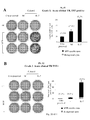
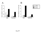
- the first two models allow the enumeration of Ag-specific T cells at the single cell level in the context of tumor disease (17) and of dendritic cell-based vaccination (18). These models allow validating the concept of T cell accumulation in vitro in an Ag-free environment.
- the third model is based on the engraftment of human T cells in immunedeficient mice, thus allowing to evaluate the immune competence of genetically modified central memory T cells.
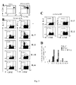
- TS/A-LACK tumors TS/A adenocarcinoma tumor cells expressing the Leishmania Major -derived antigenic protein LACK
- LACK-specific T cells are studied in the peripheral lymphoid organs (lymph nodes, spleen or blood) by flow cytometry with fluorescent LACK-peptide/MHC class II multimers.
- LACK-specific T cells can also be independently characterized by Ag-induced intracellular cytokine release.
- TS/A-LACK-specific T cells can be traced in BALB/c mice and in 16.2 ⁇ TCR transgenic mice, which express a transgenic TCR ⁇ chain specific for LACK allowing an easier characterization of LACK-specific CD4 T-cell response.
- TS/A cells naturally express the envelope protein gp70 of an endogenous MuLV for which an immunodominant epitope was previously described (AH-1, (19)).
- AH-1 an endogenous MuLV for which an immunodominant epitope was previously described
- Tag IV-specific T cells were also characterized by antigen-specific cytokine secretion assay ex vivo in order to enumerate TAG IV-specific CD8 T cell response.
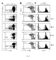
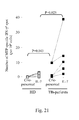
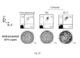
- T lymphocytes were derived from healthy donors and Mycobacterium tuberculosis infected patients and analyzed ex vivo and after an IL-7-driven short-term culture by antigen-specific cytokine release.
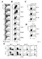
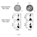
- IL-7 favored the accumulation of antigen-specific IL-2 and IFN- ⁇ -producing intermediate memory T cells by sustaining their in vitro proliferation and survival.
- IL-7 efficacy relied on in vivo antigen encounter, optimal cytokine amounts, and high cell density conditions, and was prevented by anti-LFA-1 antibody and by Cyclosporin A.
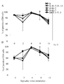
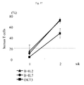
- IL-7 was markedly more efficient than IL-2 and IL-15 for CD4 memory T cell expansion, while IL-15 and IL-2 favored CD8 memory T cell expansion.
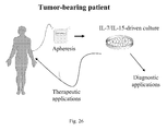
- the present invention has both diagnostic and therapeutic implication. On one hand it will aid the identification of rare populations of clinically relevant pathogen/tumor-specific T cells, and on the other hand it will also ameliorate current adoptive immunotherapeutic strategies.
- Model 3 Another aspect of this invention relies on the concept that central memory cells, upon TCR triggering in the presence of co-stimulation and culture with gamma-cytokines, can expand in vitro and be genetically modified by a viral vector, while maintaining their functional phenotype.
- T lymphocytes have a tremendous potential to cure cancer, infections, immuno-deficiencies and autoimmunity. Moreover, it can be used to modulate the immune responses occurring in the context of transplantation. Genetic modification is aimed at broaden the therapeutic interval of T lymphocytes by increasing their efficacy and/or limiting their toxicity. This is achieved by the transfer of genes encoding for novel receptors, biologically active products, resistance and control factors. Control factors are expected to provide selective in vivo elimination/inactivation of gene-modified cells if a toxic/unwanted effect ensues.
- T cells 1) mediate a direct anti-tumor effect (graft-versus-leukemia-GvL); 2) promote the engraftment of hematopoietic precursors; 3) provide an intact immune system to transplanted patients thus allowing to abate the incidence and severity of post-transplant infections.
- T-cells may also react against healthy host tissues, thus leading to the life-threatening graft-versus-host disease (GvHD) (20).
- GvHD graft-versus-host disease
- Genetic modification of T-cells with a retroviral vector expressing the Herpes Simplex Virus-thymidine kinase (TK) suicide gene confers selective sensitivity to the pro-drug ganciclovir (GCV).
- GCV pro-drug ganciclovir
- T-cell therapy and T-cell gene therapy depends on the ability of T-cells to proliferate and survive long-term in vivo. To achieve this goal, T-cells need to properly home to secondary lymphoid organs, where appropriate encounter with the antigen occurs and induces T-cells to acquire effector functions. It is becoming increasingly recognized that these attributes tends to segregate at early stages of mature T-cell differentiation, and in particular in the central memory compartment. Genetic modification with viral vectors may alter T-cell physiology. In particular, genetic modification through retroviral vectors (RV) requires cellular proliferation. This is currently achieved by activation with polyclonal stimuli and culture in the presence of high doses of recombinant human IL-2.
- RV retroviral vectors
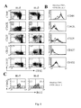
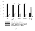
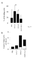
- Results indicate that the production of gene-modified lymphocytes with beads in the presence of IL-7 and IL-15 is feasible and that these cells have a physiologic CD4/CD8 ratio and a central memory functional phenotype, as defined by i) an absence of CD45RA expression and presence of CD62L expression, ii) a co-expression of the molecules CD27 and CD28 and iii) a production of IL-2 in the absence of IFN-y and/or IL-4.
- fully functional central memory recombinant lymphocytes means central memory T-cells with long-term survival potential, able to home to peripheral lymphoid organs, and to differentiate into effector cells upon antigen re-encounter in vivo.
- an in vitro method for expanding rare populations of antigen specific memory T cells in a sample comprising the step of exposing said sample to an effective amount of at least one cytokine receptor agonist able to selectively expand said rare populations of antigen specific memory T cells.
- the cytokine receptor agonist is a cytokine or a derivative thereof.
- the at least one cytokine receptor agonist is a IL-7 receptor agonist or a IL-15 receptor agonist, preferably a IL-15 receptor agonist or a IL-7 receptor agonist is also present, respectively.
- the rare populations of antigen specific memory T cells comprise CD4 + and/or CD8 + and/or ⁇ and/or NKT T cell populations.
- said sample is a biological sample belonging to the group of: blood and other liquid samples of biological origin, solid tissue samples, tissue cultures of cells derived therefrom and the progeny thereof, isolated cells from biological samples as i.e. PBMCs.
- said specific ligand is the specific antigen, or a derivative thereof for one of said rare populations of antigen specific memory T cells, more preferably the specific antigen is associated to a microbial pathogen including but not limited to Mycobacterium, Pneumocystic carinii, Plasmodium falciparum, Candida, Toxoplasma, CMV, EBV, BPV, HCV, HBV, HIV.
- the antigen is a tumor-associated antigen.
- the antigen is an allergen.
- the antigen is a self-antigen.
- the specific antigen is present as an antigen-MHC complex, or a derivative thereof.
- the detecting of said expanded rare populations of antigen specific memory T cells is performed by a binding assay.
- the detecting of said expanded rare populations of antigen specific memory T cells is performed by a cytokine release assay.
- the detecting of said expanded rare populations of antigen specific memory T cells is performed by a proliferation assay.
- cells are labeled with a fluorescent vital dye before incubating the sample with the specific ligand and the detecting step is performed by a dye dilution assay.
- It is a further object of the invention a kit for carrying out the method for detecting a rare population of antigen specific memory T cells in a sample as above described comprising at least one cytokine receptor agonist; at least one ligand specific for the rare populations of antigen specific memory T cells; detecting means.
- said specific ligand is the specific antigen or a derivative thereof for one of said rare populations of antigen specific memory T cells; more preferably the specific antigen is associated to a microbial pathogen including but not limited to Mycobacterium, Pneumocystic carinii, Palsmodium falciparum, Candida, Toxoplasma, CMV, EBV, BPV, HCV, HBV, HIV.
- the antigen is a tumor-associated antigen.
- the antigen is an allergen.
- the antigen is a self-antigen.
- the specific antigen is present as an antigen-MHC complex, or a derivative thereof.
- the isolating of said expanded rare populations of antigen specific memory T cells is performed by a binding step.
- the isolating of said expanded rare populations of antigen specific memory T cells is performed by measuring cytokine and cytotoxin production, including but not limited to ELISPOT assay, ELISA assay, flow cytometry cytokine detection assay for IL-2, IFN-g, IL-4, IL-5, IL-10, TNF-alfa, TGF-beta, granzymes.
- rare T cell populations isolated according to the method as described above for the treatment and/or the prevention of immune-, infectious-, cancer-, allergy-, auto-immune-related pathologies.
- said rare T cell populations are genetically modified.
- the populations of memory T cells comprise CD4 + and/or CD8 + and/or ⁇ and/or NKT T cell populations.
- lymphocytes are derived from a biological sample belonging to the group of: blood and other liquid samples of biological origin, solid tissue samples, tissue cultures of cells derived therefrom and the progeny thereof, isolated cells from biological samples as i.e. PBMCs.
- the specific lymphocyte activating receptor agonist is conjugated to cell-mimicking supports, more preferably the cell-mimicking supports are paramagnetic beads.
- one of the lymphocyte activating receptor agonists is specific for the CD3 polypeptide, preferably another of the lymphocyte activating receptor agonists is specific for a costimulatory receptor, i.e. CD28.
- the at least one cytokine receptor agonist is a IL-7 receptor agonist or a IL-15 receptor agonist, preferably a IL-15 receptor agonist or a IL-7 receptor agonist is also present, respectively.
- the vector is a viral vector.
- the exogenous gene encodes for a suicide gene, and/or a marker gene, and/or a biologically active molecule, and/or a receptor, and/or a soluble factor retained in the cell or released outside the cell, and/or a gene conferring resistance to a prodrug.
- mice Seven to 8-week old BALB/c and CD45.2 + C57BL/6 mice were purchased from Charles River (Charles River Italia, Milano, Italy).
- CD45.1 + C57BL/6, DO11.10 and 16.2 ⁇ Transgenic (Tg) (BALB/c background) (25) mice were bred in the Institute specific pathogen free facility.
- TS/A and TS/A-LACK mouse mammary adenocarcinoma were previously described (17, 19, 26).
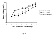
- Exponentially growing 4x10 5 tumor cells were subcutaneously injected in 100 ⁇ l of PBS in the right flank of syngeneic mice (BALB/c). Typically, five mice per group were used in each experiment.
- mice Twenty days after tumor cell injection mice were sacrificed and the axillary, brachial and inguinal peripheral lymph nodes (LN) draining and distal (non draining LN) to the site of tumor growth were recovered.
- LN peripheral lymph nodes
- DC dendritic cell
- mice were sacrificed fourteen days after DC administration and the axillary, brachial and inguinal LN were surgically excised.
- LN cells were cultured in 24 well plates at the density of 5x10 6 in complete medium in the absence or in the presence of recombinant murine IL-7 (200 ng/ml), IL-2 (20 ng/ml), IL-6 (45 ng/ml), or IL-15 (100 ng/ml) (all from Peprotech).
- LN cells were in vitro stimulated with the LACK-derived MHC II-restricted peptide (5 ⁇ M, (25)) and 5x10 6 irradiated syngeneic splenocytes. As a control similar cultures were set up from syngeneic naive mice.
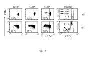
- LN cells were labeled with the fluorescent dye CFSE (5-(and-6)-carboxyfluorescein diacetate, succinimilyl ester) at the final concentration of 1 ⁇ M accordingly to manufacturer instruction.
- CFSE fluorescent dye
- DC Dendritic cell
- Bone Marrow derived DC were obtained as previously described (18). Briefly, CD45.2 + C57BL/6 bone marrow precursors were propagated for 7 days in complete Iscove's medium containing 25 ng/ml recombinant murine GM-CSF and 5 ng/ml recombinant murine IL-4 (Pharmingen, San Diego, CA). Then, BMDC were matured at 37° C in the presence of LPS (1 ⁇ g/ml, Sigma, Milan, Italy) for 8 hours and pulsed for 1 hour with 10 ⁇ g/ml of the large T Ag-derived Tag IV peptide (18).
- LPS 1 ⁇ g/ml, Sigma, Milan, Italy
- DC maturation and purity were routinely evaluated by flow cytometry after staining with mAb recognizing CD11c, MHC class II, B7.1, B7.2 and CD40 molecules (all from Pharmingen).
- 2x10 5 pulsed mature DC were subcutaneously injected in 200 ⁇ l of PBS in the right flank of syngeneic C57BL/6 mice.
- I-A d /LACK multimer staining was previously described. Briefly I-A d /LACK dimers (MHC II-peptide complexes, 3 ⁇ g/sample) are multimerized by the addition of Alexa 488-coupled protein A (Molecular Probes Inc., Eugene, 0.3 ⁇ g/sample) in PBS for 30 minutes at room temperature. Free protein A binding sites were saturated by the addition of total IgG (1 ⁇ g/sample).
- 6x10 5 cells were first incubated with a blocking buffer (5% rat serum + 95% culture supernatant of 2.4G2 anti-FcR mAb-producing hybridoma cells, 20 minutes) and then stained with the multimers (1h at 4°C, in PBS supplemented with 0.5% BSA). The cells were then stained with anti-CD4, anti-CD44, anti-CD11b, anti-B220, anti-CD8a mAbs (PharMingen, San Diego, CA, USA) and TO-PRO-3 (1 nM, Molecular Probes).
- a blocking buffer 5% rat serum + 95% culture supernatant of 2.4G2 anti-FcR mAb-producing hybridoma cells, 20 minutes
- the multimers (1h at 4°C, in PBS supplemented with 0.5% BSA.
- the cells were then stained with anti-CD4, anti-CD44, anti-CD11b, anti-B220, anti-CD8a mAbs (PharMingen,
- 3x10 5 CD4 + or 10 3 CD4 + I-A d /LACK + events were collected by excluding all of the anti-CD11b + , anti-B220 + , anti-CD8a + and TO-PRO-3 + events.
- the cells were surface stained with anti-CD4 or anti-CD8 mAb, and anti-CD44, anti-CD127, anti-CD25, anti-CD132 and anti-CD62L mAbs (all from PharMingen except anti-CD127 Ab, A7R34 clone, from Bioscience), and fixed, permeabilized and further stained with anti-Bcl-2 mAb according to to manufacturer instruction.
- 5 ⁇ m polystyrene sulfate latex beads were coated with I-A d /LACK dimers (20 ⁇ g/ml) and anti-CD28 mAb (37.51; 2 ⁇ g/ml) (LACK aAPC) or with anti-CD28 mAb only (control aAPC). Coating of the proteins was monitored by flow cytometry analysis. Typically 5x10 5 LN cells were cultured with 5x10 6 aAPC for 5 hours at 37°C. Brefeldin A (5 ⁇ g/ml, Sigma) was added to the cultures for the last 2 hours.
- Cytokine release induced by LACK aAPC was comparable to the one induced by LACK-pulsed syngeneic splenocytes (not shown).
- splenocytes were derived from DO 11.10 and CD45.1 + C57BL/6 mice and used as antigen presenting cells.
- Splenocytes (3x10 7 cells/ml) were pulsed with 1 ⁇ M AH-1 (19) and 10 ⁇ g/ml Tag IV (18) peptides for 1 hour at 37° C and then used to stimulate syngeneic LN cells derived from the LN of tumor-bearing mice and DC-vaccinated mice, respectively.
- CD4 + , KJ 1.26 - or CD8 + CD45.1 - events were then collected on a FACS Calibur.
- the total number of Ag-specific IL-2 + /IFN- ⁇ + T cells was determined by multiplying the percentage by the total number of Trypan Blue-negative LN cells.
- PBMC Peripheral blood mononuclear cells
- the ELISPOT assay for IFN- ⁇ secretion was performed as previously reported (28). Briefly cells were seeded in duplicate at 5x10 4 cells/well in 96-well plates (MAIPS4510; Millipore, Bedford, Mass.) pre-coated with anti-IFN- ⁇ capture mAb (B-B1; Diaclone, Besançon, France) in the presence of autologous irradiated PBMC (5x10 4 cells/well), and a pool of MTP peptides for 18 h at 37° C in air plus 5% CO 2 .
- Biotinylated anti-IFN- ⁇ detection mAb (B-G1; Diaclone) was added for 4 h, followed by the addition of streptavidin-alkaline phosphatase conjugate (Amersham Pharmacia Biotech Europe GmbH, Freiburg, Germany) for 1 h. After a washing step, the nitroblue tetrazolium-BCIP (5-bromo-4-chloro-3-indolylphosphate; Sigma, St. Louis, Mo.) chromogenic substrate was added. Individual spot forming cells (SFC) were counted using an automated image analysis system ELISPOT reader (AID-GmbH, Strassberg, Germany).
- a pool of six synthetic Mycobacterium tuberculosis peptides (MTP; Primm srl, Milano, Italy) with a length of 20 amino acids, >70% purified, derived from the sequences of ESAT-6 and CFP-10 secretory proteins of M. tuberculosis were used at a final concentration of 2 ⁇ g/ml per peptide for the detection of a specific response (28).
- PBMCs in medium alone or stimulated with phytohemagglutinin (PHA-P; Sigma) 5 ⁇ g/ml were respectively used as negative and positive controls.
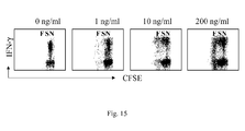
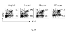
- MTP-specific IFN- ⁇ release was analyzed at the single cell level by intracellular cytokine secretion assay, Briefly, 0.6x10 6 CFSE labeled cells -were re-stimulated for 6 hours in the presence of human anti-CD28 stimulating mAb (2 ⁇ g/ml) and 3x10 6 autologous irradiated (5000 rad) PBMCs pulsed with HLA-DR-restricted MTP (4 ⁇ g/ml) or left unpulsed, in negative controls. In the last 5 hours Brefeldin A (10 ⁇ g/ml) was added to the cells. Thereafter the cells were fixed, permeabilized and stained with anti-CD4, anti-IL-2 and anti-IFN- ⁇ mAbs and analyzed on a FACS Calibur.
- TST tuberculin skin test
- Biocinetest-PPD tuberculin Choiron Italia srl, Milano, Italy
- the size of induration was evaluated after 48-72 hours (an induration ⁇ 10 mm was classified as positive).
- PBMC Peripheral blood mononuclear cells
- PBMC peripheral blood mononuclear cells
- PBMC peripheral blood mononuclear cells
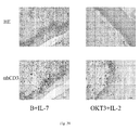
- anti-aCD3 OKT3 30ng/ml, OrthoBiotech, Raritan, NJ
- para-magnetic baCD3/CD28 3:1 beads/T-cell
- T-cells were enriched by baCD3/CD28 before culture.
- Cells activated with aCD3 were cultured with human recombinant IL-2 at 600 IU/ml (Chiron, Emeryville, CA).
- Cells activated with baCD3/CD28 were cultured: 1. In the absence of cytokines; 2. with human recombinant IL-7 at the minimal concentration of 5 ng/ml (Peprotech, London, UK); 3. with human recombinant IL-7 and IL-15 both at the minimal concentration of 5 ng/ml each (Peprotech, London, UK).
- cells were transduced with the SFCMM3 (39-40) retroviral supernatant by spinoculation at 2400 rpm for 2h at 37°C with 8 ⁇ g/ml polybrene (Sigma, St Louis, MO).
- the SFCMM3 retroviral vector encodes for the TK suicide gene under the LTR promoter and for a truncated form of the low affinity receptor for nerve growth factor (ALNGFR) under the SV40 promoter (27).
- the retroviral supernatant was provided by Molmed s.p.a.
- cell-sized beads used for the stimulation were removed from T-cells, according to manufactureur's instructions.
- transduced lymphocytes were positively selected according to the protocol that follows (protocol entitled: Positive immuneselection of transduced lymphocytes).
- transduced lymphocytes were positively selected. Since transduced cells expressed ⁇ LNGFr on their surface, anti-LNGFr antibodies covered with magnetic beads were used to separate transduced from untransduced lymphocytes.
- Cells were collected from plates in tubes, washed (1500 rpm, 10 min. at room temperature-RT) and resuspended in WB at a final concentration of 5x10 6 /ml.
- Anti-LNGFr antibody 20.1 was then added to the cell suspension (1 g/20x10 6 ) and cells were placed to rotate at 10 rpm for 30 min at RT.
- T-cells were then washed once and resuspended in WB at 25x10 6 /ml.
- Dynabeads M-450 Sheep anti-Mouse IgG were then added (5x10 6 beads/10 6 positive cells) and cells were placed to rotate at 10 rpm for 30' at room temperature. Transduced cells were then magnetically selected.
- tubes were placed near the magnet for 3 min and the negative fraction was discarded. This procedure was repeated for a total of three times. Finally the fraction of cells bound to the beads was removed from the magnet, washed, and resuspended in fresh medium with the appropriate cytokine cocktail at a concentration of 1x10 6 cells/ml.
- Flow cytometry was used for analysis of surface phenotype, transduction efficiency, cell cycle, and cytokine production.
- the following antibodies (Pharmingen) were used: FITC-conjugated mAb to human CD4, CD8, CD45RA, CD27 and IFN- ⁇ , (PE)-conjugated mAb to human CD4, CD8, LNGFR, CD62L, CD28 and IL-4, peridinin chlorophyll-a protein (PerCP)-conjugated mAb to mouse CD45 (Ly5.1), and allophycocyanin (APC)-conjugated mAb to human CD3 Samples were run through a Facscalibur flow-cytometer (Becton Dickinson, Mountain View, CA) after isotype-matched fluorochrome-conjugated irrelevant mAb-stained control and data were analyzed using CellQuest Software (Becton Dickinson).
- Fluorochrome-conjugated antibodies to CD127, CD122, CCR7, and to mouse CD45 were also utilized to stain lymphocytes.
- cytokine production cells were seeded in 24-well plates (1x10 6 /ml) and stimulated with 50ng/ml PMA (Sigma) and 1 ⁇ g/ml ionomycin (Sigma). After 4h, brefeldin A (Sigma) were added for additional 2h (10 ⁇ g/ml). Cells were then stained with the appropriate fluorochrome-conjugated anti-surface marker antibodies and fixed with 1% para-formaldehyde at 4°C for 10 min. Intracellular staining was performed with the appropriate fluorochrome-conjugated anti-cytokine antibodies after incubation for 20 min at RT in PBS 2% FBS containing 0,05% saponin (Sigma).
- CFSE consists of a fluorescein molecule containing a succinimidyl ester functional group and two acetate moieties. CFSE diffuses freely into cells and intracellular esterases cleave the acetate groups converting it to a fluorescent, membrane impermeant dye. The dye is not transferred to adjacent cells. CFSE is retained by the cell in the cytoplasm and does not adversely affect cellular function. During each round of cell division, the relative intensity of the dye decreases by half.
- CFSE-stained transduced T-cells were placed in culture in 24-well-plates with 2000 cGy irradiated allogeneic PBMCs (stimulators) in a 1:1 ratio. No cytokines were added to cell culture. CFSE-stained transduced T-cells placed in culture in the absence of stimulators were used as negative control. Cells placed in culture with soluble anti-CD3 antibody were used as positive control.
- To-Pro-3 is a high red intercalating DNA dye, detectable in fluorescence 4, which offers the possibility to co-stain cells with FITC, PE and APC conjugated antibodies. Its function is to reveal the dead cell fraction.
- mice with immunological defects in the adaptive (scid, recombination-activating genes -/- ) as well as in the innate compartment (NOD, common ⁇ chain -/- ) are commonly used to study human lymphocyte biology in vivo.
- NOD/scid mice to test the activity of central memory genetically modified lymphocytes in vivo.
- Six- to 8-week-old female NOD/scid mice were obtained from Charles-River Italia (Calco, Italy). The experimental protocol was approved by the internal committee for animal studies of our Institute (Institutional Animal Care and Use Committee [IACUC]). Mice were treated according to the following protocols:
- mice 6-8 weeks old female NOD/scid mice were obtained from Charles-River Italia (Calco, Italy).
- mice were transferred from laminar-flow isolators to normal cages and kept under specific pathogen-free conditions receiving sterile water and irradiated pellets ad libitum.
- mice were given 1mg blocking anti-mouse IL-2R ⁇ monoclonal antibody i.p. to neutralize residual NK activity.
- the anti-IL2R ⁇ antibody was produced as described from the TM ⁇ -1 hybridoma kindly provided by Prof. Tanaka (Osaka University, Japan).
- mice received total body irradiation with a single dose of 350 cGy (gamma irradiation from a linear accelerator) and were immediately infused with unmodified PBL or human lymphocytes transduced with the SFCMM3 retroviral vector (28).
- Unmodified PBL were obtained from PBMC after the depletion of contaminating monocytes, B- and NK-cells with Pan T-cell isolation kit (Miltenyi, Bergisch Gladbach, Germany). Cells were resuspended in 500 ⁇ l X-VIVO15 medium and infused i.p. Mice were then monitored for GvHD by calculating weight loss. Moribund mice were sacrificed for ethical reasons.
- mice were weighted and evaluated for xenogeneic GvHD according to the following score: weight loss (0 for weight loss ⁇ 10%, 1 for 10%-25%, 2 for >25%), hunching (0-2), activity (0-2), fur texture (0-2), and skin integrity (0-2), maximum index 10. Weight loss was also estimated as an independent variable, since it was considered the most objective criterion (Table 1).
- mice were transplanted with human skin and infused with allogeneic genetically modified lymphocytes to evaluate their ability to home to the human skin and mediate an allogenic GvH reaction.
- mice were transferred from laminar-flow isolators to normal cages and kept under specific pathogen-free conditions receiving sterile water and irradiated pellets ad libitum.
- mice are anesthetized with 12-18 mg avertin/mouse intraperitoneally. They were then depilated on the back, and an horizontal skin incision was performed bilaterally on the animal's back.
- mice were placed in a heated box for about 30 min. and finally transferred into their cages.
- mice To facilitate engraftment of human lymphocytes in NOD/scid mice, we functionally inactivated NK cells with anti-mouse IL-2 receptor ⁇ (TM ⁇ -1) antibodies prior to lymphocytes transfer.
- the antibody was produced from the TM ⁇ -1 hybridoma kindly provided by Prof Tanaka (Osaka University, Japan).
- mice received total body irradiation with a single dose of 300 cGy ( ⁇ irradiation from a linear accelerator). Animals were then weighted and immediately infused with transduced human lymphocytes that had been harvested at day 9 after initial stimulation. Cells were infused intravenously in 250 ⁇ L saline solution.
- mice were sacrificed and the two pieces of human skin removed bilaterally.
- Formalin-fixed, paraffin-embedded skin was cut in 4- ⁇ m thick sections and stained with hematoxylin and eosin for morphologic evaluation.
- Immunohistochemical assessment for the presence of human T lymphocytes was carried out with monoclonal anti-human CD3 antibody (Dako, Glostrup, Denmark) at 1:100 dilution, by way of the avidin/biotin peroxidase complex method using an automized Dako immunostainer. Staining reaction was revealed by the tetrahydrochloride chromogen method and sections were counterstained with hematoxylin. Pictures were taken with a Zeiss Axiocam HRC.
- IL-7 favors the detection of rare tumor-specific CD4 + T cells without the need of Ag-driven cell expansion.
- the enumeration of Ag-specific T cells might be critical to several clinical conditions, for which the presence of Ag-specific T cells, is of diagnostic and prognostic interest.
- the authors recently developed a preclinical mouse model of tumor-disease with TS/A tumor cells expressing the Leishmania Major- derived model Ag LACK (TS/A-LACK). While LACK-specific na ⁇ ve CD4 + T cells can not be identified in unmanipulated BALB/c mice (29), the authors recently identified LACK-specific Ag-experienced CD44 high CD4 + T lymphocytes in TS/A-LACK tumor-bearing mice by fluorescent MHC class II/Ag multimers staining and by LACK-specific IL-2 and IFN- ⁇ intracellular release (20).
- IL-7 known to favor survival of memory CD4 + T cells (7, 9-14) might enrich the frequency of tumor-specific CD4 + T cells.
- Control TS/A and TS/A-LACK tumor cells were subcutaneously injected in BALB/c mice. Twenty days after tumor cell injection, all the mice had developed measurable tumors. Mice were sacrificed and the tumor draining and non draining LN were surgically excised. While the formers contained a population of LACK-specific Ag-experienced CD44 high CD4 + T lymphocytes capable of IL-2 and IFN- ⁇ production, the latter remained unaware of the tumor and present LACK-specific naive CD4 + T lymphocytes (31).
- the frequency of LACK-specific CD4 + T cells was analyzed ex vivo, and after 7 days in culture in the presence of recombinant IL-7 without any further Ag stimulation ( Fig. 1 ).
- the draining LN of TS/A-LACK-tumor bearing animals showed a low but significant frequency of CD4 + I-A d /LACK + cells expressing high levels of CD44 ( Fig. 1A ), and capable of producing IL-2 and/or IFN- ⁇ upon LACK stimulation ( Fig. 1B ).
- the frequencies of CD4 + I-A d /LACK + CD44 high and LACK-specific cytokine-producing cells Fig.
- IL-2 and IFN- ⁇ -producing cells possibly representing intermediate memory T lymphocytes previously described as poly-functional (30-33), were mostly enriched for.
- the frequency of LACK-specific T cells in the tumor-draining LN of control TS/A-tumor bearing mice and from the non draining LN of TS/A as wells as TS/A-LACK mice was comparable to the one found in naive BALB/c mice ex vivo ( Fig. 1C , D and not shown), and remained within background measures after the culture in IL-7 ( Fig. 1C, D ).
- IL-7 enriches LN cultures for in vivo -primed tumor-specific Ag-experienced CD4 + T cells thus favoring their enumeration bypassing the need of in vitro Ag-driven cell expansion.
- IL-7 and IL-2 but not antigenic stimulation favor the accumulation of tumor-specific CD4 + T cells.
- Re-stimulation with Ag is most commonly used to expand, and in some instances to identify Ag-specific CD4 + T cells (34). Furthermore, in addition to IL-7, also IL-2 and IL-15 control memory T cells proliferation (13, 35-37).
- LN cells from TS/A-LACK tumor-bearing mice were cultured in the presence of irradiated singeneic APC unpulsed (APC) or pulsed with the LACK-derived peptide (Ag/APC) or in the presence of optimal amounts of IL-7, IL-2, IL-15 and IL-6, as control, and analyzed by flow cytometry.
- the frequency of CD4 + I-A d /LACK + CD44 high T cells was slightly higher in cultured cells when compared to the one found ex vivo ( Fig. 1A ), but no difference was detected among control (APC) and Ag-stimulated cultures (Ag/APC) ( Fig. 2A ).
- culturing the cells in IL-7 increased the frequency ( Fig. 2A, B ) as well as the total number ( Fig. 2C, D ) of LACK-specific CD4 + T cells above the ones found in control (not shown) and IL-6-driven cultures.
- IL-2 enriched LN cultures of CD4 + I-A d /LACK + CD44 high T cells ( Fig.
- CD4 + I-A d /LACK + CD44 high T cells remained comparable to the one of control culture, but LACK-specific CD4 + T cells capable of cytokine-secretion were enriched for in several independent experiments.
- IL-2/IFN- ⁇ -secreting CD4 + T cells were mostly enriched for, and better favored in IL-7-driven culture ( Fig. 2C,D ).
- IL-7 and IL-2 sustain the Ag-independent spontaneous proliferation and survival of in vivo -primed tumor-specific CD4 + T cells.
- LACK-specific T cells In the presence of IL-7, a higher frequency of cells derived from tumor-draining LN completed 1-3 cell division, and LACK-specific T cells, identified by their ability to secrete IL-2 and IFN-g upon LACK-specific restimulation were markedly enriched for and had a lower CFSE content (MFI: 44) ( Fig. 3B and C ). Indeed, LACK-specific CD4 + T cells had performed more than 3-4 cell cycles, which distinguishes them from cells undergoing slow homeostatic-like cell division (less than 4 cell cycles). IL-7-driven homeostatic cell division was also found in the LN of naive control mice ( Fig. 5C ). However, LACK-specific CD4 + T cells were not enriched for in these cultures (not shown).
- LACK-specific T cells found in tumor-draining LN and accumulating in vitro in response to IL-7 possibly represent recently primed unpolarized intermediate memory T cells, also found in vivo in chronically infected patients (32).
- IL-7 In addition to IL-7, also IL-2 supported the in vitro proliferation of a fraction of CD4 + T cells and increased the number of LACK-specific memory lymphocytes. In contrast, IL-15 and IL-6 failed to support either proliferation of the cells, or the accumulation of LACK-specific CD4 + T cells over the one found in control (nil) cultures ( Fig. 3B, and D ). Thus, IL-7 and IL-2 are capable of supporting the in vitro expansion of in vivo Ag-experienced memory T cells.
- IL-7, IL-2 and to a reduced extent IL-15, but not IL-6, TNF- ⁇ , and IL-10 enriched the cultures for these cells.
- LACK-specific CD4 + T cells capable of IL-2 and IFN- ⁇ LACK-specific release were contained again within CFSE dim cells ( Fig. 4E ).
- IL-7 and IL-2 enrich LN cultures of Ag-experienced CD4 + T cells by sustaining their in vitro proliferation bypassing the need of Ag-stimulation.
- IL-7 To further characterize the relative potency of IL-7 and IL-2 in promoting the accumulation of Ag-experienced CD4 + T cells, CFSE labeled cells were stained with the fluorescent dye TO-PRO-3, able to identify viable and dead cells within proliferating cells. IL-7 best preserved the viability of the cultures with only 15% of TO-PRO-3 + , dead cells after a week. At difference, up to 47% of the cells maintained in IL-2 and 60%, 57%, and 73% of the cells cultured in the absence of exogenous cytokine or in the presence of IL-15 and IL-6 resulted TO-PRO-3 + ( Fig. 5A ).
- IL-7 and IL-2 to favor T cell survival is linked to their capacity to regulate the expression of the anti-apoptotic factor Bcl-2 (10, 38).
- Bcl-2 levels in CFSE-labeled LN cultures maintained in the absence and in the presence of recombinant cytokines.
- CFSE dim cells expressed optimal levels of Bcl-2 ( Fig. 5B ), suggesting that cells primed in vivo had received a pro-survival signal.
- IL-7 was unique in favoring the survival of both CFSE dim and CFSE bright cells.
- TS/A-LACK tumor-draining LN cells were labeled with the CFSE vital dye and cultured for a week in the presence of IL-7 or IL-2 alone.
- CFSE dim cells maintained in IL-7 alone, a fraction of CD4 + T cells, comparable to the one found ex vivo ( Fig.
- CD44 and of CD62L retained high or low expression of CD44 and of CD62L ( Fig. 6A, B ). These cells are possibly representative of naive, effector and central memory lymphocytes (8, 39-41).
- IL-7-treated CFSE dim cells expressed intermediate levels of CD25, and CD132, and downregulated the surface expression of CD127, as previously reported (42).
- IL-7 In cultures derived from 16.2 ⁇ transgenic mice, while both IL-7 and IL-2 enriched for Ag-experienced CD4 + T cells with a surface phenotype comparable to the one found ex vivo ( Fig. 7A ), only IL-7 maintained the original ratio between CD44 high and low cells among I-A d /LACK + and I-A d /LACK- CD4 + T cells ( Fig. 7B ).
- the ability to preserve the relative lymphocyte representation might be relevant when attempting to exploit the short-term culture to determine the frequency of Ag-specific T cells in biological samples.
- Bcl-2 + cells expressed low levels of the LN homing molecule CD62L in IL-2-cultured cells
- in the presence of IL-7 up to 53% of CFSE dim CD4 + T cells maintained an optimal expression of Bcl-2 and CD62L ( Fig. 6C ).
- IL-7, IL-2 and IL-15 expand Ag-specific specific memory CD8 + T cells in an Ag-independent manner.
- IL-7 peptide-specific CD8 + T cells induced by a dendritic cell (DC)-based vaccine.
- DC dendritic cell
- C57BL/6 mice were vaccinated with bone marrow derived DC pulsed with the MHC class I restricted Tag IV peptide (DC-Tag) derived from the SV40 Large T antigen (18).
- DC-Tag MHC class I restricted Tag IV peptide
- LN cells were analyzed by Ag-specific intracellular cytokine release ex vivo and after the cytokine-driven cultures.
- As a control LN cells were also derived from naive unvaccinated C57BL/6 mice.
- Tag IV specific CD8 + T cells capable of producing only IFN- ⁇ or IL-2 and IFN- ⁇ after Ag re-stimulation were undetectable in naive mice, and detectable at low frequencies in DC-vaccinated mice (0% and 0.37%, respectively).
- IL-7 IL-2
- IL-15 in the absence of Ag restimulation the frequency (3.94%, 1.83% and 1.95%, respectively) as well as the total number ( Fig. 8 ) of Tag IV-specific CD8 + T cells were markedly increased in the cultures derived from DC-vaccinated mice and not from naive mice (not shown).
- IL-7 can be used to reveal tumor-and vaccine-induced CD8 + T cell responses even in the absence of Ag re-stimulation.
- IL-7 reveals antigen-specific CD8 + T cells otherwise undetectable ex vivo.
- TS/A cells naturally express the envelope protein gp70 of an endogenous MuLV for which an immunodominant epitope was previously described (AH-1, (19)).
- AH-1 an endogenous MuLV for which an immunodominant epitope was previously described
- the authors compared the AH-1-specific CD8 + T cell responses ex vivo and after a week in culture without or with IL-7, IL-2, IL-15 and IL-6 in the absence of Ag re-stimulation. Lymphocytes were analyzed by intracellular cytokine release upon stimulation with unpulsed and AH-1-pulsed syngeneic splenocytes ( Figure 10 ).
- AH-1-specific CD8 + T cells were undetectable ex vivo since the frequency of cytokine producing cells remain within background levels ( Figure 10A ) and was comparable to the one found in naive mice (not shown).
- LN cultures derived from TS/A-LACK tumor-bearing LN and maintained with or without recombinant cytokines contained a variable frequency of cells producing IFN- ⁇ independently from AH-1 re-stimulation ( Fig 10B ).
- IL-7 the cultures were enriched for AH-1-specific CD8 + T cells able to produce only IFN- ⁇ or IL-2 and IFN- ⁇ upon Ag re-stimulation ( Figure 10B ).
- IL-2 increased the frequency and total number of AH-1-specific CD8 + T cells ( Figure 10B and C ), these were not increased by IL-15- and IL-6 where the frequency remained comparable to the one found in cultures derived from naive mice and within background levels (data not shown).
- IL-7, and IL-2 are able to reveal tumor-specific CD8 + T cells otherwise undetectable ex vivo, bypassing the need for in vitro Ag-driven cell expansion.
- Interleukin-7 synergizes with a Cyclosporin A-sensitive signal for the selective expansion of memory CD4 + T cells.
- naive T cells were derived from the LN of naive or TS/A-LACK-tumor bearing16.2 ⁇ mice, which are transgenic mice having a sizeable frequency of LACK-specific naive CD4 + T cells (25), labeled with CFSE and cultured in the presence of optimal IL-7 amounts, and the indicated inhibitory agents.
- naive T cells were stimulated with LACK-pulsed antigen presenting cells (APC) in the absence or in the presence of the selected inhibitors ( Fig. 11A, B ).
- APC LACK-pulsed antigen presenting cells
- Ag-driven T cell proliferation was markedly inhibited by anti-MHC class II mAb, and CsA, and partially hampered by anti-ICAM-1, anti-LFA-1 mAb ( Fig.
- IL-7 is able to drive the accumulation of fast proliferating IL-2/IFN- ⁇ + intermediate memory CD4 + T cells by the synergy with a cell-derived CsA sensitive signal possibly mediated by adhesion molecules and/or self peptide/MHC interaction.
- IL-7 sustains the selective accumulation of fast-dividing peripheral blood human CD4 memory T lymphocytes.
- IL-7 and IL-15 sustain a slow homeostatic-like cell division of both central memory and effector human memory T cells (13).
- PBMC peripheral blood-derived T lymphocytes.
- CFSE peripheral blood-derived T lymphocytes
- IL-7 In addition to IL-7, also IL-2 and IL-15 sustained the in vitro proliferation of human CD4 + T cells ( Fig. 17A ), as also reported elsewhere (13), and the upregulation of Bcl-2 expression ( Fig. 18 ). However, most of the cells proliferating in response to IL-2 and IL-15 completed 1-4 cycle of slow homeostatic-like cell division, and fast-proliferating memory T cells failed to accumulate.
- IL-7 mostly supported the slow homeostatic division of a fraction of CD8 + T lymphocytes, while IL-2 and IL-15 allowed the accumulation of a population of fast-dividing CD8 + T lymphocytes ( Fig. 17B ) suggesting an analogous role for these cytokines on the two different T cell subsets.
- IL-7 driven T cell expansion of peripheral blood human T lymphocytes is sensitive to Cyclosporin A.
- the IL-7 and IL-15-driven slow homeostatic-like cell division of human memory T cells was reported to be insensitive to CsA, and instead rely on p38-dependent signaling (13).
- the IL-7-driven accumulation of fast-proliferating CD4 + T cells was sensitive to CsA, and to RAPA, and to a lesser extent to SB ( Fig. 19A ) and PP2 (not shown).
- CsA did not prevent the cytokine-driven slow proliferation consistently with previous report (13).
- CD45RA - , CD62L + memory T cells appeared to be the largest fraction of spontaneously proliferating CD4 T cells, were mostly enriched for by the ex vivo expansion protocol, and were most sensitive to CsA inhibition ( Fig. 19B and C ).
- the authors identified a population of CD45RA - , CD62L + memory CD4 + T cells able of spontaneous in vitro fast proliferation in the peripheral blood of healthy donors.
- the accumulation of these memory CD4 + T cells was favored by IL-7, was cell density-dependent and CsA-sensitive.
- IL-7-driven short-term cultures aid the enumeration of Mycobacterium tuberculosis-specific CD4 + T cells in human subjects.
- IL-7 sustains the in vitro expansion of a fast-proliferating memory T cells possibly programmed in vivo to proliferate in vitro
- crio-preserved PBMC samples of M. tuberculosis-infected (TB) patients were analyzed at the time of thaw or after a week in culture with optimal IL-7 amounts, by MTP-specific ELISPOT analysis (43). Patients were chosen based on their clinical history and manifestation of acute TB (clinic and culture confirmed), on their positive reaction to the TST, and on the ability to respond to MTP in the ELISPOT-IFN- ⁇ assay.
- Crio-preserved PBMC from not infected healthy donors were also analyzed as control.
- Pt#1 showed a sizeable number of IFN- ⁇ + spots upon MTP-specific restimulation ( Fig. 20A ), at the time of thawing (Crio-preserved: 950 SFCx10 6 PBMC).
- control medium Nail
- the number of IFN- ⁇ + MTP-specific cells doubled (1890 SFCx10 6 PBMC), reflecting the increased frequency of CD4 + T cells in cultured cells (numbers in brackets in Fig. 20A ).
- IFN- ⁇ + MTP-specific cells 3930 SFCx10 6 PBMC
- Pt#2 and Pt#3 had detectable MTP-specific T cells at the time of sample collection (not shown), but not in crio-preserved/thawed samples ( Fig. 20B , 20C ).
- IFN- ⁇ + MTP-specific spots were instead revealed.
- IL-7 resulted in the increase in absolute numbers of MTP-specific T cells in all the TB-patients analyzed, and as a result, the difference among healthy donors and TB patients in the number of MTP-specific IFN- ⁇ producing cells gained in significance ( Fig. 21 ).
- MTP-specific T cells accumulating in response to IL-2 were mostly represented by IL-2/IFN- ⁇ + memory CD4 T cells as determined by MTP-induced intracellular cytokine secretion ( Fig. 22 ). Furthermore, the expansion of MTP-specific IL-2/IFN- ⁇ + memory CD4 T cells was prevented in the presence of CsA ( Fig. 23 ).
- IL-7-driven short-term cultures aid the enumeration of Candida antigen-specific human T lymphocytes.
- IL-7 could enhance the identification of T lymphocytes specific for Candida Albicans-derived antigens.
- PBMCs from Pt#1 were analyzed at the time of thawing or after a 7 days culture in plain medium or in the presence of IL-7, by an ELISPOT assay performed with unpulsed or C.Albicans-derived Ag-pulsed irradiated autologous PBMC.
- C.Albicans-specific T cells capable of IFN- ⁇ release were enriched for by the short-term culture in IL-7 ( Fig. 24A, B ).
- the adoptive cell therapy with IL-7/IL-15 cultured memory T cells delays tumor growth in vivo.
- lymph nodes were derived from TS/A-LACK tumor-bearing mice and cultured for 7 days in optimal cell density (5x10 6 cells/ml) and optimal cytokines amounts (50 ng/ml).
- optimal cell density 5x10 6 cells/ml
- optimal cytokines 50 ng/ml.
- naive T cells derived from a control mouse were also cultured in the same conditions. Thereafter 10 7 cultured cells bearing comparable frequencies of CD4 and CD8 T cells were adoptively transferred into naive BALB/c mice.
- mice 48 hours later mice were challenged with 300.000 TS/A-LACK cells and tumor growth was monitored overtime.
- TS/A-LACK tumors rapidly developed in control mice and in mice adoptively transferred with cytokine-treated naive T cells.
- tumor growth was significantly delayed in mice adoptively transferred with cytokine-treated tumor-bearing mice derived T cells ( Fig. 25 ). This indicates that the IL-7/IL-15-driven cultures determined the expansion of a population of clinically relevant memory T cells.
- Cell proliferation is required for retroviral transduction of T lymphocytes.
- the authors activated PBMC with aCD3 or baCD3/CD28.
- Cells were activated with baCD3/CD28 and cultured with IL-7 and IL-15 or with aCD3 and cultured with IL-2 ( Figure 27A ).
- Cells were transduced at day 2, by spinoculation, with the SFCMM3 retroviral vector. Transduction was performed at the same time and following the same protocol.
- Activation with baCD3/CD28 in the presence of IL-7 and IL-15 promoted a higher T cell expansion ( Figure 27A ) and led to a higher transduction efficiency than activation with aCD3 in the presence of IL-2 ( Figure 27B ).
- the physiological CD4/CD8 ratio was analyzed and found to be better maintained in transduced cells activated with baCD3/CD28 and cultured with IL-7 and IL-15 than with cells activated with aCD3 and cultured with IL-2 ( Figure 27C ).
- transduced T-cells activated with aCD3 and cultured with IL-2 were mainly CD45RA - CD62L - , i.e, effector memory cells.
- transduced CD4 + T-cells activated with baCD3/CD28 and cultured IL-7 and IL-15 were highly enriched for CD45RA - CD62L + , i.e., central memory cells ( Figure 28 A) .
- Activation with CD3/CD28 beads followed by culture in the presence of cytokine (IL2, IL7+IL15 or IL7) preserves the physiological CD4/CD8 ratio in transduced T lymphocytes.
- Activation with CD3/CD28 beads followed by culture in the presence of cytokine induces a significantly higher proliferation rate of transduced cells than other culture conditions.
- Protocol of ex vivo gene transfer designed for clinical application must fulfil to the relevant criteria related to feasibility: one of the major feasibility issue in the clinical translation of a gene therapy approach relates to cell number and cell expansion in vitro.
- FIG 30C a statistically significant difference was observed in cell numbers obtained between genetically modified cells stimulated with beads and cultured with cytokines, compared to the other conditions (beads alone and OKT3+IL-2).
- Cytokine receptors' expressions are tightly regulated during T-cell stimulation.
- IL-2/IL-15 receptor ⁇ expression increases after T cell activation and then decreases to an intermediate level of expression that is retained throughout the memory-cell phase (13) .
- Figure 32 shows the expression kinetic of CD122 in transduced lymphocytes during 13 days of culture.
- T-cells, and in particular CD4 + cells up-regulated CD 122 after activation, reaching a peak around day 4, when almost 100% of genetically modified cells expressed the molecule.
- Cells then slowly down-regulated CD122 expression, reaching the same level of expression observed before stimulation 13 days after the beginning of T-cell culture, when cells had reached the resting state.
- IL-2 receptor ⁇ (CD25) is a relevant activation marker for T-lymphocytes.
- naive T cells do not express CD25; however, its expression is rapidly upregulated by T-cell activation and usually declines before the proliferative peak of the response.
- Transduced cells activated with anti-CD3/CD28 beads + IL-7 show the maximal expression of IL-7 receptor ⁇ , a marker of long-surviving memory T-cells
- IL-7 receptor ⁇ (CD127) is constitutively expressed by naive T cells. Its expression is downregulated by T-cell activation (in a specular manner compared to CD25) and such down-regulation might promote cell death. Conversely, expression of CD127 increases as the immune response proceeds, reaching high levels in memory T cells.
- IL-7 is a potent survival factor for memory T-lymphocytes: triggering of the receptor by IL7 promotes T cell survival and proliferation and protects cells from apoptosis through different intracellular signal pathways.
- IL-7 receptor ⁇ underwent a deep down-regulation after stimulation (approximately between day 1 and day 6).
- Transduced lymphocytes generated with beads + IL-7 have the highest proliferative potential, when stimulated with an allogenic antigen.
- Transduced T cells generated with each of the five conditions were stained with CFSE and co-cultured with irradiated allogeneic PBMCs. After 1 week we counted cell numbers and analyzed CFSE dilution by FACS to evaluate the percentage of dividing cells. As shown in figure 35A , we found a statistically significant difference between IL-7-containing conditions and the other protocols. in CD3 + and CD8 + T-cells: Indeed, a high percentage (40%) of transduced cells generated with beads CD3/CD28 + IL-7 had divided in 1 week after allogeneic stimulation. On the contrary, only 20% of transduced cells generated with OKT3- had divided in the same culture conditions.
- Transduced lymphocytes generated with beads + IL-7 have the lowest sensitivity to death.
- AICD activation induced cell death
- Transduced cells cultured in IL2 proved highly sensitive to AICD and death by neglect.
- transduced cells generated with beads CD3/CD28 + IL-7 displayed the lowest mortality, comparable to that of unmodified lymphocytes. This observation may be related to the persistent expression of CD127 on a high proportion (33% of CD8 + and 52% of CD4 + ) of genetically modified lymphocytes generated with beads CD3/CD28 and IL-7.
- transduced lymphocytes generated with beads CD3/CD28 and IL-2 who proved highly sensitive to cell death, showed the lowest proportion of cells expressing CD127 + cells (30%).
- FACS plots in figure 36A show a representative example of CD127 detection in CFSE-stained transduced T-lymphocytes 10 days after allogeneic stimulation.
- Transduced lymphocytes generated with beads + IL-7 preserve a central memory phenotype after allogeneic stimulation.
- Immunological memory is ensured by a self-renewal capacity of memory cells, that, upon antigen re-enconteer, divide, and generate both effectors, able to directly eliminate the pathogen, and memory cells, able to protect the host long-term.
- CCR7 a marker of central memory cells
- Transduced lymphocytes generated with beads + IL-7 have the highest alloreactive potential in vivo
- mice peripheral blood increased from week one to week 2 after infusion.
- Transduced cells generated with beads engrafted at higher extent than transduced cells generated with OKT-3.
- the difference was more evident at the second week after T cell infusion ( Fig 37 ).
- Activation with beads CD3/CD28 and IL-7 stimulation confer to transduced cells the highest reactivity against xenogeneic antigens.
- Xenogeneic GvHD was monitored according to a clinical score described in material and methods and by measuring weight loss. Infused
- NOD/Scid mice progressively lost their weight and some of them eventually died for xenogeneic GvHD or were sacrificed for ethical reasons.
- the most xeno-reactive T-cells were those stimulated with anti CD3/CD28 beads and cultured with IL-7, followed by transduced cells generated with CD3/CD28 beads and IL-2.
- the stimulation with soluble anti CD3 antibodies did not generate strongly xeno-reactive T-cells, as mice infused with these lymphocytes did show neither a significant body weight loss, nor the appearance of other clinical xeno-GvHD signs ( Fig 38 ).
- NOD/Scid human skin chimera mouse model consists of NOD/Scid mice, that had undergone skin transplantation, through the insertion of two pieces of human abdominal epidermis into two subcutaneous pockets on the mouse back and that were subsequently infused intravenously with genetically modified cells.
- Human skin transplantation allows to investigate T-cell reactivity against allogeneic antigens, by histological studies.
- Transplanted human skin indeed, contains not only epidermal and stromal cells, but also some antigen presenting cells, which are able to attract circulating lymphocytes and possibly promote their activation.
- T cells play a central role in the generation of immunity to pathogens, to tumors, and to immuno-deficiencies and in autoimmune disorders, it has been difficult to use them as diagnostic and prognostic markers of immunocompetence in humans. Furthermore, techniques suitable for the expansion of non polarized poly-functional intermediate and central memory lymphocytes are currently missing.
- IL-7, and IL-15 can be used to enrich biological samples, such as peripheral LN, blood, and tumor for in vivo primed Ag (tumor/pathogens/allergens/self antigens)-specific CD4 + or CD8 + T cells.
- IL-7 better maintained the original lymphocyte phenotype and representation and better favored the survival of all T lymphocyte subsets, allowing the detection and expansion of rare CD4 + T memory lymphocytes.
Landscapes
- Health & Medical Sciences (AREA)
- Life Sciences & Earth Sciences (AREA)
- Engineering & Computer Science (AREA)
- Immunology (AREA)
- Chemical & Material Sciences (AREA)
- General Health & Medical Sciences (AREA)
- Biomedical Technology (AREA)
- Cell Biology (AREA)
- Medicinal Chemistry (AREA)
- Hematology (AREA)
- Molecular Biology (AREA)
- Biotechnology (AREA)
- Animal Behavior & Ethology (AREA)
- Veterinary Medicine (AREA)
- Public Health (AREA)
- Pharmacology & Pharmacy (AREA)
- Microbiology (AREA)
- Organic Chemistry (AREA)
- Genetics & Genomics (AREA)
- Urology & Nephrology (AREA)
- Zoology (AREA)
- Bioinformatics & Cheminformatics (AREA)
- Biochemistry (AREA)
- Epidemiology (AREA)
- Wood Science & Technology (AREA)
- Virology (AREA)
- Nuclear Medicine, Radiotherapy & Molecular Imaging (AREA)
- General Chemical & Material Sciences (AREA)
- Chemical Kinetics & Catalysis (AREA)
- General Physics & Mathematics (AREA)
- Food Science & Technology (AREA)
- Physics & Mathematics (AREA)
- Analytical Chemistry (AREA)
- Pathology (AREA)
- Oncology (AREA)
- Tropical Medicine & Parasitology (AREA)
- Mycology (AREA)
- General Engineering & Computer Science (AREA)
- Communicable Diseases (AREA)
- Proteomics, Peptides & Aminoacids (AREA)
Priority Applications (3)
| Application Number | Priority Date | Filing Date | Title |
|---|---|---|---|
| PL08102851T PL1956080T3 (pl) | 2005-08-08 | 2006-08-03 | Zastosowanie IL-7 oraz IL-15 do genetycznej modyfikacji komórek T pamięci |
| SI200631202T SI1956080T1 (sl) | 2005-08-08 | 2006-08-03 | Uporaba IL-7 in IL-15 za genetsko modifikacijo spominskih limfocitov T |
| CY20111101280T CY1112293T1 (el) | 2005-08-08 | 2011-12-22 | Χρηση των il-7 και il-15 για τη γενετικη τροποποιηση μνημονικων τ λεμφοκυτταρων |
Applications Claiming Priority (2)
| Application Number | Priority Date | Filing Date | Title |
|---|---|---|---|
| US70650305P | 2005-08-08 | 2005-08-08 | |
| EP06796252A EP1941028A2 (fr) | 2005-08-08 | 2006-08-03 | Utilisation de cytokines a chaine gamma commune pour la visualisation, l'isolation et la modification genetique de lymphocytes t a memoire |
Related Parent Applications (2)
| Application Number | Title | Priority Date | Filing Date |
|---|---|---|---|
| EP06796252.2 Division | 2006-08-03 | ||
| EP06796252A Division EP1941028A2 (fr) | 2005-08-08 | 2006-08-03 | Utilisation de cytokines a chaine gamma commune pour la visualisation, l'isolation et la modification genetique de lymphocytes t a memoire |
Publications (3)
| Publication Number | Publication Date |
|---|---|
| EP1956080A2 true EP1956080A2 (fr) | 2008-08-13 |
| EP1956080A3 EP1956080A3 (fr) | 2008-09-10 |
| EP1956080B1 EP1956080B1 (fr) | 2011-09-28 |
Family
ID=37440920
Family Applications (2)
| Application Number | Title | Priority Date | Filing Date |
|---|---|---|---|
| EP08102851A Active EP1956080B1 (fr) | 2005-08-08 | 2006-08-03 | Utilisation de IL-7 et IL-15 pour la modification génétique des lymphocytes T à mémoire |
| EP06796252A Withdrawn EP1941028A2 (fr) | 2005-08-08 | 2006-08-03 | Utilisation de cytokines a chaine gamma commune pour la visualisation, l'isolation et la modification genetique de lymphocytes t a memoire |
Family Applications After (1)
| Application Number | Title | Priority Date | Filing Date |
|---|---|---|---|
| EP06796252A Withdrawn EP1941028A2 (fr) | 2005-08-08 | 2006-08-03 | Utilisation de cytokines a chaine gamma commune pour la visualisation, l'isolation et la modification genetique de lymphocytes t a memoire |
Country Status (17)
| Country | Link |
|---|---|
| US (3) | US8999715B2 (fr) |
| EP (2) | EP1956080B1 (fr) |
| JP (1) | JP2009504151A (fr) |
| KR (1) | KR20080048455A (fr) |
| CN (1) | CN101273122A (fr) |
| AT (1) | ATE526395T1 (fr) |
| AU (1) | AU2006277577A1 (fr) |
| CA (1) | CA2618580A1 (fr) |
| CY (1) | CY1112293T1 (fr) |
| DK (1) | DK1956080T3 (fr) |
| ES (1) | ES2374504T3 (fr) |
| IL (1) | IL189143A0 (fr) |
| NO (1) | NO20081108L (fr) |
| PL (1) | PL1956080T3 (fr) |
| PT (1) | PT1956080E (fr) |
| SI (1) | SI1956080T1 (fr) |
| WO (1) | WO2007017915A2 (fr) |
Cited By (3)
| Publication number | Priority date | Publication date | Assignee | Title |
|---|---|---|---|---|
| US8956828B2 (en) | 2009-11-10 | 2015-02-17 | Sangamo Biosciences, Inc. | Targeted disruption of T cell receptor genes using engineered zinc finger protein nucleases |
| WO2017034833A1 (fr) * | 2015-08-21 | 2017-03-02 | Mayo Foundation For Medical Education And Research | Méthodes et matériels pour multiplier des cellules t spécifiques d'antigènes en culture |
| WO2018102761A1 (fr) * | 2016-12-02 | 2018-06-07 | City Of Hope | Procédés de préparation et d'accroissement de lymphocytes t exprimant des récepteurs antigéniques chimériques et d'autres récepteurs |
Families Citing this family (18)
| Publication number | Priority date | Publication date | Assignee | Title |
|---|---|---|---|---|
| CN101824400B (zh) * | 2009-03-05 | 2012-08-08 | 中国科学院微生物研究所 | 一种放大增殖抗原特异性t细胞的方法 |
| ES2659217T3 (es) | 2012-03-28 | 2018-03-14 | Gadeta B.V. | Intercambio combinatorio de cadena receptora de célula T gamma 9 delta 2 |
| CA2906970C (fr) | 2013-03-21 | 2021-05-18 | Ospedale San Raffaele Srl | Interruption ciblee de genes de recepteur des lymphocytes t a l'aide de nucleases proteiques a doigt de zinc genetiquement modifiees |
| IL303247A (en) * | 2014-12-29 | 2023-07-01 | Novartis Ag | Methods for preparing cells expressing a chimeric receptor antigen |
| US20180171294A1 (en) * | 2015-03-26 | 2018-06-21 | The Trustees Of The University Of Pennsylvania | In vitro artificial lymph node method for sensitization and expansion of t cells for therapy and epitope mapping |
| JP7114457B2 (ja) | 2015-04-17 | 2022-08-08 | ザ トラスティーズ オブ ザ ユニバーシティ オブ ペンシルバニア | キメラ抗原受容体発現細胞の有効性および増殖を改善するための方法 |
| TW201708538A (zh) | 2015-07-21 | 2017-03-01 | 諾華公司 | 改良免疫細胞之功效及擴展之方法 |
| CA3026180A1 (fr) | 2016-06-10 | 2017-12-14 | Gadeta B.V. | Recepteurs de lymphocytes t gamma delta restreints a l'antigene leucocytaire humain et leurs methodes d'utilisation |
| KR20190033066A (ko) * | 2016-06-24 | 2019-03-28 | 맥마스터 유니버시티 | 입양 세포 전달 및 종양붕괴 바이러스 병용 치료 |
| KR102501827B1 (ko) * | 2016-09-16 | 2023-02-22 | 베이롤 칼리지 오브 메드신 | 바이러스-특이적인 t-세포의 활성화 및 확장을 위한 플랫폼 |
| RU2019133280A (ru) | 2017-03-22 | 2021-04-22 | Новартис Аг | Композиции и способы для иммуноонкологии |
| KR20200033930A (ko) * | 2017-07-28 | 2020-03-30 | 브리스톨-마이어스 스큅 컴퍼니 | 체크포인트 억제제에 대한 예측성 말초 혈액 바이오마커 |
| AU2019375997A1 (en) | 2018-11-08 | 2021-06-03 | Neximmune, Inc. | T cell compositions with improved phenotypic properties |
| CU20190021A7 (es) * | 2019-03-15 | 2020-10-20 | Centre Hospitalier Univ Vaudois | Método para la expansión y diferenciación de linfocitos t y células nk en terapias de transferencia adoptiva |
| CN111077311B (zh) * | 2019-12-30 | 2023-05-09 | 北京立康生命科技有限公司 | 一种酶联免疫斑点检测试剂盒及其检测方法 |
| EP4039808A1 (fr) | 2021-02-08 | 2022-08-10 | Ospedale San Raffaele S.r.l. | Arn guides et leurs utilisations |
| CN113433308A (zh) * | 2021-06-02 | 2021-09-24 | 河北森朗泰禾生物科技有限公司 | 离体血液样品的分析方法和免疫力评估装置 |
| CN117192115A (zh) * | 2023-11-03 | 2023-12-08 | 赛德特(北京)生物工程有限公司 | 检测t淋巴细胞免疫活性的方法 |
Citations (1)
| Publication number | Priority date | Publication date | Assignee | Title |
|---|---|---|---|---|
| US20050074822A1 (en) | 2003-07-29 | 2005-04-07 | Douglas Nixon | Method of detecting antigen-specific T lymphocytes |
Family Cites Families (3)
| Publication number | Priority date | Publication date | Assignee | Title |
|---|---|---|---|---|
| US7572631B2 (en) * | 2000-02-24 | 2009-08-11 | Invitrogen Corporation | Activation and expansion of T cells |
| EP2295588B1 (fr) * | 2004-05-27 | 2018-03-07 | The Trustees Of The University Of Pennsylvania | Nouvel antigène artificiel présentant des cellules et utilisations associées |
| WO2011140170A1 (fr) * | 2010-05-04 | 2011-11-10 | Yeda Research And Development Co. Ltd. | Immunothérapie à base de cellules allogéniques redirigées |
-
2006
- 2006-08-03 DK DK08102851.6T patent/DK1956080T3/da active
- 2006-08-03 AT AT08102851T patent/ATE526395T1/de active
- 2006-08-03 AU AU2006277577A patent/AU2006277577A1/en not_active Abandoned
- 2006-08-03 CN CNA2006800351105A patent/CN101273122A/zh active Pending
- 2006-08-03 ES ES08102851T patent/ES2374504T3/es active Active
- 2006-08-03 PL PL08102851T patent/PL1956080T3/pl unknown
- 2006-08-03 WO PCT/IT2006/000600 patent/WO2007017915A2/fr active Application Filing
- 2006-08-03 PT PT08102851T patent/PT1956080E/pt unknown
- 2006-08-03 JP JP2008525729A patent/JP2009504151A/ja active Pending
- 2006-08-03 CA CA002618580A patent/CA2618580A1/fr not_active Abandoned
- 2006-08-03 KR KR1020087003174A patent/KR20080048455A/ko not_active Application Discontinuation
- 2006-08-03 EP EP08102851A patent/EP1956080B1/fr active Active
- 2006-08-03 SI SI200631202T patent/SI1956080T1/sl unknown
- 2006-08-03 EP EP06796252A patent/EP1941028A2/fr not_active Withdrawn
-
2008
- 2008-01-31 IL IL189143A patent/IL189143A0/en unknown
- 2008-03-03 NO NO20081108A patent/NO20081108L/no not_active Application Discontinuation
-
2011
- 2011-09-22 US US13/240,009 patent/US8999715B2/en active Active
- 2011-12-22 CY CY20111101280T patent/CY1112293T1/el unknown
-
2015
- 2015-03-17 US US14/660,484 patent/US9974808B2/en active Active
-
2018
- 2018-05-04 US US15/970,945 patent/US11395835B2/en active Active
Patent Citations (1)
| Publication number | Priority date | Publication date | Assignee | Title |
|---|---|---|---|---|
| US20050074822A1 (en) | 2003-07-29 | 2005-04-07 | Douglas Nixon | Method of detecting antigen-specific T lymphocytes |
Non-Patent Citations (43)
Cited By (7)
| Publication number | Priority date | Publication date | Assignee | Title |
|---|---|---|---|---|
| US8956828B2 (en) | 2009-11-10 | 2015-02-17 | Sangamo Biosciences, Inc. | Targeted disruption of T cell receptor genes using engineered zinc finger protein nucleases |
| US10155011B2 (en) | 2009-11-10 | 2018-12-18 | Sangamo Therapeutics, Inc. | Targeted disruption of T cell receptor genes using engineered zinc finger protein nucleases |
| US11439666B2 (en) | 2009-11-10 | 2022-09-13 | Sangamo Therapeutics, Inc. | Targeted disruption of T cell receptor genes using engineered zinc finger protein nucleases |
| WO2017034833A1 (fr) * | 2015-08-21 | 2017-03-02 | Mayo Foundation For Medical Education And Research | Méthodes et matériels pour multiplier des cellules t spécifiques d'antigènes en culture |
| US10647961B2 (en) | 2015-08-21 | 2020-05-12 | Mayo Foundation For Medical Education And Research | Methods and materials for expanding antigen-specific T cells in culture |
| US11739296B2 (en) | 2015-08-21 | 2023-08-29 | Mayo Foundation For Medical Education And Research | Methods and materials for expanding antigen-specific T cells in culture |
| WO2018102761A1 (fr) * | 2016-12-02 | 2018-06-07 | City Of Hope | Procédés de préparation et d'accroissement de lymphocytes t exprimant des récepteurs antigéniques chimériques et d'autres récepteurs |
Also Published As
| Publication number | Publication date |
|---|---|
| KR20080048455A (ko) | 2008-06-02 |
| US20180333434A1 (en) | 2018-11-22 |
| CY1112293T1 (el) | 2015-12-09 |
| NO20081108L (no) | 2008-05-05 |
| ATE526395T1 (de) | 2011-10-15 |
| JP2009504151A (ja) | 2009-02-05 |
| ES2374504T3 (es) | 2012-02-17 |
| CA2618580A1 (fr) | 2007-02-15 |
| US8999715B2 (en) | 2015-04-07 |
| IL189143A0 (en) | 2008-08-07 |
| WO2007017915A2 (fr) | 2007-02-15 |
| SI1956080T1 (sl) | 2012-02-29 |
| CN101273122A (zh) | 2008-09-24 |
| PT1956080E (pt) | 2011-12-30 |
| US20120027802A1 (en) | 2012-02-02 |
| EP1956080B1 (fr) | 2011-09-28 |
| EP1941028A2 (fr) | 2008-07-09 |
| PL1956080T3 (pl) | 2012-02-29 |
| WO2007017915A3 (fr) | 2007-06-07 |
| US11395835B2 (en) | 2022-07-26 |
| AU2006277577A1 (en) | 2007-02-15 |
| US20150306142A1 (en) | 2015-10-29 |
| EP1956080A3 (fr) | 2008-09-10 |
| US9974808B2 (en) | 2018-05-22 |
| DK1956080T3 (da) | 2012-01-23 |
Similar Documents
| Publication | Publication Date | Title |
|---|---|---|
| US11395835B2 (en) | Use of common gamma chain cytokines for the visualization, isolation and genetic modification of memory T lymphocytes | |
| JP7242761B2 (ja) | キメラ抗原受容体及び他の受容体の発現に対するt細胞 | |
| US20100035282A1 (en) | Use of common gamma chain cytokines for the visualization, isolation and genetic modification of memory t lymphocytes | |
| Kaneko et al. | IL-7 and IL-15 allow the generation of suicide gene–modified alloreactive self-renewing central memory human T lymphocytes | |
| Beckhove et al. | Specifically activated memory T cell subsets from cancer patients recognize and reject xenotransplanted autologous tumors | |
| Raimondi et al. | Mammalian target of rapamycin inhibition and alloantigen-specific regulatory T cells synergize to promote long-term graft survival in immunocompetent recipients | |
| EP1715889A2 (fr) | Lymphocytes t regulateurs supprimant l'auto-immunite | |
| Bondanza et al. | IL-7 receptor expression identifies suicide gene–modified allospecific CD8+ T cells capable of self-renewal and differentiation into antileukemia effectors | |
| KR20170067751A (ko) | 저산소 및 정상산소 교대 조건에서 골수 침윤성 림프구의 활성화 | |
| WO2003034820A1 (fr) | Immunotherapie adoptive des cellules th1 | |
| BR112021008461A2 (pt) | métodos para isolamento e expansão de células | |
| US11779599B2 (en) | Methods and uses for dendritic cell therapy | |
| WO2023216799A1 (fr) | Lignée cellulaire nkt humaine et son utilisation | |
| WO2004027052A1 (fr) | Immunotherapie adoptive de cellule th1 | |
| CN113613723A (zh) | 包含nk细胞的细胞群体的制造方法 | |
| KR20210015873A (ko) | 천연 킬러 세포를 제조하는 방법 및 이의 용도 | |
| Hasmim et al. | Expression of CD94 by ex vivo-differentiated NK cells correlates with the in vitro and in vivo acquisition of cytotoxic features | |
| US20240117309A1 (en) | Methods for expanding t cell populations | |
| JP2023153286A (ja) | Nk細胞を含む細胞集団の製造方法 | |
| Smith | Mechanisms of Thymic Involution and Therapies to Prevent or Treat the loss of Thymic Epithelial Cells | |
| Zanon | Stem cell-like properties of memory T cells in human immune reconstitution | |
| JPWO2004016774A1 (ja) | 抑制性nk細胞受容体陽性細胞の増幅方法 | |
| He et al. | TNfR2-agonist facilitates high purity expansion of human Treg starting from low purity isolated Treg | |
| JP2003033175A (ja) | 選択的免疫応答抑制を誘導する末梢血樹状細胞サブセット | |
| Wieczorek | In vitro induction and expansion of CD8 positive T cells: a method applicable for the generation of low-frequency antigen specific CD8 positive T cells |
Legal Events
| Date | Code | Title | Description |
|---|---|---|---|
| PUAI | Public reference made under article 153(3) epc to a published international application that has entered the european phase |
Free format text: ORIGINAL CODE: 0009012 |
|
| PUAL | Search report despatched |
Free format text: ORIGINAL CODE: 0009013 |
|
| AC | Divisional application: reference to earlier application |
Ref document number: 1941028 Country of ref document: EP Kind code of ref document: P |
|
| AK | Designated contracting states |
Kind code of ref document: A2 Designated state(s): AT BE BG CH CY CZ DE DK EE ES FI FR GB GR HU IE IS IT LI LT LU LV MC NL PL PT RO SE SI SK TR |
|
| AX | Request for extension of the european patent |
Extension state: AL BA HR MK RS |
|
| AK | Designated contracting states |
Kind code of ref document: A3 Designated state(s): AT BE BG CH CY CZ DE DK EE ES FI FR GB GR HU IE IS IT LI LT LU LV MC NL PL PT RO SE SI SK TR |
|
| AX | Request for extension of the european patent |
Extension state: AL BA HR MK RS |
|
| 17P | Request for examination filed |
Effective date: 20090216 |
|
| 17Q | First examination report despatched |
Effective date: 20090318 |
|
| AKX | Designation fees paid |
Designated state(s): AT BE BG CH CY CZ DE DK EE ES FI FR GB GR HU IE IS IT LI LT LU LV MC NL PL PT RO SE SI SK TR |
|
| GRAP | Despatch of communication of intention to grant a patent |
Free format text: ORIGINAL CODE: EPIDOSNIGR1 |
|
| REG | Reference to a national code |
Ref country code: DE Ref legal event code: R079 Ref document number: 602006024771 Country of ref document: DE Free format text: PREVIOUS MAIN CLASS: C12N0005060000 Ipc: C12N0005070000 |
|
| RTI1 | Title (correction) |
Free format text: USE OF IL-7 AND IL-15 FOR THE GENETIC MODIFICATION OF MEMORY T LYMPHOCYTES |
|
| RIC1 | Information provided on ipc code assigned before grant |
Ipc: A61K 48/00 20060101ALI20110314BHEP Ipc: G01N 33/569 20060101ALI20110314BHEP Ipc: C12N 5/07 20100101AFI20110314BHEP |
|
| GRAS | Grant fee paid |
Free format text: ORIGINAL CODE: EPIDOSNIGR3 |
|
| GRAA | (expected) grant |
Free format text: ORIGINAL CODE: 0009210 |
|
| AC | Divisional application: reference to earlier application |
Ref document number: 1941028 Country of ref document: EP Kind code of ref document: P |
|
| AK | Designated contracting states |
Kind code of ref document: B1 Designated state(s): AT BE BG CH CY CZ DE DK EE ES FI FR GB GR HU IE IS IT LI LT LU LV MC NL PL PT RO SE SI SK TR |
|
| REG | Reference to a national code |
Ref country code: GB Ref legal event code: FG4D |
|
| REG | Reference to a national code |
Ref country code: CH Ref legal event code: EP |
|
| REG | Reference to a national code |
Ref country code: IE Ref legal event code: FG4D |
|
| REG | Reference to a national code |
Ref country code: NL Ref legal event code: T3 |
|
| REG | Reference to a national code |
Ref country code: DE Ref legal event code: R096 Ref document number: 602006024771 Country of ref document: DE Effective date: 20111215 |
|
| REG | Reference to a national code |
Ref country code: RO Ref legal event code: EPE |
|
| REG | Reference to a national code |
Ref country code: SE Ref legal event code: TRGR |
|
| REG | Reference to a national code |
Ref country code: PT Ref legal event code: SC4A Free format text: AVAILABILITY OF NATIONAL TRANSLATION Effective date: 20111209 |
|
| REG | Reference to a national code |
Ref country code: DK Ref legal event code: T3 |
|
| REG | Reference to a national code |
Ref country code: ES Ref legal event code: FG2A Ref document number: 2374504 Country of ref document: ES Kind code of ref document: T3 Effective date: 20120217 |
|
| REG | Reference to a national code |
Ref country code: PL Ref legal event code: T3 |
|
| REG | Reference to a national code |
Ref country code: GR Ref legal event code: EP Ref document number: 20110402953 Country of ref document: GR Effective date: 20120206 |
|
| REG | Reference to a national code |
Ref country code: EE Ref legal event code: FG4A Ref document number: E006347 Country of ref document: EE Effective date: 20111209 |
|
| REG | Reference to a national code |
Ref country code: HU Ref legal event code: AG4A Ref document number: E013243 Country of ref document: HU |
|
| PLBE | No opposition filed within time limit |
Free format text: ORIGINAL CODE: 0009261 |
|
| STAA | Information on the status of an ep patent application or granted ep patent |
Free format text: STATUS: NO OPPOSITION FILED WITHIN TIME LIMIT |
|
| 26N | No opposition filed |
Effective date: 20120629 |
|
| REG | Reference to a national code |
Ref country code: DE Ref legal event code: R097 Ref document number: 602006024771 Country of ref document: DE Effective date: 20120629 |
|
| REG | Reference to a national code |
Ref country code: PT Ref legal event code: PC4A Owner name: OSPEDALE SAN RAFFAELE S.R.L (ABREVIADAMENTE S, IT Effective date: 20130118 |
|
| REG | Reference to a national code |
Ref country code: SI Ref legal event code: SP73 Owner name: OSPEDALE SAN RAFFAELE S.R.L.; IT Effective date: 20130117 |
|
| REG | Reference to a national code |
Ref country code: FR Ref legal event code: TP Owner name: OSPEDALE SAN RAFFAELE S.R.L., IT Effective date: 20130221 |
|
| REG | Reference to a national code |
Ref country code: DE Ref legal event code: R082 Ref document number: 602006024771 Country of ref document: DE Representative=s name: CABINET CHAILLOT, FR |
|
| REG | Reference to a national code |
Ref country code: GB Ref legal event code: 732E Free format text: REGISTERED BETWEEN 20130425 AND 20130501 |
|
| REG | Reference to a national code |
Ref country code: ES Ref legal event code: PC2A Owner name: OSPEDALE SAN RAFFAELE S.R.L. Effective date: 20130606 |
|
| REG | Reference to a national code |
Ref country code: DE Ref legal event code: R082 Ref document number: 602006024771 Country of ref document: DE Representative=s name: CABINET CHAILLOT, FR Effective date: 20130429 Ref country code: DE Ref legal event code: R081 Ref document number: 602006024771 Country of ref document: DE Owner name: OSPEDALE SAN RAFFAELE S.R.L, IT Free format text: FORMER OWNER: FONDAZIONE CENTRO SAN RAFFAELE DEL MONTE TABOR, MAILAND/MILANO, IT Effective date: 20130429 |
|
| REG | Reference to a national code |
Ref country code: NL Ref legal event code: SD Effective date: 20130625 |
|
| REG | Reference to a national code |
Ref country code: HU Ref legal event code: GB9C Owner name: OSPEDALE SAN RAFFAELE S.R.L., IT Free format text: FORMER OWNER(S): FONDAZIONE CENTRO SAN RAFFAELE DEL MONTE TABOR, IT Ref country code: HU Ref legal event code: FH1C Free format text: FORMER REPRESENTATIVE(S): IFJ. SZENTPETERI ADAM, SBGK SZABADALMI UEGYVIVOEI IRODA, HU Representative=s name: DANUBIA SZABADALMI ES JOGI IRODA KFT., HU |
|
| REG | Reference to a national code |
Ref country code: SK Ref legal event code: T3 Ref document number: E 14533 Country of ref document: SK |
|
| REG | Reference to a national code |
Ref country code: AT Ref legal event code: PC Ref document number: 526395 Country of ref document: AT Kind code of ref document: T Owner name: OSPEDALE SAN RAFFAELE S.R.L., IT Effective date: 20131009 |
|
| REG | Reference to a national code |
Ref country code: EE Ref legal event code: GB1A Ref document number: E006347 Country of ref document: EE |
|
| REG | Reference to a national code |
Ref country code: FR Ref legal event code: PLFP Year of fee payment: 11 |
|
| REG | Reference to a national code |
Ref country code: FR Ref legal event code: PLFP Year of fee payment: 12 |
|
| REG | Reference to a national code |
Ref country code: FR Ref legal event code: PLFP Year of fee payment: 13 |
|
| P01 | Opt-out of the competence of the unified patent court (upc) registered |
Effective date: 20230523 |
|
| P02 | Opt-out of the competence of the unified patent court (upc) changed |
Effective date: 20230527 |
|
| PGFP | Annual fee paid to national office [announced via postgrant information from national office to epo] |
Ref country code: NL Payment date: 20230821 Year of fee payment: 18 Ref country code: LU Payment date: 20230821 Year of fee payment: 18 |
|
| PGFP | Annual fee paid to national office [announced via postgrant information from national office to epo] |
Ref country code: TR Payment date: 20230801 Year of fee payment: 18 Ref country code: RO Payment date: 20230726 Year of fee payment: 18 Ref country code: MC Payment date: 20230823 Year of fee payment: 18 Ref country code: IT Payment date: 20230731 Year of fee payment: 18 Ref country code: IE Payment date: 20230822 Year of fee payment: 18 Ref country code: GB Payment date: 20230822 Year of fee payment: 18 Ref country code: FI Payment date: 20230821 Year of fee payment: 18 Ref country code: EE Payment date: 20230814 Year of fee payment: 18 Ref country code: CZ Payment date: 20230728 Year of fee payment: 18 Ref country code: CY Payment date: 20230720 Year of fee payment: 18 Ref country code: CH Payment date: 20230902 Year of fee payment: 18 Ref country code: BG Payment date: 20230823 Year of fee payment: 18 Ref country code: AT Payment date: 20230822 Year of fee payment: 18 |
|
| PGFP | Annual fee paid to national office [announced via postgrant information from national office to epo] |
Ref country code: SK Payment date: 20230725 Year of fee payment: 18 Ref country code: SI Payment date: 20230720 Year of fee payment: 18 Ref country code: SE Payment date: 20230821 Year of fee payment: 18 Ref country code: PT Payment date: 20230720 Year of fee payment: 18 Ref country code: PL Payment date: 20230719 Year of fee payment: 18 Ref country code: IS Payment date: 20230810 Year of fee payment: 18 Ref country code: HU Payment date: 20230823 Year of fee payment: 18 Ref country code: GR Payment date: 20230822 Year of fee payment: 18 Ref country code: FR Payment date: 20230823 Year of fee payment: 18 Ref country code: DK Payment date: 20230823 Year of fee payment: 18 Ref country code: DE Payment date: 20230821 Year of fee payment: 18 Ref country code: BE Payment date: 20230821 Year of fee payment: 18 |
|
| PGFP | Annual fee paid to national office [announced via postgrant information from national office to epo] |
Ref country code: LV Payment date: 20230816 Year of fee payment: 18 Ref country code: LT Payment date: 20230721 Year of fee payment: 18 |
|
| PGFP | Annual fee paid to national office [announced via postgrant information from national office to epo] |
Ref country code: ES Payment date: 20231027 Year of fee payment: 18 |