EP1945806B1 - Markers for the prediction of outcome of anthracycline treatment - Google Patents
Markers for the prediction of outcome of anthracycline treatment Download PDFInfo
- Publication number
- EP1945806B1 EP1945806B1 EP06805804A EP06805804A EP1945806B1 EP 1945806 B1 EP1945806 B1 EP 1945806B1 EP 06805804 A EP06805804 A EP 06805804A EP 06805804 A EP06805804 A EP 06805804A EP 1945806 B1 EP1945806 B1 EP 1945806B1
- Authority
- EP
- European Patent Office
- Prior art keywords
- dna
- genomic dna
- seq
- sequence
- homo sapiens
- Prior art date
- Legal status (The legal status is an assumption and is not a legal conclusion. Google has not performed a legal analysis and makes no representation as to the accuracy of the status listed.)
- Active
Links
- 238000011282 treatment Methods 0.000 title claims description 93
- 229940045799 anthracyclines and related substance Drugs 0.000 title claims description 44
- 108020004414 DNA Proteins 0.000 claims description 429
- 101000595669 Homo sapiens Pituitary homeobox 2 Proteins 0.000 claims description 108
- 108091034117 Oligonucleotide Proteins 0.000 claims description 107
- 102100036090 Pituitary homeobox 2 Human genes 0.000 claims description 105
- 238000000034 method Methods 0.000 claims description 101
- 238000007069 methylation reaction Methods 0.000 claims description 101
- 230000011987 methylation Effects 0.000 claims description 100
- 239000000523 sample Substances 0.000 claims description 97
- 150000007523 nucleic acids Chemical class 0.000 claims description 68
- 230000000295 complement effect Effects 0.000 claims description 56
- 102000039446 nucleic acids Human genes 0.000 claims description 52
- 108020004707 nucleic acids Proteins 0.000 claims description 52
- 238000004458 analytical method Methods 0.000 claims description 47
- 239000002773 nucleotide Substances 0.000 claims description 46
- 125000003729 nucleotide group Chemical group 0.000 claims description 46
- 108090000623 proteins and genes Proteins 0.000 claims description 44
- LSNNMFCWUKXFEE-UHFFFAOYSA-M Bisulfite Chemical compound OS([O-])=O LSNNMFCWUKXFEE-UHFFFAOYSA-M 0.000 claims description 37
- 238000009396 hybridization Methods 0.000 claims description 37
- 230000003321 amplification Effects 0.000 claims description 34
- 238000003199 nucleic acid amplification method Methods 0.000 claims description 34
- 108091029430 CpG site Proteins 0.000 claims description 28
- OPTASPLRGRRNAP-UHFFFAOYSA-N cytosine Chemical class NC=1C=CNC(=O)N=1 OPTASPLRGRRNAP-UHFFFAOYSA-N 0.000 claims description 28
- 239000003153 chemical reaction reagent Substances 0.000 claims description 17
- 239000012634 fragment Substances 0.000 claims description 17
- 230000002062 proliferating effect Effects 0.000 claims description 16
- 108091093037 Peptide nucleic acid Proteins 0.000 claims description 14
- ISAKRJDGNUQOIC-UHFFFAOYSA-N Uracil Chemical compound O=C1C=CNC(=O)N1 ISAKRJDGNUQOIC-UHFFFAOYSA-N 0.000 claims description 12
- 210000000481 breast Anatomy 0.000 claims description 12
- 229940104302 cytosine Drugs 0.000 claims description 12
- 230000001105 regulatory effect Effects 0.000 claims description 11
- 238000002560 therapeutic procedure Methods 0.000 claims description 11
- 239000000203 mixture Substances 0.000 claims description 10
- 238000001356 surgical procedure Methods 0.000 claims description 10
- 239000012472 biological sample Substances 0.000 claims description 7
- 238000001574 biopsy Methods 0.000 claims description 7
- 229940079593 drug Drugs 0.000 claims description 7
- 239000003814 drug Substances 0.000 claims description 7
- 102000015694 estrogen receptors Human genes 0.000 claims description 7
- 108010038795 estrogen receptors Proteins 0.000 claims description 7
- 238000004519 manufacturing process Methods 0.000 claims description 7
- 102000004190 Enzymes Human genes 0.000 claims description 6
- 108090000790 Enzymes Proteins 0.000 claims description 6
- 238000002512 chemotherapy Methods 0.000 claims description 6
- 229940011871 estrogen Drugs 0.000 claims description 6
- 239000000262 estrogen Substances 0.000 claims description 6
- 238000011269 treatment regimen Methods 0.000 claims description 6
- 229940035893 uracil Drugs 0.000 claims description 6
- 238000012163 sequencing technique Methods 0.000 claims description 5
- 239000000126 substance Substances 0.000 claims description 5
- TWRXJAOTZQYOKJ-UHFFFAOYSA-L Magnesium chloride Chemical compound [Mg+2].[Cl-].[Cl-] TWRXJAOTZQYOKJ-UHFFFAOYSA-L 0.000 claims description 4
- 239000000872 buffer Substances 0.000 claims description 4
- WBZKQQHYRPRKNJ-UHFFFAOYSA-L disulfite Chemical compound [O-]S(=O)S([O-])(=O)=O WBZKQQHYRPRKNJ-UHFFFAOYSA-L 0.000 claims description 4
- 229940079826 hydrogen sulfite Drugs 0.000 claims description 4
- 238000004393 prognosis Methods 0.000 claims description 4
- 238000002679 ablation Methods 0.000 claims description 3
- 239000003886 aromatase inhibitor Substances 0.000 claims description 3
- 229940046844 aromatase inhibitors Drugs 0.000 claims description 3
- 210000004369 blood Anatomy 0.000 claims description 3
- 239000008280 blood Substances 0.000 claims description 3
- 210000001124 body fluid Anatomy 0.000 claims description 3
- 239000002834 estrogen receptor modulator Substances 0.000 claims description 3
- XLXSAKCOAKORKW-UHFFFAOYSA-N gonadorelin Chemical class C1CCC(C(=O)NCC(N)=O)N1C(=O)C(CCCN=C(N)N)NC(=O)C(CC(C)C)NC(=O)CNC(=O)C(NC(=O)C(CO)NC(=O)C(CC=1C2=CC=CC=C2NC=1)NC(=O)C(CC=1NC=NC=1)NC(=O)C1NC(=O)CC1)CC1=CC=C(O)C=C1 XLXSAKCOAKORKW-UHFFFAOYSA-N 0.000 claims description 3
- 210000002445 nipple Anatomy 0.000 claims description 3
- 230000002611 ovarian Effects 0.000 claims description 3
- 239000012188 paraffin wax Substances 0.000 claims description 3
- 238000001959 radiotherapy Methods 0.000 claims description 3
- 238000011897 real-time detection Methods 0.000 claims description 3
- 108010006785 Taq Polymerase Proteins 0.000 claims description 2
- 238000001815 biotherapy Methods 0.000 claims description 2
- 230000006607 hypermethylation Effects 0.000 claims description 2
- 238000009169 immunotherapy Methods 0.000 claims description 2
- 229910001629 magnesium chloride Inorganic materials 0.000 claims description 2
- 238000002966 oligonucleotide array Methods 0.000 claims 1
- 241000282414 Homo sapiens Species 0.000 description 168
- 108010088412 Trefoil Factor-1 Proteins 0.000 description 99
- 102000008817 Trefoil Factor-1 Human genes 0.000 description 99
- JLCPHMBAVCMARE-UHFFFAOYSA-N [3-[[3-[[3-[[3-[[3-[[3-[[3-[[3-[[3-[[3-[[3-[[5-(2-amino-6-oxo-1H-purin-9-yl)-3-[[3-[[3-[[3-[[3-[[3-[[5-(2-amino-6-oxo-1H-purin-9-yl)-3-[[5-(2-amino-6-oxo-1H-purin-9-yl)-3-hydroxyoxolan-2-yl]methoxy-hydroxyphosphoryl]oxyoxolan-2-yl]methoxy-hydroxyphosphoryl]oxy-5-(5-methyl-2,4-dioxopyrimidin-1-yl)oxolan-2-yl]methoxy-hydroxyphosphoryl]oxy-5-(6-aminopurin-9-yl)oxolan-2-yl]methoxy-hydroxyphosphoryl]oxy-5-(6-aminopurin-9-yl)oxolan-2-yl]methoxy-hydroxyphosphoryl]oxy-5-(6-aminopurin-9-yl)oxolan-2-yl]methoxy-hydroxyphosphoryl]oxy-5-(6-aminopurin-9-yl)oxolan-2-yl]methoxy-hydroxyphosphoryl]oxyoxolan-2-yl]methoxy-hydroxyphosphoryl]oxy-5-(5-methyl-2,4-dioxopyrimidin-1-yl)oxolan-2-yl]methoxy-hydroxyphosphoryl]oxy-5-(4-amino-2-oxopyrimidin-1-yl)oxolan-2-yl]methoxy-hydroxyphosphoryl]oxy-5-(5-methyl-2,4-dioxopyrimidin-1-yl)oxolan-2-yl]methoxy-hydroxyphosphoryl]oxy-5-(5-methyl-2,4-dioxopyrimidin-1-yl)oxolan-2-yl]methoxy-hydroxyphosphoryl]oxy-5-(6-aminopurin-9-yl)oxolan-2-yl]methoxy-hydroxyphosphoryl]oxy-5-(6-aminopurin-9-yl)oxolan-2-yl]methoxy-hydroxyphosphoryl]oxy-5-(4-amino-2-oxopyrimidin-1-yl)oxolan-2-yl]methoxy-hydroxyphosphoryl]oxy-5-(4-amino-2-oxopyrimidin-1-yl)oxolan-2-yl]methoxy-hydroxyphosphoryl]oxy-5-(4-amino-2-oxopyrimidin-1-yl)oxolan-2-yl]methoxy-hydroxyphosphoryl]oxy-5-(6-aminopurin-9-yl)oxolan-2-yl]methoxy-hydroxyphosphoryl]oxy-5-(4-amino-2-oxopyrimidin-1-yl)oxolan-2-yl]methyl [5-(6-aminopurin-9-yl)-2-(hydroxymethyl)oxolan-3-yl] hydrogen phosphate Polymers Cc1cn(C2CC(OP(O)(=O)OCC3OC(CC3OP(O)(=O)OCC3OC(CC3O)n3cnc4c3nc(N)[nH]c4=O)n3cnc4c3nc(N)[nH]c4=O)C(COP(O)(=O)OC3CC(OC3COP(O)(=O)OC3CC(OC3COP(O)(=O)OC3CC(OC3COP(O)(=O)OC3CC(OC3COP(O)(=O)OC3CC(OC3COP(O)(=O)OC3CC(OC3COP(O)(=O)OC3CC(OC3COP(O)(=O)OC3CC(OC3COP(O)(=O)OC3CC(OC3COP(O)(=O)OC3CC(OC3COP(O)(=O)OC3CC(OC3COP(O)(=O)OC3CC(OC3COP(O)(=O)OC3CC(OC3COP(O)(=O)OC3CC(OC3COP(O)(=O)OC3CC(OC3COP(O)(=O)OC3CC(OC3COP(O)(=O)OC3CC(OC3CO)n3cnc4c(N)ncnc34)n3ccc(N)nc3=O)n3cnc4c(N)ncnc34)n3ccc(N)nc3=O)n3ccc(N)nc3=O)n3ccc(N)nc3=O)n3cnc4c(N)ncnc34)n3cnc4c(N)ncnc34)n3cc(C)c(=O)[nH]c3=O)n3cc(C)c(=O)[nH]c3=O)n3ccc(N)nc3=O)n3cc(C)c(=O)[nH]c3=O)n3cnc4c3nc(N)[nH]c4=O)n3cnc4c(N)ncnc34)n3cnc4c(N)ncnc34)n3cnc4c(N)ncnc34)n3cnc4c(N)ncnc34)O2)c(=O)[nH]c1=O JLCPHMBAVCMARE-UHFFFAOYSA-N 0.000 description 78
- 230000004083 survival effect Effects 0.000 description 44
- 238000003556 assay Methods 0.000 description 34
- 208000037265 diseases, disorders, signs and symptoms Diseases 0.000 description 27
- 230000000903 blocking effect Effects 0.000 description 24
- 108091028043 Nucleic acid sequence Proteins 0.000 description 23
- 206010028980 Neoplasm Diseases 0.000 description 22
- 230000002441 reversible effect Effects 0.000 description 19
- 239000003550 marker Substances 0.000 description 18
- 201000010099 disease Diseases 0.000 description 17
- 238000001514 detection method Methods 0.000 description 16
- 230000000692 anti-sense effect Effects 0.000 description 14
- 210000004027 cell Anatomy 0.000 description 14
- 238000003752 polymerase chain reaction Methods 0.000 description 14
- 201000011510 cancer Diseases 0.000 description 12
- 230000000875 corresponding effect Effects 0.000 description 12
- 238000004925 denaturation Methods 0.000 description 12
- 230000036425 denaturation Effects 0.000 description 12
- 230000008569 process Effects 0.000 description 11
- 238000006243 chemical reaction Methods 0.000 description 10
- 208000035475 disorder Diseases 0.000 description 10
- 206010027476 Metastases Diseases 0.000 description 9
- 108091008146 restriction endonucleases Proteins 0.000 description 9
- LRSASMSXMSNRBT-UHFFFAOYSA-N 5-methylcytosine Chemical compound CC1=CNC(=O)N=C1N LRSASMSXMSNRBT-UHFFFAOYSA-N 0.000 description 8
- 230000000694 effects Effects 0.000 description 8
- 239000011159 matrix material Substances 0.000 description 8
- 108090000765 processed proteins & peptides Proteins 0.000 description 8
- 230000035945 sensitivity Effects 0.000 description 8
- 239000007790 solid phase Substances 0.000 description 8
- 239000000243 solution Substances 0.000 description 8
- 210000001519 tissue Anatomy 0.000 description 8
- 230000027455 binding Effects 0.000 description 7
- 238000007403 mPCR Methods 0.000 description 7
- 230000009401 metastasis Effects 0.000 description 7
- 238000012360 testing method Methods 0.000 description 7
- 108700039691 Genetic Promoter Regions Proteins 0.000 description 6
- 238000000137 annealing Methods 0.000 description 6
- 230000008901 benefit Effects 0.000 description 6
- 230000004048 modification Effects 0.000 description 6
- 238000012986 modification Methods 0.000 description 6
- 102000004196 processed proteins & peptides Human genes 0.000 description 6
- 230000004044 response Effects 0.000 description 6
- 108091029523 CpG island Proteins 0.000 description 5
- 238000009098 adjuvant therapy Methods 0.000 description 5
- 230000001413 cellular effect Effects 0.000 description 5
- 239000003795 chemical substances by application Substances 0.000 description 5
- 238000005516 engineering process Methods 0.000 description 5
- 230000001973 epigenetic effect Effects 0.000 description 5
- 238000011156 evaluation Methods 0.000 description 5
- 230000014509 gene expression Effects 0.000 description 5
- 230000001965 increasing effect Effects 0.000 description 5
- 102000054765 polymorphisms of proteins Human genes 0.000 description 5
- 235000007319 Avena orientalis Nutrition 0.000 description 4
- 241000209763 Avena sativa Species 0.000 description 4
- 235000007558 Avena sp Nutrition 0.000 description 4
- 206010006187 Breast cancer Diseases 0.000 description 4
- 208000026310 Breast neoplasm Diseases 0.000 description 4
- 230000030933 DNA methylation on cytosine Effects 0.000 description 4
- AOJJSUZBOXZQNB-TZSSRYMLSA-N Doxorubicin Chemical compound O([C@H]1C[C@@](O)(CC=2C(O)=C3C(=O)C=4C=CC=C(C=4C(=O)C3=C(O)C=21)OC)C(=O)CO)[C@H]1C[C@H](N)[C@H](O)[C@H](C)O1 AOJJSUZBOXZQNB-TZSSRYMLSA-N 0.000 description 4
- 239000012491 analyte Substances 0.000 description 4
- 230000015572 biosynthetic process Effects 0.000 description 4
- 230000000973 chemotherapeutic effect Effects 0.000 description 4
- 210000000349 chromosome Anatomy 0.000 description 4
- 230000003247 decreasing effect Effects 0.000 description 4
- 230000001419 dependent effect Effects 0.000 description 4
- 230000002255 enzymatic effect Effects 0.000 description 4
- 230000006870 function Effects 0.000 description 4
- 230000002068 genetic effect Effects 0.000 description 4
- UYTPUPDQBNUYGX-UHFFFAOYSA-N guanine Chemical compound O=C1NC(N)=NC2=C1N=CN2 UYTPUPDQBNUYGX-UHFFFAOYSA-N 0.000 description 4
- 238000001325 log-rank test Methods 0.000 description 4
- 238000001840 matrix-assisted laser desorption--ionisation time-of-flight mass spectrometry Methods 0.000 description 4
- 238000005259 measurement Methods 0.000 description 4
- 238000002493 microarray Methods 0.000 description 4
- 230000035772 mutation Effects 0.000 description 4
- 238000002203 pretreatment Methods 0.000 description 4
- 238000003753 real-time PCR Methods 0.000 description 4
- RWQNBRDOKXIBIV-UHFFFAOYSA-N thymine Chemical compound CC1=CNC(=O)NC1=O RWQNBRDOKXIBIV-UHFFFAOYSA-N 0.000 description 4
- 238000000018 DNA microarray Methods 0.000 description 3
- 238000003657 Likelihood-ratio test Methods 0.000 description 3
- 108020005187 Oligonucleotide Probes Proteins 0.000 description 3
- 238000012408 PCR amplification Methods 0.000 description 3
- 238000002944 PCR assay Methods 0.000 description 3
- 238000003070 Statistical process control Methods 0.000 description 3
- 230000003466 anti-cipated effect Effects 0.000 description 3
- 230000006399 behavior Effects 0.000 description 3
- 230000033228 biological regulation Effects 0.000 description 3
- STQGQHZAVUOBTE-VGBVRHCVSA-N daunorubicin Chemical compound O([C@H]1C[C@@](O)(CC=2C(O)=C3C(=O)C=4C=CC=C(C=4C(=O)C3=C(O)C=21)OC)C(C)=O)[C@H]1C[C@H](N)[C@H](O)[C@H](C)O1 STQGQHZAVUOBTE-VGBVRHCVSA-N 0.000 description 3
- 230000007423 decrease Effects 0.000 description 3
- 238000003795 desorption Methods 0.000 description 3
- 230000029087 digestion Effects 0.000 description 3
- 239000000975 dye Substances 0.000 description 3
- 150000002500 ions Chemical class 0.000 description 3
- 238000010801 machine learning Methods 0.000 description 3
- 238000004949 mass spectrometry Methods 0.000 description 3
- 230000001404 mediated effect Effects 0.000 description 3
- 239000012528 membrane Substances 0.000 description 3
- 239000002751 oligonucleotide probe Substances 0.000 description 3
- 230000037361 pathway Effects 0.000 description 3
- 102000040430 polynucleotide Human genes 0.000 description 3
- 108091033319 polynucleotide Proteins 0.000 description 3
- 239000002157 polynucleotide Substances 0.000 description 3
- 238000012545 processing Methods 0.000 description 3
- 239000000047 product Substances 0.000 description 3
- 102000004169 proteins and genes Human genes 0.000 description 3
- 230000002829 reductive effect Effects 0.000 description 3
- 210000002966 serum Anatomy 0.000 description 3
- 238000007619 statistical method Methods 0.000 description 3
- 238000003786 synthesis reaction Methods 0.000 description 3
- 230000008685 targeting Effects 0.000 description 3
- 230000001960 triggered effect Effects 0.000 description 3
- 239000013598 vector Substances 0.000 description 3
- 238000005406 washing Methods 0.000 description 3
- 101150084750 1 gene Proteins 0.000 description 2
- 102000053602 DNA Human genes 0.000 description 2
- 230000007067 DNA methylation Effects 0.000 description 2
- 108010067770 Endopeptidase K Proteins 0.000 description 2
- XEEYBQQBJWHFJM-UHFFFAOYSA-N Iron Chemical compound [Fe] XEEYBQQBJWHFJM-UHFFFAOYSA-N 0.000 description 2
- PXHVJJICTQNCMI-UHFFFAOYSA-N Nickel Chemical compound [Ni] PXHVJJICTQNCMI-UHFFFAOYSA-N 0.000 description 2
- 239000002202 Polyethylene glycol Substances 0.000 description 2
- OIRDTQYFTABQOQ-KQYNXXCUSA-N adenosine Chemical compound C1=NC=2C(N)=NC=NC=2N1[C@@H]1O[C@H](CO)[C@@H](O)[C@H]1O OIRDTQYFTABQOQ-KQYNXXCUSA-N 0.000 description 2
- 239000002671 adjuvant Substances 0.000 description 2
- 238000004422 calculation algorithm Methods 0.000 description 2
- 238000004364 calculation method Methods 0.000 description 2
- 230000015556 catabolic process Effects 0.000 description 2
- HVYWMOMLDIMFJA-DPAQBDIFSA-N cholesterol Chemical compound C1C=C2C[C@@H](O)CC[C@]2(C)[C@@H]2[C@@H]1[C@@H]1CC[C@H]([C@H](C)CCCC(C)C)[C@@]1(C)CC2 HVYWMOMLDIMFJA-DPAQBDIFSA-N 0.000 description 2
- 238000003776 cleavage reaction Methods 0.000 description 2
- 239000000356 contaminant Substances 0.000 description 2
- 230000002596 correlated effect Effects 0.000 description 2
- 238000007405 data analysis Methods 0.000 description 2
- 238000006731 degradation reaction Methods 0.000 description 2
- 230000037430 deletion Effects 0.000 description 2
- 238000012217 deletion Methods 0.000 description 2
- 238000013461 design Methods 0.000 description 2
- 238000011161 development Methods 0.000 description 2
- 238000003745 diagnosis Methods 0.000 description 2
- 229960004679 doxorubicin Drugs 0.000 description 2
- 230000002124 endocrine Effects 0.000 description 2
- 230000004049 epigenetic modification Effects 0.000 description 2
- 210000003527 eukaryotic cell Anatomy 0.000 description 2
- 238000002474 experimental method Methods 0.000 description 2
- 238000000605 extraction Methods 0.000 description 2
- 238000001502 gel electrophoresis Methods 0.000 description 2
- 238000012239 gene modification Methods 0.000 description 2
- 230000005017 genetic modification Effects 0.000 description 2
- 235000013617 genetically modified food Nutrition 0.000 description 2
- 239000011521 glass Substances 0.000 description 2
- 229940088597 hormone Drugs 0.000 description 2
- 239000005556 hormone Substances 0.000 description 2
- 238000003780 insertion Methods 0.000 description 2
- 230000037431 insertion Effects 0.000 description 2
- 230000007246 mechanism Effects 0.000 description 2
- 238000002844 melting Methods 0.000 description 2
- 230000008018 melting Effects 0.000 description 2
- 238000007855 methylation-specific PCR Methods 0.000 description 2
- 238000010208 microarray analysis Methods 0.000 description 2
- 230000036961 partial effect Effects 0.000 description 2
- 239000013610 patient sample Substances 0.000 description 2
- 239000012071 phase Substances 0.000 description 2
- 229920001223 polyethylene glycol Polymers 0.000 description 2
- 238000011002 quantification Methods 0.000 description 2
- 230000009467 reduction Effects 0.000 description 2
- 238000000611 regression analysis Methods 0.000 description 2
- 230000000717 retained effect Effects 0.000 description 2
- 238000005185 salting out Methods 0.000 description 2
- 150000003839 salts Chemical class 0.000 description 2
- 230000007017 scission Effects 0.000 description 2
- 241000894007 species Species 0.000 description 2
- 238000004611 spectroscopical analysis Methods 0.000 description 2
- 230000006641 stabilisation Effects 0.000 description 2
- 238000011105 stabilization Methods 0.000 description 2
- ABZLKHKQJHEPAX-UHFFFAOYSA-N tetramethylrhodamine Chemical compound C=12C=CC(N(C)C)=CC2=[O+]C2=CC(N(C)C)=CC=C2C=1C1=CC=CC=C1C([O-])=O ABZLKHKQJHEPAX-UHFFFAOYSA-N 0.000 description 2
- 238000011285 therapeutic regimen Methods 0.000 description 2
- 229940113082 thymine Drugs 0.000 description 2
- 238000013518 transcription Methods 0.000 description 2
- 230000035897 transcription Effects 0.000 description 2
- 210000002700 urine Anatomy 0.000 description 2
- BHQCQFFYRZLCQQ-UHFFFAOYSA-N (3alpha,5alpha,7alpha,12alpha)-3,7,12-trihydroxy-cholan-24-oic acid Natural products OC1CC2CC(O)CCC2(C)C2C1C1CCC(C(CCC(O)=O)C)C1(C)C(O)C2 BHQCQFFYRZLCQQ-UHFFFAOYSA-N 0.000 description 1
- JUPKGDHGFKNJON-UHFFFAOYSA-N 3,4,4a,5-tetrahydrotetracene-1,2-dione Chemical group C1=CC=C2C=C(C=C3C(CCC(C3=O)=O)C3)C3=CC2=C1 JUPKGDHGFKNJON-UHFFFAOYSA-N 0.000 description 1
- AOJJSUZBOXZQNB-VTZDEGQISA-N 4'-epidoxorubicin Chemical compound O([C@H]1C[C@@](O)(CC=2C(O)=C3C(=O)C=4C=CC=C(C=4C(=O)C3=C(O)C=21)OC)C(=O)CO)[C@H]1C[C@H](N)[C@@H](O)[C@H](C)O1 AOJJSUZBOXZQNB-VTZDEGQISA-N 0.000 description 1
- FWMNVWWHGCHHJJ-SKKKGAJSSA-N 4-amino-1-[(2r)-6-amino-2-[[(2r)-2-[[(2r)-2-[[(2r)-2-amino-3-phenylpropanoyl]amino]-3-phenylpropanoyl]amino]-4-methylpentanoyl]amino]hexanoyl]piperidine-4-carboxylic acid Chemical compound C([C@H](C(=O)N[C@H](CC(C)C)C(=O)N[C@H](CCCCN)C(=O)N1CCC(N)(CC1)C(O)=O)NC(=O)[C@H](N)CC=1C=CC=CC=1)C1=CC=CC=C1 FWMNVWWHGCHHJJ-SKKKGAJSSA-N 0.000 description 1
- XAUDJQYHKZQPEU-KVQBGUIXSA-N 5-aza-2'-deoxycytidine Chemical compound O=C1N=C(N)N=CN1[C@@H]1O[C@H](CO)[C@@H](O)C1 XAUDJQYHKZQPEU-KVQBGUIXSA-N 0.000 description 1
- STQGQHZAVUOBTE-UHFFFAOYSA-N 7-Cyan-hept-2t-en-4,6-diinsaeure Natural products C1=2C(O)=C3C(=O)C=4C(OC)=CC=CC=4C(=O)C3=C(O)C=2CC(O)(C(C)=O)CC1OC1CC(N)C(O)C(C)O1 STQGQHZAVUOBTE-UHFFFAOYSA-N 0.000 description 1
- ZDJRSUWWMAYYID-UHFFFAOYSA-N 9-ethyl-4,6,9,10,11-pentahydroxy-7-(5-hydroxy-6-methyl-4-morpholin-4-yloxan-2-yl)oxy-8,10-dihydro-7h-tetracene-5,12-dione;hydrochloride Chemical compound Cl.C12=C(O)C=3C(=O)C4=C(O)C=CC=C4C(=O)C=3C(O)=C2C(O)C(CC)(O)CC1OC(OC(C)C1O)CC1N1CCOCC1 ZDJRSUWWMAYYID-UHFFFAOYSA-N 0.000 description 1
- 229920000936 Agarose Polymers 0.000 description 1
- 102000002260 Alkaline Phosphatase Human genes 0.000 description 1
- 108020004774 Alkaline Phosphatase Proteins 0.000 description 1
- 201000004384 Alopecia Diseases 0.000 description 1
- 241000726103 Atta Species 0.000 description 1
- 206010065553 Bone marrow failure Diseases 0.000 description 1
- 239000002126 C01EB10 - Adenosine Substances 0.000 description 1
- 208000005623 Carcinogenesis Diseases 0.000 description 1
- 206010048610 Cardiotoxicity Diseases 0.000 description 1
- 239000004380 Cholic acid Substances 0.000 description 1
- RYGMFSIKBFXOCR-UHFFFAOYSA-N Copper Chemical compound [Cu] RYGMFSIKBFXOCR-UHFFFAOYSA-N 0.000 description 1
- 108020005124 DNA Adducts Proteins 0.000 description 1
- 102000012410 DNA Ligases Human genes 0.000 description 1
- 108010061982 DNA Ligases Proteins 0.000 description 1
- 238000007400 DNA extraction Methods 0.000 description 1
- 230000002112 DNA intercalation Effects 0.000 description 1
- 102000052510 DNA-Binding Proteins Human genes 0.000 description 1
- 108700020911 DNA-Binding Proteins Proteins 0.000 description 1
- 241000238557 Decapoda Species 0.000 description 1
- 206010061818 Disease progression Diseases 0.000 description 1
- 206010059866 Drug resistance Diseases 0.000 description 1
- HTIJFSOGRVMCQR-UHFFFAOYSA-N Epirubicin Natural products COc1cccc2C(=O)c3c(O)c4CC(O)(CC(OC5CC(N)C(=O)C(C)O5)c4c(O)c3C(=O)c12)C(=O)CO HTIJFSOGRVMCQR-UHFFFAOYSA-N 0.000 description 1
- 108060002716 Exonuclease Proteins 0.000 description 1
- 108700028146 Genetic Enhancer Elements Proteins 0.000 description 1
- 206010019280 Heart failures Diseases 0.000 description 1
- 102000006947 Histones Human genes 0.000 description 1
- 108010033040 Histones Proteins 0.000 description 1
- 102100027893 Homeobox protein Nkx-2.1 Human genes 0.000 description 1
- 101001010139 Homo sapiens Glutathione S-transferase P Proteins 0.000 description 1
- 101000632178 Homo sapiens Homeobox protein Nkx-2.1 Proteins 0.000 description 1
- 101000845269 Homo sapiens Transcription termination factor 1 Proteins 0.000 description 1
- 101000638886 Homo sapiens Urokinase-type plasminogen activator Proteins 0.000 description 1
- XDXDZDZNSLXDNA-TZNDIEGXSA-N Idarubicin Chemical compound C1[C@H](N)[C@H](O)[C@H](C)O[C@H]1O[C@@H]1C2=C(O)C(C(=O)C3=CC=CC=C3C3=O)=C3C(O)=C2C[C@@](O)(C(C)=O)C1 XDXDZDZNSLXDNA-TZNDIEGXSA-N 0.000 description 1
- XDXDZDZNSLXDNA-UHFFFAOYSA-N Idarubicin Natural products C1C(N)C(O)C(C)OC1OC1C2=C(O)C(C(=O)C3=CC=CC=C3C3=O)=C3C(O)=C2CC(O)(C(C)=O)C1 XDXDZDZNSLXDNA-UHFFFAOYSA-N 0.000 description 1
- 238000010824 Kaplan-Meier survival analysis Methods 0.000 description 1
- 241000102542 Kara Species 0.000 description 1
- 102000003960 Ligases Human genes 0.000 description 1
- 108090000364 Ligases Proteins 0.000 description 1
- 102000016397 Methyltransferase Human genes 0.000 description 1
- 108060004795 Methyltransferase Proteins 0.000 description 1
- 239000000020 Nitrocellulose Substances 0.000 description 1
- 101710163270 Nuclease Proteins 0.000 description 1
- 239000004677 Nylon Substances 0.000 description 1
- 206010067777 Oncologic complication Diseases 0.000 description 1
- 229910019142 PO4 Inorganic materials 0.000 description 1
- 239000004793 Polystyrene Substances 0.000 description 1
- 102100025803 Progesterone receptor Human genes 0.000 description 1
- 238000011529 RT qPCR Methods 0.000 description 1
- MEFKEPWMEQBLKI-AIRLBKTGSA-N S-adenosyl-L-methioninate Chemical compound O[C@@H]1[C@H](O)[C@@H](C[S+](CC[C@H](N)C([O-])=O)C)O[C@H]1N1C2=NC=NC(N)=C2N=C1 MEFKEPWMEQBLKI-AIRLBKTGSA-N 0.000 description 1
- 108091081021 Sense strand Proteins 0.000 description 1
- 238000012300 Sequence Analysis Methods 0.000 description 1
- 241000270295 Serpentes Species 0.000 description 1
- BQCADISMDOOEFD-UHFFFAOYSA-N Silver Chemical compound [Ag] BQCADISMDOOEFD-UHFFFAOYSA-N 0.000 description 1
- 108020004682 Single-Stranded DNA Proteins 0.000 description 1
- 238000002872 Statistical quality control Methods 0.000 description 1
- 229910000831 Steel Inorganic materials 0.000 description 1
- 241000187747 Streptomyces Species 0.000 description 1
- 208000037065 Subacute sclerosing leukoencephalitis Diseases 0.000 description 1
- 206010042297 Subacute sclerosing panencephalitis Diseases 0.000 description 1
- RYYWUUFWQRZTIU-UHFFFAOYSA-N Thiophosphoric acid Chemical group OP(O)(S)=O RYYWUUFWQRZTIU-UHFFFAOYSA-N 0.000 description 1
- 102000040945 Transcription factor Human genes 0.000 description 1
- 108091023040 Transcription factor Proteins 0.000 description 1
- 108700025716 Tumor Suppressor Genes Proteins 0.000 description 1
- 102000044209 Tumor Suppressor Genes Human genes 0.000 description 1
- 108010046308 Type II DNA Topoisomerases Proteins 0.000 description 1
- 102000007537 Type II DNA Topoisomerases Human genes 0.000 description 1
- 102100031358 Urokinase-type plasminogen activator Human genes 0.000 description 1
- 206010047700 Vomiting Diseases 0.000 description 1
- 230000001594 aberrant effect Effects 0.000 description 1
- 230000021736 acetylation Effects 0.000 description 1
- 238000006640 acetylation reaction Methods 0.000 description 1
- 239000002253 acid Substances 0.000 description 1
- USZYSDMBJDPRIF-SVEJIMAYSA-N aclacinomycin A Chemical compound O([C@H]1[C@@H](O)C[C@@H](O[C@H]1C)O[C@H]1[C@H](C[C@@H](O[C@H]1C)O[C@H]1C[C@]([C@@H](C2=CC=3C(=O)C4=CC=CC(O)=C4C(=O)C=3C(O)=C21)C(=O)OC)(O)CC)N(C)C)[C@H]1CCC(=O)[C@H](C)O1 USZYSDMBJDPRIF-SVEJIMAYSA-N 0.000 description 1
- 229960004176 aclarubicin Drugs 0.000 description 1
- 231100000403 acute toxicity Toxicity 0.000 description 1
- 230000007059 acute toxicity Effects 0.000 description 1
- 229960001570 ademetionine Drugs 0.000 description 1
- 229960005305 adenosine Drugs 0.000 description 1
- 238000009260 adjuvant endocrine therapy Methods 0.000 description 1
- 238000011256 aggressive treatment Methods 0.000 description 1
- 238000012152 algorithmic method Methods 0.000 description 1
- 125000001931 aliphatic group Chemical group 0.000 description 1
- 238000005904 alkaline hydrolysis reaction Methods 0.000 description 1
- 230000029936 alkylation Effects 0.000 description 1
- 238000005804 alkylation reaction Methods 0.000 description 1
- 231100000360 alopecia Toxicity 0.000 description 1
- 230000004075 alteration Effects 0.000 description 1
- 239000004411 aluminium Substances 0.000 description 1
- 229910052782 aluminium Inorganic materials 0.000 description 1
- XAGFODPZIPBFFR-UHFFFAOYSA-N aluminium Chemical compound [Al] XAGFODPZIPBFFR-UHFFFAOYSA-N 0.000 description 1
- 238000013103 analytical ultracentrifugation Methods 0.000 description 1
- 230000033115 angiogenesis Effects 0.000 description 1
- 230000000118 anti-neoplastic effect Effects 0.000 description 1
- 239000007864 aqueous solution Substances 0.000 description 1
- 238000003491 array Methods 0.000 description 1
- 238000013528 artificial neural network Methods 0.000 description 1
- 238000002820 assay format Methods 0.000 description 1
- 230000003115 biocidal effect Effects 0.000 description 1
- 238000010170 biological method Methods 0.000 description 1
- 239000010839 body fluid Substances 0.000 description 1
- 239000012888 bovine serum Substances 0.000 description 1
- 239000007853 buffer solution Substances 0.000 description 1
- 238000007475 c-index Methods 0.000 description 1
- 244000309466 calf Species 0.000 description 1
- 230000036952 cancer formation Effects 0.000 description 1
- 231100000504 carcinogenesis Toxicity 0.000 description 1
- 231100000259 cardiotoxicity Toxicity 0.000 description 1
- 230000008859 change Effects 0.000 description 1
- 238000012512 characterization method Methods 0.000 description 1
- 235000012000 cholesterol Nutrition 0.000 description 1
- BHQCQFFYRZLCQQ-OELDTZBJSA-N cholic acid Chemical compound C([C@H]1C[C@H]2O)[C@H](O)CC[C@]1(C)[C@@H]1[C@@H]2[C@@H]2CC[C@H]([C@@H](CCC(O)=O)C)[C@@]2(C)[C@@H](O)C1 BHQCQFFYRZLCQQ-OELDTZBJSA-N 0.000 description 1
- 229960002471 cholic acid Drugs 0.000 description 1
- 235000019416 cholic acid Nutrition 0.000 description 1
- 230000007665 chronic toxicity Effects 0.000 description 1
- 231100000160 chronic toxicity Toxicity 0.000 description 1
- 230000002860 competitive effect Effects 0.000 description 1
- 150000001875 compounds Chemical class 0.000 description 1
- 229910052802 copper Inorganic materials 0.000 description 1
- 239000010949 copper Substances 0.000 description 1
- 230000008878 coupling Effects 0.000 description 1
- 238000010168 coupling process Methods 0.000 description 1
- 238000005859 coupling reaction Methods 0.000 description 1
- 239000003431 cross linking reagent Substances 0.000 description 1
- 238000002425 crystallisation Methods 0.000 description 1
- 231100000433 cytotoxic Toxicity 0.000 description 1
- 230000001472 cytotoxic effect Effects 0.000 description 1
- 229960000975 daunorubicin Drugs 0.000 description 1
- 238000000354 decomposition reaction Methods 0.000 description 1
- 239000012649 demethylating agent Substances 0.000 description 1
- KXGVEGMKQFWNSR-UHFFFAOYSA-N deoxycholic acid Natural products C1CC2CC(O)CCC2(C)C2C1C1CCC(C(CCC(O)=O)C)C1(C)C(O)C2 KXGVEGMKQFWNSR-UHFFFAOYSA-N 0.000 description 1
- 230000030609 dephosphorylation Effects 0.000 description 1
- 238000006209 dephosphorylation reaction Methods 0.000 description 1
- 238000000502 dialysis Methods 0.000 description 1
- 230000004069 differentiation Effects 0.000 description 1
- 238000009792 diffusion process Methods 0.000 description 1
- 230000005750 disease progression Effects 0.000 description 1
- 230000009977 dual effect Effects 0.000 description 1
- 238000009261 endocrine therapy Methods 0.000 description 1
- 229940034984 endocrine therapy antineoplastic and immunomodulating agent Drugs 0.000 description 1
- 229960001904 epirubicin Drugs 0.000 description 1
- 229960005420 etoposide Drugs 0.000 description 1
- VJJPUSNTGOMMGY-MRVIYFEKSA-N etoposide Chemical compound COC1=C(O)C(OC)=CC([C@@H]2C3=CC=4OCOC=4C=C3[C@@H](O[C@H]3[C@@H]([C@@H](O)[C@@H]4O[C@H](C)OC[C@H]4O3)O)[C@@H]3[C@@H]2C(OC3)=O)=C1 VJJPUSNTGOMMGY-MRVIYFEKSA-N 0.000 description 1
- 230000007717 exclusion Effects 0.000 description 1
- 102000013165 exonuclease Human genes 0.000 description 1
- 238000001914 filtration Methods 0.000 description 1
- 230000030279 gene silencing Effects 0.000 description 1
- 230000011365 genetic imprinting Effects 0.000 description 1
- 230000007614 genetic variation Effects 0.000 description 1
- PCHJSUWPFVWCPO-UHFFFAOYSA-N gold Chemical compound [Au] PCHJSUWPFVWCPO-UHFFFAOYSA-N 0.000 description 1
- 229910052737 gold Inorganic materials 0.000 description 1
- 239000010931 gold Substances 0.000 description 1
- 101150008380 gstp1 gene Proteins 0.000 description 1
- 230000003394 haemopoietic effect Effects 0.000 description 1
- 230000009001 hormonal pathway Effects 0.000 description 1
- 108091008039 hormone receptors Proteins 0.000 description 1
- 238000001794 hormone therapy Methods 0.000 description 1
- 230000007062 hydrolysis Effects 0.000 description 1
- 238000006460 hydrolysis reaction Methods 0.000 description 1
- 125000002887 hydroxy group Chemical group [H]O* 0.000 description 1
- 229960000908 idarubicin Drugs 0.000 description 1
- 230000006872 improvement Effects 0.000 description 1
- 239000012535 impurity Substances 0.000 description 1
- 238000010348 incorporation Methods 0.000 description 1
- 230000001939 inductive effect Effects 0.000 description 1
- 238000011221 initial treatment Methods 0.000 description 1
- 239000000138 intercalating agent Substances 0.000 description 1
- 238000000752 ionisation method Methods 0.000 description 1
- 229910052742 iron Inorganic materials 0.000 description 1
- 125000005647 linker group Chemical group 0.000 description 1
- 150000002632 lipids Chemical class 0.000 description 1
- 238000004811 liquid chromatography Methods 0.000 description 1
- 210000001165 lymph node Anatomy 0.000 description 1
- 210000004698 lymphocyte Anatomy 0.000 description 1
- 210000004962 mammalian cell Anatomy 0.000 description 1
- 230000010534 mechanism of action Effects 0.000 description 1
- 230000004060 metabolic process Effects 0.000 description 1
- 230000001394 metastastic effect Effects 0.000 description 1
- 206010061289 metastatic neoplasm Diseases 0.000 description 1
- MYWUZJCMWCOHBA-VIFPVBQESA-N methamphetamine Chemical compound CN[C@@H](C)CC1=CC=CC=C1 MYWUZJCMWCOHBA-VIFPVBQESA-N 0.000 description 1
- 125000002496 methyl group Chemical group [H]C([H])([H])* 0.000 description 1
- 230000001035 methylating effect Effects 0.000 description 1
- 238000012775 microarray technology Methods 0.000 description 1
- 229960001156 mitoxantrone Drugs 0.000 description 1
- KKZJGLLVHKMTCM-UHFFFAOYSA-N mitoxantrone Chemical compound O=C1C2=C(O)C=CC(O)=C2C(=O)C2=C1C(NCCNCCO)=CC=C2NCCNCCO KKZJGLLVHKMTCM-UHFFFAOYSA-N 0.000 description 1
- 238000010369 molecular cloning Methods 0.000 description 1
- 229910052759 nickel Inorganic materials 0.000 description 1
- 229920001220 nitrocellulos Polymers 0.000 description 1
- 229920001778 nylon Polymers 0.000 description 1
- 238000005457 optimization Methods 0.000 description 1
- 125000000913 palmityl group Chemical group [H]C([*])([H])C([H])([H])C([H])([H])C([H])([H])C([H])([H])C([H])([H])C([H])([H])C([H])([H])C([H])([H])C([H])([H])C([H])([H])C([H])([H])C([H])([H])C([H])([H])C([H])([H])C([H])([H])[H] 0.000 description 1
- 239000002245 particle Substances 0.000 description 1
- 238000003909 pattern recognition Methods 0.000 description 1
- 239000008188 pellet Substances 0.000 description 1
- 235000021317 phosphate Nutrition 0.000 description 1
- 150000003904 phospholipids Chemical group 0.000 description 1
- 150000008300 phosphoramidites Chemical class 0.000 description 1
- 150000003013 phosphoric acid derivatives Chemical class 0.000 description 1
- -1 phosphorothioate nucleic acids Chemical class 0.000 description 1
- 210000002381 plasma Anatomy 0.000 description 1
- 239000004033 plastic Substances 0.000 description 1
- 229920003023 plastic Polymers 0.000 description 1
- 231100000572 poisoning Toxicity 0.000 description 1
- 230000000607 poisoning effect Effects 0.000 description 1
- 229920000768 polyamine Chemical group 0.000 description 1
- 229920002223 polystyrene Polymers 0.000 description 1
- 238000007781 pre-processing Methods 0.000 description 1
- 238000001556 precipitation Methods 0.000 description 1
- 230000002265 prevention Effects 0.000 description 1
- 108090000468 progesterone receptors Proteins 0.000 description 1
- 238000000746 purification Methods 0.000 description 1
- 239000002516 radical scavenger Substances 0.000 description 1
- 150000003254 radicals Chemical class 0.000 description 1
- 239000011535 reaction buffer Substances 0.000 description 1
- 230000008707 rearrangement Effects 0.000 description 1
- 238000004153 renaturation Methods 0.000 description 1
- 230000008439 repair process Effects 0.000 description 1
- 238000011160 research Methods 0.000 description 1
- 239000011347 resin Substances 0.000 description 1
- 229920005989 resin Polymers 0.000 description 1
- 230000028327 secretion Effects 0.000 description 1
- 238000000926 separation method Methods 0.000 description 1
- 239000010703 silicon Substances 0.000 description 1
- 229910052710 silicon Inorganic materials 0.000 description 1
- 239000004332 silver Substances 0.000 description 1
- 229910052709 silver Inorganic materials 0.000 description 1
- 238000011125 single therapy Methods 0.000 description 1
- 238000009097 single-agent therapy Methods 0.000 description 1
- 239000007787 solid Substances 0.000 description 1
- 238000011895 specific detection Methods 0.000 description 1
- 239000007921 spray Substances 0.000 description 1
- 239000010959 steel Substances 0.000 description 1
- 239000000758 substrate Substances 0.000 description 1
- 239000013589 supplement Substances 0.000 description 1
- 230000001629 suppression Effects 0.000 description 1
- 230000009897 systematic effect Effects 0.000 description 1
- 150000003568 thioethers Chemical class 0.000 description 1
- 230000036962 time dependent Effects 0.000 description 1
- 230000002103 transcriptional effect Effects 0.000 description 1
- 238000012546 transfer Methods 0.000 description 1
- 230000032258 transport Effects 0.000 description 1
- 239000012808 vapor phase Substances 0.000 description 1
- 238000011179 visual inspection Methods 0.000 description 1
- XLYOFNOQVPJJNP-UHFFFAOYSA-N water Substances O XLYOFNOQVPJJNP-UHFFFAOYSA-N 0.000 description 1
Images


Classifications
-
- C—CHEMISTRY; METALLURGY
- C12—BIOCHEMISTRY; BEER; SPIRITS; WINE; VINEGAR; MICROBIOLOGY; ENZYMOLOGY; MUTATION OR GENETIC ENGINEERING
- C12Q—MEASURING OR TESTING PROCESSES INVOLVING ENZYMES, NUCLEIC ACIDS OR MICROORGANISMS; COMPOSITIONS OR TEST PAPERS THEREFOR; PROCESSES OF PREPARING SUCH COMPOSITIONS; CONDITION-RESPONSIVE CONTROL IN MICROBIOLOGICAL OR ENZYMOLOGICAL PROCESSES
- C12Q1/00—Measuring or testing processes involving enzymes, nucleic acids or microorganisms; Compositions therefor; Processes of preparing such compositions
- C12Q1/68—Measuring or testing processes involving enzymes, nucleic acids or microorganisms; Compositions therefor; Processes of preparing such compositions involving nucleic acids
- C12Q1/6876—Nucleic acid products used in the analysis of nucleic acids, e.g. primers or probes
- C12Q1/6883—Nucleic acid products used in the analysis of nucleic acids, e.g. primers or probes for diseases caused by alterations of genetic material
- C12Q1/6886—Nucleic acid products used in the analysis of nucleic acids, e.g. primers or probes for diseases caused by alterations of genetic material for cancer
-
- C—CHEMISTRY; METALLURGY
- C12—BIOCHEMISTRY; BEER; SPIRITS; WINE; VINEGAR; MICROBIOLOGY; ENZYMOLOGY; MUTATION OR GENETIC ENGINEERING
- C12Q—MEASURING OR TESTING PROCESSES INVOLVING ENZYMES, NUCLEIC ACIDS OR MICROORGANISMS; COMPOSITIONS OR TEST PAPERS THEREFOR; PROCESSES OF PREPARING SUCH COMPOSITIONS; CONDITION-RESPONSIVE CONTROL IN MICROBIOLOGICAL OR ENZYMOLOGICAL PROCESSES
- C12Q2600/00—Oligonucleotides characterized by their use
- C12Q2600/106—Pharmacogenomics, i.e. genetic variability in individual responses to drugs and drug metabolism
-
- C—CHEMISTRY; METALLURGY
- C12—BIOCHEMISTRY; BEER; SPIRITS; WINE; VINEGAR; MICROBIOLOGY; ENZYMOLOGY; MUTATION OR GENETIC ENGINEERING
- C12Q—MEASURING OR TESTING PROCESSES INVOLVING ENZYMES, NUCLEIC ACIDS OR MICROORGANISMS; COMPOSITIONS OR TEST PAPERS THEREFOR; PROCESSES OF PREPARING SUCH COMPOSITIONS; CONDITION-RESPONSIVE CONTROL IN MICROBIOLOGICAL OR ENZYMOLOGICAL PROCESSES
- C12Q2600/00—Oligonucleotides characterized by their use
- C12Q2600/118—Prognosis of disease development
-
- C—CHEMISTRY; METALLURGY
- C12—BIOCHEMISTRY; BEER; SPIRITS; WINE; VINEGAR; MICROBIOLOGY; ENZYMOLOGY; MUTATION OR GENETIC ENGINEERING
- C12Q—MEASURING OR TESTING PROCESSES INVOLVING ENZYMES, NUCLEIC ACIDS OR MICROORGANISMS; COMPOSITIONS OR TEST PAPERS THEREFOR; PROCESSES OF PREPARING SUCH COMPOSITIONS; CONDITION-RESPONSIVE CONTROL IN MICROBIOLOGICAL OR ENZYMOLOGICAL PROCESSES
- C12Q2600/00—Oligonucleotides characterized by their use
- C12Q2600/154—Methylation markers
-
- C—CHEMISTRY; METALLURGY
- C12—BIOCHEMISTRY; BEER; SPIRITS; WINE; VINEGAR; MICROBIOLOGY; ENZYMOLOGY; MUTATION OR GENETIC ENGINEERING
- C12Q—MEASURING OR TESTING PROCESSES INVOLVING ENZYMES, NUCLEIC ACIDS OR MICROORGANISMS; COMPOSITIONS OR TEST PAPERS THEREFOR; PROCESSES OF PREPARING SUCH COMPOSITIONS; CONDITION-RESPONSIVE CONTROL IN MICROBIOLOGICAL OR ENZYMOLOGICAL PROCESSES
- C12Q2600/00—Oligonucleotides characterized by their use
- C12Q2600/156—Polymorphic or mutational markers
-
- C—CHEMISTRY; METALLURGY
- C12—BIOCHEMISTRY; BEER; SPIRITS; WINE; VINEGAR; MICROBIOLOGY; ENZYMOLOGY; MUTATION OR GENETIC ENGINEERING
- C12Q—MEASURING OR TESTING PROCESSES INVOLVING ENZYMES, NUCLEIC ACIDS OR MICROORGANISMS; COMPOSITIONS OR TEST PAPERS THEREFOR; PROCESSES OF PREPARING SUCH COMPOSITIONS; CONDITION-RESPONSIVE CONTROL IN MICROBIOLOGICAL OR ENZYMOLOGICAL PROCESSES
- C12Q2600/00—Oligonucleotides characterized by their use
- C12Q2600/16—Primer sets for multiplex assays
Definitions
- the present invention relates to methods for predicting the outcome of anthracycline treatment, characterized in that the epigenetic modifications of the genomic DNA associated with said genes and/or genomic sequences and/or regulatory or promoter regions thereof are determined.
- the invention also relates to nucleic acid sequences, oligonucleotides and antibodies which can be used in the described methods.
- Anthracyclines are a large group of compounds synthesized by different Streptomyces species. They possess antibiotic activity and have cytotoxic effects on eukaryotic cells. All anthracyclines have a tetrahydronaphthacenedione ring structure attached by a glycosidic linkage to a sugar molecule, structural diversity of anthracyclines is generated by modifications of the backbone including a large number of different side chains.
- Anthracyclines have excellent antineoplastic activity in metastati197c, neoadjuvant, and adjuvant settings and are used in the treatment of various haematopoietic and solid tumours.
- Commonly used anthracyclines include but are not lmited to mitoxantrone, doxorubicin, aclarubicin, daunorubicin, epirubicin and idarubicin.
- their mechanism of chemotherapeutic action is unclear involves noncovalent DNA intercalation, formation of covalent DNA adducts, topoisomerase II (topo II) poisoning, and free radical effects on cellular membranes and DNA.
- topoisomerase II topo II
- the clinical utility of anthracyclines are limited due to acute and chronic toxicities, particularly cardiotoxicity, myelosuppression, nausea and vomiting, and alopecia.
- Heart failure following anthracycline therapy is a major clinical problem in cancer treatment.
- the establishment of predictors of the anthracycline treatment outcome would allow the identification and exclusion of individuals who would not benefit from said treatment, and thus to increase the safety of anthracycline treatment.
- determining which patients would be adequately treated by anthracycline treatment alone the over-treatment of patients can be prevented. Accordingly there is a longfelt need in the art for determining which patients will benefit from Anthracycline treatment.
- Topo IIalpha gene Methylation of the gene Topo IIalpha gene was recently observed in the cell line K562/MX2, which displays resistance to the anthracyclines KRN 8602 (MX2), etoposide and doxorubicin ( Asano et al. Br J Cancer. 2005 Apr 25;92(8):1486-92 .). Sensitivity to the drug was restored by treatment with the demethylating agent 5-Aza-2'-deoxycytidine, thereby implying that Topo Iialpha methylation is a mechanism of drug resistance.
- the person skilled in the art when considering WO 2004/035803 in light of Asano et al. would not have a reasonable expectation of success that a methylation marker indicative of response to treatment targeting a hormone pathway would be a predictor of response to a treatment with an unrelated mechanism of action.
- the present invention provides a novel method for predicting the outcome of anthracycline treatment of a patient with a cell proliferative disorder of the breast by determining the CpG methylation status of at least one gene selected from the group consisting of PITX2 and PLAU and predicting therefrom the outcome of anthracycline treatment.
- the present invention provides a means for the prediction of anthracycline treatment by means of analysis of the methylation of at least one gene selected from the group consisting of PITX2 and PLAU.
- the technical effect of this is to provide a predictor of treatment outcome specific to treatment of a cell proliferative disorder of the breast as opposed to other treatments that may be treated by means of anthracyclines.
- the objective technical problem solved by the method of the present invention is to predict outcome of anthracyline treatment of a cell proliferative disorder of the breast
- 5-methylcytosine is the most frequent covalent base modification in the DNA of eukaryotic cells. Methylation of DNA can play an important role in the control of gene expression in mammalian cells. It plays a role, for example, in the regulation of the transcription, in genetic imprinting, and in tumorigenesis. DNA methyltransferases are involved in DNA methylation and catalyze the transfer of a methyl group from S-adenosylmethionine to cytosine residues to form 5-methylcytosine, a modified base that is found mostly at CpG sites in the genome. The presence of methylated CpG islands in the promoter region of genes can suppress their expression.
- This process may be due to the presence of 5-methylcytosine, which apparently interferes with the binding of transcription factors or other DNA-binding proteins to block transcription.
- 5-methylcytosine which apparently interferes with the binding of transcription factors or other DNA-binding proteins to block transcription.
- aberrant or accidental methylation of CpG islands in the promoter region has been observed for many cancer-related genes, resulting in the silencing of their expression.
- Such genes include tumor suppressor genes, genes that suppress metastasis and angiogenesis, and genes that repair DNA ( Momparler and Bovenzi (2000) J. Cell Physiol. 183:145-54 ). Therefore, the identification of 5-methylcytosine as a component of genetic information is of considerable interest.
- 5-methylcytosine positions cannot be identified by sequencing since 5-methylcytosine has the same base pairing behaviour as cytosine.
- the epigenetic information carried by 5-methylcytosine is completely lost during PCR amplification.
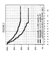
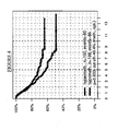
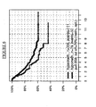
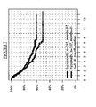
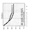
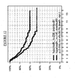
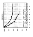
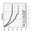
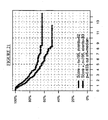
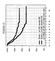
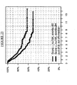
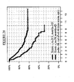
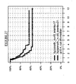
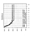
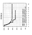
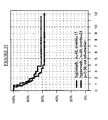
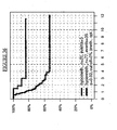
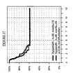
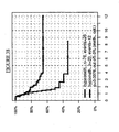
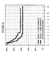
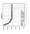
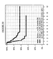
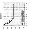
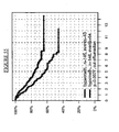
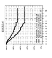
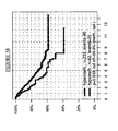
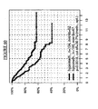
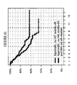
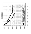
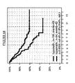
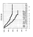
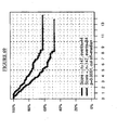
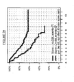
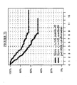
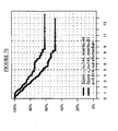
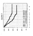
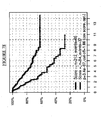
- Figures 1 to 78 show the Kaplan-Meier estimated disease-free survival curves for single assays or combinations of assays according to Example 2 and Table 5.
- the black plot shows the proportion of disease free patients in the sample set with above median or optimised cut off methylation levels
- the grey plot shows the proportion of disease free patients in the population with below median or optimised cut-off methylation levels.
- Also indicated on each plot are the number of events (i.e. metasasis) and number of individuals in each of the two sets. Proportion of metastasis free patients is shown on the Y-axis, time in years is shown on the X-axis.
- Characterization of a cancer in terms predicting treatment outcome enables the physician to make an informed decision as to a therapeutic regimen with appropriate risk and benefit trade offs to the patient.
- estrogen receptor positive and/or "progesterone receptor positive” when used to describe a cell proliferative disorder are taken to mean that the proliferating cells express said hormone receptor.
- the term 'aggressiveness' is taken to mean one or more of high likelihood of relapse post surgery; below average or below median patient survival; below average or below median disease free survival; below average or below median relapse-free survival; above average tumor-related complications; fast progression of tumor or metastases.
- an appropriate treatment or treatments may be selected from the group consisting of chemotherapy, radiotherapy, surgery, biological therapy, immunotherapy, antibody treatments, treatments involving molecularly targeted drugs, estrogen receptor modulator treatments, estrogen receptor down-regulator treatments, aromatase inhibitors treatments, ovarian ablation, treatments providing LHRH analogues or other centrally acting drugs influencing estrogen production.
- a cancer is characterized as 'aggressive' it is particularly preferred that a treatment such as, but not limited to, chemotherapy is provided in addition to or instead of an endocrine targeting therapy.
- a treatment such as, but not limited to, chemotherapy is provided in addition to or instead of an endocrine targeting therapy.
- Indicators of tumor aggressiveness standard in the art include but are not limited to, tumor stage, tumor grade, nodal status and survival.
- survival shall be taken to include all of the following: survival until mortality, also known as overall survival (wherein said mortality may be either irrespective of cause or tumor related); "recurrence-free survival” (wherein the term recurrence shall include both localized and distant recurrence) ; metastasis free survival; disease free survival (wherein the term disease shall include cancer and diseases associated therewith).
- the length of said survival may be calculated by reference to a defined start point (e.g. time of diagnosis or start of treatment) and end point (e.g. death, recurrence or metastasis).
- prognostic marker shall be taken to mean an indicator of the likelihood of progression of the disease, in particular aggressiveness and metastatic potential of a tumor.
- the term 'predictive marker' shall be taken to mean an indicator of response to therapy, said response is preferably defined according to patient survival. It is preferably used to define patients with high, low and intermediate length of survival or recurrence after treatment, that is the result of the inherent heterogeneity of the disease process.
- the term predictive marker may in some situations fall within the remit of a herein described 'prognostic marker', for example, wherein a prognostic marker differentiates between patients with different survival outcomes pursuant to a treatment, said marker is also a predictive marker for said treatment. Therefore, unless otherwise stated the two terms shall not be taken to be mutually exclusive.
- the activity of the transcribed gene may be affected by genetic variations such as but not limited to genetic modifications (including but not limited to SNPs. point mutations, deletions, insertions, repeat length, rearrangements and other polymorphisms),
- endocrine therapy or “endocrine treatment” are meant to comprise any therapy, treatment or treatments targeting the estrogen receptor pathway or estrogen synthesis pathway or estrogen conversion pathway, which is involved in estrogen metabolism, production or secretion.
- Said treatments include, but are not limited to estrogen receptor modulators, estrogen receptor down-regulators, aromatase inhibitors, ovarian ablation, LHRH analogues and other centrally acting drugs influencing estrogen production.
- the term "monotherapy” shall be taken to mean the use of a single drug or other therapy.
- chemotherapy is taken to mean the use of pharmaceutical or chemical substances to treat cancer. This definition excludes radiation therapy (treatment with high energy rays or particles), hormone therapy (treatment with hormones or hormone analogues) and surgical treatment.
- adjuvant treatment is taken to mean a therapy of a cancer patient immediately following an initial non chemotherapeutical therapy, e.g. surgery.
- adjuvant therapy is to decrease the risk of recurrence.
- determining a suitable treatment regimen for the subject is taken to mean the determination of a treatment regimen (i.e. a single therapy or a combination of different therapies that are used for the prevention and/or treatment of the cancer in the patient) for a patient that is started, modified and/or ended based or essentially based or at least partially based on the results of the analysis according to the present invention.
- a treatment regimen i.e. a single therapy or a combination of different therapies that are used for the prevention and/or treatment of the cancer in the patient
- a patient that is started, modified and/or ended based or essentially based or at least partially based on the results of the analysis according to the present invention.
- One example is starting an adjuvant endocrine therapy after surgery, another would be to modify the dosage of a particular chemotherapy.
- the determination can, in addition to the results of the analysis according to the present invention, be based on personal characteristics of the subject to be treated. In most cases, the actual determination of the suitable treatment regimen for the subject will be performed by the attending
- obtaining a biological sample or "obtaining a sample from a subject”, shall not be taken to include the active retrieval of a sample from an individual, e.g. the performance of a biopsy. Said terms shall be taken to mean the obtainment of a sample previously isolated from an individual. Said samples may be isolated by any means standard in the art, including but not limited to biopsy, surgical removal, body fluids isolated by means of aspiration. Furthermore said samples may be provided by third parties including but not limited to clinicians, couriers, commercial sample providers and sample collections.
- CpG island refers to a contiguous region of genomic DNA that satisfies the criteria of (1) having a frequency of CpG dinucleotides corresponding to an "Observed/Expected Ratio” >0.6, and (2) having a "GC Content” >0.5.
- CpG islands are typically, but not always, between about 0.2 to about 1 kb in length.
- regulatory region of a gene is taken to mean nucleotide sequences which affect the expression of a gene.
- Said regulatory regions may be located within, proximal or distal to said gene.
- Said regulatory regions include but are not limited to constitutive promoters, tissue-specific promoters, developmental-specific promoters, inducible promoters and the like.
- Promoter regulatory elements may also include certain enhancer sequence elements that control transcriptional or translational efficiency of the gene.
- methylation refers to the presence or absence of 5-methylcytosine ("5-mCyt”) at one or a plurality of CpG dinucleotides within a DNA sequence.
- methylation state is taken to mean the degree of methylation present in a nucleic acid of interest, this may be expressed in absolute or relative terms i.e. as a percentage or other numerical value or by comparison to another tissue and therein described as hypermethylated, hypomethylated or as having significantly similar or identical methylation status.
- hemi-methylation refers to the methylation state of a CpG methylation site, where only a single cytosine in one of the two CpG dinucleotide sequences-of the double stranded CpG methylation site is methylated (e.g., 5'-NNCMGNN-3' (top strand): 3'-NNGCNN-5' (bottom strand)).
- hypomethylation refers to the average methylation state corresponding to an increased presence of 5-mCyt at one or a plurality of CpG dinucleotides within a DNA sequence of a test DNA sample, relative to the amount of 5-mCyt found at corresponding CpG dinucleotides within a normal control DNA sample.
- hypomethylation refers to the average methylation state corresponding to a decreased presence of 5-mCyt at one or a plurality of CpG dinucleotides within a DNA sequence of a test DNA sample, relative to the amount of 5-mCyt found at corresponding CpG dinucleotides within a normal control DNA sample.
- microarray refers broadly to both “DNA microarrays," and 'DNA chip(s),' as recognized in the art, encompasses all art-recognized solid supports, and encompasses all methods for affixing nucleic acid molecules thereto or synthesis of nucleic acids thereon.
- Genetic parameters are mutations and polymorphisms of genes and sequences further required for their regulation. To be designated as genetic modifications or mutations are, in particular, insertions, deletions, point mutations, inversions and polymorphisms and, particularly preferred, SNPs (single nucleotide polymorphisms).
- Epigenetic modifications or “epigenetic parameters” are modifications of DNA bases of genomic DNA and sequences further required for their regulation, in particular, cytosine methylations thereof. Further epigenetic parameters include, for example, the acetylation of histones which, however, cannot be directly analyzed using the described method but which, in turn, correlate with the DNA methylation.
- bisulfite reagent refers to a reagent comprising bisulfite, disulfite, hydrogen sulfite or combinations thereof, useful as disclosed herein to distinguish between methylated and unmethylated CpG dinucleotide sequences.
- Methods refers to any assay for determining the methylation state of one or more CpG dinucleotide sequences within a sequence of DNA.
- MS.AP-PCR Metal-Sensitive Arbitrarily-Primed Polymerase Chain Reaction
- Methods of the present invention refers to the art-recognized fluorescence-based real-time PCR technique described by Eads et al., Cancer Res. 59:2302-2306, 1999 .
- HeavyMethylTM assay in the embodiment thereof implemented herein, refers to a methylation assay comprising methylation specific blocking probes covering CpG positions between the amplification primers.
- Ms-SNuPE Metal-sensitive Single Nucleotide Primer Extension
- MSP Metal-specific PCR
- COBRA combined Bisulfite Restriction Analysis
- hybridization is to be understood as a bond of an oligonucleotide to a complementary sequence along the lines of the Watson-Crick base pairings in the sample DNA, forming a duplex structure.
- “Stringent hybridization conditions,” as defined herein, involve hybridizing at 68°C in 5x SSC/5x Denhardt's solution/1.0% SDS, and washing in 0.2x SSC/0.1% SDS at room temperature, or involve the art-recognized equivalent thereof (e.g., conditions in which a hybridization is carried out at 60°C in 2.5 x SSC buffer, followed by several washing steps at 37°C in a low buffer concentration, and remains stable).
- Moderately stringent conditions as defined herein, involve including washing in 3x SSC at 42°C, or the art-recognized equivalent thereof.
- the parameters of salt concentration and temperature can be varied to achieve the optimal level of identity between the probe and the target nucleic acid.
- Background DNA refers to any nucleic acids which originate from sources other than the cancer cells to be analysed.
- said marker is used as a predictive marker of outcome of a anthracycline treatment, thereby enabling the physician to determine if said treatment is of benefit to a patient.
- patient survival can be evaluated before or during treatment for a cell proliferative disorder of the breast suitable for treatment with anthracyclines, in order to provide critical information to the patient and clinician as to the likely progression of the disease when treated by means of a therapy comprising at least one anthracycline. It will be appreciated, therefore, that the methods exemplified herein can serve to improve a patient's quality of life and odds of treatment success by allowing both patient and clinician a more accurate assessment of the patient's treatment options.
- the present invention makes available a method for the improved treatment of cell proliferative disorders, by enabling the improved prediction of a patient's survival, in particular by predicting the likelihood of relapse post-surgery both with or without anthracycline treatment.
- the method according to the invention may be used for the analysis of breast cancer suitable for treatment with anthracyclines.
- the method according to the invention may be used to provide a prediction of patient survival and/or relapse following treatment by means of a therapy comprising at least one anthracyline.
- said prediction is defined in terms of patient survival and/or relapse.
- patients survival times and/or relapse are predicted according to their epigenic modifications.
- said patients are tested prior to receiving any adjuvant anthracycline treatment.
- the invention relates to new methods and sequences, which may be used as tools for the selection of suitable treatments of patients diagnosed with cell proliferative disorder of the breast based on a prediction of likelihood of relapse, survival or outcome.
- One aspect of the invention is the provision of methods for providing a prediction of outcome of a treatment comprising at least one anthracycline of a patient with a cell proliferative disorder of the breast.
- said prognosis and/or prediction is provided in terms of likelihood of relapse or the survival of said patient. It is further preferred that said survival is disease free survival or metastasis free survival. It is also preferred that said disease is breast cancer.
- said patients are analyzed prior to receiving any treatment comprising at least one anthracycline.
- the present invention discloses a method for the use of at least one gene selected from PITX2 and PLAU as a marker of the prediction of outcome of anthracycline treatment.
- the invention provides significant improvements over the state of the art in that it provides the first proliferative disorder of the breast treatment response markers for a treatment comprising an anthracycline.
- the invention is carried out by means of CpG methylation analysis of at least one of the genes PITX2 and PLAU. It is further preferred that the methylation, state of the CpG dinucleotides within the genomic sequence of said genes according to SEQ ID NOs: 1, 2 and 4 to 6 and sequences complementary thereto are analyzed.
- the methylation state of the CpG dinucleotides within the genomic sequence of said genes according to Table 1 (SEQ ID NO: 1 and SEQ ID NO: 2) and sequences complementary thereto are analyzed.
- Table 4 provides particularly preferred CpG rich sequences of the genes according to Table 1. Accordingly it is preferred that said CpG positions are within a CpG rich region of said genes as provided in Table 4 (SEQ ID NO: 4 to SEQ ID NO: 6).
- the methylation pattern of the genes according to the present invention and their promoter and regulatory elements have heretofore not been analyzed with regard to prediction of outcome of anthracylcine treatment.
- the method for the analysis of methylation comprises contacting a nucleic acid sample obtained from a subject with at least one reagent or a series of reagents, wherein said reagent or series of reagents, distinguishes between methylated and non-methylated CpG dinucleotides within the target nucleic acid.
- said method comprises the following steps: In the first step, a sample of the tissue to be analyzed is obtained.
- the source may be any suitable source, preferably, the source of the sample is selected from the group consisting of histological slides, biopsies, paraffin-embedded tissue, bodily fluids, plasma, serum, stool, urine, blood, nipple aspirate and combinations thereof.
- the source is tumor tissue, biopsies, serum, urine, blood or nipple aspirate.
- the most preferred source is the tumor sample, surgically removed from the patient or a biopsy sample of said patient.
- Genomic DNA may be isolated by any means standard in the art, including the use of commercially available kits. Briefly, wherein the DNA of interest is encapsulated in/by a cellular membrane the biological sample must be disrupted and lysed by enzymatic, chemical or mechanical means. The DNA solution may then be cleared of proteins and other contaminants e.g. by digestion with proteinase K. The genomic DNA is then recovered from the solution. This may be carried out by means of a variety of methods including salting out, organic extraction or binding of the DNA to a solid phase support. The choice of method will be affected by several factors including time, expense and required quantity of DNA.
- genomic DNA sample is then treated in such a manner that cytosine bases which are unmethylated at the 5'-position are converted to uracil, thymine, or another base which is dissimilar to cytosine in terms of hybridization behavior. This will be understood as “treatment” or “pre-treatment” herein.
- the above described pre-treatment of genomic DNA is preferably carried out with bisulfite (hydrogen sulfite, disulfite) and subsequent alkaline hydrolysis which results in a conversion of non-methylated cytosine nucleobases to uracil or to another base which is dissimilar to cytosine in terms of base pairing behavior.
- the treated DNA is then analyzed in order to determine the methylation state of at least one of the genes PITX2 and PLAU and/or regulatory regions thereof associated with outcome of a treatment comprising at least one anthracycline. It is further preferred that the sequences of said genes as described in the accompanying sequence listing (see Table 1, or more preferably Table 4) are analyzed.
- fragments of the pretreated DNA are amplified.
- the source of the DNA is free DNA from serum, or DNA extracted from paraffin it is particularly preferred that the size of the amplificate fragment is between 100 and 200 base pairs in length, and wherein said DNA source is extracted from cellular sources (e.g. tissues, biopsies, cell lines) it is preferred that the amplificate is between 100 and 350 base pairs in length.
- said amplificates comprise at least one 20 base pair sequence comprising at least three CpG dinucleotides.
- Said amplification is carried out using sets of primer oligonucleotides according to the present invention, and a preferably heat-stable polymerase.
- the amplification of several DNA segments can be carried out simultaneously in one and the same reaction vessel, in one embodiment of the method preferably six or more fragments are amplified simultaneously.
- the amplification is carried out using a polymerase chain reaction (PCR).
- the set of primer oligonucleotides includes at least two oligonucleotides whose sequences are each reverse complementary, identical, or hybridize under stringent or highly stringent conditions to an at least 18-base-pair long segment of a base sequence selected from the group consisting SEQ ID NO: 9-12,15-20,25-28 and 31-36 and sequences complementary thereto.
- the set of primer oligonucleotides includes at least two oligonucleotides whose sequences are each reverse complementary, identical, or hybridize under stringent or highly stringent conditions to an at least 18-base-pair long segment of a pretreated sequence as listed in Table 1 (SEQ ID NO: 9 to SEQ ID NO: 12 and (SEQ ID NO: 25 to SEQ ID NO: 28) and sequences complementary thereto.
- the set of primer oligonucleotides includes at least two oligonucleotides whose sequences are each reverse complementary, identical, or hybridize under stringent or highly stringent conditions to an at least 18-base-pair long segment of a pretreated sequence as listed in Table 4 (SEQ ID NO: 15 to SEQ ID NO: 20 and SEQ ID NO: 31 to SEQ ID NO: 36) and sequences complementary thereto.
- the methylation status of pre-selected CpG positions within the nucleic acid sequences comprising SEQ ID NO: 1, 2 and 4 to 6 may be detected by use of methylation-specific primer oligonucleotides.
- This technique has been described in United States Patent No. 6,265,171 to Herman .
- the use of methylation status specific primers for the amplification of bisulfite treated DNA allows the differentiation between methylated and unmethylated nucleic acids.
- MSP primers pairs contain at least one primer which hybridizes to a bisulfite treated CpG dinucleotide. Therefore, the sequence of said primers comprises at least one CpG , TpG or CpA dinucleotide.
- MSP primers specific for non-methylated DNA contain a "T' at the 3' position of the C position in the CpG.
- the base sequence of said primers is required to comprise a sequence having a length of at least 18 nucleotides which hybridizes to a pretreated nucleic acid sequence according to SEQ ID NO: 9-12,15-20,25-28 and 31-36 and sequences complementary thereto, wherein the base sequence of said oligomers comprises at least one CpG, tpG or Cpa dinucleotide.
- said sequence has a length of at least 18 nucleotides which hybridizes to a L pretreated nucleic acid sequence selected from Table I (SEQ ID NO: 9 to SEQ ID NO: 12 and SEQ ID NO: 25 to SEQ ID NO: 28 and sequences complementary thereto.
- said sequence has a length of at least 18 nucleotides which hybridizes to a pretreated nucleic acid sequence selected from Table 4 (SEQ ID NO: 15 to SEQ ID NO: 20 and SEQ ID NO: 31 to SEQ ID NO: 36), and sequences complementary thereto.
- the MSP primers comprise between 2 and 4 CpG , tpG or Cpa dinucleotides. It is further preferred that said dinucleotides are located within the 3' half of the primer e.g. wherein a primer is 18 bases in length the specified dinucleotides are located within the first 9 bases form the 3'end of the molecule.
- said primers should further comprise several bisulfite converted bases (i.e. cytosine converted to thymine, or on the hybridizing strand, guanine converted to adenosine). In a further preferred embodiment said primers are designed so as to comprise no more than 2 cytosine or guanine bases.
- the fragments obtained by means of the amplification can carry a directly or indirectly detectable label.
- the detection may be carried out and visualized by means of, e.g., matrix assisted laser desorption/ionization mass spectrometry (MALDI) or using electron spray mass spectrometry (ESI).
- MALDI matrix assisted laser desorption/ionization mass spectrometry
- ESI electron spray mass spectrometry
- Matrix Assisted Laser Desorption/Ionization Mass Spectrometry is a very efficient development for the analysis of biomolecules ( Karas and Hillenkamp, Anal Chem., 60:2299-301. 1988 ).
- An analyte is embedded in a light-absorbing matrix.
- the matrix is evaporated by a short laser pulse thus transporting the analyte molecule into the vapor phase in an unfragmented manner.
- the analyte is ionized by collisions with matrix molecules.
- An applied voltage accelerates the ions into a field-free flight tube. Due to their different masses, the ions are accelerated at different rates. Smaller ions reach the detector sooner than bigger ones.
- MALDI-TOF spectrometry is well suited to the analysis of peptides and proteins.
- the analysis of nucleic acids is somewhat more difficult ( Gut and Beck, Current Innovations and Future Trends, 1:147-57, 1995 ).
- the sensitivity with respect to nucleic acid analysis is approximately 100-times less than for peptides, and decreases disproportionally with increasing fragment size.
- nucleic acids having a multiply negatively charged backbone the ionisation process via the matrix is considerably less efficient.
- MALDI-TOF spectrometry the selection of the matrix plays an eminently important role. For the desorption of peptides, several very efficient matrixes have been found which produce a very fine crystallisation.
- the amplification of step three is carried out in the presence of at least one species of blocker oligonucleotides.
- blocker oligonucleotides has been described by Yu et al., BioTechniques 23:714-720, 1997 .
- the use of blocking oligonucleotides enables the improved specificity of the amplification of a subpopulation of nucleic acids.
- Blocking probes hybridized to a nucleic acid suppress, or hinder the polymerase mediated amplification of said nucleic acid.
- blocking oligonucleotides are designed so as to hybridize to background DNA.
- said oligonucleotides are designed so as to hinder or suppress the amplification of unmethylated nucleic acids as opposed to methylated nucleic acids or vice versa.
- Blocking probe oligonucleotides are hybridized to the bisulfite treated nucleic acid concurrently with the PCR primers. PCR amplification of the nucleic acid is terminated at the 5' position of the blocking probe, such that amplification of a nucleic acid is suppressed where the complementary sequence to the blocking probe is present.
- the probes may be designed to hybridize to the bisulfite treated nucleic acid in a methylation status specific manner.
- the sequence of said blocking oligonucleotides should be identical or complementary to a sequence at least 18 base pairs in length selected from the group consisting of SEQ ID NO: 9-12,15-20,25-28 and 31-36 preferably comprising one or more CpG, TpG or CpA dinucleotides.
- sequence of said blocking oligonucleotides should be identical or complementary to a sequence at least 18 base pairs in length selected from a pretreated nucleic acid sequence selected from Table 1 (SEQ ID NO: 9 to SEQ ID NO: 12 and SEQ ID NO: 25 to SEQ ID NO: 28), and sequences complementary thereto preferably comprising one or more CpG, TpG or CpA dinucleotides.
- sequence of said blocking oligonucleotides should be identical or complementary to a sequence at least 18 base pairs in length selected from a pretreated nucleic acid sequence selected from Table 4 (SEQ ID NO: 15 to SEQ ID NO: 20 and SEQ ID NO: 31 to SEQ ID NO: 36), and sequences complementary thereto preferably comprising one or more CpG, TpG or CpA dinucleotides.
- blocker oligonucleotides For PCR methods using blocker oligonucleotides, efficient disruption of polymerase-mediated amplification requires that blocker oligonucleotides not be elongated by the polymerase. Preferably, this is achieved through the use of blockers that are 3'-deoxyoligonucleotides or oligonucleotides derivatised at the 3' position with other than a "free" hydroxyl group.
- 3'-O-acetyl oligonucleotides are representative of a preferred class of blocker molecule.
- polymerase-mediated decomposition of the blocker oligonucleotides should be precluded.
- such preclusion comprises either use of a polymerase lacking 5'-3' exonuclease activity, or use of modified blocker oligonucleotides having, for example, thioate bridges at the 5'-termini thereof that render the blocker molecule nuclease-resistant.
- Particular applications may not require such 5' modifications of the blocker. For example, if the blocker- and primer-binding sites overlap, thereby precluding binding of the primer (e.g., with excess blocker), degradation of the blocker oligonucleotide will be substantially precluded. This is because the polymerase will not extend the primer toward, and through (in the 5'-3' direction) the blocker - a process that normally results in degradation of the hybridized blocker oligonucleotide.
- a particularly preferred blocker/PCR embodiment for purposes of the present invention and as implemented herein, comprises the use of peptide nucleic acid (PNA) oligomers as blocking oligonucleotides.
- PNA peptide nucleic acid
- Such PNA blocker oligomers are ideally suited, because they are neither decomposed nor extended by the polymerase.
- the binding site of the blocking oligonucleotide is identical to, or overlaps with that of the primer and thereby hinders the hybridization of the primer to its binding site.
- two or more such blocking oligonucleotides are used.
- the hybridization of one of the blocking oligonucleotides hinders the hybridization of a forward primer, and the hybridization of another of the probe (blocker) oligonucleotides hinders the hybridization of a reverse primer that binds to the amplificate product of said forward primer.
- the blocking oligonucleotide hybridizes to a location between the reverse and forward primer positions of the treated background DNA, thereby hindering the elongation of the primer oligonucleotides.
- the blocking oligonucleotides are present in at least 5 times the concentration of the primers.
- the amplificates obtained during the third step of the method are analyzed in order to ascertain the methylation status of the CpG dinucleotides prior to the treatment.
- the presence or absence of an amplificate is in itself indicative of the methylation state of the CpG positions covered by the primers and or blocking oligonucleotide, according to the base sequences thereof.
- All possible known molecular biological methods may be used for this detection, including, but not limited to gel electrophoresis, sequencing, liquid chromatography, hybridizations, real time PCR analysis or combinations thereof. This step of the method further acts as a qualitative control of the preceding steps.
- amplificates obtained by means of both standard and methylation specific PCR are further analyzed in order to determined the CpG methylation status of the genomic DNA isolated in the first step of the method. This may be carried out by means of hybridization-based methods such as, but not limited to, array technology and probe based technologies as well as by means of techniques such as sequencing and template directed extension.
- the amplificates synthesized in step three are subsequently hybridized to an array or a set of oligonucleotides and/or PNA probes.
- the hybridization takes place in the following manner: the set of probes used during the hybridization is preferably composed of at least 2 oligonucleotides or PNA-oligomers; in the process, the amplificates serve as probes which hybridize to oligonucleotides previously bonded to a solid phase; the non-hybridized fragments are subsequently removed; said oligonucleotides contain at least one base sequence having a length of at least 9 nucleotides which is reverse complementary or identical to a segment of the base sequences specified in the of SEQ ID NO: 9-12,15-20,25-28 and 31-36 and the segment comprises at least one CpG , TpG or CpA dinucleotide.
- said oligonucleotides contain at least one base sequence having a length of at least 9 nucleotides which is reverse complementary or identical to a segment of the pretreated sequences specified in Table 1 (SEQ ID NO: 9 to SEQ ID NO: 12 and SEQ ID NO: 25 to SEQ ID NO: 28) and the segment comprises at least one CpG, TpG or CpA dinucleotide.
- said oligonucleotides contain at least one base sequence having a length of at least 9 nucleotides which is reverse complementary or identical to a segment of the pretreated sequences specified in Table 4 (SEQ ID NO: 15 to SEQ ID NO: 20 and SEQ ID NO: 31 to SEQ ID NO: 36) and the segment comprises at least one CpG, TpG or CpA dinucleotide.
- said oligonucleotides contain at least one base sequence having a length of at least 9 nucleotides which is reverse complementary or identical to a segment of the base sequences specified in the of SEQ ID NO: 9-12,15-20,25-28 and 31-36 and the segment comprises at least one CpG , TpG or CpA dinucleotide.
- said oligonucleotides contain at least one base sequence having a length of at least 9 nucleotides which is reverse complementary or identical to a segment of the pretreated sequences specified in Table 1 (SEQ ID NO: 9 to SEQ ID NO: 12 and SEQ ID NO: 25 to SEQ ID NO: 28) and the segment comprises at least one CpG , TpG or CpA dinucleotide.
- said oligonucleotides contain at least one base sequence having a length of at least 9 nucleotides which is reverse complementary or identical to a segment of the pretreated sequences specified in Table 4 (SEQ ID NO: 15 to SEQ ID NO: 20 and SEQ ID NO: 31 to SEQ ID NO: 36) and the segment comprises at least one CpG, TpG or CpA dinucleotide.
- said dinucleotide is present in the central third of the oligomer.
- said dinucleotide is preferably the fifth to ninth nucleotide from the 5'-end of a 13-mer.
- one oligonucleotide exists for the analysis of each CpG dinucleotide within the sequence according to of SEQ ID NO: 4 to SEQ ID NO: 6 and the equivalent positions within of SEQ ID NO: 15 to SEQ ID NO: 20 and SEQ ID NO: 31 to SEQ ID NO: 36.
- oligonucleotide exists for the analysis of each CpG dinucleotide within the sequence according to SEQ ID NO: 12 and 4-6 and the equivalent positions within SEQ ID NO 9-12,15-20,25-28 and 31-36 Said oligonucleotides may also be present in the form of peptide nucleic acids.
- the non-hybridized amplificates are then removed.
- the hybridized amplificates are detected.
- labels attached to the amplificates are identifiable at each position of the solid phase at which an oligonucleotide sequence is located.
- the genomic methylation status of the CpG positions may be ascertained by means of oligonucleotide probes that are hybridized to the bisulfite treated DNA concurrently with the PCR amplification primers (wherein said primers may either be methylation specific or standard).
- a particularly preferred embodiment of this method is the use of fluorescence-based Real Time Quantitative PCR ( Heid et al., Genome Res. 6:986-994, 1996 : also see United States Patent No. 6,331,393 ).
- the TaqManTM assay employs a dual-labeled fluorescent oligonucleotide probe.
- the TaqManTM PCR reaction employs the use of a non-extendible interrogating oligonucleotide, called a TaqManTM probe, which is designed to hybridize to a CpG-rich sequence located between the forward and reverse amplification primers.
- the TaqManTM probe further comprises a fluorescent "reporter moiety” and a "quencher moiety” covalently hound to linker moieties (e.g., phosphoramidites) attached to the nucleotides of the TaqManTM oligonucleotide.
- linker moieties e.g., phosphoramidites
- Hybridized probes are displaced and broken down by the polymerase of the amplification reaction thereby leading to an increase in fluorescence.
- the probe be methylation specific, as described in United States Patent No. 6,331,393 , also known as the MethyLight assay.
- the second preferred embodiment of this MethyLight technology is the use of dual-probe technology (Lightcycler®), each probe carrying donor or recipient fluorescent moieties, hybridization of two probes in proximity to each other is indicated by an increase or fluorescent amplification primers. Both these techniques may be adapted in a manner suitable for use with bisulfite treated DNA, and moreover for methylation analysis within CpG dinucleotides.
- the fourth step of the method comprises the use of template-directed oligonucleotide extension, such as MS-SNuPE as described by Gonzalgo and Jones, Nucleic Acids Res. 25:2529-2531, 1997 .
- the methylation specific single nucleotide extension primer (MS-SNuPE primer) is identical or complementary to a sequence at least nine but preferably no more than twenty five nucleotides in length of one or more of the sequences taken from the group to SEQ ID NO: 9-12, 15-20, 25-28 and 31-36.
- said MS-SNuPE primer is identical or complementary to a sequence at least nine but preferably no more than twenty five nucleotides in length of one or more of the pretreated sequences selected from Table 1 (SEQ ID NO: 9 to SEQ ID NO: 12 and SEQ ID NO: 25 to SEQ ID NO: 28).
- said MS-SNuPE primer is identical or complementary to a sequence at least nine but preferably no more than twenty five nucleotides in length of one or more of the pretreated sequences selected from Table 4 (SEQ ID NO: 15 to SEQ ID NO: 20 and SEQ ID NO: 31 to SEQ ID NO: 36). It is preferred to use fluorescently labeled nucleotides, instead of radiolabeled nucleotides.
- the fourth step of the method comprises sequencing and subsequent sequence analysis of the amplificate generated in the third step of the method ( Sanger F., et al., Proc Natl Acad Sci USA 74:5463-5467, 1977 ).
- genomic nucleic acids are isolated and treated according to the first three steps of the method outlined above, namely:
- said methylation specific primers comprise a sequence having a length of at least 9 nucleotides which hybridizes to a treated nucleic acid sequence according to one of SEQ ID NO: 9-12, 15-20, 25-28 and 31-36 and sequences complementary thereto, wherein the base sequence of said oligomers comprises at least one CpG dinucleotide.
- said methylation specific primers comprise a sequence having a length of at least 9 nucleotides which hybridizes to a pretreated nucleic acid sequence selected from Table 1 (SEQ ID NO: 9 to SEQ ID NO: 12 and SEQ ID NO: 25 to SEQ ID NO:28) and sequences complementary thereto, wherein the base sequence of said oligomers comprises at least one CpG dinucleotide.
- said methylation specific primers comprise a sequence having a length of at least 9 nucleotides which hybridizes to a pretreated nucleic acid sequence selected from Table 4 (SEQ ID NO: 15 to SEQ ID NO: 20 and SEQ ID NO: 31 to SEQ ID NO:36) and sequences complementary thereto, wherein the base sequence of said oligomers comprises at least one CpG dinucleotide.
- further methylation specific primers may also be used for the analysis of a gene panel as described above wherein said primers comprise a sequence having a length of at least 9 nucleotides which hybridizes to a treated nucleic acid sequence according to one of SEQ ID NO: 9-12, 15-20, 25-28 and 31-36 and sequences complementary thereto, wherein the base sequence of said oligomers comprises at least one CpG dinucleotide.
- the subsequent amplification of d) is carried out in the presence of blocking oligonucleotides, as described above.
- said blocking oligonucleotides comprise a sequence having a length of at least 9 nucleotides which hybridizes to a treated nucleic acid sequence according to one of SEQ ID NO: 9-12, 15-20, 25-28 and 31-36 and sequences complementary thereto, wherein the base sequence of said oligomers comprises at least one CpG, TpG or CpA dinucleotide.
- said blocking oligonucleotides comprise a sequence having a length of at least 9 nucleotides which hybridizes to a pretreated nucleic acid sequence selected from Table 1 (SEQ ID NO: 9 to SEQ ID NO: 12 and SEQ ID NO: 25 to SEQ ID NO: 28) and sequences complementary thereto, wherein the base sequence of said oligomers comprises at least one CpG dinucleotide.
- said blocking oligonucleotides comprise a sequence having a length of at least 9 nucleotides which hybridizes to a pretreated nucleic acid sequence selected from Table 4 (SEQ ID NO: 15 to SEQ ID NO: 20 and SEQ ID NO: 31 to SEQ ID NO: 36) and sequences complementary thereto, wherein the base sequence of said oligomers comprises at least one CpG dinucleotide.
- Step e) of the method namely the detection of the specific amplificates indicative of the methylation status of one or more CpG positions according to SEQ ID NO: 9 to SEQ ID NO: 12 and SEQ ID NO: 25 to SEQ ID NO: 28, is carried out by means of real-time detection methods as described above.
- Additional embodiments of the invention provide a method for the analysis of the methylation status of at least one gene selected from PITX2 and PLAU and/or regulatory regions thereof without the need for pre-treatment.
- a methylation sensitive restriction enzyme reagent or a series of restriction enzymes reagents comprising methylation sensitive restriction enzyme reagents that distinguishes between methylated and non-methylated CpG dinucleotides within a target region are utilized in determining methylation, for example but not limited to DMH.
- Genomic DNA is isolated from tissue or cellular sources.
- Genomic DNA may be isolated by any means standard in the art, including the use of commercially available kits. Briefly, wherein the DNA of interest is encapsulated in by a cellular membrane the biological sample must be disrupted and lysed by enzymatic, chemical or mechanical means. The DNA solution may then be cleared of proteins and other contaminants, e.g., by digestion with proteinase K. The genomic DNA is then recovered from the solution. This may be carried out by means of a variety of methods including salting out, organic extraction or binding of the DNA to a solid phase support. The choice of method will be affected by several factors including time, expense and required quantity of DNA.
- the genomic double-stranded DNA is used in the analysis.
- the DNA may be cleaved prior to treatment with methylation sensitive restriction enzymes.
- methylation sensitive restriction enzymes Such methods are known in the art and may include both physical and enzymatic means.
- Particularly preferred is the use of one or a plurality of restriction enzymes which are not methylation sensitive, and whose recognition sites are AT rich and do not comprise CG dinucleotides. The use of such enzymes enables the conservation of CpG islands and CpG rich regions in the fragmented DNA.
- the non-methylation-specific restriction enzymes are preferably selected from the group consisting of MseI, BfaI, Csp6I, Tru1I, Tvu1I, Tru9I, Tvu9I, MaeI and XspI. Particularly preferred is the use of two or three such enzymes. Particularly preferred is the use of a combination of MseI, Bfal and Csp6I.
- the fragmented DNA may then be ligated to adaptor oligonucleotides in order to facilitate subsequent enzymatic amplification.
- the ligation of oligonucleotides to blunt and sticky ended DNA fragments is known in the art, and is carried out by means of dephosphorylation of the ends (e.g. using calf or shrimp alkaline phosphatase) and subsequent ligation using ligase enzymes (e.g. T4 DNA ligase) in the presence of dATPs.
- the adaptor oligonucleotides are typically at least 18 base pairs in length.
- the DNA (or fragments thereof) is then digested with one or more methylation sensitive restriction enzymes.
- the digestion is carried out such that hydrolysis of the DNA at the restriction site is informative of the methylation status of a specific CpG dinucleotide of at least one gene selected from PITX2 and PLAU.
- the methylation-specific restriction enzyme is selected from the group consisting of Bsi El, Hga 1 HinPl, Hpy99I, Ava I, Bce AI, Bsa HI, Bisl, BstUI, BshI236I, AccII, BstFNI, McrBC, GlaI, MvnI, HpaII (HapII), HhaI, AciI, SmaI, HinPII, HpyCH4IV, Eagl and mixtures of two or more of the above enzymes.
- Preferred is a mixture containing the restriction enzymes BstUI, HpaII, HpyCH4IV and HinPII.
- the restriction fragments are amplified.
- This is preferably carried out using a polymerase chain reaction, and said amplifycates may carry suitable detectable labels as discussed above, namely fluorophore labels, radionuclides and mass labels.
- amplification by means of an amplification enzyme and at least two primers comprising, in each case a contiguous sequence at least 16 nucleotides in length that is complementary to, or hybridizes under moderately stringent or stringent conditions to a sequence selected from the group consisting SEQ ID NO: 1, 2, 4-6 and 9, or more preferably SEQ ID NO: 4 to SEQ ID NO: 6 and complements thereof.
- said contiguous sequence is at least 16, 20 or 25 nucleotides in length.
- said primers may be complementary to any adaptors linked to the fragments.
- the amplificates are detected.
- the detection may be by any means standard in the art, for example, but not limited to gel electrophoresis analysis, hybridization analysis, incorporation of detectable tags within the PCR products, DNA array analysis, MALDI or ESI analysis.
- said detection is carried out by hybridisation to at least one nucleic acid or peptide nucleic acid comprising in each case a contiguous sequence at least 16 nucleotides in length that is complementary to, or hybridizes under moderately stringent or stringent conditions to a sequence selected from the group consisting SEQ ID NO: 1, 2, 4-6 and 9 or more preferably SEQ ID NO: 4 to SEQ ID NO: 6, and complements thereof.
- said contiguous sequence is at least 16, 20 or 25 nucleotides in length.
- the prognosis of the patient after treatment with anthracyclines is determined.
- the outcome of Anthracycline treatment is deduced based upon the methylation state or level of at least one CpG dinucleotide sequence of at least one gene selected from PITX2 and PLAU.
- said methylation is determined by quantitative means the cut-off point for determining said the presence of methylation is preferably zero (i.e. wherein a sample displays any degree of methylation it is determined as having a methylated status at the analysed CpG position). Nonetheless, it is foreseen that the person skilled in the art may wish to adjust said cut-off value in order to provide an assay of a particularly preferred sensitivity or specificity.
- said cut-off value may be increased (thus increasing the specificity), said cut off value may be within a range selected form the group consisting of 0%-5%, 5%-10%, 10%-15%, 15%-20%, 20%-30% and 30%-50%. Particularly preferred are the cut-offs 10%, 15%, 25%, and 30%.
- the correlation of methylation with prognosis after treatment with anthracyclines may be determined according to Table 5.
- Patients with predicted positive outcome after said treatment will accordingly have a decreased absolute reduction of risk of recurrence and death after treatment with primary or adjuvant treatment.
- Patients with predicted negative outcome after said treatment will accordingly have a relatively larger absolute reduction of risk of recurrence and death after said treatment. Accordingly patients with a negative outcome will be considered more suitable candidates for aggressive treatment such as chemotherapy or other adjuvant therapies than patients with a positive outcome.
- Patients with a positive outcome may accordingly be prevented from over prescription of e.g. chemotherapeutic treatment.
- the classification of the sample is carried out by algorithmic means.
- machine learning predictors are trained on the methylation patterns at the investigated CpG sites of the samples with known status.
- a selection of the CpG positions which are discriminative for the machine learning predictor are used in the panel.
- both methods are combined; that is, the machine learning classifier is trained only on the selected CpG positions that are significantly differentially methylated between the classes according to the statistical analysis.
- Treatment is suitable to convert at least one unmethylated cytosine base of the genomic DNA sequence to uracil or another base that is detectably dissimilar to cytosine in terms of hybridization.
- the genomic sequences in question may comprise one, or more, consecutive or random methylated CpG positions.
- Said treatment preferably comprises use of a reagent selected from the group consisting of bisulfite, hydrogen sulfite, disulfite, and combinations thereof.
- the objective comprises analysis of a non-naturally occurring modified nucleic acid comprising a sequence of at least 16 contiguous nucleotide bases in length of a sequence selected from the group consisting of SEQ ID NO: 1, 2 and 4-6, wherein said sequence comprises at least one CpG, TpA or CpA dinucleotide and sequences complementary thereto.
- the sequences of SEQ ID NO: 9-12, 15-20, 25-18 and 31-36 provide non-naturally occurring modified versions of the nucleic acid according to SEQ ID NO: 1, 2 and 4-6, wherein the modification of each genomic sequence results in the synthesis of a nucleic acid having a sequence that is unique and distinct from said genomic sequence as follows.
- genomic DNA For each sense strand genomic DNA, e.g., SEQ ID NO: I, four converted versions are disclosed.
- a first version wherein “C” to "T,” but “CpG” remains “CpG” (i.e., corresponds to case where, for the genomic sequence, all "C” residues of CpG dinucleotide sequences are methylated and are thus not converted);
- a second version discloses the complement of the disclosed genomic DNA sequence (i.e. antisense strand), wherein “C” to "T,” but “CpG” remains “CpG” (i.e., corresponds to case where, for all "C” residues of CpG dinucleotide sequences are methylated and are thus not converted).
- the 'upmethylated' converted sequences of SEQ ID NO: 1, 2 and 4-6 correspond to SEQ ID NO: 9-12 and 15-20
- a third chemically converted version of each genomic sequences is provided, wherein "C” to "T” for all "C” residues, including those of "CpG" dinucleotide sequences (i.e., corresponds to case where, for the genomic sequences, all "C” residues of CpG dinucleotide sequences are unmethylated); a final chemically converted version of each sequence, discloses the complement of the disclosed genomic DNA sequence (i.e.
- the invention further discloses oligonucleotide or oligomer for detecting the cytosine methylation state within genomic or pre-treated DNA, according to SEQ ID NO: 1, 2 and 4-6 and SEQ ID NO: 9-12, 15-20, 25-28 and 31-36.
- Said oligonucleotide or oligomer comprising a nucleic and SEQ ID NO: 9-12, 15-20, 25-28 and 31-36.
- Said oligonucleotide or oligomer comprising a nucleic acid sequence having a length of at least nine (9) nucleotides which hybridizes, under moderately stringent or stringent conditions (as defined herein above), to a treated nucleic acid sequence according to SEQ ID NO: 9-12, 15-20, 25-28 and 31-36 and/or sequences complementary thereto, or to a genomic sequence according to SEQ ID NO: 1, 2 and 4-6 and/or sequences complementary thereto.
- Acid molecules e.g., oligonucleotides and peptide nucleic acid (PNA) molecules (PNA-oligomers) that hybridize under moderately stringent and/or stringent hybridization conditions to all or a portion of the sequences SEQ ID NO: 9-12, 15-20, 25-28 and 31-36, or to the complements thereof.
- the hybridizing portion of the hybridizing nucleic acids is typically at least 9, 15, 20, 25, 30 or 35 nucleotides in length. However, longer molecules have inventive utility, and are thus within the scope of the present invention.
- the hybridizing portion of the inventive hybridizing nucleic acids is at least 95%, or at least 98%, or 100% identical to the sequence or to a portion thereof or SEQ ID NO:9-12, 15-20, 25-28 and 31-36, or to the complements thereof.
- Hybridizing nucleic acids of the type described herein can be used, for example as a primer (e.g., a PCR primer), or a diagnostic and/or prognostic probe or primer.
- a primer e.g., a PCR primer
- a diagnostic and/or prognostic probe or primer e.g., a PCR primer
- hybridization of the oligonucleotide probe to a nucleic acid sample is performed under stringent conditions and the probe is 100% identical to the target sequence.
- Nucleic acid duplex or hybrid stability is expressed as the melting temperature or Tm, which is the temperature at which a probe dissociates from a target DNA. This melting temperature is used to define the required stringency conditions.
- target sequences that are related and substantially identical to the corresponding sequence of SEQ ID NO: 1, 2 and 4-6 (such as allelic variants and SNPs), rather than identical, it is useful to first establish the lowest temperature at which only homologous hybridization occurs with a particular concentration of salt (e.g., SSC or SSPE). Then, assuming that 1% mismatching results in a 1°C decrease in the Tm, the temperature of the final wash in the hybridisation reaction is reduced accordingly (for example, if sequences having > 95% identity with the probe are sought, the final wash temperature is decreased by 5°C). In practice, the change in Tm can be between 0.5°C and 1.5°C per 1% mismatch.
- salt e.g., SSC or SSPE
- the set is limited to those oligomers that comprise at least one CpG, TpG or CpA dinucleotide.
- Examples of 20-mer oligonucleotides include the following set of oligomers (and the antisense set complementary thereto), indicated by polynucleotide positions with reference to SEQ ID NO: 1: 1-20, 2-21, 3-22, 4-23, 5-24, ising and 29982-30001.
- the set is limited to those oligomers that comprise at least one CpG, TpG or CpA dinucleotide.
- examples of 25-mer oligonucleotides include the following set of oligomers (and the antisense set complementary thereto), indicated by polynucleotide positions with reference to SEQ ID NO: 1: 1-25, 2-26, 3-27, 4-28, 5-29, ............and 29977-30001.
- the set is limited to those oligomers that comprise at least one CpG, TpG or CpA dinucleotide.
- oligonucleotides or oligomers according to the present invention constitute effective tools useful to ascertain genetic and epigenetic parameters of the genomic sequence corresponding to SEQ ID NO: 1, 2 and 4-6.
- Preferred sets of such oligonucleotides or modified oligonucleotides of length X are those consecutively overlapping sets of oligomers corresponding to SEQ ID NO: 9-12, 15-20, 25-28 and 31-36 (and to the complements thereof).
- said oligomers comprise at least one CpG, TpG or CpA dinucleotide.
- Particularly preferred oligonucleotides or oligomers according to the present invention are those in which the cytosine of the CpG dinucleotide (or of the corresponding converted TpG or CpA dinculeotide) sequences is within the middle third of the oligonucleotide; that is, where the oligonucleotide is, for example. 13 bases in length, the CpG, TpG or CpA dinucleotide is positioned within the fifth to ninth nucleotide from the 5'-end.
- the oligonucleotides of the invention can also be modified by chemically linking the oligonucleotide to one or more moieties or conjugates to enhance the activity, stability or detection of the oligonucleotide.
- moieties or conjugates include chromophores, fluorophores, lipids such as cholesterol, cholic acid, thioether, aliphatic chains, phospholipids, polyamines, polyethylene glycol (PEG), palmityl moieties, and others as disclosed in, for example, United States Patent Numbers 5.514,758 , 5,574,142 , 5,585,481 , 5,587,371 , 5,597,696 and 5,958,773 .
- the probes may also exist in the form of a PNA (peptide nucleic acid) which has particularly preferred pairing properties.
- the oligonucleotide may include other appended groups such as peptides, and may include hybridization-triggered cleavage agents ( Krol et al., BioTechniques 6:958-976, 1988 ) or intercalating agents ( Zon, Pharm. Res. 5:539-549, 1988 ).
- the oligonucleotide may be conjugated to another molecule, e.g., a chromophore, fluorophor, peptide, hybridization-triggered cross-linking agent, transport agent, hybridisation-triggered cleavage agent, etc.
- the oligonucleotide may also comprise at least one art-recognized modified sugar and/or base moiety, or may comprise a modified backbone or non-natural internucleoside linkage.
- oligonucleotides or oligomers according to particular embodiments of the present invention are typically used in 'sets,' which contain at least one oligomer for analysis of each of the CpG dinucleotides of genomic sequences SEQ ID NO: 1, 2 and 4-6 and sequences complementary thereto, or to the corresponding CpG, TpG or CpA dinucleotide within a sequence of the treated nucleic acids according to SEQ ID NO: 9-12, 15-20, 25-28 and 31-36 and sequences complementary thereto.
- SEQ ID NO: 9-12 15-20, 25-28 and 31-36 and sequences complementary thereto.
- the present invention provides a set of at least two (2) (oligonucleotides and/or PNA-oligomers) useful for detecting the cytosine methylation state of treated genomic DNA (SEQ ID NO: 9-12, 15-20, 25-28 and 31-36), or in genomic DNA (SEQ ID NO: 1, 2 and 4-6 and sequences complementary thereto).
- These probes enable diagnosis, and/or classification of genetic and epigenetic parameters of cell proliferative disorders.
- the set of oligomers may also be used for detecting single nucleotide polymorphisms (SNPs) in treated genomic DNA (SEQ ID NO: 9-12, 15-20, 25-28 and 31-36), or in genomic DNA (SEQ ID NO: 1, 2 and 4-6 and sequences complementary thereto).
- SNPs single nucleotide polymorphisms
- At least one, and more preferably all members of a set of oligonucleotides is bound to a solid phase.
- the present invention provides a set of at least two (2) oligonucleotides that are used as 'primer' oligonucleotides for amplifying DNA sequences of one of SEQ ID NO: 9-12, 15-20, 25-28 and 31-36 and sequences complementary thereto, or segments thereof.
- the oligonucleotides may constitute all or part of an "array” or "DNA chip” (i.e., an arrangement of different oligonucleotides and/or PNA-oligomers bound to a solid phase).
- Such an array of different oligonucleotide- and/or PNA-oligomer sequences can be characterized, for example, in that it is arranged on the solid phase in the form of a rectangular or hexagonal lattice.
- the solid-phase surface may he composed of silicon, glass, polystyrene, aluminium, steel, iron, copper, nickel, silver, or gold. Nitrocellulose as well as plastics such as nylon, which can exist in the form of pellets or also as resin matrices, may also be used.
- Fluorescently labeled probes are often used for the scanning of immobilized DNA arrays.
- the simple attachment of Cy3 and Cy5 dyes to the 5'-OH of the specific probe are particularly suitable for fluorescence labels.
- the detection of the fluorescence of the hybridized probes may be carried out, for example, via a confocal microscope. Cy3 and Cy5 dyes, besides many others, are commercially available.
- the oligonucleotides, or particular sequences thereof may constitute all or part of an "virtual array" wherein the oligonucleotides, or particular sequences thereof, are used, for example, as 'specifiers' as part of, or in combination with a diverse population of unique labeled probes to analyze a complex mixture of analytes.
- a method for example is described in US 2003/0013091 (United States serial number 09/898,743. published 16 January 2003 ).
- enough labels are generated so that each nucleic acid in the complex mixture (i.e.. each analyte) can be uniquely bound by a unique label and thus detected (each label is directly counted, resulting in a digital read-out of each molecular species in the mixture).
- composition of matter useful for providing a prediction of outcome of treatment comprising at least one anthracycline of cancer patients.
- Said composition comprising at least one nucleic acid 18 base pairs in length of a segment of the nucleic acid sequence disclosed in SEQ ID NO: 9-12, 15-20, 25-28 and 31-36, and one or more substances taken from the group comprising : magnesium chloride, dNTP, taq polymerase, bovine serum albumen, an oligomer in particular an oligonucleotide or peptide nucleic acid (PNA)-oligomer, said oligomer comprising in each case at least one base sequence having a length of at least 9 nucleotides which is complementary to, or hybridizes under moderately stringent or stringent conditions to a pretreated genomic DNA according to one of the SEQ ID NO: 9-12, 15-20, 25-28 and 31-36 and sequences complementary thereto.
- said composition of matter comprises a buffer solution appropriate for the stabilization of said nucleic
- Samples were obtained from 384 lymphnode positive breast cancer patients who had received adjuvant treatment with an anthracycline based treatment regimen. Samples were received from four academic partners in the form of fresh frozen tissues.
- DNA from samples was isolated using commercially available kits.
- PCR assays were designed to match bisulfite treated DNA and to allow amplification independent of the methylation status of the respective fragment.
- a standardized primer design workflow optimized by the applicant for bisulfite treated DNA was employed. Individual PCR assays were considered established when successful amplification on bisulfite treated DNA was reproducable and no background amplification of genomic DNA was detectable, ensuring bisulfite DNA specific amplification. Primers are listed in Table 2.
- mPCR multiplex PCR
- PCR products from each individual sample were then hybridized to glass slides carrying a pair of immobilized oligonucleotides for each CpG position under analysis.
- the samples were grouped into processing batches in order to avoid a potential process-bias.
- the samples were processed randomized for bisulfite batches.
- Each detection oligonucleotide was designed to hybridize to the bisulphite converted sequence around one CpG site which was either originally unmethylated (TG) or methylated (CG). See Table 3 for further details of all hybridization oligonucleotides used (both informative and non-informative.) Hybridization conditions were selected to allow the detection of the single nucleotide differences between the TG and CG variants.
- Fluorescent signals from each hybridized oligonucleotide were detected using genepix scanner and software. Ratios for the two signals (from the CG oligonucleotide and the TG oligonucleotide used to analyze each CpG position) were calculated based on comparison of intensity of the fluorescent signals.
- the samples were processed in randomized batches. For each bisulfite treated DNA sample 2 hybridizations were performed. This means that for each sample a total number of 4 chips were processed.
- EpiScape contains a data warehouse that supports queries to sample, genome and laboratory management databases, respectively. It encompasses a variety of statistical tools for analyzing and visualizing methylation array data. In the following sections we summarize the most important data analysis techniques that were applied for analyzing the data.
- the log methylation ratio (log(CG/TG)) at each CpG position was determined according to a standardized preprocessing pipeline that includes the following steps:
- Principal component analysis projects measurement vectors (e.g. chip data, methylation profiles on several CpG sites etc.) onto a new coordinate system.
- the new coordinate axes are referred to as principal components.
- the first principal component spans the direction of largest variance of the data.
- Subsequent components are ordered by decreasing variance and are orthogonal to each other.
- Different CpG positions contribute with different weights to the extension of the data cloud along different components.
- PCA is an unsuper-vised technique, i.e. it does not take into account any group or label information of the data points (for further details see e.g. Ripley, B. D. 1996. Pattern Recognition and Neural Networks, Cambridge, UK, Cambridge University Press .).
- PCA is typically used to project high dimensional data (in our case methylation-array data) onto lower dimensional subspaces in order to visualize or extract features with high variance from the data.
- high dimensional data in our case methylation-array data
- 2 dimensional projections for statistical quality control of the data.
- T 2 charts Use of T 2 charts to monitor the chip production process allows us to efficiently detect and eliminate most systematic error sources.
- MFS metastases-free survival times
- the hazard i.e. the instantaneous risk of a relapse, is modeled as h t
- x h 0 t ⁇ exp ⁇ x and h t
- x 1 , ... , x k h 0 t ⁇ exp ⁇ 1 ⁇ x 1 + ... + ⁇ 1 ⁇ x k for univariate and multiple regression analyses, respectively, where t is the time measured in months after surgery, h 0 ( t ) is the (unspecified) baseline hazard, x i are the covariates (e.g. measurements of the assays) and ⁇ i are the regression coefficients (parameters of the model). ⁇ i will be estimated by maximizing the partial likelihood of the Cox Proportional Hazard model.
- Survival curves are estimated from MFS data using Kaplan-Meier estimator for survival (Kaplan and Meier, 1958). Log-rank tests (Cox and Oates 1984) are used to test for differences of two survival curves, e.g. survival in hyper- vs. hypomethylated groups.
- the method of calculating sensitivity and specificity using the Bayes-formula is based on the Kaplan-Meier estimates (Heagerty et al. 2000) for the survival probabilities in the marker positive and marker negative groups for a given time T Threshold .
- the ROCs were calculated for different reference times T Threshold (36 months, 48 months, 60 months, 72 months) and time dependent AUCs were calculated.
- the preferred assay format for clinically useful CpG methylation detection is a Real-Time PCR based assay. Accordingly the markers were further analysed by means of a quantitative methylation assay (hereinafter also referred to as the "QM" assay).
- QM quantitative methylation assay
- the QM assay is based on non-methylation specific amplification of a target sequence and a methylation specific detection by competitive hybridization of two different probes specific for the CG or the TG status, respectively.
- TaqMan probes were used that were labeled with two different fluorescence dyes (see below) and were further modified by a quencher molecule (see below).
- the amount of sample DNA amplified is quantified by reference to the gene GSTP1 to normalize for input DNA.
- the primers and the probe for analysis of the GSTP1 gene lack CpG dinucleotides so that amplification is possible regardless of methylation levels. As there are no methylation variable positions, only one probe oligonucleotide is required.
- TFF1 assay 1 (also referred to as TFF1(1) and TFF1-1) amplifies a target region of the promoter region of said gene on chromosome 21q22.3.
- TFF1 assay 2 (also referred to as TFF1(2) and TFF1-2) amplifies a target region of the exon 2 region of said gene, again on chromosome 21q22.3.
- PLAU assay 1 (also referred to as PLAU(1) and PLAU-1) amplifies a target region of the promoter region of said gene on chromosome 10q42.
- PITX2 assay 7b (also referred to as PITX2 (7b) and PITX2 (7b) amplifies a target region of the promoter C region of said gene on chromosome 4q25-q27.
- Primer and probe sequences were: TFF1(2) Primer + Probe sequences SEQ ID NO: Reporter Quencher Forward primer GATGGTATTAGGATAGAAGTATTA 83 Reverse primer CCCTCCCAATATACAAATAAAAACTA 84 CG-probe CACCGTTCGTAAAATCC 85 FAM BHQ TG-probe ACACCATTCATAAAATCCCCTAAT 86 HEX BHQ PITX2(7b) Primer + Probe sequences SEQ ID NO: Reporter Quencher Forward primer GAtAGGTAGGTGATATTAGATttt 87 Reverse primer CCTAAATACCTAAAACTAAACTAC 88 CG-probe cgactcctattcgaccgcccg 89 FAM BHQ TG-probe cccaactcctattcaaccacccaaaa 90 HEX BHQ PLAU(1) Primer + Probe sequences SEQ ID NO: Reporter Quencher Forward primer gtttttttaaatttt
- TFF1-1 Temperature Time Initial Denaturation 95°C 10 min Denaturation 95°C 15 sec Annealing 60°C 60 sec Cycles 50 Emulation off TFF1-2 Temperature Time Initial Denaturation 95°C 10 min Denaturation 95°C 15 sec Annealing 58°C 60 sec Cycles 50 Emulation off PLAU-1 Temperature Time Initial Denaturation 95°C 10 min Denaturation 95°C 15 sec Annealing 60°C 60 sec Cycles 50 Emulation off PITX2-7b Temperature Time Initial Denaturation 95°C 10 min Denaturation 95°C 15 sec Annealing 62°C 60 sec Cycles 50 Emulation off PITX2-1 Temperature Time Initial Denaturation 95°C 10 min Denaturation 95°C 15 sec Annealing 62°C 60 sec Cycles 50 Emulation off TFF1 Temperature Time Initial Denaturation 95°C 10 min Denaturation 95°C 15 sec Annealing 60°C 60 sec Cycles 50 Emulation off TFF1 Temperature Time Initial Denaturation 95°C 10 min Denaturation 95°C 15 sec Anne
- Reaction solution was: Eurogentec reaction buffer 1 x Eurogentec MgC12 3 mM DNTPs (MBI) 250 ⁇ M Primer mix 625 nM CG Probe 200 nM TG Probe 200 nM HotGoldStar-Taq 1U Water
- the reactions are calibrated by reference to DNA standards of known methylation levels in order to quantify the levels of methlyation within the sample.
- the DNA standards were composed of bisulfite treated phi29 amplified human genomic DNA (Promega) (i.e. unmethlyated), and/or phi29 amplified genomic DNA treated with Sss1 Methylase enzyme (thereby methylating each CpG postion in the sample), which is then treated with bisulfite solution. Seven different reference standards were used with 0%, (i.e. phi29 amplified genomic DNA only), 5%, 10%, 25%, 50%, 75% and 100% (i.e. phi29 Sss1 treated genomic only).
- the GSTP1-C3 assay design makes it suitable for quantitating DNAs from different sources.
- C meth 100 1 + 2 CT CG - CT TG % , where CT CG denotes the threshold cycle of the CG reporter (FAM channel) and CT TG denotes the threshold cycle of the TG reporter (VIC channel).
- the thresholds for the cycles were determined by human experts after a visual inspection of the Amplification Plots [ABI PRISM 7900 HT Sequence Detection System User Guide].
- the values for the cycles were calculated with these thresholds by the ABI 7900 software. Whenever the amplification curve did not exceed the threshold, the value of the cycle was set to the maximum cycle, i.e. 50.
- DFS disease-free survival times
- MFS metastasis free survival
- the hazard i.e. the instantaneous risk of a relapse, is modeled as h t
- x h 0 t ⁇ exp ⁇ x and h t
- x 1 , ... , x k h 0 t ⁇ exp ⁇ 1 ⁇ x 1 + ... + ⁇ 1 ⁇ x k for univariate and multiple regression analyses, respectively, where t is the time measured in months after surgery, h 0 ( t ) is the baseline hazard, x is the vector of covariates (e.g. measurements of the assays) and ⁇ is the vector of regression coefficients (parameters of the model). ⁇ will be estimated by maximizing the partial likelihood of the Cox proportional hazard model
- the AIC is related to the predictory performance of a model, smaller values promise better performance. Whereas the inclusion of additional variables always improves the model fit and thus increases the likelihood, the second term penalizes the estimation of additional parameters.
- the best model will present a compromise model with good fit and usually a small or moderate number of variables. Stepwise regression calculation with AIC was done with the R function "step".
- Survival curves are estimated from DFS/MFS data using the Kaplan-Meier method (Kaplan and Meier, 1958). Log-rank tests were used to test for differences of two survival curves, e.g. survival in hyper- vs. hypomethylated groups. For a description of this test see (Cox and Oates, 1984). For the Kaplan Meier Analysis the functions "survfit” and “survdiff" of the "survival" package were used.
- Results are provided in figures 1 to 78 according to Table 5.
- a Kaplan-Maier plot is provided in all samples, ER+ only and ER- only.
- Kaplan-Maier plots are provided based on an optimised %methylation or cox score cut off, as well as the median cut-off.
Description
- The present invention relates to methods for predicting the outcome of anthracycline treatment, characterized in that the epigenetic modifications of the genomic DNA associated with said genes and/or genomic sequences and/or regulatory or promoter regions thereof are determined. The invention also relates to nucleic acid sequences, oligonucleotides and antibodies which can be used in the described methods.
- Anthracyclines are a large group of compounds synthesized by different Streptomyces species. They possess antibiotic activity and have cytotoxic effects on eukaryotic cells. All anthracyclines have a tetrahydronaphthacenedione ring structure attached by a glycosidic linkage to a sugar molecule, structural diversity of anthracyclines is generated by modifications of the backbone including a large number of different side chains.
- Anthracyclines have excellent antineoplastic activity in metastati197c, neoadjuvant, and adjuvant settings and are used in the treatment of various haematopoietic and solid tumours. Commonly used anthracyclines include but are not lmited to mitoxantrone, doxorubicin, aclarubicin, daunorubicin, epirubicin and idarubicin. Although their mechanism of chemotherapeutic action is unclear involves noncovalent DNA intercalation, formation of covalent DNA adducts, topoisomerase II (topo II) poisoning, and free radical effects on cellular membranes and DNA. However, the clinical utility of anthracyclines are limited due to acute and chronic toxicities, particularly cardiotoxicity, myelosuppression, nausea and vomiting, and alopecia.
- Heart failure following anthracycline therapy is a major clinical problem in cancer treatment. The establishment of predictors of the anthracycline treatment outcome would allow the identification and exclusion of individuals who would not benefit from said treatment, and thus to increase the safety of anthracycline treatment. Furthermore by determining which patients would benefit from Anthracycline treatment, but wherein said predicted outcome is sub-optimal patients can be recommended for further chemotherapeutic or other treatments. Conversely by determining which patients would be adequately treated by anthracycline treatment alone the over-treatment of patients can be prevented. Accordingly there is a longfelt need in the art for determining which patients will benefit from Anthracycline treatment.
- Methylation of the gene Topo IIalpha gene was recently observed in the cell line K562/MX2, which displays resistance to the anthracyclines KRN 8602 (MX2), etoposide and doxorubicin (Asano et al. Br J Cancer. 2005 Apr 25;92(8):1486-92.). Sensitivity to the drug was restored by treatment with the demethylating agent 5-Aza-2'-deoxycytidine, thereby implying that Topo Iialpha methylation is a mechanism of drug resistance. The person skilled in the art when considering
WO 2004/035803 in light of Asano et al. would not have a reasonable expectation of success that a methylation marker indicative of response to treatment targeting a hormone pathway would be a predictor of response to a treatment with an unrelated mechanism of action. - The present invention provides a novel method for predicting the outcome of anthracycline treatment of a patient with a cell proliferative disorder of the breast by determining the CpG methylation status of at least one gene selected from the group consisting of PITX2 and PLAU and predicting therefrom the outcome of anthracycline treatment.
- The technical differences between the state of the art and the present invention are that the present invention provides a means for the prediction of anthracycline treatment by means of analysis of the methylation of at least one gene selected from the group consisting of PITX2 and PLAU. The technical effect of this is to provide a predictor of treatment outcome specific to treatment of a cell proliferative disorder of the breast as opposed to other treatments that may be treated by means of anthracyclines.
- Thus, the objective technical problem solved by the method of the present invention is to predict outcome of anthracyline treatment of a cell proliferative disorder of the breast
- 5-methylcytosine is the most frequent covalent base modification in the DNA of eukaryotic cells. Methylation of DNA can play an important role in the control of gene expression in mammalian cells. It plays a role, for example, in the regulation of the transcription, in genetic imprinting, and in tumorigenesis. DNA methyltransferases are involved in DNA methylation and catalyze the transfer of a methyl group from S-adenosylmethionine to cytosine residues to form 5-methylcytosine, a modified base that is found mostly at CpG sites in the genome. The presence of methylated CpG islands in the promoter region of genes can suppress their expression. This process may be due to the presence of 5-methylcytosine, which apparently interferes with the binding of transcription factors or other DNA-binding proteins to block transcription. In different types of tumors, aberrant or accidental methylation of CpG islands in the promoter region has been observed for many cancer-related genes, resulting in the silencing of their expression. Such genes include tumor suppressor genes, genes that suppress metastasis and angiogenesis, and genes that repair DNA (Momparler and Bovenzi (2000) J. Cell Physiol. 183:145-54). Therefore, the identification of 5-methylcytosine as a component of genetic information is of considerable interest. However, 5-methylcytosine positions cannot be identified by sequencing since 5-methylcytosine has the same base pairing behaviour as cytosine. Moreover, the epigenetic information carried by 5-methylcytosine is completely lost during PCR amplification.
-
Figures 1 to 78 show the Kaplan-Meier estimated disease-free survival curves for single assays or combinations of assays according to Example 2 and Table 5. The black plot shows the proportion of disease free patients in the sample set with above median or optimised cut off methylation levels, the grey plot shows the proportion of disease free patients in the population with below median or optimised cut-off methylation levels. Also indicated on each plot are the number of events (i.e. metasasis) and number of individuals in each of the two sets. Proportion of metastasis free patients is shown on the Y-axis, time in years is shown on the X-axis. - Characterization of a cancer in terms predicting treatment outcome enables the physician to make an informed decision as to a therapeutic regimen with appropriate risk and benefit trade offs to the patient.
- In the context of the present invention the terms "estrogen receptor positive" and/or "progesterone receptor positive" when used to describe a cell proliferative disorder are taken to mean that the proliferating cells express said hormone receptor.
- In the context of the present invention the term 'aggressiveness' is taken to mean one or more of high likelihood of relapse post surgery; below average or below median patient survival; below average or below median disease free survival; below average or below median relapse-free survival; above average tumor-related complications; fast progression of tumor or metastases. According to the aggressiveness of the disease an appropriate treatment or treatments may be selected from the group consisting of chemotherapy, radiotherapy, surgery, biological therapy, immunotherapy, antibody treatments, treatments involving molecularly targeted drugs, estrogen receptor modulator treatments, estrogen receptor down-regulator treatments, aromatase inhibitors treatments, ovarian ablation, treatments providing LHRH analogues or other centrally acting drugs influencing estrogen production. Wherein a cancer is characterized as 'aggressive' it is particularly preferred that a treatment such as, but not limited to, chemotherapy is provided in addition to or instead of an endocrine targeting therapy. Indicators of tumor aggressiveness standard in the art include but are not limited to, tumor stage, tumor grade, nodal status and survival.
- Unless stated otherwise as used herein the term "survival" shall be taken to include all of the following: survival until mortality, also known as overall survival (wherein said mortality may be either irrespective of cause or tumor related); "recurrence-free survival" (wherein the term recurrence shall include both localized and distant recurrence) ; metastasis free survival; disease free survival (wherein the term disease shall include cancer and diseases associated therewith). The length of said survival may be calculated by reference to a defined start point (e.g. time of diagnosis or start of treatment) and end point (e.g. death, recurrence or metastasis).
- As used herein the term "prognostic marker" shall be taken to mean an indicator of the likelihood of progression of the disease, in particular aggressiveness and metastatic potential of a tumor.
- As used herein the term 'predictive marker' shall be taken to mean an indicator of response to therapy, said response is preferably defined according to patient survival. It is preferably used to define patients with high, low and intermediate length of survival or recurrence after treatment, that is the result of the inherent heterogeneity of the disease process.
- As defined herein the term predictive marker may in some situations fall within the remit of a herein described 'prognostic marker', for example, wherein a prognostic marker differentiates between patients with different survival outcomes pursuant to a treatment, said marker is also a predictive marker for said treatment. Therefore, unless otherwise stated the two terms shall not be taken to be mutually exclusive.
- Furthermore the activity of the transcribed gene may be affected by genetic variations such as but not limited to genetic modifications (including but not limited to SNPs. point mutations, deletions, insertions, repeat length, rearrangements and other polymorphisms),
- The terms "endocrine therapy" or "endocrine treatment" are meant to comprise any therapy, treatment or treatments targeting the estrogen receptor pathway or estrogen synthesis pathway or estrogen conversion pathway, which is involved in estrogen metabolism, production or secretion. Said treatments include, but are not limited to estrogen receptor modulators, estrogen receptor down-regulators, aromatase inhibitors, ovarian ablation, LHRH analogues and other centrally acting drugs influencing estrogen production.
- The term "monotherapy" shall be taken to mean the use of a single drug or other therapy.
- In the context of the present invention the term "chemotherapy" is taken to mean the use of pharmaceutical or chemical substances to treat cancer. This definition excludes radiation therapy (treatment with high energy rays or particles), hormone therapy (treatment with hormones or hormone analogues) and surgical treatment.
- In the context of the present invention the term "adjuvant treatment" is taken to mean a therapy of a cancer patient immediately following an initial non chemotherapeutical therapy, e.g. surgery. In general, the purpose of an adjuvant therapy is to decrease the risk of recurrence.
- In the context of the present invention the term "determining a suitable treatment regimen for the subject" is taken to mean the determination of a treatment regimen (i.e. a single therapy or a combination of different therapies that are used for the prevention and/or treatment of the cancer in the patient) for a patient that is started, modified and/or ended based or essentially based or at least partially based on the results of the analysis according to the present invention. One example is starting an adjuvant endocrine therapy after surgery, another would be to modify the dosage of a particular chemotherapy. The determination can, in addition to the results of the analysis according to the present invention, be based on personal characteristics of the subject to be treated. In most cases, the actual determination of the suitable treatment regimen for the subject will be performed by the attending physician or doctor.
- In the context of this invention the terms "obtaining a biological sample" or "obtaining a sample from a subject", shall not be taken to include the active retrieval of a sample from an individual, e.g. the performance of a biopsy. Said terms shall be taken to mean the obtainment of a sample previously isolated from an individual. Said samples may be isolated by any means standard in the art, including but not limited to biopsy, surgical removal, body fluids isolated by means of aspiration. Furthermore said samples may be provided by third parties including but not limited to clinicians, couriers, commercial sample providers and sample collections.
- In the context of the present invention, the term "CpG island" refers to a contiguous region of genomic DNA that satisfies the criteria of (1) having a frequency of CpG dinucleotides corresponding to an "Observed/Expected Ratio" >0.6, and (2) having a "GC Content" >0.5. CpG islands are typically, but not always, between about 0.2 to about 1 kb in length.
- In the context of the present invention the term "regulatory region" of a gene is taken to mean nucleotide sequences which affect the expression of a gene. Said regulatory regions may be located within, proximal or distal to said gene. Said regulatory regions include but are not limited to constitutive promoters, tissue-specific promoters, developmental-specific promoters, inducible promoters and the like. Promoter regulatory elements may also include certain enhancer sequence elements that control transcriptional or translational efficiency of the gene.
- In the context of the present invention, the term "methylation" refers to the presence or absence of 5-methylcytosine ("5-mCyt") at one or a plurality of CpG dinucleotides within a DNA sequence.
- In the context of the present invention the term "methylation state" is taken to mean the degree of methylation present in a nucleic acid of interest, this may be expressed in absolute or relative terms i.e. as a percentage or other numerical value or by comparison to another tissue and therein described as hypermethylated, hypomethylated or as having significantly similar or identical methylation status.
- In the context of the present invention, the term "hemi-methylation" or "hemimethylation" refers to the methylation state of a CpG methylation site, where only a single cytosine in one of the two CpG dinucleotide sequences-of the double stranded CpG methylation site is methylated (e.g., 5'-NNCMGNN-3' (top strand): 3'-NNGCNN-5' (bottom strand)).
- In the context of the present invention, the term "hypermethylation" refers to the average methylation state corresponding to an increased presence of 5-mCyt at one or a plurality of CpG dinucleotides within a DNA sequence of a test DNA sample, relative to the amount of 5-mCyt found at corresponding CpG dinucleotides within a normal control DNA sample.
- In the context of the present invention, the term "hypomethylation" refers to the average methylation state corresponding to a decreased presence of 5-mCyt at one or a plurality of CpG dinucleotides within a DNA sequence of a test DNA sample, relative to the amount of 5-mCyt found at corresponding CpG dinucleotides within a normal control DNA sample.
- In the context of the present invention, the term "microarray" refers broadly to both "DNA microarrays," and 'DNA chip(s),' as recognized in the art, encompasses all art-recognized solid supports, and encompasses all methods for affixing nucleic acid molecules thereto or synthesis of nucleic acids thereon.
- "Genetic parameters" are mutations and polymorphisms of genes and sequences further required for their regulation. To be designated as genetic modifications or mutations are, in particular, insertions, deletions, point mutations, inversions and polymorphisms and, particularly preferred, SNPs (single nucleotide polymorphisms).
- "Epigenetic modifications" or "epigenetic parameters" are modifications of DNA bases of genomic DNA and sequences further required for their regulation, in particular, cytosine methylations thereof. Further epigenetic parameters include, for example, the acetylation of histones which, however, cannot be directly analyzed using the described method but which, in turn, correlate with the DNA methylation.
- In the context of the present invention, the term "bisulfite reagent" refers to a reagent comprising bisulfite, disulfite, hydrogen sulfite or combinations thereof, useful as disclosed herein to distinguish between methylated and unmethylated CpG dinucleotide sequences.
- In the context of the present invention, the term "Methylation assay" refers to any assay for determining the methylation state of one or more CpG dinucleotide sequences within a sequence of DNA.
- In the context of the present invention, the term "MS.AP-PCR" (Methylation-Sensitive Arbitrarily-Primed Polymerase Chain Reaction) refers to the art-recognized technology that allows for a global scan of the genome using CG-rich primers to focus on the regions most likely to contain CpG dinucleotides, and described by Gonzalgo et al., Cancer Research 57:594-599, 1997.
- In the context of the present invention, the term "MethyLight" refers to the art-recognized fluorescence-based real-time PCR technique described by Eads et al., Cancer Res. 59:2302-2306, 1999.
- In the context of the present invention, the term "HeavyMethyl™" assay, in the embodiment thereof implemented herein, refers to a methylation assay comprising methylation specific blocking probes covering CpG positions between the amplification primers.
- The term "Ms-SNuPE" (Methylation-sensitive Single Nucleotide Primer Extension) refers to the art-recognized assay described by Gonzalgo and Jones, Nucleic Acids Res. 25:2529-2531, 1997.
- In the context of the present invention the term "MSP" (Methylation-specific PCR) refers to the art-recognized methylation assay described by Herman et al. Proc. Natl. Acad. Sci. USA 93:9821-9826, 1996, and by
US Patent No. 5,786,146 . - In the context of the present invention the term "COBRA" (Combined Bisulfite Restriction Analysis) refers to the art-recognized methylation assay described by Xiong and Laird, Nucleic Acids Res. 25:2532-2534, 1997.
- In the context of the present invention the term "hybridization" is to be understood as a bond of an oligonucleotide to a complementary sequence along the lines of the Watson-Crick base pairings in the sample DNA, forming a duplex structure.
- "Stringent hybridization conditions," as defined herein, involve hybridizing at 68°C in 5x SSC/5x Denhardt's solution/1.0% SDS, and washing in 0.2x SSC/0.1% SDS at room temperature, or involve the art-recognized equivalent thereof (e.g., conditions in which a hybridization is carried out at 60°C in 2.5 x SSC buffer, followed by several washing steps at 37°C in a low buffer concentration, and remains stable). Moderately stringent conditions, as defined herein, involve including washing in 3x SSC at 42°C, or the art-recognized equivalent thereof. The parameters of salt concentration and temperature can be varied to achieve the optimal level of identity between the probe and the target nucleic acid. Guidance regarding such conditions is available in the art, for example, by Sambrook et al., 1989, Molecular Cloning, A Laboratory Manual, Cold Spring Harbor Press, N.Y.; and Ausubel et al. (eds.), 1995, Current Protocols in Molecular Biology, (John Wiley and Sons, N.Y.) at Unit 2.10.
- "Background DNA" as used herein refers to any nucleic acids which originate from sources other than the cancer cells to be analysed.
- Using the methods and nucleic acids described herein, statistically significant models of patient relapse, disease free survival, metastasis free survival, overall survival and/or disease progression can be developed and utilized to assist patients and clinicians in determining suitable treatment options to be included in the therapeutic regimen.
- In a further aspect of the invention said marker is used as a predictive marker of outcome of a anthracycline treatment, thereby enabling the physician to determine if said treatment is of benefit to a patient.
- Using the methods and nucleic acids as described herein, patient survival can be evaluated before or during treatment for a cell proliferative disorder of the breast suitable for treatment with anthracyclines, in order to provide critical information to the patient and clinician as to the likely progression of the disease when treated by means of a therapy comprising at least one anthracycline. It will be appreciated, therefore, that the methods exemplified herein can serve to improve a patient's quality of life and odds of treatment success by allowing both patient and clinician a more accurate assessment of the patient's treatment options.
- The present invention makes available a method for the improved treatment of cell proliferative disorders, by enabling the improved prediction of a patient's survival, in particular by predicting the likelihood of relapse post-surgery both with or without anthracycline treatment.
- The method according to the invention may be used for the analysis of breast cancer suitable for treatment with anthracyclines.
- The method according to the invention may be used to provide a prediction of patient survival and/or relapse following treatment by means of a therapy comprising at least one anthracyline.
- It is particularly preferred that said prediction is defined in terms of patient survival and/or relapse. In this embodiment patients survival times and/or relapse are predicted according to their epigenic modifications. In this aspect of the invention it is particularly preferred that said patients are tested prior to receiving any adjuvant anthracycline treatment.
- In a further aspect the invention relates to new methods and sequences, which may be used as tools for the selection of suitable treatments of patients diagnosed with cell proliferative disorder of the breast based on a prediction of likelihood of relapse, survival or outcome.
- One aspect of the invention is the provision of methods for providing a prediction of outcome of a treatment comprising at least one anthracycline of a patient with a cell proliferative disorder of the breast. Preferably said prognosis and/or prediction is provided in terms of likelihood of relapse or the survival of said patient. It is further preferred that said survival is disease free survival or metastasis free survival. It is also preferred that said disease is breast cancer.
- It is preferred that said patients are analyzed prior to receiving any treatment comprising at least one anthracycline.
- The present invention discloses a method for the use of at least one gene selected from PITX2 and PLAU as a marker of the prediction of outcome of anthracycline treatment.
- The sequences of said genes and preferred CpG rich regions thereof are disclosed in the sequence listing.
- In one aspect the invention provides significant improvements over the state of the art in that it provides the first proliferative disorder of the breast treatment response markers for a treatment comprising an anthracycline.
- The invention is carried out by means of CpG methylation analysis of at least one of the genes PITX2 and PLAU. It is further preferred that the methylation, state of the CpG dinucleotides within the genomic sequence of said genes according to SEQ ID NOs: 1, 2 and 4 to 6 and sequences complementary thereto are analyzed.
- In one embodiment the methylation state of the CpG dinucleotides within the genomic sequence of said genes according to Table 1 (SEQ ID NO: 1 and SEQ ID NO: 2) and sequences complementary thereto are analyzed. Table 4 provides particularly preferred CpG rich sequences of the genes according to Table 1. Accordingly it is preferred that said CpG positions are within a CpG rich region of said genes as provided in Table 4 (SEQ ID NO: 4 to SEQ ID NO: 6).
- The methylation pattern of the genes according to the present invention and their promoter and regulatory elements have heretofore not been analyzed with regard to prediction of outcome of anthracylcine treatment.
- Most preferably the following method is used to detect methylation within the genes PITX2 and PLAU and/or regulatory or promoter regions thereof.
- The method for the analysis of methylation comprises contacting a nucleic acid sample obtained from a subject with at least one reagent or a series of reagents, wherein said reagent or series of reagents, distinguishes between methylated and non-methylated CpG dinucleotides within the target nucleic acid.
- Preferably, said method comprises the following steps: In the first step, a sample of the tissue to be analyzed is obtained. The source may be any suitable source, preferably, the source of the sample is selected from the group consisting of histological slides, biopsies, paraffin-embedded tissue, bodily fluids, plasma, serum, stool, urine, blood, nipple aspirate and combinations thereof. Preferably, the source is tumor tissue, biopsies, serum, urine, blood or nipple aspirate. The most preferred source, is the tumor sample, surgically removed from the patient or a biopsy sample of said patient.
- The DNA is then isolated from the sample. Genomic DNA may be isolated by any means standard in the art, including the use of commercially available kits. Briefly, wherein the DNA of interest is encapsulated in/by a cellular membrane the biological sample must be disrupted and lysed by enzymatic, chemical or mechanical means. The DNA solution may then be cleared of proteins and other contaminants e.g. by digestion with proteinase K. The genomic DNA is then recovered from the solution. This may be carried out by means of a variety of methods including salting out, organic extraction or binding of the DNA to a solid phase support. The choice of method will be affected by several factors including time, expense and required quantity of DNA.
- The genomic DNA sample is then treated in such a manner that cytosine bases which are unmethylated at the 5'-position are converted to uracil, thymine, or another base which is dissimilar to cytosine in terms of hybridization behavior. This will be understood as "treatment" or "pre-treatment" herein.
- The above described pre-treatment of genomic DNA is preferably carried out with bisulfite (hydrogen sulfite, disulfite) and subsequent alkaline hydrolysis which results in a conversion of non-methylated cytosine nucleobases to uracil or to another base which is dissimilar to cytosine in terms of base pairing behavior. Enclosing the DNA to be analyzed in an agarose matrix, thereby preventing the diffusion and renaturation of the DNA (bisulfite only reacts with single-stranded DNA), and replacing all precipitation and purification steps with fast dialysis (Olek A, et al., A modified and improved method for bisulfite based cytosine methylation analysis, Nucleic Acids Res. 24:5064-6, 1996) is one preferred example how to perform said pre-treatment. It is further preferred that the bisulfite treatment is carried out in the presence of a radical scavenger or DNA denaturing agent.
- The treated DNA is then analyzed in order to determine the methylation state of at least one of the genes PITX2 and PLAU and/or regulatory regions thereof associated with outcome of a treatment comprising at least one anthracycline. It is further preferred that the sequences of said genes as described in the accompanying sequence listing (see Table 1, or more preferably Table 4) are analyzed.
- In the third step of the method, fragments of the pretreated DNA are amplified. Wherein the source of the DNA is free DNA from serum, or DNA extracted from paraffin it is particularly preferred that the size of the amplificate fragment is between 100 and 200 base pairs in length, and wherein said DNA source is extracted from cellular sources (e.g. tissues, biopsies, cell lines) it is preferred that the amplificate is between 100 and 350 base pairs in length. It is particularly preferred that said amplificates comprise at least one 20 base pair sequence comprising at least three CpG dinucleotides. Said amplification is carried out using sets of primer oligonucleotides according to the present invention, and a preferably heat-stable polymerase. The amplification of several DNA segments can be carried out simultaneously in one and the same reaction vessel, in one embodiment of the method preferably six or more fragments are amplified simultaneously. Typically, the amplification is carried out using a polymerase chain reaction (PCR). The set of primer oligonucleotides includes at least two oligonucleotides whose sequences are each reverse complementary, identical, or hybridize under stringent or highly stringent conditions to an at least 18-base-pair long segment of a base sequence selected from the group consisting SEQ ID NO: 9-12,15-20,25-28 and 31-36 and sequences complementary thereto. In a first embodiment the set of primer oligonucleotides includes at least two oligonucleotides whose sequences are each reverse complementary, identical, or hybridize under stringent or highly stringent conditions to an at least 18-base-pair long segment of a pretreated sequence as listed in Table 1 (SEQ ID NO: 9 to SEQ ID NO: 12 and (SEQ ID NO: 25 to SEQ ID NO: 28) and sequences complementary thereto. In a further preferred embodiment the set of primer oligonucleotides includes at least two oligonucleotides whose sequences are each reverse complementary, identical, or hybridize under stringent or highly stringent conditions to an at least 18-base-pair long segment of a pretreated sequence as listed in Table 4 (SEQ ID NO: 15 to SEQ ID NO: 20 and SEQ ID NO: 31 to SEQ ID NO: 36) and sequences complementary thereto.
- In an alternate embodiment of the method, the methylation status of pre-selected CpG positions within the nucleic acid sequences comprising SEQ ID NO: 1, 2 and 4 to 6 may be detected by use of methylation-specific primer oligonucleotides. This technique (MSP) has been described in United States Patent No.
6,265,171 to Herman . The use of methylation status specific primers for the amplification of bisulfite treated DNA allows the differentiation between methylated and unmethylated nucleic acids. MSP primers pairs contain at least one primer which hybridizes to a bisulfite treated CpG dinucleotide. Therefore, the sequence of said primers comprises at least one CpG , TpG or CpA dinucleotide. MSP primers specific for non-methylated DNA contain a "T' at the 3' position of the C position in the CpG. Preferably, therefore, the base sequence of said primers is required to comprise a sequence having a length of at least 18 nucleotides which hybridizes to a pretreated nucleic acid sequence according to SEQ ID NO: 9-12,15-20,25-28 and 31-36 and sequences complementary thereto, wherein the base sequence of said oligomers comprises at least one CpG, tpG or Cpa dinucleotide. In a first embodiment said sequence has a length of at least 18 nucleotides which hybridizes to a L pretreated nucleic acid sequence selected from Table I (SEQ ID NO: 9 to SEQ ID NO: 12 and SEQ ID NO: 25 to SEQ ID NO: 28 and sequences complementary thereto. However in a preferred embodiment said sequence has a length of at least 18 nucleotides which hybridizes to a pretreated nucleic acid sequence selected from Table 4 (SEQ ID NO: 15 to SEQ ID NO: 20 and SEQ ID NO: 31 to SEQ ID NO: 36), and sequences complementary thereto. - In this embodiment of the method according to the invention it is particularly preferred that the MSP primers comprise between 2 and 4 CpG , tpG or Cpa dinucleotides. It is further preferred that said dinucleotides are located within the 3' half of the primer e.g. wherein a primer is 18 bases in length the specified dinucleotides are located within the first 9 bases form the 3'end of the molecule. In addition to the CpG , tpG or Cpa dinucleotides it is further preferred that said primers should further comprise several bisulfite converted bases (i.e. cytosine converted to thymine, or on the hybridizing strand, guanine converted to adenosine). In a further preferred embodiment said primers are designed so as to comprise no more than 2 cytosine or guanine bases.
- The fragments obtained by means of the amplification can carry a directly or indirectly detectable label. Preferred are labels in the form of fluorescence labels, radionuclides, or detachable molecule fragments having a typical mass which can be detected in a mass spectrometer. Where said labels are mass labels, it is preferred that the labeled amplificates have a single positive or negative net charge, allowing for better detectability in the mass spectrometer. The detection may be carried out and visualized by means of, e.g., matrix assisted laser desorption/ionization mass spectrometry (MALDI) or using electron spray mass spectrometry (ESI).
- Matrix Assisted Laser Desorption/Ionization Mass Spectrometry (MALDI-TOF) is a very efficient development for the analysis of biomolecules (Karas and Hillenkamp, Anal Chem., 60:2299-301. 1988). An analyte is embedded in a light-absorbing matrix. The matrix is evaporated by a short laser pulse thus transporting the analyte molecule into the vapor phase in an unfragmented manner. The analyte is ionized by collisions with matrix molecules. An applied voltage accelerates the ions into a field-free flight tube. Due to their different masses, the ions are accelerated at different rates. Smaller ions reach the detector sooner than bigger ones. MALDI-TOF spectrometry is well suited to the analysis of peptides and proteins. The analysis of nucleic acids is somewhat more difficult (Gut and Beck, Current Innovations and Future Trends, 1:147-57, 1995). The sensitivity with respect to nucleic acid analysis is approximately 100-times less than for peptides, and decreases disproportionally with increasing fragment size. Moreover, for nucleic acids having a multiply negatively charged backbone, the ionisation process via the matrix is considerably less efficient. In MALDI-TOF spectrometry, the selection of the matrix plays an eminently important role. For the desorption of peptides, several very efficient matrixes have been found which produce a very fine crystallisation. There are now several responsive matrixes for DNA, however, the difference in sensitivity between peptides and nucleic acids has not been reduced. This difference in sensitivity can be reduced, however, by chemically modifying the DNA in such a manner that it becomes more similar to a peptide. For example, phosphorothioate nucleic acids, in which the usual phosphates of the backbone are substituted with thiophosphates, can be converted into a charge-neutral DNA using simple alkylation chemistry (Gut and Beck, Nucleic Acids Res. 23: 1367-73, 1995). The coupling of a charge tag to this modified DNA results in an increase in MALDI-TOF sensitivity to the same level as that found for peptides. A further advantage of charge tagging is the increased stability of the analysis against impurities, which makes the detection of unmodified substrates considerably more difficult.
- In a particularly preferred embodiment of the method the amplification of step three is carried out in the presence of at least one species of blocker oligonucleotides. The use of such blocker oligonucleotides has been described by Yu et al., BioTechniques 23:714-720, 1997. The use of blocking oligonucleotides enables the improved specificity of the amplification of a subpopulation of nucleic acids. Blocking probes hybridized to a nucleic acid suppress, or hinder the polymerase mediated amplification of said nucleic acid. In one embodiment of the method blocking oligonucleotides are designed so as to hybridize to background DNA. In a further embodiment of the method said oligonucleotides are designed so as to hinder or suppress the amplification of unmethylated nucleic acids as opposed to methylated nucleic acids or vice versa.
- Blocking probe oligonucleotides are hybridized to the bisulfite treated nucleic acid concurrently with the PCR primers. PCR amplification of the nucleic acid is terminated at the 5' position of the blocking probe, such that amplification of a nucleic acid is suppressed where the complementary sequence to the blocking probe is present. The probes may be designed to hybridize to the bisulfite treated nucleic acid in a methylation status specific manner. For example, for detection of methylated nucleic acids within a population of unmethylated nucleic acids, suppression of the amplification of nucleic acids which are unmethylated at the position in question would be carried out by the use of blocking probes comprising a 'TpG' at the position in question, as opposed to a 'CpG.' In one embodiment of the method the sequence of said blocking oligonucleotides should be identical or complementary to a sequence at least 18 base pairs in length selected from the group consisting of SEQ ID NO: 9-12,15-20,25-28 and 31-36 preferably comprising one or more CpG, TpG or CpA dinucleotides. In a first embodiment of the method the sequence of said blocking oligonucleotides should be identical or complementary to a sequence at least 18 base pairs in length selected from a pretreated nucleic acid sequence selected from Table 1 (SEQ ID NO: 9 to SEQ ID NO: 12 and SEQ ID NO: 25 to SEQ ID NO: 28), and sequences complementary thereto preferably comprising one or more CpG, TpG or CpA dinucleotides. In a preferred embodiment of the method the sequence of said blocking oligonucleotides should be identical or complementary to a sequence at least 18 base pairs in length selected from a pretreated nucleic acid sequence selected from Table 4 (SEQ ID NO: 15 to SEQ ID NO: 20 and SEQ ID NO: 31 to SEQ ID NO: 36), and sequences complementary thereto preferably comprising one or more CpG, TpG or CpA dinucleotides.
- For PCR methods using blocker oligonucleotides, efficient disruption of polymerase-mediated amplification requires that blocker oligonucleotides not be elongated by the polymerase. Preferably, this is achieved through the use of blockers that are 3'-deoxyoligonucleotides or oligonucleotides derivatised at the 3' position with other than a "free" hydroxyl group. For example, 3'-O-acetyl oligonucleotides are representative of a preferred class of blocker molecule.
- Additionally, polymerase-mediated decomposition of the blocker oligonucleotides should be precluded. Preferably, such preclusion comprises either use of a polymerase lacking 5'-3' exonuclease activity, or use of modified blocker oligonucleotides having, for example, thioate bridges at the 5'-termini thereof that render the blocker molecule nuclease-resistant. Particular applications may not require such 5' modifications of the blocker. For example, if the blocker- and primer-binding sites overlap, thereby precluding binding of the primer (e.g., with excess blocker), degradation of the blocker oligonucleotide will be substantially precluded. This is because the polymerase will not extend the primer toward, and through (in the 5'-3' direction) the blocker - a process that normally results in degradation of the hybridized blocker oligonucleotide.
- A particularly preferred blocker/PCR embodiment, for purposes of the present invention and as implemented herein, comprises the use of peptide nucleic acid (PNA) oligomers as blocking oligonucleotides. Such PNA blocker oligomers are ideally suited, because they are neither decomposed nor extended by the polymerase.
- In one embodiment of the method, the binding site of the blocking oligonucleotide is identical to, or overlaps with that of the primer and thereby hinders the hybridization of the primer to its binding site. In a further preferred embodiment of the method, two or more such blocking oligonucleotides are used. In a particularly preferred embodiment, the hybridization of one of the blocking oligonucleotides hinders the hybridization of a forward primer, and the hybridization of another of the probe (blocker) oligonucleotides hinders the hybridization of a reverse primer that binds to the amplificate product of said forward primer.
- In an alternative embodiment of the method, the blocking oligonucleotide hybridizes to a location between the reverse and forward primer positions of the treated background DNA, thereby hindering the elongation of the primer oligonucleotides.
- It is particularly preferred that the blocking oligonucleotides are present in at least 5 times the concentration of the primers.
- In the fourth step of the method, the amplificates obtained during the third step of the method are analyzed in order to ascertain the methylation status of the CpG dinucleotides prior to the treatment.
- In embodiments where the amplificates are obtained by means of MSP amplification and/or blocking oligonucleotides, the presence or absence of an amplificate is in itself indicative of the methylation state of the CpG positions covered by the primers and or blocking oligonucleotide, according to the base sequences thereof. All possible known molecular biological methods may be used for this detection, including, but not limited to gel electrophoresis, sequencing, liquid chromatography, hybridizations, real time PCR analysis or combinations thereof. This step of the method further acts as a qualitative control of the preceding steps.
- In the fourth step of the method amplificates obtained by means of both standard and methylation specific PCR are further analyzed in order to determined the CpG methylation status of the genomic DNA isolated in the first step of the method. This may be carried out by means of hybridization-based methods such as, but not limited to, array technology and probe based technologies as well as by means of techniques such as sequencing and template directed extension.
- In one embodiment of the method, the amplificates synthesized in step three are subsequently hybridized to an array or a set of oligonucleotides and/or PNA probes. In this context, the hybridization takes place in the following manner: the set of probes used during the hybridization is preferably composed of at least 2 oligonucleotides or PNA-oligomers; in the process, the amplificates serve as probes which hybridize to oligonucleotides previously bonded to a solid phase; the non-hybridized fragments are subsequently removed; said oligonucleotides contain at least one base sequence having a length of at least 9 nucleotides which is reverse complementary or identical to a segment of the base sequences specified in the of SEQ ID NO: 9-12,15-20,25-28 and 31-36 and the segment comprises at least one CpG , TpG or CpA dinucleotide. In a first embodiment said oligonucleotides contain at least one base sequence having a length of at least 9 nucleotides which is reverse complementary or identical to a segment of the pretreated sequences specified in Table 1 (SEQ ID NO: 9 to SEQ ID NO: 12 and SEQ ID NO: 25 to SEQ ID NO: 28) and the segment comprises at least one CpG, TpG or CpA dinucleotide.
- In a preferred embodiment said oligonucleotides contain at least one base sequence having a length of at least 9 nucleotides which is reverse complementary or identical to a segment of the pretreated sequences specified in Table 4 (SEQ ID NO: 15 to SEQ ID NO: 20 and SEQ ID NO: 31 to SEQ ID NO: 36) and the segment comprises at least one CpG, TpG or CpA dinucleotide.
- In further embodiments said oligonucleotides contain at least one base sequence having a length of at least 9 nucleotides which is reverse complementary or identical to a segment of the base sequences specified in the of SEQ ID NO: 9-12,15-20,25-28 and 31-36 and the segment comprises at least one CpG , TpG or CpA dinucleotide.
- In one embodiment said oligonucleotides contain at least one base sequence having a length of at least 9 nucleotides which is reverse complementary or identical to a segment of the pretreated sequences specified in Table 1 (SEQ ID NO: 9 to SEQ ID NO: 12 and SEQ ID NO: 25 to SEQ ID NO: 28) and the segment comprises at least one CpG , TpG or CpA dinucleotide.
- In a preferred embodiment said oligonucleotides contain at least one base sequence having a length of at least 9 nucleotides which is reverse complementary or identical to a segment of the pretreated sequences specified in Table 4 (SEQ ID NO: 15 to SEQ ID NO: 20 and SEQ ID NO: 31 to SEQ ID NO: 36) and the segment comprises at least one CpG, TpG or CpA dinucleotide.
- In a preferred embodiment, said dinucleotide is present in the central third of the oligomer. For example, wherein the oligomer comprises one CpG dinucleotide, said dinucleotide is preferably the fifth to ninth nucleotide from the 5'-end of a 13-mer. In a further embodiment one oligonucleotide exists for the analysis of each CpG dinucleotide within the sequence according to of SEQ ID NO: 4 to SEQ ID NO: 6 and the equivalent positions within of SEQ ID NO: 15 to SEQ ID NO: 20 and SEQ ID NO: 31 to SEQ ID NO: 36. One oligonucleotide exists for the analysis of each CpG dinucleotide within the sequence according to SEQ ID NO: 12 and 4-6 and the equivalent positions within SEQ ID NO 9-12,15-20,25-28 and 31-36 Said oligonucleotides may also be present in the form of peptide nucleic acids. The non-hybridized amplificates are then removed. The hybridized amplificates are detected. In this context, it is preferred that labels attached to the amplificates are identifiable at each position of the solid phase at which an oligonucleotide sequence is located.
- In yet a further embodiment of the method, the genomic methylation status of the CpG positions may be ascertained by means of oligonucleotide probes that are hybridized to the bisulfite treated DNA concurrently with the PCR amplification primers (wherein said primers may either be methylation specific or standard).
- A particularly preferred embodiment of this method is the use of fluorescence-based Real Time Quantitative PCR (Heid et al., Genome Res. 6:986-994, 1996: also see United States Patent No.
6,331,393 ). There are two preferred embodiments of utilizing this method. One embodiment, known as the TaqMan™ assay employs a dual-labeled fluorescent oligonucleotide probe. The TaqMan™ PCR reaction employs the use of a non-extendible interrogating oligonucleotide, called a TaqMan™ probe, which is designed to hybridize to a CpG-rich sequence located between the forward and reverse amplification primers. The TaqMan™ probe further comprises a fluorescent "reporter moiety" and a "quencher moiety" covalently hound to linker moieties (e.g., phosphoramidites) attached to the nucleotides of the TaqMan™ oligonucleotide. Hybridized probes are displaced and broken down by the polymerase of the amplification reaction thereby leading to an increase in fluorescence. For analysis of methylation within nucleic acids subsequent to bisulfite treatment, it is required that the probe be methylation specific, as described in United States Patent No.6,331,393 , also known as the MethyLight assay. The second preferred embodiment of this MethyLight technology is the use of dual-probe technology (Lightcycler®), each probe carrying donor or recipient fluorescent moieties, hybridization of two probes in proximity to each other is indicated by an increase or fluorescent amplification primers. Both these techniques may be adapted in a manner suitable for use with bisulfite treated DNA, and moreover for methylation analysis within CpG dinucleotides. - Also any combination of these probes or combinations of these probes with other known probes may be used.
- In a further preferred embodiment of the method, the fourth step of the method comprises the use of template-directed oligonucleotide extension, such as MS-SNuPE as described by Gonzalgo and Jones, Nucleic Acids Res. 25:2529-2531, 1997. In said embodiment it is preferred that the methylation specific single nucleotide extension primer (MS-SNuPE primer) is identical or complementary to a sequence at least nine but preferably no more than twenty five nucleotides in length of one or more of the sequences taken from the group to SEQ ID NO: 9-12, 15-20, 25-28 and 31-36. In one embodiment said MS-SNuPE primer is identical or complementary to a sequence at least nine but preferably no more than twenty five nucleotides in length of one or more of the pretreated sequences selected from Table 1 (SEQ ID NO: 9 to SEQ ID NO: 12 and SEQ ID NO: 25 to SEQ ID NO: 28).
- In a preferred embodiment said MS-SNuPE primer is identical or complementary to a sequence at least nine but preferably no more than twenty five nucleotides in length of one or more of the pretreated sequences selected from Table 4 (SEQ ID NO: 15 to SEQ ID NO: 20 and SEQ ID NO: 31 to SEQ ID NO: 36). It is preferred to use fluorescently labeled nucleotides, instead of radiolabeled nucleotides.
- In yet a further embodiment of the method, the fourth step of the method comprises sequencing and subsequent sequence analysis of the amplificate generated in the third step of the method (Sanger F., et al., Proc Natl Acad Sci USA 74:5463-5467, 1977).
- In the most preferred embodiment of the methylation analysis method the genomic nucleic acids are isolated and treated according to the first three steps of the method outlined above, namely:
- a) obtaining, from a subject, a biological sample having subject genomic DNA;
- b) extracting or otherwise isolating the genomic DNA;
- c) treating the genomic DNA of b), or a fragment thereof, with one or more reagents to convert cytosine bases that are unmethylated in the 5-position thereof to uracil or to another base that is detectably dissimilar to cytosine in terms of hybridization properties; and wherein
- d) amplifying subsequent to treatment in c) is carried out in a methylation specific manner, namely by use of methylation specific primers or blocking oligonucleotides, and further wherein
- e) detecting of the amplificates is carried out by means of a real-time detection probe, as described above.
- Preferably, where the subsequent amplification of d) is carried out by means of methylation specific primers, as described above, said methylation specific primers comprise a sequence having a length of at least 9 nucleotides which hybridizes to a treated nucleic acid sequence according to one of SEQ ID NO: 9-12, 15-20, 25-28 and 31-36 and sequences complementary thereto, wherein the base sequence of said oligomers comprises at least one CpG dinucleotide. In one embodiment said methylation specific primers comprise a sequence having a length of at least 9 nucleotides which hybridizes to a pretreated nucleic acid sequence selected from Table 1 (SEQ ID NO: 9 to SEQ ID NO: 12 and SEQ ID NO: 25 to SEQ ID NO:28) and sequences complementary thereto, wherein the base sequence of said oligomers comprises at least one CpG dinucleotide.
- It is preferred that said methylation specific primers comprise a sequence having a length of at least 9 nucleotides which hybridizes to a pretreated nucleic acid sequence selected from Table 4 (SEQ ID NO: 15 to SEQ ID NO: 20 and SEQ ID NO: 31 to SEQ ID NO:36) and sequences complementary thereto, wherein the base sequence of said oligomers comprises at least one CpG dinucleotide.
- Additionally, further methylation specific primers may also be used for the analysis of a gene panel as described above wherein said primers comprise a sequence having a length of at least 9 nucleotides which hybridizes to a treated nucleic acid sequence according to one of SEQ ID NO: 9-12, 15-20, 25-28 and 31-36 and sequences complementary thereto, wherein the base sequence of said oligomers comprises at least one CpG dinucleotide.
- In an alternative most preferred embodiment of the method, the subsequent amplification of d) is carried out in the presence of blocking oligonucleotides, as described above. It is particularly preferred that said blocking oligonucleotides comprise a sequence having a length of at least 9 nucleotides which hybridizes to a treated nucleic acid sequence according to one of SEQ ID NO: 9-12, 15-20, 25-28 and 31-36 and sequences complementary thereto, wherein the base sequence of said oligomers comprises at least one CpG, TpG or CpA dinucleotide.
- It is preferred that said blocking oligonucleotides comprise a sequence having a length of at least 9 nucleotides which hybridizes to a pretreated nucleic acid sequence selected from Table 1 (SEQ ID NO: 9 to SEQ ID NO: 12 and SEQ ID NO: 25 to SEQ ID NO: 28) and sequences complementary thereto, wherein the base sequence of said oligomers comprises at least one CpG dinucleotide.
- It is preferred that said blocking oligonucleotides comprise a sequence having a length of at least 9 nucleotides which hybridizes to a pretreated nucleic acid sequence selected from Table 4 (SEQ ID NO: 15 to SEQ ID NO: 20 and SEQ ID NO: 31 to SEQ ID NO: 36) and sequences complementary thereto, wherein the base sequence of said oligomers comprises at least one CpG dinucleotide.
- Step e) of the method, namely the detection of the specific amplificates indicative of the methylation status of one or more CpG positions according to SEQ ID NO: 9 to SEQ ID NO: 12 and SEQ ID NO: 25 to SEQ ID NO: 28, is carried out by means of real-time detection methods as described above.
- Additional embodiments of the invention provide a method for the analysis of the methylation status of at least one gene selected from PITX2 and PLAU and/or regulatory regions thereof without the need for pre-treatment.
- Methods are known in the art wherein a methylation sensitive restriction enzyme reagent, or a series of restriction enzymes reagents comprising methylation sensitive restriction enzyme reagents that distinguishes between methylated and non-methylated CpG dinucleotides within a target region are utilized in determining methylation, for example but not limited to DMH.
- In the first step of such additional embodiments, the genomic DNA sample is isolated from tissue or cellular sources. Genomic DNA may be isolated by any means standard in the art, including the use of commercially available kits. Briefly, wherein the DNA of interest is encapsulated in by a cellular membrane the biological sample must be disrupted and lysed by enzymatic, chemical or mechanical means. The DNA solution may then be cleared of proteins and other contaminants, e.g., by digestion with proteinase K. The genomic DNA is then recovered from the solution. This may be carried out by means of a variety of methods including salting out, organic extraction or binding of the DNA to a solid phase support. The choice of method will be affected by several factors including time, expense and required quantity of DNA.
- Once the nucleic acids have been extracted, the genomic double-stranded DNA is used in the analysis.
- In a preferred embodiment, the DNA may be cleaved prior to treatment with methylation sensitive restriction enzymes. Such methods are known in the art and may include both physical and enzymatic means. Particularly preferred is the use of one or a plurality of restriction enzymes which are not methylation sensitive, and whose recognition sites are AT rich and do not comprise CG dinucleotides. The use of such enzymes enables the conservation of CpG islands and CpG rich regions in the fragmented DNA. The non-methylation-specific restriction enzymes are preferably selected from the group consisting of MseI, BfaI, Csp6I, Tru1I, Tvu1I, Tru9I, Tvu9I, MaeI and XspI. Particularly preferred is the use of two or three such enzymes. Particularly preferred is the use of a combination of MseI, Bfal and Csp6I.
- The fragmented DNA may then be ligated to adaptor oligonucleotides in order to facilitate subsequent enzymatic amplification. The ligation of oligonucleotides to blunt and sticky ended DNA fragments is known in the art, and is carried out by means of dephosphorylation of the ends (e.g. using calf or shrimp alkaline phosphatase) and subsequent ligation using ligase enzymes (e.g. T4 DNA ligase) in the presence of dATPs. The adaptor oligonucleotides are typically at least 18 base pairs in length.
- In the third step, the DNA (or fragments thereof) is then digested with one or more methylation sensitive restriction enzymes. The digestion is carried out such that hydrolysis of the DNA at the restriction site is informative of the methylation status of a specific CpG dinucleotide of at least one gene selected from PITX2 and PLAU.
- Preferably, the methylation-specific restriction enzyme is selected from the group consisting of Bsi El,
Hga 1 HinPl, Hpy99I, Ava I, Bce AI, Bsa HI, Bisl, BstUI, BshI236I, AccII, BstFNI, McrBC, GlaI, MvnI, HpaII (HapII), HhaI, AciI, SmaI, HinPII, HpyCH4IV, Eagl and mixtures of two or more of the above enzymes. Preferred is a mixture containing the restriction enzymes BstUI, HpaII, HpyCH4IV and HinPII. - In the fourth step, which is optional but a preferred embodiment, the restriction fragments are amplified. This is preferably carried out using a polymerase chain reaction, and said amplifycates may carry suitable detectable labels as discussed above, namely fluorophore labels, radionuclides and mass labels. Particularly preferred is amplification by means of an amplification enzyme and at least two primers comprising, in each case a contiguous sequence at least 16 nucleotides in length that is complementary to, or hybridizes under moderately stringent or stringent conditions to a sequence selected from the group consisting SEQ ID NO: 1, 2, 4-6 and 9, or more preferably SEQ ID NO: 4 to SEQ ID NO: 6 and complements thereof. Preferably said contiguous sequence is at least 16, 20 or 25 nucleotides in length. In an alternative embodiment said primers may be complementary to any adaptors linked to the fragments.
- In the fifth step the amplificates are detected. The detection may be by any means standard in the art, for example, but not limited to gel electrophoresis analysis, hybridization analysis, incorporation of detectable tags within the PCR products, DNA array analysis, MALDI or ESI analysis. Preferably said detection is carried out by hybridisation to at least one nucleic acid or peptide nucleic acid comprising in each case a contiguous sequence at least 16 nucleotides in length that is complementary to, or hybridizes under moderately stringent or stringent conditions to a sequence selected from the group consisting SEQ ID NO: 1, 2, 4-6 and 9 or more preferably SEQ ID NO: 4 to SEQ ID NO: 6, and complements thereof. Preferably said contiguous sequence is at least 16, 20 or 25 nucleotides in length.
- In the final step of the method the prognosis of the patient after treatment with anthracyclines is determined.
- Subsequent to the determination of the methylation state or level of the genomic nucleic acids the outcome of Anthracycline treatment is deduced based upon the methylation state or level of at least one CpG dinucleotide sequence of at least one gene selected from PITX2 and PLAU. Wherein said methylation is determined by quantitative means the cut-off point for determining said the presence of methylation is preferably zero (i.e. wherein a sample displays any degree of methylation it is determined as having a methylated status at the analysed CpG position). Nonetheless, it is foreseen that the person skilled in the art may wish to adjust said cut-off value in order to provide an assay of a particularly preferred sensitivity or specificity. Accordingly said cut-off value may be increased (thus increasing the specificity), said cut off value may be within a range selected form the group consisting of 0%-5%, 5%-10%, 10%-15%, 15%-20%, 20%-30% and 30%-50%. Particularly preferred are the cut-
offs 10%, 15%, 25%, and 30%. - The correlation of methylation with prognosis after treatment with anthracyclines (i.e. treatment response) may be determined according to Table 5. Patients with predicted positive outcome after said treatment will accordingly have a decreased absolute reduction of risk of recurrence and death after treatment with primary or adjuvant treatment. Patients with predicted negative outcome after said treatment will accordingly have a relatively larger absolute reduction of risk of recurrence and death after said treatment. Accordingly patients with a negative outcome will be considered more suitable candidates for aggressive treatment such as chemotherapy or other adjuvant therapies than patients with a positive outcome. Patients with a positive outcome may accordingly be prevented from over prescription of e.g. chemotherapeutic treatment.
- It is particularly preferred that the classification of the sample is carried out by algorithmic means.
- In one embodiment machine learning predictors are trained on the methylation patterns at the investigated CpG sites of the samples with known status. A selection of the CpG positions which are discriminative for the machine learning predictor are used in the panel. In a particularly preferred embodiment of the method, both methods are combined; that is, the machine learning classifier is trained only on the selected CpG positions that are significantly differentially methylated between the classes according to the statistical analysis.
- The development of algorithmic methods for the classification of a sample based on the methylation status of the CpG positions within the panel are demonstrated in the examples.
- Disclosed are treated nucleic acids, derived from genomic SEQ ID NO: 1, 2 and 4-6, wherein the treatment is suitable to convert at least one unmethylated cytosine base of the genomic DNA sequence to uracil or another base that is detectably dissimilar to cytosine in terms of hybridization. The genomic sequences in question may comprise one, or more, consecutive or random methylated CpG positions. Said treatment preferably comprises use of a reagent selected from the group consisting of bisulfite, hydrogen sulfite, disulfite, and combinations thereof. In a preferred embodiment of the invention, the objective comprises analysis of a non-naturally occurring modified nucleic acid comprising a sequence of at least 16 contiguous nucleotide bases in length of a sequence selected from the group consisting of SEQ ID NO: 1, 2 and 4-6, wherein said sequence comprises at least one CpG, TpA or CpA dinucleotide and sequences complementary thereto. The sequences of SEQ ID NO: 9-12, 15-20, 25-18 and 31-36, provide non-naturally occurring modified versions of the nucleic acid according to SEQ ID NO: 1, 2 and 4-6, wherein the modification of each genomic sequence results in the synthesis of a nucleic acid having a sequence that is unique and distinct from said genomic sequence as follows. For each sense strand genomic DNA, e.g., SEQ ID NO: I, four converted versions are disclosed. A first version wherein "C" to "T," but "CpG" remains "CpG" (i.e., corresponds to case where, for the genomic sequence, all "C" residues of CpG dinucleotide sequences are methylated and are thus not converted); a second version discloses the complement of the disclosed genomic DNA sequence (i.e. antisense strand), wherein "C" to "T," but "CpG" remains "CpG" (i.e., corresponds to case where, for all "C" residues of CpG dinucleotide sequences are methylated and are thus not converted). The 'upmethylated' converted sequences of SEQ ID NO: 1, 2 and 4-6 correspond to SEQ ID NO: 9-12 and 15-20 A third chemically converted version of each genomic sequences is provided, wherein "C" to "T" for all "C" residues, including those of "CpG" dinucleotide sequences (i.e., corresponds to case where, for the genomic sequences, all "C" residues of CpG dinucleotide sequences are unmethylated); a final chemically converted version of each sequence, discloses the complement of the disclosed genomic DNA sequence (i.e. antisense strand), wherein "C" to "T" for all "C" residues, including those of "CpG" dinucleotide sequences (i.e., corresponds to case where, for the complement (antisense strand) of each genomic sequence, all "C" residues of CpG dinucleotide sequences are unmethylated). The 'downmethylated' converted sequences of SEQ ID NO: 1, 2 and 4-6 correspond to SEQ ID NO: 25-28 and 31-36
- The invention further discloses oligonucleotide or oligomer for detecting the cytosine methylation state within genomic or pre-treated DNA, according to SEQ ID NO: 1, 2 and 4-6 and SEQ ID NO: 9-12, 15-20, 25-28 and 31-36. Said oligonucleotide or oligomer comprising a nucleic and SEQ ID NO: 9-12, 15-20, 25-28 and 31-36. Said oligonucleotide or oligomer comprising a nucleic acid sequence having a length of at least nine (9) nucleotides which hybridizes, under moderately stringent or stringent conditions (as defined herein above), to a treated nucleic acid sequence according to SEQ ID NO: 9-12, 15-20, 25-28 and 31-36 and/or sequences complementary thereto, or to a genomic sequence according to SEQ ID NO: 1, 2 and 4-6 and/or sequences complementary thereto.
- Disclosed are acid molecules (e.g., oligonucleotides and peptide nucleic acid (PNA) molecules (PNA-oligomers)) that hybridize under moderately stringent and/or stringent hybridization conditions to all or a portion of the sequences SEQ ID NO: 9-12, 15-20, 25-28 and 31-36, or to the complements thereof. The hybridizing portion of the hybridizing nucleic acids is typically at least 9, 15, 20, 25, 30 or 35 nucleotides in length. However, longer molecules have inventive utility, and are thus within the scope of the present invention.
- Preferably, the hybridizing portion of the inventive hybridizing nucleic acids is at least 95%, or at least 98%, or 100% identical to the sequence or to a portion thereof or SEQ ID NO:9-12, 15-20, 25-28 and 31-36, or to the complements thereof.
- Hybridizing nucleic acids of the type described herein can be used, for example as a primer (e.g., a PCR primer), or a diagnostic and/or prognostic probe or primer. Preferably, hybridization of the oligonucleotide probe to a nucleic acid sample is performed under stringent conditions and the probe is 100% identical to the target sequence. Nucleic acid duplex or hybrid stability is expressed as the melting temperature or Tm, which is the temperature at which a probe dissociates from a target DNA. This melting temperature is used to define the required stringency conditions.
- For target sequences that are related and substantially identical to the corresponding sequence of SEQ ID NO: 1, 2 and 4-6 (such as allelic variants and SNPs), rather than identical, it is useful to first establish the lowest temperature at which only homologous hybridization occurs with a particular concentration of salt (e.g., SSC or SSPE). Then, assuming that 1% mismatching results in a 1°C decrease in the Tm, the temperature of the final wash in the hybridisation reaction is reduced accordingly (for example, if sequences having > 95% identity with the probe are sought, the final wash temperature is decreased by 5°C). In practice, the change in Tm can be between 0.5°C and 1.5°C per 1% mismatch.
- Examples of oligonucleotides of length X (in nucleotides), as indicated by polynucleotide positions with reference to, e.g., SEQ ID NO:1, include those corresponding to sets (sense and antisense sets) of consecutively overlapping oligonucleotides of length X, where the oligonucleotides within each consecutively overlapping set (corresponding to a given X value) are defined as the finite set of Z oligonucleotides from nucleotide positions:

where n=1, 2, 3,... (Y-(X-1));
where Y equals the length (nucleotides or base pairs) of SEQ ID NO: 1 (30001);
where X equals the common length (in nucleotides) of each oligonucleotide in the set (e.g.. X=20 for a set of consecutively overlapping 20-mers); and
where the number (Z) of consecutively overlapping oligomers of length X for a given SEQ ID NO of length Y is equal to Y-(X-1). For example Z= 30001-19= 29982 for either sense or antisense sets of SEQ ID NO: 1, where X=20. - Preferably, the set is limited to those oligomers that comprise at least one CpG, TpG or CpA dinucleotide.
- Examples of 20-mer oligonucleotides include the following set of oligomers (and the antisense set complementary thereto), indicated by polynucleotide positions with reference to SEQ ID NO: 1: 1-20, 2-21, 3-22, 4-23, 5-24, ...... and 29982-30001.
- Preferably, the set is limited to those oligomers that comprise at least one CpG, TpG or CpA dinucleotide.
- Likewise, examples of 25-mer oligonucleotides include the following set of oligomers (and the antisense set complementary thereto), indicated by polynucleotide positions with reference to SEQ ID NO: 1: 1-25, 2-26, 3-27, 4-28, 5-29, ............and 29977-30001.
- Preferably, the set is limited to those oligomers that comprise at least one CpG, TpG or CpA dinucleotide.
- The present invention encompasses, for each of SEQ ID NO: 1, 2 and 4-6 and SEQ ID NO: 9-12, 15-20, 25-28 and 31-36 (sense and antisense), multiple consecutively overlapping sets of oligonucleotides or modified oligonucleotides of length X, where, e.g., X= 9, 10, 17, 20, 22, 23, 25, 27, 30 or 35 nucleotides.
- The oligonucleotides or oligomers according to the present invention constitute effective tools useful to ascertain genetic and epigenetic parameters of the genomic sequence corresponding to SEQ ID NO: 1, 2 and 4-6. Preferred sets of such oligonucleotides or modified oligonucleotides of length X are those consecutively overlapping sets of oligomers corresponding to SEQ ID NO: 9-12, 15-20, 25-28 and 31-36 (and to the complements thereof). Preferably, said oligomers comprise at least one CpG, TpG or CpA dinucleotide.
- Particularly preferred oligonucleotides or oligomers according to the present invention are those in which the cytosine of the CpG dinucleotide (or of the corresponding converted TpG or CpA dinculeotide) sequences is within the middle third of the oligonucleotide; that is, where the oligonucleotide is, for example. 13 bases in length, the CpG, TpG or CpA dinucleotide is positioned within the fifth to ninth nucleotide from the 5'-end.
- The oligonucleotides of the invention can also be modified by chemically linking the oligonucleotide to one or more moieties or conjugates to enhance the activity, stability or detection of the oligonucleotide. Such moieties or conjugates include chromophores, fluorophores, lipids such as cholesterol, cholic acid, thioether, aliphatic chains, phospholipids, polyamines, polyethylene glycol (PEG), palmityl moieties, and others as disclosed in, for example, United States Patent Numbers
5.514,758 ,5,574,142 ,5,585,481 ,5,587,371 ,5,597,696 and5,958,773 . The probes may also exist in the form of a PNA (peptide nucleic acid) which has particularly preferred pairing properties. Thus, the oligonucleotide may include other appended groups such as peptides, and may include hybridization-triggered cleavage agents (Krol et al., BioTechniques 6:958-976, 1988) or intercalating agents (Zon, Pharm. Res. 5:539-549, 1988). To this end, the oligonucleotide may be conjugated to another molecule, e.g., a chromophore, fluorophor, peptide, hybridization-triggered cross-linking agent, transport agent, hybridisation-triggered cleavage agent, etc. - The oligonucleotide may also comprise at least one art-recognized modified sugar and/or base moiety, or may comprise a modified backbone or non-natural internucleoside linkage.
- The oligonucleotides or oligomers according to particular embodiments of the present invention are typically used in 'sets,' which contain at least one oligomer for analysis of each of the CpG dinucleotides of genomic sequences SEQ ID NO: 1, 2 and 4-6 and sequences complementary thereto, or to the corresponding CpG, TpG or CpA dinucleotide within a sequence of the treated nucleic acids according to SEQ ID NO: 9-12, 15-20, 25-28 and 31-36 and sequences complementary thereto. However, it is anticipated that for economic or other factors it may be preferable to analyze a limited selection of the CpG dinucleotides within said sequences, and the content of the set of oligonucleotides is altered accordingly.
- Therefore, in particular embodiments, the present invention provides a set of at least two (2) (oligonucleotides and/or PNA-oligomers) useful for detecting the cytosine methylation state of treated genomic DNA (SEQ ID NO: 9-12, 15-20, 25-28 and 31-36), or in genomic DNA (SEQ ID NO: 1, 2 and 4-6 and sequences complementary thereto). These probes enable diagnosis, and/or classification of genetic and epigenetic parameters of cell proliferative disorders. The set of oligomers may also be used for detecting single nucleotide polymorphisms (SNPs) in treated genomic DNA (SEQ ID NO: 9-12, 15-20, 25-28 and 31-36), or in genomic DNA (SEQ ID NO: 1, 2 and 4-6 and sequences complementary thereto).
- In preferred embodiments, at least one, and more preferably all members of a set of oligonucleotides is bound to a solid phase.
- In further embodiments, the present invention provides a set of at least two (2) oligonucleotides that are used as 'primer' oligonucleotides for amplifying DNA sequences of one of SEQ ID NO: 9-12, 15-20, 25-28 and 31-36 and sequences complementary thereto, or segments thereof.
- It is anticipated that the oligonucleotides may constitute all or part of an "array" or "DNA chip" (i.e., an arrangement of different oligonucleotides and/or PNA-oligomers bound to a solid phase). Such an array of different oligonucleotide- and/or PNA-oligomer sequences can be characterized, for example, in that it is arranged on the solid phase in the form of a rectangular or hexagonal lattice. The solid-phase surface may he composed of silicon, glass, polystyrene, aluminium, steel, iron, copper, nickel, silver, or gold. Nitrocellulose as well as plastics such as nylon, which can exist in the form of pellets or also as resin matrices, may also be used. An overview of the prior art in oligomer array manufacturing can be gathered from a special edition of Nature Genetics (Nature Genetics Supplement, , and from the literature cited therein). Fluorescently labeled probes are often used for the scanning of immobilized DNA arrays. The simple attachment of Cy3 and Cy5 dyes to the 5'-OH of the specific probe are particularly suitable for fluorescence labels. The detection of the fluorescence of the hybridized probes may be carried out, for example, via a confocal microscope. Cy3 and Cy5 dyes, besides many others, are commercially available.
- It is also anticipated that the oligonucleotides, or particular sequences thereof, may constitute all or part of an "virtual array" wherein the oligonucleotides, or particular sequences thereof, are used, for example, as 'specifiers' as part of, or in combination with a diverse population of unique labeled probes to analyze a complex mixture of analytes. Such a method, for example is described in
US 2003/0013091 (United Statesserial number 09/898,743. published 16 January 2003 - Further described is a composition of matter useful for providing a prediction of outcome of treatment comprising at least one anthracycline of cancer patients. Said composition comprising at least one
nucleic acid 18 base pairs in length of a segment of the nucleic acid sequence disclosed in SEQ ID NO: 9-12, 15-20, 25-28 and 31-36, and one or more substances taken from the group comprising : magnesium chloride, dNTP, taq polymerase, bovine serum albumen, an oligomer in particular an oligonucleotide or peptide nucleic acid (PNA)-oligomer, said oligomer comprising in each case at least one base sequence having a length of at least 9 nucleotides which is complementary to, or hybridizes under moderately stringent or stringent conditions to a pretreated genomic DNA according to one of the SEQ ID NO: 9-12, 15-20, 25-28 and 31-36 and sequences complementary thereto. It is preferred that said composition of matter comprises a buffer solution appropriate for the stabilization of said nucleic acid in an aqueous solution and enabling polymerase based reactions within said solution. Suitable buffers are known in the art and commercially available. - While the present invention has been described with specificity in accordance with certain of its preferred embodiments, the following examples and figures serve only to illustrate the invention and are not intended to limit the invention within the principles and scope of the broadest interpretations and equivalent configurations thereof which are only limited by the appended claims.
Table 1 Genomic sequences and treated variants thereof according to the invention Gene Accession No. Genomic SEQ ID NO: Pretreated methylated sequence (sense) SEQ ID NO: Pretreated methylated strand (antisense) SEQ ID NO: Pretreated unmethylated sequence (sense) SEQ ID NO: Pretreated unmethylated sequence (antisense) SEQ ID NO: PITX2 NM_002658 1 9 10 25 26 PLAU NM_000325 2 11 12 27 28 TFF1 NM_003225 3 13 14 29 30 Table 2: Primers and amplificates according to Example 1 Gene Forward Primer SEQ ID NO: Reverse Primer SEQ ID NO: Amplificate Genomic SEQ ID NO: Amplificate pretreated methylated sequence (sense) SEQID NO: Amplificate pretreated methylated strand (antisense) SEQ ID NO: Amplificate pretreated unmethylated sequence (sense) SEQ ID NO: Amplificate pretreated unmethylated sequence (antisense) SEQ ID NO: PITX2 41 42 53 59 60 71 72 PITX2 43 44 54 61 62 73 74 PLAU 45 46 55 63 64 75 76 PLAU 47 48 56 65 66 77 78 TFF1 49 50 57 67 68 79 80 TFF1 51 52 58 69 70 81 82 Table 3: Detection oligonucleotides according to Example 1 SEQ ID NO: Gene Oligo ID 110 PITX2 1214A167 111 PITX2 1214B177 112 PITX2 880A178 113 PITX2 880B2210 114 PITY2 930A163 115 PITX2 930B203 116 PITX2 984A164 117 PITX2 984B214 118 PITX2 1063A193 119 PITX2 1063B226 120 PITX2 50B183 121 PITX2 50A164 122 PITX2 180A194 123 PITX2 180B234 124 PITX2 216A182 125 PITX2 216B184 126 PITX2 341B174 127 PITX2 341A166 128 PITX2 24A182 129 PITX2 24B182 130 PLAU 28A188 131 PLAU 28B2110 132 PLAU 134B242 133 PLAU 134A223 134 PLAU 188A177 135 PLAU 188B208 136 PLAU 404A177 137 PLAU 404B1810 138 TFF1 361A183 139 TFF1 361B193 140 TFF1 386A178 141 TFF1 386B198 142 TFF1 390B222 143 TFF1 390A203 144 TFF1 386A172 145 TFF1 386B194 146 PLAU 460A163 147 PLAU 460B186 148 PLAU 487B202 149 PLAU 487A173 150 PLAU 465B182 151 PLAU 465A163 152 PLAU 473A162 153 PLAU 473B174 154 PLAU 482A172 155 PLAU 482B183 156 TFF1 102A166 157 TFF1 102B187 158 TFF1 150A194 159 TFF1 150B215 160 TFF1 140A174 161 TFF1 140B218 162 TFF1 144B212 163 TTF1 144A163 164 TFF1 140A182 165 TFF1 140B212 Table 4: CpG rich regions of the genes according to Table 1 Gene Genomic SEQ ID NO: Pretreated methylated sequence (sense) SEQ ID NO: Pretreated methylated strand (antisense) SEQ ID NO: Pretreated unmethylated sequence (sense) SEQ ID NO: Pretreated unmethylated sequence (antisense) SEQ ID NO: PITX2 4 15 16 31 32 PITX2 5 17 18 33 34 PLAU 6 19 20 35 36 TFF1 7 21 22 37 38 TFF1 8 23 24 39 40 Table 5: Figures and results according to Example 2. Gene Assay Cut-off Sample Set Figure Anthracycline treatment outcome when hyper-methylated (or underex-pressed) PITX2 1 Median All 1 Negative PITX2 1 Optimized All 2 Negative TFF1 Median All 3 Positive TFF1 Optimized All 4 Positive TFF1 1 Median All 5 Positive TFF1 1 Optimized All 6 Positive TFF1 2 Median All 7 Positive TFF1 2 Optimized All 8 Positive PLAU 1 Median All 9 Negative PLAU 1 Optimized All 10 Negative PITX2 1 + 7b Median All 11 Negative PITX2 1 + 7b Optimized All 12 Negative PITX2(1+7b)+ PLAU+TFF1 Median All 13 Negative PITX2(1+7b)+ PLAU+TFF1 Optimized All 14 Negative PITX2 7b Median All 15 Negative PITX2 7b Optimized All 16 Negative PITX21+PLAU Median All 17 Negative PITX21+PLAU Optimized All 18 Negative PITX2(1)+TFF1 Median All 19 Negative PITX2(1)+TFF1 Optimized All 20 Negative TFF1+PLAU Median All 21 "Negative TFF1+PLAU Optimized All 22 Negative PITX2(1)+TFFI+PLAU Median All 23 Negative PITX2(1)+TFF1+PLAU Optimized All 24 Negative All Median All 25 Negative All Optimized All 26 Negative PITX2 1 Median ER- 27 Negative PITX2 1 Optimized ER- 28 Negative TFF1 Median ER- 29 Negative TFF1 Optimized ER- 30 Negative TFF1 1 Median ER- 31 Positive TFF1 1 Optimized ER- 32 Positive TFF1 2 Median ER- 33 Negative TFF1 2 Optimized ER- 34 Negative PLAU 1 Median ER- 35 Negative PLAU 1 Optimized ER- 36 Negative PITX2 1 + 7b Median ER- 37 Positive PITX2 1 + 7b Optimized ER- 38 Positive PITX2(1+7b)+PLAU+TFF1 Median ER- 39 Negative PITX2(1+7b)+PLAU+TFF1 Optimized ER- 40 Negative PITX2 7b Median ER- 41 Negative PITX2 7b Optimized ER- 42 Negative PITX21+PLAU Median ER- 43 Negative PITX21+PLAU Optimized ER- 44 Negative PITX2(1)+TFF1 Median ER- 45 Negative PITX2(1)+TFF1 Optimized ER- 46 Negative TFF1+PLAU Median ER- 47 Positive TFF1+PLAU Optimized ER- 48 Positive PITX2(1)+TFF1+PLAU Median ER- 49 Negative PITX2(1)+TFF1+PLAU Optimized ER- 50 Negative All Median ER- 51 Negative All Optimized ER- 52 Negative PITX2 1 Median ER+ 53 Negative PITX2 1 Optimized ER+ 54 Negative TFF1 Median ER+ 55 Positive TFF1 Optimized ER+ 56 Positive TFF1 1 Median ER+ 57 Positive TFF1 1 Optimized ER+ 58 Positive TFF1 2 Median ER+ 59 Positive TFF1 2 Optimized ER+ 60 Positive PLAU 1 Median ER+ 61 Negative PLAU 1 Optimized ER+ 62 Negative PITX2 1 + 7b Median ER+ 63 Negative PITX2 1 + 7b Optimized ER+ 64 Negative PITX2(1+7b)+PLAU+TFF1 Median ER+ 65 Negative PITX2(1+7b)+PLAU+TFF1 Optimized ER+ 66 Negative PITX2 7b Median ER+ 67 Negative PITX2 7b Optimized ER+ 68 Negative PITX21+PLAU Median ER+ 69 Negative PITX21+PLAU Optimized ER+ 70 Negative PITX2(1)+TFF1 Median ER+ 71 Negative PITX2(1)+TFF1 Optimized ER+ 72 Negative TFF1+PLAU Median ER+ 73 Negative TFF1+PLAU Optimized ER+ 74 Negative PITX2(1)+TFF1+PLAU Median ER+ 75 Negative PITX2(1)+TFF1+PLAU Optimized ER+ 76 Negative All Median 77 Negative All Optimized ER+ 78 Negative Table 6: Results of Example 1 Sample set Oligo SepD c index 36AUC 48AUC 60AUC 72AUC p(LRT) ER- TFF1:102A166 0,08 0,507 0,5 0,53 0,53 0,53 0,97 ER- TFF1:140A174 0,09 0,514 0,49 0,52 0,52 0,52 0,82 ER- TFF1:140A182 0,09 0,496 0,5 0,53 0,53 0,53 0,79 ER- TFF1:144A163 0,23 0,541 0,58 0,54 0,54 0,54 0,49 ER- TFF1:150A194 -0,01 0,497 0,51 0,54 0,54 0,54 0,9 ER- TFF1:361A183 0,23 0,535 0,54 0,56 0,56 0,56 0,31 ER- TFF1:386A172 0,43 0,585 0,59 0,61 0,61 0,61 0,47 ER- TFF1:386A178 0,1 0,506 0,49 0,52 0,52 0,52 0,73 ER- TFF1:390A203 0,52 0,608 0,62 0,63 0,63 0,63 0,34 ER- PLAU:134A223 0,8 0,626 0,67 0,68 0,68 0,68 0,01 ER- PLAU:188A177 0,04 0,51 0,49 0,52 0,52 0,52 0,97 ER- PLAU:28A188 0,11 0,52 0,54 0,54 0,54 0,54 0,78 ER- PLAU:404A177 0,16 0,507 0,52 0,53 0,53 0,53 0,51 ER- PLAU:460A163 0,22 0,543 0,55 0,55 0,55 0,55 0,67 ER- PLAU:465A163 0,26 0,527 0,53 0,55 0,55 0,55 0,24 ER- PLAU:473A162 0,72 0,648 0,67 0,67 0,67 0,67 0,012 ER- PLAU:482A172 0,49 0,594 0,6 0,59 0,59 0,59 0,17 ER- PLAU:487A173 0,2 0,542 0,56 0,55 0,55 0,55 0,35 ER- PITX2:1063A193 0,13 0,514 0,52 0,53 0,53 0,53 0,85 ER- PITX2:1214A167 0,4 0,549 0,56 0,59 0,59 0,59 0,14 ER- PITX2:180A194 0,21 0,558 0,57 0,57 0,57 0,57 0,8 ER- PITX2:216A 182 0,34 0,578 0,6 0,6 0,6 0,6 0,15 ER- PITX2:24A182 0,32 0,572 0,56 0,58 0,58 0,58 0,071 ER- PITX2:341A166 0,07 0,523 0,52 0,49 0,49 0,49 0,97 ER- PITX2:50A164 0,26 0,545 0,56 0,54 0,54 0,54 0,42 ER- PITX2:880A178 0,34 0,54 0,58 0,58 0,58 0,58 0,22 ER- PITX2:930A163 0,36 0,56 0,57 0,57 0,57 0,57 0,2 ER- PITX2:984A164 0,16 0,548 0,54 0,55 0,55 0,55 0,28 ER+ TFF1:102A166 0,1 0,534 0,55 0,55 0,55 0,55 4,50E-01 ER+ TFF1:140A174 0,13 0,557 0,59 0,59 0,58 0,56 4,10E-01 ER+ TFF1:140A182 0,28 0,55 0,55 0,52 0,54 0,57 8,20E-02 ER+ TFF1:144A163 0,04 0,513 0,55 0,5 0,51 0,52 9,70E-01 ER+ TFF1:150A194 0,19 0,525 0,5 0,51 0,52 0,53 3,70E-01 ER+ TFF1:361A183 0,21 0,522 0,53 0,5 0,51 0,51 3,20E-01 ER+ TFF1:386A172 0,18 0,516 0,49 0,49 0,52 0,51 3,80E-01 ER+ TFF1:386A178 0,13 0,508 0,48 0,49 0,51 0,52 4,50E-01 ER+ TFF1:390A203 0,18 0,525 0,52 0,51 0,52 0,52 3,50E-01 ER+ PLAU:134A223 0,12 0,53 0,52 0,52 0,53 0,55 3,30E-01 ER+ PLAU:188A177 0,25 0,552 0,57 0,58 0,58 0,57 1,20E-01 ER+ PLAU:28A188 0,1 0,535 0,55 0,54 0,55 0,55 4,90E-01 ER+ PLAU:404A177 0,47 0,581 0,6 0,61 0,61 0,61 4,70E-03 ER+ PLAU:460A163 0,35 0,577 0,59 0,61 0,59 0,59 1,50E-02 ER+ PLAU:465A163 0,33 0,56 0,58 0,61 0,58 0,56 2,90E-02 ER+ PLAU:473A162 0,34 0,585 0,63 0,6 0,58 0,59 9,00E-03 ER+ PLAU:482A172 0,3 0,557 0,56 0,58 0,56 0,57 1,20E-02 ER+ PLAU:487A173 0,33 0,556 0,56 0,55 0,55 0,57 2,20E-02 ER+ PITX2:1063A193 0,09 0,538 0,57 0,57 0,54 0,52 6,70E-01 ER+ PITX2:1214A167 0,29 0,567 0,59 0,58 0,58 0,58 1,40E-01 ER+ PITX2:180A194 0,16 0,55 0,59 0,57 0,54 0,54 3,50E-01 ER+ PITX2:216A182 0,35 0,583 0,61 0,6 0,58 0,58 2,40E-02 ER+ PITX2:24A182 0,42 0,592 0,65 0,61 0,61 0,6 9,40E-04 ER+ PITX2:341A166 0,11 0,55 0,6 0,56 0,55 0,56 5,40E-01 ER+ PITX2:50A164 0,45 0,592 0,61 0,61 0,62 0,62 5,50E-03 ER+ PITX2:880A178 0,16 0,522 0,5 0,54 0,52 0,54 2,90E-01 ER+ PITX2:930A163 0,04 0,488 0,48 0,48 0,5 0,5 9,70E-01 ER+ PITX2:984A 164 0,1 0,527 0,5 0,53 0,55 0,55 3,40E-01 All TFF1:102A166 0,11 0,533 0,54 0,55 0,55 0,55 0,45 All TFF1:140A174 0,08 0,534 0,53 0,55 0,55 0,54 0,57 All TFF1:140A182 0,17 0,535 0,54 0,5 0,53 0,54 0,19 All TFF1:144A163 0,1 0,524 0,56 0,52 0,52 0,53 0,67 All TFF1:150A194 0,11 0,508 0,49 0,48 0,5 0,51 0,6 All TFF1:361A183 0,17 0,514 0,5 0,51 0,52 0,52 0,28 All TFF1:386A172 0,19 0,52 0,5 0,51 0,53 0,53 0,36 All TFF1:386A178 0,06 0,492 0,45 0,47 0,5 0,51 0,61 All TFF1:390A203 0,23 0,539 0,53 0,53 0,54 0,54 0,28 All PLAU:134A223 0,1 0,513 0,52 0,52 0,52 0,5 0,56 All PLAU:188A177 0,18 0,54 0,55 0,56 0,56 0,56 0,18 All PLAU:28A188 0,03 0,515 0,52 0,52 0,52 0,52 0,74 All PLAU:404A177 0,36 0,558 0,57 0,58 0,58 0,59 0,0079 All PLAU:460A163 0,28 0,559 0,56 0,58 0,57 0,57 0,029 All PLAU:465A163 0,29 0,548 0,55 0,58 0,57 0,56 0,022 All PLAU:473A162 0,42 0,593 0,62 0,61 0,6 0,61 0,0008 All PLAU:482A172 0,32 0,554 0,55 0,56 0,56 0,57 0,0078 All PLAU:487A173 0,27 0,542 0,54 0,53 0,54 0,56 0,025 All PITX2:1063A193 0 0,513 0,52 0,53 0,51 0,5 0,99 All PITX2:1214A167 0,25 0,549 0,55 0,55 0,56 0,57 0,1 All PITX2:180A194 0,17 0,549 0,57 0,56 0,55 0,55 0,39 All PITX2:216A182 0,34 0,576 0,6 0,59 0,58 0,59 0,011 All PITX2:24A182 0,36 0,575 0,6 0,59 0,59 0,59 0,00056 All PITX2:341A166 0,07 0,532 0,56 0,55 0,54 0,55 0,64 All PITX2:50A164 0,34 0,565 0,56 0,57 0,58 0,59 0,016 All PITX2:880A178 0,02 0,511 0,54 0,51 0,51 0,49 0,91 All PITX2:930A163 0,08 0,535 0,55 0,55 0,53 0,52 0,42 All PITX2:984A164 0,05 0,513 0,48 0,51 0,53 0,53 0,37 - To evaluate marker candidates a significant number of patient samples were analyzed using the applicant's proprietary methylation sensitive Microarray technology.
- Samples were obtained from 384 lymphnode positive breast cancer patients who had received adjuvant treatment with an anthracycline based treatment regimen. Samples were received from four academic partners in the form of fresh frozen tissues.
- DNA from samples was isolated using commercially available kits.
- To amplify all gene fragments, PCR assays were designed to match bisulfite treated DNA and to allow amplification independent of the methylation status of the respective fragment. A standardized primer design workflow optimized by the applicant for bisulfite treated DNA was employed. Individual PCR assays were considered established when successful amplification on bisulfite treated DNA was reproducable and no background amplification of genomic DNA was detectable, ensuring bisulfite DNA specific amplification. Primers are listed in Table 2.
- To allow efficient amplification, individual PCR assays were combined into multiplex PCR (mPCR) assays usually combining up to 8 primer pairs into one mPCR assay. Several multiplex PCR sets were calculated based on the primer sequences of the individual PCR amplificates and tested on lymphocyte DNA. Based on ALF express analyses the best performing combination of multiplex PCR sets were chosen.
- Total genomic DNA of all samples and controls was bisulfite treated converting unmethylated cytosines to uracil. Methylated cytosines are conserved. Bisulfite treatment was performed according to the applicant's optimized proprietary bisulfite treatment procedure. In order to avoid a potential process bias, the samples were randomized into processing batches.
- All PCR products from each individual sample were then hybridized to glass slides carrying a pair of immobilized oligonucleotides for each CpG position under analysis. For hybridizations, the samples were grouped into processing batches in order to avoid a potential process-bias. The samples were processed randomized for bisulfite batches. Each detection oligonucleotide was designed to hybridize to the bisulphite converted sequence around one CpG site which was either originally unmethylated (TG) or methylated (CG). See Table 3 for further details of all hybridization oligonucleotides used (both informative and non-informative.) Hybridization conditions were selected to allow the detection of the single nucleotide differences between the TG and CG variants.
- Fluorescent signals from each hybridized oligonucleotide were detected using genepix scanner and software. Ratios for the two signals (from the CG oligonucleotide and the TG oligonucleotide used to analyze each CpG position) were calculated based on comparison of intensity of the fluorescent signals.
- The samples were processed in randomized batches. For each bisulfite treated
DNA sample 2 hybridizations were performed. This means that for each sample a total number of 4 chips were processed. - 1820 chips were processed in total. In addition to the 435 patients samples these further included 80 control chips; 40 hybridised to completely methylated control DNA and 40 hybridised to completely methylated control DNA. Of these a total of 153 were excluded due to poor quality. The remaining 1667 chips covered all 435 samples. Of these 435 samples only 384 had complete follow ups, the remaining were excluded.
- For the analysis of chip data, Epigenomics' proprietary software ('Episcape') was used. EpiScape contains a data warehouse that supports queries to sample, genome and laboratory management databases, respectively. It encompasses a variety of statistical tools for analyzing and visualizing methylation array data. In the following sections we summarize the most important data analysis techniques that were applied for analyzing the data.
- The log methylation ratio (log(CG/TG)) at each CpG position was determined according to a standardized preprocessing pipeline that includes the following steps:
- ● For each spot the median background pixel intensity is subtracted from the median foreground pixel intensity. This gives a good estimate of background corrected hybridization intensities.
- ● For both CG and TG detection oligonucleotides of each CpG position the background corrected median of the 4 redundant spot intensities is taken.
- ● For each chip and each CG/TG oligo pair, the log(CG/TG) ratio is calculated.
- ● For each sample the median of log(CG/TG) intensities over the redundant chip repetitions is taken.
- This log ratio has the property that the hybridization noise has approximately constant variance over the full range of possible methylation rates (see e.g. Huber W, Von Heydebreck A, Sultmann H, Poustka A, Vingron M. 2002. Variance stabilization applied to Microarray data calibration and to the quantification of differential expression. Bioinformatics. 18 Suppl 1: S96-S104.)
- Principle component analysis (PCA) projects measurement vectors (e.g. chip data, methylation profiles on several CpG sites etc.) onto a new coordinate system. The new coordinate axes are referred to as principal components. The first principal component spans the direction of largest variance of the data. Subsequent components are ordered by decreasing variance and are orthogonal to each other. Different CpG positions contribute with different weights to the extension of the data cloud along different components. PCA is an unsuper-vised technique, i.e. it does not take into account any group or label information of the data points (for further details see e.g. Ripley, B. D. 1996. Pattern Recognition and Neural Networks, Cambridge, UK, Cambridge University Press.).
- PCA is typically used to project high dimensional data (in our case methylation-array data) onto lower dimensional subspaces in order to visualize or extract features with high variance from the data. In the present report we used 2 dimensional projections for statistical quality control of the data. We investigated the effect of different process parameters on the chip data in order to rule out that changing process parameters caused large alterations in the measurement values.
- A robust version of PCA was used to detect single outlier chips and exclude them from further analysis (Model F, Koenig T, Piepenbrock C, Adorjan P. 2002. Statistical process control for large scale Microarray experiments. Bioinformatics. 18 Suppl 1:S155-163.).
- To control the general stability of the chip production process we use methods from the field of multivariate statistical process control (MVSPC). Our major tool is the T2 control chart, which is used to detect significant deviations of the chip process from normal working conditions (Model F, Koenig T, Piepenbrock C, Adorjan P. 2002. Statistical process control for large scale Microarray experiments. Bioinformatics. 18 Suppl 1:S155-163.).
- ● Order the chip data with respect to a process parameter (e.g. hybridization data or spotting robot).
- ● Define a historic data set, which describes the chip process under normal working conditions (e.g. the first 75 hybridized chips). In the chart, data from the historical data set are indicated by a special plot symbol.
- ● Compute the distance of every new chip to the historic data set. If the distance of several consecutive chips exceeds a given control limit the process has to be regarded as out of control.
- Use of T2 charts to monitor the chip production process allows us to efficiently detect and eliminate most systematic error sources.
- The relation between metastases-free survival times (MFS) and covariates are analyzed using Cox Proportional Hazard models (Cox and Oates 1984; Harrel 2001).
- The hazard, i.e. the instantaneous risk of a relapse, is modeled as

and
for univariate and multiple regression analyses, respectively, where t is the time measured in months after surgery, h 0(t) is the (unspecified) baseline hazard, xi are the covariates (e.g. measurements of the assays) and β i are the regression coefficients (parameters of the model). β i will be estimated by maximizing the partial likelihood of the Cox Proportional Hazard model. - Likelihood ratio tests are performed to test whether methylation is related to the hazard. The difference between

- For multiple Cox regression models a stepwise procedure (Venables and Ripley 1999; Harrel 2001) is used in order to find sub-models including only relevant variables. Two effects are usually achieved by these procedures:
- ● Variables (methylation rates) that are basically unrelated to the dependent variable (DFS/MFS) are excluded as they do not add relevant information to the model.
- ● Out of a set of highly correlated variables, only the one with the best relation to the dependent variable is retained.
- The applied algorithm aims at minimizing the Akaike information criterion (AIC), which is defined as

- Survival curves are estimated from MFS data using Kaplan-Meier estimator for survival (Kaplan and Meier, 1958). Log-rank tests (Cox and Oates 1984) are used to test for differences of two survival curves, e.g. survival in hyper- vs. hypomethylated groups.
- The method of calculating sensitivity and specificity using the Bayes-formula is based on the Kaplan-Meier estimates (Heagerty et al. 2000) for the survival probabilities in the marker positive and marker negative groups for a given time TThreshold .
- The ROCs were calculated for different reference times TThreshold (36 months, 48 months, 60 months, 72 months) and time dependent AUCs were calculated.
- Results are provided in Table 6, and show:
- SepD: Separation Score D (Royston and Sauerbrei)
- c index: Concordance index C (Harrel)
- 36AUC: time dependend area under the receiver operating curve, 36 months
- 48AUC: time dependend area under the receiver operating curve, 48 mths
- 60AUC: time dependend area under the receiver operating curve, 60 mths
- 72AUC: time dependend area under the receiver operating curve, 72 mths
- p(LRT): p-value of likelihood ratio test for cox regression model
- The preferred assay format for clinically useful CpG methylation detection is a Real-Time PCR based assay. Accordingly the markers were further analysed by means of a quantitative methylation assay (hereinafter also referred to as the "QM" assay).
- The QM assay is based on non-methylation specific amplification of a target sequence and a methylation specific detection by competitive hybridization of two different probes specific for the CG or the TG status, respectively. For the present study. TaqMan probes were used that were labeled with two different fluorescence dyes (see below) and were further modified by a quencher molecule (see below).
- Evaluation of the QM assay raw data is possible with two different methods:
- 1. Measuring absolute fluorescence intensities (FI) in the logarithmic phase of amplification
- 2. Difference in threshold cycles (Ct) of CG and TG specific probe.
- Results of this study were generated by using the Ct method.
- In the following series of quantitative methylation assays the amount of sample DNA amplified is quantified by reference to the gene GSTP1 to normalize for input DNA. For standardization, the primers and the probe for analysis of the GSTP1 gene lack CpG dinucleotides so that amplification is possible regardless of methylation levels. As there are no methylation variable positions, only one probe oligonucleotide is required.
- In total six assays were developed. TFF1 assay 1 (also referred to as TFF1(1) and TFF1-1) amplifies a target region of the promoter region of said gene on chromosome 21q22.3. TFF1 assay 2 (also referred to as TFF1(2) and TFF1-2) amplifies a target region of the
exon 2 region of said gene, again on chromosome 21q22.3. - PLAU assay 1 (also referred to as PLAU(1) and PLAU-1) amplifies a target region of the promoter region of said gene on chromosome 10q42. PITX2 assay 7b (also referred to as PITX2 (7b) and PITX2 (7b) amplifies a target region of the promoter C region of said gene on chromosome 4q25-q27.
- Primer and probe sequences were:
TFF1(2) Primer + Probe sequences SEQ ID NO: Reporter Quencher Forward primer GATGGTATTAGGATAGAAGTATTA 83 Reverse primer CCCTCCCAATATACAAATAAAAACTA 84 CG- probe CACCGTTCGTAAAATCC 85 FAM BHQ TG- probe ACACCATTCATAAAATCCCCTAAT 86 HEX BHQ PITX2(7b) Primer + Probe sequences SEQ ID NO: Reporter Quencher Forward primer GAtAGGTAGGTGATATTAGATttt 87 Reverse primer CCTAAATACCTAAAACTAAACTAC 88 CG- probe cgactcctattcgaccgcccg 89 FAM BHQ TG- probe cccaactcctattcaaccacccaaaaa 90 HEX BHQ PLAU(1) Primer + Probe sequences SEQ ID NO: Reporter Quencher Forward primer gttttttttaaatttttgtgag 91 Reverse primer cctcccctatcttacaa 92 CG- probe ACCCGaACCCCGCGTaCTTC 93 FAM BHQ TG- probe ACCCAaACCCCACATaCTTCCACA 94 HEX BHQ TFF1(1) Primer + Probe sequences SEQ ID NO: Reporter Quencher Forward primer ggtggttattgttttttttttgt 95 Reverse primer taaacaaacaaaacctaccctataa 96 CG- probe tccgaaactcgaacgacctct 97 FAM BHQ TG- probe tccaaaactcaaacaacctctcatc 98 HEX BHQ TFF1 Primer + Probe sequences SEQ ID NO: Reporter Quencher Forward primer CCCTCCCAaTaTaCAAATAAaaaCTa 99 Reverse primer AGTTGGTGATGTTGATTAGAGTT 100 CG-probe ACACCGTTCGTAAAA- 101 FAM MGBNFQ TG- probe ACACCATTCATAAAAT 102 VIC MGBNFQ PITX2(1) Primer + Probe sequences SEQ ID NO: Reporter Quencher Forward primer TTCTaaTCCTCCTTTCCACAaTaa 103 Reverse primer GtAGGGGAGGGAAGtAGATGtt 104 CG-probe AGtCGGAGtCGGGAGAGCGA 105 FAM TAMRA TG-probe AGtTGGAGtTGGGAGAGTGAAAGGAGA 106 VIC TAMRA - Detection was done at the end of each cycle for all assays.
TFF1-1 Temperature Time Initial Denaturation 95° C 10 min Denaturation 95° C 15 sec Annealing 60° C 60 sec Cycles 50 Emulation off TFF1-2 Temperature Time Initial Denaturation 95° C 10 min Denaturation 95° C 15 sec Annealing 58° C 60 sec Cycles 50 Emulation off PLAU-1 Temperature Time Initial Denaturation 95° C 10 min Denaturation 95° C 15 sec Annealing 60° C 60 sec Cycles 50 Emulation off PITX2-7b Temperature Time Initial Denaturation 95° C 10 min Denaturation 95° C 15 sec Annealing 62° C 60 sec Cycles 50 Emulation off PITX2-1 Temperature Time Initial Denaturation 95° C 10 min Denaturation 95° C 15 sec Annealing 62° C 60 sec Cycles 50 Emulation off TFF1 Temperature Time Initial Denaturation 95° C 10 min Denaturation 95° C 15 sec Annealing 60° C 60 sec Cycles 50 Emulation off - Reaction solution was:
Eurogentec reaction buffer 1 x Eurogentec MgC12 3 mM DNTPs (MBI) 250µM Primer mix 625 nM CG Probe 200 nM TG Probe 200 nM HotGoldStar-Taq 1U Water - In total 395 anthracycline treated breast cancer samples were analysed. Of these 97 were confirmed as having estrogen receptor negative disease, and 276 were confirmed as having estrogen receptor positive disease.
- DNA was extracted suign commercially available kits, DNA was then bisulfite treated based on the method disclosed by Olek et al. Nucleic Acids Res. 1996 , and optimised to the applicant's laboratory workflow.
- The reactions are calibrated by reference to DNA standards of known methylation levels in order to quantify the levels of methlyation within the sample. The DNA standards were composed of bisulfite treated phi29 amplified human genomic DNA (Promega) (i.e. unmethlyated), and/or phi29 amplified genomic DNA treated with Sss1 Methylase enzyme (thereby methylating each CpG postion in the sample), which is then treated with bisulfite solution. Seven different reference standards were used with 0%, (i.e. phi29 amplified genomic DNA only), 5%, 10%, 25%, 50%, 75% and 100% (i.e. phi29 Sss1 treated genomic only).
- 2000 ng batches of human genomic DNA (Promega) were treated with bisulfite. To generate methylated MDA DNA, 13 tubes of 4.5 µg MDA-DNA (700ng/µl) was treated with Sss1.
- The GSTP1-C3 assay design makes it suitable for quantitating DNAs from different sources.
- The following oligonucleotides were used in the reaction to amplify the control amplificate:
- Control Primer1: GGAGTGGAGGAAATTGAGAT (SEQ ID NO: 107)
- Control Primer2: CCACACAACAAATACTCAAAAC (SEQ ID NO: 108)
- Control Probe: FAM-TGGGTGTTTGTAATTTTTGTTTTGTGTTAGGTT-TAMRA (SEQ ID NO: 109)
- Cycle program (40 cycles): 95 °C, 10 min; 95 °C, 15 sec; 58 °C, 1 min
- All analyses were based on CT evaluation (evaluation using fluorescence intensities are available upon request). Assuming optimal real-time PCR conditions in the exponential amplification phase, the concentration of methylated DNA (Cmeth) can be determined by

where
CTCG denotes the threshold cycle of the CG reporter (FAM channel) and
CTTG denotes the threshold cycle of the TG reporter (VIC channel). - The thresholds for the cycles were determined by human experts after a visual inspection of the Amplification Plots [ABI PRISM 7900 HT Sequence Detection System User Guide]. The values for the cycles (CTCG and CTTG ) were calculated with these thresholds by the ABI 7900 software. Whenever the amplification curve did not exceed the threshold, the value of the cycle was set to the maximum cycle, i.e. 50.
- The relation between disease-free survival times (DFS) (or metastasis free survival, MFS) and covariates are modeled using Cox Proportional Hazard models (Cox and Oates, 1984; Harrel, 2001).
- The hazard, i.e. the instantaneous risk of a relapse, is modeled as

and
for univariate and multiple regression analyses, respectively, where t is the time measured in months after surgery, h 0(t) is the baseline hazard, x is the vector of covariates (e.g. measurements of the assays) and β is the vector of regression coefficients (parameters of the model). β will be estimated by maximizing the partial likelihood of the Cox proportional hazard model - Likelihood ratio tests are performed to test whether methylation is related to the hazard. The difference between

- The assumption of proportional hazards were checked by scaled Schoenfeld residuals (Thernau et al., 2000). For the calculation, analysis and diagnostic of the Cox Proportional Hazard Model the R functions coxph, coxph.zph of the "survival" package were used.
- For multivariate Cox regression models a stepwise procedure (Venables et al., 1999; Harrel, 2001) was used in order to find sub-models including only relevant variables. Two effects are usually achieved by these procedures:
- ● Variables (methylation rates) that are basically unrelated to the dependent variable (DFS/MFS) are excluded as they do not add relevant information to the model.
- ● Out of a set of highly correlated variables, only the one with the best relation to the dependent variable is retained.
- Inclusion of both types of variables can lead to numerical instabilities and a loss of power. Moreover, the predictory performance can be low due to overfitting.
- The applied algorithm aims at minimizing the Akaike information criterion (AIC) which is defined as

- The AIC is related to the predictory performance of a model, smaller values promise better performance. Whereas the inclusion of additional variables always improves the model fit and thus increases the likelihood, the second term penalizes the estimation of additional parameters. The best model will present a compromise model with good fit and usually a small or moderate number of variables. Stepwise regression calculation with AIC was done with the R function "step".
- Survival curves are estimated from DFS/MFS data using the Kaplan-Meier method (Kaplan and Meier, 1958). Log-rank tests were used to test for differences of two survival curves, e.g. survival in hyper- vs. hypomethylated groups. For a description of this test see (Cox and Oates, 1984). For the Kaplan Meier Analysis the functions "survfit" and "survdiff" of the "survival" package were used.
- To check whether our marker panel gives additional and independent information, other relevant clinical factors were included in the cox proportional hazard model and the p-values for the weights for every factor were calculated (Wald-Test) (Themau et al., 2000). For the analysis of additional factors in the Cox Proportional Hazard model, the R function "coxph" was used.
- After filtering of measuring points not fulfilling quality criteria and performing a Cox analyses, Kaplan-Meier survival curves for each single marker were generated.
- Two different cut-off points were used for analyses:
- ● Median measured methylation
- ● Optimized methylation cut-off
- Based on the results of the single marker evaluations, it was decided to build models using the marker candidates PITX2, TFF1, and PLAU. All possible combinations of these markers were evaluated.
- Results are provided in
figures 1 to 78 according to Table 5. For each assay and/or panel, a Kaplan-Maier plot is provided in all samples, ER+ only and ER- only. For each of said sample sets Kaplan-Maier plots are provided based on an optimised %methylation or cox score cut off, as well as the median cut-off. -
- <110> Epigenomics AG
- <120> Markers for the prediction of outcome of anthracycline treatment.
- <130> E30841PCT
- <160> 165
- <210> 1
<211> 30001
<212> DNA
<213> Homo Sapiens - <400> 1


- <210> 2
<211> 9001
<212> DNA
<213> Homo Sapiens - <400> 2


- <210> 3
<211> 6501
<212> DNA
<213> Homo Sapiens - <400> 3


- <210> 4
<211> 12001
<212> DNA
<213> Homo Sapiens - <400> 4


- <210> 5
<211> 4001
<212> DNA
<213> Homo Sapiens - <400> 5


- <210> 6
<211> 2501
<212> DNA
<213> Homo Sapiens - <400> 6

- <210> 7
<211> 901
<212> DNA
<213> Homo Sapiens - <400> 7

- <210> 8
<211> 1301
<212> DNA
<213> Homo Sapiens - <400> 8

- <210> 9
<211> 30001
<212> DNA
<213> Artificial Sequence - <220>
<223> chemically treated genomic DNA (Homo sapiens) - <400> 9


- <210> 10
<211> 30001
<212> DNA
<213> Artificial Sequence - <220>
<223> chemically treated genomic DNA (Homo sapiens) - <400> 10


- <210> 11
<211> 9001
<212> DNA
<213> Artificial Sequence - <220>
<223> chemically treated genomic DNA (Homo sapiens) - <400> 11


- <210> 12
<211> 9001
<212> DNA
<213> Artificial Sequence - <220>
<223> chemically treated genomic DNA (Homo sapiens) - <400> 12


- <210> 13
<211> 6501
<212> DNA
<213> Artificial Sequence - <220>
<223> chemically treated genomic DNA (Homo sapiens) - <400> 13


- <210> 14
<211> 6501
<212> DNA
<213> Artificial Sequence - <220>
<223> chemically treated genomic DNA (Homo sapiens) - <400> 14


- <210> 15
<211> 12001
<212> DNA
<213> Artificial Sequence - <220>
<223> chemically treated genomic DNA (Homo sapiens) - <400> 15


- <210> 16
<211> 12001
<212> DNA
<213> Artificial Sequence - <220>
<223> chemically treated genomic DNA (Homo sapiens) - <400> 16


- <210> 17
<211> 4001
<212> DNA
<213> Artificial Sequence - <220>
<223> chemically treated genomic DNA (Homo sapiens) - <400> 17


- <210> 18
<211> 4001
<212> DNA
<213> Artificial Sequence - <220>
<223> chemically treated genomic DNA (Homo sapiens) - <400> 18


- <210> 19
<211> 2501
<212> DNA
<213> Artificial Sequence - <220>
<223> chemically treated genomic DNA (Homo sapiens) - <400> 19


- <210> 20
<211> 2501
<212> DNA
<213> Artificial Sequence - <220>
<223> chemically treated genomic DNA (Homo sapiens) - <400> 20

- <210> 21
<211> 901
<212> DNA
<213> Artificial Sequence - <220>
<223> chemically treated genomic DNA (Homo sapiens) - <400> 21

- <210> 22
<211> 901
<212> DNA
<213> Artificial Sequence - <220>
<223> chemically treated genomic DNA (Homo sapiens) - <400> 22

- <210> 23
<211> 1301
<212> DNA
<213> Artificial Sequence - <220>
<223> chemically treated genomic DNA (Homo sapiens) - <400> 23


- <210> 24
<211> 1301
<212> DNA
<213> Artificial Sequence - <220>
<223> chemically treated genomic DNA (Homo sapiens) - <400> 24

- <210> 25
<211> 30001
<212> DNA
<213> Artificial Sequence - <220>
<223> chemically treated genomic DNA (Homo sapiens) - <400> 25


- <210> 26
<211> 30001
<212> DNA
<213> Artificial Sequence - <220>
<223> chemically treated genomic DNA (Homo sapiens) - <400> 26


- <210> 27
<211> 9001
<212> DNA
<213> Artificial Sequence - <220>
<223> chemically treated genomic DNA (Homo sapiens) - <400> 27


- <210> 28
<211> 9001
<212> DNA
<213> Artificial Sequence - <220>
<223> chemically treated genomic DNA (Homo sapiens) - <400> 28


- <210> 29
<211> 6501
<212> DNA
<213> Artificial Sequence - <220>
<223> chemically treated genomic DNA (Homo sapiens) - <400> 29


- <210> 30
<211> 6501
<212> DNA
<213> Artificial Sequence - <220>
<223> chemically treated genomic DNA (Homo sapiens) - <400> 30


- <210> 31
<211> 12001
<212> DNA
<213> Artificial Sequence - <220>
<223> chemically treated genomic DNA (Homo sapiens) - <400> 31


- <210> 32
<211> 12001
<212> DNA
<213> Artificial Sequence - <220>
<223> chemically treated genomic DNA (Homo sapiens) - <400> 32


- <210> 33
<211> 4001
<212> DNA
<213> Artificial Sequence - <220>
<223> chemically treated genomic DNA (Homo sapiens) - <400> 33


- <210> 34
<211> 4001
<212> DNA
<213> Artificial Sequence - <220>
<223> chemically treated genomic DNA (Homo sapiens) - <400> 34


- <210> 35
<211> 2501
<212> DNA
<213> Artificial Sequence - <220>
<223> chemically treated genomic DNA (Homo sapiens) - <400> 35


- <210> 36
<211> 2501
<212> DNA
<213> Artificial Sequence - <220>
<223> chemically treated genomic DNA (Homo sapiens) - <400> 36

- <210> 37
<211> 901
<212> DNA
<213> Artificial Sequence - <220>
<223> chemically treated genomic DNA (Homo sapiens) - <400> 37

- <210> 38
<211> 901
<212> DNA
<213> Artificial Sequence - <220>
<223> chemically treated genomic DNA (Homo sapiens) - <400> 38

- <210> 39
<211> 1301
<212> DNA
<213> Artificial Sequence - <220>
<223> chemically treated genomic DNA (Homo sapiens) - <400> 39


- <210> 40
<211> 1301
<212> DNA
<213> Artificial Sequence - <220>
<223> chemically treated genomic DNA (Homo sapiens) - <400> 40

- <210> 41
<211> 21
<212> DNA
<213> Artificial Sequence - <220>
<223> chemically treated genomic DNA (Homo sapiens) - <400> 41
gtaggggagg gaagtagatgt 21 - <210> 42
<211> 23
<212> DNA
<213> Artificial Sequence - <220>
<223> chemically treated genomic DNA (Homo sapiens) - <400> 42
tcctcaactc tacaaacctaaaa 23 - <210> 43
<211> 22
<212> DNA
<213> Artificial Sequence - <220>
<223> chemically treated genomic DNA (Homo sapiens) - <400> 43
gagtgatagg gataggtagg tg 22 - <210> 44
<211> 27
<212> DNA
<213> Artificial Sequence - <220>
<223> chemically treated genomic DNA (Homo sapiens) - <400> 44
ccttaaacta aacaaaaact aaattcc 27 - <210> 45
<211> 18
<212> DNA
<213> Artificial Sequence - <220>
<223> chemically treated genomic DNA (Homo sapiens) - <400> 45
ggtaaggggg tttgaggt 18 - <210> 46
<211> 19
<212> DNA
<213> Artificial Sequence - <220>
<223> chemically treated genomic DNA (Homo sapiens) - <400> 46
tccctcccct atcttacaa 19 - <210> 47
<211> 22
<212> DNA
<213> Artificial Sequence - <220>
<223> chemically treated genomic DNA (Homo sapiens) - <400> 47
gagagagata gttggggagt tt 22 - <210> 48
<211> 25
<212> DNA
<213> Artificial Sequence - <220>
<223> chemically treated genomic DNA (Homo sapiens) - <400> 48
caaacaaact tcatctacca aatac 25 - <210> 49
<211> 22
<212> DNA
<213> Artificial Sequence - <220>
<223> chemically treated genomic DNA (Homo sapiens) - <400> 49
tgttagagtt ggttgtggtt tt 22 - <210> 50
<211> 23
<212> DNA
<213> Artificial Sequence - <220>
<223> chemically treated genomic DNA (Homo sapiens) - <400> 50
aactttctaa atctcaaatc cct 23 - <210> 51
<211> 22
<212> DNA
<213> Artificial Sequence - <220>
<223> chemically treated genomic DNA (Homo sapiens) - <400> 51
ttggtgatgt tgattagagttt 22 - <210> 52
<211> 22
<212> DNA
<213> Artificial Sequence - <220>
<223> chemically treated genomic DNA (Homo sapiens) - <400> 52
taaaacacct tacattttccct 22 - <210> 53
<211> 408
<212> DNA
<213> Artificial Sequence - <220>
<223> chemically treated genomic DNA (Homo sapiens) - <400> 53

- <210> 54
<211> 477
<212> DNA
<213> Artificial Sequence - <220>
<223> chemically treated genomic DNA (Homo sapiens) - <400> 54

- <210> 55
<211> 339
<212> DNA
<213> Artificial Sequence - <220>
<223> chemically treated genomic DNA (Homo sapiens) - <400> 55

- <210> 56
<211> 453
<212> DNA
<213> Artificial Sequence - <220>
<223> chemically treated genomic DNA (Homo sapiens) - <400> 56

- <210> 57
<211> 402
<212> DNA
<213> Artificial Sequence - <220>
<223> chemically treated genomic DNA (Homo sapiens) - <400> 57

- <210> 58
<211> 449
<212> DNA
<213> Artificial Sequence - <220>
<223> chemically treated genomic DNA (Homo sapiens) - <400> 58

- <210> 59
<211> 408
<212> DNA
<213> Artificial Sequence - <220>
<223> chemically treated genomic DNA (Homo sapiens) - <400> 59


- <210> 60
<211> 408
<212> DNA
<213> Artificial Sequence - <220>
<223> chemically treated genomic DNA (Homo sapiens) - <400> 60

- <210> 61
<211> 477
<212> DNA
<213> Artificial Sequence - <220>
<223> chemically treated genomic DNA (Homo sapiens) - <400> 61

- <210> 62
<211> 477
<212> DNA
<213> Artificial Sequence - <220>
<223> chemically treated genomic DNA (Homo sapiens) - <400> 62

- <210> 63
<211> 339
<212> DNA
<213> Artificial Sequence - <220>
<223> chemically treated genomic DNA (Homo sapiens) - <400> 63

- <210> 64
<211> 339
<212> DNA
<213> Artificial Sequence - <220>
<223> chemically treated genomic DNA (Homo sapiens) - <400> 64

- <210> 65
<211> 453
<212> DNA
<213> Artificial Sequence - <220>
<223> chemically treated genomic DNA (Homo sapiens) - <400> 65

- <210> 66
<211> 453
<212> DNA
<213> Artificial Sequence - <220>
<223> chemically treated genomic DNA (Homo sapiens) - <400> 66


- <210> 67
<211> 402
<212> DNA
<213> Artificial Sequence - <220>
<223> chemically treated genomic DNA (Homo sapiens) - <400> 67

- <210> 68
<211> 402
<212> DNA
<213> Artificial Sequence - <220>
<223> chemically treated genomic DNA (Homo sapiens) - <400> 68

- <210> 69
<211> 449
<212> DNA
<213> Artificial Sequence - <220>
<223> chemically treated genomic DNA (Homo sapiens) - <400> 69

- <210> 70
<211> 449
<212> DNA
<213> Artificial Sequence - <220>
<223> chemically treated genomic DNA (Homo sapiens) - <400> 70

- <210> 71
<211> 408
<212> DNA
<213> Artificial Sequence - <220>
<223> chemically treated genomic DNA (Homo sapiens) - <400> 71

- <210> 72
<211> 408
<212> DNA
<213> Artificial Sequence - <220>
<223> chemically treated genomic DNA (Homo sapiens) - <400> 72

- <210> 73
<211> 477
<212> DNA
<213> Artificial Sequence - <220>
<223> chemically treated genomic DNA (Homo sapiens) - <400> 73

- <210> 74
<211> 477
<212> DNA
<213> Artificial Sequence - <220>
<223> chemically treated genomic DNA (Homo sapiens) - <400> 74

- <210> 75
<211> 339
<212> DNA
<213> Artificial Sequence - <220>
<223> chemically treated genomic DNA (Homo sapiens) - <400> 75

- <210> 76
<211> 339
<212> DNA
<213> Artificial Sequence - <220>
<223> chemically treated genomic DNA (Homo sapiens) - <400> 76

- <210> 77
<211> 453
<212> DNA
<213> Artificial Sequence - <220>
<223> chemically treated genomic DNA (Homo sapiens) - <400> 77

- <210> 78
<211> 453
<212> DNA
<213> Artificial Sequence - <220>
<223> chemically treated genomic DNA (Homo sapiens) - <400> 78

- <210> 79
<211> 402
<212> DNA
<213> Artificial Sequence - <220>
<223> chemically treated genomic DNA (Homo sapiens) - <400> 79

- <210> 80
<211> 402
<212> DNA
<213> Artificial Sequence - <220>
<223> chemically treated genomic DNA (Homo sapiens) - <400> 80

- <210> 81
<211> 449
<212> DNA
<213> Artificial Sequence - <220>
<223> chemically treated genomic DNA (Homo sapiens) - <400> 81

- <210> 82
<211> 420
<212> DNA
<213> Artificial Sequence - <220>
<223> chemically treated genomic DNA (Homo sapiens) - <400> 82

- <210> 83
<211> 24
<212> DNA
<213> Artificial Sequence - <220>
<223> chemically treated genomic DNA (Homo sapiens) - <400> 83
gatggtatta ggatagaagt atta 24 - <210> 84
<211> 26
<212> DNA
<213> Artificial Sequence - <220>
<223> chemically treated genomic DNA (Homo sapiens) - <400> 84
ccctcccaat atacaaataa aaacta 26 - <210> 85
<211> 17
<212> DNA
<213> Artificial Sequence - <220>
<223> chemically treated genomic DNA (Homo sapiens) - <400> 85
caccgttcgt aaaatcc 17 - <210> 86
<211> 24
<212> DNA
<213> Artificial Sequence - <220>
<223> chemically treated genomic DNA (Homo sapiens) - <400> 86
acaccattca taaaatcccc taat 24 - <210> 87
<211> 24
<212> DNA
<213> Artificial Sequence - <220>
<223> chemically treated genomic DNA (Homo sapiens) - <400> 87
gataggtagg tgatattaga tttt 24 - <210> 88
<211> 24
<212> DNA
<213> Artificial Sequence - <220>
<223> chemically treated genomic DNA (Homo sapiens) - <400> 88
cctaaatacc taaaactaaa ctac 24 - <210> 89
<211> 21
<212> DNA
<213> Artificial Sequence - <220>
<223> chemically treated genomic DNA (Homo sapiens) - <400> 89
cgactcctat tcgaccgcccg 21 - <210> 90
<211> 27
<212> DNA
<213> Artificial Sequence - <220>
<223> chemically treated genomic DNA (Homo sapiens) - <400> 90
cccaactcct attcaaccac ccaaaaa 27 - <210> 91
<211> 22
<212> DNA
<213> Artificial Sequence - <220>
<223> chemically treated genomic DNA (Homo sapiens) - <400> 91
gtttttttta aatttttgtgag 22 - <210> 92
<211> 17
<212> DNA
<213> Artificial Sequence - <220>
<223> chemically treated genomic DNA (Homo sapiens) - <400> 92
cctcccctat cttacaa 17 - <210> 93
<211> 20
<212> DNA
<213> Artificial Sequence - <220>
<223> chemically treated genomic DNA (Homo sapiens) - <400> 93
acccgaaccc cgcgtacttc 20 - <210> 94
<211> 24
<212> DNA
<213> Artificial Sequence - <220>
<223> chemically treated genomic DNA (Homo sapiens) - <400> 94
acccaaaccc cacatacttc caca 24 - <210> 95
<211> 23
<212> DNA
<213> Artificial Sequence - <220>
<223> chemically treated genomic DNA (Homo sapiens) - <400> 95
ggtggttatt gttttttttttgt 23 - <210> 96
<211> 25
<212> DNA
<213> Artificial Sequence - <220>
<223> chemically treated genomic DNA (Homo sapiens) - <400> 96
taaacaaaca aaacctaccc tataa 25 - <210> 97
<211> 21
<212> DNA
<213> Artificial Sequence - <220>
<223> chemically treated genomic DNA (Homo sapiens) - <400> 97
tccgaaactc gaacgacctct 21 - <210> 98
<211> 25
<212> DNA
<213> Artificial Sequence - <220>
<223> chemically treated genomic DNA (Homo sapiens) - <400> 98
tccaaaactc aaacaacctc tcatc 25 - <210> 99
<211> 26
<212> DNA
<213> Artificial Sequence - <220>
<223> chemically treated genomic DNA (Homo sapiens) - <400> 99
ccctcccaat atacaaataa aaacta 26 - <210> 100
<211> 23
<212> DNA
<213> Artificial Sequence - <220>
<223> chemically treated genomic DNA (Homo sapiens) - <400> 100
agttggtgat gttgattaga gtt 23 - <210> 101
<211> 15
<212> DNA
<213> Artificial Sequence - <220>
<223> chemically treated genomic DNA (Homo sapiens) - <400> 101
acaccgttcg taaaa 15 - <210> 102
<211> 16
<212> DNA
<213> Artificial Sequence - <220>
<223> chemically treated genomic DNA (Homo sapiens) - <400> 102
acaccattca taaaat 16 - <210> 103
<211> 24
<212> DNA
<213> Artificial Sequence - <220>
<223> chemically treated genomic DNA (Homo sapiens) - <400> 103
ttctaatcct cctttccaca ataa 24 - <210> 104
<211> 22
<212> DNA
<213> Artificial Sequence - <220>
<223> chemically treated genomic DNA (Homo sapiens) - <400> 104
gtaggggagg gaagtagatgtt 22 - <210> 105
<211> 20
<212> DNA
<213> Artificial Sequence - <220>
<223> chemically treated genomic DNA (Homo sapiens) - <400> 105
agtcggagtc gggagagcga 20 - <210> 106
<211> 27
<212> DNA
<213> Artificial Sequence - <220>
<223> chemically treated genomic DNA (Homo sapiens) - <400> 106
agttggagtt gggagagtga aaggaga 27 - <210> 107
<211> 20
<212> DNA
<213> Artificial Sequence - <220>
<223> chemically treated genomic DNA (Homo sapiens) - <400> 107
ggagtggagg aaattgagat 20 - <210> 108
<211> 22
<212> DNA
<213> Artificial Sequence - <220>
<223> chemically treated genomic DNA (Homo sapiens) - <400> 108
ccacacaaca aatactcaaaac 22 - <210> 109
<211> 33
<212> DNA
<213> Artificial Sequence - <220>
<223> chemically treated genomic DNA (Homo sapiens) - <400> 109
tgggtgtttg taatttttgt tttgtgttag gtt 33 - <210> 110
<211> 16
<212> DNA
<213> Artificial Sequence - <220>
<223> chemically treated genomic DNA (Homo sapiens) - <400> 110
agtagtgcgg ggtcgg 16 - <210> 111
<211> 17
<212> DNA
<213> Artificial Sequence - <220>
<223> chemically treated genomic DNA (Homo sapiens) - <400> 111
agtagtgtgg ggttggg 17 - <210> 112
<211> 17
<212> DNA
<213> Artificial Sequence - <220>
<223> chemically treated genomic DNA (Homo sapiens) - <400> 112
tgtagttacg acgcggt 17 - <210> 113
<211> 22
<212> DNA
<213> Artificial Sequence - <220>
<223> chemically treated genomic DNA (Homo sapiens) - <400> 113
gttgtagtta tgatgtggtt tt 22 - <210> 114
<211> 16
<212> DNA
<213> Artificial Sequence - <220>
<223> chemically treated genomic DNA (Homo sapiens) - <400> 114
tttcgggcgg tcgaat 16 - <210> 115
<211> 20
<212> DNA
<213> Artificial Sequence - <220>
<223> chemically treated genomic DNA (Homo sapiens) - <400> 115
ttttgggtgg ttgaatagga 20 - <210> 116
<211> 16
<212> DNA
<213> Artificial Sequence - <220>
<223> chemically treated genomic DNA (Homo sapiens) - <400> 116
taggcgagcg acggat 16 - <210> 117
<211> 21
<212> DNA
<213> Artificial Sequence - <220>
<223> chemically treated genomic DNA (Homo sapiens) - <400> 117
taggtgagtg atggattaga t 21 - <210> 118
<211> 19
<212> DNA
<213> Artificial Sequence - <220>
<223> chemically treated genomic DNA (Homo sapiens) - <400> 118
tgtcgagacg ttatttcgt 19 - <210> 119
<211> 22
<212> DNA
<213> Artificial Sequence - <220>
<223> chemically treated genomic DNA (Homo sapiens) - <400> 119
tggtgttgag atgttatttt gt 22 - <210> 120
<211> 18
<212> DNA
<213> Artificial Sequence - <220>
<223> chemically treated genomic DNA (Homo sapiens) - <400> 120
agttgggaga gtgaaagg 18 - <210> 121
<211> 16
<212> DNA
<213> Artificial Sequence - <220>
<223> chemically treated genomic DNA (Homo sapiens) - <400> 121
gagtcgggag agcgaa 16 - <210> 122
<211> 19
<212> DNA
<213> Artificial Sequence - <220>
<223> chemically treated genomic DNA (Homo sapiens) - <400> 122
tttgcgttaa ttcggtagt 19 - <210> 123
<211> 23
<212> DNA
<213> Artificial Sequence - <220>
<223> chemically treated genomic DNA (Homo sapiens) - <400> 123
tttgtgttaa tttggtagtt tta 23 - <210> 124
<211> 18
<212> DNA
<213> Artificial Sequence - <220>
<223> chemically treated genomic DNA (Homo sapiens) - <400> 124
agcgttaggg tagcgggg 18 - <210> 125
<211> 18
<212> DNA
<213> Artificial Sequence - <220>
<223> chemically treated genomic DNA (Homo sapiens) - <400> 125
ggagtgttag ggtagtgg 18 - <210> 126
<211> 17
<212> DNA
<213> Artificial Sequence - <220>
<223> chemically treated genomic DNA (Homo sapiens) - <400> 126
gggatgaggt gtgtgga 17 - <210> 127
<211> 16
<212> DNA
<213> Artificial Sequence - <220>
<223> chemically treated genomic DNA (Homo sapiens) - <400> 127
ttgggacgag gcgtgt 16 - <210> 128
<211> 18
<212> DNA
<213> Artificial Sequence - <220>
<223> chemically treated genomic DNA (Homo sapiens) - <400> 128
agcgggtcga agagtcgg 18 - <210> 129
<211> 18
<212> DNA
<213> Artificial Sequence - <220>
<223> chemically treated genomic DNA (Homo sapiens) - <400> 129
agtgggttga agagttgg 18 - <210> 130
<211> 18
<212> DNA
<213> Artificial Sequence - <220>
<223> chemically treated genomic DNA (Homo sapiens) - <400> 130
ttggttatcg cgggtatt 18 - <210> 131
<211> 21
<212> DNA
<213> Artificial Sequence - <220>
<223> chemically treated genomic DNA (Homo sapiens) - <400> 131
gtttggttat tgtgggtattt 21 - <210> 132
<211> 24
<212> DNA
<213> Artificial Sequence - <220>
<223> chemically treated genomic DNA (Homo sapiens) - <400> 132
tttgattttg ttattatgag agtt 24 - <210> 133
<211> 22
<212> DNA
<213> Artificial Sequence - <220>
<223> chemically treated genomic DNA (Homo sapiens) - <400> 133
tttcgatttc gttattatgaga 22 - <210> 134
<211> 17
<212> DNA
<213> Artificial Sequence - <220>
<223> chemically treated genomic DNA (Homo sapiens) - <400> 134
ttttggtcgt gagcgat 17 - <210> 135
<211> 20
<212> DNA
<213> Artificial Sequence - <220>
<223> chemically treated genomic DNA (Homo sapiens) - <400> 135
gttttggttg tgagtgattt 20 - <210> 136
<211> 17
<212> DNA
<213> Artificial Sequence - <220>
<223> chemically treated genomic DNA (Homo sapiens) - <400> 136
aagaggtcgt cgggatt 17 - <210> 137
<211> 18
<212> DNA
<213> Artificial Sequence - <220>
<223> chemically treated genomic DNA (Homo sapiens) - <400> 137
aggaagaggt tgttggga 18 - <210> 138
<211> 18
<212> DNA
<213> Artificial Sequence - <220>
<223> chemically treated genomic DNA (Homo sapiens) - <400> 138
aggcgatttc gagttagg 18 - <210> 139
<211> 19
<212> DNA
<213> Artificial Sequence - <220>
<223> chemically treated genomic DNA (Homo sapiens) - <400> 139
aggtgatttt gagttaggg 19 - <210> 140
<211> 17
<212> DNA
<213> Artificial Sequence - <220>
<223> chemically treated genomic DNA (Homo sapiens) - <400> 140
tgagaggtcg ttcgagt 17 - <210> 141
<211> 19
<212> DNA
<213> Artificial Sequence - <220>
<223> chemically treated genomic DNA (Homo sapiens) - <400> 141
tgagaggttg tttgagttt 19 - <210> 142
<211> 22
<212> DNA
<213> Artificial Sequence - <220>
<223> chemically treated genomic DNA (Homo sapiens) - <400> 142
tttgagtttt ggattttata gg 22 - <210> 143
<211> 20
<212> DNA
<213> Artificial Sequence - <220>
<223> chemically treated genomic DNA (Homo sapiens) - <400> 143
gttcgagttt cggattttat 20 - <210> 144
<211> 17
<212> DNA
<213> Artificial Sequence - <220>
<223> chemically treated genomic DNA (Homo sapiens) - <400> 144
gtcgttcgag tttcgga 17 - <210> 145
<211> 19
<212> DNA
<213> Artificial Sequence - <220>
<223> chemically treated genomic DNA (Homo sapiens) - <400> 145
aggttgtttg agttttgga 19 - <210> 146
<211> 16
<212> DNA
<213> Artificial Sequence - <220>
<223> chemically treated genomic DNA (Homo sapiens) - <400> 146
gagcgttgcg gaagta 16 - <210> 147
<211> 18
<212> DNA
<213> Artificial Sequence - <220>
<223> chemically treated genomic DNA (Homo sapiens) - <400> 147
tgtgagtgtt gtggaagt 18 - <210> 148
<211> 20
<212> DNA
<213> Artificial Sequence - <220>
<223> chemically treated genomic DNA (Homo sapiens) - <400> 148
gttgttgagt gttgtaagat 20 - <210> 149
<211> 17
<212> DNA
<213> Artificial Sequence - <220>
<223> chemically treated genomic DNA (Homo sapiens) - <400> 149
ggtcgttgag cgttgta 17 - <210> 150
<211> 18
<212> DNA
<213> Artificial Sequence - <220>
<223> chemically treated genomic DNA (Homo sapiens) - <400> 150
tgtggaagta tgtggggt 18 - <210> 151
<211> 16
<212> DNA
<213> Artificial Sequence - <220>
<223> chemically treated genomic DNA (Homo sapiens) - <400> 151
ttgcggaagt acgcgg 16 - <210> 152
<211> 16
<212> DNA
<213> Artificial Sequence - <220>
<223> chemically treated genomic DNA (Homo sapiens) - <400> 152
tacgcggggt tcgggt 16 - <210> 153
<211> 17
<212> DNA
<213> Artificial Sequence - <220>
<223> chemically treated genomic DNA (Homo sapiens) - <400> 153
agtatgtggg gtttggg 17 - <210> 154
<211> 17
<212> DNA
<213> Artificial Sequence - <220>
<223> chemically treated genomic DNA (Homo sapiens) - <400> 154
ttcgggtcgt tgagcgt 17 - <210> 155
<211> 18
<212> DNA
<213> Artificial Sequence - <220>
<223> chemically treated genomic DNA (Homo sapiens) - <400> 155
gtttgggttg ttgagtgt 18 - <210> 156
<211> 16
<212> DNA
<213> Artificial Sequence - <220>
<223> chemically treated genomic DNA (Homo sapiens) - <400> 156
gagggacgtc gatggt 16 - <210> 157
<211> 18
<212> DNA
<213> Artificial Sequence - <220>
<223> chemically treated genomic DNA (Homo sapiens) - <400> 157
ggagggatgt tgatggta 18 - <210> 158
<211> 19
<212> DNA
<213> Artificial Sequence - <220>
<223> chemically treated genomic DNA (Homo sapiens) - <400> 158
gtgtcgtcga aatagtagt 19 - <210> 159
<211> 21
<212> DNA
<213> Artificial Sequence - <220>
<223> chemically treated genomic DNA (Homo sapiens) - <400> 159
ggtgttgttg aaatagtagtt 21 - <210> 160
<211> 17
<212> DNA
<213> Artificial Sequence - <220>
<223> chemically treated genomic DNA (Homo sapiens) - <400> 160
tttacgaacg gtgtcgt 17 - <210> 161
<211> 21
<212> DNA
<213> Artificial Sequence - <220>
<223> chemically treated genomic DNA (Homo sapiens) - <400> 161
ggattttatg aatggtgttgt 21 - <210> 162
<211> 21
<212> DNA
<213> Artificial Sequence - <220>
<223> chemically treated genomic DNA (Homo sapiens) - <400> 162
aatggtgttg ttgaaatagt a 21 - <210> 163
<211> 16
<212> DNA
<213> Artificial Sequence - <220>
<223> chemically treated genomic DNA (Homo sapiens) - <400> 163
gaacggtgtc gtcgaa 16 - <210> 164
<211> 18
<212> DNA
<213> Artificial Sequence - <220>
<223> chemically treated genomic DNA (Homo sapiens) - <400> 164
tacgaacggt gtcgtcga 18 - <210> 165
<211> 21
<212> DNA
<213> Artificial Sequence - <220>
<223> chemically treated genomic DNA (Homo sapiens) - <400> 165
tatgaatggt gttgttgaaat 21
Claims (9)
- A method for predicting the outcome of anthracycline treatment of a subject with a cell proliferative disorder of the breast, comprisinga) contacting genomic DNA isolated from the biological sample with at least one reagent, or series of reagents that distinguishes between methylated and non-methylated CpG dinucleotides;b) determining, based on the contacting, a methylation state of at least one CpG dinucleotide sequence of at least one gene and/or genomic sequence selected from the group consisting ofPITX2 and PLAU and/or regulatory sequences thereof within said sample, andc) providing, based on the determined methylation state, a prediction of the outcome of a treatment comprising at least one anthracycline of said subject with a cell proliferative disorder of the breast.
- The method according to claim 1, further comprisingc) determining a suitable treatment regimen for said subject.
- The method according to claim 2, wherein said suitable treatment regimen comprises one or more therapies selected from the group consisting of chemotherapy, radiotherapy, surgery, biological therapy, immunotherapy, antibodies, molecularly targeted drugs, estrogen receptor modulators, estrogen receptor down-regulators, aromatase inhibitors, ovarian ablation, LHRH analogues, and other centrally acting drugs influencing estrogen production.
- The method according to claim 1, wherein said sample is selected from the group consisting of cell lines, histological slides, paraffin embedded tissues, biopsies, tissue embedded in paraffin, bodily fluids, nipple aspirate, blood, and all possible combinations thereof.
- The method according to any of claims 1-4, comprising contacting genomic DNA isolated from said biological sample obtained from said subject with at least one reagent, or series of reagents that distinguishes between methylated and non- methylated CpG dinucleotides within at least one target region of the genomic DNA, wherein the target region, comprises, or hybridizes under stringent conditions to a sequence of at least 16 contiguous nucleotides of at least one gene selected from the group consisting of PITX2 and PLAU and/or regulatory regions thereof wherein said contiguous nucleotides comprise at least one CpG dinucleotide sequence, and whereby predicting the outcome of anthracycline treatment of cell proliferative disorders of the breast is at least in part, afforded.
- A method for predicting the outcome of anthracycline treatment of a subject with a cell proliferative disorder of the breast according to any of claims 1-4, wherein steps a) and b) of claim 1 are performed by,a) isolating genomic DNA from a biological sample taken from a subject;b) treating said genomic DNA, or a fragment thereof, with one or more reagents, in order to convert 5-position unmethylated cytosine bases to uracil or to another base that is detectably dissimilar to cytosine in terms of hybridization properties;c) contacting said treated genomic DNA, or the treated fragment thereof, with an amplification enzyme and at least two primers comprising, in each case a contiguous sequence at least 18 nucleotides in length that is complementary to, or hybridizes under moderately stringent or stringent conditions to a sequence selected from the group consisting of SEQ ID NO: 9-12, 15-20, 25-28 and 31-36 and complements thereof, wherein the treated DNA or a fragment thereof is either amplified to produce one or more amplificates, or is not amplified;d) determining, based on the presence or absence of, or on the quantity or on a property of said amplificate, the methylation state of at least one CpG dinucleotide sequence of at least one gene and/or genomic sequence selected from the group consisting of PITX2 and PLAU, or an average, or a value reflecting an average methylation state of a plurality of CpG dinucleotide sequences of at least one gene and/or genomic sequence selected from the group consisting of PITX2 and PLAU, ande) determining from said methylation state the outcome of treatment comprising at least one anthracycline of said subject, wherein hypermethylation is indicative for a negative prognosis.
- The method according to claim 6, wherein said one or more reagents comprises a solution selected from the group consisting of bisulfite, hydrogen sulfite, disulfite, and combinations thereof.
- The method according to claim 6 or 7 wherein step d) is carried out by means of one or more methods taken from the group consisting of oligonucleotide hybridization analysis, Ms-SnuPE, sequencing, Real Time detection probes, and oligonucleotide array analysis.
- Use of a composition comprising: a) a nucleic acid comprising a sequence at least 18 bases in length of a segment of the chemically pretreated genomic DNA according to one of the sequences taken from the group comprising of SEQ ID NO: 9-12, 15-20, 25-28 and 31-36 and sequences complementary thereto, and b) a buffer comprising at least one of the following substances: magnesium chloride, dNTP, of taq polymerase, an oligomer, in particular an oligonucleotide or peptide nucleic acid (PNA)-oligomer, said oligomer comprising in each case at least one base sequence having a length of at least 9 nucleotides which is complementary to, or hybridizes under moderately stringent or stringent conditions to a pre-treated genomic DNA according to one of the of SEQ ID NO: 9-12, 15-20, 25-28, and 31-36 and sequences complementary thereto, in predicting the outcome of anthracycline treatment of a subject with a cell proliferative disorder of the breast.
Priority Applications (1)
| Application Number | Priority Date | Filing Date | Title |
|---|---|---|---|
| EP06805804A EP1945806B1 (en) | 2005-09-21 | 2006-09-21 | Markers for the prediction of outcome of anthracycline treatment |
Applications Claiming Priority (6)
| Application Number | Priority Date | Filing Date | Title |
|---|---|---|---|
| EP05090266 | 2005-09-21 | ||
| EP06090015 | 2006-01-30 | ||
| EP06090050 | 2006-04-07 | ||
| EP06090081 | 2006-05-18 | ||
| EP06805804A EP1945806B1 (en) | 2005-09-21 | 2006-09-21 | Markers for the prediction of outcome of anthracycline treatment |
| PCT/EP2006/009193 WO2007039128A1 (en) | 2005-09-21 | 2006-09-21 | Markers for the prediction of outcome of anthracycline treatment |
Publications (2)
| Publication Number | Publication Date |
|---|---|
| EP1945806A1 EP1945806A1 (en) | 2008-07-23 |
| EP1945806B1 true EP1945806B1 (en) | 2013-02-27 |
Family
ID=37663133
Family Applications (1)
| Application Number | Title | Priority Date | Filing Date |
|---|---|---|---|
| EP06805804A Active EP1945806B1 (en) | 2005-09-21 | 2006-09-21 | Markers for the prediction of outcome of anthracycline treatment |
Country Status (4)
| Country | Link |
|---|---|
| US (1) | US10053735B2 (en) |
| EP (1) | EP1945806B1 (en) |
| ES (1) | ES2404066T3 (en) |
| WO (1) | WO2007039128A1 (en) |
Families Citing this family (9)
| Publication number | Priority date | Publication date | Assignee | Title |
|---|---|---|---|---|
| ES2404066T3 (en) | 2005-09-21 | 2013-05-23 | Epigenomics Ag | Markers for predicting the outcome of an anthracycline treatment |
| WO2007039290A2 (en) * | 2005-10-03 | 2007-04-12 | Epigenomics Ag | Methods and nucleic acids for the analysis of gene expression associated with the prognosis of cell proliferative disorders |
| US7770717B2 (en) * | 2005-10-12 | 2010-08-10 | Scanvaegt International A/S | Device for transfer of items |
| AU2008289148A1 (en) * | 2007-08-16 | 2009-02-26 | Aventis Inc. | Gene expression markers of recurrence risk in cancer patients after chemotherapy |
| US20130065233A1 (en) * | 2010-03-03 | 2013-03-14 | Zymo Research Corporation | Detection of dna methylation |
| WO2015169857A1 (en) | 2014-05-07 | 2015-11-12 | Université Libre de Bruxelles | Breast cancer epigenetic markers useful in anthracycline treatment prognosis |
| CN107592885B (en) | 2015-02-24 | 2021-04-30 | 兹莫研究公司 | Assays for determining DNA methylation and markers for DNA methylation of cancer |
| US11230737B2 (en) | 2016-02-11 | 2022-01-25 | Therawis Diagnostics Gmbh | Methods for assessing the treatment response of TNBC patients to neo-adjuvant chemotherapy by analysing CpG methylation |
| CN109804086B (en) | 2016-08-10 | 2023-06-13 | 格里尔公司 | Method for preparing double-tag DNA library for sequencing sulfite transformation |
Citations (1)
| Publication number | Priority date | Publication date | Assignee | Title |
|---|---|---|---|---|
| EP1561821A2 (en) * | 2003-12-11 | 2005-08-10 | Epigenomics AG | Prognostic markers for prediction of treatment response and/or survival of breast cell proliferative disorder patients |
Family Cites Families (14)
| Publication number | Priority date | Publication date | Assignee | Title |
|---|---|---|---|---|
| US5585481A (en) | 1987-09-21 | 1996-12-17 | Gen-Probe Incorporated | Linking reagents for nucleotide probes |
| US5457183A (en) | 1989-03-06 | 1995-10-10 | Board Of Regents, The University Of Texas System | Hydroxylated texaphyrins |
| US5574142A (en) | 1992-12-15 | 1996-11-12 | Microprobe Corporation | Peptide linkers for improved oligonucleotide delivery |
| US5597696A (en) | 1994-07-18 | 1997-01-28 | Becton Dickinson And Company | Covalent cyanine dye oligonucleotide conjugates |
| US5786146A (en) | 1996-06-03 | 1998-07-28 | The Johns Hopkins University School Of Medicine | Method of detection of methylated nucleic acid using agents which modify unmethylated cytosine and distinguishing modified methylated and non-methylated nucleic acids |
| US6017704A (en) | 1996-06-03 | 2000-01-25 | The Johns Hopkins University School Of Medicine | Method of detection of methylated nucleic acid using agents which modify unmethylated cytosine and distinguishing modified methylated and non-methylated nucleic acids |
| US5958773A (en) | 1998-12-17 | 1999-09-28 | Isis Pharmaceuticals Inc. | Antisense modulation of AKT-1 expression |
| US6331393B1 (en) | 1999-05-14 | 2001-12-18 | University Of Southern California | Process for high-throughput DNA methylation analysis |
| US6783933B1 (en) * | 1999-09-15 | 2004-08-31 | The Johns Hopkins University School Of Medicine | CACNA1G polynucleotide, polypeptide and methods of use therefor |
| US7473767B2 (en) | 2001-07-03 | 2009-01-06 | The Institute For Systems Biology | Methods for detection and quantification of analytes in complex mixtures |
| ATE437962T1 (en) * | 2002-10-01 | 2009-08-15 | Epigenomics Ag | METHOD FOR THE TREATMENT OF PROLIFERATIVE DISEASES OF BREAST CELLS |
| AU2004248120B2 (en) * | 2003-05-28 | 2009-04-23 | Genomic Health, Inc. | Gene expression markers for predicting response to chemotherapy |
| WO2006071466A2 (en) | 2004-12-02 | 2006-07-06 | Epigenomics Ag | Methods and nucleic acids for the analysis of gene expression associated with the prognosis of prostate cell proliferative disorders |
| ES2404066T3 (en) | 2005-09-21 | 2013-05-23 | Epigenomics Ag | Markers for predicting the outcome of an anthracycline treatment |
-
2006
- 2006-09-21 ES ES06805804T patent/ES2404066T3/en active Active
- 2006-09-21 EP EP06805804A patent/EP1945806B1/en active Active
- 2006-09-21 WO PCT/EP2006/009193 patent/WO2007039128A1/en active Application Filing
- 2006-09-21 US US12/067,633 patent/US10053735B2/en active Active
Patent Citations (1)
| Publication number | Priority date | Publication date | Assignee | Title |
|---|---|---|---|---|
| EP1561821A2 (en) * | 2003-12-11 | 2005-08-10 | Epigenomics AG | Prognostic markers for prediction of treatment response and/or survival of breast cell proliferative disorder patients |
Non-Patent Citations (1)
| Title |
|---|
| SHAH JINESH N ET AL: "Methylation screening of the TGFBI promoter in human lung and prostate cancer by methylation-specific PCR", BMC CANCER, BIOMED CENTRAL, LONDON, GB, vol. 8, no. 1, 3 October 2008 (2008-10-03), pages 284, XP021042922, ISSN: 1471-2407, DOI: 10.1186/1471-2407-8-284 * |
Also Published As
| Publication number | Publication date |
|---|---|
| WO2007039128A1 (en) | 2007-04-12 |
| EP1945806A1 (en) | 2008-07-23 |
| US10053735B2 (en) | 2018-08-21 |
| ES2404066T3 (en) | 2013-05-23 |
| US20090111707A1 (en) | 2009-04-30 |
Similar Documents
| Publication | Publication Date | Title |
|---|---|---|
| EP1831399B1 (en) | Methods and nucleic acids for the analysis of gene expression associated with the prognosis of prostate cell proliferative disorders | |
| EP1945806B1 (en) | Markers for the prediction of outcome of anthracycline treatment | |
| EP2115165B1 (en) | Methods and nucleic acids for analyses of cell proliferative disorders | |
| EP2045335B1 (en) | Methods for the analysis of cellular proliferative disorders | |
| US9988683B2 (en) | Method and nucleic acids for the analysis of colon cell proliferative disorders | |
| EP2250287B1 (en) | Detection and prognosis of lung cancer | |
| US20040029128A1 (en) | Methods and nucleic acids for the analysis of CpG dinucleotide methylation status associated with the calcitonin gene | |
| EP3269826B1 (en) | Methods and nucleic acids for the analysis of gene expression associated with the development of prostate cell proliferative disorders | |
| AU2010201017A1 (en) | Prognostic markers for prediction of treatment response and/or survival of breast cell proliferative disorder patients | |
| WO2007085497A2 (en) | Markers for the prediction of outcome of anthracycline treatment | |
| US20090162836A1 (en) | Prognostic and Diagnostic Markers for Cell Proliferative Disorders of The Breast Tissues | |
| EP2099938B1 (en) | Methods and nucleic acids for the analysis of gene expression associated with the development of prostate cell proliferative disorders | |
| US20040146868A1 (en) | Methods and nucleic acids for the analysis of CpG dinucleotide methylation status associated with the development of peripheral zone prostate cancer | |
| EP2309005B1 (en) | Methods for preservation of genomic DNA sequence complexity | |
| WO2005121360A2 (en) | Prognostic markers for prediction of treatment response and/or survival of breast cell proliferative disorder patients | |
| EP1636387A2 (en) | Methods and nucleic acids for the analysis of colorectal cell proliferative disorders | |
| EP2966181B1 (en) | Methods and nucleic acids for analysis of bladder carcinoma | |
| WO2006002344A1 (en) | Methods and nucleic acids for the detection of metastasis of colon cell proliferative disorders | |
| WO2007003397A2 (en) | Method and nucleic acids for the improved treatment of cancers | |
| EP1561821B1 (en) | Prognostic markers for prediction of treatment response and/or survival of breast cell proliferative disorder patients | |
| WO2006133866A2 (en) | Prognostic markers for prediction of treatment response and/or survival of breast cell proliferative disorder patients |
Legal Events
| Date | Code | Title | Description |
|---|---|---|---|
| PUAI | Public reference made under article 153(3) epc to a published international application that has entered the european phase |
Free format text: ORIGINAL CODE: 0009012 |
|
| 17P | Request for examination filed |
Effective date: 20080320 |
|
| AK | Designated contracting states |
Kind code of ref document: A1 Designated state(s): AT BE BG CH CY CZ DE DK EE ES FI FR GB GR HU IE IS IT LI LT LU LV MC NL PL PT RO SE SI SK TR |
|
| 17Q | First examination report despatched |
Effective date: 20080724 |
|
| DAX | Request for extension of the european patent (deleted) | ||
| GRAP | Despatch of communication of intention to grant a patent |
Free format text: ORIGINAL CODE: EPIDOSNIGR1 |
|
| GRAS | Grant fee paid |
Free format text: ORIGINAL CODE: EPIDOSNIGR3 |
|
| GRAA | (expected) grant |
Free format text: ORIGINAL CODE: 0009210 |
|
| AK | Designated contracting states |
Kind code of ref document: B1 Designated state(s): AT BE BG CH CY CZ DE DK EE ES FI FR GB GR HU IE IS IT LI LT LU LV MC NL PL PT RO SE SI SK TR |
|
| REG | Reference to a national code |
Ref country code: GB Ref legal event code: FG4D |
|
| REG | Reference to a national code |
Ref country code: CH Ref legal event code: EP |
|
| REG | Reference to a national code |
Ref country code: CH Ref legal event code: NV Representative=s name: HANS ULRICH SEIFERT SEIFERT AND PARTNER, CH Ref country code: AT Ref legal event code: REF Ref document number: 598582 Country of ref document: AT Kind code of ref document: T Effective date: 20130315 |
|
| REG | Reference to a national code |
Ref country code: IE Ref legal event code: FG4D |
|
| REG | Reference to a national code |
Ref country code: DE Ref legal event code: R096 Ref document number: 602006034802 Country of ref document: DE Effective date: 20130425 |
|
| REG | Reference to a national code |
Ref country code: ES Ref legal event code: FG2A Ref document number: 2404066 Country of ref document: ES Kind code of ref document: T3 Effective date: 20130523 |
|
| REG | Reference to a national code |
Ref country code: AT Ref legal event code: MK05 Ref document number: 598582 Country of ref document: AT Kind code of ref document: T Effective date: 20130227 |
|
| REG | Reference to a national code |
Ref country code: LT Ref legal event code: MG4D |
|
| PG25 | Lapsed in a contracting state [announced via postgrant information from national office to epo] |
Ref country code: SE Free format text: LAPSE BECAUSE OF FAILURE TO SUBMIT A TRANSLATION OF THE DESCRIPTION OR TO PAY THE FEE WITHIN THE PRESCRIBED TIME-LIMIT Effective date: 20130227 Ref country code: AT Free format text: LAPSE BECAUSE OF FAILURE TO SUBMIT A TRANSLATION OF THE DESCRIPTION OR TO PAY THE FEE WITHIN THE PRESCRIBED TIME-LIMIT Effective date: 20130227 Ref country code: LT Free format text: LAPSE BECAUSE OF FAILURE TO SUBMIT A TRANSLATION OF THE DESCRIPTION OR TO PAY THE FEE WITHIN THE PRESCRIBED TIME-LIMIT Effective date: 20130227 Ref country code: IS Free format text: LAPSE BECAUSE OF FAILURE TO SUBMIT A TRANSLATION OF THE DESCRIPTION OR TO PAY THE FEE WITHIN THE PRESCRIBED TIME-LIMIT Effective date: 20130627 Ref country code: BG Free format text: LAPSE BECAUSE OF FAILURE TO SUBMIT A TRANSLATION OF THE DESCRIPTION OR TO PAY THE FEE WITHIN THE PRESCRIBED TIME-LIMIT Effective date: 20130527 |
|
| REG | Reference to a national code |
Ref country code: NL Ref legal event code: VDEP Effective date: 20130227 |
|
| PG25 | Lapsed in a contracting state [announced via postgrant information from national office to epo] |
Ref country code: PL Free format text: LAPSE BECAUSE OF FAILURE TO SUBMIT A TRANSLATION OF THE DESCRIPTION OR TO PAY THE FEE WITHIN THE PRESCRIBED TIME-LIMIT Effective date: 20130227 Ref country code: FI Free format text: LAPSE BECAUSE OF FAILURE TO SUBMIT A TRANSLATION OF THE DESCRIPTION OR TO PAY THE FEE WITHIN THE PRESCRIBED TIME-LIMIT Effective date: 20130227 Ref country code: PT Free format text: LAPSE BECAUSE OF FAILURE TO SUBMIT A TRANSLATION OF THE DESCRIPTION OR TO PAY THE FEE WITHIN THE PRESCRIBED TIME-LIMIT Effective date: 20130627 Ref country code: GR Free format text: LAPSE BECAUSE OF FAILURE TO SUBMIT A TRANSLATION OF THE DESCRIPTION OR TO PAY THE FEE WITHIN THE PRESCRIBED TIME-LIMIT Effective date: 20130528 Ref country code: SI Free format text: LAPSE BECAUSE OF FAILURE TO SUBMIT A TRANSLATION OF THE DESCRIPTION OR TO PAY THE FEE WITHIN THE PRESCRIBED TIME-LIMIT Effective date: 20130227 Ref country code: BE Free format text: LAPSE BECAUSE OF FAILURE TO SUBMIT A TRANSLATION OF THE DESCRIPTION OR TO PAY THE FEE WITHIN THE PRESCRIBED TIME-LIMIT Effective date: 20130227 Ref country code: LV Free format text: LAPSE BECAUSE OF FAILURE TO SUBMIT A TRANSLATION OF THE DESCRIPTION OR TO PAY THE FEE WITHIN THE PRESCRIBED TIME-LIMIT Effective date: 20130227 |
|
| PG25 | Lapsed in a contracting state [announced via postgrant information from national office to epo] |
Ref country code: DK Free format text: LAPSE BECAUSE OF FAILURE TO SUBMIT A TRANSLATION OF THE DESCRIPTION OR TO PAY THE FEE WITHIN THE PRESCRIBED TIME-LIMIT Effective date: 20130227 Ref country code: NL Free format text: LAPSE BECAUSE OF FAILURE TO SUBMIT A TRANSLATION OF THE DESCRIPTION OR TO PAY THE FEE WITHIN THE PRESCRIBED TIME-LIMIT Effective date: 20130227 Ref country code: RO Free format text: LAPSE BECAUSE OF FAILURE TO SUBMIT A TRANSLATION OF THE DESCRIPTION OR TO PAY THE FEE WITHIN THE PRESCRIBED TIME-LIMIT Effective date: 20130227 Ref country code: CZ Free format text: LAPSE BECAUSE OF FAILURE TO SUBMIT A TRANSLATION OF THE DESCRIPTION OR TO PAY THE FEE WITHIN THE PRESCRIBED TIME-LIMIT Effective date: 20130227 Ref country code: SK Free format text: LAPSE BECAUSE OF FAILURE TO SUBMIT A TRANSLATION OF THE DESCRIPTION OR TO PAY THE FEE WITHIN THE PRESCRIBED TIME-LIMIT Effective date: 20130227 Ref country code: EE Free format text: LAPSE BECAUSE OF FAILURE TO SUBMIT A TRANSLATION OF THE DESCRIPTION OR TO PAY THE FEE WITHIN THE PRESCRIBED TIME-LIMIT Effective date: 20130227 |
|
| PG25 | Lapsed in a contracting state [announced via postgrant information from national office to epo] |
Ref country code: CY Free format text: LAPSE BECAUSE OF FAILURE TO SUBMIT A TRANSLATION OF THE DESCRIPTION OR TO PAY THE FEE WITHIN THE PRESCRIBED TIME-LIMIT Effective date: 20130227 |
|
| PLBE | No opposition filed within time limit |
Free format text: ORIGINAL CODE: 0009261 |
|
| STAA | Information on the status of an ep patent application or granted ep patent |
Free format text: STATUS: NO OPPOSITION FILED WITHIN TIME LIMIT |
|
| 26N | No opposition filed |
Effective date: 20131128 |
|
| REG | Reference to a national code |
Ref country code: DE Ref legal event code: R097 Ref document number: 602006034802 Country of ref document: DE Effective date: 20131128 |
|
| PG25 | Lapsed in a contracting state [announced via postgrant information from national office to epo] |
Ref country code: MC Free format text: LAPSE BECAUSE OF FAILURE TO SUBMIT A TRANSLATION OF THE DESCRIPTION OR TO PAY THE FEE WITHIN THE PRESCRIBED TIME-LIMIT Effective date: 20130227 |
|
| REG | Reference to a national code |
Ref country code: IE Ref legal event code: MM4A |
|
| PG25 | Lapsed in a contracting state [announced via postgrant information from national office to epo] |
Ref country code: IE Free format text: LAPSE BECAUSE OF NON-PAYMENT OF DUE FEES Effective date: 20130921 |
|
| PG25 | Lapsed in a contracting state [announced via postgrant information from national office to epo] |
Ref country code: TR Free format text: LAPSE BECAUSE OF FAILURE TO SUBMIT A TRANSLATION OF THE DESCRIPTION OR TO PAY THE FEE WITHIN THE PRESCRIBED TIME-LIMIT Effective date: 20130227 |
|
| PG25 | Lapsed in a contracting state [announced via postgrant information from national office to epo] |
Ref country code: HU Free format text: LAPSE BECAUSE OF FAILURE TO SUBMIT A TRANSLATION OF THE DESCRIPTION OR TO PAY THE FEE WITHIN THE PRESCRIBED TIME-LIMIT; INVALID AB INITIO Effective date: 20060921 Ref country code: LU Free format text: LAPSE BECAUSE OF NON-PAYMENT OF DUE FEES Effective date: 20130921 |
|
| REG | Reference to a national code |
Ref country code: FR Ref legal event code: PLFP Year of fee payment: 10 |
|
| REG | Reference to a national code |
Ref country code: FR Ref legal event code: PLFP Year of fee payment: 11 |
|
| REG | Reference to a national code |
Ref country code: FR Ref legal event code: PLFP Year of fee payment: 12 |
|
| REG | Reference to a national code |
Ref country code: DE Ref legal event code: R082 Ref document number: 602006034802 Country of ref document: DE Representative=s name: ZWICKER SCHNAPPAUF & PARTNER PATENTANWAELTE PA, DE Ref country code: DE Ref legal event code: R082 Ref document number: 602006034802 Country of ref document: DE Representative=s name: ZSP PATENTANWAELTE PARTG MBB, DE Ref country code: DE Ref legal event code: R081 Ref document number: 602006034802 Country of ref document: DE Owner name: THERAWIS DIAGNOSTICS GMBH, DE Free format text: FORMER OWNER: EPIGENOMICS AG, 10178 BERLIN, DE |
|
| REG | Reference to a national code |
Ref country code: CH Ref legal event code: PUE Owner name: THERAWIS DIAGNOSTICS GMBH, DE Free format text: FORMER OWNER: EPIGENOMICS AG, DE |
|
| REG | Reference to a national code |
Ref country code: GB Ref legal event code: 732E Free format text: REGISTERED BETWEEN 20180329 AND 20180404 |
|
| REG | Reference to a national code |
Ref country code: FR Ref legal event code: TP Owner name: THERAWIS DIAGNOSTICS GMBH, DE Effective date: 20180613 |
|
| REG | Reference to a national code |
Ref country code: ES Ref legal event code: PC2A Effective date: 20180917 Ref country code: ES Ref legal event code: PC2A Owner name: THERAWIS DIAGNOSTICS GMBH Effective date: 20180917 Ref country code: FR Ref legal event code: PLFP Year of fee payment: 13 |
|
| REG | Reference to a national code |
Ref country code: DE Ref legal event code: R082 Ref document number: 602006034802 Country of ref document: DE Representative=s name: ZWICKER SCHNAPPAUF & PARTNER PATENTANWAELTE PA, DE |
|
| P01 | Opt-out of the competence of the unified patent court (upc) registered |
Effective date: 20230516 |
|
| PGFP | Annual fee paid to national office [announced via postgrant information from national office to epo] |
Ref country code: GB Payment date: 20230921 Year of fee payment: 18 |
|
| PGFP | Annual fee paid to national office [announced via postgrant information from national office to epo] |
Ref country code: FR Payment date: 20230918 Year of fee payment: 18 Ref country code: DE Payment date: 20230920 Year of fee payment: 18 |
|
| PGFP | Annual fee paid to national office [announced via postgrant information from national office to epo] |
Ref country code: ES Payment date: 20231019 Year of fee payment: 18 |
|
| PGFP | Annual fee paid to national office [announced via postgrant information from national office to epo] |
Ref country code: IT Payment date: 20230929 Year of fee payment: 18 Ref country code: CH Payment date: 20231001 Year of fee payment: 18 |