EP1919930B1 - New nucleic acid base pairs - Google Patents
New nucleic acid base pairs Download PDFInfo
- Publication number
- EP1919930B1 EP1919930B1 EP06842206.2A EP06842206A EP1919930B1 EP 1919930 B1 EP1919930 B1 EP 1919930B1 EP 06842206 A EP06842206 A EP 06842206A EP 1919930 B1 EP1919930 B1 EP 1919930B1
- Authority
- EP
- European Patent Office
- Prior art keywords
- optionally substituted
- pna
- hydrogen
- chosen
- linker
- Prior art date
- Legal status (The legal status is an assumption and is not a legal conclusion. Google has not performed a legal analysis and makes no representation as to the accuracy of the status listed.)
- Not-in-force
Links
- 0 *C(C(N(*)*)=N1)=C(*)N(*)C1=S Chemical compound *C(C(N(*)*)=N1)=C(*)N(*)C1=S 0.000 description 4
Images








Classifications
-
- A—HUMAN NECESSITIES
- A61—MEDICAL OR VETERINARY SCIENCE; HYGIENE
- A61K—PREPARATIONS FOR MEDICAL, DENTAL OR TOILETRY PURPOSES
- A61K49/00—Preparations for testing in vivo
- A61K49/001—Preparation for luminescence or biological staining
- A61K49/0013—Luminescence
- A61K49/0017—Fluorescence in vivo
- A61K49/0019—Fluorescence in vivo characterised by the fluorescent group, e.g. oligomeric, polymeric or dendritic molecules
- A61K49/0021—Fluorescence in vivo characterised by the fluorescent group, e.g. oligomeric, polymeric or dendritic molecules the fluorescent group being a small organic molecule
- A61K49/0041—Xanthene dyes, used in vivo, e.g. administered to a mice, e.g. rhodamines, rose Bengal
-
- C—CHEMISTRY; METALLURGY
- C12—BIOCHEMISTRY; BEER; SPIRITS; WINE; VINEGAR; MICROBIOLOGY; ENZYMOLOGY; MUTATION OR GENETIC ENGINEERING
- C12Q—MEASURING OR TESTING PROCESSES INVOLVING ENZYMES, NUCLEIC ACIDS OR MICROORGANISMS; COMPOSITIONS OR TEST PAPERS THEREFOR; PROCESSES OF PREPARING SUCH COMPOSITIONS; CONDITION-RESPONSIVE CONTROL IN MICROBIOLOGICAL OR ENZYMOLOGICAL PROCESSES
- C12Q1/00—Measuring or testing processes involving enzymes, nucleic acids or microorganisms; Compositions therefor; Processes of preparing such compositions
- C12Q1/68—Measuring or testing processes involving enzymes, nucleic acids or microorganisms; Compositions therefor; Processes of preparing such compositions involving nucleic acids
- C12Q1/6804—Nucleic acid analysis using immunogens
-
- A—HUMAN NECESSITIES
- A61—MEDICAL OR VETERINARY SCIENCE; HYGIENE
- A61K—PREPARATIONS FOR MEDICAL, DENTAL OR TOILETRY PURPOSES
- A61K47/00—Medicinal preparations characterised by the non-active ingredients used, e.g. carriers or inert additives; Targeting or modifying agents chemically bound to the active ingredient
- A61K47/50—Medicinal preparations characterised by the non-active ingredients used, e.g. carriers or inert additives; Targeting or modifying agents chemically bound to the active ingredient the non-active ingredient being chemically bound to the active ingredient, e.g. polymer-drug conjugates
- A61K47/51—Medicinal preparations characterised by the non-active ingredients used, e.g. carriers or inert additives; Targeting or modifying agents chemically bound to the active ingredient the non-active ingredient being chemically bound to the active ingredient, e.g. polymer-drug conjugates the non-active ingredient being a modifying agent
- A61K47/56—Medicinal preparations characterised by the non-active ingredients used, e.g. carriers or inert additives; Targeting or modifying agents chemically bound to the active ingredient the non-active ingredient being chemically bound to the active ingredient, e.g. polymer-drug conjugates the non-active ingredient being a modifying agent the modifying agent being an organic macromolecular compound, e.g. an oligomeric, polymeric or dendrimeric molecule
- A61K47/61—Medicinal preparations characterised by the non-active ingredients used, e.g. carriers or inert additives; Targeting or modifying agents chemically bound to the active ingredient the non-active ingredient being chemically bound to the active ingredient, e.g. polymer-drug conjugates the non-active ingredient being a modifying agent the modifying agent being an organic macromolecular compound, e.g. an oligomeric, polymeric or dendrimeric molecule the organic macromolecular compound being a polysaccharide or a derivative thereof
-
- A—HUMAN NECESSITIES
- A61—MEDICAL OR VETERINARY SCIENCE; HYGIENE
- A61K—PREPARATIONS FOR MEDICAL, DENTAL OR TOILETRY PURPOSES
- A61K47/00—Medicinal preparations characterised by the non-active ingredients used, e.g. carriers or inert additives; Targeting or modifying agents chemically bound to the active ingredient
- A61K47/50—Medicinal preparations characterised by the non-active ingredients used, e.g. carriers or inert additives; Targeting or modifying agents chemically bound to the active ingredient the non-active ingredient being chemically bound to the active ingredient, e.g. polymer-drug conjugates
- A61K47/51—Medicinal preparations characterised by the non-active ingredients used, e.g. carriers or inert additives; Targeting or modifying agents chemically bound to the active ingredient the non-active ingredient being chemically bound to the active ingredient, e.g. polymer-drug conjugates the non-active ingredient being a modifying agent
- A61K47/68—Medicinal preparations characterised by the non-active ingredients used, e.g. carriers or inert additives; Targeting or modifying agents chemically bound to the active ingredient the non-active ingredient being chemically bound to the active ingredient, e.g. polymer-drug conjugates the non-active ingredient being a modifying agent the modifying agent being an antibody, an immunoglobulin or a fragment thereof, e.g. an Fc-fragment
- A61K47/6835—Medicinal preparations characterised by the non-active ingredients used, e.g. carriers or inert additives; Targeting or modifying agents chemically bound to the active ingredient the non-active ingredient being chemically bound to the active ingredient, e.g. polymer-drug conjugates the non-active ingredient being a modifying agent the modifying agent being an antibody, an immunoglobulin or a fragment thereof, e.g. an Fc-fragment the modifying agent being an antibody or an immunoglobulin bearing at least one antigen-binding site
- A61K47/6843—Medicinal preparations characterised by the non-active ingredients used, e.g. carriers or inert additives; Targeting or modifying agents chemically bound to the active ingredient the non-active ingredient being chemically bound to the active ingredient, e.g. polymer-drug conjugates the non-active ingredient being a modifying agent the modifying agent being an antibody, an immunoglobulin or a fragment thereof, e.g. an Fc-fragment the modifying agent being an antibody or an immunoglobulin bearing at least one antigen-binding site the antibody targeting a material from animals or humans
-
- A—HUMAN NECESSITIES
- A61—MEDICAL OR VETERINARY SCIENCE; HYGIENE
- A61K—PREPARATIONS FOR MEDICAL, DENTAL OR TOILETRY PURPOSES
- A61K47/00—Medicinal preparations characterised by the non-active ingredients used, e.g. carriers or inert additives; Targeting or modifying agents chemically bound to the active ingredient
- A61K47/50—Medicinal preparations characterised by the non-active ingredients used, e.g. carriers or inert additives; Targeting or modifying agents chemically bound to the active ingredient the non-active ingredient being chemically bound to the active ingredient, e.g. polymer-drug conjugates
- A61K47/51—Medicinal preparations characterised by the non-active ingredients used, e.g. carriers or inert additives; Targeting or modifying agents chemically bound to the active ingredient the non-active ingredient being chemically bound to the active ingredient, e.g. polymer-drug conjugates the non-active ingredient being a modifying agent
- A61K47/68—Medicinal preparations characterised by the non-active ingredients used, e.g. carriers or inert additives; Targeting or modifying agents chemically bound to the active ingredient the non-active ingredient being chemically bound to the active ingredient, e.g. polymer-drug conjugates the non-active ingredient being a modifying agent the modifying agent being an antibody, an immunoglobulin or a fragment thereof, e.g. an Fc-fragment
- A61K47/6889—Conjugates wherein the antibody being the modifying agent and wherein the linker, binder or spacer confers particular properties to the conjugates, e.g. peptidic enzyme-labile linkers or acid-labile linkers, providing for an acid-labile immuno conjugate wherein the drug may be released from its antibody conjugated part in an acidic, e.g. tumoural or environment
-
- A—HUMAN NECESSITIES
- A61—MEDICAL OR VETERINARY SCIENCE; HYGIENE
- A61K—PREPARATIONS FOR MEDICAL, DENTAL OR TOILETRY PURPOSES
- A61K49/00—Preparations for testing in vivo
- A61K49/001—Preparation for luminescence or biological staining
- A61K49/0013—Luminescence
- A61K49/0017—Fluorescence in vivo
- A61K49/0019—Fluorescence in vivo characterised by the fluorescent group, e.g. oligomeric, polymeric or dendritic molecules
- A61K49/0021—Fluorescence in vivo characterised by the fluorescent group, e.g. oligomeric, polymeric or dendritic molecules the fluorescent group being a small organic molecule
- A61K49/0041—Xanthene dyes, used in vivo, e.g. administered to a mice, e.g. rhodamines, rose Bengal
- A61K49/0043—Fluorescein, used in vivo
-
- A—HUMAN NECESSITIES
- A61—MEDICAL OR VETERINARY SCIENCE; HYGIENE
- A61K—PREPARATIONS FOR MEDICAL, DENTAL OR TOILETRY PURPOSES
- A61K49/00—Preparations for testing in vivo
- A61K49/001—Preparation for luminescence or biological staining
- A61K49/0013—Luminescence
- A61K49/0017—Fluorescence in vivo
- A61K49/005—Fluorescence in vivo characterised by the carrier molecule carrying the fluorescent agent
- A61K49/0056—Peptides, proteins, polyamino acids
-
- C—CHEMISTRY; METALLURGY
- C07—ORGANIC CHEMISTRY
- C07C—ACYCLIC OR CARBOCYCLIC COMPOUNDS
- C07C231/00—Preparation of carboxylic acid amides
- C07C231/02—Preparation of carboxylic acid amides from carboxylic acids or from esters, anhydrides, or halides thereof by reaction with ammonia or amines
-
- C—CHEMISTRY; METALLURGY
- C07—ORGANIC CHEMISTRY
- C07C—ACYCLIC OR CARBOCYCLIC COMPOUNDS
- C07C233/00—Carboxylic acid amides
- C07C233/01—Carboxylic acid amides having carbon atoms of carboxamide groups bound to hydrogen atoms or to acyclic carbon atoms
- C07C233/34—Carboxylic acid amides having carbon atoms of carboxamide groups bound to hydrogen atoms or to acyclic carbon atoms having the nitrogen atom of at least one of the carboxamide groups bound to a carbon atom of a hydrocarbon radical substituted by amino groups
- C07C233/35—Carboxylic acid amides having carbon atoms of carboxamide groups bound to hydrogen atoms or to acyclic carbon atoms having the nitrogen atom of at least one of the carboxamide groups bound to a carbon atom of a hydrocarbon radical substituted by amino groups with the substituted hydrocarbon radical bound to the nitrogen atom of the carboxamide group by an acyclic carbon atom
- C07C233/36—Carboxylic acid amides having carbon atoms of carboxamide groups bound to hydrogen atoms or to acyclic carbon atoms having the nitrogen atom of at least one of the carboxamide groups bound to a carbon atom of a hydrocarbon radical substituted by amino groups with the substituted hydrocarbon radical bound to the nitrogen atom of the carboxamide group by an acyclic carbon atom having the carbon atom of the carboxamide group bound to a hydrogen atom or to a carbon atom of an acyclic saturated carbon skeleton
-
- C—CHEMISTRY; METALLURGY
- C07—ORGANIC CHEMISTRY
- C07H—SUGARS; DERIVATIVES THEREOF; NUCLEOSIDES; NUCLEOTIDES; NUCLEIC ACIDS
- C07H19/00—Compounds containing a hetero ring sharing one ring hetero atom with a saccharide radical; Nucleosides; Mononucleotides; Anhydro-derivatives thereof
- C07H19/02—Compounds containing a hetero ring sharing one ring hetero atom with a saccharide radical; Nucleosides; Mononucleotides; Anhydro-derivatives thereof sharing nitrogen
- C07H19/04—Heterocyclic radicals containing only nitrogen atoms as ring hetero atom
- C07H19/06—Pyrimidine radicals
-
- C—CHEMISTRY; METALLURGY
- C07—ORGANIC CHEMISTRY
- C07H—SUGARS; DERIVATIVES THEREOF; NUCLEOSIDES; NUCLEOTIDES; NUCLEIC ACIDS
- C07H19/00—Compounds containing a hetero ring sharing one ring hetero atom with a saccharide radical; Nucleosides; Mononucleotides; Anhydro-derivatives thereof
- C07H19/02—Compounds containing a hetero ring sharing one ring hetero atom with a saccharide radical; Nucleosides; Mononucleotides; Anhydro-derivatives thereof sharing nitrogen
- C07H19/04—Heterocyclic radicals containing only nitrogen atoms as ring hetero atom
- C07H19/06—Pyrimidine radicals
- C07H19/10—Pyrimidine radicals with the saccharide radical esterified by phosphoric or polyphosphoric acids
-
- C—CHEMISTRY; METALLURGY
- C07—ORGANIC CHEMISTRY
- C07H—SUGARS; DERIVATIVES THEREOF; NUCLEOSIDES; NUCLEOTIDES; NUCLEIC ACIDS
- C07H19/00—Compounds containing a hetero ring sharing one ring hetero atom with a saccharide radical; Nucleosides; Mononucleotides; Anhydro-derivatives thereof
- C07H19/02—Compounds containing a hetero ring sharing one ring hetero atom with a saccharide radical; Nucleosides; Mononucleotides; Anhydro-derivatives thereof sharing nitrogen
- C07H19/04—Heterocyclic radicals containing only nitrogen atoms as ring hetero atom
- C07H19/16—Purine radicals
-
- C—CHEMISTRY; METALLURGY
- C07—ORGANIC CHEMISTRY
- C07H—SUGARS; DERIVATIVES THEREOF; NUCLEOSIDES; NUCLEOTIDES; NUCLEIC ACIDS
- C07H19/00—Compounds containing a hetero ring sharing one ring hetero atom with a saccharide radical; Nucleosides; Mononucleotides; Anhydro-derivatives thereof
- C07H19/02—Compounds containing a hetero ring sharing one ring hetero atom with a saccharide radical; Nucleosides; Mononucleotides; Anhydro-derivatives thereof sharing nitrogen
- C07H19/04—Heterocyclic radicals containing only nitrogen atoms as ring hetero atom
- C07H19/16—Purine radicals
- C07H19/20—Purine radicals with the saccharide radical esterified by phosphoric or polyphosphoric acids
-
- C—CHEMISTRY; METALLURGY
- C07—ORGANIC CHEMISTRY
- C07H—SUGARS; DERIVATIVES THEREOF; NUCLEOSIDES; NUCLEOTIDES; NUCLEIC ACIDS
- C07H21/00—Compounds containing two or more mononucleotide units having separate phosphate or polyphosphate groups linked by saccharide radicals of nucleoside groups, e.g. nucleic acids
-
- C—CHEMISTRY; METALLURGY
- C08—ORGANIC MACROMOLECULAR COMPOUNDS; THEIR PREPARATION OR CHEMICAL WORKING-UP; COMPOSITIONS BASED THEREON
- C08G—MACROMOLECULAR COMPOUNDS OBTAINED OTHERWISE THAN BY REACTIONS ONLY INVOLVING UNSATURATED CARBON-TO-CARBON BONDS
- C08G69/00—Macromolecular compounds obtained by reactions forming a carboxylic amide link in the main chain of the macromolecule
- C08G69/02—Polyamides derived from amino-carboxylic acids or from polyamines and polycarboxylic acids
-
- C—CHEMISTRY; METALLURGY
- C12—BIOCHEMISTRY; BEER; SPIRITS; WINE; VINEGAR; MICROBIOLOGY; ENZYMOLOGY; MUTATION OR GENETIC ENGINEERING
- C12Q—MEASURING OR TESTING PROCESSES INVOLVING ENZYMES, NUCLEIC ACIDS OR MICROORGANISMS; COMPOSITIONS OR TEST PAPERS THEREFOR; PROCESSES OF PREPARING SUCH COMPOSITIONS; CONDITION-RESPONSIVE CONTROL IN MICROBIOLOGICAL OR ENZYMOLOGICAL PROCESSES
- C12Q1/00—Measuring or testing processes involving enzymes, nucleic acids or microorganisms; Compositions therefor; Processes of preparing such compositions
- C12Q1/68—Measuring or testing processes involving enzymes, nucleic acids or microorganisms; Compositions therefor; Processes of preparing such compositions involving nucleic acids
- C12Q1/6876—Nucleic acid products used in the analysis of nucleic acids, e.g. primers or probes
-
- G—PHYSICS
- G01—MEASURING; TESTING
- G01N—INVESTIGATING OR ANALYSING MATERIALS BY DETERMINING THEIR CHEMICAL OR PHYSICAL PROPERTIES
- G01N33/00—Investigating or analysing materials by specific methods not covered by groups G01N1/00 - G01N31/00
- G01N33/48—Biological material, e.g. blood, urine; Haemocytometers
- G01N33/50—Chemical analysis of biological material, e.g. blood, urine; Testing involving biospecific ligand binding methods; Immunological testing
- G01N33/53—Immunoassay; Biospecific binding assay; Materials therefor
- G01N33/5306—Improving reaction conditions, e.g. reduction of non-specific binding, promotion of specific binding
Definitions
- the bases are an expansion of a DNA based encoding system.
- the system is based on Watson-Crick type Acceptor:Donor hydrogen bond base pairing patterns.
- oligomers such as DNA and PNA
- oligomers are used in countless applications as molecular labels/recognizers and probes (see e.g. WO9220703 or WO03080857 ).
- the labels are recognized in a modular fashion by Watson-Crick base pairing, where A:::T, G:::C and vice versa, i.e., from the label follows the complementary strand.
- DNA and PNA are limited by some inherent drawbacks.
- DNA while having several desirable properties such as easy enzymatic or chemical synthesis, is poly anionic with each base contributing with a negatively charged phosphate moiety.
- DNA will hybridize to its complement with high affinity and specificity, it will also bind unspecifically to any poly cationic species such as many proteins, metal ions and surfaces.
- PNA is a non charged polypeptide oligomer, it does not exhibit such unspecific affinity for poly cations or poly anions. However, the lack of charge gives generally lower solubility and increases inter and intra-molecular interactions and tends to make PNA highly self-aggregating.
- DNA strands of even moderate length will form intramolecular hairpins and other complex structures that basically hide the bases from recognition by the complementary oligomer. This is especially pronounced for PNAs where charge-charge repulsions do not tend to straighten the backbone.
- the self structure may significantly slow down the rate of and can even ultimately prevent the desired hybridization between complementary strands.
- the problem may be further augmented when for amplification purposes more than one label-PNA/DNA is attached to the same probe. Intermolecular interactions may then contribute to aggregation and formation of undesirable complexes that are not readily available for recognition by the complementary strand.
- RNA and/or DNA present in the sample producing false results.
- PNA which by merit of its higher self-affinity. than DNA/RNA might specifically recognize complementary PNA even in the presence of competing DNA/RNA, is limited by a very slight difference in discrimination.
- the inventor has found that by increasing the number of bases, complementary labels with varying mutual affinity can be obtained.
- the affinity may also be modulated by the use of strong 3 hydrogen bond G:::C-type base pairs over weaker 2 hydrogen bond A::T-type base pairs. This is in sharp contrast to molecular recognition via proteins where there are no similar simple rules governing complex interactions. It has also been discovered that increasing the number of bases may overcome at least one of the drawbacks outlined above.
- compositions that comprise more than one non-natural base.
- Bases used herein include the natural bases, A, G, T, C, and U, and the purine-like and pyrimidine-like molecules disclosed herein and any other non-natural base that may interact with another base via a Watson Crick-type, wobble, and/or Hoogsteen-type hydrogen bonding geometry.
- the bases may be used in molecular entities, such as PNA oligomer s where the bases are attached to a backbone.
- the non-natural bases of the invention may be chosen from the purine-like molecules and the pyrimidine-like molecule disclosed herein.
- a composition comprising at least one molecular entity, which comprises a set of molecules, each comprising at least one non-natural base, such as, for example, at least one purine-like molecule and at least one pyrimidine-like molecule.
- the molecular entity may, for example, be double stranded and the backbone of each strand may be the same or different.
- the interaction between the at least one purine-like molecule and the at least one pyrimidine-like molecule may comprise two or three hydrogen bonds.
- the interaction between the at least one purine-like molecule and the at least one pyrimidine-like molecule may comprise an interaction chosen from a Watson Crick-type wobble and a Hoogsteen-type hydrogen bonding geometry.
- Bases on an entity may also be chosen and/or paired with other bases where there are single interactions, no interactions or repulsions. This may enable one to fine tune the affinity of the bases for one another.
- compositions of the invention may be used in a method of labeling a biological substance comprising the step of binding at least one composition of the invention to the biological substance. Methods of making the compositions of the invention and methods of designing probes using the non-natural bases of the invention are also disclosed.
- a linker includes any molecule that may help to join other atoms, molecules, or functional groups together through chemical bonds.
- a linker may include, for example, any molecule connecting any one of the bases of formula (1)-(16) to a backbone or any molecule that is serves to join another substance to any base or backbone.
- a backbone may be any type of polymer including polyamide (e.g., peptide) backbones and sugar-phosphate backbones.
- base and nucleobase refer to any purine-like or pyrimidine-like molecule that may be comprised in a nucleic acid or nucleic acid analog.
- a non-natural base means any nucleobase other than: Adenine, A; Guanine, G; Urasil, U; Thymine, T; Cytosine, C.
- a non-natual base may also be referred to as a nucleic acid analog with sequences or oligimers, such as two or more of these bases referred to as nucleic acid analog segments.
- a group that is optionally substituted may include a substitution with a group chosen from hydroxy, halogen, and -SH.
- a molecular entity includes isolated base pair; oligomer entities ; polymer entities; double strand entities made of polymers, oligomers, or both; triple strand entities made of polymers, oligomers or both; quad strand entities made of polymers, oligomers or both; single or multi-strand entities made of polymer, oligomer, or both, interacting with isolated molecules; single or multi-strand entities made of polymer, oligomer, or both which includes with folds (such as loops, hairpins, and bulges); helical or helical-like entities and topisomers thereof, coil or coil-like entities and topisomers thereof, superhelical entities (such as toroidal helix, or interwound helix) and topisomers thereof, nicked helix or nicked superhelix entities and topisomers thereof, intercollated helix or superhelix entities and topisomers thereof, right-handed helical entities (such as B-type helices or A-type
- Base pairs includes any type of hydrogen bonding interaction between two bases and their adjacent backbone, including, but not limited to, geometry such as Watson-Crick, wobble, and Hoogsteen.
- Double stranded encompasses a double strand of any type of polymeric backbone and encompasses at least PNA, RNA, and DNA polymer backbones, and includes, but is not limited to, any type of hydrogen-bonded base-pairing geometry such as Watson-Crick, wobble, and Hoogsteen.
- Bind or hybridize, and like terms, when referring to natural or non-natural bases includes the formation of base pairs and/or double strands.
- Set of, as used herein means at least two of an item.
- a probe includes any molecular entity that is capable of recognizing a target substance present in a sample.
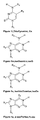
- Figure 1 provides examples of non-natural bases.
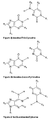
- Figure 7 provides additional examples.
- the invention describes a general method or principle of generating a diversity of bases and base pairs. For example, the amino group/"h" and carbonyl/thiocarbonyl on either the purine or on the pyrimidine are used to pair the bases.
- the hydrogen bond in question is either facing the minor groove or with the hydrogen bond in question facing the major groove.
- non-natural bases disclosed herein are capable of forming stable three hydrogen bond base pairs between purines and pyrimidines, such as
- bases can be used to create molecular entities such as pairs of oligomers that pair well with each other and the number of hydrogen bonds or affinity of the base pairs can be used to fine tune affinity.
- these entities result in reduced self structure and increased diversity with Uracils facing Diaminopurines or Adenines and Adenines further facing 2-Thiouracils, and 2-Oxo-pyrimidine pairs with both Guanine and ThioGuanine.
- the bases of the invention can be defined as C, D, G, U from figure 1 and their derivatives, such as for example, one carbonyl-thiocarbonyl substitution (Cs, Gs, U2s, and U4s) or one deamination (A, isoA, I (inosine) and P (pyrimidinone)).
- set of base pairing rules which one may use as a guide to design additional bases or molecular entities or probes that interact: 1) When part of complementary entities or oligomers, the base pairs obey Watson-Crick rules with hydrogen donors pairing with hydrogen acceptors, via hydrogen bonds, for example, pairing purine bases with pyrimidine bases. For example, C:G share three mutual hydrogen bonds and are thus very stable, while A:U share two mutual hydrogen bonds and contribute to complex stability.
- non-natural bases of the invention can be made using synthesis techniques well known in the art. Examples are provided in the examples section below.
- compositions comprising molecular entities which are a pair of PNA oligomers, wherein each of the PNA oligomers comprises at least one molecule chosen from: wherein
- the at least one molecular entity is not the isolated at least one molecule.
- R 1 is chosen from hydrogen, halogen, methyl and ethyl.
- R 2 is not a phosphate-ribonucleotide backbone, and is not a phosphate-deoxyribonucleotide.
- R 1 , R 3 , and R 4 are each is hydrogen.
- R 3 is chosen is chosen from optionally substituted (C 1 -C 20 )-alkyl, -NR 6 R 7 , optionally substituted (C 1 -C 20 )-alkoxy, optionally substituted (C 3 -C 8 )-cycloalkyl, and linker.
- R 2 and R 5 can be identical or different and are chosen from linker, backbone, and linker bound to backbone.
- the backbone is chosen from a polyamide backbone and a sugar-phosphate backbone.
- R 1 , R 3 , and R 4 each is hydrogen;
- R 2 and R 5 can be identical or different radicals and are chosen from backbone and linker bound to backbone; and the backbone is chosen from a polyamide backbone and a sugar-phosphate backbone.
- R 8 , R 9 , R 10 , R 11 , R 12 , and R 13 can be identical or different, and are chosen from hydrogen, halogen, and optionally substituted (C 1 -C 4 )-alkyl.
- R 14 is chosen from hydrogen, optionally substituted (C 1 -C 4 )-alkyl, optionally substituted (C 1 -C 4 )-alkoxy, and optionally substituted (C 3 -C 6 )-cycloalkyl
- R 8 , R 9 , R 12 , and R 13 can be identical or different, and are chosen from hydrogen, methyl ethyl, and acetyl.
- R 12 and R 13 can be identical or different, and are chosen from optionally substituted (C 1 -C 20 )-alkoxy, optionally substituted (C 3 -C 8 )-cycloalkyl, and optionally substituted (C 1 -C 20 )-alkyl, and R 14 is chosen from optionally substituted (C 1 -C 20 )-alkyl, optionally substituted (C 1 -C 20 )-alkoxy, and optionally substituted (C 3 -C 8 )-cycloalkyl.
- the at least one molecular entity is double stranded
- the backbone of each strand may be the optionally same
- the backbone is polyamide backbone
- any of the R groups R1 - R6 present are methyl.
- R1 to R6 are indenpently chosen from H and methyl.
- bases of the invention are the structures shown in Figure 1 with the following substituents: Base (Symbol) R2 R3 R4 R5 R6 A H or CH 3 H H isoA H or CH 3 H H D H or CH 3 H G H or CH 3 H Gs H or CH 3 H I H or CH 3 H U H or CH 3 U2s H or CH 3 U4s H or CH 3 C H or CH 3 H Py-2o H or CH 3 H or CH 3 Cs H or CH 3 H isoG H or CH 3 H isoGs H or CH 3 H Pu-2o H or CH 3 H isoC H H or CH 3 isoCs H H or CH 3 Py-4o H or CH 3 H or CH 3
- one or more of the H or CH 3 are independently substituted with a halogen such as Cl or F.
- a halogen such as Cl or F.
- Figure 7 illustrates other base pairing schemes compatible with the invention. Those involve Xanthine (X) or 2-thio-xanthine (2X) or 6-thio-xanthine (6X). X pairs with each of 2,4-diaminopyrimidine-5-yl, 2-aminopyrimidine-5-yl, and 4-aminopyrimidine-5-yl, while 2X also pairs with 2-aminopyrimidine-5-yl and 6X also pairs with 4-aminopyrimidine-5-yl. Accordingly, the choice of X or 2X or 6X in one strand opposed to the aminopyrimidine derivatives in the opposite strand allows one to control the level of degeneracy in a nucleic acid sequence.
- R 1 or "BB" in Figures 1 , 2 or may be a point of attachment to a polymer backbone, for instance, a polyamide backbone.
- the bases and base-pairs are chosen from a sub-set of those depicted in Figures 1 and 2, and 7 , depending on the needs of the application.
- a composition comprising molecular entities, which are a pair of PNA oligomers which comprise a set of molecules comprising non-natural base purine-like molecules (such as formulas (1), (2), (3), (7), (9) and (10), and salts thereof) and pyrimidine-like molecules (such as formulas (4), (5), (6), (8), (11) and (12), and salts thereof).
- the set of molecules is chosen from:
- the purine-like and pyrimidine-like molecules are chosen from a subset of the (a) through (f) pairings.
- the set of molecules is chosen from the following pairings::
- a pair of molecular entities according to claim 1 may comprise the three hydrogen bond base pair IsoCytosine and isoGuanine.
- IsoCytosine and isoGuanine are 2 hydrogen bond base pairs:
- a pair of molecular entities according to claim 1 may comprise the three hydrogen bond base pair 1,4-diaminopyrimidine-5-yl and Xanthine.
- Xanthine also may form two hydrogen bond base pairs with 1-diaminopyrimidien-5-yl or with 4-diaminopyrimidine-5-yl. In each of those pairings, one of the two amino groups from 1,4-diaminopyrimidine-5-yl is removed. See Figure 7 , left side pairings.
- 1 or 4-diaminopyrimidine-5-yl can form a two hydrogen bond base pair with a Xanthine derivative in which one of the two carbonyl oxygens is replaced by sulfur: 2-thioXanthine (X2s) or 6-thioXanthine (X6s).
- X2s 2-thioXanthine
- X6s 6-thioXanthine
- the base pairs do not formally pair with any of the naturally occurring bases.
- the bases of the invention are used with the natural bases only, for example a single pair being sufficient to provide no DNA affinity and increased diversity.
- Non-natural bases comprising halogen and alkyl groups
- purine and pyrimidine-like molecules can also be used to vary affinity.
- the number of hydrogen bonds may be varied by substituting the H for a halogen.
- the bases of the invention are halogen-comprising non-natural bases, for example,
- base pairs include two hydrogen bonds
- fluorine and chlorine are quite small substituents that may have a relatively small steric effect on base pairing, i.e. 2-fluoroAdenine will pair with both uracil and 2-thioUracil, essentially acting like an analogue of Adenine.
- Sterically smaller alternatives, such as fluorine and chlorine, to an amino group can be used that face both a carbonyl and a thiocarbonyl,
- larger halogens such as Iodine may be used to affect binding patterns, i.e. 2-lodo-Adenine may pair with Uracil, but not with 2-thio-Uracil.
- 6-bromo-Adenine could pair with uracil via two hydrogen bond, such as A : U.
- binding to 2-thioUracil may be further enhanced by a favorable hydrophobic interactions between the bromine and the thiocarbonyl creating a pair of triple hydrogen bond strength similar to D : U.
- the bases of the invention are methyl analogues, i.e. Me for Ha or H.
- 2-methyl-Adenine may be used as an Adenine analogue with PNAs. While it will strongly repell 2-thioUracil, it may bind to Uracil with significantly reduced affinity relative to Adenine.
- the above mentioned derivatives have hydrogen/halogen or an amino group in a potentially Watson-Crick like base pairing position facing either a carbonyl or thiocarbonyl on the "partner" base, i.e., the pattern determining the base pairing. Any other substitutions however is also possible, that does not directly influence the Watson-Crick like base pairing rules.
- Non-limiting examples include: Pyrimidines substituted in the 5-position; i.e. bromouracil; analogues of C with one 4-N substituent, i.e. 4-N-alkyl and 4-N-acyl derivatives of C/C2s; purines halogenated in the 8-position, i.e 8-bromo-Adenine and 9-deaza and/or 8-aza purine derivatives; 2 and 6-amino"purines" with small N-substituents, i.e. N-6-Acetyl-diaminopurine, N-6-Metyl-Guanine, and N-Metyl-Adenine.
- the molecular entities have PNA backbones and are (PNA) oligomers.
- the increased number of bases will provide an increased degeneracy over natural bases.
- the degeneracy minimizes secondary and tertiary structure.
- the degeneracy also may provide the advantage of one probe that can be used to recognize more than one target exclusively.
- the system allows multiple different PNAs to be recognized with the same affinity by one other PNA complementary to all of the PNAs, yet also allow each PNA to be recognized with unique specificity by other complementary, yet different PNAs.
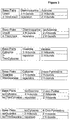
- 12 12 ⁇ 9x10 12 different 12'mers can be constructed. See Figure 6 , for example. These can be divided into 91 subgroups, according to the number of complementary 12'mers with which they pair, from 1 to half a million. On average, each 12'mer will pair with 1400 others, creating 10 16 different base pair combinations.
- the information system of the invention is expended relative to "linear" information systems such as binary strings, bar codes, or natural oligomers based on A, C, G, U and T.
- what is read not only depends on the encoding entity, such as an oligomer, but also on which of the numerous complementary PNAs are selected to "read” it, i.e., bind or not bind.
- the diversity of the system provides a degeneracy that limits cross reactivity. As one 12'mer will pair with 1400 others, for example, this limits the multiplexing capability of the system, i.e., only a limited number of pairs can work simultaneously and independently of each other with no cross reactivity. Triple vs. double vs. single hydrogen bonded base pairs are distinguished by different affinity.
- a single hydrogen bond pair, such as I:P would similarly be expected to be weaker than I:C. This again provides even more diversity since for example, I and P each form tow stable two hydrogen bond complexs thus I will bind stronger to C and Cs than to P and P will bind stronger to G and Gs than to I.
- each pair of oligomers may also be quite unique with very different binding affinities.
- the bases can thereby, for example, be used in a method to design a probe. Since, for example, each molecular entity such as a oligonucleotide made from a series of the bases of the invention, will bind to so many other molecular entities, a series of probes may be chosen or designed that differs slightly in the number of interactions, such as hydrogen bonds, that may be involved in the binding. Thus, for example, an oligonucleotide of 8 bases will have thousands of potential binding partners using the bases of the invention. Each of these may have slightly different strengths of interaction. Thus, one can chose a probe that is very sensitive to binding conditions, such as temperature, or design a series of probes that each bind at a slightly different temperature.
- the differences in temperature can be useful, in for example, immunohistochemistry in a system that can be varied by slight changes in temperature.
- the number of possible binding partners allows one to fine tune a binding partner for affinity, speed, temperature, low specific affinity for other probes or other compounds present in a sample and other uses.
- a method of designing a probe may include a screening method where the thousands of possibilities for a certain length oligomer or numerous size oligomers are screened for the desired strength of hybridization and unique hybridization properties.
- An entity may be chosen based on fast hybridization kinetics, combined with sufficient length to provide stability as links in supra molecular visualization and amplification systems and diagnostics.
- Such a system would be analogous to a hotel where each guest has a key unique to her own room, yet there is also a "master key" that will open every room.
- the binding affinity between entities of base pairs may be a relative predictable function of length and triple, double, and single hydrogen bonded base pairs.
- the kinetic stability of the complexes once formed may follow a similar trend.
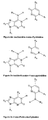
- Binding affinity may be inversely proportional to the stability of the complex. For example, the longer and the more potential bond reach of an oligomer, the greater chance that it will be highly self structured and thus slow to hybridize. This can be expressed as a rate determining step preceding actual double-strand formation. More specifically, when two oligomers form a double-strand, the energy barrier to reaching the transition state will increase with the structure reorganization required of each of the hybridizing oligomers. Thus, one can reduce the energy barrier if the stable structures of the oligomers before hybridization are aligned and/or by preparing oligomers that require little reorganization to reach the transition state because of lack of a stable self structure ( Figure 5 ).
- intra molecular structure is a priori reduced by using more bases.
- the 4 bases of DNA can form 16 pairs. Of these, 4 will be matches (25%) contributing on average with 2.5 hydrogen bonds.
- 12 bases disclosed herein can form 144 pairs. Of these, only 22 will be matches (15%) and on average contribute with 2.1 hydrogen bonds. Since several consecutive base pairs are needed for stable intra molecular structures to form, this effect becomes quite pronounced. If it is assumed that 10 mutual hydrogen bonds are needed for a hair pin structure to form, and if 2 x 4 random DNA bases match, there is a 0.39% chance of hair pin formation. In contrast, using 12 bases instead of 4, there is only a 0.0084 % chance of a hair pin because 2 X 5 bases must match for it to form. Adding more bases also aids in smart design of a probe because one may avoid aggregates or crystalline structures.
- the bases of the invention may be used to prepare molecular entities such as PNA oligomers with at least one of high binding affinity, variable binding affinity, rapid binding kinetics and absence of interference with RNA/DNA.
- the degeneracy of the bases may also be utilized to design a molecular encoding system similar to the natural information encoding system of DNA but greatly enhanced because of the greater number of bases.
- the bases of the invention may be used in any application in which natural bases, DNA, PNA, etc, are used.
- a linker may also be used to connect at least one molecular entity to another or to bind a molecular entity to another composition.
- linkers include 6-amino-hexanoic acid, succimidyl 4-(N-malemidomethyl) cylohexane-1-carboxylate (SMCC), homobifunctional linkers such as glutaric dialdehyde, hexane di-isocyanate, dimethylapimidate, 1,5-difluoro-2,4-dinitrobenzene, divinyl sulfone (DVS), heterobifunctional linkers like e.g. N-gamma-maleimidobytyroloxy succinimide ester (GMBS), and zero length linkers such as 1-ethyl-3-(3-dimethylaminopropyl)cabodiimide
- GMBS N-gamma-maleimidobytyroloxy succinimide ester
- zero length linkers such as 1-eth
- PEG polyethylene glycol
- the present invention may also use a long uncharged linker comprising at least two units of the Formula I.
- R1 and R2 may comprise either NH or O, while R3 may comprise methyl, ethyl, propyl, CH2-O-CH2, and (CH2-O-CH2)2.
- the linker comprises at least two units of the Formula I wherein R1 and R2 are both NH and R3 is CH2-O-CH2. See the examples that follow and the International Application entitled "MONOMERIC AND POLYMERIC
- Fluorosceins may also be added to the linkers. When fluorosceins are spaced apart by an L30 oligomer larger than L90 in an aqueous environment there may be negligible quenching. This contrasts with PEG-based spacers in aqueous solutions which may form a collapsed coiled structure.
- PNA probes with multiple fluorophores spaced via polymeric L30 may have higher water solubility, less aggregation and less non-specific binding, as compared to probes with just one fluorophore and no L30 spacer.
- Multiple PNAs joined together via L30 polymers may not aggregate and may have good solubility even with a total of 50-100 PNA bases in the same compound.
- Compounds and compositions of the invention may also include a detectable label which is any molecule which may be detected directly or indirectly so as to reveal the presence of a target in the sample.
- a direct detectable label is used.
- Direct detectable labels may be detected per se without the need for additional molecules. Examples include fluorescent dyes, radioactive substances, and metal particles.
- indirect detectable labels are used, which require the employment of one or more additional molecules. Examples include enzymes that affect a color change in a suitable substrate, as well as any molecule that may be specifically recognized by another substance carrying a label or react with a substance carrying a label.
- Other examples of indirect detectable labels thus include the non-natural bases of the present invention, antibodies, antigens, nucleic acids and nucleic acid analogs, ligands, substrates, and haptens.
- detectable labels examples include fluorophores, chromophores, electrochemiluminescent labels, bioluminescent labels, polymers, polymer particles, bead or other solid surfaces, gold or other metal particles or heavy atoms, spin labels, radioisotopes, enzyme substrates, haptens, antigens, Quantum Dots, aminohexyl, pyrene, nucleic acids or nucleic acid analogs, or proteins, such as receptors, peptide ligands or substrates, enzymes, and antibodies (including antibody fragments).
- Some detectable labels according to this invention comprise “color labels,” in which the target is detected by the presence of a color, or a change in color in the sample.
- color labels are chromophores, fluorophores, chemiluminescent compounds, electrochemiluminescent labels, bioluminescent labels, and enzymes that catalyze a color change in a substrate.
- more than one type of color may be used, for instance, by attaching distinguishable color labels to a single detection unit or by using more than one detection unit, each carrying a different and distinguishable color label.
- Fluorophores as described herein are molecules that emit detectable electro-magnetic radiation upon excitation with electro-magnetic radiation at one or more wavelengths.
- a large variety of fluorophores are known in the art and are developed by chemists for use as detectable molecular labels and can be conjugated to the linkers of the present invention.
- fluorescein or its derivatives such as fluorescein-5-isothiocyanate (FITC), 5-(and 6)-carboxyfluorescein, 5- or 6-carboxyfluorescein, 6-(fluorescein)-5-(and 6)-carboxamido hexanoic acid, fluorescein isothiocyanate, rhodamine or its derivatives such as tetramethylrhodamine and tetramethylrhodamine-5-(and-6)-isothiocyanate (TRITC).
- FITC fluorescein-5-isothiocyanate
- 5- or 6-carboxyfluorescein 5- or 6-carboxyfluorescein
- 6-(fluorescein)-5-(and 6)-carboxamido hexanoic acid fluorescein isothiocyanate
- rhodamine or its derivatives such as tetramethylrhodamine and tetramethylrhodamine-5-(
- fluorophores that could be conjugated to the instant linkers include: coumarin dyes such as (diethyl-amino)coumarin or 7-amino-4-methylcoumarin-3-acetic acid, succinimidyl ester (AMCA); sulforhodamine 101 sulfonyl chloride (TexasRedTM or TexasRedTM sulfonyl chloride; 5-(and-6)-carboxyrhodamine 101, succinimidyl ester, also known as 5-(and-6)-carboxy-X-rhodamine, succinimidyl ester (CXR); lissamine or lissamine derivatives such as lissamine rhodamine B sulfonyl Chloride (LisR); 5-(and-6)-carboxyfluorescein, succinimidyl ester (CFI); fluorescein-5-isothiocyanate (FITC); 7-diethylaminocoumarin
- fluorescent proteins such as green fluorescent protein and its analogs or derivatives, fluorescent amino acids such as tyrosine and tryptophan and their analogs, fluorescent nucleosides, and other fluorescent molecules such as Cy2, Cy3, Cy 3.5, Cy5, Cy5.5, Cy 7, IR dyes, Dyomics dyes, phycoerythrine, Oregon green 488, pacific blue, rhodamine green, and Alexa dyes.
- fluorescent labels which may be used in the invention include and conjugates of R-phycoerythrin or allophycoerythrin, inorganic fluorescent labels such as particles based on semiconductor material like coated CdSe nanocrystallites.
- polymer particles labels which may be used in the invention include micro particles, beads, or latex particles of polystyrene, PMMA or silica, which can be embedded with fluorescent dyes, or polymer micelles or capsules which contain dyes, enzymes or substrates.
- metal particles which may be used in the invention include gold particles and coated gold particles, which can be converted by silver stains.
- haptens that may be conjugated in some embodiments are fluorophores, myc, nitrotyrosine, biotin, avidin, strepavidin, 2,4-dinitrophenyl, digoxigenin, bromodeoxy uridine, sulfonate, acetylaminoflurene, mercury trintrophonol, and estradiol.
- Examples of enzymes which may be used in the invention comprise horse radish peroxidase (HRP), alkaline phosphatase (AP), beta-galactosidase (GAL), glucose-6-phosphate dehydrogenase, beta-N-acetylglucosaminidase, ß-glucuronidase, invertase, Xanthine Oxidase, firefly luciferase and glucose oxidase (GO).
- HRP horse radish peroxidase
- AP alkaline phosphatase
- GAL beta-galactosidase
- glucose-6-phosphate dehydrogenase beta-N-acetylglucosaminidase
- ß-glucuronidase invertase
- Xanthine Oxidase firefly luciferase
- glucose oxidase GO
- HRP horse radish peroxidase
- DAB 3,3'-diaminobenzidine
- AEC Benzidine dihydrochloride
- HRR Hanker-Yates reagent
- IB Indophane blue
- TMB tetramethylbenzidine
- CN 4-chloro-1-naphtol
- ⁇ -NP ⁇ -naphtol pyronin
- OD o-dianisidine
- BCIP 5-bromo-4-chloro-3-indolylphosphate
- NBT Nitro blue tetrazolium
- TNBT tetranitro blue tetrazolium
- Examples of commonly used substrates for Alkaline Phosphatase include Naphthol-AS-B1-phosphate/fast red TR (NABP/FR), Naphthol-AS-MX-phosphate/fast red TR (NAMP/FR), Naphthol-AS-B1-phosphate/fast red TR (NABP/FR), Naphthol-AS-MX-phosphate/fast red TR (NAMP/FR), Naphthol-AS-B1-phosphate/new fuschin (NABP/NF), bromochloroindolyl phosphate/nitroblue tetrazolium (BCIP/NBT), 5-Bromo-4-chloro-3-indolyl-b(beta) -d (delta)-galactopyranoside (BCIG).
- luminescent labels which may be used in the invention include luminol, isoluminol, acridinium esters, 1,2-dioxetanes and pyridopyridazines.
- electrochemiluminescent labels include ruthenium derivatives.
- radioactive labels examples include radioactive isotopes of iodide, cobalt, selenium, hydrogen, carbon, sulfur and phosphorous.
- the detection unit may comprise from 1 up to 500 detectable label molecules.
- the detectable label is an enzyme, which may be conjugated to a polymer, such that the number of enzyme molecules conjugated to each polymer molecule is, for instance, 1 to 200, 2 to 50, or 2 to 25.
- the detectable label is a gold particle, a radioactive isotope, or a color label, e.g. a low molecular weight fluorochrome, and the number of detectable labels conjugated to each polymer molecule is, for instance, 1 to 500, or for instance, 2 to 200.
- the detectable label is a protein fluorochrome and the number of detectable labels conjugated to each polymer molecule is 1-50, 2-20. In some embodiments, the number of detectable label molecules conjugated to each polymer is 1-200, 2-50, 2-25, or is 10-20, 5-10, or 1-5. Add some more number ranges to pick from later on if necessary.
- the detectable label can be detected by numerous methods, including, for example, reflectance, transmittance, light scatter, optical rotation, and fluorescence or combinations hereof in the case of optical labels or by film, scintillation counting, or phosphorimaging in the case of radioactive labels. See, e.g., Larsson, 1988, Immunocytochemistry: Theory and Practice, (CRC Press, Boca Raton, FL ); Methods in Molecular Biology, vol. 80 1998, John D. Pound (ed.) (Humana Press, Totowa, NJ ). In some embodiments, more than one detectable label is employed.
- the different colors may have different, distinguishable colors. In some embodiments both colors can be detected simultaneously, such as by fusion or juxtaposition of the signals, signal enhancement or quenching, or detection of multiple colors in the sample.
- the exact choice of detectable label or combinations of detectable labels may be based on personal preferences in combinations with restrictions of the sample type, sample preparation method, detection method and equipment, and optional contrasting labels used in the sample.
- the chosen hybridization conditions are "stringent conditions," meaning herein conditions for hybridization and washes under which nucleotide sequences that are significantly complementary to each other remain bound to each other.
- the conditions are such that sequences at least 70%, at least 80%, at least 85-90% complementary remain bound to each other. The percent complementary is determined as described in Altschul et al. (1997) Nucleic Acids Res. 25:3389-3402 .
- the chosen hybridization conditions are "high stringency conditions.”
- An example of high stringency hybridization conditions is hybridization in 4X sodium chloride/sodium citrate (SSC) at 65-70°C or hybridization in 4X SSC plus 50% formamide at 42-50°C, followed by one or more washes in 1X SSC, at 65-70°C.
- additional reagents may be added to hybridization and/or wash buffers, e.g., blocking agents (BSA or salmon sperm DNA), detergents (SDS), chelating agents (EDTA), Ficoll, PVP, etc.
- the chosen conditions are “moderately stringent conditions.”
- Moderate stringency includes conditions that can be readily determined by those having ordinary skill in the art based on, for example, the length of the molecular entity or a specific nucleic acid pr nucleic acid analog segment. Exemplified conditions are set forth by Sambrook et al. Molecular Cloning: A Laboratory Manual, 2d ed. Vol. 1, pp.
- Low stringency conditions may include, as used herein, conditions that can be readily determined by those having ordinary skill in the art based on, for example, the length of the molecular entity.
- Low stringency may include, for example, pretreating the segment for 6 hours at 40°C in a solution containing 35% formamide, 5 x SSC, 50 mM Tris-HCl (pH 7.5), 5 mM EDTA, 0.1% PVP, 0.1% Ficoll, 1% BSA, and 500 ⁇ g/ml denatured salmon sperm DNA.
- Hybridizations are carried out in the same solution with the following modifications: 0.02% PVP, 0.02% Ficoll, 0.2% BSA, 100 ⁇ g/ml salmon sperm DNA, 10% W/V dextran sulfate, and 5-20x106 CPM probe is used. Samples are incubated in hybridization mixture for 18-20 hours at 40°C, and then washed for 1.5 h at 55°C in a solution containing 2 x SSC, 25 mM Tris-HCl (pH 7.4), 5 mM EDTA, and 0.1 % SDS. The wash solution is replaced with fresh solution and incubated an additional 1.5 h at 60°C.
- the compounds and compositions of the invention, including any molecular entity may also comprise at least one polymer.
- a "polymer,” as used herein, may be any molecule that facilitates covalent or non-covalent attachment of one or more other components or molecular entities. For instance, the polymer may facilitate the attachment of one or more probes, molecular entities, nucleic acid analog segments, and or detectable labels.
- the polymer may be a soluble molecule or an insoluble molecule and may have any shape including a linear polymer, branched polymer, bead or other globular shaped polymer.
- suitable polymers include polysaccharides such as dextrans, carboxy methyl dextran, dextran polyaldehyde, carboxymethyl dextran lactone, and cyclodextrins; pullulans, schizophyllan, scleroglucan, xanthan, gellan, O-ethylamino guaran, chitins and chitosans such as 6-O-carboxymethyl chitin and N-carboxymethyl chitosan; derivatized cellolosics such as carboxymethyl cellulose, carboxymethyl hydroxyethyl cellulose, hydroxyethyl cellulose, 6-amino-6-deoxy cellulose and O-ethylamine cellulose; hydroxylated starch, hydroxypropyl starch, hydroxyethyl starch, carrageenans, alginates, and agarose; synthetic polysaccharides such as ficoll and carboxymethylated ficoll; vinyl polymers including poly(acrylic acid
- Properties of the polymer can be varied, depending on the desired application, to optimize performance. Examples of parameters that may be considered in the choice of a polymer include the length of the polymer and branching of the polymer. Furthermore, the polymer may carry various substituents. The substituents may be chemically protected and/or activated, allowing the polymer to be derivatized further.
- compositions according to the invention may be comprised in a kit.
- the kit may optionally comprise one or more binding agents, and suitable reagents for, for instance: antigen retrieval, sample dilution, reagent dilution, blocking of non-specific binding, blocking of endogenous enzyme activity, or blocking of repetitive sequences.
- the kit may optionally also comprise at least one container, instructions for use, and reference targets or samples.
- Example 1 b Alternative method of preparing pyrimidinone monomer
- Step 1 In dry equipment 9.2 g of solid Na in small pieces was dissolved in 400 mL ethanol (99.9%), with stirring. Hydroxypyrimidine hydrochlorid, 26.5 g, was added, and the mixture was stirred for 10 minutes at 50°C. Then 24.4 mL Ethyl bromoacetate (98%) was added and the mixture stirred at 50°C for 1 hour. The reaction was followed using Thin Layer Chromatography (TLC).
- TLC Thin Layer Chromatography
- the ethanol was evaporated leaving a white compound, which was dissolved in 70 mL of water and extracted with 20 mL DCM. Another 30 mL of water was added to the water phase, which was extracted with 3x100 mL DCM.
- the DCM-phase from the first extraction contains a lot of product, but also some impurities, wherefore this phase was extracted twice with water. These two water phases then were back extracted with DCM.
- Step 3 Pyrimidinone acetic acid 11.1 g and triethylamine 12.5 mL were dissolved in N,N-dimethylformamide (DMF) 24 ml, HBTU 26.2 g was added plus 6 mL extra DMF. After 2 minutes a solution of PNA-Backbone ethylester 14.7g dissolved in 15 mL DMF was added. The reaction mixture was stirred at room temperature and followed using TLC. After 11 ⁇ 2hour precipitate had formed. This was filtered off.
- DMF N,N-dimethylformamide
- Step 5 To make a test on the P-monomer 3 consecutive P's were coupled to Boc-L300-Lys(Fmoc) -resin, following normal PNA standard procedure. The product was cleaved from the resin and precipitated also following standard procedures: HPPP-L300-Lys(Fmoc). Maldi-Tof on the crude product: 6000 (calc. 6000) showing only minor impurities.
- Diaminopurine (10 g) and 40 g of K 2 CO 3 were added to 85 mL of DMF and stirred for 30 minutes. The mixture was cooled in a water bath to 15°C. Ethyl bromoacetate (3 mL) was added three times with 20 minute intervals between each addition. This mixture was then stirred for 20 minutes at 15°C. The mixture was left in the water bath for another 75 minutes, and the temperature increased to 18°C. The DMF was removed by filtering and the remaining K 2 CO 3 was added to 100 mL of ethanol and refluxed for 5 minutes. Filtering and repeated reflux of the K 2 CO 3 in 50 mL ethanol, filtering. The pooled ethanol phases were placed in a freezer, after which crystals formed. These crystals were filtered, washed with cold ethanol, filtered again and then dried in a desiccator overnight. The yield for this step was 12 g (76 %).
- Example 18a an MBHA-resin was loaded with Boc-Lys(Dde)-OH.
- amino acids were coupled according to PNA solid phase procedure provided in Example 18d yielding Boc-L 90 -Lys(Fmoc)-L 30 -Lys(Dde).
- the Boc and Fmoc protections groups were removed and the amino groups marked with flourescein using the procedure in Example 18e.
- the Dde protection group was removed and 0.4 M cysteine was added according to the procedure in Example 18b.
- the PNA was cleaved from the resin, precipitated with ether and purified on HPLC according to Example 18d.
- the product was found to have a molecular weight of 3062 using MALDI-TOF mass spectrometry; the calculated molecular weight is 3061.
- Example 8 Synthesis of a conjugate made from sequence AA from Example 5, DexVS70, and Flu(10)
- the above four compounds were mixed. The mixture was placed in a water bath at 30° C for 16 hours. The mixture was added to 50 nmol of freeze-dried PNA (sequence AA from Example 5). The mixture was placed in a water bath at 30° C for 30 minutes. The conjugating reaction was quenched with 50 ⁇ L of 500 mM cysteine for 30 minutes at 30° C. Purification of the product was performed using FPLC: column SUPERDEX®- 200, buffer 10 mM Hepes 100 mM NaCl, method 7 bank 2, Loop 1 mL. Two fractions were collected: one with the product and one with the residue.
- the conjugation ratio of Flu 2 to DexVS70 was 9.4.
- the conjugation ratio of PNA (sequence AA) to DexVS70 was 1.2.
- Dextran (molecular weight 70 kDa) was activated with divinylsulphone to a degree of 92 reactive groups/dextran polymer, and is designated DexVS70.
- the antibody Anti-Human-BCL2 is designated AHB.
- 105 ⁇ L DexVS70 7.5 nmol 800 ⁇ L AHB conc. (2.9 g/L) 15.1 nmol 25 ⁇ L 4 M NaCl 32 ⁇ L 0.8 M NaHCO 3 (pH 9.5)
- the above four compounds were mixed and placed in a water bath at 30° C for 65 minutes. From this mixture, 875 ⁇ L was taken out and added to the indicated number of nmol of PNA in the table below; before the addition the PNA had been dissolved in the ⁇ L of H 2 O indicated in the table below. Then the number of ⁇ Ls of 0.8 M NaHCO 3 (pH 9.5) was added according to the table below. The resulting mixture was placed in a water bath at 30° C for 70 minutes. Quenching was performed by adding 6 mg of solid cysteine (0.05 M) to the mixture and letting it stand for 30 minutes at 30° C.
- the precipitate was washed with ether and was then dissolved in 100 ⁇ L TFA.
- the precipitate was washed with ether and then dried using N 2 gas.
- the above five components are mixed together placed in a water bath at 30° C for 16 hours. Five hundred microliters of this mixture are added to 50 nmol PNA, which is previously dissolved in 40 ⁇ L H 2 O. Then, 10 ⁇ L of 0.8 M NaHCO 3 (pH 9.5) is added. The mixture is then placed in a water bath at 30° C for 2 hours. Quenching is performed by adding 55 ⁇ L of 110 mM cysteine and letting the resultant mixture set for 30 minutes at 30° C.
- the above five components are mixed and placed in a water bath at 30° C for 40 minutes. Two hundred and ninety ⁇ L is taken out of the mixture and added to 50 nmol of PNA, which is previously dissolved in 40 ⁇ L of H 2 O. Then, 10 ⁇ L of 0.8 M NaHCO 3 (pH 9.5) is added and the mixture placed in a water bath at 30° C for 1 hour. Quenching is performed by adding 34 ⁇ L of 500 mM cysteine and letting the resultant mixture set for 30 minutes at 30° C.
- Dextran (molecular weight 70 kDa) is activated with divinylsulfone to a degree of 92 reactive groups/dextran polymer.
- PNA1 100 nmol
- DexVS70 10 nmol
- To this mixture 12.5 ⁇ L of PNA2 (12.5 nmol) dissolved in H 2 O is added, and then 30 ⁇ L of NaHCO 3 (pH 9.5) is added and the solution mixed.
- the resultant mixture is placed in a water bath at 30° C for 35 minutes. Quenching was performed by adding 18.3 ⁇ L of 500 mM cysteine in Hepes and letting this mixture set for 30 minutes at 30° C.
- 6-Benzyloxypurine Sodiumhydride (60 % Dispersion in mineral oil;3,23g;80 mmol) was slowly added to benzyl alcohol (30 ml;34,7 mmol). After the addition of more benzyl alcohol (10 ml) and 6-chloropurine (5,36 g; ). The reaction mixture was heated to 100°C for 4 hours. When the reaction mixture has reached room temperature, water (1ml) was slowly added. 6-Benzyloxypurine was precipitated by the addition of acetic acid (4,6 ml) and diethylether (550 ml). The precipitate was separated by filtration (11,72 g). Re-crystallization from ether gave (4,78 g; 65,4 %).
- 6-Benzyloxypurine (4,18 g; 18,5 mmol) was added to a suspension of potassium carbonate (3,1 g; 22,4 mmol) in DMF (100 ml). After 15 min., bromoacetic acid methyl ester (1,93 ml; 20,4 mmol) was added. The reaction was monitored by TLC in butanol:acetic acid:water 4:1:1. Upon completion, the reaction mixture was partitioned between water (600 ml) and ethyl acetate (600 ml). The organic phase was dried over magnesium sulfate and evaporated to a volume of ⁇ 10 ml and precipitated with pet.
- Boc-PNA-Diaminopurine-(N6-Z)-monomer was prepared according to Gerald Haaima, Henrik F. Hansen, Leif Christensen, Otto Dahl and Peter E. Nielsen; Nucleic Acids Research, 1997, Vol 25, Issue 22 4639-4643 .
- Boc-PNA-2-Thiouracil-(S-4-MeOBz)-monomer was prepared according to Jesper Lohse, Otto Dahl and Peter E. Nielsen; Proceedings of the National Academy of Science of the United States of America, 1999, Vol 96, Issue 21, 11804-11808 .
- Boc-PNA-Adenine-(Z)-monomer was from PE Biosystems catalog GEN063011.
- Boc-PNA-Cytosine-(Z)-monomer was from PE Biosystems cat. GEN063013.
- Boc-PNA-Guanine-(Z)-monomer was from PE Biosystems cat. GEN063012.
- Boc-PNA-Thymine-monomer was from PE Biosystems cat. GEN063010.
- IsoAdenine (2-aminopurine) may be prepared as a PNA-monomer by 9-N alkylation with methylbromoacetate, protection of the amino group with benzylchloroformate, hydrolysis of the methyl ester, carbodiimide mediate coupling to methyl-(2-Boc-aminoethyl)-glycinate, and finally hydrolysis of the methyl ester.
- 4-thiouracil may be prepared as a PNA-monomer by S-protection with 4-methoxy-benzylchloride, 1-N alkylation with methylbromoacetate, hydrolysis of the methyl ester, carbodiimide mediate coupling to methyl-(2-Boc-aminoethyl)-glycinate, and finally hydrolysis of the methyl ester.
- Thiocytosine may be prepared as a PNA monomer by treating the Boc-PNA-cytosine(Z)-monomer methyl ester with Lawessons reagent, followed by hydrolysis of the methyl ester.
- halogenated bases are commercially available, and may be converted to PNA monomers analogously to the non-halogenated bases. These include the guanine analog 8-bromo-guanine, the adenine analogs 8-bromo-adenine and 2-fluoro-adenine, the isoadenine analog 2-amino-6-chloro-purine, the 4-thiouracil analog 5-fluoro-4-thio-uracil, and the 2-thiouracil analog 5-chloro-2-thiouracil.
- Boc-PNA-Uracil monomers were first described in " Uracil og 5-bromouracil I PNA," a bachelor project by Kristine Kils ⁇ Jensen, K ⁇ benhavns Universitet 1992 .
- the PNA is cleaved from the resin with TFA/TFMSA/m-cresol/thioanisol (at a ratio of 6/2/1/1).
- the PNA is then precipitated with ether and purified on HPLC.
- MALDI-TOF mass spectrometry is used to determine the molecular weight of the product.
- Tonsil tissue samples were fixed in neutral buffered formalin, NBF (10 mM NaH 2 PO 4 / Na 2 HPO 4 , pH 7.0), 145 mM NaCl, and 4% formaldehyde (all obtained from Merck, Whitehouse Station, NJ). The samples were incubated overnight in a ventilated laboratory hood at room temperature.
- the tissue samples were placed in a marked plastic histocapsule (Sakura, Japan). Dehydration was performed by sequential incubation in 70% ethanol twice for 45 min, 96% ethanol twice for 45 min, 99% ethanol twice for 45 min, and xylene twice for 45 min. The samples were subsequently transferred to melted paraffin (melting point 56-58°C) (Merck, Whitehouse Station, NJ) and incubated overnight (12-16 hours) at 60°C. The paraffin-infiltrated samples were transferred to fresh warm paraffin and incubated for an additional 60 min prior to paraffin embedding in a cast (Sekura, Japan). The samples were cooled to form the final paraffin blocks. The marked paraffin blocks containing the embedded tissue samples were stored at room temperature in the dark.
- paraffin blocks were cut and optionally also mounted in a microtome (0355 model RM2065, Feather S35 knives, set at 5.0 micrometer; Leica, Bannockburn, IL). The first few millimeters were cut and discarded. Paraffin sections 4-6 micrometers thick were then cut and collected at room temperature. The sections were gently stretched on a 45-60°C hot water bath before being mounted onto marked microscope glass slides (SUPERFROST ® Plus; Fisher, Medford, MA), two tissue sections per slide. The slides were then dried and baked in an oven at 60°C.
- TBST Tris-buffered saline with TWEEN®
- TBST comprises 50 mM Tris adjusted to pH 7.6 with HCl; 150 mM NaCl; 0.05 % TWEEN®20.
- the slides were deparaffinated by subsequently incubation in xylene twice for 5 min ⁇ 2 min, 96% ethanol twice for 2min +/- 30 sec and 70% ethanol twice for 2 min +/- 30 sec.
- the slides were immersed in deionized water and left for 1 to 5 min.
- Antigens in the sample were retrieved by immersing the slides in a container containing Antigen Retrieval Solution, pH 6.0 (DakoCytomation code No. K5204 Vial 7 or optional code No. K5205 Vial 7).
- the container was closed with a perforated lid and placed in the middle of a microwave oven and left boiling for 10 min. The container was removed from the oven and allowed to cool at room temperature for 20 min. The samples were rinsed in deionized water.
- Antigens in the sample were retrieved by immersing the slides in a beaker containing Antigen Retrieval Solution, pH 6.0 (DakoCytomation code No. K5204 Vial 7 or optional code No. K5205 Vial 7). The samples were incubated for 40 min in a water bath at 95-100°C. The beaker was removed from the water bath and allowed to cool at room temperature for 20 min. The samples were rinsed in deionized water.
- TBST Tris-buffered saline with TWEEN ®
- Monoclonal Mouse Anti-Human Cytokeratin (DakoCytomation code No. M3515) diluted 1:300, 1:900 and 1:1600; monoclonal Mouse Anti-Human CD20cy (DakoCytomation code No. M0755) diluted 1:2000, 1:8000 and 1:14000; and monoclonal Mouse Anti-Human Ki-67 Antigen (DakoCytomation code No. M7240) diluted 1:400, 1:1200 and 1:2400 were used.
- the antibodies were diluted in ChemMateTM Antibody Diluent (DakoCytomation code No. S2022), applied on the tissue samples, and incubated for 30 min in a humid chamber at ambient temperature. The slides were individually rinsed and washed in TBST for 5 min.
- Antibody / Dextran / PNA1 conjugate recognition unit is also called "PNA1 conjugate” in the examples that follow.
- the PNA1 conjugate comprises 70,000 molecular weight dextran.
- Table 1 summarizes PNA1 conjugates based on a secondary antibody: goat anti-mouse Ig, called herein GAM (DakoCytomation code No. Z0420).
- Table 2 summarizes PNA1 conjugates based on a primary antibody: mouse anti-human BCL2 oncoprotein, such as Clone 124 (DakoCytomation code No. M0887). The primary antibody was protein A-purified prior to conjugation.
- the conjugates were diluted in BBA (50 mM Tris adjusted to pH 7.6 with HCl; 150 mM NaCl; 2% BSA; 0.02% bronidox; 2.44 mM 4-aminoantipyrin) and were applied on the tissue sample in a range of dilutions, then incubated for 30 min in a humid chamber at ambient temperature. The slides were individually rinsed and washed in TBST for 5 min. Table 5 .
- PNA1 conjugates for direct recognition of targets anti-BCL2 / Dextran / PNA1 Conjugate No. Sequence ⁇ M Dex Ab / Dex PNA1 / Dex D14128 U s GU s DPP TTG D 0.8 1.1 5.6 D14126 U s GU s DPP TTG D 1.0 1.2 2.9 D14122 U s GU s DPP TTG D 1.1 1.6 9.5
- the letters A, C, G, U, and T stand for the natural bases adenine, cytosine, guanine, uracil, and thymine.
- P stands for pyrimidinone, D for 2,6-diaminopurine, and U s for 2-thiouracil.
- the samples were washed in deionized water for 30 sec. Then, 1 % glutardialdehyde (Merck Art. No. 820603), called herein GA, diluted in 22 mM calcium phosphate buffer, pH 7.2, was applied, and the samples were incubated for 10 min in a humid chamber at ambient temperature. The samples were washed in deionized water for 30 sec and in TBST for 5 min.
- PNA 1 -PNA 2 / Dextran conjugate is also called "PNA 1 -PNA 2 " in the following examples.
- Table7 summarizes the compositions of PNA 1 -PNA 2 conjugates.
- PNA 1 is complementary to the PNA1 conjugate
- PNA 2 is complementary to the PNA2 conjugates D14079 and D13155 described in step 13 below.
- the sequence of PNA 1 is CU s G s G s DD TU s D G s DC and the sequence of PNA 2 is U s GU s DPP TTG D, in which U s stands for 2-thio-uracil, G s stands for 2-amino-6-thioxopurine, D stands for diaminopurine, and P stands for pyrimidinone.
- U s stands for 2-thio-uracil
- G s stands for 2-amino-6-thioxopurine
- D stands for diaminopurine
- P stands for pyrimidinone.
- the conjugates, diluted in BBA, were applied to the tissue samples in a range of dilutions, and the samples were then incubated for 30 min in a humid chamber at ambient temperature. The samples were individually rinsed and washed in TBST for 5 min.
- Horse Radish Peroxidase (HRP) / Dextran / PNA2 conjugates are also called "PNA2 conjugate" in the examples that follow, and are listed in table 8.
- the PNA2 conjugates comprise 70.000 Da molecular weight dextran.
- the conjugates diluted in BBA were applied to the tissue samples in a range of dilutions, and samples were incubated for 30 min in a humid chamber at ambient temperature. The samples were individually rinsed and washed twice in TBST for 5 min. Table 8.
- PNA2 conjugates HRP / Dextran / PNA2 Conjugate No.
- the diaminobenzidine chromogenic substrate solution, DAB+ (DakoCytomation code No. K3468) was applied on the tissue samples, and the samples were incubated for 10 min in a humid chamber at ambient temperature. The samples were washed with deionized water for 5 min.
- tissue samples were immersed in Mayers Hematoxylin (Bie & Berntsen Code No. LAB00254) for 3 min, rinsed in tap water for 5 min, and finally rinsed with deionized water.
- the tissue staining was examined in a bright field microscope at 10x, 20x or 40x magnification. Both the specific and the non-specific staining intensity were described with a score-system using the range 0 to 3+ with 0.5+ score interval.
- ChemMateTM EnVisionTM Detection kit Rabbit/Mouse (DakoCytomation code No. K5007 bottle A) was used as a reference, and was included in all experiments for testing in parallel with the PNA conjugates. K5007 was used according to manufacturer's instructions.
- the antibodies were used in the following dilutions: M3515 at 1:900, M0755 at 1:8000, and M7240 at 1:1200.
- the staining intensity of the K5007 reference using the primary antibody M3515 diluted 1:900 was set to 2+ in order to compare and assess the staining result of the PNA conjugate tested. If the reference deviated more than ⁇ 0.5, the test was repeated.
- the various visualization system combinations of the invention were tested on routine tissue samples.
- the staining performance was compared with a reference visualization system, using EnVisionTM and a very dilute antibody from DakoCytomation.
- the practical dynamic range of quantitative IHC may be narrow, and e.g. strongly stained (+3) tissues are not easy to compare with respect to intensity. Therefore, on purpose, the staining intensity of the reference system was adjusted to be approximately +2. This was done in order to better monitor and compare differences in staining intensity with the system of the invention.
- the conjugates to be tested were diluted to the final concentrations 0.05 ⁇ M and 0.2 ⁇ M.
- two PNA2 conjugates were used in the final concentration 0.05 ⁇ M.
- the first reference, for example, PNA2, D13108 was known to give non-specific nuclear staining, and so was used as a positive control.
- the second reference, for example, PNA2, D13155 was known not to give any non-specific nuclear staining, and was used as a negative control.
- 250 ⁇ L of each reagent was applied unless otherwise specified.
- Tonsil tissues were taken through the steps 1-4, 6-8, 10, 11, and 13-17 above. Step 11 was left out for the tonsils not fixed with 1% GA. In general, 250 ⁇ L of each reagent was applied unless otherwise specified.
- Tonsil tissues were taken through the steps 1-4, 6-8, and 10-17 above. Step 11 was left out for the tonsils not fixed with 1% GA. In general, 250 ⁇ L of each reagent was applied unless otherwise specified.
- Tonsil tissues were taken through the steps 1-4, 6, 7, 9 -11, and 13-17 above. Step 11 was left out for the tonsils not fixed with 1 % GA.
- Conjugates comprising example PNA segments were tested for their ability to specifically hybridize according to the invention. Tonsil tissues were taken through the steps 1-4, 6-8, 10 and 13-17 above. K5007 was included as a reference to secure the level of the staining. The concentration of the conjugates was 0.08 ⁇ M for PNA1 and 0.05 ⁇ M for PNA2.
- the PNA pair D13102-D13106 was used as a starting point for further investigation of introducing base substitutions in either PNA1 or PNA2 conjugates. Tonsil tissues were taken through the steps 1-4, 6-8, 10 and 13-17. Each of the three different PNA1 conjugates was tested with each of the three different PNA2 conjugates. The concentration of the conjugates used was 0.08 ⁇ M for PNA1 and 0.05 ⁇ M for PNA2. Table 10.
- PNA2 PNA1: D13106 TCAAGGTACA D13148 TCA AG s G s TAC A D13155 TCD DG s G s TAC A D13102 TGT ACC TTG A 2.5 0 2 D13150 TGTAPPTTG A 2.5 0.5 3 D13171 U s GU s DPP TTG D 3 2.5 3
- Table 10 shows the effect of base substitutions on the specific binding between paired PNA variants. No non-specific binding was observed. D13102 tested with D13106 gave a specific staining of 2.5+. Replacement of 2 G's with 2 G s 's (D13148) resulted in the abolishment of specific staining, but by introducing 2 D's instead of 2 A's (D13155) achieved a specific staining of 2+. When the 2 C's in D13102 were replaced with 2 P's (D13150) and tested with D13106, the specific staining was unchanged at 2.5+, despite the lower number of hydrogen bonds as compared to the PNA-pair D13102-D13106.
- Test of D13150 with D13148 resulted in a reduced specific staining of 0.5+, whereas specific staining to 3+ was observed for the D13150-D13155 pair.
- the replacement in D13150 of 2 A's with 2 D's and of 2 T's with 2 U s 's resulted in improved specific binding compared to D13106.
- This modified PNA1 was now able to bind specifically to D13148 with a score of 2.5+, and also bound to D13155.
- the two PNA-pairs D13150-D13155 and D13161-D13159, were tested for cross-reactivity. Tonsil tissues were taken through the steps 1-4, 6-8, 10, and 13-17. The concentration of conjugates used was 0.16 ⁇ M for PNA1 and 0.1 ⁇ M for PNA2.
- PNA2 conjugates Two PNA pairs, D14083-D14079 and D14096-D14089, were tested at different PNA2 concentrations for the purpose of determining the optimal concentration of PNA2 conjugates.
- concentrations used were 0.08 ⁇ M for PNA1 conjugates and 0.025; 0.05; 0.1 and 0.2 ⁇ M for PNA2 conjugates.
- Tonsil tissues were taken through the steps 1-4, 6-8, 10, and 13-17.
- the optimal concentration of the PNA2 conjugate was 100 nM. See Table 13 below. Table 13: Determination of PNA2 conjugate concentration. PNA1 1 % GA fixation of PNA1 PNA2 Specific staining 0 . 025 ⁇ M 0 . 05 ⁇ M 0.1 ⁇ M 0.2 ⁇ M D14083 - D14079 1.5 2 3 2.5 D14096 - D14089 0.5 1 2 1.5
- Tonsil tissues were taken through the steps 1-4, 6-8, 10, 11, and 13-17. Step 11 was omitted for tissues not fixed with 1% GA.
- Example 27 Standard synthesis of an alkaline phosphatase - dextran - PNA conjugate molecule
- the mixture was placed in a water bath at 40°C for 3 hours. Quenching was performed by adding 30.6 ⁇ L of 0.1 M ethanolamine and letting the mixture stand for 30 minutes in water bath at 40°C.
- the product was purified on FPLC with:
- Example 28 Method of synthesizing mono and 2,4-diamino-pyrimidine-5-yl PNA monomers.
- 2,4-diamino-pyrimidine-5-yl may be introduced into DNA-oligomers by methods known in the art (e.g. S.A. Benner et al., Nucleic Acid Research 24(7): 1308-1313 (1996 )).
- a corresponding PNA oligomer is prepared by chlorinating pyrimidine-5-acetic acid to yield 2-chloro-pyrimidine-5-acetic acid, 4-chloro-pyrimidine-5-acetic acid, and 2,4-dichloro-pyrimidine-5-acetic acid. Separation of isomers, followed by high temperature and pressure treatment with ammonia, gives the three corresponding amino-pyrimidine derivatives (see Figure 7 ).
- the amino-pyrimidine derivatives are separated and amino-protected, then coupled to a protected PNA backbone ester. Ester hydrolysis results in PNA monomers for production of PNA oligomers containing 2-amino; 4-amino; and/or 2,4-diamino pyrimidine-5-yl bases.
- Example 29 Synthesis of xanthine and thio-xanthine-coupled PNA monomers.
- Xanthine, 2-thio-xanthine, and 6-thio-xanthine are commercially available, for instance, from ScienceLab.com. Further, S.A. Benner et al., Nucleic Acid Research 24(7): 1308-1313 (1996 ) teaches the preparation of a xanthosine-DNA monomer, including a less acidic and preferable 7-deaza analog, and notes the preferred protection of both oxygens during solid phase synthesis.
- Xanthine PNA-monomers as well as 2-thio and 6-thio xanthine monomers, are prepared by:
Landscapes
- Health & Medical Sciences (AREA)
- Chemical & Material Sciences (AREA)
- Life Sciences & Earth Sciences (AREA)
- Organic Chemistry (AREA)
- Engineering & Computer Science (AREA)
- General Health & Medical Sciences (AREA)
- Molecular Biology (AREA)
- Biochemistry (AREA)
- Biotechnology (AREA)
- Genetics & Genomics (AREA)
- Veterinary Medicine (AREA)
- Public Health (AREA)
- Animal Behavior & Ethology (AREA)
- Epidemiology (AREA)
- Immunology (AREA)
- Bioinformatics & Cheminformatics (AREA)
- Medicinal Chemistry (AREA)
- Biomedical Technology (AREA)
- Proteomics, Peptides & Aminoacids (AREA)
- Zoology (AREA)
- Pharmacology & Pharmacy (AREA)
- Analytical Chemistry (AREA)
- Wood Science & Technology (AREA)
- Chemical Kinetics & Catalysis (AREA)
- Physics & Mathematics (AREA)
- Microbiology (AREA)
- Pathology (AREA)
- Biophysics (AREA)
- Hematology (AREA)
- Urology & Nephrology (AREA)
- General Engineering & Computer Science (AREA)
- Polymers & Plastics (AREA)
- Cell Biology (AREA)
- Food Science & Technology (AREA)
- General Physics & Mathematics (AREA)
- Measuring Or Testing Involving Enzymes Or Micro-Organisms (AREA)
- Peptides Or Proteins (AREA)
- Pharmaceuticals Containing Other Organic And Inorganic Compounds (AREA)
- Addition Polymer Or Copolymer, Post-Treatments, Or Chemical Modifications (AREA)
Abstract
Description
- This invention relates to non-natural bases and base pairs. The bases are an expansion of a DNA based encoding system. For example, the system is based on Watson-Crick type Acceptor:Donor hydrogen bond base pairing patterns.
- Mutually complementary pairs of oligomers, such as DNA and PNA, are used in countless applications as molecular labels/recognizers and probes (see e.g.
WO9220703 WO03080857 - The use of DNA and PNA, however, is limited by some inherent drawbacks. For example, DNA, while having several desirable properties such as easy enzymatic or chemical synthesis, is poly anionic with each base contributing with a negatively charged phosphate moiety. Thus, while DNA will hybridize to its complement with high affinity and specificity, it will also bind unspecifically to any poly cationic species such as many proteins, metal ions and surfaces.
- Since PNA is a non charged polypeptide oligomer, it does not exhibit such unspecific affinity for poly cations or poly anions. However, the lack of charge gives generally lower solubility and increases inter and intra-molecular interactions and tends to make PNA highly self-aggregating.
- Also limiting the use of a base pairing oligomer is it's self-structure. DNA strands of even moderate length will form intramolecular hairpins and other complex structures that basically hide the bases from recognition by the complementary oligomer. This is especially pronounced for PNAs where charge-charge repulsions do not tend to straighten the backbone. The self structure may significantly slow down the rate of and can even ultimately prevent the desired hybridization between complementary strands.
- The problem may be further augmented when for amplification purposes more than one label-PNA/DNA is attached to the same probe. Intermolecular interactions may then contribute to aggregation and formation of undesirable complexes that are not readily available for recognition by the complementary strand.
- Additionally, in most samples, more than one potential target will be present. For example, in almost any bioassay, there will be RNA and/or DNA present. Thus, the probes may interact specifically yet undesirably with RNA and DNA present in the sample producing false results. Even PNA, which by merit of its higher self-affinity. than DNA/RNA might specifically recognize complementary PNA even in the presence of competing DNA/RNA, is limited by a very slight difference in discrimination.
- The inventor has found that by increasing the number of bases, complementary labels with varying mutual affinity can be obtained. The affinity may also be modulated by the use of strong 3 hydrogen bond G:::C-type base pairs over weaker 2 hydrogen bond A::T-type base pairs. This is in sharp contrast to molecular recognition via proteins where there are no similar simple rules governing complex interactions. It has also been discovered that increasing the number of bases may overcome at least one of the drawbacks outlined above.
- Compositions are disclosed that comprise more than one non-natural base. Bases used herein include the natural bases, A, G, T, C, and U, and the purine-like and pyrimidine-like molecules disclosed herein and any other non-natural base that may interact with another base via a Watson Crick-type, wobble, and/or Hoogsteen-type hydrogen bonding geometry. The bases may be used in molecular entities, such as PNA oligomers where the bases are attached to a backbone.
- The non-natural bases of the invention may be chosen from the purine-like molecules and the pyrimidine-like molecule disclosed herein. In one embodiment, a composition is disclosed comprising at least one molecular entity, which comprises a set of molecules, each comprising at least one non-natural base, such as, for example, at least one purine-like molecule and at least one pyrimidine-like molecule. The molecular entity, may, for example, be double stranded and the backbone of each strand may be the same or different.
- The interaction between the at least one purine-like molecule and the at least one pyrimidine-like molecule may comprise two or three hydrogen bonds. For example, the interaction between the at least one purine-like molecule and the at least one pyrimidine-like molecule may comprise an interaction chosen from a Watson Crick-type wobble and a Hoogsteen-type hydrogen bonding geometry. Bases on an entity may also be chosen and/or paired with other bases where there are single interactions, no interactions or repulsions. This may enable one to fine tune the affinity of the bases for one another.
- Compositions of the invention may be used in a method of labeling a biological substance comprising the step of binding at least one composition of the invention to the biological substance. Methods of making the compositions of the invention and methods of designing probes using the non-natural bases of the invention are also disclosed.
-
-
Figure 1 (a-r): Examples of natural and non-natural bases and their names and symbols. Where:- R1 denotes the attachment point to the backbone.
- R2 is, for example, substituents in the 8-postion of purines: such as hydrogen, halogens, or other small substituents i.e., methyl, ethyl.
- R3 is, for example, substituents on hydrogen bonding exocyclic amino groups on bases other than cytosine: such as hydrogen, methyl, ethyl, acetyl.
- R4 is, for example, substituents that face a carbonyl in place of an aminogroup: such as hydrogen, fluorine and chlorine.
- R5 is, for example, substituents in the 5-position of pyrimidines: for example, fluorofors, hydrogen, halogens, and substituted and unsubstituted groups of C1-C20. This position, for example, allows bulky substituents, if desired.
- R6 is, for example, substituents on the hydrogen bonding excocylic amino group of cytosine. This position also allows bulky substituents, for example, alkyl, acyl, and substituted and unsubstituted groups of C1-C20.
-
Figure 2 (a-q): Examples of base pairs of the invention. Where:- R1 denotes the attachment point to the backbone.
- R2 is, for example, substituents in the 8-postion of purines: such as hydrogen, halogens, or other small substituents i.e., methyl, ethyl.
- R3 is, for example, substituents on hydrogen bonding exocyclic amino groups on bases other than cytosine: such as hydrogen, methyl, ethyl, acetyl.
- R4 is, for example, substituents that face a carbonyl in place of an aminogroup: such as hydrogen, fluorine and chlorine.
- R5 is, for example, substituents in the 5-position of pyrimidines: for example, fluorofors, hydrogen, halogens, and substituted and unsubstituted groups of C1-C20. This position, for example, allows bulky substituents, if desired.
- R6 is, for example, substituents on the hydrogen bonding excocylic amino group of cytosine. This position also allows bulky substituents, for example, alkyl, acyl, and substituted and unsubstituted groups of C1-C20.
-
Figure 3 : Examples of the pairing of base pairs ofFigure 1 and2 with the number of hydrogen bonds involved in the pairings. -
Figure 4 : An Example of 4 sets of probes designed using the base of the invention. Beginning with the set of probes made of 6'mers of G and Gs (G/Gs probes), a set of probes made of 6'mers of C and P may be designed such that each probe only recognizes one of the G/Gs probes. Another series of 6'mers of C and P may be designed such that some probes will recognize two of the three G/Gs probes. While a 6'mer of P will recognize all three G/Gs probes. -
Figure 5 : Illustrates the difference in energy barrier to complex formation if the stable structures of the oligomers pre-complexation are aligned and/or by preparing oligomers that require little reorganization to reach the complexation transition state because of lack of a stable self structure (Figure 5 ). -
Figure 6 : Illustrates the interactions between each of the 18 bases shown inFigure 1 :3 refers to three hydrogen bonds being present between the bases; 2 refers to two hydrogen bonds being present between the bases; 1 is the presence of one hydrogen bond; and X is a repulsion or no H bonding between the pairs. There are 3 three bond base pairs, 12 two bond base pairs, and 2 single bond base pairs. -
Figure 7 : Illustrates further examples of bases and base-pairs, specifically, interactions between a series of base-pairs involving Xanthine, 2-thio-xanthine, and 6-thio-xanthine. 4-aminopyrimidine-5-yl can pair with both Xanthine and 6-thio-xanthine, while 2-aminopyrimidine-5-yl can pair with both Xanthine and 2-thio-xanthine. 2,4-diaminopyrimidine-5-yl only pairs with xanthine. - In the descriptions below the following definitions apply.
- A linker includes any molecule that may help to join other atoms, molecules, or functional groups together through chemical bonds. A linker may include, for example, any molecule connecting any one of the bases of formula (1)-(16) to a backbone or any molecule that is serves to join another substance to any base or backbone.
- A backbone may be any type of polymer including polyamide (e.g., peptide) backbones and sugar-phosphate backbones.
- As used herein, the terms base and nucleobase refer to any purine-like or pyrimidine-like molecule that may be comprised in a nucleic acid or nucleic acid analog.
- A non-natural base, as used herein, means any nucleobase other than: Adenine, A; Guanine, G; Urasil, U; Thymine, T; Cytosine, C. A non-natual base may also be referred to as a nucleic acid analog with sequences or oligimers, such as two or more of these bases referred to as nucleic acid analog segments.
- A group that is optionally substituted may include a substitution with a group chosen from hydroxy, halogen, and -SH.
- A molecular entity includes isolated base pair; oligomer entities ; polymer entities; double strand entities made of polymers, oligomers, or both; triple strand entities made of polymers, oligomers or both; quad strand entities made of polymers, oligomers or both; single or multi-strand entities made of polymer, oligomer, or both, interacting with isolated molecules; single or multi-strand entities made of polymer, oligomer, or both which includes with folds (such as loops, hairpins, and bulges); helical or helical-like entities and topisomers thereof, coil or coil-like entities and topisomers thereof, superhelical entities (such as toroidal helix, or interwound helix) and topisomers thereof, nicked helix or nicked superhelix entities and topisomers thereof, intercollated helix or superhelix entities and topisomers thereof, right-handed helical entities (such as B-type helices or A-type helices), left-handed helical entities (such as Z form helices) and topisomers thereof.
- Base pairs, as used herein, includes any type of hydrogen bonding interaction between two bases and their adjacent backbone, including, but not limited to, geometry such as Watson-Crick, wobble, and Hoogsteen.
- Double stranded encompasses a double strand of any type of polymeric backbone and encompasses at least PNA, RNA, and DNA polymer backbones, and includes, but is not limited to, any type of hydrogen-bonded base-pairing geometry such as Watson-Crick, wobble, and Hoogsteen.
- Bind or hybridize, and like terms, when referring to natural or non-natural bases includes the formation of base pairs and/or double strands.
- Set of, as used herein means at least two of an item.
- A probe, as used herein, includes any molecular entity that is capable of recognizing a target substance present in a sample.
- As used herein, all numbers are approximate, and may be varied to account for errors in measurement and rounding of significant digits.
-
Figure 1 provides examples of non-natural bases.Figure 7 provides additional examples. The invention describes a general method or principle of generating a diversity of bases and base pairs. For example, the amino group/"h" and carbonyl/thiocarbonyl on either the purine or on the pyrimidine are used to pair the bases. The hydrogen bond in question is either facing the minor groove or with the hydrogen bond in question facing the major groove. - Similar to natural bases A, C, G, U, and T, some of non-natural bases disclosed herein are capable of forming stable three hydrogen bond base pairs between purines and pyrimidines, such as
- Diaminopurine : Uracil; Guanine : Cylosine; isoGuanine : isoCytosine
- Substituting one amino group with a Hydrogen or small Halogen (collectively "h") on one of the bases creates six types of bases :
- 2-amino-6-"h"-purines
- 6-amino-2-"h"-purines
- 6-oxo-2-"h"-purines
- 2-oxo-4-"h"-pyrimidines
- 2-oxo-6-"h"-purines
- 4-oxo-2-"h"-pyrimidines
- 2,4 dioxo and 4-oxo-2-thioxo pyrimidines
- 2,4 dioxo and 2-oxo-4-thioxo pyrimidines
- 4-amino-2-oxo and 4-amino-2-thioxo pyrimidines
- 6-oxo-2-amino and 6-thioxo-2-amino purines
- 2-amino-4-oxo and 2-amino-4-thioxo pyrimidines
- 6-oxo-2-amino and 6-thioxo-2-amino purines
- 4-oxo-2-thioxo pyrimidines will not pair with 2,6-diaminopurines or 2-amino-6-"h"-purines;
- 2-oxo-4-thioxo pyrimidines will not pair with 2,6-diaminopurines or 6-amino-2-"h"-purines;
- 4-amino-2-thioxo pyrimidines will not pair with 2-amino-6-thioxopurines or 2-"h"-6-thioxopurines;
- 6-thioxo-2-amino purines will not pair with 4-amino-2-oxopyrimidines or 4-amino-2-thioxopyrimidines;
- 2-amino-4-thioxo pyrimidines will not pair with 2-thioxo-6-aminopurines or 2-thioxo-6-"h"-purines; and
- 6-thioxo-2-amino purines will not pair with 2-thioxo-6-aminopurines or 2-thioxo-6-"h"-purines.
- In each of the above cases, two bases A and B have three mutual hydrogen bonds and form a very stable mutual pair. By substituting one of the hydrogen bonding amino groups on one of the bases, say A, with hydrogen or a small halogen, we get a new base C, and by substituting the facing carbonyl on B, we get a thiocarbonylated base D.
- C will now pair with both B and D, forming two stable, two hydrogen bond pairs. A will however not pair with D (or other analogues of B with a thiocarbonyl facing an amino group in A). For example, 2-ThioUracil does not pair with Diaminopurine but pairs with Adenine, while Adenine pairs with both Uracil and 2-ThioUracil, i.e. these 4 bases follow the described pattern, A=Diaminopurine, B=Uracil, C=Adenine, D=2-ThioUracil.
- These bases can be used to create molecular entities such as pairs of oligomers that pair well with each other and the number of hydrogen bonds or affinity of the base pairs can be used to fine tune affinity. In one embodiment, these entities result in reduced self structure and increased diversity with Uracils facing Diaminopurines or Adenines and Adenines further facing 2-Thiouracils, and 2-Oxo-pyrimidine pairs with both Guanine and ThioGuanine.
- The bases of the invention can be defined as C, D, G, U from
figure 1 and their derivatives, such as for example, one carbonyl-thiocarbonyl substitution (Cs, Gs, U2s, and U4s) or one deamination (A, isoA, I (inosine) and P (pyrimidinone)). - Also disclosed is an example of set of base pairing rules which one may use as a guide to design additional bases or molecular entities or probes that interact: 1) When part of complementary entities or oligomers, the base pairs obey Watson-Crick rules with hydrogen donors pairing with hydrogen acceptors, via hydrogen bonds, for example, pairing purine bases with pyrimidine bases. For example, C:G share three mutual hydrogen bonds and are thus very stable, while A:U share two mutual hydrogen bonds and contribute to complex stability.
- 2) Thiocarbonyls, such as on Cs, due to their increased size, repel bases with facing aminogroups, despite two other mutual hydrogen bonds (disallowing, for example, D:U2s and D:U4s) but may pair via two hydrogen bonds with bases that have ahydrogen facing the thiocarbonyls, allowing additional base pairs that also contribute to complex stability, such as Cs:I.
- 3) Some base pairs, such as I:P, only share a single central hydrogen bond, such as I:P, and is expected to be significantly weaker.
- These rules provide an information encoding system that is expanded in comparison to use of natural base in that bases pair with either one (Cs, Gs, U2s, U4s, D), two (A, IsoA, C, G) or three other bases (I, P, U) with the same (A:U2s = A:U) or different affinity (D:U > A:U). Thus, for example, within the same reading frame the system allows multiple different PNAs to be recognized with the same affinity by one other PNA complementary to all of the PNAs, yet also allow each PNA to be recognized with unique specificity by other complementary, yet different PNAs.
- The non-natural bases of the invention can be made using synthesis techniques well known in the art. Examples are provided in the examples section below.
- Disclosed herein are compositions comprising molecular entities which are a pair of PNA oligomers, wherein each of the PNA oligomers comprises at least one molecule chosen from:


- R1 is chosen from hydrogen, halogen, optionally substituted (C1-C4)-alkyl, -NR6R7, optionally substituted (C1-C4)-alkoxy, optionally substituted (C3-C6)-cycloalkyl, linker, backbone, and linker bound to a backbone;
- R2 is chosen from hydrogen, optionally substituted (C1-C4)-alkyl, -NR6R7, optionally substituted (C1-C4)-alkoxy, optionally substituted (C3-C6)-cycloalkyl, linker, backbone, and linker bound to backbone;
- R3 is group chosen from hydrogen, halogen, optionally substituted (C1-C20)-alkyl, -NR6R7, optionally substituted (C1-C20)-alkoxy, optionally substituted (C3-C8)-cycloalkyl, linker, backbone, and linker bound to backbone;
- R4 is chosen from hydrogen, halogen, optionally substituted (C1-C4)-alkyl, -NR6R7, optionally substituted (C1-C4)-alkoxy, optionally substituted (C3-C6)-cycloalkyl, linker, backbone, and linker bound to backbone;
- R4a and R4b can be identical or different, and are chosen from hydrogen, fluorine and chlorine.
- R5 is chosen from hydrogen, optionally substituted (C1-C4)-alkyl, -NR6R7, optionally substituted (C1-C4)-alkoxy, optionally substituted (C3-C6)-cycloalkyl, linker, backbone, and linker bound to backbone;
- R6 and R7 can be identical or different, and are chosen from hydrogen and optionally substituted (C1-C20)-alkyl;
- R8, R9, R10, R11, R12, and R13 can be identical or different, and are chosen from hydrogen, halogen,

- R14 is chosen from hydrogen, optionally substituted (C1-C20)-alkyl, optionally substituted (C1-C20)-alkoxy, and optionally substituted (C3-C8)-cycloalkyl;
- R15 and R16 are chosen from oxygen and sulfur;
- R17 and R18 are chosen from hydrogen and -NH2.
- In some exemplary embodiments, the at least one molecular entity is not the isolated at least one molecule.
- In some exemplary embodiments, R1 is chosen from hydrogen, halogen, methyl and ethyl.
- In some exemplary embodiments, R2 is not a phosphate-ribonucleotide backbone, and is not a phosphate-deoxyribonucleotide.
- In some exemplary embodiments, R1, R3, and R4 are each is hydrogen.
- In some exemplary embodiments R3 is chosen is chosen from optionally substituted (C1-C20)-alkyl, -NR6R7, optionally substituted (C1-C20)-alkoxy, optionally substituted (C3-C8)-cycloalkyl, and linker.
- In some exemplary embodiments, R2 and R5 can be identical or different and are chosen from linker, backbone, and linker bound to backbone.
- In some exemplary embodiments the backbone is chosen from a polyamide backbone and a sugar-phosphate backbone.
- In some exemplary embodiments, R1, R3, and R4 each is hydrogen; R2 and R5 can be identical or different radicals and are chosen from backbone and linker bound to backbone; and the backbone is chosen from a polyamide backbone and a sugar-phosphate backbone.
- In some exemplary embodiments R8, R9, R10, R11, R12, and R13 can be identical or different, and are chosen from hydrogen, halogen,
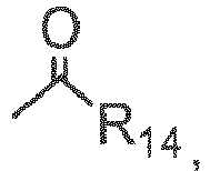
- In some exemplary embodiments, R14 is chosen from hydrogen, optionally substituted (C1-C4)-alkyl, optionally substituted (C1-C4)-alkoxy, and optionally substituted (C3-C6)-cycloalkyl
- In some exemplary embodiments R8, R9, R12, and R13 can be identical or different, and are chosen from hydrogen, methyl ethyl, and acetyl.
- In some exemplary embodiments if the at least one molecule is chosen from formula (6), then R12 and R13 can be identical or different, and are chosen from

- In some exemplary embodiments, the at least one molecular entity is double stranded, the backbone of each strand may be the optionally same, and the backbone is polyamide backbone.
- In some embodiments, in the compounds of
Figure 1 andFigure 2 , any of the R groups, R1 - R6 present are H, for example, R2=H, R3=H, R4=H, R5=H, and R6=H, if they are present. In another embodiment, any of the R groups R1 - R6 present are methyl. In another embodiment, R1 to R6 are indenpently chosen from H and methyl. For example, some specific embodiments of bases of the invention are the structures shown inFigure 1 with the following substituents:Base (Symbol) R2 R3 R4 R5 R6 A H or CH3 H H isoA H or CH3 H H D H or CH3 H G H or CH3 H Gs H or CH3 H I H or CH3 H U H or CH3 U2s H or CH3 U4s H or CH3 C H or CH3 H Py-2o H or CH3 H or CH3 Cs H or CH3 H isoG H or CH3 H isoGs H or CH3 H Pu-2o H or CH3 H isoC H H or CH3 isoCs H H or CH3 Py-4o H or CH3 H or CH3 - In another embodiment, one or more of the H or CH3 are independently substituted with a halogen such as Cl or F. (Note that
Figures 1 and2 and the above table use a different numbering of R groups than do the drawings above.) -
Figure 7 illustrates other base pairing schemes compatible with the invention. Those involve Xanthine (X) or 2-thio-xanthine (2X) or 6-thio-xanthine (6X). X pairs with each of 2,4-diaminopyrimidine-5-yl, 2-aminopyrimidine-5-yl, and 4-aminopyrimidine-5-yl, while 2X also pairs with 2-aminopyrimidine-5-yl and 6X also pairs with 4-aminopyrimidine-5-yl. Accordingly, the choice of X or 2X or 6X in one strand opposed to the aminopyrimidine derivatives in the opposite strand allows one to control the level of degeneracy in a nucleic acid sequence. R1 or "BB" inFigures 1 ,2 , or may be a point of attachment to a polymer backbone, for instance, a polyamide backbone. - In some embodiments, the bases and base-pairs are chosen from a sub-set of those depicted in
Figures 1 and2, and 7 , depending on the needs of the application. In one embodiment, a composition comprising molecular entities, which are a pair of PNA oligomers which comprise a set of molecules comprising non-natural base purine-like molecules (such as formulas (1), (2), (3), (7), (9) and (10), and salts thereof) and pyrimidine-like molecules (such as formulas (4), (5), (6), (8), (11) and (12), and salts thereof). - In exemplary embodiments, the set of molecules is chosen from:
- (a) at least one purine-like molecule of formula (1) or a salt thereof and at least one pyrimidine-like molecule of formula (5) or a salt thereof;
- (b) at least one purine-like molecule of formula (2) or a salt thereof and at least one pyrimidine-like molecule of formula (6), or a salt thereof;
- (c) at least one purine-like molecule of formula (3), or a salt thereof and at least one pyrimidine-like molecule of formula (8), or a salt thereof;
- (d) at least one purine-like molecule of formula (7), or a salt thereof and at least one pyrimidine-like molecule of formula (4), or a salt thereof;
- (e) at least one purine-like molecule of formula (9), or a salt thereof and at least one pyrimidine-like molecule of formula (12) or a salt thereof;
- (f) at least one purine-like molecule of formula (10), or a salt thereof and at least one pyrimidine-like molecule of formula (11), or a salt thereof;
- In some embodiments, the purine-like and pyrimidine-like molecules are chosen from a subset of the (a) through (f) pairings.
- The specific choice of non-natural and natural base-pairs around which to build a sequence may depend on the needs of the application.
- In one embodiment, the set of molecules is chosen from the following pairings::
- (a)

- (b)

- (c)

- (d)

- (e)

- (f)

- Examples of the pairing of certain base pairs comprised in the composition of claim 1are shown in the following tables. The number of indicated hydrogen bonds is also given.
Table A Base Pairs formula (10) formula (1) formula (7) 3 H-bonds 2 H-bonds formula (8) repulsion 2 H-bonds Table B Base Pairs formula (10) formula (2) formula (7) 3 H-bonds 2 H-bonds formula (9) repulsion 2 H-bonds Table C Base Pairs formula (3) formula (11) formula (5) 3 H-bonds 2 H-bonds formula (6) repulsion 2 H-bonds Table D Base Pairs formula (3) formula (4) formula (5) 3 H-bonds repulsion formula (12) 2 H-bonds 2 H-bonds Table E Base Pairs formula (13) formula (15) formula (16) 3 H-bonds 2 H-bonds formula (17) repulsion 2 H-bonds Table F Base Pairs formula (13) formula (14) formula (16) 3 H-bonds repulsion formula (18) 2 H-bonds 2 H-bonds - In one embodiment, a pair of molecular entities according to
claim 1 may comprise the three hydrogen bond base pair IsoCytosine and isoGuanine. For example, the following are 2 hydrogen bond base pairs: - 4-oxo-pyrimidine and isoGuanine
- 4-oxo-pyrimidine and isoThioGuanine
- isoCytosine and 2-oxo-purine
- isoThioCytosine and 2-oxo-purine
- In another embodiment, a pair of molecular entities according to
claim 1 may comprise the three hydrogenbond base pair 1,4-diaminopyrimidine-5-yl and Xanthine. Xanthine also may form two hydrogen bond base pairs with 1-diaminopyrimidien-5-yl or with 4-diaminopyrimidine-5-yl. In each of those pairings, one of the two amino groups from 1,4-diaminopyrimidine-5-yl is removed. SeeFigure 7 , left side pairings. In yet another embodiment, 1 or 4-diaminopyrimidine-5-yl can form a two hydrogen bond base pair with a Xanthine derivative in which one of the two carbonyl oxygens is replaced by sulfur: 2-thioXanthine (X2s) or 6-thioXanthine (X6s). Mixing and matching these sorts of base pairings allows one to create a strand that is capable of hybridizing to more than one sequence on an opposite strand, thus allowing degeneracy in the binding of molecular entities carrying the different strands. - In one embodiment, the base pairs do not formally pair with any of the naturally occurring bases. In a further embodiment, the bases of the invention are used with the natural bases only, for example a single pair being sufficient to provide no DNA affinity and increased diversity.
- Other purine and pyrimidine-like molecules can also be used to vary affinity. For example, the number of hydrogen bonds may be varied by substituting the H for a halogen. In one embodiment, the bases of the invention are halogen-comprising non-natural bases, for example,
- 4-fluoro-2-oxo-pyrimidine (4-Ha-P)
- 2-fluoro-Adenine (2-Ha-A)
- 6-fluoro-isoAdenine (6-Ha-isoA)
- 6-fluoro-Inosine (6-Ha-I)
- Examples of base pairs include two hydrogen bonds
- 4-Ha-P : G
- 4-Ha-P : Gs
- 2-Ha-A : U
- 2-Ha-A: U 2s
- 6-Ha-isoA : U
- 6-Ha-isoA : U4s
- 6-Ha-I : C
- 6-Ha-I : Cs
- 4-Ha-P : I
- 4-Ha-P : 6-Ha-I
- P : 6-Ha-I
- 4-Ha-P : 6-Ha-I.
- For example, fluorine and chlorine are quite small substituents that may have a relatively small steric effect on base pairing, i.e. 2-fluoroAdenine will pair with both uracil and 2-thioUracil, essentially acting like an analogue of Adenine. Sterically smaller alternatives, such as fluorine and chlorine, to an amino group can be used that face both a carbonyl and a thiocarbonyl, In one embodiment, larger halogens such as Iodine may be used to affect binding patterns, i.e. 2-lodo-Adenine may pair with Uracil, but not with 2-thio-Uracil.
- In another embodiment, 6-bromo-Adenine could pair with uracil via two hydrogen bond, such as A : U. Moreover, the binding to 2-thioUracil may be further enhanced by a favorable hydrophobic interactions between the bromine and the thiocarbonyl creating a pair of triple hydrogen bond strength similar to D : U.
- In one embodiment, the bases of the invention are methyl analogues, i.e. Me for Ha or H. For example, 2-methyl-Adenine may be used as an Adenine analogue with PNAs. While it will strongly repell 2-thioUracil, it may bind to Uracil with significantly reduced affinity relative to Adenine.
- In one embodiment, the above mentioned derivatives have hydrogen/halogen or an amino group in a potentially Watson-Crick like base pairing position facing either a carbonyl or thiocarbonyl on the "partner" base, i.e., the pattern determining the base pairing. Any other substitutions however is also possible, that does not directly influence the Watson-Crick like base pairing rules.
- Non-limiting examples include: Pyrimidines substituted in the 5-position; i.e. bromouracil; analogues of C with one 4-N substituent, i.e. 4-N-alkyl and 4-N-acyl derivatives of C/C2s; purines halogenated in the 8-position, i.e 8-bromo-Adenine and 9-deaza and/or 8-aza purine derivatives; 2 and 6-amino"purines" with small N-substituents, i.e. N-6-Acetyl-diaminopurine, N-6-Metyl-Guanine, and N-Metyl-Adenine.
- In one embodiment of the invention, the molecular entities have PNA backbones and are (PNA) oligomers.
- One of skill in the art will appreciate that the increased number of bases will provide an increased degeneracy over natural bases. In one embodiment, the degeneracy minimizes secondary and tertiary structure. The degeneracy also may provide the advantage of one probe that can be used to recognize more than one target exclusively.
- As described above, because of the increased diversity, within the same reading frame the system allows multiple different PNAs to be recognized with the same affinity by one other PNA complementary to all of the PNAs, yet also allow each PNA to be recognized with unique specificity by other complementary, yet different PNAs.
- For example, using twelve bases, 1212 ∼ 9x10¹² different 12'mers can be constructed. See
Figure 6 , for example. These can be divided into 91 subgroups, according to the number of complementary 12'mers with which they pair, from 1 to half a million. On average, each 12'mer will pair with 1400 others, creating 1016 different base pair combinations. - Thus, the information system of the invention is expended relative to "linear" information systems such as binary strings, bar codes, or natural oligomers based on A, C, G, U and T. In the present invention, what is read not only depends on the encoding entity, such as an oligomer, but also on which of the numerous complementary PNAs are selected to "read" it, i.e., bind or not bind.
- The diversity of the system provides a degeneracy that limits cross reactivity. As one 12'mer will pair with 1400 others, for example, this limits the multiplexing capability of the system, i.e., only a limited number of pairs can work simultaneously and independently of each other with no cross reactivity. Triple vs. double vs. single hydrogen bonded base pairs are distinguished by different affinity.
- For example, deaminated bases are expected to pair with approximately the same affinity with either carbonyl or thiocarbonyl bases (A:U = A:U2s; Aiso:U = Aiso:U4s; I:C = I:Cs; P:G = P:Gs). A single hydrogen bond pair, such as I:P, would similarly be expected to be weaker than I:C. This again provides even more diversity since for example, I and P each form tow stable two hydrogen bond complexs thus I will bind stronger to C and Cs than to P and P will bind stronger to G and Gs than to I.
- Thus, for each large number of bases produced from a group of bases, many of the bases will be similar and provide a potential for cross hydridization. However, each pair of oligomers may also be quite unique with very different binding affinities.
- The bases can thereby, for example, be used in a method to design a probe. Since, for example, each molecular entity such as a oligonucleotide made from a series of the bases of the invention, will bind to so many other molecular entities, a series of probes may be chosen or designed that differs slightly in the number of interactions, such as hydrogen bonds, that may be involved in the binding. Thus, for example, an oligonucleotide of 8 bases will have thousands of potential binding partners using the bases of the invention. Each of these may have slightly different strengths of interaction. Thus, one can chose a probe that is very sensitive to binding conditions, such as temperature, or design a series of probes that each bind at a slightly different temperature. The differences in temperature can be useful, in for example, immunohistochemistry in a system that can be varied by slight changes in temperature. The number of possible binding partners allows one to fine tune a binding partner for affinity, speed, temperature, low specific affinity for other probes or other compounds present in a sample and other uses.
- A method of designing a probe may include a screening method where the thousands of possibilities for a certain length oligomer or numerous size oligomers are screened for the desired strength of hybridization and unique hybridization properties.
- An entity may be chosen based on fast hybridization kinetics, combined with sufficient length to provide stability as links in supra molecular visualization and amplification systems and diagnostics.
- For example, one may also use the system to design a series of probes or an information encoding system where each probe specifically hybridizes to a different target. One may further designs a specific detection system, in which one probe hybridizes to a group of or all of the probes (
Figure 4 ). Such a system would be analogous to a hotel where each guest has a key unique to her own room, yet there is also a "master key" that will open every room. - The binding affinity between entities of base pairs may be a relative predictable function of length and triple, double, and single hydrogen bonded base pairs. The kinetic stability of the complexes once formed may follow a similar trend.
- Binding affinity may be inversely proportional to the stability of the complex. For example, the longer and the more potential bond reach of an oligomer, the greater chance that it will be highly self structured and thus slow to hybridize. This can be expressed as a rate determining step preceding actual double-strand formation. More specifically, when two oligomers form a double-strand, the energy barrier to reaching the transition state will increase with the structure reorganization required of each of the hybridizing oligomers. Thus, one can reduce the energy barrier if the stable structures of the oligomers before hybridization are aligned and/or by preparing oligomers that require little reorganization to reach the transition state because of lack of a stable self structure (
Figure 5 ). - For example, intra molecular structure is a priori reduced by using more bases. For example, the 4 bases of DNA can form 16 pairs. Of these, 4 will be matches (25%) contributing on average with 2.5 hydrogen bonds. For example, 12 bases disclosed herein can form 144 pairs. Of these, only 22 will be matches (15%) and on average contribute with 2.1 hydrogen bonds. Since several consecutive base pairs are needed for stable intra molecular structures to form, this effect becomes quite pronounced. If it is assumed that 10 mutual hydrogen bonds are needed for a hair pin structure to form, and if 2 x 4 random DNA bases match, there is a 0.39% chance of hair pin formation. In contrast, using 12 bases instead of 4, there is only a 0.0084 % chance of a hair pin because 2
X 5 bases must match for it to form. Adding more bases also aids in smart design of a probe because one may avoid aggregates or crystalline structures. - In one embodiment, the bases of the invention may be used to prepare molecular entities such as PNA oligomers with at least one of high binding affinity, variable binding affinity, rapid binding kinetics and absence of interference with RNA/DNA.
- The degeneracy of the bases may also be utilized to design a molecular encoding system similar to the natural information encoding system of DNA but greatly enhanced because of the greater number of bases.
- The bases of the invention may be used in any application in which natural bases, DNA, PNA, etc, are used.
- In addition to connecting a base to the backbone, a linker may also be used to connect at least one molecular entity to another or to bind a molecular entity to another composition. A person of ordinary skill in the art of molecular conjugation knows numerous linkers. Examples include 6-amino-hexanoic acid, succimidyl 4-(N-malemidomethyl) cylohexane-1-carboxylate (SMCC), homobifunctional linkers such as glutaric dialdehyde, hexane di-isocyanate, dimethylapimidate, 1,5-difluoro-2,4-dinitrobenzene, divinyl sulfone (DVS), heterobifunctional linkers like e.g. N-gamma-maleimidobytyroloxy succinimide ester (GMBS), and zero length linkers such as 1-ethyl-3-(3-dimethylaminopropyl)cabodiimide
- Longer linker molecules based upon polyethylene glycol (PEG) are also available in the art. (See, for example, Discrete PEG (dPEG)™ modification reagents available from Quanta Biodesign, Ltd., Powell, OH, or at www.quantabiodesign.com; PEG-based reagents available from EMD Biosciences, Inc., San Diego, CA, described in Novabiochem April, 2004, "Product focus: PEG reagents - bifunctional amino-PEG-acid spacers" brochure, available at www.novabiochem.com; and see Baumeister et al., Biopolymers, 71: 339 (2003); Kumar & Aldrich, Org. Lett., 5: 613 (2003). (See also, "Chemistry of Protein Conjugation and Cross-Linking" Shan S. Wong CRC Press, Boca Raton, Florida, USA, 1993; "BioConjugate Techniques" Greg T. Hermanson Academic Press, San Diego, California, USA, 1996; "Catalog of Polyethylene Glycol and Derivatives for Advanced PEGylation, 2004" Nektar Therapeutics Inc, Huntsville, Alabama, USA).
- The present invention may also use a long uncharged linker comprising at least two units of the Formula I.

- In Formula I, R1 and R2 may comprise either NH or O, while R3 may comprise methyl, ethyl, propyl, CH2-O-CH2, and (CH2-O-CH2)2. For example, in some embodiments of the instant invention, the linker comprises at least two units of the Formula I wherein R1 and R2 are both NH and R3 is CH2-O-CH2. See the examples that follow and the International Application entitled "MONOMERIC AND POLYMERIC
- Fluorosceins may also be added to the linkers. When fluorosceins are spaced apart by an L30 oligomer larger than L90 in an aqueous environment there may be negligible quenching. This contrasts with PEG-based spacers in aqueous solutions which may form a collapsed coiled structure.
- For example, PNA probes with multiple fluorophores spaced via polymeric L30 may have higher water solubility, less aggregation and less non-specific binding, as compared to probes with just one fluorophore and no L30 spacer. Multiple PNAs joined together via L30 polymers may not aggregate and may have good solubility even with a total of 50-100 PNA bases in the same compound.
- Compounds and compositions of the invention, including any molecular entity, may also include a detectable label which is any molecule which may be detected directly or indirectly so as to reveal the presence of a target in the sample. In some embodiments of the invention, a direct detectable label is used. Direct detectable labels may be detected per se without the need for additional molecules. Examples include fluorescent dyes, radioactive substances, and metal particles. In other embodiments of the invention, indirect detectable labels are used, which require the employment of one or more additional molecules. Examples include enzymes that affect a color change in a suitable substrate, as well as any molecule that may be specifically recognized by another substance carrying a label or react with a substance carrying a label. Other examples of indirect detectable labels thus include the non-natural bases of the present invention, antibodies, antigens, nucleic acids and nucleic acid analogs, ligands, substrates, and haptens.
- Examples of detectable labels which may be used in the invention include fluorophores, chromophores, electrochemiluminescent labels, bioluminescent labels, polymers, polymer particles, bead or other solid surfaces, gold or other metal particles or heavy atoms, spin labels, radioisotopes, enzyme substrates, haptens, antigens, Quantum Dots, aminohexyl, pyrene, nucleic acids or nucleic acid analogs, or proteins, such as receptors, peptide ligands or substrates, enzymes, and antibodies (including antibody fragments).
- Some detectable labels according to this invention comprise "color labels," in which the target is detected by the presence of a color, or a change in color in the sample. Examples of "color labels" are chromophores, fluorophores, chemiluminescent compounds, electrochemiluminescent labels, bioluminescent labels, and enzymes that catalyze a color change in a substrate. In some embodiments, more than one type of color may be used, for instance, by attaching distinguishable color labels to a single detection unit or by using more than one detection unit, each carrying a different and distinguishable color label.
- "Fluorophores" as described herein are molecules that emit detectable electro-magnetic radiation upon excitation with electro-magnetic radiation at one or more wavelengths. A large variety of fluorophores are known in the art and are developed by chemists for use as detectable molecular labels and can be conjugated to the linkers of the present invention. Examples include fluorescein or its derivatives, such as fluorescein-5-isothiocyanate (FITC), 5-(and 6)-carboxyfluorescein, 5- or 6-carboxyfluorescein, 6-(fluorescein)-5-(and 6)-carboxamido hexanoic acid, fluorescein isothiocyanate, rhodamine or its derivatives such as tetramethylrhodamine and tetramethylrhodamine-5-(and-6)-isothiocyanate (TRITC). Other example fluorophores that could be conjugated to the instant linkers include: coumarin dyes such as (diethyl-amino)coumarin or 7-amino-4-methylcoumarin-3-acetic acid, succinimidyl ester (AMCA); sulforhodamine 101 sulfonyl chloride (TexasRedTM or TexasRedTM sulfonyl chloride; 5-(and-6)-carboxyrhodamine 101, succinimidyl ester, also known as 5-(and-6)-carboxy-X-rhodamine, succinimidyl ester (CXR); lissamine or lissamine derivatives such as lissamine rhodamine B sulfonyl Chloride (LisR); 5-(and-6)-carboxyfluorescein, succinimidyl ester (CFI); fluorescein-5-isothiocyanate (FITC); 7-diethylaminocoumarin-3-carboxylic acid, succinimidyl ester (DECCA); 5-(and-6)-carboxytetramethylrhodamine, succinimidyl ester (CTMR); 7-hydroxycoumarin-3-carboxylic acid, succinimidyl ester (HCCA); 6->fluorescein-5-(and-6)-carboxamido!hexanoic acid (FCHA); N-(4,4-difluoro-5,7-dimethyl-4-bora-3a,4a-diaza-3-indacenepropionic acid, succinimidyl ester; also known as 5,7-dimethylBODIPYTM propionic acid, succinimidyl ester (DMBP); "activated fluorescein derivative" (FAP), available from Molecular Probes, Inc.; eosin-5-isothiocyanate (EITC); erythrosin-5-isothiocyanate (ErITC); and CascadeTM Blue acetylazide (CBAA) (the O-acetylazide derivative of 1-hydroxy-3,6,8-pyrenetrisulfonic acid). Yet other potential fluorophores useful in this invention include fluorescent proteins such as green fluorescent protein and its analogs or derivatives, fluorescent amino acids such as tyrosine and tryptophan and their analogs, fluorescent nucleosides, and other fluorescent molecules such as Cy2, Cy3, Cy 3.5, Cy5, Cy5.5, Cy 7, IR dyes, Dyomics dyes, phycoerythrine, Oregon green 488, pacific blue, rhodamine green, and Alexa dyes. Yet other examples of fluorescent labels which may be used in the invention include and conjugates of R-phycoerythrin or allophycoerythrin, inorganic fluorescent labels such as particles based on semiconductor material like coated CdSe nanocrystallites.
- A number of the fluorophores above, as well as others, are available commercially, from companies such as Molecular Probes, Inc. (Eugene, OR), Pierce Chemical Co. (Rockford, IL), or Sigma-Aldrich Co. (St. Louis, MO).
- Examples of polymer particles labels which may be used in the invention include micro particles, beads, or latex particles of polystyrene, PMMA or silica, which can be embedded with fluorescent dyes, or polymer micelles or capsules which contain dyes, enzymes or substrates.
- Examples of metal particles which may be used in the invention include gold particles and coated gold particles, which can be converted by silver stains.
- Examples of haptens that may be conjugated in some embodiments are fluorophores, myc, nitrotyrosine, biotin, avidin, strepavidin, 2,4-dinitrophenyl, digoxigenin, bromodeoxy uridine, sulfonate, acetylaminoflurene, mercury trintrophonol, and estradiol.
- Examples of enzymes which may be used in the invention comprise horse radish peroxidase (HRP), alkaline phosphatase (AP), beta-galactosidase (GAL), glucose-6-phosphate dehydrogenase, beta-N-acetylglucosaminidase, ß-glucuronidase, invertase, Xanthine Oxidase, firefly luciferase and glucose oxidase (GO).
- Examples of commonly used substrates for horse radish peroxidase (HRP) include 3,3'-diaminobenzidine (DAB), diaminobenzidine with nickel enhancement, 3-amino-9-ethylcarbazole (AEC), Benzidine dihydrochloride (BDHC), Hanker-Yates reagent (HYR), Indophane blue (IB), tetramethylbenzidine (TMB), 4-chloro-1-naphtol (CN), α-naphtol pyronin (α-NP), o-dianisidine (OD), 5-bromo-4-chloro-3-indolylphosphate (BCIP), Nitro blue tetrazolium (NBT), 2-(p-iodophenyl)-3-p-nitrophenyl-5-phenyl tetrazolium chloride (INT), tetranitro blue tetrazolium (TNBT), 5-bromo-4-chloro-3-indoxyl-beta-D-galactoside/ferro-ferricyanide (BCIG/FF).
- Examples of commonly used substrates for Alkaline Phosphatase include Naphthol-AS-B1-phosphate/fast red TR (NABP/FR), Naphthol-AS-MX-phosphate/fast red TR (NAMP/FR), Naphthol-AS-B1-phosphate/fast red TR (NABP/FR), Naphthol-AS-MX-phosphate/fast red TR (NAMP/FR), Naphthol-AS-B1-phosphate/new fuschin (NABP/NF), bromochloroindolyl phosphate/nitroblue tetrazolium (BCIP/NBT), 5-Bromo-4-chloro-3-indolyl-b(beta) -d (delta)-galactopyranoside (BCIG).
- Examples of luminescent labels which may be used in the invention include luminol, isoluminol, acridinium esters, 1,2-dioxetanes and pyridopyridazines. Examples of electrochemiluminescent labels include ruthenium derivatives.
- Examples of radioactive labels which may be used in the invention include radioactive isotopes of iodide, cobalt, selenium, hydrogen, carbon, sulfur and phosphorous.
- In some embodiments the detection unit may comprise from 1 up to 500 detectable label molecules. In some embodiments, the detectable label is an enzyme, which may be conjugated to a polymer, such that the number of enzyme molecules conjugated to each polymer molecule is, for instance, 1 to 200, 2 to 50, or 2 to 25. In some embodiments, the detectable label is a gold particle, a radioactive isotope, or a color label, e.g. a low molecular weight fluorochrome, and the number of detectable labels conjugated to each polymer molecule is, for instance, 1 to 500, or for instance, 2 to 200. In some embodiments, the detectable label is a protein fluorochrome and the number of detectable labels conjugated to each polymer molecule is 1-50, 2-20. In some embodiments, the number of detectable label molecules conjugated to each polymer is 1-200, 2-50, 2-25, or is 10-20, 5-10, or 1-5. Add some more number ranges to pick from later on if necessary.
- The detectable label can be detected by numerous methods, including, for example, reflectance, transmittance, light scatter, optical rotation, and fluorescence or combinations hereof in the case of optical labels or by film, scintillation counting, or phosphorimaging in the case of radioactive labels. See, e.g., Larsson, 1988, Immunocytochemistry: Theory and Practice, (CRC Press, Boca Raton, FL); Methods in Molecular Biology, vol. 80 1998, John D. Pound (ed.) (Humana Press, Totowa, NJ). In some embodiments, more than one detectable label is employed.
- When more than one color label is used, the different colors may have different, distinguishable colors. In some embodiments both colors can be detected simultaneously, such as by fusion or juxtaposition of the signals, signal enhancement or quenching, or detection of multiple colors in the sample. The exact choice of detectable label or combinations of detectable labels may be based on personal preferences in combinations with restrictions of the sample type, sample preparation method, detection method and equipment, and optional contrasting labels used in the sample.
- Two different base pairs or molecular entities may specifically hybridize. In some embodiments, the chosen hybridization conditions are "stringent conditions," meaning herein conditions for hybridization and washes under which nucleotide sequences that are significantly complementary to each other remain bound to each other. The conditions are such that sequences at least 70%, at least 80%, at least 85-90% complementary remain bound to each other. The percent complementary is determined as described in Altschul et al. (1997) Nucleic Acids Res. 25:3389-3402.
- Specified conditions of stringency are known in the art and can be found in Current Protocols in Molecular Biology, John Wiley & Sons, Inc. (Ausubel et al. 1995 eds.),
sections - In other embodiments, the chosen hybridization conditions are "high stringency conditions." An example of high stringency hybridization conditions is hybridization in 4X sodium chloride/sodium citrate (SSC) at 65-70°C or hybridization in 4X SSC plus 50% formamide at 42-50°C, followed by one or more washes in 1X SSC, at 65-70°C. It will be understood that additional reagents may be added to hybridization and/or wash buffers, e.g., blocking agents (BSA or salmon sperm DNA), detergents (SDS), chelating agents (EDTA), Ficoll, PVP, etc.
- In yet other embodiments, the chosen conditions are "moderately stringent conditions." Moderate stringency, as used herein, includes conditions that can be readily determined by those having ordinary skill in the art based on, for example, the length of the molecular entity or a specific nucleic acid pr nucleic acid analog segment. Exemplified conditions are set forth by Sambrook et al. Molecular Cloning: A Laboratory Manual, 2d ed. Vol. 1, pp. 1.101-104, Cold Spring Harbor Laboratory Press (1989), and include use of a prewashing solution of 5X SSC, 0.5% SDS, 1.0 mM EDTA (pH 8.0), hybridization conditions of 50% formamide, 6X SSC at 42°C (or other similar hybridization solution, such as Stark's solution, in 50% formamide at 42°C), and washing conditions of 60°C, 0.5X SSC, 0.1 % SDS.
- In some embodiments, the chosen conditions are "low stringency" conditions. Low stringency conditions may include, as used herein, conditions that can be readily determined by those having ordinary skill in the art based on, for example, the length of the molecular entity. Low stringency may include, for example, pretreating the segment for 6 hours at 40°C in a solution containing 35% formamide, 5 x SSC, 50 mM Tris-HCl (pH 7.5), 5 mM EDTA, 0.1% PVP, 0.1% Ficoll, 1% BSA, and 500 µg/ml denatured salmon sperm DNA. Hybridizations are carried out in the same solution with the following modifications: 0.02% PVP, 0.02% Ficoll, 0.2% BSA, 100 µg/ml salmon sperm DNA, 10% W/V dextran sulfate, and 5-20x106 CPM probe is used. Samples are incubated in hybridization mixture for 18-20 hours at 40°C, and then washed for 1.5 h at 55°C in a solution containing 2 x SSC, 25 mM Tris-HCl (pH 7.4), 5 mM EDTA, and 0.1 % SDS. The wash solution is replaced with fresh solution and incubated an additional 1.5 h at 60°C.
- The compounds and compositions of the invention, including any molecular entity may also comprise at least one polymer. A "polymer," as used herein, may be any molecule that facilitates covalent or non-covalent attachment of one or more other components or molecular entities. For instance, the polymer may facilitate the attachment of one or more probes, molecular entities, nucleic acid analog segments, and or detectable labels. The polymer may be a soluble molecule or an insoluble molecule and may have any shape including a linear polymer, branched polymer, bead or other globular shaped polymer.
- Examples of suitable polymers include polysaccharides such as dextrans, carboxy methyl dextran, dextran polyaldehyde, carboxymethyl dextran lactone, and cyclodextrins; pullulans, schizophyllan, scleroglucan, xanthan, gellan, O-ethylamino guaran, chitins and chitosans such as 6-O-carboxymethyl chitin and N-carboxymethyl chitosan; derivatized cellolosics such as carboxymethyl cellulose, carboxymethyl hydroxyethyl cellulose, hydroxyethyl cellulose, 6-amino-6-deoxy cellulose and O-ethylamine cellulose; hydroxylated starch, hydroxypropyl starch, hydroxyethyl starch, carrageenans, alginates, and agarose; synthetic polysaccharides such as ficoll and carboxymethylated ficoll; vinyl polymers including poly(acrylic acid), poly(acryl amides), poly(acrylic esters), poly(2-hydroxy ethyl methacrylate), poly(methyl methacrylate), poly(maleic acid), poly(maleic anhydride), poly(acrylamide), poly(ethyl-co-vinyl acetate), poly(methacrylic acid), poly(vinylalcohol), poly(vinyl alcohol-co-vinyl chloroacetate), aminated poly(vinyl alcohol), and co block polymers thereof; poly ethylene glycol (PEG) or polypropylene glycol or poly(ethylene oxide-co-propylene oxides) containing polymer backbones including linear, comb-shaped or hyperbranched polymers and dendrimers, including branched PAMAM-dendrimers; poly amino acids including polylysines, polyglutamic acid, polyurethanes, poly(ethylene imines), pluriol; proteins including albumins, immunoglobulins, and virus-like proteins (VLP), and polynucleotides, DNA, PNA, LNA, oligonucleotides and oligonucleotide dendrimer constructs. Also contemplated is the use of mixed polymers, i.e., a polymer comprised of one or more of the above examples including any of the polymers, the co-block polymers and random co-polymers.
- Properties of the polymer can be varied, depending on the desired application, to optimize performance. Examples of parameters that may be considered in the choice of a polymer include the length of the polymer and branching of the polymer. Furthermore, the polymer may carry various substituents. The substituents may be chemically protected and/or activated, allowing the polymer to be derivatized further.
- Compositions according to the invention may be comprised in a kit. The kit may optionally comprise one or more binding agents, and suitable reagents for, for instance: antigen retrieval, sample dilution, reagent dilution, blocking of non-specific binding, blocking of endogenous enzyme activity, or blocking of repetitive sequences. The kit may optionally also comprise at least one container, instructions for use, and reference targets or samples.
-
- 1. In dry equipment 4.6 g of solid Na in small pieces was added to 400 mL ethanol (99.9%), and was dissolved by stirring. Hydroxypyrimidine hydrochloride, 13.2 g, was added and the mixture refluxed for 10 minutes. Then 12.2 mL ethyl-bromoacetate (98%) was added and the mixture refluxed for 1½ hour. The reaction was followed using Thin Layer Chromatography (TLC). The ethanol was evaporated leaving a white compound, which was dissolved in a mixture of 80 mL of 1 M NaCitrate (pH 4.5) and 40 mL of 2M NaOH. This solution was extracted four times with 100 mL Dichloromethane (DCM). The DCM phases were pooled and washed with 10 mL NaCitrate/NaOH - mixture. The washed DCM phases were evaporated under reduced pressure and resulted in 17.2 g of crude solid product. This crude solid product was recrystallized with ethylacetate giving a yellow powder. The yield for this step was 11.45 g (63 %).
- 2. The yellow powder, 12.45 g. from above was hydrolyzed by refluxing overnight in a mixture of 36 mL DIPEA, 72 mL water and 72 mL dioxane. The solvent was evaporated and water was removed from the residue by evaporation from toluene. The yield for this step was 100%.
- 3. OBS. Pyrimidinone acetic acid (10.5 g), 16.8 g PNA-backbone ethylester, 12.3 g DHBT-OH, 19 mL Triethylamine was dissolved in 50 mL N,N-dimethylformamide (DMF). DIPIDIC (11.8 mL) was added and the mixture stirred overnight at room temperature. The product was taken up in 100 mL DCM and extracted three times with 100 mL of dilute aqueous NaHCO3. The organic phase was extracted twice with a mixture of 80 mL of 1 M NaCitrate and 20 mL of 4M HCl. Because TLC showed that some material was in the citrate phase, it was extracted twice with DCM. The organic phases were pooled and evaporated. Because there was a precipitation of urea, the product was dissolved in a DCM, and the urea filtered off. Subsequent evaporation left an orange oil. Purification of the orange oil was performed on a silica column with 10% methanol in DCM. The fractions were collected and evaporated giving a yellow foam. The yield for this step was 7.0 g (26.8 %).
- 4. The yellow foam (8.0 g) was hydrolyzed by reflux overnight in 11 mL DIPEA, 22 mL water, and 22 mL dioxane. The solvent was evaporated and the oil was dehydrated by evaporation from toluene leaving an orange foam. The yield for this step was 100 %.
-
Step 1. In dry equipment 9.2 g of solid Na in small pieces was dissolved in 400 mL ethanol (99.9%), with stirring. Hydroxypyrimidine hydrochlorid, 26.5 g, was added, and the mixture was stirred for 10 minutes at 50°C. Then 24.4 mL Ethyl bromoacetate (98%) was added and the mixture stirred at 50°C for 1 hour. The reaction was followed using Thin Layer Chromatography (TLC). - The ethanol was evaporated leaving a white compound, which was dissolved in 70 mL of water and extracted with 20 mL DCM. Another 30 mL of water was added to the water phase, which was extracted with 3x100 mL DCM. The DCM-phase from the first extraction contains a lot of product, but also some impurities, wherefore this phase was extracted twice with water. These two water phases then were back extracted with DCM.
- The combined DCM phases were pooled and washed with 10 mL water. The washed DCM phases were evaporation under reduced pressure and resulted in 25.1 g yellow powder. The yield for this step was 25.1 g = 69 %. Maldi-Tof: 181.7 (calc. 182).
-
Step 2. 34.86 g yellow powder from above was dissolved in 144 mL 2M NaOH. After stirring 10 minutes at room temperature, the mixture was cooled in an ice bath. Now 72 mL 4 M HCl (cold) was added. The product precipitated. After stirring for 5 minutes, the precipitate was filtered and thoroughly washed with ice water. Drying in a dessicator under reduced pressure left 18.98 g yellow powder. The yield for this step was 18.98 g = 64 %. -
Step 3. Pyrimidinone acetic acid 11.1 g and triethylamine 12.5 mL were dissolved in N,N-dimethylformamide (DMF) 24 ml, HBTU 26.2 g was added plus 6 mL extra DMF. After 2 minutes a solution of PNA-Backbone ethylester 14.7g dissolved in 15 mL DMF was added. The reaction mixture was stirred at room temperature and followed using TLC. After 1½hour precipitate had formed. This was filtered off. - The product was taken up in 100 mL DCM and extracted with 2 x 100 mL dilute aqueous NaHCO3. Both of the aqueous phases were washed with a little DCM. The organic phases were pooled and evaporated. Evaporation left an orange oil. Purification of the product was done on a silica column with 10-20% methanol in ethylacetate. The fractions were collected and evaporated giving a yellow oil. The oil was dissolved and evaporated twice from ethanol. The yield from this step was 20.68 g = 90 %.
-
Step 4. The yellow oil (18.75 g) was dissolved in 368 mL 0.2 M Ba(OH)2. Stirring for 10 minutes before 333 mL 0.221 M H2SO4 was added. A precipitation was performed immediately. Filtration through cellite, which was washed with water. The solvent was evaporated. Before the evaporation was at end, the product was centrifuged to get rid of the very rest of the precipitation. Re-evaporation of the solvent left a yellow oil. The yield from this step was 13.56 g = 78 %. -
Step 5. To make a test on the P-monomer 3 consecutive P's were coupled to Boc-L300-Lys(Fmoc) -resin, following normal PNA standard procedure. The product was cleaved from the resin and precipitated also following standard procedures: HPPP-L300-Lys(Fmoc). Maldi-Tof on the crude product: 6000 (calc. 6000) showing only minor impurities. -
- 1. 6-Chloroguanine (4.93 g) and 10.05 g K2CO3 was stirred with 40 mL DMF for 10 minutes at room temperature. The reaction mixture was placed in a water bath at room temperature and 3.55 mL ethyl bromoacetate was added. The mixture was stirred in a water bath until TLC (20% Methanol/DCM) showed that the reaction was finished. The precipitated carbonate was filtered off and washed twice with 10 mL DMF. The solution, which was a little cloudy, was added to 300 ml water, whereby it became clear. On an ice bath the target compound slowly precipitated. After filtration the crystals were washed with cold ethanol and dried in a desiccator. The yield for this step was 3.3 g (44.3 %) of ethyl chloroguanine acetate.
- 2. Ethyl chloroguanine acetate (3.3 g) was dissolved by reflux in 50 mL absolute ethanol. Thiourea (1.08 g) was added. After a refluxing for a short time, precipitate slowly began forming. According to TLC (20% Methanol/DCM) the reaction was finished in 45 minutes. Upon completion, the mixture was cooled on an ice bath. The precipitate was then filtered and dried overnight in a desiccator. The yield for this step was 2.0 g (60 %) ethyl thioguanine acetate.
- 3. Ethyl thioguanine acetate (3.57 g) was dissolved in 42 mL DMF. Benzylbromide (2.46 mL) was then added and the mixture stirred in an oil bath at 45°C. The reaction was followed using TLC (25% Methanol/DCM). After 3 hours all basis material was consumed. The
step 3 target compound precipitated upon evaporation under reduced pressure and high temperature. The precipitate was recrystallized in absolute ethanol, filtered and then dried in a desiccator. The yield for this step was 3.88 g (82 %) of methyl benzyl thioguanine ethylester. - 4. Methyl benzyl thioguanine ethylester (5.68 g) was dissolved in 12.4 mL of 2M NaOH and 40 mL THF, and then stirred for 10 minutes. The THF was evaporated by. This was repeated. The material was dissolved in water and then 6.2 mL of 4M HCl was added, whereby the target product precipitated. Filtering and drying in a desiccator. The yield for this step was 4.02 g (77 %).
- 5. The product of step 4 (4.02 g), 3.45 g backboneethylester, 9 mL DMF, 3 mL pyridine, 2.1 mL triethylamine and 7.28 g PyBop were mixed and then stirred at room temperature. After 90 minutes a solid precipitation formed. The product was taken up in 125 mL DCM and 25 mL methanol. This solution was then extracted, first with a mixture of 80 mL of 1 M NaCitrate and 20 mL of 4M HCl, and then with 100 mL dilute aqueous NaHCO3. Evaporation of the organic phase gave a solid material. The material was dissolved in 175 mL boiling ethanol. The volume of the solution was reduced to about 100 mL by boiling. Upon cooling in an ice bath, the target product precipitate. The crystals were filtered, washed with cold ethanol and then dried in a desiccator. The yield of this step was 6.0 g (86 %.)
- 6. The product of step 5 (6.0 g) was dissolved in 80 mL THF, 7.5 mL 2M NaOH and 25 mL water. The solution became clear after ten minutes of stirring. THF was evaporated. Water (50 mL) was added to the mixture. THF was evaporated. Water (50 mL) was added to the mixture. When the pH was adjusted by the addition of 3.75 mL of 4M HCl, thio-guanine monomer precipitated. It was then filtered, washed with water and dried in a desiccator. The yield for this step was 5.15 g (91 %).
- 1. Diaminopurine (10 g) and 40 g of K2CO3 were added to 85 mL of DMF and stirred for 30 minutes. The mixture was cooled in a water bath to 15°C. Ethyl bromoacetate (3 mL) was added three times with 20 minute intervals between each addition. This mixture was then stirred for 20 minutes at 15°C. The mixture was left in the water bath for another 75 minutes, and the temperature increased to 18°C. The DMF was removed by filtering and the remaining K2CO3 was added to 100 mL of ethanol and refluxed for 5 minutes. Filtering and repeated reflux of the K2CO3 in 50 mL ethanol, filtering. The pooled ethanol phases were placed in a freezer, after which crystals formed. These crystals were filtered, washed with cold ethanol, filtered again and then dried in a desiccator overnight. The yield for this step was 12 g (76 %).
-
- 1. A solution of 146 mL of 2,2'-(Ethylenedioxy)bis(ethylamine) (98%) in 360 mL of THF was cooled in an ice bath. Di-tert-butyl dicarbonate (97%) (65 g) in 260 mL THF was added dropwise over one hour. The solvent was evaporated. The remaining oil was dissolved in water and then evaporated off. The oily product was dissolved in 300 mL water, extracted with 300 mL DCM, then washed twice with 150 mL of DCM. The collected organic phase was washed with 50 mL of water before evaporating to about half the volume. The organic phase was then extracted with 400 mL of 1 M NaCitrate (pH 4.5), and then extracted again with 50 mL of 1 M NaCitrate (pH 4.5). The aqueous phases were washed with 50 mL DCM before cooling on an ice bath. While stirring, 100 mL of 10M NaOH was added to the aqueous washed aqueous phases resulting in pH of 13-14. In a separation funnel the product separated on its own. It was shaken with 300 mL DCM and 50 ml water. The organic phase was evaporated, yielding a white oil. The yield for this step was 48.9 g (65.7 %). The product had a predicted molecular formula of C11H24N2O4 (MW 248.3).
- 2. Boc-amine (76.2 g) was dissolved in 155 mL pyridine. Diglycolic anhydride (54.0 g) (90%) was added. After stirring for 15 minutes the intermediate product separated out and then 117 mL Acetic Anhydride (min. 98%) was added and the mixture stirred at 95°C for 1 hour. The solution was then put under reduced pressure and evaporated. Water (117 mL) was added, and the mixture was then stirred for 15 minutes, after which 272 mL of water and 193 mL of DCM were added. The organic layer was extracted twice with 193 mL of 1 M Na2CO3 and then twice with a mixture of 72 mL of 4M HCl and 289 mL of 1 M NaCitrate. After each extraction the aqueous phase was washed with a little DCM. The collected organic phase was washed with 150 mL of water. The solvent was evaporated leaving the product as an orange oil. This yield for this step was 100.3 g (0.29 mol) (94 %). The product had a predicted molecular formula of C15H26N2O7 (MW 346.4).
- 3. The product from step 2 (100.3 g) was dissolved in an equal amount of THF and was then added dropwise to 169.4 mL of 2,2'-(Ethylendioxy)bis(ethylamine) at 60°C over the period of 1 hour. The amine was distilled from the reaction mixture at 75-80° C and a pressure of 3x10⁻¹ mBar. The residue from the distillation was taken up in a mixture of 88 mL of 4M HCl and 350mL of 1 M NaCitrate and then extracted three times with 175 mL of DCM. The aqueous phase was cooled in an ice bath and was cautiously added to 105 mL of 10M NaOH while stirring. In a separation funnel the product slowly separated from the solution. When separated 100 mL of water and 950 mL of DCM were added to the product. Stirring for some minutes before pouring to a separation funnel. The pH in the aqueous phase should be 14. The aqueous phase was extracted four times with 150 mL of DCM. The solvent was evaporated. The oily residue was dehydrated by evaporation from toluene, giving a yellow oil. The yield for this step was 115.48 g (81 %). The product had a predicted molecular formula of C21H42N4O9 (MW 494.6).
- 4. The Boc-amine (115.48 g) from
step 3 was dissolved in 115 mL of pyridine. Diglycolic anhydride (40.6 g) (90%) was added and the mixture stirred for 15 minutes, after which the intermediate product came out. Acetic Anhydride (97 mL) (min. 98%) was added and the mixture stirred at 95°C for 1 hour. The mixture was then evaporated under reduced pressure. The mixture was then cooled and then 80 mL of water was added. This mixture was stirred for 15 minutes and then 200 mL of water and 150 mL of DCM were added. The organic layer was extracted twice with 150 mL of 1 M Na2CO3 and then twice with a mixture of 53 mL of 4M HCl and 213 mL of 1 M NaCitrate. After each extraction the aqueous phase was washed with a little DCM. The collected organic phase was washed with 150 mL of water. The solvent was evaporated. The oily residue was dehydrated by evaporation from toluene, giving a yellow oil. The yield for this step was 125 g (92 %). The product had a predicted molecular formula of C25H44N4O12 (MW 592.6), with a mass spectrometry determined molecular weight of 492.5.
Further purifying of the product could be done on a silica column with a gradient from 5-10% methanol in DCM. The yield from the column purification was 69% and produced a white oil. - 5. White oil (12.4 g) from
step 4 was dissolved in a mixture of 12 mL water and 12mL 1,4-Dioxane (99 %) and was then heated to reflux. DIPEA (6 mL) was added and refluxed for 30 minutes. This mixture was cooled and then evaporated. The oily residue was dehydrated by evaporation from toluene, giving a yellow oil. The product had a predicted molecular formula of C25H46N4O14 (MW 610.6). - All made by PNA standard procedures (see Example 18)
Table 1. Sequence designation PNA sequences1 N-terminal C-terminal Molecular Weight Seq. AA TCD-DGsGs-TAC-A Flu-L30- -Lys(Cys) 8805 Seq. AB UsGUs-DPP-TTG-D Flu-L30- -Lys(Cys) 8727 Seq. AC CUsGs-GsDD-TUsD-GsDC Flu-L30- -Lys(Cys) 9413 Seq. AD GTP-TAA-TTP-PAG Flu-L30- -Lys(Cys) 9203 Seq. AE DGsT-CGsD-DGsG-UsCUs Flu-L30- -Lys(Cys) 9413 Seq. AF AGA-CPT-TPG-APT Flu-L30- -Lys(Cys) 9187 Seq. AG TCD-DII-TAC-A Flu-L30- -Lys(Cys) 8742 1 Flu is fluorescein; T is thiamine; C is cytosine; D is diaminopurine; Gs is thioguanine; A is Adenine; Us is 2/4-thiouracil; G is guanine; P is pyrimidone; I is inosine. -
- BA: Flu-L30-DGT-DTC-GTD-CCG-Lys(Acetyl)
- BB: Flu-L30-DGT-DTC-GTD-CCG-Lys(Cys)
- BC: Flu-L30-DGT-DTC-GTD-CCG-Lys(Lys)3
- Using procedure provided in Example 18a, an MBHA-resin was loaded with Boc-Lys(Dde)-OH. Using a peptide synthesizer, amino acids were coupled according to PNA solid phase procedure provided in Example 18d yielding Boc-L90-Lys(Fmoc)-L30-Lys(Dde). The Boc and Fmoc protections groups were removed and the amino groups marked with flourescein using the procedure in Example 18e. Then, the Dde protection group was removed and 0.4 M cysteine was added according to the procedure in Example 18b. The PNA was cleaved from the resin, precipitated with ether and purified on HPLC according to Example 18d. The product was found to have a molecular weight of 3062 using MALDI-TOF mass spectrometry; the calculated molecular weight is 3061.
- Dextran (with a molecular weight of 70 kDa) was activated with divinylsulfone to a degree of 92 reactive groups/dextran polymer; this product is designated DexVS70.
280 µL DexVS70 20 nmol 66 µL Flu2Cys 160 nmol (prepared from Example 7) 25 µL 0.8 M NaHCO3 pH = 9.5 29 µL H2O - The above four compounds were mixed. The mixture was placed in a water bath at 30° C for 16 hours. The mixture was added to 50 nmol of freeze-dried PNA (sequence AA from Example 5). The mixture was placed in a water bath at 30° C for 30 minutes. The conjugating reaction was quenched with 50 µL of 500 mM cysteine for 30 minutes at 30° C. Purification of the product was performed using FPLC: column SUPERDEX®- 200, buffer 10 mM Hepes 100 mM NaCl, method 7
bank 2,Loop 1 mL. Two fractions were collected: one with the product and one with the residue. The relative absorbance Flu2 (ε500nm = 146000 M-1, ε260nm = 43350 M-1) and PNA (ε500nm = 73000 M-1, ε260nm =104000 M-1) was used to calculate the average conjugation ratio of Flu2, PNA, and DexVS70. The conjugation ratio of Flu2 to DexVS70 was 9.4. The conjugation ratio of PNA (sequence AA) to DexVS70 was 1.2. - Using the procedure of Example 14, the conjugate HRP-DexVS70-Seq. AA was made. The ratio of HRP to DexVS70 is 12.2; the ratio of Seq. AA to Dex70 is 1.2.
- The synthesis of GaM-DexVS70-Seq. AB was performed using the procedure in Example 16 with the following changes as indicated.
- Dextran (molecular weight 70 kDa) is activated with divinylsulfone to a degree of 92 reactive groups/dextran polymer.
105.0 µL DexVS70 7.5 nmol 57.0 µL Goat anti mouse Imuno globuline (GAM-Ig) 15 nmol 8.9 µL 4 M NaCl 10.6 µL 0.8 M NaHCO3 (pH = 9.5) 144.5 µL H2O - The above five components were mixed and placed in a water bath at 30° C for 40 minutes. Two hundred and ninety µL were taken out of the mixture and added to 100 nmol of Seq. AB, which was previously dissolved in 80 µL of H2O. Then, 20 µL of 0.8 M NaHCO3 (pH 9.5) was added and the mixture placed in a water bath at 30° C for 1 hour. Quenching was performed by adding 39 µL of 500 mM cysteine and letting the resultant mixture set for 30 minutes at 30° C.
- Purification of the product on FPLC: column SUPERDEX® - 200, buffer 10 mM Hepes 100 mM NaCl, method 7
bank 2,Loop 1 mL. Two fractions were collected: one with the product and one with the residue. Relative absorbance PNA(Flu) (ε500nm = 73000 M-1) and GAM (ε278nm = 213000 M-1) (correction factor for PNA at 278 nm is due to the specific PNA and is calculated: 278/500 nm) was used to calculate the average conjugation ratio of PNA, GAM and DexVS70. The ratio of PNA to DexVS70 was 5.3 and the ratio of GaM to DexVS70 was 0.8. -
Table 2. Conjugate designation ratio PNA1 PNA1 nmol PNA1 to DexVS PNA2 PNA2 nmol PNA2 to DexVS DexVS Conj. CA 1:9 Seq. AA 12.5 1.02 Seq. AD 100 8.2 DexVS70 Conj. CB 1:6 Seq. AC 40 1.5 Seq. AB 200 7.4 DexVS70 Conj. CC 1:16 Seq. AC 13.3 0.84 Seq.AB 200 12.7 DexVS150 Conj. CD 1:6 Seq. AC 40 2.3 Seq. AB 200 11.5 DexVS150 All conjugates were made by standard conjugation procedures of Example 17. - Dextran (molecular weight 70 kDa) was activated with divinylsulphone to a degree of 92 reactive groups/dextran polymer, and is designated DexVS70. The antibody Anti-Human-BCL2 is designated AHB.
105 µL DexVS70 7.5 nmol 800 µL AHB conc. (2.9 g/L) 15.1 nmol 25 µL 4 M NaCl 32 µL 0.8 M NaHCO3 (pH = 9.5) - The above four compounds were mixed and placed in a water bath at 30° C for 65 minutes. From this mixture, 875 µL was taken out and added to the indicated number of nmol of PNA in the table below; before the addition the PNA had been dissolved in the µL of H2O indicated in the table below. Then the number of µLs of 0.8 M NaHCO3 (pH 9.5) was added according to the table below. The resulting mixture was placed in a water bath at 30° C for 70 minutes. Quenching was performed by adding 6 mg of solid cysteine (0.05 M) to the mixture and letting it stand for 30 minutes at 30° C.
- Purification of the product on FPLC: column SUPERDEX® - 200, buffer 10 mM Hepes 100 mM NaCl, method 7
bank 2,Loop 1 mL. Two fractions were collected: one with the product and one with the residue. Relative absorbance PNA(Flu) (ε500nm = 73000 M-1) and AHB (ε278nm = 213000 M-1) (correction factor for PNA at 278 nm is due to the specific PNA and is calculated: 278/500 nm) was used to calculate the average conjugation ratio of PNA, AHB and DexVS70. - Conjugates with different ratios PNA are shown in the following table.
Table 3. Conjugate designation nmol of PNA added µL of H2O added µL of 0.8 M NaHCO3 (pH 9.5) added PNA to DexVS70 AHB to DexVS70 Conj. DA 100 75 25 9.5 1.6 Conj. DB 33 30 10 2.9 1.2 Conj. DC 67 60 20 5.6 1.1 - All Standard procedures are described in Example 18.
- 1. An MBHA-resin was loaded with Boc-L30-Lys(Fmoc)-L90-Lys(Fmoc)-L90-Lys(Fmoc) using a standard loading procedure to a loading of 0.084 mmol/g.
- 2. To this resin, Boc-Lys(Fmoc)-L30-AAC-GGG-ATA-ACT-GCA-CCT-was coupled using the peptide synthesizer machine following standard PNA solid phase chemistry. Fmoc protection groups were removed and the amino groups were labeled with fluorescein. After cleaving and precipitation the PNA was dissolved in TFA. The precipitate was washed with ether. The precipitate was dissolved in 200 µL NMP To this
solution 6 mg Fmoc-Osu was added and dissolved. Next, DIPEA (9 µL) was added and the reaction was followed using MALDI-TOF mass spectrometry. After 30 minutes the reaction was finished and the PNA was precipitated and washed with ether. - HPLC after dissolving the PNA in 30 % CH3CN and 10 % TFA/H2O gave three pure fractions. The fractions were pooled and lyophilized. The lyophilized PNA was then dissolved in 192 µL NMP. Piperidine (4 µL) and 4 µL DBU was added to this solution which set for 30 minutes. Analysis by MALDI-TOF mass spectrometry gave a molecular weight of 10777.
- The precipitate was washed with ether and was then dissolved in 100 µL TFA. The precipitate was washed with ether and then dried using N2 gas.
- Dextran (molecular weight 70 kDa) is activated with divinylsulfone to a degree of 92 reactive groups/dextran polymer.
192 µL DexVS70 13.7 nmol 255 µL horse radish peroxidase (HRP) 602 nmol 15 µL 4 M NaCl 19 µL 0.8 M NaHCO3 pH = 9.5 119 µL H2O - The above five components are mixed together placed in a water bath at 30° C for 16 hours. Five hundred microliters of this mixture are added to 50 nmol PNA, which is previously dissolved in 40 µL H2O. Then, 10 µL of 0.8 M NaHCO3 (pH 9.5) is added. The mixture is then placed in a water bath at 30° C for 2 hours. Quenching is performed by adding 55 µL of 110 mM cysteine and letting the resultant mixture set for 30 minutes at 30° C.
- Purification of the product is performed by FPLC: column SUPERDEX® - 200, buffer 10 mM Hepes 100 mM NaCl, method 7
bank 2,Loop 1 mL. - Two fractions are collected: one with the product and one with the residue. Relative absorbance HRP (ε404nm= 83000 M-1, ε500nm= 9630 M-1) and PNA(Flu) (ε500nm = 73000 M-1) is used to calculate the average conjugation ratio of HRP, PNA and DexVS70.
- Dextran (molecular weight 70 kDa) is activated with divinylsulfone to a degree of 92 reactive groups/dextran polymer (DexVS70).
105.0 µL DexVS70 7.5 nmol 57.0 µL Goat anti mouse Imuno globuline (GAM) 15 nmol 8.9 µL 4 M NaCl 10.6 µL 0.8 M NaHCO3 (pH = 9.5) 144.5 µL H2O - The above five components are mixed and placed in a water bath at 30° C for 40 minutes. Two hundred and ninety µL is taken out of the mixture and added to 50 nmol of PNA, which is previously dissolved in 40 µL of H2O. Then, 10 µL of 0.8 M NaHCO3 (pH 9.5) is added and the mixture placed in a water bath at 30° C for 1 hour. Quenching is performed by adding 34 µL of 500 mM cysteine and letting the resultant mixture set for 30 minutes at 30° C.
- Purification of the product on FPLC: column SUPERDEX® - 200, buffer 10 mM Hepes 100 mM NaCl, method 7
bank 2,Loop 1 mL. Two fractions are collected: one with the product and one with the residue. Relative absorbance PNA(Flu) (ε500nm = 73000 M-1) and GAM (ε278nm = 213000 M-1) (correction factor for PNA at 278 nm is due to the specific PNA and is calculated: 278/500 nm) was used to calculate the average conjugation ratio of PNA, GAM and DexVS70. - Dextran (molecular weight 70 kDa) is activated with divinylsulfone to a degree of 92 reactive groups/dextran polymer. PNA1 (100 nmol) is dissolved in 140 µL of DexVS70 (10 nmol). To this mixture 12.5 µL of PNA2 (12.5 nmol) dissolved in H2O is added, and then 30 µL of NaHCO3 (pH 9.5) is added and the solution mixed. The resultant mixture is placed in a water bath at 30° C for 35 minutes. Quenching was performed by adding 18.3 µL of 500 mM cysteine in Hepes and letting this mixture set for 30 minutes at 30° C.
- Purification of the product on FPLC: column SUPERDEX® - 200, buffer 10 mM Hepes 100 mM NaCl, method 7
bank 2,Loop 1 mL. Two fractions are collected: one with the product and one with the residue. Relative absorbance PNA(Flu) (ε500nm = 73000 M-1) and the proportion between the two PNA's is used to calculate the average conjugation ratio of PNA, PNA and DexVS70. - 6-Benzyloxypurine. Sodiumhydride (60 % Dispersion in mineral oil;3,23g;80 mmol) was slowly added to benzyl alcohol (30 ml;34,7 mmol). After the addition of more benzyl alcohol (10 ml) and 6-chloropurine (5,36 g; ). The reaction mixture was heated to 100°C for 4 hours. When the reaction mixture has reached room temperature, water (1ml) was slowly added. 6-Benzyloxypurine was precipitated by the addition of acetic acid (4,6 ml) and diethylether (550 ml). The precipitate was separated by filtration (11,72 g). Re-crystallization from ether gave (4,78 g; 65,4 %). Melting point was 175 - 177 °C (litt. 170-172 °C)[Ramazaeva N., 1989 #473] 1 H-NMR (DMSO-d6): 8,53 (1 H,s); 8,39 (1 H, s); 7,54-7,35 (5H, m); 5,62 (2H,s).
- Methyl (6-(Benzyloxy)purin-9-yl) acetate. 6-Benzyloxypurine (4,18 g; 18,5 mmol) was added to a suspension of potassium carbonate (3,1 g; 22,4 mmol) in DMF (100 ml). After 15 min., bromoacetic acid methyl ester (1,93 ml; 20,4 mmol) was added. The reaction was monitored by TLC in butanol:acetic acid:water 4:1:1. Upon completion, the reaction mixture was partitioned between water (600 ml) and ethyl acetate (600 ml). The organic phase was dried over magnesium sulfate and evaporated to a volume of ∼10 ml and precipitated with pet. ether. The two products were separated by column chromatography using ethyl acetate as the solvent. The products were precipitated in pet. ether. Yield: 2,36 g (43 %). Melting point: 111,5-115°C. UV λmax= 250 nm (9-alkylated); λmax= 260 nm (7-alkylated). 1 H-NMR (DMSO-d6):8,60 (1 H, s); 8,43 (1 H,s); 7,6-7,35 (5H, m); 5,69 (2H, s); 5,26 (2H, s); 3,75 (3H, s).
- (6-(Benzyloxy)purin-9-yl) acetic acid. Methyl (6-(Benzyloxy)purin-9-yl) acetate (2,10 g; 7,0 mmol) was dissolved in methanol (70 ml) and 0,1 M NaOH (85 ml) is added. After 15 min. the pH of the reaction mixture was lowered by addition of 0,1 M HCl (∼ 80 ml) to
pH 3. The precipitate was separated from the mixture by filtration and washed with water and ether. Yield: 1,80 g (90,2 %). 1 H-NMR (DMSO-d6):8,55 (1 H,s); 8,37 (1 H,s); 7,55-7,30 (5H, m); 5,64 (2H, s); 5,09 (2H, s). - N-((6-(Benzyloxy)purin-9-yl) acetyl)-N-(2-Boc-aminoethyl)glycine. Ethyl N-(2-Boc-aminoethyl)glycinate (0,285 g; 1,15 mmol), (6-(benzyloxy)purin-9-yl) acetic acid (0,284 g; 1,0 mmol) and 3-hydroxy-1,2,3 benzotriazin-4(3H)-one (0,180 ;1,1 mmol) was dissolved in dichlormethane/dimethylformamide 1:1 (10 ml). After addition of dicycloehexylcarbodiimide (0,248 g; 1,2 mmol) the reaction was left over night. The precipitate was removed by filtration. The organic phase was extracted twice with saturated sodium bicarbonate, dried with magnesium sulfate and evaporated to a oil. Column purification on silica using dichloromethane with 0-5 % methanol as elutant yields the monomer ester which was dissolved in methanol (10 ml). Then, 0,1 M NaOH (12 ml) was added. After 30 min the reaction was filtered and pH adjusted with saturated KHSO4 / water (1:3) to 2,7. The water phase was extracted twice with ethyl acetate (2 x 100 ml). The combined organic phases were dried over magnesium sulfate and evaporated to a volume of 10 ml. Precipitation with pet. ether yielded the monomer (0,15 g; 31 %). 1H-NMR (DMSO-d6):8,51 (1H, s); 8,23 (1H, s); 7,6-7,3 (5H, m); 5,64 (2H, s); 5,31 (ma.) + 5,13 (mi.) (2H, s); 4,23 (mi.) + 3,98 (ma.) (2H, s); 3,55-3,00 (4H, m); 1,36 (9H, s).

- The Boc-PNA-Diaminopurine-(N6-Z)-monomer was prepared according to Gerald Haaima, Henrik F. Hansen, Leif Christensen, Otto Dahl and Peter E. Nielsen; Nucleic Acids Research, 1997, Vol 25, Issue 22 4639-4643.
- The Boc-PNA-2-Thiouracil-(S-4-MeOBz)-monomer was prepared according to Jesper Lohse, Otto Dahl and Peter E. Nielsen; Proceedings of the National Academy of Science of the United States of America, 1999, Vol 96, Issue 21, 11804-11808.
- The Boc-PNA-Adenine-(Z)-monomer was from PE Biosystems catalog GEN063011.
- The Boc-PNA-Cytosine-(Z)-monomer was from PE Biosystems cat. GEN063013.
- The Boc-PNA-Guanine-(Z)-monomer was from PE Biosystems cat. GEN063012.
- The Boc-PNA-Thymine-monomer was from PE Biosystems cat. GEN063010.
- IsoAdenine (2-aminopurine) may be prepared as a PNA-monomer by 9-N alkylation with methylbromoacetate, protection of the amino group with benzylchloroformate, hydrolysis of the methyl ester, carbodiimide mediate coupling to methyl-(2-Boc-aminoethyl)-glycinate, and finally hydrolysis of the methyl ester.
- 4-thiouracil may be prepared as a PNA-monomer by S-protection with 4-methoxy-benzylchloride, 1-N alkylation with methylbromoacetate, hydrolysis of the methyl ester, carbodiimide mediate coupling to methyl-(2-Boc-aminoethyl)-glycinate, and finally hydrolysis of the methyl ester.
- Thiocytosine may be prepared as a PNA monomer by treating the Boc-PNA-cytosine(Z)-monomer methyl ester with Lawessons reagent, followed by hydrolysis of the methyl ester.
- A number of halogenated bases are commercially available, and may be converted to PNA monomers analogously to the non-halogenated bases. These include the guanine analog 8-bromo-guanine, the adenine analogs 8-bromo-adenine and 2-fluoro-adenine, the isoadenine analog 2-amino-6-chloro-purine, the 4-thiouracil analog 5-fluoro-4-thio-uracil, and the 2-thiouracil analog 5-chloro-2-thiouracil.
- Boc-PNA-Uracil monomers were first described in "Uracil og 5-bromouracil I PNA," a bachelor project by Kristine Kilså Jensen, Københavns Universitet 1992.
-
- a. Loading of resins. P-methyl-BHA-resin (3 g) is loaded with Boc-Lys(Fmoc)-OH 15 mmol/g resin. The lysine is dissolved in NMP and activated with 0.95 equivalents (eq.) HATU and 2 eq. DIPEA. After loading the resin, it is capped by adding a solution of (Ac)2O / NMP/ pyridine (at a ratio of 1/2/2) and letting it set for at least 1 hour or until Kaiser test was negative. After washing with DCM, the resin is dried in a dessicator. Quantitative Kaiser test typically gives a loading of 0.084 mmol/g.
- b. Amino Acid Couplings. The Boc protection group is removed from the resin with TFA / m-cresol (at a ratio of 95/5) 2x5 min. The resin is then washed with DCM, pyridine and DMF before coupling with the amino acid, which is dissolved in NMP in a concentration between 0.2 and 0.4 M and activated with 0.95 eq. of HATU and 2 eq of DIPEA for 2 minutes. The coupling is complete when the Kaiser test is negative. Capping occurring by exposing the resin for 3 minutes to (Ac)2O / pyridine / NMP (at a ratio of 1/2/2). The resin is then washed with DMF and DCM
- c. Boc-L300-Lys(Fmoc)-resin. To the loaded Boc-Lys(Fmoc)-resin, L30-Linker in a concentration of 0.26 M was coupled using standard amino acid coupling procedure. This was done 10 times giving Boc-L300-Lys(Fmoc)-resin.
- d. PNA solid phase. On a peptide synthesizer (ABI 433A, Applied Biosystems) PNA monomers are coupled to the resin using standard procedures for amino acid coupling and standard PNA chemistry. Then the resin is handled in a glass vial to remove protections groups and to label with either other amino acids or flourophores.
- Removal of the indicated protection groups is achieved with the following conditions:
- Boc: TFA / m-cresol (at a ratio of 95/5) 2x5 min.
- Fmoc: 20 % piperidine in DMF 2x5 min.
- Dde: 3 % hydrazine in DMF 2x5 min.
- When the synthesis is finished, the PNA is cleaved from the resin with TFA/TFMSA/m-cresol/thioanisol (at a ratio of 6/2/1/1). The PNA is then precipitated with ether and purified on HPLC. MALDI-TOF mass spectrometry is used to determine the molecular weight of the product.
- e. Labeling with fluorescein. 5(6)-carboxy fluorescein is dissolved in NMP to a concentration of 0.2 M. Activation is performed with 0.9 eq. HATU and 1 eq. DIPEA for 2 min before coupling for at least 2x20 min or until the Kaiser test is negative.
- In order to make better conjugations at one time we tried to give the PNA a loading. Both PNA's were made by PNA standard procedures (See Example 18).
- 1. Flu-L30-Glu-TCA-AGG-TAC-A-Glu-L300-Lys(Cys)
- Glu = glutamate has negative loadings and for the easiness the PNA is designated -A4-
- 2. Flu-L30-Lys(Me)2-TGT-ACC-TTG-A-Lys(Me)2-L330-Lys(cys)
- Lys(Me)2 = Boc-Lys(Me)2-OH has positive loadings and the PNA is designated +T+
- Tonsil tissue samples were fixed in neutral buffered formalin, NBF (10 mM NaH2PO4/ Na2HPO4, pH 7.0), 145 mM NaCl, and 4% formaldehyde (all obtained from Merck, Whitehouse Station, NJ). The samples were incubated overnight in a ventilated laboratory hood at room temperature.
- The tissue samples were placed in a marked plastic histocapsule (Sakura, Japan). Dehydration was performed by sequential incubation in 70% ethanol twice for 45 min, 96% ethanol twice for 45 min, 99% ethanol twice for 45 min, and xylene twice for 45 min. The samples were subsequently transferred to melted paraffin (melting point 56-58°C) (Merck, Whitehouse Station, NJ) and incubated overnight (12-16 hours) at 60°C. The paraffin-infiltrated samples were transferred to fresh warm paraffin and incubated for an additional 60 min prior to paraffin embedding in a cast (Sekura, Japan). The samples were cooled to form the final paraffin blocks. The marked paraffin blocks containing the embedded tissue samples were stored at room temperature in the dark.
- The paraffin blocks were cut and optionally also mounted in a microtome (0355 model RM2065, Feather S35 knives, set at 5.0 micrometer; Leica, Bannockburn, IL). The first few millimeters were cut and discarded. Paraffin sections 4-6 micrometers thick were then cut and collected at room temperature. The sections were gently stretched on a 45-60°C hot water bath before being mounted onto marked microscope glass slides (SUPERFROST® Plus; Fisher, Medford, MA), two tissue sections per slide. The slides were then dried and baked in an oven at 60°C. The slides were deparaffinated by incubating twice in xylene for 5 min ± 2 min twice, then in 96% ethanol for 2min +/- 30 sec, then twice in 70% ethanol for 2 min +/- 30 sec, and then once in Tris-buffered saline with TWEEN® (called herein TBST) for 5 min. TBST comprises 50 mM Tris adjusted to pH 7.6 with HCl; 150 mM NaCl; 0.05 % TWEEN®20. The slides were deparaffinated by subsequently incubation in xylene twice for 5 min ± 2 min, 96% ethanol twice for 2min +/- 30 sec and 70% ethanol twice for 2 min +/- 30 sec. The slides were immersed in deionized water and left for 1 to 5 min.
- Samples were incubated with a 3% hydrogen peroxide solution for 5 min. to quench endogenous peroxidase activity, followed by washing in deionized water for 1 to 5 min.
- Antigens in the sample were retrieved by immersing the slides in a container containing Antigen Retrieval Solution, pH 6.0 (DakoCytomation code No. K5204 Vial 7 or optional code No. K5205 Vial 7). The container was closed with a perforated lid and placed in the middle of a microwave oven and left boiling for 10 min. The container was removed from the oven and allowed to cool at room temperature for 20 min. The samples were rinsed in deionized water.
- Antigens in the sample were retrieved by immersing the slides in a beaker containing Antigen Retrieval Solution, pH 6.0 (DakoCytomation code No. K5204 Vial 7 or optional code No. K5205 Vial 7). The samples were incubated for 40 min in a water bath at 95-100°C. The beaker was removed from the water bath and allowed to cool at room temperature for 20 min. The samples were rinsed in deionized water.
- To ensure good coverage of reagent on the tissue sample, the area on the slide with tissue was encircled with a silicone rubber barrier using DakoCytomation Pen (DakoCytomation code No. 2002). The slides were transferred to a rack and placed in a beaker containing Tris-buffered saline with TWEEN® (called herein TBST) and left for 5 min. TBST comprises 50 mM Tris adjusted to pH 7.6 with HCl; 150 mM NaCl; 0.05 % TWEEN®20.
- Monoclonal Mouse anti-Human Cytokeratin (DakoCytomation code No. M3515) diluted 1:900 in ChemMate™ Antibody Diluent (DakoCytomation code No.S2022) was applied on the tissue samples and incubated for 30 min in a humid chamber at ambient temperature. The slides were individually rinsed and then washed in TBST for 5 min.
- Monoclonal Mouse Anti-Human Cytokeratin (DakoCytomation code No. M3515) diluted 1:300, 1:900 and 1:1600; monoclonal Mouse Anti-Human CD20cy (DakoCytomation code No. M0755) diluted 1:2000, 1:8000 and 1:14000; and monoclonal Mouse Anti-Human Ki-67 Antigen (DakoCytomation code No. M7240) diluted 1:400, 1:1200 and 1:2400 were used. The antibodies were diluted in ChemMate™ Antibody Diluent (DakoCytomation code No. S2022), applied on the tissue samples, and incubated for 30 min in a humid chamber at ambient temperature. The slides were individually rinsed and washed in TBST for 5 min.
- Antibody / Dextran / PNA1 conjugate recognition unit is also called "PNA1 conjugate" in the examples that follow. The PNA1 conjugate comprises 70,000 molecular weight dextran. Table 1 summarizes PNA1 conjugates based on a secondary antibody: goat anti-mouse Ig, called herein GAM (DakoCytomation code No. Z0420). Table 2 summarizes PNA1 conjugates based on a primary antibody: mouse anti-human BCL2 oncoprotein, such as Clone 124 (DakoCytomation code No. M0887). The primary antibody was protein A-purified prior to conjugation. The conjugates were diluted in BBA (50 mM Tris adjusted to pH 7.6 with HCl; 150 mM NaCl; 2% BSA; 0.02% bronidox; 2.44 mM 4-aminoantipyrin) and were applied on the tissue sample in a range of dilutions, then incubated for 30 min in a humid chamber at ambient temperature. The slides were individually rinsed and washed in TBST for 5 min.
Table 5. PNA1 conjugates useful in indirect recognition of targets: GAM / Dextran / PNA1 Conjugate No. Sequence µM Dex GAM / Dex PNA1 / Dex D14120 AGA CPT TPG DPT 1.25 1.1 4.3 D14102 GTP TAA TTP PAG 1.02 1.0 9.1 D14096 GTP TAD TTP PAG 1.15 1.4 4.2 D14083 UsGUs DPP TTG D 0.87 0.8 5.3 D13171 UsGUs DPP TTG D 1.21 1.0 7.5 D13161 TTG APP TTA G 2.11 1.1 6.0 D13150 TGT APP TTGA 2.20 1.1 4.2 D13102 TGT ACC TTGA 2.53 1.1 2.5 D12102 TGT ACC TTGA 2.50 1.3 4.5 Table 6. PNA1 conjugates for direct recognition of targets: anti-BCL2 / Dextran / PNA1 Conjugate No. Sequence µM Dex Ab / Dex PNA1 / Dex D14128 UsGUs DPP TTG D 0.8 1.1 5.6 D14126 UsGUs DPP TTG D 1.0 1.2 2.9 D14122 UsGUs DPP TTG D 1.1 1.6 9.5 In the above tables, the letters A, C, G, U, and T, stand for the natural bases adenine, cytosine, guanine, uracil, and thymine. P stands for pyrimidinone, D for 2,6-diaminopurine, and Us for 2-thiouracil. - The samples were washed in deionized water for 30 sec. Then, 1 % glutardialdehyde (Merck Art. No. 820603), called herein GA, diluted in 22 mM calcium phosphate buffer, pH 7.2, was applied, and the samples were incubated for 10 min in a humid chamber at ambient temperature. The samples were washed in deionized water for 30 sec and in TBST for 5 min.
- PNA1-PNA2/ Dextran conjugate is also called "PNA1-PNA2" in the following examples. Table7 summarizes the compositions of PNA1-PNA2 conjugates. PNA1 is complementary to the PNA1 conjugate, and PNA2 is complementary to the PNA2 conjugates D14079 and D13155 described in step 13 below. The sequence of PNA1 is CUsGsGsDD TUsD GsDC and the sequence of PNA2 is UsGUs DPP TTG D, in which Us stands for 2-thio-uracil, Gs stands for 2-amino-6-thioxopurine, D stands for diaminopurine, and P stands for pyrimidinone. The conjugates, diluted in BBA, were applied to the tissue samples in a range of dilutions, and the samples were then incubated for 30 min in a humid chamber at ambient temperature. The samples were individually rinsed and washed in TBST for 5 min. When testing a PNA1-PNA2conjugate, fixed concentrations of 0.08 µM PNA1 and 0.05 µM PNA2 were used.
Table 7. PNA1-PNA2/ Dextran conjugates Conjugate No. Molecular weight of dextran PNA1 / dex PNA2 / dex µM PNA1 D14119 150.000 2.3 11.5 4.2 D14106 150.000 0.8 12.7 1.3 D14104 70.000 1.5 7.5 3.9 - Horse Radish Peroxidase (HRP) / Dextran / PNA2 conjugates are also called "PNA2 conjugate" in the examples that follow, and are listed in table 8. The PNA2 conjugates comprise 70.000 Da molecular weight dextran. The conjugates diluted in BBA were applied to the tissue samples in a range of dilutions, and samples were incubated for 30 min in a humid chamber at ambient temperature. The samples were individually rinsed and washed twice in TBST for 5 min.
Table 8. PNA2 conjugates: HRP / Dextran / PNA2 Conjugate No. Sequence µM PNA HRP / Dex PNA2 / Dex D14133 TCD DII TAC A 1.6 14.0 1.0 D14111 DGsT CGsD DGsG UsCUs 3.9 11.4 2.1 D14110 DGT CGsD DGsG UsCUs 3.0 12.6 1.6 D11089 CUsGs GsDD TUsD GsDC 2.1 14.1 1.5 D14086 UsCGs GsDD TUsD GDC 1.9 11.0 1.0 D14079 TCD DGsGs TAC A 1.9 12.2 1.2 D13159 CTA AGsGs TCA A 1.9 12.9 1.3 D13155 TCD DGsGs TAC A 2.4 12.7 1.6 D13148 TCA AGsGs TAC A 1.9 11.6 0.8 D13122 CTA AGG TCA A 3.2 13.0 2.1 D13108 GTG TGT GT 4.3 12.0 2.3 D13106 TCA AGG TAC A 2.6 12.4 1.3 D12120 TCD DGG TAC A 1.0 18.3 0.6 D12094 TCA AGG TAC A 3.0 14.6 0.9 In Table 8, in addition to the nucleobase letter schemes provided for Tables 5-7, I stands for inosine. - The diaminobenzidine chromogenic substrate solution, DAB+ (DakoCytomation code No. K3468) was applied on the tissue samples, and the samples were incubated for 10 min in a humid chamber at ambient temperature. The samples were washed with deionized water for 5 min.
- The tissue samples were immersed in Mayers Hematoxylin (Bie & Berntsen Code No. LAB00254) for 3 min, rinsed in tap water for 5 min, and finally rinsed with deionized water.
- Cover slips were applied to the tissue samples using the aqueous mounting media, Faramount (DakoCytomation code No. S3025).
- The tissue staining was examined in a bright field microscope at 10x, 20x or 40x magnification. Both the specific and the non-specific staining intensity were described with a score-system using the range 0 to 3+ with 0.5+ score interval. ChemMate™ EnVision™ Detection kit Rabbit/Mouse (DakoCytomation code No. K5007 bottle A) was used as a reference, and was included in all experiments for testing in parallel with the PNA conjugates. K5007 was used according to manufacturer's instructions. The antibodies were used in the following dilutions: M3515 at 1:900, M0755 at 1:8000, and M7240 at 1:1200. The staining intensity of the K5007 reference using the primary antibody M3515 diluted 1:900 was set to 2+ in order to compare and assess the staining result of the PNA conjugate tested. If the reference deviated more than ± 0.5, the test was repeated.
- In the examples, the various visualization system combinations of the invention were tested on routine tissue samples. The staining performance was compared with a reference visualization system, using EnVision™ and a very dilute antibody from DakoCytomation. The practical dynamic range of quantitative IHC may be narrow, and e.g. strongly stained (+3) tissues are not easy to compare with respect to intensity. Therefore, on purpose, the staining intensity of the reference system was adjusted to be approximately +2. This was done in order to better monitor and compare differences in staining intensity with the system of the invention.
- The protocol allowed for a quick test of PNA2 conjugates for non-specific staining. Tonsil tissues were taken through the steps 1-5, 7, 13-14, 15 (in which the slides were immersed in a bath of Hematoxylin Mayer for 1 min.), and 16-17, above. The conjugates to be tested were diluted to the final concentrations 0.05 µM and 0.2 µM. As references, two PNA2 conjugates were used in the final concentration 0.05 µM. The first reference, for example, PNA2, D13108, was known to give non-specific nuclear staining, and so was used as a positive control. The second reference, for example, PNA2, D13155, was known not to give any non-specific nuclear staining, and was used as a negative control. In general, 250 µL of each reagent was applied unless otherwise specified.
- Tonsil tissues were taken through the steps 1-4, 6-8, 10, 11, and 13-17 above. Step 11 was left out for the tonsils not fixed with 1% GA. In general, 250 µL of each reagent was applied unless otherwise specified.
- Tonsil tissues were taken through the steps 1-4, 6-8, and 10-17 above. Step 11 was left out for the tonsils not fixed with 1% GA. In general, 250 µL of each reagent was applied unless otherwise specified.
- Tonsil tissues were taken through the steps 1-4, 6, 7, 9 -11, and 13-17 above. Step 11 was left out for the tonsils not fixed with 1 % GA. A further negative control, mouse IgG1 (DakoCytomation code No. X0931) diluted 1:300 in S2022 was included the protocol for the PNA conjugates. In general, 250 µL of each reagent was applied unless otherwise specified.
- Conjugates comprising example PNA segments were tested for their ability to specifically hybridize according to the invention. Tonsil tissues were taken through the steps 1-4, 6-8, 10 and 13-17 above. K5007 was included as a reference to secure the level of the staining. The concentration of the conjugates was 0.08 µM for PNA1 and 0.05 µM for PNA2.
- The results listed in Table 10 show the staining intensities for a representative number of PNA pairs tested. The PNA pairs did not demonstrate any non-specific binding. The specific staining, in general, was directly proportional to the number of hydrogen bonds involved in the base-pairing. It was important that each PNA did not interact with itself. Substitution of T (thymine) with Us (2-thiouracil) in some PNAs could prevent such intra-PNA interactions. The unspecific staining intensity was increased by substituting an A with a D. On the other hand, we also observed that replacement of one G (guanine) with Gs (2-amino-6-thioxopurine) in the same PNA could circumvent the unspecific binding introduced by D. For instance, the staining of the PNA1 conjugate D14102 was improved by substituting the D in D14096 with an A in D14102. As is apparent from Table 9, this small change resulted in an increase of the specific staining score by 1+.
Table 9. PNA1 PNA2 Specific staining intensity Non-specific staining intensity D13161 D13159 2 0 D14083 D14079 2.5 0 D14096 D14089 1.5 0 D14102 D14089 2.5 0 D14120 D14114 1.5 0 - The PNA pair D13102-D13106 was used as a starting point for further investigation of introducing base substitutions in either PNA1 or PNA2 conjugates. Tonsil tissues were taken through the steps 1-4, 6-8, 10 and 13-17. Each of the three different PNA1 conjugates was tested with each of the three different PNA2 conjugates. The concentration of the conjugates used was 0.08 µM for PNA1 and 0.05 µM for PNA2.
Table 10. PNA2: PNA1: D13106 TCAAGGTACA D13148 TCA AGsGs TAC A D13155 TCD DGsGs TAC A D13102 TGT ACC TTG A 2.5 0 2 D13150 TGTAPPTTG A 2.5 0.5 3 D13171 UsGUs DPP TTG D 3 2.5 3 - Table 10 shows the effect of base substitutions on the specific binding between paired PNA variants. No non-specific binding was observed. D13102 tested with D13106 gave a specific staining of 2.5+. Replacement of 2 G's with 2 Gs's (D13148) resulted in the abolishment of specific staining, but by introducing 2 D's instead of 2 A's (D13155) achieved a specific staining of 2+. When the 2 C's in D13102 were replaced with 2 P's (D13150) and tested with D13106, the specific staining was unchanged at 2.5+, despite the lower number of hydrogen bonds as compared to the PNA-pair D13102-D13106. Test of D13150 with D13148 resulted in a reduced specific staining of 0.5+, whereas specific staining to 3+ was observed for the D13150-D13155 pair. The replacement in D13150 of 2 A's with 2 D's and of 2 T's with 2 Us's (D13171) resulted in improved specific binding compared to D13106. This modified PNA1 was now able to bind specifically to D13148 with a score of 2.5+, and also bound to D13155.
- This experiment clearly demonstrates the use of PNA pairs in the present invention. Furthermore, it shows the ability of fine tuning the specific binding by introducing base substitutions using either natural as well as non-natural bases.
- The two PNA-pairs, D13150-D13155 and D13161-D13159, were tested for cross-reactivity. Tonsil tissues were taken through the steps 1-4, 6-8, 10, and 13-17. The concentration of conjugates used was 0.16 µM for PNA1 and 0.1 µM for PNA2.
- As apparent from Table 11, PNA1 D13150 did not cross react with PNA2 D13159, but PNA1 D13161 cross reacted with PNA2 D13155. We therefore excluded the PNA pair D13161-D13159 due to the cross-reaction between D13161 and D13155. No non-specific staining was observed.
Table 11: Test of specific binding and cross reactivity PNA2: D13155 D13159 PNA1: D13150 2.0 0 D13161 1 1.5 - Three PNA-pairs, D14083-D14079, D14102-D14089 and D14120-D14114 were tested for cross-reactivity. Tonsil tissues were taken through the steps 1-4, 6-8, 10, and 13-17. The concentration of the conjugates used was 0.08 µM for PNA1 and 0.05 µM for PNA2.
- The PNA conjugates listed in Table 12 only bound to their complementary partner and did not cross react to any of the other PNA conjugates tested. No non-specific staining was observed. See Table 12 below.
Table 12: Test of specific binding and cross reactivity PNA2: D14079 D14089 D14114 PNA1: D14083 1.5 0 0 D14102 0 1 0 D14120 0 0 1.5 - Two PNA pairs, D14083-D14079 and D14096-D14089, were tested at different PNA2 concentrations for the purpose of determining the optimal concentration of PNA2 conjugates. The concentrations used were 0.08 µM for PNA1 conjugates and 0.025; 0.05; 0.1 and 0.2 µM for PNA2 conjugates.
- Tonsil tissues were taken through the steps 1-4, 6-8, 10, and 13-17. The optimal concentration of the PNA2 conjugate was 100 nM. See Table 13 below.
Table 13: Determination of PNA2 conjugate concentration. PNA1 1% GA fixation of PNA1 PNA2 Specific staining 0.025µM 0.05µM 0.1µM 0.2µM D14083 - D14079 1.5 2 3 2.5 D14096 - D14089 0.5 1 2 1.5 - Tonsil tissues were taken through the steps 1-4, 6-8, 10, 11, and 13-17. Step 11 was omitted for tissues not fixed with 1% GA.
- Fixation of PNA1 conjugates with 1% GA resulted in a stronger specific staining than without fixation and the optimal concentration of the PNA2 conjugate was now determined to be 50 nM. See Table 14 below.
Table 14: Effect of 1% GA fixation on the determination of PNA2 conjugate concentration. PNA1 1% GA fixation of PNA1 PNA2 Specific staining 0.025µM 0.05µM 0.1µM 0.2µM D14083 - D14079 2 2 2.5 2.5 D14083 + D14079 2.5 3 2.5 3 - Alkaline Phosphatase ("AP") (from Calf Intestine, EIA grade) was dialyzed overnight against 2mM HEPES, pH 7.2; 0.1 M NaCl; 0.02mM ZnCl2. Dextran (molecular weight 70kDa) was activated with divinylsulfone to a degree of 92 reactive groups per dextran polymer (DexVS70).
- The three components below were mixed together and placed in a water bath at 40°C for 30 minutes.
192.0 µL DexVS70 13.7 nmol 41.0 µL PNA 41 nmol PNA dissolved in H2O 6.0 µL 1 M NaHCO3 - 108.0µL of the DexVS70-PNA conjugate was taken out and added to a mixture of:
160.0µL AP 43.4 nmol 7.7µL 1M NaHCO3 30.6µL 20mM Hepes, pH 7.2; 1 M NaCl; 50mM MgCl2; 1 mM ZnCl2 - The mixture was placed in a water bath at 40°C for 3 hours. Quenching was performed by adding 30.6 µL of 0.1 M ethanolamine and letting the mixture stand for 30 minutes in water bath at 40°C. The product was purified on FPLC with:
- Column Superdex-200, buffer: 2mM HEPES, pH 7.2; 0.1 M NaCl; 5mM MgCl2; 0.1 mM ZnCl2. Two fractions were collected, one with the product and one with the residue.
- In comparison to the experiment described above, another conjugate was made with extended conjugation time. The three components below were mixed together and placed in a water bath at 40°C for 30 minutes.
192.0µL DexVS70 13.7 nmol 41.0µL PNA 41nmol PNA dissolved in H2O 6.0µL 1M NaHCO3 - 108.0µL of the DexVS70-PNA conjugate was taken out and added to a mixture of:
160.0µL AP 43.4 nmol 7.7µL 1M NaHCO3 30.6µL 20mM Hepes, pH 7.2; 1M NaCl; 50mM MgCl2; 1 mM ZnCl2 - The mixture was placed in a water bath at 40°C for 5 hours. Quenching was performed by adding 30.6µL 0.1 M Ethanolamine and letting the mixture stand for 30 minutes in water bath at 40°C. Purification of the product on FPLC: Column Superdex-200, buffer: 2mM Hepes, pH 7.2; 0.1 M NaCl; 5mM MgCl2; 0.1 mM ZnCl2. Two fractions were collected: One with the product and one with the residue.
- Relative absorbance PNA(Flu) (ε500nm= 73000M-1) and AP(ε278nm= 140000M-1. Corrected for absorbance from PNA at 278nm, this correction factor is due to the specific PNA and it is calculated: 278/500nm) was used to calculate the average conjugation ratio of PNA, AP and DexVS70.
- AP-DexVS70-PNA, 3hrs:
- PNA/DexVS70: 1.8
- AP/DexVS70: 1.8
- AP-DexVS70-PNA, 5hrs:
- PNA/DexVS70: 2.0
- AP/DexVS70: 2.4
- Due to these results, it is recommended to follow a procedure in which the conjugation time (AP + DexVS70-PNA) is 5 hours.
- 2,4-diamino-pyrimidine-5-yl may be introduced into DNA-oligomers by methods known in the art (e.g. S.A. Benner et al., Nucleic Acid Research 24(7): 1308-1313 (1996)). A corresponding PNA oligomer is prepared by chlorinating pyrimidine-5-acetic acid to yield 2-chloro-pyrimidine-5-acetic acid, 4-chloro-pyrimidine-5-acetic acid, and 2,4-dichloro-pyrimidine-5-acetic acid. Separation of isomers, followed by high temperature and pressure treatment with ammonia, gives the three corresponding amino-pyrimidine derivatives (see
Figure 7 ). The amino-pyrimidine derivatives are separated and amino-protected, then coupled to a protected PNA backbone ester. Ester hydrolysis results in PNA monomers for production of PNA oligomers containing 2-amino; 4-amino; and/or 2,4-diamino pyrimidine-5-yl bases.
- Xanthine, 2-thio-xanthine, and 6-thio-xanthine are commercially available, for instance, from ScienceLab.com. Further, S.A. Benner et al., Nucleic Acid Research 24(7): 1308-1313 (1996) teaches the preparation of a xanthosine-DNA monomer, including a less acidic and preferable 7-deaza analog, and notes the preferred protection of both oxygens during solid phase synthesis.
- Xanthine PNA-monomers, as well as 2-thio and 6-thio xanthine monomers, are prepared by:
- 1. Protecting both oxygens or both oxygen and sulphur with appropriate protection groups such as (possibly substituted) benzyl.
- 2. Alkylating at N-9 with ethyl bromacetate. (Separating N-7 alkylated byproduct.)
- 3. Hydrolyzing the ethyl ester.
- 4. HBTU or Carbodiimide-mediated coupling of the nucleobase-acids to 2-Boc-aminoethyl-ethylglycinate.
- 5. Hydrolyzing the resulting monomer ester to the monomer free acid.
- 6. The resulting monomers may be used in Merrifield solid phase synthesis of xanthine, 2-thio-xanthine and 6-thio-xanthine-containing PNAs.

| name | number | HRP | GaM | equiv. | HRP/Dex | GaM/Dex | PNA/Dex |
| -A4- | D 13041 | D 13050 | 9 | 12.3 | 0.13 | ||
| -A4- | D 13041 | D 13060 | 7 | 0.94 | 0.66 | ||
| +T4+ | D 13042 | D 13058 | 9 | 13.5 | 0.19 | ||
| +T4+ | D 13042 | D 13056 | 7 | 1.42 | 0.45 |
| As it is shown in the scheme, PNAs with loading are not good at coupling. |
Claims (8)
- A composition comprising a pair of PNA oligomers which(i) hybridize to each other through base-pairing that comprises more than one of the following combinations :


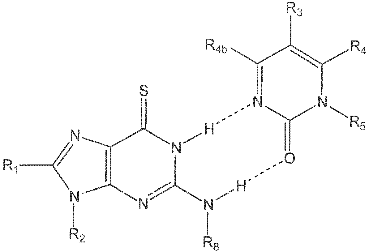


 R1 is chosen from hydrogen, halogen, optionally substituted (C1-C4)-alkyl, -NR6R7, optionally substituted (C1-C4)-alkoxy, optionally substituted (C3-C6)-cycloalkyl, linker,-a polyamide backbone, and linker bound to a polyamide backbone;R2 is chosen from hydrogen, optionally substituted (C1-C4)-alkyl, -NR6R7,optionally substituted (C1-C4)-alkoxy, optionally substituted (C3-C6)-cycloalkyl, linker, a polyamide backbone, and linker bound to a polyamide backbone;R3 is group chosen from hydrogen, halogen, optionally substituted (C1-C20)-alkyl, -NR6R7, optionally substituted (C1-C20)-alkoxy, optionally substituted (C3-C8)-cycloalkyl, linker, a polyamide backbone, and linker bound to a polyamide backbone;R4 is chosen from hydrogen, halogen, optionally substituted (C1-C4)-alkyl, -NR6R7, optionally substituted (C1-C4)-alkoxy, optionally substituted (C3-C6)-cycloalkyl, linker, a polyamide backbone, and linker bound to a polyamide backbone;R4a and R4b can be identical or different, and are chosen from hydrogen,fluorine and chlorine.R5 is chosen from hydrogen, optionally substituted (C1-C4)-alkyl, -NR6R7,optionally substituted (C1-C4)-alkoxy, optionally substituted (C3-C6)-cycloalkyl, linker, a polyamide backbone, and linker bound to a polyamide backbone;R6 and R7 can be identical or different, and are chosen from hydrogen andoptionally substituted (C1-C20)-alkyl;R8, R12,-can be identical or different, and are chosen from hydrogen, halogen,
 (ii) do not interfere with RNA or DNA.
 - The composition according to claim 1, wherein R1, and R3 each is hydrogen.
- The composition according to claim 1, wherein R2 and R5 can be identical or different and are chosen from linker, a polyamide backbone, and linker bound to a polyamide backbone
- The composition according to claim 1, wherein:R1, R3, and R4 each is hydrogen;R2 and R5 can be identical or different and are chosen from a polyamide backbone and linker bound to a polyamide backbone.
- The composition of claim 1, comprising a polymer selected from polysaccharides, pullulans, chitins and chitosans, derivatized cellolosics, hydroxylated starch, carrageenans, alginates, and agarose; synthetic polysaccharides such as ficoll and carboxymethylated ficoll; vinyl polymers; poly ethylene glycol (PEG) or polypropylene glycol or poly(ethylene oxide-co-propylene oxides) containing polymer backbones including linear, comb-shaped or hyperbranched polymers and dendrimers, including branched PAMAM-dendrimers; poly amino acids including polylysines, polyglutamic acid, polyurethanes, poly(ethylene imines), pluriol; proteins including albumins, immunoglobulins, and virus-like proteins (VLP), and polynucleotides such as DNA, RNA, LNA.
- The composition of claim 5 wherein the polymer is a dextran.
- The composition of claim 1, comprising a linker.
- The composition according to claim 7, wherein the linker is chosen from a molecule comprising polyethylene glycol and a molecule comprising at least two units according to the Formula I:

Applications Claiming Priority (4)
| Application Number | Priority Date | Filing Date | Title |
|---|---|---|---|
| US69540805P | 2005-07-01 | 2005-07-01 | |
| US69540905P | 2005-07-01 | 2005-07-01 | |
| US69541005P | 2005-07-01 | 2005-07-01 | |
| PCT/IB2006/003306 WO2007045998A2 (en) | 2005-07-01 | 2006-06-30 | New nucleic acid base pairs |
Publications (2)
| Publication Number | Publication Date |
|---|---|
| EP1919930A2 EP1919930A2 (en) | 2008-05-14 |
| EP1919930B1 true EP1919930B1 (en) | 2016-09-14 |
Family
ID=37708983
Family Applications (4)
| Application Number | Title | Priority Date | Filing Date |
|---|---|---|---|
| EP20060820855 Withdrawn EP1920072A2 (en) | 2005-07-01 | 2006-06-30 | In situ hybridization detection method |
| EP06809193.3A Not-in-force EP1910575B1 (en) | 2005-07-01 | 2006-06-30 | Immunohistochemistry detection method |
| EP06842206.2A Not-in-force EP1919930B1 (en) | 2005-07-01 | 2006-06-30 | New nucleic acid base pairs |
| EP20060808856 Not-in-force EP1907592B1 (en) | 2005-07-01 | 2006-06-30 | Monomeric and polymeric linkers useful for conjugating biological molecules and other substances |
Family Applications Before (2)
| Application Number | Title | Priority Date | Filing Date |
|---|---|---|---|
| EP20060820855 Withdrawn EP1920072A2 (en) | 2005-07-01 | 2006-06-30 | In situ hybridization detection method |
| EP06809193.3A Not-in-force EP1910575B1 (en) | 2005-07-01 | 2006-06-30 | Immunohistochemistry detection method |
Family Applications After (1)
| Application Number | Title | Priority Date | Filing Date |
|---|---|---|---|
| EP20060808856 Not-in-force EP1907592B1 (en) | 2005-07-01 | 2006-06-30 | Monomeric and polymeric linkers useful for conjugating biological molecules and other substances |
Country Status (6)
| Country | Link |
|---|---|
| US (6) | US8741566B2 (en) |
| EP (4) | EP1920072A2 (en) |
| AT (1) | ATE501277T1 (en) |
| DE (1) | DE602006020577D1 (en) |
| DK (1) | DK1907592T3 (en) |
| WO (4) | WO2007045998A2 (en) |
Families Citing this family (118)
| Publication number | Priority date | Publication date | Assignee | Title |
|---|---|---|---|---|
| ATE501277T1 (en) | 2005-07-01 | 2011-03-15 | Dako Denmark As | MONOMERIC AND POLYMERIC LINKERS FOR CONJUGING BIOLOGICAL MOLECULES AND OTHER MATERIALS |
| GB0517097D0 (en) * | 2005-08-19 | 2005-09-28 | Solexa Ltd | Modified nucleosides and nucleotides and uses thereof |
| EP2155782A2 (en) | 2007-03-26 | 2010-02-24 | Dako Denmark A/S | Mhc peptide complexes and uses thereof in infectious diseases |
| EP2167537A2 (en) | 2007-07-03 | 2010-03-31 | Dako Denmark A/S | Compiled methods for analysing and sorting samples |
| CN101836117B (en) | 2007-09-18 | 2014-07-23 | 丹麦达科有限公司 | Rapid and sensitive method for detecting biological targets |
| US10611818B2 (en) | 2007-09-27 | 2020-04-07 | Agilent Technologies, Inc. | MHC multimers in tuberculosis diagnostics, vaccine and therapeutics |
| DK2254592T3 (en) | 2008-02-28 | 2019-09-09 | Dako Denmark As | MHC multimers for Borrelia diagnostics and disease |
| WO2009114182A1 (en) | 2008-03-13 | 2009-09-17 | Pacific Biosciences Of California, Inc. | Labeled reactants and their uses |
| WO2010009735A2 (en) | 2008-07-23 | 2010-01-28 | Dako Denmark A/S | Combinatorial analysis and repair |
| US11992518B2 (en) | 2008-10-02 | 2024-05-28 | Agilent Technologies, Inc. | Molecular vaccines for infectious disease |
| WO2010037402A1 (en) | 2008-10-02 | 2010-04-08 | Dako Denmark A/S | Molecular vaccines for infectious disease |
| EP4241767B1 (en) | 2008-11-10 | 2024-10-23 | Arbutus Biopharma Corporation | Novel lipids and compositions for the delivery of therapeutics |
| EP2398907B1 (en) | 2009-02-19 | 2014-04-02 | Dako Denmark A/S | Conjugate molecules |
| WO2011028952A1 (en) | 2009-09-02 | 2011-03-10 | Xencor, Inc. | Compositions and methods for simultaneous bivalent and monovalent co-engagement of antigens |
| WO2011047680A1 (en) | 2009-10-20 | 2011-04-28 | Dako Denmark A/S | Immunochemical detection of single target entities |
| ES2606012T3 (en) * | 2010-02-26 | 2017-03-17 | Ventana Medical Systems, Inc. | In situ hybridization with PolyTag probes |
| CA2806252C (en) | 2010-07-29 | 2019-05-14 | Xencor, Inc. | Antibodies with modified isoelectric points |
| CA2816472A1 (en) | 2010-11-08 | 2012-05-18 | Dako Denmark A/S | Quantification of single target molecules in histological samples |
| WO2012071428A2 (en) | 2010-11-22 | 2012-05-31 | Solulink, Inc. | Methods and/or use of oligonucleotide conjugates for assays and detections |
| WO2012076010A1 (en) | 2010-12-06 | 2012-06-14 | Dako Denmark A/S | Combined histological stain |
| CN103597089B (en) | 2011-04-19 | 2017-04-12 | 丹麦达科有限公司 | method for enzyme-mediated signal amplification |
| US9150912B2 (en) * | 2011-05-06 | 2015-10-06 | Qiagen Gmbh | Oligonucleotides comprising a label associated through a linker |
| WO2013035688A1 (en) * | 2011-09-09 | 2013-03-14 | コニカミノルタエムジー株式会社 | Method for staining tissue |
| US12466897B2 (en) | 2011-10-10 | 2025-11-11 | Xencor, Inc. | Heterodimeric human IgG1 polypeptides with isoelectric point modifications |
| US10851178B2 (en) | 2011-10-10 | 2020-12-01 | Xencor, Inc. | Heterodimeric human IgG1 polypeptides with isoelectric point modifications |
| EP3604555B1 (en) | 2011-10-14 | 2024-12-25 | President and Fellows of Harvard College | Sequencing by structure assembly |
| WO2013067268A1 (en) * | 2011-11-03 | 2013-05-10 | Tripath Imaging, Inc. | Methods and compositions for preparing samples for immunostaining |
| CA2853862C (en) * | 2011-11-08 | 2020-05-12 | Dako Denmark A/S | New method for evaluation of target in histological sample |
| GEP201706656B (en) | 2011-11-11 | 2017-04-25 | Pfizer | 2-thiopyrimidinones |
| WO2014163886A1 (en) | 2013-03-12 | 2014-10-09 | President And Fellows Of Harvard College | Method of generating a three-dimensional nucleic acid containing matrix |
| EP4491741B1 (en) | 2011-12-22 | 2025-10-15 | President And Fellows Of Harvard College | Compositions and methods for analyte detection |
| ES2991004T3 (en) | 2011-12-22 | 2024-12-02 | Harvard College | Methods for the detection of analytes |
| WO2013148498A1 (en) | 2012-03-27 | 2013-10-03 | Ventana Medical Systems, Inc. | Signaling conjugates and methods of use |
| WO2013177046A1 (en) | 2012-05-21 | 2013-11-28 | Solulink, Inc. | Methods and/or use of oligonucleotide conjugates for suppressing background due to cross-hybridization |
| US9914967B2 (en) | 2012-06-05 | 2018-03-13 | President And Fellows Of Harvard College | Spatial sequencing of nucleic acids using DNA origami probes |
| US9701759B2 (en) | 2013-01-14 | 2017-07-11 | Xencor, Inc. | Heterodimeric proteins |
| US10131710B2 (en) | 2013-01-14 | 2018-11-20 | Xencor, Inc. | Optimized antibody variable regions |
| US11053316B2 (en) | 2013-01-14 | 2021-07-06 | Xencor, Inc. | Optimized antibody variable regions |
| US9605084B2 (en) | 2013-03-15 | 2017-03-28 | Xencor, Inc. | Heterodimeric proteins |
| US10487155B2 (en) | 2013-01-14 | 2019-11-26 | Xencor, Inc. | Heterodimeric proteins |
| US10968276B2 (en) | 2013-03-12 | 2021-04-06 | Xencor, Inc. | Optimized anti-CD3 variable regions |
| CA2898100C (en) * | 2013-01-14 | 2023-10-10 | Xencor, Inc. | Novel heterodimeric proteins |
| CA2897987A1 (en) | 2013-01-15 | 2014-07-24 | Xencor, Inc. | Rapid clearance of antigen complexes using novel antibodies |
| EP2970486B1 (en) | 2013-03-15 | 2018-05-16 | Xencor, Inc. | Modulation of t cells with bispecific antibodies and fc fusions |
| US10858417B2 (en) | 2013-03-15 | 2020-12-08 | Xencor, Inc. | Heterodimeric proteins |
| EP3936521B1 (en) * | 2013-03-15 | 2026-02-11 | Xencor, Inc. | Heterodimeric proteins |
| US10519242B2 (en) | 2013-03-15 | 2019-12-31 | Xencor, Inc. | Targeting regulatory T cells with heterodimeric proteins |
| US10106624B2 (en) | 2013-03-15 | 2018-10-23 | Xencor, Inc. | Heterodimeric proteins |
| CN110669826B (en) | 2013-04-30 | 2025-01-07 | 加州理工学院 | Multiplex molecular labeling by sequential hybridization barcoding |
| JP6573899B2 (en) | 2013-11-17 | 2019-09-11 | クアンタム−エスアイ インコーポレイテッドQuantum−Si Incorporated | Integrated device with external light source for probing, detecting, and analyzing molecules |
| JP6775422B2 (en) | 2014-03-28 | 2020-10-28 | ゼンコー・インコーポレイテッドXencor、 Inc. | Bispecific antibody that binds to CD38 and CD3 |
| CN103969436B (en) * | 2014-05-08 | 2015-09-16 | 东南大学 | The new method that a kind of hypersensitive alkaline phosphatase detects |
| WO2015188839A2 (en) * | 2014-06-13 | 2015-12-17 | Immudex Aps | General detection and isolation of specific cells by binding of labeled molecules |
| US10179932B2 (en) | 2014-07-11 | 2019-01-15 | President And Fellows Of Harvard College | Methods for high-throughput labelling and detection of biological features in situ using microscopy |
| CN107075545A (en) | 2014-07-30 | 2017-08-18 | 哈佛学院院长及董事 | Probe library construction |
| CN112903639B (en) | 2014-08-08 | 2022-09-13 | 宽腾矽公司 | Integrated device with external light source for detection, detection and analysis of molecules |
| US10174363B2 (en) | 2015-05-20 | 2019-01-08 | Quantum-Si Incorporated | Methods for nucleic acid sequencing |
| WO2016023010A1 (en) | 2014-08-08 | 2016-02-11 | Quantum-Si Incorporated | Optical system and assay chip for probing, detecting, and analyzing molecules |
| EP3194935B1 (en) | 2014-08-08 | 2018-10-31 | Quantum-si Incorporated | Integrated device for temporal binning of received photons |
| JP2017536556A (en) * | 2014-10-08 | 2017-12-07 | アラトム, エルエルシー | High resolution imaging of tissue proteins |
| SG11201704283PA (en) | 2014-11-26 | 2017-06-29 | Xencor Inc | Heterodimeric antibodies that bind cd3 and tumor antigens |
| JP2017536830A (en) | 2014-11-26 | 2017-12-14 | ゼンコー・インコーポレイテッドXencor、 Inc. | Heterodimeric antibodies that bind to CD3 and CD38 |
| US10259887B2 (en) | 2014-11-26 | 2019-04-16 | Xencor, Inc. | Heterodimeric antibodies that bind CD3 and tumor antigens |
| WO2016105450A2 (en) | 2014-12-22 | 2016-06-30 | Xencor, Inc. | Trispecific antibodies |
| US10227411B2 (en) | 2015-03-05 | 2019-03-12 | Xencor, Inc. | Modulation of T cells with bispecific antibodies and FC fusions |
| US10036066B2 (en) | 2015-03-17 | 2018-07-31 | Dako Denmark A/S | In situ hybridization detection method |
| MA42035A (en) | 2015-05-05 | 2018-03-14 | Pfizer | 2-THIOPYRIMIDINONES |
| KR20170007181A (en) | 2015-07-10 | 2017-01-18 | 3스캔 인크. | Spatial multiplexing of histological stains |
| WO2017079406A1 (en) | 2015-11-03 | 2017-05-11 | President And Fellows Of Harvard College | Method and apparatus for volumetric imaging of a three-dimensional nucleic acid containing matrix |
| JP7058219B2 (en) | 2015-12-07 | 2022-04-21 | ゼンコア インコーポレイテッド | Heterodimer antibody that binds to CD3 and PSMA |
| EP4336184A3 (en) | 2015-12-18 | 2024-06-05 | Agilent Technologies, Inc. (A Delaware Corporation) | Chromogenic peroxidase substrates |
| TW202139919A (en) | 2016-02-17 | 2021-11-01 | 美商太斯萊特健康股份有限公司 | Sensor and device for lifetime imaging and detection applications |
| WO2017160662A1 (en) * | 2016-03-12 | 2017-09-21 | The Regents Of The University Of California | Biodegradable vectors for efficient rna delivery |
| AU2017257624B2 (en) | 2016-04-25 | 2023-05-25 | President And Fellows Of Harvard College | Hybridization chain reaction methods for in situ molecular detection |
| MA45255A (en) | 2016-06-14 | 2019-04-17 | Xencor Inc | BISPECIFIC CONTROL POINT INHIBITORS ANTIBODIES |
| KR20190020341A (en) | 2016-06-28 | 2019-02-28 | 젠코어 인코포레이티드 | Heterozygous antibodies that bind to somatostatin receptor 2 |
| US10793632B2 (en) | 2016-08-30 | 2020-10-06 | Xencor, Inc. | Bispecific immunomodulatory antibodies that bind costimulatory and checkpoint receptors |
| GB2569252A (en) | 2016-08-31 | 2019-06-12 | Harvard College | Methods of combining the detection of biomolecules into a single assay using fluorescent in situ sequencing |
| BR112019007281A2 (en) | 2016-10-14 | 2019-07-09 | Xencor Inc | heterodimeric protein, nucleic acid and expression vector compositions, expression vector, host cell, and methods for producing heterodimeric protein and for treating cancer in a patient |
| KR102476709B1 (en) | 2016-11-21 | 2022-12-09 | 나노스트링 테크놀로지스, 인크. | Chemical compositions and methods of using same |
| CN106674134B (en) * | 2016-11-30 | 2019-01-01 | 辽宁大学 | A kind of novel fluorescence probe and preparation method thereof and the application in detection 6-thioguanine |
| CN110300806B (en) | 2016-12-16 | 2024-03-19 | 艾瑞特姆有限责任公司 | Molecular detection using ligated amplification |
| CN110121364A (en) * | 2016-12-19 | 2019-08-13 | 文塔纳医疗系统公司 | Peptide nucleic acid conjugate |
| US10845308B2 (en) | 2016-12-22 | 2020-11-24 | Quantum-Si Incorporated | Integrated photodetector with direct binning pixel |
| US20210389313A1 (en) * | 2017-01-26 | 2021-12-16 | Confer Health, Inc. | Magnetic particle-based immunoassay and methods of using the same |
| WO2018218150A1 (en) | 2017-05-26 | 2018-11-29 | President And Fellows Of Harvard College | Systems and methods for high-throughput image-based screening |
| AU2018291497A1 (en) | 2017-06-30 | 2020-01-16 | Xencor, Inc. | Targeted heterodimeric Fc fusion proteins containing IL-15/IL-15Ra and antigen binding domains |
| KR102626317B1 (en) | 2017-07-24 | 2024-01-18 | 퀀텀-에스아이 인코포레이티드 | High-Intensity Labeled Reactant Compositions and Sequencing Methods |
| BR112020000826A2 (en) | 2017-07-24 | 2020-07-21 | Quantum-Si Incorporated | massively parallel bio-optoelectronic instrument |
| US10981992B2 (en) | 2017-11-08 | 2021-04-20 | Xencor, Inc. | Bispecific immunomodulatory antibodies that bind costimulatory and checkpoint receptors |
| AU2018366199A1 (en) | 2017-11-08 | 2020-05-28 | Xencor, Inc. | Bispecific and monospecific antibodies using novel anti-PD-1 sequences |
| WO2019120635A1 (en) * | 2017-12-18 | 2019-06-27 | Ventana Medical Systems, Inc. | Peptide nucleic acid conjugates |
| AU2018390418B2 (en) | 2017-12-19 | 2023-12-21 | Xencor, Inc. | Engineered IL-2 Fc fusion proteins |
| AU2019247415A1 (en) | 2018-04-04 | 2020-10-22 | Xencor, Inc. | Heterodimeric antibodies that bind fibroblast activation protein |
| JP2021520829A (en) | 2018-04-18 | 2021-08-26 | ゼンコア インコーポレイテッド | TIM-3 targeted heterodimer fusion protein containing IL-15 / IL-15RA Fc fusion protein and TIM-3 antigen binding domain |
| CA3097593A1 (en) | 2018-04-18 | 2019-10-24 | Xencor, Inc. | Pd-1 targeted heterodimeric fusion proteins containing il-15/il-15ra fc-fusion proteins and pd-1 antigen binding domains and uses thereof |
| JP7525402B2 (en) | 2018-05-14 | 2024-07-30 | ナノストリング テクノロジーズ,インコーポレイティド | Chemical compositions and methods of using same |
| AU2019288394B2 (en) | 2018-06-22 | 2025-01-30 | Quantum-Si Incorporated | Integrated photodetector with charge storage bin of varied detection time |
| EP3820943A1 (en) | 2018-07-13 | 2021-05-19 | Quantum-si Incorporated | Biconjugatable labels and methods of use |
| KR20210063379A (en) * | 2018-09-24 | 2021-06-01 | 브리스톨-마이어스 스큅 컴퍼니 | Barcoded peptide-MHC complexes and uses thereof |
| SG11202103192RA (en) | 2018-10-03 | 2021-04-29 | Xencor Inc | Il-12 heterodimeric fc-fusion proteins |
| WO2020076976A1 (en) | 2018-10-10 | 2020-04-16 | Readcoor, Inc. | Three-dimensional spatial molecular indexing |
| KR102624804B1 (en) * | 2018-11-16 | 2024-01-12 | 에프. 호프만-라 로슈 아게 | Streptavidin coated solid phase with absence of binding pairs |
| CA3122569A1 (en) | 2018-12-13 | 2020-06-18 | President And Fellows Of Harvard College | Amplification methods and systems for merfish and other applications |
| WO2020127222A2 (en) | 2018-12-17 | 2020-06-25 | Immudex Aps | Panel comprising borrelia mhc multimers |
| CN113614097A (en) | 2019-01-23 | 2021-11-05 | 宽腾矽公司 | High-strength labeled reactant compositions and sequencing methods |
| WO2020180726A1 (en) | 2019-03-01 | 2020-09-10 | Xencor, Inc. | Heterodimeric antibodies that bind enpp3 and cd3 |
| US11287422B2 (en) | 2019-09-23 | 2022-03-29 | Element Biosciences, Inc. | Multivalent binding composition for nucleic acid analysis |
| CN115362264A (en) | 2020-01-29 | 2022-11-18 | 10X基因组学有限公司 | Compositions and methods for analyte detection |
| WO2021231976A1 (en) | 2020-05-14 | 2021-11-18 | Xencor, Inc. | Heterodimeric antibodies that bind prostate specific membrane antigen (psma) and cd3 |
| US20220049303A1 (en) | 2020-08-17 | 2022-02-17 | Readcoor, Llc | Methods and systems for spatial mapping of genetic variants |
| EP4228763A1 (en) * | 2020-10-16 | 2023-08-23 | Zytox Therapeutics AB | Pna probes for pretargeted imaging and therapy |
| KR20230156079A (en) | 2021-03-09 | 2023-11-13 | 젠코어 인코포레이티드 | Heterodimeric antibody binding to CD3 and CLDN6 |
| US11859012B2 (en) | 2021-03-10 | 2024-01-02 | Xencor, Inc. | Heterodimeric antibodies that bind CD3 and GPC3 |
| CN118647732A (en) * | 2022-01-21 | 2024-09-13 | 领先细胞医疗诊断有限公司 | Method and kit for detecting protein and jointly detecting protein and nucleic acid |
| AU2023325064A1 (en) | 2022-08-15 | 2025-03-06 | Element Biosciences, Inc. | Spatially resolved surface capture of nucleic acids |
| US12607551B2 (en) * | 2023-12-22 | 2026-04-21 | Cytek Biosciences, Inc. | Image capture and automated regulation of liquid jet breakoff for cell sorters and flow cytometers |
| US20260092309A1 (en) * | 2024-09-30 | 2026-04-02 | Celnovte Biotechnology, Inc. | Ultra sensitive probes for detecting biomarkers |
Family Cites Families (44)
| Publication number | Priority date | Publication date | Assignee | Title |
|---|---|---|---|---|
| US4447574A (en) | 1982-06-29 | 1984-05-08 | Standard Oil Company (Indiana) | Injection moldable amide-imide polymers containing calcium moieties |
| US4447474A (en) * | 1982-08-30 | 1984-05-08 | Neefe Charles W | Method of selectively tinting soft contact lenses |
| US5166315A (en) * | 1989-12-20 | 1992-11-24 | Anti-Gene Development Group | Sequence-specific binding polymers for duplex nucleic acids |
| EP0333832B1 (en) * | 1987-09-22 | 1992-06-03 | Temple University | Artificial dna base pair analogues |
| US5359100A (en) * | 1987-10-15 | 1994-10-25 | Chiron Corporation | Bifunctional blocked phosphoramidites useful in making nucleic acid mutimers |
| US5656731A (en) * | 1987-10-15 | 1997-08-12 | Chiron Corporation | Nucleic acid-amplified immunoassay probes |
| US5539082A (en) | 1993-04-26 | 1996-07-23 | Nielsen; Peter E. | Peptide nucleic acids |
| US6228982B1 (en) | 1992-05-22 | 2001-05-08 | Benget Norden | Double-stranded peptide nucleic acids |
| US5714331A (en) | 1991-05-24 | 1998-02-03 | Buchardt, Deceased; Ole | Peptide nucleic acids having enhanced binding affinity, sequence specificity and solubility |
| US5766855A (en) | 1991-05-24 | 1998-06-16 | Buchardt, Deceased; Ole | Peptide nucleic acids having enhanced binding affinity and sequence specificity |
| DK51092D0 (en) | 1991-05-24 | 1992-04-15 | Ole Buchardt | OLIGONUCLEOTIDE ANALOGUE DESCRIBED BY PEN, MONOMERIC SYNTHONES AND PROCEDURES FOR PREPARING THEREOF, AND APPLICATIONS THEREOF |
| US5719262A (en) | 1993-11-22 | 1998-02-17 | Buchardt, Deceased; Ole | Peptide nucleic acids having amino acid side chains |
| US5641625A (en) | 1992-05-22 | 1997-06-24 | Isis Pharmaceuticals, Inc. | Cleaving double-stranded DNA with peptide nucleic acids |
| AU4406793A (en) * | 1992-06-04 | 1993-12-30 | Clover Consolidated, Limited | Water-soluble polymeric carriers for drug delivery |
| GB9211979D0 (en) | 1992-06-05 | 1992-07-15 | Buchard Ole | Uses of nucleic acid analogues |
| US5527675A (en) | 1993-08-20 | 1996-06-18 | Millipore Corporation | Method for degradation and sequencing of polymers which sequentially eliminate terminal residues |
| DE4331012A1 (en) | 1993-09-13 | 1995-03-16 | Bayer Ag | Nucleic acid-binding oligomers with N-branching for therapy and diagnostics |
| US7375198B2 (en) * | 1993-10-26 | 2008-05-20 | Affymetrix, Inc. | Modified nucleic acid probes |
| US5538848A (en) * | 1994-11-16 | 1996-07-23 | Applied Biosystems Division, Perkin-Elmer Corp. | Method for detecting nucleic acid amplification using self-quenching fluorescence probe |
| GB2284209A (en) | 1993-11-25 | 1995-05-31 | Ole Buchardt | Nucleic acid analogue-induced transcription of RNA from a double-stranded DNA template |
| IL112759A0 (en) * | 1994-02-25 | 1995-05-26 | Khepri Pharmaceuticals Inc | Novel cysteine protease inhibitors |
| US5705333A (en) | 1994-08-05 | 1998-01-06 | The Regents Of The University Of California | Peptide-based nucleic acid mimics(PENAMS) |
| US5681702A (en) * | 1994-08-30 | 1997-10-28 | Chiron Corporation | Reduction of nonspecific hybridization by using novel base-pairing schemes |
| US6361951B1 (en) * | 1995-06-27 | 2002-03-26 | The University Of North Carolina At Chapel Hill | Electrochemical detection of nucleic acid hybridization |
| US20040033973A1 (en) | 2002-08-16 | 2004-02-19 | Muthiah Manoharan | Compounds and oligomeric compounds comprising novel nucleobases |
| AU4040097A (en) * | 1996-07-12 | 1998-02-09 | Tm Technologies, Inc. | Signal amplification method |
| WO1998030575A1 (en) * | 1997-01-08 | 1998-07-16 | Proligo Llc | Bioconjugation of macromolecules |
| US6107470A (en) | 1997-05-29 | 2000-08-22 | Nielsen; Peter E. | Histidine-containing peptide nucleic acids |
| US6294331B1 (en) * | 1997-08-08 | 2001-09-25 | The United States Of America As Represented By The Department Of Health And Human Services | Methods for assessing genetic and phenotypic markers by simultaneous multicolor visualization of chromogenic dyes using brightfield microscopy and spectral imaging |
| US6342381B1 (en) | 1998-02-27 | 2002-01-29 | Buckman Laboratories Internationals, Inc. | Enzyme stabilization with pre-superpolyamide or pre-fiber-forming polyamide oligomers |
| US6287772B1 (en) * | 1998-04-29 | 2001-09-11 | Boston Probes, Inc. | Methods, kits and compositions for detecting and quantitating target sequences |
| JP2002543813A (en) * | 1999-05-07 | 2002-12-24 | イェール ユニバーシティ | Multiple label analysis |
| JP2001252097A (en) | 2000-03-10 | 2001-09-18 | Fuji Photo Film Co Ltd | Method for improving efficiency of nucleic acid hybridization |
| US6451588B1 (en) * | 2000-06-30 | 2002-09-17 | Pe Corporation (Ny) | Multipartite high-affinity nucleic acid probes |
| EP1360489A1 (en) * | 2001-02-05 | 2003-11-12 | Graffinity Pharmaceuticals Aktiengesellschaft | Low affinity screening method |
| WO2002068695A2 (en) * | 2001-02-22 | 2002-09-06 | Arcturus Engineering, Inc. | Quantitative immunohistochemistry (qihc) |
| WO2002083952A1 (en) * | 2001-04-12 | 2002-10-24 | Caliper Technologies Corp. | Systems and methods for high throughput genetic analysis |
| AU2002348533A1 (en) * | 2001-10-09 | 2003-05-26 | Vertex Pharmaceuticals Incorporated | Process for synthesizing aspartic and glutamic acid derivatives and diazoketone intermediates thereof |
| JP2005520554A (en) | 2002-03-21 | 2005-07-14 | ボストン プローブス,インコーポレイテッド | PNA oligomers, oligomer sets, methods and kits for the detection of Bacillus Anthracis |
| JP2005521410A (en) | 2002-03-29 | 2005-07-21 | ジェネンテック・インコーポレーテッド | Methods and compositions for detection and quantification of nucleic acid analysis samples |
| WO2004091542A2 (en) * | 2003-04-15 | 2004-10-28 | Covx Pharmaceuticals, Inc. | Nitrogen containing integrin targeting compounds |
| US7256198B2 (en) * | 2004-02-18 | 2007-08-14 | Wyeth | Pyrimidoindolones and methods for using same |
| EP1766089A1 (en) * | 2004-06-30 | 2007-03-28 | Applera Corporation | Analog probe complexes |
| ATE501277T1 (en) | 2005-07-01 | 2011-03-15 | Dako Denmark As | MONOMERIC AND POLYMERIC LINKERS FOR CONJUGING BIOLOGICAL MOLECULES AND OTHER MATERIALS |
-
2006
- 2006-06-30 AT AT06808856T patent/ATE501277T1/en not_active IP Right Cessation
- 2006-06-30 EP EP20060820855 patent/EP1920072A2/en not_active Withdrawn
- 2006-06-30 EP EP06809193.3A patent/EP1910575B1/en not_active Not-in-force
- 2006-06-30 US US11/993,528 patent/US8741566B2/en active Active
- 2006-06-30 WO PCT/IB2006/003306 patent/WO2007045998A2/en not_active Ceased
- 2006-06-30 DK DK06808856T patent/DK1907592T3/en active
- 2006-06-30 WO PCT/IB2006/002584 patent/WO2007015168A2/en not_active Ceased
- 2006-06-30 US US11/993,571 patent/US9017992B2/en active Active
- 2006-06-30 WO PCT/IB2006/003130 patent/WO2007023390A2/en not_active Ceased
- 2006-06-30 EP EP06842206.2A patent/EP1919930B1/en not_active Not-in-force
- 2006-06-30 DE DE200660020577 patent/DE602006020577D1/en active Active
- 2006-06-30 EP EP20060808856 patent/EP1907592B1/en not_active Not-in-force
- 2006-06-30 US US11/993,565 patent/US8481697B2/en not_active Expired - Fee Related
- 2006-06-30 WO PCT/IB2006/003123 patent/WO2007026252A2/en not_active Ceased
- 2006-06-30 US US11/993,545 patent/US8546309B2/en active Active
-
2013
- 2013-08-21 US US13/971,996 patent/US9598720B2/en active Active
-
2014
- 2014-04-14 US US14/251,934 patent/US9340824B2/en active Active
Non-Patent Citations (1)
| Title |
|---|
| RAO T V S ET AL: "Incorporation of 4-thiothymidine into DNA by the klenow fragment and HIV-1 reverse transcriptase", BIOORGANIC & MEDICINAL CHEMISTRY LETTERS, PERGAMON, ELSEVIER SCIENCE, GB, vol. 10, no. 9, 1 May 2000 (2000-05-01), pages 907 - 910, XP004199040, ISSN: 0960-894X, DOI: DOI:10.1016/S0960-894X(00)00123-2 * |
Also Published As
| Publication number | Publication date |
|---|---|
| WO2007023390A3 (en) | 2007-08-02 |
| WO2007026252A3 (en) | 2007-09-13 |
| US8481697B2 (en) | 2013-07-09 |
| WO2007023390A2 (en) | 2007-03-01 |
| EP1907592A2 (en) | 2008-04-09 |
| US9598720B2 (en) | 2017-03-21 |
| EP1919930A2 (en) | 2008-05-14 |
| US20140080145A1 (en) | 2014-03-20 |
| US9017992B2 (en) | 2015-04-28 |
| WO2007026252A2 (en) | 2007-03-08 |
| WO2007015168A3 (en) | 2007-08-30 |
| EP1907592B1 (en) | 2011-03-09 |
| DK1907592T3 (en) | 2011-06-27 |
| US20140315204A1 (en) | 2014-10-23 |
| WO2007015168A2 (en) | 2007-02-08 |
| WO2007045998A3 (en) | 2008-04-10 |
| US20100120054A1 (en) | 2010-05-13 |
| US20100221705A1 (en) | 2010-09-02 |
| EP1920072A2 (en) | 2008-05-14 |
| WO2007045998A2 (en) | 2007-04-26 |
| US8741566B2 (en) | 2014-06-03 |
| US20110177500A1 (en) | 2011-07-21 |
| DE602006020577D1 (en) | 2011-04-21 |
| ATE501277T1 (en) | 2011-03-15 |
| EP1910575B1 (en) | 2014-12-10 |
| EP1910575A2 (en) | 2008-04-16 |
| US8546309B2 (en) | 2013-10-01 |
| US9340824B2 (en) | 2016-05-17 |
| US20100105030A1 (en) | 2010-04-29 |
Similar Documents
| Publication | Publication Date | Title |
|---|---|---|
| US8481697B2 (en) | Nucleic acid base pairs | |
| ES2624450T3 (en) | Fast and sensitive method for the detection of biological targets | |
| JP5313509B2 (en) | Multi-signal labeling reagent, labeling method and quantification method thereof | |
| US20100105145A1 (en) | Method of simultaneously visualizing multiple biological targets | |
| CA2628883A1 (en) | Novel 3'-modified oligonucleotide derivatives | |
| EP1140963B1 (en) | Functionalised polynucleotide compound, optionally marked and method for detecting a target nucleic acid | |
| EP1385829A4 (en) | Nucleic acid labeling compounds | |
| JP2022535580A (en) | Hybridization of all LNA oligonucleotides | |
| US20100075319A1 (en) | Blocking Agents Comprising Non-Natural Nucleic Acids and Detection Methods Using such Blocking Agents | |
| US10036066B2 (en) | In situ hybridization detection method | |
| WO2022268239A1 (en) | Carbohydrate derivatives and kits for cell surface labeling | |
| US20080032288A1 (en) | Labeling Reagents, Methods for the Synthesis of Such Reagents and Methods for the Detection of Biological Molecules | |
| US20100151460A1 (en) | Systems and Methods for Biological and Chemical Detection, Comprising Automatic Selection of Reagent Sets | |
| ES2360366T3 (en) | MONOMERAL LINKS AND USEFUL POLYMERS TO CONJUG BIOLOGICAL MOLECULES AND OTHER SUBSTANCES. | |
| EP0244860B1 (en) | Polynucleotide probes and a method for their preparation | |
| Afari | Reversible Base-Filling of Oligonucleotides through formation of N-Methoxy-1, 3-Oxazinane and-Oxazolidine Nucleosides | |
| JPWO2001096355A1 (en) | Nucleoside Derivatives | |
| JPWO2001096356A1 (en) | Antisense molecules and methods for controlling gene function expression using the same | |
| AU2012200508A1 (en) | Labeling reagents, methods for the synthesis of such reagents and methods for the detection of biological molecules |
Legal Events
| Date | Code | Title | Description |
|---|---|---|---|
| PUAI | Public reference made under article 153(3) epc to a published international application that has entered the european phase |
Free format text: ORIGINAL CODE: 0009012 |
|
| 17P | Request for examination filed |
Effective date: 20080125 |
|
| AK | Designated contracting states |
Kind code of ref document: A2 Designated state(s): AT BE BG CH CY CZ DE DK EE ES FI FR GB GR HU IE IS IT LI LT LU LV MC NL PL PT RO SE SI SK TR |
|
| AX | Request for extension of the european patent |
Extension state: AL BA HR MK RS |
|
| R17D | Deferred search report published (corrected) |
Effective date: 20080410 |
|
| DAX | Request for extension of the european patent (deleted) | ||
| 17Q | First examination report despatched |
Effective date: 20110805 |
|
| GRAP | Despatch of communication of intention to grant a patent |
Free format text: ORIGINAL CODE: EPIDOSNIGR1 |
|
| INTG | Intention to grant announced |
Effective date: 20160406 |
|
| GRAS | Grant fee paid |
Free format text: ORIGINAL CODE: EPIDOSNIGR3 |
|
| GRAA | (expected) grant |
Free format text: ORIGINAL CODE: 0009210 |
|
| AK | Designated contracting states |
Kind code of ref document: B1 Designated state(s): AT BE BG CH CY CZ DE DK EE ES FI FR GB GR HU IE IS IT LI LT LU LV MC NL PL PT RO SE SI SK TR |
|
| REG | Reference to a national code |
Ref country code: GB Ref legal event code: FG4D |
|
| REG | Reference to a national code |
Ref country code: CH Ref legal event code: EP |
|
| REG | Reference to a national code |
Ref country code: IE Ref legal event code: FG4D |
|
| REG | Reference to a national code |
Ref country code: AT Ref legal event code: REF Ref document number: 828868 Country of ref document: AT Kind code of ref document: T Effective date: 20161015 |
|
| REG | Reference to a national code |
Ref country code: DE Ref legal event code: R096 Ref document number: 602006050310 Country of ref document: DE |
|
| REG | Reference to a national code |
Ref country code: LT Ref legal event code: MG4D |
|
| REG | Reference to a national code |
Ref country code: NL Ref legal event code: MP Effective date: 20160914 |
|
| PG25 | Lapsed in a contracting state [announced via postgrant information from national office to epo] |
Ref country code: LT Free format text: LAPSE BECAUSE OF FAILURE TO SUBMIT A TRANSLATION OF THE DESCRIPTION OR TO PAY THE FEE WITHIN THE PRESCRIBED TIME-LIMIT Effective date: 20160914 Ref country code: FI Free format text: LAPSE BECAUSE OF FAILURE TO SUBMIT A TRANSLATION OF THE DESCRIPTION OR TO PAY THE FEE WITHIN THE PRESCRIBED TIME-LIMIT Effective date: 20160914 |
|
| REG | Reference to a national code |
Ref country code: AT Ref legal event code: MK05 Ref document number: 828868 Country of ref document: AT Kind code of ref document: T Effective date: 20160914 |
|
| PG25 | Lapsed in a contracting state [announced via postgrant information from national office to epo] |
Ref country code: GR Free format text: LAPSE BECAUSE OF FAILURE TO SUBMIT A TRANSLATION OF THE DESCRIPTION OR TO PAY THE FEE WITHIN THE PRESCRIBED TIME-LIMIT Effective date: 20161215 Ref country code: SE Free format text: LAPSE BECAUSE OF FAILURE TO SUBMIT A TRANSLATION OF THE DESCRIPTION OR TO PAY THE FEE WITHIN THE PRESCRIBED TIME-LIMIT Effective date: 20160914 Ref country code: ES Free format text: LAPSE BECAUSE OF FAILURE TO SUBMIT A TRANSLATION OF THE DESCRIPTION OR TO PAY THE FEE WITHIN THE PRESCRIBED TIME-LIMIT Effective date: 20160914 Ref country code: LV Free format text: LAPSE BECAUSE OF FAILURE TO SUBMIT A TRANSLATION OF THE DESCRIPTION OR TO PAY THE FEE WITHIN THE PRESCRIBED TIME-LIMIT Effective date: 20160914 Ref country code: NL Free format text: LAPSE BECAUSE OF FAILURE TO SUBMIT A TRANSLATION OF THE DESCRIPTION OR TO PAY THE FEE WITHIN THE PRESCRIBED TIME-LIMIT Effective date: 20160914 |
|
| PG25 | Lapsed in a contracting state [announced via postgrant information from national office to epo] |
Ref country code: EE Free format text: LAPSE BECAUSE OF FAILURE TO SUBMIT A TRANSLATION OF THE DESCRIPTION OR TO PAY THE FEE WITHIN THE PRESCRIBED TIME-LIMIT Effective date: 20160914 Ref country code: RO Free format text: LAPSE BECAUSE OF FAILURE TO SUBMIT A TRANSLATION OF THE DESCRIPTION OR TO PAY THE FEE WITHIN THE PRESCRIBED TIME-LIMIT Effective date: 20160914 |
|
| REG | Reference to a national code |
Ref country code: FR Ref legal event code: PLFP Year of fee payment: 12 |
|
| PG25 | Lapsed in a contracting state [announced via postgrant information from national office to epo] |
Ref country code: CZ Free format text: LAPSE BECAUSE OF FAILURE TO SUBMIT A TRANSLATION OF THE DESCRIPTION OR TO PAY THE FEE WITHIN THE PRESCRIBED TIME-LIMIT Effective date: 20160914 Ref country code: BG Free format text: LAPSE BECAUSE OF FAILURE TO SUBMIT A TRANSLATION OF THE DESCRIPTION OR TO PAY THE FEE WITHIN THE PRESCRIBED TIME-LIMIT Effective date: 20161214 Ref country code: PL Free format text: LAPSE BECAUSE OF FAILURE TO SUBMIT A TRANSLATION OF THE DESCRIPTION OR TO PAY THE FEE WITHIN THE PRESCRIBED TIME-LIMIT Effective date: 20160914 Ref country code: AT Free format text: LAPSE BECAUSE OF FAILURE TO SUBMIT A TRANSLATION OF THE DESCRIPTION OR TO PAY THE FEE WITHIN THE PRESCRIBED TIME-LIMIT Effective date: 20160914 Ref country code: SK Free format text: LAPSE BECAUSE OF FAILURE TO SUBMIT A TRANSLATION OF THE DESCRIPTION OR TO PAY THE FEE WITHIN THE PRESCRIBED TIME-LIMIT Effective date: 20160914 Ref country code: PT Free format text: LAPSE BECAUSE OF FAILURE TO SUBMIT A TRANSLATION OF THE DESCRIPTION OR TO PAY THE FEE WITHIN THE PRESCRIBED TIME-LIMIT Effective date: 20170116 Ref country code: IS Free format text: LAPSE BECAUSE OF FAILURE TO SUBMIT A TRANSLATION OF THE DESCRIPTION OR TO PAY THE FEE WITHIN THE PRESCRIBED TIME-LIMIT Effective date: 20170114 Ref country code: BE Free format text: LAPSE BECAUSE OF FAILURE TO SUBMIT A TRANSLATION OF THE DESCRIPTION OR TO PAY THE FEE WITHIN THE PRESCRIBED TIME-LIMIT Effective date: 20160914 |
|
| REG | Reference to a national code |
Ref country code: DE Ref legal event code: R097 Ref document number: 602006050310 Country of ref document: DE |
|
| PG25 | Lapsed in a contracting state [announced via postgrant information from national office to epo] |
Ref country code: IT Free format text: LAPSE BECAUSE OF FAILURE TO SUBMIT A TRANSLATION OF THE DESCRIPTION OR TO PAY THE FEE WITHIN THE PRESCRIBED TIME-LIMIT Effective date: 20160914 |
|
| PLBE | No opposition filed within time limit |
Free format text: ORIGINAL CODE: 0009261 |
|
| STAA | Information on the status of an ep patent application or granted ep patent |
Free format text: STATUS: NO OPPOSITION FILED WITHIN TIME LIMIT |
|
| PG25 | Lapsed in a contracting state [announced via postgrant information from national office to epo] |
Ref country code: DK Free format text: LAPSE BECAUSE OF FAILURE TO SUBMIT A TRANSLATION OF THE DESCRIPTION OR TO PAY THE FEE WITHIN THE PRESCRIBED TIME-LIMIT Effective date: 20160914 |
|
| PGFP | Annual fee paid to national office [announced via postgrant information from national office to epo] |
Ref country code: GB Payment date: 20170628 Year of fee payment: 12 Ref country code: FR Payment date: 20170511 Year of fee payment: 12 |
|
| 26N | No opposition filed |
Effective date: 20170615 |
|
| PGFP | Annual fee paid to national office [announced via postgrant information from national office to epo] |
Ref country code: DE Payment date: 20170627 Year of fee payment: 12 |
|
| PG25 | Lapsed in a contracting state [announced via postgrant information from national office to epo] |
Ref country code: SI Free format text: LAPSE BECAUSE OF FAILURE TO SUBMIT A TRANSLATION OF THE DESCRIPTION OR TO PAY THE FEE WITHIN THE PRESCRIBED TIME-LIMIT Effective date: 20160914 |
|
| PG25 | Lapsed in a contracting state [announced via postgrant information from national office to epo] |
Ref country code: MC Free format text: LAPSE BECAUSE OF FAILURE TO SUBMIT A TRANSLATION OF THE DESCRIPTION OR TO PAY THE FEE WITHIN THE PRESCRIBED TIME-LIMIT Effective date: 20160914 |
|
| REG | Reference to a national code |
Ref country code: CH Ref legal event code: PL |
|
| REG | Reference to a national code |
Ref country code: IE Ref legal event code: MM4A |
|
| PG25 | Lapsed in a contracting state [announced via postgrant information from national office to epo] |
Ref country code: LI Free format text: LAPSE BECAUSE OF NON-PAYMENT OF DUE FEES Effective date: 20170630 Ref country code: CH Free format text: LAPSE BECAUSE OF NON-PAYMENT OF DUE FEES Effective date: 20170630 Ref country code: LU Free format text: LAPSE BECAUSE OF NON-PAYMENT OF DUE FEES Effective date: 20170630 Ref country code: IE Free format text: LAPSE BECAUSE OF NON-PAYMENT OF DUE FEES Effective date: 20170630 |
|
| REG | Reference to a national code |
Ref country code: DE Ref legal event code: R119 Ref document number: 602006050310 Country of ref document: DE |
|
| GBPC | Gb: european patent ceased through non-payment of renewal fee |
Effective date: 20180630 |
|
| PG25 | Lapsed in a contracting state [announced via postgrant information from national office to epo] |
Ref country code: GB Free format text: LAPSE BECAUSE OF NON-PAYMENT OF DUE FEES Effective date: 20180630 Ref country code: DE Free format text: LAPSE BECAUSE OF NON-PAYMENT OF DUE FEES Effective date: 20190101 Ref country code: FR Free format text: LAPSE BECAUSE OF NON-PAYMENT OF DUE FEES Effective date: 20180630 |
|
| PG25 | Lapsed in a contracting state [announced via postgrant information from national office to epo] |
Ref country code: HU Free format text: LAPSE BECAUSE OF FAILURE TO SUBMIT A TRANSLATION OF THE DESCRIPTION OR TO PAY THE FEE WITHIN THE PRESCRIBED TIME-LIMIT; INVALID AB INITIO Effective date: 20060630 |
|
| PG25 | Lapsed in a contracting state [announced via postgrant information from national office to epo] |
Ref country code: CY Free format text: LAPSE BECAUSE OF NON-PAYMENT OF DUE FEES Effective date: 20160914 |
|
| PG25 | Lapsed in a contracting state [announced via postgrant information from national office to epo] |
Ref country code: TR Free format text: LAPSE BECAUSE OF FAILURE TO SUBMIT A TRANSLATION OF THE DESCRIPTION OR TO PAY THE FEE WITHIN THE PRESCRIBED TIME-LIMIT Effective date: 20160914 |