EP1907420B1 - Plexin d1 as a target for tumor diagnosis and therapy - Google Patents
Plexin d1 as a target for tumor diagnosis and therapy Download PDFInfo
- Publication number
- EP1907420B1 EP1907420B1 EP06776353.2A EP06776353A EP1907420B1 EP 1907420 B1 EP1907420 B1 EP 1907420B1 EP 06776353 A EP06776353 A EP 06776353A EP 1907420 B1 EP1907420 B1 EP 1907420B1
- Authority
- EP
- European Patent Office
- Prior art keywords
- plexin
- antibody
- carcinomas
- tumor
- cells
- Prior art date
- Legal status (The legal status is an assumption and is not a legal conclusion. Google has not performed a legal analysis and makes no representation as to the accuracy of the status listed.)
- Not-in-force
Links
Images


Classifications
-
- C—CHEMISTRY; METALLURGY
- C07—ORGANIC CHEMISTRY
- C07K—PEPTIDES
- C07K16/00—Immunoglobulins [IGs], e.g. monoclonal or polyclonal antibodies
- C07K16/18—Immunoglobulins [IGs], e.g. monoclonal or polyclonal antibodies against material from animals or humans
- C07K16/28—Immunoglobulins [IGs], e.g. monoclonal or polyclonal antibodies against material from animals or humans against receptors, cell surface antigens or cell surface determinants
-
- A—HUMAN NECESSITIES
- A61—MEDICAL OR VETERINARY SCIENCE; HYGIENE
- A61K—PREPARATIONS FOR MEDICAL, DENTAL OR TOILETRY PURPOSES
- A61K39/00—Medicinal preparations containing antigens or antibodies
- A61K39/395—Antibodies; Immunoglobulins; Immune serum, e.g. antilymphocytic serum
- A61K39/39533—Antibodies; Immunoglobulins; Immune serum, e.g. antilymphocytic serum against materials from animals
- A61K39/39558—Antibodies; Immunoglobulins; Immune serum, e.g. antilymphocytic serum against materials from animals against tumor tissues, cells, antigens
-
- A—HUMAN NECESSITIES
- A61—MEDICAL OR VETERINARY SCIENCE; HYGIENE
- A61K—PREPARATIONS FOR MEDICAL, DENTAL OR TOILETRY PURPOSES
- A61K47/00—Medicinal preparations characterised by the non-active ingredients used, e.g. carriers or inert additives; Targeting or modifying agents chemically bound to the active ingredient
- A61K47/50—Medicinal preparations characterised by the non-active ingredients used, e.g. carriers or inert additives; Targeting or modifying agents chemically bound to the active ingredient the non-active ingredient being chemically bound to the active ingredient, e.g. polymer-drug conjugates
- A61K47/51—Medicinal preparations characterised by the non-active ingredients used, e.g. carriers or inert additives; Targeting or modifying agents chemically bound to the active ingredient the non-active ingredient being chemically bound to the active ingredient, e.g. polymer-drug conjugates the non-active ingredient being a modifying agent
- A61K47/68—Medicinal preparations characterised by the non-active ingredients used, e.g. carriers or inert additives; Targeting or modifying agents chemically bound to the active ingredient the non-active ingredient being chemically bound to the active ingredient, e.g. polymer-drug conjugates the non-active ingredient being a modifying agent the modifying agent being an antibody, an immunoglobulin or a fragment thereof, e.g. an Fc-fragment
- A61K47/6801—Drug-antibody or immunoglobulin conjugates defined by the pharmacologically or therapeutically active agent
- A61K47/6803—Drugs conjugated to an antibody or immunoglobulin, e.g. cisplatin-antibody conjugates
- A61K47/6811—Drugs conjugated to an antibody or immunoglobulin, e.g. cisplatin-antibody conjugates the drug being a protein or peptide, e.g. transferrin or bleomycin
- A61K47/6817—Toxins
-
- A—HUMAN NECESSITIES
- A61—MEDICAL OR VETERINARY SCIENCE; HYGIENE
- A61K—PREPARATIONS FOR MEDICAL, DENTAL OR TOILETRY PURPOSES
- A61K47/00—Medicinal preparations characterised by the non-active ingredients used, e.g. carriers or inert additives; Targeting or modifying agents chemically bound to the active ingredient
- A61K47/50—Medicinal preparations characterised by the non-active ingredients used, e.g. carriers or inert additives; Targeting or modifying agents chemically bound to the active ingredient the non-active ingredient being chemically bound to the active ingredient, e.g. polymer-drug conjugates
- A61K47/51—Medicinal preparations characterised by the non-active ingredients used, e.g. carriers or inert additives; Targeting or modifying agents chemically bound to the active ingredient the non-active ingredient being chemically bound to the active ingredient, e.g. polymer-drug conjugates the non-active ingredient being a modifying agent
- A61K47/68—Medicinal preparations characterised by the non-active ingredients used, e.g. carriers or inert additives; Targeting or modifying agents chemically bound to the active ingredient the non-active ingredient being chemically bound to the active ingredient, e.g. polymer-drug conjugates the non-active ingredient being a modifying agent the modifying agent being an antibody, an immunoglobulin or a fragment thereof, e.g. an Fc-fragment
- A61K47/6835—Medicinal preparations characterised by the non-active ingredients used, e.g. carriers or inert additives; Targeting or modifying agents chemically bound to the active ingredient the non-active ingredient being chemically bound to the active ingredient, e.g. polymer-drug conjugates the non-active ingredient being a modifying agent the modifying agent being an antibody, an immunoglobulin or a fragment thereof, e.g. an Fc-fragment the modifying agent being an antibody or an immunoglobulin bearing at least one antigen-binding site
- A61K47/6849—Medicinal preparations characterised by the non-active ingredients used, e.g. carriers or inert additives; Targeting or modifying agents chemically bound to the active ingredient the non-active ingredient being chemically bound to the active ingredient, e.g. polymer-drug conjugates the non-active ingredient being a modifying agent the modifying agent being an antibody, an immunoglobulin or a fragment thereof, e.g. an Fc-fragment the modifying agent being an antibody or an immunoglobulin bearing at least one antigen-binding site the antibody targeting a receptor, a cell surface antigen or a cell surface determinant
-
- A—HUMAN NECESSITIES
- A61—MEDICAL OR VETERINARY SCIENCE; HYGIENE
- A61K—PREPARATIONS FOR MEDICAL, DENTAL OR TOILETRY PURPOSES
- A61K51/00—Preparations containing radioactive substances for use in therapy or testing in vivo
- A61K51/02—Preparations containing radioactive substances for use in therapy or testing in vivo characterised by the carrier, i.e. characterised by the agent or material covalently linked or complexing the radioactive nucleus
- A61K51/04—Organic compounds
- A61K51/08—Peptides, e.g. proteins, carriers being peptides, polyamino acids, proteins
- A61K51/10—Antibodies or immunoglobulins; Fragments thereof, the carrier being an antibody, an immunoglobulin or a fragment thereof, e.g. a camelised human single domain antibody or the Fc fragment of an antibody
- A61K51/1018—Antibodies or immunoglobulins; Fragments thereof, the carrier being an antibody, an immunoglobulin or a fragment thereof, e.g. a camelised human single domain antibody or the Fc fragment of an antibody against material from animals or humans
-
- A—HUMAN NECESSITIES
- A61—MEDICAL OR VETERINARY SCIENCE; HYGIENE
- A61K—PREPARATIONS FOR MEDICAL, DENTAL OR TOILETRY PURPOSES
- A61K9/00—Medicinal preparations characterised by special physical form
- A61K9/0012—Galenical forms characterised by the site of application
- A61K9/0019—Injectable compositions; Intramuscular, intravenous, arterial, subcutaneous administration; Compositions to be administered through the skin in an invasive manner
-
- A—HUMAN NECESSITIES
- A61—MEDICAL OR VETERINARY SCIENCE; HYGIENE
- A61P—SPECIFIC THERAPEUTIC ACTIVITY OF CHEMICAL COMPOUNDS OR MEDICINAL PREPARATIONS
- A61P25/00—Drugs for disorders of the nervous system
-
- A—HUMAN NECESSITIES
- A61—MEDICAL OR VETERINARY SCIENCE; HYGIENE
- A61P—SPECIFIC THERAPEUTIC ACTIVITY OF CHEMICAL COMPOUNDS OR MEDICINAL PREPARATIONS
- A61P25/00—Drugs for disorders of the nervous system
- A61P25/28—Drugs for disorders of the nervous system for treating neurodegenerative disorders of the central nervous system, e.g. nootropic agents, cognition enhancers, drugs for treating Alzheimer's disease or other forms of dementia
-
- A—HUMAN NECESSITIES
- A61—MEDICAL OR VETERINARY SCIENCE; HYGIENE
- A61P—SPECIFIC THERAPEUTIC ACTIVITY OF CHEMICAL COMPOUNDS OR MEDICINAL PREPARATIONS
- A61P29/00—Non-central analgesic, antipyretic or antiinflammatory agents, e.g. antirheumatic agents; Non-steroidal antiinflammatory drugs [NSAID]
-
- A—HUMAN NECESSITIES
- A61—MEDICAL OR VETERINARY SCIENCE; HYGIENE
- A61P—SPECIFIC THERAPEUTIC ACTIVITY OF CHEMICAL COMPOUNDS OR MEDICINAL PREPARATIONS
- A61P35/00—Antineoplastic agents
-
- A—HUMAN NECESSITIES
- A61—MEDICAL OR VETERINARY SCIENCE; HYGIENE
- A61P—SPECIFIC THERAPEUTIC ACTIVITY OF CHEMICAL COMPOUNDS OR MEDICINAL PREPARATIONS
- A61P37/00—Drugs for immunological or allergic disorders
-
- A—HUMAN NECESSITIES
- A61—MEDICAL OR VETERINARY SCIENCE; HYGIENE
- A61P—SPECIFIC THERAPEUTIC ACTIVITY OF CHEMICAL COMPOUNDS OR MEDICINAL PREPARATIONS
- A61P37/00—Drugs for immunological or allergic disorders
- A61P37/08—Antiallergic agents
-
- A—HUMAN NECESSITIES
- A61—MEDICAL OR VETERINARY SCIENCE; HYGIENE
- A61P—SPECIFIC THERAPEUTIC ACTIVITY OF CHEMICAL COMPOUNDS OR MEDICINAL PREPARATIONS
- A61P9/00—Drugs for disorders of the cardiovascular system
- A61P9/10—Drugs for disorders of the cardiovascular system for treating ischaemic or atherosclerotic diseases, e.g. antianginal drugs, coronary vasodilators, drugs for myocardial infarction, retinopathy, cerebrovascula insufficiency, renal arteriosclerosis
-
- C—CHEMISTRY; METALLURGY
- C07—ORGANIC CHEMISTRY
- C07K—PEPTIDES
- C07K14/00—Peptides having more than 20 amino acids; Gastrins; Somatostatins; Melanotropins; Derivatives thereof
- C07K14/435—Peptides having more than 20 amino acids; Gastrins; Somatostatins; Melanotropins; Derivatives thereof from animals; from humans
- C07K14/705—Receptors; Cell surface antigens; Cell surface determinants
-
- C—CHEMISTRY; METALLURGY
- C07—ORGANIC CHEMISTRY
- C07K—PEPTIDES
- C07K2317/00—Immunoglobulins specific features
- C07K2317/20—Immunoglobulins specific features characterized by taxonomic origin
- C07K2317/22—Immunoglobulins specific features characterized by taxonomic origin from camelids, e.g. camel, llama or dromedary
-
- C—CHEMISTRY; METALLURGY
- C07—ORGANIC CHEMISTRY
- C07K—PEPTIDES
- C07K2317/00—Immunoglobulins specific features
- C07K2317/50—Immunoglobulins specific features characterized by immunoglobulin fragments
- C07K2317/56—Immunoglobulins specific features characterized by immunoglobulin fragments variable (Fv) region, i.e. VH and/or VL
- C07K2317/569—Single domain, e.g. dAb, sdAb, VHH, VNAR or nanobody®
Definitions
- the present disclosure relates to the identification of a novel targetable protein that can be used in the treatment and diagnosis of tumors, in particular solid tumors, and disorders that involve inflammation, in particular rheumatoid arthritis, atherosclerosis and multiple sclerosis.
- VEGF-A Vascular Endothelial Growth Factor-A
- a number of compounds that target the VEGF-A signaling pathway has been developed with the aim to inhibit angiogenesis and, consequently, tumor growth.
- tumors may have been growing for months or even years at the time of diagnosis, and a significant proportion of the vasculature may be more or less mature and thus insensitive to angiogenesis inhibition. This situation is in sharp contrast to that in most animal models in which, as a rule, aggressive, fast-growing tumors are studied.
- patients that are candidates for anti-angiogenic therapy are typically patients with disseminated, uncontrollable cancer and growth of metastases may not always be strictly dependent on angiogenesis. Because most metastases are blood-borne, they grow out in organs with intrinsically high vessel densities like liver, lung and brain where they can grow in an angiogenesis-independent fashion by co-option of pre-existent vessels.
- an angiogenesis inhibitor that very effectively inhibits tumor growth in a number of subcutaneous tumor models ( Wedge, SR et al., Cancer Res 62:4645-4655 (2002 )) does not inhibit growth of infiltrative tumors in mouse brain. Moreover, upon treatment of mice carrying highly angiogenic brain tumors, angiogenesis inhibition did not result in a halt of further tumor progression, but rather in a progression after a phenotypic shift towards co-option and infiltration ( Leenders, WP et al., Clin Cancer Res 10:6222-6230 (2004 )). These results imply that anti-angiogenic therapy should be supplemented by vascular targeting therapies in which the existing tumor vascular bed is attacked, resulting in secondary tumor cell death due to disruption of the tumor's blood supply.
- RA rheumatoid arthritis
- atherosclerosis angiogenesis and activation of the vasculature is also often part of the pathology.
- the vasculature here paves the way for inflammatory cells to extravasate and exert their destructive action. Such diseases can thus also benefit from targeting to blood vessels.
- plexin D1 is expressed on the luminal side of endothelial cells in tumor blood vessels, on the tumor cells themselves and on activated macrophages that are found in tumors, in inflammation and in atherosclerotic plaques.
- the disclosure thus relates to plexin D1 for use as a targetable protein in the treatment or diagnosis of disorders that involve expression of plexin D1.
- Plexins comprise a family of large, single-pass membrane proteins with homology to scatter factor receptors, encoded by the MET gene family.
- Members of the plexin family share Sema domains, Met-related sequences (MRS), a transmembrane region and intracellular motifs that are predictive of Rac/Rho-GTPase signalling ( Figure 1 ).
- plexins can be regarded as regulators of migration.
- Plexins are receptors for the semaphorins, a family of secreted, GPI-anchored or transmembrane proteins which is subdivided in seven subclasses. Each plexin has its own (set of) semaphorin binding partners, and each plexin-semaphorin combination results in a specific response. Class 3 semaphorins are potent axon repellants and are as such involved in morphogenesis of the nervous system (for review, see ( Pasterkamp, RJ et al., Curr Opin Neurobiol 13:79-89 (2003 ); Fujisawa, H, J Neurobiol 59:24-33 (2004 )).
- plexin binding partners For activation of plexins by the semaphorins, additional plexin binding partners may be required. These binding partners, neuropilin-1 and -2 (NP-1 and NP-2) have no signalling motifs in the intracellular domain and are thought of as a passive coreceptors, enabling the interaction between sempahorins and plexins.
- plexins form yet larger membrane complexes with and activate signalling receptors as Off Track (Otk) and the scatter factor receptors Met and Ron.
- Otk Off Track
- Met and Ron the scatter factor receptors Met and Ron.
- a direct interaction between plexinA1 and the angiogenic Vascular Endothelial Growth Factor-receptor-2 (VEGFR2) has also been demonstrated ( Toyofuku, T et al., E-publication in Genes Dev 18:435-447 (2004 )).
- NP-1 binds to plexin family members but also to VEGFR2
- VEGFR2 VEGFR2
- NP-1 and plexins establishing a link between plexins and angiogenesis (see also Weinstein, BM, Cell, 120:299-302 (2005 )).
- VEGF-A165 Vascular Endothelial Growth Factor-A
- VEGF-A165 the potent angiogenic factor Vascular Endothelial Growth Factor-A
- VEGF-A165 binding site on NP-1 overlaps with that for semaphorin 3A ( Miao, H Q et al., J Cell Biol 146:233-242 (1999 )).
- semaphorin 3A Miao, H Q et al., J Cell Biol 146:233-242 (1999 )
- VEGF-A binding to NP-1 promotes migration of endothelial cells by competing for binding of class 3 semaphorins, which is generally followed by F-actin depolymerization and repulsion of cell extensions ( Bachelder, RE, Cancer Res 63:5230-5233 (2003 )).
- VEGF-A and class 3 semaphorins Similar antagonistic behaviour of VEGF-A and class 3 semaphorins have been described in a neuronal progenitor cell line ( Bagnard, D et al., J Neurosci 21:3332-3341 (2001 )) and tumor cells (Bachelder (2003), supra ). Since antagonistic effects were observed in tumor cells that are devoid of VEGF receptors, it is conceivable that the underlying mechanism involves members of the plexin family, establishing a further link between plexins and VEGF-A signaling.
- plxnD1 family member plexin D1
- endothelial cells of the vasculature during early stages of development van der Zwaag, B et al., Dev Dyn 225:336-343 (2002 )
- Torres-Vazquez, J et al., Dev Cell 7:117-123 (2004 ) In adult vasculature, plxnD1 is absent.
- Plxnd1 -knockout mice and zebrafish carrying mutations in the plxnd1 gene are characterized by maldevelopment of the cardiovascular system (Gitler, AD et al. (2004), supra ; Torres-Vazquez, J et al.,(2004), supra ).
- Neuropilin-1 (NP-1) and NP-1/Neuropilin-2 (NP-2) double knock-out mice also suffer from lethal defects in vascularization and aortic arch malformations during embryonic development ( Kawasaki, T et al., Development 126:4895-4902 (1999 ); Takashima, S et al., Proc Natl Acad Sci USA 99:3657-3662 (2002 ); Gu, C et al., Dev Cell 5:45-57 (2003 )).
- PlxnD1 is also a receptor for semaphorin 3E, and this interaction does not require neuropilins for Semaphorin 3E-mediated signalling ( Gu, C et al., Science 307:265-268 (2005 )).
- plexin D1 is also involved in angiogenesis during tumor growth and is expressed on the luminal side of endothelial cells in tumor blood vessels.
- Plexin D1 was furthermore found to be expressed by activated macrophages.
- Plexin D1 was also found to be expressed on tumour cells in a wide variety of tumor types.
- the present disclosure thus relates to plexin D1 for use as a targetable protein in the treatment or diagnosis of disorders that involve expression of plexin D1.
- Diagnosis is effected by detecting the presence of plexin D1 or a plexin D1 encoding nucleic acid in the body or a bodily tissue or fluid.
- Treatment is effected by targeting plexin D1 for delivery of therapeutics to the site where treatment is needed, by interfering in the interaction between plexin D1 and its ligands, by interfering in the expression of the plexin D1 gene or by capturing plexin D1 ligands to inhibit interaction with plexin D1.
- the disclosure thus furthermore relates to the use of molecules that bind plexin D1, a nucleic acid encoding plexine D1 or a ligand of plexin D1 for the preparation of a therapeutical composition for the treatment or diagnosis of disorders that involve expression of plexin D1. All these molecules will be identified herein as “binding molecules” or “binding entities”.
- the disorders comprise in particular disorders in which plexin D1 is expressed on tumor cells, tumor blood vessels or activated macrophages.
- the tumor cells on which plexin D1 is expressed comprise brain tumors, in particular astrocytomas, oligodendrogliomas and hemangioblastomas, colon carcinomas, in particular ductal carcinomas of the colon, prostate carcinomas, renal cell carcinomas, in particular renal clear cell carcinomas, mamma carcinomas, in particular ductal carcinomas of the breast, ovary carcinomas, squamous cell carcinomas, melanomas, lung carcinomas, in particular small-cell lung carcinomas and non-small cell lung carcinomas, soft tissue sarcomas etc.
- brain tumors in particular astrocytomas, oligodendrogliomas and hemangioblastomas
- colon carcinomas in particular ductal carcinomas of the colon, prostate carcinomas
- renal cell carcinomas in particular renal clear cell carcinomas
- mamma carcinomas in particular ductal carcinomas of the breast, ovary carcinomas
- squamous cell carcinomas melanomas
- lung carcinomas in particular small-cell lung carcinomas and
- disorders that are treated according to the disclosure are inflammatory diseases, they are in particular autoimmune disease, more in particular rheumatoid arthritis, or they are atherosclerosis or multiple sclerosis.
- Molecules that bind plexin D1 are for example selected from antibodies, antibody fragments, proteins, protein domains, peptides, small molecules. These molecules can be used to target plexin.
- Molecules that bind the nucleic acid encoding plexin D1 are for example oligonucleotides, such as RNA or DNA aptamers, for example selected from siRNA, antisense RNA, antisense phosphothio-oligonucleotides. These molecules can be used to interfere with the expression of plexin D1.
- Molecules that bind a plexin D1 ligand are for example selected from antibodies against ligands, the soluble ectodomain of plexin D1 or small molecules, such as peptides, that bind plexin D1 ligands. These molecules can be used to capture circulating plexin D1 ligand, prevent binding of the ligand to plexin D1 on tumor vessel cells, tumor cells or activated macrophages and interfere with the function of plexin D1 on these cells.
- the binding molecule is suitably labelled with a detectable marker.
- a detectable marker is for example selected from a radioactive label, paramagnetic label, a fluorescent label, a chemiluminescent label.
- Diagnosis can be performed in a sample of a bodily fluid or tissue in vivo, in situ or ex vivo. Examples of diagnostic techniques are in situ hybridization of for example plexin D1 mRNA or immunohistochemistry on biopsies or tumor cells.
- the binding molecule is for example provided with an entity that damages or kills the tumor cell and/or the tumor endothelial cell, in particular a cytotoxic entity, such as a radionuclide, a toxin, boron for Boron Neutron Capture Therapy (BNCT), or a prodrug that is coupled to the binding entity via a cleavable linker, which is activated in response to cleavage of that linker, or apoptosis-inducing peptides, an example of which is the (KLAKLAK) 2 sequence.
- a cytotoxic entity such as a radionuclide, a toxin, boron for Boron Neutron Capture Therapy (BNCT), or a prodrug that is coupled to the binding entity via a cleavable linker, which is activated in response to cleavage of that linker, or apoptosis-inducing peptides, an example of which is the (KLAKLAK) 2 sequence.
- BNCT Boron Neutron Capture Therapy
- therapy may be effected by inducing local thrombosis in the tumor vessels to block the blood supply to the tumor and induce cell death.
- Tissue Factor TF
- plexin D1 can be targeted with specific binding molecules upon intravenous administration since plexin D1 is expressed on the luminal side of endothelial cells in tumor blood vessels.
- Therapeutic compounds for damaging or killing tumor cells that are coupled to the binding molecule can reach the tumor from within and compounds that induce thrombosis are easily delivered to their site of action.
- Interference with plexin D1 function represents a way to inhibit angiogenesis, to inhibit tumor cell migration, and to inhibit macrophage migration.
- the disclosure provides methods of treating or suppressing disorders in which plexin D1 is involved, by using the specific presence of plexin D1 to deliver therapeutics locally to diseased tissues, and/or by interference in the function of plexin D1 or in the interaction between plexin D1 and its ligands.
- plexin D1 can be used as a targetable marker on tumor blood vessels, as a targetable protein involved in tumor angiogenesis, as a targetable marker on tumor cells and as a targetable protein involved in cellular migration.
- the disclosure thus also relates to the use of molecules which bind to plexin D1, its gene or mRNA or its ligands in diagnosis and therapy.
- All kinds of specific binding molecules, and derivatives thereof can be used in the methods as disclosed herein, in particular proteinaceous compounds, such as, but not limited to, antibodies, antibody fragments, single domain antibody fragments, other proteinaceous binding domains, such as, but not limited to, lipocalins, and small molecules that specifically bind plexin D1 or its ligands.
- proteinaceous compounds such as, but not limited to, antibodies, antibody fragments, single domain antibody fragments, other proteinaceous binding domains, such as, but not limited to, lipocalins, and small molecules that specifically bind plexin D1 or its ligands.
- nucleic acid molecules such as DNA or RNA aptamers can be used.
- plexin D1 or plexin D1 ligand binding molecules are antibodies, in particular monoclonal antibodies, more in particular human or humanized antibodies in which the constant regions of the original antibody are substituted with the constant regions of human antibodies, or fragments thereof which still bind to plexin D1 or its ligand.
- the antibody is preferably a human IgG1 antibody.
- human antibody isotypes are also encompassed by the present disclosure, including IgG2, IgG3, IgG4, IgM, IgA1, IgA2, IgAsec, IgD and IgE.
- all animal-derived antibodies of various isotypes can be used in the methods as disclosed herein.
- the antibodies can be full-size antibodies or antigen-binding fragments of antibodies, including Fab, F(ab')2, single chain Fv fragments, or single domain VHH, VH or VL single domains.
- antibodies against plexin D1 are human monoclonal antibodies produced by a hybridoma cell which includes a B cell obtained from an immunized transgenic animal having a genome comprising a human heavy chain transgene and a human light chain transgene, fused to an immortalized cell, or an animal-derived antibody or antibody fragment produced by a hybridoma cell which includes a B cell obtained from an immunized animal, fused to an immortalized cell, or human and animal antibodies, produced by a eukaryotic cell transfected with the cDNA or genomic DNA encoding said antibody or antibody fragment.
- VHH Llama antibodies with affinity to plexin D1 are provided, more specifically Llama single domain antibodies A12 ( SEQ ID NO:1 ) and F8 ( SEQ ID NO:2 ), either or not displayed on M13 bacteriophages, also known to those with skill in the art as phage-display VHH antibodies.
- a preferred single chain antibody is derived from antibody 11F5H6 and 17E9C12.
- the sequence of the single chain antibody is shown in SEQ ID NO:3 and SEQ ID NO:4.
- the antibodies for use as described herein may be high affinity antibodies which are evoked in non-transgenic laboratory animals, or in a transgenic animal in which the endogenous globulin locus has been substituted for the human globulin locus, thus allowing production of human antibodies in such animals ( Jakobovits, A. Curr Opin Biotechnol 6:561-566 (1995 )).
- the description further provides a method of producing the antibodies as described herein, comprising immunizing an animal with plexin D1, or a cell expressing plexin D1, or a nucleic acid encoding plexin D1, or parts of the extracellular domain of plexin D1, such that antibodies against plexin D1 are produced by the B cells of the animal, isolating the B cells from the animal and fusing the B cells to a myeloma cell line to obtain immortalized cells that secrete the antibody.
- the animal is preferably a transgenic animal having a genome comprising a human heavy chain transgene and a human light chain transgene so that the resulting antibody is humanized.
- the method includes immunizing a laboratory animal with a synthetic peptide, chosen from the plexin D1 extracellular domain, for example peptide 47-63 corresponding to the amino terminus of the mature plexin D1 amino acid sequence. Immunizations are however preferably done with recombinant extracellular domains, preferably a region with low similarity to other family members of the plexins, for example a region comprising amino acids 47-546, lacking the Met-Related-Sequences.
- Said recombinant plexin D1 extracellular domains can be produced in E.coli cells by inserting the coding nucleic acids in a suitable prokaryotic expression vector, for instance under control of the ⁇ -galactosidase promoter, transformation of E.coli cells with said vector, and isolation of the recombinant proteins from purified inclusion bodies.
- recombinant plexin D1 extracellular domain which is produced by eukaryotic cells, therefore containing posttranslational modifications which are most similar to those present in native plexin D1, for example by chinese hamster ovary (CHO) cells after transfection with a vector, containing the coding nucleic acids for said extracellular domains under the control of a cytomegalovirus promoter.
- Recombinant extracellular plexin D1 fragments may or may not be fused to tags facilitating purification, e.g. a VSV tag or a constant region of a heavy chain of an immunoglobulin.
- the method of producing the antibody may also comprise cloning the antibody coding regions from said plexin D1 specific B-cells into an expression vector and expressing the coding sequence.
- the expression vector is pHENIXHISVSV, enabling expression by E.coli host cells of the antibody, flanked at the carboxyterminal end by a Vesicular Stomatis Virus (VSV-tag) and a His*8 tag.
- VSV tag is meant to facilitate immunohistochemical detection, using specific antibodies.
- the His*8 tag is meant to facilitate purification based on Nickel affinity chromatography.
- Other expression vectors can likewise be used.
- the present disclosure provides an isolated single domain antibody A12, having a dissociation constant of less than 2x10 -8 M, which binds to the amino terminus of plexin D1 and which detects plexin D1 in immunohistochemical stainings and homes to plexin D1-expressing tumor blood vessels, and also to an isolated single domain antibody F8, having a dissociation constant of less than 3x10 -8 M, which binds to the amino terminus of plexin D1 and which detects plexin D1 in immunohistochemical stainings and homes to plexin D1-expressing tumor blood vessels.
- Both isolated single domain antibody may be fused to the constant region of a human IgG1 heavy chain or the constant region of a mouse IgG1 heavy chain.
- Fully human antibodies are used within the methods as described herein.
- humanised or laboratory animal-derived antibodies may be used.
- the description further provides bispecific antibodies that have a binding specificity for plexin D1, and a binding specificity for a human antigen presenting cell, or for an Fc receptor, wherein the Fc receptor is a Fc (gamma) R1 or a human Fc(alpha) receptor.
- nucleic acid molecules encoding the preferred antibodies, or antigen-binding portions.
- Recombinant expression vectors which include nucleic acids which encode the antibodies as described herein, as well as host cells transfected with such vectors, are also disclosed.
- binding molecules as disclosed herein are small molecules that specifically bind to Plexin D1.
- small molecule refers often to molecules with molecular weights of 500 or below. The term is commonly used these days and thus clear to the skilled person.
- small molecule libraries are already available or are being developed. An example of such library is the NIH Molecular Libraries Small Molecule Repository (MLSMR). Such libraries are subjected to High Throughput Screening (HTS) to identify molecules that bind to plexin D1. The description also provides small molecules that result from a screen of such libraries.
- peptides or aptamers Ulrich, Med. Chem. 1(2):199-208 (2005 ) that bind to extracellular domains of plexin D1 and thereby interfere with binding of plexin D1 ligands to plexin D1.
- peptides or aptamers may also bind to the plexin D1 binding sites of the plexin D1 ligands and thereby interfere with ligand binding to plexin D1.
- siRNA small interfering RNA
- antisense RNA or antisense phosphothio-nucleotides.
- Small interfering RNA comprises small strands of RNA that interfere with the translation of messenger RNA. SiRNA binds to the complementary portion of the target messenger RNA and tag it for degradation thus inhibiting gene expression. This is commonly known as gene "silencing". SiRNA is usually 21 to 23 nucleotides long.
- Antisense RNA is an RNA molecule transcribed from the coding, rather than the template, strand of DNA, so that it is complementary to the sense mRNA.
- Formation of a duplex between the sense and antisense RNA molecules blocks translation and may also subject both molecules to double strand-specific nucleases thus inhibiting expression of the gene. Inhibition of expression of the gene can be used to block angiogenesis and migration of tumor cells and macrophages.
- the above described binding molecules bind to plexin D1, its gene or its ligand in eukaryotic cells. Said molecules specifically accumulate in tumors upon intravenous injection or specifically accumulate in tumor blood vessels upon intravenous injection.
- the antibodies, fragments thereof, small molecules and other proteinaceous compounds that all bind plexin D1 can be used in various ways.
- the description provides compounds that bind to the intracellular domain of plexin D1 and which prevent signalling by plexin D1.
- binding molecules bind to plexin D1 to interfere with the formation of multicomponent membrane complexes by inhibiting binding of plexin D1 ligands, in particular neuropilin-1, neuropilin-2, semaphorin 3C, sempaphorin 3E, VEGF-receptor 1, VEGF-receptor-2 or VEGF-A, to plexin D1.
- plexin D1 ligands in particular neuropilin-1, neuropilin-2, semaphorin 3C, sempaphorin 3E, VEGF-receptor 1, VEGF-receptor-2 or VEGF-A
- Such binding molecules lead to inhibition of ligand-induced GTPase signalling by plexin D1 or to inhibition of migration of cells expressing plexin D1, in particular tumor-associated endothelial cells, tumor cells or macrophages.
- Also provided herein is a method of inducing lysis of a cell expressing plexin D1, comprising contacting a cell expressing plexin D1 with the binding molecules, in particular the antibodies as described herein, in the presence of human effector cells, such that lysis of the cell expressing plexin D1 occurs.
- the binding molecule is combined with or coupled to an effector compound that can detect the presence of plexin D1 for diagnostic purposes or that can perform an effect on the cell expressing plexin D1.
- the diagnostic or therapeutic effector compound can be directly coupled to the binding molecule or can be present in a transport vehicle, such as a nanodevice, in particular a liposome or polymersome, that is coupled to the binding molecule.
- the binding molecule can be a bispecific antibody that binds both plexin D1 and the effector compound thus targeting the effector compound to a site or cell where plexin D1 is expressed.
- binding molecules in a method of diagnosing a disease mediated by expression of plexin D1, which method comprises intravenous delivery of the proteinaceous, aptameric or small molecule plexin D1 binding molecules, conjugated to an effector compound allowing in vivo detection of the binding molecules.
- Diagnostic effector compounds are for example radioisotopes or contrast agents for Magnetic Resonance Imaging (MRI), such as gadolinium-DTPA, or fluorescent dyes.
- MRI Magnetic Resonance Imaging
- radioactive substance examples include, but are not limited to technetium 99m ( 99m Tc), iodine-123 ( 123 I), iodine-131 ( 131 I), rhenium-186 or -188 ( 186/188 Re), gallium-67 ( 67 Ga) the beta-radiation emitting substances yttrium-90 ( 90 Y) or lutetium-177 ( 177 Lu), the positron emitting isotopes Fluorine-18 ( 18 F) and Carbon-11 ( 11 C).
- Such radioisotopes can be used to either detect or damage or kill cells expressing plexin D1.
- isotopes are used for diagnosis and therapy. The skilled person is well aware of which isotope to use for which tissue and for which type of use.
- Theproteinaceous, aptameric and small molecular binding molecules for use as described herein can be combined with or coupled to a toxic agent, such as chemotherapeutic agent either directly or in a transport vehicle, in particular a nanodevice, such as a liposome or polymersome.
- a toxic agent such as chemotherapeutic agent either directly or in a transport vehicle, in particular a nanodevice, such as a liposome or polymersome.
- a plexin D1 binding entity as described herein is coupled to one or more chemotherapeutic agents selected from the group consisting of nitrogen mustards (e.g., cyclophosphamide and ifosfamide), aziridines (e.g., thiotepa), alkyl sulfonates (e.g., busulfan), nitrosoureas (e.g., carmustine and streptozocin), platinum complexes (e.g., carboplatin and cisplatin), non-classical alkylating agents (e.g., dacarbazine and temozolamide), folate analogs (e.g., methotrexate), purine analogs (e.g., fludarabine and mercaptopurine), adenosine analogs (e.g., cladribine and pentostatin), pyrimidine analogs (e.g., fluorouracil (alone or in combination with leuco
- Chemotherapeutic agents are preferably selected from the group consisting of doxorubicin, cisplatin, bleomycin sulfate, carmustine, chlorambucil and cyclophosphamide hydroxyurea. Other compounds are known to the person skilled in the art.
- a tumor can also be treated by blocking its blood supply by inducing local thrombosis in the tumor vasculature.
- the binding molecules as described herein can be used to target thrombosis inducing molecules, such as the blood coagulation co-factor TF (Tissue Factor), a radioactive entity or a toxin, such as ricin to the site of the tumor.
- Effector compounds can be coupled to the binding molecule, in particular a plexin D1 binding molecule, or can be present in a nanodevice, such as a liposome or polymersome, that is coupled to the plexin D1 binding molecule.
- An alternative method of treating cancer or an inflammatory disorder as described herein is with boron.
- the binding molecules as described herein can be conjugated to transport vehicles, in particular nanodevices, such as liposomes or polymersomes, that are filled with boron to obtain a therapeutic composition. After delivery and accumulation of this composition in the diseased area, this area is irradiated with neutrons, resulting in emission of radioactive and cytotoxic alpha-particles that damage or kill the tumor endothelial cells, tumor cells and/or activated macrophages.
- tumor blood vessel targeting antibodies have high affinities towards plexin D1, for example higher than 10 -8 , preferably higher than 10 -9 , more preferably higher than 10 -10 M.
- High affinity and the high molecular weight of antibodies will however restrict penetration into tumor tissue. Therefore, the nucleic acids that encode said monoclonal antibodies, obtained via RT-PCR cloning, can be used to generate antibody derivatives, for example antibodies that lack the constant region and are monovalent, or antibody fragments that are adapted to optimal affinities for blood vessel targeting or tumor penetration by mutagenic procedures. These antibody derivatives will have lower affinities and lower molecular weight and will have improved tumor cell targeting properties.
- Different binding molecules as described herein can be combined in a mixture.
- the members of the mixture have a varying affinity.
- An example of such a combination is a mixture of monoclonal antibodies and/or antibody fragments or a mixture of antibodies with small molecules.
- the monoclonal antibodies having high affinity can be used for targeting vessels, whereas the smaller fragments having a lower affinity are better able to penetrate and reach the tumor cells.
- a mixture of plexin D1 binding molecules can be used together with plexin D1 ligand binding molecules and/or with molecules that bind nucleic acids encoding plexin D1.
- plexin D1 ligand binding molecules can be combined with molecules that bind nucleic acids encoding plexin D1.
- the plexin D1 binding molecules as described herein can be used in a method of treating a disease mediated by expression of plexin D1, comprising intravenous delivery of the binding molecules as described herein at a dose, effective in treating that disease.
- the binding molecules can also be used in a method of diagnosing a disease mediated by expression of plexin D1, comprising intravenous delivery of conjugates of plexine D1 binding molecules with a paramagnetic, fluorescent or radioactive tracer followed by magnetic resonance imaging, optical imaging, SPECT or PET.
- the binding molecules can further be used in a method of treating or suppressing a disease mediated by expression of plexin D1, comprising intravenous delivery of the proteinaceous and small molecular binding molecules as described herein or a composition of proteinacous and small molecular binding molecules.
- the disease to be treated or diagnosed can be cancer, an inflammatory disease, in particular an autoimmune disease, such as rheumatoid arthritis, or atherosclerosis, or multiple sclerosis.
- an inflammatory disease in particular an autoimmune disease, such as rheumatoid arthritis, or atherosclerosis, or multiple sclerosis.
- Diagnosis can be performed in vivo and in vitro.
- An in vivo method is described above and can be performed with magnetic resonance imaging (MRI) or with SPECT or PET cameras after accumulation of the radioactively labelled binding molecule in the diseased tissue.
- MRI magnetic resonance imaging
- SPECT SPECT
- PET PET
- Another diagnostic method comprises detecting the presence of plexin D1 in a sample in vitro or ex vivo.
- Such method comprises contacting the sample with binding molecules of plexine D1, or nucleic acids that bind the plexin D1 gene or its mRNA or a copy DNA derived from this mRNA, all bound to a detectable marker, under conditions that a complex between the antibody and plexin D1 forms, and detecting the formation of the complex.
- the complex can be detected by visualising the detectable marker.
- Samples can be bodily fluids, such as blood, serum, plasma, saliva, urine, semen, faeces, or tissues, such as biopsies of tumor cells.
- the description further provides an expression vector, comprising the coding sequence for Llama antibody F8 or A12, or for the single chain antibody derived from antibody 11F5H6 and suitable regulatory sequences.
- the description also provides a cell transfected with the said expression vector.
- the de further relates to the recombinant protein obtainable by expressing the expression vector.
- the description also provides an expression vector, comprising the coding sequence for the extracellular domain of plexin D1, optionally fused to a constant region of a human heavy chain and suitable regulatory sequences.
- the description also provides a cell transfected with the said expression vector.
- the description further provides the recombinant protein obtainable by expressing the expression vector.
- the recombinant protein comprises the extracellular domain of plexin D1, which binds to plexin D1 ligands and thus prevents binding of the ligands to cell associated plexin D1.
- the coding sequence codes for a recombinant protein comprising amino acids 47-506 of the extracellular domain of plexin D1, which binds to plexin D1 ligands and thus prevents binding of the ligands to cell-associated plexin D1, or amino acids 507-1274 of the extracellular domain of plexin D1, which binds to plexin D1 ligands and thus prevents binding of the ligands to cell associated plexin D1.
- Such recombinant protein may carry mutations that increase the affinity for plexin D1 ligands and thereby having increased potency as decoy receptor. Such mutations are usually induced by making changes in the coding sequence used for producing the recombinant protein.
- the description further provides the use of the binding molecules as described herein in a method of treating or suppressing a disease mediated by plexin D1, comprising intravenous or intratumoral delivery of decoy plexin D1 extracellular domains as described above, or in a method of treating or suppressing a disease mediated by plexin D1, comprising intravenous delivery of adenoviruses or lentiviruses, containing the coding nucleotides for the recombinant extracellular domains of plexin D1 or parts thereof as described above.
- plexin D1 receptors interfere with the interaction between plexin D1 and its ligands, in particular neuropilin-1, semaphorin 3C and sempahorin 3E, and thereby interfere with plexin D1 function.
- the decoy plexin D1 receptors have increased affinity towards plexin D1 ligands, as compared to plexin D1.
- increased affinity may be obtained by creating a library of plexin D1 extracellular domains, carrying at random introduced mutations, and selecting decoy receptors with most potent antagonistic behaviour in cell migration assays.
- said proteinaceous molecules including peptides, polypeptides and glycosylated polypeptides or polypeptides having other post- or peritranslational modifications
- the antagonists as described herein are preferably produced from a nucleic acid or an expression vector as described herein, preferably in a host cell.
- the molecules as described herein may also be used in a combination of treatment methods as described above and/or with conventional therapies or anti-angiogenic therapy for further preventing the formation of a tumor neovasculature, or radiotherapy and/or adjuvant chemotherapy.
- the description further provides a method of identifying molecules that are capable of binding to plexin D1, which method comprises contacting a collection of molecules with plexin D1 and selecting the molecules from the collection that show binding to plexin D1 as plexin D1 binding molecules.
- the collection of molecules can for example be present in small molecule libraries, on a protein array, etc.
- the techniques for screening collections of molecules are in itself known.
- the description provides the identification of the target that is to be bound, which is plexin D1.
- binding molecule is used for all types of binding molecules, i.e. the ones that bind plexin D1, the ones that bind a nucleic acid encoding the plexin D1 gene and the ones that bind plexin D1 ligands. All these types of molecules can be coupled to effector compounds as described above.
- Plexin D1 is expressed on neurons but also endothelial cells in angiogenic vessels during embryogenesis.
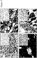
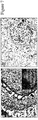
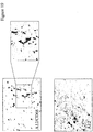
- the present disclosure demonstrates that plexin D1 is expressed on tumor-associated blood vessels but not on normal blood vessels. This has been shown by in situ hybridization of mouse brains, containing angiogenic human melanoma lesions ( Figure 2 ).
- the animal tumor model is described in ( Kusters, B et al., Cancer Res 63:5408-5413 (2003 )).
- tumor cells are injected via a microsurgical procedure in the right carotid artery, resulting in tumor growth in the parenchyma of the right brain hemisphere. After three weeks, at the onset of neurological symptoms, mice are sacrificed and brains removed and fixed in formalin.
- RNA probes were generated by transcription using T3 and T7 RNA polymerase, respectively, from a PCR product, encompassing 600 bases in the 3'-untranslated region, and which was flanked by T7 and T3 promoters (Van der Zwaag et al. (2002), supra ).
- PBS phosphate buffered saline
- plexin D1 RNA High levels of plexin D1 RNA were observed in vessels of angiogenic Mel57 tumors ( Figure 2 ) using a mouse-specific plexin D1 RNA probe. Tumor cells were also positive for the transcript. The non-perfect homology between mouse and human plexin D1 results in a weaker signal in the human tumor cells using the mouse probe.
- plexin D1 RNA expression in human tumor samples we performed in situ hybridizations with a human-specific plexin D1 RNA probe.
- High plexin D1 RNA expression levels were found in a number of human tumors, of which (glioblastoma multiforme, brain metastases of sarcoma, renal cell carcinoma, adenocarcinoma of the colon and of the breast), both in tumor vasculature and tumor cells.
- a summary of plexin D1-expressing tumor types is given in Table 1.
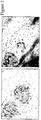
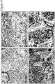
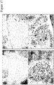
- Figure 3 shows some examples of in situ hybridizations, e.g. a glioblastoma, a brain metastasis of melanoma and a brain metastasis of colon carcinoma.
- Plexin D1 RNA was found not only on the tumor vasculature, but also excessively on the tumor cells themselves. Importantly as in figure 4A , no plexin D1 RNA expression is observed in normal brain vasculature. In figure 4B a CD31 staining is shown, demonstrating that abundant vessels are present in these sections.
- plexin D1 protein antibodies were selected with affinity towards plexin D1.
- a M13 pHENIX phage library was constructed expressing Llama single domain V-H antibodies, constructed by RT-PCR from Llama B-lymphocytes as described ( van Koningsbruggen, S et al., J Immunol Methods 279:149-161 (2003 )).
- the resulting library had a complexity of 8x10 8 clones.
- Eighty percent of plasmids contained full-length sdab insert as determined by PCR analysis and immunological dot-blot-detection of the VSV-G-tag in sdabs (see below).
- the phage library was propagated as phagemids in E. coli TG1 bacteria. Phage particles were rescued by infection with trypsin sensitive helper phage M13K07 (50). Phages were purified and concentrated from the culture supernatant by precipitation with 20% Polyethyleneglycol/2.5 M NaCl via standard methodology.
- the eluate was used to infect log-phase TG1 cells to amplify PLXND1-binding phages and calculate number of binders.
- PLXND1-binding phages with PCR-confirmed full-length sdab inserts were tested for specificity towards plexin D1.
- Wells of DNA-binding plates or immunoplates (Nunc) were coated overnight at 4°C with PLXND1-peptide or an irrelevant peptide (1 ⁇ g/well in PBS/0.5 M NaCl pH 9.0), Bovine serum albumin (1 ⁇ g/well in 50 mM NaHCO 3 pH 9.6) or human Immunoglobulin G (1 ⁇ g/well in 50 mM NaHCO 3 pH 9.6).
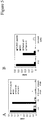
- FIG. 5A shows that M13 phage associated antibodies A12 and F8 bind specifically to plexin D1 peptide, but not to bovine serum albumin, immunoglobulins or an irrelevant peptide.
- soluble single domain antibodies was induced in log-phase E.coli TG1 cells by culturing at 30°C in 2xTYA medium/1 mM IPTG.
- Sdabs were collected by osmotic lysis using ice-cold TES buffer (200mM TrisHCl, 0.5 mM EDTA, 500 mM sucrose) containing a protease inhibitor cocktail (Roche, Basel, Switzerland).
- Sdab concentrations were estimated via dot-blot analysis using the mouse monoclonal anti-VSV-G P5D4, alkaline phosphatase-conjugated rabbit anti-mouse immunoglobulin (Dako, Denmark) and NBT/BCIP staining.
- PLXND1-peptide-KLH conjugate 27 ⁇ g/ml in Na-Acetate, pH 4.0
- BSA 1 ⁇ g/ml in Na-Acetate, pH 5.0
- Unreacted groups were inactivated by 1 M ethanolamine, pH 8.5.
- Ni-affinity-purified sdabs Six concentrations of Ni-affinity-purified sdabs (in the range of 1 mM to 50 ⁇ M) were used to determine the dissociation constants (Kds) of the interaction with the PLXND1-peptide. After each experiment, regeneration of the sensor surface was performed with 10 mM NaOH. Specific binding, defined by binding to a PLXND1-surface minus binding to a control BSA-surface, was analyzed using the BIAevaluation 4.1 software and a 1:1 Langmuir binding model.
- Affinities of single domain antibodies A12 and F8 were 2.1 x 10 -8 M and 3.5 x 10 -8 M, respectively ( Figure 6 ).
- the single domain antibodies are tagged at the carboxyterminal end with a VSV-His-tag, enabling immunohistochemical stainings using an anti-VSV antibody.
- the following protocol was followed for immunohistochemical stainings with single domain antibodies A12 and F8. Following deparaffinization, endogenous peroxidase activity was blocked by incubation with 0.03% H 2 O2. Antigen retrieval was performed by treatment with pronase according to standard protocols. Subsequently, slides were pre-incubated with normal horse or goat serum (to block non-specific binding sites in sections of human and mouse tissues, respectively), followed by incubation with sdabs for 1 hr.
- Sdabs were detected by sequential 1 hr incubations with a mouse or rabbit anti-VSV-G antiserum (Sigma-Aldrich Chemie B.V., Zwijndrecht, The Netherlands), biotinylated anti-mouse or anti-rabbit antibody as appropriate (Vector, Burlingame, CA), and avidin-biotin peroxidase complex (Vector, Burlingame, CA). Finally, peroxidase was visualized by the 3-amino-9-ethylcarbazole (ScyTek, Utah, USA) peroxidase reaction with haematoxylin as counterstain. All steps were performed at RT.
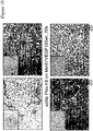
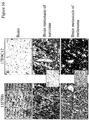
- the specificity of the antibody A12 and F8 for plexin D1 in immunohistochemical stainings was first examined by staining mouse embryos in which expression patterns of plexin D1 on the RNA level were well characterized (Van der Zwaag et al. (2002), supra ), and comparing profiles with immunostainings with anti-endothelial antibody anti-CD31 (DAKO, Glostrup, Denmark). In growth plate of trabecular bone of mice embryos at E16.5, immunostaining was observed on CD31-positive blood vessels. The staining profile correlated well to in situ hybrization for the plexin D1 transcript ( figure 7A ). The blood vessel origin of PLXND1 expression was further confirmed by performing stainings on serial sections with sdabs and anti-human anti-CD31 antibody (anti-human CD31).
- plexin D1 occurs on premalignant cells.
- melanoma consisting of benign nevi, dysplastic nevi, radial growth phase melanoma, invasive melanoma and disseminated melanoma.
- Melanocytes in benign nevi and dysplastic nevi do not express the protein, whereas malignantly transformed cells, both in radially growth phase and vertical growth phase tumors are positive for the protein ( Figure 9 and Table II ).
- Plexin D1 expression is related to the activation state of the endothelial cells in tumor blood vessels.
- Treatment with ZD6474 resulted in a decrease of plexin D1 expression on tumor-associated blood vessels in a dose dependent manner ( Figure 10 ).
- plexin D1 expression is a characteristic of activated endothelial cells.
- plexin D1 expressed in normal brain, heart, skin, kidney, spleen, intestine, endometrium was examined by immunohistochemistry using antibody A12. Vessels in proliferative myometrium expressed plexin D1, showing that plexin D1 is associated not only with pathological angiogenesis, but also with physiological angiogenesis (not shown).
- co-immunostainings were performed with the CD68 macrophage marker. These stainings revealed that a subpopulation of macrophages expressed the protein ( figure 11 ). Also fibroblasts in skin and some proliferating intestinal epithelial cells were found to express plexin D1 (not shown).
- plexin D1 protein on tumor blood vessels suggests that plexin D1 is accessible via intravenous injection.
- PBS phosphate buffered saline
- tumors were dissected from 10 ⁇ m brain sections using laser capture dissection microscopy (Leica laser dissection microscope). Equivalent areas were dissected from unaffected brain, contralateral to the tumor.
- phages were eluted from dissected tissue samples using trypsin treatment and used to infect TG1 cells. Numbers of colony-forming phages were counted and used as a measure of tumor homing.
- mice were injected transcranially with E98, a glioma xenograft line.
- E98 tumors are maintained as subcutaneous tumors.
- a Balbc/c nu/nu athymic mouse carrying a subcutaneous E98 tumor was killed and the tumor removed.
- the tumor was minced with a sterile scalpel and the homogenate was passed through a sterile 70 ⁇ m mesh nylon filter.
- M13 phages displaying single domain antibody F8 were injected intravenously, and after five minutes the mouse was subjected to cardiac perfusion with 15 ml of phosphate buffered saline.
- mice were killed, brains removed and fixed in formalin.
- Four ⁇ m sections were subjected to immunohistochemistry with anti-M13 antibody, and serial sections were stained immunohistochemically with antibodies against CD34 (endothelial marker) and glut-1 (a marker for pre-existent brain endothelial cells ( Kusters, B et al., Cancer Res 62:341-345 (2002 )).
- CD34 endothelial marker
- glut-1 a marker for pre-existent brain endothelial cells
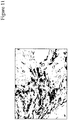
- Phages carrying anti-plexin D1 single domain antibodies accumulated specifically in tumor associated blood vessels, but not in normal vessels ( Figure 13 ). Importantly, phages also accumulated in tumor blood vessels that were positive for glut-1, and which therefore can be considered as pre-existent blood vessels, rather than newly formed blood vessels. This indicates that not only angiogenic blood vessels are subject to targeting with anti-plexin D1 antibodies, but also non-angiogenic, yet activated blood vessels in tumors.
- Recombinant plexin D1 ectodomains inhibit angiogenesis
- Human melanoma Mel57 cells were transfected with the VEGF-A 165 coding sequence in vector pIREShyg. Stably transfected cells were selected by culturing in 200 ⁇ g/ml hygromycin in Dulbecco's Modified Eagle medium (DMEM) supplemented with 10% fetal calf serum (FCS) and penicillin/streptomycin. Because expression of the hygromycin resistance gene is linked to that of the VEGF-A cDNA via the internal ribosomal entry site (IRES), all hygromycin- resistant cells will produce the VEGF-A protein also.
- DMEM Dulbecco's Modified Eagle medium
- FCS fetal calf serum
- Stably transfected Mel57-VEGF cells were subsequently transfected with pIRESneo-PlexinD1 ED.
- the vector contains the cDNA encoding the extracellular domain from nucleotides 1-2745, linked via the IRES to expression of the neomycin resistance gene.
- Double transfectants were injected in the right carotid artery of nude mice, and tumors were allowed to develop.
- mice were subjected to Gadolinium-DTPA enhanced magnetic resonance imaging. Subsequently, mice were sacrificed, brains fixed in formalin and subjected to immunohistochemical stainings to examine the tumor vasculature.
- the plexin D1 ectodomain does not prevent activation of endothelial cells by VEGF-A 165 , but it does prevent the formation of neovasculature.
- the protein was used to immunize BALB c/c mouse 25 according to standard procedures.
- Figure 15 shows the chacteristics of the mouse serum.
- the mouse immune serum specifically recognized E.coli recombinant protein 47-506 (52 kDa, lane 1), and a second recombinant plexin D1 sequence of 18 kDa, comprising amino acids 225-388 (thus lying completely within the sequence that was used for immunization, lane 2).
- the pre-immune serum did not show such a reactivity (panel A).
- the mouse immune serum panel D
- the pre-immune serum panel C
- the B-lymphocytes of this mouse were considered suitable to generate hybridomas of spleen B-lymphocytes with myeloma cell line SP2/0.
- Monoclonal antibody 11F5H6 is able to recognize tumour blood vessels
- mice were decapitated, and brains removed and snap-frozen or fixed in formalin. Frozen sections of 4 ⁇ m were stained with anti-IgM antibody.
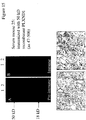
- Figure 17A it is shown that antibody 11F5H6 homes to and accumulates in tumour vessels but not in normal vessels (compare anti-IgM staining in Figure 17A with the anti-endothelial CD31 staining in Figure 17B ). Such staining is not seen when performing anti-IgM staining on non-injected mice.
- 11F5H6 is a promising antibody which allows tumour targeting.
- Plexin D1 is expressed in macrophages in mouse models of rheumatoid arthritis ( Figure 18 ). A subset of macrophages in human atherosclerotic plaques also expresses plexin D1 ( Figure 19 ). Stainings were performed with single domain antibody A12. In Figure 19 , a double staining was performed, displaying plexin D1 in red and the macrophage marker CD68 in blue. A purple color indicates co-expression.
- A12 (SEQ ID NO:1) :
- Table 1 PLXND1 expression in human tissues Tissue PLXND1 expression
Description
- The present disclosure relates to the identification of a novel targetable protein that can be used in the treatment and diagnosis of tumors, in particular solid tumors, and disorders that involve inflammation, in particular rheumatoid arthritis, atherosclerosis and multiple sclerosis.
- To grow beyond a size of 2-3 mm3, tumors have to recruit a neovasculature via angiogenesis. Tumors accomplish this via expression of Vascular Endothelial Growth Factor-A (VEGF-A), either induced by hypoxia in the tumor centre or as a result of malfunctioning tumor suppressor gene products or activated proto-oncogenes. A number of compounds that target the VEGF-A signaling pathway has been developed with the aim to inhibit angiogenesis and, consequently, tumor growth. Although such anti-angiogenic therapies have been effective in animal tumor models, translation to the clinical level has so far proven to be less successful (Eichhorn, ME et al., Drug Resist Update 7:125-138 (2004)).
- For this, there is a number of possible explanations. In clinically relevant situations, tumors may have been growing for months or even years at the time of diagnosis, and a significant proportion of the vasculature may be more or less mature and thus insensitive to angiogenesis inhibition. This situation is in sharp contrast to that in most animal models in which, as a rule, aggressive, fast-growing tumors are studied. Furthermore, patients that are candidates for anti-angiogenic therapy are typically patients with disseminated, uncontrollable cancer and growth of metastases may not always be strictly dependent on angiogenesis. Because most metastases are blood-borne, they grow out in organs with intrinsically high vessel densities like liver, lung and brain where they can grow in an angiogenesis-independent fashion by co-option of pre-existent vessels.
- Indeed, an angiogenesis inhibitor that very effectively inhibits tumor growth in a number of subcutaneous tumor models (Wedge, SR et al., Cancer Res 62:4645-4655 (2002)) does not inhibit growth of infiltrative tumors in mouse brain. Moreover, upon treatment of mice carrying highly angiogenic brain tumors, angiogenesis inhibition did not result in a halt of further tumor progression, but rather in a progression after a phenotypic shift towards co-option and infiltration (Leenders, WP et al., Clin Cancer Res 10:6222-6230 (2004)). These results imply that anti-angiogenic therapy should be supplemented by vascular targeting therapies in which the existing tumor vascular bed is attacked, resulting in secondary tumor cell death due to disruption of the tumor's blood supply.
- To accomplish effective vascular targeting therapy, markers have to be identified that have specificity for tumor vasculature. Much effort has already been put in this, but with varying success. Effective vascular tumor targeting has been accomplished using single chain antibodies, directed against the fibronectin ED-B domain, which is selectively expressed and deposited in the extracellular matrix of newly formed vessels in angiogenic tumors (Santimaria, M et al., Clin Cancer Res 9:571-579 (2003)). Targeting of avß3-integrin (the expression of which is restricted to immature vessels) using RGD peptides or Vitaxin yielded disappointing result whereas endoglin-expression was not specific for tumor blood vessels (Posey, JA et al., Cancer Biother Radiopharm 16:125-132 (2001); Balza, E et al., Int J Cancer 94:579-585 (2001)).
- In inflammatory diseases such as rheumatoid arthritis (RA) or atherosclerosis, angiogenesis and activation of the vasculature is also often part of the pathology. The vasculature here paves the way for inflammatory cells to extravasate and exert their destructive action. Such diseases can thus also benefit from targeting to blood vessels.
- It is therefore the object of the present disclosure to provide a new targetable protein that can be used in the treatment and diagnosis of cancer and inflammatory diseases or diseases that involve inflammation.
- In the research that led to the present disclosure it was found that plexin D1 is expressed on the luminal side of endothelial cells in tumor blood vessels, on the tumor cells themselves and on activated macrophages that are found in tumors, in inflammation and in atherosclerotic plaques.
- The disclosure thus relates to plexin D1 for use as a targetable protein in the treatment or diagnosis of disorders that involve expression of plexin D1.
- The plexin family of receptors consists of four classes (PLXNA-D) and nine members in mammals. Plexins comprise a family of large, single-pass membrane proteins with homology to scatter factor receptors, encoded by the MET gene family. Members of the plexin family share Sema domains, Met-related sequences (MRS), a transmembrane region and intracellular motifs that are predictive of Rac/Rho-GTPase signalling (
Figure 1 ). - Since signalling via GTPases results in cytoskeletal rearrangements, events that are critically involved in formation of filopodia and lammelipodia and cellular migration, plexins can be regarded as regulators of migration.
- Plexins are receptors for the semaphorins, a family of secreted, GPI-anchored or transmembrane proteins which is subdivided in seven subclasses. Each plexin has its own (set of) semaphorin binding partners, and each plexin-semaphorin combination results in a specific response. Class 3 semaphorins are potent axon repellants and are as such involved in morphogenesis of the nervous system (for review, see (Pasterkamp, RJ et al., Curr Opin Neurobiol 13:79-89 (2003); Fujisawa, H, J Neurobiol 59:24-33 (2004)). For activation of plexins by the semaphorins, additional plexin binding partners may be required. These binding partners, neuropilin-1 and -2 (NP-1 and NP-2) have no signalling motifs in the intracellular domain and are thought of as a passive coreceptors, enabling the interaction between sempahorins and plexins.
- Some plexins form yet larger membrane complexes with and activate signalling receptors as Off Track (Otk) and the scatter factor receptors Met and Ron. A direct interaction between plexinA1 and the angiogenic Vascular Endothelial Growth Factor-receptor-2 (VEGFR2) has also been demonstrated (Toyofuku, T et al., E-publication in Genes Dev 18:435-447 (2004)). Because NP-1 binds to plexin family members but also to VEGFR2, it is conceivable that multicomponent membrane protein complexes exist that encompass VEGFR2, NP-1 and plexins, establishing a link between plexins and angiogenesis (see also Weinstein, BM, Cell, 120:299-302 (2005)).
- Neuropilins are also co-receptors for the potent angiogenic factor Vascular Endothelial Growth Factor-A (VEGF-A165) and enhance its affinity for VEGFR2. Interestingly, the VEGF-A165 binding site on NP-1 overlaps with that for semaphorin 3A (Miao, H Q et al., J Cell Biol 146:233-242 (1999)). It has been postulated that VEGF-A binding to NP-1 promotes migration of endothelial cells by competing for binding of class 3 semaphorins, which is generally followed by F-actin depolymerization and repulsion of cell extensions (Bachelder, RE, Cancer Res 63:5230-5233 (2003)). Similar antagonistic behaviour of VEGF-A and class 3 semaphorins have been described in a neuronal progenitor cell line (Bagnard, D et al., J Neurosci 21:3332-3341 (2001)) and tumor cells (Bachelder (2003), supra). Since antagonistic effects were observed in tumor cells that are devoid of VEGF receptors, it is conceivable that the underlying mechanism involves members of the plexin family, establishing a further link between plexins and VEGF-A signaling.
- The present inventors have previously found that the family member plexin D1 (plxnD1) is not only expressed in neuronal cells, but also in endothelial cells of the vasculature during early stages of development (van der Zwaag, B et al., Dev Dyn 225:336-343 (2002)), an observation that was confirmed by two other groups (Gitler, AD et al., Dev Cell 7:107-116 (2004); Torres-Vazquez, J et al., Dev Cell 7:117-123 (2004)). In adult vasculature, plxnD1 is absent. Plxnd1-knockout mice and zebrafish carrying mutations in the plxnd1 gene are characterized by maldevelopment of the cardiovascular system (Gitler, AD et al. (2004), supra; Torres-Vazquez, J et al.,(2004), supra). Neuropilin-1 (NP-1) and NP-1/Neuropilin-2 (NP-2) double knock-out mice also suffer from lethal defects in vascularization and aortic arch malformations during embryonic development (Kawasaki, T et al., Development 126:4895-4902 (1999); Takashima, S et al., Proc Natl Acad Sci USA 99:3657-3662 (2002); Gu, C et al., Dev Cell 5:45-57 (2003)).
- Furthermore, morpholino-mediated knock-down of NP-1 in zebrafish leads to maldevelopment of intersegmental vessels, and in this model system a clear link between NP-1 and VEGF-A165 has been established (Lee, P et al., Proc Natl Acad Sci USA 99:10470-10475 (2002)). The resemblance of the phenotypes of plxnd1, neuropilin-1 and semaphorin 3C knock-out mice (Feiner, L et al., Development 128:3061-3070 (2001)) is consistent with the finding that Plexin D1 is a neuropilin-1-dependent receptor for semaphorin 3C (Gitler, AD et al. (2004), supra). However, PlxnD1 is also a receptor for semaphorin 3E, and this interaction does not require neuropilins for Semaphorin 3E-mediated signalling (Gu, C et al., Science 307:265-268 (2005)).
- According to the disclosure it was now found that plexin D1 is also involved in angiogenesis during tumor growth and is expressed on the luminal side of endothelial cells in tumor blood vessels. Plexin D1 was furthermore found to be expressed by activated macrophages. Plexin D1 was also found to be expressed on tumour cells in a wide variety of tumor types.
- The present disclosure thus relates to plexin D1 for use as a targetable protein in the treatment or diagnosis of disorders that involve expression of plexin D1.
- Diagnosis is effected by detecting the presence of plexin D1 or a plexin D1 encoding nucleic acid in the body or a bodily tissue or fluid.
- Treatment is effected by targeting plexin D1 for delivery of therapeutics to the site where treatment is needed, by interfering in the interaction between plexin D1 and its ligands, by interfering in the expression of the plexin D1 gene or by capturing plexin D1 ligands to inhibit interaction with plexin D1.
- The disclosure thus furthermore relates to the use of molecules that bind plexin D1, a nucleic acid encoding plexine D1 or a ligand of plexin D1 for the preparation of a therapeutical composition for the treatment or diagnosis of disorders that involve expression of plexin D1. All these molecules will be identified herein as "binding molecules" or "binding entities".
- The disorders comprise in particular disorders in which plexin D1 is expressed on tumor cells, tumor blood vessels or activated macrophages.
- The tumor cells on which plexin D1 is expressed comprise brain tumors, in particular astrocytomas, oligodendrogliomas and hemangioblastomas, colon carcinomas, in particular ductal carcinomas of the colon, prostate carcinomas, renal cell carcinomas, in particular renal clear cell carcinomas, mamma carcinomas, in particular ductal carcinomas of the breast, ovary carcinomas, squamous cell carcinomas, melanomas, lung carcinomas, in particular small-cell lung carcinomas and non-small cell lung carcinomas, soft tissue sarcomas etc.
- When the disorders that are treated according to the disclosure are inflammatory diseases, they are in particular autoimmune disease, more in particular rheumatoid arthritis, or they are atherosclerosis or multiple sclerosis.
- Molecules that bind plexin D1 are for example selected from antibodies, antibody fragments, proteins, protein domains, peptides, small molecules. These molecules can be used to target plexin.
- Molecules that bind the nucleic acid encoding plexin D1 are for example oligonucleotides, such as RNA or DNA aptamers, for example selected from siRNA, antisense RNA, antisense phosphothio-oligonucleotides. These molecules can be used to interfere with the expression of plexin D1.
- Molecules that bind a plexin D1 ligand are for example selected from antibodies against ligands, the soluble ectodomain of plexin D1 or small molecules, such as peptides, that bind plexin D1 ligands. These molecules can be used to capture circulating plexin D1 ligand, prevent binding of the ligand to plexin D1 on tumor vessel cells, tumor cells or activated macrophages and interfere with the function of plexin D1 on these cells.
- For diagnosis, the binding molecule is suitably labelled with a detectable marker. Such a detectable marker is for example selected from a radioactive label, paramagnetic label, a fluorescent label, a chemiluminescent label. Diagnosis can be performed in a sample of a bodily fluid or tissue in vivo, in situ or ex vivo. Examples of diagnostic techniques are in situ hybridization of for example plexin D1 mRNA or immunohistochemistry on biopsies or tumor cells.
- For treatment, the binding molecule is for example provided with an entity that damages or kills the tumor cell and/or the tumor endothelial cell, in particular a cytotoxic entity, such as a radionuclide, a toxin, boron for Boron Neutron Capture Therapy (BNCT), or a prodrug that is coupled to the binding entity via a cleavable linker, which is activated in response to cleavage of that linker, or apoptosis-inducing peptides, an example of which is the (KLAKLAK)2 sequence. Such peptides are added to the binding entity by molecular genetic engineering techniques. The entities described above may be directly conjugated to the binding entity, or they may be present in nanodevices, such as liposomes or polymersomes, that are conjugated to the binding entity.
- Boron Neutron Capture Therapy (BNCT) comprises irradiation of a diseased area, such as a tumor or an inflammation, in which boron has accumulated after intravenous injection of the liposomal conjugate, with neutrons, after which boron atoms will decay to lithium under emission of destructive alpha particles.
- Alternatively, therapy may be effected by inducing local thrombosis in the tumor vessels to block the blood supply to the tumor and induce cell death. An example of such molecule is Tissue Factor (TF).
- Advantageously, plexin D1 can be targeted with specific binding molecules upon intravenous administration since plexin D1 is expressed on the luminal side of endothelial cells in tumor blood vessels. Therapeutic compounds for damaging or killing tumor cells that are coupled to the binding molecule can reach the tumor from within and compounds that induce thrombosis are easily delivered to their site of action.
- Interference with plexin D1 function represents a way to inhibit angiogenesis, to inhibit tumor cell migration, and to inhibit macrophage migration. Thus, the disclosure provides methods of treating or suppressing disorders in which plexin D1 is involved, by using the specific presence of plexin D1 to deliver therapeutics locally to diseased tissues, and/or by interference in the function of plexin D1 or in the interaction between plexin D1 and its ligands.
- The disclosure is thus based on the fact that plexin D1 can be used as a targetable marker on tumor blood vessels, as a targetable protein involved in tumor angiogenesis, as a targetable marker on tumor cells and as a targetable protein involved in cellular migration.
- The disclosure thus also relates to the use of molecules which bind to plexin D1, its gene or mRNA or its ligands in diagnosis and therapy. All kinds of specific binding molecules, and derivatives thereof can be used in the methods as disclosed herein, in particular proteinaceous compounds, such as, but not limited to, antibodies, antibody fragments, single domain antibody fragments, other proteinaceous binding domains, such as, but not limited to, lipocalins, and small molecules that specifically bind plexin D1 or its ligands. For binding to the plexin D1 gene or the mRNA transcribed from the plexin D1 gene nucleic acid molecules, such as DNA or RNA aptamers can be used.
- As further disclosed herein, plexin D1 or plexin D1 ligand binding molecules are antibodies, in particular monoclonal antibodies, more in particular human or humanized antibodies in which the constant regions of the original antibody are substituted with the constant regions of human antibodies, or fragments thereof which still bind to plexin D1 or its ligand.
- The antibody is preferably a human IgG1 antibody. However, other human antibody isotypes are also encompassed by the present disclosure, including IgG2, IgG3, IgG4, IgM, IgA1, IgA2, IgAsec, IgD and IgE. Also all animal-derived antibodies of various isotypes can be used in the methods as disclosed herein.
- The antibodies can be full-size antibodies or antigen-binding fragments of antibodies, including Fab, F(ab')2, single chain Fv fragments, or single domain VHH, VH or VL single domains.
- Preferably, antibodies against plexin D1 are human monoclonal antibodies produced by a hybridoma cell which includes a B cell obtained from an immunized transgenic animal having a genome comprising a human heavy chain transgene and a human light chain transgene, fused to an immortalized cell, or an animal-derived antibody or antibody fragment produced by a hybridoma cell which includes a B cell obtained from an immunized animal, fused to an immortalized cell, or human and animal antibodies, produced by a eukaryotic cell transfected with the cDNA or genomic DNA encoding said antibody or antibody fragment.
- As disclosed herein, single domain (VHH) Llama antibodies with affinity to plexin D1 are provided, more specifically Llama single domain antibodies A12 (SEQ ID NO:1) and F8 (SEQ ID NO:2), either or not displayed on M13 bacteriophages, also known to those with skill in the art as phage-display VHH antibodies.
- A preferred single chain antibody is derived from antibody 11F5H6 and 17E9C12. The sequence of the single chain antibody is shown in SEQ ID NO:3 and SEQ ID NO:4.
- The antibodies for use as described herein may be high affinity antibodies which are evoked in non-transgenic laboratory animals, or in a transgenic animal in which the endogenous globulin locus has been substituted for the human globulin locus, thus allowing production of human antibodies in such animals (Jakobovits, A. Curr Opin Biotechnol 6:561-566 (1995)).
- The description further provides a method of producing the antibodies as described herein, comprising immunizing an animal with plexin D1, or a cell expressing plexin D1, or a nucleic acid encoding plexin D1, or parts of the extracellular domain of plexin D1, such that antibodies against plexin D1 are produced by the B cells of the animal, isolating the B cells from the animal and fusing the B cells to a myeloma cell line to obtain immortalized cells that secrete the antibody. The animal is preferably a transgenic animal having a genome comprising a human heavy chain transgene and a human light chain transgene so that the resulting antibody is humanized.
- As disclosed herein, the method includes immunizing a laboratory animal with a synthetic peptide, chosen from the plexin D1 extracellular domain, for example peptide 47-63 corresponding to the amino terminus of the mature plexin D1 amino acid sequence. Immunizations are however preferably done with recombinant extracellular domains, preferably a region with low similarity to other family members of the plexins, for example a region comprising amino acids 47-546, lacking the Met-Related-Sequences. Said recombinant plexin D1 extracellular domains can be produced in E.coli cells by inserting the coding nucleic acids in a suitable prokaryotic expression vector, for instance under control of the β-galactosidase promoter, transformation of E.coli cells with said vector, and isolation of the recombinant proteins from purified inclusion bodies. It is preferred however that antibodies are evoked by immunization with said recombinant plexin D1 extracellular domain which is produced by eukaryotic cells, therefore containing posttranslational modifications which are most similar to those present in native plexin D1, for example by chinese hamster ovary (CHO) cells after transfection with a vector, containing the coding nucleic acids for said extracellular domains under the control of a cytomegalovirus promoter. Recombinant extracellular plexin D1 fragments may or may not be fused to tags facilitating purification, e.g. a VSV tag or a constant region of a heavy chain of an immunoglobulin.
- The method of producing the antibody may also comprise cloning the antibody coding regions from said plexin D1 specific B-cells into an expression vector and expressing the coding sequence. It is preferred that the expression vector is pHENIXHISVSV, enabling expression by E.coli host cells of the antibody, flanked at the carboxyterminal end by a Vesicular Stomatis Virus (VSV-tag) and a His*8 tag. The VSV tag is meant to facilitate immunohistochemical detection, using specific antibodies. The His*8 tag is meant to facilitate purification based on Nickel affinity chromatography. Other expression vectors can likewise be used.
- More specifically the present disclosure provides an isolated single domain antibody A12, having a dissociation constant of less than 2x10⁻⁸ M, which binds to the amino terminus of plexin D1 and which detects plexin D1 in immunohistochemical stainings and homes to plexin D1-expressing tumor blood vessels, and also to an isolated single domain antibody F8, having a dissociation constant of less than 3x10⁻⁸ M, which binds to the amino terminus of plexin D1 and which detects plexin D1 in immunohistochemical stainings and homes to plexin D1-expressing tumor blood vessels. Both isolated single domain antibody may be fused to the constant region of a human IgG1 heavy chain or the constant region of a mouse IgG1 heavy chain.
- Preferably fully human antibodies are used within the methods as described herein. Also, humanised or laboratory animal-derived antibodies may be used.
- The description further provides bispecific antibodies that have a binding specificity for plexin D1, and a binding specificity for a human antigen presenting cell, or for an Fc receptor, wherein the Fc receptor is a Fc (gamma) R1 or a human Fc(alpha) receptor.
- The description provides also nucleic acid molecules encoding the preferred antibodies, or antigen-binding portions. Recombinant expression vectors which include nucleic acids which encode the antibodies as described herein, as well as host cells transfected with such vectors, are also disclosed.
- Other binding molecules as disclosed herein are small molecules that specifically bind to Plexin D1. The term "small molecule" refers often to molecules with molecular weights of 500 or below. The term is commonly used these days and thus clear to the skilled person. Moreover, small molecule libraries are already available or are being developed. An example of such library is the NIH Molecular Libraries Small Molecule Repository (MLSMR). Such libraries are subjected to High Throughput Screening (HTS) to identify molecules that bind to plexin D1. The description also provides small molecules that result from a screen of such libraries.
- Other compounds that can be used in the methods as described herein comprise peptides or aptamers (Ulrich, Med. Chem. 1(2):199-208 (2005)) that bind to extracellular domains of plexin D1 and thereby interfere with binding of plexin D1 ligands to plexin D1. Conversely, such peptides or aptamers may also bind to the plexin D1 binding sites of the plexin D1 ligands and thereby interfere with ligand binding to plexin D1.
- For interfering with the expression of the plexin D1 gene another type of binding molecule is used, in particular siRNA, antisense RNA or antisense phosphothio-nucleotides. Small interfering RNA (siRNA) comprises small strands of RNA that interfere with the translation of messenger RNA. SiRNA binds to the complementary portion of the target messenger RNA and tag it for degradation thus inhibiting gene expression. This is commonly known as gene "silencing". SiRNA is usually 21 to 23 nucleotides long. Antisense RNA is an RNA molecule transcribed from the coding, rather than the template, strand of DNA, so that it is complementary to the sense mRNA. Formation of a duplex between the sense and antisense RNA molecules blocks translation and may also subject both molecules to double strand-specific nucleases thus inhibiting expression of the gene. Inhibition of expression of the gene can be used to block angiogenesis and migration of tumor cells and macrophages.
- Preferably, the above described binding molecules bind to plexin D1, its gene or its ligand in eukaryotic cells. Said molecules specifically accumulate in tumors upon intravenous injection or specifically accumulate in tumor blood vessels upon intravenous injection.
- The antibodies, fragments thereof, small molecules and other proteinaceous compounds that all bind plexin D1 can be used in various ways.
- Also described herein are compounds that bind to the extracellular part of plexin D1 and which binding results in interference of plexin D1 function. Alternatively, the description provides compounds that bind to the intracellular domain of plexin D1 and which prevent signalling by plexin D1.
- Such binding molecules bind to plexin D1 to interfere with the formation of multicomponent membrane complexes by inhibiting binding of plexin D1 ligands, in particular neuropilin-1, neuropilin-2, semaphorin 3C, sempaphorin 3E, VEGF-
receptor 1, VEGF-receptor-2 or VEGF-A, to plexin D1. Such binding molecules lead to inhibition of ligand-induced GTPase signalling by plexin D1 or to inhibition of migration of cells expressing plexin D1, in particular tumor-associated endothelial cells, tumor cells or macrophages. - Also provided herein is a method of inducing lysis of a cell expressing plexin D1, comprising contacting a cell expressing plexin D1
with the binding molecules, in particular the antibodies as described herein, in the presence of human effector cells, such that lysis of the cell expressing plexin D1 occurs. - As disclosed herein, the binding molecule is combined with or coupled to an effector compound that can detect the presence of plexin D1 for diagnostic purposes or that can perform an effect on the cell expressing plexin D1. The diagnostic or therapeutic effector compound can be directly coupled to the binding molecule or can be present in a transport vehicle, such as a nanodevice, in particular a liposome or polymersome, that is coupled to the binding molecule. Alternatively, the binding molecule can be a bispecific antibody that binds both plexin D1 and the effector compound thus targeting the effector compound to a site or cell where plexin D1 is expressed.
- The description thus provides the use of such binding molecules in a method of diagnosing a disease mediated by expression of plexin D1, which method comprises intravenous delivery of the proteinaceous, aptameric or small molecule plexin D1 binding molecules, conjugated to an effector compound allowing in vivo detection of the binding molecules.
- Diagnostic effector compounds are for example radioisotopes or contrast agents for Magnetic Resonance Imaging (MRI), such as gadolinium-DTPA, or fluorescent dyes.
- Examples of radioactive substance comprise, but are not limited to technetium99m (99mTc), iodine-123 (123I), iodine-131 (131I), rhenium-186 or -188 (186/188Re), gallium-67 (67Ga) the beta-radiation emitting substances yttrium-90 (90Y) or lutetium-177 (177Lu), the positron emitting isotopes Fluorine-18 (18F) and Carbon-11 (11C). Such radioisotopes can be used to either detect or damage or kill cells expressing plexin D1. Usually different isotopes are used for diagnosis and therapy. The skilled person is well aware of which isotope to use for which tissue and for which type of use.
- Theproteinaceous, aptameric and small molecular binding molecules for use as described herein can be combined with or coupled to a toxic agent, such as chemotherapeutic agent either directly or in a transport vehicle, in particular a nanodevice, such as a liposome or polymersome.
- A plexin D1 binding entity as described herein is coupled to one or more chemotherapeutic agents selected from the group consisting of nitrogen mustards (e.g., cyclophosphamide and ifosfamide), aziridines (e.g., thiotepa), alkyl sulfonates (e.g., busulfan), nitrosoureas (e.g., carmustine and streptozocin), platinum complexes (e.g., carboplatin and cisplatin), non-classical alkylating agents (e.g., dacarbazine and temozolamide), folate analogs (e.g., methotrexate), purine analogs (e.g., fludarabine and mercaptopurine), adenosine analogs (e.g., cladribine and pentostatin), pyrimidine analogs (e.g., fluorouracil (alone or in combination with leucovorin) and gemcitabine), substituted ureas (e.g., hydroxyurea), antitumor antibiotics (e.g., bleomycin and doxorubicin), epipodophyllotoxins (e.g., etoposide and teniposide), microtubule agents (e.g., docetaxel and paclitaxel), camptothecin analogs (e.g., irinotecan and topotecan), enzymes (e.g., asparaginase), cytokines (e.g., interleukin-2 and interferon-[alpha]), monoclonal antibodies (e.g., trastuzumab and bevacizumab), recombinant toxins and immunotoxins (e.g., recombinant cholera toxin-B and TP-38), cancer gene therapies, and cancer vaccines (e.g., vaccine against telomerase).
- Chemotherapeutic agents are preferably selected from the group consisting of doxorubicin, cisplatin, bleomycin sulfate, carmustine, chlorambucil and cyclophosphamide hydroxyurea. Other compounds are known to the person skilled in the art.
- A tumor can also be treated by blocking its blood supply by inducing local thrombosis in the tumor vasculature. The binding molecules as described herein can be used to target thrombosis inducing molecules, such as the blood coagulation co-factor TF (Tissue Factor), a radioactive entity or a toxin, such as ricin to the site of the tumor. Effector compounds can be coupled to the binding molecule, in particular a plexin D1 binding molecule, or can be present in a nanodevice, such as a liposome or polymersome, that is coupled to the plexin D1 binding molecule.
- An alternative method of treating cancer or an inflammatory disorder as described herein is with boron. The binding molecules as described herein can be conjugated to transport vehicles, in particular nanodevices, such as liposomes or polymersomes, that are filled with boron to obtain a therapeutic composition. After delivery and accumulation of this composition in the diseased area, this area is irradiated with neutrons, resulting in emission of radioactive and cytotoxic alpha-particles that damage or kill the tumor endothelial cells, tumor cells and/or activated macrophages.
- It is desirable that for tumor blood vessel targeting antibodies have high affinities towards plexin D1, for example higher than 10-8, preferably higher than 10-9, more preferably higher than 10-10 M. High affinity and the high molecular weight of antibodies will however restrict penetration into tumor tissue. Therefore, the nucleic acids that encode said monoclonal antibodies, obtained via RT-PCR cloning, can be used to generate antibody derivatives, for example antibodies that lack the constant region and are monovalent, or antibody fragments that are adapted to optimal affinities for blood vessel targeting or tumor penetration by mutagenic procedures. These antibody derivatives will have lower affinities and lower molecular weight and will have improved tumor cell targeting properties.
- Different binding molecules as described herein can be combined in a mixture. Specifically, the members of the mixture have a varying affinity. An example of such a combination is a mixture of monoclonal antibodies and/or antibody fragments or a mixture of antibodies with small molecules. The monoclonal antibodies having high affinity can be used for targeting vessels, whereas the smaller fragments having a lower affinity are better able to penetrate and reach the tumor cells. Alternatively, a mixture of plexin D1 binding molecules can be used together with plexin D1 ligand binding molecules and/or with molecules that bind nucleic acids encoding plexin D1. Or plexin D1 ligand binding molecules can be combined with molecules that bind nucleic acids encoding plexin D1.
- The plexin D1 binding molecules as described herein can be used in a method of treating a disease mediated by expression of plexin D1, comprising intravenous delivery of the binding molecules as described herein at a dose, effective in treating that disease.
- The binding molecules can also be used in a method of diagnosing a disease mediated by expression of plexin D1, comprising intravenous delivery of conjugates of plexine D1 binding molecules with a paramagnetic, fluorescent or radioactive tracer followed by magnetic resonance imaging, optical imaging, SPECT or PET.
- The binding molecules can further be used in a method of treating or suppressing a disease mediated by expression of plexin D1, comprising intravenous delivery of the proteinaceous and small molecular binding molecules as described herein or a composition of proteinacous and small molecular binding molecules.
- The disease to be treated or diagnosed can be cancer, an inflammatory disease, in particular an autoimmune disease, such as rheumatoid arthritis, or atherosclerosis, or multiple sclerosis.
- Diagnosis can be performed in vivo and in vitro. An in vivo method is described above and can be performed with magnetic resonance imaging (MRI) or with SPECT or PET cameras after accumulation of the radioactively labelled binding molecule in the diseased tissue.
- Another diagnostic method comprises detecting the presence of plexin D1 in a sample in vitro or ex vivo. Such method comprises contacting the sample with binding molecules of plexine D1, or nucleic acids that bind the plexin D1 gene or its mRNA or a copy DNA derived from this mRNA, all bound to a detectable marker, under conditions that a complex between the antibody and plexin D1 forms, and detecting the formation of the complex. The complex can be detected by visualising the detectable marker. Samples can be bodily fluids, such as blood, serum, plasma, saliva, urine, semen, faeces, or tissues, such as biopsies of tumor cells.
- The description further provides an expression vector, comprising the coding sequence for Llama antibody F8 or A12, or for the single chain antibody derived from antibody 11F5H6 and suitable regulatory sequences. The description also provides a cell transfected with the said expression vector. The de further relates to the recombinant protein obtainable by expressing the expression vector.
- The description also provides an expression vector, comprising the coding sequence for the extracellular domain of plexin D1, optionally fused to a constant region of a human heavy chain and suitable regulatory sequences. The description also provides a cell transfected with the said expression vector. The description further provides the recombinant protein obtainable by expressing the expression vector.
- The recombinant protein comprises the extracellular domain of plexin D1, which binds to plexin D1 ligands and thus prevents binding of the ligands to cell associated plexin D1. Preferably, the coding sequence codes for a recombinant protein comprising amino acids 47-506 of the extracellular domain of plexin D1, which binds to plexin D1 ligands and thus prevents binding of the ligands to cell-associated plexin D1, or amino acids 507-1274 of the extracellular domain of plexin D1, which binds to plexin D1 ligands and thus prevents binding of the ligands to cell associated plexin D1. Such recombinant protein may carry mutations that increase the affinity for plexin D1 ligands and thereby having increased potency as decoy receptor. Such mutations are usually induced by making changes in the coding sequence used for producing the recombinant protein.
- The description further provides the use of the binding molecules as described herein in a method of treating or suppressing a disease mediated by plexin D1, comprising intravenous or intratumoral delivery of decoy plexin D1 extracellular domains as described above, or in a method of treating or suppressing a disease mediated by plexin D1, comprising intravenous delivery of adenoviruses or lentiviruses, containing the coding nucleotides for the recombinant extracellular domains of plexin D1 or parts thereof as described above.
- These antagonistic decoy plexin D1 receptors interfere with the interaction between plexin D1 and its ligands, in particular neuropilin-1, semaphorin 3C and sempahorin 3E, and thereby interfere with plexin D1 function.
- Preferably, the decoy plexin D1 receptors have increased affinity towards plexin D1 ligands, as compared to plexin D1. Such increased affinity may be obtained by creating a library of plexin D1 extracellular domains, carrying at random introduced mutations, and selecting decoy receptors with most potent antagonistic behaviour in cell migration assays.
- In order to produce said proteinaceous molecules (including peptides, polypeptides and glycosylated polypeptides or polypeptides having other post- or peritranslational modifications) it is desirable to insert the recombinant nucleic acid which encodes fragments of the extracellular domain of plexin D1 that comprise the binding sites for semaphorin 3C, semaphorin 3E and NP-1 into expression vectors. The antagonists as described herein are preferably produced from a nucleic acid or an expression vector as described herein, preferably in a host cell.
- The molecules as described herein may also be used in a combination of treatment methods as described above and/or with conventional therapies or anti-angiogenic therapy for further preventing the formation of a tumor neovasculature, or radiotherapy and/or adjuvant chemotherapy.
- The description further provides a method of identifying molecules that are capable of binding to plexin D1, which method comprises contacting a collection of molecules with plexin D1 and selecting the molecules from the collection that show binding to plexin D1 as plexin D1 binding molecules. The collection of molecules can for example be present in small molecule libraries, on a protein array, etc. The techniques for screening collections of molecules are in itself known. The description provides the identification of the target that is to be bound, which is plexin D1.
- In this application the term "binding molecule" is used for all types of binding molecules, i.e. the ones that bind plexin D1, the ones that bind a nucleic acid encoding the plexin D1 gene and the ones that bind plexin D1 ligands. All these types of molecules can be coupled to effector compounds as described above.
- The present disclosure will be further illustrated in the Examples that follow. In the Examples reference is made to the following figures:
-
Figure 1 : Structural domains of plexin family members: four subfamilies have been identified, named plexin A-D. Horizontally hatched boxes indicate Sema domains, diagonally hatched boxes indicate Met-related sequences (MRS) motifs, and the clear box indicates the atypical MRS motif of PLXND1. Plexin B subfamily members have a potential furin-like proteolytic site, marked by a grey ribbon. The transmembrane region is marked by a shaded box and is followed by two conserved intracellular domains, together comprising the SP-domain, marked by two ovals. -
Figure 2: A ) In situ hybridization analysis of cerebral Mel57- VEGF-A165 lesions using a digoxigenin-labelled mouse-specific plxnd1 RNA probe. Tumor vessels are strongly positive (arrows) whereas brain capillaries, distant from the lesions, are negative (compare the ISH profile with the CD34 staining inFigure 2B ). -
Figure 3 : Human PLXND1-specific ISH analyses of glioblastoma multiforme (A) brain metastases of sarcoma (B), melanoma (C) and mammacarcinoma (D). Insets show CD31 stainings of serial sections. Control ISH using sense probes were negative (not shown). Note that in these tumors PLXND1 expression is not confined to the blood vessels: also in tumor cells high levels of the PLXND1 transcript are found. t=tumor, V=vessel. -
Figure 4 : ISH analysis using a human specific digoxigenin-labelled RNA probe (A) and immunohistochemical staining with CD31 (B) of normal brain. Note that vessels are abundantly present but these do not express the plexin D1 transcript. -
Figure 5 : Specificity of phages (A) and corresponding single domain antibodies (sdabs) (B) A12 and F8 for peptide H2N-ALEIQRRFPSPTPTNC-CONH2. In A, 1010 phages were allowed to bind to PLXND1-peptide, BSA, human IgG or irrelevant peptide as described in the text. After rigorous washing, bound phages were detected using an anti-M13 antibody. In B, similar incubations were performed but now with soluble sdabs. After washing, bound sdabs were detected and semi-quantified via the VSV-G-tag. -
Figure 6 : The dissociation constants (kd's) of the binding between single domain antibodies A12 and F8 were determined using the Biacore 2000 (Uppsala, Sweden) biosensor. The sensor chip and protein coupling chemicals were purchased from Biacore AB. PLXND1-peptide-KLH conjugate (27 µg/ml in Na-Acetate, pH 4.0) or BSA (1 µg/ml in Na-Acetate, pH 5.0) was coupled to activated CM5 surfaces using N-ethyl-N'- (dimethylaminopropyl) carbodiimide, N-hydroxysuccinimide, under conditions recommended by the manufacturer. Unreacted groups were inactivated by 1 M ethanolamine, pH 8.5.
Kinetic measurements were performed at 25°C with a flow rate of 10 ml/min in HBS-EP buffer (10 mM Hepes, pH 7.4, 150 mM NaCl, 3 mM EDTA, 0.005% surfactant P20). Six concentrations of Ni-affinity-purified sdabs (in the range of 1 mM to 50µM) were used to determine the dissociation constants (Kds) of the interaction with the PLXND1-peptide. After each experiment, regeneration of the sensor surface was performed with 10 mM NaOH.
Specific binding, defined by binding to a PLXND1-surface minus binding to a control BSA-surface, was analyzed using the BIAevaluation 4.1 software and a 1:1 Langmuir binding model. Affinities of single domain antibodies A12 and F8 were 2.1 x 10⁻⁸ M and 3.5 x 10⁻⁸ M. -
Figure 7 : Evaluation of plexin D1 specificity of single domain antibodies. Figure A shows immunohistochemical staining of the growth plate of trabecular bone of a mouse embryo (E16.5) with single domain antibody A12, utilizing the VSV-G tag for detection of the antibody. The inset shows an in situ hybridization of a similar embryonic structure using a mouse specific digoxigenin-labelled plexin D1 probe. Note the overlap of plexin D1 in situ hybridization and immunostaining.Figure 7B is a representative example of a Mel57- VEGF-A165 lesion in brain of a nude mouse. The vasculature which is also positive in plexin D1 ISH (see alsofigure 2 ) is immunopositive with single domain antibody F8. -
Figure 8 : Immunostainings with single domain antibody A12 on a selection of human brain tumors. Tumors shown are A) glioblastoma multiforme, metastases of B) melanoma C) mammacarcinoma and D) renal cell carcinoma. The insets in A and B consist of control stainings with anti-VSV antibody only, and show that the tumor staining is specific. Note that vessels and tumor cells are highly reactive with the antibody. -
Figure 9 : Immunostainings with single domain antibody A12 on a progression serie of melanoma. Immunostainings were performed on a nevus, a dysplastic nevus and horizontal and vertical growth phases of melanoma. Note that only the neoplastic cells express plexin D1. -
Figure 10 : Immunostainings with single domain antibody A12 on sections of Mel57-VEGF-A brain tumors in mice, treated with ZD6474. In untreated or placebo-treated mice, tumorvessels stain positive with this antibody. However, in ZD6474-treated mice, there is a dose-dependent decrease of plexin D1 expression. ZD6474 was given orally, once daily, in the dosage as indicated. -
Figure 11 : Double immunostainings with the macrophage marker CD68 (stained blue) and single domain antibody A12 (stained red) on mammacarcinoma. A subpopulation of macrophages express plexin D1 as revealed by purple staining protein. -
Figure 12 : In vivo homing of phage A12, F8 or an irrelevant phage to Mel57-VEGF165 brain lesions. Tumor-bearing mice were injected with 1012 phages in the tail vein and after 5 minutes mice were anesthesized and subjected to cardiac perfusion with 15 ml of phosphate buffered saline. Mice were sacrificed, brains removed and frozen sections were analyzed for phage content and distribution. A) M13 staining of a frozen section of brain Mel57-VEGF165 lesions. Phages are clearly vessel-associated, as evidenced by the anti-CD34 immunostaining on a serial section, shown in B). The arrows point at a CD34-positive vessel, distant from the lesion, which is not highlighted by anti-M13 staining. The inset in (A) shows a control experiment where an irrelevant phage was injected. C) Distribution of sdab F8 after intravenous injection in tumor bearing mice. Sdabs are visualized by immunohistochemistry using an anti-VSV antibody. Note that the sdab is detected in tumor vessels but not in normal brain capillaries. The inset shows the control experiment where an irrelevant sdab was injected. An interstitial localization was observed, consistent with the leaky nature of the vessels in these tumors. D) Quantification of phage homing. Tumor tissue was dissected from 10 µm frozen sections using laser capture dissection microscopy. Number of colony-forming phages (cfp) were counted after infection of TG1 cells. Twenty-fold more F8 phages were eluted from tumors than from comparable areas of unaffected brain tissue. -
Figure 13 : Single domain antibody homes to tumor vessels that are not per se newly formed. Nude mice were inoculated with a cell suspension of 1,5x10⁵ cells of the human glioma xenograft E98, which was obtained from a subcutaneous E98 tumor. After 3 weeks, phages carrying single domain antibody F8 were injected in the tail vein, and after 5 minutes mice were anesthesized and subjected to cardiac perfusion using 15 ml of phosphate buffered saline. Mice were then sacrificed, brains removed and fixed in formalin. Serial sections were stained with antibodies against M13 p8 protein (A), the endothelial marker CD34 (B) and glut-1 (C, a marker for pre-existent brain capillaries). Comparison of A, B and C reveals that not only newly formed tumor vessels accumulate phage F8, but also non-dilated brain vessels that express glut-1 and that therefore are considered pre-existent brain vessels that had been incorporated in the tumor. -
Figure 14 : Effects of extracellular domains of plexin D1 on the development of tumor vasculature. Double transfectants of the human melanoma cell line Mel57, expressing VEGF-A165 and the extracellular domain of plexin D1 comprising amino acids 1-850, were injected in the right internal carotid artery of nude mice. After three weeks the mice were subjected to Gadolinium-DTPA enhanced magnetic resonance imaging.Figure 14A shows MR images of two control mice carrying Mel57 brain tumors that express VEGF-A165 only,Figure 14B shows MR images of brains of two mice carrying the double transfectant. Vascular leakage, as assayed by Gd-DTPA extravasation, tends to be less in the double tranfectants, suggesting that VEGF-A-induced vascular leakage is counteracted by the plexin D1 ectodomain. More importantly, blood vessels in the double transfected tumors are activated, as indicated by upregulation of CD34, yet they express glut-1, strongly suggesting that these vessels are pre-existent vessels that are incorporated in the tumor by the phenomenon of co-option. Note that the blood vessels in the tumors expressing VEGF-A165 only, are negative for glut-1 and therefore can be considered to be newly formed. -
Figure 15 : Western blots were generated with recombinant plexin D1 ectodomains, expressed in E.coli and encompassing amino acids 47-506 (lanes 1) or 225-388 (lanes 2). Serum of mouse 25 was tested before (panel A) and after (panel B) immunization with plexin D1 region 47-506. As shown infigure 15B , the mouse immune serum specifically recognized E.coli recombinant protein 47-506 (52 kDa, lane 1) and the protein encompassing plexin D1 residues 225-388 (a 18 kDa protein which lies completely within the sequence that was used for immunization, lane 2). The pre-immune serum did not show such a reactivity (panel A). When tested in immunohistochemical stainings on a brain metastasis of an alveolar soft tissue sarcoma, the mouse immune serum (panel D), but not the pre-immune serum (panel C), showed positivity towards blood vessels and tumor cells, a staining pattern which was similar to that of single domain antibody A12. -
Figure 16 : Immunohistochemistry with monoclonal IgM antibodies, obtained from B-lymphocytes from mouse 25. Antibodies 11F5H6 and 17E9C12 were selected based on reactivity against protein 47-506 in ELISA, and were analysed for their potential to detect plexin D1 in frozen sections of human tumours. These antibodies showed strong positivity in brain metastases of sarcoma and melanoma, as illustrated in the figure. Of note, the insets in panels C-F represent control stainings in which the primary antibody was omitted. Panels A and B show that these antibodies do not notably recognize vessel structures in normal brain tissue. -
Figure 17 : Tumor homing of antibody 11F5H6
To further evaluate whether monoclonal antibody 11F5H6 is able to recognize tumour blood vessels, angiogenic Mel57-VEGF-A tumours were grown in brains of nude mice, essentially as described in example 10. Antibody 11F5H6 (1 mg) was injected in a lateral tail vein and allowed to circulate for 15 minutes. After this period, the mice were anaesthetized with 1.3% isoflurane and the chest was opened, upon which a cardiac perfusion was performed with 20 ml phosphate buffered saline. After this procedure, mice were decapitated, and brains removed and snap-frozen or fixed in formalin. Frozen sections of 4 µm were stained with anti-IgM antibody. Infigure 17A it is shown that antibody 11F5H6 homes to and accumulates in tumour vessels but not in normal vessels (compare anti-IgM staining infigure 17A with the anti-endothelial CD31 staining infigure 17B ). Such staining is not seen when performing anti-IgM staining on non-injected mice. Thus, 11F5H6 is a promising antibody which allows tumour targeting. -
Figure 18 : Expression of Plexin D1 in macrophages in a mouse model of rheumatoid arthritis. Stainings were performed with single domain antibody A12 -
Figure 19 : expression of Plexin D1 in atherosclerosis. A subset of macrophages in human atherosclerotic plaques expresses plexin D1. Stainings were performed with single domain antibody A12. A double staining was performed, displaying plexin D1 in red and the macrophage marker CD68 in blue. A purple color indicates co-expression. - The tables show the following:
- Table I: Analysis of different pathologies for plexin D1 expression.
- Table II: Plexin D1 expression in melanocytic lesions increases from benign to malignant leions
- Plexin D1 is expressed on neurons but also endothelial cells in angiogenic vessels during embryogenesis. The present disclosure demonstrates that plexin D1 is expressed on tumor-associated blood vessels but not on normal blood vessels. This has been shown by in situ hybridization of mouse brains, containing angiogenic human melanoma lesions (
Figure 2 ). The animal tumor model is described in (Kusters, B et al., Cancer Res 63:5408-5413 (2003)). In short, tumor cells are injected via a microsurgical procedure in the right carotid artery, resulting in tumor growth in the parenchyma of the right brain hemisphere. After three weeks, at the onset of neurological symptoms, mice are sacrificed and brains removed and fixed in formalin. - Sections of 4 µm were subjected to in situ hybridization with digoxigenin-labeled sense and antisense RNA fragments. RNA probes were generated by transcription using T3 and T7 RNA polymerase, respectively, from a PCR product, encompassing 600 bases in the 3'-untranslated region, and which was flanked by T7 and T3 promoters (Van der Zwaag et al. (2002), supra).
- In situ hybridizations using antisense RNA probes and sense RNA probes as negative controls, were performed using standard protocols. Sections were deparaffinated by melting paraffin at 60°C and subsequent treatments with xylene and ethanol. After rehydration in phosphate buffered saline (PBS) a proteinase K digestion was performed (10 µg/ml PBS in 20 mM Tris-HCl pH7.4/5 mM EDTA) for 15 minutes at 37°C. Sections were postfixed in 4% buffered formaldehyde for 10 minutes, and acetylated in 0.1 M acetic acid anhydrid. Slides were washed subsequently in 2xSSC (sodium Citrate/sodium chloride) and milliQ. After drying, slides were hybridized with digoxigenin-labeled RNA probes overnight at 65°C in 50% formamide/2xSSC.
- High levels of plexin D1 RNA were observed in vessels of angiogenic Mel57 tumors (
Figure 2 ) using a mouse-specific plexin D1 RNA probe. Tumor cells were also positive for the transcript. The non-perfect homology between mouse and human plexin D1 results in a weaker signal in the human tumor cells using the mouse probe. - To investigate plexin D1 RNA expression in human tumor samples, we performed in situ hybridizations with a human-specific plexin D1 RNA probe. High plexin D1 RNA expression levels were found in a number of human tumors, of which (glioblastoma multiforme, brain metastases of sarcoma, renal cell carcinoma, adenocarcinoma of the colon and of the breast), both in tumor vasculature and tumor cells. A summary of plexin D1-expressing tumor types is given in Table 1.
Figure 3 shows some examples of in situ hybridizations, e.g. a glioblastoma, a brain metastasis of melanoma and a brain metastasis of colon carcinoma. Plexin D1 RNA was found not only on the tumor vasculature, but also excessively on the tumor cells themselves. Importantly as infigure 4A , no plexin D1 RNA expression is observed in normal brain vasculature. Infigure 4B a CD31 staining is shown, demonstrating that abundant vessels are present in these sections. - To detect plexin D1 protein, antibodies were selected with affinity towards plexin D1. To this end, a M13 pHENIX phage library was constructed expressing Llama single domain V-H antibodies, constructed by RT-PCR from Llama B-lymphocytes as described (van Koningsbruggen, S et al., J Immunol Methods 279:149-161 (2003)). The population of resulting cDNAs encoding V-H-single domain antibody (sdab) fragments was ligated into phagemid vector pHENIXHis8VSV (results not shown), resulting in a fusion product with a 8*His-tag and a VSV-G-tag at the C-terminus. After electroporation in E. coli TG1 cells, ampicillin-resistant colonies were collected and pooled.
- The resulting library had a complexity of 8x10⁸ clones. Eighty percent of plasmids contained full-length sdab insert as determined by PCR analysis and immunological dot-blot-detection of the VSV-G-tag in sdabs (see below). The phage library was propagated as phagemids in E. coli TG1 bacteria. Phage particles were rescued by infection with trypsin sensitive helper phage M13K07 (50). Phages were purified and concentrated from the culture supernatant by precipitation with 20% Polyethyleneglycol/2.5 M NaCl via standard methodology.
- To select for phages, displaying antibodies with affinity towards plexin D1, immunotubes (Nunc, Roskilde, Denmark) were coated overnight at 4°C with 5 µg/ml KLH-conjugated peptide (H2N-ALEIQRRFPSPTPTNC-CONH2, corresponding to amino acids 1-16 of the mature human PLXND1 protein (accession no. AY116661) in 50 mM NaHCO3 (pH 9.6). Of note, the glutamic acid on position 3 in this peptide is a lysine in the mouse sequence, the remaining amino acids are homologous to mouse plxnd1.
- After rigorous washing with PBS/0.05% Tween 20 (PBST), non-specific binding sites were blocked with 5% marvel in PBST (MPBST, 1 hr at room temperature (RT)) and 1013 phage particles from the library stock were incubated with the immobilized peptide for 90 min at RT. After rigorous washing with PBST and PBS, bound phages were eluted by trypsin treatment (10 mg/ml, 30 min RT).
- After trypsin inactivation with 1% newborn calf serum, the eluate was used to infect log-phase TG1 cells to amplify PLXND1-binding phages and calculate number of binders.
- To enrich for binding phages, four rounds of selection were performed. From the second round on, selections were performed against unconjugated peptides, immobilized on DNA-binding plates (Costar) to prevent selection of KLH-binders.
- Individual PLXND1-binding phages with PCR-confirmed full-length sdab inserts were tested for specificity towards plexin D1. Wells of DNA-binding plates or immunoplates (Nunc) were coated overnight at 4°C with PLXND1-peptide or an irrelevant peptide (1 µg/well in PBS/0.5 M NaCl pH 9.0), Bovine serum albumin (1 µg/well in 50 mM NaHCO3 pH 9.6) or human Immunoglobulin G (1 µg/well in 50 mM NaHCO3 pH 9.6). After blocking non-specific binding sites with MPBST, wells were incubated with phages in MPBST for 1 hr at RT and non-bound phages removed by rigorous washing. Bound phages were detected using HRP-conjugated anti-M13 (Amersham Pharmacia Biotech, Piscataway, NJ, USA) and tetramethylbenzidine (TMB; bioMérieux B.V., Netherlands). The reaction was terminated with 2M H2SO4 and enzymatic activity quantified by measuring absorbance at 450 nm using an ELISA reader.
- Using this selection procedure, phages displaying V-H single domain antibodies A12 and F8 on their surfaces were identified as specific binders.
Figure 5A shows that M13 phage associated antibodies A12 and F8 bind specifically to plexin D1 peptide, but not to bovine serum albumin, immunoglobulins or an irrelevant peptide. - Expression of soluble single domain antibodies was induced in log-phase E.coli TG1 cells by culturing at 30°C in 2xTYA medium/1 mM IPTG. Sdabs were collected by osmotic lysis using ice-cold TES buffer (200mM TrisHCl, 0.5 mM EDTA, 500 mM sucrose) containing a protease inhibitor cocktail (Roche, Basel, Switzerland). Sdab concentrations were estimated via dot-blot analysis using the mouse monoclonal anti-VSV-G P5D4, alkaline phosphatase-conjugated rabbit anti-mouse immunoglobulin (Dako, Denmark) and NBT/BCIP staining. Sdabs were tested in ELISA for PLXND1-peptide specificity. Single domain antibodies A12 and F8 did not bind to irrelevant peptide, not to bovine serum albumin, and not to human immunoglobin G (
Figure 5B ). The dissociation constants (kd's) of the binding between single domain antibodies A12 and F8 were determined using the Biacore 2000 (Uppsala, Sweden) biosensor. The sensor chip and protein coupling chemicals were purchased from Biacore AB. PLXND1-peptide-KLH conjugate (27 µg/ml in Na-Acetate, pH 4.0) or BSA (1 µg/ml in Na-Acetate, pH 5.0) was coupled to activated CM5 surfaces using N-ethyl-N'-(dimethylaminopropyl) carbodiimide, N-hydroxysuccinimide, under conditions recommended by the manufacturer. Unreacted groups were inactivated by 1 M ethanolamine, pH 8.5. - Kinetic measurements were performed at 25°C with a flow rate of 10 ml/min in HBS-EP buffer (10 mM Hepes, pH 7.4, 150 mM NaCl, 3 mM EDTA, 0.005% surfactant P20).
- Six concentrations of Ni-affinity-purified sdabs (in the range of 1 mM to 50µM) were used to determine the dissociation constants (Kds) of the interaction with the PLXND1-peptide. After each experiment, regeneration of the sensor surface was performed with 10 mM NaOH. Specific binding, defined by binding to a PLXND1-surface minus binding to a control BSA-surface, was analyzed using the BIAevaluation 4.1 software and a 1:1 Langmuir binding model.
- Affinities of single domain antibodies A12 and F8 were 2.1 x 10⁻⁸ M and 3.5 x 10⁻⁸ M, respectively (
Figure 6 ). - The single domain antibodies are tagged at the carboxyterminal end with a VSV-His-tag, enabling immunohistochemical stainings using an anti-VSV antibody. The following protocol was followed for immunohistochemical stainings with single domain antibodies A12 and F8. Following deparaffinization, endogenous peroxidase activity was blocked by incubation with 0.03% H2O2. Antigen retrieval was performed by treatment with pronase according to standard protocols. Subsequently, slides were pre-incubated with normal horse or goat serum (to block non-specific binding sites in sections of human and mouse tissues, respectively), followed by incubation with sdabs for 1 hr. Sdabs were detected by sequential 1 hr incubations with a mouse or rabbit anti-VSV-G antiserum (Sigma-Aldrich Chemie B.V., Zwijndrecht, The Netherlands), biotinylated anti-mouse or anti-rabbit antibody as appropriate (Vector, Burlingame, CA), and avidin-biotin peroxidase complex (Vector, Burlingame, CA). Finally, peroxidase was visualized by the 3-amino-9-ethylcarbazole (ScyTek, Utah, USA) peroxidase reaction with haematoxylin as counterstain. All steps were performed at RT.
- The specificity of the antibody A12 and F8 for plexin D1 in immunohistochemical stainings was first examined by staining mouse embryos in which expression patterns of plexin D1 on the RNA level were well characterized (Van der Zwaag et al. (2002), supra), and comparing profiles with immunostainings with anti-endothelial antibody anti-CD31 (DAKO, Glostrup, Denmark). In growth plate of trabecular bone of mice embryos at E16.5, immunostaining was observed on CD31-positive blood vessels. The staining profile correlated well to in situ hybrization for the plexin D1 transcript (
figure 7A ). The blood vessel origin of PLXND1 expression was further confirmed by performing stainings on serial sections with sdabs and anti-human anti-CD31 antibody (anti-human CD31). - Four µm sections cerebral mouse xenografts of the human melanoma cell line Mel57-VEGF-A (Kusters et al. (2003), supra) were stained with single domain antibody F8, according to the protocol exemplified in Example 4. The antibody clearly recognized plexin D1 on tumor blood vessels (
Figure 7B ). To further investigate plexin D1 protein expression on tumors, archival paraffin-embedded or tumor tissue of different origin (glioblastoma multiforme (Figure8A ), brain metastases of melanoma (Figure 8B ) colon carcinoma (Figure 8C ) and renal cell carcinoma (Figure 8D ) were immunostained with anti-PLXND1 sdabs. Immunohistochemistry using antibody A12 and comparison with anti-human CD31 stainings on serial sections, showed expression on all tumors examined and confirmed plexin D1 expression on the protein level in tumor cells and in tumor blood vessels. - To investigate whether expression of plexin D1 occurs on premalignant cells, we stained a progression series of melanoma, consisting of benign nevi, dysplastic nevi, radial growth phase melanoma, invasive melanoma and disseminated melanoma. Melanocytes in benign nevi and dysplastic nevi do not express the protein, whereas malignantly transformed cells, both in radially growth phase and vertical growth phase tumors are positive for the protein (
Figure 9 and Table II). - Plexin D1 expression is related to the activation state of the endothelial cells in tumor blood vessels. Treatment with ZD6474, an inhibitor of VEGFR2 and EGFR, was previously shown to block angiogenesis in a mouse brain tumor model, resulting in a phenotypic shift from an angiogenic to a non-angiogenic, vessel co-opting phenotype (43). Treatment with ZD6474 resulted in a decrease of plexin D1 expression on tumor-associated blood vessels in a dose dependent manner (
Figure 10 ). Thus, plexin D1 expression is a characteristic of activated endothelial cells. - Expression of plexin D1 in normal brain, heart, skin, kidney, spleen, intestine, endometrium was examined by immunohistochemistry using antibody A12. Vessels in proliferative myometrium expressed plexin D1, showing that plexin D1 is associated not only with pathological angiogenesis, but also with physiological angiogenesis (not shown).
- In some instances, co-immunostainings were performed with the CD68 macrophage marker. These stainings revealed that a subpopulation of macrophages expressed the protein (
figure 11 ). Also fibroblasts in skin and some proliferating intestinal epithelial cells were found to express plexin D1 (not shown). - To further examine the involvement of plexin D1 in diseases with prominent macrophage involvement, immunohistochemical stainings were performed on atherosclerotic plaques, multiple sclerosis and rheumatoid arthritis. Macrophages express plexin D1.
- The expression of plexin D1 protein on tumor blood vessels suggests that plexin D1 is accessible via intravenous injection. To test this, 2x10⁵ stably transfected Mel57 cells expressing the VEGF-A165 isoform were microsurgically injected into the right internal carotid artery of BALB/C nude mice. After 18 days, when animals showed neurological symptoms (Kusters et al., (2003), supra), 1012 PLXND1-binding phages of clones A12, F8 or non-relevant phages were injected in the tail vein of nude mice, carrying established Mel57-VEGF-A165 brain metastases (n=2 for A12, n=4 for F8, n=3 for control phage).
- In two other groups of mice, we intravenously injected 30 µg sdab F8 or a control sdab (n=2 for each group). After 5 minutes, mice were anaesthetized using isoflurane, the chests were opened, and non-bound phages were washed from the system by cardiac perfusion with 15 ml of phosphate buffered saline (PBS). Then, mice were sacrificed by cervical dislocation, and parts of brains, hearts, lungs, livers, spleens and kidneys were snap frozen in liquid nitrogen.
- Other parts were fixed in formalin to be paraffin-embedded. After short hematoxylin staining, tumors were dissected from 10 µm brain sections using laser capture dissection microscopy (Leica laser dissection microscope). Equivalent areas were dissected from unaffected brain, contralateral to the tumor.
- Subsequently, phages were eluted from dissected tissue samples using trypsin treatment and used to infect TG1 cells. Numbers of colony-forming phages were counted and used as a measure of tumor homing. To qualitatively assess tumor homing by phages or sdabs, 4 µm sections, serial to the sections used for laser dissection, were stained with anti-M13 p8 antibody (Abcam Limited, Cambridge, UK) to detect bound phages, or anti-VSV-G antibodies (Sigma-Aldrich) to detect single domain antibodies.
- Intravenous injection of M13 phages displaying anti-PLXND1 single domain antibody F8, but not phages carrying irrelevant single domain antibodies, in mice carrying angiogenic melanoma lesions resulted in accumulation of phages in tumor vessels but not to detectable specific presence of phages in normal brain vessels, nor blood vessels in liver, spleen, kidney (
Figure 12A ,D and not shown). This indicates that plexin D1 is expressed at the luminal side of the endothelial cell specifically in tumor blood vessels and thus can be used as a targetable marker. - Injection of the partially purified single domain antibody accordingly led to preferential tumor localization (
Figure 12C ). In the latter situation it must be considered that the small molecular weight of 20 kDa of the single domain antibodies enable extravasation from the highly permeable tumor vessels and accumulation in the tumor interstitium. This latter effect is non-specific and is also observed with non-relevant single domain antibodies. It is envisioned that antibodies of small molecular weight and relatively low affinities have higher penetrability through tumors and are more suitable for targeting the tumor cell compartment. - Mice were injected transcranially with E98, a glioma xenograft line. E98 tumors are maintained as subcutaneous tumors. A Balbc/c nu/nu athymic mouse carrying a subcutaneous E98 tumor was killed and the tumor removed. The tumor was minced with a sterile scalpel and the homogenate was passed through a sterile 70 µm mesh nylon filter. Twenty µl of the resulting cell suspension, containing 150,000 cells, was injected transcranially in the brain of nude mice. After 3 weeks, M13 phages displaying single domain antibody F8 were injected intravenously, and after five minutes the mouse was subjected to cardiac perfusion with 15 ml of phosphate buffered saline.
- The mice were killed, brains removed and fixed in formalin. Four µm sections were subjected to immunohistochemistry with anti-M13 antibody, and serial sections were stained immunohistochemically with antibodies against CD34 (endothelial marker) and glut-1 (a marker for pre-existent brain endothelial cells (Kusters, B et al., Cancer Res 62:341-345 (2002)).
- Phages carrying anti-plexin D1 single domain antibodies accumulated specifically in tumor associated blood vessels, but not in normal vessels (
Figure 13 ). Importantly, phages also accumulated in tumor blood vessels that were positive for glut-1, and which therefore can be considered as pre-existent blood vessels, rather than newly formed blood vessels. This indicates that not only angiogenic blood vessels are subject to targeting with anti-plexin D1 antibodies, but also non-angiogenic, yet activated blood vessels in tumors. - Human melanoma Mel57 cells were transfected with the VEGF-A165 coding sequence in vector pIREShyg. Stably transfected cells were selected by culturing in 200 µg/ml hygromycin in Dulbecco's Modified Eagle medium (DMEM) supplemented with 10% fetal calf serum (FCS) and penicillin/streptomycin. Because expression of the hygromycin resistance gene is linked to that of the VEGF-A cDNA via the internal ribosomal entry site (IRES), all hygromycin- resistant cells will produce the VEGF-A protein also. Stably transfected Mel57-VEGF cells were subsequently transfected with pIRESneo-PlexinD1 ED. The vector contains the cDNA encoding the extracellular domain from nucleotides 1-2745, linked via the IRES to expression of the neomycin resistance gene.
- Double transfectants were injected in the right carotid artery of nude mice, and tumors were allowed to develop. At the onset of neurological symptoms (approximately 18 days) mice were subjected to Gadolinium-DTPA enhanced magnetic resonance imaging. Subsequently, mice were sacrificed, brains fixed in formalin and subjected to immunohistochemical stainings to examine the tumor vasculature.
- When compared to controls, consisting of tumors expressing VEGF-A only, Gd-DTPA enhancement in T1-weighted magnetic resonance imaging (MRI) was less (compare
Figure 14A , representing two examples of Mel57- VEGF-A165 tumors, with 14B representing two examples of Mel57-VEGF-A165/PLEXIND1-ED tumors. In the tumors expressing VEGF-A165 and Plexin D1 ectodomain, vasculature shows upregulation of the endothelial marker CD34, (a hallmark of endothelial activation by VEGF-A165). The vasculature in tumors expressing VEGF-A165 only, is negative for the brain endothelial cell marker glut-1, which is consistent with the fact that these vessels are newly made and therefore lack brain-endothelial cell specific markers. As can be seen infigure 12B , the vessels that are associated with tumors that express the plexin D1 ectodomain too, do express glut-1. This is a strong indication that these vessels are actually pre-existent. Thus, the plexin D1 ectodomain does not prevent activation of endothelial cells by VEGF-A165, but it does prevent the formation of neovasculature. - A protein sequence, corresponding to amino acids 47-506 (the 459 most amino terminal amino acids of the mature protein), was expressed in E.coli M15 pREP4 cells, using the expression vecor pQE16 (Qiagen). The recombinant protein, which was produced in the bacterial cells as inclusion bodies, was dissolved in denaturing buffer, containing 4M urea and 1 mM dithiothreitol (DTT) and afterwards gradually dialysed against PBS. The protein was used to immunize BALB c/c mouse 25 according to standard procedures.
-
Figure 15 shows the chacteristics of the mouse serum. As shown inFigure 15B , the mouse immune serum specifically recognized E.coli recombinant protein 47-506 (52 kDa, lane 1), and a second recombinant plexin D1 sequence of 18 kDa, comprising amino acids 225-388 (thus lying completely within the sequence that was used for immunization, lane 2). The pre-immune serum did not show such a reactivity (panel A). - When tested in immunohistochemical stainings on a brain metastasis of an alveolar soft tissue sarcoma, the mouse immune serum (panel D), but not the pre-immune serum (panel C), showed positivity towards blood vessels and tumor cells, a staining pattern which was similar to that of single domain antibody A12. Thus, the B-lymphocytes of this mouse were considered suitable to generate hybridomas of spleen B-lymphocytes with myeloma cell line SP2/0.
- From these hybridomas a number of antibody-producing cell lines were selected based on reactivity against protein 47-506 in ELISA, and were analysed for their potential to detect plexin D1 in frozen sections of human tumours. Of these, 11F5H6 and 17E9C12, both antibodies of the IgM subtype, showed strong positivity in brain metastases of sarcoma and melanoma, as illustrated in
Figure 16 . The insets in panels C-F represent control stainings in which the primary antibody was omitted. Panels A and B show that these antibodies do not notably recognize vessel structures in normal brain tissue. - To further evaluate whether monoclonal antibody 11F5H6 is able to recognize tumour blood vessels, angiogenic Mel57-VEGF-A tumours were grown in brains of nude mice, essentially as described in Example 10. Antibody 11F5H6 (1 mg) was injected in a lateral tail vein and allowed to circulate for 15 minutes. After this period, the mice were anaesthetized with 1.3% isoflurane and the chest was opened, upon which a cardiac perfusion was performed with 20 ml phosphate buffered saline.
- After this procedure mice were decapitated, and brains removed and snap-frozen or fixed in formalin. Frozen sections of 4 µm were stained with anti-IgM antibody. In
Figure 17A it is shown that antibody 11F5H6 homes to and accumulates in tumour vessels but not in normal vessels (compare anti-IgM staining inFigure 17A with the anti-endothelial CD31 staining inFigure 17B ). Such staining is not seen when performing anti-IgM staining on non-injected mice. Thus, 11F5H6 is a promising antibody which allows tumour targeting. - Plexin D1 is expressed in macrophages in mouse models of rheumatoid arthritis (
Figure 18 ). A subset of macrophages in human atherosclerotic plaques also expresses plexin D1 (Figure 19 ). Stainings were performed with single domain antibody A12. InFigure 19 , a double staining was performed, displaying plexin D1 in red and the macrophage marker CD68 in blue. A purple color indicates co-expression. -


-


-

-


Table 1 PLXND1 expression in human tissues Tissue PLXND1 expression Malignant Adenocarcinoma of oesophagus (n=1) Tumor vessels and tumor cells Adenocarcinoma of rectum (n=5) Tumor vessels, tumor cells and macrophages Adenocarcinoma of prostate (n=1) Tumor vessels and tumor cells Alveolar soft part sarcoma of femur (n=1) Tumor vessels and tumor cells Astrocytoma (n=1) Tumor vessels Carcinoid tumor of lung (n=1) Tumor vessels, tumor cells and macrophages Ductal carcinoma in situ of mamma (n=5) Tumor vessel, tumor cells, macrophages, fibroblasts Follicular lymphoma (n=8) Tumor vessels Glioblastoma Multiforme (n=3) Tumor vessels and tumor cells Brain metastasis of adenocarcinoma (n=4) (mamma, lung, rectum) Tumor vessels and tumor cells Brain metastasis of alveolar soft part sarcoma (n=1) Tumor vessels and tumor cells Brain metastasis of renal cell carcinoma (n=1) Tumor vessels and tumor cells Liver metastasis of adenocarcinoma colon (n=2) Tumor vessels, tumor cells and macrophages Lobular carcinoma in situ of mamma (n=3) Tumor vessels and tumor cells weakly positive, macrophages and fibroblasts Lymph node metastasis ductal mamma carcinoma (n=1) Tumor cells and some tumor vessels Ovary metastasis of adenocarcinoma colon (n=1) Tumor cells and myofibroblasts Renal cell carcinoma (n=1) Tumor vasculature and tumor cells Urothelial cell carcinoma of prostate (n=2) Tumor vessel, tumor cells and macrophages Non-malignant Bladder (n=1) Macrophages Blood vessel, atherosclerosis (n=6) Macrophages Bone marrow (n=2) Brain cortex (n=1) Some neurons perinuclear Brain, Alzheimer + CAA (n=1) Endometrium Proliferation phase (n=5) Macrophages Secretion phase (n=4) Macrophages Secretion/menstruation phase (n=1) Macrophages Endometriosis interna (n=1) Macrophages Heart (n=1) Some muscle cells perinuclear Large intestine (n=1) Some luminal staining of epithelium, macrophages, fibroblasts Liver (n=1) Liver cells perinuclear granular, macrophages Lung (n=2) Macrophages Mamma (n=2) Some epithelial cells perinuclear Mamma, ductal hyperplasia (n=1) Focal epithelial cells perinuclear, macrophages Oesophagus (n=1) Macrophages Small intestine (n=1) Some luminal staining of epithelium, macrophages, fibroblasts Spleen (n=1) Macrophages Table 2 PLXND1 expression in melanoma progression series Absent Moderate Abundant Naevocellular naevi (n=18) 18 Atypical naevi (n=14) 14 Melanomas in situ (n=5) 5 Primary melanomas (n=26) 4 2 20 Melanoma metastases Lymph node (n=9) 1 2 6 Skin (n=5) 1 1 3 Brain (n=5) 5 Lung (n=1) 1
Claims (10)
- Antibody or antibody fragment directed against plexin D1 for use in the treatment of a disorder that involves the expression of plexin D1 on tumor cells or activated macrophages, wherein the disorder is selected from the group consisting of brain tumors, astrocytomas, oligodendrogliomas, hemangioblastomas, colon carcinomas, ductal carcinomas of the colon, prostate carcinomas, renal cell carcinomas, renal clear cell carcinomas, ovary carcinomas, squamous cell carcinomas, melanomas, lung carcinomas, small-cell lung carcinomas, non-small cell lung carcinomas and soft tissue sarcomas.
- The antibody or antibody fragment for use according to claim 1 coupled to an effector compound.
- The antibody or antibody fragment for use according to claim 2 wherein the effector compound is selected from the group consisting of toxin, a thrombosis-inducing compound, a chemotherapeutic agent, a radioactive moiety, an apoptosis-inducing peptide, a peptide comprising the peptide KLAKLAKKLAKLAK, ricin, a liposome, a polymersome, a polymersome composed of block copolymers, a truncated tissue factor, doxorubicin, cisplatin, bleomycin sulfate, carmustine, chlorambucil, cyclophosphamide hydroxyurea, technetium 99m, iodine-123, iodine-131, rhenium-186 or -188, gallium-67, yttrium-90, and lutetium-177.
- The antibody or antibody fragment for use according to any one of claims 1 - 3, wherein the disorder is ovarian carcinoma.
- The antibody or antibody fragment for use according to any one of claims 1 - 4 wherein the antibody or antibody fragment is directed against the soluble ectodomain of plexin D1.
- In vitro method for diagnosing a disorder in a subject wherein the disorder involves the expression of plexin D1 on tumor cells or activated macrophages, comprising the steps of:a) Providing a sample from the subject comprising cells or macrophages,b) Contacting the sample with an antibody or antibody fragment directed against plexin D1,c) Detecting binding of the antibody with the cells or macrophages in the sample,wherein binding of the antibody or the antibody fragment to the cells or macrophages indicates the presence of a disorder selected from the group consisting of brain tumors, astrocytomas, oligodendrogliomas, hemangioblastomas, colon carcinomas, ductal carcinomas of the colon, prostate carcinomas, renal cell carcinomas, renal clear cell carcinomas, ovary carcinomas, squamous cell carcinomas, melanomas, lung carcinomas, small-cell lung carcinomas, non-small cell lung carcinomas and soft tissue sarcomas.
- In vitro method according to claim 6 wherein the antibody or antibody fragment is labeled with a detectable marker.
- In vitro method according to claim 7 wherein the detectable marker is selected from the group consisting of a radioactive label, a paramagnetic label, a fluorescent label and a chemiluminescent label.
- In vitro method according to any one of claims 6 - 8 wherein the sample is from bodily tissue or fluid.
- In vitro method according to any one of claims 6 - 9 wherein the disorder is ovarian carcinoma.
Priority Applications (1)
| Application Number | Priority Date | Filing Date | Title |
|---|---|---|---|
| EP06776353.2A EP1907420B1 (en) | 2005-07-21 | 2006-07-20 | Plexin d1 as a target for tumor diagnosis and therapy |
Applications Claiming Priority (3)
| Application Number | Priority Date | Filing Date | Title |
|---|---|---|---|
| EP05076675 | 2005-07-21 | ||
| PCT/EP2006/007241 WO2007009816A2 (en) | 2005-07-21 | 2006-07-20 | Plexin d1 as a target for tumor diagnosis and therapy |
| EP06776353.2A EP1907420B1 (en) | 2005-07-21 | 2006-07-20 | Plexin d1 as a target for tumor diagnosis and therapy |
Publications (2)
| Publication Number | Publication Date |
|---|---|
| EP1907420A2 EP1907420A2 (en) | 2008-04-09 |
| EP1907420B1 true EP1907420B1 (en) | 2017-04-05 |
Family
ID=37115996
Family Applications (1)
| Application Number | Title | Priority Date | Filing Date |
|---|---|---|---|
| EP06776353.2A Not-in-force EP1907420B1 (en) | 2005-07-21 | 2006-07-20 | Plexin d1 as a target for tumor diagnosis and therapy |
Country Status (12)
| Country | Link |
|---|---|
| US (4) | US20100119445A1 (en) |
| EP (1) | EP1907420B1 (en) |
| JP (1) | JP2009506985A (en) |
| CN (1) | CN101287758A (en) |
| AU (1) | AU2006271880A1 (en) |
| BR (1) | BRPI0613425A2 (en) |
| CA (1) | CA2615744C (en) |
| DK (1) | DK1907420T3 (en) |
| ES (1) | ES2632360T3 (en) |
| IL (1) | IL188688A0 (en) |
| PL (1) | PL1907420T3 (en) |
| WO (1) | WO2007009816A2 (en) |
Families Citing this family (11)
| Publication number | Priority date | Publication date | Assignee | Title |
|---|---|---|---|---|
| TW200934512A (en) * | 2008-01-10 | 2009-08-16 | Genentech Inc | PLEXIND1 agonists and their use |
| EP2329044B1 (en) * | 2008-08-27 | 2016-05-18 | Oncotherapy Science, Inc. | Prmt1 for target genes of cancer therapy and diagnosis |
| CN101914542B (en) * | 2009-04-28 | 2013-03-06 | 中国科学院化学研究所 | Aptamer for typing different non-small cell lung cancer subtypes and screening method thereof |
| EP2385121A1 (en) | 2010-05-06 | 2011-11-09 | Netris Pharma | Antagonists of Sema3E/PlexinD1 interaction as anti-cancer agents |
| EP2487242A1 (en) | 2011-02-09 | 2012-08-15 | Ruprecht-Karls-Universität Heidelberg | B-type plexin antagonists and uses thereof |
| US20160146806A1 (en) * | 2013-05-17 | 2016-05-26 | Amplimmune, Inc. | Receptors for b7-h4 |
| AU2015359262B2 (en) | 2014-12-11 | 2021-02-04 | Citryll B.V. | Method for the treatment of idiopathic pulmonary fibrosis |
| CN106967789A (en) * | 2017-01-26 | 2017-07-21 | 上海长海医院 | A kind of prostate cancer marker PLXNA1 and its application |
| JP7067800B2 (en) * | 2017-04-12 | 2022-05-16 | 国立大学法人九州大学 | Neuropathic pain markers and their use |
| EP3693380A1 (en) | 2019-02-11 | 2020-08-12 | Max-Planck-Gesellschaft zur Förderung der Wissenschaften e.V. | Dual inhibition of plexin-b1 and plexin-b2 |
| CN116459356B (en) * | 2023-04-24 | 2024-03-19 | 中国人民解放军总医院第一医学中心 | Multi-mode molecular imaging nano probe for early warning and dynamic monitoring of atherosclerosis plaque and application thereof |
Family Cites Families (10)
| Publication number | Priority date | Publication date | Assignee | Title |
|---|---|---|---|---|
| US6020145A (en) * | 1989-06-30 | 2000-02-01 | Bristol-Myers Squibb Company | Methods for determining the presence of carcinoma using the antigen binding region of monoclonal antibody BR96 |
| WO2001014420A2 (en) * | 1999-08-25 | 2001-03-01 | University Of Torino | Novel members of the plexin family and uses thereof |
| US7442776B2 (en) * | 1999-10-08 | 2008-10-28 | Young David S F | Cancerous disease modifying antibodies |
| WO2001090190A2 (en) * | 2000-05-26 | 2001-11-29 | National Research Council Of Canada | Single-domain antigen-binding antibody fragments derived from llama antibodies |
| WO2004016758A2 (en) * | 2002-08-15 | 2004-02-26 | Genzyme Corporation | Brain endothelial cell expression patterns |
| SE0203137D0 (en) * | 2002-10-24 | 2002-10-24 | Karolinska Innovations Ab | Drug target in cancer therapy |
| US20070015152A1 (en) * | 2002-12-12 | 2007-01-18 | Novartis Ag | Methods for diagnosing and treating schizophrenia |
| CA2519630A1 (en) * | 2003-03-20 | 2004-10-07 | Dana-Farber Cancer Institute, Inc. | Gene expression in breast cancer |
| ATE423200T1 (en) * | 2003-10-30 | 2009-03-15 | Kazusa Dna Res Inst Foundation | NEW PLEXIN POLYPEPTIDE, DNA CODING IT AND USE THEREOF |
| DE102004037860A1 (en) * | 2004-08-04 | 2006-03-16 | Friedrich-Alexander-Universität Erlangen-Nürnberg | Turmormarker for the diagnosis of carcinomas and / or derived metastases |
-
2006
- 2006-07-20 US US11/996,166 patent/US20100119445A1/en not_active Abandoned
- 2006-07-20 WO PCT/EP2006/007241 patent/WO2007009816A2/en active Application Filing
- 2006-07-20 CN CNA2006800303915A patent/CN101287758A/en active Pending
- 2006-07-20 DK DK06776353.2T patent/DK1907420T3/en active
- 2006-07-20 CA CA2615744A patent/CA2615744C/en active Active
- 2006-07-20 EP EP06776353.2A patent/EP1907420B1/en not_active Not-in-force
- 2006-07-20 AU AU2006271880A patent/AU2006271880A1/en not_active Abandoned
- 2006-07-20 PL PL06776353T patent/PL1907420T3/en unknown
- 2006-07-20 ES ES06776353.2T patent/ES2632360T3/en active Active
- 2006-07-20 JP JP2008521907A patent/JP2009506985A/en active Pending
- 2006-07-20 BR BRPI0613425-4A patent/BRPI0613425A2/en not_active Application Discontinuation
-
2008
- 2008-01-09 IL IL188688A patent/IL188688A0/en unknown
-
2012
- 2012-07-03 US US13/541,296 patent/US9422358B2/en active Active
-
2016
- 2016-07-14 US US15/210,676 patent/US9988449B2/en active Active
-
2018
- 2018-05-03 US US15/970,116 patent/US20180319882A1/en not_active Abandoned
Non-Patent Citations (1)
| Title |
|---|
| KIM K J ET AL: "INHIBITION OF VASCULAR ENDOTHELIAL GROWTH FACTOR-INDUCED ANGIOGENESIS SUPPRESSES TUMOUR GROWTH IN VIVO", NATURE, NATURE PUBLISHING GROUP, UNITED KINGDOM, vol. 362, 29 April 1993 (1993-04-29), pages 841 - 844, XP002900444, ISSN: 0028-0836, DOI: 10.1038/362841A0 * |
Also Published As
| Publication number | Publication date |
|---|---|
| US9422358B2 (en) | 2016-08-23 |
| WO2007009816A3 (en) | 2007-03-29 |
| ES2632360T3 (en) | 2017-09-12 |
| US20120321554A1 (en) | 2012-12-20 |
| CA2615744C (en) | 2018-03-27 |
| WO2007009816A2 (en) | 2007-01-25 |
| US9988449B2 (en) | 2018-06-05 |
| US20180319882A1 (en) | 2018-11-08 |
| BRPI0613425A2 (en) | 2012-10-30 |
| IL188688A0 (en) | 2008-08-07 |
| CA2615744A1 (en) | 2007-01-25 |
| JP2009506985A (en) | 2009-02-19 |
| US20100119445A1 (en) | 2010-05-13 |
| EP1907420A2 (en) | 2008-04-09 |
| PL1907420T3 (en) | 2017-09-29 |
| AU2006271880A1 (en) | 2007-01-25 |
| CN101287758A (en) | 2008-10-15 |
| DK1907420T3 (en) | 2017-07-17 |
| US20160311900A1 (en) | 2016-10-27 |
Similar Documents
| Publication | Publication Date | Title |
|---|---|---|
| US9988449B2 (en) | Plexin D1 as a target for tumor diagnosis and therapy | |
| US11891436B2 (en) | Monoclonal antibodies to growth and differentiation factor 15 (GDF-15) | |
| US10118961B2 (en) | Modified antibody containing the cleavable peptide with the amino acid sequence TGRGPSWV | |
| Villa et al. | A high‐affinity human monoclonal antibody specific to the alternatively spliced EDA domain of fibronectin efficiently targets tumor neo‐vasculature in vivo | |
| JP6865585B2 (en) | Monoclonal antibody against growth differentiation factor 15 (GDF-15) and its use for treating cancer cachexia and cancer | |
| CN104072614A (en) | Anti-alpha[v]beta[6] antibodies and uses thereof | |
| JP5957637B2 (en) | Anti-TissueFactor monoclonal antibody | |
| Brack et al. | Molecular targeting of angiogenesis for imaging and therapy | |
| JP2012508027A (en) | Completely human antibody against N-cadherin | |
| US20220242947A1 (en) | Provasopressin antagonists and uses thereof | |
| Putelli et al. | A fibrin-specific monoclonal antibody from a designed phage display library inhibits clot formation and localizes to tumors in vivo | |
| JP2013542720A (en) | Anti-ephrin-B2 antibody and use thereof | |
| CN110734494B (en) | anti-TSPAN 8 monoclonal antibody and application thereof | |
| MX2008000919A (en) | Plexin d1 as a target for tumor diagnosis and therapy | |
| EP2880056B1 (en) | Method of generating antibodies | |
| WO2010037856A2 (en) | DYNACTIN-1/p150glued AS A TARGET FOR TUMOR DIAGNOSIS AND THERAPY AND A METHOD FOR IDENTIFYING TARGETABLE DISEASE-RELATED PROTEINS | |
| CN117843793A (en) | Anti-mesothelin antibodies, antigen binding fragments and uses thereof | |
| KR20220157686A (en) | Anti-bcam antibody or antigen-binding fragment thereof |
Legal Events
| Date | Code | Title | Description |
|---|---|---|---|
| PUAI | Public reference made under article 153(3) epc to a published international application that has entered the european phase |
Free format text: ORIGINAL CODE: 0009012 |
|
| 17P | Request for examination filed |
Effective date: 20080115 |
|
| AK | Designated contracting states |
Kind code of ref document: A2 Designated state(s): AT BE BG CH CY CZ DE DK EE ES FI FR GB GR HU IE IS IT LI LT LU LV MC NL PL PT RO SE SI SK TR |
|
| 17Q | First examination report despatched |
Effective date: 20100504 |
|
| DAX | Request for extension of the european patent (deleted) | ||
| RAP1 | Party data changed (applicant data changed or rights of an application transferred) |
Owner name: MODIQUEST B.V. |
|
| REG | Reference to a national code |
Ref country code: DE Ref legal event code: R079 Ref document number: 602006052178 Country of ref document: DE Free format text: PREVIOUS MAIN CLASS: C07K0014705000 Ipc: A61K0039395000 |
|
| RIC1 | Information provided on ipc code assigned before grant |
Ipc: C07K 14/705 20060101ALI20160809BHEP Ipc: A61K 39/395 20060101AFI20160809BHEP Ipc: C07K 16/28 20060101ALI20160809BHEP |
|
| RAP1 | Party data changed (applicant data changed or rights of an application transferred) |
Owner name: MODIQUEST B.V. |
|
| GRAP | Despatch of communication of intention to grant a patent |
Free format text: ORIGINAL CODE: EPIDOSNIGR1 |
|
| INTG | Intention to grant announced |
Effective date: 20161024 |
|
| RIN1 | Information on inventor provided before grant (corrected) |
Inventor name: ROODINK, ILSE Inventor name: RAATS, JOZEF, MARIA, HENDRIK Inventor name: LEENDERS, WILHELMUS, PETRUS, JOHANNES |
|
| GRAS | Grant fee paid |
Free format text: ORIGINAL CODE: EPIDOSNIGR3 |
|
| GRAA | (expected) grant |
Free format text: ORIGINAL CODE: 0009210 |
|
| AK | Designated contracting states |
Kind code of ref document: B1 Designated state(s): AT BE BG CH CY CZ DE DK EE ES FI FR GB GR HU IE IS IT LI LT LU LV MC NL PL PT RO SE SI SK TR |
|
| REG | Reference to a national code |
Ref country code: GB Ref legal event code: FG4D |
|
| REG | Reference to a national code |
Ref country code: CH Ref legal event code: EP |
|
| REG | Reference to a national code |
Ref country code: AT Ref legal event code: REF Ref document number: 881169 Country of ref document: AT Kind code of ref document: T Effective date: 20170415 |
|
| REG | Reference to a national code |
Ref country code: IE Ref legal event code: FG4D |
|
| REG | Reference to a national code |
Ref country code: DE Ref legal event code: R096 Ref document number: 602006052178 Country of ref document: DE |
|
| REG | Reference to a national code |
Ref country code: DK Ref legal event code: T3 Effective date: 20170707 |
|
| REG | Reference to a national code |
Ref country code: SE Ref legal event code: TRGR |
|
| REG | Reference to a national code |
Ref country code: NL Ref legal event code: FP |
|
| REG | Reference to a national code |
Ref country code: FR Ref legal event code: PLFP Year of fee payment: 12 |
|
| REG | Reference to a national code |
Ref country code: LT Ref legal event code: MG4D |
|
| REG | Reference to a national code |
Ref country code: ES Ref legal event code: FG2A Ref document number: 2632360 Country of ref document: ES Kind code of ref document: T3 Effective date: 20170912 |
|
| PG25 | Lapsed in a contracting state [announced via postgrant information from national office to epo] |
Ref country code: GR Free format text: LAPSE BECAUSE OF FAILURE TO SUBMIT A TRANSLATION OF THE DESCRIPTION OR TO PAY THE FEE WITHIN THE PRESCRIBED TIME-LIMIT Effective date: 20170706 Ref country code: LT Free format text: LAPSE BECAUSE OF FAILURE TO SUBMIT A TRANSLATION OF THE DESCRIPTION OR TO PAY THE FEE WITHIN THE PRESCRIBED TIME-LIMIT Effective date: 20170405 |
|
| PG25 | Lapsed in a contracting state [announced via postgrant information from national office to epo] |
Ref country code: IS Free format text: LAPSE BECAUSE OF FAILURE TO SUBMIT A TRANSLATION OF THE DESCRIPTION OR TO PAY THE FEE WITHIN THE PRESCRIBED TIME-LIMIT Effective date: 20170805 Ref country code: LV Free format text: LAPSE BECAUSE OF FAILURE TO SUBMIT A TRANSLATION OF THE DESCRIPTION OR TO PAY THE FEE WITHIN THE PRESCRIBED TIME-LIMIT Effective date: 20170405 Ref country code: BG Free format text: LAPSE BECAUSE OF FAILURE TO SUBMIT A TRANSLATION OF THE DESCRIPTION OR TO PAY THE FEE WITHIN THE PRESCRIBED TIME-LIMIT Effective date: 20170705 |
|
| REG | Reference to a national code |
Ref country code: DE Ref legal event code: R097 Ref document number: 602006052178 Country of ref document: DE |
|
| PG25 | Lapsed in a contracting state [announced via postgrant information from national office to epo] |
Ref country code: RO Free format text: LAPSE BECAUSE OF FAILURE TO SUBMIT A TRANSLATION OF THE DESCRIPTION OR TO PAY THE FEE WITHIN THE PRESCRIBED TIME-LIMIT Effective date: 20170405 Ref country code: SK Free format text: LAPSE BECAUSE OF FAILURE TO SUBMIT A TRANSLATION OF THE DESCRIPTION OR TO PAY THE FEE WITHIN THE PRESCRIBED TIME-LIMIT Effective date: 20170405 Ref country code: CZ Free format text: LAPSE BECAUSE OF FAILURE TO SUBMIT A TRANSLATION OF THE DESCRIPTION OR TO PAY THE FEE WITHIN THE PRESCRIBED TIME-LIMIT Effective date: 20170405 Ref country code: EE Free format text: LAPSE BECAUSE OF FAILURE TO SUBMIT A TRANSLATION OF THE DESCRIPTION OR TO PAY THE FEE WITHIN THE PRESCRIBED TIME-LIMIT Effective date: 20170405 |
|
| PLBE | No opposition filed within time limit |
Free format text: ORIGINAL CODE: 0009261 |
|
| STAA | Information on the status of an ep patent application or granted ep patent |
Free format text: STATUS: NO OPPOSITION FILED WITHIN TIME LIMIT |
|
| 26N | No opposition filed |
Effective date: 20180108 |
|
| PG25 | Lapsed in a contracting state [announced via postgrant information from national office to epo] |
Ref country code: SI Free format text: LAPSE BECAUSE OF FAILURE TO SUBMIT A TRANSLATION OF THE DESCRIPTION OR TO PAY THE FEE WITHIN THE PRESCRIBED TIME-LIMIT Effective date: 20170405 |
|
| PG25 | Lapsed in a contracting state [announced via postgrant information from national office to epo] |
Ref country code: LU Free format text: LAPSE BECAUSE OF NON-PAYMENT OF DUE FEES Effective date: 20170720 |
|
| REG | Reference to a national code |
Ref country code: FR Ref legal event code: PLFP Year of fee payment: 13 |
|
| PG25 | Lapsed in a contracting state [announced via postgrant information from national office to epo] |
Ref country code: MC Free format text: LAPSE BECAUSE OF FAILURE TO SUBMIT A TRANSLATION OF THE DESCRIPTION OR TO PAY THE FEE WITHIN THE PRESCRIBED TIME-LIMIT Effective date: 20170405 Ref country code: HU Free format text: LAPSE BECAUSE OF FAILURE TO SUBMIT A TRANSLATION OF THE DESCRIPTION OR TO PAY THE FEE WITHIN THE PRESCRIBED TIME-LIMIT; INVALID AB INITIO Effective date: 20060720 |
|
| PGFP | Annual fee paid to national office [announced via postgrant information from national office to epo] |
Ref country code: PL Payment date: 20190624 Year of fee payment: 14 |
|
| REG | Reference to a national code |
Ref country code: AT Ref legal event code: UEP Ref document number: 881169 Country of ref document: AT Kind code of ref document: T Effective date: 20170405 |
|
| PG25 | Lapsed in a contracting state [announced via postgrant information from national office to epo] |
Ref country code: CY Free format text: LAPSE BECAUSE OF NON-PAYMENT OF DUE FEES Effective date: 20170405 |
|
| PGFP | Annual fee paid to national office [announced via postgrant information from national office to epo] |
Ref country code: DK Payment date: 20190723 Year of fee payment: 14 Ref country code: SE Payment date: 20190719 Year of fee payment: 14 Ref country code: FI Payment date: 20190722 Year of fee payment: 14 Ref country code: IE Payment date: 20190723 Year of fee payment: 14 |
|
| PGFP | Annual fee paid to national office [announced via postgrant information from national office to epo] |
Ref country code: BE Payment date: 20190718 Year of fee payment: 14 |
|
| PGFP | Annual fee paid to national office [announced via postgrant information from national office to epo] |
Ref country code: AT Payment date: 20190722 Year of fee payment: 14 |
|
| PGFP | Annual fee paid to national office [announced via postgrant information from national office to epo] |
Ref country code: CH Payment date: 20190719 Year of fee payment: 14 |
|
| PG25 | Lapsed in a contracting state [announced via postgrant information from national office to epo] |
Ref country code: TR Free format text: LAPSE BECAUSE OF FAILURE TO SUBMIT A TRANSLATION OF THE DESCRIPTION OR TO PAY THE FEE WITHIN THE PRESCRIBED TIME-LIMIT Effective date: 20170405 |
|
| PG25 | Lapsed in a contracting state [announced via postgrant information from national office to epo] |
Ref country code: PT Free format text: LAPSE BECAUSE OF FAILURE TO SUBMIT A TRANSLATION OF THE DESCRIPTION OR TO PAY THE FEE WITHIN THE PRESCRIBED TIME-LIMIT Effective date: 20170405 |
|
| PGFP | Annual fee paid to national office [announced via postgrant information from national office to epo] |
Ref country code: NL Payment date: 20200727 Year of fee payment: 15 |
|
| PGFP | Annual fee paid to national office [announced via postgrant information from national office to epo] |
Ref country code: DE Payment date: 20200721 Year of fee payment: 15 Ref country code: ES Payment date: 20200922 Year of fee payment: 15 Ref country code: GB Payment date: 20200727 Year of fee payment: 15 Ref country code: FR Payment date: 20200722 Year of fee payment: 15 |
|
| PGFP | Annual fee paid to national office [announced via postgrant information from national office to epo] |
Ref country code: IT Payment date: 20200724 Year of fee payment: 15 |
|
| REG | Reference to a national code |
Ref country code: FI Ref legal event code: MAE |
|
| REG | Reference to a national code |
Ref country code: CH Ref legal event code: PL |
|
| REG | Reference to a national code |
Ref country code: DK Ref legal event code: EBP Effective date: 20200731 |
|
| REG | Reference to a national code |
Ref country code: SE Ref legal event code: EUG |
|
| REG | Reference to a national code |
Ref country code: AT Ref legal event code: MM01 Ref document number: 881169 Country of ref document: AT Kind code of ref document: T Effective date: 20200720 |
|
| REG | Reference to a national code |
Ref country code: BE Ref legal event code: MM Effective date: 20200731 |
|
| PG25 | Lapsed in a contracting state [announced via postgrant information from national office to epo] |
Ref country code: FI Free format text: LAPSE BECAUSE OF NON-PAYMENT OF DUE FEES Effective date: 20200720 Ref country code: LI Free format text: LAPSE BECAUSE OF NON-PAYMENT OF DUE FEES Effective date: 20200731 Ref country code: CH Free format text: LAPSE BECAUSE OF NON-PAYMENT OF DUE FEES Effective date: 20200731 |
|
| PG25 | Lapsed in a contracting state [announced via postgrant information from national office to epo] |
Ref country code: BE Free format text: LAPSE BECAUSE OF NON-PAYMENT OF DUE FEES Effective date: 20200731 Ref country code: AT Free format text: LAPSE BECAUSE OF NON-PAYMENT OF DUE FEES Effective date: 20200720 Ref country code: SE Free format text: LAPSE BECAUSE OF NON-PAYMENT OF DUE FEES Effective date: 20200721 |
|
| PG25 | Lapsed in a contracting state [announced via postgrant information from national office to epo] |
Ref country code: DK Free format text: LAPSE BECAUSE OF NON-PAYMENT OF DUE FEES Effective date: 20200731 Ref country code: IE Free format text: LAPSE BECAUSE OF NON-PAYMENT OF DUE FEES Effective date: 20200720 |
|
| REG | Reference to a national code |
Ref country code: DE Ref legal event code: R119 Ref document number: 602006052178 Country of ref document: DE |
|
| REG | Reference to a national code |
Ref country code: NL Ref legal event code: MM Effective date: 20210801 |
|
| GBPC | Gb: european patent ceased through non-payment of renewal fee |
Effective date: 20210720 |
|
| PG25 | Lapsed in a contracting state [announced via postgrant information from national office to epo] |
Ref country code: GB Free format text: LAPSE BECAUSE OF NON-PAYMENT OF DUE FEES Effective date: 20210720 Ref country code: DE Free format text: LAPSE BECAUSE OF NON-PAYMENT OF DUE FEES Effective date: 20220201 |
|
| PG25 | Lapsed in a contracting state [announced via postgrant information from national office to epo] |
Ref country code: NL Free format text: LAPSE BECAUSE OF NON-PAYMENT OF DUE FEES Effective date: 20210801 Ref country code: FR Free format text: LAPSE BECAUSE OF NON-PAYMENT OF DUE FEES Effective date: 20210731 |
|
| PG25 | Lapsed in a contracting state [announced via postgrant information from national office to epo] |
Ref country code: IT Free format text: LAPSE BECAUSE OF NON-PAYMENT OF DUE FEES Effective date: 20210720 |
|
| PG25 | Lapsed in a contracting state [announced via postgrant information from national office to epo] |
Ref country code: PL Free format text: LAPSE BECAUSE OF NON-PAYMENT OF DUE FEES Effective date: 20200720 |
|
| REG | Reference to a national code |
Ref country code: ES Ref legal event code: FD2A Effective date: 20220906 |
|
| PG25 | Lapsed in a contracting state [announced via postgrant information from national office to epo] |
Ref country code: ES Free format text: LAPSE BECAUSE OF NON-PAYMENT OF DUE FEES Effective date: 20210721 |