EP1898359A2 - Verbesserung der visuellen Wahrnehmung durch dynamische Hinweise - Google Patents
Verbesserung der visuellen Wahrnehmung durch dynamische Hinweise Download PDFInfo
- Publication number
- EP1898359A2 EP1898359A2 EP07017191A EP07017191A EP1898359A2 EP 1898359 A2 EP1898359 A2 EP 1898359A2 EP 07017191 A EP07017191 A EP 07017191A EP 07017191 A EP07017191 A EP 07017191A EP 1898359 A2 EP1898359 A2 EP 1898359A2
- Authority
- EP
- European Patent Office
- Prior art keywords
- pixel
- function
- static
- set forth
- motion
- Prior art date
- Legal status (The legal status is an assumption and is not a legal conclusion. Google has not performed a legal analysis and makes no representation as to the accuracy of the status listed.)
- Withdrawn
Links
Images




Classifications
-
- G—PHYSICS
- G06—COMPUTING OR CALCULATING; COUNTING
- G06T—IMAGE DATA PROCESSING OR GENERATION, IN GENERAL
- G06T5/00—Image enhancement or restoration
-
- G—PHYSICS
- G06—COMPUTING OR CALCULATING; COUNTING
- G06T—IMAGE DATA PROCESSING OR GENERATION, IN GENERAL
- G06T2207/00—Indexing scheme for image analysis or image enhancement
- G06T2207/30—Subject of image; Context of image processing
- G06T2207/30004—Biomedical image processing
- G06T2207/30068—Mammography; Breast
Definitions
- the invention relates a system and method for reading images, and more particularly, to a system and method for adding dynamic cues into static images allowing for combinations of all visual sensitivities to be used in order to augment perception and hereby enable an observer to detect patterns or objects previously not noticeable
- Medical images often contain very small and hardly detectable objects or patterns, which can be of grave importance for diagnosis. Design of perceptual aids for detection of these kinds of objects is an ongoing research area. Most work focuses on static image processing, meaning the final outcome is an image or a map of possible locations of lesions. Typically, lesions can be detected by differences in color, intensity, structure or texture between the lesion and its surroundings.
- HVS human vision system
- the human vision system is a very powerful tool for perception and image processing. Amongst its basic capabilities are figure segmentation, registration and processing of still images. It is also very well adapted for efficient pattern recognition. Brian A. Wandell, Foundations of Vision, Sinauer Associates Inc., Sunderland, Mass., May 1995 ; Bruno A. Olshausen and David J. Field, Sparse coding with an overcomplete basis set: A strategy employed by v1?, Vision Research, 37:3311-3325, 1997 .
- This aspect of the HVS is what image analysis is largely based upon, i.e. the ability of a person to distinguish between normal and abnormal patterns.
- An objective of the present invention is to aid in the detection and to identify objects and features in images.
- the method is adapted to the biological HVS, which is very well suited for perception of moving objects, explaining the existence of motion sensitivity and flicker sensitivity apart from traditional amplitude sensitivity.
- the introduction of artificial movement and/or flicker (dynamic cues) into static images allowing for combinations of all visual sensitivities to be used in order to augment perception and hereby enable the observer to detect patterns or objects previously not noticeable.
- a further objective of the present invention is to aid radiologists and general medical doctors in perception of medical images.
- the introduction of artificial movement and/or flicker (dynamic cues) into static medical images allowing for combinations of all visual sensitivities to be used in order to augment perception and hereby enable the radiologist to detect patterns or objects previously not noticeable.
- the present invention provides a method for transforming a static image into a movie to aid in the detection and to identify objects and features in images.
- This movie the observer sees motion or flicker which depends on the features of the static image.
- the system receives as input static images and generates as output a movie containing the dynamic cues.
- Two kinds of dynamic cues are utilized: spatial movement (the pixel is moved around in some fashion) and temporal variation of the intensity at a fixed location (there is no change in position, only in its intensity).
- the definition of the motion is described by a "motion function,” which express mathematically the kind of movement that will affect the pixels.
- the characteristics of the motion depend on the data through what we call the "observation function".
- the method can be well suited for radiology because of its technical merits but also because it does not replace the human observers, who have to take responsibility for the diagnosis. This method can also be useful in other areas in which a human observer is employed. Some of these could be: industrial screening, military surveying, astronomical observations, etc.
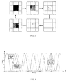
- FIG. 1 depicts a schematic representation of laterally oscillating movement function
- FIG. 2 depicts a schematic representation of pulsating movement function
- FIG. 3 depicts a schematic representation of temporal movement function with pulsating intensity
- FIG. 4 depicts a graphical representation of a phase shift of two pixels with slightly different intensities
- FIG. 5 depicts a light intensity phase shift of two pixels with slightly different intensities
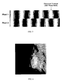
- FIG. 6 is an image of an exemplary mammogram
- FIGS. 7A-7I depicts a sequence of frames of the mammogram of FIG. 6 generated using the method of the present invention
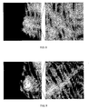
- FIG. 8 is a zoomed in image of the mammogram of FIG. 6;
- FIG. 9 is a zoomed in image of the generated frame of FIG. 7F;
- FIG. 10 depicts a pooled mean ROC curve for non-radiologists
- FIG. 11 depicts a pooled mean ROC curve for radiologists.
- FIG. 12 depicts a schematic diagram of a computer system for implementing the method of the present invention.
- the present invention provides a method for transforming a static image into a movie to aid in the detection and to identify objects and features in images.
- this movie the observer sees motion or flicker which depends on the features of the static image.
- the system receives as input static images and generates as output a movie containing the dynamic cues.
- Spatial motion can be understood as the name implies, as objects moving in space throughout the frames.
- the objects to be moved will be the pixels (picture el ements).
- temporal motion refers to a pixel not moving in space but changing its intensity in time.
- motion function which expresses mathematically the kind of movement that will affect the pixels.
- the characteristics of the motion depend on the data through what we call the "observation function”.
- An exemplary method of introducing this artificial movement to images is based upon two functions: the motion function f mov and the observation function f obs .
- the motion function specifies the type of movement while the observation function sets the parameters for this motion.
- I k ( i, j ) is a discrete sequence of m ⁇ n pixels dependent on the motion function f mov () which defines the movement of the pixels through the sequence of frames.
- This movement can be spatial or temporal, meaning that a pixel can vary its spatial location or its intensity in time.
- FIG. 1 illustrates a possible implementation of spatial movement as a sideway oscillation of every pixel at constant amplitude and a frequency that depends on each pixels original intensity. In this way a light pixel may oscillate from one side to the other faster than a darker pixel.
- FIG. 2 Another way of introducing spatial motion is a pulsating motion as shown in FIG. 2. As with lateral oscillation, the frequency of pulsations depends on the original intensity of each pixel.
- I k ( i, j ) represents the intensity of the pixel of coordinates ( i, j ) in the k th frame
- f DC () is an offset for every pixel
- a is the amplitude of the sine wave
- f 0 is its fundamental frequency
- f obs () is the observation function and ⁇ the phase shift.
- the rate of change of position (oscillating movement), size (pulsating movement) or gray level (pulsating intensity) can be varied according to different functions.
- Equation 2 implements a sine wave.
- FIG. 5 shows the same two pixels sinusoidally changing intensities from white to black with frequencies proportional to their original intensity. Both pixels start out with a slightly different gray intensity, which is not perceptually visible. After a short while the phase shift becomes clear as one pixel exhibits a faster cycle than the other.
- this is similar to dynamically changing image contrast, which by itself is a much used tool in medical imaging.
- the viewer is presented with a sequence of images with sinusoidally changing contrast.
- the sequence of medical images is provided for illustrative purposes only, and is not intend to limit the scope or purview of the invention.
- the sequence of real medical images is comprised of 30 frames generated from a static digital mammogram shown in FIG. 6 using a sinusoidally pulsating intensity motion function and an Unsharp Mask observation function.
- FIG. 7A-7I shows samples of this sequence.
- the mammogram presented here contains a malignant cluster of microcalcifications in the central right section barely visible in the original image, shown in FIG. 7A. Nevertheless, in some of the subsequent frames it becomes fairly visible. In this particular example this holds true especially for frame 15, as shown in FIG. 7F.
- the original image and frame 15 are shown in FIGS. 8 and 9 respectively.
- a perfect diagnosis would have an associated ROC curve consistent of a vertical line connecting the points (0, 0) and (0, 1) and a horizontal line continuing from (0, 1) on to (1, 1). The area underneath this curve equals 1. Every other test in between would have an AUC somewhere between 0.5 and 1. The higher the AUC, the better is the test.
- n y 124 mammograms digitized at 200 micron pixel edge with resolutions between 640 ⁇ 640 and 970 ⁇ 970 pixels per image and a gray-level resolution of 8 bits per pixel taken from the Mammographic Image Analysis Society (MIAS) mammographic database was prepared.
- MIAS Mammographic Image Analysis Society
- the test was performed with five radiologists and five non-radiologists.
- the resulting bi-normally fitted ROC curves and the associated AUC scores were computed using the ROCKIT program proposed by Metz et al.
- two different averaging techniques were used on both radiologist and non-radiologist data sets. The first one is the simple average mean, where the bi-normally fitted vertical intercept and slope values are averaged and a curve is calculated from the resulting values. This method tends to overestimate the results, but still lies within the extremes of the individual results.
- Vit Drga The Theory of Group Operating Characteristic Analysis in Discrimination Tasks, PhD thesis, Victoria University of Wellington, New Zealand, 1999 . This method usually tends to underestimate the results, but since it underestimates both test categories it is still well suited for comparison purposes.
- Tables 1 and 2 show significant improvement in diagnosis for both experts and nonexperts.
- the 8.4% improvement for radiologist medical doctors indicates vast possibilities of improvement of diagnosis if perfected and clinically implemented as a standard visualization tool for radiologists. It also has to be considered that in these tests, these medical doctors had ample experience in traditional static mammography analysis, but had never before seen dynamic cues in this kind of application.
- All the training provided before the test was a training set consisting of four images with and four images without microcalcifications viewed using dynamic cues. It is a clear indication of the potential of dynamic cues that results turned out that positive in spite of short training.
- the motion and observation function i.e. temporal pulsating intensity motion function and Unsharp Mask observation function are exemplary functions.
- Other implemented motion functions can include laterally oscillating functions as shown in FIG. 1 and spatially pulsating functions as shown in FIG. 2.
- Other observation functions can include the identity matrix and wavelet based transformations.
- the wavelet transform based methods tested here were adapted versions of the algorithms proposed by Wang et al. Ted C. Wang and Nicolaos B. Karayiannis. Detection of microcalcifications in digital mammograms using wavelets. IEEE Transactions on Medical Imaging, 17(4):498-509, August 1988 .
- a strength of the dynamic cues method lies in its use of the highly evolved human brain functions already present, but not used in the context of image analysis, without discarding traditional static two-dimensional brain functions. In effect, it uses brain functions every mammal is highly trained for movement.
- FIG.12 is a block diagram of a computer system useful for implementing the methods of the present invention.
- the computer system includes one or more processors, such as processor 100.
- the processor 100 is connected to a communication infrastructure 102 (e.g., a communications bus, cross-over bar, or network).
- a communication infrastructure 102 e.g., a communications bus, cross-over bar, or network.
- the computer system can include a display interface 104 that forwards graphics, text, and other data from the communication infrastructure 102 (or from a frame buffer not shown) for display on the display unit 106.
- the computer system also includes a main memory 108, preferably random access memory (RAM), and may also include a secondary memory 110.
- the secondary memory 110 may include, for example, a hard disk drive 112 and/or a removable storage drive 114, representing a floppy disk drive, a magnetic tape drive, an optical disk drive, etc.
- the removable storage drive 114 reads from and/or writes to a removable storage unit 116 in a manner well known to those having ordinary skill in the art.
- Removable storage unit 116 represents a floppy disk, magnetic tape, optical disk, etc. which is read by and written to by removable storage drive 114.
- the removable storage unit 116 includes a computer usable storage medium having stored therein computer software and/or data.
- the secondary memory 110 may include other similar means for allowing computer programs or other instructions to be loaded into the computer system.
- Such means may include, for example, a removable storage unit 118 and an interface 120. Examples of such may include a program cartridge and cartridge interface (such as that found in video game devices), a removable memory chip (such as an EPROM, or PROM) and associated socket, and other removable storage units 118 and interfaces 120 which allow software and data to be transferred from the removable storage unit 118 to the computer system.
- the computer system may also include a communications interface 122.
- Communications interface 122 allows software and data to be transferred between the computer system and external devices. Examples of communications interface 122 may include a modem, a network interface (such as an Ethernet card), a communications port, a PCMCIA slot and card, etc.
- Software and data transferred via communications interface 122 are in the form of signals which may be, for example, electronic, electromagnetic, optical, or other signals capable of being received by communications interface 122. These signals are provided to communications interface 122 via a communications path (i.e., channel) 124.
- This channel 124 carries signals and may be implemented using wire or cable, fiber optics, a phone line, a cellular phone link, an RF link, and/or other communications channels.
- the terms "computer program medium,” “computer usable medium,” and “computer readable medium” are used to generally refer to media such as main memory 108 and secondary memory 110, removable storage drive 114, a hard disk installed in hard disk drive 112, and signals. These computer program products are means for providing software to the computer system.
- the computer readable medium allows the computer system to read data, instructions, messages or message packets, and other computer readable information from the computer readable medium.
- the computer readable medium may include nonvolatile memory, such as Floppy, ROM, Flash memory, Disk drive memory, CD-ROM, and other permanent storage. It is useful, for example, for transporting information, such as data and computer instructions, between computer systems.
- the computer readable medium may comprise computer readable information in a transitory state medium such as a network link and/or a network interface, including a wired network or a wireless network, that allow a computer to read such computer readable information.
- Computer programs are stored in main memory 108 and/or secondary memory 110. Computer programs may also be received via communications interface 122. Such computer programs, when executed, enable the computer system to perform the features of the present invention as discussed herein. In particular, the computer programs, when executed, enable the processor 100 to perform the features of the computer system. Accordingly, such computer programs represent controllers of the computer system.
Landscapes
- Physics & Mathematics (AREA)
- General Physics & Mathematics (AREA)
- Engineering & Computer Science (AREA)
- Theoretical Computer Science (AREA)
- Image Processing (AREA)
- Apparatus For Radiation Diagnosis (AREA)
- Image Analysis (AREA)
Applications Claiming Priority (1)
| Application Number | Priority Date | Filing Date | Title |
|---|---|---|---|
| US11/469,921 US20080056548A1 (en) | 2006-09-05 | 2006-09-05 | Enhancement of visual perception through dynamic cues |
Publications (2)
| Publication Number | Publication Date |
|---|---|
| EP1898359A2 true EP1898359A2 (de) | 2008-03-12 |
| EP1898359A3 EP1898359A3 (de) | 2008-04-30 |
Family
ID=38830980
Family Applications (1)
| Application Number | Title | Priority Date | Filing Date |
|---|---|---|---|
| EP07017191A Withdrawn EP1898359A3 (de) | 2006-09-05 | 2007-09-03 | Verbesserung der visuellen Wahrnehmung durch dynamische Hinweise |
Country Status (3)
| Country | Link |
|---|---|
| US (1) | US20080056548A1 (de) |
| EP (1) | EP1898359A3 (de) |
| JP (1) | JP2008173447A (de) |
Cited By (1)
| Publication number | Priority date | Publication date | Assignee | Title |
|---|---|---|---|---|
| WO2013185787A1 (en) * | 2012-06-15 | 2013-12-19 | Imcube Labs Gmbh | Apparatus and method for compositing an image from a number of visual objects |
Families Citing this family (8)
| Publication number | Priority date | Publication date | Assignee | Title |
|---|---|---|---|---|
| US8452599B2 (en) | 2009-06-10 | 2013-05-28 | Toyota Motor Engineering & Manufacturing North America, Inc. | Method and system for extracting messages |
| US8269616B2 (en) * | 2009-07-16 | 2012-09-18 | Toyota Motor Engineering & Manufacturing North America, Inc. | Method and system for detecting gaps between objects |
| US8337160B2 (en) * | 2009-10-19 | 2012-12-25 | Toyota Motor Engineering & Manufacturing North America, Inc. | High efficiency turbine system |
| US8237792B2 (en) * | 2009-12-18 | 2012-08-07 | Toyota Motor Engineering & Manufacturing North America, Inc. | Method and system for describing and organizing image data |
| US8424621B2 (en) | 2010-07-23 | 2013-04-23 | Toyota Motor Engineering & Manufacturing North America, Inc. | Omni traction wheel system and methods of operating the same |
| US9691289B2 (en) * | 2010-12-22 | 2017-06-27 | Brightstar Learning | Monotonous game-like task to promote effortless automatic recognition of sight words |
| RU2014149357A (ru) * | 2012-05-09 | 2016-06-27 | Конинклейке Филипс Н.В. | Устройство и способ для поддержки изменения поведения человека |
| WO2014210430A1 (en) | 2013-06-27 | 2014-12-31 | Tractus Corporation | Systems and methods for tissue mapping |
Family Cites Families (38)
| Publication number | Priority date | Publication date | Assignee | Title |
|---|---|---|---|---|
| US5488952A (en) * | 1982-02-24 | 1996-02-06 | Schoolman Scientific Corp. | Stereoscopically display three dimensional ultrasound imaging |
| US5040225A (en) * | 1987-12-07 | 1991-08-13 | Gdp, Inc. | Image analysis method |
| US5287096A (en) * | 1989-02-27 | 1994-02-15 | Texas Instruments Incorporated | Variable luminosity display system |
| US5845639A (en) * | 1990-08-10 | 1998-12-08 | Board Of Regents Of The University Of Washington | Optical imaging methods |
| US5465718A (en) * | 1990-08-10 | 1995-11-14 | Hochman; Daryl | Solid tumor, cortical function, and nerve tissue imaging methods and device |
| US5590268A (en) * | 1993-03-31 | 1996-12-31 | Kabushiki Kaisha Toshiba | System and method for evaluating a workspace represented by a three-dimensional model |
| JP3380609B2 (ja) * | 1993-12-24 | 2003-02-24 | コニカ株式会社 | 放射線画像の照射野領域抽出装置 |
| US5592085A (en) * | 1994-10-19 | 1997-01-07 | Mayo Foundation For Medical Education And Research | MR imaging of synchronous spin motion and strain waves |
| US6037774A (en) * | 1994-10-19 | 2000-03-14 | Mayo Foundation For Medical Education And Research | Inertial driver device for MR elastography |
| US5919139A (en) * | 1997-12-19 | 1999-07-06 | Diasonics Ultrasound | Vibrational doppler ultrasonic imaging |
| US6549801B1 (en) * | 1998-06-11 | 2003-04-15 | The Regents Of The University Of California | Phase-resolved optical coherence tomography and optical doppler tomography for imaging fluid flow in tissue with fast scanning speed and high velocity sensitivity |
| US6362850B1 (en) * | 1998-08-04 | 2002-03-26 | Flashpoint Technology, Inc. | Interactive movie creation from one or more still images in a digital imaging device |
| US6112112A (en) * | 1998-09-18 | 2000-08-29 | Arch Development Corporation | Method and system for the assessment of tumor extent in magnetic resonance images |
| US6283917B1 (en) * | 1998-10-01 | 2001-09-04 | Atl Ultrasound | Ultrasonic diagnostic imaging system with blurring corrected spatial compounding |
| US6117081A (en) * | 1998-10-01 | 2000-09-12 | Atl Ultrasound, Inc. | Method for correcting blurring of spatially compounded ultrasonic diagnostic images |
| US6277074B1 (en) * | 1998-10-02 | 2001-08-21 | University Of Kansas Medical Center | Method and apparatus for motion estimation within biological tissue |
| US6068597A (en) * | 1999-04-13 | 2000-05-30 | Lin; Gregory Sharat | Vibrational resonance ultrasonic Doppler spectrometer and imager |
| US6611274B1 (en) * | 1999-10-12 | 2003-08-26 | Microsoft Corporation | System method, and computer program product for compositing true colors and intensity-maped colors into a frame buffer |
| US6511427B1 (en) * | 2000-03-10 | 2003-01-28 | Acuson Corporation | System and method for assessing body-tissue properties using a medical ultrasound transducer probe with a body-tissue parameter measurement mechanism |
| US6583624B1 (en) * | 2000-06-15 | 2003-06-24 | Mayo Foundation For Medical Education | Method for visualizing charged particle motion using magnetic resonance imaging |
| US6473634B1 (en) * | 2000-11-22 | 2002-10-29 | Koninklijke Philips Electronics N.V. | Medical imaging at two temporal resolutions for tumor treatment planning |
| US6558324B1 (en) * | 2000-11-22 | 2003-05-06 | Siemens Medical Solutions, Inc., Usa | System and method for strain image display |
| US20060079773A1 (en) * | 2000-11-28 | 2006-04-13 | Allez Physionix Limited | Systems and methods for making non-invasive physiological assessments by detecting induced acoustic emissions |
| US6804384B2 (en) * | 2001-06-12 | 2004-10-12 | Mclean Hospital Corporation | Color magnetic resonance imaging |
| US7117026B2 (en) * | 2002-06-12 | 2006-10-03 | Koninklijke Philips Electronics N.V. | Physiological model based non-rigid image registration |
| JP3675796B2 (ja) * | 2002-08-05 | 2005-07-27 | コーリンメディカルテクノロジー株式会社 | 血圧測定装置 |
| US6659953B1 (en) * | 2002-09-20 | 2003-12-09 | Acuson Corporation | Morphing diagnostic ultrasound images for perfusion assessment |
| CN1685354A (zh) * | 2002-09-24 | 2005-10-19 | 伊斯曼柯达公司 | 用于计算机辅助检测提示阅读医疗图像的方法和系统 |
| JP3932482B2 (ja) * | 2002-10-18 | 2007-06-20 | 株式会社日立メディコ | 超音波診断装置 |
| JP4474106B2 (ja) * | 2003-02-27 | 2010-06-02 | キヤノン株式会社 | 画像処理装置、画像処理方法、記録媒体及びプログラム |
| JP4380176B2 (ja) * | 2003-02-28 | 2009-12-09 | コニカミノルタホールディングス株式会社 | 医用画像処理装置及び異常陰影候補の検出結果の表示方法 |
| AU2004203173A1 (en) * | 2003-07-14 | 2005-02-03 | Sunnybrook And Women's College And Health Sciences Centre | Optical image-based position tracking for magnetic resonance imaging |
| CA2457376C (en) * | 2003-10-14 | 2015-09-15 | The University Of British Columbia | Method for imaging the mechanical properties of tissue |
| US20050228271A1 (en) * | 2004-04-06 | 2005-10-13 | Diebold Gerald G | Differential x-ray acoustic imaging |
| US7555151B2 (en) * | 2004-09-02 | 2009-06-30 | Siemens Medical Solutions Usa, Inc. | System and method for tracking anatomical structures in three dimensional images |
| US8989349B2 (en) * | 2004-09-30 | 2015-03-24 | Accuray, Inc. | Dynamic tracking of moving targets |
| US7627386B2 (en) * | 2004-10-07 | 2009-12-01 | Zonaire Medical Systems, Inc. | Ultrasound imaging system parameter optimization via fuzzy logic |
| US7563228B2 (en) * | 2005-01-24 | 2009-07-21 | Siemens Medical Solutions Usa, Inc. | Stereoscopic three or four dimensional ultrasound imaging |
-
2006
- 2006-09-05 US US11/469,921 patent/US20080056548A1/en not_active Abandoned
-
2007
- 2007-09-03 EP EP07017191A patent/EP1898359A3/de not_active Withdrawn
- 2007-09-04 JP JP2007228990A patent/JP2008173447A/ja active Pending
Non-Patent Citations (1)
| Title |
|---|
| HERRMANN A. ET AL: "Oscillating intensity display of soft tissue lesions in MRI", IEEE TRANSACTIONS ON MEDICAL IMAGING USA, vol. MI-6, no. 4, December 1987 (1987-12-01), pages 370 - 373, ISSN: 0278-0062 * |
Cited By (1)
| Publication number | Priority date | Publication date | Assignee | Title |
|---|---|---|---|---|
| WO2013185787A1 (en) * | 2012-06-15 | 2013-12-19 | Imcube Labs Gmbh | Apparatus and method for compositing an image from a number of visual objects |
Also Published As
| Publication number | Publication date |
|---|---|
| EP1898359A3 (de) | 2008-04-30 |
| US20080056548A1 (en) | 2008-03-06 |
| JP2008173447A (ja) | 2008-07-31 |
Similar Documents
| Publication | Publication Date | Title |
|---|---|---|
| US11983799B2 (en) | System and method for synthesizing low-dimensional image data from high-dimensional image data using an object grid enhancement | |
| EP2158575B1 (de) | Erkennung einer hirnblutung in ct-bilddaten | |
| US7680307B2 (en) | Systems and methods for image segmentation with a multi-stage classifier | |
| Niemeijer et al. | Image structure clustering for image quality verification of color retina images in diabetic retinopathy screening | |
| US7599542B2 (en) | System and method for detection and display of diseases and abnormalities using confidence imaging | |
| US6246782B1 (en) | System for automated detection of cancerous masses in mammograms | |
| US9275465B2 (en) | System for preparing an image for segmentation | |
| JP6877942B2 (ja) | 医用画像処理装置及び医用画像処理プログラム | |
| JP2008173447A (ja) | 動的キューを通しての視覚的知覚の向上 | |
| US8031917B2 (en) | System and method for smart display of CAD markers | |
| EP3232936A1 (de) | Verfahren und system zur verarbeitung von tomosynthesedaten | |
| US20060280348A1 (en) | Method of screening cellular tissue | |
| WO2009122724A1 (ja) | 画像処理装置および方法並びにプログラム | |
| US9514548B2 (en) | Method and system for presenting and using four dimensional data from a medical imaging system | |
| Mall et al. | Fixated and not fixated regions of mammograms: a higher-order statistical analysis of visual search behavior | |
| Adillion et al. | Line operator as preprocessing method for cnn-based osteoporosis detection in dental panoramic radiograph | |
| US6421469B1 (en) | Image data manipulation for improved image visualization and analysis | |
| Bandyopadhyay | Pre-processing and segmentation of brain image for tumor detection | |
| Plett et al. | Enhancement of visual perception through dynamic cues: An application to mammograms | |
| US12450738B2 (en) | Automated multi-class segmentation of digital mammogram | |
| Plessis et al. | Context-dependent enhancements for radiological images | |
| Myers et al. | Human and quasi-Bayesian observers of images limited by quantum noise, object-variability, and artifacts | |
| Devanna et al. | Analysis and applications of modified non-subsampled contourlet transform | |
| Apelt et al. | Contrast sensitivity in mammographic softcopy reading | |
| Mishra et al. | Dyadic wavelet transform based classification of microcalcifications using SVM |
Legal Events
| Date | Code | Title | Description |
|---|---|---|---|
| PUAI | Public reference made under article 153(3) epc to a published international application that has entered the european phase |
Free format text: ORIGINAL CODE: 0009012 |
|
| AK | Designated contracting states |
Kind code of ref document: A2 Designated state(s): AT BE BG CH CY CZ DE DK EE ES FI FR GB GR HU IE IS IT LI LT LU LV MC MT NL PL PT RO SE SI SK TR |
|
| AX | Request for extension of the european patent |
Extension state: AL BA HR MK YU |
|
| PUAL | Search report despatched |
Free format text: ORIGINAL CODE: 0009013 |
|
| AK | Designated contracting states |
Kind code of ref document: A3 Designated state(s): AT BE BG CH CY CZ DE DK EE ES FI FR GB GR HU IE IS IT LI LT LU LV MC MT NL PL PT RO SE SI SK TR |
|
| AX | Request for extension of the european patent |
Extension state: AL BA HR MK RS |
|
| RIC1 | Information provided on ipc code assigned before grant |
Ipc: G06T 5/00 20060101AFI20080327BHEP |
|
| 17P | Request for examination filed |
Effective date: 20081021 |
|
| 17Q | First examination report despatched |
Effective date: 20081121 |
|
| AKX | Designation fees paid |
Designated state(s): AT BE BG CH CY CZ DE DK EE ES FI FR GB GR HU IE IS IT LI LT LU LV MC MT NL PL PT RO SE SI SK TR |
|
| STAA | Information on the status of an ep patent application or granted ep patent |
Free format text: STATUS: THE APPLICATION IS DEEMED TO BE WITHDRAWN |
|
| 18D | Application deemed to be withdrawn |
Effective date: 20120211 |