EP1890587B1 - Protective cap for arthroscopic instruments - Google Patents
Protective cap for arthroscopic instruments Download PDFInfo
- Publication number
- EP1890587B1 EP1890587B1 EP06760606.1A EP06760606A EP1890587B1 EP 1890587 B1 EP1890587 B1 EP 1890587B1 EP 06760606 A EP06760606 A EP 06760606A EP 1890587 B1 EP1890587 B1 EP 1890587B1
- Authority
- EP
- European Patent Office
- Prior art keywords
- cap
- sheath
- arthroscope
- disposed
- instrument
- Prior art date
- Legal status (The legal status is an assumption and is not a legal conclusion. Google has not performed a legal analysis and makes no representation as to the accuracy of the status listed.)
- Active
Links
- 230000001681 protective effect Effects 0.000 title description 15
- 239000012530 fluid Substances 0.000 claims description 23
- 238000004891 communication Methods 0.000 claims description 11
- 230000003287 optical effect Effects 0.000 claims description 9
- 238000001356 surgical procedure Methods 0.000 description 18
- 230000006378 damage Effects 0.000 description 10
- 239000000463 material Substances 0.000 description 10
- 210000001519 tissue Anatomy 0.000 description 10
- 238000000034 method Methods 0.000 description 9
- 239000000835 fiber Substances 0.000 description 7
- 238000009966 trimming Methods 0.000 description 7
- 238000005286 illumination Methods 0.000 description 4
- 230000002262 irrigation Effects 0.000 description 4
- 238000003973 irrigation Methods 0.000 description 4
- 238000003384 imaging method Methods 0.000 description 3
- 208000014674 injury Diseases 0.000 description 3
- 238000004519 manufacturing process Methods 0.000 description 3
- 229920001634 Copolyester Polymers 0.000 description 2
- PPBRXRYQALVLMV-UHFFFAOYSA-N Styrene Chemical compound C=CC1=CC=CC=C1 PPBRXRYQALVLMV-UHFFFAOYSA-N 0.000 description 2
- 210000000988 bone and bone Anatomy 0.000 description 2
- 210000000845 cartilage Anatomy 0.000 description 2
- 239000003814 drug Substances 0.000 description 2
- 229920001971 elastomer Polymers 0.000 description 2
- 239000000806 elastomer Substances 0.000 description 2
- 238000003780 insertion Methods 0.000 description 2
- 230000037431 insertion Effects 0.000 description 2
- 210000003127 knee Anatomy 0.000 description 2
- 229920001296 polysiloxane Polymers 0.000 description 2
- 239000000523 sample Substances 0.000 description 2
- 229920006132 styrene block copolymer Polymers 0.000 description 2
- 229940124597 therapeutic agent Drugs 0.000 description 2
- 229920002397 thermoplastic olefin Polymers 0.000 description 2
- 229920002803 thermoplastic polyurethane Polymers 0.000 description 2
- 230000008733 trauma Effects 0.000 description 2
- 108700028490 CAP protocol 2 Proteins 0.000 description 1
- JOYRKODLDBILNP-UHFFFAOYSA-N Ethyl urethane Chemical compound CCOC(N)=O JOYRKODLDBILNP-UHFFFAOYSA-N 0.000 description 1
- FAPWRFPIFSIZLT-UHFFFAOYSA-M Sodium chloride Chemical compound [Na+].[Cl-] FAPWRFPIFSIZLT-UHFFFAOYSA-M 0.000 description 1
- 229910000831 Steel Inorganic materials 0.000 description 1
- 239000004433 Thermoplastic polyurethane Substances 0.000 description 1
- 229920004482 WACKER® Polymers 0.000 description 1
- 208000027418 Wounds and injury Diseases 0.000 description 1
- NIXOWILDQLNWCW-UHFFFAOYSA-N acrylic acid group Chemical group C(C=C)(=O)O NIXOWILDQLNWCW-UHFFFAOYSA-N 0.000 description 1
- 239000000853 adhesive Substances 0.000 description 1
- 230000001070 adhesive effect Effects 0.000 description 1
- 229910052782 aluminium Inorganic materials 0.000 description 1
- XAGFODPZIPBFFR-UHFFFAOYSA-N aluminium Chemical compound [Al] XAGFODPZIPBFFR-UHFFFAOYSA-N 0.000 description 1
- 210000003484 anatomy Anatomy 0.000 description 1
- 210000001264 anterior cruciate ligament Anatomy 0.000 description 1
- 239000008280 blood Substances 0.000 description 1
- 210000004369 blood Anatomy 0.000 description 1
- 239000003086 colorant Substances 0.000 description 1
- 238000005520 cutting process Methods 0.000 description 1
- 230000001934 delay Effects 0.000 description 1
- 210000002082 fibula Anatomy 0.000 description 1
- 238000001914 filtration Methods 0.000 description 1
- 238000002347 injection Methods 0.000 description 1
- 239000007924 injection Substances 0.000 description 1
- 238000007689 inspection Methods 0.000 description 1
- 239000004816 latex Substances 0.000 description 1
- 229920000126 latex Polymers 0.000 description 1
- 230000005499 meniscus Effects 0.000 description 1
- 229910052751 metal Inorganic materials 0.000 description 1
- 239000002184 metal Substances 0.000 description 1
- HLXZNVUGXRDIFK-UHFFFAOYSA-N nickel titanium Chemical compound [Ti].[Ti].[Ti].[Ti].[Ti].[Ti].[Ti].[Ti].[Ti].[Ti].[Ti].[Ni].[Ni].[Ni].[Ni].[Ni].[Ni].[Ni].[Ni].[Ni].[Ni].[Ni].[Ni].[Ni].[Ni] HLXZNVUGXRDIFK-UHFFFAOYSA-N 0.000 description 1
- 229910001000 nickel titanium Inorganic materials 0.000 description 1
- 210000004417 patella Anatomy 0.000 description 1
- 230000007170 pathology Effects 0.000 description 1
- 238000002428 photodynamic therapy Methods 0.000 description 1
- 229920000515 polycarbonate Polymers 0.000 description 1
- 239000004417 polycarbonate Substances 0.000 description 1
- 229920000098 polyolefin Polymers 0.000 description 1
- 210000002967 posterior cruciate ligament Anatomy 0.000 description 1
- 230000000717 retained effect Effects 0.000 description 1
- 239000012781 shape memory material Substances 0.000 description 1
- 238000004904 shortening Methods 0.000 description 1
- 229910052710 silicon Inorganic materials 0.000 description 1
- 239000010703 silicon Substances 0.000 description 1
- 239000011780 sodium chloride Substances 0.000 description 1
- 210000004872 soft tissue Anatomy 0.000 description 1
- 239000010959 steel Substances 0.000 description 1
- 230000001225 therapeutic effect Effects 0.000 description 1
- 229920001169 thermoplastic Polymers 0.000 description 1
- 229920002725 thermoplastic elastomer Polymers 0.000 description 1
- 229920001187 thermosetting polymer Polymers 0.000 description 1
- 239000004416 thermosoftening plastic Substances 0.000 description 1
- 210000002303 tibia Anatomy 0.000 description 1
- 210000000689 upper leg Anatomy 0.000 description 1
- XLYOFNOQVPJJNP-UHFFFAOYSA-N water Substances O XLYOFNOQVPJJNP-UHFFFAOYSA-N 0.000 description 1
Images


Classifications
-
- A—HUMAN NECESSITIES
- A61—MEDICAL OR VETERINARY SCIENCE; HYGIENE
- A61B—DIAGNOSIS; SURGERY; IDENTIFICATION
- A61B1/00—Instruments for performing medical examinations of the interior of cavities or tubes of the body by visual or photographical inspection, e.g. endoscopes; Illuminating arrangements therefor
- A61B1/313—Instruments for performing medical examinations of the interior of cavities or tubes of the body by visual or photographical inspection, e.g. endoscopes; Illuminating arrangements therefor for introducing through surgical openings, e.g. laparoscopes
- A61B1/317—Instruments for performing medical examinations of the interior of cavities or tubes of the body by visual or photographical inspection, e.g. endoscopes; Illuminating arrangements therefor for introducing through surgical openings, e.g. laparoscopes for bones or joints, e.g. osteoscopes, arthroscopes
-
- A—HUMAN NECESSITIES
- A61—MEDICAL OR VETERINARY SCIENCE; HYGIENE
- A61B—DIAGNOSIS; SURGERY; IDENTIFICATION
- A61B1/00—Instruments for performing medical examinations of the interior of cavities or tubes of the body by visual or photographical inspection, e.g. endoscopes; Illuminating arrangements therefor
- A61B1/00064—Constructional details of the endoscope body
- A61B1/00071—Insertion part of the endoscope body
- A61B1/0008—Insertion part of the endoscope body characterised by distal tip features
- A61B1/00096—Optical elements
-
- A—HUMAN NECESSITIES
- A61—MEDICAL OR VETERINARY SCIENCE; HYGIENE
- A61B—DIAGNOSIS; SURGERY; IDENTIFICATION
- A61B1/00—Instruments for performing medical examinations of the interior of cavities or tubes of the body by visual or photographical inspection, e.g. endoscopes; Illuminating arrangements therefor
- A61B1/00064—Constructional details of the endoscope body
- A61B1/00071—Insertion part of the endoscope body
- A61B1/0008—Insertion part of the endoscope body characterised by distal tip features
- A61B1/00101—Insertion part of the endoscope body characterised by distal tip features the distal tip features being detachable
-
- A—HUMAN NECESSITIES
- A61—MEDICAL OR VETERINARY SCIENCE; HYGIENE
- A61B—DIAGNOSIS; SURGERY; IDENTIFICATION
- A61B1/00—Instruments for performing medical examinations of the interior of cavities or tubes of the body by visual or photographical inspection, e.g. endoscopes; Illuminating arrangements therefor
- A61B1/00131—Accessories for endoscopes
- A61B1/00135—Oversleeves mounted on the endoscope prior to insertion
-
- A—HUMAN NECESSITIES
- A61—MEDICAL OR VETERINARY SCIENCE; HYGIENE
- A61B—DIAGNOSIS; SURGERY; IDENTIFICATION
- A61B1/00—Instruments for performing medical examinations of the interior of cavities or tubes of the body by visual or photographical inspection, e.g. endoscopes; Illuminating arrangements therefor
- A61B1/00131—Accessories for endoscopes
- A61B1/00137—End pieces at either end of the endoscope, e.g. caps, seals or forceps plugs
-
- A—HUMAN NECESSITIES
- A61—MEDICAL OR VETERINARY SCIENCE; HYGIENE
- A61B—DIAGNOSIS; SURGERY; IDENTIFICATION
- A61B1/00—Instruments for performing medical examinations of the interior of cavities or tubes of the body by visual or photographical inspection, e.g. endoscopes; Illuminating arrangements therefor
- A61B1/00142—Instruments for performing medical examinations of the interior of cavities or tubes of the body by visual or photographical inspection, e.g. endoscopes; Illuminating arrangements therefor with means for preventing contamination, e.g. by using a sanitary sheath
-
- A—HUMAN NECESSITIES
- A61—MEDICAL OR VETERINARY SCIENCE; HYGIENE
- A61B—DIAGNOSIS; SURGERY; IDENTIFICATION
- A61B1/00—Instruments for performing medical examinations of the interior of cavities or tubes of the body by visual or photographical inspection, e.g. endoscopes; Illuminating arrangements therefor
- A61B1/012—Instruments for performing medical examinations of the interior of cavities or tubes of the body by visual or photographical inspection, e.g. endoscopes; Illuminating arrangements therefor characterised by internal passages or accessories therefor
-
- A—HUMAN NECESSITIES
- A61—MEDICAL OR VETERINARY SCIENCE; HYGIENE
- A61B—DIAGNOSIS; SURGERY; IDENTIFICATION
- A61B1/00—Instruments for performing medical examinations of the interior of cavities or tubes of the body by visual or photographical inspection, e.g. endoscopes; Illuminating arrangements therefor
- A61B1/00064—Constructional details of the endoscope body
- A61B1/00071—Insertion part of the endoscope body
- A61B1/0008—Insertion part of the endoscope body characterised by distal tip features
- A61B1/00089—Hoods
-
- A—HUMAN NECESSITIES
- A61—MEDICAL OR VETERINARY SCIENCE; HYGIENE
- A61B—DIAGNOSIS; SURGERY; IDENTIFICATION
- A61B1/00—Instruments for performing medical examinations of the interior of cavities or tubes of the body by visual or photographical inspection, e.g. endoscopes; Illuminating arrangements therefor
- A61B1/00163—Optical arrangements
- A61B1/00174—Optical arrangements characterised by the viewing angles
- A61B1/00177—Optical arrangements characterised by the viewing angles for 90 degrees side-viewing
-
- A—HUMAN NECESSITIES
- A61—MEDICAL OR VETERINARY SCIENCE; HYGIENE
- A61B—DIAGNOSIS; SURGERY; IDENTIFICATION
- A61B1/00—Instruments for performing medical examinations of the interior of cavities or tubes of the body by visual or photographical inspection, e.g. endoscopes; Illuminating arrangements therefor
- A61B1/00163—Optical arrangements
- A61B1/00174—Optical arrangements characterised by the viewing angles
- A61B1/00183—Optical arrangements characterised by the viewing angles for variable viewing angles
Definitions
- the inventions described below relate the field of arthroscopic surgical instruments.
- Arthroscopic surgery involves using optical instruments, such as an arthroscope, to visualize an operating field inside or near a joint of a patient.
- the same instrument or other instruments may be used to perform a surgical procedure in the operating field.
- Common instruments used in addition to the arthroscope include a trimming instrument for cutting tissue and an irrigation instrument for irrigating the surgical field.
- Each of the instruments requires its own incision to be introduced into the surgical field; thus, many surgeons prefer to use only a trimming instrument and an arthroscope during arthroscopic surgical procedures.
- Arthroscopes are fragile in relation to the forces applied during arthroscopic surgery, so a rigid cannula is placed over the arthroscope to reinforce it.
- the distal end of the rigid cannula is pointed, usually sharp, and so the rigid cannula can scratch or gouge soft tissue within the operating field.
- the rigid cannula can also become stuck between bones or cartilage during a procedure.
- a rigid cannula can also damage metal prosthetics used to replace joints, resulting in a shortening of the useful life of the prosthetic and forcing the patient to undergo additional, painful surgeries to correct the problem.
- An additional problem associated with arthroscopic surgery is maintaining a clear surgical field during surgery. Blood and debris can cloud the field, impairing a surgeon's ability to visualize tissue.
- One method of solving this problem is to use the irrigation instrument to clear the surgical field with saline; however, many surgeons strongly prefer to avoid the additional trauma caused by inserting a third instrument. These surgeons will perform arthroscopic surgeries despite problems with visualizing the surgical field.
- a further problem associated with arthroscopic surgery is accidental damage to the arthroscope.
- the arthroscope is often damaged if the working end of a trimming instrument accidentally strikes the sensitive optical components on the distal portion of the arthroscope.
- the arthroscope may also be damaged if the arthroscope becomes stuck between bones, cartilage or other tissue and excessive force must be used to free the arthroscope.
- Arthroscopes are expensive, costing thousands of dollars, so accidental damage to arthroscopes is a significant cost problem.
- a damaged arthroscope could cause delays during surgery and broken pieces of the arthroscope could be deposited in the surgical field. Both situations are harmful to the patient. Thus, devices and methods are needed to prevent accidental damage to arthroscopes during surgery.
- US patent 6,761,684 B1 discloses an endoscope tip protection system for shielding the optical component at the distal end of an endoscope.
- the system comprises a tubular sheath which is optically transparent at least at its distal end, and adapted to fit onto and be retained on the scope tip.
- the scope/sheath assembly is then inserted into an open ended tubular fiber optic illuminating cannula which is adapted to receive the scope/sheath assembly and prevent the tubular sheath from falling out.
- US patent US 6,095,970 A discloses an endoscope includes a insertion tube which is inserted into a human body, an imaging device, an object optical system which forms image on the imaging device, a detachable unit accommodating the imaging device and the object optical system.
- the detachable unit is detachably mounted to a mounting portion of the insertion tube.
- At least one first contact is provided to the detachable unit.
- At least one second contact is provided to the mounting portion.
- the first and second contacts are electrically connected when the detachable unit is mounted to the mounting portion.
- a waterproof arrangement is provided to prevent water from entering a connecting potion of the first and second contacts.
- German utility model DE 9215725 U1 discloses a device for the lighting and inspection of hollow and gap spaces, in particular an endoscope, with an optical carrier.
- US patent 5,735,792 A discloses a surgical instrument incorporating visualizing optics is disclosed.
- the instrument comprises a handle supporting a rigid hollow shaft, an expanded tip is located on the distal end of the shaft providing a surface that will not tear or easily penetrate tissue.
- a fiber-optic assembly containing at least one illumination transmitting fiber, a plurality of image-carrying fibers and an objective lens mounted near the distal end of the image fibers runs the length of the shaft.
- the objective lens is positioned relative to an opening in the tip allowing viewing. Light for illuminating the surgical site passes through one or more illumination fibers and through the opening in the tip of the probe.
- Light reflected from the tissue at the tip of the probe is focused onto the distal face of the image bundle by an objective lens and is then transmitted to the proximal end of the image bundle.
- Connectors on the proximal ends of the illumination fibers and image fibers facilitate their being coupled to an illumination source and a viewing device.
- the invention is defined in the claims and provides for a protective cap that is placed over the distal portion of an arthroscope.
- the cap is made of a transparent, yet durable material that prevents accidental damage to the arthroscope caused by trimming instruments or impacts with hard tissue within the surgical field. Holes may be placed in the cap to provide for the inflow and outflow of fluids from the cap.
- One or more lenses or filters may be provided within the cap to adjust the field of view as seen through the arthroscope.
- Figure 1 shows a method of performing arthroscopic surgery on a patient using an arthroscopic instrument 2 sheathed in an atraumatic introducer sheath 3.
- the various parts of the arthroscope are shown in phantom to indicate their positions inside the sheath.
- Various anatomical landmarks in a patient's knee 4 are shown for reference, including the femur 5 , patella 6 , posterior cruciate ligament 7 , anterior cruciate ligament 8 , meniscus 9 , tibia 10 and fibula 11 .
- the surgeon introduces the arthroscope 2 into the knee via a first incision 12 in order to visualize the surgical field.
- a trimming instrument 13 is introduced through a second incision 14 to remove or trim tissue that the surgeon determines should be removed or trimmed.
- an irrigating instrument 15 may be introduced through a third incision 16 in order to irrigate the surgical field and thereby maintain a clear view.
- a combined arthroscope and inflow/outflow atraumatic sheath may replace the irrigating instrument.
- the arthroscope 2 is an optical instrument 17 surrounded by a rigid cannula 18 having a distal edge that typically is cut at an angle.
- the arthroscope has been inserted into a resilient outer introducer sheath or atraumatic sheath 3 that extends over the rigid cannula.
- the distal tip 19 of the atraumatic sheath extends distally just past the distal end of the arthroscope and rigid cannula to further protect the patient.
- Figures 2 and 3 show an atraumatic protective cap 30 having a cap body 28 and a lens 29 and disposed over the distal portion 31 of an arthroscopic instrument.
- the cap body 28 is sized and dimensioned and of such a profile as to allow passage into restricted joint anatomy.
- the cap is disposed on the end of the sheath 3 and slides over the distal end of the arthroscope as the sheath is pulled over the arthroscope as shown in Figure 2 .
- the sheath is also shown in our co-pending application 10/769,629, filed January 29, 2004 , which is hereby incorporated by reference in its entirety.
- the sheath can be a tube of polymeric material sized and dimensioned to slip fit over the outer surface of a cannula or endoscope.
- the sheath may further be provided with longitudinal ribs characterizing fluid flow lumens between the outer surface of the cannula and the inner surface of the sheath.
- the cap may also be provided as a separate device from the sheath as illustrated in Figure 3 .
- the cap is preferably provided with a rounded, or bulbous, shape that reduces the chances of injuring tissue during the surgical procedure.
- the cap is optically transparent to wavelengths of light used during surgery, though only that portion of the cap covering the view port of the arthroscope need be transparent.
- the view port is that portion of the arthroscope through which the surgeon visualizes the surgical field.
- the material of the cap is sufficiently durable that the cap will prevent accidental damage to the arthroscope caused by unintended contact with the working end of a trimming instrument, by unintended contact with burrs or other hard tissue within the surgical field or because of excessive force applied to the arthroscope.
- the body of the cap may be manufactured from thermoplastic elastomers.
- styrenic block copolymers such as silicone, urethane or latex may also be used to manufacture the cap body.
- Sterilizable elastomers are typically used to make the body of the cap while optically clear polycarbonate materials comprise the viewing lens 29 in the cap 30.
- Other materials suitable for the viewing lens include molded acrylic, styrene, polyolefin or silicon.
- the body of the cap and lens can also be manufactured from the same optically clear material reducing manufacturing and assembly costs.
- suitable material includes optically clear silicone available from Wacker SiliconesTM and having a hardness ranging from approximately 30 Shore A to approximately 40 Shore D.
- One or more holes 32 may be placed in the cap to provide for suction, irrigation or the injection of therapeutic agents. Similar holes 33 may also be placed in the sheath. The holes are in fluid communication with one or more of the lumens disposed in the sheath or arthroscope or disposed between the sheath and arthroscope.
- a fluid source 34 in fluid communication with one or more lumens is provided to irrigate the surgical field or to inject therapeutic agents into the surgical field.
- a vacuum source 35 in fluid communication with one or more of the lumens provides suction.
- a manifold 36 disposed on the sheath or arthroscope distributes the flow of fluids within the sheath or arthroscope.
- the sheath and protective cap are pulled over the arthroscope.
- the surgeon then inserts the arthroscope into the surgical field and subsequently performs a surgical procedure on the patient. If, during the procedure, the working end of a trimming instrument accidentally strikes the cap, the cap will prevent damage to the arthroscope. Likewise, the cap will prevent damage to the arthroscope if the arthroscope strikes a burr or other hard piece of tissue.
- the cap is releasably attached to the sheath so that the cap may be easily replaced if damaged by these events.
- Figure 4 shows a cross section of a multi-lumen protective cap 30 and an arthroscope 2 disposed within the cap.
- One or more holes 37 extend from the outer diameter of the cap to the outer lumen 38 .
- one or more additional holes 39 extend from the outer diameter of the cap, through the outer lumen and to the inner lumen 40 of the cap.
- the holes 39 communicating with the inner lumen do not communicate with the outer lumen, thereby isolating the inner and outer lumens.
- suction may be provided through one lumen and simultaneous irrigation provided through the other lumen.
- the cap may be provided as part of a system providing fluid inflow and outflow to a surgical site where fluid inflow and outflow is accomplished by devices other than an arthroscope or arthroscope sheath.
- the cap 30 or cap body 28 shown in Figures 2 through 4 may be integrally formed with a sheath 3 , attached to a sheath 3 , or fit over a sheath 3 .
- the cap may also be provided without the sheath.
- the cap may be placed directly over the distal portion of the arthroscope without an atraumatic sheath.
- the cap can be held to the arthroscope by friction fit between the scope and the cap, by a shrink tube, by an adhesive, by detents or by any other suitable mechanism.
- the cap is removably attached so that the cap may be easily replaced.
- holes 32 may be provided to provide for the inflow and outflow of fluids.
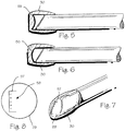
- Figure 5 shows a protective cap 30 having a concave lens 55 disposed at the distal end of the cap.
- the lens is provided just inside and proximal the distal end of the cap.
- the concave lens provides for a wide-angle view of the surgical field.
- Figure 6 shows a protective cap 30 having a convex lens 56 disposed at the distal end of the cap.
- the lens is provided just inside and proximal the distal end of the cap.
- the convex lens provides for a magnified view of an object within the surgical field.
- Figure 7 and Figure 8 show a reticule 57 for use with an arthroscopic instrument.
- the reticule may be etched into a lens 29 disposed within the cap or may be otherwise suitably placed on the cap or even on arthroscope itself.
- the reticule is marked with a scale 58 with which the surgeon can measure the size of objects seen through the arthroscope. The surgeon may also use the reticule to align the arthroscope within the surgical field.
- filters may be provided within the cap to reduce light reflected into the arthroscope or to block certain wavelengths of light.
- the filtered lens may comprise a polarizing filter, a bandpass filter, a color filter, or an interference filter. These filters can be used in conjunction with specialized light sources (eg. Ultraviolet or Infrared) and video processing for therapeutic and diagnostic purposes.
- the cap may be part of a complete system to diagnose pathology using different wavelengths of light and/or colors of light and filtering the light. Further, the cap may also be provided as part of system that delivers photonic energy to a surgical site to control and visualize photodynamic therapy.
- the sheath, the cap or the sheath with the cap combination may be configured to allow viewing around an object or obstruction. This may be accomplished through the use of a right angle prism, pentaprism, roof prism, retro-reflector or mirror disposed within the cap.
- the mirror may be flat, concave or convex to produce a normal, reduced, or magnified image.
- an atraumatic sheath 3 for use over an endoscope is provided with a mirror 67 having a hinge 68.
- a pull-wire 69 is coupled to the mirror 67 and is disposed within the sheath 3 .
- the pull-wire 69 is further coupled to an articulating knob 70. When the knob is manipulated, the pull-wire moves the mirror and changes the viewing angle 71.
- Figure 10 illustrates an atraumatic sheath 3 device that allows viewing around an object or obstruction through the use of a deflectable material or flexible mount 72 having a mirror 67.
- the mount may be manufactured from formable materials such as aluminum or steel having spring characteristics.
- a pull-wire 69 or other manipulation device may also be coupled to the mount to change viewing angle 71 when the wire is manipulated manually using an articulating knob 70.
- the mount may also be manufactured from shape memory materials such as Nitinol® that may be manipulated using electrical current to change the angle of the mirror.
- the cap and the sheath with the cap may also be modified for use with other types of endoscopes or for use with other delicate instruments that are subject to damage during a surgical procedure.
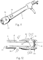
- Figures 11 and 12 illustrate a protective cap 30 over an arthroscopic sheath 3.
- the sheath 3 comprises an inner lumen 63 in fluid communication with a vacuum source and a hole dispose 33 in the sheath in fluid communication with the vacuum source and a surgical site within a patient 1.
- a backstop or flange 65 is disposed within an inner diameter of a bore within the cap. The bore is sized and dimensioned to friction fit over the sheath 3.
- the flange 65 prevents the sheath 3 from being further pushed into the cap and extends inwardly to come in contact with an outer diameter of the rigid cannula 18 disposed within the sheath 3. This contact forms a seal between the flange 65 and the outer diameter of the rigid cannula 18.
- the rigid cannula 18 is provided with a lumen in fluid communication with a fluid source the cap. Holes 32 disposed in the cap are in fluid communication with the surgical site and the lumen within the rigid cannula 18 as well as the fluid source allowing fluid to flow from the fluid source to the surgical site.
- the cap further comprises a concave lens 55 coupled to the distal portion 66 of the cap 3.
Landscapes
- Health & Medical Sciences (AREA)
- Life Sciences & Earth Sciences (AREA)
- Surgery (AREA)
- Biophysics (AREA)
- Biomedical Technology (AREA)
- Veterinary Medicine (AREA)
- Nuclear Medicine, Radiotherapy & Molecular Imaging (AREA)
- Optics & Photonics (AREA)
- Pathology (AREA)
- Radiology & Medical Imaging (AREA)
- Public Health (AREA)
- Engineering & Computer Science (AREA)
- Physics & Mathematics (AREA)
- Heart & Thoracic Surgery (AREA)
- Medical Informatics (AREA)
- Molecular Biology (AREA)
- Animal Behavior & Ethology (AREA)
- General Health & Medical Sciences (AREA)
- Orthopedic Medicine & Surgery (AREA)
- Physical Education & Sports Medicine (AREA)
- Endoscopes (AREA)
- Surgical Instruments (AREA)
Description
- The inventions described below relate the field of arthroscopic surgical instruments.
- Arthroscopic surgery involves using optical instruments, such as an arthroscope, to visualize an operating field inside or near a joint of a patient. The same instrument or other instruments may be used to perform a surgical procedure in the operating field. Common instruments used in addition to the arthroscope include a trimming instrument for cutting tissue and an irrigation instrument for irrigating the surgical field. Each of the instruments requires its own incision to be introduced into the surgical field; thus, many surgeons prefer to use only a trimming instrument and an arthroscope during arthroscopic surgical procedures.
- Arthroscopes are fragile in relation to the forces applied during arthroscopic surgery, so a rigid cannula is placed over the arthroscope to reinforce it. The distal end of the rigid cannula is pointed, usually sharp, and so the rigid cannula can scratch or gouge soft tissue within the operating field. The rigid cannula can also become stuck between bones or cartilage during a procedure. A rigid cannula can also damage metal prosthetics used to replace joints, resulting in a shortening of the useful life of the prosthetic and forcing the patient to undergo additional, painful surgeries to correct the problem.
- An additional problem associated with arthroscopic surgery is maintaining a clear surgical field during surgery. Blood and debris can cloud the field, impairing a surgeon's ability to visualize tissue. One method of solving this problem is to use the irrigation instrument to clear the surgical field with saline; however, many surgeons strongly prefer to avoid the additional trauma caused by inserting a third instrument. These surgeons will perform arthroscopic surgeries despite problems with visualizing the surgical field.
- A further problem associated with arthroscopic surgery is accidental damage to the arthroscope. The arthroscope is often damaged if the working end of a trimming instrument accidentally strikes the sensitive optical components on the distal portion of the arthroscope. The arthroscope may also be damaged if the arthroscope becomes stuck between bones, cartilage or other tissue and excessive force must be used to free the arthroscope. Arthroscopes are expensive, costing thousands of dollars, so accidental damage to arthroscopes is a significant cost problem. A damaged arthroscope could cause delays during surgery and broken pieces of the arthroscope could be deposited in the surgical field. Both situations are harmful to the patient. Thus, devices and methods are needed to prevent accidental damage to arthroscopes during surgery.
-
US patent 6,761,684 B1 discloses an endoscope tip protection system for shielding the optical component at the distal end of an endoscope. The system comprises a tubular sheath which is optically transparent at least at its distal end, and adapted to fit onto and be retained on the scope tip. The scope/sheath assembly is then inserted into an open ended tubular fiber optic illuminating cannula which is adapted to receive the scope/sheath assembly and prevent the tubular sheath from falling out. -
US patent US 6,095,970 A discloses an endoscope includes a insertion tube which is inserted into a human body, an imaging device, an object optical system which forms image on the imaging device, a detachable unit accommodating the imaging device and the object optical system. The detachable unit is detachably mounted to a mounting portion of the insertion tube. At least one first contact is provided to the detachable unit. At least one second contact is provided to the mounting portion. The first and second contacts are electrically connected when the detachable unit is mounted to the mounting portion. A waterproof arrangement is provided to prevent water from entering a connecting potion of the first and second contacts. - German utility model
DE 9215725 U1 discloses a device for the lighting and inspection of hollow and gap spaces, in particular an endoscope, with an optical carrier. -
US patent 5,735,792 A discloses a surgical instrument incorporating visualizing optics is disclosed. The instrument comprises a handle supporting a rigid hollow shaft, an expanded tip is located on the distal end of the shaft providing a surface that will not tear or easily penetrate tissue. A fiber-optic assembly, containing at least one illumination transmitting fiber, a plurality of image-carrying fibers and an objective lens mounted near the distal end of the image fibers runs the length of the shaft. The objective lens is positioned relative to an opening in the tip allowing viewing. Light for illuminating the surgical site passes through one or more illumination fibers and through the opening in the tip of the probe. Light reflected from the tissue at the tip of the probe is focused onto the distal face of the image bundle by an objective lens and is then transmitted to the proximal end of the image bundle. Connectors on the proximal ends of the illumination fibers and image fibers facilitate their being coupled to an illumination source and a viewing device. - The invention is defined in the claims and provides for a protective cap that is placed over the distal portion of an arthroscope. The cap is made of a transparent, yet durable material that prevents accidental damage to the arthroscope caused by trimming instruments or impacts with hard tissue within the surgical field. Holes may be placed in the cap to provide for the inflow and outflow of fluids from the cap.
- Methods are not part of the invention. One or more lenses or filters may be provided within the cap to adjust the field of view as seen through the arthroscope.
-
-
Figure 1 shows a method of performing arthroscopic surgery on a patient. -
Figure 2 shows a protective cap disposed over the distal portion of an arthroscopic instrument. -
Figure 3 shows a cross section of a protective cap disposed over the distal portion of an arthroscopic instrument. -
Figure 4 shows a cross section of a multi-lumen protective cap. -
Figure 5 shows a protective cap having a concave lens disposed at the distal end of the cap. -
Figure 6 shows a protective cap having a convex lens disposed at the distal end of the cap. -
Figure 7 shows cap with a reticule for use with an arthroscopic instrument. -
Figure 8 shows a reticule for use with an arthroscopic instrument. -
Figure 9 illustrate an atraumatic sheath for use over an endoscope provided with a mirror having a hinge. -
Figure 10 illustrates an atraumatic sheath devices that allows viewing around an obstruction. -
Figure 11 shows a protective cap over an arthroscopic sheath. -
Figure 12 shows a cross-section of protective cap over an arthroscopic sheath. -
Figure 1 shows a method of performing arthroscopic surgery on a patient using anarthroscopic instrument 2 sheathed in an atraumatic introducersheath 3. (The various parts of the arthroscope are shown in phantom to indicate their positions inside the sheath.) Various anatomical landmarks in a patient'sknee 4 are shown for reference, including thefemur 5, patella 6, posterior cruciate ligament 7, anterior cruciate ligament 8, meniscus 9,tibia 10 andfibula 11. During surgery, the surgeon introduces thearthroscope 2 into the knee via afirst incision 12 in order to visualize the surgical field. Atrimming instrument 13 is introduced through asecond incision 14 to remove or trim tissue that the surgeon determines should be removed or trimmed. Optionally, anirrigating instrument 15 may be introduced through athird incision 16 in order to irrigate the surgical field and thereby maintain a clear view. As illustrated below, a combined arthroscope and inflow/outflow atraumatic sheath may replace the irrigating instrument. - The
arthroscope 2 is anoptical instrument 17 surrounded by arigid cannula 18 having a distal edge that typically is cut at an angle. To protect the patient from unintended injury or trauma during the procedure, the arthroscope has been inserted into a resilient outer introducer sheath oratraumatic sheath 3 that extends over the rigid cannula. Preferably, thedistal tip 19 of the atraumatic sheath extends distally just past the distal end of the arthroscope and rigid cannula to further protect the patient. -
Figures 2 and 3 show an atraumaticprotective cap 30 having acap body 28 and alens 29 and disposed over the distal portion 31 of an arthroscopic instrument. Thecap body 28 is sized and dimensioned and of such a profile as to allow passage into restricted joint anatomy. Preferably, the cap is disposed on the end of thesheath 3 and slides over the distal end of the arthroscope as the sheath is pulled over the arthroscope as shown inFigure 2 . (The sheath is also shown in ourco-pending application 10/769,629, filed January 29, 2004 Figure 3 . The cap is preferably provided with a rounded, or bulbous, shape that reduces the chances of injuring tissue during the surgical procedure. The cap is optically transparent to wavelengths of light used during surgery, though only that portion of the cap covering the view port of the arthroscope need be transparent. (The view port is that portion of the arthroscope through which the surgeon visualizes the surgical field.) The material of the cap is sufficiently durable that the cap will prevent accidental damage to the arthroscope caused by unintended contact with the working end of a trimming instrument, by unintended contact with burrs or other hard tissue within the surgical field or because of excessive force applied to the arthroscope. Preferably, the body of the cap may be manufactured from thermoplastic elastomers. However, other materials used to manufacture the body of the cap may include styrenic block copolymers (SBCs), thermoplastic olefins (TPOs), thermoplastic vulcanisates (TPVs), thermoplastic polyurethane elastomers (TPUs), copolyesters (COPEs) and copolyamides (COPAs). Thermosets such as silicone, urethane or latex may also be used to manufacture the cap body. Sterilizable elastomers are typically used to make the body of the cap while optically clear polycarbonate materials comprise theviewing lens 29 in thecap 30. Other materials suitable for the viewing lens include molded acrylic, styrene, polyolefin or silicon. Alternatively, the body of the cap and lens can also be manufactured from the same optically clear material reducing manufacturing and assembly costs. Such suitable material includes optically clear silicone available from Wacker Silicones™ and having a hardness ranging from approximately 30 Shore A to approximately 40 Shore D. - One or
more holes 32 may be placed in the cap to provide for suction, irrigation or the injection of therapeutic agents.Similar holes 33 may also be placed in the sheath. The holes are in fluid communication with one or more of the lumens disposed in the sheath or arthroscope or disposed between the sheath and arthroscope. Afluid source 34 in fluid communication with one or more lumens is provided to irrigate the surgical field or to inject therapeutic agents into the surgical field. Avacuum source 35 in fluid communication with one or more of the lumens provides suction. A manifold 36 disposed on the sheath or arthroscope distributes the flow of fluids within the sheath or arthroscope. - In use, the sheath and protective cap are pulled over the arthroscope. The surgeon then inserts the arthroscope into the surgical field and subsequently performs a surgical procedure on the patient. If, during the procedure, the working end of a trimming instrument accidentally strikes the cap, the cap will prevent damage to the arthroscope. Likewise, the cap will prevent damage to the arthroscope if the arthroscope strikes a burr or other hard piece of tissue. Preferably, the cap is releasably attached to the sheath so that the cap may be easily replaced if damaged by these events.
-
Figure 4 shows a cross section of a multi-lumenprotective cap 30 and anarthroscope 2 disposed within the cap. One ormore holes 37 extend from the outer diameter of the cap to theouter lumen 38. In addition, one or moreadditional holes 39 extend from the outer diameter of the cap, through the outer lumen and to theinner lumen 40 of the cap. Theholes 39 communicating with the inner lumen do not communicate with the outer lumen, thereby isolating the inner and outer lumens. Thus, suction may be provided through one lumen and simultaneous irrigation provided through the other lumen. Alternatively, the cap may be provided as part of a system providing fluid inflow and outflow to a surgical site where fluid inflow and outflow is accomplished by devices other than an arthroscope or arthroscope sheath. - The
cap 30 orcap body 28 shown inFigures 2 through 4 may be integrally formed with asheath 3, attached to asheath 3, or fit over asheath 3. The cap may also be provided without the sheath. The cap may be placed directly over the distal portion of the arthroscope without an atraumatic sheath. The cap can be held to the arthroscope by friction fit between the scope and the cap, by a shrink tube, by an adhesive, by detents or by any other suitable mechanism. Preferably, the cap is removably attached so that the cap may be easily replaced. As with the cap disposed on the sheath, holes 32 may be provided to provide for the inflow and outflow of fluids. -
Figure 5 shows aprotective cap 30 having aconcave lens 55 disposed at the distal end of the cap. The lens is provided just inside and proximal the distal end of the cap. The concave lens provides for a wide-angle view of the surgical field. -
Figure 6 shows aprotective cap 30 having aconvex lens 56 disposed at the distal end of the cap. The lens is provided just inside and proximal the distal end of the cap. The convex lens provides for a magnified view of an object within the surgical field. -
Figure 7 and Figure 8 show a reticule 57 for use with an arthroscopic instrument. The reticule may be etched into alens 29 disposed within the cap or may be otherwise suitably placed on the cap or even on arthroscope itself. The reticule is marked with ascale 58 with which the surgeon can measure the size of objects seen through the arthroscope. The surgeon may also use the reticule to align the arthroscope within the surgical field. - In addition to the lenses and reticule shown in
Figures 5 through 8 , filters may be provided within the cap to reduce light reflected into the arthroscope or to block certain wavelengths of light. The filtered lens may comprise a polarizing filter, a bandpass filter, a color filter, or an interference filter. These filters can be used in conjunction with specialized light sources (eg. Ultraviolet or Infrared) and video processing for therapeutic and diagnostic purposes. Thus, the cap may be part of a complete system to diagnose pathology using different wavelengths of light and/or colors of light and filtering the light. Further, the cap may also be provided as part of system that delivers photonic energy to a surgical site to control and visualize photodynamic therapy. - Other lenses, such as spherical lenses, may also be provided within the cap to change the field of view. Multiple lenses may also be provided within the cap to adjust the field of view. As illustrated in
Figures 9 and 10 , the sheath, the cap or the sheath with the cap combination may be configured to allow viewing around an object or obstruction. This may be accomplished through the use of a right angle prism, pentaprism, roof prism, retro-reflector or mirror disposed within the cap. The mirror may be flat, concave or convex to produce a normal, reduced, or magnified image. As illustrated inFigure 9 , anatraumatic sheath 3 for use over an endoscope is provided with amirror 67 having ahinge 68. A pull-wire 69 is coupled to themirror 67 and is disposed within thesheath 3. The pull-wire 69 is further coupled to an articulatingknob 70. When the knob is manipulated, the pull-wire moves the mirror and changes theviewing angle 71. -
Figure 10 illustrates anatraumatic sheath 3 device that allows viewing around an object or obstruction through the use of a deflectable material orflexible mount 72 having amirror 67. The mount may be manufactured from formable materials such as aluminum or steel having spring characteristics. A pull-wire 69 or other manipulation device may also be coupled to the mount to changeviewing angle 71 when the wire is manipulated manually using an articulatingknob 70. The mount may also be manufactured from shape memory materials such as Nitinol® that may be manipulated using electrical current to change the angle of the mirror. The cap and the sheath with the cap may also be modified for use with other types of endoscopes or for use with other delicate instruments that are subject to damage during a surgical procedure. -
Figures 11 and 12 illustrate aprotective cap 30 over anarthroscopic sheath 3. Thesheath 3 comprises aninner lumen 63 in fluid communication with a vacuum source and a hole dispose 33 in the sheath in fluid communication with the vacuum source and a surgical site within apatient 1. A backstop orflange 65 is disposed within an inner diameter of a bore within the cap. The bore is sized and dimensioned to friction fit over thesheath 3. - The
flange 65 prevents thesheath 3 from being further pushed into the cap and extends inwardly to come in contact with an outer diameter of therigid cannula 18 disposed within thesheath 3. This contact forms a seal between theflange 65 and the outer diameter of therigid cannula 18. Therigid cannula 18 is provided with a lumen in fluid communication with a fluid source the cap.Holes 32 disposed in the cap are in fluid communication with the surgical site and the lumen within therigid cannula 18 as well as the fluid source allowing fluid to flow from the fluid source to the surgical site. The cap further comprises aconcave lens 55 coupled to thedistal portion 66 of thecap 3. - Thus, while the preferred embodiments of the devices and methods have been described in reference to the athroscopic surgical environment in which they were developed, they are merely illustrative of the principles of the inventions. Other embodiments and configurations for use with other types of endoscopes may be devised for use in other surgical and non-surgical environments without departing from the spirit of the inventions and the scope of the appended claims.
Claims (6)
- A cap (30) in combination with an arthroscopic instrument (2) comprising an optical instrument and a rigid cannula (18) surrounding the optical instrument, said cap comprising:a body (28) bulbous in shape and characterized by a distal end and proximal end;a bore having an inner diameter and disposed within the body (28), said inner diameter of said bore sized and dimensioned to friction fit over an outer diameter of the rigid cannula (18);a lens (29) coupled to the distal end of the body (28).
- The cap (30) of claim 1 wherein the lens (29) is concave (55).
- The cap (30) of claim 1 wherein the lens (29) is convex (56).
- The cap (30) of claim 1 wherein the lens (29) comprises a reticule (57).
- The cap (30) of claim 1 further comprising one or more holes (32) disposed in the cap (30), said one or more holes (32) capable of being in fluid communication with a lumen disposed in the arthroscopic instrument (2), and wherein a fluid source (34) is in fluid communication with the lumen.
- The cap (30) of claim 1 further comprising one or more holes (32) disposed in the cap (30), said one or more holes (32) capable of being in fluid communication with a lumen disposed in the arthroscopic instrument (2), and wherein a vacuum source (35) is in fluid communication with the lumen.
Applications Claiming Priority (2)
| Application Number | Priority Date | Filing Date | Title |
|---|---|---|---|
| US11/142,990 US7553278B2 (en) | 2005-06-01 | 2005-06-01 | Protective cap for arthroscopic instruments |
| PCT/US2006/021188 WO2006130730A2 (en) | 2005-06-01 | 2006-05-31 | Protective cap for arthroscopic instruments |
Publications (3)
| Publication Number | Publication Date |
|---|---|
| EP1890587A2 EP1890587A2 (en) | 2008-02-27 |
| EP1890587A4 EP1890587A4 (en) | 2012-06-13 |
| EP1890587B1 true EP1890587B1 (en) | 2017-07-12 |
Family
ID=37482300
Family Applications (1)
| Application Number | Title | Priority Date | Filing Date |
|---|---|---|---|
| EP06760606.1A Active EP1890587B1 (en) | 2005-06-01 | 2006-05-31 | Protective cap for arthroscopic instruments |
Country Status (4)
| Country | Link |
|---|---|
| US (4) | US7553278B2 (en) |
| EP (1) | EP1890587B1 (en) |
| JP (1) | JP5256027B2 (en) |
| WO (1) | WO2006130730A2 (en) |
Families Citing this family (99)
| Publication number | Priority date | Publication date | Assignee | Title |
|---|---|---|---|---|
| WO2007093994A2 (en) * | 2006-02-16 | 2007-08-23 | Vision - Sciences Inc. | Endoscope with imaging capsule |
| US7655004B2 (en) | 2007-02-15 | 2010-02-02 | Ethicon Endo-Surgery, Inc. | Electroporation ablation apparatus, system, and method |
| WO2008119118A1 (en) * | 2007-03-30 | 2008-10-09 | Polartechnics Limited | Sheath system for tissue probe |
| US8075572B2 (en) | 2007-04-26 | 2011-12-13 | Ethicon Endo-Surgery, Inc. | Surgical suturing apparatus |
| US8100922B2 (en) | 2007-04-27 | 2012-01-24 | Ethicon Endo-Surgery, Inc. | Curved needle suturing tool |
| US8226548B2 (en) * | 2007-07-07 | 2012-07-24 | Cannuflow, Inc. | Rigid arthroscope system |
| US8579897B2 (en) | 2007-11-21 | 2013-11-12 | Ethicon Endo-Surgery, Inc. | Bipolar forceps |
| US8568410B2 (en) | 2007-08-31 | 2013-10-29 | Ethicon Endo-Surgery, Inc. | Electrical ablation surgical instruments |
| US8262655B2 (en) | 2007-11-21 | 2012-09-11 | Ethicon Endo-Surgery, Inc. | Bipolar forceps |
| US20090112059A1 (en) * | 2007-10-31 | 2009-04-30 | Nobis Rudolph H | Apparatus and methods for closing a gastrotomy |
| US8480657B2 (en) | 2007-10-31 | 2013-07-09 | Ethicon Endo-Surgery, Inc. | Detachable distal overtube section and methods for forming a sealable opening in the wall of an organ |
| US8262680B2 (en) | 2008-03-10 | 2012-09-11 | Ethicon Endo-Surgery, Inc. | Anastomotic device |
| US8679003B2 (en) | 2008-05-30 | 2014-03-25 | Ethicon Endo-Surgery, Inc. | Surgical device and endoscope including same |
| US8771260B2 (en) | 2008-05-30 | 2014-07-08 | Ethicon Endo-Surgery, Inc. | Actuating and articulating surgical device |
| US8070759B2 (en) | 2008-05-30 | 2011-12-06 | Ethicon Endo-Surgery, Inc. | Surgical fastening device |
| US8652150B2 (en) | 2008-05-30 | 2014-02-18 | Ethicon Endo-Surgery, Inc. | Multifunction surgical device |
| US8317806B2 (en) | 2008-05-30 | 2012-11-27 | Ethicon Endo-Surgery, Inc. | Endoscopic suturing tension controlling and indication devices |
| US8114072B2 (en) | 2008-05-30 | 2012-02-14 | Ethicon Endo-Surgery, Inc. | Electrical ablation device |
| US8906035B2 (en) * | 2008-06-04 | 2014-12-09 | Ethicon Endo-Surgery, Inc. | Endoscopic drop off bag |
| US8403926B2 (en) | 2008-06-05 | 2013-03-26 | Ethicon Endo-Surgery, Inc. | Manually articulating devices |
| US8361112B2 (en) | 2008-06-27 | 2013-01-29 | Ethicon Endo-Surgery, Inc. | Surgical suture arrangement |
| US8262563B2 (en) | 2008-07-14 | 2012-09-11 | Ethicon Endo-Surgery, Inc. | Endoscopic translumenal articulatable steerable overtube |
| US8888792B2 (en) | 2008-07-14 | 2014-11-18 | Ethicon Endo-Surgery, Inc. | Tissue apposition clip application devices and methods |
| US8211125B2 (en) | 2008-08-15 | 2012-07-03 | Ethicon Endo-Surgery, Inc. | Sterile appliance delivery device for endoscopic procedures |
| US8529563B2 (en) | 2008-08-25 | 2013-09-10 | Ethicon Endo-Surgery, Inc. | Electrical ablation devices |
| US8241204B2 (en) | 2008-08-29 | 2012-08-14 | Ethicon Endo-Surgery, Inc. | Articulating end cap |
| US8480689B2 (en) | 2008-09-02 | 2013-07-09 | Ethicon Endo-Surgery, Inc. | Suturing device |
| US8409200B2 (en) | 2008-09-03 | 2013-04-02 | Ethicon Endo-Surgery, Inc. | Surgical grasping device |
| US8114119B2 (en) | 2008-09-09 | 2012-02-14 | Ethicon Endo-Surgery, Inc. | Surgical grasping device |
| US8337394B2 (en) | 2008-10-01 | 2012-12-25 | Ethicon Endo-Surgery, Inc. | Overtube with expandable tip |
| US8157834B2 (en) | 2008-11-25 | 2012-04-17 | Ethicon Endo-Surgery, Inc. | Rotational coupling device for surgical instrument with flexible actuators |
| US8172772B2 (en) | 2008-12-11 | 2012-05-08 | Ethicon Endo-Surgery, Inc. | Specimen retrieval device |
| US8361066B2 (en) | 2009-01-12 | 2013-01-29 | Ethicon Endo-Surgery, Inc. | Electrical ablation devices |
| US8828031B2 (en) | 2009-01-12 | 2014-09-09 | Ethicon Endo-Surgery, Inc. | Apparatus for forming an anastomosis |
| US9226772B2 (en) | 2009-01-30 | 2016-01-05 | Ethicon Endo-Surgery, Inc. | Surgical device |
| US8252057B2 (en) | 2009-01-30 | 2012-08-28 | Ethicon Endo-Surgery, Inc. | Surgical access device |
| US8037591B2 (en) | 2009-02-02 | 2011-10-18 | Ethicon Endo-Surgery, Inc. | Surgical scissors |
| US20110098704A1 (en) | 2009-10-28 | 2011-04-28 | Ethicon Endo-Surgery, Inc. | Electrical ablation devices |
| US8608652B2 (en) | 2009-11-05 | 2013-12-17 | Ethicon Endo-Surgery, Inc. | Vaginal entry surgical devices, kit, system, and method |
| US8353487B2 (en) | 2009-12-17 | 2013-01-15 | Ethicon Endo-Surgery, Inc. | User interface support devices for endoscopic surgical instruments |
| US8496574B2 (en) | 2009-12-17 | 2013-07-30 | Ethicon Endo-Surgery, Inc. | Selectively positionable camera for surgical guide tube assembly |
| US8506564B2 (en) | 2009-12-18 | 2013-08-13 | Ethicon Endo-Surgery, Inc. | Surgical instrument comprising an electrode |
| US9028483B2 (en) | 2009-12-18 | 2015-05-12 | Ethicon Endo-Surgery, Inc. | Surgical instrument comprising an electrode |
| US9005198B2 (en) | 2010-01-29 | 2015-04-14 | Ethicon Endo-Surgery, Inc. | Surgical instrument comprising an electrode |
| GB201007920D0 (en) * | 2010-05-12 | 2010-06-30 | Park Medical Ltd Q | Sheath for protecting endoscope probe |
| US20120029289A1 (en) * | 2010-07-29 | 2012-02-02 | Cannuflow, Inc. | Optical Cap for Use With Arthroscopic System |
| US20140316199A1 (en) * | 2010-07-29 | 2014-10-23 | Cannuflow, Inc. | Arthroscopic system |
| US9375139B2 (en) | 2010-07-29 | 2016-06-28 | Cannuflow, Inc. | Arthroscopic system |
| US10092291B2 (en) | 2011-01-25 | 2018-10-09 | Ethicon Endo-Surgery, Inc. | Surgical instrument with selectively rigidizable features |
| ES2901382T3 (en) | 2011-02-16 | 2022-03-22 | Massachusetts Gen Hospital | Optical coupler for an endoscope |
| US9314620B2 (en) | 2011-02-28 | 2016-04-19 | Ethicon Endo-Surgery, Inc. | Electrical ablation devices and methods |
| US9233241B2 (en) | 2011-02-28 | 2016-01-12 | Ethicon Endo-Surgery, Inc. | Electrical ablation devices and methods |
| US9254169B2 (en) | 2011-02-28 | 2016-02-09 | Ethicon Endo-Surgery, Inc. | Electrical ablation devices and methods |
| US9049987B2 (en) | 2011-03-17 | 2015-06-09 | Ethicon Endo-Surgery, Inc. | Hand held surgical device for manipulating an internal magnet assembly within a patient |
| KR101226683B1 (en) | 2011-10-10 | 2013-01-25 | 김재환 | Cover for arthroscopic surgical instrument |
| US8986199B2 (en) | 2012-02-17 | 2015-03-24 | Ethicon Endo-Surgery, Inc. | Apparatus and methods for cleaning the lens of an endoscope |
| ES2951058T3 (en) | 2012-03-09 | 2023-10-17 | 3Shape As | 3D scanner with steam autoclavable tip containing a heated optical element |
| US20130253266A1 (en) | 2012-03-22 | 2013-09-26 | Codman & Shurtleff, Inc. | Fluid management catheter and methods of using same |
| KR101371927B1 (en) * | 2012-04-27 | 2014-03-26 | 고려대학교 산학협력단 | Bead for stitching, needle for stitching and side suction cap and apparatus for stitching internal organ using the same |
| US9427255B2 (en) | 2012-05-14 | 2016-08-30 | Ethicon Endo-Surgery, Inc. | Apparatus for introducing a steerable camera assembly into a patient |
| DE102012105370A1 (en) * | 2012-06-20 | 2013-12-24 | Karl Storz Gmbh & Co. Kg | Endoscopic sleeve, endoscope assembly and method for providing an endoscope assembly |
| US9078662B2 (en) | 2012-07-03 | 2015-07-14 | Ethicon Endo-Surgery, Inc. | Endoscopic cap electrode and method for using the same |
| US9545290B2 (en) | 2012-07-30 | 2017-01-17 | Ethicon Endo-Surgery, Inc. | Needle probe guide |
| US10314649B2 (en) | 2012-08-02 | 2019-06-11 | Ethicon Endo-Surgery, Inc. | Flexible expandable electrode and method of intraluminal delivery of pulsed power |
| US9572623B2 (en) | 2012-08-02 | 2017-02-21 | Ethicon Endo-Surgery, Inc. | Reusable electrode and disposable sheath |
| US9277957B2 (en) | 2012-08-15 | 2016-03-08 | Ethicon Endo-Surgery, Inc. | Electrosurgical devices and methods |
| US9451875B2 (en) | 2012-12-07 | 2016-09-27 | Cook Medical Technologies Llc | Flexible lens |
| DE102013102024A1 (en) * | 2013-02-01 | 2014-08-21 | Firma Trokamed Gmbh | arthroscopy shaft |
| US10098527B2 (en) | 2013-02-27 | 2018-10-16 | Ethidcon Endo-Surgery, Inc. | System for performing a minimally invasive surgical procedure |
| US20140275768A1 (en) * | 2013-03-13 | 2014-09-18 | Covidien Lp | Thoracic Scope With Skirt And Gap |
| JP2014212835A (en) * | 2013-04-23 | 2014-11-17 | ショーダテクトロン株式会社 | Hood for endoscope, and endoscope with same hood for endoscope |
| CN105636530A (en) * | 2013-08-19 | 2016-06-01 | 史密夫和内修有限公司 | Bone removal under direct visualization |
| GB2520332A (en) * | 2013-11-18 | 2015-05-20 | Meditech Endoscopy Ltd | Gripping Device |
| US9459442B2 (en) | 2014-09-23 | 2016-10-04 | Scott Miller | Optical coupler for optical imaging visualization device |
| WO2016051421A1 (en) * | 2014-09-30 | 2016-04-07 | Shah Kaushikkumar Vallabhadas | A sheath assembly and mutihole catheter for different fields of endoscopic surgery involving suction, irrigation and material removal. |
| GB201418173D0 (en) | 2014-10-14 | 2014-11-26 | Meditech Endoscopy Ltd | Instrument tip protector |
| US10729419B2 (en) | 2014-10-23 | 2020-08-04 | Medos International Sarl | Biceps tenodesis implants and delivery tools |
| US10751161B2 (en) | 2014-10-23 | 2020-08-25 | Medos International Sárl | Biceps tenodesis anchor implants |
| US10076374B2 (en) | 2014-10-23 | 2018-09-18 | Medos International Sárl | Biceps tenodesis delivery tools |
| US10034742B2 (en) | 2014-10-23 | 2018-07-31 | Medos International Sarl | Biceps tenodesis implants and delivery tools |
| US10856966B2 (en) | 2014-10-23 | 2020-12-08 | Medos International Sarl | Biceps tenodesis implants and delivery tools |
| US9693856B2 (en) | 2015-04-22 | 2017-07-04 | DePuy Synthes Products, LLC | Biceps repair device |
| US10548467B2 (en) | 2015-06-02 | 2020-02-04 | GI Scientific, LLC | Conductive optical element |
| KR20180041140A (en) | 2015-07-21 | 2018-04-23 | 지아이 사이언티픽, 엘엘씨 | Endoscope accessory with angle adjustable exhaust port |
| EP4241649A3 (en) * | 2016-02-02 | 2023-11-15 | Boston Scientific Scimed Inc. | Laser lithotripsy medical device |
| US10231824B2 (en) | 2016-04-08 | 2019-03-19 | Medos International Sárl | Tenodesis anchoring systems and tools |
| US10231823B2 (en) | 2016-04-08 | 2019-03-19 | Medos International Sarl | Tenodesis implants and tools |
| WO2018207594A1 (en) * | 2017-05-10 | 2018-11-15 | オリンパス株式会社 | Hood for endscope, and endoscope system |
| JP7208993B2 (en) * | 2017-08-04 | 2023-01-19 | ブリガム アンド ウィメンズ ホスピタル,インク. | Veress-type needle with illuminated guides and safety features |
| US11382662B2 (en) | 2017-08-04 | 2022-07-12 | The Brigham And Women's Hospital, Inc. | Trocars and veress-type needles with illuminated guidance and safety features |
| US11219355B2 (en) | 2017-08-14 | 2022-01-11 | Medos International Sarl | Surgical instruments with reflective mirror-like surfaces |
| US11849917B2 (en) * | 2017-09-11 | 2023-12-26 | Eyelum Ltd. | Disposable miniature endoscopy system |
| US20200237200A1 (en) * | 2017-09-18 | 2020-07-30 | Veena Moktali | A digital device facilitating body cavity screening and diagnosis |
| WO2019136228A1 (en) * | 2018-01-05 | 2019-07-11 | Boston Scientific Scimed, Inc. | Fluorophore imaging devices, systems, and methods for an endoscopic procedure |
| US20190231177A1 (en) * | 2018-01-31 | 2019-08-01 | UVision360, Inc. | Flexible imaging window |
| DE102018110082A1 (en) * | 2018-04-26 | 2019-10-31 | avateramedical GmBH | Sterile endoscope cover |
| US11903557B2 (en) | 2019-04-30 | 2024-02-20 | Psip2 Llc | Endoscope for imaging in nonvisible light |
| US20220133138A1 (en) * | 2020-10-29 | 2022-05-05 | Clearmind Biomedical, Inc. | Dilator-less and obturator-less introducer for viewing and acting on internal passageways or tissue |
| WO2022249116A2 (en) * | 2021-05-26 | 2022-12-01 | Psip2 Llc | Endoscope |
Family Cites Families (50)
| Publication number | Priority date | Publication date | Assignee | Title |
|---|---|---|---|---|
| US3051176A (en) * | 1959-12-11 | 1962-08-28 | Alberti Franz | Rectoscopic devices |
| JPS57129407A (en) | 1981-02-03 | 1982-08-11 | Olympus Optical Co Ltd | Hard endoscope |
| US4470407A (en) * | 1982-03-11 | 1984-09-11 | Laserscope, Inc. | Endoscopic device |
| JP2628627B2 (en) * | 1985-01-11 | 1997-07-09 | オリンパス光学工業株式会社 | Aspheric objective lens for endoscope |
| US4856495A (en) * | 1986-09-25 | 1989-08-15 | Olympus Optical Co., Ltd. | Endoscope apparatus |
| US4809679A (en) * | 1986-11-19 | 1989-03-07 | Olympus Optical Co., Ltd. | Forceps plug for endoscopes |
| US4782819A (en) * | 1987-02-25 | 1988-11-08 | Adair Edwin Lloyd | Optical catheter |
| US4727416A (en) * | 1987-03-05 | 1988-02-23 | Fuji Optical Systems, Inc. | Electronic video dental camera |
| US4809678A (en) * | 1987-08-14 | 1989-03-07 | Klein Richard S | Endoscope for preventing patient contamination |
| US5029574A (en) * | 1988-04-14 | 1991-07-09 | Okamoto Industries, Inc. | Endoscopic balloon with a protective film thereon |
| US4886049A (en) | 1988-05-17 | 1989-12-12 | Darras Robert L | Medical instrument cover |
| JP2534867Y2 (en) * | 1989-11-09 | 1997-05-07 | 株式会社町田製作所 | Interchangeable endoscope for direct and side view |
| US5257617A (en) * | 1989-12-25 | 1993-11-02 | Asahi Kogaku Kogyo Kabushiki Kaisha | Sheathed endoscope and sheath therefor |
| US5191878A (en) * | 1990-04-12 | 1993-03-09 | Olympus Optical Co., Ltd. | Endoscope device |
| US5237984A (en) | 1991-06-24 | 1993-08-24 | Xomed-Treace Inc. | Sheath for endoscope |
| US5370649A (en) * | 1991-08-16 | 1994-12-06 | Myriadlase, Inc. | Laterally reflecting tip for laser transmitting fiber |
| DE9215725U1 (en) | 1992-11-19 | 1993-01-14 | Schölly Fiberoptic GmbH, 7819 Denzlingen | Device for lighting and inspection of cavities and gaps |
| US5735792A (en) | 1992-11-25 | 1998-04-07 | Clarus Medical Systems, Inc. | Surgical instrument including viewing optics and an atraumatic probe |
| US5536236A (en) * | 1993-02-12 | 1996-07-16 | Olympus Optical Co., Ltd. | Covered endoscope system |
| US5447148A (en) | 1993-07-08 | 1995-09-05 | Vision Sciences, Inc. | Endoscopic contamination protection system to facilitate cleaning of endoscopes |
| US5573493A (en) * | 1993-10-08 | 1996-11-12 | United States Surgical Corporation | Endoscope attachment for changing angle of view |
| JP2802244B2 (en) * | 1994-08-29 | 1998-09-24 | オリンパス光学工業株式会社 | Endoscope sheath |
| US6184923B1 (en) * | 1994-11-25 | 2001-02-06 | Olympus Optical Co., Ltd. | Endoscope with an interchangeable distal end optical adapter |
| US5607441A (en) * | 1995-03-24 | 1997-03-04 | Ethicon Endo-Surgery, Inc. | Surgical dissector |
| JP3461974B2 (en) * | 1995-05-31 | 2003-10-27 | 株式会社町田製作所 | Endoscope |
| JPH0998938A (en) * | 1995-10-04 | 1997-04-15 | Fuji Photo Optical Co Ltd | Protector of insertion part of endoscope |
| JPH09140659A (en) * | 1995-11-24 | 1997-06-03 | Fuji Photo Optical Co Ltd | Guiding cap for insertion of endoscope |
| JPH10192297A (en) * | 1996-05-09 | 1998-07-28 | Olympus Optical Co Ltd | Securing device of cavity for bone operation |
| US6095970A (en) * | 1997-02-19 | 2000-08-01 | Asahi Kogaku Kogyo Kabushiki Kaisha | Endoscope |
| US5807237A (en) * | 1997-03-31 | 1998-09-15 | Tindel; Nathaniel L. | Endoscopic device |
| JP3748994B2 (en) * | 1997-08-22 | 2006-02-22 | オリンパス株式会社 | Endoscope fitting |
| IL122111A (en) * | 1997-11-04 | 2004-06-01 | Sightline Techn Ltd | Video rectoscope |
| JPH11249014A (en) * | 1998-03-03 | 1999-09-17 | Olympus Optical Co Ltd | Image pickup optical system and image pickup device using it |
| US6010450A (en) * | 1998-06-29 | 2000-01-04 | Welch Allyn, Inc. | Measuring adapter for viewing instrument |
| US5916145A (en) | 1998-08-07 | 1999-06-29 | Scimed Life Systems, Inc. | Device and method of using a surgical assembly with mesh sheath |
| US6761684B1 (en) * | 2000-08-10 | 2004-07-13 | Linvatec Corporation | Endoscope tip protection system |
| US6537209B1 (en) * | 2000-09-14 | 2003-03-25 | Itconcepts, Inc. | Optical system of lateral observation endoscope |
| JP3533163B2 (en) * | 2000-09-18 | 2004-05-31 | ペンタックス株式会社 | Endoscope tip |
| JP3717777B2 (en) * | 2000-09-29 | 2005-11-16 | フジノン株式会社 | Endoscope device |
| JP2002136472A (en) * | 2000-11-02 | 2002-05-14 | Olympus Optical Co Ltd | Endoscope |
| IT1320124B1 (en) * | 2000-12-20 | 2003-11-18 | Faro Spa | PERFECTED DEVICE FOR DENTAL INTERVENTIONS. |
| JP2002233491A (en) * | 2001-02-08 | 2002-08-20 | Asahi Optical Co Ltd | End portion of endoscope having end cap |
| DE10121450A1 (en) * | 2001-04-27 | 2002-11-21 | Storz Endoskop Gmbh Schaffhaus | Optical instrument, in particular an endoscope, with an exchangeable head |
| JP3985466B2 (en) | 2001-06-07 | 2007-10-03 | フジノン株式会社 | Endoscope lens device |
| US20030018340A1 (en) | 2001-06-29 | 2003-01-23 | Branch Thomas P. | Method and apparatus for installing cannula |
| JP2003279862A (en) * | 2002-03-25 | 2003-10-02 | Machida Endscope Co Ltd | Omnidirectional endoscopic device |
| AU2003240831A1 (en) * | 2002-05-30 | 2003-12-19 | The Board Of Trustees Of The Leland Stanford Junior University | Apparatus and method for coronary sinus access |
| DE10254609B4 (en) * | 2002-11-22 | 2017-12-07 | Stm Medizintechnik Starnberg Gmbh | endoscope head |
| WO2005000096A2 (en) * | 2003-06-05 | 2005-01-06 | Hydrocision, Inc. | Disposable endoscope and method of making a disposable endoscope |
| US8172747B2 (en) * | 2003-09-25 | 2012-05-08 | Hansen Medical, Inc. | Balloon visualization for traversing a tissue wall |
-
2005
- 2005-06-01 US US11/142,990 patent/US7553278B2/en active Active
-
2006
- 2006-05-31 EP EP06760606.1A patent/EP1890587B1/en active Active
- 2006-05-31 WO PCT/US2006/021188 patent/WO2006130730A2/en active Application Filing
- 2006-05-31 JP JP2008514825A patent/JP5256027B2/en active Active
-
2009
- 2009-06-25 US US12/491,566 patent/US8216131B2/en active Active
-
2012
- 2012-07-10 US US13/545,886 patent/US9167955B2/en active Active
-
2015
- 2015-10-27 US US14/924,586 patent/US9833134B2/en active Active
Non-Patent Citations (1)
| Title |
|---|
| None * |
Also Published As
| Publication number | Publication date |
|---|---|
| EP1890587A4 (en) | 2012-06-13 |
| US20160045105A1 (en) | 2016-02-18 |
| JP5256027B2 (en) | 2013-08-07 |
| WO2006130730A2 (en) | 2006-12-07 |
| US9833134B2 (en) | 2017-12-05 |
| US20120277533A1 (en) | 2012-11-01 |
| US8216131B2 (en) | 2012-07-10 |
| WO2006130730A3 (en) | 2007-04-19 |
| US20090326328A1 (en) | 2009-12-31 |
| EP1890587A2 (en) | 2008-02-27 |
| US7553278B2 (en) | 2009-06-30 |
| US9167955B2 (en) | 2015-10-27 |
| US20060276692A1 (en) | 2006-12-07 |
| JP2008541947A (en) | 2008-11-27 |
Similar Documents
| Publication | Publication Date | Title |
|---|---|---|
| EP1890587B1 (en) | Protective cap for arthroscopic instruments | |
| US20200337531A1 (en) | Rigid Endoscope System | |
| JP7026645B2 (en) | Clot drainage and visualization device and usage | |
| US4736733A (en) | Endoscope with removable eyepiece | |
| US5213092A (en) | Aspirating endoscope | |
| US5667472A (en) | Surgical instrument and method for use with a viewing system | |
| US5152278A (en) | Surgical endoscope apparatus | |
| US5419309A (en) | Tip cleaning accessory for rigid endoscopic instrument | |
| US7413542B2 (en) | Atraumatic arthroscopic instrument sheath | |
| Badr-El-Dine et al. | Instrumentation and technologies in endoscopic ear surgery | |
| US20160374539A1 (en) | Atraumatic Arthroscopic Instrument Sheath | |
| US20120029289A1 (en) | Optical Cap for Use With Arthroscopic System | |
| US11337598B2 (en) | Laser video endoscope | |
| TW201707662A (en) | Illuminated ophthalmic infusion line and associated devices, systems, and methods | |
| MX2011001098A (en) | Swing prism endoscope. | |
| US20210298953A1 (en) | Miniature precision medical device | |
| CN216675697U (en) | Endoscope cleaning device | |
| CN216652246U (en) | Surgical kit and cleaning device for use with an endoscope |
Legal Events
| Date | Code | Title | Description |
|---|---|---|---|
| PUAI | Public reference made under article 153(3) epc to a published international application that has entered the european phase |
Free format text: ORIGINAL CODE: 0009012 |
|
| 17P | Request for examination filed |
Effective date: 20080102 |
|
| AK | Designated contracting states |
Kind code of ref document: A2 Designated state(s): AT BE BG CH CY CZ DE DK EE ES FI FR GB GR HU IE IS IT LI LT LU LV MC NL PL PT RO SE SI SK TR |
|
| DAX | Request for extension of the european patent (deleted) | ||
| A4 | Supplementary search report drawn up and despatched |
Effective date: 20120511 |
|
| RIC1 | Information provided on ipc code assigned before grant |
Ipc: A61B 1/00 20060101ALI20120508BHEP Ipc: A61B 1/06 20060101AFI20120508BHEP |
|
| 17Q | First examination report despatched |
Effective date: 20130426 |
|
| RAP1 | Party data changed (applicant data changed or rights of an application transferred) |
Owner name: CANNUFLOW, INC. |
|
| GRAP | Despatch of communication of intention to grant a patent |
Free format text: ORIGINAL CODE: EPIDOSNIGR1 |
|
| INTG | Intention to grant announced |
Effective date: 20170228 |
|
| GRAS | Grant fee paid |
Free format text: ORIGINAL CODE: EPIDOSNIGR3 |
|
| GRAA | (expected) grant |
Free format text: ORIGINAL CODE: 0009210 |
|
| AK | Designated contracting states |
Kind code of ref document: B1 Designated state(s): AT BE BG CH CY CZ DE DK EE ES FI FR GB GR HU IE IS IT LI LT LU LV MC NL PL PT RO SE SI SK TR |
|
| REG | Reference to a national code |
Ref country code: GB Ref legal event code: FG4D |
|
| REG | Reference to a national code |
Ref country code: CH Ref legal event code: EP |
|
| REG | Reference to a national code |
Ref country code: AT Ref legal event code: REF Ref document number: 907554 Country of ref document: AT Kind code of ref document: T Effective date: 20170715 |
|
| REG | Reference to a national code |
Ref country code: IE Ref legal event code: FG4D |
|
| REG | Reference to a national code |
Ref country code: DE Ref legal event code: R096 Ref document number: 602006053010 Country of ref document: DE |
|
| REG | Reference to a national code |
Ref country code: NL Ref legal event code: MP Effective date: 20170712 |
|
| REG | Reference to a national code |
Ref country code: LT Ref legal event code: MG4D |
|
| REG | Reference to a national code |
Ref country code: AT Ref legal event code: MK05 Ref document number: 907554 Country of ref document: AT Kind code of ref document: T Effective date: 20170712 |
|
| PG25 | Lapsed in a contracting state [announced via postgrant information from national office to epo] |
Ref country code: SE Free format text: LAPSE BECAUSE OF FAILURE TO SUBMIT A TRANSLATION OF THE DESCRIPTION OR TO PAY THE FEE WITHIN THE PRESCRIBED TIME-LIMIT Effective date: 20170712 Ref country code: AT Free format text: LAPSE BECAUSE OF FAILURE TO SUBMIT A TRANSLATION OF THE DESCRIPTION OR TO PAY THE FEE WITHIN THE PRESCRIBED TIME-LIMIT Effective date: 20170712 Ref country code: NL Free format text: LAPSE BECAUSE OF FAILURE TO SUBMIT A TRANSLATION OF THE DESCRIPTION OR TO PAY THE FEE WITHIN THE PRESCRIBED TIME-LIMIT Effective date: 20170712 Ref country code: LT Free format text: LAPSE BECAUSE OF FAILURE TO SUBMIT A TRANSLATION OF THE DESCRIPTION OR TO PAY THE FEE WITHIN THE PRESCRIBED TIME-LIMIT Effective date: 20170712 Ref country code: FI Free format text: LAPSE BECAUSE OF FAILURE TO SUBMIT A TRANSLATION OF THE DESCRIPTION OR TO PAY THE FEE WITHIN THE PRESCRIBED TIME-LIMIT Effective date: 20170712 |
|
| PG25 | Lapsed in a contracting state [announced via postgrant information from national office to epo] |
Ref country code: GR Free format text: LAPSE BECAUSE OF FAILURE TO SUBMIT A TRANSLATION OF THE DESCRIPTION OR TO PAY THE FEE WITHIN THE PRESCRIBED TIME-LIMIT Effective date: 20171013 Ref country code: PL Free format text: LAPSE BECAUSE OF FAILURE TO SUBMIT A TRANSLATION OF THE DESCRIPTION OR TO PAY THE FEE WITHIN THE PRESCRIBED TIME-LIMIT Effective date: 20170712 Ref country code: IS Free format text: LAPSE BECAUSE OF FAILURE TO SUBMIT A TRANSLATION OF THE DESCRIPTION OR TO PAY THE FEE WITHIN THE PRESCRIBED TIME-LIMIT Effective date: 20171112 Ref country code: ES Free format text: LAPSE BECAUSE OF FAILURE TO SUBMIT A TRANSLATION OF THE DESCRIPTION OR TO PAY THE FEE WITHIN THE PRESCRIBED TIME-LIMIT Effective date: 20170712 Ref country code: LV Free format text: LAPSE BECAUSE OF FAILURE TO SUBMIT A TRANSLATION OF THE DESCRIPTION OR TO PAY THE FEE WITHIN THE PRESCRIBED TIME-LIMIT Effective date: 20170712 Ref country code: BG Free format text: LAPSE BECAUSE OF FAILURE TO SUBMIT A TRANSLATION OF THE DESCRIPTION OR TO PAY THE FEE WITHIN THE PRESCRIBED TIME-LIMIT Effective date: 20171012 |
|
| REG | Reference to a national code |
Ref country code: DE Ref legal event code: R097 Ref document number: 602006053010 Country of ref document: DE |
|
| PG25 | Lapsed in a contracting state [announced via postgrant information from national office to epo] |
Ref country code: DK Free format text: LAPSE BECAUSE OF FAILURE TO SUBMIT A TRANSLATION OF THE DESCRIPTION OR TO PAY THE FEE WITHIN THE PRESCRIBED TIME-LIMIT Effective date: 20170712 Ref country code: CZ Free format text: LAPSE BECAUSE OF FAILURE TO SUBMIT A TRANSLATION OF THE DESCRIPTION OR TO PAY THE FEE WITHIN THE PRESCRIBED TIME-LIMIT Effective date: 20170712 Ref country code: RO Free format text: LAPSE BECAUSE OF FAILURE TO SUBMIT A TRANSLATION OF THE DESCRIPTION OR TO PAY THE FEE WITHIN THE PRESCRIBED TIME-LIMIT Effective date: 20170712 |
|
| PLBE | No opposition filed within time limit |
Free format text: ORIGINAL CODE: 0009261 |
|
| STAA | Information on the status of an ep patent application or granted ep patent |
Free format text: STATUS: NO OPPOSITION FILED WITHIN TIME LIMIT |
|
| REG | Reference to a national code |
Ref country code: FR Ref legal event code: PLFP Year of fee payment: 13 |
|
| PG25 | Lapsed in a contracting state [announced via postgrant information from national office to epo] |
Ref country code: SK Free format text: LAPSE BECAUSE OF FAILURE TO SUBMIT A TRANSLATION OF THE DESCRIPTION OR TO PAY THE FEE WITHIN THE PRESCRIBED TIME-LIMIT Effective date: 20170712 Ref country code: EE Free format text: LAPSE BECAUSE OF FAILURE TO SUBMIT A TRANSLATION OF THE DESCRIPTION OR TO PAY THE FEE WITHIN THE PRESCRIBED TIME-LIMIT Effective date: 20170712 Ref country code: IT Free format text: LAPSE BECAUSE OF FAILURE TO SUBMIT A TRANSLATION OF THE DESCRIPTION OR TO PAY THE FEE WITHIN THE PRESCRIBED TIME-LIMIT Effective date: 20170712 |
|
| 26N | No opposition filed |
Effective date: 20180413 |
|
| PG25 | Lapsed in a contracting state [announced via postgrant information from national office to epo] |
Ref country code: SI Free format text: LAPSE BECAUSE OF FAILURE TO SUBMIT A TRANSLATION OF THE DESCRIPTION OR TO PAY THE FEE WITHIN THE PRESCRIBED TIME-LIMIT Effective date: 20170712 |
|
| REG | Reference to a national code |
Ref country code: CH Ref legal event code: PL |
|
| REG | Reference to a national code |
Ref country code: BE Ref legal event code: MM Effective date: 20180531 |
|
| PG25 | Lapsed in a contracting state [announced via postgrant information from national office to epo] |
Ref country code: MC Free format text: LAPSE BECAUSE OF FAILURE TO SUBMIT A TRANSLATION OF THE DESCRIPTION OR TO PAY THE FEE WITHIN THE PRESCRIBED TIME-LIMIT Effective date: 20170712 |
|
| PG25 | Lapsed in a contracting state [announced via postgrant information from national office to epo] |
Ref country code: CH Free format text: LAPSE BECAUSE OF NON-PAYMENT OF DUE FEES Effective date: 20180531 Ref country code: LI Free format text: LAPSE BECAUSE OF NON-PAYMENT OF DUE FEES Effective date: 20180531 |
|
| REG | Reference to a national code |
Ref country code: IE Ref legal event code: MM4A |
|
| PG25 | Lapsed in a contracting state [announced via postgrant information from national office to epo] |
Ref country code: LU Free format text: LAPSE BECAUSE OF NON-PAYMENT OF DUE FEES Effective date: 20180531 |
|
| PG25 | Lapsed in a contracting state [announced via postgrant information from national office to epo] |
Ref country code: IE Free format text: LAPSE BECAUSE OF NON-PAYMENT OF DUE FEES Effective date: 20180531 |
|
| PG25 | Lapsed in a contracting state [announced via postgrant information from national office to epo] |
Ref country code: BE Free format text: LAPSE BECAUSE OF NON-PAYMENT OF DUE FEES Effective date: 20180531 |
|
| PG25 | Lapsed in a contracting state [announced via postgrant information from national office to epo] |
Ref country code: TR Free format text: LAPSE BECAUSE OF FAILURE TO SUBMIT A TRANSLATION OF THE DESCRIPTION OR TO PAY THE FEE WITHIN THE PRESCRIBED TIME-LIMIT Effective date: 20170712 |
|
| PG25 | Lapsed in a contracting state [announced via postgrant information from national office to epo] |
Ref country code: PT Free format text: LAPSE BECAUSE OF FAILURE TO SUBMIT A TRANSLATION OF THE DESCRIPTION OR TO PAY THE FEE WITHIN THE PRESCRIBED TIME-LIMIT Effective date: 20170712 Ref country code: HU Free format text: LAPSE BECAUSE OF FAILURE TO SUBMIT A TRANSLATION OF THE DESCRIPTION OR TO PAY THE FEE WITHIN THE PRESCRIBED TIME-LIMIT; INVALID AB INITIO Effective date: 20060531 |
|
| PG25 | Lapsed in a contracting state [announced via postgrant information from national office to epo] |
Ref country code: CY Free format text: LAPSE BECAUSE OF FAILURE TO SUBMIT A TRANSLATION OF THE DESCRIPTION OR TO PAY THE FEE WITHIN THE PRESCRIBED TIME-LIMIT Effective date: 20170712 |
|
| PGFP | Annual fee paid to national office [announced via postgrant information from national office to epo] |
Ref country code: GB Payment date: 20240527 Year of fee payment: 19 |
|
| PGFP | Annual fee paid to national office [announced via postgrant information from national office to epo] |
Ref country code: DE Payment date: 20240530 Year of fee payment: 19 |
|
| PGFP | Annual fee paid to national office [announced via postgrant information from national office to epo] |
Ref country code: FR Payment date: 20240527 Year of fee payment: 19 |