EP1722717B1 - Hydrogele mit geladenen oberflächen für den knorpelersatz - Google Patents
Hydrogele mit geladenen oberflächen für den knorpelersatz Download PDFInfo
- Publication number
- EP1722717B1 EP1722717B1 EP04794020.0A EP04794020A EP1722717B1 EP 1722717 B1 EP1722717 B1 EP 1722717B1 EP 04794020 A EP04794020 A EP 04794020A EP 1722717 B1 EP1722717 B1 EP 1722717B1
- Authority
- EP
- European Patent Office
- Prior art keywords
- implant
- hydrogel
- cartilage
- anchoring
- surgical implant
- Prior art date
- Legal status (The legal status is an assumption and is not a legal conclusion. Google has not performed a legal analysis and makes no representation as to the accuracy of the status listed.)
- Active
Links
Images

Classifications
-
- A—HUMAN NECESSITIES
- A61—MEDICAL OR VETERINARY SCIENCE; HYGIENE
- A61F—FILTERS IMPLANTABLE INTO BLOOD VESSELS; PROSTHESES; DEVICES PROVIDING PATENCY TO, OR PREVENTING COLLAPSING OF, TUBULAR STRUCTURES OF THE BODY, e.g. STENTS; ORTHOPAEDIC, NURSING OR CONTRACEPTIVE DEVICES; FOMENTATION; TREATMENT OR PROTECTION OF EYES OR EARS; BANDAGES, DRESSINGS OR ABSORBENT PADS; FIRST-AID KITS
- A61F2/00—Filters implantable into blood vessels; Prostheses, i.e. artificial substitutes or replacements for parts of the body; Appliances for connecting them with the body; Devices providing patency to, or preventing collapsing of, tubular structures of the body, e.g. stents
- A61F2/02—Prostheses implantable into the body
- A61F2/30—Joints
- A61F2/30721—Accessories
- A61F2/30749—Fixation appliances for connecting prostheses to the body
-
- A—HUMAN NECESSITIES
- A61—MEDICAL OR VETERINARY SCIENCE; HYGIENE
- A61F—FILTERS IMPLANTABLE INTO BLOOD VESSELS; PROSTHESES; DEVICES PROVIDING PATENCY TO, OR PREVENTING COLLAPSING OF, TUBULAR STRUCTURES OF THE BODY, e.g. STENTS; ORTHOPAEDIC, NURSING OR CONTRACEPTIVE DEVICES; FOMENTATION; TREATMENT OR PROTECTION OF EYES OR EARS; BANDAGES, DRESSINGS OR ABSORBENT PADS; FIRST-AID KITS
- A61F2/00—Filters implantable into blood vessels; Prostheses, i.e. artificial substitutes or replacements for parts of the body; Appliances for connecting them with the body; Devices providing patency to, or preventing collapsing of, tubular structures of the body, e.g. stents
- A61F2/02—Prostheses implantable into the body
- A61F2/30—Joints
- A61F2/30756—Cartilage endoprostheses
-
- A—HUMAN NECESSITIES
- A61—MEDICAL OR VETERINARY SCIENCE; HYGIENE
- A61L—METHODS OR APPARATUS FOR STERILISING MATERIALS OR OBJECTS IN GENERAL; DISINFECTION, STERILISATION OR DEODORISATION OF AIR; CHEMICAL ASPECTS OF BANDAGES, DRESSINGS, ABSORBENT PADS OR SURGICAL ARTICLES; MATERIALS FOR BANDAGES, DRESSINGS, ABSORBENT PADS OR SURGICAL ARTICLES
- A61L27/00—Materials for grafts or prostheses or for coating grafts or prostheses
- A61L27/14—Macromolecular materials
-
- A—HUMAN NECESSITIES
- A61—MEDICAL OR VETERINARY SCIENCE; HYGIENE
- A61L—METHODS OR APPARATUS FOR STERILISING MATERIALS OR OBJECTS IN GENERAL; DISINFECTION, STERILISATION OR DEODORISATION OF AIR; CHEMICAL ASPECTS OF BANDAGES, DRESSINGS, ABSORBENT PADS OR SURGICAL ARTICLES; MATERIALS FOR BANDAGES, DRESSINGS, ABSORBENT PADS OR SURGICAL ARTICLES
- A61L27/00—Materials for grafts or prostheses or for coating grafts or prostheses
- A61L27/50—Materials characterised by their function or physical properties, e.g. injectable or lubricating compositions, shape-memory materials, surface modified materials
- A61L27/52—Hydrogels or hydrocolloids
-
- A—HUMAN NECESSITIES
- A61—MEDICAL OR VETERINARY SCIENCE; HYGIENE
- A61F—FILTERS IMPLANTABLE INTO BLOOD VESSELS; PROSTHESES; DEVICES PROVIDING PATENCY TO, OR PREVENTING COLLAPSING OF, TUBULAR STRUCTURES OF THE BODY, e.g. STENTS; ORTHOPAEDIC, NURSING OR CONTRACEPTIVE DEVICES; FOMENTATION; TREATMENT OR PROTECTION OF EYES OR EARS; BANDAGES, DRESSINGS OR ABSORBENT PADS; FIRST-AID KITS
- A61F2/00—Filters implantable into blood vessels; Prostheses, i.e. artificial substitutes or replacements for parts of the body; Appliances for connecting them with the body; Devices providing patency to, or preventing collapsing of, tubular structures of the body, e.g. stents
- A61F2/02—Prostheses implantable into the body
- A61F2/30—Joints
- A61F2/3094—Designing or manufacturing processes
- A61F2/30965—Reinforcing the prosthesis by embedding particles or fibres during moulding or dipping
-
- A—HUMAN NECESSITIES
- A61—MEDICAL OR VETERINARY SCIENCE; HYGIENE
- A61F—FILTERS IMPLANTABLE INTO BLOOD VESSELS; PROSTHESES; DEVICES PROVIDING PATENCY TO, OR PREVENTING COLLAPSING OF, TUBULAR STRUCTURES OF THE BODY, e.g. STENTS; ORTHOPAEDIC, NURSING OR CONTRACEPTIVE DEVICES; FOMENTATION; TREATMENT OR PROTECTION OF EYES OR EARS; BANDAGES, DRESSINGS OR ABSORBENT PADS; FIRST-AID KITS
- A61F2/00—Filters implantable into blood vessels; Prostheses, i.e. artificial substitutes or replacements for parts of the body; Appliances for connecting them with the body; Devices providing patency to, or preventing collapsing of, tubular structures of the body, e.g. stents
- A61F2/02—Prostheses implantable into the body
- A61F2/30—Joints
- A61F2/32—Joints for the hip
-
- A—HUMAN NECESSITIES
- A61—MEDICAL OR VETERINARY SCIENCE; HYGIENE
- A61F—FILTERS IMPLANTABLE INTO BLOOD VESSELS; PROSTHESES; DEVICES PROVIDING PATENCY TO, OR PREVENTING COLLAPSING OF, TUBULAR STRUCTURES OF THE BODY, e.g. STENTS; ORTHOPAEDIC, NURSING OR CONTRACEPTIVE DEVICES; FOMENTATION; TREATMENT OR PROTECTION OF EYES OR EARS; BANDAGES, DRESSINGS OR ABSORBENT PADS; FIRST-AID KITS
- A61F2/00—Filters implantable into blood vessels; Prostheses, i.e. artificial substitutes or replacements for parts of the body; Appliances for connecting them with the body; Devices providing patency to, or preventing collapsing of, tubular structures of the body, e.g. stents
- A61F2/02—Prostheses implantable into the body
- A61F2/30—Joints
- A61F2/40—Joints for shoulders
-
- A—HUMAN NECESSITIES
- A61—MEDICAL OR VETERINARY SCIENCE; HYGIENE
- A61F—FILTERS IMPLANTABLE INTO BLOOD VESSELS; PROSTHESES; DEVICES PROVIDING PATENCY TO, OR PREVENTING COLLAPSING OF, TUBULAR STRUCTURES OF THE BODY, e.g. STENTS; ORTHOPAEDIC, NURSING OR CONTRACEPTIVE DEVICES; FOMENTATION; TREATMENT OR PROTECTION OF EYES OR EARS; BANDAGES, DRESSINGS OR ABSORBENT PADS; FIRST-AID KITS
- A61F2/00—Filters implantable into blood vessels; Prostheses, i.e. artificial substitutes or replacements for parts of the body; Appliances for connecting them with the body; Devices providing patency to, or preventing collapsing of, tubular structures of the body, e.g. stents
- A61F2/02—Prostheses implantable into the body
- A61F2/30—Joints
- A61F2/42—Joints for wrists or ankles; for hands, e.g. fingers; for feet, e.g. toes
- A61F2/4241—Joints for wrists or ankles; for hands, e.g. fingers; for feet, e.g. toes for hands, e.g. fingers
-
- A—HUMAN NECESSITIES
- A61—MEDICAL OR VETERINARY SCIENCE; HYGIENE
- A61F—FILTERS IMPLANTABLE INTO BLOOD VESSELS; PROSTHESES; DEVICES PROVIDING PATENCY TO, OR PREVENTING COLLAPSING OF, TUBULAR STRUCTURES OF THE BODY, e.g. STENTS; ORTHOPAEDIC, NURSING OR CONTRACEPTIVE DEVICES; FOMENTATION; TREATMENT OR PROTECTION OF EYES OR EARS; BANDAGES, DRESSINGS OR ABSORBENT PADS; FIRST-AID KITS
- A61F2/00—Filters implantable into blood vessels; Prostheses, i.e. artificial substitutes or replacements for parts of the body; Appliances for connecting them with the body; Devices providing patency to, or preventing collapsing of, tubular structures of the body, e.g. stents
- A61F2/02—Prostheses implantable into the body
- A61F2/30—Joints
- A61F2/42—Joints for wrists or ankles; for hands, e.g. fingers; for feet, e.g. toes
- A61F2/4261—Joints for wrists or ankles; for hands, e.g. fingers; for feet, e.g. toes for wrists
-
- A—HUMAN NECESSITIES
- A61—MEDICAL OR VETERINARY SCIENCE; HYGIENE
- A61F—FILTERS IMPLANTABLE INTO BLOOD VESSELS; PROSTHESES; DEVICES PROVIDING PATENCY TO, OR PREVENTING COLLAPSING OF, TUBULAR STRUCTURES OF THE BODY, e.g. STENTS; ORTHOPAEDIC, NURSING OR CONTRACEPTIVE DEVICES; FOMENTATION; TREATMENT OR PROTECTION OF EYES OR EARS; BANDAGES, DRESSINGS OR ABSORBENT PADS; FIRST-AID KITS
- A61F2/00—Filters implantable into blood vessels; Prostheses, i.e. artificial substitutes or replacements for parts of the body; Appliances for connecting them with the body; Devices providing patency to, or preventing collapsing of, tubular structures of the body, e.g. stents
- A61F2/02—Prostheses implantable into the body
- A61F2/30—Joints
- A61F2/46—Special tools or methods for implanting or extracting artificial joints, accessories, bone grafts or substitutes, or particular adaptations therefor
- A61F2/468—Testing instruments for artificial joints
-
- A—HUMAN NECESSITIES
- A61—MEDICAL OR VETERINARY SCIENCE; HYGIENE
- A61F—FILTERS IMPLANTABLE INTO BLOOD VESSELS; PROSTHESES; DEVICES PROVIDING PATENCY TO, OR PREVENTING COLLAPSING OF, TUBULAR STRUCTURES OF THE BODY, e.g. STENTS; ORTHOPAEDIC, NURSING OR CONTRACEPTIVE DEVICES; FOMENTATION; TREATMENT OR PROTECTION OF EYES OR EARS; BANDAGES, DRESSINGS OR ABSORBENT PADS; FIRST-AID KITS
- A61F2/00—Filters implantable into blood vessels; Prostheses, i.e. artificial substitutes or replacements for parts of the body; Appliances for connecting them with the body; Devices providing patency to, or preventing collapsing of, tubular structures of the body, e.g. stents
- A61F2/02—Prostheses implantable into the body
- A61F2/30—Joints
- A61F2002/30001—Additional features of subject-matter classified in A61F2/28, A61F2/30 and subgroups thereof
- A61F2002/30003—Material related properties of the prosthesis or of a coating on the prosthesis
- A61F2002/30004—Material related properties of the prosthesis or of a coating on the prosthesis the prosthesis being made from materials having different values of a given property at different locations within the same prosthesis
- A61F2002/30006—Material related properties of the prosthesis or of a coating on the prosthesis the prosthesis being made from materials having different values of a given property at different locations within the same prosthesis differing in density or specific weight
-
- A—HUMAN NECESSITIES
- A61—MEDICAL OR VETERINARY SCIENCE; HYGIENE
- A61F—FILTERS IMPLANTABLE INTO BLOOD VESSELS; PROSTHESES; DEVICES PROVIDING PATENCY TO, OR PREVENTING COLLAPSING OF, TUBULAR STRUCTURES OF THE BODY, e.g. STENTS; ORTHOPAEDIC, NURSING OR CONTRACEPTIVE DEVICES; FOMENTATION; TREATMENT OR PROTECTION OF EYES OR EARS; BANDAGES, DRESSINGS OR ABSORBENT PADS; FIRST-AID KITS
- A61F2/00—Filters implantable into blood vessels; Prostheses, i.e. artificial substitutes or replacements for parts of the body; Appliances for connecting them with the body; Devices providing patency to, or preventing collapsing of, tubular structures of the body, e.g. stents
- A61F2/02—Prostheses implantable into the body
- A61F2/30—Joints
- A61F2002/30001—Additional features of subject-matter classified in A61F2/28, A61F2/30 and subgroups thereof
- A61F2002/30003—Material related properties of the prosthesis or of a coating on the prosthesis
- A61F2002/3006—Properties of materials and coating materials
- A61F2002/30062—(bio)absorbable, biodegradable, bioerodable, (bio)resorbable, resorptive
-
- A—HUMAN NECESSITIES
- A61—MEDICAL OR VETERINARY SCIENCE; HYGIENE
- A61F—FILTERS IMPLANTABLE INTO BLOOD VESSELS; PROSTHESES; DEVICES PROVIDING PATENCY TO, OR PREVENTING COLLAPSING OF, TUBULAR STRUCTURES OF THE BODY, e.g. STENTS; ORTHOPAEDIC, NURSING OR CONTRACEPTIVE DEVICES; FOMENTATION; TREATMENT OR PROTECTION OF EYES OR EARS; BANDAGES, DRESSINGS OR ABSORBENT PADS; FIRST-AID KITS
- A61F2/00—Filters implantable into blood vessels; Prostheses, i.e. artificial substitutes or replacements for parts of the body; Appliances for connecting them with the body; Devices providing patency to, or preventing collapsing of, tubular structures of the body, e.g. stents
- A61F2/02—Prostheses implantable into the body
- A61F2/30—Joints
- A61F2002/30001—Additional features of subject-matter classified in A61F2/28, A61F2/30 and subgroups thereof
- A61F2002/30316—The prosthesis having different structural features at different locations within the same prosthesis; Connections between prosthetic parts; Special structural features of bone or joint prostheses not otherwise provided for
- A61F2002/30329—Connections or couplings between prosthetic parts, e.g. between modular parts; Connecting elements
- A61F2002/30331—Connections or couplings between prosthetic parts, e.g. between modular parts; Connecting elements made by longitudinally pushing a protrusion into a complementarily-shaped recess, e.g. held by friction fit
-
- A—HUMAN NECESSITIES
- A61—MEDICAL OR VETERINARY SCIENCE; HYGIENE
- A61F—FILTERS IMPLANTABLE INTO BLOOD VESSELS; PROSTHESES; DEVICES PROVIDING PATENCY TO, OR PREVENTING COLLAPSING OF, TUBULAR STRUCTURES OF THE BODY, e.g. STENTS; ORTHOPAEDIC, NURSING OR CONTRACEPTIVE DEVICES; FOMENTATION; TREATMENT OR PROTECTION OF EYES OR EARS; BANDAGES, DRESSINGS OR ABSORBENT PADS; FIRST-AID KITS
- A61F2/00—Filters implantable into blood vessels; Prostheses, i.e. artificial substitutes or replacements for parts of the body; Appliances for connecting them with the body; Devices providing patency to, or preventing collapsing of, tubular structures of the body, e.g. stents
- A61F2/02—Prostheses implantable into the body
- A61F2/30—Joints
- A61F2002/30001—Additional features of subject-matter classified in A61F2/28, A61F2/30 and subgroups thereof
- A61F2002/30316—The prosthesis having different structural features at different locations within the same prosthesis; Connections between prosthetic parts; Special structural features of bone or joint prostheses not otherwise provided for
- A61F2002/30329—Connections or couplings between prosthetic parts, e.g. between modular parts; Connecting elements
- A61F2002/30476—Connections or couplings between prosthetic parts, e.g. between modular parts; Connecting elements locked by an additional locking mechanism
- A61F2002/30487—Circumferential cooperating grooves and beads on cooperating lateral surfaces of a mainly longitudinal connection
-
- A—HUMAN NECESSITIES
- A61—MEDICAL OR VETERINARY SCIENCE; HYGIENE
- A61F—FILTERS IMPLANTABLE INTO BLOOD VESSELS; PROSTHESES; DEVICES PROVIDING PATENCY TO, OR PREVENTING COLLAPSING OF, TUBULAR STRUCTURES OF THE BODY, e.g. STENTS; ORTHOPAEDIC, NURSING OR CONTRACEPTIVE DEVICES; FOMENTATION; TREATMENT OR PROTECTION OF EYES OR EARS; BANDAGES, DRESSINGS OR ABSORBENT PADS; FIRST-AID KITS
- A61F2/00—Filters implantable into blood vessels; Prostheses, i.e. artificial substitutes or replacements for parts of the body; Appliances for connecting them with the body; Devices providing patency to, or preventing collapsing of, tubular structures of the body, e.g. stents
- A61F2/02—Prostheses implantable into the body
- A61F2/30—Joints
- A61F2002/30001—Additional features of subject-matter classified in A61F2/28, A61F2/30 and subgroups thereof
- A61F2002/30316—The prosthesis having different structural features at different locations within the same prosthesis; Connections between prosthetic parts; Special structural features of bone or joint prostheses not otherwise provided for
- A61F2002/30535—Special structural features of bone or joint prostheses not otherwise provided for
- A61F2002/30594—Special structural features of bone or joint prostheses not otherwise provided for slotted, e.g. radial or meridian slot ending in a polar aperture, non-polar slots, horizontal or arcuate slots
-
- A—HUMAN NECESSITIES
- A61—MEDICAL OR VETERINARY SCIENCE; HYGIENE
- A61F—FILTERS IMPLANTABLE INTO BLOOD VESSELS; PROSTHESES; DEVICES PROVIDING PATENCY TO, OR PREVENTING COLLAPSING OF, TUBULAR STRUCTURES OF THE BODY, e.g. STENTS; ORTHOPAEDIC, NURSING OR CONTRACEPTIVE DEVICES; FOMENTATION; TREATMENT OR PROTECTION OF EYES OR EARS; BANDAGES, DRESSINGS OR ABSORBENT PADS; FIRST-AID KITS
- A61F2/00—Filters implantable into blood vessels; Prostheses, i.e. artificial substitutes or replacements for parts of the body; Appliances for connecting them with the body; Devices providing patency to, or preventing collapsing of, tubular structures of the body, e.g. stents
- A61F2/02—Prostheses implantable into the body
- A61F2/30—Joints
- A61F2/30721—Accessories
- A61F2/30749—Fixation appliances for connecting prostheses to the body
- A61F2002/30751—Fixation appliances for connecting prostheses to the body for attaching cartilage scaffolds to underlying bone
-
- A—HUMAN NECESSITIES
- A61—MEDICAL OR VETERINARY SCIENCE; HYGIENE
- A61F—FILTERS IMPLANTABLE INTO BLOOD VESSELS; PROSTHESES; DEVICES PROVIDING PATENCY TO, OR PREVENTING COLLAPSING OF, TUBULAR STRUCTURES OF THE BODY, e.g. STENTS; ORTHOPAEDIC, NURSING OR CONTRACEPTIVE DEVICES; FOMENTATION; TREATMENT OR PROTECTION OF EYES OR EARS; BANDAGES, DRESSINGS OR ABSORBENT PADS; FIRST-AID KITS
- A61F2/00—Filters implantable into blood vessels; Prostheses, i.e. artificial substitutes or replacements for parts of the body; Appliances for connecting them with the body; Devices providing patency to, or preventing collapsing of, tubular structures of the body, e.g. stents
- A61F2/02—Prostheses implantable into the body
- A61F2/30—Joints
- A61F2/30767—Special external or bone-contacting surface, e.g. coating for improving bone ingrowth
- A61F2/30771—Special external or bone-contacting surface, e.g. coating for improving bone ingrowth applied in original prostheses, e.g. holes or grooves
- A61F2002/30878—Special external or bone-contacting surface, e.g. coating for improving bone ingrowth applied in original prostheses, e.g. holes or grooves with non-sharp protrusions, for instance contacting the bone for anchoring, e.g. keels, pegs, pins, posts, shanks, stems, struts
- A61F2002/30891—Plurality of protrusions
- A61F2002/30892—Plurality of protrusions parallel
-
- A—HUMAN NECESSITIES
- A61—MEDICAL OR VETERINARY SCIENCE; HYGIENE
- A61F—FILTERS IMPLANTABLE INTO BLOOD VESSELS; PROSTHESES; DEVICES PROVIDING PATENCY TO, OR PREVENTING COLLAPSING OF, TUBULAR STRUCTURES OF THE BODY, e.g. STENTS; ORTHOPAEDIC, NURSING OR CONTRACEPTIVE DEVICES; FOMENTATION; TREATMENT OR PROTECTION OF EYES OR EARS; BANDAGES, DRESSINGS OR ABSORBENT PADS; FIRST-AID KITS
- A61F2/00—Filters implantable into blood vessels; Prostheses, i.e. artificial substitutes or replacements for parts of the body; Appliances for connecting them with the body; Devices providing patency to, or preventing collapsing of, tubular structures of the body, e.g. stents
- A61F2/02—Prostheses implantable into the body
- A61F2/30—Joints
- A61F2/30767—Special external or bone-contacting surface, e.g. coating for improving bone ingrowth
- A61F2/30771—Special external or bone-contacting surface, e.g. coating for improving bone ingrowth applied in original prostheses, e.g. holes or grooves
- A61F2002/30878—Special external or bone-contacting surface, e.g. coating for improving bone ingrowth applied in original prostheses, e.g. holes or grooves with non-sharp protrusions, for instance contacting the bone for anchoring, e.g. keels, pegs, pins, posts, shanks, stems, struts
- A61F2002/30891—Plurality of protrusions
- A61F2002/30894—Plurality of protrusions inclined obliquely with respect to each other
-
- A—HUMAN NECESSITIES
- A61—MEDICAL OR VETERINARY SCIENCE; HYGIENE
- A61F—FILTERS IMPLANTABLE INTO BLOOD VESSELS; PROSTHESES; DEVICES PROVIDING PATENCY TO, OR PREVENTING COLLAPSING OF, TUBULAR STRUCTURES OF THE BODY, e.g. STENTS; ORTHOPAEDIC, NURSING OR CONTRACEPTIVE DEVICES; FOMENTATION; TREATMENT OR PROTECTION OF EYES OR EARS; BANDAGES, DRESSINGS OR ABSORBENT PADS; FIRST-AID KITS
- A61F2/00—Filters implantable into blood vessels; Prostheses, i.e. artificial substitutes or replacements for parts of the body; Appliances for connecting them with the body; Devices providing patency to, or preventing collapsing of, tubular structures of the body, e.g. stents
- A61F2/02—Prostheses implantable into the body
- A61F2/30—Joints
- A61F2/38—Joints for elbows or knees
- A61F2002/3895—Joints for elbows or knees unicompartimental
-
- A—HUMAN NECESSITIES
- A61—MEDICAL OR VETERINARY SCIENCE; HYGIENE
- A61F—FILTERS IMPLANTABLE INTO BLOOD VESSELS; PROSTHESES; DEVICES PROVIDING PATENCY TO, OR PREVENTING COLLAPSING OF, TUBULAR STRUCTURES OF THE BODY, e.g. STENTS; ORTHOPAEDIC, NURSING OR CONTRACEPTIVE DEVICES; FOMENTATION; TREATMENT OR PROTECTION OF EYES OR EARS; BANDAGES, DRESSINGS OR ABSORBENT PADS; FIRST-AID KITS
- A61F2/00—Filters implantable into blood vessels; Prostheses, i.e. artificial substitutes or replacements for parts of the body; Appliances for connecting them with the body; Devices providing patency to, or preventing collapsing of, tubular structures of the body, e.g. stents
- A61F2/02—Prostheses implantable into the body
- A61F2/30—Joints
- A61F2/46—Special tools or methods for implanting or extracting artificial joints, accessories, bone grafts or substitutes, or particular adaptations therefor
- A61F2002/4631—Special tools or methods for implanting or extracting artificial joints, accessories, bone grafts or substitutes, or particular adaptations therefor the prosthesis being specially adapted for being cemented
-
- A—HUMAN NECESSITIES
- A61—MEDICAL OR VETERINARY SCIENCE; HYGIENE
- A61F—FILTERS IMPLANTABLE INTO BLOOD VESSELS; PROSTHESES; DEVICES PROVIDING PATENCY TO, OR PREVENTING COLLAPSING OF, TUBULAR STRUCTURES OF THE BODY, e.g. STENTS; ORTHOPAEDIC, NURSING OR CONTRACEPTIVE DEVICES; FOMENTATION; TREATMENT OR PROTECTION OF EYES OR EARS; BANDAGES, DRESSINGS OR ABSORBENT PADS; FIRST-AID KITS
- A61F2210/00—Particular material properties of prostheses classified in groups A61F2/00 - A61F2/26 or A61F2/82 or A61F9/00 or A61F11/00 or subgroups thereof
- A61F2210/0004—Particular material properties of prostheses classified in groups A61F2/00 - A61F2/26 or A61F2/82 or A61F9/00 or A61F11/00 or subgroups thereof bioabsorbable
-
- A—HUMAN NECESSITIES
- A61—MEDICAL OR VETERINARY SCIENCE; HYGIENE
- A61F—FILTERS IMPLANTABLE INTO BLOOD VESSELS; PROSTHESES; DEVICES PROVIDING PATENCY TO, OR PREVENTING COLLAPSING OF, TUBULAR STRUCTURES OF THE BODY, e.g. STENTS; ORTHOPAEDIC, NURSING OR CONTRACEPTIVE DEVICES; FOMENTATION; TREATMENT OR PROTECTION OF EYES OR EARS; BANDAGES, DRESSINGS OR ABSORBENT PADS; FIRST-AID KITS
- A61F2220/00—Fixations or connections for prostheses classified in groups A61F2/00 - A61F2/26 or A61F2/82 or A61F9/00 or A61F11/00 or subgroups thereof
- A61F2220/0008—Fixation appliances for connecting prostheses to the body
-
- A—HUMAN NECESSITIES
- A61—MEDICAL OR VETERINARY SCIENCE; HYGIENE
- A61F—FILTERS IMPLANTABLE INTO BLOOD VESSELS; PROSTHESES; DEVICES PROVIDING PATENCY TO, OR PREVENTING COLLAPSING OF, TUBULAR STRUCTURES OF THE BODY, e.g. STENTS; ORTHOPAEDIC, NURSING OR CONTRACEPTIVE DEVICES; FOMENTATION; TREATMENT OR PROTECTION OF EYES OR EARS; BANDAGES, DRESSINGS OR ABSORBENT PADS; FIRST-AID KITS
- A61F2220/00—Fixations or connections for prostheses classified in groups A61F2/00 - A61F2/26 or A61F2/82 or A61F9/00 or A61F11/00 or subgroups thereof
- A61F2220/0025—Connections or couplings between prosthetic parts, e.g. between modular parts; Connecting elements
- A61F2220/0033—Connections or couplings between prosthetic parts, e.g. between modular parts; Connecting elements made by longitudinally pushing a protrusion into a complementary-shaped recess, e.g. held by friction fit
-
- A—HUMAN NECESSITIES
- A61—MEDICAL OR VETERINARY SCIENCE; HYGIENE
- A61F—FILTERS IMPLANTABLE INTO BLOOD VESSELS; PROSTHESES; DEVICES PROVIDING PATENCY TO, OR PREVENTING COLLAPSING OF, TUBULAR STRUCTURES OF THE BODY, e.g. STENTS; ORTHOPAEDIC, NURSING OR CONTRACEPTIVE DEVICES; FOMENTATION; TREATMENT OR PROTECTION OF EYES OR EARS; BANDAGES, DRESSINGS OR ABSORBENT PADS; FIRST-AID KITS
- A61F2250/00—Special features of prostheses classified in groups A61F2/00 - A61F2/26 or A61F2/82 or A61F9/00 or A61F11/00 or subgroups thereof
- A61F2250/0014—Special features of prostheses classified in groups A61F2/00 - A61F2/26 or A61F2/82 or A61F9/00 or A61F11/00 or subgroups thereof having different values of a given property or geometrical feature, e.g. mechanical property or material property, at different locations within the same prosthesis
- A61F2250/0015—Special features of prostheses classified in groups A61F2/00 - A61F2/26 or A61F2/82 or A61F9/00 or A61F11/00 or subgroups thereof having different values of a given property or geometrical feature, e.g. mechanical property or material property, at different locations within the same prosthesis differing in density or specific weight
-
- A—HUMAN NECESSITIES
- A61—MEDICAL OR VETERINARY SCIENCE; HYGIENE
- A61L—METHODS OR APPARATUS FOR STERILISING MATERIALS OR OBJECTS IN GENERAL; DISINFECTION, STERILISATION OR DEODORISATION OF AIR; CHEMICAL ASPECTS OF BANDAGES, DRESSINGS, ABSORBENT PADS OR SURGICAL ARTICLES; MATERIALS FOR BANDAGES, DRESSINGS, ABSORBENT PADS OR SURGICAL ARTICLES
- A61L2400/00—Materials characterised by their function or physical properties
- A61L2400/10—Materials for lubricating medical devices
-
- A—HUMAN NECESSITIES
- A61—MEDICAL OR VETERINARY SCIENCE; HYGIENE
- A61L—METHODS OR APPARATUS FOR STERILISING MATERIALS OR OBJECTS IN GENERAL; DISINFECTION, STERILISATION OR DEODORISATION OF AIR; CHEMICAL ASPECTS OF BANDAGES, DRESSINGS, ABSORBENT PADS OR SURGICAL ARTICLES; MATERIALS FOR BANDAGES, DRESSINGS, ABSORBENT PADS OR SURGICAL ARTICLES
- A61L2400/00—Materials characterised by their function or physical properties
- A61L2400/18—Modification of implant surfaces in order to improve biocompatibility, cell growth, fixation of biomolecules, e.g. plasma treatment
-
- A—HUMAN NECESSITIES
- A61—MEDICAL OR VETERINARY SCIENCE; HYGIENE
- A61L—METHODS OR APPARATUS FOR STERILISING MATERIALS OR OBJECTS IN GENERAL; DISINFECTION, STERILISATION OR DEODORISATION OF AIR; CHEMICAL ASPECTS OF BANDAGES, DRESSINGS, ABSORBENT PADS OR SURGICAL ARTICLES; MATERIALS FOR BANDAGES, DRESSINGS, ABSORBENT PADS OR SURGICAL ARTICLES
- A61L2430/00—Materials or treatment for tissue regeneration
- A61L2430/06—Materials or treatment for tissue regeneration for cartilage reconstruction, e.g. meniscus
-
- A—HUMAN NECESSITIES
- A61—MEDICAL OR VETERINARY SCIENCE; HYGIENE
- A61L—METHODS OR APPARATUS FOR STERILISING MATERIALS OR OBJECTS IN GENERAL; DISINFECTION, STERILISATION OR DEODORISATION OF AIR; CHEMICAL ASPECTS OF BANDAGES, DRESSINGS, ABSORBENT PADS OR SURGICAL ARTICLES; MATERIALS FOR BANDAGES, DRESSINGS, ABSORBENT PADS OR SURGICAL ARTICLES
- A61L2430/00—Materials or treatment for tissue regeneration
- A61L2430/24—Materials or treatment for tissue regeneration for joint reconstruction
Definitions
- This invention relates to surgical implants for replacing or repairing hyaline cartilage, in joints such as knees, hips, shoulders, etc.
- all references to implants, surgery, etc. refer to surgical (which includes arthroscopic) implantation of a device into a mammalian joint.
- hydrogels are materials that are somewhat flexible and pliable, and do not have rigid or crystalline structures. In hydrated form, they contain water molecules, which can permeate through a matrix (i.e., three-dimensional network) of flexible crosslinked fibers. In animals, nearly all types of soft tissues are hydrogels, with matrices made of collagen (a bundled protein that provides tensile strength) and proteoglycan filaments (extremely thin protein strands surrounded by hyaluronate, a natural polymer).
- collagen a bundled protein that provides tensile strength
- proteoglycan filaments extremeely thin protein strands surrounded by hyaluronate, a natural polymer.
- hydrogels Because natural tissues are hydrogels, many efforts have been made to use hydrogels as cell culture materials. While these materials have numerous laboratory uses, the use of hydrogel implants to replace injured or diseased cartilage, in surgery on humans, has been very limited, for a number of reasons. The only sales of such hydrogel implants that are known to the Applicant are occurring in Europe, by a business venture involving Salumedica (a European company) and Arthrex (an American company). Those implants are believed to be limited to relatively small "plugs" being used and tested to see whether they can repair relatively small cartilage defects.
- PVA polyvinyl alcohol
- the implants described herein will be designed to completely replace an entire segment of cartilage (such as an entire femoral runner, tibial plateau, or patellar surface), rather than attempting to insert a small plug or disc of synthetic hydrogel into a defect that will remain surrounded by natural cartilage.
- hydrogels described in science or medical articles include hydrogels made of collagen, the natural protein that provides the matrix that surrounds and supports cells in nearly all soft tissues in animals.
- collagen hydrogels suffer from problems and limitations, if used in implants for replacing cartilage. Those problems include: (1) the risk that a foreign protein will provoke a tissue rejection, especially if the protein is from a non-human source such as cowhide (the source of most collagen available for testing and use); (2) collagen fibers are typically digested, resorbed, and replaced within a span of months, as part of natural tissue regeneration processes; (3) toxic chemicals are usually needed to crosslink collagen fibers in ways that will create matrices; and, (4) collagen has less strength and durability than various types of known synthetic polymers.
- the Applicant has avoided collagen, and has focused instead on synthetic polymers for creating implants designed to last at least 10 years (and preferably for the entire remaining life of the patient, especially in the case of elderly patients).
- knee and hip replacements today use rigid metallic and plastic components.
- the tibial bone in the shin
- the entire upper segment of bone is replaced with a titanium alloy piece, with a high-density polyethylene (HDPE) plastic coating on the upper condyle, to provide the tibial plateau.
- the lower end of the femur in the thigh is also sawed off, and replaced by a hard metal piece having rounded "runners" made of a cobalt chrome alloy.
- the surface of the acetabular socket of the pelvic bone is cut and grinded away, and replaced by a titanium piece with a polyethylene coating.
- the upper head of the femur is also sawed off, and replaced with a metal piece having a rounded surface made of cobalt chrome.
- hydrogels use long organic molecular chains that are not straight, and instead have "zig-zag" structures, usually with angles of about 110 degrees between adjacent bonds on each carbon atom. These "zigzagging" polymer chains are useful, since they allow the molecules to be either compressed or stretched in an elastic and springy manner without breaking, but they cannot provide the type of reinforcement or strength that could be provided by crystalline lattices.
- Crosslinking attachments that connect hydrogel molecules to each other also contribute to the elasticity and pliability of the gels. These crosslinking attachments do not occur at close intervals, or high densities. Instead, they occur semi-randomly, in ways usually described by average distances between adjacent crosslinking bonds.
- a typical hydrogel polymer will usually have crosslinking groups spaced apart from each other by about 3 to 10 atoms, and to provide even more permeability for water molecules, the crosslinking groups often are attached to side-chains that are several atoms long.
- hydrogels Because the low strength and durability of hydrogels is well known, and limits their utility, there have been numerous efforts to create stronger hydrogels. These efforts generally fall into two categories.
- One set of efforts involves the chemistry of the polymeric strands that form a hydrogel.
- two chemical approaches for making relatively strong, tough, and durable hydrogels involve: (1) polyacrylonitrile mixtures, as described in patents such as US 4,420,589 (Stoy 1983 ), 4,493,618 (Stoy et al 1990 ), 5,688,855 (Stoy et al 1997 ), and 6,593,451 (Stoy 2003 ); and (2) hydrophilic polyurethanes, described in items such as US patent 4,424,305 (Gould et al 1984 ) and Gorman et al 1998.
- Polyacrylonitrile compounds have been the subject of most of the research efforts to date by the Applicant herein, because they offer promising levels of toughness and durability, even when formulated as permeable and lubricious hydrogels.
- polyurethane compounds also merit close and careful attention, because they can be formulated in various ways that will create hydrogels.
- the resin has the general formula HO-X-OH, where X is a variable that represents any organic molecule; since a hydroxy group (-OH) coupled to a carbon atom creates an alcohol, this resin can be referred to as an alcohol resin.
- polyurethane compounds By utilizing these and other options, researchers and companies have developed polyurethane compounds into a broad class of highly adaptable and useful polymers that can be given a wide variety of structures and performance traits. Therefore, polyurethanes merit careful attention as candidate hydrogels for testing and evaluation as described herein.
- Fiber-reinforced hydrogels One class of fiber-reinforced hydrogels deserves attention in passing, but it is not relevant to any efforts to replace cartilage in articulating joints.
- researchers have tried to develop fiber-reinforced hydrogels for repairing or replacing damaged spinal discs.
- spinal discs are made of a completely different type of "fibro-cartilage". They do not have smooth and slippery "articulating" surfaces, because they must not allow any sliding motion to occur, between vertebral bones. Indeed, one of their most crucial functions is to prevent and prohibit any sliding motion between vertebrae, because any transverse sliding motion, in a spine, would injure and possibly even shear (i.e., transversely cut) the spinal cord.
- spinal discs evolved with a type of cartilage that has long fibers extending outwardly from the planar surfaces of each disc. Those fibers extend well into the vertebral bones, to form strong transition zones that prevent any sliding motion that might otherwise pinch or shear the spinal cord.
- hyaline cartilage has a totally different structure that actively promotes smooth, slippery, lubricated sliding motion.
- any references herein to cartilage, implants, or similar terms are limited to devices having at least one smooth articulating surface (this includes hyaline or "condylar" cartilage, as well as meniscal or labral cartilage), and excludes implants designed for spinal repair.
- any use herein of terms such as matrix, mesh, fiber, fibrous, or strand is intended to refer to a matrix made of fibers that will readily flex and bend, at room or body temperatures.
- fiber mesh is sometimes used to describe entirely different types of implants.
- Many implants designed to be anchored to bones have porous anchoring surfaces, to promote ingrowth of tissue into the implants (which promotes stronger anchoring of the implant to a bone). These porous surfaces are often made of thin strands of titanium or other hard metal, compressed into final shape while the metal was hot enough to be soft and nearly molten.
- That assemblage still needed work on various aspects (such as, for example, detailed design of an anchoring system with enough strength and security to reliably prevent it from failing, even 20 years or more after implantation, without allowing any of the anchoring devices to disrupt the smoothness of a hydrogel surface that would be just a few millimeters away).
- the '956 resorbable matrix patent contains a Figure 6 that is worth noting, and accompanying text that begins under the heading, "Selectively Permeable Outer Membranes” in column 18. That passage describes lubricin and surface-active phospholipids (SAPL), as well as other major components of synovial fluid, but it does not say anything about the electrical charges or charge densities on lubricin or cartilage surfaces, which have recently been recognized to be important factors, as discussed below in the "Detailed Description” section.
- SAPL surface-active phospholipids
- a middle layer, shown by callout number 56 in FIG. 1 is made of a polycarbonate polyurethane blend referred to as "55-D", and has "properties similar to natural cartilage".
- the upper (proximal) surface which will provide the exposed articulating surface after implantation of the plug, is made of "polycarbonate polyurethane 80-A or a thermoplastic hydrogel coating, which has properties similar to those of hyaline cartilage.”
- polycarbonate polyurethane 80-A or a thermoplastic hydrogel coating, which has properties similar to those of hyaline cartilage.
- Flat and planar gluing or adhesion surfaces may be adequate, for certain types of very strong polymers (including polyurethane) that will not fill up and swell with water to become hydrogels.
- flat and planar gluing or adhesion surfaces are not adequate for bonding a hydrogel material to a hard anchoring material, in a surgical implant that will need to have a reliable design life of at least 10 or preferably 20 years (or even longer), in a knee or other load-bearing joint.
- mesh-reinforced composite hydrogel implants as disclosed herein are substantially different from, and are not suggested by, a glued-together laminated implant as taught by US patents 6,626,945 and 6,632,246 .
- one object of this invention is to disclose an improved composite hydrogel implant device for surgical replacement of damaged or diseased hyaline cartilage, containing at least one smooth and lubricious articulating surface, and having an internal flexible reinforcing mesh, and having a porous yet flexible anchoring surface, which provides an integrated unit having greater strength and durability than any previously known flexible implant having a hydrogel articulating surface.
- Another object of this invention is to disclose a composite implant having a hydrogel articulating surface that has been chemically treated in a way that renders it more lubricious and durable, and that imparts to the hydrogel surface a negative charge density that emulates the negative charge on natural cartilage.
- Another object of this invention is to disclose a surgical implant having a hydrogel component that partially encloses a flexible fibrous matrix, wherein the articulating surface of the hydrogel is covered by a layer treated by sulfonation or other chemical means, to provide a more lubricious and durable articulating surface having a negative charge.
- Another object of this invention is to disclose a composite reinforced hydrogel implant with an improved anchoring system, comprising pegs that can be securely affixed in accommodating receptacles that have been securely inserted into prepared recesses, in hard bone, prior to insertion of the composite hydrogel implant.
- Another object of this invention is to disclose a composite reinforced hydrogel implant that has sufficient strength and durability for use in replacing hyaline cartilage in a load-bearing joint, without requiring an internal reinforcing layer that poses a danger of damaging the hydrogel portion of the implant if the recipient suffers a fall or accident that causes a very high instantaneous loading.
- the underlying problem is solved by providing a surgical implant according to claim 1
- the underlying problem is solved by providing a method according to claim 12.
- Advantageous embodiments can be implemented according to the dependent claims.
- hydrogel device for surgical implantation to replace damaged hyaline or meniscal cartilage in a mammalian joint is disclosed, with a combination of enhancements and improvements over previous proposed implants.
- One improvement comprises a hydrogel surface that has been chemically treated, by sulfonation or other means, to give it a negative electrical charge that emulates natural cartilage and improves its interactions with certain components of synovial fluid.
- Another improvement comprises a porous anchoring surface provided with anchoring pegs that will lock in place when pressed into receptacles that have been set and anchored in hard bone, prior to insertion of the implant.
- a third improvement comprises eliminating a non-planar plastic interface layer that posed a risk of lacerating or puncturing the hydrogel layer, and replacing that plastic layer with transitional gradients between the fibrous mesh and the porous anchoring layer.
- hydrogel implants for replacing damaged or diseased cartilage in mammalian joints are disclosed, having enhancements that render them stronger, more lubricious, and more durable than any previously known hydrogel implants. These implants are believed to have enough strength and durability for use in load-bearing joints such as knees and hips, and they also can be adapted (such as by proper sizing) for use in other joints such as shoulders, wrists, fingers, etc.
- Implant 100 comprises a hydrophilic polymer that forms a flexible hydrogel when saturated with an aqueous solution, such as physiological saline.
- aqueous solution such as physiological saline.
- synthetic polymers include certain types of polyacrylate compounds and derivatives (including polyacrylonitrile (PAN) compounds, regarded as one category of polyacrylate compounds), and polyurethane hydrogels.
- PAN polyacrylonitrile
- candidate materials preferably should be manufactured and/or surface-treated in ways that will give them negative charge densities, on their articulating surfaces, which will emulate the charge density of natural cartilage and promote beneficial interactions with positively-charged components of mammalian synovial fluid.
- Implant 100 has three main layers, which preferably should have gradient-type transition zones between them, rather than abrupt planar boundaries.
- Surface layer 110 has a completely smooth and lubricious articulating surface 112, which has been chemically treated (such as by sulfonation) in a way that extends down to a depth or transition zone represented by dotted line 115.
- Interior layer 120 (and a portion of surface layer 110) is reinforced with a flexible fibrous mesh 122, which can have gradient zones if desired, such as a relatively low density zone 122A (to allow maximum permeability for water molecules), a medium density zone 122B, and a heavy density zone 122C which forms an interface 125 with porous anchoring layer 150.
- Anchoring layer 150 will directly contact a prepared bone surface after implantation, and is made of a porous material that promotes tissue ingrowth, for stronger anchoring.
- Anchoring pegs 170 are securely affixed to anchoring layer 150. As illustrated in FIG. 2 , anchoring pegs 170 will lock into place when pressed into accommodating receptacles 180, which are designed to be inserted and securely affixed (such as by cementing, threadings, etc.) into recesses that will be drilled or otherwise prepared in hard bone that will support the implant. Insertion and placement of the anchoring receptacles 180 preferably should be carried out prior to insertion of the implant 100 into the joint, as described below.
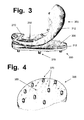
- FIG. 3 illustrates a uni-compartmental implant for a knee joint, comprising femoral runner implant 200, and tibial plateau implant 300.
- Femoral implant 200 has a smooth articulating surface 212, anchoring layer 250, and anchoring pegs 270.
- Tibial implant 300 has a smooth articulating surface 312, a meniscal replacement wedge 330 molded into the implant's outer (lateral) periphery, and anchoring pegs 370, with a distribution such as shown in FIG. 4 .
- That patent relates to cannulas and catheters, which are tubes used to drain seepage and fluids from an injured or surgically repaired portion of a patient's body, or to drain urine from the bladder for a patient who will be bedridden and unable to get to a toilet.
- cannulas and catheters which are tubes used to drain seepage and fluids from an injured or surgically repaired portion of a patient's body, or to drain urine from the bladder for a patient who will be bedridden and unable to get to a toilet.
- hydrophobic plastics such as polyvinyl chloride
- those types of tubes commonly cause irritation, inflammation, or other problems. Therefore, Wichterle and Stoy developed a method of using sulfuric acid to treat acrylonitrile tubes, to make the surfaces of the tubes more slippery and wettable, in the hope that such tubes would be medically useful.
- the Applicant obtained the surface-treated samples from Hymedix (Princeton, New Jersey). That company is no longer in existence, but a key individual George Stoy (one of the sons of Artur Stoy, who did the earlier work with PAN) now works with PragTech Inc. (Flemington, New Jersey). It is believed that those materials (prior to the sulfuric acid treatment, which was carried out in the Applicant's labs) were made by methods such as described in US patent 6,593,451 (Stoy ).
- sulfuration Because sulfur has various oxidation or valence states (as reflected in terms such as sulfates, sulfites, sulfides, sulfones, etc.), processes or reactions that add sulfur atoms to a hydrogel surface are referred to herein as "sulfuration".
- sulfonic acid groups R-SO 3 H
- sulfonation can be used; however, it should be recognized that other reagents may be useful that do not have sulfonic acid groups, such as sodium sulfite or bisulfate, sulfur dioxide or trioxide, a mixture called oleum (SO 3 dissolved in sulfuric acid), and other reagents known to those skilled in sulfur chemistry (see, e.g., E.E. Gilbert, Sulfonation and Related Reactions (Interscience Publishers, New York, 1965 )).
- Any group that contains at least one sulfur atom and at least one oxygen atom can be referred to as a sulfate group; accordingly, many but not all sulfur donors will contribute sulfate groups to hydrogel surfaces.
- Terms such as moiety or end group imply that an atom or cluster of atoms is located at the end of a chain or side chain.
- Terms such as crosslink or bridge usually imply that an atom or cluster of atoms is (or will be, after a reaction occurs) located within a chain of some sort, and will not be located at the end of a chain.
- bonds formed directly between sulfur and carbon have different bond strengths (and "breakability") than bonds with an oxygen atom between the sulfur and the carbon, nitrogen, etc.
- bonds formed directly between sulfur and carbon or between sulfur and other non-oxygen atoms, such as nitrogen or phosphorus
- bond strengths and "breakability"
- sulfur donors that create carbon-oxygen-sulfur linkages will need to be scrutinized carefully, since those indirect linkages are likely to be at greater risk of being broken (especially when contacted by biological fluids that contain esterases and other enzymes) than other sulfur-containing bond structures that can be created by skilled chemists.
- sulfuric acid H 2 SO 4 , or SO 4 -2 in ionic form
- sulfuration of (and negative charge densities on) hydrogel surfaces can render hydrogel surfaces more lubricious and durable, this approach to chemically treating a hydrogel surface is likely to enable even stronger and more durable hydrogels, if other donor reagents are used that will not create indirect linkages (with oxygen atoms between carbon and sulfur atom), and instead will directly bond a sulfur or other electronegative atom to a carbon, nitrogen, or other selected atom or group in the polymer being treated.
- hydrogels containing direct carbon-sulfur bonds can also be created by using sulfur-containing monomer reagents to form a hydrogel polymer; however, this will require careful evaluation of whether only the surface layers should include sulfur atoms, or whether sulfur atoms can be incorporated throughout the entire polymer without reducing strength, durability, or other desired traits.
- Some sulfur-donating reagents can create crosslinking bonds or bridges that will connect different polymeric chains or other molecular structures.
- Such reagents merit evaluation, because higher numbers of crosslinking bonds, in a polymeric material, can impart greater strength, toughness, and durability to a polymer.
- Crosslinking reactions are widely used to give greater strength and durability to various materials. As an illustration, most "tanning" chemicals that process animal skins into tough and durable leather do their work by creating crosslinking bonds between collagen fibers in the animal skins.
- electronegative surface-treating agents that do not contain sulfur can be evaluated for use herein.
- reagents that substitute halogen atoms for hydrogen protons, in a polymer offer good candidates.
- the bonds between hydrogen protons and carbon atoms in a polymer are not strong, and replacement of hydrogen by other atoms or moieties can often lead to stronger, more durable polymers.
- Especially promising hydrogels are those in which some or all of the hydrogen atoms are replaced by fluorine. This principle is described in US patents 4,621,107 and 4,900,793 (Lagow et al ), which disclose "perfluorinated" elastomers in which essentially all hydrogen atoms are replaced by fluorine.
- Fluorinated and perfluorinated polymers have better resistance to corrosion and wear than conventional polymers; accordingly, fluorinating agents, and prepolymer reagents that contain fluorine instead of hydrogen or chlorine in at least some locations, can be evaluated for use as described herein, and offer promising candidates for creating even stronger and more durable hydrogel compounds.
- monomers that can incorporate (or treating agents that can donate) non-sulfurous bivalent or multivalent electronegative atoms (such as nitrogen or phosphorus) into a treated polymeric surface can be evaluated for use herein to impart negative charges to a hydrogel surface, and/or to provide higher strength to a hydrogel surface due to crosslinking or other effects.
- FCD fixed charge density
- negative charge density can be measured by using a vibratome to cut cartilage into very thin slices (such as 0.2 mm thick), then weighing each layer, and then equilibrating that known weight of cartilage with a known concentration of sodium ions (Na + ) in a low-concentration saline solution.
- Na + sodium ions
- FCD values have also been developed, including ways that express charge density in milli-equivalent (mEq) rather than millimolar levels, which can indicate the concentrations of proteoglycans in cartilage.
- mEq milli-equivalent
- Other articles that discuss the negative charge density of cartilage include Maroudas et al 1969, Stanescu et al 1982 and 1986, Van Damme et al 1992, Buckwalter et al 2000, and Maniwa et al 2001.
- cartilage has a negative electrical charge, believed to be caused mainly by sulfate and carboxyl groups on chondroitin sulfate chains, sulfate groups on keratan sulfate chains, and carboxyl groups on hyaluronate chains (hydroxy groups on collagen fibers may also contribute somewhat to the negative charges on cartilage).
- a decrease in charge density indicates that cartilage is suffering from a problem which, if not corrected, may lead to a breakdown and loss of the cartilage.
- lubricin As described and illustrated in US patent 6,530,956 (by the same Applicant herein), lubricin molecules form molecular complexes with surface-active phospholipid (SAPL) molecules. These two types of molecules alternate back and forth between free (unassociated) forms, and complexes that are held together by ionic attraction. The complexes are assumed to form under resting conditions, when no compressive or shear forces are being exerted on a joint.
- SAPL surface-active phospholipid
- lubricin/SAPL complexes can be separated, without damaging either component, in a manner comparable to pulling apart two relatively weak magnets.
- the separated components will eventually recombine, presumably after the activity ends and the joint returns to a resting state, thereby reforming new complexes, which will be ready to repeat the cycle when activity begins again.
- a negative charge density, on a cartilage surface can help promote the proper formation, alignment, and activity of lubricin-SAPL complexes, by helping to ensure that the "heads" of lubricin molecules will line up in the desired manner, in a joint that is at rest.
- the negative electrical charge density on a synthetic hydrogel implant should emulate the negative electrical charge density on natural, healthy cartilage. Accordingly, unless experimental evidence indicates otherwise, the charge density on a hydrogel implant surface should have a fixed charge density within a range of about -50 to about -250 mM, measured by sodium equilibration.
- the charge density on a synthetic polymer can be altered and controlled to any level of interest, by methods known to those skilled in the art. As one example, if a lower charge density is desired, a polymer surface can be treated with a lower concentration of a sulfur-donating or similar reagent, and the treatment period can be reduced. Alternately, if a higher charge density is desired, a polymer surface can be treated with longer treatment periods, by repetitive treatments, by a series of treatments using progressively stronger reagents, and/or by using pre-polymeric reagents with higher quantities of electronegative groups.
- samples of such treated polymer surfaces having a range of charge densities can be tested to evaluate their wear factors, their coefficients of friction, and any other parameter of interest.
- an optimal charge density can be determined for any type of polymer of interest (such as polyacrylonitriles or polyurethanes that have been sulfonated, phosphorylated, or otherwise treated).
- Examples 3 and 4 disclose preferred methods of testing candidate surface-treated polymers, to evaluate the effects of a candidate chemical treatment on (i) the strength and durability of the resulting treated polymer, and (ii) the ability of a treated polymer to interact properly with synovial fluid.
- synovial fluid from cows.
- synovial fluid assertedly is commercially available, the only available preparation was inordinately expensive, so the Applicant and an assistant obtained it by visiting a slaughterhouse, and using a syringe to aspirate fluid from the ankle joints of freshly-killed carcasses. Roughly 5 ml could be obtained from a typical ankle joint.
- the fluid was filtered twice through a 20 micron filter, then 0.2% w/v sodium azide (a preservative) was added with stirring. It was filtered again, then it was frozen at -20°C until use.
- Synovial fluid is not widely used by researchers and companies that test candidate materials for replacing cartilage, for two reasons. First, it is quite expensive. Second, it makes reliable testing more time-consuming, since it provides extremely good lubrication that tends to mask and obscure the differences in performance between candidate materials being compared against each other. To avoid those problems, most researchers and companies use bovine blood serum as a lubricating liquid, in most such tests.
- the Applicant ran various tests, using bovine blood serum in some tests, and synovial fluid in other tests.
- tests using synovial fluid generated more consistent results, while tests using blood serum tended to give more scattered results.
- any shear forces that seek to push and slide the soft coating layer off of the hard support layer will effectively "focus on” the flat and planar boundary between the soft and hard materials, and will pose a greater risk of damaging the implant.
- steps can be taken to reduce this risk (such as by making the layer of plastic material relatively thin and flexible, especially on the tops of the protrusions), such steps will reduce the strength and reinforcing benefits of the molded reinforcing layer. Even if such steps are taken, some risk of cutting through an overlying layer of hydrogel, in a manner that might puncture or lacerate the smooth surface of the implant, will remain, if a molded plastic interface with holes cut through it is embedded in a hydrogel implant that is only a few millimeters thick.
- a preferred approach resides in the use of gradients, to prevent the formation or existence of a stark or sharp boundary between a hard anchoring material, and a hydrogel coating layer.
- Such gradients can be created and provided, in both: (i) the flexible fibrous mesh that will reinforce the layer of hydrogel material, and (ii) the porous anchoring material that will press against the prepared bone surface, and promote tissue ingrowth.
- gradients can offer certain advantages (such as lower impedance to water permeability through the outermost surfaces of a gel layer) which can enable an implant to more closely simulate native and natural cartilage near the articulating surface, while having improved strength, toughness, and durability due to higher levels of reinforcement in the lower layers. Therefore, gradients in the reinforcing materials merit testing and evaluation as this invention progresses through the mechanical and then animal tests that will be required before human clinical trials can commence.
- gradients can be created in any of several ways, and the selection of preferred methods and fiber types for providing such a gradient, in a reinforcing mesh, will depend on the methods and fiber type(s) being used to create the mesh.
- a first type of fiber having a first selected thickness and density can be used to form all or the majority of a first layer or zone, while a second type of fiber having a second selected thickness and density can be used to form a second layer or zone (or the majority thereof).
- a first layer of fibers having a first selected thickness can be laid onto a conveyor belt at a first selected density
- a second layer of fibers having a second selected thickness can be laid onto a conveyor belt at a second selected density.
- the bi-layer mat can be compressed and needle-punched or otherwise treated.
- the use of continuous fibers in one layer of a mesh, and chopped fibers in a different layer of a mesh can provide mechanical and performance gradients of the type anticipated herein.
- Porosity gradients in harder materials, for the anchoring surface of an implant can also be created by means such as described above, or by various other means known to those skilled in the art.
- granular particles that will dissolve in a selected liquid such as grains of salt or sugar, which will dissolve in water
- pre-polymeric liquids or granules can be mixed with pre-polymeric liquids or granules, at concentrations or ratios that will vary depending on their depth in a molding cavity.
- a controlled concentration of soluble grains can be positioned in the bottom of a mold, to create an anchoring surface, while different concentrations of soluble grains can be positioned in higher layers of the mold.
- a solvent is used to remove the grains, leaving behind a porosity gradient.
- a liquid pre-polymer that will form a hydrogel is then permeated through the harder porous polymer, and cured to form the hydrogel, which will be reinforced by the stronger polymer.
- calcium phosphate mixtures similar to hydroxyapatite, the crystalline mineral that forms hard bone can be coated (such as by "sputter-coating", which is more precise and even than liquid plating methods) onto various types of porous materials (such as thick but flexible screen meshes made of very thin metal wires) that have been provided in advance with density gradients.
- density and stiffness gradients can be created using any of various means, in both (i) a flexible mesh of the type that will be used to reinforce a hydrogel material, and (ii) a porous material that will promote tissue ingrowth, in an anchoring layer.
- a gradual and non-planar transition zone can be created between a soft hydrogel material and a harder anchoring layer, in an implant as described herein.
- Such transition zones can eliminate the need for a non-planar interface layer made of molded and perforated plastic, which could pose a risk of lacerating or puncturing the hydrogel layer in a person who suffers a fall or other accident.
- the anchoring layer and the reinforcing mesh in a hydrogel can be created from a single type of material, if the material is provided with adequate gradients in density, thicknesses of the strands, or other parameters.
- a highly flexible reinforcing mesh that would pose no danger of lacerating or puncturing a hydrogel can be made of wire (or polymer) strands that are so thin they are difficult to see with the naked eye. As these super-thin strands increase in thickness, they become stiffer as a function of cross-sectional area (i.e., as a squared function of the radius or diameter). At one or more transition zones, the strands can be made thicker, and can be woven together more densely.
- the stranded mesh can be sufficiently thick and dense to make it difficult to flex and bend without substantial effort.
- This type of mesh can provide an anchoring layer, while the thinner strands will be much more flexible and will reinforce the hydrogel, close to the articulating surface.
- Any suitable method can be used to anchor an implant as described herein to a prepared bone surface.
- the Applicant After studying various factors, issues, and items of prior art, the Applicant has identified and adapted a certain type of anchoring system that is believed, for reasons discussed below, to offer a preferred candidate system for early evaluation.
- the anchoring system that is disclosed and preferred herein, as illustrated in FIGS. 1 and 2 , uses anchoring pegs 170 that are designed to lock into place when pressed into locking receptacles 180.
- Receptacles 180 are designed to be inserted and affixed (such as by cementing, screws, press-fitting of a ridged shape, etc., or by providing the entire receptacle with an externally-threaded surface shape) into recesses that will be drilled or otherwise created in the hard bone that will support the implant, prior to insertion of the implant 100 into the joint that is being repaired.
- the system developed and disclosed by the Applicant herein works in a different manner.
- the placements of the locking receptacles 180 will be established, with the help of one or more templates, tool guides, computerized navigational equipment, or comparable devices that are already known in the art, or that can be developed by experts working on this particular step in the surgical procedure.
- the appropriate holes can be drilled, machined, or otherwise created in the bone, and the receptacles can be emplaced therein, with the assistance (if desired) of cement, anchoring pins, or other means.
- a cement can be used that will not harden for more than an hour, to allow the pegs of the implant to be inserted into the receptacles before the cement fully hardens and establishes their exact final alignment.
- means can be provided to allow the pegs to be removed from the receptacles, if and when the need arises, by means that may include deliberately destroying the remainder of the implant (allowing it to be completely replaced by a new implant, without requiring any alteration of any sort in the anchoring receptacles that remain embedded in the bone).
- removal means may include, for example, a rotatable mechanism (with screw-type threads, a so-called "bayonet" fitting, etc.) that can be accessed only from the top of the peg, by going through the hydrogel layer of the implant.
- Alternate removal means might include, for example, inserting a shim-type device into a slot between two halves of a peg, to expand the peg segments in a way that locks them into place, requiring the shim to be removed before the peg can be removed.
- Still other means that can reliably prevent accidental disengagement, but that can be removed by a surgeon if the hydrogel portion of an implant must be replaced, can be developed, if desired.
- a paste-type adhesive that will cure into a hardened material can be inserted into the receptacle, just before the pegs are inserted, in a quantity that will ensure that the gaps in the split pegs (as illustrated in FIG. 2 ) will be filled by the soft material.
- the paste will then harden after the surgery is completed, while the joint is still immobilized, in a manner that effectively locks the pegs in place.
- the outer diameter (and circumference) of a receptacle is substantially larger than the diameter (and circumference) of a peg. This allows wider receptacles to provide larger and stronger "gripping surface" areas than thin pegs, which should be slender enough to allow a flexible implant to be at least partially rolled up, during insertion, for arthroscopic insertion.
- each peg 170 can be attached to the bone anchoring layer 150 by means of a hinged device or flap, to allow the peg to be rotated into a flatter position, parallel or at least closer to anchoring layer 150, during insertion.
- hinges or flaps on different pegs on the implant 100 can be provided with different placements and directional orientations, if desired, so that all of the pegs will reinforce each other and, acting together, prevent any transverse displacement of the anchoring layer in any one direction, during the initial recovery stage while tissue is growing into the porous anchoring layer.
- anchoring layer 150 can be provided with smaller "peg attachment devices", for easier and more compact arthroscopic insertion, and the full-length pegs 170 can be affixed, one at a time, to the smaller attachment devices, after the peg receptacles have been emplaced in the underlying bone, and after the implant 100 has been inserted into the joint and is being prepared to be permanently locked in place by inserting the affixed pegs into the receptacles.
- the chemical or other steps for treating the surface of a meniscal or labral implant, to give it more lubricious upper and/or lower surfaces and a negative charge density that emulates natural cartilage, can be adapted directly from the methods disclosed above, for treating condylar implants.
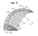
- FIG. 5 illustrates a meniscal implant 10, containing a hydrogel polymer 20 indicated by stippling.
- Meniscal implant 10 has an arc-like or crescent shape, comparable to a curved claw, with an inner surface or edge 22, and a peripheral or rim surface 24.
- the treated surface layer (or "skin") of the meniscal implant is shown by a heavy line, indicated by callout number 26 in the cutaway segment at the bottom of the drawing.
- Reinforcing mesh 40 comprises two classes of long fibers.
- Peripheral (or anchoring) strands 42 extend out of the implant 10, around the rim surface 24, which does not need to be smooth because it is not an articulating surface; five anchoring reinforcements 50, located around the periphery of the mesh 40, are also shown.
- Penetrating mesh strands 44 enter and pass through the hydrogel polymer 20. Due to the woven, knitted, or other three-dimensional structure of the mesh, a single long strand in mesh 40 might provide a peripheral/anchoring strand 42 at one location, and a penetrating strand 44 at another location.
- Fibrous strands 46 are visible in the perspective view portion of the drawing, because the thin layer of hydrogel material that covers fibrous strands 46 is essentially clear and transparent, due to its high water content. However, those fibers preferably should not be exposed on either of the upper or lower articulating surfaces of implant 10, because their presence on an articulating surface would generate elevated levels or risks of roughness and abrasion. Accordingly, strands 46 are shown for illustration only, to indicate that the mesh extends throughout and reinforced essentially the entire hydrogel component, other than the treated surface layer 26.
- Granular PVA (grade 71-30, with an average molecular weight of about 140 kilodalton) was supplied at no cost by DuPont.
- PVP average molecular weight about 40 kd was obtained from Sigma Chemical. When PVA/PVP copolymers were tested, they contained a ratio of 99 % PVA and 1% PVP, by weight. In either case, a total polymer weight of 10% w/v in distilled and deionized water was used. The mixture was stirred for 20 minutes, by which time the solution appeared to be completely uniform and consistent. It was heated to 85°C overnight, then cooled to room temperature, and stirred again for 20 minutes.
- a punch was used to remove circular samples, usually with 0.67, 1.5, or 1.625 inch diameters, depending on the tests that were planned. Before testing, these samples were fully hydrated and swelled in aqueous phosphate-buffered saline (PBS) solution, for 1.5 to 2 days.
- PBS phosphate-buffered saline
- a third method of preparation was also tested, in which a combination of partial dehydration and freeze-thawing was used.
- This method involved pouring a quantity of the polymeric liquid into a shallow mold, to a level indicated by a mark at a fixed height, and then dehydrating the polymer at 37°C until the level of the remaining material had decreased to the height of a lower mark.
- This partial dehydration was then followed by several cycles of freeze-thawing.
- the resulting materials were shown to have intermediate levels of strength and durability, between the fully dehydrated samples and the freeze-thawed samples, and this method offered good control over the thickness of the resulting hydrogel.
- Sample sheets of polyacrylonitrile were provided by the PragTech company (Flemington, New Jersey). These sheets were of a type designated as "Qpan” by Pragtech.
- the exact details of the process use to manufacture the "Qpan” class of PAN are proprietary, and may be covered by one or more currently pending patent applications (including US applications 09/383,020 and 10/193,578, both by Stoy et al and accessible on the U.S. Patent Office website).
- Methods for manufacturing polyacrylonitrile are disclosed in various patents that can be located by searching for "Stoy” as the inventor, in the U.S. patent database (www.uspto.gov).
- Circular samples of the Qpan polymer were cut from the Pragtech sheets by means of a punch. These samples were sulfonated by the same procedures disclosed above for the PVA/PVP polymers.
- the tribometer machine is used to rub pins having smooth, flat-faced surfaces made of a known type of plastic, called "ultrahigh-molecular-weight polyethylene" (UHMWPE), against smooth disks made of a very hard cobalt-chromium alloy (supplied by Biomet Inc., www.biomet.com).
- UHMWPE ultrahigh-molecular-weight polyethylene
- the pins Prior to the tests, the pins (having 0.5 inch diameters for the standardizing tests) were pre-soaked in distilled water for a month, to minimize fluid absorption during the test. A load of 253 newtons was applied to the pins, to generate an average contact stress of 3.54 megapascals (Mpa).
- the tribometer was programmed to move the table, which supported the discs, m a circular wear path having a 50 mm perimeter.
- the wear cycle frequency was 1 hertz (i.e., 1 cycle per second), giving a sliding velocity of 50 mm/s.
- the pins When a test is ready to begin, the pins are lowered onto the discs, until a known amount of force (expressed in newtons) is exerted on the pins. Based on the surface area of the pins, this generates a controllable amount of pressure (expressed in megapascals, mPa) on the UHMWPE surfaces at the bottoms of the pins.
- mPa controllable amount of pressure
- Newborn bovine calf serum (ICN Biomedicals) was diluted to 50 % (by volume) with distilled water and used as the lubricant.
- the lubricant contained 0.2 % sodium azide, and 20 millimolar ethylene-diamine-tetraacetate (EDTA), as preservatives.
- the temperature of the lubricant was maintained at 37 ⁇ 1°C throughout the test period, using a recirculating temperature control unit. The test was done for 2 million cycles, amounting to a wear path length of 100 km (about 62 miles).
- hydrogels were conducted in either 100% fetal bovine serum, or 100% synovial fluid. Prior to usage in the wear tests, 0.2 % sodium azide was added as an antibacterial agent, and the lubricant was filtered twice through a 20 micron filter. The hydrogel samples, after being attached to the pins and the discs, were soaked in the lubricant for at least two hours before the start of any test. Temperatures of all lubricants were maintained at 37 ⁇ 1°C throughout the test period.
- the machine can test up to 6 samples at once, each using its own pin.
- the hydrogel samples affixed to the pins and the discs were taken from the same sheet of material, to ensure that they had the same thicknesses.
- An "upper” hydrogel sample is affixed to the bottom of each pin used, using a cyanoacrylate adhesive, while “lower” hydrogel samples were attached firmly to stainless steel disks (1.7 inch diameter) with the help of acrylic fixtures. These fixtures also provide a shallow tray, which holds lubricant at a depth that will cover and bathe both of the hydrogel samples throughout a test.
- samples that were sulfonated were treated on only one side, to allow the untreated side to provide better adhesion to the pins.
- a force level (usually ranging from 100 to 170 newtons) was chosen for that test.
- a force of 150 newtons was applied to six pins, it generated an average contact pressure of 2.9 megapascals (Mpa), which falls within normal physiologic stress levels that are typical in an adult joint.
- the platform that supports the stainless steel discs and the lower samples begins to move in a programmed motion. While the standardization tests used a circular motion, the wear-testing of hydrogels used a straight-line reciprocating motion, 30 mm in length. The cycle frequency was increased to 1.67 hertz, which maintained the sliding velocity at 50 mm/sec, which is the ASTM F732 standard recommendation. If a test continued for 1 million cycles, the total sliding distance was 30 kilometers (about 18 miles).
- polyacrylonitrile polymers provided more durable hydrogels than poly(vinyl alcohol) polymers; and (ii) sulfonation of the polymer surface increased the lubricity (slipperiness) of either type of polymer, and can provide a useful improvement for cartilage-replacing implants.
- coefficient of friction values were determined for a number of hydrogel samples. These values were measured while a sample articulated against the same type of material, using either bovine blood serum or bovine synovial fluid as the lubricant, and using various force and pressure loadings. Those values are provided in Table 1. TABLE 1 COEFFICIENTS OF FRICTION FOR HYDROGEL SAMPLES MATERIAL LIQUID & LOADING COEFF.
Claims (15)
- Chirurgisches Implantat (100) zum Ersatz von Knorpel in einem Säugetier-Gelenk, aufweisend ein Hydrogelmaterial, das ein synthetisches Polymer enthält,
dadurch gekennzeichnet, dass das chirurgische Implantat (100) mindestens eine glatte und schmierfähige Oberfläche (112) mit einer negativen elektrischen Ladungsdichte hat, die schmierende Wechselwirkungen mit Säugetier-Synovialflüssigkeit fördert. - Chirurgisches Implantat (100) gemäß Anspruch 1, wobei eine flexible, fibröse Verstärkungsmatrix (122) in mindestens einem Teil des Hydrogelmaterials eingebettet ist.
- Chirurgisches Implantat (100) gemäß Anspruch 1 oder Anspruch 2, wobei die glatte und schmierfähige Oberfläche (112) eine negative elektrische Ladungsdichte gemessen durch Äquilibrierung mit Natrium innerhalb eines Bereichs von ungefähr -50 bis ungefähr -250 mmol hat.
- Chirurgisches Implantat (100) gemäß Anspruch 1 oder Anspruch 2, wobei die glatte und schmierfähige Oberfläche (112) Schwefelatome enthält.
- Chirurgisches Implantat (100) gemäß Anspruch 1 oder Anspruch 2, wobei die glatte und schmierfähige Oberfläche (112) Fluoratome enthält.
- Chirurgisches Implantat (100) gemäß Anspruch 1 oder Anspruch 2, wobei die glatte und schmierfähige Oberfläche (112) mit einem chemischen Reagens behandelt wurde, das Vernetzungsverbindungen zwischen Polymermolekülen herstellt.
- Chirurgisches Implantat (100) gemäß Anspruch 1 oder Anspruch 2, wobei das Implantat eine Verankerungsoberfläche hat, die mit Mitteln zum Verankern des Implantats auf einer präparierten Knochenoberfläche ausgestattet ist.
- Chirurgisches Implantat (100) gemäß Anspruch 7, wobei die Mittel zum Verankern des Implantats auf einer präparierten Knochenoberfläche Stifte (170) beinhaltet, die dazu ausgelegt sind, in entsprechende Aufnahmen (180) zu passen, die vor dem Einsetzen des chirurgischen Implantats (100) in ein Gelenk in eine präparierte Knochenoberfläche eingesetzt werden können.
- Chirurgisches Implantat (100) gemäß Anspruch 7, wobei mindestens ein Teil der Verankerungsoberfläche ein poröses Material (150) aufweist, das nach der chirurgischen Implantierung ein Einwachsen von Gewebe fördert.
- Chirurgisches Implantat gemäß Anspruch 1 oder Anspruch 2, wobei das Implantat (100) zwei glatte und schmierfähige Oberflächen hat und so abgemessen und ausgelegt ist, dass es Meniskus- oder Labrumgewebe ersetzt.
- Chirurgisches Implantat gemäß Anspruch 1 oder Anspruch 2, wobei das synthetische Polymer aus der Gruppe ausgewählt ist, die aus Polyacrylnitril- und Polyurethanverbindungen und -derivaten besteht.
- Verfahren zur Herstellung eines nicht-resorbierbaren chirurgischen Implantats (100) zum Ersetzen von Knorpel in einem Säugetier-Gelenk, dadurch gekennzeichnet, dass das Verfahren die folgenden Schritte aufweist:a. Erzeugen eines Hydrogelmaterials, das mindestens eine glatte Oberfläche (112) zur Herstellung einer Gelenkigkeit nach der chirurgischen Implantierung aufweist, undb. Unterziehen mindestens einer glatten Oberfläche des Hydrogelmaterials einer chemischen Behandlung, die der glatten Oberfläche (112) eine negative elektrische Ladung verleiht, und zwar in einer Weise, die eine Ladungsdichte erzeugt, die schmierende Wechselwirkungen zwischen der glatten Oberfläche (112) und Säugetier-Synovialflüssigkeit fördert.
- Verfahren gemäß Anspruch 12, wobei die chemische Behandlung auf einer glatten Oberfläche eine negative elektrische Ladungsdichte gemessen durch Äquilibrierung mit Natrium innerhalb eines Bereichs von ungefähr -50 bis ungefähr -250 mmol erzeugt.
- Verfahren gemäß Anspruch 12, wobei sich eine fibröse Verstärkungsmatrix durch mindestens einen Teil des Hydrogelmaterials erstreckt.
- Verfahren gemäß Anspruch 12, wobei das Hydrogelmaterial aus einem synthetischen Polymer hergestellt ist, das aus der Gruppe ausgewählt ist, die aus Polyacrylnitril- und Polyurethanverbindungen und -derivaten besteht.
Applications Claiming Priority (4)
| Application Number | Priority Date | Filing Date | Title |
|---|---|---|---|
| US10/677,444 US9314339B2 (en) | 2000-03-27 | 2003-10-02 | Implants for replacing cartilage, with negatively-charged hydrogel surfaces and flexible matrix reinforcement |
| US56217604P | 2004-04-14 | 2004-04-14 | |
| PCT/US2004/032504 WO2005032426A1 (en) | 2003-10-02 | 2004-10-04 | Hydrogels having charged surfaces for cartilage replacement |
| US11/105,677 US20050287187A1 (en) | 2003-10-02 | 2005-04-14 | Hydrogel implants for replacing hyaline cartilage, with charged surfaces and improved anchoring |
Publications (3)
| Publication Number | Publication Date |
|---|---|
| EP1722717A1 EP1722717A1 (de) | 2006-11-22 |
| EP1722717A4 EP1722717A4 (de) | 2011-03-02 |
| EP1722717B1 true EP1722717B1 (de) | 2013-06-19 |
Family
ID=38328214
Family Applications (1)
| Application Number | Title | Priority Date | Filing Date |
|---|---|---|---|
| EP04794020.0A Active EP1722717B1 (de) | 2003-10-02 | 2004-10-04 | Hydrogele mit geladenen oberflächen für den knorpelersatz |
Country Status (3)
| Country | Link |
|---|---|
| US (1) | US20050287187A1 (de) |
| EP (1) | EP1722717B1 (de) |
| WO (1) | WO2005032426A1 (de) |
Families Citing this family (89)
| Publication number | Priority date | Publication date | Assignee | Title |
|---|---|---|---|---|
| US8177841B2 (en) | 2000-05-01 | 2012-05-15 | Arthrosurface Inc. | System and method for joint resurface repair |
| US7678151B2 (en) * | 2000-05-01 | 2010-03-16 | Ek Steven W | System and method for joint resurface repair |
| EP2314257B9 (de) | 2000-05-01 | 2013-02-27 | ArthroSurface, Inc. | System zur Gelenkoberflächenerneuerung |
| US6610067B2 (en) | 2000-05-01 | 2003-08-26 | Arthrosurface, Incorporated | System and method for joint resurface repair |
| US7163541B2 (en) | 2002-12-03 | 2007-01-16 | Arthrosurface Incorporated | Tibial resurfacing system |
| US7713305B2 (en) * | 2000-05-01 | 2010-05-11 | Arthrosurface, Inc. | Articular surface implant |
| US7901408B2 (en) | 2002-12-03 | 2011-03-08 | Arthrosurface, Inc. | System and method for retrograde procedure |
| US8388624B2 (en) | 2003-02-24 | 2013-03-05 | Arthrosurface Incorporated | Trochlear resurfacing system and method |
| AU2006203909A1 (en) | 2003-11-20 | 2006-07-13 | Arthrosurface, Inc. | System and method for retrograde procedure |
| CA2546582A1 (en) | 2003-11-20 | 2005-06-09 | Arthrosurface, Inc. | Retrograde delivery of resurfacing devices |
| US7951163B2 (en) * | 2003-11-20 | 2011-05-31 | Arthrosurface, Inc. | Retrograde excision system and apparatus |
| CA2558623C (en) | 2004-02-06 | 2013-04-16 | Georgia Tech Research Corporation | Surface directed cellular attachment |
| WO2005077304A1 (en) | 2004-02-06 | 2005-08-25 | Georgia Tech Research Corporation | Load bearing biocompatible device |
| US7651015B2 (en) * | 2004-02-13 | 2010-01-26 | Intelligent Coffee Company, Llc | Liquid concentrate/extract beverage dispenser with replaceable concentrate/extract cartridge |
| US20050278025A1 (en) * | 2004-06-10 | 2005-12-15 | Salumedica Llc | Meniscus prosthesis |
| WO2006004885A2 (en) | 2004-06-28 | 2006-01-12 | Arthrosurface, Inc. | System for articular surface replacement |
| US20090088846A1 (en) | 2007-04-17 | 2009-04-02 | David Myung | Hydrogel arthroplasty device |
| US7828853B2 (en) | 2004-11-22 | 2010-11-09 | Arthrosurface, Inc. | Articular surface implant and delivery system |
| US8096303B2 (en) * | 2005-02-08 | 2012-01-17 | Koninklijke Philips Electronics N.V | Airway implants and methods and devices for insertion and retrieval |
| US20060224244A1 (en) * | 2005-03-31 | 2006-10-05 | Zimmer Technology, Inc. | Hydrogel implant |
| US7291169B2 (en) * | 2005-04-15 | 2007-11-06 | Zimmer Technology, Inc. | Cartilage implant |
| US20060235542A1 (en) * | 2005-04-15 | 2006-10-19 | Zimmer Technology, Inc. | Flexible segmented bearing implant |
| CA2610871A1 (en) * | 2005-06-06 | 2006-12-14 | The General Hospital Corporation | Tough hydrogels |
| CA2617217A1 (en) * | 2005-07-29 | 2007-02-08 | Arthrosurface, Inc. | System and method for articular surface repair |
| EP1924208B1 (de) | 2005-08-31 | 2012-08-22 | Zimmer GmbH | Femur Implantat |
| US20070088444A1 (en) * | 2005-10-13 | 2007-04-19 | Robert A Hodorek | Method for repairing a bone defect using a formable implant which hardens in vivo |
| US8403985B2 (en) * | 2005-11-02 | 2013-03-26 | Zimmer, Inc. | Joint spacer implant |
| US8308807B2 (en) * | 2005-11-09 | 2012-11-13 | Zimmer, Gmbh | Implant with differential anchoring |
| US8038920B2 (en) * | 2006-01-25 | 2011-10-18 | Carticept Medical, Inc. | Methods of producing PVA hydrogel implants and related devices |
| US8603171B2 (en) * | 2006-01-25 | 2013-12-10 | Mimedx Group, Inc. | Spinal disc implants with flexible keels and methods of fabricating implants |
| US20070179617A1 (en) * | 2006-01-25 | 2007-08-02 | Spinemedica Corporation | Prosthetic wide range motion facets and methods of fabricating same |
| US20070179607A1 (en) * | 2006-01-31 | 2007-08-02 | Zimmer Technology, Inc. | Cartilage resurfacing implant |
| WO2007125060A1 (de) | 2006-04-28 | 2007-11-08 | Zimmer Gmbh | Implantat |
| US20070293947A1 (en) * | 2006-06-19 | 2007-12-20 | Mansmann Kevin A | Multi-part implants for combined repair of hyaline and meniscal cartilage in joints |
| US9358029B2 (en) | 2006-12-11 | 2016-06-07 | Arthrosurface Incorporated | Retrograde resection apparatus and method |
| US8334044B2 (en) * | 2007-02-06 | 2012-12-18 | The Board Of Trustees Of The Leland Stanford Junior University | Hydrogel-metal assembly |
| CA2618125A1 (en) * | 2007-02-08 | 2008-08-08 | Zimmer, Inc. | Hydrogel proximal interphalangeal implant |
| EP2187844A2 (de) | 2007-07-31 | 2010-05-26 | Zimmer, Inc. | Prothetische gelenkspalt-interpositionsvorrichtung mit inneren anlageflächen |
| US9539097B2 (en) | 2007-11-08 | 2017-01-10 | Linares Medical Devices, Llc | Hip and knee joint assemblies incorporating debris collection architecture between the ball and seat interface |
| US8828088B2 (en) * | 2007-11-08 | 2014-09-09 | Linares Medical Devices, Llc | Joint assembly incorporating undercut surface design to entrap accumulating wear debris from plastic joint assembly |
| US8979938B2 (en) * | 2007-11-08 | 2015-03-17 | Linares Medical Devices, Llc | Artificial knee implant including liquid ballast supporting / rotating surfaces and incorporating flexible multi-material and natural lubricant retaining matrix applied to a joint surface |
| US8702801B2 (en) * | 2008-02-25 | 2014-04-22 | Linares Medical Devices, Llc | Artificial wear resistant plug for mounting to existing joint bone |
| CN102014800B (zh) | 2008-02-28 | 2014-04-30 | 比奥波利公司 | 局部关节表面置换植入物、仪器和方法 |
| US9216085B2 (en) | 2008-02-28 | 2015-12-22 | Biopoly, Llc | Partial joint resurfacing implant, instrumentation, and method |
| WO2009111481A1 (en) | 2008-03-03 | 2009-09-11 | Arthrosurface Incorporated | Bone resurfacing system and method |
| WO2009120776A2 (en) * | 2008-03-26 | 2009-10-01 | Linares Medical Devices, Llc | Joint construction, such as for use by athletes |
| AU2009268687A1 (en) * | 2008-07-07 | 2010-01-14 | Biomimedica, Inc | Hydrophilic interpenetrating polymer networks derived from hydrophobic polymers |
| US20120209396A1 (en) | 2008-07-07 | 2012-08-16 | David Myung | Orthopedic implants having gradient polymer alloys |
| GB0812631D0 (en) * | 2008-07-10 | 2008-08-20 | Imp Innovations Ltd | Modular knee implants |
| WO2010014446A1 (en) * | 2008-07-28 | 2010-02-04 | Zimmer, Inc. | Mosaicplasty constructs |
| KR20110040969A (ko) | 2008-08-05 | 2011-04-20 | 바이오미메디카, 인코포레이티드 | 폴리우레탄-그라프트된 하이드로겔 |
| US20100151114A1 (en) * | 2008-12-17 | 2010-06-17 | Zimmer, Inc. | In-line treatment of yarn prior to creating a fabric |
| WO2010092065A1 (en) * | 2009-02-11 | 2010-08-19 | INSERM (Institut National de la Santé et de la Recherche Médicale) | Endo-prosthesis for cartilage lesions |
| US10945743B2 (en) | 2009-04-17 | 2021-03-16 | Arthrosurface Incorporated | Glenoid repair system and methods of use thereof |
| WO2010121250A1 (en) | 2009-04-17 | 2010-10-21 | Arthrosurface Incorporated | Glenoid resurfacing system and method |
| WO2010121246A1 (en) | 2009-04-17 | 2010-10-21 | Arthrosurface Incorporated | Glenoid resurfacing system and method |
| US20100324693A1 (en) * | 2009-06-23 | 2010-12-23 | Hardenbrook Frederick H | Carpometacarpal joint spacer |
| CA2792048A1 (en) | 2010-03-05 | 2011-09-09 | Arthrosurface Incorporated | Tibial resurfacing system and method |
| US8926705B2 (en) | 2010-05-10 | 2015-01-06 | Linares Medical Devices, Llc | Implantable joint assembly featuring debris entrapment chamber subassemblies along with opposing magnetic fields generated between articulating implant components in order to minimize frictional force and associated wear |
| US8668739B2 (en) | 2010-08-20 | 2014-03-11 | Zimmer, Inc. | Unitary orthopedic implant |
| EP2609154B1 (de) | 2010-08-27 | 2020-04-22 | Hyalex Orthopaedics, Inc. | Aus hydrophoben polymeren gewonnene interpenetrierende hydrophobe und hydrophile polymernetzwerke sowie verfahren zu ihrer herstellung |
| US9474620B2 (en) * | 2010-09-04 | 2016-10-25 | Lew C. Schon | Talonavicular joint prosthesis and its method of implantation |
| US8617240B2 (en) | 2010-11-17 | 2013-12-31 | Charles D. Hightower | Moldable cushion for implants |
| US20130190873A1 (en) * | 2012-01-20 | 2013-07-25 | Kevin A. Mansmann | Rim anchoring systems for flexible surgical implants for replacing cartilage |
| US9066716B2 (en) | 2011-03-30 | 2015-06-30 | Arthrosurface Incorporated | Suture coil and suture sheath for tissue repair |
| CA3048437C (en) | 2011-05-26 | 2022-06-21 | Cartiva, Inc. | Tapered joint implant and related tools |
| US20130103157A1 (en) | 2011-10-03 | 2013-04-25 | Lampros Kourtis | Polymeric adhesive for anchoring compliant materials to another surface |
| EP3335673A1 (de) | 2011-11-21 | 2018-06-20 | Biomimedica, Inc | Systeme zur verankerung orthopädischer implantate an knochen |
| US20130165982A1 (en) | 2011-12-22 | 2013-06-27 | Arthrosurface Incorporated | System and Method for Bone Fixation |
| WO2014008126A1 (en) | 2012-07-03 | 2014-01-09 | Arthrosurface Incorporated | System and method for joint resurfacing and repair |
| US9492200B2 (en) | 2013-04-16 | 2016-11-15 | Arthrosurface Incorporated | Suture system and method |
| US10624748B2 (en) | 2014-03-07 | 2020-04-21 | Arthrosurface Incorporated | System and method for repairing articular surfaces |
| US11607319B2 (en) | 2014-03-07 | 2023-03-21 | Arthrosurface Incorporated | System and method for repairing articular surfaces |
| US20150250472A1 (en) | 2014-03-07 | 2015-09-10 | Arthrosurface Incorporated | Delivery System for Articular Surface Implant |
| WO2016161026A1 (en) | 2015-03-31 | 2016-10-06 | Cartiva, Inc. | Carpometacarpal (cmc) implants and methods |
| CA2981061A1 (en) | 2015-03-31 | 2016-10-06 | Cartiva, Inc. | Hydrogel implants with porous materials and methods |
| EP3282961A4 (de) | 2015-04-14 | 2018-12-05 | Cartiva, Inc. | Werkzeug zur erzeugung einer konischen öffnung in gewebe und zugehörige verfahren |
| US11077228B2 (en) | 2015-08-10 | 2021-08-03 | Hyalex Orthopaedics, Inc. | Interpenetrating polymer networks |
| US20180236136A1 (en) * | 2015-08-10 | 2018-08-23 | Hyalex Orthopaedics, Inc. | Interpenetrating polymer networks |
| CN108289733A (zh) * | 2015-10-05 | 2018-07-17 | 孚美公司 | 医疗植入物和用于医疗植入物的固定系统 |
| US9943414B2 (en) * | 2015-12-30 | 2018-04-17 | Wasas, Llc. | System and method for non-binding allograft subtalar joint implant |
| EP3528750B1 (de) * | 2016-10-20 | 2020-07-08 | Technische Universiteit Eindhoven | Biomimetisches funktionelles und regeneratives knorpelimplantat |
| GB201708233D0 (en) | 2017-05-23 | 2017-07-05 | Orthox Ltd | Implantable tissue repair devices and methods for manufacturing the same |
| CA3108761A1 (en) | 2017-08-04 | 2019-02-07 | Arthrosurface Incorporated | Multicomponent articular surface implant |
| US10869950B2 (en) | 2018-07-17 | 2020-12-22 | Hyalex Orthopaedics, Inc. | Ionic polymer compositions |
| GB2616360B (en) | 2019-03-12 | 2023-11-29 | Arthrosurface Inc | Humeral and glenoid articular surface implant systems and methods |
| IL299617A (en) | 2020-07-01 | 2023-03-01 | Univ Duke | Nanofibrous reinforcement of interconnected hydrogels |
| WO2023278386A1 (en) * | 2021-06-28 | 2023-01-05 | Exogenesis Corporation | Protection of orthopedic implants from wear |
| US11701452B2 (en) | 2021-10-12 | 2023-07-18 | Duke University | Synthetic hydrogel composite |
Family Cites Families (9)
| Publication number | Priority date | Publication date | Assignee | Title |
|---|---|---|---|---|
| US4183884A (en) * | 1973-01-24 | 1980-01-15 | Ceskoslovenska Akademie Ved | Method for manufacturing hydrogel tubes |
| WO1987005038A1 (en) * | 1986-02-17 | 1987-08-27 | Commonwealth Scientific And Industrial Research Or | Implantable materials |
| JP3007903B2 (ja) * | 1991-03-29 | 2000-02-14 | 京セラ株式会社 | 人工椎間板 |
| EP0677297B1 (de) * | 1993-09-24 | 2000-12-13 | Takiron Co. Ltd. | Implantatmaterial |
| US5556429A (en) * | 1994-05-06 | 1996-09-17 | Advanced Bio Surfaces, Inc. | Joint resurfacing system |
| US6629997B2 (en) * | 2000-03-27 | 2003-10-07 | Kevin A. Mansmann | Meniscus-type implant with hydrogel surface reinforced by three-dimensional mesh |
| US7361195B2 (en) * | 2001-07-16 | 2008-04-22 | Depuy Products, Inc. | Cartilage repair apparatus and method |
| US20030212456A1 (en) * | 2002-05-09 | 2003-11-13 | John Lipchitz | Implant for tissue repair |
| US20040070107A1 (en) * | 2002-10-09 | 2004-04-15 | Pragtech, Inc. | Method of making lubricious polyacrylonitrile artificial joint components and resulting product |
-
2004
- 2004-10-04 EP EP04794020.0A patent/EP1722717B1/de active Active
- 2004-10-04 WO PCT/US2004/032504 patent/WO2005032426A1/en active Application Filing
-
2005
- 2005-04-14 US US11/105,677 patent/US20050287187A1/en not_active Abandoned
Also Published As
| Publication number | Publication date |
|---|---|
| EP1722717A1 (de) | 2006-11-22 |
| US20050287187A1 (en) | 2005-12-29 |
| WO2005032426A1 (en) | 2005-04-14 |
| EP1722717A4 (de) | 2011-03-02 |
Similar Documents
| Publication | Publication Date | Title |
|---|---|---|
| EP1722717B1 (de) | Hydrogele mit geladenen oberflächen für den knorpelersatz | |
| US9314339B2 (en) | Implants for replacing cartilage, with negatively-charged hydrogel surfaces and flexible matrix reinforcement | |
| US11129722B2 (en) | Meniscus prosthetic device selection and implantation methods | |
| US20200229933A1 (en) | Tensioned Meniscus Prosthetic Devices and Associated Methods | |
| US6629997B2 (en) | Meniscus-type implant with hydrogel surface reinforced by three-dimensional mesh | |
| US8361147B2 (en) | Meniscus prosthetic devices with anti-migration features | |
| US7611653B1 (en) | Manufacturing and material processing for prosthetic devices | |
| AU605723B2 (en) | Prosthetic meniscus | |
| US6686437B2 (en) | Medical implants made of wear-resistant, high-performance polyimides, process of making same and medical use of same | |
| JP5753075B2 (ja) | 半月板の人工器官及びこれに関連する方法 | |
| ES2427428T3 (es) | Hidrogeles que presentan superficies cargadas para la sustitución de cartílago | |
| Dumbleton | The clinical significance of wear in total hip and knee prostheses | |
| Abela et al. | Elastomers for the Development of Orthopaedic Implants-A Review | |
| Adesina | An Investigation into an All Polymer Knee Joint Replacement | |
| Mostakhdemin | Design and Synthesis of Bilayer Nanocomposite Hydrogels for Minimally Invasive Cartilage Replacement | |
| Austin | Wear of PEEK all-polymer articulations for cervical spinal disc arthroplasty |
Legal Events
| Date | Code | Title | Description |
|---|---|---|---|
| PUAI | Public reference made under article 153(3) epc to a published international application that has entered the european phase |
Free format text: ORIGINAL CODE: 0009012 |
|
| 17P | Request for examination filed |
Effective date: 20060913 |
|
| AK | Designated contracting states |
Kind code of ref document: A1 Designated state(s): AT BE BG CH CY CZ DE DK EE ES FI FR GB GR HU IE IT LI LU MC NL PL PT RO SE SI SK TR |
|
| DAX | Request for extension of the european patent (deleted) | ||
| A4 | Supplementary search report drawn up and despatched |
Effective date: 20110128 |
|
| 17Q | First examination report despatched |
Effective date: 20110519 |
|
| REG | Reference to a national code |
Ref country code: DE Ref legal event code: R079 Ref document number: 602004042499 Country of ref document: DE Free format text: PREVIOUS MAIN CLASS: A61F0002080000 Ipc: A61L0027520000 |
|
| RIC1 | Information provided on ipc code assigned before grant |
Ipc: A61F 2/30 20060101ALI20121016BHEP Ipc: A61L 27/52 20060101AFI20121016BHEP |
|
| GRAP | Despatch of communication of intention to grant a patent |
Free format text: ORIGINAL CODE: EPIDOSNIGR1 |
|
| GRAS | Grant fee paid |
Free format text: ORIGINAL CODE: EPIDOSNIGR3 |
|
| GRAA | (expected) grant |
Free format text: ORIGINAL CODE: 0009210 |
|
| AK | Designated contracting states |
Kind code of ref document: B1 Designated state(s): AT BE BG CH CY CZ DE DK EE ES FI FR GB GR HU IE IT LI LU MC NL PL PT RO SE SI SK TR |
|
| REG | Reference to a national code |
Ref country code: GB Ref legal event code: FG4D |
|
| REG | Reference to a national code |
Ref country code: CH Ref legal event code: EP |
|
| REG | Reference to a national code |
Ref country code: AT Ref legal event code: REF Ref document number: 617266 Country of ref document: AT Kind code of ref document: T Effective date: 20130715 |
|
| REG | Reference to a national code |
Ref country code: IE Ref legal event code: FG4D |
|
| REG | Reference to a national code |
Ref country code: DE Ref legal event code: R096 Ref document number: 602004042499 Country of ref document: DE Effective date: 20130814 |
|
| REG | Reference to a national code |
Ref country code: ES Ref legal event code: FG2A Ref document number: 2427428 Country of ref document: ES Kind code of ref document: T3 Effective date: 20131030 |
|
| PG25 | Lapsed in a contracting state [announced via postgrant information from national office to epo] |
Ref country code: GR Free format text: LAPSE BECAUSE OF FAILURE TO SUBMIT A TRANSLATION OF THE DESCRIPTION OR TO PAY THE FEE WITHIN THE PRESCRIBED TIME-LIMIT Effective date: 20130920 Ref country code: SI Free format text: LAPSE BECAUSE OF FAILURE TO SUBMIT A TRANSLATION OF THE DESCRIPTION OR TO PAY THE FEE WITHIN THE PRESCRIBED TIME-LIMIT Effective date: 20130619 Ref country code: SE Free format text: LAPSE BECAUSE OF FAILURE TO SUBMIT A TRANSLATION OF THE DESCRIPTION OR TO PAY THE FEE WITHIN THE PRESCRIBED TIME-LIMIT Effective date: 20130619 Ref country code: FI Free format text: LAPSE BECAUSE OF FAILURE TO SUBMIT A TRANSLATION OF THE DESCRIPTION OR TO PAY THE FEE WITHIN THE PRESCRIBED TIME-LIMIT Effective date: 20130619 |
|
| REG | Reference to a national code |
Ref country code: AT Ref legal event code: MK05 Ref document number: 617266 Country of ref document: AT Kind code of ref document: T Effective date: 20130619 |
|
| PG25 | Lapsed in a contracting state [announced via postgrant information from national office to epo] |
Ref country code: BG Free format text: LAPSE BECAUSE OF FAILURE TO SUBMIT A TRANSLATION OF THE DESCRIPTION OR TO PAY THE FEE WITHIN THE PRESCRIBED TIME-LIMIT Effective date: 20130919 |
|
| REG | Reference to a national code |
Ref country code: NL Ref legal event code: VDEP Effective date: 20130619 |
|
| PG25 | Lapsed in a contracting state [announced via postgrant information from national office to epo] |
Ref country code: SK Free format text: LAPSE BECAUSE OF FAILURE TO SUBMIT A TRANSLATION OF THE DESCRIPTION OR TO PAY THE FEE WITHIN THE PRESCRIBED TIME-LIMIT Effective date: 20130619 Ref country code: CY Free format text: LAPSE BECAUSE OF FAILURE TO SUBMIT A TRANSLATION OF THE DESCRIPTION OR TO PAY THE FEE WITHIN THE PRESCRIBED TIME-LIMIT Effective date: 20130710 Ref country code: BE Free format text: LAPSE BECAUSE OF FAILURE TO SUBMIT A TRANSLATION OF THE DESCRIPTION OR TO PAY THE FEE WITHIN THE PRESCRIBED TIME-LIMIT Effective date: 20130619 Ref country code: AT Free format text: LAPSE BECAUSE OF FAILURE TO SUBMIT A TRANSLATION OF THE DESCRIPTION OR TO PAY THE FEE WITHIN THE PRESCRIBED TIME-LIMIT Effective date: 20130619 Ref country code: CZ Free format text: LAPSE BECAUSE OF FAILURE TO SUBMIT A TRANSLATION OF THE DESCRIPTION OR TO PAY THE FEE WITHIN THE PRESCRIBED TIME-LIMIT Effective date: 20130619 Ref country code: EE Free format text: LAPSE BECAUSE OF FAILURE TO SUBMIT A TRANSLATION OF THE DESCRIPTION OR TO PAY THE FEE WITHIN THE PRESCRIBED TIME-LIMIT Effective date: 20130619 Ref country code: PT Free format text: LAPSE BECAUSE OF FAILURE TO SUBMIT A TRANSLATION OF THE DESCRIPTION OR TO PAY THE FEE WITHIN THE PRESCRIBED TIME-LIMIT Effective date: 20131021 |
|
| PG25 | Lapsed in a contracting state [announced via postgrant information from national office to epo] |
Ref country code: NL Free format text: LAPSE BECAUSE OF FAILURE TO SUBMIT A TRANSLATION OF THE DESCRIPTION OR TO PAY THE FEE WITHIN THE PRESCRIBED TIME-LIMIT Effective date: 20130619 Ref country code: RO Free format text: LAPSE BECAUSE OF FAILURE TO SUBMIT A TRANSLATION OF THE DESCRIPTION OR TO PAY THE FEE WITHIN THE PRESCRIBED TIME-LIMIT Effective date: 20130619 Ref country code: PL Free format text: LAPSE BECAUSE OF FAILURE TO SUBMIT A TRANSLATION OF THE DESCRIPTION OR TO PAY THE FEE WITHIN THE PRESCRIBED TIME-LIMIT Effective date: 20130619 |
|
| PG25 | Lapsed in a contracting state [announced via postgrant information from national office to epo] |
Ref country code: CY Free format text: LAPSE BECAUSE OF FAILURE TO SUBMIT A TRANSLATION OF THE DESCRIPTION OR TO PAY THE FEE WITHIN THE PRESCRIBED TIME-LIMIT Effective date: 20130619 |
|
| PLBE | No opposition filed within time limit |
Free format text: ORIGINAL CODE: 0009261 |
|
| STAA | Information on the status of an ep patent application or granted ep patent |
Free format text: STATUS: NO OPPOSITION FILED WITHIN TIME LIMIT |
|
| PG25 | Lapsed in a contracting state [announced via postgrant information from national office to epo] |
Ref country code: DK Free format text: LAPSE BECAUSE OF FAILURE TO SUBMIT A TRANSLATION OF THE DESCRIPTION OR TO PAY THE FEE WITHIN THE PRESCRIBED TIME-LIMIT Effective date: 20130619 |
|
| 26N | No opposition filed |
Effective date: 20140320 |
|
| PG25 | Lapsed in a contracting state [announced via postgrant information from national office to epo] |
Ref country code: MC Free format text: LAPSE BECAUSE OF FAILURE TO SUBMIT A TRANSLATION OF THE DESCRIPTION OR TO PAY THE FEE WITHIN THE PRESCRIBED TIME-LIMIT Effective date: 20130619 |
|
| REG | Reference to a national code |
Ref country code: CH Ref legal event code: PL |
|
| REG | Reference to a national code |
Ref country code: DE Ref legal event code: R097 Ref document number: 602004042499 Country of ref document: DE Effective date: 20140320 |
|
| REG | Reference to a national code |
Ref country code: IE Ref legal event code: MM4A |
|
| PG25 | Lapsed in a contracting state [announced via postgrant information from national office to epo] |
Ref country code: LI Free format text: LAPSE BECAUSE OF NON-PAYMENT OF DUE FEES Effective date: 20131031 Ref country code: CH Free format text: LAPSE BECAUSE OF NON-PAYMENT OF DUE FEES Effective date: 20131031 |
|
| PG25 | Lapsed in a contracting state [announced via postgrant information from national office to epo] |
Ref country code: IE Free format text: LAPSE BECAUSE OF NON-PAYMENT OF DUE FEES Effective date: 20131004 |
|
| PG25 | Lapsed in a contracting state [announced via postgrant information from national office to epo] |
Ref country code: TR Free format text: LAPSE BECAUSE OF FAILURE TO SUBMIT A TRANSLATION OF THE DESCRIPTION OR TO PAY THE FEE WITHIN THE PRESCRIBED TIME-LIMIT Effective date: 20130619 |
|
| PG25 | Lapsed in a contracting state [announced via postgrant information from national office to epo] |
Ref country code: HU Free format text: LAPSE BECAUSE OF FAILURE TO SUBMIT A TRANSLATION OF THE DESCRIPTION OR TO PAY THE FEE WITHIN THE PRESCRIBED TIME-LIMIT; INVALID AB INITIO Effective date: 20041004 Ref country code: LU Free format text: LAPSE BECAUSE OF NON-PAYMENT OF DUE FEES Effective date: 20131004 |
|
| REG | Reference to a national code |
Ref country code: FR Ref legal event code: PLFP Year of fee payment: 12 |
|
| PGFP | Annual fee paid to national office [announced via postgrant information from national office to epo] |
Ref country code: ES Payment date: 20150915 Year of fee payment: 12 |
|
| PGFP | Annual fee paid to national office [announced via postgrant information from national office to epo] |
Ref country code: FR Payment date: 20150908 Year of fee payment: 12 |
|
| PGFP | Annual fee paid to national office [announced via postgrant information from national office to epo] |
Ref country code: IT Payment date: 20151026 Year of fee payment: 12 |
|
| REG | Reference to a national code |
Ref country code: FR Ref legal event code: ST Effective date: 20170630 |
|
| PG25 | Lapsed in a contracting state [announced via postgrant information from national office to epo] |
Ref country code: FR Free format text: LAPSE BECAUSE OF NON-PAYMENT OF DUE FEES Effective date: 20161102 |
|
| PG25 | Lapsed in a contracting state [announced via postgrant information from national office to epo] |
Ref country code: IT Free format text: LAPSE BECAUSE OF NON-PAYMENT OF DUE FEES Effective date: 20161004 |
|
| PG25 | Lapsed in a contracting state [announced via postgrant information from national office to epo] |
Ref country code: ES Free format text: LAPSE BECAUSE OF NON-PAYMENT OF DUE FEES Effective date: 20161005 |
|
| REG | Reference to a national code |
Ref country code: ES Ref legal event code: FD2A Effective date: 20180628 |
|
| PGFP | Annual fee paid to national office [announced via postgrant information from national office to epo] |
Ref country code: GB Payment date: 20220930 Year of fee payment: 19 |
|
| PGFP | Annual fee paid to national office [announced via postgrant information from national office to epo] |
Ref country code: DE Payment date: 20220930 Year of fee payment: 19 |