EP1668158B1 - Rna-nachweis und quantifizierung - Google Patents
Rna-nachweis und quantifizierung Download PDFInfo
- Publication number
- EP1668158B1 EP1668158B1 EP04780049A EP04780049A EP1668158B1 EP 1668158 B1 EP1668158 B1 EP 1668158B1 EP 04780049 A EP04780049 A EP 04780049A EP 04780049 A EP04780049 A EP 04780049A EP 1668158 B1 EP1668158 B1 EP 1668158B1
- Authority
- EP
- European Patent Office
- Prior art keywords
- rna
- rna molecule
- template
- dna
- specific
- Prior art date
- Legal status (The legal status is an assumption and is not a legal conclusion. Google has not performed a legal analysis and makes no representation as to the accuracy of the status listed.)
- Not-in-force
Links
Images


Classifications
-
- C—CHEMISTRY; METALLURGY
- C12—BIOCHEMISTRY; BEER; SPIRITS; WINE; VINEGAR; MICROBIOLOGY; ENZYMOLOGY; MUTATION OR GENETIC ENGINEERING
- C12Q—MEASURING OR TESTING PROCESSES INVOLVING ENZYMES, NUCLEIC ACIDS OR MICROORGANISMS; COMPOSITIONS OR TEST PAPERS THEREFOR; PROCESSES OF PREPARING SUCH COMPOSITIONS; CONDITION-RESPONSIVE CONTROL IN MICROBIOLOGICAL OR ENZYMOLOGICAL PROCESSES
- C12Q1/00—Measuring or testing processes involving enzymes, nucleic acids or microorganisms; Compositions therefor; Processes of preparing such compositions
- C12Q1/68—Measuring or testing processes involving enzymes, nucleic acids or microorganisms; Compositions therefor; Processes of preparing such compositions involving nucleic acids
- C12Q1/6813—Hybridisation assays
Definitions
- the present invention relates to methods for detecting nucleic acid sequences, the presence of which is a positive indicator of a pathogenic agent, contaminant, and/or normal or abnormal genes.
- PCR polymerase chain reaction
- LCR ligase chain reaction
- the preparation of the target nucleic acid is a procedural impediment required for subsequent steps such as amplification and detection.
- Target nucleic acid preparation is time and labor intensive and, thus, generally unsuitable for a clinical setting, where rapid and accurate results are required.
- Another problem, which is particularly pronounced when using PCR and SDA, is the necessity for empirically determining optimal conditions for target nucleic acid amplification for each target.
- conditions required for standardizing quantitation assessments can also vary from sample to sample. This lack of precision manifests itself most dramatically when the diagnostic assay is implemented in multiplex format, that is, in a format designed for the simultaneous detection of several different target sequences.
- the present invention is directed to a method for detecting at least one specific RNA molecule in a population comprising a plurality of different RNA molecules, said method comprising:
- the method of the invention is directed to the detection of a specific messenger RNA (mRNA) molecule.
- mRNA messenger RNA
- the population comprising a plurality of different RNA molecules is derived from a sample.
- the sample is a biological sample.
- detecting a specific RNA molecule is a positive indicator of a presence of a microorganism, pathogen, or gene in a sample.
- the DNA and/or RNA sequences of the hybrid template are modified.
- Exemplary modifications of the DNA sequences of a hybrid template of the method include, but are not limited to, 3' amino group modification.
- Exemplary modifications of the RNA sequences of a hybrid template of the method include, but are not limited to, 2'-O-methyl group modification.
- the riboendonuclease used is RNase H.
- the polymerase used is Klenow DNA polymerase.
- At least one hybrid template is bound to a solid matrix to produce a hybrid template bound solid matrix.
- a hybrid template bound solid matrix (e.g., an RNA chip) may be produced by the method of the invention.
- Such RNA chips may comprise a plurality of different hybrid templates.
- An RNA chip may comprise a plurality of different hybrid templates that are specific for a single microorganism, pathogen, or gene.
- Exemplary hybrid templates include, but are not limited to, SEQ ID NOs: 14, 15, 5, 6, 20, 23, 26, and 29. See, for example, Figures 8 , 9A , 9B , 10 , 11 , 12 , and 13 for details pertaining to microorganisms and viruses for which these hybrid templates may be used in accordance with the present invention as tools for detection thereof.
- an RNA chip may comprise a plurality of different hybrid templates that are specific for a plurality of microorganisms, pathogens, or genes.
- Methods of using a hybrid template bound solid matrix, such as an RNA chip, for detecting a specific RNA molecule in a sample are foreseen, wherein detecting a specific RNA molecule in a sample is a positive indicator of a presence of a microorganism, pathogen, or gene in the sample.
- the present invention is also directed to a method for detecting at least one specific RNA molecule in a population comprising a plurality of different RNA molecules, said method comprising:
- the method is directed to the detection of a specific messenger RNA (mRNA) molecule.
- mRNA messenger RNA
- the population comprising a plurality of different RNA molecules is derived from a sample.
- the sample is a biological sample.
- detecting a specific RNA molecule is a positive indicator of a presence of a microorganism, pathogen, or gene in a sample.
- the DNA and/or RNA sequences of the tripartite hybrid template are modified.
- Exemplary modifications of the DNA sequences of a tripartite hybrid template of the method include, but are not limited to, 3' amino group modification.
- Exemplary modifications of the RNA sequences of a hybrid template of the method include, but are not limited to, 2'-O-methyl group modification.
- the riboendonuclease used is RNase H.
- the polymerase used is Klenow DNA polymerase.
- a tripartite hybrid template is bound to a solid matrix to produce a hybrid template bound solid matrix.
- a hybrid template bound solid matrix (e.g., an RNA chip) may be produced by this method, wherein a tripartite hybrid template(s) is bound to a solid matrix.
- RNA chips may comprise a plurality of different tripartite hybrid templates.
- an RNA chip may comprise a plurality of different tripartite hybrid templates that are specific for a single microorganism, pathogen, or gene.
- an RNA chip may comprise a plurality of different tripartite hybrid templates that are specific for a plurality of microorganisms, pathogens, or genes.
- Methods of using a hybrid template bound solid matrix, such as an RNA chip, for detecting a specific RNA molecule in a sample are foreseen, wherein the RNA chip comprises different bound tripartite hybrid templates, and detecting a specific RNA molecule in a sample is a positive indicator of a presence of a microorganism, pathogen, or gene in the sample.
- a hybrid template bound solid matrix such as an RNA chip
- a kit may comprise materials for practicing the method of the present invention as described herein, including: RNase H; Klenow DNA polymerase; a buffer compatible with RNase H and Klenow DNA polymerase activities; a positive control RNA; a hybrid template and/or tripartite hybrid template specific for said control RNA; and instructional materials.
- Figure 1 shows an autoradiogram and procedural flowchart depicting labeling of RNA at an internal site after RNase H digestion.
- Lane 1 Klenow extension of RNA50 without the RNase H digestion; lane 2, digestion and extension on a control template (DNA2.0.10); lane 3, Klenow extension of RNA50 on the DNA template (DNA20.8) after the RNase H digestion on the DNA-2'-O-Me-RNA20.8 hybrid template; lane 4, Klenow extension of RNA50 on the same DNA-2'-O-Me-RNA20.8 hybrid template after RNase H digestion.
- the bold sequences are sequences for RNase H digestion guidance and Klenow extension template, and the underlined sequences are complementary to the RNA substrate after the RNase H digestion.
- RNA50 SEQ ID NO: 7
- Hybrid Template DNA-2'-O-Me-RNA2-0.8 SEQ ID NO: 3
- Digested RNA40 SEQ ID NO: 10
- Template bound to RNA40 SEQ ID NO: 11
- Labeled RNA41 SEQ ID NO: 12
- Template bound to RNA41 SEQ ID NO: 11
- Figure 2 shows an autoradiogram and cartoon illustrating selective labeling and detection of lacZ mRNA in E. coli total RNA via RNase H digestion and DNA polymerase extension.
- Lane 1 marker; lane 2, total RNA (0.4 ⁇ g) isolated from IPTG induced E. coli cells; lane 3, total RNA (0.4 ⁇ g) isolated from glucose repressed E. coli cells; lane 4, IPTG-induced total RNA (0.4 ⁇ g), no Klenow.
- the autoradiography film was exposed for one day before development.
- Figure 3 shows an autoradiogram and schematic depicting the labeling and detection of RNA31 and lacZ mRNA on template DNA-2'-O-Me-RNA35.1.
- 1 ⁇ L of [ ⁇ - 32 P]-dATP (3000 Ci/mmol, 10 mCi/mL) and cold dATP (1 x 10 -12 moles) were used for each labeling reaction of lanes 4-10.
- Glucose-repressed E. coli total RNA (1 ⁇ g) was individually added to each sample of lanes 4-7.
- Lane 1 & 2 RNA marker (24 nt.); lane 3, empty; lane 4, 5x10 -15 moles of RNA31; lane 5, 5x10 -16 moles of RNA31; lane 6, 5x10 -17 moles of RNA31; lane 7, 5x10 -18 moles of RNA31; lane 8, IPTG-induced E. coli total RNA (1 ⁇ g); lane 9, glucose-repressed E. coli total RNA (1 ⁇ g); lane 10, yeast total mRNA (10 ng) isolated from lacZ mRNA-expressing yeast system.
- Figure 4 shows a cartoon illustrating the detection of mRNA with enzyme labeling and chemiluminescence.
- Figure 5 shows an autoradiogram which visualizes selective labeling of lacZ mRNA on a 96-well plate.
- Spot 1 negative control
- Spot 2 total mRNA isolated from the glucose culture
- Spot 3 total mRNA isolated from the galactose culture
- Spot 4 positive control
- RNA24 RNA24 addition to experiment in Spot 2
- Figure 6 is a flowchart of RNA specific detection on a plate or microchip.
- Figure 7 is a stick figure illustrating the general design of a hybrid template.
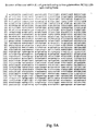
- Figure 8 shows a nucleic acid sequence of a bacterial Rps F gene (SEQ ID NO: 13). Nucleic acid sequences comprising Template 1 (SEQ ID NO: 14) and Template 2 (SEQ ID NO: 15) and their targeting sequences (SEQ ID Nos: 16 and 17, respectively) in the Rps F gene are also indicated.
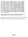
- Figure 9A and 9B show a nucleic acid sequence of an E. coli lacZ gene open reading frame encoding beta-galactosidase (EC 3.2.1.23) (SEQ ID NO: 18). Nucleic acid sequences comprising Template 1 (SEQ ID NO: 5) and Template 2 (SEQ ID NO: 6) and their targeting sequences in the E. coli lacZ gene open reading frame are also indicated.
- Figure 10 shows a nucleic acid sequence of an exoA gene of S. meliloti strain 1021 (SEQ ID NO: 19). Nucleic acid sequences comprising a Template (SEQ ID NO: 20) and its targeting sequences (SEQ ID NO: 21) in the exoA gene are also indicated.
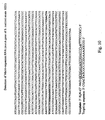
- Figure 11 shows a nucleic acid sequence of a PF2NC15 polyprotein gene of Hepatitis C Virus (SEQ ID NO: 22). Nucleic acid sequences comprising a Template (SEQ ID NO: 23) and its targeting sequences (SEQ ID NO: 24) in the PF2NC15 polyprotein gene are also indicated.
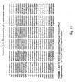
- Figure 12 shows a nucleic acid sequence of a human immunodeficiency virus-1 (HIV-1) envelope (env) gene (SEQ ID NO: 25). Nucleic acid sequences comprising a Template (SEQ ID NO: 26) and its targeting sequences (SEQ ID NO: 27) in the env gene are also indicated.
- HSV-1 human immunodeficiency virus-1 envelope (env) gene
- Figure 13 shows a nucleic acid sequence of a SARS gene (SEQ ID NO: 28). Nucleic acid sequences comprising a Template (SEQ ID NO: 29) and its targeting sequences (SEQ ID NO: 30) in the SARS gene are also indicated.
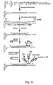
- Figure 14 shows a schematic flow chart of specific RNA detection on a microplate.
- Figures 15A and B show autoradiograms which visualize enzymatic detection of RNA on 96-well microplates.
- Target RNA24.1 (1 pmole) and template DNA35.1 (100 pmole).
- B Detection sensitivity studies were as follows: well 1, no RNA24.1; well 2, 1x10 -15 mole; well 3, 1x10 -14 mole; well 4, 1x10 -13 mole. The film was exposed for one hour (A) or five hours (B) after substrate addition.
- Figure 16 shows an autoradiogram revealing selective detection of lacZ RNA on a microplate.
- Total mRNA and RNA24.1 used for each experiment were 0.1 ⁇ g and 10 fmole, respectively (6 hr exposure).
- Well 1 galactose-induced mRNA; well 2, glucose-repressed mRNA; well 3, no RNA (negative control); well 4, glucose-repressed mRNA and RNA24.1; and well 5, RNA24.1 (positive control).
- the present invention is directed to a novel method for directly detecting a specific RNA molecule in a sample.
- a sample may comprise a plurality of different RNA species.
- the method is used to detect directly a specific RNA molecule in a sample. Details pertaining to the method of the invention and products generated using the method are clearly set forth herein below.
- Ribonuclease H is an endoribonuclease which specifically hydrolyzes the phosphodiester bonds of RNA which is hybridized to DNA. This enzyme does not digest single stranded nucleic acids, double-stranded DNA, or double stranded RNA.
- Such systems can be used to identify microorganisms (e.g., pathogens), validate new drug targets, and provide diagnostic disease indicators.
- microorganisms e.g., pathogens
- FDA Food and Drug Adminstration
- infectious diseases such as, human immunodeficiency virus (HIV) and Mycobacterium tuberculosis (MTb).
- Microarray analysis and real-time PCR are the most popular technologies in this area ( Golub et al. (1999) Science 286:531-537 ; Trottier et al. (2002) J. Virol. Methods 103:89-99 ).
- Microarrays comprised of oligonucleotides or complementary DNA (cDNA) have been used successfully in gene expression profiling studies. Such studies provide information on expression levels of individual genes and reveal patterns of coordinated gene expression. This information can be used in drug discovery, cancer monitoring, cancer type classification, and identification of microorganisms, viruses, and other pathogens in a sample (Golub, et al. 1999, supra; Young et al. (2002) J Virol Methods 103:27-39 ).
- technologies directed to the use of high-density microarrays allow gene expression profiling of over tens of thousands of genes (Lockhart and Winzeler, 2000, supra).
- Nucleic acid-detection methods for viral and bacterial real-time analysis, using reverse transcription and polymerase chain reaction (PCR) technologies have significantly improved the precision of pathogen detection and shortened analysis time, features which are especially useful under emergency conditions.
- Real-time PCR technology allows accurate quantitation of gene expression and gene expression patterns in multiple samples and over a large dynamic range. Though these methods are used to analyze mRNA expression levels, the quantitation is actually determined indirectly by measuring the amount of amplified cDNA or bound probe, rather than the amount of RNA.
- oligonucleotide microarray procedure generally consists of the following steps: reverse transcription, DNA polymerization, transcription, biotin-streptavidin interactions or antibody binding (or the like), and fluorophore labeling.
- the signals are amplified during the transcription step and subsequent steps, wherein fluorescent labels are incorporated.
- the fluorophores are activated by laser excitation to emit detectable fluorescent signals.
- RNAs present in an RNA mixture include, but are not limited to, viral and bacterial RNAs in RNA samples, or specific mRNA transcripts in samples comprising total RNA.
- the present invention involves a modified terminal RNA labeling method which directly labels and detects specific RNA molecules in a mixture. The method is fundamentally different from existing methods that have been used to determine gene expression patterns in that it does not require reverse transcription, PCR, in vitro transcription, or gel electrophoresis.
- the method of the present invention dramatically accelerates the speed with which a specific RNA can be detected in a mixture of molecules and thus expedites the detection of a deleterious nucleic acid molecule and/or pathogen associated with a specific nucleic acid molecule (e.g. a specific RNA molecule).
- the method of the present invention therefore, provides an accurate indicator of the presence of a disease and/or microorganism in a sample.
- this direct RNA detection microchip technology is simple, rapid, accurate, sensitive, high-throughput, and cost-effective, it is an ideal assay for point-of-care disease diagnosis, detection of microbial contamination in food and/or water supplies, and pathogen detection in biodefense.
- the above mentioned novel chip technology utilizes a method developed by the present inventor which enables, for the first time, the direct labeling/detection of a specific mRNA in total mRNA or a sample comprising total RNA.
- the inventor devised a method to remove the 3'-region which is conserved among most eukaryotic mRNA transcripts [e.g., the poly(A) tail and 3'-untranslated region (3'-UTR)] from a specific mRNA and, thereby expose intrinsic 3'-sequences for labeling and detection.
- the method involves an Rnase H digestion protocol which takes advantage of the ability of Rnase H to digest RNA which has formed a duplex with a DNA sequence ( Nakamura and Oda. (1991) Proc. Natl. Acad. Sci. USA 88:11535-11539 ).
- the method relies on the selection of a 2'-O-Me-RNA/DNA hybrid which binds to a specific mRNA and protects a unique internal sequence of the mRNA from Rnase H mediated digestion (via RNA/RNA duplex formation), but also binds/positions other regions of the mRNA (such as the 3'-region), so as to render these regions susceptible to Rnase H digestion (via RNA/DNA duplex formation).
- the overhang formed following Rnase H mediated digestion serves as a recognition/extension site for a DNA polymerase (e.g., Klenow) on the fragment of the specific mRNA whereby nucleotide labels may be incorporated to effect detection of the specific mRNA.
- a DNA polymerase e.g., Klenow
- the present invention is based in part on a method developed by Huang and Szostak [(1996) Nucleic Acids Res. 24, 4360-1 ] for labeling the 3'-termini of RNA.
- This method took advantage of a natural function of DNA polymerases: elongation of RNA primers on DNA templates. This observation was subsequently investigated further and shown to be applicable to the development of a method for labeling and detecting specific RNA transcripts.
- Huang and Szostak [(2003) Anal Biochem 315:129-133 ] discovered that the ready availability of short synthetic DNA template allows an RNA of known 3'-terminal sequence to be selectively extended in a template-dependent manner at its 3'-end, which facilitates labeling and detection of the specific RNA in an RNA mixture, without separation, purification, reverse transcription, or PCR.
- the contents of each of Huang and Szostak [(1996) Nucleic Acids Res. 24, 4360-1 and Huang and Szostak [(2003) Anal Biochem 315:129-133 ] are incorporated herein by reference in their entirety. Methodology relating to labeling and modification of RNA 3'-termini are also described in U.S. Patent No. 6,238,865 (issued to Huang and Szostak ), the entire contents of which is incorporated herein by reference.
- the present inventor has also modified the radioactive labeling method of Huang and Szostak [(2003), supra )] to become an enzyme labeling method, which uses enzymes such as peroxidase or alkaline phosphatase to catalyze chemiluminescent reactions ( Pollard-Knight et al. (1990) Anal. Biochem. 185, 84-89 ; Reddy et al. (1999) Biotechniques 26710-714 ). Details pertaining to using the method of the present invention with various labeling/detection methods are described in greater detail herein below.
- RNAs can be labeled initially with antigens and subsequently labeled with enzymes, such as alkaline phosphatase (AP), which can catalyze a chemiluminescent reaction.
- AP alkaline phosphatase
- DNA and RNA polymerases have been screened and examined for the ability to catalyze RNA 3'-extension on a DNA template.
- Enzymes including E. coli DNA polymerase I, the Klenow fragment of E . coli DNA polymerase I ( Sanger et al. (1977) Proc. Natl. Acad. Sci. USA 74:5463-5467 ), T4 DNA polymerase, T7 DNA polymerase, T7 RNA polymerase, M-MuLV reverse transcriptase, and Taq DNA polymerase have been tested for utility in the present method by incubating each enzyme with a 5'- 32 P-labeled RNA, dNTPs, and a DNA template.
- RNA terminal-labeling Three methods for RNA terminal-labeling are commonly used: 5'-labeling with T4 polynucleotide kinase and [ ⁇ - 32 P]-ATP ( Sambrook et al. (1989) Molecular Cloning: A Laboratory Manual, Cold Spring Harbor Laboratory Press: Cold Spring Harb or); 3'-labeling with T4 RNA ligase, and 3',5'-[5'- 32 P]-pCp ( England and Uhlenbeck. (1978) Nature 275:560-561 ); and 3'-labeling with poly(A) polymerase and [ ⁇ - 32 P]-cordycepin 5'-triphosphate (CoTP or 3'-deoxy-ATP; Linger and Keller. (1993) Nucleic Acids Res.
- RNA substrates in an RNA mixture are labeled at either 5' or 3' termini.
- the non-specific labeling feature of these methods provides an advantage when labeling and detection of all RNAs in an RNA mixture is desired. This advantage, however, becomes a drawback when labeling and detection of a specific RNA in an RNA mixture is desired ( Sorensen et al. (2000) J. Lab Clin. Med. 136:209-217 ). Specific labeling may be necessitated when analyzing, for example, a specific viral RNA, ribosomal RNA, or cellular mRNA in a total RNA sample. In order to use these conventional methods in direct detection and analysis of a specific RNA, separation steps are required to isolate the specific RNA from the mixture.
- RNA of interest such as an mRNA, or other functional RNA
- protein targets for analysis RNA of interest (such as an mRNA, or other functional RNA) or protein targets for analysis
- RNA sequence (6-80 nt.) intended for incorporation into a template should be analyzed using computer-assisted folding programs, such as Mfold ( M. Zuker, Rensselaer Polytechnic Institute ) to assess its potential for formation of secondary structure. If the sequence is predicted to form a secondary structure, a different RNA sequence should be considered.
- Design (I) comprises a 5'-end DNA (1-30 nt.) and a 3'-end 2'-MeO-RNA (5-79 nt.).
- the DNA is designated herein template DNA and the 2'-MeO-RNA is designated herein binding MeO-RNA.
- the targeted RNA sequence section bound to the template DNA is referred to as the labeling region and the targeted RNA sequence section bound to the binding MeO-RNA sequence is referred to as the binding region.
- Design (II) comprises 5'-end and 3'-end DNA sequences (1-30 nt. each) flanking the central 2'-MeO-RNA sequence (4-78 nt.).
- the 5'-end DNA is called template DNA
- the 3'-end DNA is called digestion DNA
- the 2'-MeO-RNA is called binding MeO-RNA. See Figure 7 .
- the targeted RNA sequence section bound to the template DNA is referred to as the labeling region
- the targeted RNA sequence section bound to the digestion DNA is referred to as the digestion region
- the targeted RNA sequence section bound to the binding MeO-RNA sequence is referred to as the binding region.
- the first several 5'-nucleotides (at least two nucleotides) in the labeling region may be selected to be a single kind of nucleotide, so as to produce, for example, a stretch of 5'-AAAA.
- a hybrid template ( I or II ) may be chemically synthesized on solid phase and purified by HPLC or gel electrophoresis. Techniques directed to the synthesis and purification of such sequences are known in the art and routinely practiced.
- Figures 8-13 provide nucleic acid sequences of a subset of exemplary genes, some of which are associated with various microorganisms and/or pathogens, which may be used in the detection methods of the present invention. Also presented in Figures 8-13 are sequences of hybrid templates useful in the method of the invention for detection of the specific gene (i.e., the RNA) indicated.
- the specific gene i.e., the RNA
- Oligonucleotides Oligonucleotides, total RNA, and Enzymes
- DNA20.8 (5'-TGAATCAGCATCTAGCTACG-3') (SEQ ID NO: 1), DNA20.10 (5'-GGCTACAGGAAG-GCCAGACG-3') (SEQ ID NO: 2), DNA-2'-O-Me-RNA20.8 [a hybrid template, 5'-d(TGAAT)-2'-O-Me-(CAGCAUCUAGCUACG)-3'] (SEQ ID NO: 3), RNA31 (5'-AUGUGGAUUGGCGAUAAAAAACAACU-GCUGU-3', fragment of lacZ mRNA from 2302 to 2331 with an 3'-overhang U) (SEQ ID NO: 4), DNA-2'-O-Me - RNA30.5 [5'-d(CAGCAGTTGTTTTT-T)-2'-Me-ribo(AUCG-CCAAUCCACAU)-3', complementary to lacZ mRNA from 2305-2334 nt,] (SEQ ID NO: 5), and DNA-2'-O-Me
- RNA50 (5'-GGAGAGUAUGCAGUAGUCAUCGCGACGUAGCUAGAUG-CUGAUUCAACUAC-3') (SEQ ID NO: 7) was prepared by in vitro transcription of synthetic oligodeoxynucleotide templates with T7 RNA polymerase. The above DNAs and RNA were purified by gel electrophoresis.
- IPTG Isopropyl- ⁇ -D-1-thiogalactopyranoside
- RNase H digestion reactions (5 ⁇ L) were generally carried out at 37°C for 1 hr in buffer [20 mM Tris-HCl (pH 7.5), 100 mM KCl, 10 mM MgCl 2 , 0.1 mM DTT, and 5% (w/v) sucrose], with RNA (1 pM-10 nM), DNA template or DNA-2'-O-Me-RNA hybrid template (1-500 nM), and RNase H (0.4 U/ ⁇ L). After ethanol precipitation of the digested RNA-DNA hybrid, Klenow extension was conducted.
- Klenow extension reactions (5 ⁇ L) were generally performed at 37°C for 1 hr in buffer [10 mM Tris-HCl (pH 7.5), 17.5 mM DTT, and 5 mM MgCl 2 ], with RNA (1 pM-10 nM), DNA template (1-500 nM), Klenow (0.5 U/ ⁇ L), and 0.1-1 ⁇ L of [ ⁇ - 32 P]-dATP (3000 Ci/mmol, 10 mCi/mL). Electrophoresis on polyacrylamide gels (3-12%) was used to separate nucleic acids by size, and radioactively labeled nucleic acids were visualized by autoradiography. In experiments where the same buffer (a buffer mixture of RNase H and Klenow buffer at a 2:8 ratio) was used for both RNase H and Klenow reactions, the intervening ethanol precipitation step was omitted.
- buffer a buffer mixture of RNase H and Klenow buffer at a 2:8 ratio
- eukaryotic mRNA transcripts generally comprise a poly(A) tail and 3'-untranslated region (3'-UTR), it is not possible to directly detect and analyze a specific mRNA using the 3'-labeling and detection methods previously described. See commentary herein above for additional details.
- the 3'-region which is conserved among most mRNA transcripts must, therefore, be removed from the specific mRNA in order to expose its intrinsic 3'-sequence for labeling and detection.
- the present inventor developed an RNase H digestion protocol with which to remove the 3-'region of a specific RNA transcript. Since RNase H is capable of digesting RNA which has formed a duplex with a DNA sequence ( Nakamura and Oda. (1991) Proc. Natl. Acad. Sci. USA 88:11535-11539 ), the poly(A) tail and 3'-UTR can be removed by RNase H digestion after formation of such an RNA/DNA duplex. As previously reported, a 2'-methylated RNA sequence can bind to RNA and form a stable RNA/RNA duplex and the formation of the duplex provides a mechanism for protecting the bound RNA from RNase H digestion ( Hayase et al.
- RNase H recognizes the RNA/DNA duplex region and digests the RNA strand of the duplex.
- Klenow recognizes the 2'-O-Me-RNA-DNA hybrid as a template and is capable of catalyzing a nucleotide extension reaction on the hybrid template.
- the extension process on a hybrid template is shown to be as efficient as that observed on a non-hybrid, "regular" DNA template.
- RNA50 on a DNA-2'-O-Me-RNA20.8 hybrid template removes the 3' region of the RNA and generates the complex of digested RNA40 and the bound template with an overhang sequence 5'-TGAAT-3'.
- the Klenow extension introduced one 32 P-labeled dA of [ ⁇ - 32 P]-dATP (complementary to the first 3'-nucleotide of the overhang sequence) to the RNA40 fragment.
- the hybrid template is competed out using the DNA template 100-fold over the hybrid after RNase H digestion and heat denaturing ( Fig. 1 ).
- cleavage of the common 3'-region of eukaryotic mRNAs is required to expose intrinsic internal sequences for selective RNA labeling and detection.
- the 3'-region of a test mRNA (lacZ mRNA) has been selectively removed to expose its internal sequences.
- the sequence of the hybrid template for lacZ mRNA labeling and detection was designed based on the coding region of the target RNA, which is publicly available via GenBank (http://www.ncbi.nlm.nih.gov/).
- RNA sequence 25-50 nt.
- the selected template for the lacZ RNA labeling and detection is DNA-2'-O-Me-RNA30.5 hybrid template [5'-d(CAGCAGTTGTTTTTT)-2'-Meribo(AUCGCCAAUCCAC-AU)-3'] (SEQ ID NO: 5), which is complementary to lacZ RNA from nucleotide positions 2305-2334.
- SEQ ID NO: 5 DNA-2'-O-Me-RNA30.5 hybrid template [5'-d(CAGCAGTTGTTTTTT)-2'-Meribo(AUCGCCAAUCCAC-AU)-3'] (SEQ ID NO: 5), which is complementary to lacZ RNA from nucleotide positions 2305-2334.
- six bases from nucleotide 2320-2325 are all adenine ('A's). These 'A's served as the template for multiple rounds of ⁇ - 32 P-dATP incorporation during subsequent Klenow extension steps, which followed RNase H digestion to
- a lacZ-expressing plasmid is introduced into yeast.
- the expression of lacZ from this plasmid is controlled by a galactose (Gal) promoter which can be induced in response to the presence of galactose in the media.
- the promoter is not induced in the presence of glucose, which serves as an experimental negative control condition.
- Two total mRNA samples were prepared: one sample was derived from galactose-induced yeast comprising the lacZ-expressing plasmid and a second sample was derived from yeast comprising the lacZ-expressing plasmid which were maintained in glucose-containing media, in the absence of galactose.
- RNA31 (5'-AUGUGGAUUGGCGAUAAAAAACAACUG-CUGU-3', fragment of lacZ mRNA from 2302 to 2331 with a 3'-overhang U) (SEQ ID NO: 4) on DNA-2'-O-Me-RNA35.1 [5'-d(GTTGTTTTTT)-2'-Meribo(AUCGCCAAUCCACAU)-d(CTCTGAA-AGA)-3' (SEQ ID NO: 6), complementary to lacZ mRNA from 2292 to 2326 nt]. See Figure 3 .
- DNA-2'-O-Me-RNA30.5, DNA-2'-O-Me-RNA35.1 was designed for Rnase H double digestion of lacZ mRNA at both 3' and 5' regions.
- the double digestion generates a short central lacZ mRNA fragment. See Figure 3 .
- This short labeled fragment is indeed observed with the ITPG-induced total RNA (Lane 8), and absent in glucose repressed cells (Lane 9), which is consistent with the results presented in Fig. 2 .
- This short fragment is also observed in the total mRNA isolated from lacZ mRNA-expressing yeast (Lane 10).
- This double-digestion approach is also capable of detecting mRNA fragments, as well as full-length mRNA. Therefore, mRNA fragments arising from degradation can also be assayed using the method of the present invention, which further increases the detection sensitivity.
- the ability to detect even degraded RNA illustrates yet another significant advantage of the present method over previously described methods for indirectly detecting RNA. Since E. coli and yeast comprise thousands of mRNA species ( Rhodius et al. (2002) Annu. Rev. Microbiol. 56:599-624 ; Ross-Macdonald et al. (1999) Nature 402:413-418 ), the experimental results presented herein also underscore the selectivity of the present invention even in the presence of a plurality of other mRNA transcripts.
- a novel method is described herein that combines RNase H mediated cleavage of the 3'-region of mRNA and Klenow selective labeling of RNA 3'-termini.
- the method of the present invention has been used to selectively label and detect a specific mRNA transcript (i.e., LacZ mRNA) in a total RNA sample comprising thousands of different RNA transcripts.
- a specific mRNA transcript i.e., LacZ mRNA
- Figure 2 Lanes 2-4
- the method exhibits a high degree of sensitivity with regard to labeling and detection.
- the present inventor has successfully developed and used a novel method that combines RNase H cleavage and Klenow labeling to selectively label and detect a specific RNA (e.g., mRNA) in a total RNA sample.
- RNA e.g., mRNA
- This direct and rapid RNA detection method has great potential for RNA quantification, especially individual mRNA quantification, which is difficult to achieve using DNA microarray and real-time PCR technologies ( Freeman et al. (1999) BioTechniques 26:112-125 ; Lockhart and Winzeler (2000) Nature 405:827-836 ).
- the method of the present invention provides significant experimental advantages over microarray and real-time PCR technologies.
- the method of the present invention is complementary to conventional RNA detection methods, such as Northern blotting. Indeed, because this method for specific mRNA labeling allows assay of both fragmented and full-length mRNA, it greatly advances studies of mRNA decay and metabolic regulation. Total RNA, rather than mRNA, can be used for such labeling and detection studies, thereby obviating the need for mRNA isolation procedures which can result in degradation.
- the present method is also compatible with the use of non-radioactive labels, such as fluorophore and antigen labels (Freeman et al. 1999, supra).
- Such labels can be incorporated, and the labeling and detection determined by standard approaches, including the use of conjugated alkaline phosphatase or peroxidase to catalyze chemiluminescence reactions and ELISA quantitation ( Young et al. (2002) J. Virol. Methods 103:27-39 ).
- the DNA-2'-O-Me-RNA hybrid template system has been developed, which enables RNase H and Klenow to share the same template and buffer. This methodological feature shortens the number of experimental steps and reduces the time required to obtain results.
- Gel electrophoresis can also be avoided by immobilizing the template on solid supports, such as a microplate or microchip surface ( Benters et al. (2002) Nucl. Acids. Res. 30:e10 ). After RNA substrate immobilization, RNase H, Klenow, non-incorporated labels, and buffers can simply be washed away after each step. Indeed, the multi-label incorporation system is extremely useful for enhancing the detection sensitivity of the method, especially on solid phase or for long RNA transcripts analyzed by gel electrophoresis. See Examples III and IV for additional details.
- RNA labeling method is highly sensitive and allows detection of RNA at attomole levels. As shown herein, the detection sensitivity can be further enhanced by extending the length of the over-hang sequence of the DNA template.
- ELISA and micro-spotting techniques in conjunction with the present method also serves to increase the detection sensitivity.
- the present method is also extremelyly selective, as demonstrated by selective labeling and detection of lacZ mRNA in the presence of thousands ofmRNAs. Therefore, as described herein below, this method can be used to advantage in microplate-based rapid and high-throughput detection technology, and in microchip-based rapid gene expression profiling technology.
- RNAs expressed uniquely in each organism and which can be used as positive indicators of a contaminant (e.g., a pathogen) in a sample is of paramount importance in such settings.
- Contaminant specific RNAs or "fingerprint" RNAs may include, without limitation, mRNA, ribosomal RNA, heteronuclear RNA, and mitochondrial RNA.
- fingerprint RNAs is a powerful tool useful in the determination of organism identity and/or cellular phenotype.
- detection and identification of fingerprint RNAs using this rapid, sensitive, and selective strategy can lead to identification of microorganisms (such as pathogens), diseases, and/or characterization of disease status. This feature of the invention is described in greater detail elsewhere in the specification.
- the method of the present invention has been modified for use with a solid matrix.
- a solid matrix In order to further increase detection sensitivity, simplify the detection procedure, and avoid using radioactive material, fluorophore and enzyme labels were evaluated after template immobilization on a solid phase.
- Solid matrices envisioned for use in the present invention include, without limitation, 96-well plates and microchips. It should be understood, however, that a variety of solid matrices are known in the art and may be used in the method of the invention.
- the protocol developed for using fluorophore labeling in the present invention is similar to that used for radioactive labeling, but fluorophore-labeled dNTPs are used instead of radioactively labeled ⁇ - 32 -dNTPs.
- the fluorescent signals are detected and quantified with microplate fluorometer or imaging system.
- Enzyme labeling however, offered much greater sensitivity than the fluorophore labeling. This finding was likely a result of signal amplification that occurs during the course of an enzyme catalyzed reaction, such as that mediated by alkaline phosphatase. See Figure 4 .
- the chemiluminescence detection may be performed by microplate luminometer, imaging system, or film detection. Although it is possible to detect RNA with fluorophore labels, the greater sensitivity observed with enzymatic labeling presents this approach as the exemplary labeling method at the present time. Moreover, chemiluminescence detection is simpler and requires less sophisticated equipment, attributes which further underscore its utility. For all of these reasons, additional experiments were performed using enzymatic labeling methodology.
- LacZ mRNA was used as a test model RNA with which to evaluate the method of the present invention on solid phase. See Figure 2 for schematic. After incubation of total mRNA sample in a 96-well plate (DNA-Bind TM , purchased from Coming) on which the lacZ-mRNA hybrid template [5'-d( CACCAGTTCTTTTTT )-2'-Me-ribo( AUCGCCAAU-CCACAU )-NH 2 -3', binding to the lacZ mRNA from 2305-2334 nt.] (SEQ ID NO: 5) had been previously immobilized, lacZ mRNA was bound specifically to the plate via the hybrid template and unbound mRNAs were washed away.
- DNA-Bind TM purchased from Coming
- RNA24 [5'- AUGUGGAUUGGCGAU AAAAAACAA- 3' (SEQ ID NO: 8), the lacZ mRNA sequence from 2305-2328 nt.] was chemically synthesized and served as a positive control for the experiment; the underlined sequence is the binding region, and the italicized sequence is the digestible RNA-DNA duplex region.
- RNA24 As anticipated, the positive control RNA24 was detected on the solid phase, whereas the negative control (no RNA) produced a signal not distinguishable from background levels. Consistent with the specific detection achieved using the method of the invention in solution (see Example II), lacZ mRNA present in galactose-induced cultures was specifically detectable using the present method in the context of presentation on a solid matrix. The minimal levels of lacZ mRNA present in glucose cultures (due to leaky expression) were also detectable, but at a level not significantly above background levels. Addition of RNA24 (positive control RNA) to the glucose sample, however, produced a strong signal, indicating that the presence of non-specific RNA in a sample does not interfere with detection of a specific RNA.
- Templates for example hybrid templates as described herein above, are first immobilized on a microchip, after which an RNA sample is added to the microchip matrix and incubated. After washing to remove unbound RNA, bound RNA is digested by RNase H to expose internal intrinsic sequences and then extended by Klenow DNA polymerase to incorporate antigen-labeled dNTPs. Bound RNA is subsequently labeled with enzymes by treating the microchip with antibody-enzyme conjugate. After washing the microchip to remove the non-specifically bound enzymes, substrate is added to generate chemiluminescent signals.
- RNA microarray technologies have demonstrated that the detection of emitted fluorescent light from microspots or even nanospots is possible using a microarray reader (scanner) or high-resolution imaging system (Lockhart and Winzeler, 2000, supra; Trottier et al., 2002, supra), the detection of chemiluminescent light emitted from microspots on a RNA microchip is well within the capabilities of imaging technology.
- the distance between the template-containing microspots on the RNA microchip should be large enough to prevent spot-to-spot interference during RNA sample binding, enzymatic steps, and chemiluminescent signal detection.
- the microchip may be designed and prepared using glass chips (2.2 x 2.2 cm) surface-functionalized with COOH functional groups, which can be activated with N-hydroxylsuccinimide (NHS) for coupling with templates (e.g., hybrid templates) comprising 3'-terminal NH 2 groups.
- templates e.g., hybrid templates
- Such preparations may be achieved using established protocols known in the art ( Zhou and Huang. (1993) Indian Journal of Chemistry 32B:35-39 ; Zhao et al. (2001) Nucleic Acids Res. 29:955-959 ; Manning et al. (2003) Materials Science and Engineering C 23:347-351 ).
- gold-coated glass chips can be utilized for microchip preparation ( Medalia et al.
- RNA microchip To demonstrate direct RNA detection on a microchip, a low-density chip containing 16 templates on an area of 1.6 x 1.6 centimeters may be prepared. Subsequently, a microchip comprising 144 different templates on an area of 1.2 x 1.2 centimeters may be prepared using a microarrayer. It is anticipated that spot size on such a microchip will be approximately 600 ⁇ in diameter and the spot-spot gap approximately 400 ⁇ . If, for example, eighteen fingerprint RNAs are assessed for each microorganism, it is possible to monitor eight microorganisms simultaneously on a single microchip. Eight non-pathogenic microorganisms, including bacteria, viruses, yeasts, and fungi, may also be evaluated using the RNA microchip technology.
- Eight sets of eighteen fingerprint RNAs may be chosen based on their high expression levels, a determination of which can be obtained via gene expression profiling. Such genes are good positive indicators for the presence of the microorganism in question. Such gene expression profiles can be purchased (Invitrogen, for example provides such services), determined experimentally, or potentially identified by reviewing the scientific literature germane to the microorganisms to be detected. A skilled artisan would be aware of these approaches and such considerations would be well within his/her capabilities.
- the immobilized templates e.g., hybrid templates
- immobilized on the microchip are designed based on the fingerprint RNAs, and each spot on the microchip can represent a different template recognizing a different fingerprint RNA.
- Gene expression profiles of cells or organisms, including pathogens, may vary due to cell cycle stage, nutrient availability, and/or environmental conditions.
- 100 fingerprint RNAs or more may be chosen from each organism or cell subtype to prevent misleading results associated with potential gene expression variation.
- the long-term goal of this application of the method of the invention is to spot 10,000 templates on a microchip (2.5 x 2.5 cm), which would enable the detection of approximately 100 of the most virulent viruses, bacteria, and other pathogens.
- microchips are ideally suited for biodefense applications and/or detection of pathogen-caused disease.
- the RNA microchip technology may also be used in non-pathogenic disease analysis, disease classification, and microbial contaminant detection in food and/or water supplies for example.
- hybrid templates are designed according to the following strategy.
- a hybrid template e.g., 5'-DNA-2'-O-Me-RNA-3'
- a hybrid template is designed to comprise two regions: a 5' DNA and a 3' 2'-O-methylated-RNA sequence. See Figure 4 .
- the 5'-DNA sequence allows RNase H to cleave in the 3' region of target RNAs prior to the Klenow extension step. This is a particularly important step when analyzing eukaryotic mRNA which generally comprise a 3'-poly(A) tail and 3'-UTR.
- the 5'-region of the target RNA can also be removed by the RNaseH digestion. See Figure 6 .
- the template [5'-DNA-(2'-O-Me-RNA)-DNA-3'] is designed to contain three regions: a 5'-DNA sequence, a middle 2'-O-methyl-RNA sequence, and a 3'-DNA sequence.
- the 5'- and 3'-DNA sequences allow RNase H to cleave both 3' and 5' regions of target RNAs, such as, for example, mRNAs. After the RNase H digestion and washing, only the target RNA fragment complementary to the 2'-O-Me-RNA sequence remains on the template, thus creating an RNA microchip for labeling and detection.
- the 2'-O-Me-RNA region of the template is 4-78 nucleotides long, which provides sufficient sequence specificity while allowing stable RNA-RNA duplex formation capable of surviving the treatments involved in the present method.
- the DNA regions of the template are 1-30 nucleotides long, which allows RNase H recognition of the RNA-DNA duplexes.
- Table 1 shows systems for immobilizing the hybrid templates.
- -COOH activated form: -CO-NOS
- Au (gold) -X-R (X S or Se)
- the sequence of the template is designed based on the fingerprint RNA sequence, which can be determined based on nucleic acid sequence data banks (e.g., GenBank), sequence projects, or genomic research.
- the RNA sequence region ( ⁇ 6-80 nucleotides) used for the template design is chosen after examination of the RNA secondary structure using computer folding programs, such as Mfold (Genetics Computer Group, Madison, WI).
- Mfold Genetics Computer Group, Madison, WI.
- the 5'-DNA region of the template which serves as a template for Klenow extension, can be any sequence.
- RNA binding and Klenow extension it may be advantageous to use a three-region template [5'-DNA-(2'-O-Me-RNA)-DNA-3'], which allows RNase H cleavage of both 5' and 3' regions of target RNAs, and leaves just the complementary RNA fragment for labeling and detection.
- the 3'-termini of the templates is immobilized on the solid phase.
- This arrangement allows the DNA polymerase to extend target RNA 3'-ends on the hybrid templates.
- Glass and polystyrene microchip functionalized with COOH or NH 2 groups, or gold plating can be used as the solid support.
- the hybrid template may be immobilized on the microchip using several conventional systems (see Table I), including, (I) well-established protocols for immobilizing the 3'-NH 2 -template on a COOH-functionalized surface (Manning et al., 2003, supra ).
- an amino group (NH 2 ) is introduced into the 3'-terminal of the template during solid phase synthesis, and this NH 2 group is coupled with the activated -COOH group (such as -CO-NOS).
- the activated -COOH group such as -CO-NOS.
- An alternate procedure which can be used to immobilize the template on an NH 2 -functionalized surface involves the introduction of a ribonucleotide residue into the 3'-terminal of the template during solid phase synthesis. The diol functionality on this residue is converted to two aldehyde functional groups by NaIO 4 oxidation, prior to coupling with the amino group on the solid surface ( Lemaitre et al. (1987) Proc Natl Acad Sci USA 84:648-652 ).
- a sulfide- or selenide-modified template can be immobilized on a gold surface based on a number of strategies known in the art (Medalia et al., 2002, supra; Hopfner et al., 1999, supra; Du et al. (2002) J. Am. Chem. Soc. 124:24-25 ).
- the surface of the microchip may be capped with a variety of capping reagents. See Table 1. Such protocols are known to skilled artisans familiar with experimental variations designed to investigate the positive, negative, or neutral surface best suited for minimizing background noise. For instance, after immobilization of the sulfide- or selenide-modified templates on a gold surface, sulfide- or selenide-containing reagents are generally used to saturate the surface, which prevents sulfide and mercapto functionalities of the enzyme from binding to the gold surface.
- RNase H digestion is used to remove the 3'-region of target RNA thereby exposing internal intrinsic sequences for Klenow extension. Unlike site specific cleavage of DNA sequences, which is routine, site specific cleavage of RNA sequences is technically challenging. It has, however, been reported that RNase H is able to digest RNA strands when bound to DNA sequences. Although RNase H cleaves RNA non-specifically with regard to sequence, a bound DNA sequence can serve as a guide that directs RNase H to digest a specific region of RNA (i.e., the DNA bound region). Thus, the bound DNA transforms RNase H into a site-specific RNA endonuclease.
- the present inventor has developed an approach to remove the 3'-region of target RNA, including the poly(A) tail and 3'-UTR located in the 3'-region of most eukaryotic mRNAs.
- a DNA-RNA hybrid template is designed to facilitate use of the same template for both RNase H digestion and Klenow extension.
- both RNaseH and Klenow DNA polymerase recognize the hybrid 3'-DNA-2'-O-Me-RNA-3' templates.
- Klenow polymerase recognizes the hybrid template of the invention as well as a DNA template.
- a hybrid template 5'-DNA-2'-O-Me-RNA-3' comprising 5' DNA and 3' 2'-O-methylated-RNA sequences, allows RNase H to cleave the 3' region of target RNAs prior to the Klenow extension step.
- the hybrid template 5'-DNA-(2'-O-Me-RNA)-DNA-3' comprising 5'-DNA, middle 2'-O-methyl-RNA, and 3'-DNA sequences, enables RNase H to cleave both 3' and 5' regions of target RNAs.
- a target RNA fragment complementary to the 2'-O-Me-RNA sequence remains on the template.
- Such bound target RNA fragments are, therefore, available for Klenow extension and are consequently "tagged" for detection by incorporation of labeled nucleotide.
- template immobilization is enzymatically compatible with both RNase H and Klenow polymerase activity. As described herein, reaction conditions compatible with the RNase H digestion and Klenow extension were developed that enabled these reactions to be performed simultaneously.
- the present invention is compatible with a variety of labeling systems, including but not limited to radioactive labeling, fluorophore labeling, and enzyme labeling.
- Enzyme labeling was chosen as a preferred labeling system because it is sensitive, safe and accessible method (Pollard-Knight et al. 1990, supra; Reddy et al., 1999, supra).
- Klenow polymerase mediated extension may be used to integrate antigen labels via the incorporation of antigen-labeled-dNTPs, such as 12-biotin-dATP.
- the length of the 5'-region DNA sequence of the template may be used to control the number of the antigen-dNTPs incorporated into the bound RNAs ( Huang and Szostak.
- the 5'-DNA region of the template can be any sequence.
- the antigen labeling is converted to enzyme labeling via treating a chip, for example, with an antibody-enzyme conjugate, such as an anti-biotin antibody-alkaline phosphatase conjugate.
- an antibody-enzyme conjugate such as an anti-biotin antibody-alkaline phosphatase conjugate.
- various aspects of the reaction can be varied, including the length of the 5'-region DNA of the template, antigen linker size, and the purity of the antibody-enzyme conjugate. Such considerations are well known in the art and familiar to skilled artisans. Systems that utilize small molecules and binder-enzyme conjugates, such as biotin and avidin-alkaline phosphatase conjugates are also envisioned as compatible with the method of the present invention.
- the alkaline phosphatase and dioxetane derivative substrate e.g. CDP-Star TM , Sigma
- CDP-Star TM dioxetane derivative substrate
- additional substrate can be supplemented or added continuously to maintain steady signal emission.
- the RNA microchip of the present invention can be placed in a chamber, which facilitates supplementation with fresh substrate.
- RNA microarray reader scanner
- chemiluminescent detection sensitivity of this RNA direct detection system is comparable with the real-time PCR.
- RNA detection signal produced by the present method is amplified through the enzyme-catalyzed reaction (Pollard-Knight et al. 1990, supra; Reddy et al., 1999, supra ), enzymatic labeling of target RNAs offers high sensitivity.
- the present inventor has determined that the detection sensitivity of alkaline phosphatase RNA labeling may reach as high as 10 -22 moles on a microspot using the dioxetane substrate. Thus, approximately one hundred alkaline phosphatase molecules are detectable. If every bound RNA is labeled on average with several enzyme molecules, therefore, dozens of the target RNA are detectable.
- RNA microchip detection does not suffer from similar partial degradation problems.
- Background noise associated with chemiluminescence detection systems which is caused by non-specific binding of the enzyme conjugate to the chip, can be minimized by extensive washing which can be used to remove essentially all of the non-specifically bound enzyme molecules.
- Various methods for chip surface capping and protein blocking may also be utilized to further reduce background noise.
- Capping reagents and blocking proteins such as bovine serum albumin (BSA) are known in the art and compatible with the method of the present invention.
- BSA bovine serum albumin
- a microchip may be placed in a "virgin" detection chamber, which has not been exposed to any of the method steps of the invention.
- RNA chip of the invention Sixteen designed templates with 3'-NH 2 groups may be immobilized on sixteen DNA-binding spots (each one 2.5 mm in diameter) on a glass microchip (1.6 x 1.6 cm) activated with NHS groups. To each DNA-binding spot, 1 ⁇ L of coupling buffer (2x) and 1 ⁇ L of the template (1 pmole) are added. After the chip is incubated for 0.5 hour at 37°C, the chip is washed with post-coupling washing buffer (3x 1 mL) to remove the unbound templates. After heat denaturing, 1 ⁇ L of RNA sample is added to the SSC buffer (200 ⁇ L).
- RNA is removed by washing the chip three times, each time with 1 mL of SSC buffer.
- RNase H 2 U/ ⁇ L
- RNase H buffer 200 ⁇ L
- a solution of 1 ⁇ L of Klenow (5 U/ ⁇ L), 1 ⁇ L of dATP-Biotin (50 mM), and Klenow buffer 200 ⁇ L is added to the chip surface, and the chip is incubated with shaking for 15 minutes at 37°C.
- the unbound dATP is washed away with blocking buffer (3 x 1 mL).
- a solution of anti-biotin antibody-AP conjugate (1 ⁇ L, 1 ⁇ g/ ⁇ L) and blocking buffer (200 ⁇ L) is added to the chip surface, and the chip is incubated with shaking for 10 minutes at room temperature. After washing the chip with washing buffer (5 x 1 mL), alkaline phosphatase buffer (1 mL) is used to wash the chip.
- the solution of 180 ⁇ L of the CDP substrate (Sigma) and 20 ⁇ L of the alkaline phosphatase buffer (10x) is added to the chip surface, followed by chemiluminescent detection with a high-resolution imaging system or microchip reader.
- the substrate may be re-added or supplemented continuously to maintain a steady signal emission.
- the compatibility of an RNA microchip of the invention with manipulations in a chamber facilitates such substrate supplementation.
- the protocol for the microchip containing 100 templates is analogous to the protocol described here.
- Example III the system has been utilized successfully for RNA detection and quantification on a solid phase via immobilization of the template to a surface such as a microplate or microchip.
- the data presented in this example confirm and extend the applicability of the present system for the detection of RNA transcripts in the context of solid phase presentation.
- RNA-DNA hybrid template containing 3'-NH 2 group on the DNA-binding plate coupling buffer (10 ⁇ L, 50 mM Na 2 HPO 4 , 10 mM EDTA, pH 9.0), RNase-free water (89 ⁇ L), and the 3'-NH 2 -template (1 ⁇ L, 0.1-0.6 mM) is added to the DNA-binding 96-well plate (Coming), and the plate is incubated for one hour at 37°C. Each well is then washed three times with post-coupling washing buffer (250 ⁇ L, 150 mM NaCl, 100 mM Maleate, pH 7.5) to remove the non-immobilized templates.
- post-coupling washing buffer 250 ⁇ L, 150 mM NaCl, 100 mM Maleate, pH 7.5
- RNase H Digestion after addition of RNase H buffer [50 ⁇ L, 50 mM Tris-HCl (pH 7.5), 40 mM KCl, 6 mM MgCl 2 , 1 mM DTT, 0.1 mg/mL BSA] to each well, RNase H (1.0 ⁇ L, 0.2 units/ ⁇ L) is added to each well, followed by 30 minute incubation at 37°C.
- RNase H buffer 50 ⁇ L, 50 mM Tris-HCl (pH 7.5), 40 mM KCl, 6 mM MgCl 2 , 1 mM DTT, 0.1 mg/mL BSA
- Klenow Extension After draining the RNase H solution from each well, Klenow buffer [50 ⁇ L, 10 mM Tris-Cl (pH 7.5), 5 mM MgCl 2 , 7.5 mM DTT] is added to each well, followed by addition of Klenow fragment (1 ⁇ L, 5 units/ ⁇ L) and Biotin-7-dATP (1 ⁇ L, 1 mM). The plate is incubated for 1 hour at 37°C. Subsequently, the unincorporated biotin-dATP is removed from each well by washing twice with blocking buffer (250 ⁇ L, 1X, Sigma). Moreover, blocking buffer (250 ⁇ L, 5X, Sigma) is used to wash each well.
- blocking buffer 250 ⁇ L, 5X, Sigma
- Enzyme Binding and chemiluminescence detection after the polymerase extension, blocking buffer (100 ⁇ L, 1X, Sigma) is added to each well, followed by addition of the antibiotin-AP conjugate [1 ⁇ L, 300 fold-diluted conjugate with blocking buffer (1X, Sigma)]. The plate is then incubated for 20 minutes at room temperature. After the incubation, each well is washed 4 times with washing buffer (250 ⁇ L, 1X, Sigma) and once with alkaline phosphatase buffer (250 ⁇ L, 1X, Sigma). Finally, the CDP substrate (90 ⁇ L, Sigma) and alkaline phosphatase buffer (10 ⁇ L, 10X, Sigma) are added to each well. Film is exposed on the transparent bottom of the DNA-binding plate to record chemiluminescence emitted. Chemiluminescence may also be recorded by luminometer microplate reader.
- mRNA with a 3'-poly(A) can be labeled and detected in a total RNA sample using a poly(T) template [Huang and Szostak (2003) supra] labeling and detection of a specific mRNA transcript has heretofore proven challenging due to shared 3'-sequences, such as the 3'-untranslated region (3'-UTR) and 3'-poly(A) tail of mRNA transcripts of eukaryotic organisms.
- its 3'-region is preferably removed to expose its unique internal sequences for selective labeling and detection.
- RNA endonucleases capable of selectively cutting RNA are not readily available.
- the present inventor has, however, discovered that RNase H can be used as an "RNA endonuclease" in the presence of a DNA guiding sequence since RNase H is capable of cutting RNA in an RNA/DNA duplex [Nakamura and Oda (1991) supra; Hayase et al. (1990) supra].
- RNA/2'-Me-RNA duplexes An additional level of control is accorded by the enzymatic properties of RNase H, which is not capable of digesting RNA/RNA duplexes, including RNA/2'-Me-RNA duplexes [Nakamura and Oda (1991) supra; Hayase et al. (1990) supra ].
- the present inventor designed a 5'-DNA-(2'-Me-RNA)-3' hybrid template, wherein the DNA and RNA sequences serve as a guiding sequence and a protecting sequence, respectively.
- the 5'-DNA sequence also serves as the template for Klenow extension.
- Immobilization of the 3'-terminus of the DNA-RNA hybrid template on a microplate allows immediate Klenow labeling following RNase H digestion of the mRNA 3'-region. Since the undigested 5'-region of mRNA may interfere with the reactivity of the solid surface and enzyme function, 5'-DNA-(2'-Me-RNA)-DNA-3' templates were also designed. As shown in Figure 14 , the 3'-DNA sequence can also guide RNase H to cut off the RNA 5'-region. Double digestion of the RNA target leaves a short RNA fragment hybridized to its template on solid phase for detection and quantitation. This template design also enables detection of partially degraded mRNAs in real-life samples.
- the size of the template should be sufficiently long. Experiments by the present inventor show that a 10 nucleotide 3'-DNA sequence facilitates effective removal of the RNA 5'-region by RNase H.
- the hybrid template can be immobilized through a 3'-NH 2 group on a microplate via N-hydroxylsuccinimide (NHS) displacement to produce a functionalized microplate [Benters et al. (2002) supra].
- NHS N-hydroxylsuccinimide
- Incubation of a mixed RNA population on a functionalized microplate results in hybridization of a specific RNA to the template and the unbound RNAs can subsequently be removed by washing.
- Hapten labels (such as biotin) can be introduced via Klenow-mediated extension following RNase H digestion of bound/hybridized RNA.
- the enzyme-binder conjugate [e.g., anti-biotin antibody-alkaline phosphate (AP) conjugate] specifically binds to the immobilized RNA target via binding of the hapten label.
- AP anti-biotin antibody-alkaline phosphate
- An immobilized enzyme may be capable of, for example, catalyzing a chemiluminescence reaction in the presence of substrates (e.g., a dioxetane substrate) [Young et al. (2002) supra], which allows detection of a specific bound RNA.
- substrates e.g., a dioxetane substrate
- the signal detected in the present system is amplified via enzyme-catalyzed substrate turnover [ Saghatelian et al. J. Am. Chem. Soc. 2003, 125, 344-345 ; Liu et al. J. Am. Chem. Soc. 2003, 125, 6642-6643 ].
- RNA24.1 (5'-AUGUGGAUUGGCGAUAAAAAACAA-3' (SEQ ID NO: 8), a section of the lacZ mRNA sequence) is used as the target RNA, and DNA35.2 [5'-d(GTTGTTTTTT)-2'-Me-RNA(AUCGCCAAUCCACAU)-d(CTGTGAAAGA)-NH 2 -3'] (SEQ ID NO: 9) is utilized as the template for RNA 24.1 and the double digestion template for lacZ mRNA.
- RNA detection sensitivity can reach as high as 1 fmole (10 -15 mole) of RNA ( Fig. 15B ).
- Figure 15B the film was exposed on the microplate for five hours after the dioxetane substrate addition.
- background signals also increase.
- signal due to background is reduced with shorter exposure times ( Fig. 15A ).
- the signal/noise ratio and sensitivity can be significantly increased using smaller micro-well plates or microchips.
- the present inventor has increased the washing steps to reduce the amount of non-specifically bound conjugate.
- washing steps may be altered to change the number of washing cycles and/or the stringency of the wash conditions.
- Other approaches such as protein blocking and chemical coating [ Stratis-Cullum et al. Anal. Chem. 2003, 75, 275-280 ], can also reduce and/or prevent non-specific sticking of enzyme conjugates.
- Other adaptations useful for optimizing the present invention with regard to a preferred signal/noise ratio and desired sensitivity are also known to a skilled artisan.
- yeast mRNA samples were prepared.
- One sample contains lacZ mRNA isolated from a yeast strain (CWXY2) containing galactose-inducible lacZ-expressing plasmids (PEG202/Ras, PJG4-5/Raf, pCWX24) [ Xu et al. Proc. Natl. Acad. Sci. USA 1997, 94, 12473-12478 ; Huang and Alsaidi, Analytical Biochemistry, 2003, 322, 269-274 ], and the other is isolated from glucose-repressed cells that do not express lacZ mRNA [Barkley and Bourgeois, 1978, supra; Khodursky et al.
- the present inventor has developed a novel system for specific RNA detection on a microplate by immobilizing the hybrid templates and using enzyme labeling for detection (e.g., AP).
- enzyme labeling for detection e.g., AP
- the system of the present invention is direct, simple, cost-effective and rapid and does not require reverse transcription, PCR, transcription, laser excitation and fluorescence detection.
- This method is extremelyly selective, in that only lacZ mRNA was specifically detected among all of the mRNA molecules present in the pool of cellular RNA transcripts, and sensitive, exhibiting an ability to detect a specific RNA at the fmole level.
- the detection sensitivity can be further increased by using a smaller plate or a microchip (see Example IV for details). Moreover, experimental time and steps are further reduced when the present system involves utilization of a microchip as a solid phase. Reduction of background signals can also be used effectively to increase the detection sensitivity [Stratis-Cullum et al. 2003, supra].
- the present method is also particularly well suited to analyses of environmental samples, wherein mRNAs are frequently present in a partially degraded state, since only a short portion of an mRNA molecule is needed for detection in this method.
- This novel strategy has great potential for use in rapid on-site detection of bacteria and viruses via identification of their signature RNAs. As indicated herein above, this strategy is applicable to RNA microarray technology achieved by systematic template immobilization on microchips. This approach facilitates rapid detection of pathogens and diseases in emergency situations, for point-of-care diagnosis, and for direct gene expression profiling.
Landscapes
- Chemical & Material Sciences (AREA)
- Organic Chemistry (AREA)
- Life Sciences & Earth Sciences (AREA)
- Zoology (AREA)
- Wood Science & Technology (AREA)
- Proteomics, Peptides & Aminoacids (AREA)
- Health & Medical Sciences (AREA)
- Engineering & Computer Science (AREA)
- Microbiology (AREA)
- Biochemistry (AREA)
- Physics & Mathematics (AREA)
- Molecular Biology (AREA)
- Biotechnology (AREA)
- Biophysics (AREA)
- Analytical Chemistry (AREA)
- Immunology (AREA)
- Bioinformatics & Cheminformatics (AREA)
- General Engineering & Computer Science (AREA)
- General Health & Medical Sciences (AREA)
- Genetics & Genomics (AREA)
- Measuring Or Testing Involving Enzymes Or Micro-Organisms (AREA)
- Pharmaceuticals Containing Other Organic And Inorganic Compounds (AREA)
Claims (19)
- Verfahren zum Nachweisen mindestens eines spezifischen RNA-Moleküls in einer Population, umfassend mehrere verschiedene RNA-Moleküle, wobei das Verfahren umfasst:(a) Herstellen eines Hybridtemplats, umfassend einen ersten Teil und einen zweiten Teil, worin der erste und zweite Teil des Hybridtemplats operativ verbunden sind, und worin der erste Teil eine RNA-Sequenz ist, die komplementär ist zu einer internen Sequenz des spezifischen RNA-Moleküls, und worin der zweite Teil eine DNA-Sequenz ist, die komplementär ist zu einer Region, die ist zur internen Sequenz des spezifischen RNA-Moleküls proximal;(b) Binden des Hybridtemplats an das spezifische RNA-Molekül, worin die Bindung einen Komplex erzeugt, der das spezifische RNA-Molekül und das Hybridtemplat umfasst, und worin die Bindung zur Bildung eines doppelsträngigen RNA/RNA-Duplex an der internen Sequenz des spezifischen RNA-Moleküls und einem doppelsträngigen RNA/DNA-Duplex an der Region, die proximal ist zu der internen Sequenz des spezifischen RNA-Moleküls ist, führt;(c) Verdau des Komplexes mit einer Riboendonuklease, die in der Lage zum Verdau doppelsträngiger RNA/DNA-Duplexe ist, worin der Verdau das spezifische RNA-Molekül bei der Region spaltet, die proximal zu der internen Sequenz ist, und einen intakten doppelsträngigen RNA/RNA-Duplex an der internen Sequenz zurücklässt, um einen verdauten Komplex zu erzeugen, umfassend ein trunkiertes spezifisches RNA-Molekül und gebundenes Hybridtemplat;(d) Durchführen einer Extension des Verdaukomplexes, worin das Hybridtemplat als ein Templat zur Extension des trunkierten spezifischen RNA-Moleküls wirkt, und die Extension mindestens eine nachweisbare Markierung in ein extendiertes bzw. verlängertes RNA-Molekül einbaut; und(e) Nachweisen eines Vorliegens der mindestens einen nachweisbaren Markierung in dem extendierten RNA-Molekül, worin das Vorliegen und die mindestens eine nachweisbare Markierung in dem extendierten RNA-Molekül einen positiven Indikator liefert zum Nachweisen eines spezifischen RNA-Moleküls in einer Population, umfassend mehrere verschiedene RNA-Moleküle.
- Verfahren zum Nachweisen mindestens eines spezifischen RNA-Moleküls in einer Population, umfassend mehrere verschiedene RNA-Moleküle, wobei das Verfahren umfasst:(a) Herstellen eines Hybridtemplats, umfassend einen mittleren Teil und zwei Teile, die den mittleren Teil flankieren, worin der mittlere Teil und die flankierenden Teile des Hybridtemplats operativ verbunden sind und worin der mittlere Teil eine RNA-Sequenz umfasst, die komplementär ist zu einer internen Sequenz des spezifischen RNA-Moleküls, und worin die flankierenden Teile DNA-Sequenzen umfassen, die komplementär sind zu Regionen, die die interne Sequenz des spezifischen RNA-Moleküls flankieren;(b) Binden des Hybridtemplats an das spezifische RNA-Molekül, worin das Binden einen Komplex erzeugt, der das spezifische RNA-Molekül umfasst, und worin die Bindung zur Bildung eines doppelsträngigen RNA/RNA-Duplex an der internen Sequenz des spezifischen RNA-Moleküls und doppelsträngigen RNA/DNA-Duplexen an den Regionen, die die interne Sequenz des spezifischen RNA-Moleküls flankieren, führt;(c) Verdau des Komplexes mit einer Riboendonuklease, die in der Lage ist zum Verdau von doppelsträngigen RNA/DNA-Duplexen, worin der Verdau das spezifische RNA-Molekül an den Regionen spaltet, die die interne Sequenz flankieren und einen intakten doppelsträngigen RNA/RNA-Duplex an der internen Sequenz zurücklassen, um einen verdauten Komplex zu produzieren, umfassend ein trunkiertes spezifisches RNA-Molekül, und gebundenes Hybridtemplat;(d) Durchführen einer Extension des verdauten Komplexes, worin das Hybridtemplat als ein Templat zur Extension des trunkierten spezifischen RNA-Moleküls wirkt, und wobei die Extension mindestens eine nachweisbare Markierung in ein extendiertes RNA-Molekül einbaut; und(e) Nachweisen eines Vorliegens mindestens einer nachweisbaren Markierung in dem extendierten RNA-Molekül, worin das Vorliegen der mindestens einen nachweisbaren Markierung in dem extendierten RNA-Molekül einen positiven Indikator zum Nachweisen eines spezifischen RNA-Moleküls in einer Population liefert, umfassend mehrere verschiedene RNA-Moleküle.
- Verfahren zum Markieren mindestens eines spezifischen RNA-Moleküls, worin das Verfahren umfasst:(a) Binden eines Hybridtemplats an das spezifische RNA-Molekül, wobei das Hybridtemplat einen ersten Teil und einen zweiten Teil umfasst, worin der erste und zweite Teil des Hybridtemplats operativ verbunden sind, und worin der erste Teil eine RNA-Sequenz ist, die komplementär ist zu einer internen Sequenz des RNA-Moleküls, und der zweite Teil eine DNA-Sequenz ist, die komplementär ist zu einer Region, die proximal zur internen Sequenz des RNA-Moleküls ist, und worin die Bindung einen Komplex produziert, umfassend das spezifische RNA-Molekül, hybridisiert an das Hybridtemplat, und wobei Bindung zur Bildung eines doppelsträngigen RNA/RNA-Duplex an der internen Sequenz des spezifischen RNA-Moleküls und einem doppelsträngigen RNA/DNA-Duplex an der Region, die proximal zur internen Sequenz des spezifischen RNA-Moleküls ist, führt;(b) Verdau des Komplexes mit einer Riboendonuklease, die in der Lage ist zum Verdau von doppelsträngigen RNA/DNA-Duplexen, worin der Verdau das spezifische RNA-Molekül an der Region spaltet, die proximal ist zu der internen Sequenz, und einen intakten doppelsträngigen RNA/RNA-Duplex an der internen Sequenz zurücklässt, um einen verdauten Komplex zu produzieren, umfassend ein trunkiertes spezifisches RNA-Molekül, und gebundenes Hybridtemplat; und(c) Durchführen einer Extension des verdauten Komplexes, worin das Hybridtemplat als ein Templat zur Extension des trunkierten spezifischen RNA-Moleküls dient und die Extension mindestens eine nachweisbare Markierung in ein extendiertes RNA-Molekül einbaut.
- Verfahren zum Markieren mindestes eines spezifischen RNA-Moleküls, wobei das Verfahren umfasst:(a) Binden eines Hybridtemplats an das RNA-Molekül, worin das Hybridtemplat einen mittleren Teil und zwei Teile umfasst, die den mittleren Teil flankieren, worin die mittleren und flankierenden Teile des Hybridtemplats operativ verbunden sind und worin der mittlere Teil eine RNA-Sequenz umfasst, die komplementär ist zu einer internen Sequenz des spezifischen RNA-Moleküls und wobei die flankierenden Teile DNA-Sequenzen umfassen, die komplementär sind zu Regionen, die die interne Sequenz des spezifischen RNA-Moleküls flankieren, und worin die Bindung einen Komplex produziert, umfassend das spezifische RNA-Molekül, hybridisiert an das Hybridtemplat, und wobei die Bindung zur Bildung eines doppelsträngigen RNA/RNA-Duplex an der internen Sequenz des spezifischen RNA-Moleküls und doppelsträngigen RNA/DNA-Duplexen an den Regionen, die die interne Sequenz des spezifischen RNA-Moleküls flankieren, führt;(b) Verdau des Komplexes mit einer Riboendonuklease, die in der Lage ist zum Verdau von doppelsträngigen RNA/DNA-Duplexen, worin der Verdau das spezifische RNA-Molekül an den Regionen spaltet, die die interne Sequenz flankieren und einen intakten doppelsträngigen RNA/RNA-Duplex an der internen Sequenz zurücklässt, um einen verdauten Komplex zu produzieren, umfassend ein trunkiertes spezifisches RNA-Molekül, und gebundenes Hybridtemplat; und(c) Durchführen einer Extension des Verdaukomplexes, worin das Hybridtemplat als ein Templat zur Extension des trunkierten spezifischen RNA-Moleküls dient und wobei die Extension mindestens eine nachweisbare Markierung in ein extendiertes RNA-Molekül einbaut.
- Verfahren nach Anspruch 3 oder 4, weiterhin umfassend das Nachweisen mindestens einer nachweisbaren Markierung in dem extendierten RNA-Molekül, worin der Nachweis der mindestens einen nachweisbaren Markierung in dem extendierten RNA-Molekül einen Positiv-Indikator zum Nachweis des RNA-Moleküls liefert.
- Verfahren nach einem der Ansprüche 1, 2, 3 oder 4, worin das RNA-Molekül ein Messenger-RNA (mRNA)-Molekül ist.
- Verfahren nach Anspruch 5, worin das spezifische RNA-Molekül eine Population ist, umfassend mehrere verschiedene RNA-Moleküle.
- Verfahren nach Anspruch 7, worin die Population aus mehreren verschiedenen RNA-Molekülen von einer Probe stammt.
- Verfahren nach Anspruch 1 oder 2, worin die Population mehrerer verschiedener RNA-Moleküle von einer Probe stammt.
- Verfahren nach Anspruch 8 oder 9, worin die Probe eine biologische Probe ist.
- Verfahren nach Anspruch 8 oder 9, worin der Nachweis des spezifischen RNA-Moleküls ein Positiv-Indikator des Vorliegens eines Mikroorganismus, Pathogens oder Gens in der Probe ist.
- Verfahren nach einem der Ansprüche 1, 2, 3 oder 4, worin die Riboendonuklease RNaseH ist.
- Verfahren nach einem der Ansprüche 1, 2, 3 oder 4, worin das Hybridtemplat an eine feste Matrix gebunden ist.
- Verfahren nach einem der Ansprüche 1, 2, 3 oder 4, worin die Extension des verdauten Komplexes eine Polymerase-vermittelte Extension ist.
- Verfahren nach Anspruch 14, worin die Polymerase Klenow-DNA-Polymerase ist.
- Verfahren nach einem der Ansprüche 1, 2, 3 oder 4, worin mindestens eine der DNA-Sequenzen des Hybridtemplats eine Modifikation enthält.
- Verfahren nach Anspruch 16, worin mindestens eine der DNA-Sequenzen des Hybridtemplats durch eine 3'-Aminogruppe modifiziert wird.
- Verfahren nach einem der Ansprüche 1, 2, 3 oder 4, worin die RNA-Sequenz des Hybridtemplats eine Modifikation enthält.
- Verfahren nach Anspruch 18, worin die RNA-Sequenz des Hybridtemplats modifiziert wird durch eine 2'-O-Methylgruppe.
Applications Claiming Priority (2)
| Application Number | Priority Date | Filing Date | Title |
|---|---|---|---|
| US49429003P | 2003-08-11 | 2003-08-11 | |
| PCT/US2004/025147 WO2005019469A2 (en) | 2003-08-11 | 2004-08-05 | Rna detection and quantitation |
Publications (3)
| Publication Number | Publication Date |
|---|---|
| EP1668158A2 EP1668158A2 (de) | 2006-06-14 |
| EP1668158A4 EP1668158A4 (de) | 2007-05-23 |
| EP1668158B1 true EP1668158B1 (de) | 2010-10-20 |
Family
ID=34215865
Family Applications (1)
| Application Number | Title | Priority Date | Filing Date |
|---|---|---|---|
| EP04780049A Not-in-force EP1668158B1 (de) | 2003-08-11 | 2004-08-05 | Rna-nachweis und quantifizierung |
Country Status (6)
| Country | Link |
|---|---|
| US (1) | US7354716B2 (de) |
| EP (1) | EP1668158B1 (de) |
| AT (1) | ATE485396T1 (de) |
| AU (1) | AU2004267398A1 (de) |
| DE (1) | DE602004029694D1 (de) |
| WO (1) | WO2005019469A2 (de) |
Families Citing this family (10)
| Publication number | Priority date | Publication date | Assignee | Title |
|---|---|---|---|---|
| US7645578B2 (en) * | 2006-10-31 | 2010-01-12 | Agilent Technologies, Inc. | Cleavage of RNA at redundant sites |
| WO2011049964A1 (en) * | 2009-10-19 | 2011-04-28 | Lab Scientific Group | Rna detection and quantitation |
| US9005891B2 (en) | 2009-11-10 | 2015-04-14 | Genomic Health, Inc. | Methods for depleting RNA from nucleic acid samples |
| KR101317709B1 (ko) * | 2010-05-14 | 2013-10-15 | (주)진매트릭스 | Rna 변이 분석방법 |
| CA2811333C (en) | 2010-09-16 | 2020-05-12 | Gen-Probe Incorporated | Capture probes immobilizable via l-nucleotide tail |
| CN104115478B (zh) * | 2011-12-16 | 2016-04-20 | 利-考股份有限公司 | 发光成像扫描仪 |
| US20150291953A1 (en) * | 2012-11-02 | 2015-10-15 | Enzymatics Inc. | Methods and kits for nucleic acid sample preparation for sequencing |
| CN109937254B (zh) | 2016-09-15 | 2023-05-30 | 阿谢尔德克斯有限责任公司 | 核酸样品制备方法 |
| CA3037190A1 (en) | 2016-09-15 | 2018-03-22 | ArcherDX, Inc. | Methods of nucleic acid sample preparation for analysis of cell-free dna |
| EP3643790A4 (de) * | 2017-06-23 | 2021-03-24 | Eiken Kagaku Kabushiki Kaisha | Verfahren zur nukleinsäuredetektion, primer zur nukleinsäuredetektion und kit zur nukleinsäuredetektion |
Family Cites Families (21)
| Publication number | Priority date | Publication date | Assignee | Title |
|---|---|---|---|---|
| US4876187A (en) * | 1985-12-05 | 1989-10-24 | Meiogenics, Inc. | Nucleic acid compositions with scissile linkage useful for detecting nucleic acid sequences |
| US5011769A (en) * | 1985-12-05 | 1991-04-30 | Meiogenics U.S. Limited Partnership | Methods for detecting nucleic acid sequences |
| US6077668A (en) * | 1993-04-15 | 2000-06-20 | University Of Rochester | Highly sensitive multimeric nucleic acid probes |
| JPH09506762A (ja) * | 1993-08-18 | 1997-07-08 | アイディー バイオメディカル コーポレイション | 近接配列−酵素分子を利用して標的核酸配列を検出するための組成物および方法 |
| AU8102694A (en) * | 1993-11-17 | 1995-06-06 | Id Biomedical Corporation | Cycling probe cleavage detection of nucleic acid sequences |
| ATE206764T1 (de) * | 1994-07-16 | 2001-10-15 | Roche Diagnostics Gmbh | Verfahren zum sensitiven nachweis von nukleinsäuren |
| US5858665A (en) * | 1996-07-25 | 1999-01-12 | Navix, Inc. | Homogeneous diagnostic assay method utilizing simultaneous target and signal amplification |
| US6274316B1 (en) * | 1997-07-03 | 2001-08-14 | Id Biomedical Corporation | Compositions and methods for detecting vancomycin resistant enterococci by cycling probe reactions |
| US6238865B1 (en) * | 1997-10-17 | 2001-05-29 | Guangtian Chen | Simple and efficient method to label and modify 3′-termini of RNA using DNA polymerase and a synthetic template with defined overhang nucleotides |
| US6503747B2 (en) * | 1998-07-14 | 2003-01-07 | University Of Hawaii | Serotype-specific probes for Listeria monocytogenes |
| US6297016B1 (en) * | 1999-10-08 | 2001-10-02 | Applera Corporation | Template-dependent ligation with PNA-DNA chimeric probes |
| US20020001810A1 (en) * | 2000-06-05 | 2002-01-03 | Farrell Michael Patrick | Q-beta replicase based assays; the use of chimeric DNA-RNA molecules as probes from which efficient Q-beta replicase templates can be generated in a reverse transcriptase dependent manner |
| US6380377B1 (en) * | 2000-07-14 | 2002-04-30 | Applied Gene Technologies, Inc. | Nucleic acid hairpin probes and uses thereof |
| US7662791B2 (en) * | 2000-08-02 | 2010-02-16 | University Of Southern California | Gene silencing using mRNA-cDNA hybrids |
| AU2002305073A1 (en) * | 2001-03-22 | 2002-10-08 | Research Foundation Of The City University Of New York | Synthesis of selenium-derivatized nucleosides, nucleotides, phosphoramidites, triphosphates and nucleic acids |
| US20020160401A1 (en) * | 2001-03-29 | 2002-10-31 | Yasuyuki Nozaki | Biochip and method of designing probes |
| EP1385999B1 (de) * | 2001-04-30 | 2012-12-12 | Ventana Medical Systems, Inc. | Automatisiertes in situ oder mikroarray hybridisierungsverfahren und reagenzien dafür |
| WO2003054214A2 (en) * | 2001-12-10 | 2003-07-03 | Denton Richard V | Tsunami chain reaction - geometric dna amplification |
| CN1537954A (zh) * | 2003-04-18 | 2004-10-20 | ������������ʽ���� | 表达基因分析方法以及用于表达基因分析的探针试剂盒 |
| US7601823B2 (en) * | 2004-10-22 | 2009-10-13 | Cornell University | Nucleic acid inhibitors of glutamate receptors |
| ITRM20050067A1 (it) * | 2005-02-17 | 2006-08-18 | Istituto Naz Per Le Malattie I | Metodo per la rivelazione di acidi nucleici virali o di origine virale nelle urine. |
-
2004
- 2004-08-05 EP EP04780049A patent/EP1668158B1/de not_active Not-in-force
- 2004-08-05 WO PCT/US2004/025147 patent/WO2005019469A2/en active Application Filing
- 2004-08-05 US US10/912,556 patent/US7354716B2/en active Active
- 2004-08-05 DE DE602004029694T patent/DE602004029694D1/de active Active
- 2004-08-05 AU AU2004267398A patent/AU2004267398A1/en not_active Abandoned
- 2004-08-05 AT AT04780049T patent/ATE485396T1/de not_active IP Right Cessation
Also Published As
| Publication number | Publication date |
|---|---|
| DE602004029694D1 (de) | 2010-12-02 |
| ATE485396T1 (de) | 2010-11-15 |
| US7354716B2 (en) | 2008-04-08 |
| US20050164226A1 (en) | 2005-07-28 |
| EP1668158A2 (de) | 2006-06-14 |
| WO2005019469A3 (en) | 2005-10-13 |
| WO2005019469A2 (en) | 2005-03-03 |
| AU2004267398A1 (en) | 2005-03-03 |
| EP1668158A4 (de) | 2007-05-23 |
Similar Documents
| Publication | Publication Date | Title |
|---|---|---|
| US7927798B2 (en) | Detection of nucleic acids from whole blood | |
| US6165714A (en) | Devices and methods for detecting nucleic acid analytes in samples | |
| EP1880206B1 (de) | Multiplex-einfang von nukleinsäuren | |
| US7718365B2 (en) | Microarray analysis of RNA | |
| EP2057181B1 (de) | Verfahren und substanzen zur isolierung und erkennung kleiner polynukleotide | |
| US20050084851A1 (en) | Method | |
| US20030105320A1 (en) | Affinity-shifted probes for quantifying analyte polynucleotides | |
| AU2004290036B2 (en) | Nucleic acid detection method having increased sensitivity | |
| JP2005511030A (ja) | 核酸の増幅方法 | |
| US9416398B2 (en) | Generic buffer for amplification | |
| EP1723261A1 (de) | Nachweis von strp, wie etwa dem fragiles-x-syndrom | |
| CA2427319A1 (en) | Partially double-stranded nucleic acids, methods of making, and use thereof | |
| US20100304377A1 (en) | Method of detecting norovirus rna | |
| JP4662578B2 (ja) | 抗生物質耐性微生物を同定するための方法およびキット | |
| EP1668158B1 (de) | Rna-nachweis und quantifizierung | |
| CN116829735A (zh) | 检测靶核酸序列的方法 | |
| EP1647602A1 (de) | Testverfahren der Array-gestützten vergleichenden Genomhybridisierung | |
| JP2006525809A5 (de) | ||
| US20050095606A1 (en) | Partially double-stranded nucleic acids, methods of making, and use thereof | |
| RU2338787C2 (ru) | Обнаружение молекулярных колоний посредством гибридизации на мембранах с флуоресцентными зондами | |
| US20240018571A1 (en) | Methods, compositions, and kits for nucleic acid detection | |
| WO2023170144A1 (en) | Method of detection of a target nucleic acid sequence | |
| Marín Romero | Dynamical chemical labelling to profile circulating micrornas in body fluids | |
| WO2022165353A1 (en) | Sequence conversion and signal amplifier dna having abasic nucleic acids, and detection methods using same | |
| JP2007267650A (ja) | 神経毒性を検出するための方法およびキット |
Legal Events
| Date | Code | Title | Description |
|---|---|---|---|
| PUAI | Public reference made under article 153(3) epc to a published international application that has entered the european phase |
Free format text: ORIGINAL CODE: 0009012 |
|
| 17P | Request for examination filed |
Effective date: 20060310 |
|
| AK | Designated contracting states |
Kind code of ref document: A2 Designated state(s): AT BE BG CH CY CZ DE DK EE ES FI FR GB GR HU IE IT LI LU MC NL PL PT RO SE SI SK TR |
|
| RAP1 | Party data changed (applicant data changed or rights of an application transferred) |
Owner name: RESEARCH FOUNDATION OF CUNY |
|
| DAX | Request for extension of the european patent (deleted) | ||
| RIC1 | Information provided on ipc code assigned before grant |
Ipc: C12Q 1/68 20060101AFI20051018BHEP |
|
| RAP1 | Party data changed (applicant data changed or rights of an application transferred) |
Owner name: GEORGIA STATE UNIVERSITY RESEARCH FOUNDATION, INC. |
|
| A4 | Supplementary search report drawn up and despatched |
Effective date: 20070425 |
|
| 17Q | First examination report despatched |
Effective date: 20080626 |
|
| GRAP | Despatch of communication of intention to grant a patent |
Free format text: ORIGINAL CODE: EPIDOSNIGR1 |
|
| GRAS | Grant fee paid |
Free format text: ORIGINAL CODE: EPIDOSNIGR3 |
|
| GRAA | (expected) grant |
Free format text: ORIGINAL CODE: 0009210 |
|
| AK | Designated contracting states |
Kind code of ref document: B1 Designated state(s): AT BE BG CH CY CZ DE DK EE ES FI FR GB GR HU IE IT LI LU MC NL PL PT RO SE SI SK TR |
|
| REG | Reference to a national code |
Ref country code: GB Ref legal event code: FG4D |
|
| REG | Reference to a national code |
Ref country code: CH Ref legal event code: EP |
|
| REG | Reference to a national code |
Ref country code: IE Ref legal event code: FG4D |
|
| REF | Corresponds to: |
Ref document number: 602004029694 Country of ref document: DE Date of ref document: 20101202 Kind code of ref document: P |
|
| REG | Reference to a national code |
Ref country code: NL Ref legal event code: VDEP Effective date: 20101020 |
|
| PG25 | Lapsed in a contracting state [announced via postgrant information from national office to epo] |
Ref country code: AT Free format text: LAPSE BECAUSE OF FAILURE TO SUBMIT A TRANSLATION OF THE DESCRIPTION OR TO PAY THE FEE WITHIN THE PRESCRIBED TIME-LIMIT Effective date: 20101020 Ref country code: FI Free format text: LAPSE BECAUSE OF FAILURE TO SUBMIT A TRANSLATION OF THE DESCRIPTION OR TO PAY THE FEE WITHIN THE PRESCRIBED TIME-LIMIT Effective date: 20101020 Ref country code: SI Free format text: LAPSE BECAUSE OF FAILURE TO SUBMIT A TRANSLATION OF THE DESCRIPTION OR TO PAY THE FEE WITHIN THE PRESCRIBED TIME-LIMIT Effective date: 20101020 Ref country code: BG Free format text: LAPSE BECAUSE OF FAILURE TO SUBMIT A TRANSLATION OF THE DESCRIPTION OR TO PAY THE FEE WITHIN THE PRESCRIBED TIME-LIMIT Effective date: 20110120 Ref country code: SE Free format text: LAPSE BECAUSE OF FAILURE TO SUBMIT A TRANSLATION OF THE DESCRIPTION OR TO PAY THE FEE WITHIN THE PRESCRIBED TIME-LIMIT Effective date: 20101020 Ref country code: NL Free format text: LAPSE BECAUSE OF FAILURE TO SUBMIT A TRANSLATION OF THE DESCRIPTION OR TO PAY THE FEE WITHIN THE PRESCRIBED TIME-LIMIT Effective date: 20101020 |
|
| PG25 | Lapsed in a contracting state [announced via postgrant information from national office to epo] |
Ref country code: GR Free format text: LAPSE BECAUSE OF FAILURE TO SUBMIT A TRANSLATION OF THE DESCRIPTION OR TO PAY THE FEE WITHIN THE PRESCRIBED TIME-LIMIT Effective date: 20110121 Ref country code: BE Free format text: LAPSE BECAUSE OF FAILURE TO SUBMIT A TRANSLATION OF THE DESCRIPTION OR TO PAY THE FEE WITHIN THE PRESCRIBED TIME-LIMIT Effective date: 20101020 |
|
| PG25 | Lapsed in a contracting state [announced via postgrant information from national office to epo] |
Ref country code: ES Free format text: LAPSE BECAUSE OF FAILURE TO SUBMIT A TRANSLATION OF THE DESCRIPTION OR TO PAY THE FEE WITHIN THE PRESCRIBED TIME-LIMIT Effective date: 20110131 Ref country code: EE Free format text: LAPSE BECAUSE OF FAILURE TO SUBMIT A TRANSLATION OF THE DESCRIPTION OR TO PAY THE FEE WITHIN THE PRESCRIBED TIME-LIMIT Effective date: 20101020 Ref country code: CZ Free format text: LAPSE BECAUSE OF FAILURE TO SUBMIT A TRANSLATION OF THE DESCRIPTION OR TO PAY THE FEE WITHIN THE PRESCRIBED TIME-LIMIT Effective date: 20101020 |
|
| PLBE | No opposition filed within time limit |
Free format text: ORIGINAL CODE: 0009261 |
|
| STAA | Information on the status of an ep patent application or granted ep patent |
Free format text: STATUS: NO OPPOSITION FILED WITHIN TIME LIMIT |
|
| PG25 | Lapsed in a contracting state [announced via postgrant information from national office to epo] |
Ref country code: DK Free format text: LAPSE BECAUSE OF FAILURE TO SUBMIT A TRANSLATION OF THE DESCRIPTION OR TO PAY THE FEE WITHIN THE PRESCRIBED TIME-LIMIT Effective date: 20101020 Ref country code: PL Free format text: LAPSE BECAUSE OF FAILURE TO SUBMIT A TRANSLATION OF THE DESCRIPTION OR TO PAY THE FEE WITHIN THE PRESCRIBED TIME-LIMIT Effective date: 20101020 Ref country code: RO Free format text: LAPSE BECAUSE OF FAILURE TO SUBMIT A TRANSLATION OF THE DESCRIPTION OR TO PAY THE FEE WITHIN THE PRESCRIBED TIME-LIMIT Effective date: 20101020 Ref country code: SK Free format text: LAPSE BECAUSE OF FAILURE TO SUBMIT A TRANSLATION OF THE DESCRIPTION OR TO PAY THE FEE WITHIN THE PRESCRIBED TIME-LIMIT Effective date: 20101020 |
|
| 26N | No opposition filed |
Effective date: 20110721 |
|
| REG | Reference to a national code |
Ref country code: DE Ref legal event code: R097 Ref document number: 602004029694 Country of ref document: DE Effective date: 20110721 |
|
| PG25 | Lapsed in a contracting state [announced via postgrant information from national office to epo] |
Ref country code: IT Free format text: LAPSE BECAUSE OF FAILURE TO SUBMIT A TRANSLATION OF THE DESCRIPTION OR TO PAY THE FEE WITHIN THE PRESCRIBED TIME-LIMIT Effective date: 20101020 |
|
| REG | Reference to a national code |
Ref country code: IE Ref legal event code: MM4A |
|
| PG25 | Lapsed in a contracting state [announced via postgrant information from national office to epo] |
Ref country code: IE Free format text: LAPSE BECAUSE OF NON-PAYMENT OF DUE FEES Effective date: 20110805 |
|
| PG25 | Lapsed in a contracting state [announced via postgrant information from national office to epo] |
Ref country code: CY Free format text: LAPSE BECAUSE OF EXPIRATION OF PROTECTION Effective date: 20101020 |
|
| PG25 | Lapsed in a contracting state [announced via postgrant information from national office to epo] |
Ref country code: TR Free format text: LAPSE BECAUSE OF FAILURE TO SUBMIT A TRANSLATION OF THE DESCRIPTION OR TO PAY THE FEE WITHIN THE PRESCRIBED TIME-LIMIT Effective date: 20101020 |
|
| PG25 | Lapsed in a contracting state [announced via postgrant information from national office to epo] |
Ref country code: HU Free format text: LAPSE BECAUSE OF FAILURE TO SUBMIT A TRANSLATION OF THE DESCRIPTION OR TO PAY THE FEE WITHIN THE PRESCRIBED TIME-LIMIT Effective date: 20101020 |
|
| PG25 | Lapsed in a contracting state [announced via postgrant information from national office to epo] |
Ref country code: PT Free format text: LAPSE BECAUSE OF FAILURE TO SUBMIT A TRANSLATION OF THE DESCRIPTION OR TO PAY THE FEE WITHIN THE PRESCRIBED TIME-LIMIT Effective date: 20101020 |
|
| PGFP | Annual fee paid to national office [announced via postgrant information from national office to epo] |
Ref country code: LU Payment date: 20140903 Year of fee payment: 11 Ref country code: MC Payment date: 20140902 Year of fee payment: 11 |
|
| PG25 | Lapsed in a contracting state [announced via postgrant information from national office to epo] |
Ref country code: LU Free format text: LAPSE BECAUSE OF NON-PAYMENT OF DUE FEES Effective date: 20150805 Ref country code: MC Free format text: LAPSE BECAUSE OF NON-PAYMENT OF DUE FEES Effective date: 20150831 |
|
| REG | Reference to a national code |
Ref country code: FR Ref legal event code: PLFP Year of fee payment: 13 |
|
| REG | Reference to a national code |
Ref country code: FR Ref legal event code: PLFP Year of fee payment: 14 |
|
| REG | Reference to a national code |
Ref country code: FR Ref legal event code: PLFP Year of fee payment: 15 |
|
| PGFP | Annual fee paid to national office [announced via postgrant information from national office to epo] |
Ref country code: DE Payment date: 20180814 Year of fee payment: 15 Ref country code: FR Payment date: 20180827 Year of fee payment: 15 |
|
| PGFP | Annual fee paid to national office [announced via postgrant information from national office to epo] |
Ref country code: GB Payment date: 20180822 Year of fee payment: 15 Ref country code: CH Payment date: 20180822 Year of fee payment: 15 |
|
| REG | Reference to a national code |
Ref country code: DE Ref legal event code: R119 Ref document number: 602004029694 Country of ref document: DE |
|
| GBPC | Gb: european patent ceased through non-payment of renewal fee |
Effective date: 20190805 |
|
| PG25 | Lapsed in a contracting state [announced via postgrant information from national office to epo] |
Ref country code: LI Free format text: LAPSE BECAUSE OF NON-PAYMENT OF DUE FEES Effective date: 20190831 Ref country code: CH Free format text: LAPSE BECAUSE OF NON-PAYMENT OF DUE FEES Effective date: 20190831 |
|
| PG25 | Lapsed in a contracting state [announced via postgrant information from national office to epo] |
Ref country code: FR Free format text: LAPSE BECAUSE OF NON-PAYMENT OF DUE FEES Effective date: 20190831 Ref country code: DE Free format text: LAPSE BECAUSE OF NON-PAYMENT OF DUE FEES Effective date: 20200303 |
|
| PG25 | Lapsed in a contracting state [announced via postgrant information from national office to epo] |
Ref country code: GB Free format text: LAPSE BECAUSE OF NON-PAYMENT OF DUE FEES Effective date: 20190805 |