EP1660532B1 - Fusionspolypeptide und deren verwendung für die antivaskuläre tumortherapie - Google Patents
Fusionspolypeptide und deren verwendung für die antivaskuläre tumortherapie Download PDFInfo
- Publication number
- EP1660532B1 EP1660532B1 EP04764346A EP04764346A EP1660532B1 EP 1660532 B1 EP1660532 B1 EP 1660532B1 EP 04764346 A EP04764346 A EP 04764346A EP 04764346 A EP04764346 A EP 04764346A EP 1660532 B1 EP1660532 B1 EP 1660532B1
- Authority
- EP
- European Patent Office
- Prior art keywords
- ttf
- fusion polypeptide
- tumor
- endothelial cells
- binding
- Prior art date
- Legal status (The legal status is an assumption and is not a legal conclusion. Google has not performed a legal analysis and makes no representation as to the accuracy of the status listed.)
- Expired - Lifetime
Links
- 206010028980 Neoplasm Diseases 0.000 title claims description 159
- 108090000765 processed proteins & peptides Proteins 0.000 title claims description 157
- 102000004196 processed proteins & peptides Human genes 0.000 title claims description 122
- 229920001184 polypeptide Polymers 0.000 title claims description 95
- 230000004927 fusion Effects 0.000 title claims description 91
- 230000002137 anti-vascular effect Effects 0.000 title description 10
- 238000002560 therapeutic procedure Methods 0.000 title description 10
- 230000027455 binding Effects 0.000 claims description 56
- 108010000499 Thromboplastin Proteins 0.000 claims description 47
- 102000002262 Thromboplastin Human genes 0.000 claims description 47
- 210000002889 endothelial cell Anatomy 0.000 claims description 38
- 150000001413 amino acids Chemical class 0.000 claims description 27
- 239000012634 fragment Substances 0.000 claims description 27
- 230000023555 blood coagulation Effects 0.000 claims description 24
- 210000004027 cell Anatomy 0.000 claims description 19
- 108020004707 nucleic acids Proteins 0.000 claims description 12
- 102000039446 nucleic acids Human genes 0.000 claims description 12
- 150000007523 nucleic acids Chemical class 0.000 claims description 12
- 239000013598 vector Substances 0.000 claims description 9
- 108010053299 glycyl-arginyl-glycyl-aspartyl-seryl-proline Proteins 0.000 claims description 6
- 239000008194 pharmaceutical composition Substances 0.000 claims description 6
- NTEDOEBWPRVVSG-FQUUOJAGSA-N (2s)-1-[(2r)-2-[[(2s)-2-[[2-[[(2s)-2-[(2-aminoacetyl)amino]-5-(diaminomethylideneamino)pentanoyl]amino]acetyl]amino]-3-carboxypropanoyl]amino]-3-hydroxypropanoyl]pyrrolidine-2-carboxylic acid Chemical compound NC(N)=NCCC[C@H](NC(=O)CN)C(=O)NCC(=O)N[C@@H](CC(O)=O)C(=O)N[C@H](CO)C(=O)N1CCC[C@H]1C(O)=O NTEDOEBWPRVVSG-FQUUOJAGSA-N 0.000 claims description 4
- 230000003213 activating effect Effects 0.000 claims description 4
- 239000002671 adjuvant Substances 0.000 claims description 3
- 125000004122 cyclic group Chemical group 0.000 claims description 3
- 239000003937 drug carrier Substances 0.000 claims description 3
- 239000000546 pharmaceutical excipient Substances 0.000 claims 1
- 108020001507 fusion proteins Proteins 0.000 description 44
- 102000037865 fusion proteins Human genes 0.000 description 44
- FAPWRFPIFSIZLT-UHFFFAOYSA-M Sodium chloride Chemical compound [Na+].[Cl-] FAPWRFPIFSIZLT-UHFFFAOYSA-M 0.000 description 38
- 239000013615 primer Substances 0.000 description 38
- 210000003556 vascular endothelial cell Anatomy 0.000 description 24
- 125000003275 alpha amino acid group Chemical group 0.000 description 23
- 239000011780 sodium chloride Substances 0.000 description 22
- 239000002773 nucleotide Substances 0.000 description 21
- 125000003729 nucleotide group Chemical group 0.000 description 21
- 241000282414 Homo sapiens Species 0.000 description 20
- 108091034117 Oligonucleotide Proteins 0.000 description 19
- 201000011510 cancer Diseases 0.000 description 17
- 108010044426 integrins Proteins 0.000 description 13
- 102000006495 integrins Human genes 0.000 description 13
- 238000004519 manufacturing process Methods 0.000 description 13
- IYMAXBFPHPZYIK-BQBZGAKWSA-N Arg-Gly-Asp Chemical class NC(N)=NCCC[C@H](N)C(=O)NCC(=O)N[C@@H](CC(O)=O)C(O)=O IYMAXBFPHPZYIK-BQBZGAKWSA-N 0.000 description 12
- 238000002347 injection Methods 0.000 description 12
- 239000007924 injection Substances 0.000 description 12
- 238000002360 preparation method Methods 0.000 description 12
- 102100022749 Aminopeptidase N Human genes 0.000 description 11
- 241000699670 Mus sp. Species 0.000 description 11
- 208000007536 Thrombosis Diseases 0.000 description 11
- 210000004204 blood vessel Anatomy 0.000 description 11
- 238000011580 nude mouse model Methods 0.000 description 10
- 230000004614 tumor growth Effects 0.000 description 10
- 241000699660 Mus musculus Species 0.000 description 9
- 239000000872 buffer Substances 0.000 description 9
- 239000003814 drug Substances 0.000 description 9
- 230000000694 effects Effects 0.000 description 9
- 230000033115 angiogenesis Effects 0.000 description 8
- 230000015572 biosynthetic process Effects 0.000 description 8
- 229940079593 drug Drugs 0.000 description 8
- 230000012010 growth Effects 0.000 description 8
- 201000001441 melanoma Diseases 0.000 description 8
- 238000003752 polymerase chain reaction Methods 0.000 description 8
- 206010061216 Infarction Diseases 0.000 description 7
- 108010047852 Integrin alphaVbeta3 Proteins 0.000 description 7
- 206010028851 Necrosis Diseases 0.000 description 7
- JLCPHMBAVCMARE-UHFFFAOYSA-N [3-[[3-[[3-[[3-[[3-[[3-[[3-[[3-[[3-[[3-[[3-[[5-(2-amino-6-oxo-1H-purin-9-yl)-3-[[3-[[3-[[3-[[3-[[3-[[5-(2-amino-6-oxo-1H-purin-9-yl)-3-[[5-(2-amino-6-oxo-1H-purin-9-yl)-3-hydroxyoxolan-2-yl]methoxy-hydroxyphosphoryl]oxyoxolan-2-yl]methoxy-hydroxyphosphoryl]oxy-5-(5-methyl-2,4-dioxopyrimidin-1-yl)oxolan-2-yl]methoxy-hydroxyphosphoryl]oxy-5-(6-aminopurin-9-yl)oxolan-2-yl]methoxy-hydroxyphosphoryl]oxy-5-(6-aminopurin-9-yl)oxolan-2-yl]methoxy-hydroxyphosphoryl]oxy-5-(6-aminopurin-9-yl)oxolan-2-yl]methoxy-hydroxyphosphoryl]oxy-5-(6-aminopurin-9-yl)oxolan-2-yl]methoxy-hydroxyphosphoryl]oxyoxolan-2-yl]methoxy-hydroxyphosphoryl]oxy-5-(5-methyl-2,4-dioxopyrimidin-1-yl)oxolan-2-yl]methoxy-hydroxyphosphoryl]oxy-5-(4-amino-2-oxopyrimidin-1-yl)oxolan-2-yl]methoxy-hydroxyphosphoryl]oxy-5-(5-methyl-2,4-dioxopyrimidin-1-yl)oxolan-2-yl]methoxy-hydroxyphosphoryl]oxy-5-(5-methyl-2,4-dioxopyrimidin-1-yl)oxolan-2-yl]methoxy-hydroxyphosphoryl]oxy-5-(6-aminopurin-9-yl)oxolan-2-yl]methoxy-hydroxyphosphoryl]oxy-5-(6-aminopurin-9-yl)oxolan-2-yl]methoxy-hydroxyphosphoryl]oxy-5-(4-amino-2-oxopyrimidin-1-yl)oxolan-2-yl]methoxy-hydroxyphosphoryl]oxy-5-(4-amino-2-oxopyrimidin-1-yl)oxolan-2-yl]methoxy-hydroxyphosphoryl]oxy-5-(4-amino-2-oxopyrimidin-1-yl)oxolan-2-yl]methoxy-hydroxyphosphoryl]oxy-5-(6-aminopurin-9-yl)oxolan-2-yl]methoxy-hydroxyphosphoryl]oxy-5-(4-amino-2-oxopyrimidin-1-yl)oxolan-2-yl]methyl [5-(6-aminopurin-9-yl)-2-(hydroxymethyl)oxolan-3-yl] hydrogen phosphate Polymers Cc1cn(C2CC(OP(O)(=O)OCC3OC(CC3OP(O)(=O)OCC3OC(CC3O)n3cnc4c3nc(N)[nH]c4=O)n3cnc4c3nc(N)[nH]c4=O)C(COP(O)(=O)OC3CC(OC3COP(O)(=O)OC3CC(OC3COP(O)(=O)OC3CC(OC3COP(O)(=O)OC3CC(OC3COP(O)(=O)OC3CC(OC3COP(O)(=O)OC3CC(OC3COP(O)(=O)OC3CC(OC3COP(O)(=O)OC3CC(OC3COP(O)(=O)OC3CC(OC3COP(O)(=O)OC3CC(OC3COP(O)(=O)OC3CC(OC3COP(O)(=O)OC3CC(OC3COP(O)(=O)OC3CC(OC3COP(O)(=O)OC3CC(OC3COP(O)(=O)OC3CC(OC3COP(O)(=O)OC3CC(OC3COP(O)(=O)OC3CC(OC3CO)n3cnc4c(N)ncnc34)n3ccc(N)nc3=O)n3cnc4c(N)ncnc34)n3ccc(N)nc3=O)n3ccc(N)nc3=O)n3ccc(N)nc3=O)n3cnc4c(N)ncnc34)n3cnc4c(N)ncnc34)n3cc(C)c(=O)[nH]c3=O)n3cc(C)c(=O)[nH]c3=O)n3ccc(N)nc3=O)n3cc(C)c(=O)[nH]c3=O)n3cnc4c3nc(N)[nH]c4=O)n3cnc4c(N)ncnc34)n3cnc4c(N)ncnc34)n3cnc4c(N)ncnc34)n3cnc4c(N)ncnc34)O2)c(=O)[nH]c1=O JLCPHMBAVCMARE-UHFFFAOYSA-N 0.000 description 7
- 230000007574 infarction Effects 0.000 description 7
- 230000005764 inhibitory process Effects 0.000 description 7
- 238000000034 method Methods 0.000 description 7
- 230000017074 necrotic cell death Effects 0.000 description 7
- 239000000126 substance Substances 0.000 description 7
- 210000005166 vasculature Anatomy 0.000 description 7
- AOJJSUZBOXZQNB-TZSSRYMLSA-N Doxorubicin Chemical compound O([C@H]1C[C@@](O)(CC=2C(O)=C3C(=O)C=4C=CC=C(C=4C(=O)C3=C(O)C=21)OC)C(=O)CO)[C@H]1C[C@H](N)[C@H](O)[C@H](C)O1 AOJJSUZBOXZQNB-TZSSRYMLSA-N 0.000 description 6
- 108010054265 Factor VIIa Proteins 0.000 description 6
- 101000635804 Homo sapiens Tissue factor Proteins 0.000 description 6
- 206010054094 Tumour necrosis Diseases 0.000 description 6
- 108010073929 Vascular Endothelial Growth Factor A Proteins 0.000 description 6
- 230000004913 activation Effects 0.000 description 6
- 230000000259 anti-tumor effect Effects 0.000 description 6
- 238000001727 in vivo Methods 0.000 description 6
- 230000008685 targeting Effects 0.000 description 6
- 210000001519 tissue Anatomy 0.000 description 6
- QZDDFQLIQRYMBV-UHFFFAOYSA-N 2-[3-nitro-2-(2-nitrophenyl)-4-oxochromen-8-yl]acetic acid Chemical compound OC(=O)CC1=CC=CC(C(C=2[N+]([O-])=O)=O)=C1OC=2C1=CC=CC=C1[N+]([O-])=O QZDDFQLIQRYMBV-UHFFFAOYSA-N 0.000 description 5
- 108010049990 CD13 Antigens Proteins 0.000 description 5
- 101000757160 Homo sapiens Aminopeptidase N Proteins 0.000 description 5
- 102000000424 Matrix Metalloproteinase 2 Human genes 0.000 description 5
- 108010016165 Matrix Metalloproteinase 2 Proteins 0.000 description 5
- 241001465754 Metazoa Species 0.000 description 5
- 108010019530 Vascular Endothelial Growth Factors Proteins 0.000 description 5
- 102000005789 Vascular Endothelial Growth Factors Human genes 0.000 description 5
- 229940012414 factor viia Drugs 0.000 description 5
- 238000001802 infusion Methods 0.000 description 5
- 238000002642 intravenous therapy Methods 0.000 description 5
- 230000036961 partial effect Effects 0.000 description 5
- 150000003904 phospholipids Chemical class 0.000 description 5
- 102000004169 proteins and genes Human genes 0.000 description 5
- 108090000623 proteins and genes Proteins 0.000 description 5
- 230000001225 therapeutic effect Effects 0.000 description 5
- 206010053567 Coagulopathies Diseases 0.000 description 4
- 102000004190 Enzymes Human genes 0.000 description 4
- 108090000790 Enzymes Proteins 0.000 description 4
- 241000588724 Escherichia coli Species 0.000 description 4
- 102000016359 Fibronectins Human genes 0.000 description 4
- 108010067306 Fibronectins Proteins 0.000 description 4
- 201000008808 Fibrosarcoma Diseases 0.000 description 4
- 102100041003 Glutamate carboxypeptidase 2 Human genes 0.000 description 4
- 101000892862 Homo sapiens Glutamate carboxypeptidase 2 Proteins 0.000 description 4
- 230000002491 angiogenic effect Effects 0.000 description 4
- 108010072041 arginyl-glycyl-aspartic acid Proteins 0.000 description 4
- 229940088598 enzyme Drugs 0.000 description 4
- 238000002474 experimental method Methods 0.000 description 4
- 238000000338 in vitro Methods 0.000 description 4
- 230000006698 induction Effects 0.000 description 4
- 210000004185 liver Anatomy 0.000 description 4
- 239000003550 marker Substances 0.000 description 4
- 239000012528 membrane Substances 0.000 description 4
- 239000002504 physiological saline solution Substances 0.000 description 4
- 102000005962 receptors Human genes 0.000 description 4
- 108020003175 receptors Proteins 0.000 description 4
- 238000011160 research Methods 0.000 description 4
- 239000000758 substrate Substances 0.000 description 4
- 238000012360 testing method Methods 0.000 description 4
- 210000004881 tumor cell Anatomy 0.000 description 4
- 238000002965 ELISA Methods 0.000 description 3
- 108010036395 Endoglin Proteins 0.000 description 3
- 102100037241 Endoglin Human genes 0.000 description 3
- 102000008394 Immunoglobulin Fragments Human genes 0.000 description 3
- 108010021625 Immunoglobulin Fragments Proteins 0.000 description 3
- 241001529936 Murinae Species 0.000 description 3
- 241000699666 Mus <mouse, genus> Species 0.000 description 3
- 206010029113 Neovascularisation Diseases 0.000 description 3
- 108010029485 Protein Isoforms Proteins 0.000 description 3
- 102000001708 Protein Isoforms Human genes 0.000 description 3
- 108010000134 Vascular Cell Adhesion Molecule-1 Proteins 0.000 description 3
- 102100023543 Vascular cell adhesion protein 1 Human genes 0.000 description 3
- 238000010171 animal model Methods 0.000 description 3
- 238000013459 approach Methods 0.000 description 3
- 210000004369 blood Anatomy 0.000 description 3
- 239000008280 blood Substances 0.000 description 3
- 229960004679 doxorubicin Drugs 0.000 description 3
- 229960003722 doxycycline Drugs 0.000 description 3
- XQTWDDCIUJNLTR-CVHRZJFOSA-N doxycycline monohydrate Chemical compound O.O=C1C2=C(O)C=CC=C2[C@H](C)[C@@H]2C1=C(O)[C@]1(O)C(=O)C(C(N)=O)=C(O)[C@@H](N(C)C)[C@@H]1[C@H]2O XQTWDDCIUJNLTR-CVHRZJFOSA-N 0.000 description 3
- 210000002216 heart Anatomy 0.000 description 3
- 238000001990 intravenous administration Methods 0.000 description 3
- 210000003734 kidney Anatomy 0.000 description 3
- 210000004072 lung Anatomy 0.000 description 3
- 238000010172 mouse model Methods 0.000 description 3
- 239000008188 pellet Substances 0.000 description 3
- LQRJAEQXMSMEDP-XCHBZYMASA-N peptide a Chemical compound N([C@@H](CC=1C2=CC=CC=C2NC=1)C(=O)N[C@@H](C)C(=O)NCCCC[C@@H](NC(=O)[C@H](C)NC(=O)[C@H](CC=1C2=CC=CC=C2NC=1)NC(=O)C(\NC(=O)[C@@H](CCCCN)NC(=O)CNC(C)=O)=C/C=1C=CC=CC=1)C(N)=O)C(=O)C(\NC(=O)[C@@H](CCCCN)NC(=O)CNC(C)=O)=C\C1=CC=CC=C1 LQRJAEQXMSMEDP-XCHBZYMASA-N 0.000 description 3
- 238000002823 phage display Methods 0.000 description 3
- 238000000746 purification Methods 0.000 description 3
- 238000002415 sodium dodecyl sulfate polyacrylamide gel electrophoresis Methods 0.000 description 3
- 230000009870 specific binding Effects 0.000 description 3
- 230000002792 vascular Effects 0.000 description 3
- 210000003462 vein Anatomy 0.000 description 3
- 238000001262 western blot Methods 0.000 description 3
- NHBKXEKEPDILRR-UHFFFAOYSA-N 2,3-bis(butanoylsulfanyl)propyl butanoate Chemical compound CCCC(=O)OCC(SC(=O)CCC)CSC(=O)CCC NHBKXEKEPDILRR-UHFFFAOYSA-N 0.000 description 2
- FMYBFLOWKQRBST-UHFFFAOYSA-N 2-[bis(carboxymethyl)amino]acetic acid;nickel Chemical compound [Ni].OC(=O)CN(CC(O)=O)CC(O)=O FMYBFLOWKQRBST-UHFFFAOYSA-N 0.000 description 2
- FWMNVWWHGCHHJJ-SKKKGAJSSA-N 4-amino-1-[(2r)-6-amino-2-[[(2r)-2-[[(2r)-2-[[(2r)-2-amino-3-phenylpropanoyl]amino]-3-phenylpropanoyl]amino]-4-methylpentanoyl]amino]hexanoyl]piperidine-4-carboxylic acid Chemical compound C([C@H](C(=O)N[C@H](CC(C)C)C(=O)N[C@H](CCCCN)C(=O)N1CCC(N)(CC1)C(O)=O)NC(=O)[C@H](N)CC=1C=CC=CC=1)C1=CC=CC=C1 FWMNVWWHGCHHJJ-SKKKGAJSSA-N 0.000 description 2
- 239000003155 DNA primer Substances 0.000 description 2
- 101100481408 Danio rerio tie2 gene Proteins 0.000 description 2
- KCXVZYZYPLLWCC-UHFFFAOYSA-N EDTA Chemical compound OC(=O)CN(CC(O)=O)CCN(CC(O)=O)CC(O)=O KCXVZYZYPLLWCC-UHFFFAOYSA-N 0.000 description 2
- 102100038083 Endosialin Human genes 0.000 description 2
- 101710144543 Endosialin Proteins 0.000 description 2
- 108010014173 Factor X Proteins 0.000 description 2
- 102000003974 Fibroblast growth factor 2 Human genes 0.000 description 2
- 108090000379 Fibroblast growth factor 2 Proteins 0.000 description 2
- 208000032843 Hemorrhage Diseases 0.000 description 2
- 208000017604 Hodgkin disease Diseases 0.000 description 2
- 206010058467 Lung neoplasm malignant Diseases 0.000 description 2
- 102000002274 Matrix Metalloproteinases Human genes 0.000 description 2
- 108010000684 Matrix Metalloproteinases Proteins 0.000 description 2
- 206010027476 Metastases Diseases 0.000 description 2
- 108010067902 Peptide Library Proteins 0.000 description 2
- 239000007983 Tris buffer Substances 0.000 description 2
- 108091008605 VEGF receptors Proteins 0.000 description 2
- 102000009484 Vascular Endothelial Growth Factor Receptors Human genes 0.000 description 2
- 125000000539 amino acid group Chemical group 0.000 description 2
- 239000000427 antigen Substances 0.000 description 2
- 108091007433 antigens Proteins 0.000 description 2
- 102000036639 antigens Human genes 0.000 description 2
- 230000021164 cell adhesion Effects 0.000 description 2
- 238000005119 centrifugation Methods 0.000 description 2
- 230000008859 change Effects 0.000 description 2
- 238000012512 characterization method Methods 0.000 description 2
- 239000003795 chemical substances by application Substances 0.000 description 2
- 108010039524 chondroitin sulfate proteoglycan 4 Proteins 0.000 description 2
- 230000006957 competitive inhibition Effects 0.000 description 2
- 208000035250 cutaneous malignant susceptibility to 1 melanoma Diseases 0.000 description 2
- 238000002845 discoloration Methods 0.000 description 2
- 210000003989 endothelium vascular Anatomy 0.000 description 2
- 238000001943 fluorescence-activated cell sorting Methods 0.000 description 2
- 230000005714 functional activity Effects 0.000 description 2
- 210000001035 gastrointestinal tract Anatomy 0.000 description 2
- 230000002008 hemorrhagic effect Effects 0.000 description 2
- 238000005213 imbibition Methods 0.000 description 2
- 238000011534 incubation Methods 0.000 description 2
- 230000001939 inductive effect Effects 0.000 description 2
- 230000002401 inhibitory effect Effects 0.000 description 2
- 230000000977 initiatory effect Effects 0.000 description 2
- 230000003993 interaction Effects 0.000 description 2
- 239000003446 ligand Substances 0.000 description 2
- 201000005296 lung carcinoma Diseases 0.000 description 2
- 230000010534 mechanism of action Effects 0.000 description 2
- 230000009401 metastasis Effects 0.000 description 2
- 210000000056 organ Anatomy 0.000 description 2
- 239000013612 plasmid Substances 0.000 description 2
- 210000002307 prostate Anatomy 0.000 description 2
- 230000002797 proteolythic effect Effects 0.000 description 2
- 230000002829 reductive effect Effects 0.000 description 2
- 230000000284 resting effect Effects 0.000 description 2
- 239000007787 solid Substances 0.000 description 2
- 238000010186 staining Methods 0.000 description 2
- LENZDBCJOHFCAS-UHFFFAOYSA-N tris Chemical compound OCC(N)(CO)CO LENZDBCJOHFCAS-UHFFFAOYSA-N 0.000 description 2
- TZCPCKNHXULUIY-RGULYWFUSA-N 1,2-distearoyl-sn-glycero-3-phosphoserine Chemical compound CCCCCCCCCCCCCCCCCC(=O)OC[C@H](COP(O)(=O)OC[C@H](N)C(O)=O)OC(=O)CCCCCCCCCCCCCCCCC TZCPCKNHXULUIY-RGULYWFUSA-N 0.000 description 1
- QKNYBSVHEMOAJP-UHFFFAOYSA-N 2-amino-2-(hydroxymethyl)propane-1,3-diol;hydron;chloride Chemical compound Cl.OCC(N)(CO)CO QKNYBSVHEMOAJP-UHFFFAOYSA-N 0.000 description 1
- 102000009088 Angiopoietin-1 Human genes 0.000 description 1
- 108010048154 Angiopoietin-1 Proteins 0.000 description 1
- 108010039627 Aprotinin Proteins 0.000 description 1
- 206010006187 Breast cancer Diseases 0.000 description 1
- 208000026310 Breast neoplasm Diseases 0.000 description 1
- 108010059480 Chondroitin Sulfate Proteoglycans Proteins 0.000 description 1
- 102000005598 Chondroitin Sulfate Proteoglycans Human genes 0.000 description 1
- 102100023804 Coagulation factor VII Human genes 0.000 description 1
- 208000001333 Colorectal Neoplasms Diseases 0.000 description 1
- 238000011537 Coomassie blue staining Methods 0.000 description 1
- 108010069514 Cyclic Peptides Proteins 0.000 description 1
- 102000001189 Cyclic Peptides Human genes 0.000 description 1
- 206010012689 Diabetic retinopathy Diseases 0.000 description 1
- 108010016626 Dipeptides Proteins 0.000 description 1
- 241000672609 Escherichia coli BL21 Species 0.000 description 1
- 108010023321 Factor VII Proteins 0.000 description 1
- 108010074860 Factor Xa Proteins 0.000 description 1
- 208000009849 Female Genital Neoplasms Diseases 0.000 description 1
- 238000012413 Fluorescence activated cell sorting analysis Methods 0.000 description 1
- ZWZWYGMENQVNFU-UHFFFAOYSA-N Glycerophosphorylserin Natural products OC(=O)C(N)COP(O)(=O)OCC(O)CO ZWZWYGMENQVNFU-UHFFFAOYSA-N 0.000 description 1
- 102100036242 HLA class II histocompatibility antigen, DQ alpha 2 chain Human genes 0.000 description 1
- 208000021519 Hodgkin lymphoma Diseases 0.000 description 1
- 208000010747 Hodgkins lymphoma Diseases 0.000 description 1
- 101000930801 Homo sapiens HLA class II histocompatibility antigen, DQ alpha 2 chain Proteins 0.000 description 1
- 101001002709 Homo sapiens Interleukin-4 Proteins 0.000 description 1
- 241000124008 Mammalia Species 0.000 description 1
- 102000012750 Membrane Glycoproteins Human genes 0.000 description 1
- 108010090054 Membrane Glycoproteins Proteins 0.000 description 1
- 108010006035 Metalloproteases Proteins 0.000 description 1
- 102000005741 Metalloproteases Human genes 0.000 description 1
- 102000016943 Muramidase Human genes 0.000 description 1
- 108010014251 Muramidase Proteins 0.000 description 1
- 201000003793 Myelodysplastic syndrome Diseases 0.000 description 1
- 108010062010 N-Acetylmuramoyl-L-alanine Amidase Proteins 0.000 description 1
- 108010047562 NGR peptide Proteins 0.000 description 1
- 206010029260 Neuroblastoma Diseases 0.000 description 1
- 208000015914 Non-Hodgkin lymphomas Diseases 0.000 description 1
- 108010038807 Oligopeptides Proteins 0.000 description 1
- 102000015636 Oligopeptides Human genes 0.000 description 1
- 240000007594 Oryza sativa Species 0.000 description 1
- 235000007164 Oryza sativa Nutrition 0.000 description 1
- 239000002202 Polyethylene glycol Substances 0.000 description 1
- 108010067787 Proteoglycans Proteins 0.000 description 1
- 102000016611 Proteoglycans Human genes 0.000 description 1
- 206010039491 Sarcoma Diseases 0.000 description 1
- 108010034546 Serratia marcescens nuclease Proteins 0.000 description 1
- 102000012753 TIE-2 Receptor Human genes 0.000 description 1
- 108010090091 TIE-2 Receptor Proteins 0.000 description 1
- 229920004890 Triton X-100 Polymers 0.000 description 1
- 239000013504 Triton X-100 Substances 0.000 description 1
- 108060008682 Tumor Necrosis Factor Proteins 0.000 description 1
- 102000000852 Tumor Necrosis Factor-alpha Human genes 0.000 description 1
- XSQUKJJJFZCRTK-UHFFFAOYSA-N Urea Chemical compound NC(N)=O XSQUKJJJFZCRTK-UHFFFAOYSA-N 0.000 description 1
- 108010034265 Vascular Endothelial Growth Factor Receptors Proteins 0.000 description 1
- 102100039037 Vascular endothelial growth factor A Human genes 0.000 description 1
- 102100033177 Vascular endothelial growth factor receptor 2 Human genes 0.000 description 1
- 238000002835 absorbance Methods 0.000 description 1
- 238000001042 affinity chromatography Methods 0.000 description 1
- 230000003024 amidolytic effect Effects 0.000 description 1
- 210000004102 animal cell Anatomy 0.000 description 1
- 239000005557 antagonist Substances 0.000 description 1
- 230000001772 anti-angiogenic effect Effects 0.000 description 1
- 230000000118 anti-neoplastic effect Effects 0.000 description 1
- 230000001028 anti-proliverative effect Effects 0.000 description 1
- 230000006907 apoptotic process Effects 0.000 description 1
- 229960004405 aprotinin Drugs 0.000 description 1
- 238000003556 assay Methods 0.000 description 1
- 239000012148 binding buffer Substances 0.000 description 1
- 230000004071 biological effect Effects 0.000 description 1
- 230000033228 biological regulation Effects 0.000 description 1
- 208000034158 bleeding Diseases 0.000 description 1
- 230000000740 bleeding effect Effects 0.000 description 1
- 230000037396 body weight Effects 0.000 description 1
- 210000004781 brain capillary Anatomy 0.000 description 1
- 210000000481 breast Anatomy 0.000 description 1
- 208000003362 bronchogenic carcinoma Diseases 0.000 description 1
- 239000004202 carbamide Substances 0.000 description 1
- 230000015556 catabolic process Effects 0.000 description 1
- 230000009087 cell motility Effects 0.000 description 1
- 238000012412 chemical coupling Methods 0.000 description 1
- 238000006243 chemical reaction Methods 0.000 description 1
- 239000003153 chemical reaction reagent Substances 0.000 description 1
- 238000002512 chemotherapy Methods 0.000 description 1
- 210000000038 chest Anatomy 0.000 description 1
- 239000003593 chromogenic compound Substances 0.000 description 1
- 238000003776 cleavage reaction Methods 0.000 description 1
- 238000010367 cloning Methods 0.000 description 1
- 108010055222 clotting enzyme Proteins 0.000 description 1
- 239000002299 complementary DNA Substances 0.000 description 1
- 238000007796 conventional method Methods 0.000 description 1
- 230000008878 coupling Effects 0.000 description 1
- 239000007819 coupling partner Substances 0.000 description 1
- 238000010168 coupling process Methods 0.000 description 1
- 238000005859 coupling reaction Methods 0.000 description 1
- 239000013078 crystal Substances 0.000 description 1
- 108010045325 cyclic arginine-glycine-aspartic acid peptide Proteins 0.000 description 1
- 231100000433 cytotoxic Toxicity 0.000 description 1
- 229940127089 cytotoxic agent Drugs 0.000 description 1
- 239000002254 cytotoxic agent Substances 0.000 description 1
- 231100000599 cytotoxic agent Toxicity 0.000 description 1
- 238000011393 cytotoxic chemotherapy Methods 0.000 description 1
- 230000001472 cytotoxic effect Effects 0.000 description 1
- 230000006378 damage Effects 0.000 description 1
- 230000002950 deficient Effects 0.000 description 1
- 239000013578 denaturing buffer Substances 0.000 description 1
- 238000013461 design Methods 0.000 description 1
- 238000001514 detection method Methods 0.000 description 1
- 238000011161 development Methods 0.000 description 1
- 238000003745 diagnosis Methods 0.000 description 1
- 230000004069 differentiation Effects 0.000 description 1
- 238000009509 drug development Methods 0.000 description 1
- 230000003511 endothelial effect Effects 0.000 description 1
- 230000008029 eradication Effects 0.000 description 1
- 210000003743 erythrocyte Anatomy 0.000 description 1
- 238000011156 evaluation Methods 0.000 description 1
- 208000021045 exocrine pancreatic carcinoma Diseases 0.000 description 1
- 239000013604 expression vector Substances 0.000 description 1
- 238000000605 extraction Methods 0.000 description 1
- 229940012413 factor vii Drugs 0.000 description 1
- 230000002349 favourable effect Effects 0.000 description 1
- 230000009036 growth inhibition Effects 0.000 description 1
- PJJJBBJSCAKJQF-UHFFFAOYSA-N guanidinium chloride Chemical compound [Cl-].NC(N)=[NH2+] PJJJBBJSCAKJQF-UHFFFAOYSA-N 0.000 description 1
- 201000011066 hemangioma Diseases 0.000 description 1
- 238000007490 hematoxylin and eosin (H&E) staining Methods 0.000 description 1
- 125000000487 histidyl group Chemical group [H]N([H])C(C(=O)O*)C([H])([H])C1=C([H])N([H])C([H])=N1 0.000 description 1
- 238000010562 histological examination Methods 0.000 description 1
- 102000055229 human IL4 Human genes 0.000 description 1
- 210000004408 hybridoma Anatomy 0.000 description 1
- 230000003100 immobilizing effect Effects 0.000 description 1
- 230000008105 immune reaction Effects 0.000 description 1
- 229940127121 immunoconjugate Drugs 0.000 description 1
- 230000002163 immunogen Effects 0.000 description 1
- 238000012151 immunohistochemical method Methods 0.000 description 1
- 230000001024 immunotherapeutic effect Effects 0.000 description 1
- 239000002596 immunotoxin Substances 0.000 description 1
- 230000001976 improved effect Effects 0.000 description 1
- 210000003000 inclusion body Anatomy 0.000 description 1
- 239000003112 inhibitor Substances 0.000 description 1
- ZPNFWUPYTFPOJU-LPYSRVMUSA-N iniprol Chemical compound C([C@H]1C(=O)NCC(=O)NCC(=O)N[C@H]2CSSC[C@H]3C(=O)N[C@@H](CCCCN)C(=O)N[C@@H](C)C(=O)N[C@@H](CCCNC(N)=N)C(=O)N[C@H](C(N[C@H](C(=O)N[C@@H](CCCNC(N)=N)C(=O)N[C@@H](CC=4C=CC(O)=CC=4)C(=O)N[C@@H](CC=4C=CC=CC=4)C(=O)N[C@@H](CC=4C=CC(O)=CC=4)C(=O)N[C@@H](CC(N)=O)C(=O)N[C@@H](C)C(=O)N[C@@H](CCCCN)C(=O)N[C@@H](C)C(=O)NCC(=O)N[C@@H](CC(C)C)C(=O)N[C@@H](CSSC[C@H](NC(=O)[C@H](CC(O)=O)NC(=O)[C@H](CCC(O)=O)NC(=O)[C@H](C)NC(=O)[C@H](CO)NC(=O)[C@H](CCCCN)NC(=O)[C@H](CC=4C=CC=CC=4)NC(=O)[C@H](CC(N)=O)NC(=O)[C@H](CC(N)=O)NC(=O)[C@H](CCCNC(N)=N)NC(=O)[C@H](CCCCN)NC(=O)[C@H](C)NC(=O)[C@H](CCCNC(N)=N)NC2=O)C(=O)N[C@@H](CCSC)C(=O)N[C@@H](CCCNC(N)=N)C(=O)N[C@@H]([C@@H](C)O)C(=O)N[C@@H](CSSC[C@H](NC(=O)[C@H](CC=2C=CC=CC=2)NC(=O)[C@H](CC(O)=O)NC(=O)[C@H]2N(CCC2)C(=O)[C@@H](N)CCCNC(N)=N)C(=O)N[C@@H](CC(C)C)C(=O)N[C@@H](CCC(O)=O)C(=O)N2[C@@H](CCC2)C(=O)N2[C@@H](CCC2)C(=O)N[C@@H](CC=2C=CC(O)=CC=2)C(=O)N[C@@H]([C@@H](C)O)C(=O)NCC(=O)N2[C@@H](CCC2)C(=O)N3)C(=O)NCC(=O)NCC(=O)N[C@@H](C)C(O)=O)C(=O)N[C@@H](CCC(N)=O)C(=O)N[C@H](C(=O)N[C@@H](CC=2C=CC=CC=2)C(=O)N[C@H](C(=O)N1)C(C)C)[C@@H](C)O)[C@@H](C)CC)=O)[C@@H](C)CC)C1=CC=C(O)C=C1 ZPNFWUPYTFPOJU-LPYSRVMUSA-N 0.000 description 1
- 238000007912 intraperitoneal administration Methods 0.000 description 1
- 238000010253 intravenous injection Methods 0.000 description 1
- BPHPUYQFMNQIOC-NXRLNHOXSA-N isopropyl beta-D-thiogalactopyranoside Chemical compound CC(C)S[C@@H]1O[C@H](CO)[C@H](O)[C@H](O)[C@H]1O BPHPUYQFMNQIOC-NXRLNHOXSA-N 0.000 description 1
- 238000005304 joining Methods 0.000 description 1
- 208000032839 leukemia Diseases 0.000 description 1
- 230000004807 localization Effects 0.000 description 1
- 201000005249 lung adenocarcinoma Diseases 0.000 description 1
- 239000012139 lysis buffer Substances 0.000 description 1
- 229960000274 lysozyme Drugs 0.000 description 1
- 235000010335 lysozyme Nutrition 0.000 description 1
- 239000004325 lysozyme Substances 0.000 description 1
- 238000002595 magnetic resonance imaging Methods 0.000 description 1
- 238000012423 maintenance Methods 0.000 description 1
- 230000003211 malignant effect Effects 0.000 description 1
- 238000004949 mass spectrometry Methods 0.000 description 1
- 210000001370 mediastinum Anatomy 0.000 description 1
- 108020004999 messenger RNA Proteins 0.000 description 1
- 230000004048 modification Effects 0.000 description 1
- 238000012986 modification Methods 0.000 description 1
- 210000000066 myeloid cell Anatomy 0.000 description 1
- 239000006225 natural substrate Substances 0.000 description 1
- 239000013642 negative control Substances 0.000 description 1
- 239000002547 new drug Substances 0.000 description 1
- 235000015097 nutrients Nutrition 0.000 description 1
- 238000006213 oxygenation reaction Methods 0.000 description 1
- 238000004806 packaging method and process Methods 0.000 description 1
- 208000008443 pancreatic carcinoma Diseases 0.000 description 1
- WTJKGGKOPKCXLL-RRHRGVEJSA-N phosphatidylcholine Chemical compound CCCCCCCCCCCCCCCC(=O)OC[C@H](COP([O-])(=O)OCC[N+](C)(C)C)OC(=O)CCCCCCCC=CCCCCCCCC WTJKGGKOPKCXLL-RRHRGVEJSA-N 0.000 description 1
- 229920001223 polyethylene glycol Polymers 0.000 description 1
- 239000002243 precursor Substances 0.000 description 1
- 230000008569 process Effects 0.000 description 1
- 239000000047 product Substances 0.000 description 1
- 230000000750 progressive effect Effects 0.000 description 1
- 230000002062 proliferating effect Effects 0.000 description 1
- 230000001737 promoting effect Effects 0.000 description 1
- 230000005855 radiation Effects 0.000 description 1
- 108091008598 receptor tyrosine kinases Proteins 0.000 description 1
- 102000027426 receptor tyrosine kinases Human genes 0.000 description 1
- 108010013773 recombinant FVIIa Proteins 0.000 description 1
- 238000003259 recombinant expression Methods 0.000 description 1
- 229940068953 recombinant fviia Drugs 0.000 description 1
- 230000009467 reduction Effects 0.000 description 1
- 230000001105 regulatory effect Effects 0.000 description 1
- 230000003252 repetitive effect Effects 0.000 description 1
- 230000004044 response Effects 0.000 description 1
- 201000006845 reticulosarcoma Diseases 0.000 description 1
- 208000029922 reticulum cell sarcoma Diseases 0.000 description 1
- 235000009566 rice Nutrition 0.000 description 1
- 238000007363 ring formation reaction Methods 0.000 description 1
- 230000007017 scission Effects 0.000 description 1
- 239000000243 solution Substances 0.000 description 1
- 238000007920 subcutaneous administration Methods 0.000 description 1
- 239000006228 supernatant Substances 0.000 description 1
- 239000000725 suspension Substances 0.000 description 1
- 238000003786 synthesis reaction Methods 0.000 description 1
- 238000010998 test method Methods 0.000 description 1
- 230000002885 thrombogenetic effect Effects 0.000 description 1
- 230000001732 thrombotic effect Effects 0.000 description 1
- 210000001541 thymus gland Anatomy 0.000 description 1
- 230000001988 toxicity Effects 0.000 description 1
- 231100000419 toxicity Toxicity 0.000 description 1
- 230000007675 toxicity by organ Effects 0.000 description 1
- 231100000155 toxicity by organ Toxicity 0.000 description 1
- 239000003053 toxin Substances 0.000 description 1
- 231100000765 toxin Toxicity 0.000 description 1
- 108700012359 toxins Proteins 0.000 description 1
- 230000003827 upregulation Effects 0.000 description 1
- 239000004066 vascular targeting agent Substances 0.000 description 1
- 239000011534 wash buffer Substances 0.000 description 1
- 210000005253 yeast cell Anatomy 0.000 description 1
Images










Classifications
-
- C—CHEMISTRY; METALLURGY
- C07—ORGANIC CHEMISTRY
- C07K—PEPTIDES
- C07K14/00—Peptides having more than 20 amino acids; Gastrins; Somatostatins; Melanotropins; Derivatives thereof
- C07K14/435—Peptides having more than 20 amino acids; Gastrins; Somatostatins; Melanotropins; Derivatives thereof from animals; from humans
- C07K14/745—Blood coagulation or fibrinolysis factors
-
- A—HUMAN NECESSITIES
- A61—MEDICAL OR VETERINARY SCIENCE; HYGIENE
- A61P—SPECIFIC THERAPEUTIC ACTIVITY OF CHEMICAL COMPOUNDS OR MEDICINAL PREPARATIONS
- A61P35/00—Antineoplastic agents
-
- A—HUMAN NECESSITIES
- A61—MEDICAL OR VETERINARY SCIENCE; HYGIENE
- A61P—SPECIFIC THERAPEUTIC ACTIVITY OF CHEMICAL COMPOUNDS OR MEDICINAL PREPARATIONS
- A61P35/00—Antineoplastic agents
- A61P35/02—Antineoplastic agents specific for leukemia
-
- A—HUMAN NECESSITIES
- A61—MEDICAL OR VETERINARY SCIENCE; HYGIENE
- A61K—PREPARATIONS FOR MEDICAL, DENTAL OR TOILETRY PURPOSES
- A61K38/00—Medicinal preparations containing peptides
-
- C—CHEMISTRY; METALLURGY
- C07—ORGANIC CHEMISTRY
- C07K—PEPTIDES
- C07K2319/00—Fusion polypeptide
- C07K2319/01—Fusion polypeptide containing a localisation/targetting motif
Definitions
- the present invention relates to fusion polypeptides composed of at least two peptides.
- a peptide consists of 3 to 30 amino acids and allows selective binding of the fusion polypeptide to endothelial cells in tumor vessels.
- the other peptide consists of the tissue factor TF (tissue factor) or a fragment thereof, wherein the tissue factor and the fragment are characterized in that they can activate blood coagulation upon binding of the fusion polypeptide to endothelial cells in tumor vessels.
- the peptides can be linked to each other either directly or via a linker with up to 15 amino acids.
- the invention further relates to the use of these fusion proteins in the antivascular therapy of tumor diseases and their use in the manufacture of a medicament for the treatment of tumor diseases.
- Neoangiogenesis is particularly necessary for the maintenance of expansive tumor growth, as it is the only way to ensure adequate oxygenation, nutrient supply to the tumor and the removal of tumor breakdown products.
- antivascular therapeutic strategies have been developed in the prior art in addition to the anti-angiogenic therapeutic strategies which intervene in the complex process of growth and differentiation of the blood vessels, which are based on destruction of the tumor blood vessels and an associated tumor infarction aim.
- target structures in the vascular endothelium of the tumor which do not occur on resting endothelial cells in normal tissue.
- target structures could be used to deliver cytotoxic agents or certain toxins to the vascular endothelial cells of the tumor rather than to the tumor cells themselves.
- Target structures that could be used for this purpose are bFGF (basic fibroblast growth factor), vascular endothelial growth factor (VEGF) and VEGF receptor 2 (VEGFR-2), endoglin, endosialin, a fibronectin isoform (ED-B domain), the integrins ⁇ v ⁇ 3 , ⁇ v ⁇ 5 , ⁇ 1 ⁇ 1 and ⁇ 1 ⁇ 2 , the aminopeptidase N, the NG 2 proteoglycan and the matrix metalloproteinases 2 and 9 (MMP 2 and 9) (2-13).
- the alpha1 integrins specifically bind to an agent that has been used in the prior art for chemotherapy (doxorubicin).
- doxorubicin an agent that has been used in the prior art for chemotherapy
- An alternative antivascular therapeutic approach is the selective activation of blood clotting in tumor vessels to induce tumor necrosis.
- a bispecific F (ab ') 2 antibody fragment has been generated which is directed against truncated tissue factor (tTF) and an MHC class II antigen.
- tTF truncated tissue factor
- antivascular therapy could be demonstrated by administration of the antibody in a murine neuroblastoma model (FIGS. 14 and 47).
- an immunoconjugate was used, which specifically couples tTF to a naturally occurring marker of the tumor vascular endothelium, vascular cell adhesion molecule-1 (VCAM-1) (15).
- VCAM-1 vascular cell adhesion molecule-1
- tTF was coupled to an inhibitor of the prostate-specific membrane antigen (17).
- This fusion protein induced selective infarct necrosis in a rat prostate model after intravenous administration.
- the administration of this fusion protein in combination with a low-dose cytotoxic substance (doxorubicin) led to a massive tumor regression up to complete tumor eradication (17).
- Other tTF fusion proteins consisting of antibody fragments against VEGFR, endoglin and VCAM-1 have recently been described (18).
- the size of the coupling partner by means of which the peptide content, which can activate blood coagulation, should be directed to the tumor tissue, can also sterically hinder the formation of the macromolecular enzyme and substrate complex factor VIIa / FX essential for blood coagulation.
- the formation of the complex can also be hindered by the fact that the peptide which can activate blood coagulation, by the relatively large fusion partner has an altered conformation.
- fusion polypeptides in which a selective binding domain, for example an integrin-binding domain of fibronectin, which comprises eg RGD peptides, or the dipeptide D- ⁇ -E, which binds to PSMA (prostate-specific membrane antigen), is coupled to the N-terminus of a tissue factor polypeptide.
- a selective binding domain for example an integrin-binding domain of fibronectin, which comprises eg RGD peptides, or the dipeptide D- ⁇ -E, which binds to PSMA (prostate-specific membrane antigen)
- PSMA prote-specific membrane antigen
- Hu et al. (46) describe various fusion proteins and their use for generating thrombosis in tumor vessels, i.a. a fusion protein of a 9 amino acid oligopeptide containing the sequence RGD coupled to the truncated form of the tissue factor. Again, the RGD peptides were linked to the N-terminus of tTF to RGD-tTF. Functional analyzes revealed that the RGD-containing fusion protein did not cause significant inhibition of tumor growth.
- constructs known in the art were therefore constructed so that the selective binding domain was linked to the N-terminus of the tissue factor polypeptide. It has even been emphasized that this design must be chosen because the N-terminus, due to structural models, is a particularly favorable location for a link that does not inhibit the initiation of thrombosis.
- fusion polypeptides comprising a peptide of 3 to 30 amino acids enabling selective binding of the fusion polypeptide to tumor vascular endothelial cells and the tissue factor TF (tissue factor) or a fragment thereof, the tissue factor and the fragment being characterized in that they can activate blood clotting on binding of the fusion polypeptide to tumor vascular endothelial cells, which peptides are coupled to each other either directly or via a linker of up to 15 amino acids.
- the peptide which allows selective binding of the fusion polypeptide to tumor vascular endothelial cells is at the C-terminus of the peptide, which can activate blood coagulation upon binding of the fusion polypeptide to tumor vascular endothelial cells.
- the present invention further relates to pharmaceutical compositions containing corresponding fusion polypeptides and their use for the treatment of tumors.
- fusion polypeptides from a particularly small peptide which allows selective binding of the fusion polypeptide to tumor vascular endothelial cells and a peptide which can activate blood clotting upon binding of the fusion polypeptide to tumor vascular endothelial cells, are particularly advantageous for antivascular tumor therapy.
- the small size of the polypeptide which allows binding to the tumor vascular endothelial cells, enhances the alignment of the fusion protein with the endothelial cell phospholipid membrane.
- the formation of the essential clotting enzyme / substrate complex is not hindered sterically and the tissue factor TF, which can activate blood clotting, undergoes no conformational change.
- the peptide which is capable of activating blood coagulation upon binding of the fusion polypeptide to tumor vascular endothelial cells is the Tissue Factor TF having the sequence shown in SEQ ID NO: 1 (SEQ ID NO: 1).
- Fig. 12 shown amino acid sequence.
- Tissue factor sequences are further included according to the invention which have an amino acid homology of at least 70% or at least 80% to the SEQ ID NO: 1 ( Fig. 12 ), with sequences having a homology of at least 95% being particularly preferred.
- the determination of the degree of homology is carried out by writing the two sequences on top of each other, whereby four gaps of 100 amino acids in length are possible in order to achieve the greatest possible match of the sequences to be compared (see also Dayhoff, Atlas of Protein Sequence and Structure, 5, FIG. 124, 1972). Subsequently, the percentage of amino acid residues the shorter of the two amino acid chains that faces identical amino acid residues on the other chain.
- the peptide which can activate blood clotting in tumor vessels upon binding of the fusion polypeptide to tumor vascular endothelial cells may further be a fragment of the tissue factor TF or a fragment of a TF homologous sequence.
- the fragment has the sequence shown in SEQ ID NO: 2 ( Fig. 13 ).
- the in SEQ ID NO: 2 ( Fig. 13 ) (tTF 1-218 or tTF for short) comprises the N-terminal 218 amino acids of TF.
- fragments lacking up to 10 amino acids at the N-terminus can be used (tTF 11-218 ).
- fragments can be used which lack up to 8 amino acids at the C-terminus (tTF 1-210 ), such as (tTF 1-214 ).
- the present invention relates to fusion polypeptides in which the peptide, which allows selective binding to the endothelial cells of the tumor vasculature, is coupled to the C-terminus of the peptide, which can activate blood clotting.
- tumor vessel endothelial cells or “endothelial cells in tumor vessels” are understood according to the invention cells that line the blood vessels in a tumor. According to the invention, it has been found that this arrangement ensures alignment of the fusion protein perpendicular to the phospholipid membrane of the endothelial cell, which is particularly advantageous for initiating blood clotting. This orientation corresponds to the natural orientation of the TF in the induction of blood clotting. As in FIG.
- the peptide that allows selective binding of the fusion polypeptide to tumor vascular endothelial cells can be any peptide that is 3-30 amino acids in length and binds to high specificity tumor vascular endothelial cells.
- Corresponding peptides can be isolated from peptide libraries by methods conventional in the art. They may have a linear or cyclic structure, depending on the peptide library chosen.
- the peptides that allow selective binding of the fusion polypeptide to tumor vascular endothelial cells include Amino acid sequence RGD or NGR. Both sequences were known in the prior art for their specific binding to integrins, in particular ⁇ v ⁇ 3 and ⁇ v ⁇ 5 integrins (RGD peptides), as well as cell adhesion motifs (NGR peptides) (see (8)). According to the invention, it has now surprisingly been found that these peptides are particularly suitable for being part of a fusion polypeptide whose other part is a peptide which can activate blood clotting in tumors upon binding of the fusion polypeptide to tumor vascular endothelial cells.
- the invention also includes fusion proteins with cyclic RGD peptides, since the cyclization enhances the affinity for integrins (as described, for example, in Publication 21).
- the present invention furthermore relates to fusion polypeptides which have one of the meanings shown in SEQ ID NO: 3-8 ( Fig. 14-19 ) has shown sequences.
- the present invention relates to nucleic acids encoding a fusion polypeptide as described above.
- Corresponding nucleic acids may be, for example, one of the amino acids shown in SEQ ID NO: 10-15 (FIG. Fig. 21-26 ) have shown sequences.
- the present invention relates to vectors comprising one of the above-mentioned nucleic acids.
- Corresponding vectors usually also comprise regulatory sequences for the expression of the nucleic acid.
- Such vectors are well described in the art and can be purchased commercially from a variety of companies.
- the present invention relates to cells containing one of said nucleic acids or vectors.
- the cells are usually used for the Expression of the nucleic acid and the recombinant production of the fusion polypeptides of the invention used.
- a variety of cells can be used, including E. coli , yeast cells and animal cell lines, such as CHO or COS cells. Corresponding cells and their use are comprehensively described in the prior art.
- polypeptides of the invention according to claim 1 can also be prepared by other suitable methods, for example by chemical coupling of individual peptides.
- individual peptides can be prepared by conventional methods, e.g. by chemical synthesis or by heterologous expression, and then by joining together.
- the present invention also relates to pharmaceutical compositions comprising the above-described fusion polypeptides, nucleic acids, vectors or cells.
- the pharmaceutical compositions may further comprise pharmaceutically acceptable carriers, adjuvants or adjuvants.
- the polypeptides in such a drug may be in a modified state, e.g. pegylated, i. coupled to a polyethylene glycol molecule.
- the fusion polypeptides according to the invention or medicaments containing these fusion polypeptides are used for the treatment of tumor diseases, in particular for antivascular tumor therapy.
- Bronchial carcinomas and other tumors of the thorax and the mediastinum, breast and other gynecological tumors, colorectal carcinomas, pancreatic carcinomas and other tumors of the gastrointestinal tract, malignant melanomas and, for example, can be treated as tumor diseases which can be treated with the aid of the fusion polypeptides or fusion polypeptides according to the invention other tumors of the skin, tumors in the head and neck area, prostate and other genitourinary tumors, sarcomas, endocrine-active tumors, leukemias and myelodysplastic syndromes as well as Hodgkin's lymphomas and non-Hodgkin's lymphomas.
- benign tumors such as hemangiomas, and neovascularization in diabetic retinopathy may also be treated.
- fusion polypeptides or drugs In addition to intravenous administration, subcutaneous and intraperitoneal administration of the fusion polypeptides or drugs is also possible. In addition, by packaging in pharmaceutical vehicles, which prevents cleavage of the fusion polypeptides in the gastrointestinal tract, it would also be possible to administer the fusion polypeptides or drugs orally.
- a combination of the administration of the fusion polypeptides of the invention with other therapeutic approaches e.g. cytotoxic chemotherapy or radiation.
- a combination with other drugs e.g. a combination with factor VIIa or doxycycline is possible, but a combination of the polypeptide according to the invention with factor VIIa or doxycycline is preferably not necessary.
- tTF Tissue Factor TF
- PCR polymerase chain reaction
- constructs were chosen such that, based on the known X-ray crystal structure of the tTF: FVIIa complex (19), a perpendicular orientation of the tTF fusion protein to the endothelial cell phospholipid membrane is ensured, which corresponds to the orientation of the native TF. On the other hand, it was considered that the steric hinderance of tTF for the interaction with FVIIa and the macromolecular substrate FX should not arise from the chosen structure.
- RGD sequence for the ⁇ v ⁇ 3 integrin and the NGR sequence for CD13 results in tumor selectivity since these receptors are selectively and specifically high-density on tumor endothelial cells, with few exceptions , can not be expressed on resting endothelial cells in normal tissue (see FIG. 1 ).
- tTF and the described fusion proteins tTF-RGD, tTF-NGR, tTF-GALNGRSHAG and tTF-cycloNGR1-3 were transformed and expressed by means of pET30a (+) in E. coli (BL21).
- Transformed IPTG-induced E. coli BL21 DE3 were centrifuged and taken up in 5-7 ml lysis buffer (10 mM Tris-HCl, pH 7.5, 150 mM NaCl, 1 mM MgCl 2 , 10 ⁇ g / ml aprotinin, 2 mg / ml lysozyme) / g pellet, and 20 ⁇ l Benzonase (Novagen) added.
- the constructs were purified to homogeneity via a nickel-nitrilotriacetic acid column (Ni-NTA, Novagen) via the additionally introduced His-tag sequences of the construct.
- Ni-NTA nickel-nitrilotriacetic acid column
- the purification and folding of the proteins was carried out with the His Binding Buffer Kit (Novagen). It was then dialyzed against TBS buffer (20 mM Tris, 150 mM NaCl, pH 7.4).
- Example 3 Binding of tTF fusion proteins to ⁇ v ⁇ 3 in vitro and in vivo
- tTF-RGD specific binding of tTF-RGD to ⁇ v ⁇ 3 on endothelial cells was evaluated.
- the differential binding of biotinylated tTF and tTF-RGD to endothelial cells was investigated in suspension analyzed by FACS (Fluorescence Activated Cell Sorting).
- FACS Fluorescence Activated Cell Sorting
- a cultured endothelial cell thus corresponds to a tumor endothelial cell in terms of its expression pattern with respect to ⁇ v ⁇ 3 . Accordingly, a cultured endothelial cell can be used as a model system for the specific binding of substances to tumor endothelial cells and also allows statements about the expected toxicity.
- the detection method used was streptavidin-phycoerythrin.
- the measured fluorescence intensity was eight times higher for tTF-RGD than for tTF ( FIG. 6A ).
- the binding of 0.1 ⁇ M tTF-RGD to endothelial cells could be competitively reduced by 75% by the administration of 1 ⁇ M of the synthetic peptide GRGDSP ( FIG. 6B ).
- the fusion proteins tTF-RGD and tTF-NGR were evaluated for their effects and side effects on xenografts of human tumors in thymusless nude mice.
- the models established in our laboratory were used (33, 34).
- the cell lines CCL185 (human adenocarcinoma of the lung) and M-21 (human melanoma) were injected subcutaneously into the flank of male BALB / c / nude mice (9-12 weeks old). After reaching a tumor volume of about 50-100 mm 3 (CCL185) or 400-600 mm 3 (M-21), the mice were randomly assigned to four groups.
- Group 1 received only physiological saline (NaCl), group 2 tTF, group 3 tTF-RGD, group 4 tTF-NGR (each 1.5-2.0 mg / kg body weight (KG) of the protein).
- the injection took place in the tail vein of the animals at 1-3 daily intervals (depending on the growth rate of the respective cell line).
- tTF-RGD or tTF-NGR fusion proteins were significantly inhibited in their growth compared to tTF or NaCl or reduced in size to a partial remission (see FIGS. 7 and 8th ).
- FIG. 9A shows a macroscopic in vivo image of the tumor-bearing mouse for 20 min. after injection of the tTF-NGR fusion protein (left half of the picture) or NaCl (right half of the picture).
- the macroscopic Image with bluish-livid discoloration of the tumor after injection of tTF-NGR indicates tumor necrosis.
- Table 1 Effect of tTF-RGD on the growth of M21 tumors in mice treatment Average tumor volume (mm 3 ) P compared buffer P compared tTF n Day 0 Day 7 buffer 590 +/- 77 994 +/- 140 ns 9 tTF 558 +/- 47 931 +/- 147 ns 11 tTF-RGD 585 +/- 85 514 +/- 81 ⁇ 0.01 ⁇ 0.05 7 ns: not significant Effect of tTF-RGD on the growth of HT1080 tumors in mice treatment Average tumor volume (mm 3 ) P compared buffer P compared tTF n Day 0 Day 7 buffer 1671 +/- 296 2431 +/- 559 ns 15 tTF 1751 +/- 269 2335 +/- 398 ns 14 tTF-RGD 1725 +/- 197 1241 +/- 122 ⁇ 0.05 ⁇ 0.01 12 ns: not significant Effect of tTF-RGD on the growth of CCL185 tumors
- tTF fusion proteins can be readily constructed by one skilled in the art based on the disclosure of the present invention.
- Potential candidates are the peptides TAASGVRSMH and LTLRWVGLMS, which bind to NG 2, the murine homologue of human melanoma proteoglycan (12). The expression of NG 2 is limited to tumor cells and angiogenic vessels of a tumor (35).
- Another candidate is the synthetic peptide TTHWGFTL, which selectively and potently inhibits matrix metalloproteinase-2 (MMP-2) (13). Since the integrin ⁇ v ⁇ 3 apparently also binds the MMP-2 in an RGD-independent manner, this localizes the active enzyme to the surface of the angiogenic blood vessels (36).
- a construct consisting of tTF and this MMP-2 inhibitory peptide could also mediate the selective binding of tTF 1-218 to the endothelial cell membrane of tumor vessels.
Landscapes
- Health & Medical Sciences (AREA)
- Chemical & Material Sciences (AREA)
- Life Sciences & Earth Sciences (AREA)
- Organic Chemistry (AREA)
- Medicinal Chemistry (AREA)
- General Health & Medical Sciences (AREA)
- Hematology (AREA)
- Veterinary Medicine (AREA)
- Zoology (AREA)
- Animal Behavior & Ethology (AREA)
- Nuclear Medicine, Radiotherapy & Molecular Imaging (AREA)
- Public Health (AREA)
- General Chemical & Material Sciences (AREA)
- Chemical Kinetics & Catalysis (AREA)
- Proteomics, Peptides & Aminoacids (AREA)
- Toxicology (AREA)
- Pharmacology & Pharmacy (AREA)
- Gastroenterology & Hepatology (AREA)
- Biochemistry (AREA)
- Biophysics (AREA)
- Genetics & Genomics (AREA)
- Molecular Biology (AREA)
- Oncology (AREA)
- Peptides Or Proteins (AREA)
- Medicines That Contain Protein Lipid Enzymes And Other Medicines (AREA)
- Micro-Organisms Or Cultivation Processes Thereof (AREA)
- Pharmaceuticals Containing Other Organic And Inorganic Compounds (AREA)
Description
- Die vorliegende Erfindung betrifft Fusionspolypeptide, die aus mindestens zwei Peptiden zusammengesetzt sind. Ein Peptid besteht aus 3 bis 30 Aminosäuren und ermöglicht eine selektive Bindung des Fusionspolypeptides an Endothelzellen in Tumorgefäßen. Das andere Peptid besteht aus dem Gewebefaktor TF (Tissue Factor) oder einem Fragment davon, wobei der Gewebefaktor und das Fragment dadurch gekennzeichnet sind, daß sie die Blutgerinnung bei Bindung des Fusionspolypeptides an Endothelzellen in Tumorgefäßen aktivieren können. Die Peptide können entweder unmittelbar oder über einen Linker mit bis zu 15 Aminosäuren miteinander verbunden sein. Die Erfindung betrifft ferner die Verwendung dieser Fusionsproteine bei der antivaskulären Therapie von Tumorerkrankungen sowie deren Verwendung bei der Herstellung eines Arzneimittels zur Behandlung von Tumorerkrankungen.
- Eine adäquate Neovaskularisierung ist eine Voraussetzung für ein progressives Tumorwachstum (1). Die Neoangiogenese ist insbesondere für die Aufrechterhaltung eines expansiven Tumorwachstums erforderlich, da nur so eine ausreichende Oxygenierung, die Versorgung des Tumors mit Nährstoffen und der Abtransport von Tumorabbauprodukten gewährleistet wird.
- Zur Bekämpfung von Tumoren sind daher im Stand der Technik neben den anti-angiogenen Therapiestrategien, welche in den komplexen Prozeß des Wachstums und der Differenzierung der Blutgefäße eingreifen, auch antivaskuläre Therapiestrategien entwickelt worden, die auf eine Zerstörung der Tumorblutgefäße und einen damit verbundenen Tumor-Infarkt zielen.
- Voraussetzung für diese Strategien ist die Identifizierung von Zielstrukturen im Gefäßendothel des Tumors, welche auf ruhenden Endothelzellen im normalen Gewebe nicht vorkommen. Solche spezifischen Zielstrukturen könnten benutzt werden, um Zytostatika oder bestimmte Toxine an die Gefäßendothelzellen des Tumors und weniger an die Tumorzellen selbst heranzubringen. Zielstrukturen, die für diesen Zweck verwendet werden könnten sind bFGF (basic fibroblast growth factor), VEGF (vascular endothelial growth factor) und VEGF Rezeptor 2 (VEGFR-2), Endoglin, Endosialin, eine Fibronektin-Isoform (ED-B Domäne), die Integrine αvβ3, αvβ5, α1β1 und α1β2, die Aminopeptidase N, das NG2 Proteoglykan und die Matrix Metalloproteinasen 2 und 9 (MMP 2 und 9) (2-13). Arap et al. (8) koppelten beispielsweise Peptide, die alpha1-Integrine spezifisch binden, an einen Wirkstoff, der im Stand der Technik für die Chemotherapie verwendet worden war (Doxorubicin). Im Tiermodell zeigte sich, daß die antineoplastische Wirkung des Doxorubicins durch Kopplung an die Peptide verbessert werden konnte.
- Ein alternativer antivaskulärer Therapieansatz besteht in der selektiven Aktivierung der Blutgerinnung in Tumorgefäßen, um eine Tumornekrose zu induzieren. Beispielsweise wurde ein bispezifisches F(ab')2-Antikörper Fragment erzeugt, das gegen löslichen Gewebefaktor (truncated tissue factor, tTF) und ein MHC-Klasse II Antigen gerichtet ist. Nach experimenteller Induktion des Antigens in Tumorendothelzellen, konnte eine antivaskuläre Therapie durch Verabreichung des Antikörpers in einem murinen Neuroblastom Modell gezeigt werden (14 und 47). In einer zweiten Studie der gleichen Arbeitsgruppe wurde ein Immunkonjugat eingesetzt, welches tTF gezielt an einen natürlich vorkommenden Marker des Tumorgefäß-Endothels, VCAM-1 (vascular cell adhesion molecule-1), koppelt (15).
- In einem sehr ähnlichen Ansatz wurde ein Antikörperfragment (scFv), welches spezifisch für die onkofetale ED-B Domäne ist, mit tTF fusioniert. Die generierten Fusionsproteine, scFv-tTF, führten zu einem vollständigen und selektiven Infarkt in verschiedenen Tumoren im Mausmodell (16).
- Alternativ dazu wurde tTF an einen Inhibitor des Prostata-spezifischen Membran Antigens gekoppelt (17). Dieses Fusionsprotein induzierte eine selektive Infarktnekrose in einem Ratten Prostata Modell nach intravenöser Gabe. Die Gabe dieses Fusionsproteins führte in Kombination mit einer niedrig-dosierten zytotoxischen Substanz (Doxorubicin) zu einer massiven Tumorregression bis hin zur kompletten Tumor-Eradikation (17). Andere tTF-Fusionsproteine, bestehend aus Antikörperfragmenten gegen VEGFR, Endoglin und VCAM-1, wurden kürzlich beschrieben (18).
- Die im Stand der Technik für die antivaskuläre Tumortherapie hergestellten Moleküle weisen jedoch Nachteile auf. Es ist insbesondere davon auszugehen, daß diese Moleküle aufgrund ihrer Größe immunogen sind. Die Behandlung von Säugetieren mit diesen Molekülen wird daher eine Immunreaktion gegen die Moleküle auslösen, wodurch eine wiederholte Verabreichung der Moleküle unmöglich wird.
- Die Größe des Kopplungspartners, mittels dessen der Peptidanteil, der die Blutgerinnung aktivieren kann, auf das Tumorgewebe gerichtet werden soll, kann ferner die für die Blutgerinnung wesentliche Bildung des makromolekularen Enzym- und Substratkomplexes Faktor VIIa/FX sterisch hindern. Die Bildung des Komplexes kann auch dadurch behindert werden, daß das Peptid, welches die Blutgerinnung aktivieren kann, durch die relativ großen Fusionspartner eine geänderte Konformation aufweist.
- Im Stand der Technik (
WO 03/035688 - Hu et al. (46) beschreiben verschiedene Fusionsproteine und deren Verwendung zur Erzeugung von Thrombosen in Tumorgefäßen, u.a. ein Fusionsprotein aus einem Oligopeptid mit 9 Aminosäuren, das die Sequenz RGD enthält, das an die verkürzte Form des Tissue-Faktors gekoppelt wurde. Auch hier wurden die RGD-Peptide mit dem N-Terminus von tTF zu RGD-tTF verknüpft. Funktionsanalysen ergaben, dass das RGD enthaltende Fusionsprotein keine signifikante Hemmung des Tumorwachstums verursachte.
- Die im Stand der Technik bekannten Konstrukte wurden daher so aufgebaut, dass die selektive Bindungsdomäne mit dem N-Terminus des Tissue-Faktor-Polypeptids verknüpft wurde. Es wurde sogar betont, dass dieser Aufbau gewählt werden muß, da der N-Terminus aufgrund von Strukturmodellen ein besonders günstiger Ort für eine Verknüpfung sei, welche die Initiation der Thrombose nicht hemme.
- Gegenüber dem Stand der Technik stellt sich daher die Aufgabe, alternative thrombogene Wirkstoffe zur Verfügung zu stellen, die das Tumorwachstum in vivo effektiv hemmen können.
- Dieses Problem wurde nunmehr durch Fusionspolypeptide gelöst, welche ein Peptid von 3 - 30 Aminosäuren, daß eine selektive Bindung des Fusionspolypeptides an Tumorgefäßendothelzellen ermöglicht, und den Gewebefaktor TF (Tissue Factor) oder ein Fragment davon umfassen, wobei der Gewebefaktor und das Fragment dadurch gekennzeichnet sind, daß sie die Blutgerinnung bei Bindung des Fusionspolypeptides an Tumorgefäßendothelzellen aktivieren können, wobei diese Peptide entweder unmittelbar oder über einen Linker mit bis zu 15 Aminosäuren aneinander gekoppelt sind. Dabei ist das Peptid, das eine selektive Bindung des Fusionspolypeptides an Tumorgefäßendothelzellen ermöglicht, an den C-Terminus des Peptids, das die Blutgerinnung bei Bindung des Fusionspolypeptides an Tumorgefäßendothelzellen aktivieren kann, gekoppelt. Die vorliegende Erfindung betrifft ferner Arzneimittel, welche entsprechende Fusionspolypeptide enthalten und deren Verwendung zur Behandlung von Tumoren.
-
- Fig.1:
- Schematische Darstellung der Bindung der Fusionsproteine tTF-RGD und tTF-NGR an αvβ3 und CD13. Durch die Spezifität der RGD-Sequenz für das αvβ3-Integrin und der NGR-Sequenz für CD13 (Aminopeptidase N) wird die Tumorselektivität erreicht. Diese Rezeptoren werden selektiv und spezifisch in hoher Dichte auf Tumor-Endothelzellen exprimiert, nicht aber auf ruhenden Endothelzellen im normalen Gewebe (abgesehen von wenigen Ausnahmen). Die Darstellung der Fusionsproteine ist stark schematisiert und erlaubt keine Aussage hinsichtlich der Primärsequenz.
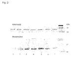
- Fig.2:
- SDS-PAGE und Western-Blot-Analyse von rekombinantem tTF1-218 und tTF-Fusionsproteinen. Die Reinheit des tTF und der tTF-Fusionsproteine wurde nach Extraktion aus E.coli (BL21 DE3) und "refolding" über einen linearen Harnstoff-Gradienten (6M - 1M) mittels SDS-PAGE mit anschließender Coomassie-blue-Färbung kontrolliert. Die Identität der Proteine wurde mittels Western-Blot unter Verwendung eines monoklonalen anti-Tissue-Factor-Antikörpers (Klon VIC7, American Diagnostics) verifiziert. Belegung der einzelnen Bahnen: 1=tTF; 2=tTF-RGD; 3=tTF-NGR; 4=tTF-cycloNGR1; 5=tTF-cycloNGR2; 6=tTF-cycloNGR3; 7=tTF-GALNGRSHAG; M = Molekulargewichtsmarker.
- Fig.3:
- Bestimmung der Michaeliskonstanten (Km) für die Aktivierung von FX durch FVIIa/tTF1-218 bzw. FVIIa/tTF1-218-Fusionsproteine. Die Parameter der Michaelis-Menten-Kinetik wurden nach der von Ruf angegebenen Methode berechnet (45).
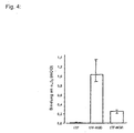
- Fig.4:
- Bindung von tTF, tTF-RGD und tTF-NGR an das Integrin αvβ3. Die Bindung von 0,1µM tTF, tTF-RGD und tTF-NGR an immobilisiertes αvβ3 wurde mit einem polyklonalen Antikörper gegen humanen TF (American Diagnostica) in einem ELISA quantifiziert. Die Ergebnisse sind als Median und Interquartilbereich dargestellt. Die Unterschiede der Bindung zwischen tTF-RGD und tTF bzw. zwischen tTF-NGR und tTF waren statistisch signifikant (p<0,001, Mann-Whitney-Test).
- Fig.5:
- Spezifität der Bindung von tTF-RGD an das Integrin αvβ3. Die Bindung von tTF-RGD (0,1µM) an immobilisiertes αvβ3 wurde mittels kompetitiver Inhibition mit dem synthetischen Peptid GRGDSP (1-10µM) signifikant gehemmt (p<0,001, Mann-Whitney-Test für beide RGD Peptidkonzentrationen).
- Fig.6:
- Bindung von tTF und tTF-RGD an humane Endothelzellen. A: FACS Analyse von Endothelzellen, die mit 0,1 µM tTF (2) oder mit 0,1 µM tTF-RGD (3) für 60 min. bei 4°C inkubiert wurden. B: Durch kompetitive Inhibition des tTF-RGD Fusionsproteins mit 1 µM GRGDSP war eine 75%ige Reduktion der Bindung darstellbar (4). Die Kurven 1 in A und B zeigen die negative Kontrolle.
- Fig.7:
- Inhibition eines als Xenotransplantat in thymuslosen Nackt-Mäusen wachsenden menschlichen Lungenkarzinoms (CCL185) durch intravenöse Therapie mit tTF-Fusionsproteinen (tTF-RGD, n=6; tTF-NGR, n=6) im Vergleich zum Wachstum der Tumoren bei Infusion von physiologischer Kochsalzlösung (NaCl, n=8) oder tTF (n=1). Die senkrechten Pfeile kennzeichnen die Zeitpunkte der Injektionen mit den jeweiligen Substanzen.
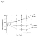
- Fig.8:
- Inhibition und Teilremission eines als Xenotransplantat in thymuslosen Nackt-Mäusen wachsenden menschlichen malignen Melanoms (M21) durch intravenöse Therapie mit tTF-Fusionsproteinen (tTF-RGD, n=3; tTF-NGR, n=3) im Vergleich zum Wachstum der Tumoren bei Infusion von physiologischer Kochsalzlösung (NaCl, n=4) oder tTF (n=4). Die senkrechten Pfeile kennzeichnen die Zeitpunkte der Injektionen mit den jeweiligen Substanzen.
- Fig.9:
- Makroskopische in vivo-Aufnahme einer tumortragenden Maus 20 min. nach Injektion des tTF-NGR Fusionsproteins (A, linke Bildhälfte) bzw. NaCl (A, rechte Bildhälfte). Das makroskopische Bild mit bläulich-livider Verfärbung des Tumors nach Injektion von tTF-NGR deutet auf eine Tumornekrose hin. Nach 60 min. wurden beide Mäuse exsanguiniert, der Tumor in toto exstipiert und histologisch untersucht. In B ist die hämorrhagische Imbibierung des mit tTF-NGR behandelten Tumors als Zeichen der sekundären Einblutung infolge der beginnenden Tumornekrose sichtbar. Im Gegensatz hierzu scheint der mit NaCl behandelte Tumor vital zu sein (C).
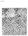
- Fig.10:
- Histologie des Melanom-Tumors 1 Stunde nach intravenöser Injektion von tTF-RGD (A und B), tTF-NGR (C und D) und Kochsalz (E und F) in die Schwanzvene der tumortragenden Nacktmaus. Die Blutgefässe erscheinen bei den mit den tTF Fusionsproteinen behandelten Tumoren thrombotisch verschlossen (Pfeile). Im Versorgungsgebiet des durch ein Blutgerinnsel verschlossenen Gefäßes können ausgedehnte Tumor-Nekrosen beobachtet werden (A-D). Photographiert wurden repräsentative Areale der Tumoren (A, C und E: 200fache Vergrößerung, B, D und F 400fache Vergrößerung; HE-Färbung (Färbung z.B. beschrieben in H.C. Burck, Histologische Technik - Leitfaden für die Herstellung mikroskopischer Präparate in Unterricht und Praxis 5. Auflage, Thieme Verlag, Stuttgart 1982, Seiten 109 ff.).
- Fig.11:
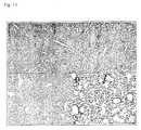
- Repräsentative Histologien von Herz (A), Niere (B), Leber (C) und Lunge (D) 1 Stunde nach Injektion von 4mg/kg KG tTF-NGR. In keinem dieser Organe waren mikroskopisch Thrombosen oder Nekrosen nachweisbar. (HE-Färbung; 200fache Vergrößerung).
- Fig.12:
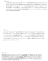
- Aminosäuresequenz des humanen Gewebefaktors (TF).
- Fig.13:
- Aminosäuresequenz des trunkierten humanen Gewebefaktors tTF1-218 (im Rahmen der vorliegenden Anmeldung auch kurz als tTF bezeichnet).
- Fig.14:
- Aminosäuresequenz des Fusionspolypeptids tTF-GRGDSP (auch kurz als tTF-RGD bezeichnet).
- Fig.15:
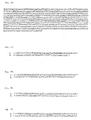
- Aminosäuresequenz des Fusionspolypeptids tTF-GNGRAHA (auch kurz als tTF-NGR bezeichnet).
- Fig.16:
- Aminosäuresequenz des Fusionspolypeptids tTF-GALNGRSHAG.
Fig.17 : Aminosäuresequenz des Fusionspolypeptids tTF-GCNGRCG (auch kurz als tTF-cycloNGR1 bezeichnet). - Fig.18:
- Aminosäuresequenz des Fusionspolypeptids tTF-GCNGRCVSGCAGRC (auch kurz als tTF-cycloNGR2 bezeichnet).
- Fig.19:
- Aminosäuresequenz des Fusionspolypeptids tTF-GCVLNGRMEC (auch kurz als tTF-cycloNGR3 bezeichnet).
- Fig.20:
- Nukleotidsequenz des trunkierten humanen Gewebefaktors tTF1-218 (im Rahmen der vorliegenden Anmeldung kurz als tTF bezeichnet).
- Fig.21:
- Nukleotidsequenz des Fusionspolypeptids tTF-GRGDSP (auch kurz als tTF-RGD bezeichnet).
- Fig.22:
- Nukleotidsequenz des Fusionspolypeptids tTF-GNGRAHA (auch kurz als tTF-NGR bezeichnet).
- Fig.23:
- Nukleotidsequenz des Fusionspolypeptids tTF-GALNGRSHAG.
- Fig.24:
- Nukleotidsequenz des Fusionspolypeptids tTF-GCNGRCG (auch kurz als tTF-cycloNGR1 bezeichnet).
- Fig.25:
- Nukleotidsequenz des Fusionspolypeptids tTF-GCNGRCVSGCAGRC (auch kurz als tTF-cycloNGR2 bezeichnet).
- Fig.26:
- Nukleotidsequenz des Fusionspolypeptids tTF-GCVLNGRMEC (auch kurz als tTF-cycloNGR3 bezeichnet).
- Fig.27:
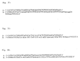
- Nukleotidsequenz der Oligonukleotide zur Herstellung von tTF1-218. A: 5'-Primer; B: 3'-Primer.
- Fig.28:
- Nukleotidsequenz der Oligonukleotide zur Herstellung von tTF-GRGDSP. A: 5'-Primer; B: 3'-Primer.
- Fig.29:
- Nukleotidsequenz der Oligonukleotide zur Herstellung von tTF-GNGRAHA. A: 5'-Primer; B: 3'-Primer.
- Fig.30:
- Nukleotidsequenz der Oligonukleotide zur Herstellung von tTF-GCNGRCG. A: 5'-Primer; B: 3'-Primer.
- Fig.31:
- Nukleotidsequenz der Oligonukleotide zur Herstellung von tTF-GCNGRCVSGCAGRC. A: 5'-Primer; B: 3'-Primer.
- Fig.32:
- Nukleotidsequenz der Oligonukleotide zur Herstellung von tTF-GCVLNGRMEC. A: 5'-Primer; B: 3'-Primer.
- Fig.33:
- Nukleotidsequenz der Oligonukleotide zur Herstellung von tTF-GALNGRSHAG. A: 5'-Primer; B: 3'-Primer.
- Fig.34:
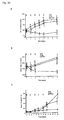
- a: Inhibition und Teilremission eines als Xenotransplantat in thymuslosen Nackt-Mäusen wachsenden menschlichen malignen Melanoms (M21) durch intravenöse Therapie mit tTF-Fusionsproteinen (tTF-RGD, n=7) im Vergleich zum Wachstum der Tumoren bei Infusion von physiologischer Kochsalzlösung (NaCl, n=9) oder tTF (n=11). Die senkrechten Pfeile kennzeichnen die Zeitpunkte der Injektionen mit den jeweiligen Substanzen. b: Inhibition eines als Xenotransplantat in thymuslosen Nackt-Mäusen wachsenden menschlichen Fibrosarkoms (HT1080) durch intravenöse Therapie mit tTF-Fusionsproteinen (tTF-RGD, n=12) im Vergleich zum Wachstum der Tumoren bei Infusion von physiologischer Kochsalzlösung (NaCl, n=15) oder tTF (n=14). Die senkrechten Pfeile kennzeichnen die Zeitpunkte der Injektionen mit den jeweiligen Substanzen. c: Inhibition eines als Xenotransplantat in thymuslosen Nackt-Mäusen wachsenden menschlichen Lungenkarzinoms (CCL185) durch intravenöse Therapie mit tTF-Fusionsproteinen (tTF-RG3D, n=11) im Vergleich zum Wachstum der Tumoren bei Infusion von physiologischer Kochsalzlösung (NaCl, n=10) oder tTF (n=5). Die senkrechten Pfeile kennzeichnen die Zeitpunkte der Injektionen mit den jeweiligen Substanzen. Die statistische Signifikanz wurde jeweils mit dem Mann-Whitney-Test für unabhängige Gruppen untersucht, wobei P-Werte unter 0,05 als signifikant betrachtet wurden. * gibt die statistische Signifikanz des Unterschieds zwischen tTF-RGD und Puffer an.
- Fig.35:
- Makroskopische Aufnahme einer einen M21-Tumor tragenden Maus nach Ende der Behandlung (Tag 7) mit tTF-RGD Fusionsprotein (A, C) bzw. NaCl (B, D). Der Größenunterschied und das unterschiedliche Erscheinungsbild der tTF-RGD-behandelten Tumoren, die im Gegensatz zu dem vital erscheinenden Kontrolltumor klare Zeichen von Nekrose zeigen, ist deutlich erkennbar.
- Fig.36:
- H-E-Färbung von Tumoren und Organen von mit tTF-RGD und physiologischer Kochsalzlösung behandelten Mäusen In mit tTF-RGD behandelten Tieren wurde starke Thrombose und Nekrose von Tumorzellen beobachtet (A: 200x, B: 400x). Pfeile zeigen Beispiele der Thrombosen in Blutgefäßen des Tumors. In mit Kochsalzlösung behandelten Tieren tritt keine offentsichtliche Thrombose oder Nekrose auf (C:200x, D: 400x). Pfeile zeigen intakte Blöugefäße des Tumors mit einigen Erythrocyten. Herz (E), Lunge (F), Leber (G), und Niere der mit tTF-RGD behandelten Tiere zeigten keine sichtbare Thrombose oder Nekrose.
- Fig.37:
- Wirkung von tTF-NGR in einem Fibrosarkom-Modell Fibrosarkom (HT1080) tragende Mäuse wurden ohne (pre tTF-NGR) bzw. 6 Stunden nach (post tTF-NGR) i.v. Gabe von tTF-NGR durch Kernspintomographie (MRI) untersucht. Gezeigt ist die hohe (high) oder niedrige (low) vaskuläre Volumenfraktion (Vascular volume fraction).
- Diese Probleme aus dem Stand der Technik wurden nunmehr durch Fusionspolypeptide gelöst, die folgende Peptide umfassen:
- a) ein Peptid von 3 bis 30 Aminosäuren, das eine selektive Bindung des Fusionspolypeptides an Tumorgefäßendothelzellen ermöglicht; und
- b) den Gewebefaktor TF (Tissue Factor) oder ein Fragment davon, wobei der Gewebefaktor und das Fragment dadurch gekennzeichnet sind, daß sie die Blutgerinnung bei Bindung des Fusionspolypeptides an Tumorgefäßendothelzellen aktivieren können,
- Die erfindungsgemäßen Fusionspolypeptide können neben den Sequenzen a) und b) weitere Sequenzen umfassen, soweit diese die sterische Konformation des Fusionspolypeptids nicht beeinträchtigen und die Bildung des die Blutgerinnung auslösenden Enzym- und Substratkomplexes nicht hindern. Die erfindungsgemäßen Fusionspolypeptide können beispielsweise die Sequenzen eines His-Tags enthalten, welche die rekombinante Expression und Reinigung des Peptids vereinfachen (vgl. Beispiele). Die Gegenwart dieser Sequenzen ist jedoch nicht notwendig. Gemäß einer bevorzugten Ausführungsform der Erfindung besteht das Fusionspolypeptid daher aus:
- a) einem Peptid von 3 bis 30 Aminosäuren, das eine selektive Bindung des Fusionspolypeptides an Tumorgefäßendothelzellen ermöglicht; und
- b) dem Gewebefaktor TF (Tissue Factor) oder einem Fragment davon, wobei der Gewebefaktor und das Fragment dadurch gekennzeichnet sind, daß sie die Blutgerinnung bei Bindung des Fusionspolypeptides an Tumorgefäßendothelzellen aktivieren können,
- a) einem Peptid von 3 bis 30 Aminosäuren, das eine selektive Bindung des Fusionspolypeptides an Tumorgefäßendothelzellen ermöglicht; und
- b) dem Gewebefaktor TF (Tissue Factor) oder einem Fragment davon, wobei der Gewebefaktor und das Fragment dadurch gekennzeichnet sind, daß sie die Blutgerinnung bei Bindung des Fusionspolypeptides an Tumorgefäßendothelzellen aktivieren können,
- Erfindungsgemäß konnte somit überraschenderweise gezeigt werden, daß Fusionspolypeptide aus einem besonders kleinen Peptid, das eine selektive Bindung des Fusionspolypeptides an Tumorgefäßendothelzellen ermöglicht und einem Peptid, daß die Blutgerinnung bei der Bindung des Fusionspolypeptides an Tumorgefäßendothelzellen aktivieren kann, für die antivaskuläre Tumortherapie besonders vorteilhaft sind. Die geringe Größe des Polypeptides, das die Bindung an die Tumorgefäßendothelzellen ermöglicht, verbessert die Ausrichtung des Fusionsproteins zur Phospholipidmembran der Endothelzelle. Die Bildung des für die Blutgerinnung wesentlichen Enzym-/Substratkomplexes wird nicht sterisch behindert und der Gewebefaktor TF, der die Blutgerinnung aktivieren kann, erfährt keine Konformationsveränderung.
- Gemäß einer bevorzugten Ausführungsform der vorliegenden Erfindung handelt es sich bei dem Peptid, das die Blutgerinnung bei Bindung des Fusionspolypeptides an Tumorgefäßendothelzellen aktivieren kann um den Gewebefaktor TF mit der in SEQ ID NO:1 (
Fig. 12 ) dargestellten Aminosäure-Sequenz. Erfindungsgemäß sind ferner Gewebefaktor-Sequenzen umfaßt, die eine Aminosäure-Homologie von mindestens 70% oder mindestens 80% zu der SEQ ID NO:1 (Fig. 12 ) aufweisen, wobei Sequenzen mit einer Homologie von mindestens 95 % besonders bevorzugt sind. Die Bestimmung des Homologiegrades erfolgt, indem man die beiden Sequenzen übereinander schreibt, wobei vier Lücken auf einer Länge von 100 Aminosäuren möglich sind, um größtmögliche Übereinstimmung der zu vergleichenden Sequenzen zu erzielen (vgl. auch Dayhoff, Atlas of Protein Sequence and Structure, 5, 124, 1972). Anschließend wird der Prozentsatz der Aminosäurereste der kürzeren der beiden Aminosäureketten ermittelt, der identischen Aminosäureresten auf der anderen Kette gegenübersteht. - Das Peptid, das die Blutgerinnung in Tumorgefäßen bei Bindung des Fusionspolypeptides an Tumorgefäßendothelzellen aktivieren kann, kann ferner ein Fragment des Gewebefaktors TF oder ein Fragment einer zu TF homologen Sequenz sein. Vorzugsweise weist das Fragment die in SEQ ID NO:2 (
Fig. 13 ) dargestellte Sequenz auf. Die in SEQ ID NO:2 (Fig. 13 ) dargestellte Sequenz (tTF1-218 oder kurz tTF) umfaßt die N-terminalen 218 Aminosäuren von TF. Ferner können erfindungsgemäß auch Fragmente von tTF verwendet werden, denen gegenüber tTF mehrere Aminosäuren am N-Terminus oder C-Terminus fehlen. Beispielsweise können Fragmente verwendet werden, denen bis zu 10 Aminosäuren am N-Terminus fehlen (tTF11-218). Des weiteren können Fragmente verwendet werden, denen bis zu 8 Aminosäuren am C-Terminus fehlen (tTF1-210), wie z.B. (tTF1-214). - Die vorliegende Erfindung betrifft Fusionspolypeptide, in denen das Peptid, das eine selektive Bindung an die Endothelzellen der Tumorgefäße ermöglicht, an den C-Terminus des Peptids gekoppelt ist, das die Blutgerinnung aktivieren kann. Unter "Tumorgefäßendothelzellen" bzw. "Endothelzellen in Tumorgefäßen" werden dabei erfindungsgemäß Zellen verstanden, die die Blutgefäße in einem Tumor auskleiden. Erfindungsgemäß wurde festgestellt, daß diese Anordnung eine Ausrichtung des Fusionsproteins senkrecht zur Phospholipidmembran der Endothelzelle gewährleistet, welche für die Auslösung der Blutgerinnung besonders vorteilhaft ist. Diese Orientierung entspricht der natürlichen Ausrichtung des TF bei der Induktion der Blutgerinnung. Wie in
Figur 3 gezeigt, ergeben sich für alle so hergestellten Konstrukte bezüglich der Aktivierung von Faktor X durch FVIIa/tTF1-218 bzw. FVIIa/tTF1-218-Fusionsproteine sehr ähnliche Michaelis-Menten-Kinetiken. Demgegenüber wurde im Stand der Technik das die Gerinnung aktivierende Peptid an den C-Terminus des Targeting-Moleküls gekoppelt (vgl. (16)). Die erfindungsgemäßen Fusionspolypeptide unterscheiden sich somit grundsätzlich von denen im Stand der Technik genannten Peptiden. - Das Peptid, das eine selektive Bindung des Fusionspolypeptides an Tumorgefäßendothelzellen ermöglicht, kann ein beliebiges Peptid sein, das eine Länge von 3 - 30 Aminosäuren aufweist und Tumorgefäßendothelzellen mit hoher Spezifität bindet. Entsprechende Peptide können aus Peptidbibliotheken durch im Stand der Technik übliche Verfahren isoliert werden. Sie können - je nach gewählter Peptidbibliothek - eine lineare oder cyclische Struktur aufweisen.
- Gemäß einer Ausführungsform der vorliegenden Erfindung umfassen die Peptide, die eine selektive Bindung des Fusionspolypeptides an Tumorgefäßendothelzellen ermöglichen, die Aminosäuresequenz RGD oder NGR. Beide Sequenzen waren im Stand der Technik für Ihre spezifische Bindung an Integrine, insbesondere αvβ3 und αvβ5 Integrine (RGD-Peptide), sowie als Zelladhäsionsmotive (NGR-Peptide) bekannt (vgl. (8)). Erfindungsgemäß wurde nunmehr überraschenderweise gezeigt, daß diese Peptide insbesondere dazu geeignet sind, Teil eines Fusionspolypeptides zu sein, dessen anderer Teil ein Peptid ist, das die Blutgerinnung in Tumoren bei Bindung des Fusionspolypeptides an Tumorgefäßendothelzellen aktivieren kann.
- Besonders vorteilhafte Wirkungen wurden mit den linearen Peptiden mit den Sequenzen GRGDSP, GNGRAHA und GALNGRSHAG und den zyklischen Peptiden mit den Sequenzen GCNGRCG, GCNGRCVSGCAGRC und GCVLNGRMEC erhalten. Es konnte gezeigt werden, daß Fusionspolypeptide, welche diese Sequenzen und die Sequenz der ersten 218 Aminosäuren von humanem TF umfassen, in hohem Maße für die antivaskuläre Tumortherapie geeignet sind. Insbesondere konnte gezeigt werden, daß diese Fusionspolypeptide das Wachstum von Tumoren signifikant hemmen oder die Tumoren in Ihrer Größe reduzieren (sieh
Fig. 7 und8 ). Die beobachtete Induktion einer Teilremission der Tumoren (vgl.Fig. 8 ) verweist aufgrund der hohen Vorhersagekraft des Mausmodells (42, 43, 44) auf die zu erwartenden positiven Resultate bei der humanen Tumortherapie. - Weiterhin umfaßt die Erfindung auch Fusionsproteine mit zyklischen RGD-Peptiden, da durch die Zyklisierung die Affinität für Integrine verstärkt wird (wie beispielsweise in der Veröffentlichung 21 beschrieben).
- Die vorliegende Erfindung betrifft ferner Fusionspolypeptide, die eine der in SEQ ID NO:3-8 (
Fig. 14-19 ) dargestellten Sequenzen aufweist. - Gemäß einer weiteren Ausführungsform betrifft die vorliegende Erfindung Nukleinsäuren, die für ein Fusionspolypeptid, wie oben beschrieben, kodieren. Entsprechende Nukleinsäuren können beispielsweise eine der in SEQ ID NO: 10-15 (
Fig. 21-26 ) dargestellten Sequenzen aufweisen. - In einem weiteren Aspekt betrifft die vorliegende Erfindung Vektoren, welche eine der oben genannten Nukleinsäuren umfassen. Entsprechende Vektoren umfassen üblicherweise ferner regulatorische Sequenzen für die Expression der Nukleinsäure. Solche Vektoren sind im Stand der Technik umfassend beschrieben und können von einer Vielzahl von Firmen kommerziell erworben werden.
- In einer weiteren Ausführungsform betrifft die vorliegende Erfindung Zellen, welche eine der genannten Nukleinsäuren oder Vektoren enthalten. Die Zellen werden üblicherweise für die Expression der Nukleinsäure und die rekombinante Herstellung der erfindungsgemäßen Fusionspolypeptide verwendet. Für diesen Zweck können eine Vielzahl von Zellen zum Einsatz gelangen, darunter E.coli, Hefezellen und tierische Zelllinien, wie CHO- oder COS-Zellen. Entsprechende Zellen und deren Verwendung sind im Stand der Technik umfassend beschrieben.
- Die erfindungsgemäßen Polypeptide nach Anspruch 1 können darüber hinaus auch durch andere geeignete Verfahren hergestellt werden, beispielsweise durch chemische Kopplung einzelner Peptide. So lassen sich einzelne Peptide nach im Stand der Technik üblichen Verfahren herstellen, z.B. durch chemische Synthese oder mittels heterologer Expression, und anschließend mittels Kopplung aneinanderfügen.
- Schließlich betrifft die vorliegende Erfindung auch Arzneimittel, welche die oben beschriebenen Fusionspolypeptide, Nukleinsäuren, Vektoren oder Zellen umfassen. Die Arzneimittel können ferner pharmazeutisch verträgliche Träger, Hilfsmittel oder Adjuvanzien umfassen. Ferner können die Polypeptide in einem solchen Arzneimittel in einem modifizierten Zustand vorliegen, z.B. pegyliert, d.h. gekoppelt an ein Polyäthylenglykolmolekül.
- Die erfindungsgemäßen Fusionspolypeptide bzw. diese Fusionspolypeptide enthaltende Arzneimittel werden für die Behandlung von Tumorerkrankungen, insbesondere für die antivaskuläre Tumortherapie verwendet. Als Tumorerkrankungen, die sich mit Hilfe der erfindungsgemäßen Fusionspolypeptide bzw. diese Fusionspolypeptide enthaltener Arzneimittel behandeln lassen, kommen beispielsweise Bronchialkarzinome und andere Tumoren des Thorax und des Mediastinums, Mammakarzinome und andere gynäkologische Tumoren, kolorektale Karzinome, Pankreaskarzinome und andere Tumoren des Gastrointestinaltraktes, maligne Melanome und andere Tumoren der Haut, Tumoren im Kopf-Hals-Bereich, Prostatakarzinome und andere Urogenitaltumoren, Sarkome, endokrin-aktive Tumoren, Leukämien und Myelodysplastische Syndrome sowie Hodgkin-Lymphome und Non-Hodgkin-Lymphome in Betracht.
- Ferner können auch gutartige Tumoren, wie beispielsweise Hämangiome, und die Gefäßneubildung bei der diabetischen Retinopathie behandelt werden.
- Neben der intravenösen Verabreichung ist auch die subkutane und intraperitonale Verabreichung der Fusionspolypeptide bzw. Arzneimittel möglich. Durch Verpackung in pharmazeutische Vehikel, die eine Spaltung der Fusionspolypeptide im Magen-Darm-Trakt verhindert, wäre es darüber hinaus auch möglich, die Fusionspolypeptide bzw. Arzneimittel oral zu verabreichen.
- Vorteilhaft kann weiterhin eine Kombination der Verabreichung der erfindungsgemäßen Fusionspolypeptide mit anderen therapeutischen Ansätzen, z.B. einer cytotoxischen Chemotherapie oder Bestrahlung, sein. Auch eine Kombination mit anderen Wirkstoffen, z.B. eine Kombination mit Faktor Vlla oder Doxycyclin ist möglich, bevorzugt ist eine Kombination des erfindungsgemäßen Polypeptids mit Faktor VIIa oder Doxycyclin jedoch nicht notwendig.
- Die Erfindung wird anhand der nachfolgenden Beispiele näher beschrieben:
- Die für die N-terminalen 218 Aminosäuren des Gewebefaktors TF (im folgenden als tTF bezeichnet) kodierende cDNA wurde mittels Polymerasekettenreaktion (PCR) unter Verwendung der in SEQ ID NO:16 und SEQ ID NO:17 (
Fig. 27 ) synthetisiert und in den Expressionsvektor pET-30a(+) (Novagen) kloniert. Die rekombinanten Plasmide wurden in E. coli (BL21) transformiert, exprimiert und gereinigt (Qiagen Plasmid Kit). - Neben dem trunkierten Gewebefaktor tTF wurden tTF-Peptid-Fusionsproteine konstruiert, in denen die Targeting-Peptide zunächst an das carboxyterminale Ende des löslichen Gewebefaktors tTF gebunden sind. Es wurden folgende lineare Fusionsproteine konstruiert:
- tTF-GRGDSP (SEQ ID NO:3;
Fig.14 ; im folgenden als tTF-RGD bezeichnet; es wurden die PCR-Primer SEQ ID NO:18 und SEQ ID NO:19 (Fig. 28 ) verwendet); - tTF-GNGRAHA (SEQ ID NO:4;
Fig.15 ; im folgenden als tTF-NGR bezeichnet; es wurden die PCR-Primer SEQ ID NO:20 und SEQ ID NO:21 (Fig.29 ) verwendet); - tTF-GALNGRSHAG (SEQ ID NO:5;
Fig.16 ; es wurden die PCR-Primer SEQ ID NO:28 und SEQ ID NO:29 (Fig. 33 ) verwendet); - Darüber hinaus wurden die folgenden cyclischen Fusionsproteine synthetisiert:
- tTF-GCNGRCG (SEQ ID NO:6;
Fig.17 ; im folgenden als tTF-cycloNGR1 bezeichnet; es wurden die PCR-Primer SEQ ID NO:22 und SEQ ID NO:23 (Fig.30 ) verwendet), - tTF-GCNGRCVSGCAGRC (SEQ ID NO:7;
Fig.18 ; im folgenden als tTF-cycloNGR2 bezeichnet; es wurden die PCR-Primer SEQ ID NO:24 und SEQ ID NO:25 (Fig.31 ) verwendet), - tTF-GCVLNGRMEC (SEQ ID NO:8;
Fig.19 ; im folgenden als tTF-cycloNGR3 bezeichnet; es wurden die PCR-Primer SEQ ID NO:26 und SEQ ID NO:27 (Fig.32 ) verwendet) - Alle Konstrukte (einschließlich tTF) wurden in dem Vektor pET30a(+) exprimiert, der die zusätzliche Expression eines N-terminalen "Affinitäts-Tag" aus 6 Histidin-Resten und wenigen Vektorkodierten Aminosäuren vermittelt. Mit Hilfe dieses Affinitäts-Tags konnten die Konstrukte durch Affinitätschromatographie über eine Nickel-Nitrilotriessigsäure Säule (Ni-NTA, Novagen) gereinigt werden. Der Affinitäts-Tag ist in SEQ ID NO:30 dargestellt. SEQ ID NO:31 und SEQ ID NO:32 zeigen exemplarisch die vollständigen Aminosäuresequenzen von tTF-GRGDSP und tTF-GNGRAHA mit Affinitäts-Tag.
- Die Konstrukte wurden so gewählt, daß auf der Basis der bekannten Röntgenkristallstruktur des tTF:FVIIa-Komplexes (19) eine senkrechte Ausrichtung des tTF-Fusionsproteins zur Phospholipidmembran der Endothelzelle gewährleistet wird, was der Ausrichtung des nativem TF entspricht. Andererseits wurde berücksichtigt, daß aus der gewählten Struktur keine sterische Behinderung von tTF für die Interaktion mit FVIIa und dem makromolekularen Substrat FX entstehen soll. Durch die Spezifität der RGD-Sequenz für das αvβ3-Integrin und der NGR-Sequenz für CD13 (Aminopeptidase N) wird eine Tumorselektivität erreicht, da diese Rezeptoren selektiv und spezifisch in hoher Dichte auf Tumor-Endothelzellen, aber von wenigen Ausnahmen abgesehen, nicht auf ruhenden Endothelzellen im normalen Gewebe exprimiert werden (siehe
Figur 1 ). - tTF und die beschriebenen Fusionsproteine tTF-RGD, tTF-NGR, tTF-GALNGRSHAG und tTF-cycloNGR1-3 wurden mittels pET30a(+) in E. coli (BL21) transformiert und exprimiert. Transformierte, mit IPTG induzierte E. coli BL21 DE3 wurden zentrifugiert und in 5-7 ml Lysispuffer (10 mM Tris-HCl, pH7,5; 150 mM NaCl; 1 mM MgCl2; 10 µg/ml Aprotinin; 2 mg/ml Lysozym)/g Pellet aufgenommen und 20 µl Benzonase (Novagen) hinzugefügt. Nach 90 min Inkubation bei Raumtemperatur (RT) und einer Zentrifugation bei 12 000 g, 20 min, 4°C, wurde das Pellet resuspendiert und durch Ultraschallbehandlung in Waschpuffer (10 mM Tris/HCl, pH 7,5; 1 mM EDTA; 3% Triton X-100) homogenisiert. "Inclusion bodies" wurden über Nacht bei RT in 2-4 ml /g Pellet an denaturierendem Puffer (6 M Guanidiniumchlorid, 0,5 M NaCl, 20 mM NaH2PO4, 1 mMDTT) gelöst. Der Überstand einer Zentrifugation (5 000 g, 30 min, 4°C) wurde mit einem 0,22 µg-Filter filtriert. Die Konstrukte wurden über eine Nickel-Nitrilotriessigsäure Säule (Ni-NTA, Novagen) über die zusätzlich eingeführten His-Tag Sequenzen des Konstrukts bis zur Homogenität gereinigt. Die Reinigung und Faltung der Proteine wurde mit dem His Bind Buffer Kit (Novagen) durchgeführt. Anschließend wurde gegen TBS-Puffer (20 mM Tris, 150 mM NaCl, pH 7,4) dialysiert.
- Durch SDS-PAGE, Western Blot und Massenspektroskopie Analysen konnte die Identität der Proteine bestätigt werden (siehe
Figur 2 ). - Die funktionelle Aktivität dieser Fusionsproteine hinsichtlich Kofaktoraktivität bei der Aktivierung von Faktor X zu Faktor Xa durch Faktor Vlla konnte in vitro durch Michaelis-Menten Analysen gezeigt werden. Die Fähigkeit von tTF bzw. der tTF-Fusionspolypeptide, die spezifische proteolytische Aktivierung von FX durch FVIIa in Anwesenheit von Phospholipiden zu verstärken, wurde in einer leichten Modifikation nach der von Ruf beschriebenen Methode bestimmt (45). Hierzu wurden je 20 µl der folgenden Reagenzien in Mikrotiter-Platten pipettiert: (a) 50 nM rekombinanter FVIIa (Novo-Nordisk) in TBS-BSA; b) 0,16 nM - 1,6 µM tTF/tTF-Fusionspolypeptid in TBS-BSA; (c) 25 mM CaCl2 und 500 µM Phospholipidvesikel (Phosphatidylcholin / Phosphatidylserin, 70/30, M/M; Sigma). Nach 10 Min. Inkubation bei Raumtemperatur wurden 20 µl des natürlichen Substrats FX (Enzyme Research Laboratories) in einer Konzentration von 5 µM hinzugegeben. Anschließend wurde eine Probe in minütlichen Abständen abpipettiert und die Reaktion durch Zugabe von 100 mM EDTA-Lösung gestoppt. Die gebildete Menge an FXa wurde durch Zugabe des chromogenen Substrats Spectrozym FXa in einem Microplate Reader durch Bestimmung der Absorptionsänderung bei 405 nm gemessen und die Parameter für die Michaelis-Menten-Kinetik nach der von Ruf angegebenen Methode analysiert. Die Ergebnisse zeigen, daß sowohl tTF als auch die tTF-Fusionspolypeptide unter diesen Bedingungen funktionell aktiv sind (
Fig. 3 ). Die ermittelten Michaleliskonstanten (Km) der Fusionspolypeptide lagen zwischen 0,12 -1,2 nM (Fig. 3 ), somit im unteren Bereich, welcher für tTF publiziert wurde. Somit kann angenommen werden, daß durch die Fusionierung von tTF mit den Peptiden die funktionelle Aktivität unbeeinflusst bleibt. - Die Bindung von tTF-RGD und tTF-NGR an das Integrin αvβ3 konnte in einem ELISA (Enzyme Linked Immunosorbent Assay) demonstriert werden, indem gereinigtes αvβ3 an Mikrotiterplatten immobilisiert worden war (siehe
Figur 4 ). Die Spezifität der Bindung von tTF-RGD an αvβ3 wird dadurch unterstrichen, daß das synthetische Peptid mit der Sequenz GRGDSP (Fa. Gibco) die Bindung von tTF-RGD an αvβ3 in diesem Testsystem kompetitiv inhibiert (sieheFigur 5 ). - Anschließend wurde die spezifische Bindung von tTF-RGD an αvβ3 auf Endothelzellen evaluiert. Hierzu wurde die differentielle Bindung von biotinyliertem tTF und tTF-RGD an Endothelzellen in Suspension mittels FACS (Fluorescence Activated Cell Sorting) analysiert. Hierbei macht man sich experimentell zu Nutze, daß alle in Gewebekultur gehaltenen Endothelzellen aktiviert sind, d.h. αvβ3-Moleküle exprimieren. Dies läßt sich mittels verschiedener immunhistochemischer Verfahren nachweisen. Eine kultivierte Endothelzelle entspricht somit hinsichtlich ihres Expressionsmusters in Bezug auf αvβ3 einer Tumorendothelzelle. Demgemäß läßt sich eine kultivierte Endothelzelle als Modellsystem für die spezifische Bindung von Substanzen an Tumorendothelzellen nutzen und ermöglicht ferner Aussagen über die zu erwartende Toxizität.
- Als Detektionsmethode wurde Streptavidin-Phycoerythrin benutzt. Die gemessene Fluoreszenzintensität lag bei tTF-RGD um den Faktor acht höher als für tTF (
Figur 6A ). Ferner konnte die Bindung von 0,1 µM tTF-RGD an Endothelzellen kompetitiv durch die Gabe von 1 µM des synthetischen Peptids GRGDSP um 75% gesenkt werden (Figur 6B ). Dies unterstreicht die Spezifität der Bindung von tTF-RGD an RGD-bindende Rezeptoren auf der Endothelzelloberfläche wie αvβ3. - Die Fusionsproteine tTF-RGD und tTF-NGR wurden hinsichtlich ihrer Wirkungen und Nebenwirkungen an Xenotransplantaten menschlicher Tumoren in thymuslosen Nacktmäusen evaluiert. Hierzu wurden die in unserem Labor etablierten Modelle verwendet (33, 34). Die Zellinien CCL185 (humanes Adenokarzinom der Lunge) bzw. M-21 (humanes Melanom) wurden subkutan in die Flanke männlicher BALB/c/Nackt-Mäuse (9-12 Wochen alt) injiziert. Nach Erreichen eines Tumorvolumens von etwa 50-100 mm3 (CCL185) bzw. 400-600 mm3 (M-21) wurden die Mäuse randomisiert vier Gruppen zugeordnet. Gruppe 1 erhielt nur physiologische Kochsalzlösung (NaCl), Gruppe 2 tTF, Gruppe 3 tTF-RGD, Gruppe 4 tTF-NGR (jeweils 1,5-2,0 mg/kg Körpergewicht (KG) des Proteins). Die Injektion erfolgte in die Schwanzvene der Tiere in 1-3 täglichen Intervallen (in Abhängigkeit von der Wachstumsgeschwindigkeit der jeweiligen Zellinie). Hier zeigte sich eine erhebliche therapeutische Aktivität der Fusionsproteine. Die Tumoren der mit tTF-RGD bzw. tTF-NGR Fusionsproteinen behandelten Mäuse wurden im Vergleich zu tTF oder NaCl in ihrem Wachstum signifikant gehemmt oder in ihrer Größe bis hin zu einer Teilremission reduziert (siehe
Figuren 7 und8 ). - Um den Wirkungsmechanismus der Thrombosierung von Tumorgefässen zu belegen, wurde folgendes Experiment durchgeführt: Hierzu wurde die humane Melanom Zellinie in die Flanke von zwei männlichen Balb/c/Nacktmäusen injiziert. Bei Erreichen einer Tumorgrösse von ca. 500 mm3 wurden 2,0 mg/kg KG tTF-NGR oder NaCl in die Schwanzvene injiziert.
Figur 9A zeigt eine makroskopische in vivo Aufnahme der tumortragenden Maus 20 min. nach Injektion des tTF-NGR Fusionsproteins (linke Bildhälfte) bzw. NaCl (rechte Bildhälfte). Das makroskopische Bild mit bläulich-livider Verfärbung des Tumors nach Injektion von tTF-NGR deutet auf eine Tumornekrose hin. Nach 60 min. wurden die Mäuse exsanguiniert, der Tumor in toto exstipiert und histologisch untersucht. InFigur 9B ist die hämorrhagische Imbibierung des mit tTF-NGR behandelten Tumors als Zeichen der sekundären Einblutung infolge der beginnenden Tumornekrose sichtbar. Im Gegensatz hierzu scheint der mit NaCl behandelte Tumor vital zu sein (Figur 9C). - Die histologische Untersuchung des Melanom-Tumors zeigt mikroskopisch sichtbare Thrombenbildung in den Blutgefässen (
Figur 10A-D ). Durch diesen Befund wird der angenommene anti-Tumor-Wirkungsmechanismus von tTF-NGR, d.h. die Induktion von Thromben in den Blutgefässen, belegt. Die hohe Selektivität von tTF-NGR für Tumorblutgefässe wird durch den fehlenden histologischen Nachweis von Gerinnsel- und Nekrosenbildung im normalen Gewebe wie Herz, Niere, Leber und Lunge demonstriert (Figur 11A-D ). Selbst repetitive hohe Dosen von tTF-NGR (4mg/kg KG) führten zu keinerlei sichtbarer Thrombenbildung oder Organtoxizität. - Die Antitumoraktivität des tTF-RGD-Fusionsproteins wurde ferner in BALB/c/Nackt-Mäusen mit Fibrosarkomen (HT1080) untersucht. Diese Tumore wachsen schnell und sind gut vaskularisiert. The Ergebnisse zweier Experimente sind in Tabelle 2 und
Figur 34 zusammengefaßt. Nach der zweiten Injektion von tTF-RGD wurde im Vergleich zu Kontollgruppen eine signifikante Wachstumsinhibition der HT1080 Tumore beobachtet. Dieser Effekt hielt bis zum Ende des Experiments an Tag 7 an (P=0,021 für tTF-RGD gegenüber der Puffer-Kontrolle (physiologische Kochsalzlösung), P=0,005 für tTF-RGD gegenüber tTF). Ähnlich wie bei den früheren Experimenten wurde eine Teilregression des Tumorvolumens in diesem Modell beobachtet.Tabelle 1: Wirkung von tTF-RGD auf das Wachstum von M21-Tumoren in Mäusen Behandlung Durchschnittliches Tumorvolumen (mm3) P ggü. Puffer P ggü. tTF n Tag 0 Tag 7 Puffer 590 +/-77 994 +/-140 ns 9 tTF 558+/-47 931 +/-147 ns 11 tTF-RGD 585+/-85 514+/-81 <0,01 <0,05 7 ns: nicht signifikant Tabelle 2: Wirkung von tTF-RGD auf das Wachstum von HT1080-Tumoren in Mäusen Behandlung Durchschnittliches Tumorvolumen (mm3) P ggü. Puffer P ggü. tTF n Tag 0 Tag 7 Puffer 1671+/-296 2431+/-559 ns 15 tTF 1751 +/-269 2335+/-398 ns 14 tTF-RGD 1725+/-197 1241+/-122 <0,05 <0,01 12 ns: nicht signifikant Tabelle 3: Wirkung von tTF-RGD auf das Wachstum von CCL185-Tumoren in Mäusen Behandlung Durchschnittliches Tumorvolumen (mm3) P ggü. Puffer P ggü. tTF n Tag 0 Tag 7 Puffer 39+/-3 467+/-137 ns 9 tTF 44+/-8 764+/-148 ns 5 tTF-RGD 45+/-5 130+/-19 <0,01 <0,01 10 ns: nicht signifikant - Weitere tTF-Fusionsproteine können vom Fachmann auf der Basis der Offenbarung der vorliegenden Erfindung problemlos konstruiert werden. Potentielle Kandidaten sind die Peptide TAASGVRSMH und LTLRWVGLMS, welche an NG 2 binden, das murine Homolog des humanen Melanom Proteoglykans (12). Die Expression von NG 2 ist beschränkt auf Tumorzellen und angiogenen Gefäßen eines Tumors (35). Ein weiterer Kandidat ist das synthetische Peptid TTHWGFTL, welches selektiv und potent die Matrix-Metallo-Proteinase-2 (MMP-2) inhibiert (13). Da das Integrin αvβ3 offenbar auch die MMP-2 in einer RGD unabhängigen Weise bindet, wird hierdurch das aktive Enzym an die Oberfläche der angiogenen Blutgefäße lokalisiert (36). Ein Konstrukt bestehend aus tTF und diesem MMP-2 inhibitorischen Peptid könnte ebenfalls die selektive Bindung von tTF1-218 an die Endothelzellmembran von Tumorgefäßen vermitteln.
-
- 1. Folkman J, Watson K, Ingber D, Hanahan D: Induction of angiogenesis during the transition from hyperplasia to neoplasia. Nature 339: 58-61, 1989
- 2. Dvrorak HJ, Sioussat TM, Brown LF, Berse B, Nagy JA, Sotrel A, Manseau EJ: Distribution of vascular permeability factor (vascular endothelial growth factor) in tumors - Concentration in tumor blood vessels. J Exp Med 174: 1275-1278, 1991
- 3. Dvorak HJ, Brown LF, Detmar M, Dvorak AM: Vascular permeability factor/vascular endothelial growth factor, microvascular hyperpermeability, and angiogenesis. Am J Pathol 146: 1029-1039, 1995
- 4. Terman BJ, Dougher-Vermazen M: Biological properties of VEGF/VPF receptors. Cancer Metastasis Rev 15: 159-163, 1996
- 5. Burrows FJ, Derbyshire EJ, Tazzari PL, Amlot P, Gazdzar AF, King SW, Letarte M, Vitetta ES, Thorpe PE: Upregulation of endoglin on vascular endothelial cells in human solid tumors: Implications for diagnosis and therapy. Clin Cancer Res 1: 1623-1634, 1995
- 6. Rettig WJ, Garinchesa P, Healey JH, Su SL, Jaffe EA, Old LJ: Identification of endosialin, a cell surface glycoprotein of vascular endothelial cells in human cancer. Proc Natl Acad Sci USA 89: 10832-10836, 1992
- 7. Carnemolla B, Balza E, Siri A, Zardi L, Nicrotra MR, Bigotti A, Natali PG: A tumor-associated fibronectin isoform generated by alternative splicing of messenger RNA precursors. J Cell Biol 108: 1139-1148, 1989
- 8. Arap W, Pasqualini R, Ruoslahti E: Cancer treatment by targeted drug delivery to tumor vasculature in a mouse model. Science 279: 377-380, 1998
- 9. Senger DR, Claffey KP, Benes JE, Peruzzi CA, Sergiou AP, Detmar M: Angiogenesis promoted by vascular endothelial growth factor: regulation through α1β1 and α2β1 integrins. Proc Natl Acad Sci USA 94: 13612-13617, 1997
- 10. Olson TA, Mohanraj D, Roy S, Ramakrishnan S: Targeting the tumor vasculature: inhibition of tumor growth by a vascular endothelial growth factor-toxin conjugate. Int J Cancer 73: 865-870, 1997
- 11. Bhagwat SV, Lahdenranta J, Giordano R, Arap W, Pasqualini R, Shapiro LH: CD13/APN is activated by angiogenic signals and is essential for capillary tube formation. Blood 97: 652-659, 2001
- 12. Burg MA, Pasqualini R, Arap W, Ruoslahti E, Stallcup WB: NG2 Proteoglycan-binding peptides target tumor neovasculature. Cancer Res 59: 2869-2874, 1999
- 13. Koivunen E, Arap W, Valtanen H, Rainisalo A, Medina OP, Heikkila P, Kantor C, Gahmberg CG, Salo T, Konttinen YT, Sorsa T, Ruoslahti E, Pasqualini R: Tumor targeting with a selective gelatinase inhibitor. Nat Biotechnol 17: 768-774, 1999
- 14. Huang X, Molema G, King S, Watkins L, Edgington TS, Thorpe PE: Tumor infarction in mice by antibody-directed targeting of tissue factor to tumor vasculature. Science 275: 547-550, 1997
- 15. Ran S, Gao B, Duffy S, Watkins L, Rote N, Thorpe PE: Infarction of solid Hodgkin's tumors in mice by antibody-directed targeting of tissue factor to tumor vasculature. Cancer Res 58: 4646-4653, 1998
- 16. Nilsson F, Kosmehl H, Zardi L, Neri D: Targeted delivery of tissue factor to the ED-B domain of fibronectin, a marker of angiogenesis, mediates the infarction of solid tumors in mice. Cancer Res 61: 711-716, 2001
- 17. Liu C, Huang H, Donate F, Dickinson C, Santucci R, EI-Sheikh A, Vessella R, Edgington TS. Prostate-specific membrane antigen directed selective thrombotic infarction of tumors. Cancer Res 62: 5470-5475, 2002
- 18. Gottstein C, Wels W, Ober B, Thorpe PE: Generation and characterisation of recombinant vascular targeting agents from hybridoma cell lines. BioTechniques 30: 190-200, 2001
- 19. Morrissey JH, Macik BG, Neuenschwander PF, Comp PC: Quantitation of activated factor VII levels in plasma using a tissue factor mutant selectively deficient in promoting factor VII activation. Blood 81: 734-744, 1993
- 20. Banner DW, D'Arcy A, Chène C, Winkler FK, Guha A, Konigsberg WH, Nemerson Y, Kirchhofer D: The crystal structure of the complex of blood coagulation factor Vlla with soluble tissue factor. Nature 380: 41-46, 1996
- 21. Koivunen E, Gay DA, Ruoslahti E: Selection of peptides binding to the α5β1 integrin from phage display library. J Biol Chem 268: 20205-20210, 1993
- 22. Healy JM, Murayama O, Maeda T, Yoshino K, Sekiguchi K, Kikuchi M: Peptide ligands for integrin αvβ3 selected from random phage display libraries. Biochemistry 34: 3948-3955, 1995
- 23. Pasqualini R, Koivunen E, Kain R, Lahdenranta J, Sakamoto M, Stryhn A, Ashmun RA, Shapiro LH, Arap W, Ruoslahti E: Aminopeptidase N is a receptor for tumor-homing peptides and a target for inhibiting angiogenesis. Cancer Res 60: 722-727, 2000
- 24. Curnis F, Sacchi A, Borgna L, Magni F, Gasparri A, Corti A: Enhancement of tumor necrosis factor α antitumor immunotherapeutic properties by targeted delivery to aminopeptidase N (CD 13). Nature Biotechnology 18: 1185-1190, 2000
- 25. Ellerby HM, Arap W, Ellerby LM, Kain R, Andrusiak R, Del Rio G, Krajewski S, Lombardo CR, Rao R, Ruoslahti E, Bredesen DE, Pasqualini R: Anti-cancer activity of targeted proapoptotic peptides. Nature Med 5: 1032-1038, 1999
- 26. Ruoslahti E: Targeting tumor vasculature with homing peptides from phage display. Cancer Biol 10:435-442,2000
- 27. Pasqualini R, Koivunen E, Kain R, Lahdenranta J, Sakamoto M, Stryhn A, Ashmun RA, Shapiro LH, Arap W, Ruohslahti E. Aminopeptidase N is a receptor for tumor-homing peptides and a traget for inhibiting angiogenesis. Cancer Res 60: 722-727, 2000
- 28. Curnis F, Arrigoni G, Sacchi A, Fischetti L, Arap W, Pasqualini R, Corti A. Differential binding of drugs containing the NGR motif to CD13 isoforms in tumor vessels, epithelia, and myeloid cells. Cancer Res 62: 867-874, 2002
- 29. Senger DR, Claffey KP, Benes JE, Perruzzi CA, Sergiou AP, Detmar M: Angiogenesis promoted by vascular endothelial growth factor: regulation through □1□1 and □2□1 integrins. Proc Natl Acad Sci USA 94: 13612-13617, 1997
- 30. Yun Z, Menter DG, Nicolson GL: Involvement of integrin αvβ3 in cell adhesion, motility and liver metastasis of murine RAW117 large cell lymphoma. Cancer Res 56: 3103-3111, 1996
- 31. Brooks PC, Clark RAF, Cheresh DA: Requirement of vascular integrin αvβ3 for angiogenesis. Science 264; 569-571, 1994
- 32. Brooks PC, Montgomery AM, Rosenfeld M, Reisfeld RA, Hu T, Klier G, Cheresh DA: Integrin αvβ3 antagonists promote tumor regression by inducing apoptosis of angiogenic blood vessels. Cell 92: 391-400, 1998
- 33. Topp MS, Koenigsmann M, Mire-Sluis A, Oberberg D, Eitelbach F, von Marschall Z, Notter M, Reufi B, Stein H, Thiel E, Berdel WE: Recombinant human interleukin-4 inhibits growth of some human lung tumor cell lines in vitro and in vivo. Blood 82: 2837-2844, 1993
- 34. Topp MS, Papadimitriou CA, Eitelbach F, Koenigsmann M, Oelmann E, Koehler B, Oberberg D, Reufi B, Stein H, Thiel E, Berdel WE: Recombinant human interleukin 4 has antiproliferative activity on human tumor cell lines derived from epithelial and non-epithelial histologies. Cancer Res 55: 2173-2176, 1995
- 35. Schrappe M, Klier FG, Spiro RC, Gladson CL: Correlation of chondroitin sulfate proteoglycan expression on proliferating brain capillary endothelial cells with the malignant phenotype of astroglial cells. Cancer Res 51: 4986-4993, 1991
- 36. Brooks PC: Localization of matrix metalloproteinase MMP-2 to the surface of invasive cells by interaction with integrin αvβ3. Cell 85: 683-693, 1996
- 37. Brooks PC, Silletti S, von Schalscha TL, Friedlander M, Cheresh DA: Disruption of Angiogenesis by PEX, a noncatalytic metalloproteinase fragment with integrin binding activity. Cell 92: 391-400, 1998
- 38. Schnurch H, Risau W: Expression of tie2, a member of a novel family of receptor tyrosine kinase in the endothelial cell lineage. Development 119: 957-968, 1993
- 39. Peters KG, Coogan A, Berry D, Marks J, Iglehart JD, Kontos CD, Rao P, Sankar S, Trogan E: Expression of tie2/tek in breast tumor vasculature provides a new marker for evaluation of tumor angiogenesis. Br J Cancer 77: 51-56, 1998
- 40. Suri C, Jones PF, Patan S, Bartunkova S, Maisonpierre PC, Davis S, Sato TN, Yancopoulos GD: requisite role of angiopoietin-1, a ligand for the tie2 receptor, during embryonic angiogenesis. Cell 87: 1171-1180, 1996
- 41. Maisonpierre PC, Suri C, Jones PF, Bartunkova S, Wiegand SJ, Radziejewski C, Compton D, McClain J, Aldrich TH, Papadopoulos N, Daly TH, Davis S, Sato TN, Yancopoulos GD:Angiopoietin-2, a natural antagonist for tie2 that disrupts in vivo angiogenesis. Science 277: 55-60, 1997
- 42. Scholz CC, Berger DP, Winterhalter BR, Henß H, Fiebig HH: Correlation of drug response in patients and in the clonogenic assay with solid human tumour xenografts. Eur J Cancer 26: 901-905, 1990
- 43. Fiebig HH, Berger DP, Dengler WA, Wallbrecher E, Winterhalter BR: Combined in vitro/in vivo test procedure with human tumor xenografts for new drug development. Contrib. Oncol. Basel, Karger 42: 321-351, 1992.
- 44. Fiebig HH, Burger AM: Human tumor xenografts and explants. Tumor Models in Cancer Research, eds B.A. Teicher, Humana Press Inc., Totowa, NJ, 2002.
- 45. Ruf W, Rehemtulla A, Edgington TS: Phospholipid-independent and -dependent Interactions required for tissue factor receptor and cofactor function. J Biol Chem 266: 2158-2166, 1991.
- 46. Hu P, Yan J, Sharifi J, Bai T, Khawla LA, Epstein AL: Comparison of three different targeted tissue factor fusion proteins for inducing tumor vessel thrombosis. Cancer Research 63: 5046-5053.
- 47. Rippmann JF, Pfizenmaier K, Mattes R, Rettig WJ, Moosmayer D: Fusion of the tissue factor extracellular domain to a tumour stroma specific single-chain fragment variable antibody results in an antigen-specific coagulation-promoting molecule. Biochem J. (2000) 349, 805-812.
-
- <110> Medizinische Klinik und Poliklinik A des Universitätsklinikums Münster
- <120> Fusionspolypeptide für die antivaskuläre Tumortherapie
- <130> P 51875
- <160> 31
- <170> PatentIn version 3.1
- <210> 1
<211> 263
<212> PRT
<213> Homo sapiens - <220>
<221> Aminosäuresequenz von humanem TF - <400> 1

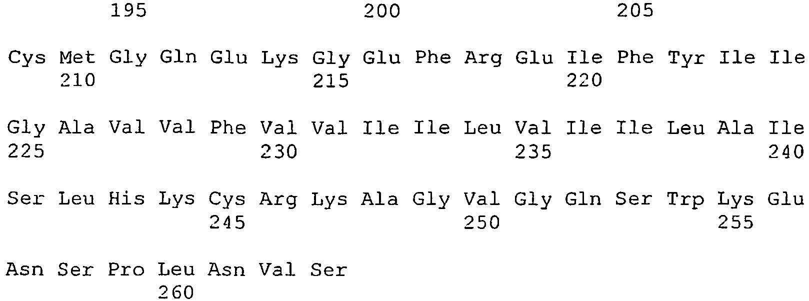
- <210> 2
<211> 2
<212> PRT
<213> Homo sapiens - <220>
<221> Aminosäuresequenz von tTF1-218 - <400> 2
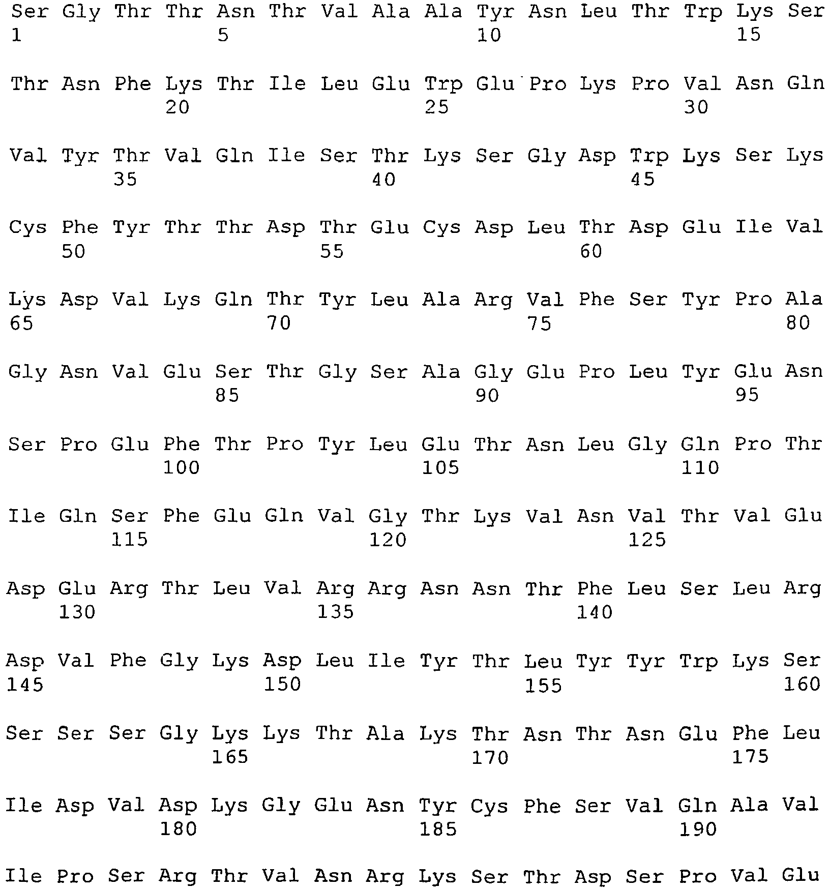

- <210> 3
<211> 224
<212> PRT
<213> Artificial - <220>
<221> Aminosäuresequenz von tTF-GRGDSP - <400> 3

- <210> 4
<211> 225
<212> PRT
<213> Artificial - <220>
<221> Aminosäuresequenz von tTF-GNGRAHA - <400> 4

- <210> 5
<211> 228
<212> PRT
<213> Artificial - <220>
<221> Aminosäuresequenz von tTF-GALNGRSHAG - <400> 5

- <210> 6
<211> 225
<212> PRT
<213> Artificial - <220>
<221> Aminosäuresequenz von tTF-GCNGRCG - <400> 6

- <210> 7
<211> 232
<212> PRT
<213> Artificial - <220>
<221> Aminosäuresequenz von tTF-GCNGRCVSGCAGRC - <400> 7

- <210> 8
<211> 228
<212> PRT
<213> Artificial - <220>
<221> Aminosäuresequenz von tTF-GCVLNGRMEC - <400> 8

- <210> 9
<211> 654
<212> DNA
<213> Artifical - <220>
<221> Nukleotidsequenz von tTF1-218 - <400> 9

- <210> 10
<211> 672
<212> DNA
<213> Artificial - <220>
<221> Nukleotidsequenz von tTF-GRGDSP - <400> 10


- <210> 11
<211> 675
<212> DNA
<213> Artificial - <220>
<221> Nukleotidsequenz von tTF-GNGRAHA - <400> 11

- <210> 12
<211> 684
<212> DNA
<213> Artificial - <220>
<221> Nukleotidsequenz von tTF-GALNGRSHAG - <400> 12


- <210> 13
<211> 675
<212> DNA
<213> Artificial - <220>
<221> Nukleotidsequenz von tTF-GCNGRCG - <400> 13

- <210> 14
<211> 696
<212> DNA
<213> Artificial - <220>
<221> Nukleotidsequenz von tTF-GCNGRCVSGCAGRC - <400> 14


- <210> 15
<211> 684
<212> DNA
<213> Artificial - <220>
<221> Nukleotidsequenz von tTF-GCVLNGRMEC - <400> 15

- <210> 16
<211> 45
<212> DNA
<213> Artifical - <220>
<221> 5' Oligonukleotidprimer für die Herstellung von tTF1-218 - <400> 16
catgccatgg gatcaggcac tacaaatact gtggcagcat ataat 45 - <210> 17
<211> 40
<212> DNA
<213> Artifical - <220>
<221> 3' Oligonukleotidprimer für die Herstellung von tTF1-218 - <400> 17
cgggatccta ttatctgaat tcccctttct cctggcccat 40 - <210> 18
<211> 45
<212> DNA
<213> Artificial - <220>
<221> 5' Oligonukleotidprimer für die Herstellung von tTF-GRGDSP - <400> 18
catgccatgg gatcaggcac tacaaatact gtggcagcat ataat 45 - <210> 19
<211> 43
<212> DNA
<213> Artificial - <220>
<221> 3' Oligonukleotidprimer für die Herstellung von tTF-GRGDSP - <400> 19
cgggatccta ttatggagaa tcacctcttc ctctgaattc ccc 43 - <210> 20
<211> 45
<212> DNA
<213> Artificial - <220>
<221> 5' Oligonukleotidprimer für die Herstellung von tTF-GNGRAHA - <400> 20
catgccatgg gatcaggcac tacaaatact gtggcagcat ataat 45 - <210> 21
<211> 46
<212> DNA
<213> Artificial - <220>
<221> 3' Oligonukleotidprimer für die Herstellung von tTF-GNGRAHA - <400> 21
cgggatccta ttatgcatgt gctcttccgt tacctctgaa ttcccc 46 - <210> 22
<211> 45
<212> DNA
<213> Artificial - <220>
<221> 5' Oligonukleotidprimer für die Herstellung von tTF-GCNGRCG - <400> 22
catgccatgg gatcaggcac tacaaatact gtggcagcat ataat 45 - <210> 23
<211> 46
<212> DNA
<213> Artificial - <220>
<221> 3' Oligonukleotidprimer für die Herstellung von tTF-GCNGRCG - <400> 23
cgggatccta ttaaccacat ctaccgttgc agcctctgaa ttcccc 46 - <210> 24
<211> 45
<212> DNA
<213> Artificial - <220>
<221> 5' Oligonukleotidprimer für die Herstellung von tTF-GCNGRCVSGCAGRC - <400> 24
catgccatgg gatcaggcac tacaaatact gtggcagcat ataat 45 - <210> 25
<211> 67
<212> DNA
<213> Artificial - <220>
<221> 3' Oligonukleotidprimer für die Herstellung von tTF-GCNGRCVSGCAGRC - <400> 25


- <210> 26
<211> 45
<212> DNA
<213> Artificial - <220>
<221> 5' Oligonukleotidprimer für die Herstellung von tTF-GCVLNGRMEC - <400> 26
catgccatgg gatcaggcac tacaaatact gtggcagcat ataat 45 - <210> 27
<211> 55
<212> DNA
<213> Artificial - <220>
<221> 3' Oligonukleotidprimer für die Herstellung von tTF-GCVLNGRMEC - <400> 27
cgggatccta ttagcattcc atcctaccat ttaagacgca tcctctgaat tcccc 55 - <210> 28
<211> 45
<212> DNA
<213> Artificial - <220>
<221> 5' Oligonukleotidprimer für die Herstellung von tTF-GALNGRSHAG - <400> 28
catgccatgg gatcaggcac tacaaatact gtggcagcat ataat 45 - <210> 29
<211> 55
<212> DNA
<213> Artificial - <220>
<221> 3' Oligonukleotidprimer für die Herstellung von tTF-GALNGRSHAG - <400> 29
cgggatccta ttaaccagcg tgagatcttc catttaaagc acctctgaat tcccc 55 - <210> 30
<211> 45
<212> PRT
<213> Artificial - <220>
<221> Aminosäuresequenz des Affinitäts-tags - <400> 30

- <210> 31
<211> 269
<212> PRT
<213> Artificial - <220>
<221> Aminosäuresequenz von tTF-GRGDSP mit N-terminalem Affinitäts-tag - <400> 31
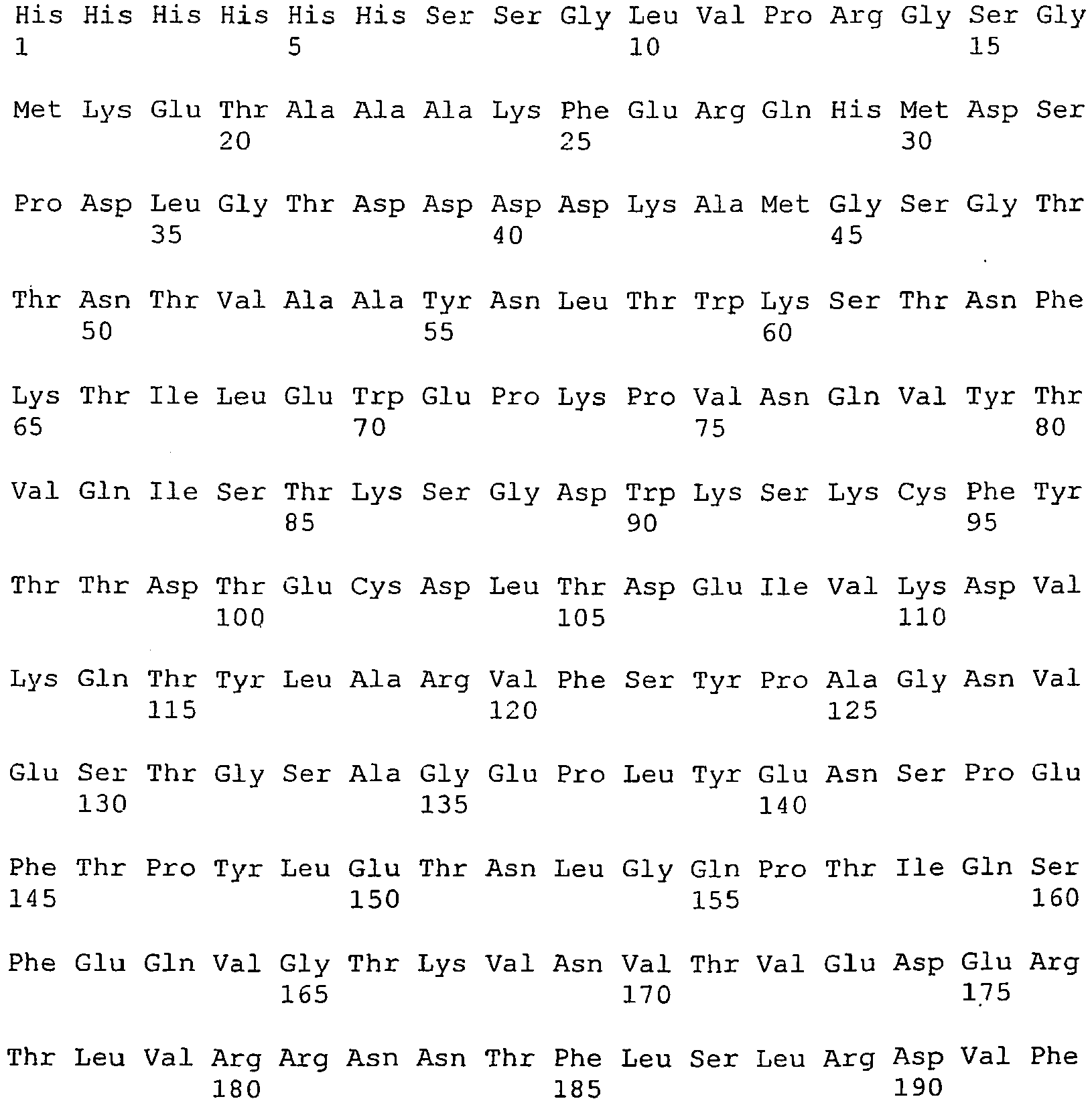

- <210> 32
<211> 270
<212> PRT
<213> Artificial - <220>
<221> Aminosäuresequenz von tTF-GNGRAHA mit N-terminalem Affinitäts-tag - <400> 32


Claims (16)
- Fusionspolypeptid, umfassenda) ein Peptid von 3 bis 30 Aminosäuren, das eine selektive Bindung des Fusionspolypeptides an Tumorgefäßendothelzellen ermöglicht; undb) den Gewebefaktor TF (Tissue Factor) oder ein Fragment davon, wobei der Gewebefaktor und das Fragment dadurch gekennzeichnet sind, daß sie die Blutgerinnung bei Bindung des Fusionspolypeptides an Tumorgefäßendothelzellen aktivieren können,wobei die Peptide a) und b) entweder unmittelbar oder über einen Linker mit bis zu 15 Aminosäuren aneinander gekoppelt sind, dadurch gekennzeichnet, daß das Peptid, das eine selektive Bindung des Fusionspolypeptides an Tumorgefäßendothelzellen ermöglicht, an den C-Terminus des Peptids, das die Blutgerinnung bei Bindung des Fusionspolypeptides an Tumorgefäßendothelzellen aktivieren kann, gekoppelt ist.
- Fusionspolypeptid nach Anspruch 1, bestehend aus den Peptiden a) und b) und einem Linker mit bis zu 15 Aminosäuren.
- Fusionspolypeptid nach Anspruch 1, wobei die Peptide a) und b) unmittelbar aneinander gekoppelt sind.
- Fusionspolypeptid nach einem der Ansprüche 1 bis 3, dadurch gekennzeichnet, daß das Peptid, das die Blutgerinnung bei Bindung des Fusionspolypeptides an Tumorgefäßendothelzellen aktivieren kann, der Gewebefaktor TF ist, der die in SEQ ID NO:1 dargestellte Sequenz aufweist.
- Fusionspolypeptid nach einem der Ansprüche 1 bis 3, dadurch gekennzeichnet, daß das Peptid, das die Blutgerinnung bei Bindung des Fusionspolypeptides an Tumorgefäßendothelzellen aktivieren kann, ein Fragment des Gewebefaktors TF ist, das vorzugsweise die in SEQ ID NO:2 dargestellte Sequenz aufweist.
- Fusionspolypeptid nach einem der Ansprüche 1 bis 5, dadurch gekennzeichnet, daß das Peptid von 3 bis 30 Aminosäuren, das eine selektive Bindung des Fusionspolypeptides an Tumorgefäßendothelzellen ermöglicht, eine lineare oder cyclische Struktur aufweist.
- Fusionspolypeptid nach einem der Ansprüche 1 bis 6, dadurch gekennzeichnet, daß das Peptid von 3 bis 30 Aminosäuren, das eine selektive Bindung des Fusionspolypeptides an Tumorgefäßendothelzellen ermöglicht, die Aminosäuresequenz RGD oder NGR umfaßt
- Fusionspolypeptid nach Anspruch 7, dadurch gekennzeichnet, daß das Peptid, das eine selektive Bindung des Fusionspolypeptides an Tumorgefäßendothelzellen ermöglicht, aus der Gruppe bestehend aus GRGDSP und GNGRAHA ausgewählt wird.
- Fusionspolypeptid nach Anspruch 7, dadurch gekennzeichnet, daß das Peptid, das eine selektive Bindung des Fusionspolypeptides an Tumorgefäßendothelzellen ermöglicht, aus der Gruppe bestehend aus GCNGRCG, GCNGRCVSGCAGRC, GCVLNGRMEC und GALNGRSHAG ausgewählt wird.
- Fusionspolypeptid nach den Ansprüchen 1 bis 9, dadurch gekennzeichnet, daß es eine der in SEQ ID NO:3-8 dargestellten Sequenzen aufweist.
- Nukleinsäure, die für ein Fusionspolypeptid nach einem der Ansprüche 1 bis 10 kodiert.
- Nukleinsäure nach Anspruch 11, dadurch gekennzeichnet, daß sie eine der in SEQ ID NO:10-15 dargestellten Sequenzen aufweist.
- Vektor, der eine Nukleinsäure nach Anspruch 11 oder 12 umfaßt.
- Zelle, die eine Nukleinsäure nach Anspruch 11 oder 12 oder einen Vektor nach Anspruch 14 umfaßt.
- Arzneimittel, das ein Fusionspolypeptid nach einem der Ansprüche 1 bis 10, eine Nukleinsäure nach Anspruch 11 oder 12, einen Vektor nach Anspruch 13 oder eine Zelle nach Anspruch 14 umfaßt.
- Arzneimittel nach Anspruch 15, das ferner pharmazeutisch verträgliche Träger, Hilfsmittel oder Adjuvantien umfaßt.
Priority Applications (2)
| Application Number | Priority Date | Filing Date | Title |
|---|---|---|---|
| SI200431981T SI1660532T1 (sl) | 2003-08-22 | 2004-08-20 | Fuzijski polipeptidi in njihova uporaba pri antivaskularnem zdravljenju tumorjev |
| PL04764346T PL1660532T3 (pl) | 2003-08-22 | 2004-08-20 | Polipeptydy fuzyjne i ich zastosowanie w przeciwnaczyniowej terapii guzów nowotworowych |
Applications Claiming Priority (2)
| Application Number | Priority Date | Filing Date | Title |
|---|---|---|---|
| DE10338733A DE10338733A1 (de) | 2003-08-22 | 2003-08-22 | Fusionspolypeptide und deren Verwendung für die antivaskuläre Tumortherapie |
| PCT/EP2004/009364 WO2005021593A1 (de) | 2003-08-22 | 2004-08-20 | Fusionspolypeptide und deren verwendung für die antivaskuläre tumortherapie |
Publications (2)
| Publication Number | Publication Date |
|---|---|
| EP1660532A1 EP1660532A1 (de) | 2006-05-31 |
| EP1660532B1 true EP1660532B1 (de) | 2012-10-24 |
Family
ID=34201891
Family Applications (1)
| Application Number | Title | Priority Date | Filing Date |
|---|---|---|---|
| EP04764346A Expired - Lifetime EP1660532B1 (de) | 2003-08-22 | 2004-08-20 | Fusionspolypeptide und deren verwendung für die antivaskuläre tumortherapie |
Country Status (13)
| Country | Link |
|---|---|
| US (1) | US7618943B2 (de) |
| EP (1) | EP1660532B1 (de) |
| JP (1) | JP5149506B2 (de) |
| CN (1) | CN1871258B (de) |
| AU (1) | AU2004268031B2 (de) |
| CA (1) | CA2535856C (de) |
| DE (1) | DE10338733A1 (de) |
| DK (1) | DK1660532T3 (de) |
| ES (1) | ES2397922T3 (de) |
| PL (1) | PL1660532T3 (de) |
| PT (1) | PT1660532E (de) |
| SI (1) | SI1660532T1 (de) |
| WO (1) | WO2005021593A1 (de) |
Families Citing this family (8)
| Publication number | Priority date | Publication date | Assignee | Title |
|---|---|---|---|---|
| WO2008100292A2 (en) | 2006-10-16 | 2008-08-21 | Genelux Corporation | Modified vaccinia virus strains for use in diagnostic and therapeutic methods |
| KR100879239B1 (ko) * | 2007-04-19 | 2009-01-16 | (주)케어젠 | Tgfp-cap 펩타이드 및 그의 용도 |
| CN102153653B (zh) * | 2010-12-30 | 2012-08-15 | 厦门大学 | 肿瘤血管靶向多肽与组织因子的融合蛋白及其制备方法 |
| IN2015KN00571A (de) | 2012-08-13 | 2015-07-17 | Otago Innovation Ltd | |
| CN104387474B (zh) * | 2014-11-17 | 2018-01-23 | 北京华安科创生物技术有限公司 | 一种肿瘤血管梗塞剂多肽、基因、表达载体及其应用 |
| EP3960177A1 (de) * | 2020-08-26 | 2022-03-02 | Anturec Pharmaceuticals GmbH | Zusammensetzung mit ttf-ngr zur verwendung in der behandlung von weichgewebesarkom |
| WO2022236063A1 (en) * | 2021-05-06 | 2022-11-10 | FibroBiologics | Induction of tumor vascular necrosis utilizing fibroblasts |
| WO2024102818A1 (en) * | 2022-11-09 | 2024-05-16 | Wisconsin Alumni Research Foundation | Combination tumor therapy with thrombosis initiation and platelet recruitment |
Family Cites Families (8)
| Publication number | Priority date | Publication date | Assignee | Title |
|---|---|---|---|---|
| US6004555A (en) * | 1992-03-05 | 1999-12-21 | Board Of Regents, The University Of Texas System | Methods for the specific coagulation of vasculature |
| US6093399A (en) * | 1992-03-05 | 2000-07-25 | Board Of Regents, The University Of Texas System | Methods and compositions for the specific coagulation of vasculature |
| US5858724A (en) * | 1996-07-16 | 1999-01-12 | Pel-Freez | Recombinant rabbit tissue factor |
| ATE244579T1 (de) * | 1997-01-22 | 2003-07-15 | Univ Texas | Verfahren und zusammensetzungen des gewebefaktors zur koagulation und behandlung von tumoren |
| AU750414B2 (en) * | 1998-07-13 | 2002-07-18 | Board Of Regents, The University Of Texas System | Cancer treatment methods using therapeutic conjugates that bind to aminophospholipids |
| US7622437B2 (en) * | 2000-11-20 | 2009-11-24 | The Board Of Trustees Of The University Of Illinois | Tissue factor compositions and methods |
| WO2003035688A2 (en) * | 2001-10-26 | 2003-05-01 | Novartis Pharma Gmbh | Targeted thrombosis by tissue factor polypeptides |
| US7425328B2 (en) * | 2003-04-22 | 2008-09-16 | Purdue Pharma L.P. | Tissue factor antibodies and uses thereof |
-
2003
- 2003-08-22 DE DE10338733A patent/DE10338733A1/de not_active Ceased
-
2004
- 2004-08-20 CN CN2004800313848A patent/CN1871258B/zh not_active Expired - Lifetime
- 2004-08-20 DK DK04764346.5T patent/DK1660532T3/da active
- 2004-08-20 EP EP04764346A patent/EP1660532B1/de not_active Expired - Lifetime
- 2004-08-20 PL PL04764346T patent/PL1660532T3/pl unknown
- 2004-08-20 JP JP2006523616A patent/JP5149506B2/ja not_active Expired - Fee Related
- 2004-08-20 CA CA2535856A patent/CA2535856C/en not_active Expired - Lifetime
- 2004-08-20 US US10/569,076 patent/US7618943B2/en not_active Expired - Lifetime
- 2004-08-20 AU AU2004268031A patent/AU2004268031B2/en not_active Expired
- 2004-08-20 PT PT47643465T patent/PT1660532E/pt unknown
- 2004-08-20 WO PCT/EP2004/009364 patent/WO2005021593A1/de not_active Ceased
- 2004-08-20 SI SI200431981T patent/SI1660532T1/sl unknown
- 2004-08-20 ES ES04764346T patent/ES2397922T3/es not_active Expired - Lifetime
Also Published As
| Publication number | Publication date |
|---|---|
| CA2535856C (en) | 2016-06-07 |
| DE10338733A1 (de) | 2005-03-24 |
| CN1871258B (zh) | 2012-05-30 |
| CN1871258A (zh) | 2006-11-29 |
| JP5149506B2 (ja) | 2013-02-20 |
| ES2397922T3 (es) | 2013-03-12 |
| HK1087418A1 (en) | 2006-10-13 |
| US20070032419A1 (en) | 2007-02-08 |
| AU2004268031B2 (en) | 2009-09-03 |
| SI1660532T1 (sl) | 2013-02-28 |
| WO2005021593A1 (de) | 2005-03-10 |
| DK1660532T3 (da) | 2013-01-21 |
| CA2535856A1 (en) | 2005-03-10 |
| AU2004268031A1 (en) | 2005-03-10 |
| PL1660532T3 (pl) | 2013-03-29 |
| US7618943B2 (en) | 2009-11-17 |
| EP1660532A1 (de) | 2006-05-31 |
| PT1660532E (pt) | 2013-01-29 |
| JP2007528722A (ja) | 2007-10-18 |
Similar Documents
| Publication | Publication Date | Title |
|---|---|---|
| DK2739649T3 (en) | P97 FRAGMENTS WITH TRANSFER ACTIVITY | |
| JP6586412B2 (ja) | p97のフラグメントおよびその使用 | |
| DE69609585T2 (de) | Moleküle, die sich in ausgewählten Organen oder Geweben invivo Einfinden und Verfahren zu ihrer Identifizierung | |
| JP4531267B2 (ja) | ホーミングプロアポトーシス結合体およびホーミングプロアポトーシス結合体を使用する方法 | |
| DE69838544T2 (de) | Isolierung von gewebespezifischen peptidliganden und ihre verwendung zur ausrichtung von pharmazeutika auf zielorgane | |
| KR20080082608A (ko) | Vegf 유사체 및 사용 방법 | |
| US20130280162A1 (en) | uPAR-ANTAGONISTS AND USES THEREOF | |
| EP1660532A1 (de) | FUSIONSPOLYPEPTIDE UND DEREN VERWENDUNG F R DIE ANTIVASKUL&A uml;RE TUMORTHERAPIE | |
| JP2006524241A5 (de) | ||
| JP2006524241A (ja) | Stq−ペプチド | |
| US20210046149A1 (en) | Bifunctional blood brain therapies | |
| US20220144906A1 (en) | Bifunctional blood brain therapies for interleukin-1 related diseases | |
| US8329653B2 (en) | Compositions and methods for suppression of amyloid plaque formation associated with neurodegenerative disorders | |
| HK1087418B (en) | Fusion polypeptides, and use thereof in antivascular tumor therapy | |
| HK40016976A (en) | Fragments of p97 and uses thereof | |
| HK1198332B (en) | P97 fragments with transfer activity |
Legal Events
| Date | Code | Title | Description |
|---|---|---|---|
| PUAI | Public reference made under article 153(3) epc to a published international application that has entered the european phase |
Free format text: ORIGINAL CODE: 0009012 |
|
| 17P | Request for examination filed |
Effective date: 20060321 |
|
| AK | Designated contracting states |
Kind code of ref document: A1 Designated state(s): AT BE BG CH CY CZ DE DK EE ES FI FR GB GR HU IE IT LI LU MC NL PL PT RO SE SI SK TR |
|
| AX | Request for extension of the european patent |
Extension state: LV |
|
| RIN1 | Information on inventor provided before grant (corrected) |
Inventor name: MESTERS, ROLF, M. Inventor name: BERDEL, WOLFGANG, E. |
|
| RAP1 | Party data changed (applicant data changed or rights of an application transferred) |
Owner name: BERDEL, WOLFGANG Owner name: BIZIMIS, ARI, E. |
|
| 17Q | First examination report despatched |
Effective date: 20060821 |
|
| RAP1 | Party data changed (applicant data changed or rights of an application transferred) |
Owner name: BERDEL, WOLFGANG Owner name: ONCOSCIENCE AG Owner name: BIZIMIS, ARI, E. |
|
| REG | Reference to a national code |
Ref country code: HK Ref legal event code: DE Ref document number: 1087418 Country of ref document: HK |
|
| RAX | Requested extension states of the european patent have changed |
Extension state: LV Payment date: 20060405 |
|
| GRAP | Despatch of communication of intention to grant a patent |
Free format text: ORIGINAL CODE: EPIDOSNIGR1 |
|
| GRAS | Grant fee paid |
Free format text: ORIGINAL CODE: EPIDOSNIGR3 |
|
| GRAA | (expected) grant |
Free format text: ORIGINAL CODE: 0009210 |
|
| RIN1 | Information on inventor provided before grant (corrected) |
Inventor name: BERDEL, WOLFGANG, E. Inventor name: MESTERS, ROLF, M. |
|
| AK | Designated contracting states |
Kind code of ref document: B1 Designated state(s): AT BE BG CH CY CZ DE DK EE ES FI FR GB GR HU IE IT LI LU MC NL PL PT RO SE SI SK TR |
|
| AX | Request for extension of the european patent |
Extension state: LV |
|
| RAP1 | Party data changed (applicant data changed or rights of an application transferred) |
Owner name: BIZIMIS, ARI, E. Owner name: BERDEL, WOLFGANG Owner name: ONCOSCIENCE AG |
|
| REG | Reference to a national code |
Ref country code: GB Ref legal event code: FG4D Free format text: NOT ENGLISH |
|
| REG | Reference to a national code |
Ref country code: CH Ref legal event code: EP |
|
| REG | Reference to a national code |
Ref country code: AT Ref legal event code: REF Ref document number: 580908 Country of ref document: AT Kind code of ref document: T Effective date: 20121115 |
|
| REG | Reference to a national code |
Ref country code: IE Ref legal event code: FG4D Free format text: LANGUAGE OF EP DOCUMENT: GERMAN |
|
| REG | Reference to a national code |
Ref country code: DE Ref legal event code: R096 Ref document number: 502004013841 Country of ref document: DE Effective date: 20121220 |
|
| REG | Reference to a national code |
Ref country code: SE Ref legal event code: TRGR Ref country code: CH Ref legal event code: NV Representative=s name: E. BLUM AND CO. AG PATENT- UND MARKENANWAELTE , CH |
|
| REG | Reference to a national code |
Ref country code: DK Ref legal event code: T3 |
|
| REG | Reference to a national code |
Ref country code: PT Ref legal event code: SC4A Free format text: AVAILABILITY OF NATIONAL TRANSLATION Effective date: 20130123 |
|
| REG | Reference to a national code |
Ref country code: NL Ref legal event code: T3 |
|
| REG | Reference to a national code |
Ref country code: ES Ref legal event code: FG2A Ref document number: 2397922 Country of ref document: ES Kind code of ref document: T3 Effective date: 20130312 |
|
| REG | Reference to a national code |
Ref country code: PL Ref legal event code: T3 |
|
| REG | Reference to a national code |
Ref country code: SK Ref legal event code: T3 Ref document number: E 13285 Country of ref document: SK |
|
| REG | Reference to a national code |
Ref country code: HK Ref legal event code: GR Ref document number: 1087418 Country of ref document: HK |
|
| PG25 | Lapsed in a contracting state [announced via postgrant information from national office to epo] |
Ref country code: GR Free format text: LAPSE BECAUSE OF FAILURE TO SUBMIT A TRANSLATION OF THE DESCRIPTION OR TO PAY THE FEE WITHIN THE PRESCRIBED TIME-LIMIT Effective date: 20130125 Ref country code: CY Free format text: LAPSE BECAUSE OF FAILURE TO SUBMIT A TRANSLATION OF THE DESCRIPTION OR TO PAY THE FEE WITHIN THE PRESCRIBED TIME-LIMIT Effective date: 20121024 |
|
| REG | Reference to a national code |
Ref country code: HU Ref legal event code: AG4A Ref document number: E015676 Country of ref document: HU |
|
| PG25 | Lapsed in a contracting state [announced via postgrant information from national office to epo] |
Ref country code: EE Free format text: LAPSE BECAUSE OF FAILURE TO SUBMIT A TRANSLATION OF THE DESCRIPTION OR TO PAY THE FEE WITHIN THE PRESCRIBED TIME-LIMIT Effective date: 20121024 Ref country code: BG Free format text: LAPSE BECAUSE OF FAILURE TO SUBMIT A TRANSLATION OF THE DESCRIPTION OR TO PAY THE FEE WITHIN THE PRESCRIBED TIME-LIMIT Effective date: 20130124 |
|
| PG25 | Lapsed in a contracting state [announced via postgrant information from national office to epo] |
Ref country code: RO Free format text: LAPSE BECAUSE OF FAILURE TO SUBMIT A TRANSLATION OF THE DESCRIPTION OR TO PAY THE FEE WITHIN THE PRESCRIBED TIME-LIMIT Effective date: 20121024 |
|
| PLBE | No opposition filed within time limit |
Free format text: ORIGINAL CODE: 0009261 |
|
| STAA | Information on the status of an ep patent application or granted ep patent |
Free format text: STATUS: NO OPPOSITION FILED WITHIN TIME LIMIT |
|
| 26N | No opposition filed |
Effective date: 20130725 |
|
| REG | Reference to a national code |
Ref country code: DE Ref legal event code: R097 Ref document number: 502004013841 Country of ref document: DE Effective date: 20130725 |
|
| REG | Reference to a national code |
Ref country code: FR Ref legal event code: PLFP Year of fee payment: 13 |
|
| REG | Reference to a national code |
Ref country code: FR Ref legal event code: PLFP Year of fee payment: 14 |
|
| REG | Reference to a national code |
Ref country code: FR Ref legal event code: PLFP Year of fee payment: 15 |
|
| PGFP | Annual fee paid to national office [announced via postgrant information from national office to epo] |
Ref country code: NL Payment date: 20200825 Year of fee payment: 17 |
|
| PGFP | Annual fee paid to national office [announced via postgrant information from national office to epo] |
Ref country code: SK Payment date: 20200817 Year of fee payment: 17 Ref country code: PT Payment date: 20200817 Year of fee payment: 17 Ref country code: LU Payment date: 20200824 Year of fee payment: 17 Ref country code: CZ Payment date: 20200817 Year of fee payment: 17 Ref country code: DK Payment date: 20200826 Year of fee payment: 17 Ref country code: MC Payment date: 20200826 Year of fee payment: 17 |
|
| PGFP | Annual fee paid to national office [announced via postgrant information from national office to epo] |
Ref country code: PL Payment date: 20200818 Year of fee payment: 17 Ref country code: SI Payment date: 20200817 Year of fee payment: 17 |
|
| REG | Reference to a national code |
Ref country code: DK Ref legal event code: EBP Effective date: 20210831 |
|
| PG25 | Lapsed in a contracting state [announced via postgrant information from national office to epo] |
Ref country code: MC Free format text: LAPSE BECAUSE OF NON-PAYMENT OF DUE FEES Effective date: 20210831 |
|
| REG | Reference to a national code |
Ref country code: NL Ref legal event code: MM Effective date: 20210901 |
|
| REG | Reference to a national code |
Ref country code: SK Ref legal event code: MM4A Ref document number: E 13285 Country of ref document: SK Effective date: 20210820 |
|
| REG | Reference to a national code |
Ref country code: SI Ref legal event code: KO00 Effective date: 20220407 |
|
| PG25 | Lapsed in a contracting state [announced via postgrant information from national office to epo] |
Ref country code: PT Free format text: LAPSE BECAUSE OF NON-PAYMENT OF DUE FEES Effective date: 20220221 Ref country code: LU Free format text: LAPSE BECAUSE OF NON-PAYMENT OF DUE FEES Effective date: 20210820 Ref country code: CZ Free format text: LAPSE BECAUSE OF NON-PAYMENT OF DUE FEES Effective date: 20210820 |
|
| PG25 | Lapsed in a contracting state [announced via postgrant information from national office to epo] |
Ref country code: NL Free format text: LAPSE BECAUSE OF NON-PAYMENT OF DUE FEES Effective date: 20210901 |
|
| PG25 | Lapsed in a contracting state [announced via postgrant information from national office to epo] |
Ref country code: SK Free format text: LAPSE BECAUSE OF NON-PAYMENT OF DUE FEES Effective date: 20210820 Ref country code: DK Free format text: LAPSE BECAUSE OF NON-PAYMENT OF DUE FEES Effective date: 20210831 |
|
| PGFP | Annual fee paid to national office [announced via postgrant information from national office to epo] |
Ref country code: SE Payment date: 20220627 Year of fee payment: 19 Ref country code: GB Payment date: 20220630 Year of fee payment: 19 |
|
| PGFP | Annual fee paid to national office [announced via postgrant information from national office to epo] |
Ref country code: IT Payment date: 20220712 Year of fee payment: 19 Ref country code: IE Payment date: 20220712 Year of fee payment: 19 Ref country code: FI Payment date: 20220809 Year of fee payment: 19 Ref country code: ES Payment date: 20220902 Year of fee payment: 19 Ref country code: DE Payment date: 20220629 Year of fee payment: 19 Ref country code: AT Payment date: 20220725 Year of fee payment: 19 |
|
| PGFP | Annual fee paid to national office [announced via postgrant information from national office to epo] |
Ref country code: FR Payment date: 20220709 Year of fee payment: 19 Ref country code: BE Payment date: 20220718 Year of fee payment: 19 |
|
| PGFP | Annual fee paid to national office [announced via postgrant information from national office to epo] |
Ref country code: CH Payment date: 20220802 Year of fee payment: 19 |
|
| PGFP | Annual fee paid to national office [announced via postgrant information from national office to epo] |
Ref country code: TR Payment date: 20230817 Year of fee payment: 20 |
|
| PGFP | Annual fee paid to national office [announced via postgrant information from national office to epo] |
Ref country code: HU Payment date: 20230924 Year of fee payment: 20 |
|
| REG | Reference to a national code |
Ref country code: DE Ref legal event code: R119 Ref document number: 502004013841 Country of ref document: DE |
|
| REG | Reference to a national code |
Ref country code: CH Ref legal event code: PL |
|
| REG | Reference to a national code |
Ref country code: AT Ref legal event code: MM01 Ref document number: 580908 Country of ref document: AT Kind code of ref document: T Effective date: 20230820 |
|
| PG25 | Lapsed in a contracting state [announced via postgrant information from national office to epo] |
Ref country code: AT Free format text: LAPSE BECAUSE OF NON-PAYMENT OF DUE FEES Effective date: 20230820 |
|
| GBPC | Gb: european patent ceased through non-payment of renewal fee |
Effective date: 20230820 |
|
| PG25 | Lapsed in a contracting state [announced via postgrant information from national office to epo] |
Ref country code: FI Free format text: LAPSE BECAUSE OF NON-PAYMENT OF DUE FEES Effective date: 20230820 Ref country code: AT Free format text: LAPSE BECAUSE OF NON-PAYMENT OF DUE FEES Effective date: 20230820 Ref country code: CH Free format text: LAPSE BECAUSE OF NON-PAYMENT OF DUE FEES Effective date: 20230831 |
|
| REG | Reference to a national code |
Ref country code: BE Ref legal event code: MM Effective date: 20230831 |
|
| REG | Reference to a national code |
Ref country code: IE Ref legal event code: MM4A |
|
| PG25 | Lapsed in a contracting state [announced via postgrant information from national office to epo] |
Ref country code: SE Free format text: LAPSE BECAUSE OF NON-PAYMENT OF DUE FEES Effective date: 20230821 Ref country code: PL Free format text: LAPSE BECAUSE OF NON-PAYMENT OF DUE FEES Effective date: 20210820 |
|
| PG25 | Lapsed in a contracting state [announced via postgrant information from national office to epo] |
Ref country code: IE Free format text: LAPSE BECAUSE OF NON-PAYMENT OF DUE FEES Effective date: 20230820 |
|
| PG25 | Lapsed in a contracting state [announced via postgrant information from national office to epo] |
Ref country code: GB Free format text: LAPSE BECAUSE OF NON-PAYMENT OF DUE FEES Effective date: 20230820 |
|
| PG25 | Lapsed in a contracting state [announced via postgrant information from national office to epo] |
Ref country code: IT Free format text: LAPSE BECAUSE OF NON-PAYMENT OF DUE FEES Effective date: 20230820 Ref country code: IE Free format text: LAPSE BECAUSE OF NON-PAYMENT OF DUE FEES Effective date: 20230820 Ref country code: GB Free format text: LAPSE BECAUSE OF NON-PAYMENT OF DUE FEES Effective date: 20230820 Ref country code: FR Free format text: LAPSE BECAUSE OF NON-PAYMENT OF DUE FEES Effective date: 20230831 Ref country code: DE Free format text: LAPSE BECAUSE OF NON-PAYMENT OF DUE FEES Effective date: 20240301 |
|
| PG25 | Lapsed in a contracting state [announced via postgrant information from national office to epo] |
Ref country code: BE Free format text: LAPSE BECAUSE OF NON-PAYMENT OF DUE FEES Effective date: 20230831 |
|
| REG | Reference to a national code |
Ref country code: ES Ref legal event code: FD2A Effective date: 20240927 |
|
| PG25 | Lapsed in a contracting state [announced via postgrant information from national office to epo] |
Ref country code: ES Free format text: LAPSE BECAUSE OF NON-PAYMENT OF DUE FEES Effective date: 20230821 |
|
| PG25 | Lapsed in a contracting state [announced via postgrant information from national office to epo] |
Ref country code: ES Free format text: LAPSE BECAUSE OF NON-PAYMENT OF DUE FEES Effective date: 20230821 |