EP1660206B1 - Verfahren und ausrüstungen zur reinigung von proteinen mit his-tag - Google Patents
Verfahren und ausrüstungen zur reinigung von proteinen mit his-tag Download PDFInfo
- Publication number
- EP1660206B1 EP1660206B1 EP04788679A EP04788679A EP1660206B1 EP 1660206 B1 EP1660206 B1 EP 1660206B1 EP 04788679 A EP04788679 A EP 04788679A EP 04788679 A EP04788679 A EP 04788679A EP 1660206 B1 EP1660206 B1 EP 1660206B1
- Authority
- EP
- European Patent Office
- Prior art keywords
- protein
- tagged
- solid support
- proteins
- zinc
- Prior art date
- Legal status (The legal status is an assumption and is not a legal conclusion. Google has not performed a legal analysis and makes no representation as to the accuracy of the status listed.)
- Not-in-force
Links
Images


Classifications
-
- B—PERFORMING OPERATIONS; TRANSPORTING
- B01—PHYSICAL OR CHEMICAL PROCESSES OR APPARATUS IN GENERAL
- B01J—CHEMICAL OR PHYSICAL PROCESSES, e.g. CATALYSIS OR COLLOID CHEMISTRY; THEIR RELEVANT APPARATUS
- B01J45/00—Ion-exchange in which a complex or a chelate is formed; Use of material as complex or chelate forming ion-exchangers; Treatment of material for improving the complex or chelate forming ion-exchange properties
-
- C—CHEMISTRY; METALLURGY
- C07—ORGANIC CHEMISTRY
- C07K—PEPTIDES
- C07K1/00—General methods for the preparation of peptides, i.e. processes for the organic chemical preparation of peptides or proteins of any length
- C07K1/14—Extraction; Separation; Purification
- C07K1/16—Extraction; Separation; Purification by chromatography
- C07K1/22—Affinity chromatography or related techniques based upon selective absorption processes
Definitions
- Histidine-tagged proteins are recombinant proteins designed to include a polyhistidine tail (his-tag) that facilitates purification of the proteins from in vitro expression systems.
- His-tag polyhistidine tail
- the preferential binding of the his-tag to metal chelating resins has been exploited in purifying his-tagged proteins from undesired contaminating proteins using immobilized metal affinity chromatography (IMAC).
- Metal chelating resins typically include a transition metal such as Ni or Co.
- heme proteins e.g., hemoglobin or myoglobin
- metal chelating resins When both his-tagged proteins and heme proteins are applied to a metal chelating resin, the heme proteins co-purify with his-tagged proteins It is therefore difficult to separate his-tagged proteins from material containing heme proteins to obtain a preparation of his-tagged proteins of acceptable purity without a significant amount of contaminating heme proteins.
- Rabbit reticulocyte lysate is a particularly useful expression system for obtaining expression of eukaryotic sequences. Rabbit reticulocyte lysate-based systems have been found to support co-translational and post-translational modifications of expressed proteins. However, because reticulocyte lysate includes large concentrations of hemoglobin, and because of the difficulties associated with separating hemoglobin from his-tagged proteins, reticulocyte lysate expression systems have not been fully exploited for expressing his-tagged proteins.
- Lytton et al. discloses that removal of hemoglobin from his-tagged proteins produced in a rabbit reticulocyte lysate may be effected by first binding the hemoglobin and his-tagged proteins to a nickel nitrilotriacetic acid (Ni-NTA) resin in the presence of imidazole, followed by step-wise elution of hemoglobin and his-tagged proteins using an imidazole gradient
- the present invention provides a method according to claim 1 for separating heme proteins from his-tagged protein in a starting material.
- the starting material is contacted with a zinc- or cobalt-charged solid support under conditions in which the support preferentially binds to his-tagged protein relative to its binding to hemoglobin.
- the conditions include no imidazole or imidazole in a concentration of from 0 mM to 60 mM.
- imidazole may be present in a concentration of 10 to 40 mM, or from 10 mM to 20 mM.
- the bound his- tagged proteins are removed from the solid support.
- Other aspects of the present invention provide a method of characterizing a his-tagged protein according to claim 17, and a method of separating a target polypeptide from a sample according to claim 20.
- Fig. 1 shows a fluoroimage of electrophoretically separated proteins from rabbit reticulocyte lysate.
- Fig. 2 shows a fluoroimage of electrophoretically separated proteins from rabbit reticulocyte lysate.
- Fig. 3 shows proteins from rabbit reticulocyte lysate separated by SDS-PAGE and stained with GelCode TM .
- Fig. 4 shows Western blot of electrophoretically separated proteins from rabbit reticulocyte lysate probed with anti-renilla luciferase antibody.
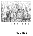
- Fig. 5 shows a Western blot of proteins from rabbit reticulocyte lysate electrophoretically separated by SDS-PAGE and probed with anti-hemoglobin antibody.
- Fig. 6 shows a fluorescent scan of electrophoretically separated proteins
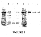
- Fig. 7 is an image of electrophoretically separated proteins.
- Fig. 8 is a Western blot of electrophoretically separated proteins.
- Fig. 9 is an image of electrophoretically separated proteins.
- Fig. 10 is an image of electrophoretically separated proteins.
- Fig. 11 is a graph showing percent inhibition of fluorescence of rhodamine R110 in the presence of hemoglobin.
- Fig. 12 is a graph showing activity of his-tagged caspase (as measured by fluorescence) recovered from rabbit reticulocyte using zinc or nickel charged particles.
- the present invention provides methods for separating a heme protein from a target polypeptide in a sample comprising heme protein and the target polypeptide material using zinc- or cobalt- charged solid support.
- the target polypeptide includes a polyhistidine tag of from five to six histidine residues.
- heme proteins from a target protein of interest is particularly important in applications in which there are contaminating heme proteins, for example, when isolating proteins from reticulocyte lysate, from whole blood, or other bodily fluids or samples containing hemoglobin.
- purification schemes can be developed to separate virtually any proteins, such methods frequently require many steps and numerous reagents, are time- and labor-intensive, and are, therefore, not amenable to use in high throughput screening or assays.
- NTA zinc- or cobalt-charged nitrilotriacetic acid
- modified siliceous-oxide coated magnetic particles contacted with a sample comprising hemoglobin and his-tagged proteins in the absence of imidazole or in the presence of low levels of imidazole (10 to 60 mM) were found to preferentially bind to the his-tagged proteins, relative to binding of heme protein to the particles, such that an increase in purity of his-tagged protein from a hemoglobin-containing starting material was achieved.
- magnetic refers to paramagnetic particles, magnetic particles as well as particles capable of being magnetized.
- hemoglobin binding to zinc- or cobalt-charged solid supports is minimal, whereas his-tagged proteins are bound with sufficiently high efficiency relative to similar supports charged with nickel.
- the methods of the invention afford an increase in purity of his-tagged proteins, relative to hemoglobin, of 1-fold or greater.
- the increase in purity is least 2-fold, 2.5-fold, 5-fold, 10-fold, 20-fold, 50-fold, or 100-fold.
- no purification scheme will result in 100% recovery of the desired protein or removal of 100% of undesired contaminants.
- Acceptable levels of hemoglobin contamination or recovery of target protein may vary, depending on the application.
- Conditions that allow preferential binding of his-tagged protein relative to hemoglobin suitably permit binding of less than 5% of hemoglobin present in the starting material, and at the same time permitting binding of at least some of the his-tagged protein.
- less than 1 % of the hemoglobin present in the starting material binds to the solid support, or even as little 0.5% or 0.1% or less of the hemoglobin present in the starting material binds to the solid support.
- a substantial increase in purification of his-tagged proteins can be achieved using the zinc- or cobalt-charged solid supports.
- NTA-modified magnetic silica particles 3-[[[bis(carboxymethyl)amino]acetyl]amino]-propyl magnetic silica particles (NTA-modified magnetic silica particles), as described in U.S. Application No. 10/689,176, filed October 20, 2003 , were used in the Examples below.
- the method is not intended to be limited to use with the particles exemplified below, but rather, is believed to have general applicability for use with any suitable zinc- or cobalt-charged solid support.
- silica magnetic particles used in the examples, other types of solid supports may be used in the methods of the invention, including, but not limited to, silica gel, siliceous oxide, solid silica such as glass or diatomaceous earth, agarose, polyacrylamide, cellulose, plastic, polysaccharide, nylon, polystyrene, or latex methacrylate.
- NTA may be linked to a solid support by other means and used in the method of the invention.
- other types of metal chelating ligands including, but not limited to, iminodiacetate (IDA) or Tris(carboxymethyl)ethylendiamin ligand (TED), for example, may be used in the practice of the invention.
- IDA iminodiacetate
- TED Tris(carboxymethyl)ethylendiamin ligand
- zinc or cobalt could be attached covalently or noncovalently to a solid support without using a chelating agent.
- a solid support could be coated with zinc or cobalt Any suitable method for coating the solid supports with a metal such as zinc or cobalt may be used (e.g., see Hyun et al. Bull. Korean Chem. Soc. 23:1724-1728, 2002 ). Gupta et al. describe attaching zinc directly to alginate beads without the use of a chelating agent ( Biotechnol. Prog. 18:78-81, 2002 ). Another approach for attaching zinc or cobalt to the solid support is to incorporate the metal into the matrix of the support ( Jana et al. Bull. Mater. Sci. 23:263-266, 2000 ).
- immobilized his-tagged proteins were eluted from the particles using imidazole (500 mM).
- immobilized his-tagged proteins may be eluted using other suitable buffers comprising imidazole in the range of from about 100 mM to about 3M imidazole.
- the conditions selected may vary according to the particular protein and the objective (e.g., enhancing yield or purity).
- Immobilized his-tagged proteins may be eluted using a suitable buffer having a pH of less than about 6.5 to enhance yield.
- an elution buffer of histidine e.g., 100 mM histidine
- 500 mM potassium acetate and 50 mM EDTA may be used.
- Buffers containing EDTA in a concentration of from about 10 mM to about 0.5 M are also suitable.
- His-tags may be at the carboxy or amino terminus and are typically five or six histidine residues in length, but may be longer.
- the methods of the invention may be used for high throughput purification of his-tagged proteins expressed in rabbit reticulocyte lysate, by expressing cDNA libraries containing his-tagged proteins followed by isolating the proteins according to the methods of the invention.
- Rabbit reticulocyte is widely used for expressing post-translationally modified proteins, such as glycosylated proteins. Using the methods disclosed herein, one may obtain preparations of modified protein from rabbit reticulocyte lysate with very little hemoglobin contamination.
- his-tagged proteins expressed and subsequently purified by the method of the invention are suitable for subsequent evaluation by mass spectrometry analysis.
- his-tagged proteins purified using zinc or cobalt charged solid supports are suitable for subsequently detecting interactions between the his-tagged proteins and other substances, including substrates (e.g., detecting the ability of the his-tagged protein to function as a kinase or protease) and other proteins (e.g., detecting the ability of his-tagged proteins to serve as a substrate for a kinase or protease).
- substrates e.g., detecting the ability of the his-tagged protein to function as a kinase or protease
- other proteins e.g., detecting the ability of his-tagged proteins to serve as a substrate for a kinase or protease
- simple protein-protein interactions and antigen-antibody binding involving the his-tagged proteins may be detected. Such activity or interactions may be evaluated by any suitable means.
- Contaminating hemoglobin ordinarily present in rabbit reticulocyte lysate typically interferes with fluorescent or luminescent based assays, in part because hemoglobin itself fluoresces and may mask the signal of a low expressing protein. Because most of the hemoglobin present in rabbit reticulocyte lysate or blood is removed during purification steps according to the present invention, interactions or activity may be conveniently detected using fluorescent or luminescent means without significant interference from hemoglobin.
- Example 1 Preparation of zinc- or cobalt-charged nitrilotriacetic acid-modified magnetic silica particles
- Siliceous oxide-coated magnetic particles were modified with nitrilotriacetic acid (NTA) to produce 3-[[[bis(carboxymethyl)amino]acetyl]amino]-propyl magnetic silica particles (NTA-modified magnetic silica particles), as described in U.S. Application No. 10/689,176, filed October 20, 2003 , which is incorporated by reference in its entirety.
- NTA nitrilotriacetic acid
- Zinc-charged particles were prepared as follows. Particles in a 4-ml aliquot of NTA-modified particles (10% w/v in water) were separated from the liquid using magnetization and the liquid discarded. The particles were contacted with 4 ml of ZnCl (100 mM) and rocked on a horizontal shaker (5-10 rpm) for 15 minutes. The particles were separated from the liquid using magnetization and the liquid discarded. The particles were contacted with a fresh 4-ml aliqout of ZnCl (100 mM), rocked on a horizontal shaker (5-10 rpm) for 15 minutes, and the particles were separated from the ZnCl solution using magnetization.
- the particles were washed with 4 ml of nanopure water and rocked on a horizontal shaker (5-10 rpm) for 5 minutes. The particles were separated from the water using magnetization and the liquid discarded. The wash step was repeated 10 times. After the final wash, 4 ml of nanopure water was added to the particles, and the particles were stored at 4°C until use.
- Cobalt-charged particles were prepared as follows. Particles in a 4-ml aliquot of NTA-modified particles (10% w/v in water) were separated from the liquid using magnetization and the liquid discarded. The particles were contacted with 4 ml of CoCl 2 (100 mM) and rocked on a horizontal shaker (5-10 rpm) for 15 minutes. The particles were separated from the liquid using magnetization and the liquid discarded. The particles were contacted with a fresh 4-ml aliquot of CoCl 2 (100 mM) and rocked on a horizontal shaker (5-10 rpm) for 15 minutes. The particles were separated from the CoCl 2 solution using magnetization.
- the particles were washed with 4 ml of nanopure water and rocked on a horizontal shaker (5-10 rpm) for 5 minutes. The particles were separated from the water using magnetization and the liquid discarded. The wash step was repeated 10 times. After the final wash, 4 ml of nanopure water was added to the particles, and the particles were stored at 4°C until use.
- An expression vector encoding his-tagged Renilla luciferase was expressed using TNT® T7 Quick Coupled Transcription/Translation System, Cat# L1170TNT (Promega Corp., Madison, WI) according to the manufacturer's instructions.
- His-tagged Renilla luciferase prepared as described in Example 2, was purified from the coupled transcription/translation reaction mixture as follows. A 50- ⁇ l aliquot of the reaction mixture was combined with 150- ⁇ l binding buffer A (sodium phosphate buffer 100 mM, imidazole 20 mM, and NaCl 400 mM) buffered at a pH in the range of 6.0 to 8.0, and combined with 30 ⁇ l (3 mg) of zinc charged particles, prepared as described in Example, for 15-15 minutes at room temperature. The particles were separated from the solution (“flowthrough solution”) using magnetization, and the particles were washed with 200 ⁇ l binding buffer three to five times.
- 150- ⁇ l binding buffer A sodium phosphate buffer 100 mM, imidazole 20 mM, and NaCl 400 mM
- Proteins were eluted by thoroughly mixing the particles with 100 ⁇ l elution buffer (HEPES 100 mM (pH 7.5) and imidazole 500 mM), separating the particles from the elution buffer by magnetization, and collecting the eluted proteins.
- HEPES 100 mM (pH 7.5) and imidazole 500 mM 100 ⁇ l elution buffer
- separating the particles from the elution buffer by magnetization and collecting the eluted proteins.
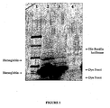
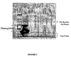
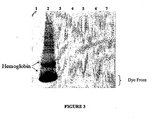
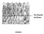
- Proteins present in the elution prepared as described in Example 3 above were evaluated using SDS-PAGE and fluorescence scanning or Western blot analysis. Results are shown in Fig. 1-4 , as described below.
- Fig. 1 shows a fluorescent scan of electrophoretically separated proteins.
- Lane 1 contains fluorescently-labeled molecular weight markers;
- lane 2 contains proteins in untreated lysate reaction mixture following expression of Renilla luciferase, as described in Example 2;
- lane 3 contains the flowthrough solution of lysate reaction mixture combined (1:3) with binding buffer buffered at pH 7.5, as described in Example 3, above;
- lane 4 contains the eluate from Example 3 above. Bands corresponding to hemoglobin, his-tagged Renilla luciferase, or the dye front are indicated on the scan. As can be seen by comparing lanes 3 and 4, the purification step results in a substantial increase in the purity of the his-tagged protein.
- lane 1 contains fluorescently-labeled molecular weight markers
- lane 2 contains proteins in untreated lysate following expression of Renilla luciferase, as described in Example 2
- lanes 3-7 contain proteins that were first bound to the support with binding buffer buffered at pH 6.0, 6.5, 7.0, 7.5, or 8.0, respectively, and then eluted with elution buffer. Bands corresponding to hemoglobin, his-tagged Renilla luciferase, or the dye front are indicated on the scan.
- Fig. 3 shows a SDS-PAGE gel stained with GelCode TM Blue Stain Reagent, product No. 24592 (Pierce Chemicals, Rockford, IL), including molecular weight markers (lane 1), untreated lysate reaction mixture (lane 2), and proteins eluted with elution buffer following binding to the solid support with binding buffer buffered at pH 6.0, 6.5, 7.0, 7.5, or 8.0 (lanes 3-7, respectively).
- This gel demonstrates that the amount of his-tagged Renilla luciferase and hemoglobin contaminant in the eluate (lanes 3-7) is below the limits of detection of this system.
- Fig. 4 shows a Western blot of electrophoretically separated proteins probed with anti-Renilla luciferase antibody, including molecular weight markers (lane 1), lysate (lane 2), and proteins eluted with elution buffer following binding to a solid support with binding buffer buffered at pH 6.0, 6.5, 7.0, 7.5, or 8.0 (lanes 3-7, respectively).
- molecular weight markers lane 1
- lysate lysate
- Non-specific binding of the antibody to proteins in the lysate, as well as specific binding to his-tagged Renilla luciferase can be seen in the lysate in lane 2.
- binding of the anti-Renilla luciferase antibody in lanes 3-7 is limited to a protein having a size consistent with that of his-
- Fig. 5 shows a Western blot of proteins found in rabbit reticulocyte lysate treated as described below and probed with anti-hemoglobin antibody.
- Lane 1 A 50 ⁇ l aliquot of rabbit reticulocyte lysate was mixed with 100 ⁇ l of binding / wash buffer B as described below in Example 5 (20 mM sodium phosphate buffer (pH 7.5) and 500 mM NaCl) and incubated with 60 ⁇ l zinc particles (5% w/v) for 15 min on orbital rocker. The particles were washed 4 times with 200 ⁇ l of binding/wash buffer and eluted with 100 ⁇ l of elution buffer (1M imidazole).
- Lane 2 A 50 ⁇ l aliquot of rabbit reticulocyte lysate was mixed with 100 ⁇ l of binding/wash buffer B, mixed by pipeting with incubated with 30 ⁇ l MagneHis TM nickel particles (10% w/v) (Catalog # V8500, Promega Corp.). The particles were washed 3 times with 150 ⁇ l of binding/wash buffer and eluted with 100 ⁇ l of elution buffer (500 mM imidazole). Lanes 3-8 contain lysate not contacted with particles and diluted to from 0-5% of the original lysate concentration. Lanes 3-8 contain 0, 0.15, 0.6, 1.25, 2.5, 5 percent lysate, respectively. Sheep anti-human hemoglobin-AP 1:1000 was used to probe the Western blot, and binding of the sheep anti-human hemoglobin-AP 1:1000 was detected Western blue stabilized substrate for alkaline phosphatase (S3841).
- His-tagged proteins were expressed using TNT® T7 Quick Coupled Transcription/Translation System, Cat# L1170TNT (Promega Corp., Madison, WI), 1 ⁇ g DNA, and 2 ⁇ l FluoroTect TM Green Lys in vitro Translation Labeling System (Cat. # L5001).
- a 50- ⁇ l aliquot of the reaction mixture was combined with 100- ⁇ l binding buffer B (20 mM sodium phosphate buffer (pH 7.5) and 500 mM NaCl), transferred to a 0.5 or 1.5 ml tube with a 60 ⁇ l (6 mg) of zinc charged particles (prepared as described in Example 1), and gently mixed for 15 minutes at room temperature. The particles were separated from the solution using magnetization, and the particles were washed with 200 ⁇ l of binding buffer three to five times. Proteins were eluted by thoroughly mixing the particles with 100 ⁇ l elution buffer (1 M imidazole (pH 7.5)), separating the particles from the elution buffer by magnetization, and collecting the eluate.
- Fig. 6 shows a fluorescent scan of electrophoretically separated proteins.
- Lane 1 contains fluorescently-labeled molecular weight markers;
- lane 2 contains proteins in 2 ⁇ l untreated lysate reaction mixture following expression of His-MAPK;
- lane 3 contains 6 ⁇ l the flowthrough solution of lysate reaction mixture combined (1:2) with binding buffer; and
- lane 4 contains 8 ⁇ l the eluate, as described above;
- lane 5 contains proteins in 2 ⁇ l untreated lysate reaction mixture following expression of His-HGF;
- lane 6 contains 6 ⁇ l the flowthrough solution of lysate reaction mixture combined (1:2) with binding buffer;
- lane 7 contains 8 ⁇ l the eluate;
- lane 8 contains proteins in 2 ⁇ l untreated lysate reaction mixture following expression of His-calmodulin;
- lane 9 contains 6 ⁇ l the flowthrough solution of lysate reaction mixture combined (1:2) with binding buffer
- Example 6 Detecting protein-protein interactions using protein pull down assays.
- the zinc-charged solid support system described above was evaluated for the ability isolate an untagged polypeptide of interest (“prey”) from other non-target molecules using a his-tagged protein (“bait”) using MyoD as the prey protein and Id as the bait protein as described below.
- MyoD and Id are members of the helix- loop-helix family of nuclear proteins.
- MyoD is a myogenic regulatory protein expressed in skeletal muscle, and Id protein is a negative regulator of myogenic differentiation that interacts with MyoD.
- Untagged MyoD prey protein was expressed in TnT T7 coupled transcription/translation lysate (Cat#L1170, Promega Corp.), 1 ⁇ g of MyoD DNA and 2 ⁇ l of 35 S methionine as recommended in the Technical Bulletin (Cat#L1170, Promega Corp.).
- His-tagged Id was expressed in an E. coli expression system according to standard protocols. Following expression, the cultured bacteria were pelleted, resuspended at a 10x concentration, and sonicated to form a bacterial lysate. The His-Id was also expressed in a TnT T7 coupled transcription/translation lysate (Cat#L1170, Promega Corp., Madison, WI).
- Zinc charged solid support with bound his-tagged Id was prepared as follows. A 100 ⁇ l aliquot of 10x concentrated lysate containing the his-tagged Id bait was added to 30 ⁇ l zinc-charged NTA-modified magnetic silica particles and incubated for 15 minutes on a shaker 1100 rpm. The particles were washed three times with 200 ⁇ l of 20 mM sodium phosphate, pH 7.4. The particles were resuspended in 30 ⁇ l of buffer, and 5 ⁇ l aliquots of the particles were transferred to new tubes. One set of samples was washed twice with 20mM sodium phosphate + 40mM imidazole and a second set was washed twice with 20mM sodium phosphate. The particles were resuspended in the wash buffer (175 ⁇ l) and incubated at 30°C for 60 minutes with gentle rocking.
- a 20 ⁇ l aliquot of TnT lysate containing MyoD was combined with the zinc solid support-bound his-Id and incubated for at room temperature for 60 minutes with gentle agitation.
- the particles were washed three times in the same final wash buffer used during preparation of the immobilized his-tagged Id, followed by a washing with 20mM sodium phosphate and an additional wash of 40 or 500mM imidazole.
- 20ul of SDS buffer (0.24 M Tris-HCl (pH 6.8), 2% SDS, 3 mM bromophenol blue, 50.4% glycerol, and 0.4 M dithiothreitol) was added to the particles, incubated for 5 minutes with shaking and the sample was collected.
- the elution sample was diluted 1:10 in SDS buffer, heated at 95°C, and loaded onto 4-20% tris-glycine gel.
- the gel was transferred to PVDF membrane, exposed to phosphorimager plate overnight, and read on a Storm TM Phosphorimager (Amersham Biosciences, Piscataway, NJ).
- Isolation of a "prey" protein co-expressed with the bait protein was compared with isolation of a prey protein expressed separately from the bait protein.
- Both his-tagged Id and untagged MyoD proteins expressed in two different TnT reactions or co-expressed in the same TnT lysate.
- the his-Id and MyoD were expressed in separate reactions, equal volumes of his-Id expressing lysate and MyoD expressing lysate were mixed.
- Zinc-charged NTA-modified magnetic silica particles were added and processed as described above.
- lanes 1 and 5 include the lysate of a TnT expression system expressing 35 S labeled MyoD; lanes 2 and 6 include his-Id bound to zinc-charged solid support and MyoD; 3 and 7 include his-RNaseH bound to zinc charged NTA-modified magnetic silica particles and MyoD; and lanes 4 and 8 include MyoD eluted from zinc charged NTA-modified magnetic silica particles in the absence of his-ID.
- protein in lanes 1-4 was washed with 500 mM imidazole, and protein in lanes 5-8 was washed with 40 mM imidazole.
- his-tagged proteins associated with zinc charged NTA-modified magnetic silica particles can be used to isolate a second protein that interacts with the his-tagged protein.
- Efficiency of recovery of MyoD is greatly enhanced by the presence of his-Id. Washing with imidazole at concentrations of 40 mM or greater was found to give acceptable results, with increasing concentrations of imidazole in the wash reducing the background.
- Lane 1 includes a rabbit reticulocyte lysate expressing untagged 35 S MyoD; lane 2 shows the proteins isolated using zinc charged silica magnetic particles; lane 3 shows protein isolated using His-RNaseHI associated zinc charged silica magnetic particles; and lane 4 shows proteins isolated using His-Id associated zinc charged silica magnetic particles.
- Example 7 Detecting protein-protein interactions of co-expressed proteins .
- His-tagged bait protein His-Id
- prey protein MyoD
- TnT T7 coupled transcription/translation lysate Cat#L1170, Promega Corp., Madison, WI
- a 60 ⁇ l aliquot of zinc particles (5% w/v) or 30 ⁇ l of nickel MagneHis resin (10% w/v) was added to a 1.5 ml tube wash with 200 ⁇ l of 20mM sodium phosphate buffer pH 7.4 and resuspended in 60 ⁇ l of 20 mM sodium phosphate buffer.
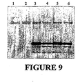
- a 5 ⁇ l aliquot of resin was mixed with 145 ⁇ l of 20mM sodium phosphate in a 1.5 ml tube. 50 ⁇ l of TnT reaction was added to resin and incubated for 1 hr at room temperature on orbital rocker. The supernatant was removed, and the resin was washed 4 times with 200 ⁇ l of 20mM sodium phosphate + 40mM imidazole, pH 7.4. The proteins were eluted by mixing the resin with 20 ⁇ l 4x SDS buffer for 5 min. The elution was collected elution and a 2 ⁇ l aliquot was mixed with 18 ⁇ l 4x SDS buffer and run a SDS-PAGE gel. The electrophoretically separated proteins were transferred to PVDF and exposed to phosphorimager plates overnight ( Fig. 9 ).
- lane 1 includes MyoD control (0.5 ⁇ g DNA); lane 2 contains MyoD control 1.0 ⁇ g DNA; lane 3 includes MyoD and his-Id from reticulocyte lysate programmed with 1.0 ⁇ g DNA isolated using zinc particles; lane 4 includes MyoD and his-Id from reticulocyte lysate programmed with 1.0 ⁇ g DNA isolated using nickel particles; lane 5 includes MyoD and his-Id from reticulocyte lysate programmed with 0.5 ⁇ g DNA isolated using zinc particles; lane 6 includes MyoD and his-Id from reticulocyte lysate programmed with 0.5 ⁇ g DNA isolated using nickel particles.
- a cDNA library coding for his-tagged proteins is expressed using Gold TNT® SP6 Express 96 System (Promega Cat#L5800) or Gold TNT® T7 Express 96 System (Promega Cat#L5600) for expressing the his-tagged proteins as recommended by the manufacture.
- Expressed his-tagged protein are purified using a zinc or cobalt charged solid support in conjunction with a robotic system, including a suitable robot such as Kingfisher, Biomek 2000 or FX.
- An automated purification of his-tagged proteins in general are described in Technical Manual #TM060 (Promega Corporation).
- His-tagged proteins expressed in rabbit reticulocyte are purified as essentially as described in Example 3 or 5, except that, following the final wash, the his-tagged proteins are eluted with an elution buffer containing 0.1% TFA in water or in 50% acetonitrile. Following elution, protein samples are dried in a Speed Vac TM and analyzed in MALDI-TOF mass spectrometer.
- His-tagged human cytochrome P450, subfamily IIIA, polypeptide 7 (CYP3A7) (Cat# E01046, Stratagene, LaJolla, CA) is expressed in Rabbit Recticulocyte TNT with Canine Pancreatic Microsomal Membrane (Promega Cat# Y40141) according to manufacture's protocol.
- the his-tagged protein is purified directly from the lysate as described in Example 3 or 5.
- the expressed protein is also purified after solubilizing the expressed membrane protein in presence of a non-ionic detergent like 1,2-Dihexanoyl- sn -Glycero-3-Phosphocholine (DHPC, Cat# 850305C, Avanti Polar Lipids, Alabaster, LA).
- Functional analysis of the expressed and purified his-human cytochrome P450, subfamily IIIA, polypeptide 7 is performed by P450-GloTM CYP3A7 Assay (Promega Cat# V8811).
- Zinc-charged solid supports may be used to purify other membrane proteins expressed in rabbit reticulocyte lysate.
- G-protein coupled receptors GPCR
- the GCPR is purified on zinc-charged solid supports using non-ionic detergents for use in ligand binding assays.
- a his-tagged GPCR (formyl peptide receptor) was selected.
- the protein was expressed in TnT T7 coupled transcription/translation lysate (Cat#L1170, Promega Corp., Madison, WI) using 35 S labeling as described above, and purified essentially according to Example 5.
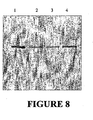
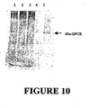
- the lysate was contacted with 60 or 180 ⁇ l zinc charged magnetic silica particles (5% w/v), the particles washed, and the proteins eluted and analyzed by SDS-PAGE ( Fig. 10 ).
- lane 1 contains TNT lysate expressing the his-GPCR; lane 2 contains flow through of 60 ⁇ l particles; lane 3 contains flow through of 180 ⁇ l resin; lane 4 contains eulution from 60 ⁇ l particles; and lane 5 contains elution from 180 ⁇ l resin.
- His-tagged glycoprotein is expressed in rabbit reticulocyte TNT with Canine Pancreatic Microsomal Membrane (Promega Cat# Y40141) according to manufacture's protocol, purified as described above, and analyzed by SDS-PAGE, by Western blot, or by mass spectrometry to determine N-linked glycosylation.
- a his-tagged cDNA library is expressed in Gold TnT (Promega Cat. # L5800) or in ProteoLinkTM In Vitro Expression Cloning System (adult human brain cDNA) (Promega, Cat# L6500).
- the his-tagged proteins are purified from heme proteins by contacting with a zinc-charged solid support (e.g., NTA-modified silica magnetic particles), followed by washing, as described above.
- Kinase activity is assayed with the his-tagged proteins bound to the solid support or after eluting the proteins.
- a fluorescence based assay may be used to detect kinase activity using either eluted his-tagged proteins or his-tagged proteins bound to the resin.
- Kinase assay may also be performed using the Kinase-Glo TM Luminescent Kinase Assay (Promega, Cat# V6711).
- Other protein kinase substrates may be used to detect kinase activity are available from EMD Biosciences, Inc. (Madison, Wisconsin)
- kinases or kinase substrates identified as described above may be cloned as described in the manufacturer's protocol.
- a cDNA library encoding his-tagged proteins is expressed as described in above and the his-tagged proteins are purified by the methods described in Examples 3, 5, or 7. Purified proteins are digested with Trypsin Gold, Mass Spectrometry Grade (Promega, Cat# V5280) and the peptides are characterized by tandem mass spectrometry. Polynucleotides encoding the proteins are identified by the methods described in ProteoLink TM In Vitro Expression Cloning System kit (Promega, Cat# L6500).
- a his-tagged protein that is a substrate for a protease is bound to zinc- or cobalt-charged solid support (e.g., a solid support prepared as described above in Example 1).
- the particle-bound his-tagged protein is incubated with reticulocyte lysate or human blood samples under the conditions described in Example 3 or 5 for a specific period of time.
- the solid support is washed and the his-tagged proteins are eluted as described above in Example 3, 5, or 7. Proteins are analyzed by SDS-PAGE, Western blot, or mass spectrometry.
- his-tagged protein substrate is added directly to the rabbit reticulocyte lysate or human blood sample and incubated for specific period of time. His-tagged proteins are then purified as described in Example 3, 5, or 7. Protease activity is analyzed by SDS-PAGE, Western blot, or mass spectrometry.
- Specific protein modifications or protease assays may be used to screen for protein markers from human blood samples.
- serum proteins may be analyzed to identify specific protein markers for prostate cancer ( Lehrer et al "Putative protein markers in the sera of men with prostatic neoplasms" BJU Int. 2003 Aug;92(3):223-5 ).
- human blood samples could be analyzed by purified his-tagged proteins for the up or down regulation of specific kinases, proteases or protein modification systems.
- a his-tagged kinase will be incubated with blood samples from a person with cancer and a person without cancer. After specific period of incubation, his-tagged proteins will be purified from the blood samples as described in Example 3 or 6. Purified proteins are then analyzed by mass spectrometry or gel analysis for studying the protein modifications.
- His-tagged proteins purified as described in the preceding Examples are suitable for studying protein-ligand interaction. His-tagged proteins from a cDNA library or a specific his-tagged coding sequence of interest are expressed in rabbit recticulocyte lysate and interaction between an expressed protein and a ligand is detected using fluorescently labeled ligand.
- an inhibitor for caspase protein may be identified by expressing his-tagged caspase protein in rabbit reticulocyte lysate and purifying the caspase proteins using a zinc charged solid support, such as zinc-charged NTA-modified silica magnetic particles.
- a multi-well method such as a 96-well plate is used.
- the bound his-caspase Prior to elution, the bound his-caspase is contacted with a fluorescently labeled inhibitor.
- the solid support is washed the proteins are eluted, and the eluted proteins analyzed using a fluorometer.
- a similar approach may be used for any protein or ligand, as well as in directed evolution studies.
- Example 17 Analyzing post-translational modifications of proteins
- Zinc-charged metal chelating resins are used to isolate proteins expressed in rabbit reticulocyte lysate from a cDNA library or a single protein coding sequence, which are then evaluated for post-translational modifications.
- a his-tagged cDNA library is expressed in rabbit reticulocyte lysate, the tagged proteins are bound to a zinc charged solid support, washed, and contacted with a fluorescently labeled antibody that recognizes a particular post-translational modification (e.g., acetylation or phosphorylation).
- a non-labeled primary antibody is used and its interaction with a his-tagged protein is detected using a fluorescently labeled secondary antibody.
- the antibody-labeled his-tagged protein may be subsequently recovered as described above, and analyzed in fluorometer.
- Zinc-charged solid supports may be used to isolate particular his-tagged protein substrates.
- brain cDNA library encoding untagged proteins is expressed in Gold TNT® SP6 Express 96 System (Promega Cat#L5800) or Gold TNT® T7 Express 96 System (Promega Cat#L5600) as recommended by the manufacture.
- the lysate is contacted with a zinc-charged solid support to which a his-tagged protein or proteins is bound.
- the his-tagged protein is evaluated for modifications.
- the cDNA clone expressing the protein responsible for modifying the his-tagged protein is identified.
- Example 18 Antibody based detection of proteins from rabbit reticulocyte lysate
- Fluorescent labeled antibodies can be used to detect his-tagged proteins expressed in rabbit reticulocyte lysate. This could be achieved by expressing and attaching the his-tagged proteins to a zinc charged solid support, followed by contacting with fluorescently labeled antibodies, washing, and detecting bound antibodies fluorescently.
- Rabbit reticulocyte-based cell free protein expression system is used to express antibody coding sequences and subsequent screening using fluorescently labeled antigens.
- In vitro antibody libraries are generated by directed evolution methods such as DNA shuffling, phage display, ribosome display, covalent display, mRNA display, or any other suitable method.
- Full length antibodies or antibody fragments including the antigen binding region are expressed with polyhistidine tags in a rabbit reticulocyte lysate expression system.
- the his-tagged antibody or antibody fragment is allowed to interact with a zinc charged solid support as described above for his-tagged proteins generally.
- Fluorescently labeled antigen is used to select the antibodies capable of interacting with the antigen to form an antigen-antibody pair. The method permits screening of large numbers of antibodies or antibody fragments in a high throughput manner.
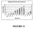
- Example 20 Detection of fluorescent dyes following removal of hemoglobin from rabbit reticulocyte lysate.
- Fluorescent compounds have been widely used for developing various functional assays such as kinase or caspase assays, for example. However, contaminating hemoglobin inhibits fluorescence of certain fluorescent compounds. The suitability of the zinc based purification system for use in assays employing fluorescent labels was evaluated using rhodamine 110 (R110) as follows.
- Example 21 Evaluation of fluorescence based caspase assay following removal of hemoglobin by zinc charged solid support .
- Rabbit reticulocyte lysate (TnT T7 coupled transcription/translation lysate (Cat#L1170, Promega Corp., Madison, WI)) or water (40 ⁇ l) was combined with 100ul 50 mM sodium phosphate containing 500mM NaCl and 10 ⁇ g purified his-tagged caspase3 (Upstate, Cat# 14-264). The mixture was incubated in a tube containing with 3 mg of zinc or nickel charged magnetic particles for 15 min on a rotary shaker at room temperature. The flow through was removed by magnetization. The particles were washed four times with 50 mM sodium phosphate + 500 mM NaCl, and the caspase eluted with 100 ⁇ l 1 M imidazole.
- the eluted material was serially diluted (1:5, 1:10, 1:20, 1:40, 1:80, 1:160, and 1:320) with phosphate buffered saline to give a final volume of 100 ⁇ l.
- Caspase activity was dected using Apo-One substrate diluted in Apo-One buffer (Apo-ONE® Homogeneous Caspase-3/7 Assay, Cat# G7792, Promega Corporation).
- the A 530 for each sample which correlates with the amount of active caspase present in the sample, was plotted as a function of the dilution factor: Absorbance of caspase purified from TNT using zinc particles (diamonds); absorbance for caspase purified from water using zinc particles (squares); and absorbance for caspase purified from TNT using nickel particles (triangles).
- samples containing only TNT (no caspase) isolated on zinc or nickel particles had aborbances that did not exceed baseline.
- results show that zinc particles are useful for isolating his-tagged proteins from hemoglobin-containing starting materials (e.g., rabbit reticulocyte lysate or blood) for subsequent analysis in a fluorescence based caspase assay.
- hemoglobin-containing starting materials e.g., rabbit reticulocyte lysate or blood
- This approach is suitable for in vitro screening of his-tagged caspases expressed in rabbit reticulocyte lysate expressed from a cDNA/mRNA library.
- caspase assay could be performed essentially as described above but using a luminiscence based assays (e.g., Caspase-Glo TM 9 Assay, Cat# G8210, Promega Corporation).
- a luminiscence based assays e.g., Caspase-Glo TM 9 Assay, Cat# G8210, Promega Corporation.
Landscapes
- Chemical & Material Sciences (AREA)
- Organic Chemistry (AREA)
- Medicinal Chemistry (AREA)
- Molecular Biology (AREA)
- Biochemistry (AREA)
- Biophysics (AREA)
- General Health & Medical Sciences (AREA)
- Genetics & Genomics (AREA)
- Health & Medical Sciences (AREA)
- Life Sciences & Earth Sciences (AREA)
- Proteomics, Peptides & Aminoacids (AREA)
- Analytical Chemistry (AREA)
- Chemical Kinetics & Catalysis (AREA)
- Peptides Or Proteins (AREA)
- Detergent Compositions (AREA)
- Enzymes And Modification Thereof (AREA)
- Measuring Or Testing Involving Enzymes Or Micro-Organisms (AREA)
- Solid-Sorbent Or Filter-Aiding Compositions (AREA)
Claims (20)
- Verfahren zum Isolieren eines his-markierten Proteins aus einem ein Hämprotein umfassenden Ausgangsmaterial, Folgendes umfassend:(a) das Kontaktieren des Ausgangsmaterials mit einem mit Zink oder Kobalt beladenen festen Träger unter Bedingungen, die es dem his-markierten Protein ermöglichen, sich im Vergleich zur Bindung an das Hämprotein bevorzugt an den festen Träger zu binden, worin die Bedingungen die Gegenwart von Imidazol in einer Konzentration von 10 mM bis 60 mM umfassen; sowie(b) das Entfernen der gebundenen his-markierten Proteine vom festen Träger.
- Verfahren nach Anspruch 1, worin die Bedingungen unter (a) die Gegenwart von Imidazol in einer Konzentration von 10 mM bis 20 mM umfassen.
- Verfahren nach Anspruch 1, worin vor dem Kontaktieren der Probe mit dem festen Träger ein Bindungspuffer mit der Probe kombiniert wird.
- Verfahren nach Anspruch 3, worin der Bindungspuffer Imidazol umfasst.
- Verfahren nach Anspruch 1, worin die Probe und ein Bindungspuffer im Wesentlichen gleichzeitig mit dem festen Träger kontaktiert werden.
- Verfahren nach Anspruch 1, worin der feste Träger vor dem Kontaktieren der Probe mit dem festen Träger mit einem Bindungspuffer gewaschen oder äquilibriert wird.
- Verfahren nach Anspruch 1, worin der feste Träger Zink umfasst.
- Verfahren nach Anspruch 1, worin der feste Träger Kobalt umfasst.
- Verfahren nach Anspruch 1, worin das his-markierte Protein durch Eluieren mit einem Elutionspuffer entfernt wird, der Imidazol in einer Konzentration von zumindest 100 mM umfasst.
- Verfahren nach Anspruch 9, worin der Elutionspuffer Imidazol in einer Konzentration von 100 mM bis 3 M umfasst.
- Verfahren nach Anspruch 1, worin das his-markierte Protein durch Eluieren mit einem Elutionspuffer entfernt wird, der EDTA in einer Konzentration von 10 mM bis 0,5 M umfasst.
- Verfahren nach Anspruch 1, worin das his-markierte Protein durch Eluieren mit einem Elutionspuffer mit einem pH von etwa 6,0 oder darunter entfernt wird.
- Verfahren nach Anspruch 1, worin das Ausgangsmaterial Kaninchen-Reticulozyten-Lysat umfasst.
- Verfahren nach Anspruch 1, worin das Hämprotein Hämoglobin ist.
- Verfahren nach Anspruch 1, worin die Bedingungen die Gegenwart von Natriumchlorid in einer Konzentration von 0 bis 0,5 M umfassen.
- Verfahren nach Anspruch 1, worin der feste Träger aus magnetischen Silicateilchen besteht.
- Verfahren zum Charakterisieren eines his-markierten Proteins, weiters umfassend:(a) das Kontaktieren eines Ausgangsmaterials, welches das his-markierte Protein und das Hämprotein umfasst, mit einem mit Zink oder Kobalt beladenen festen Träger unter Bedingungen, die es dem his-markierten Protein ermöglichen, sich im Vergleich zur Bindung an das Hämprotein bevorzugt an den festen Träger zu binden, worin die Bedingungen die Gegenwart von Imidazol in einer Konzentration von 10 mM bis 60 mM umfassen;(b) das Waschen des festen Trägers;(c) das Kontaktieren des his-markierten Proteins mit einem Substrat oder einem zweiten Protein; sowie(d) das Detektieren von Wechselwirkung zwischen dem his-markierten Protein und dem zweiten Protein oder Substrat, um das his-markierte Protein zu charakterisieren.
- Verfahren nach Anspruch 17, worin das his-markierte Portein nach Schritt (b) vom festen Träger eluiert wird.
- Verfahren nach Anspruch 17, worin die Wechselwirkung durch Detektion einer Zunahme oder Abnahme von Fluoreszenz detektiert wird.
- Verfahren zur Abtrennung eines Ziel-Polypeptids von einer Probe, die das Ziel-Polypeptid und ein Hämprotein umfasst, Folgendes umfassend:das Kontaktieren eines his-markierten Proteins mit der Fähigkeit, mit dem Ziel-Polypeptid wechselzuwirken, mit einer Probe, die das Ziel-Polypeptid und das Hämprotein umfasst, und mit einem mit Zink oder Kobalt beladenen festen Träger unter Bedingungen, die es dem his-markierten Protein ermöglichen, mit dem Ziel-Polypeptid wechselzuwirken und sich im Vergleich zur Bindung an das Hämprotein bevorzugt an den festen Träger zu binden, worin die Bedingungen die Gegenwart von Imidazol in einer Konzentration von 10 mM bis 60 mM umfassen.
Applications Claiming Priority (3)
| Application Number | Priority Date | Filing Date | Title |
|---|---|---|---|
| US50254403P | 2003-09-12 | 2003-09-12 | |
| US10/840,408 US7115397B2 (en) | 2003-09-12 | 2004-05-06 | Methods and kits for purifying his-tagged proteins |
| PCT/US2004/029580 WO2005035092A1 (en) | 2003-09-12 | 2004-09-10 | Methods and kits for purifying his-tagged proteins |
Publications (2)
| Publication Number | Publication Date |
|---|---|
| EP1660206A1 EP1660206A1 (de) | 2006-05-31 |
| EP1660206B1 true EP1660206B1 (de) | 2008-08-06 |
Family
ID=34278838
Family Applications (1)
| Application Number | Title | Priority Date | Filing Date |
|---|---|---|---|
| EP04788679A Not-in-force EP1660206B1 (de) | 2003-09-12 | 2004-09-10 | Verfahren und ausrüstungen zur reinigung von proteinen mit his-tag |
Country Status (8)
| Country | Link |
|---|---|
| US (4) | US7115397B2 (de) |
| EP (1) | EP1660206B1 (de) |
| JP (1) | JP2007505134A (de) |
| AT (1) | ATE403480T1 (de) |
| AU (1) | AU2004279361A1 (de) |
| CA (1) | CA2536066A1 (de) |
| DE (1) | DE602004015618D1 (de) |
| WO (1) | WO2005035092A1 (de) |
Families Citing this family (7)
| Publication number | Priority date | Publication date | Assignee | Title |
|---|---|---|---|---|
| CA2514010A1 (en) * | 2004-08-03 | 2006-02-03 | Axis-Shield Diagnostics Limited | Assay |
| WO2007038746A2 (en) * | 2005-09-28 | 2007-04-05 | The University Of Chicago | Versatile vectors for expression of foreign proteins in photosynthetic bacteria |
| JP5656339B2 (ja) * | 2007-03-28 | 2015-01-21 | Jsr株式会社 | タンパク質固定化担体およびその製造方法 |
| WO2011094584A2 (en) * | 2010-01-30 | 2011-08-04 | University Of Rochester | Method and system for purifying and quantitating proteins using heme fusion tags |
| BR112012027262A2 (pt) | 2010-05-19 | 2016-07-26 | Hoffmann La Roche | método para purificar e para produzir um polipeptídeo |
| CN109569546B (zh) * | 2018-07-05 | 2021-05-18 | 华侨大学 | 一种用于组氨酸标签蛋白纯化的螯合金属磁性纳米颗粒的制备方法及应用 |
| KR20220152290A (ko) * | 2020-03-09 | 2022-11-15 | 아이덱스 래보러토리즈, 인코포레이티드 | 화학 시약 시험 슬라이드에 액체 샘플을 분배하기 전에 그의 간섭 성분을 제거하는 방법 |
Family Cites Families (8)
| Publication number | Priority date | Publication date | Assignee | Title |
|---|---|---|---|---|
| US5840851A (en) * | 1993-07-23 | 1998-11-24 | Plomer; J. Jeffrey | Purification of hemoglobin |
| US5962641A (en) | 1996-08-16 | 1999-10-05 | Clontech Laboratories, Inc. | Method for purification of recombinant proteins |
| AU4187799A (en) | 1998-05-14 | 1999-11-29 | Clontech Laboratories, Inc. | Compositions and methods for protein purification based on metal ion affinity site |
| US6232083B1 (en) * | 1999-03-12 | 2001-05-15 | The Research Foundation Of State University Of New York | Methods and compositions for screening cloned proteins |
| EP1069131B1 (de) * | 1999-07-15 | 2006-03-15 | Qiagen GmbH | Verfahren zur Trennung von teilchenförmigen Substraten aus einer Lösung indem man den Teilchenverlust minimiert |
| WO2002037100A2 (en) | 2000-10-30 | 2002-05-10 | Lytton Simon D | Novel applications of nickel nitrilotriacetic acid (ni-nta) resin: hemeprotein removal, recovery, and purification from biological samples |
| US7179775B2 (en) * | 2002-09-11 | 2007-02-20 | Henkel Kommanditgesellschaft Auf Aktien | Coating removal compositions |
| US7354750B2 (en) * | 2002-10-18 | 2008-04-08 | Promega Corporation | Methods for separating molecules |
-
2004
- 2004-05-06 US US10/840,408 patent/US7115397B2/en not_active Expired - Fee Related
- 2004-09-10 WO PCT/US2004/029580 patent/WO2005035092A1/en active Application Filing
- 2004-09-10 EP EP04788679A patent/EP1660206B1/de not_active Not-in-force
- 2004-09-10 CA CA002536066A patent/CA2536066A1/en not_active Abandoned
- 2004-09-10 AU AU2004279361A patent/AU2004279361A1/en not_active Abandoned
- 2004-09-10 JP JP2006526315A patent/JP2007505134A/ja active Pending
- 2004-09-10 AT AT04788679T patent/ATE403480T1/de not_active IP Right Cessation
- 2004-09-10 DE DE602004015618T patent/DE602004015618D1/de not_active Expired - Fee Related
-
2005
- 2005-01-10 US US11/032,282 patent/US20050209129A1/en not_active Abandoned
- 2005-01-10 US US11/032,281 patent/US7150973B2/en not_active Expired - Fee Related
-
2006
- 2006-08-22 US US11/466,274 patent/US7339036B2/en not_active Expired - Fee Related
Also Published As
| Publication number | Publication date |
|---|---|
| EP1660206A1 (de) | 2006-05-31 |
| WO2005035092A1 (en) | 2005-04-21 |
| JP2007505134A (ja) | 2007-03-08 |
| US20060281906A1 (en) | 2006-12-14 |
| US7339036B2 (en) | 2008-03-04 |
| US20050209129A1 (en) | 2005-09-22 |
| US7150973B2 (en) | 2006-12-19 |
| US20050209444A1 (en) | 2005-09-22 |
| CA2536066A1 (en) | 2005-04-21 |
| US7115397B2 (en) | 2006-10-03 |
| US20050059809A1 (en) | 2005-03-17 |
| ATE403480T1 (de) | 2008-08-15 |
| DE602004015618D1 (de) | 2008-09-18 |
| AU2004279361A1 (en) | 2005-04-21 |
Similar Documents
| Publication | Publication Date | Title |
|---|---|---|
| US7354750B2 (en) | Methods for separating molecules | |
| US7339036B2 (en) | Methods and kits for purifying his-tagged proteins | |
| US7691645B2 (en) | Immunosubtraction method | |
| Guerrier et al. | Reduction of dynamic protein concentration range of biological extracts for the discovery of low-abundance proteins by means of hexapeptide ligand library | |
| AU716309B2 (en) | Screening of combinatorial peptide libraries for selection of peptide ligand useful in affinitypurification of target proteins | |
| ES2258429T3 (es) | Metodos para separar sustratos en particulas de una solucion, al tiempo que se minimiza la perdida de particulas. | |
| JP2003010647A (ja) | 蛋白質を操作する方法及び装置 | |
| EP2949750B1 (de) | Antikörperbindendes peptid | |
| CA2891820A1 (en) | Streptavidin muteins and methods of using them | |
| EP0367795A1 (de) | Affinitätstrennverfahren unter verwendung von immobilisierten flockungsmitteln | |
| CN101512013A (zh) | 与具有顺磁性质的小颗粒相连接的各种化学库 | |
| US6635420B1 (en) | Purification of substances from a biological sample | |
| WO2018043629A1 (ja) | 非天然型立体構造を形成した抗体に親和性を示すポリペプチド | |
| CN109678932B (zh) | 一种IgG抗体亲和的小分子肽及其应用 | |
| Krishna et al. | A REVIEW ON “AFFINITY CHROMATOGRAPHY | |
| Allais et al. | Regina Fraas, Juliane Diehm and Matthias Franzreb | |
| Mrabet et al. | Immobilized metal-ion affinity chromatography | |
| Brewer et al. | Fusion protein purification | |
| Harris | Micromethods in Protein Chemistry | |
| WO2003100379A2 (en) | Methods and reagents for removing contaminants from biomolecules |
Legal Events
| Date | Code | Title | Description |
|---|---|---|---|
| PUAI | Public reference made under article 153(3) epc to a published international application that has entered the european phase |
Free format text: ORIGINAL CODE: 0009012 |
|
| 17P | Request for examination filed |
Effective date: 20060323 |
|
| AK | Designated contracting states |
Kind code of ref document: A1 Designated state(s): AT BE BG CH CY CZ DE DK EE ES FI FR GB GR HU IE IT LI LU MC NL PL PT RO SE SI SK TR |
|
| REG | Reference to a national code |
Ref country code: HK Ref legal event code: DE Ref document number: 1084907 Country of ref document: HK |
|
| 17Q | First examination report despatched |
Effective date: 20060829 |
|
| DAX | Request for extension of the european patent (deleted) | ||
| 17Q | First examination report despatched |
Effective date: 20060829 |
|
| GRAP | Despatch of communication of intention to grant a patent |
Free format text: ORIGINAL CODE: EPIDOSNIGR1 |
|
| GRAS | Grant fee paid |
Free format text: ORIGINAL CODE: EPIDOSNIGR3 |
|
| GRAA | (expected) grant |
Free format text: ORIGINAL CODE: 0009210 |
|
| AK | Designated contracting states |
Kind code of ref document: B1 Designated state(s): AT BE BG CH CY CZ DE DK EE ES FI FR GB GR HU IE IT LI LU MC NL PL PT RO SE SI SK TR |
|
| REG | Reference to a national code |
Ref country code: GB Ref legal event code: FG4D |
|
| REG | Reference to a national code |
Ref country code: CH Ref legal event code: EP |
|
| REG | Reference to a national code |
Ref country code: IE Ref legal event code: FG4D |
|
| REF | Corresponds to: |
Ref document number: 602004015618 Country of ref document: DE Date of ref document: 20080918 Kind code of ref document: P |
|
| PG25 | Lapsed in a contracting state [announced via postgrant information from national office to epo] |
Ref country code: NL Free format text: LAPSE BECAUSE OF FAILURE TO SUBMIT A TRANSLATION OF THE DESCRIPTION OR TO PAY THE FEE WITHIN THE PRESCRIBED TIME-LIMIT Effective date: 20080806 Ref country code: ES Free format text: LAPSE BECAUSE OF FAILURE TO SUBMIT A TRANSLATION OF THE DESCRIPTION OR TO PAY THE FEE WITHIN THE PRESCRIBED TIME-LIMIT Effective date: 20081117 |
|
| PG25 | Lapsed in a contracting state [announced via postgrant information from national office to epo] |
Ref country code: AT Free format text: LAPSE BECAUSE OF FAILURE TO SUBMIT A TRANSLATION OF THE DESCRIPTION OR TO PAY THE FEE WITHIN THE PRESCRIBED TIME-LIMIT Effective date: 20080806 Ref country code: FI Free format text: LAPSE BECAUSE OF FAILURE TO SUBMIT A TRANSLATION OF THE DESCRIPTION OR TO PAY THE FEE WITHIN THE PRESCRIBED TIME-LIMIT Effective date: 20080806 Ref country code: SI Free format text: LAPSE BECAUSE OF FAILURE TO SUBMIT A TRANSLATION OF THE DESCRIPTION OR TO PAY THE FEE WITHIN THE PRESCRIBED TIME-LIMIT Effective date: 20080806 |
|
| PG25 | Lapsed in a contracting state [announced via postgrant information from national office to epo] |
Ref country code: BE Free format text: LAPSE BECAUSE OF FAILURE TO SUBMIT A TRANSLATION OF THE DESCRIPTION OR TO PAY THE FEE WITHIN THE PRESCRIBED TIME-LIMIT Effective date: 20080806 |
|
| PG25 | Lapsed in a contracting state [announced via postgrant information from national office to epo] |
Ref country code: DK Free format text: LAPSE BECAUSE OF FAILURE TO SUBMIT A TRANSLATION OF THE DESCRIPTION OR TO PAY THE FEE WITHIN THE PRESCRIBED TIME-LIMIT Effective date: 20080806 Ref country code: MC Free format text: LAPSE BECAUSE OF NON-PAYMENT OF DUE FEES Effective date: 20080930 Ref country code: BG Free format text: LAPSE BECAUSE OF FAILURE TO SUBMIT A TRANSLATION OF THE DESCRIPTION OR TO PAY THE FEE WITHIN THE PRESCRIBED TIME-LIMIT Effective date: 20081106 |
|
| REG | Reference to a national code |
Ref country code: CH Ref legal event code: PL |
|
| PG25 | Lapsed in a contracting state [announced via postgrant information from national office to epo] |
Ref country code: RO Free format text: LAPSE BECAUSE OF FAILURE TO SUBMIT A TRANSLATION OF THE DESCRIPTION OR TO PAY THE FEE WITHIN THE PRESCRIBED TIME-LIMIT Effective date: 20080806 Ref country code: PT Free format text: LAPSE BECAUSE OF FAILURE TO SUBMIT A TRANSLATION OF THE DESCRIPTION OR TO PAY THE FEE WITHIN THE PRESCRIBED TIME-LIMIT Effective date: 20090106 Ref country code: CZ Free format text: LAPSE BECAUSE OF FAILURE TO SUBMIT A TRANSLATION OF THE DESCRIPTION OR TO PAY THE FEE WITHIN THE PRESCRIBED TIME-LIMIT Effective date: 20080806 Ref country code: SK Free format text: LAPSE BECAUSE OF FAILURE TO SUBMIT A TRANSLATION OF THE DESCRIPTION OR TO PAY THE FEE WITHIN THE PRESCRIBED TIME-LIMIT Effective date: 20080806 |
|
| PGFP | Annual fee paid to national office [announced via postgrant information from national office to epo] |
Ref country code: DE Payment date: 20090202 Year of fee payment: 5 |
|
| PLBE | No opposition filed within time limit |
Free format text: ORIGINAL CODE: 0009261 |
|
| STAA | Information on the status of an ep patent application or granted ep patent |
Free format text: STATUS: NO OPPOSITION FILED WITHIN TIME LIMIT |
|
| REG | Reference to a national code |
Ref country code: IE Ref legal event code: MM4A |
|
| PGFP | Annual fee paid to national office [announced via postgrant information from national office to epo] |
Ref country code: GB Payment date: 20090129 Year of fee payment: 5 |
|
| 26N | No opposition filed |
Effective date: 20090507 |
|
| PG25 | Lapsed in a contracting state [announced via postgrant information from national office to epo] |
Ref country code: EE Free format text: LAPSE BECAUSE OF FAILURE TO SUBMIT A TRANSLATION OF THE DESCRIPTION OR TO PAY THE FEE WITHIN THE PRESCRIBED TIME-LIMIT Effective date: 20080806 Ref country code: IE Free format text: LAPSE BECAUSE OF NON-PAYMENT OF DUE FEES Effective date: 20080910 |
|
| PG25 | Lapsed in a contracting state [announced via postgrant information from national office to epo] |
Ref country code: IT Free format text: LAPSE BECAUSE OF FAILURE TO SUBMIT A TRANSLATION OF THE DESCRIPTION OR TO PAY THE FEE WITHIN THE PRESCRIBED TIME-LIMIT Effective date: 20080806 |
|
| PG25 | Lapsed in a contracting state [announced via postgrant information from national office to epo] |
Ref country code: CH Free format text: LAPSE BECAUSE OF NON-PAYMENT OF DUE FEES Effective date: 20080930 Ref country code: LI Free format text: LAPSE BECAUSE OF NON-PAYMENT OF DUE FEES Effective date: 20080930 |
|
| PGFP | Annual fee paid to national office [announced via postgrant information from national office to epo] |
Ref country code: FR Payment date: 20090119 Year of fee payment: 5 |
|
| PG25 | Lapsed in a contracting state [announced via postgrant information from national office to epo] |
Ref country code: SE Free format text: LAPSE BECAUSE OF FAILURE TO SUBMIT A TRANSLATION OF THE DESCRIPTION OR TO PAY THE FEE WITHIN THE PRESCRIBED TIME-LIMIT Effective date: 20081106 |
|
| GBPC | Gb: european patent ceased through non-payment of renewal fee |
Effective date: 20090910 |
|
| PG25 | Lapsed in a contracting state [announced via postgrant information from national office to epo] |
Ref country code: PL Free format text: LAPSE BECAUSE OF FAILURE TO SUBMIT A TRANSLATION OF THE DESCRIPTION OR TO PAY THE FEE WITHIN THE PRESCRIBED TIME-LIMIT Effective date: 20080806 |
|
| REG | Reference to a national code |
Ref country code: FR Ref legal event code: ST Effective date: 20100531 |
|
| PG25 | Lapsed in a contracting state [announced via postgrant information from national office to epo] |
Ref country code: LU Free format text: LAPSE BECAUSE OF NON-PAYMENT OF DUE FEES Effective date: 20080910 Ref country code: DE Free format text: LAPSE BECAUSE OF NON-PAYMENT OF DUE FEES Effective date: 20100401 Ref country code: FR Free format text: LAPSE BECAUSE OF NON-PAYMENT OF DUE FEES Effective date: 20090930 Ref country code: HU Free format text: LAPSE BECAUSE OF FAILURE TO SUBMIT A TRANSLATION OF THE DESCRIPTION OR TO PAY THE FEE WITHIN THE PRESCRIBED TIME-LIMIT Effective date: 20090207 Ref country code: CY Free format text: LAPSE BECAUSE OF FAILURE TO SUBMIT A TRANSLATION OF THE DESCRIPTION OR TO PAY THE FEE WITHIN THE PRESCRIBED TIME-LIMIT Effective date: 20080806 |
|
| PG25 | Lapsed in a contracting state [announced via postgrant information from national office to epo] |
Ref country code: TR Free format text: LAPSE BECAUSE OF FAILURE TO SUBMIT A TRANSLATION OF THE DESCRIPTION OR TO PAY THE FEE WITHIN THE PRESCRIBED TIME-LIMIT Effective date: 20080806 |
|
| PG25 | Lapsed in a contracting state [announced via postgrant information from national office to epo] |
Ref country code: GR Free format text: LAPSE BECAUSE OF FAILURE TO SUBMIT A TRANSLATION OF THE DESCRIPTION OR TO PAY THE FEE WITHIN THE PRESCRIBED TIME-LIMIT Effective date: 20081107 |
|
| PG25 | Lapsed in a contracting state [announced via postgrant information from national office to epo] |
Ref country code: GB Free format text: LAPSE BECAUSE OF NON-PAYMENT OF DUE FEES Effective date: 20090910 |
|
| REG | Reference to a national code |
Ref country code: HK Ref legal event code: WD Ref document number: 1084907 Country of ref document: HK |