EP1618127B1 - Immunogenic composition comprising a spike protein of the SARS coronavirus - Google Patents
Immunogenic composition comprising a spike protein of the SARS coronavirus Download PDFInfo
- Publication number
- EP1618127B1 EP1618127B1 EP04750188.7A EP04750188A EP1618127B1 EP 1618127 B1 EP1618127 B1 EP 1618127B1 EP 04750188 A EP04750188 A EP 04750188A EP 1618127 B1 EP1618127 B1 EP 1618127B1
- Authority
- EP
- European Patent Office
- Prior art keywords
- sars
- seq
- protein
- prt
- spike
- Prior art date
- Legal status (The legal status is an assumption and is not a legal conclusion. Google has not performed a legal analysis and makes no representation as to the accuracy of the status listed.)
- Expired - Lifetime
Links
Images



























Classifications
-
- C—CHEMISTRY; METALLURGY
- C07—ORGANIC CHEMISTRY
- C07K—PEPTIDES
- C07K14/00—Peptides having more than 20 amino acids; Gastrins; Somatostatins; Melanotropins; Derivatives thereof
- C07K14/005—Peptides having more than 20 amino acids; Gastrins; Somatostatins; Melanotropins; Derivatives thereof from viruses
-
- A—HUMAN NECESSITIES
- A61—MEDICAL OR VETERINARY SCIENCE; HYGIENE
- A61K—PREPARATIONS FOR MEDICAL, DENTAL OR TOILETRY PURPOSES
- A61K39/00—Medicinal preparations containing antigens or antibodies
- A61K39/12—Viral antigens
-
- A—HUMAN NECESSITIES
- A61—MEDICAL OR VETERINARY SCIENCE; HYGIENE
- A61K—PREPARATIONS FOR MEDICAL, DENTAL OR TOILETRY PURPOSES
- A61K39/00—Medicinal preparations containing antigens or antibodies
- A61K39/12—Viral antigens
- A61K39/215—Coronaviridae, e.g. avian infectious bronchitis virus
-
- C—CHEMISTRY; METALLURGY
- C07—ORGANIC CHEMISTRY
- C07K—PEPTIDES
- C07K16/00—Immunoglobulins [IG], e.g. monoclonal or polyclonal antibodies
- C07K16/08—Immunoglobulins [IG], e.g. monoclonal or polyclonal antibodies against material from viruses
- C07K16/10—RNA viruses
- C07K16/102—Coronaviridae (F)
-
- C—CHEMISTRY; METALLURGY
- C12—BIOCHEMISTRY; BEER; SPIRITS; WINE; VINEGAR; MICROBIOLOGY; ENZYMOLOGY; MUTATION OR GENETIC ENGINEERING
- C12N—MICROORGANISMS OR ENZYMES; COMPOSITIONS THEREOF; PROPAGATING, PRESERVING, OR MAINTAINING MICROORGANISMS; MUTATION OR GENETIC ENGINEERING; CULTURE MEDIA
- C12N7/00—Viruses; Bacteriophages; Compositions thereof; Preparation or purification thereof
-
- C—CHEMISTRY; METALLURGY
- C12—BIOCHEMISTRY; BEER; SPIRITS; WINE; VINEGAR; MICROBIOLOGY; ENZYMOLOGY; MUTATION OR GENETIC ENGINEERING
- C12Q—MEASURING OR TESTING PROCESSES INVOLVING ENZYMES, NUCLEIC ACIDS OR MICROORGANISMS; COMPOSITIONS OR TEST PAPERS THEREFOR; PROCESSES OF PREPARING SUCH COMPOSITIONS; CONDITION-RESPONSIVE CONTROL IN MICROBIOLOGICAL OR ENZYMOLOGICAL PROCESSES
- C12Q1/00—Measuring or testing processes involving enzymes, nucleic acids or microorganisms; Compositions therefor; Processes of preparing such compositions
- C12Q1/70—Measuring or testing processes involving enzymes, nucleic acids or microorganisms; Compositions therefor; Processes of preparing such compositions involving virus or bacteriophage
- C12Q1/701—Specific hybridization probes
-
- A—HUMAN NECESSITIES
- A61—MEDICAL OR VETERINARY SCIENCE; HYGIENE
- A61K—PREPARATIONS FOR MEDICAL, DENTAL OR TOILETRY PURPOSES
- A61K39/00—Medicinal preparations containing antigens or antibodies
- A61K2039/51—Medicinal preparations containing antigens or antibodies comprising whole cells, viruses or DNA/RNA
- A61K2039/525—Virus
- A61K2039/5252—Virus inactivated (killed)
-
- A—HUMAN NECESSITIES
- A61—MEDICAL OR VETERINARY SCIENCE; HYGIENE
- A61K—PREPARATIONS FOR MEDICAL, DENTAL OR TOILETRY PURPOSES
- A61K39/00—Medicinal preparations containing antigens or antibodies
- A61K2039/545—Medicinal preparations containing antigens or antibodies characterised by the dose, timing or administration schedule
-
- A—HUMAN NECESSITIES
- A61—MEDICAL OR VETERINARY SCIENCE; HYGIENE
- A61K—PREPARATIONS FOR MEDICAL, DENTAL OR TOILETRY PURPOSES
- A61K39/00—Medicinal preparations containing antigens or antibodies
- A61K2039/555—Medicinal preparations containing antigens or antibodies characterised by a specific combination antigen/adjuvant
- A61K2039/55505—Inorganic adjuvants
-
- A—HUMAN NECESSITIES
- A61—MEDICAL OR VETERINARY SCIENCE; HYGIENE
- A61K—PREPARATIONS FOR MEDICAL, DENTAL OR TOILETRY PURPOSES
- A61K39/00—Medicinal preparations containing antigens or antibodies
- A61K2039/555—Medicinal preparations containing antigens or antibodies characterised by a specific combination antigen/adjuvant
- A61K2039/55511—Organic adjuvants
-
- A—HUMAN NECESSITIES
- A61—MEDICAL OR VETERINARY SCIENCE; HYGIENE
- A61K—PREPARATIONS FOR MEDICAL, DENTAL OR TOILETRY PURPOSES
- A61K39/00—Medicinal preparations containing antigens or antibodies
- A61K2039/555—Medicinal preparations containing antigens or antibodies characterised by a specific combination antigen/adjuvant
- A61K2039/55511—Organic adjuvants
- A61K2039/55566—Emulsions, e.g. Freund's adjuvant, MF59
-
- A—HUMAN NECESSITIES
- A61—MEDICAL OR VETERINARY SCIENCE; HYGIENE
- A61K—PREPARATIONS FOR MEDICAL, DENTAL OR TOILETRY PURPOSES
- A61K39/00—Medicinal preparations containing antigens or antibodies
-
- C—CHEMISTRY; METALLURGY
- C07—ORGANIC CHEMISTRY
- C07K—PEPTIDES
- C07K2317/00—Immunoglobulins specific features
- C07K2317/30—Immunoglobulins specific features characterized by aspects of specificity or valency
- C07K2317/34—Identification of a linear epitope shorter than 20 amino acid residues or of a conformational epitope defined by amino acid residues
-
- C—CHEMISTRY; METALLURGY
- C07—ORGANIC CHEMISTRY
- C07K—PEPTIDES
- C07K2317/00—Immunoglobulins specific features
- C07K2317/70—Immunoglobulins specific features characterized by effect upon binding to a cell or to an antigen
- C07K2317/76—Antagonist effect on antigen, e.g. neutralization or inhibition of binding
-
- C—CHEMISTRY; METALLURGY
- C07—ORGANIC CHEMISTRY
- C07K—PEPTIDES
- C07K2319/00—Fusion polypeptide
- C07K2319/20—Fusion polypeptide containing a tag with affinity for a non-protein ligand
- C07K2319/21—Fusion polypeptide containing a tag with affinity for a non-protein ligand containing a His-tag
-
- C—CHEMISTRY; METALLURGY
- C12—BIOCHEMISTRY; BEER; SPIRITS; WINE; VINEGAR; MICROBIOLOGY; ENZYMOLOGY; MUTATION OR GENETIC ENGINEERING
- C12N—MICROORGANISMS OR ENZYMES; COMPOSITIONS THEREOF; PROPAGATING, PRESERVING, OR MAINTAINING MICROORGANISMS; MUTATION OR GENETIC ENGINEERING; CULTURE MEDIA
- C12N2770/00—MICROORGANISMS OR ENZYMES; COMPOSITIONS THEREOF; PROPAGATING, PRESERVING, OR MAINTAINING MICROORGANISMS; MUTATION OR GENETIC ENGINEERING; CULTURE MEDIA ssRNA viruses positive-sense
- C12N2770/00011—Details
- C12N2770/20011—Coronaviridae
- C12N2770/20021—Viruses as such, e.g. new isolates, mutants or their genomic sequences
-
- C—CHEMISTRY; METALLURGY
- C12—BIOCHEMISTRY; BEER; SPIRITS; WINE; VINEGAR; MICROBIOLOGY; ENZYMOLOGY; MUTATION OR GENETIC ENGINEERING
- C12N—MICROORGANISMS OR ENZYMES; COMPOSITIONS THEREOF; PROPAGATING, PRESERVING, OR MAINTAINING MICROORGANISMS; MUTATION OR GENETIC ENGINEERING; CULTURE MEDIA
- C12N2770/00—MICROORGANISMS OR ENZYMES; COMPOSITIONS THEREOF; PROPAGATING, PRESERVING, OR MAINTAINING MICROORGANISMS; MUTATION OR GENETIC ENGINEERING; CULTURE MEDIA ssRNA viruses positive-sense
- C12N2770/00011—Details
- C12N2770/20011—Coronaviridae
- C12N2770/20022—New viral proteins or individual genes, new structural or functional aspects of known viral proteins or genes
-
- C—CHEMISTRY; METALLURGY
- C12—BIOCHEMISTRY; BEER; SPIRITS; WINE; VINEGAR; MICROBIOLOGY; ENZYMOLOGY; MUTATION OR GENETIC ENGINEERING
- C12N—MICROORGANISMS OR ENZYMES; COMPOSITIONS THEREOF; PROPAGATING, PRESERVING, OR MAINTAINING MICROORGANISMS; MUTATION OR GENETIC ENGINEERING; CULTURE MEDIA
- C12N2770/00—MICROORGANISMS OR ENZYMES; COMPOSITIONS THEREOF; PROPAGATING, PRESERVING, OR MAINTAINING MICROORGANISMS; MUTATION OR GENETIC ENGINEERING; CULTURE MEDIA ssRNA viruses positive-sense
- C12N2770/00011—Details
- C12N2770/20011—Coronaviridae
- C12N2770/20034—Use of virus or viral component as vaccine, e.g. live-attenuated or inactivated virus, VLP, viral protein
-
- C—CHEMISTRY; METALLURGY
- C12—BIOCHEMISTRY; BEER; SPIRITS; WINE; VINEGAR; MICROBIOLOGY; ENZYMOLOGY; MUTATION OR GENETIC ENGINEERING
- C12N—MICROORGANISMS OR ENZYMES; COMPOSITIONS THEREOF; PROPAGATING, PRESERVING, OR MAINTAINING MICROORGANISMS; MUTATION OR GENETIC ENGINEERING; CULTURE MEDIA
- C12N2770/00—MICROORGANISMS OR ENZYMES; COMPOSITIONS THEREOF; PROPAGATING, PRESERVING, OR MAINTAINING MICROORGANISMS; MUTATION OR GENETIC ENGINEERING; CULTURE MEDIA ssRNA viruses positive-sense
- C12N2770/00011—Details
- C12N2770/20011—Coronaviridae
- C12N2770/20041—Use of virus, viral particle or viral elements as a vector
- C12N2770/20043—Use of virus, viral particle or viral elements as a vector viral genome or elements thereof as genetic vector
-
- C—CHEMISTRY; METALLURGY
- C12—BIOCHEMISTRY; BEER; SPIRITS; WINE; VINEGAR; MICROBIOLOGY; ENZYMOLOGY; MUTATION OR GENETIC ENGINEERING
- C12N—MICROORGANISMS OR ENZYMES; COMPOSITIONS THEREOF; PROPAGATING, PRESERVING, OR MAINTAINING MICROORGANISMS; MUTATION OR GENETIC ENGINEERING; CULTURE MEDIA
- C12N2770/00—MICROORGANISMS OR ENZYMES; COMPOSITIONS THEREOF; PROPAGATING, PRESERVING, OR MAINTAINING MICROORGANISMS; MUTATION OR GENETIC ENGINEERING; CULTURE MEDIA ssRNA viruses positive-sense
- C12N2770/00011—Details
- C12N2770/20011—Coronaviridae
- C12N2770/20051—Methods of production or purification of viral material
-
- C—CHEMISTRY; METALLURGY
- C12—BIOCHEMISTRY; BEER; SPIRITS; WINE; VINEGAR; MICROBIOLOGY; ENZYMOLOGY; MUTATION OR GENETIC ENGINEERING
- C12N—MICROORGANISMS OR ENZYMES; COMPOSITIONS THEREOF; PROPAGATING, PRESERVING, OR MAINTAINING MICROORGANISMS; MUTATION OR GENETIC ENGINEERING; CULTURE MEDIA
- C12N2770/00—MICROORGANISMS OR ENZYMES; COMPOSITIONS THEREOF; PROPAGATING, PRESERVING, OR MAINTAINING MICROORGANISMS; MUTATION OR GENETIC ENGINEERING; CULTURE MEDIA ssRNA viruses positive-sense
- C12N2770/00011—Details
- C12N2770/20011—Coronaviridae
- C12N2770/20061—Methods of inactivation or attenuation
- C12N2770/20063—Methods of inactivation or attenuation by chemical treatment
-
- G—PHYSICS
- G01—MEASURING; TESTING
- G01N—INVESTIGATING OR ANALYSING MATERIALS BY DETERMINING THEIR CHEMICAL OR PHYSICAL PROPERTIES
- G01N2333/00—Assays involving biological materials from specific organisms or of a specific nature
- G01N2333/005—Assays involving biological materials from specific organisms or of a specific nature from viruses
- G01N2333/08—RNA viruses
- G01N2333/165—Coronaviridae, e.g. avian infectious bronchitis virus
-
- G—PHYSICS
- G01—MEASURING; TESTING
- G01N—INVESTIGATING OR ANALYSING MATERIALS BY DETERMINING THEIR CHEMICAL OR PHYSICAL PROPERTIES
- G01N2469/00—Immunoassays for the detection of microorganisms
- G01N2469/20—Detection of antibodies in sample from host which are directed against antigens from microorganisms
Definitions
- the invention relates two nucleic acids and proteins from Severe Acute Respiratory Syndrome (SARS) Virus. These nucleic acids and proteins can be used in the preparation and manufacture of vaccine formulations for the treatment or prevention of SARS. Also disclosed are diagnostic reagents, kits (comprising such reagents) and methods which can be used to diagnose or identify the presence or absence of a SARS virus in a biological sample. Also disclosed are methods for the treatment or prevention of SARS utilizing small molecule viral inhibitors and combinations of small molecule viral inhibitors and kits for the treament of SARS.
- SARS Severe Acute Respiratory Syndrome
- SARS Severe Acute Respiratory Syndrome
- the Centers for Disease Control published a nucleotide sequence of a SARS-CoV strain on its website ( http: // www.cdc.gov / ncidod / sars / pdf / nucleoseq.pdf ).
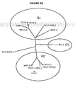
- the CDC has also published a phylogenetic tree of the predicted N, S and M proteins (attached as FIGURE 5 ). This tree places the SARS virus outside any of the previously known coronavirus groups.
- WO 2004/089983 discloses an isolated essentially mammalian positive-sense single stranded RNA virus (SARS) within the group of coronaviruses and components thereof.
- SARS single stranded RNA virus
- Accession Number AY323976 discloses the SARS coronavirus ZJ01 isolate spike glycoprotein.
- Ksiazek et al., 2003 discloses a new coronavirus associated with severe acute respiratory syndrome.
- the invention provides an immunogenic composition
- an isolated or purified polypeptide comprising: the Severe Acute Respiratory Syndrome (SARS) virus Spike polypeptide amino acid sequence SEQ ID NO: 6042, or an amino acid sequence having >80% sequence identity to SEQ ID NO: 6042, or a fragment of at least 10 consecutive amino acids of SEQ ID NO: 6042; with the proviso that the polypeptide is not MFIFLLFLTLTSGSDLDRCTTFDDVQAP (as disclosed in WO 2004/089983 ).
- SARS Severe Acute Respiratory Syndrome
- the invention also provides an immunogenic composition
- an isolated or purified polypeptide comprising amino acid sequence SEQ ID NO: 6042, or an amino acid sequence having >80% sequence identity to SEQ ID NO: 6042, or a fragment of at least 10 consecutive amino acids of SEQ ID NO: 6042; and further comprising an adjuvant. Further embodiments are defined in the appended claims.
- SARS virus has recently been identified as a new viral species.
- the SARS viral species includes the following isolates.
- a 646 base pair polynucleotide from the SARS virus has weak homology to viruses of the family Cornoaviridae.
- the Lancet article further reports that a deduced amino acid sequence (of 215 amino acids) from this sequence has about 57% sequence homology to the RNA polymerase of bovine coronavirus and murine hepatitis virus.
- Phylogenetic analysis of the protein sequences are also presented in the Lancet article showing that the polymerase sequence is most closely related to the group II coronaviruses.
- SARS viral isolates can be identified, isolated and/or sequenced by virologists skilled in the art. Virologists can readily identify new viral isolates as a SARS virus. Criteria which a virologist may use to identify new SARS isolates include: sequence homology of the new isolate to known SARS viral isolates; similar genomic organization of the new viral isolate to known SARS viral isolates; immunological (serologic) similarity or identity with known SARS viral isolates; pathology; and similarity of virion morphology with known SARS viral isolates; and similarity of infected cell morphology as that caused by known SARS viral isolates (visualized, for instance, by electron microscopy).
- New SARS isolates may also be identified by a percent homology of 99%, 95%, 92%, 90%, 85%, or 80% homology of the polynucleotide sequence for specific genomic regions for the new virus with the polynucleotide sequence for specific genomic regions of the known SARS viruses. Additionally, new SARS isolates may be identified by a percent homology of 99%, 95%, 92%, 90%, 85%, or 80% homology of the polypeptide sequence encoded by the polynucleotide of specific genomic regions of the new SARS virus to the polypeptide sequence encoded by the polynucleotides of specific regions of the known SARS virus.
- genomic regions may include regions (e.g ., gene products) which are typically in common among numerous coronaviruses, as well as group specific regions (e.g ., antigenic groups), such as, for example, any one of the following genomic regions which could be readily identified by a virologist skilled in the art: 5'untranslated region (UTR), leader sequence, ORF1a, ORF1b, nonstructural protein 2 (NS2), hemagglutinin-esterase glycoprotein (HE) (also referred to as E3), spike glycoprotein (S) (also referred to as E2), ORF3a, ORF3b, ORF3x, nonstructural protein 4 (NS4), envelope (small membrane) protein (E) (also referred to as sM), membrane glycoprotein (M) (also referred to as E1), ORF5a, ORF5b, nucleocapsid phosphoprotein (N), ORF7a, ORF7b, intergenic sequences, 3'UTR, or RNA dependent
- the SARS virus may have identifiable genomic regions with one or more the above-identified genomic regions.
- a SARS Viral antigen includes a protein encoded by any one of these genomic regions.
- a SARS viral antigen may be a protein or a fragment thereof, which is highly conserved With coronaviruses.
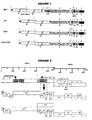
- a SARS viral antigen may be a protein or fragment thereof, which is specific to the SARS virus (as compared to known cornaviruses). (See, Figures 1-4 ).
- Electron microscopy of SARS infected cells are shown in the Lancet paper. As discussed in the paper, electron microscopy of negative stained (3% potassium phosphotungstate, pH 7.0) ultracentrifuged cell-culture extracts of SARS infected fetal rhesus kidney cells show the presence of pleomorphic enveloped virus particles of around 80-90 nm (range 70-130 nm) in diameter with surface morphology compatible with a coronavirus (see Lancet paper, Figure 1 ).
- Thin-section electron microscopy of infected cells reveals virus particles of 55-90 nm diameter within smooth walled vesicles in the cytoplasm (see Lancet paper, Figure 2B ). Electron microscopy can also be used to observe virus particles at the cell surface. Electron microscopy of a human lung biopsy sample depicts similar viral morphology. See Lancet paper Figure 2A .
- the invention relates to proteins from SARS virus. Such polypeptides are exemplified further below.
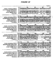
- the two Amino acid sequences within Figure 18 are SEQ ID NOS: 7188 & 7189.
- SEQ ID NO: 7188 contains an open reading frame comprising SEQ ID NO: 6042.
- the invention includes a polypeptide comprising SEQ ID NO: 6042.
- the invention includes a polypeptide comprising an amino acid sequence having sequence identity to SEQ ID NO: 6042.
- the invention includes a fragment of a polypeptide comprising SEQ ID NO: 6042.
- the invention includes an immunogenic composition comprising a polypeptide comprising SEQ ID NO: 6042, or a fragment thereof.
- SEQ ID NO: 6042 demonstrates functional homology to a coronavirus spike protein.
- Predicted transmembrane regions of SEQ ID NO: 6042 are identified below.
- SEQ ID NO: 6042 the spike protein
- SEQ ID NO: 6042 the spike protein
- Primers for amplifying the gene for spike protein and fragments thereof, such as fragments encoding the soluble ectodomain, include SEQ ID NOS: 9753-9763 (Xiao et al. (2003) Biochem Biophys Res Comm 312:1159-1164).
- SEQ ID NO: 6042 Further characterization of SEQ ID NO: 6042 is set forth below.
- SEQ ID NO: 6042 appears to have a N-terminus signaling region, followed by a surface exposed region, followed by a transmembrane region followed by a C-terminus cytoplasmic domain region.
- SEQ ID NOS: 7398 & 7399 Two oligopeptide fragments of SEQ ID NO: 6042 that are able to elicit anti-spike antibodies are SEQ ID NOS: 7398 & 7399, as described (with additional C-terminus cysteines) by Xiao et al. (2003) Biochem Biophys Res Comm 312:1159-1164 .
- C-terminal truncations of spike protein, with removal of part of the cytoplasmic region, or removal upto and including the transmembrane region, are described by Yang et al. (2004) Nature 428:561-564 .
- SEQ ID NO: 6042 is SEQ ID NO: 9962. Compared to SEQ ID NO: 6042, this sequence has Ser at residue 581 instead of Ala, and has Phe at residue 1152 instead of Leu.
- the spike protein of coronaviruses may be cleaved into two separate chains into S1 and S2.
- the chains may remain associated together to form a dimer or a trimer.
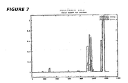
- the spike protein appears to form an alpha-helical structure in the transmembrane region of the protein, preferably in the S2 domain. This alpha-helical structure is thought to associate with at least two additional spike proteins to form a trimer. Helical or coiled regions of the spike protein are identified below. Predicted coiled-coils of SEQ ID NO: 6042 (spike protein) are at amino acids 900-1005 and 1151-1185 (see Figure 7 ).
- the spike protein is believed to play an integral role in fusion and infection of Coronaviruses with mammalian host cells. Analysis of coronavirus spike proteins as well as similar surface proteins in other viruses has identified at least two structural motifs, typically located within the S2 domain, associated with this fusion event: heptad repeats (HR) and membrane fusion peptides.
- HR heptad repeats
- At least two 4,3 hydrophobic heptad repeat (HR) domains are typically found in the ectodomain of the S2 domain of Coronaviruses.
- HR1 heptad repeat region
- HR2 heptad region
- Heptad repeats are characteristic of coiled-coil structures and the heptad repeats found in viral surface proteins (such as coronavirus spike protein) are thought to form bundled helix structures which are involved in viral entry. See Bosch et al., J. Virology (2003) 77:8801-8811 ( Figure 1B of this reference illustrates an alignment of the HR1 and HR2 regions of five coronaviruses along with SARS, annotated "HCov-SARS").
- Heptad repeats generally contain a repeating structure of seven amino acids, designated a-b-c-d-e-f-g , where hydrophobic sidechains of residues a and d typically form an apolar stripe, and electrostatic interactions are found in residues e and g .
- Position a is most frequently Leu, Ile or Ala and position d is usually Leu or Ala.
- Residues e and g are often Glu or Gln, with Arg and Lys also prominent at position g .
- Charged residues are common to positions b, c and f as these residues may be in contact with solvent. Exceptions to these general parameters are known. For instance Pro residues are sometimes found within the heptad.
- HR1 and HR2 sequences of an MHV strain have been postulated to assemble into a thermostable, oligomeric, alphahelical rold-like complex, with the HR1 and HR2 helices oriented in an antiparallel manner. Id .
- HR2 was asserted to be a strong inhitibor of both virus entry into the cell and cell-cell fusion.
- the SARS virus HR1 region comprises approximately amino acids 879 to 1005 of SEQ ID NO: 6042 or fragments thereof capable of forming at least one alpha-helical turn.
- said fragments comprise at least 7 (e.g. , at least 14, 21, 28, 35, 42, 49 or 56) amino acid residues.
- a preferred fragment of HR1 comprises amino acid residues 879 to 980 of SEQ ID NO: 6042. This preferred fragment is SEQ ID NO: 7193.
- Another preferred fragment of HR1 comprises amino acid residues 901 to 1005 of SEQ ID NO: 6042. This preferred fragment is SEQ ID NO: 7194.
- the SARS virus HR2 region comprises approximately amino acids 1144 to 1201 of SEQ ID NO: 6042, or fragments thereof capable of forming at least one alpha-helical turn.
- said fragments comprise at least 7 ( e.g. , at least 14, 21, 28, 35, 42, 49 or 56) amino acid residues.
- a preferred fragment of HR2 comprises amino acids 1144 to 1195 of SEQ ID NO: 6042. This preferred fragment is SEQ ID NO: 7196.
- Membrane Fusion peptides sequences within the spike protein are also believed to participate in fusion (and infection) of the virus with a host cell. Fusion peptides generally comprise about 16 to 26 amino acid residues which are conserved within viral families. These Membrane Fusion peptides are relatively hydrophobic and generally show an asymmetric distribution of hydrophobitiy when modeled into an alpha helix. They are also generally rich in alanine and glycine.
- PEP1 SEQ ID NO: 7197
- PEP2 SEQ ID NO: 7198
- PEP3 SEQ ID NO: 7199
- coronavirus spike proteins (and other similar surface viral proteins) are thought to undergo a conformational change upon receptor binding to the target cell membrane.
- One or more of the hydrophobic Membrane Fusion peptides are thought to become exposed and inserted into the target membrane as a result of this conformational change.
- the free energy released upon subsequent refolding of the spike protein to its most stable conformation is believed to play a role in the merger of the viral and cellular membranes.
- One or more SARS HR sequences may be used to inhibit viral entry and membrane fusion with a target mammalian host cell.
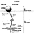
- Applicants have identified structural similarities between the SARS spike protein and the surface protein of Neisseria meningitidis , NadA (and other similar bacterial adhesion proteins).
- Another means of facilitating bacterial expression of HR sequences includes the addition of the stalk and/or anchor sequences of a NadA-like protein to the C-terminus of the recombinant HR sequences.
- Recombinant sequences containing the bacterial anchor sequence may preferably be prepared in outer membrane vesicles (the preparation of which is discussed in more detail later in the application). Recombinant sequences missing the bacterial anchor sequences may be secreted and isolated from the supernatant.
- FIGURE 42 Examples of constructs which may be used in bacterial expression systems are shown in FIGURE 42 .
- Administration of one of more of these Membrane Fusion sequences may also interfere with the ability of a coronavirus to fuse to a host cell membrane.
- Two or more of the SARS Membrane Fusion peptides may be linked together.
- the spike protein may be recombinantly produced.
- the alpha-helical portion of the spike protein is associated with the cell membrane.
- the spike proteins form a trimer within the transmembrane region of attachment.
- Predicted N-glycosylation sites of SEQ ID NO: 6042 are identified below: Position Potential Jury agreement NGlyc result 29 NYTQ SEQ ID NO: 7207 0.7751 (9/9) +++ 65 NVTG SEQ ID NO: 7208 0.8090 (9/9) +++ 109 NKSQ SEQ ID NO: 7209 0.6081 (7/9) + 119 NSTN SEQ ID NO: 7210 0.7039 (9/9) ++ 158 NCTF SEQ ID NO: 7211 0.5808 (7/9) + 227 NITN SEQ ID NO: 7212 0.7518 (9/9) +++ 269 NGTI SEQ ID NO: 7213 0.6910 (9/9) ++ 318 NITN SEQ ID NO: 7214 0.6414 (9/9) ++ 330 NATK SEQ ID NO: 7215 0.6063 (8/9) + 357 NSTF SEQ ID NO: 7216 0.5746 (8/9) + 589 NASS SEQ ID NO: 7217 0.5778 (6/9) + 602 NCTD SEQ ID NO: 72
- Predicted O-glycosylation sites are identified below: Residue No. Potential Threshold Assignment Thr 698 0.8922 0.7696 T Thr 706 0.9598 0.7870 T Thr 922 0.9141 0.7338 T Ser 36 0.8906 0.7264 S Ser 703 0.8412 0.7676 S
- Predicted phosphorylation sites of SEQ ID NO: 6042 are Ser-346, Tyr-195, and Tyr-723.
- polypeptide generally refer to a polymer of amino acid residues and are not limited to a minimum length of the product.
- peptides, oligopeptides,:dimers, mulimers, and the like are included within the definition. Both full-length proteins and fragments thereof are encompassed by the definition.
- Polypeptides of the invention can be prepared in many ways e.g. by chemical synthesis (at least in part), by digesting longer polypeptides using proteases, by translation from RNA, by purification from cell culture ( e.g. from recombinant expression), from the organism itself ( e.g. after viral culture, or direct from patients), from a cell line source etc.
- a preferred method for production of peptides ⁇ 40 amino acids long involves in vitro chemical synthesis ( Bodanszky (1993) Principles of Peptide Synthesis (ISBN: 0387564314 ); Fields et al. (1997) Methods in Enzymology 289: Solid-Phase Peptide Synthesis. ISBN: 0121821900 ).
- Solid-phase peptide synthesis is particularly preferred, such as methods based on t-Boc or Fmoc ( Chan & White (2000) Fmoc Solid Phase Peptide Synthesis ISBN: 0199637245 ) chemistry. Enzymatic synthesis ( Kullmann (1987) Enzymatic Peptide Synthesis. ISBN: 0849368413 ) may also be used in part or in full.
- biological synthesis may be used e.g. the polypeptides may be produced by translation. This may be carried out in vitro or in vivo.
- Biological methods are in general restricted to the production of polypeptides based on L-amino acids, but manipulation of translation machinery ( e.g.
- aminoacyl tRNA molecules can be used to allow the introduction of D-amino acids (or of other non natural amino acids, such as iodotyrosine or methylphenylalanine, azidohomoalanine, etc. ) ( Ibba (1996) Biotechnol Genet Eng Rev 13:197-216 .). Where D-amino acids are included, however, it is preferred to use chemical synthesis.
- Polypeptides of the disclosure may have covalent modifications at the C-terminus and/or N-terminus, particularly where they are for in vivo administration e.g by attachment of acetyl or carboxamide, as in the FuzeonTM product.
- Reference to polypeptides and the like also includes derivatives of the amino acid sequences of the disclosure.
- Such derivatives can include postexpression modifications of the polypeptide, for example, glycosylation, acetylation, phosphorylation, and the like.
- Amino acid derivatives can also include modifications to the native sequence, such as deletions, additions and substitutions (generally conservative in nature), so long as the protein maintains the desired activity. These modifications may be deliberate, as through site-directed mutagenesis, or may be accidental, such as through mutations of hosts which produce the proteins or errors due to PCR amplification.
- modifications may be made that have one or more of the following effects: reducing toxicity; facilitating cell processing ( e.g. , secretion; antigen presentation, etc. ); and facilitating presentation to B-cells and/or T-cells.
- Fram or “Portion” as used herein refers to a polypeptide consisting of only a part of the intact full-length polypeptide sequence and structure as found in nature. For instance, a fragment can include a C-terminal deletion and/or an N-terminal deletion of a protein.
- a "recombinant" protein is a protein which has been prepared by recombinant DNA techniques as described herein.
- the gene of interest is cloned and then expressed in transformed organisms, as described further below.
- the host organism expressed the foreign gene to produce the protein under expression conditions.
- isolated is meant, when referring to a polypeptide, that the indicated molecule is separate and discrete from the whole organism with which the molecule is found in nature or, when the polypeptide is not found in nature, is sufficiently free of other biological macromolecules so that the polypeptide can be used for its intended purpose.
- the polypeptides of the invention are preferably isolated polypeptides.
- an "immunogenic composition” as used herein refers to a composition that comprises an antigenic molecule where administration of the composition to a subject results in the development in the subject of a humoral and/or a cellular immune response to the antigenic molecule of interest.
- the immunogenic composition can be introduced directly into a recipient subject, such as by injection, inhalation, oral, intranasal or any other parenteral, mucosal or transdermal ( e.g. , intra-rectally or intra-vaginally) route of administration.
- derived from is used to identify the source of molecule (e.g. , a molecule can be derived from a polynucleotide, polypeptide, an immortalized cell line can be derived from any tissue, etc. ).
- a first polypeptide is "derived from” a second polypeptide if it is (i) encoded by a first polynucleotide derived from a second polynucleotide, or (ii) displays sequence identity to the second polypeptides as described above.
- a polypeptide (protein) is "derived from” a particular SARS virus if it is (i) encoded by an open reading frame of a polynucleotide of that SARS virus, or (ii) displays sequence identity, as described above, to polypeptides of that SARS virus.
- Both polynucleotide and polypeptide molecules can be physically derived from a SARS virus or produced recombinantly or synthetically, for example, based on known sequences.
- the immunogenic compositions of the invention may further comprise one or more adjuvants.
- the immunogenic compositions of the invention may be administered mucosally. Suitable routes of mucosal administration include oral, intranasal, intragastric, pulmonary, intestinal, rectal, ocular and vaginal routes.
- the immunogenic composition may be adapted for mucosal administration. For instance, where the composition is for oral administration, it may be in the form of tablets or capsules, optionally enteric-coated, liquid, transgenic plants, etc. Where the composition is for intranasal administration, it may be in the form of a nasal spray, nasal drops, gel or powder.
- the immunogenic compositions of the disclosure may be administered parenterally. Suitable routes of parenteral administration include intramuscular (IM), subcutaneous, intravenous, intraperitoneal, intradermal, transcutaneous, and transdermal (see e.g. , International patent application WO 98/20734 ) routes, as well as delivery to the interstitial space of a tissue.
- IM intramuscular
- subcutaneous intravenous
- intraperitoneal intradermal
- transcutaneous transcutaneous
- transdermal see e.g. , International patent application WO 98/20734
- the immunogenic composition may be adapted for parenteral administration, for instance in the form of an injectable that may be sterile and pyrogen free.
- compositions may be administered in conjunction with other immunoregulatory agents.
- compositions will usually include an adjuvant.
- Preferred further adjuvants include, but are not limited to, one or more of the following set forth below:
- Mineral containing compositions suitable for use as adjuvants in the disclosure include mineral salts, such as aluminium salts.and calcium salts.
- the disclosure includes mineral salts such as hydroxides (e.g. oxyhydroxides), phosphates ( e.g. hydroxyphoshpates, orthophosphates), sulphates, etc. ( e.g. see chapters 8 & 9 of Vaccine design:the subunit and adjuvant approach (1995) Powell & Newman. ISBN 0-306-44867-X .), or mixtures of different mineral compounds, with the compounds taking any suitable form ( e.g. gel, crystalline, amorphous, etc. ), and with adsorption being preferred.
- the mineral containing compositions may also be formulated as a particle of metal salt. See WO00/23105 .
- Oil-emulsion compositions suitable for use as adjuvants in the disclosure include squalene-water emulsions, such as MF59 (5% Squalene, 0.5% Tween 80, and 0.5% Span 85, formulated into submicron particles using a microfluidizer). See WO90/14837 . See also, Frey et al., "Comparison of the safety, tolerability, and immunogenicity of a MF59-adjuvanted influenza vaccine and a non-adjuvanted influenza vaccine in non-elderly adults", Vaccine (2003) 21:4234-4237 .
- Particularly preferred adjuvants for use in the compositions are submicron oil-inwater emulsions.
- Preferred submicron oil-in-water emulsions for use herein are squalene/water emulsions optionally containing varying amounts of MTP-PE, such as a submicron oil-in-water emulsion containing 4-5% w/v squalene, 0.25-1.0% w/v Tween 80TM (polyoxyelthylenesorbitan monooleate), and/or 0.25-1.0% Span 85TM (sorbitan trioleate), and, optionally, N-acetylmuramyl-L-alanyl-D-isogluatminyl-L-alanine-2-(1'-2'-dipalmitoyl- sn -glycero-3-huydroxyphosphophoryloxy)-ethylamine (MTP-PE), for example, the submicron oil-in-water
- MTP-PE may be present in an amount of about 0-500 ng/dose, more preferably 0-250 ⁇ g/dose and most preferably, 0-100 ⁇ g/dose.
- MF59-0 refers to the above submicron oil-in-water emulsion lacking MTP-PE, while the term MF59-MTP denotes a formulation that contains MTP-PE.
- MF59-100 contains 100 ⁇ g MTP-PE per dose, and so on.
- MF69 another submicron oil-in-water emulsion for use herein, contains 4.3% w/v squalene, 0.25% w/v Tween 80TM, and 0.75% w/v Span 85TM and optionally MTP-PE.
- MF75 also known as SAF, containing 10% squalene, 0.4% Tween 80TM, 5% pluronic-blocked polymer L121, and thr-MDP, also microfluidized into a submicron emulsion.
- MF75-MTP denotes an MF75 formulation that includes MTP, such as from 100-400 ⁇ g MTP-PE per dose.
- Submicron oil-in-water emulsions methods of making the same and immunostimulating agents, such as muramyl peptides, for use in the compositions, are described in detail in International Publication No. WO 90114837 and US Patent Nos. 6,299,884 and 6,45 1,325 .
- CFA Complete Freund's adjuvant
- IFA incomplete Freund's adjuvant
- Saponin formulations may also be used as adjuvants in the disclosure.
- Saponins are a heterologous group of sterol glycosides and triterpenoid glycosides that are found in the bark, leaves, stems, roots and even flowers of a wide range of plant species. Saponin from the bark of the Quillaia saponaria Molina tree have been widely studied as adjuvants. Saponin can also be commercially obtained from Smilax ornata (sarsaprilla), Gypsophilla paniculata (brides veil), and Saponaria officianalis (soap root).
- Saponin adjuvant formulations include purified formulations, such as QS21, as well as lipid formulations, such as ISCOMs.
- Saponin compositions have been purified using High Performance Thin Layer Chromatography (HP-LC) and Reversed Phase High Performance Liquid Chromatography (RP-HPLC). Specific purified fractions using these techniques have been identified, including QS7, QS17, QS18, QS21, QH-A, QH-B and QH-C.
- the saponin is QS21.
- a method of production of QS21 is disclosed in US Patent No. 5,057,540 .
- Saponin formulations may also comprise a sterol, such as cholesterol (see WO 96/33739 ).
- ISCOMs Immunostimulating Complexs
- phospholipid such as phosphatidylethanolamine or phosphatidylcholine.
- Any known saponin can be used in ISCOMs.
- the ISCOM includes one or more of Quil A, QHA and QHC.
- ISCOMS may be devoid of additional detergent. See WO00/07621 .
- Adjuvants suitable for use in the disclosure include bacterial or microbial derivatives such as:
- Such derivatives include Monophosphoryl lipid A (MPL) and 3-O-deacylated MPL (3dMPL).
- 3dMPL is a mixture of 3 De-O-acylated monophosphoryl lipid A with 4, 5 or 6 acylated chains.
- a preferred "small particle" form of 3 De-O-acylated monophosphoryl lipid A is disclosed in EP 0 689 454 .
- Such "small particles" of 3dMPL are small enough to be sterile filtered through a 0.22 micron membrane (see EP 0 689 454 ).
- Other non-toxic LPS derivatives include monophosphoryl lipid A mimics, such as aminoalkyl glucosaminide phosphate derivatives e.g. RC-529. See Johnson et al. (1999) Bioorg Med Chem Lett 9:2273-2278 .
- Lipid A derivatives include derivatives of lipid A from Escherichia coli such as OM-174.
- OM-174 is described for example in Meraldi et al., "OM-174, a New Adjuvant with a Potential for Human Use, Induces a Protective Response with Administered with the Synthetic C-Terminal Fragment 242-310 from the circumsporozoite protein of Plasmodium berghei", Vaccine (2003) 21:2485-2491 ; and Pajak, et al., "The Adjuvant OM-174 induces both the migration and maturation of murine dendritic cells in vivo", Vaccine (2003) 21:836-842 .
- Immunostimulatory oligonucleotides suitable for use as adjuvants in the disclosure include nucleotide sequences containing a CpG motif (a sequence containing an unmethylated cytosine followed by guanosine and linked by a phosphate bond). Bacterial double stranded RNA or oligonucleotides containing palindromic or poly(dG) sequences have also been shown to be immunostimulatory.
- the CpG's can include nucleotide modifications/analogs such as phosphorothioate modifications and can be double-stranded or single-stranded.
- the guanosine may be replaced with an analog such as 2'-deoxy-7-deazaguanosine. See Kandimalla, et al., "Divergent synthetic nucleotide motif recognition pattern: design and development of potent immunomodulatory oligodeoxyribonucleotide agents with distinct cytokine induction profiles", Nucleic Acids Research (2003) 31(9): 2393-2400 ; WO 02/26757 and WO 99/62923 for examples of possible analog substitutions.
- CpG oligonucleotides The adjuvant effect of CpG oligonucleotides is further discussed in Krieg, "CpG motifs: the active ingredient in bacterial extracts?", Nature Medicine (2003) 9(7): 831-835 ; McCluskie, et al., "Parenteral and mucosal prime-boost immunization strategies in mice with hepatitis B surface antigen and CpG DNA", FEMS Immunology and Medical Microbiology (2002) 32:179-185 ; WO 98/40100 ; US Patent No. 6,207,646 ; US Patent No. 6,239,116 and US Patent No. 6,429,199 .
- the CpG sequence may be directed to TLR9, such as the motif GTCGTT or TTCGTT. See Kandimalla, et al., "Toll-like receptor 9: modulation of recognition and cytokine induction by novel synthetic CpG DNAs", Biochemical Society Transactions (2003) 31 (part 3): 654-658 .
- the CpG sequence may be specific for inducing a Th1 immune response, such as a CpG-A ODN, or it may be more specific for inducing a B cell response, such a CpG-B ODN.
- CpG-A and CpG-B ODNs are discussed in Blackwell, et al., "CpG-A-Induced Monocyte IFN-gamma-Inducible Protein-10 Production is Regulated by Plasmacytoid Dendritic Cell Derived IFN-alpha", J. Immunol. (2003) 170(8):4061-4068 ; Krieg, “From A to Z on CpG”, TRENDS in Immunology (2002) 23(2): 64-65 and WO 01/95935 .
- the CpG is a CpG-A ODN.
- the CpG oligonucleotide is constructed so that the 5' end is accessible for receptor recognition.
- two CpG oligonucleotide sequences may be attached at their 3' ends to form "immunomers".
- Kandimalla "Secondary structures in CpG oligonucleotides affect immunostimulatory activity" BBRC (2003) 306:948-953 ; Kandimalla, et al., "Toll-like receptor 9: modulation of recognition and cytokine induction by novel synthetic GpG DNAs", Biochemical Society Transactions (2003) 31(part 3):664-658 ; Bhagat et al., "CpG penta- and hexadeoxyribonucleotides as potent immunomodulatory agents” BBRC (2003) 300:853-861 and WO 03/035836 .
- Bacterial ADP-ribosylating toxins and detoxified derivatives thereof may be used as adjuvants in the disclosure
- the protein is derived from E. coli (i.e. , E. coli heat labile enterotoxin "LT), cholera ("CT"), or pertussis ("PT").
- LT E. coli heat labile enterotoxin
- CT cholera
- PT pertussis
- the use of detoxified ADP-ribosylating toxins as mucosal adjuvants is described in WO 95/17211 and as parenteral adjuvants in WO 98/42375 .
- the adjuvant is a detoxified LT mutant such as LT-K63, LT-R72, and LTR192G.
- ADP-ribosylating toxins and detoxified derivaties thereof, particularly LT-K63 and LT-R72, as adjuvants can be found in the following references.
- Beignon, et al. "The LTR72 Mutant of Heat-Labile Enterotoxin of Escherichia coli Enahnces the Ability of Peptide Antigens to Elicit CD4+ T Cells and Secrete Gamma Interferon after Coapplication onto Bare Skin", Infection and Immunity (2002) 70(6):3012-3019 ; Pizza, et al., "Mucosal vaccines: non-toxic derivatives of LT and CT as mucosal adjuvants", Vaccine (2001) 19:2534-2541 ; Pizza, et al., "LTK63 and LTR72, two mucosal adjuvants ready for clinical trials" Int.
- Numerical reference for amino acid substitutions is preferably based on the alignments of the A and B subunits of ADP-ribosylating toxins set forth in Domenighini et al., Mol. Microbiol (1995) 15(6):1165-1167 .
- Human immunomodulators suitable for use as adjuvants in the disclosure include cytokines, such as interleukins (e.g. IL-1, IL-2, IL-4, IL-5, IL-6, IL-7, IL-12, etc. ), interferons (e.g. interferon- ⁇ ), macrophage colony stimulating factor, and tumor necrosis factor.
- cytokines such as interleukins (e.g. IL-1, IL-2, IL-4, IL-5, IL-6, IL-7, IL-12, etc. ), interferons (e.g. interferon- ⁇ ), macrophage colony stimulating factor, and tumor necrosis factor.
- Bioadhesives and mucoadhesives may also be used as adjuvants in the disclosure.
- Suitable bioadhesives include esterified hyaluronic acid microspheres ( Singh et al. (2001) J. Cont. Rele. 70:267-276 ) or mucoadhesives such as cross-linked derivatives of poly(acrylic acid), polyvinyl alcohol, polyvinyl pyrollidone, polysaccharides and carboxymethylcellulose. Chitosan and derivatives thereof may also be used as adjuvants in the disclosure. E.g. , WO99/27960 .
- Microparticles may also be used as adjuvants in the disclosure.
- Microparticles i.e. a particle of ⁇ 100nm to ⁇ 150 ⁇ m in diameter, more preferably ⁇ 200nm to ⁇ 30 ⁇ m in diameter, and most preferably ⁇ 500nm to ⁇ 10 ⁇ m in diameter
- materials that are biodegradable and non-toxic e.g. a poly( ⁇ -hydroxy acid), a polyhydroxybutyric acid, a polyorthoester, a polyanhydride, a polycaprolactone, etc.
- a negatively-charged surface e.g. with SDS
- a positively-charged surface e.g a cationic detergent, such as CTAB
- liposome formulations suitable for use as adjuvants are described in US Patent No. 6,090,406 , US Patent No. 5,916,588 , and EP 0 626 169 .
- Adjuvants suitable for use in the disclosure include polyoxyethylene ethers and polyoxyethylene esters. WO99/52549 . Such formulations further include polyoxyethylene sorbitan ester surfactants in combination with an octoxynol ( WO01/21207 ) as well as polyoxyethylene alkyl ethers or ester surfactants in combination with at least one additional non-ionic surfactant such as an octoxynol ( WO01/21152 ).
- Preferred polyoxyethylene ethers are selected from the following group: polyoxyethylene-9-lauryl ether (laureth 9), polyoxyethylene-9-steoryl ether, polyoxytheylene-8-steoryl ether, polyoxyethylene-4-lauryl ether, polyoxyethylene-35-lauryl ether, and polyoxyethylene-23-lauryl ether.
- PCPP J. Polyphosphazene
- PCPP formulations are described, for example, in Andrianov et al., "Preparation of hydrogel microspheres by coacervation of aqueous polyphophazene solutions", Biomaterials (1998) 19(1-3):109-115 and Payne et al., “Protein Release from Polyphosphazene Matrices", Adv. Drug. Delivery Review (1998) 31(3):185-196 .
- muramyl peptides suitable for use as adjuvants in the disclosure include N-acetyl-muramyl-L-threonyl-D-isoglutamine (thr-MDP), N-acetyl-normuramyl-L-alanyl-D-isoglutamine (nor-MDP), and N-acetylmuramyl-L-alanyl-D-isoglutaminyl-L-alanine-2-(1'-2'-dipalmitoyl- sn -glycero-3-hydroxyphosphoryloxy)-ethylamine MTP-PE).
- thr-MDP N-acetyl-muramyl-L-threonyl-D-isoglutamine
- nor-MDP N-acetyl-normuramyl-L-alanyl-D-isoglutamine
- imidazoquinolone compounds suitable for use adjuvants in the disclosure include Imiquamod and its homologues, described further in Stanley, “Imiquimod and the imidazoquinolones: mechanism of action and therapeutic potential” Clin Exp Dermatol (2002) 27(7):571-577 and Jones, “Resiquimod 3M", Curr Opin Investig Drugs (2003) 4(2):214-218 .
- VLPs Virosomes and Virus Like Particles
- Virosomes and Virus Like Particles can also be used as adjuvants in the disclosure
- These structures generally contain one or more proteins from a virus optionally combined or formulated with a phospholipid. They are generally non-pathogenic, non-replicating and generally do not contain any of the native viral genome. The viral proteins may be recombinantly produced or isolated from whole viruses.
- viral proteins suitable for use in virosomes or VLPs include proteins derived from influenza virus (such as HA or NA), Hepatitis B virus (such as core or capsid proteins), Hepatitis E virus, measles virus, Sindbis virus, Rotavirus, Foot-and-Mouth Disease virus, Retrovirus, Norwalk virus, human Papilloma virus, HIV, RNA-phages, Qß-phage (such as coat proteins), GA-phage, fr-phage, AP205 phage, and Ty (such as retrotransposon Ty protein p1).
- influenza virus such as HA or NA
- Hepatitis B virus such as core or capsid proteins
- Hepatitis E virus measles virus
- Sindbis virus Rotavirus
- Foot-and-Mouth Disease virus Retrovirus
- Norwalk virus Norwalk virus
- human Papilloma virus HIV
- RNA-phages Qß-phage (such as coat proteins)
- GA-phage f-phage
- VLPs are discussed further in WO 03/024480 , WO 03/024481 , and Niikura et al., "Chimeric Recombinant Hepatitis E Virus-Like Particles as an Oral Vaccine Vehicle Presenting Foreign Epitopes", Virology (2002) 293:273-280 ; Lenz et al., “Papillomarivurs-Like Particles Induce Acute Activation of Dendritic Cells", Journal of Immunology (2001) 5246-5355 ; Pinto, et al., “Cellular Immune Responses to Human Papillomavirus (HPV)-16 L1 Healthy Volunteers Immunized with Recombinant HPV-16 L1 Virus-Like Particles", Journal of Infectious Diseases (2003) 188:327-338 ; and Gerber et al., "Human Papillomavrisu Virus-Like Particles Are Efficient Oral Immunogens when Coa
- the disclosure may also comprise combinations of aspects of one or more of the adjuvants identified above.
- adjuvant compositions may be used in the disclosure:
- Aluminium salts and MF59 are preferred adjuvants for parenteral immunisation. Mutant bacterial toxins are preferred mucosal adjuvants.
- adjuvants suitable for use in the disclosure may also include one or more of the following:
- adjuvants suitable for mucosal or parenteral administration are also available (e.g. see chapter 7 of Vaccine design: the subunit and adjuvant aproach, eds. Powell & Newman, Plenum Press 1995 (ISBN 0-306-44867-X ).
- Mutants of LT are preferred mucosal adjuvants, in particular the "K63” and “R72” mutants ( e.g. see International patent application WO 98/18928 ), as these result in an enhanced immune response.
- Microparticles are also preferred mucosal adjuvants. These are preferably derived from a poly( ⁇ -hydroxy acid), in particular, from a poly(lactide) (“PLA”), a copolymer of D,L-lactide and glycolide or glycolic acid, such as a poly(D,L-lactide-co-glycolide) (“PLG” or "PLGA”), or a copolymer of D,L-lactide and caprolactone.
- PLA poly(lactide)
- PLA poly(lactide)
- PLA poly(D,L-lactide-co-glycolide)
- PAG poly(D,L-lactide-co-glycolide)
- caprolactone a copolymer of D,L-lactide and caprolactone.
- microparticles may be derived from any of various polymeric starting materials which have a variety of molecular weights and, in the case of the copolymers such as PLG, a variety of lactide:glycolide ratios, the selection of which will be largely a matter of choice, depending in part on the coadministered antigen.
- a SARS virus was isolated from clinical specimens of a patient in Frankfurt, Germany (FRA). The isolate was grown in Vero cells. RNA of the SARS virus was extracted and amplified by RT-PCR. Nucleotide sequence of the viral genome was determined by direct sequencing of the PCR product. Computer analysis was used to predict the features of the genome, to compare it to previously known coronaviruses and to the sequence of different SARS virus isolates.
- isolation and sequence was performed as follows. After the third passage of the SARS virus in Vero cells, viral particles were purified by ultra centrifugation from 3x10 7 cells supernatant. Viral RNA was extracted by Triazol method (Gibco-BRL). Viral RNA (200 ng) was transcribed into cDNA with avian RNaseH- thermostable reverse transcriptase following the instructions of the manufacturer (ThermoScript RT System, Invitrogen). Briefly, either 50 pmoles of oligo (dT) 20 or 25 ng of random hexamers were used to prime the RT reaction in a 20 ⁇ l final volume.
- Amplification and sequencing of the SARS genome were accomplished by direct sequencing of PCR products obtained with: i) specific primers from conserved regions of homology found through multiple alignment among known coronaviruses; ii) oligonucleotides designed around short sequences of SARS isolates available on the Web through WHO network laboratories; iii) degenerate primers to amplify the cDNA mixture with multiple overlapping fragments as end products. Gap closure was realized by long distance PCR with high fidelity Taq (Expand High Fidelity system, Roche) using primers designed on selected fragments. Sequence was collected by primer walking using a BigDye terminator chemistry (Applied Biosystems) and an automated DNA sequencer (3700 capillary model, Applied Biosystems).
- the LearnCoil-VMF program was used to predict coiled-coil regions in the spike proteins ( http: // learncoil-vmf.lcs.mit.edu / cgi-bin / vmf ). Leucine zippers were predicted with the program 2ZIP, available at http: // 2Zip.molgen.mpg.de.
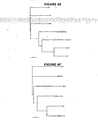
- Phylogenetic analysis was performed using the neighbor-joining algorithm as implemented in the program NEIGHBOR within the Phylogeny Inference Package (Phylip) (Felsenstein J 1993, program distributed by the author). Bootstrap analysis was always performed with 100 replicates using the program Seqboot. Trees were handled and displayed using TreeView. The program HMMER was used to generate sequence profiles from multiple sequence alignments of the S1 domains of spike proteins. Subsequently, the HMMPFAM program was used to compare the S1 domain of SARS spike to the profiles.
- the genome of this SARS virus isolate is 29,740 bases long and the overall structure of the genome is similar to that of the three known groups of coronaviruses. Starting from the 5' end a leader sequence, an untranslated region (UTR) and two overlapping open reading frames coding for one polyprotein containing the enzymes necessary for replication can be identified. They are followed by a region coding for the spike (S), envelope (E), matrix (M), nucleocapsid (N) structural proteins and eight additional ORFs specific for the SARS virus. At the 3'-end of the genome a UTR with a poly(A) is located.
- coronaviruses groups 1, 2 and 3 The overall homology to coronaviruses groups 1, 2 and 3 is low and therefore the SARS virus belongs to a new group (group 4) of coronavirus. More detailed analysis of the spike protein amino acid sequence shows that the SARS virus isolate is more closely related to coronavirus group 2.
- the complete genome sequence of the SARS virus isolate is 29,740 bp in length.
- the sequence is available on Genbank and has a GC content of 40.8%, comparable with that of known viruses of the same family.
- Genome structure is similar to that of other coronaviruses. 14 open reading frames have been predicted.
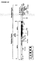
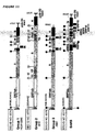
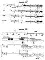
- the principal features of the genome and gene products are illustrated reported in Figure 10 and Table 10. The comparison between the SARS genome and those of group 1,2 and 3 coronaviruses is reported in Figure 11 .
- Nucleotides 1-73 contain a predicted RNA leader sequence followed by an untranslated region (UTR) of 197 nucleotides.
- the UTR is followed by two overlapping open reading frames (ORF1a, ORF1b), which encompass two-thirds of the genome (nucleotides 265-21485). They encode for a large polyprotein, which is predicted to be processed by viral proteases to generate the replicase complex.
- the 3' part of the.genome contains the genes coding for the four structural proteins (S, spike protein, E, envelope protein, M, matrix glycoprotein, and N, nucleocapsid protein), and eight predicted ORFs of unknown function ( Figure 10 ).
- IG sequence also referred to as transcription-associated sequence (TAS), which is a typical feature for coronaviruses.
- TAS transcription-associated sequence
- the IG sequence is characterized by 6-18 nucleotides present at the 3' end of the leader and can be found in front of each gene.
- the IG sequence plays a key role in RNA transcription and its regulation.
- the IG sequence of the SARS virus is present nine times in the genome ( Figure 10 ). The sequence of the leader and IG are peculiar for each coronavirus and represent a specific signature for the virus.
- the spike is a type I glycoprotein, which forms the large spikes on the surface of the virion and is responsible for receptor-binding and membrane fusion. ( Gallagher (2001) Adv Exp Med Biol 494: 183-92 ).
- the protein is 1255 residues long with 17 predicted N-glycosylation sites. It has a 13aa leader peptide and a 17 aa C-terminal membrane anchoring sequence (1202-1218).
- S1 is supposed to form the bulbous head, which stays non-covalently linked to the C-terminal membrane anchor. Cleavage is mediated by a basic aminoacid sequence, which resembles the consensus sequence for a furin cleavage site. ( Klein et.al., Biochimie 1994; 76(3-4): 217-225 ).
- Both coiled-coil regions contain a leucine-zipper motif, which is also present in the spikes of all coronaviruses.
- Leucine zippers are known to promote protein oligomerization; since the spike proteins of TGV and MHV form hetero-trimers, ( Delmas et al, J Virol 1990; 64(11):5367-5375 ) ( Godeke, et al., J Virology 2000; 74(3):1566-1571 ) it is conceivable that in SARS leucine zippers play a role in promoting and/or stabilizing a similar quaternary structure.
- the spike protein plays a major role in the biology of coronaviruses because the S1 domain contains the receptor-binding domain and the virus neutralizing epitopes, while the S2 domain is involved in the process of membrane fusion, which is essential for virus infectivity. As expected, multiple sequence alignment of different spike proteins showed a major degree of variability within the S1 domain, whereas S2 is more conserved.
- the envelope protein E is a very short polypeptide of 76 aa, involved in the morphogenesis of the virion envelope. ( Godet et al., Virology 1992; 188(2):666-675 ). Computer analysis predicts a long transmembrane domain close to the N-terminus and two N-glycosylation sites. The level of aminoacid similarity with other coronaviruses is very low and the best homology is with the small envelope protein of the transmissible gastroenteritis virus (TGV).
- TSV transmissible gastroenteritis virus
- the matrix glycoprotein (M) is a 221-residue polypeptide with a predicted molecular weight of 25 kDa.
- Computer analysis predicts a topology consisting of a short aminoterminal ectodomain, three transmembrane segments and a carboxyl terminus located at the interior side of the viral envelope.
- the SARS M glycoprotein is N-glycosylated at the N-terminus.
- SARS M protein shows highest similarity to group 2 viruses (Table 11).
- nucleocapsid protein N is a 397-residue-long phosphoprotein that interacts with viral genomic RNA to form the nucleocapsid.
- the level of conservation with other coronaviruses is low, ranging from 26,9% identity with the HCoV-229E to 37,4% identity to the Bovine coronavirus (BcoV) (Table 11).
- BcoV Bovine coronavirus
- viruses express a set of other peptides, which are generally dispensable for viability, but can influence the infectivity potential of the virus. ( de Haan et al., Virology 2002; 296(1):177-189 ). These proteins are generally conserved within members of the same serogroup, but differ profoundly among the groups. For this reason, they are generally referred to as group-specific proteins.
- Members of the group 1, represented here by HcoV-229E have two group-specific genes located between the S and E genes and sometimes one or two ORFs downstream of the N gene, preceding the 3' UTR region of the genome.
- Viruses of the group 2 have two group-specific genes (2a and HE) between ORFlb and S, as well as other two between S and E genes. Finally, the group 3 viruses, represented by the prototype AIBV, have two group-specific genes between S and E and other two between the M and N genes.
- SARS lacks the HE hemagglutinin and does not contain ORFs between the ORFlb and the S gene.
- ORF3 and ORF4 are encoded in the region between S and E, and superimpose for most of their length.
- ORF3 has an IG sequence 2 bp upstream of the ATG start codon.
- SARS contains five predicted ORFs in the region between M and N genes.
- ORF7 is located 10 bases downstream of the stop codon of M gene, and has an IG sequence 155 nucleotides upstream from the ATG start codon.
- ORF8 and ORF10 present an 10 right upstream of their ATG start codons.
- ORF9 and ORF11 shortly superimpose with the flanking genes, and for this reason they do not need an IG to activate transcription.
- ORF12 totally superimposes with the N gene and shares very low homology with a 22kDa protein of the MHV virus, coded in the corresponding region.
- ORF3, ORF7 and ORF8 contain hydrophobic segments, suggesting association with membrane structures.
- ORF3 the longest among the SARS specific gene, is the only one that encodes for a peptide containing a high number of predicted O-glycosylation sites (Table 11). Predicted N-glycosylation sites have been identified in ORF3, ORF11 and ORF12.
- FRA and TOR2 The overall difference between FRA and TOR2 was of nine nucleotides resulting in two silent mutations and seven aminoacid changes.
- the difference between FRA and Urbani is 12 nucleotides, which result in five silent mutations and seven aminoacid changes.
- For CUHK 16 nucleotides were different, five of which were silent mutations.
- For FRA and HKU 14 nucleotide changes resulted in four silent and nine productive mutations.
- BPL-SARS-CoV BPL-inactivated SARS-CoV particles
- Serum was collected on days 0 (pre-immunization), 13 (post 1st immunization), 28 (post 2nd), and 35 (1 week post 3rd immunization).
- Neutralizing antibodies were assessed for blocking SARS-CoV infection of Vero cells in vitro. After 3 immunizations, neutralization titers were in the range 1:100-1:1000, which are levels similar to those present in the serum of SARS convalescent patients.
- Balb/c mouse model for SARS infection has been developed ( Subbarao et al. (2004), J.Virol., 78:3572-77 .
- Balb/c mice are inoculated intranasally with 10 4 TCID 50 of virus.
- TCID 50 TCID 50 of virus.
- a 2-log increase in the TCID 50 virus titer can be detected in the lungs of infected mice. While virus replication is readily detected, the mice do not show any SARS disease symptoms and spontaneously clear the virus one week after inoculation.
- a decrease in virus titer in previously-immunized animals as compared to control animals demonstrates a protective effect of the vaccine being evaluated.
- mice per group are immunized three times with 5 ⁇ g BPL inactivated SARS-CoV (days 0,14, 28) either alone or in combination with MF59 and challenged with 10 4 TCID 50 of SARS-CoV on day 43. Two days following virus challenge the mice are euthanized and SARS-CoV is quantified from nasal turbinates (NT) and lungs and the mean virus titer for each mouse is measured. Control groups received PBS alone, or an influenza virus vaccine (FLU) with or without MF59 adjuvant.
- FLU influenza virus vaccine
- virus could not be detected in the BPL-SARS-CoV immunized mice.
- the lower limit of detection of infectious virus in a 10% w/v suspension of lung homogenate was 1.5 log 10 TCID 50 /gm, and in a 5% w/v suspension of nasal turbinates the limit was 1.8 log 10 TCID 50 /gm. Viral titers in the immunized mammals were thus below these threshold values.
- inactivated SARS-CoV vaccine was very efficient at preventing virus infection, as only one of eight mice immunized with the vaccine, either with or without MF59 adjuvant, was infected. Similar protection was not observed in control groups of PBS diluent, MF59 adjuvant, or influenza virus vaccine with or without adjuvant.
- Neutralization titers of sera taken from the animals in the challenge study were assessed at two weeks post-1st, one week post-2nd, and one week post-3rd immunization. Mice immunized with the vaccine with MF59 adjuvant had already developed a neutralization titer of 1:71 after the 2nd immunization, which increased to 1:588 after the 3rd immunization, whereas mice receiving the unadjuvanted vaccine did not have any neutralizing activity post-2nd and a neutralization titer of 1:64 post-3rd immunization. Sera from mice in each of the control groups did not show any neutralization activity. These data clearly demonstrate not only the ability of the inactivated SARS-CoV vaccine to induce protective levels of SARS neutralizing antibodies, but also a beneficial effect of formulating the vaccine with adjuvant for elevated neutralization titers.
- E.coli were transfected with a plasmid of interest (encoding a SARS viral antigen). Single colonies harbouring the plasmid of interest were grown overnight at 37°C in 20 ml of LB/Amp (100 ⁇ g/ml) liquid culture. Bacteria were diluted 1:30 in 1.0 L of fresh medium and grown at either 30°C or 37°C until the OD 550 reached 0.6-0.8. Expression of recombinant protein was induced with IPTG at a final concentration of 1.0 mM.
- bacteria were harvested by centrifugation at 8 000 x g for 15 minutes at 4°C and resuspended in 20 ml of 20 mM Tris-HCl (pH 7.5) and complete protease inhibitors (Boehringer-MannheimTM). All subsequent procedures were performed at 4°C or on ice.
- the pellet was resuspended in 20mM Tris-HCl (pH 7.5), 2.0% (v/v) Sarkosyl, complete protease inhibitor (1.0 mM EDTA, final concentration) and incubated for 20 minutes to dissolve inner membrane.
- Cellular debris was pelleted by centrifugation at 5000g for 10 min and the supernatant centrifuged at 75000g for 75 minutes (Beckman Ti50,33000 rpm).
- Outer membrane vesicles were washed with 20 mM Tris-HCl (pH 7.5) and centrifuged at 75 000 x g for 75 minutes or overnight.
- the OMV was finally resuspended in 500 ⁇ l of 20 mM Tris-HCl (pH 7.5), 10% v/v glycerol. Protein concentration was estimated by standard Bradford Assay (Bio-Rad), while protein concentration of inner membrane fraction was determined with the DC protein assay (Bio-Rad). Various fractions from the isolation procedure were assayed by SDS-PAGE.
- the immunogenicity, route and dosing of the recombinant spike proteins of the invention in mice may be assessed using the below detailed protocol.
- the administered antigen will elicit neutralizing antibody titers at least in the range of 1/100-1/1000.
- Increasing doses of antigen can be tested in the range from 5 to 20 ⁇ g of recombinant Spike antigen alone or mixed with an equal volume of MF59-citrate, administered SC or IM to anesthetized mice in 100 ⁇ l of inoculum.
- Groups of BALB/c mice, 6 per treatment are primed at day 0 and boosted at day 14 and 28.
- This protocol can also be used to assess the Th1/Th2 profile of the specific immune response elicited by the recombinant Spike protein.
- Neutralizing and Spike-specific antibody titers will be assessed at days 7, 21, and 35;
- IgG2a vs IgG1 isotype of the Spike-specific antibodies will be determined at days 21 and 35;
- Peripheral blood will be collected at days 7, 21, 35; lymph nodes cells at day 7, and spleen cells at day 42.
- Neutralizing and Spike-specific antibody titers and isotypes will be determined by inhibition of SARS-CoV infection of Vero cells and by ELISA, respectively.
- Proliferation of lymph node and splenic cells will be determined by 3 [H]-Thymidine uptake. Frequencies of splenic IFN- ⁇ and IL-4 producing T lymphocytes, will be determined by ELISPOT and FACS.
- the immunogenicity, route and dosing of the recombinant spike proteins of the invention in rabbits may be assessed using the below detailed protocol. Increasing doses can be tested in the range from 5 to 40 ⁇ g of recombinant Spike antigen alone or mixed with an equal volume of MF59-citrate, administered SC or IM to anesthetized animals in 200 ⁇ l of inoculum. Groups of New Zealand white female rabbits, 10 per treatment, will be immunized as shown in the table below. The animals will be primed at day 0 and boosted at days 14 and 28. Peripheral blood will be collected at days 7,21, and 35.
- Neutralizing and Spike-specific antibody titers will be determined by inhibition of SARS-CoV infection of Vero cells and by ELISA, respectively.
- Group Treatment Dose/Route Sampling interval Number of rabbits 1-4 Full-length Spike protein 40, 20, 10, 5 ⁇ g/SC 7, 21, 35 d 10 per dose level 5-8 Full-length Spike protein 40, 20, 10, 5 ⁇ g/IM 7, 21, 35 d 10 per dose level 9-12 Truncated Spike protein 40, 20, 10, 5 ⁇ g/SC 7, 21, 35 d 10 per dose level 13-16 Truncated Spike protein 40, 20, 10, 5 ⁇ g/IM 7, 21, 35 d 10 per dose level 17-20 Full-length Spike protein - MF59 40, 20, 10, 5 ⁇ g/SC 7, 21, 35 d 10 per dose level 21-24 Full-length Spike protein - MF59 40, 20, 10, 5 ⁇ g/IM 7, 21, 35 d 10 per dose level 25-28 Truncated Spike protein - MF59 40, 20,
- the immunogenicity and dosing of the recombinant spike proteins of the invention in ferrets may be assessed using the below detailed protocol.
- Three groups of ferrets, 6 for treatment, will be immunized with recombinant SARS-CoV Spike protein from CHO cell lines, alone or mixed with an equal volume of MF59-citrate, administered SC to anesthetized animals in 200 ⁇ l of inoculum.
- the recombinant Spike protein vaccine will be tested at the dose eliciting the highest neutralizing antibody titers in mice at day 35 after the second boost.
- the animals will be primed at day 0 and boosted at day 14 and 28. Peripheral blood will be collected at days 7, 21, and 35.
- Neutralizing and Spike-specific antibodies titers will be determined by inhibition of SARS-CoV infection of Vero cells and by ELISA, respectively.
- Groups Treatment Dose/Route Sampling interval Number of ferrets 1 & 2 Rec-Spike protein Y ⁇ g or 2Y ⁇ g /SC 7, 21, 35 d 6 3 & 4 Rec-Spike protein + MF59 Y ⁇ g or 2Y ⁇ g/SC 7, 21, 35 d 6 5 Saline NA/SC 7, 21, 35 d 6
- the 3 groups of ferrets, 6 animals per group, used for the immunogenicity studies above can then be used to assess efficacy of the recombinant Spike protein in protecting vaccinated animals from infection and/or disease.
- Anestethized animals will be challenged two weks after the last boost intratracheally with 10 6 median tissue culture infectious dose unit (TCID 50 ) of the SARS-CoV Utah strain.
- Infection by SARS-CoV will be assessed by taking nasal, faringeal and rectal swabs from animals for 20 days after challenge as described (12).
- the presence of SARS-CoV in sample materials will be assessed by RT-PCR and infection assay of Vero cells. Animals will be monitored for clinical signs of SARS disease by assessing sleeping time, temperature, respiratory symptoms, diarrhea, body weight and survival. Protection will be determined by the magnitude and duration of virus shedding and by duration and severity of disease symptoms and percentages of surviving animals.
- the SARS-CoV Spike glycoprotein was expressed in both full-length and truncated forms, using the nSh and nSh ⁇ TC pCMVIII constructs described above, both with hexahistidine tags.
- the vector constructs were evaluated for expression 48 hr after transfection into 293 cells and COS7 cells.
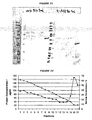
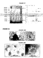
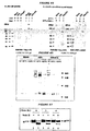
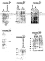
- the full-length Spike protein (nSh) was detected by western blot only in cell lysate, but not in culture media ( Figure 44 ).
- the majority of SARS-CoV full-length Spike protein was expressed in transiently-transfected COS7 cells as a high molecular glycoprotein which ran at 540 kDa in non-reducing gels ( Figure 45 ).
- the gp540 is heat labile as indicated by the complete dissociation into monomeric forms (gp170 & gp180) by boiling, but it was resistant to DTT treatment.
- These data suggest that the recombinant Spike protein is noncovalently associated into a homotrimer (gp540).
- the presence of Spike protein in homotrimeric association also was confirmed in inactivated, purified SARS-CoV virion particles. Analysis of virion proteins by western blot under the same condition used for the characterization of recombinant Spike protein generated essentially identical results ( Figure 46 ).
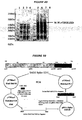
- BHK-21 cells were infected with alphavirus replicon particles expressing the SARS-CoV full-length Spike. At 6 hoursr post-infection with an MOI of 5, infected cells were labeled for 1 hr with L-[ 35 S]methionine/cysteine and chased for up to 4 hours.
- the [ 35 S]-labeled spike protein was immunoprecipitated by anti-SARS rabbit serum and digested with Endo-H. Both digested and undigested proteins were analyzed by SDS-PAGE (4% polyacrylamide).
- the full-length spike protein is synthesized as an Endo-H sensitive high-mannose glycoprotein (gp170, an ER form) that undergoes modification to an Endo-H resistant glycoprotein with complex oligosaccharides (gp180, a Golgi form).
- gp170 Endo-H sensitive high-mannose glycoprotein
- gp180 Endo-H resistant glycoprotein with complex oligosaccharides
- the conversion of gp170 into the gp180 form takes place within 2 hours ( Figure 48 ).
- EXAMPLE 10 Selection of CHO cell lines for Spike protein expression
- a pCMV3 vector expressing Spike can be used for the derivation of stable CHOK-1 cell lines, containing the CMV enhancer/promoter, ampicillin resistance, and a fused DHFR and attenuated neomycin gene for selection purposes.
- Stable cell lines can produced using the neomycin selection system in CHOK-1 cells.
- Clones can be sequenced to verify the integrity of the insert, and transient transfections can be performed using Trans-LT1 polyamine transfection reagent (PanVera Corp., Madison, WI) to assess the expression level and also the integrity of the expressed protein by ELISA and western blot analysis.
- Initial CHO cells will be selected to be free from TSEBSE contaminants and risks according to relevant regulatory standards.
- procedures involve transfection, primary screening with selective medium, followed by subcloning to assure purity of cell lines.
- Cell supernatants can be assayed using an antigen capture ELISA to quantify expression levels at all stages of selection and amplification.
- methanol fixed cells can be screened for internal expression by immunofluorescent staining using a rabbit anti-SARS antibody. Successive measurements at the T75-flask stage of expansion canbe employed to assure stability of expression levels.
- the molecular mass and integrity of the expressed proteins can be checked by PAGE both under native and reducing and denaturing conditions, followed by immunoprobing.
- the pCMV3 vectors expressing SARS-CoV Spike proteins in either full-length or truncated forms can be introduced into CHOK-1 cells using the Trans-LT-1 reagent and non-selective media. 24-48 hours post-transfection, depending on cell density, cells are split at a 1:5 ratio and the medium can be changed to selective media containing neomycin at 500 ⁇ g/ml. Any bovine serum used in these procedures will be from TSE-free sources that meet regulatory standards. Ten to fourteen days later, individual colonies can be picked and transferred to 96 well plates and cultured in complete non-selective medium. When approximately 80% of the wells are confluent, 24 hour supernatants can be screened by Spike capture ELISA.
- cells For initial expression of full length Spike protein, cells can be fixed with methanol and screened by immunofluorescent staining using a rabbit anti-SARS antibody. After low-expressing cell lines have been eliminated and there are fewer than 20-30 cell lines, capture ELISA and western blots can then be used to determine the expression level after cell lysis. A portion of each cell line can be pelleted, weighed and lysed in 1% Triton lysis buffer for determination of expression levels. Three to four clones producing the highest levels of spike protein in correct structure and conformation can be expanded to three-liter bioreactors and adapted to low serum suspension culture conditions for scale-up.
- the antigen capture ELISA assay for the SARS spike protein can be performed using 96 well flat-bottom plates coated with 250ng per well of purified immunoglobulin obtained from rabbit sera that were immunized with inactivated SARS virus. Supernatant or lysate samples are added and incubated for 2 hours at 37°C. Bound antigen is reacted against pooled SARS +ve serum or high affinity monoclonal antibody either human or mouse against SARS spike protein and detected using appropriate species-specific peroxidase-conjugated second antibody. The plates are developed using TMB substrate (Pierce, Rockford, IL), read at a wavelength of 450nm, and the concentration of protein per ml sample is derived from a standard curve (OD vs . protein concentration) based on serial dilutions of a known concentration of recombinant spike protein.
- the immunoprobing analysis will also be performed following the standard methods described by Srivastava et al. (2002) supra . Briefly, 10-20 ⁇ l of the sample is analyzed on 4-20% SDS PAGE under non-reducing/denaturing conditions with mild heating. The proteins are then transferred onto nitrocellulose membranes and reacted against polyclonal anti-Spike rabbit serum, followed by anti-rabbit Ig conjugated to Alexa 688 (Molecular Probes, Oregon). The blots are scanned using an infrared imaging system.
- the highest expressing candidate cell lines will be screened for Spike protein expression and stability in small-scale (3 liter) perfusion bioreactors.
- the candidate clones will be further evaluated for level of expression as well as integrity of expressed protein, and subsequently tested for expression stability in the absence of selection.
- the selected clones also will be tested for maintenance of the DNA sequence integrity of the integrated SARS spike protein gene.
- a lectin-based process Gluvanthus Nivalis lectin
- Full-length Spike protein will be obtained from Triton X-100 detergent extracted cells and then captured on GNA lectin, followed by hydroxyapatite and SP chromatograph. Eluted protein is then characterized by: (1) polyacrylamide gel electrophoresis (PAGE) and Coomassie staining, (2) immunoprobing with anti-SARS rabbit sera, (3) structural characterization using size exclusion chromatography (SEC), as well as mass spec analysis using MALDI-TOF.
- PAGE polyacrylamide gel electrophoresis
- SEC size exclusion chromatography
- Productivity from the CHO cell line expressing SARS spike protein should be at least 2 mg/L and for full-length Spike protein will be 3mg/100gm of cells, at steady-state cell density. Yield from one 45 day, 2.5-liter bioreactor will be -1000 mg crude protein.
- the following basic process is used, with all steps being performed at 2-8°C: the starting material, concentrated CHO cell culture supernatant (20-30X) is thawed and filtered through a 0.45 ⁇ m membrane; this material is heavily contaminated proteins from culture, as well as DNA; the first purification step is affinity chromatography using Gluvanthus Nivalis (GNA), a lectin that preferentially recognizes terminal mannose containing carbohydrates; glycosylated proteins, including SARS spike protein are captured and non-glycosylated proteins, as well as DNA, do not bind to this column; the GNA column is followed by two chromatographic steps operated in the flow through mode; the anion exchanger, DEAE, and ceramic hydroxyapatite (cHAP); DEAE binds some contaminating supernatant proteins and DNA, whereas cHAP binds any contaminating serum proteins; full-length Spike protein is purified from the cell pellet; the cells
- the purified SARS spike can be further treated to remove adventitious viruses: viral inactivation at pH 3.5 for 1 hour; the sample is then concentrated and diafiltered into a buffer at pH 4 and finally captured the purified protein using SP resin; the spike protein binds to this resin and many viruses flow through.
- the spike protein is eluted, concentrated and diafiltered into formulation buffer.
- This formulated bulk product is then filtered through a DV50 viral removal membrane followed by filtration through a 0.2 ⁇ m membrane.
- the formulated bulk is filled into suitable containers e.g. into 3.0 ml vials, in a class 100 laminar flow hood.
- process testing at each step of the purification includes protein concentration, endotoxin (LAL), bioburden, and recovery.
- LAL endotoxin
- a test for potency will evaluate the specific ability of the vaccine in an in vitro or in vivo test to effect a given response.
- the in vivo immunogenicity will be determined by dosing groups of 10 mice with various doses of the protein antigen.
- Sera will be analyzed for the presence of IgG antibodies using an ELISA.
- the criterion for passing will be based upon the number of vaccine treated animals that are seropositive compared to a reference standard.
- Other tests include General Safety, sterility, purity, identity of the vaccine (using an ELISA specific for Spike protein), and quantity & protein concentration (UV spectrophotometric absorbance procedure based on the molar absorbance of the aromatic amino acids).
- Stability testing will be performed on the bulk drug substance and on the final container product.
- Bulk product will be evaluated at temperatures of -60°C (recommended storage condition), 25 ⁇ 2 °C and 40 ⁇ 2 °C protected from light, at time points of 0, 3, 6, 9, 12 months.
- Final container product will be tested at temperatures of -60 °C, and inverted at 5 ⁇ 3 °C, 25 ⁇ 2 °C, and 40 ⁇ 2 °C at time points of 0, 3, 6, 9, 12 months.
- Stability-indicating assays may include appearance, pH, protein content, SDS-PAGE, size exclusion HPLC, and container/closure integrity, performed on single samples of bulk and triplicate vials of final container material.
- the protein purified in this way can be evaluated in mice, rabbits and ferrets as described in, and based on the results of, examples 4, 5, 8 and 9 above.
- Neutralizing and Spike-specific antibody titers will be assessed at days 7, 21, and 35 and at 2, 3, 4, and 5 months after priming; the IgG2a and IgG1 titers of Spike-specific antibodies will be determined at days 21 and 35, and at 2, 3,4, and 5 months after priming; proliferation and IFN- ⁇ and IL-4 production by splenic T cell against the recombinant Spike protein from SARS-CoV will be assessed at day 42 and at the end of the 5th month.
- Peripheral blood will be collected at days 7, 21, and 35 and at 2, 3, 4, and 5 months after priming; spleen cells at day 42 and at the end of the 5th month.
- Neutralizing and Spike-specific antibody titers and isotypes will be determined by inhibition of SARS-CoV infection of Vero cells and by ELISA, respectively. Proliferation of splenic cells will be determined by 3 [H]-thymidine uptake. Frequencies of splenic IFN- ⁇ and IL-4 producing CD4+ T lymphocytes, will be determined by ELISPOT and FACS analysis.
- the optimal dosing and schedule for recombinant Spike vaccine will be determined in ferrets. Based on the mouse results, the Spike vaccine eliciting the highest antibody neutralizing titers will be tested against a two-fold higher dose of recombinant Spike protein given in the same formulation.
- Three groups of ferrets, 6 per treatment, will be immunized SC under anesthesia with 200 ⁇ l of inoculum. The animals will be primed at day 0 and boosted at days 14 and 28. Peripheral blood will be collected at days 7, 21, and 35.
- Neutralizing and Spike-specific antibodies titers will be determined by inhibition of SARS-CoV infection of Vero cells and by ELISA, respectively. Similar to the previous ferret studies, each group of animals will be used to assess efficacy of the vaccine in protecting immunized animals from infection and/or disease.
- Immunogenicity and efficacy of the candidate vaccine also will be evaluated in nonhuman primates.
- Three groups of adult cynomolgus macaques, 4 per treatment, will be immunized with recombinant SARS-CoV Spike protein, alone or mixed with an equal volume of MF59-citrate, administered SC to anesthetized animals in 500 ⁇ l of inoculum.
- the Spike protein vaccine will be tested at the dose eliciting the highest neutralizing antibody titers in ferrets at day 35.
- the animals will be primed at day 0 and boosted at 3 and 6 weeks. Peripheral blood will be collected at weeks 1,4, and 7.
- a secondary endpoint will be to assess the Th1/Th2 profile of the specific immune response, as described above (neutralizing and Spike-specific antibody titers, frequencies of peripheral blood CD4+ T cells producing IFN- ⁇ and IL-4 in response to the recombinant Spike protein, assessed at at weeks 1, 4, and 7).
- Immunogenicity and efficacy of the inactivated virus vaccine and the purified Spike protein can be compared in non-human primates.
- Three groups of adult cynomolgus macaques, 4 for treatment, will be immunized with recombinant SARS-CoV Spike protein from CHO cell lines or with BPL-SARS-COV, given in the dose and formulation eliciting the highest neutralizing antibody titers in previous immunogenicity challenge experiments, administered SC to anesthetized animals in 500 ⁇ l of inoculum.
- the animals will be primed at day 0 and boosted at 3 and 6 weeks.
- Peripheral blood will be collected at weeks 1,4, 7.
- a secondary endpoint will be to assess the Th1/Th2 profile of the specific immune response, as described above.
- Yeast is a useful and inexpensive eukaryotic expression system. Yeast-expressed proteins are used in recombinant hepatitis B virus vaccines, and recombinant SARS antigens may also be expressed in yeast for vaccine purposes. Yeast-expression is also convenient for the production of antigens for preparing monoclonal and polyclonal antitobodies, or for use in serological assays.
- N The nucleocapsid protein (N) and two different versions of the spike glycoprotein (S) from SARS coronavirus FRA strain (AY310120) were cloned for expression in S.cerevisiae:
- a XhoI-NotI fragment of approximately 3733bp encoding the full-length spike glycoprotein was the starting point.
- PCR was used to amplify the full-length gene in two pieces: XbaI-BlnI of 2440bp and BlnI-SalI of 1306bp. These fragments were subcloned into commercial vectors (Novagen): pT7Blue2 XbaI-BlnI (5' end of spike glycoprotein) and pT7Blue2 BlnI-SalI (3' end of spike glycoprotein; Figure 50 ), respectively.
- E. coli HB101 competent cells were transformed with the PCR ligation product and plated on Luria agar plates, containing 100 ⁇ g/ml ampicillin.
- the desired clones were identified using miniscreen DNA analysis. After sequence verification and plasmid amplification of the desired subclones, it was desirable to eliminate the internal SalI site present in the XbaI-BlnI portion of the spike sequence in order to facilitate future cloning into the yeast expression vector (BamHI-SalI). Therefore, we prepared a CelII-MfeI vector from the pT7Blue2 XbaI-BlnI (5' end Spike) subclone to eliminated a 143bp sequence containing the SalI site.
- oligos DS1-6 (SEQ ID NOS: 9789-9794) were then ligated into the CelII-MfeI vector to replace the 143bp that were removed to mutate the SalI site (no aa changes), creating pT7Blue2.XbaI-BlnI ⁇ sal.
- the 5' XbaI-BlnI (from pT7Blue2.XbaI-BlnI ⁇ Sal) and the 3' BlnI-SalI (from pT7Blue2 BlnI-SalI) spike glycoprotein inserts were gel-purified and ligated them into the p893-1 XbaI-SalI vector (a vector derived from pLitmus 38 (New England Biolabs) with the alpha-factor leader sequence cloned into the BamHI -SalI sites of the MCS).
- the resulting full-length SARS Spike coding sequence was named p893-1.SARS Spike 1255 #9 ( Figure 50 ).
- E.coli HB101 competent cells were transformed with the oligo replacement ligation product and plated on Luria agar plates, containing 100 ⁇ g/ml ampicillin. The desired clones were identified using miniscreen DNA analysis. After sequence verification of the positive clones, pT7Blue2 Xba-Bln ⁇ Sal was chosen for use as a template for PCR reactions to amplify the Spike S1 1967 bp Xba-Sal fragment. The fragment was then subcloned into the p893-1 Xba-Sal vector, sequence verified, and named it p893-1.Spike S1 #11 ( Figure 51 ).
- S1-1 had SEQ ID NO: 9795 and S 1-2 has SEQ ID NO: 9796.
- the HindTII-SalI fragment was gel purified.
- the 1365 bp BamHI-HindIII ADH2/GAPDH promoter fragment was ligated along with the 1973 bp HindIII-SalI S1 fragment into the pBS24.1 BamHI-SalI vector creating the genetically engineered pd.SARS Spike S1 #2 expression plasmid ( Figure 52 ).
- S.cerevisiae strain AD3 was transformed with pd.SARS Spike S1 #2 and single transformants were checked for expression after depletion of glucose in the medium.
- the recombinant protein was expressed at high levels in yeast, as detected by Coomassie blue staining.
- yeast cells were transformed with the SARS S1 expression plasmid using the Invitrogen S.c. EasyCompTM Transformation Kit. Expression in shown in Figure 49 .
- pT7Blue2 BlnI-SalI #11 (described above) a BlnI-DraI 1056bp fragment was gel purified. This fragment was ligated with a synthetic pair of 68bp DraI-SalI kinased oligos (DRS1+2; SEQ ID NOS: 9797 & 9798) into a pT7Blue2 BlnI-SalI vector ( Figure 53 ). E.Coli HB101 competent cells were transformed with the oligo replacement ligation product and plated on Luria agar plates, containing 100 ⁇ g/ml ampicillin. The desired clones were identified using miniscreen DNA analysis.
- the clone was named pT7Blue2 BlnI-Sal Spike 1195 #7.
- the 1126bp BlnI-SalI fragment encoding the 3' end of the Spike 1195 was gel purified ( Fig.53 ).
- the 3109bp XbaI-PciI fragment was isolated from the p893-1.SARS Spike 1255 #9 (described above) and a 457bp PciI-SalI fragment from pT7Blue2.SARS Spike 1195 #7 (described above). The two fragments were cloned into the p893-1 XbaI-SalI vector, creating the p893-1.SARS Spike 1195 #34 plasmid ( Figure 54 ).
- coli HB101 competent cells were transformed with the oligo replacement ligation product and plated on Luria agar plates, containing 100 ⁇ g/ml ampicillin. The desired clones were identified using miniscreen DNA analysis. After sequence verification of the positive clone and plasmid amplification of pT7Blue2.SARS 1195 5' HindIII-BlnI #10 ( Figure 55 ), we isolated a 402bp HindIII-NcoI fragment and the 2044bp Ncoi-BlnI fragment ( Figure 55 ). It was necessary for the HindIII-BlnI isolation to be done in two steps to avoid cloning issues related to the internal HindIII site located at nucleotide number 1319 of the spike 1195 protein.
- E.coli HB101 competent cells were transformed with the the BamHI-HindIII (ADH2/GAPDH promoter), HmdIII-NcoI 402bp fragment, NcoI-BlnI 2044bp and the BlnI-SalI 1126bp fragments into the pBS24.1 BamHI SalI vector.
- the samples were plated on Luria agar plates, containing 100 ⁇ g/ml ampicillin. The desired clone was identified using miniscreen DNA analysis, thus creating the genetically engineered pd.SARS Spike 1195 #10 ( Figure 56 ).
- S.cerevisiae strain AD3 was transformed with pd.SARS Spike 1195 #10 and single transformants were checked for expression after depletion of glucose in the medium.
- the recombinant protein was detected by Coomassie blue staining.
- yeast cells were transformed with the SARS 1195 expression plasmid using the Invitrogen S.c. EasyCompTTM Transformation Kit.
- cDNA fragments containing the S protein ORF of 1255 amino acids were amplified by RT-PCR from SARS viral RNA (Frankfurt isolate) grown in Vero cells.
- the amplified PCR fragments were cloned into pBlueScript vector, sequenced, and consensus spike sequence was assembled to create a full-length SARS spike clone, pBSnSh.
- In vitro transcription of pBSnSh followed by translation in a rabbit reticulocyte lysate resulted in the production of single polypeptide with an estimated molecular mass of -140 kDa.
- the Xho I/Not I fragment without a histidine tag also was subcloned into the alphavirus replicon vector backbone pVCRchim2.1 for use in the production of an alphavirus replicon particle chimera that expresses S protein.
- Production and characterization of the replication defective alphavirus vector particles was performed essentially as described previously ( Perri et al. (2003) J. Virol. 77:10394-10403 ; Polo et al. (1999) PNAS USA. 96:4598-4603 ).
- the resultant alphavirus vector particles were named as VEE/SIN.
- COS7 cells and BHK-21 cells were maintained in Dulbecco's modified Eagle's medium supplemented with 10% fetal bovine serum at 37°C and 5% CO 2 in air.
- COS7 cells were transfected with expression plasmids (nSh, nSh ⁇ TC) using a transfection kit (TransIt-COS, Mirus) following the manufacturer's protocol. The cells were washed once with ice-cold PBS and lysed with 1x Lysis buffer (20mM MOPS, 10mM NaCl, 1.5mM MgCl 2 , and 1% Triton X-100) containing complete mini protease inhibitor (Roche). After a 30-min incubation on ice, the debris was cleared by centrifugation. The cleared lysate was either purified or used directly in western blotting.
- HRP horseradish peroxidase
- Biosource horseradish peroxidase
- ECL system chemiluminescence
- the antibodies used were a mouse monoclonal anti-histidine antibody (anti-His•tag Mab, Novagen), a rabbit polyclonlal antipeptide antibody against SARS-CoV spike proten (SmPab, Abgent), or rabbit anti-SARS sera (2BE) obtained by immunization of rabbits with purified SARS-CoV virion.
- the latter has a cell culture neutralizing titer of 1/2,500. Unless stated otherwise, antibody was used at 1/1,000 for anti-histidine antibody and SmPab and 1/10,000 for anti-SARS rabbit sera.
- PNGase F Peptide-N glycosidase F
- Cell lysates were diluted in 0.5% SDS and 1% ⁇ -mercaptoethanol and denatured at 100°C for 10 min. After 2-fold dilution with 1% NP-40 in 50mM sodium phosphate (pH 7.5), the samples were treated with PNGase F (NEB) at 37°C for 1 hr. Enzyme-treated samples were analyzed by 4-12% SDS-PAGE in reducing condition.
- the cell lysates were diluted with 50mM sodium phosphate (pH 6.0) containing 0.75% Triton-X and treated with PNGase F (Calbiochem) at 37°C for 3 hr. Enzyme-treated samples were analyzed by 4-20% SDS-PAGE in nonreducing condition.
- BHK21 cells were infected with defective alphavirus particles expressing the full-length S.
- infected cells were pulse labeled for 1 hr with L-[ 35 S] methionine/cysteine and chased for 2 or 4 hours.
- the [ 35 S]-labeled S protein was immuno-precipitated using the rabbit antiserum raised against inactivated, purified virus and then digested with Endo H.
- the Endo H treatment involved dilution with a sample buffer (50mM sodium phosphate, 0.1% SDS, 50 mM DTT, pH 6.0) and boiling for 5 min.
- the S protein expressed in COS7 cells was detected as a gp170/gp180 doublet in western blot analyses of cell lysates that were fully denatured by boiling in the presence of DTT.
- the majority of S protein was detected as a high molecular glycoprotein in the 440-669 kDa range when the same cell lysate was not heat-denatured prior to western blot analysis using SDS-PAGE ( Fig. 69 , lane 1).
- the ⁇ 500 kDa species was resistant to 10 mM DTT treatment (lane 3) and not dissociated into the monomeric form unless the lysate was first heat-denatured at 100°C (lane 4).
- this -500 kDa species represents an S protein oligomer in native conformation
- comparative analyses with virion-derived S glycoprotein derived from Vero cell cultures was performed.
- the purified virions were solubilised in 1% SDS prior to Western blot analyses after SDS PAGE.
- the presence of the -500 kDa spike protein oligomer was confirmed in virion particles ( Fig. 71 , lane 1).
- heat denaturation of solubilised virions produced the same oligomer-to-monomer conversion as seen with the full-length recombinant S (lanes 2,3).
- the oligomeric nature of virion S was further analysed in a cross-linking experiment.
- the oligomeric status of the S ⁇ spike protein was also examined after expression in COS7 cells. As shown in Figure 73 , the recombinant S ⁇ proteins present in cell lysates were also detected in high molecular weight forms of -500 kDa range when the lysate was not heated prior to SDS-PAGE and Western blot analysis (lane 1). However, the efficiency of oligomerization by intracellular S ⁇ protein appears to be much less ( ⁇ 10%) compared to that of full-length S protein under the same western analysis conditions.
- the ⁇ 500kDa protein is glycosylated, and the effect of deglycosylation on its antibody binding was investigated.
- the recombinant COS7 lysate was treated with PNGase F under non-denaturing condition (as described above) and analysed by western blot.
- deglycosylation did not affect the binding of anti-histidine Mab antibody to the treated S oligomer (lanes 2,3). However, it compromised the reactivity with the rabbit antisera raised against purified virus (lane 6).
- This antiserum binds to virion-derived S in western blot analyses only when DTT is omitted from the sample for SDS-PAGE indicating that it recognizes primarily a discontinuous, conformational epitope(s).
- This antisera has also been shown to have a high-titer of viral neutralizing antibodies. Its lack of binding to deglycosylated, recombinant S suggests that the carbohydrate actively contributes to the higher order, native structure of the S polypeptide oligomer.
- the truncated S ⁇ protein was found as a monomeric species (gp170) in both fractions (lanes 5,6). This indicates that the C-terminal TM and Cys-rich domains are required for the anchorage of the S protein to cell membrane.
- the SARS recombinant full-length S protein is thus an N-linked glycoprotein with an estimated molecular weight of 170-180,000 kDa.
- Deglycosylation with PNGase F resulted in a polypeptide of the expected size for the uncleaved, encoded polypeptide (140kDa).
- Both transient and stable expression of the full-length SARS-CoV S gene in a variety of mammalian cells, including COS7, 293, BHK21, and Huh7 cell lines consistently produced a S protein doublet (gp170/180) as detected in western blot analyses.
- Pulse-chase analyses of transfected cells demonstrated that the SARS CoV S protein was initially synthesized as an Endo H sensitive gp170 species followed by the gradual appearance of an Endo H resistant gp180 form, presumably as a result of the addition of complex carbohydrate within the Golgi apparatus.
- the recombinant S protein was not secreted into the cell culture medium unless the C-terminal 60 amino acids containing the TM-region and the Cys-rich tail were deleted.
- the quaternary structure of the full-length recombinant S protein was investigated using cross-linking treatment, heat-denaturation, and size fractionation analyses. The results data are consistent with the recombinant S protein existing as a homotrimer of ⁇ 500kDa. Similar analyses of virion-derived S yielded the same results.
- Such a trimeric structure has been reported for other enveloped RNA viruses: the hemagglutinin HA of influenza virus, the E1-E2 heterodimer of alphaviruses and the G protein of vesicular stomatitis virus. Incubations under reducing conditions indicate that the SARS-CoV S trimeric structure is non-covalently associated, and is very stable.
- S oligomers present in the cell lysate were shown to be resistant to reduction by 10 mM DTT, detergent treatment with 1% SDS, and heat denaturation at up to 60°C. Incubation at a temperature higher than >80°C resulted in the dissociation of the trimeric complex as evidenced by the decrease in trimer with the concomitant increase in the monomer bands.
- the temperature-induced appearance of the high-mannosylated gp170 (ER monomer form) as well as the complex-glycosylated gp180 (Golgi monomer form) suggests that trimerization can occur before the transport of the monomer spike protein to the medial Golgi apparatus. This is consistent with other reports for TGEV, influenza virus HA, and vesicular stomatitis virus G proteins. With these proteins, trimerization was reported to take place before addition of complex oligosaccharides in the Golgi apparatus.
- the C-terminally truncated form of S was found in the cell lysate in both oligomeric and monomeric forms at a frequency of 10% and 90%, respectively.
- the truncated protein secreted into medium was found fully glysosylated and it was essentially all in monomeric form.
- the C-terminal 60 amino acids of the S glycoprotein contains a membrane anchor region that affects the efficiency of trimerization. In S protein trimerization, it is possible that the C-terminal region is required to initiate the event and the triple-stranded coiled coil structures in the S2 stalk domain provide further stabilizing force as seen in HA oligomer of influenza virus.
- EXAMPLE 15 CHO cells for Spike protein expression
- CHO cell lines that stably express either the full-length or truncated SARS-CoV spike proteins were prepared.
- Several stably transfected CHO cell lines were obtained, and Figure 65 shows western blot data from a panel of representative clones.
- All SARS-CoV ORFs ( Figure 10 , Table 10) were cloned in the pET vector and expressed as C-terminal His-Tag fusion proteins in E. coli.
- the proteins smaller than 16KD were also expressed as N-terminal GST (Glutathione S-transferase) fusion proteins using pGEX vector.
- Nsp1 and Nsp2 the two SARS-CoV proteins with proteolytic activity, were not expressed as full length proteins due to toxicity in E . coli .
- the respective genes were instead cloned in different portions in order to separate the catalytic residues (Cys833/ His994 for Nsp1; His41/Cys145 for Nsp2) in the resulting recombinant proteins: Nsp1A from nucleotides 2719-5214 of AY310120; Nsp1B from nucleotides 5218-7371; NsplC from nucleotide 7372-9984; Nsp2A from nucleotide 9985-10416; Nsp2B from nucleotide 10476-10902.
- Nsp9 was divided into two portions: Nsp9A from nucleotide 13371-14756; Nsp9B from nucleotide 14757-16166.
- Matrix (M), ORF3 and ORF7 contain respectively three, two and one transmembrane domains. These proteins were expressed as deletion proteins excluding the first 100 amino acids (M and ORF3) or the first 18 amino acids (ORF7) that include the hydrophobic regions.
- a two-step strategy was used to amplify the cloned sequences.
- amplification of DNA fragments containing more than one gene or single gene used sequenced cDNA as template.
- Eleven cDNA sequences were amplified: (1) a fragment, named amplC1, including genes coding for protein E, protein M, orf 7-8-9-10; (2) a fragment, named amplC2, including genes coding for orf 3-4; (3) a fragment, named amplC5, including genes coding for proteins Nsp12 and Nsp13; (4) Nsp11gene; (5) P28 and P65 genes; (6) Nsp1B and NsplC genes portion; (7) a fragment, named amplC9, including genes coding for proteins Nsp2 and Nsp3; (8) a fragment, named amplNsp4-7, including genes coding for proteins Nsp4, Nsp5, Nsp6, Nsp7 and for amplification of Nsp9A gene portion; (9) Nsp
- amplification of single genes was performed using DNA fragments from the first amplification step as templates.
- the primers are shown in Table 28.
- Table 29 shows the molecular weight of the expressed fragments of SARS-CoV ORFs, whether they were cloned (+ or -), whether the cloned fragment was seen to be expressed (+ or -) and the form of protein which was chosen for purification.
- a single colony was streaked and grown overnight at 37°C on a LB/Amp (100 ⁇ g/ml) agar plate.
- An isolated colony from this plate was inoculated into 20ml of LB/Amp (100 ⁇ g/ml) liquid medium and grown overnight at 37°C with shaking.
- the overnight culture was diluted 1:30 into 1.0 L LB/Amp (100 ⁇ g/ml) liquid medium and allowed to grow at the optimal temperature (30 or 37°C) until the OD550nm reached 0.6-0.8.
- Expression of recombinant protein was induced by addition of IPTG (final concentration 1.0 mM) and the culture incubated for a further 3 hours.
- Bacteria were harvested by centrifugation at 8000 x g for 15 min at 4°C. The bacterial pellet was resuspended in 10 ml of cold buffer A (300 mM NaCl, 50 mM phosphate buffer, 10 mM imidazole, pH 8.0). Cells were disrupted by sonication (or French Press) on ice four times for 30 sec at 40W using a Branson sonifier 450 and centrifuged at 13 000xg for 30 min at 4°C. Supernatants were mixed with 150 ⁇ l Ni 2+ -resin (previously equilibrated with buffer A) and incubated at room temperature with gentle agitation for 30 min.
- cold buffer A 300 mM NaCl, 50 mM phosphate buffer, 10 mM imidazole, pH 8.0. Cells were disrupted by sonication (or French Press) on ice four times for 30 sec at 40W using a Branson sonifier 450 and centrifuged at
- the resin was Chelating Sepharose Fast Flow (Pharmacia), prepared according to the manufacturer's protocol. The batch-wise preparation was centrifuged at 700 x g for 5 min at 4°C and the supernatant discarded. The resin was washed twice (batch-wise) with 10ml buffer A for 10 min, resuspended in 1.0 ml buffer A and loaded onto a disposable column. The resin continued to be washed with buffer A at 4°C until the OD280nm of the flow-through reached 0.02-0.01.
- the resin was further washed with cold buffer B(300 mM NaCl, 50 mM phosphate buffer, 20 mM imidazole, pH 8.0) until the the OD280nm of the flow-through reached 0.02-0.01.
- the His-fusion protein was eluted by addition of 700 ⁇ l of cold elution buffer C (300 mM NaCl, 50mM phosphate buffer, 250 mM imidazole, pH 8.0) and fractions collected until the OD 280nm indicated all the recombinant protein was obtained. 20 ⁇ l aliquots of each elution fraction were analyzed by SDS-PAGE. Protein concentrations were estimated using the Bradford assay.
- the inclusion bodies were purified as follows: homogenize cells (5 g wet weight) in 25 ml 0.1M Tris HCl pH 7, 1mM EDTA, at 4°C using an ultraturrax (10000 rpm); add 1.5mg lysozyme per gram cells; mix shortly with an ultraturrax and incubate at 4°C for 30'; use sonication or high-pressure homogenization to disrupt the cells; to digest DNA, add MgCl 2 to a final concentration of 3mM and DNase to a final concentration of 10ug/ml and incubate 30' at 25°C.
- a human monoclonal antibody having neutralizing activity was tested in an ELISA assay for reactivity with the purified truncated Spike protein. Briefly, ELISA plates were coated with truncated form of the spike protein at a concentration of 1 ⁇ g/ml (100 ⁇ l/ well) by incubating the plates overnight at 4°C. The plates were washed, non-specific binding sites were blocked and then different dilutions of the antibody were added and plates were incubated for 1 hour at room temperature. At the end of incubation, the plates were washed and bound antibody was detected by using anti-human IgG conjugated to horse radish peroxidase (HRP) and an appropriate substrate.
- HRP horse radish peroxidase
- truncated spike protein was used to immunize mice and the level of binding antibodies induced against the truncated spike protein was determined by ELISA assay. Briefly a group of 10 mice were immunized with 3 ⁇ g of truncated spike protein adjuvanted in MF59 at 0, 4 and 8 weeks intervals. Sera samples were collected from these animals and assayed for antibodies induced by truncated spike protein in an ELISA assay. An additional group of 8 mice was immunized with 75 ⁇ g of DNA encoding the truncated form of the spike protein on PLG particles at 0, 4 and 13 weeks intervals, the sera were collected and analyzed as above for anti-spike antibodies as above
- the profile of binding antibodies induced in each group was plotted as geometric mean titer (GMT). Compared to a plasmid DNA vaccine expressing truncated spike antigen and delivered using a PLG microparticle formulation, the purified truncated spike protein was significantly more potent for inducing strong antibody responses. Further comparison with the antibody responses induced by inactivated BPL-SARS-CoV (already shown protective) in the same mouse strain revealed that the magnitude of antibody responses induced by purified truncated spike protein and the inactivated virus vaccine are in the same range ( Figure 62 ).
- Figure 64 shows a comparison of antibody binding levels (ELISA, X-axis) with neutralization titers (Y-axis). In general there is a very good correlation between the binding and neutralizing antibodies.
- the bottom-left grouping shows ratios 2 weeks post-3rd immunization with the DNA vaccine; the top-right grouping shows ratios 2 weeks post-2nd immunization with the protein vaccine. Both forms of vaccine show a consistent correlation.
- mice were immunized with pCMV-nSdTC plasmid, either free or with PLG microparticles. Serum from the mice was then used as the staining antibody against cultured 293 cells that had been transfected with spike, either full-length or truncated. The cells were centrifuged prior to testing and the pellet was lysed. The antibody was tested against the culture supernatant and against the cell lysate. As shown in Figure 104 , the mouse serum was able to detect spike protein in the lysate of cells that expressed full-length spike and in the supernatant of cells that expressed the truncated spike protein. Results were comparable to the staining seen when using rabbit serum that had been obtained after immunization with whole killed virus. Thus anti-spike antibodies can be induced by the use of DNA vaccination.
- the sequence of plasmid pCMVKm2 is given as SEQ ID NO: 9923.
- Genes encoding the spike protein either in full-length form (pCMVKm2 SARS Spike nS; SEQ ID NO: 9921) or in its ⁇ TC form (pCMVKm2 SARS Spike nS ⁇ TC; SEQ ID NO: 9922) were inserted into this basic vector.
- mice were immunized with these vectors, and with similar vectors encoding the N, M or E proteins. Vectors encoding the same proteins but with optimized codon usage were also prepared. Codons were optimized for efficient human expression starting from the FRA sequence (GenBank: AY310120).
- Figure 98 shows immunofluorescence (using anti-SARS rabbit serum) results after administration of the vector encoding optimsed N antigen, revealing high level expression. Mice receiving the control vector alone showed no fluorescence.
- Figure 99 compares immunofluorescence (using Abgent anti-M antibody) of the native M sequence (99A) or the codon-optimsed M sequence (107B).
- Figure 100 compares immunofluorescence (using Abgent anti-E antibody) of the native E sequence (100A) or the codon-optimsed E sequence (100B).
- mice Four groups of mice (8 mice per group) were immunized with: (1) SARS nS Spike, nSdTC truncated Spike, and N proteins; (2) pCMV-SARS-nSdTC: DNA+DNA-PLG at weeks 0,4 and 13 wks; (3) CMV-nS: DNA+DNA-PLG+VEE/SIN Rep at 0,4 and 9 wks; (4) VEE/SIN Rep-SARS-nS three times at 0, 4 and 13 wks. Sera from all groups recognized SARS nS and nSdTC proteins, and also showed virus binding and neutralization activity.
- SARS-CoV Spike protein To investigate the effect of proteolytic cleavage on SARS-CoV Spike protein, it was expressed in various forms in E.coli, including: (1) full-length S1-S2; (2) S1 alone; (3) HR1 heptad; and (4) HR2 heptad. The expressed proteins were used to raise immune rabbit sera, which were then used for visualizing western blots of Vero cells, either infected or not infected with SARS-CoV.
- Figure 101 shows a western blot using a 1:10000 dilution of antibody raised against either the S1 domain or the uncleaved S 1-S2 domains.
- Figure 102 shows a western blot using a 1:10000 dilution of antibody raised against each of the four proteins. The difference in antigen reactivity is readily apparent.
- Figure 103 shows similar data.
- Each serum was tested against four lanes, with those gour lanes being from left to right: (a) serum at 1:500 dilution, SARS-CoV-infected cells; (b) serum at 1:500 dilution, non-infected cells; (c) serum at 1:2500 dilution, SARS-CoV-infected cells; (d) serum at 1:2500 dilution, non-infected cells. Again, the difference in antigen reactivity is readily apparent.
- Figures 101-103 show that the Spike protein exists in various forms in infected Vero cells, with sizes of approx. 75kDa, 90kDa, 180kDa and >250kDa.
- the Spike protein is cleaved (at least partially) either intracellulary or after release of the particles.
- T-epitope predictions for SEQ ID NO: 6042 Epitope predictions were performed at http: // www.mpiib-berlin.mpg.de / MAPPP / binding.html using a minimum score of 0.5 and the BIMAS matrix, with a maximum of 20 results being selected. The analysis revealed 9mer and 10mer epitopes. Table 16: Epitopes for SEQ ID NO: 6042 HLA A1 - 9 mers Maximum possible score using this molecule type 5625 Rank Start position Sequence % of max.
- SEQ ID NO: 8101 1.48 % 180 2 180 SEQ ID NO: 8102 0.55 % 67.5 3 768 SEQ ID NO: 8103 0.49 % 60 4 1177 SEQ ID NO: 8104 0.49 % 60 5 380 SEQ ID NO: 8105 0.24 % 30 6 100 SEQ ID NO: 8106 0.18 % 22.5 7 786 SEQ ID NO: 8107 0.16 % 20 8 1217 SEQ ID NO: 8108 0.16 % 20 9 207 SEQ ID NO: 8109 0.14 % 18 10 1183 SEQ ID NO: 8110 0.14 % 18 11 38 SEQ ID NO: 8111 0.09 % 12 12 52 SEQ ID NO: 8112 0.09 % 12 13 8 SEQ ID NO: 8113 0.06 % 8 14 679 SEQ ID NO: 8114 0.06 % 8 15 73 SEQ ID NO: 8115 0.05 % 6.75 16 1204 SEQ ID NO: 8116 0.05 % 6.075 17 50 SEQ ID NO: 8117 0.04
- SEQ ID NO: 8261 4.44 % 240 2 312 SEQ ID NO: 8262 3.70 % 200 3 141 SEQ ID NO: 8263 1.11 % 60 4 1006 SEQ ID NO: 8264 0.66 % 36 5 411 SEQ ID NO: 3265 0.44 % 24 6 122 SEQ ID NO: 8266 0.37 % 20 7 134 SEQ ID NO: 8267 0.37 % 20 8 184 SEQ ID NO: 8268 0.37 % 20 9 367 SEQ ID NO: 8269 0.37 % 20 10 402 SEQ ID NO: 8270 0.37 % 20 11 494 SEQ ID NO: 8271 0.37 % 20 12 560 SEQ ID NO: 8272 0.37 % 20 13 626 SEQ ID NO: 8273 0.37 % 20 14 931 SEQ ID NO: 8274 0.37 % 20 15 956 SEQ ID NO: 8275 0.37 % 20 16 1117 SEQ ID NO: 8276 0.37 % 20
Landscapes
- Life Sciences & Earth Sciences (AREA)
- Health & Medical Sciences (AREA)
- Chemical & Material Sciences (AREA)
- Organic Chemistry (AREA)
- Virology (AREA)
- Immunology (AREA)
- General Health & Medical Sciences (AREA)
- Medicinal Chemistry (AREA)
- Genetics & Genomics (AREA)
- Zoology (AREA)
- Engineering & Computer Science (AREA)
- Proteomics, Peptides & Aminoacids (AREA)
- Biochemistry (AREA)
- Microbiology (AREA)
- Wood Science & Technology (AREA)
- Biophysics (AREA)
- Bioinformatics & Cheminformatics (AREA)
- Molecular Biology (AREA)
- Mycology (AREA)
- Pharmacology & Pharmacy (AREA)
- Public Health (AREA)
- Veterinary Medicine (AREA)
- General Engineering & Computer Science (AREA)
- Animal Behavior & Ethology (AREA)
- Epidemiology (AREA)
- Biotechnology (AREA)
- Pulmonology (AREA)
- Analytical Chemistry (AREA)
- Communicable Diseases (AREA)
- Physics & Mathematics (AREA)
- Biomedical Technology (AREA)
- Gastroenterology & Hepatology (AREA)
- Medicines Containing Antibodies Or Antigens For Use As Internal Diagnostic Agents (AREA)
- Peptides Or Proteins (AREA)
- Medicines That Contain Protein Lipid Enzymes And Other Medicines (AREA)
- Measuring Or Testing Involving Enzymes Or Micro-Organisms (AREA)
Description
- The invention relates two nucleic acids and proteins from Severe Acute Respiratory Syndrome (SARS) Virus. These nucleic acids and proteins can be used in the preparation and manufacture of vaccine formulations for the treatment or prevention of SARS. Also disclosed are diagnostic reagents, kits (comprising such reagents) and methods which can be used to diagnose or identify the presence or absence of a SARS virus in a biological sample. Also disclosed are methods for the treatment or prevention of SARS utilizing small molecule viral inhibitors and combinations of small molecule viral inhibitors and kits for the treament of SARS.
- An outbreak of a virulent respiratory virus, now known as Severe Acute Respiratory Syndrome (SARS), was identified in Hong Kong, China and a number of other countries around the world in 2003. Patients typically had symptoms including fever, dry cough, dyspnea, headache, and hypoxemia. Isolates of the SARS virus appear to have homology with at least the RNA polymerase gene of several known coronaviruses. A phylogenetic analysis of this homology is presented in Peiris et al., "Coronavirus as a possible cause of severe acute respiratory syndrome", Lancet, published online April 8, 2003 at http://image.thelancet.com/extras/03art3477web.pdf. Other sequenced fragments of the SARS virus genome appear to overlap with the
open reading frame 1b of coronaviruses. See, Drosten et al., "Identification of a Novel Coronavirus in Patients with Severe Acute Respiratory Syndrome", New England Journal of Medicine, published online at lettp://www.nejin.org on April 10, 2003. - The Genome Science Center in British Colombia, Canada published on its website (http:///www.bcgsc.ca/bioinfo/SARS/) a draft genome assembly of 29,736 base pairs of a virus believed to be a SARS virus, referred to as the TOR2 isolate.
- The Centers for Disease Control (CDC) published a nucleotide sequence of a SARS-CoV strain on its website (http://www.cdc.gov/ncidod/sars/pdf/nucleoseq.pdf). The CDC has also published a phylogenetic tree of the predicted N, S and M proteins (attached as
FIGURE 5 ). This tree places the SARS virus outside any of the previously known coronavirus groups. -
WO 2004/089983 discloses an isolated essentially mammalian positive-sense single stranded RNA virus (SARS) within the group of coronaviruses and components thereof. Accession Number AY323976 (DATABASE EMBL) discloses the SARS coronavirus ZJ01 isolate spike glycoprotein. Ksiazek et al., 2003 (New England J Med, Vol. 348, pages 1953-1966) discloses a new coronavirus associated with severe acute respiratory syndrome. - There is a growing need for prophylactic or therapeutic vaccines against the SARS virus.
- The invention provides an immunogenic composition comprising an isolated or purified polypeptide comprising: the Severe Acute Respiratory Syndrome (SARS) virus Spike polypeptide amino acid sequence SEQ ID NO: 6042, or an amino acid sequence having >80% sequence identity to SEQ ID NO: 6042, or a fragment of at least 10 consecutive amino acids of SEQ ID NO: 6042; with the proviso that the polypeptide is not MFIFLLFLTLTSGSDLDRCTTFDDVQAP (as disclosed in
WO 2004/089983 ). - The invention also provides an immunogenic composition comprising an isolated or purified polypeptide comprising amino acid sequence SEQ ID NO: 6042, or an amino acid sequence having >80% sequence identity to SEQ ID NO: 6042, or a fragment of at least 10 consecutive amino acids of SEQ ID NO: 6042; and further comprising an adjuvant. Further embodiments are defined in the appended claims.
-
-
FIGURE 1 : Schematic of coronavirus genome organization. -
FIGURE 2 : Schematic of coronavirus ORF1a/ORF1b gene products. -
FIGURE 3 : Alignment of coronavirus polypeptide sequences (including, -
FIGURE 4 : Alignment of spike (S) polypeptide sequences, taken fromFigure 3 , in the region of the junction of the S1 and the S2 domains, and protease cleavage site for selected coronaviruses. -
FIGURE 5 : CDC phylogenetic tree of SARS-CoV strain (Clustalx 1.82, neighbor-joining tree). -
Figure 5A shows coronavirus N protein analysis,Figure 5B shows coronavirus S protein analysis, andFigure 5C shows coronavirus M protein analysis. -
FIGURE 6 :Labels 229E: human coronavirus; MEV: murine hepatitis virus; TGV: transmissible gastroenteritis virus; AIBV: avian infectious bronchitis virus; BOVINE: Bovine coronavirus; PEDV: porcine epidemic diarrhea virus. -
FIGURE 7 : Coiled-coil prediction for SEQ ID NO: 6042, using Coils program (Figure 7A ) or LeanCoil (Figure 7B ). -
FIGURE 8 : Example of insertion of a reporter gene-of-interest at a site between existing SARS virus genes. Small nonstructural gene products are not depicted schematically. -
FIGURE 9 : Schematic depicting representative examples of SARS virus replicons. Small nonstructural gene products are not depicted schematically. -
FIGURE 10 : Genome organization of SARS coronavirus. Replicase and structural regions are shown, along with the predicted products of cleavage within ORF1a and ORF1b. The position of the 5' RNA leader sequence (L), the 3' poly(A) tract and the ribosomal frame-shift consensus between ORF1a and ORF1b are also indicated. Each box represent a protein product. They are shaded according to the level of amino acid identity with corresponding proteins of other coronaviruses. The SARS-specific genes are white. Positions of the 9 SARS-specific six-base IG sequences (5'-ACGAAC-3') are indicated by arrows. -
FIGURE 11 : Genome organization of Coronaviruses representative of group 1 (HCoV-229E, accession number: AF304460), group 2 (mouse hepatitis virus MHV, accession number: NC_001846), group 3 (avian infectious bronchitis virus AIBV, accession number: NC_001451) and SARS coronavirus. Other completely sequenced coronaviruses used in this study are available at the following accession numbers: porcine epidemic diarrhea virus (PEDV), AF353511; transmissible gastroenteritis virus (TGV), NC_002306; Bovine coronavirus (BCoV): AF220295. Red boxes represent group-specific genes. The position of the leader RNA sequence and poly(A) tract is also indicated in genomes where they are reported. The position of specific IG sequences is indicated by circles of different shades. In the SARS genome, we also find three IG sequences specific forgroup 2 coronavirus. -
FIGURE 12 : Topological model predicted for the spike protein anchored to the viral membrane. Structural and predicted functional domains are indicated. The N-terminal region (S1) is predicted to contain the receptor binding domain. Two coiled coil regions within the S2 domain, partially superimposed to leucine zipper motifs are presumably involved in oligomerization. The hydrophobic domain is responsible for membrane anchoring. -
FIGURE 13 : Phylogenetic tree obtained from the multiple sequence alignment of a 922 bp internal region of the pol gene from 12 coronaviruses and SARS. Numbers at the nodes represent the result of a bootstrap analysis and strongly support the branches. Sequences not available within the complete coronavirus genomes have been retrieved from GenBank at the following accession numbers: hemagglutinating encephalomyelitis virus of swine (PHEV), AF124988, Human OC43 virus (OC43), AF124989, canine coronavirus (CCV), AF124986, feline infectious peritonitis virus (FIPV), AF124987, turkey coronavirus (TCV), AF124991, syaloacryoadenitis virus of rats (SDAV), AF124990. -
FIGURE 14: 14A . Unrooted tree obtained from the alignment of consensus sequences of the group I and group II S1 domain of spike proteins (G1_cons and G2_cons) with those of agroup 3 spike (AIBV) and the spike of SARS virus. The number indicates the result of a bootstrap analysis. The sequences used to generate the consensus profile fromgroup 1 are: HcoV-229E, accession number P15423; porcine epidemic diarrhea virus (PEDV), acc no: NP_598310; transmissible gastroenteritis virus (TGV), acc no: NP_058424; Canine coronavirus (CCV), acc no: S41453; porcine respiratory virus (PRV), acc no: S24284; feline infectious peritonitis virus (FIPV), acc no: VGIH79. The sequences used to generate the consensus profile fromgroup 2 are: mouse hepatitis virus (MHV), acc no: NP_045300; Bovine coronavirus (BCoV), acc no: NP_150077; Human coronavirus OC43, acc no: P36334; hemagglutinating encephalomyelitis virus of swine (PHEV), acc no: AAL80031; forgroup 3, only the sequence of the spike protein of avian infectious bronchitis virus (AIBV), acc no: AAO34396 was used. 14B: Schematic representation of cysteine positions in S1 domains ofgroup group 1, G1_cons, and fromgroup 2, G2_cons). The length of the bars are not to scale. Relative cysteine positions are indicated by rectangle bars. Only cysteines perfectly conserved within each consensus are reported. Lines connect cysteines conserved between the SARS S1 domain and the consensus sequences as shown. -
FIGURE 15 : illustration of a Neisseria Adhesin A protein (NadA). -
FIGURE 16 : Raw translation from SARS coronavirus genome (reading frame +1). -
FIGURE 17 : Raw translation from SARS coronavirus genome (reading frame +3) -
FIGURE 18 :1b and Spike open reading frames, separated by *. -
FIGURE 19 : chromatogram of the capture step of SARS coronavirus on MatrixCellufine Sulfate Superformance 150/10. Analysis was on 100ml coronoavirus harvest. The left Y axis shows absorbance at 280nm. The right Y axis shows the gradient (%B). The X axis shows the volume (ml). -
FIGURE 20 : Silver-stained MCS chromatography fractions. Lanes are: (1) marker; (2) coronavirus vero cell harvest; (3) coronavirus vero cell harvest, after 0.65µm filtration; (4) flowthrough; (5) wash; (6) 20% peak (virus peak). Lanes were loaded with 1 µg of test protein. -
FIGURE 21 : Western Blot of MCS chromatography fractions. Lanes are as described forFig. 20 . -
FIGURE 22 : Linear density gradient ultracentrifugation, 15-60% sucrose (SW28, 2 hours, 20000 rpm). The graph shows protein concentration (■) and sucrose concentration (◆). 23 -
FIGURE 23 : Silver-stained density gradient fractions on NuPage 4-12% Bis-Tris-Ge (Novex), reduced conditions, heated for 10 minutes at 70°C. Lanes are: (1) marker; (2) 20% peak MCS; (3)density gradient fraction 11; (4)density gradient fraction 12; (5)density gradient fraction 13; (6)density gradient fraction 14; (7)density gradient fraction 15; (8)density gradient fraction 16; (9)density gradient fraction 17. The bulk of proteins was infractions 15 to 17.Lanes -
FIGURE 24 : Chromatogram of the Capture Step of SARS coronavirus on MCS. Details are as forFigure 19 , except that 200ml harvest was used. -
FIGURE 25 : Silverstain (left) and Western Blot (right) of chromatographic fractions. Lanes are as described forFigures 20 and21 , except that lane (6) is the 5% peak. Treatment before SDS-PAGE was at room temperature for 30 minutes. -
FIGURE 26 , Density Gradient Ultracentrifugation, 15-40% sucrose (SW28, 2 hours, 20000 rpm). The graph shows protein concentration (■) and sucrose concentration (◆). -
FIGURE 27 : Silverstain (left) and Western Blot (right) of Density Gradient Ultracentrifugation fractions on NuPage 4-12% Bis-Tris-Ge (Novex), reduced conditions. Lanes are: (1) marker; (2)density gradient fraction 6; (3)density gradient fraction 7; (4)density gradient fraction 8; (5)density gradient fraction 9; (6)density gradient fraction 10; (7)density gradient fraction 15. Fractions 7-10 (lanes 3-6) contained pure coronavirus proteins. The bulk of impurities was in fraction 15 (lane 7).Lanes -
FIGURE 28 : EM picture ofDensity Gradient Fractions 8=10.Figure 28A showsfraction 8;Figure 28B showsfraction 9;Figure 28C showsfraction 10. -
FIGURE 29 : Spike/NadA fusion constructs. -
FIGURES 30 and 31 : Results of the expression in E.coli of S1L, S1L-NadA and S1L-NadΔanchor.Figure 30 shows SDS-PAGE analysis of total lysates from BL21(DE3)/pET, BL21(DE3)/pETS1L and BL21(DE3)/pET-S1L-NadΔanchor. The bands are indicated by an arrow, and the three lanes are, from left to right: BL21(DE3)/pET; BL21(DE3)/pET-S1L; BL21(DE3)/pETS1L-NadAΔanhor.Figure 31 shows (39A) SDS-PAGE and (39B) western blot analyses of total lysates from BL21(DE3)/pET, BL21(DE3)/pET-S1L-NadA (grown under un-induced condition) and BL21(DE3)/pET-S1L-NadA (grown under induced condition). The bands are indicated by an arrow, and lanes are, from left to right: BL21(DE3)/pET; BL21(DE3)/pET-S1L-NadA; BL21(DE3)/pET-S1L-NadA. The western blot shows the presence of oligomeric forms of the protein. -
FIGURE 32 : Schematic of SARS Spike clones. -
FIGURE 33 : Transient Expression of SARS Spike Proteins (western blot of COS7 cell lysate). Each lane of the 4-20% TG SDS gel was loaded with 20µg cell lysate (total 1.2mg). The labeling antibodies are shown. -
FIGURE 34 : Western blot analyses of COS7 cell lysates on 4% TG SDS gel showing oligomerization state of intracellular S molecules. -
FIGURE 35 : Western blot analyses of COS7 cell lysates on 4-20% TG SDS gel showing Transient Expression of SARS Spike Proteins. Lanes are: (1) mock, AF; (2) mock, DF; (3) nSh, AF; (4) nSh, DF; (5) nShΔTC, AF; (6) nShΔTC, DF. Each lane was loaded with 5µl of each sample, 400µl total. The blot was labeled with antibody against the His-tagged protein. -
FIGURE 36 : Western blot analyses of COS7 cell medium on 4-20% TG SDS gel showing Transient Expression of SARS Spike Proteins. Truncated spike protein is secreted. Spike proteins were purified from the culture medium (from a 10cm plate), first by a ConA column and then finally by His•tag Magnetic beads. Each lane was loaded with one third of the material. -
FIGURE 37 : Western blot analyses of COS7 cell lysates on 4-20% TG SDS gel showing glycoslation of SARS spike proteins. In the two left-hand blots (lanes 1-5), samples were boiled in SDS and β-mercaptoethanol; in the two right-hand blots (lanes 6-11), samples were in SDS only, with no boiling. Lanes 1-8 were labeled with a monoclonal raised against the His-tag protein; labes 9-11 were labeled with rabbit anti-SARS antibody. -
FIGURE 38 : Effect of SARS spike protein expression on cell viability. -
FIGURE 39 : Western blot analyses of COS7 cell lysates on 4% TG SDS gels showing oligomerization state of intracellular spike molecules. Blots were labeled with anti-His-tag mAb. - The membrane fraction of COS7 cell lysate was fractionated by a sizing column before loading the lanes.
Fractions 7 to 14 show bands with kDa values of: 71000, 1400, 898, 572, 365,232, 148 and 99, respectively. -
FIGURE 40 : Fractionation of cells into aqueous and detergent fractions. -
FIGURE 41 : Schematic of constructs for use in OMV preparation. -
FIGURE 42 : SARS HR1 and HR2 constructs. -
FIGURE 43 : Vaccine protection froms SARS in Balb/c mouse model. -
FIGURE 44 : Expressed on Spike protein in transfected 293 cell lysates (52A) or COS7 cell culture supernatants (44B). Proteins were separated on 4-20% TG SDS gels. The label was anti-His-tag, except for the right-hand three lanes of 44B, where the label was rabbit anti-SARS serum. InFigure 44A , the left-hand three lanes were treated with DTT and were boiled, but neither treatment was used for the right-hand three lanes. InFigure 44B , no DTT was used, but all lanes were heated to 80°C for 5 minutes. -
FIGURE 45 : Western blot of Spike proteins expressed in COS7 cells. Proteins were incubated at room temperature (RT), 80°C or 100°C to check for any effect on molecular weight.FIGURE 46 shows similar experiments on SARS virions. -
FIGURE 47 : Results of a pulse chase experiment, showing expression and processing of SARS spike protein following infection with alphavirus replicon particles. Cells were treated with or without EndoH as shown. -
FIGURE 48 : Effect of heating on Spike protein trimers. -
FIGURE 49 : Coomassie blue-stained gel of yeast-expressed proteins. Lanes are: 1-SeeBlue Standard (10µl); 2-pAB24 gbl (20µg); 3-SARS Spike S1 c.1 gbl (20µg); 4-SARS Spike S1 c.2 gbl (20µg); 5-See Blue Standard (10µl); 6-pAB24 ip (5µl); 7-SARS Spike S1 c.1 (5µl); 8-SARS Spike S1 c.2 (5µl). -
FIGURES 50 to 56 : Schematics of preparation of yeast expression constructs. - FIGURES 57 to 58: Yeast-expressed sequences for Spike.
-
FIGURE 59 : Western blots showing expression of SARS spike protein from alphavirus replicon particles and replicon RNA.Figure 59A was run under non-reducing conditions and at room temperature (i.e. no heating), with lanes: (1) VEE/SIN-spike infection; (2) VEE/SIN-GFP infection; (3) Replicon-spike RNA transfection; (4) Replicon-GFP RNA transfection.Figure 51B was run with SARS virions at different temperatures, as shown. -
FIGURE 60 : induction of antibody responses in mice. Vaccine groups are: (1) Inactivated SARS Virus; (2) Truncated Recombinant Spike Protein; (3) Full length Spike: DNA+DNA:PLG+ Alphavirus; (4) full Spike: Alphavirus particles only. -
FIGURE 61 : Binding of human monoclonal antibody S3.2 to purified truncated Spike protein. The X-axis shows antibody concentration, and the Y-axis shows ELISA absorbance. The interpolation result is 2158.13. -
FIGURE 62 : Geometric mean ELISA titers of antibodies induced by the SARS-CoV spike protein delivered as different vaccines (left to right: inactivated virus; 3µg truncated spike protein; 75µg DNA encoding truncated spike protein. -
FIGURE 63 : Neutralization titers after immunization with (left) nSdΔTC protein or (right) DNA encoding nSdΔTC, delivered on PLG. -
FIGURE 64 : Correlation between the spike antigen binding and neutralizing antibodies -
FIGURE 65 : Western blot of CHO cell lines expressing Spike protein in full-length form (left) or in truncated form (right). Proteins were separated by 4-12% SDS-PAGE, with boiling in DTT and staining by polyclonal serum. -
FIGURE 66 : Structural components of SARS-CoV spike glycoprotein and expression construct. L denotes leader peptide (residues 1-13), TM the transmembrane, and Cy the cytoplasmic tail segments. The hexa-His tags are not shown. -
FIGURE 67 : Western blot analysis of SARS spike proteins expressed in COS7 cells. InFigure 67A , COS7 cells were transfected with indicated plasmid constructs and the expressed proteins incell lysates 48 hr post-transfection were analysed by SDS-PAGE (4-20% polyacrylamide) in reducing and denaturing condtions, with proteins visualized by anti-histidine Mab. InFigure 67B , proteins were collected fromcell culture medium 48 hr post-transfection and purified first by a ConA column and then by His-tag magnetic beads. Purifed proteins were analysed by SDS-PAGE (4-20% polyacrylamide) and were visualized by anti-SARS rabbit serum. -
FIGURE 68 : Endo H sensitivity of C-terminal truncated spike protein (SΔ) found in cell lysate (lanes 1,2) and culture medium (lanes 3,4). Positions of internal SΔ protein and secreted SΔ protein are marked with arrow heads. -
FIGURE 69 : Oligomeric status of the SARS spike protein. Recombinant S protein oligomer in COS7 cells transfected with the full-length spike construct (nSh). The cell lysates were treated with DTT and/or heat as indicated above each lane. The different forms of S protein in treated and untreated samples were visualized by SDS-PAGE (4% polyacrylamide) and Western blot analysis using anti-histidine MAb. -
FIGURE 70 : Effect of heat denaturation on the oligomeric status of recombinant S protein in the absence of DTT. The COS7 cell lysates were heated before the electrophoresis as indicated and the S proteins were visualized as described fogiFigure 69 . -
FIGURE 71 : Effect of heat denaturation on the oligomeric status of spike protein in SARS virion particles. SARS-CoV were grown in Vero cells, purified and solubilized from the virion particles by SDS, heat-denatured as indicated and visualized as described inFigure 69 , except that rabbit antiserum against the purified virus was used as a probe. -
FIGURE 72 : Analysis of the oligomeric status of SARS virion spike protein by cross-linking experiment. Solubilized SARS virion proteins were treated with DMS. Both untreated (-) and DMS treated (+) virion proteins were heat denatured in the absence of DTT and visualized by 4% PAGE followed by silver staining. -
FIGURES 73 & 74 : Analysis of the oligomeric status of truncated spike protein by heat denaturation. Truncated spike protein within COS7 cell lysates (73) or secreted into culture medium (74) were heat denatured as indicated in the absence of DTT and visualized by Western blot analysis. -
FIGURE 75 : Reactivity of deglycosylated full-length spike oligomer with conformational and non-conformational antibody. The full-length recombinant spike oligomer was partially deglycosylated with PNGase F in non denaturating condition and visualized by Western blot analysis using anti-histidine Mab (lane lane -
FIGURE 76 : Localization of expressed SARS spike proteins in fractionated COS7 cell lysate visualized by western blot. Cells were transfected with indicated plasmids and lysed with Dounce homogeniser inhypotonic buffer 48 hr post transfection. Cell lysate was centrifuged to obtain soluble cytosol and insoluble membrane fraction that was further solublized by 4% Triton X-100. Proteins were heated with SDS at 80 C and analysed by SDS-PAGE (4-20% polyacrylamide) in reducing condtion. Proteins were visualized by anti-histidine Mab. The cytosol fractions were loaded inlanes lanes -
FIGURE 77 : Intracellular and surface expression of recombinant full-length (A,D) or truncated (B,E) spike protein in COS7 cells. The cells were fixed at 48 hrs posttransfection and either treated with detergent (Cytofix/perm, BD Biosciences) for intracellular immunofluorescence (A,B,C) or with 2% paraformaldehyde for cell surface immunofluorescence observation (D,E,F) at x40 magnifcation. Mock transfected cells (C,F) were included as controls. -
FIGURES 78-97 : SDS-PAGE od E.coli expressed proteins. Tot = total protein; Sol = soluble protein fraction. Labels are protein names (Tables 26-30). -
FIGURE 98 . Immunofluorescence after administration of vector encoding optimsed N antigen. -
FIGURE 99 : Immunofluorescence of (A) native and (B) codon-optimsed M sequences. -
FIGURE 100 : Immunofluorescence of (A) native and (B) codon optimsed E sequences. -
FIGURES 101-103 : Western blots of Vero cells using rabbit antibodies obtained after immunization with spike proteins expressed in E.coli. -
FIGURE 104 : Spike protein expression in 293 cells. Lanes: (M) Markers; (1) Mock transfected; (2,6) cells expressing nS protein, lysate; (3,7) cells expressing nSdTC protein, lysate; (4,8) cells expressing nS protein, supernatant; (5,9) (4) cells expressing nSdTC protein, supernatant. Staining antibody: (2 to 5) mouse serum obtained after DNA immunization; (6 to 9) rabbit serum obtained after immunization with whole killed virus. -
FIGURE 105 : Schematic of coronavirus genome organization. -
FIGURE 106 : Schematic of coronavirus ORF1a/ORF1b gene products, including "*" region. -
FIGURE 107 : Western Blot of SARS protease purification fractions. -
FIGURE 108 : Cleavage of DABCYL-EDANS (a fluorescent tagged peptide with a SARS protease cleavage site) by SARS protease at different concentrations. The graph shows activity/concentration correlations with no protease (◆), 0.95 uM protease (■) and 2.86 uM protease (●). - In the event of a discrepancy between a sequence in the sequence listing and a sequence in the drawings, the drawings should take precedence.
- The practice of the present invention will employ, unless otherwise indicated, conventional methods of chemistry, biochemistry, molecular biology, immunology and pharmacology, within the skill of the art. Such techniques are explained fully in the literature. See, e.g., Remington's Pharmaceutical Sciences, Mack Publishing Company, Easton, Pa., 19th Edition (1995); Methods In Enzymology (S. Colowick and N. Kaplan, eds., Academic Press, Inc.); and Handbook of Experimental Immunology, Vols. I-IV (D.M. Weir and C.C. Blackwell, eds., 1986, Blackwell Scientific Publications); Sambrook, et al., Molecular Cloning: A Laboratory Manual (2nd Edition, 1989); Handbook of Surface and Colloidal Chemistry (Birdi, K.S. ed., CRC Press, 1997); Short Protocols in Molecular Biology, 4th ed. (Ausubel et al. eds., 1999, John Wiley & Sons); Molecular Biology Techniques: An Intensive Laboratory Course, (Ream et al., eds., 1998, Academic Press); PCR (Introduction to Biotechniques Series), 2nd ed. (Newton & Graham eds., 1997, Springer Verlag); Peters and Dalrymple, Fields Virology (2d ed), Fields et al. (eds.), B.N. Raven Press, New York, NY.
- Severe Acute Respiratory Syndrome (SARS) virus has recently been identified as a new viral species. The SARS viral species includes the following isolates.
- two virus isolates described in Peiris et al. "Coronavirus as a possible cause of severe acute respiratory syndrome" Lancet published online at http://image.thelancet.com/extras/03art3477web.pdf on April 8 2003, and the sequences deposited with GenBank at accession number AY268070.
- the isolates and viral sequences described in Drosten et al., "Identification of a Novel Coronavirus in Patients with Severe Acute Respiratory Syndrome", New England Journal of Medicine, published online at http://www.nejm.org on April 10, 2003.
- the isolates and viral sequences described on the website of the WHO network on March 25 and 24, 2003.
- the isolates and viral sequences described in Tsang et al., "A Cluster of Cases of Severe Acute Respiratory Syndrome in Hong Kong" New England Journal of Medicine, published online at http://www.nejm.org on March 31,2003.
- the isolates and viral sequences described in Poutanen et al., "Identification of Severe Acute Respiratory Syndrome in Canada" New England Journal of Medicine, published online at http://www.nejm.org on March 31, 2003.
- As described in the Lancet article, a 646 base pair polynucleotide from the SARS virus has weak homology to viruses of the family Cornoaviridae. The Lancet article further reports that a deduced amino acid sequence (of 215 amino acids) from this sequence has about 57% sequence homology to the RNA polymerase of bovine coronavirus and murine hepatitis virus. Phylogenetic analysis of the protein sequences are also presented in the Lancet article showing that the polymerase sequence is most closely related to the group II coronaviruses.
- Additional SARS viral isolates can be identified, isolated and/or sequenced by virologists skilled in the art. Virologists can readily identify new viral isolates as a SARS virus. Criteria which a virologist may use to identify new SARS isolates include: sequence homology of the new isolate to known SARS viral isolates; similar genomic organization of the new viral isolate to known SARS viral isolates; immunological (serologic) similarity or identity with known SARS viral isolates; pathology; and similarity of virion morphology with known SARS viral isolates; and similarity of infected cell morphology as that caused by known SARS viral isolates (visualized, for instance, by electron microscopy).
- New SARS isolates may also be identified by a percent homology of 99%, 95%, 92%, 90%, 85%, or 80% homology of the polynucleotide sequence for specific genomic regions for the new virus with the polynucleotide sequence for specific genomic regions of the known SARS viruses. Additionally, new SARS isolates may be identified by a percent homology of 99%, 95%, 92%, 90%, 85%, or 80% homology of the polypeptide sequence encoded by the polynucleotide of specific genomic regions of the new SARS virus to the polypeptide sequence encoded by the polynucleotides of specific regions of the known SARS virus. These genomic regions may include regions (e.g., gene products) which are typically in common among numerous coronaviruses, as well as group specific regions (e.g., antigenic groups), such as, for example, any one of the following genomic regions which could be readily identified by a virologist skilled in the art: 5'untranslated region (UTR), leader sequence, ORF1a, ORF1b, nonstructural protein 2 (NS2), hemagglutinin-esterase glycoprotein (HE) (also referred to as E3), spike glycoprotein (S) (also referred to as E2), ORF3a, ORF3b, ORF3x, nonstructural protein 4 (NS4), envelope (small membrane) protein (E) (also referred to as sM), membrane glycoprotein (M) (also referred to as E1), ORF5a, ORF5b, nucleocapsid phosphoprotein (N), ORF7a, ORF7b, intergenic sequences, 3'UTR, or RNA dependent RNA polymerase (pol). The SARS virus may have identifiable genomic regions with one or more the above-identified genomic regions. A SARS Viral antigen includes a protein encoded by any one of these genomic regions. A SARS viral antigen may be a protein or a fragment thereof, which is highly conserved With coronaviruses. A SARS viral antigen may be a protein or fragment thereof, which is specific to the SARS virus (as compared to known cornaviruses). (See,
Figures 1-4 ). - One skilled in the art could also recognize electron microscopy of a SARS virus infected mammalian cell. Electron microscopy of SARS infected cells are shown in the Lancet paper. As discussed in the paper, electron microscopy of negative stained (3% potassium phosphotungstate, pH 7.0) ultracentrifuged cell-culture extracts of SARS infected fetal rhesus kidney cells show the presence of pleomorphic enveloped virus particles of around 80-90 nm (range 70-130 nm) in diameter with surface morphology compatible with a coronavirus (see Lancet paper,
Figure 1 ). Thin-section electron microscopy of infected cells reveals virus particles of 55-90 nm diameter within smooth walled vesicles in the cytoplasm (see Lancet paper,Figure 2B ). Electron microscopy can also be used to observe virus particles at the cell surface. Electron microscopy of a human lung biopsy sample depicts similar viral morphology. See Lancet paperFigure 2A . - The invention relates to proteins from SARS virus. Such polypeptides are exemplified further below.
- The two Amino acid sequences within
Figure 18 , separated by a *, are SEQ ID NOS: 7188 & 7189. - SEQ ID NO: 7188 contains an open reading frame comprising SEQ ID NO: 6042. The invention includes a polypeptide comprising SEQ ID NO: 6042. The invention includes a polypeptide comprising an amino acid sequence having sequence identity to SEQ ID NO: 6042. The invention includes a fragment of a polypeptide comprising SEQ ID NO: 6042. The invention includes an immunogenic composition comprising a polypeptide comprising SEQ ID NO: 6042, or a fragment thereof. SEQ ID NO: 6042 demonstrates functional homology to a coronavirus spike protein.
- Predicted transmembrane regions of SEQ ID NO: 6042 are identified below.
- The sequence positions in brackets denominate the core region. Only scores above 500 are considered significant.
Inside to outside helices : 18 found Outside to inside helices : 13 found from to score center from to score center 1 ( 1) 16 ( 16) 959 9 1 ( 1) 17 ( 17) 684 10 233 ( 237) 257 ( 252) 905 244 222 ( 222) 240 ( 237) 238 229 345 ( 347) 364 ( 361) 490 354 244 ( 247) 264 ( 264) 613 254 345 ( 354) 369 ( 369) 420 362 349 ( 355) 369 ( 369) 314 362 497 ( 497) 513 ( 513) 239 506 496 ( 496) 511 ( 511) 488 503 573 ( 573) 588 ( 588) 811 580 573 ( 573) 591 ( 591) 712 581 645 ( 648) 666 ( 663) 302 656 650 ( 652) 666 ( 666) 474 659 690 ( 696) 714 ( 711) 428 704 674 ( 679) 702 ( 696) 190 686 857 ( 860) 882 ( 874) 1508 867 691 ( 696) 713 ( 711) 210 704 1031 (1031) 1046 (1046) 446 1039 866 ( 868) 886 ( 886) 1172 876 1199 (1203) 1219 (1217) 2667 1210 1198 (1201) 1215 (1215) 3221 1208 - SEQ ID NO: 6042, the spike protein, is a surface exposed polypeptide. Primers for amplifying the gene for spike protein and fragments thereof, such as fragments encoding the soluble ectodomain, include SEQ ID NOS: 9753-9763 (Xiao et al. (2003) Biochem Biophys Res Comm 312:1159-1164).
- Further characterization of SEQ ID NO: 6042 is set forth below.


- SEQ ID NO: 6042 appears to have a N-terminus signaling region, followed by a surface exposed region, followed by a transmembrane region followed by a C-terminus cytoplasmic domain region.
- Two oligopeptide fragments of SEQ ID NO: 6042 that are able to elicit anti-spike antibodies are SEQ ID NOS: 7398 & 7399, as described (with additional C-terminus cysteines) by Xiao et al. (2003) Biochem Biophys Res Comm 312:1159-1164. C-terminal truncations of spike protein, with removal of part of the cytoplasmic region, or removal upto and including the transmembrane region, are described by Yang et al. (2004) Nature 428:561-564.
- A variant of SEQ ID NO: 6042 is SEQ ID NO: 9962. Compared to SEQ ID NO: 6042, this sequence has Ser at residue 581 instead of Ala, and has Phe at residue 1152 instead of Leu.
- The spike protein of coronaviruses may be cleaved into two separate chains into S1 and S2. The chains may remain associated together to form a dimer or a trimer.
- The spike protein appears to form an alpha-helical structure in the transmembrane region of the protein, preferably in the S2 domain. This alpha-helical structure is thought to associate with at least two additional spike proteins to form a trimer. Helical or coiled regions of the spike protein are identified below. Predicted coiled-coils of SEQ ID NO: 6042 (spike protein) are at amino acids 900-1005 and 1151-1185 (see
Figure 7 ). - The spike protein is believed to play an integral role in fusion and infection of Coronaviruses with mammalian host cells. Analysis of coronavirus spike proteins as well as similar surface proteins in other viruses has identified at least two structural motifs, typically located within the S2 domain, associated with this fusion event: heptad repeats (HR) and membrane fusion peptides.
- At least two 4,3 hydrophobic heptad repeat (HR) domains are typically found in the ectodomain of the S2 domain of Coronaviruses. One heptad repeat region (HR1) is typically located adjacent to a fusion peptide while a second heptad region (HR2) is typically located near the C-terminus of the S2 domain, close to the transmembrane anchor. Heptad repeats are characteristic of coiled-coil structures and the heptad repeats found in viral surface proteins (such as coronavirus spike protein) are thought to form bundled helix structures which are involved in viral entry. See Bosch et al., J. Virology (2003) 77:8801-8811 (
Figure 1B of this reference illustrates an alignment of the HR1 and HR2 regions of five coronaviruses along with SARS, annotated "HCov-SARS"). - Heptad repeats generally contain a repeating structure of seven amino acids, designated a-b-c-d-e-f-g, where hydrophobic sidechains of residues a and d typically form an apolar stripe, and electrostatic interactions are found in residues e and g. Position a is most frequently Leu, Ile or Ala and position d is usually Leu or Ala. Residues e and g are often Glu or Gln, with Arg and Lys also prominent at position g. Charged residues are common to positions b, c and f as these residues may be in contact with solvent. Exceptions to these general parameters are known. For instance Pro residues are sometimes found within the heptad.
- The HR1 and HR2 sequences of an MHV strain have been postulated to assemble into a thermostable, oligomeric, alphahelical rold-like complex, with the HR1 and HR2 helices oriented in an antiparallel manner. Id. In this same study, HR2 was asserted to be a strong inhitibor of both virus entry into the cell and cell-cell fusion.
- HR1 and HR2 sequences have been identified in the SARS virus genome. The SARS virus HR1 region comprises approximately amino acids 879 to 1005 of SEQ ID NO: 6042 or fragments thereof capable of forming at least one alpha-helical turn. Preferably, said fragments comprise at least 7 (e.g., at least 14, 21, 28, 35, 42, 49 or 56) amino acid residues.
- A preferred fragment of HR1 comprises amino acid residues 879 to 980 of SEQ ID NO: 6042. This preferred fragment is SEQ ID NO: 7193.
- Another preferred fragment of HR1 comprises amino acid residues 901 to 1005 of SEQ ID NO: 6042. This preferred fragment is SEQ ID NO: 7194.
- The SARS virus HR2 region comprises approximately amino acids 1144 to 1201 of SEQ ID NO: 6042, or fragments thereof capable of forming at least one alpha-helical turn. Preferably, said fragments comprise at least 7 (e.g., at least 14, 21, 28, 35, 42, 49 or 56) amino acid residues. A preferred fragment of HR2 comprises amino acids 1144 to 1195 of SEQ ID NO: 6042. This preferred fragment is SEQ ID NO: 7196.
- Membrane Fusion peptides sequences within the spike protein are also believed to participate in fusion (and infection) of the virus with a host cell. Fusion peptides generally comprise about 16 to 26 amino acid residues which are conserved within viral families. These Membrane Fusion peptides are relatively hydrophobic and generally show an asymmetric distribution of hydrophobitiy when modeled into an alpha helix. They are also generally rich in alanine and glycine.
- At least three hydrophobic Membrane Fusion peptide regions have been identified within coronaviruses (PEP1, PEP2, and PEP3). See, Luo et al., "Roles in Cell-Cell Fusion of Two Conserved Hydrophobic Regions in the Murine Coronavirus Spike Protein", Virology (1998) 244:483-494.
Figure 1 of this paper shows an alignment of Membrane Fusion peptide sequences of Mouse Hepatitis Viris, Bovine Corona Virus, Feline Infectious Peritonitis Virus, Transmissible Gastroenteritis Virus and Infectious Bronchitis Virus. See also, Bosch et al., "The Coronavirus Spike Protein is a Class I Virus Fusion Protein: Structural and Functional Characterization of the Fusion Core Complex" Journal of Virology (2003) 77(16):8801-8811. - PEP1 (SEQ ID NO: 7197), PEP2 (SEQ ID NO: 7198) and PEP3 (SEQ ID NO: 7199) sequences within the SARS spike protein have been identified.
- The coronavirus spike proteins (and other similar surface viral proteins) are thought to undergo a conformational change upon receptor binding to the target cell membrane. One or more of the hydrophobic Membrane Fusion peptides are thought to become exposed and inserted into the target membrane as a result of this conformational change. The free energy released upon subsequent refolding of the spike protein to its most stable conformation is believed to play a role in the merger of the viral and cellular membranes.
- One or more SARS HR sequences, preferably HR2, or a fragment thereof may be used to inhibit viral entry and membrane fusion with a target mammalian host cell.
- As discussed in more detail later in the specification, Applicants have identified structural similarities between the SARS spike protein and the surface protein of Neisseria meningitidis, NadA (and other similar bacterial adhesion proteins). Another means of facilitating bacterial expression of HR sequences includes the addition of the stalk and/or anchor sequences of a NadA-like protein to the C-terminus of the recombinant HR sequences. Recombinant sequences containing the bacterial anchor sequence may preferably be prepared in outer membrane vesicles (the preparation of which is discussed in more detail later in the application). Recombinant sequences missing the bacterial anchor sequences may be secreted and isolated from the supernatant.
- Examples of constructs which may be used in bacterial expression systems are shown in
FIGURE 42 . - Leader NadA (1-29) - HR1 (879-980) - 6Xgly - HR2 (1144-1195) - stalk+anchor NadA (88-405)
- Leader NadA (1-29) - HR1 (879-980) - 6Xgly - HR2 (1144-1196) - stalk NadA (88-351)
- Leader NadA (1-29) - HR1 - HR2 (879-1196) - stalk+anchor NadA (88-405)
- Leader NadA (1-29) - HR1 - -HR2(879-1196)-stalk NadA (88-351)
- HR1 - HR2 (879-1196)-stalk NadA (88-351)-6xhis
- Leader NadA (1-29) - H1 - HR2 (879-1196)-anchor NadA (351-405)
- Leader NadA (1-29)-HR1-HR2 (879-1196)
- Administration of one of more of these Membrane Fusion sequences may also interfere with the ability of a coronavirus to fuse to a host cell membrane.
- Two or more of the SARS Membrane Fusion peptides may be linked together.
- The spike protein may be recombinantly produced. the alpha-helical portion of the spike protein is associated with the cell membrane. Preferably, the spike proteins form a trimer within the transmembrane region of attachment.
- Predicted N-glycosylation sites of SEQ ID NO: 6042 are identified below:
Position Potential Jury agreement NGlyc result 29 NYTQ SEQ ID NO: 7207 0.7751 (9/9) +++ 65 NVTG SEQ ID NO: 7208 0.8090 (9/9) +++ 109 NKSQ SEQ ID NO: 7209 0.6081 (7/9) + 119 NSTN SEQ ID NO: 7210 0.7039 (9/9) ++ 158 NCTF SEQ ID NO: 7211 0.5808 (7/9) + 227 NITN SEQ ID NO: 7212 0.7518 (9/9) +++ 269 NGTI SEQ ID NO: 7213 0.6910 (9/9) ++ 318 NITN SEQ ID NO: 7214 0.6414 (9/9) ++ 330 NATK SEQ ID NO: 7215 0.6063 (8/9) + 357 NSTF SEQ ID NO: 7216 0.5746 (8/9) + 589 NASS SEQ ID NO: 7217 0.5778 (6/9) + 602 NCTD SEQ ID NO: 7218 0.6882 (9/9) ++ 699 NFSI SEQ ID NO: 7219 0.5357 (7/9) + 783 NFSQ SEQ ID NO: 7220 0.6348 (9/9) ++ 1080 NGTS SEQ ID NO: 7221 0.5806 (7/9) + 1116 NNTV SEQ ID NO: 7222 0.5106 (5/9) + 1176 NESL SEQ ID NO: 7223 0.6796 (9/9) ++ - Predicted O-glycosylation sites are identified below:
Residue No. Potential Threshold Assignment Thr 698 0.8922 0.7696 T Thr 706 0.9598 0.7870 T Thr 922 0.9141 0.7338 T Ser 36 0.8906 0.7264 S Ser 703 0.8412 0.7676 S - Predicted phosphorylation sites of SEQ ID NO: 6042 are Ser-346, Tyr-195, and Tyr-723.
- Expression and functional characterization of the spike glycoprotein has been described by Xiao et al. (2003) Biochem Biophys Res Comm 312:1159-1164.
- The terms "polypeptide", "protein" and "amino acid sequence" as used herein generally refer to a polymer of amino acid residues and are not limited to a minimum length of the product. Thus, peptides, oligopeptides,:dimers, mulimers, and the like, are included within the definition. Both full-length proteins and fragments thereof are encompassed by the definition.
- Polypeptides of the invention can be prepared in many ways e.g. by chemical synthesis (at least in part), by digesting longer polypeptides using proteases, by translation from RNA, by purification from cell culture (e.g. from recombinant expression), from the organism itself (e.g. after viral culture, or direct from patients), from a cell line source etc. A preferred method for production of peptides <40 amino acids long involves in vitro chemical synthesis (Bodanszky (1993) Principles of Peptide Synthesis (ISBN: 0387564314); Fields et al. (1997) Methods in Enzymology 289: Solid-Phase Peptide Synthesis. ISBN: 0121821900). Solid-phase peptide synthesis is particularly preferred, such as methods based on t-Boc or Fmoc (Chan & White (2000) Fmoc Solid Phase Peptide Synthesis ISBN: 0199637245) chemistry. Enzymatic synthesis (Kullmann (1987) Enzymatic Peptide Synthesis. ISBN: 0849368413) may also be used in part or in full. As an alternative to chemical synthesis, biological synthesis may be used e.g. the polypeptides may be produced by translation. This may be carried out in vitro or in vivo. Biological methods are in general restricted to the production of polypeptides based on L-amino acids, but manipulation of translation machinery (e.g. of aminoacyl tRNA molecules) can be used to allow the introduction of D-amino acids (or of other non natural amino acids, such as iodotyrosine or methylphenylalanine, azidohomoalanine, etc.) (Ibba (1996) Biotechnol Genet Eng Rev 13:197-216.). Where D-amino acids are included, however, it is preferred to use chemical synthesis. Polypeptides of the disclosure may have covalent modifications at the C-terminus and/or N-terminus, particularly where they are for in vivo administration e.g by attachment of acetyl or carboxamide, as in the Fuzeon™ product.
- Reference to polypeptides and the like also includes derivatives of the amino acid sequences of the disclosure. Such derivatives can include postexpression modifications of the polypeptide, for example, glycosylation, acetylation, phosphorylation, and the like. Amino acid derivatives can also include modifications to the native sequence, such as deletions, additions and substitutions (generally conservative in nature), so long as the protein maintains the desired activity. These modifications may be deliberate, as through site-directed mutagenesis, or may be accidental, such as through mutations of hosts which produce the proteins or errors due to PCR amplification. Furthermore, modifications may be made that have one or more of the following effects: reducing toxicity; facilitating cell processing (e.g., secretion; antigen presentation, etc.); and facilitating presentation to B-cells and/or T-cells.
- "Fragment" or "Portion" as used herein refers to a polypeptide consisting of only a part of the intact full-length polypeptide sequence and structure as found in nature. For instance, a fragment can include a C-terminal deletion and/or an N-terminal deletion of a protein.
- A "recombinant" protein is a protein which has been prepared by recombinant DNA techniques as described herein. In general, the gene of interest is cloned and then expressed in transformed organisms, as described further below. The host organism expressed the foreign gene to produce the protein under expression conditions.
- By "isolated" is meant, when referring to a polypeptide, that the indicated molecule is separate and discrete from the whole organism with which the molecule is found in nature or, when the polypeptide is not found in nature, is sufficiently free of other biological macromolecules so that the polypeptide can be used for its intended purpose. The polypeptides of the invention are preferably isolated polypeptides.
- An "immunogenic composition" as used herein refers to a composition that comprises an antigenic molecule where administration of the composition to a subject results in the development in the subject of a humoral and/or a cellular immune response to the antigenic molecule of interest. The immunogenic composition can be introduced directly into a recipient subject, such as by injection, inhalation, oral, intranasal or any other parenteral, mucosal or transdermal (e.g., intra-rectally or intra-vaginally) route of administration.
- The term "derived from" is used to identify the source of molecule (e.g., a molecule can be derived from a polynucleotide, polypeptide, an immortalized cell line can be derived from any tissue, etc.).
- A first polypeptide is "derived from" a second polypeptide if it is (i) encoded by a first polynucleotide derived from a second polynucleotide, or (ii) displays sequence identity to the second polypeptides as described above. Thus, a polypeptide (protein) is "derived from" a particular SARS virus if it is (i) encoded by an open reading frame of a polynucleotide of that SARS virus, or (ii) displays sequence identity, as described above, to polypeptides of that SARS virus.
- Both polynucleotide and polypeptide molecules can be physically derived from a SARS virus or produced recombinantly or synthetically, for example, based on known sequences.
- The immunogenic compositions of the invention may further comprise one or more adjuvants.
- The immunogenic compositions of the invention may be administered mucosally. Suitable routes of mucosal administration include oral, intranasal, intragastric, pulmonary, intestinal, rectal, ocular and vaginal routes. The immunogenic composition may be adapted for mucosal administration. For instance, where the composition is for oral administration, it may be in the form of tablets or capsules, optionally enteric-coated, liquid, transgenic plants, etc. Where the composition is for intranasal administration, it may be in the form of a nasal spray, nasal drops, gel or powder.
- The immunogenic compositions of the disclosure may be administered parenterally. Suitable routes of parenteral administration include intramuscular (IM), subcutaneous, intravenous, intraperitoneal, intradermal, transcutaneous, and transdermal (see e.g., International patent application
WO 98/20734 - Vaccines of the disclosure may be administered in conjunction with other immunoregulatory agents. In particular, compositions will usually include an adjuvant. Preferred further adjuvants include, but are not limited to, one or more of the following set forth below:
- Mineral containing compositions suitable for use as adjuvants in the disclosure include mineral salts, such as aluminium salts.and calcium salts. The disclosure includes mineral salts such as hydroxides (e.g. oxyhydroxides), phosphates (e.g. hydroxyphoshpates, orthophosphates), sulphates, etc. (e.g. see
chapters 8 & 9 of Vaccine design:the subunit and adjuvant approach (1995) Powell & Newman. ISBN 0-306-44867-XWO00/23105 - Oil-emulsion compositions suitable for use as adjuvants in the disclosure include squalene-water emulsions, such as MF59 (5% Squalene, 0.5
% Tween 80, and 0.5% Span 85, formulated into submicron particles using a microfluidizer). SeeWO90/14837 - Particularly preferred adjuvants for use in the compositions are submicron oil-inwater emulsions. Preferred submicron oil-in-water emulsions for use herein are squalene/water emulsions optionally containing varying amounts of MTP-PE, such as a submicron oil-in-water emulsion containing 4-5% w/v squalene, 0.25-1.0% w/
v Tween 80™ (polyoxyelthylenesorbitan monooleate), and/or 0.25-1.0% Span 85™ (sorbitan trioleate), and, optionally, N-acetylmuramyl-L-alanyl-D-isogluatminyl-L-alanine-2-(1'-2'-dipalmitoyl-sn-glycero-3-huydroxyphosphophoryloxy)-ethylamine (MTP-PE), for example, the submicron oil-in-water emulsion known as "MF59" (International Publication No.WO 90/14837 US Patent Nos. 6,299,884 and6,451,325 ; and Ott et al., "MF59 - Design and Evaluation of a Safe and Potent Adjuvant for Human Vaccines" in Vaccine Design: The Subunit and Adjuvant Approach (Powell, M.F. and Newman, M.J. eds.) Plenum Press, New York, 1995, pp. 277-296). MF59 contains 4-5% w/v Squalene (e.g., 4.3%), 0.25-0.5% w/v Tween 80™, and 0.5% w/v Span 85™ and optionally contains various amounts of MTP-PE, formulated into submicron particles using a microfluidizer such as Model 110Y microfluidizer (Microfluidics, Newton, MA). For example, MTP-PE may be present in an amount of about 0-500 ng/dose, more preferably 0-250 µg/dose and most preferably, 0-100 µg/dose. As used herein, the term "MF59-0" refers to the above submicron oil-in-water emulsion lacking MTP-PE, while the term MF59-MTP denotes a formulation that contains MTP-PE. For instance, "MF59-100" contains 100 µg MTP-PE per dose, and so on. MF69, another submicron oil-in-water emulsion for use herein, contains 4.3% w/v squalene, 0.25% w/v Tween 80™, and 0.75% w/v Span 85™ and optionally MTP-PE. Yet another submicron oil-in-water emulsion is MF75, also known as SAF, containing 10% squalene, 0.4% Tween 80™, 5% pluronic-blocked polymer L121, and thr-MDP, also microfluidized into a submicron emulsion. MF75-MTP denotes an MF75 formulation that includes MTP, such as from 100-400 µg MTP-PE per dose. - Submicron oil-in-water emulsions, methods of making the same and immunostimulating agents, such as muramyl peptides, for use in the compositions, are described in detail in International Publication No.
WO 90114837 US Patent Nos. 6,299,884 and6,45 1,325 . - Complete Freund's adjuvant (CFA) and incomplete Freund's adjuvant (IFA) may also be used as adjuvants in the disclosure.
- Saponin formulations, may also be used as adjuvants in the disclosure. Saponins are a heterologous group of sterol glycosides and triterpenoid glycosides that are found in the bark, leaves, stems, roots and even flowers of a wide range of plant species. Saponin from the bark of the Quillaia saponaria Molina tree have been widely studied as adjuvants. Saponin can also be commercially obtained from Smilax ornata (sarsaprilla), Gypsophilla paniculata (brides veil), and Saponaria officianalis (soap root). Saponin adjuvant formulations include purified formulations, such as QS21, as well as lipid formulations, such as ISCOMs.
- Saponin compositions have been purified using High Performance Thin Layer Chromatography (HP-LC) and Reversed Phase High Performance Liquid Chromatography (RP-HPLC). Specific purified fractions using these techniques have been identified, including QS7, QS17, QS18, QS21, QH-A, QH-B and QH-C. Preferably, the saponin is QS21. A method of production of QS21 is disclosed in
US Patent No. 5,057,540 . Saponin formulations may also comprise a sterol, such as cholesterol (seeWO 96/33739 - Combinations of saponins and cholesterols can be used to form unique particles called Immunostimulating Complexs (ISCOMs). ISCOMs typically also include a phospholipid such as phosphatidylethanolamine or phosphatidylcholine. Any known saponin can be used in ISCOMs. Preferably, the ISCOM includes one or more of Quil A, QHA and QHC. ISCOMs are further described in
EP 0 109 942WO 96/11711 WO 96/33739 WO00/07621 - A review of the development of saponin based adjuvants can be found at Barr, et al., "ISCOMs and other saponin based adjuvants", Advanced Drug Delivery Reviews (1998) 32:247-271. See also Sjolander, et al., "Uptake and adjuvant activity of orally delivered saponin and ISCOM vaccines", Advanced Drug Delivery Reviews (1998) 32:321-338.
- Adjuvants suitable for use in the disclosure include bacterial or microbial derivatives such as:
- Such derivatives include Monophosphoryl lipid A (MPL) and 3-O-deacylated MPL (3dMPL). 3dMPL is a mixture of 3 De-O-acylated monophosphoryl lipid A with 4, 5 or 6 acylated chains. A preferred "small particle" form of 3 De-O-acylated monophosphoryl lipid A is disclosed in
EP 0 689 454EP 0 689 454 - Lipid A derivatives include derivatives of lipid A from Escherichia coli such as OM-174. OM-174 is described for example in Meraldi et al., "OM-174, a New Adjuvant with a Potential for Human Use, Induces a Protective Response with Administered with the Synthetic C-Terminal Fragment 242-310 from the circumsporozoite protein of Plasmodium berghei", Vaccine (2003) 21:2485-2491; and Pajak, et al., "The Adjuvant OM-174 induces both the migration and maturation of murine dendritic cells in vivo", Vaccine (2003) 21:836-842.
- Immunostimulatory oligonucleotides suitable for use as adjuvants in the disclosure include nucleotide sequences containing a CpG motif (a sequence containing an unmethylated cytosine followed by guanosine and linked by a phosphate bond). Bacterial double stranded RNA or oligonucleotides containing palindromic or poly(dG) sequences have also been shown to be immunostimulatory.
- The CpG's can include nucleotide modifications/analogs such as phosphorothioate modifications and can be double-stranded or single-stranded. Optionally, the guanosine may be replaced with an analog such as 2'-deoxy-7-deazaguanosine. See Kandimalla, et al., "Divergent synthetic nucleotide motif recognition pattern: design and development of potent immunomodulatory oligodeoxyribonucleotide agents with distinct cytokine induction profiles", Nucleic Acids Research (2003) 31(9): 2393-2400;
WO 02/26757 WO 99/62923 WO 98/40100 US Patent No. 6,207,646 ;US Patent No. 6,239,116 andUS Patent No. 6,429,199 . - The CpG sequence may be directed to TLR9, such as the motif GTCGTT or TTCGTT. See Kandimalla, et al., "Toll-like receptor 9: modulation of recognition and cytokine induction by novel synthetic CpG DNAs", Biochemical Society Transactions (2003) 31 (part 3): 654-658. The CpG sequence may be specific for inducing a Th1 immune response, such as a CpG-A ODN, or it may be more specific for inducing a B cell response, such a CpG-B ODN. CpG-A and CpG-B ODNs are discussed in Blackwell, et al., "CpG-A-Induced Monocyte IFN-gamma-Inducible Protein-10 Production is Regulated by Plasmacytoid Dendritic Cell Derived IFN-alpha", J. Immunol. (2003) 170(8):4061-4068; Krieg, "From A to Z on CpG", TRENDS in Immunology (2002) 23(2): 64-65 and
WO 01/95935 - Preferably, the CpG oligonucleotide is constructed so that the 5' end is accessible for receptor recognition. Optionally, two CpG oligonucleotide sequences may be attached at their 3' ends to form "immunomers". See, for example, Kandimalla, et al., "Secondary structures in CpG oligonucleotides affect immunostimulatory activity", BBRC (2003) 306:948-953; Kandimalla, et al., "Toll-like receptor 9: modulation of recognition and cytokine induction by novel synthetic GpG DNAs", Biochemical Society Transactions (2003) 31(part 3):664-658; Bhagat et al., "CpG penta- and hexadeoxyribonucleotides as potent immunomodulatory agents" BBRC (2003) 300:853-861 and
WO 03/035836 - Bacterial ADP-ribosylating toxins and detoxified derivatives thereof may be used as adjuvants in the disclosure Preferably, the protein is derived from E. coli (i.e., E. coli heat labile enterotoxin "LT), cholera ("CT"), or pertussis ("PT"). The use of detoxified ADP-ribosylating toxins as mucosal adjuvants is described in
WO 95/17211 WO 98/42375 - Human immunomodulators suitable for use as adjuvants in the disclosure include cytokines, such as interleukins (e.g. IL-1, IL-2, IL-4, IL-5, IL-6, IL-7, IL-12, etc.), interferons (e.g. interferon-γ), macrophage colony stimulating factor, and tumor necrosis factor.
- Bioadhesives and mucoadhesives may also be used as adjuvants in the disclosure. Suitable bioadhesives include esterified hyaluronic acid microspheres (Singh et al. (2001) J. Cont. Rele. 70:267-276) or mucoadhesives such as cross-linked derivatives of poly(acrylic acid), polyvinyl alcohol, polyvinyl pyrollidone, polysaccharides and carboxymethylcellulose. Chitosan and derivatives thereof may also be used as adjuvants in the disclosure. E.g.,
WO99/27960 - Microparticles may also be used as adjuvants in the disclosure. Microparticles (i.e. a particle of ~100nm to ~150µm in diameter, more preferably ~200nm to ~30µm in diameter, and most preferably ~500nm to ~10µm in diameter) formed from materials that are biodegradable and non-toxic (e.g. a poly(α-hydroxy acid), a polyhydroxybutyric acid, a polyorthoester, a polyanhydride, a polycaprolactone, etc.), with poly(lactide-co-glycolide) are preferred, optionally treated to have a negatively-charged surface (e.g. with SDS) or a positively-charged surface (e.g. with a cationic detergent, such as CTAB).
- Examples of liposome formulations suitable for use as adjuvants are described in
US Patent No. 6,090,406 ,US Patent No. 5,916,588 , andEP 0 626 169 - Adjuvants suitable for use in the disclosure include polyoxyethylene ethers and polyoxyethylene esters.
WO99/52549 WO01/21207 WO01/21152 - Preferred polyoxyethylene ethers are selected from the following group: polyoxyethylene-9-lauryl ether (laureth 9), polyoxyethylene-9-steoryl ether, polyoxytheylene-8-steoryl ether, polyoxyethylene-4-lauryl ether, polyoxyethylene-35-lauryl ether, and polyoxyethylene-23-lauryl ether.
- PCPP formulations are described, for example, in Andrianov et al., "Preparation of hydrogel microspheres by coacervation of aqueous polyphophazene solutions", Biomaterials (1998) 19(1-3):109-115 and Payne et al., "Protein Release from Polyphosphazene Matrices", Adv. Drug. Delivery Review (1998) 31(3):185-196.
- Examples of muramyl peptides suitable for use as adjuvants in the disclosure include N-acetyl-muramyl-L-threonyl-D-isoglutamine (thr-MDP), N-acetyl-normuramyl-L-alanyl-D-isoglutamine (nor-MDP), and N-acetylmuramyl-L-alanyl-D-isoglutaminyl-L-alanine-2-(1'-2'-dipalmitoyl-sn-glycero-3-hydroxyphosphoryloxy)-ethylamine MTP-PE).
- Examples of imidazoquinolone compounds suitable for use adjuvants in the disclosure include Imiquamod and its homologues, described further in Stanley, "Imiquimod and the imidazoquinolones: mechanism of action and therapeutic potential" Clin Exp Dermatol (2002) 27(7):571-577 and Jones, "Resiquimod 3M", Curr Opin Investig Drugs (2003) 4(2):214-218. M. Virosomes and Virus Like Particles (VLPs)
- Virosomes and Virus Like Particles (VLPs) can also be used as adjuvants in the disclosure These structures generally contain one or more proteins from a virus optionally combined or formulated with a phospholipid. They are generally non-pathogenic, non-replicating and generally do not contain any of the native viral genome. The viral proteins may be recombinantly produced or isolated from whole viruses. These viral proteins suitable for use in virosomes or VLPs include proteins derived from influenza virus (such as HA or NA), Hepatitis B virus (such as core or capsid proteins), Hepatitis E virus, measles virus, Sindbis virus, Rotavirus, Foot-and-Mouth Disease virus, Retrovirus, Norwalk virus, human Papilloma virus, HIV, RNA-phages, Qß-phage (such as coat proteins), GA-phage, fr-phage, AP205 phage, and Ty (such as retrotransposon Ty protein p1). VLPs are discussed further in
WO 03/024480 WO 03/024481 - The disclosure may also comprise combinations of aspects of one or more of the adjuvants identified above. For example, the following adjuvant compositions may be used in the disclosure:
- (1) a saponin and an oil-in-water emulsion (
WO99/11241 - (2) a saponin (e.g., QS21) + a non-toxic LPS derivative (e.g., 3dMPL) (see
WO 94/00153 - (3) a saponin (e.g., QS21) + a non-toxic LPS derivative (e.g., 3dMPL) + a cholesterol;
- (4) a saponin (e.g. QS21) + 3dMPL + IL-12 (optionally + a sterol) (
WO98/57659 - (5) combinations of 3dMPL with, for example, QS21 and/or oil-in-water emulsions (See European patent applications
0835318 ,0735898 and0761231 ); - (6) SAF, containing 10% Squalane, 0.4
% Tween - (7) Ribi™ adjuvant system (RAS), (Ribi Immunochem) containing 2% Squalene, 0.2
% Tween 80, and one or more bacterial cell wall components from the group consisting of monophosphorylipid A (MPL), trehalose dimycolate (TDM), and cell wall skeleton (CWS), preferably MPL + CWS (Detox™); and - (8) one or more mineral salts (such as an aluminum salt) + a non-toxic derivative of LPS (such as 3dPML).
- Aluminium salts and MF59 are preferred adjuvants for parenteral immunisation. Mutant bacterial toxins are preferred mucosal adjuvants.
- As mentioned above, adjuvants suitable for use in the disclosure may also include one or more of the following:
- E.coli heat-labile enterotoxin ("LT"), or detoxified mutants thereof, such as the K63 or R72 mutants;
- cholera toxin ("CT"), or detoxified mutants thereof;
- microparticles (i.e., a particle of ~100mm to ~150µm in diameter, more preferably ~200nm to ~30µm in diameter, and most preferably ~500nm to ~10µm in diameter) formed from materials that are biodegradable and non-toxic (e.g. a poly(α-hydroxy acid), a polyhydroxybutyric acid, a polyorthoester, a polyanhydride, a polycaprolactone etc.);
- a polyoxyethylene ether or a polyoxyethylene ester (see International patent application
WO 99/52549 - a polyoxyethylene sorbitan ester surfactant in combination with an octoxynol (see International patent application
WO 01/21207 WO 01/21152 - chitosan (e.g. International patent application
WO 99/27960 - an immunostimulatory oligonucleotide (e.g. a CpG oligonucleotide) and a saponin (see International patent application
WO 00/62800 - immunostimulatory double stranded RNA.
- aluminum compounds (e.g. aluminum hydroxide, aluminum phosphate, aluminum hydroxyphosphate, oxyhydroxide, orthophosphate, sulfate etc. (e.g. see
chapters 8 & 9 of Vaccine design: the subunit and adjuvant aproach, eds. Powell & Newman, Plenum Press 1995 (ISBN 0-306-44867-X) (hereinafter "Vaccine design - MF59 (5% Squalene, 0.5
% Tween 80, and 0.5% Span 85, formulated into submicron particles using a microfluidizer) (seeChapter 10 of Vaccine design; see also International patent applicationWO 90/14837 - liposomes (see
Chapters - ISCOMs (see
Chapter 23 of Vaccine design); - SAF, containing 10% Squalane, 0.4
% Tween Chapter 12 of Vaccine design); - Ribi™ adjuvant system (RAS), (Ribi Immunochem) containing 2% Squalene, 0.2
% Tween 80, and one or more bacterial cell wall components from the group consisting of monophosphorylipid A (MPL), trehalose dimycolate (TDM), and cell wall skeleton (CWS), preferably MPL + CWS (Detox™); - saponin adjuvants, such as QuilA or QS21 (see
Chapter 22 of Vaccines design), also known as Stimulon™; - ISCOMs, which may be devoid of additional detergent (
WO 00/07621 - complete Freund's adjuvant (CFA) and incomplete Freund's adjuvant (IFA);
- cytokines, such as interleukins (e.g. IL-1, IL-2, IL-4, IL-5, EL-6, IL-7, IL-12, etc.), interferons (e.g. interferon-γ), macrophage colony stimulating factor, tumor necrosis factor, etc. (see
Chapters 27 & 28 of Vaccine design); - monophosphoryl lipid A (MPL) or 3-O-deacylated MPL (3dMPL) (
e.g. chapter 21 of Vaccine design); - combinations of 3dMPL with, for example, QS21 and/or oil-in-water emulsions (European patent applications
0835318 ,0735898 and0761231 ); - oligonucleotides comprising CpG motifs (see Krieg (2000) Vaccine, 19:618-622; Krieg (2001) Curr. Opin. Mol. Ther., 2001, 3:15-24;
WO 96/02555 WO 98/16247 WO 98/18810 WO 98/40100 WO 98/55495 WO 98/37919 WO 98/52581 - a polyoxyethylene ether or a polyoxyethylene ester (International patent application
WO99/52549 - a polyoxyethylene sorbitan ester surfactant in combination with an octoxynol (International patent application
WO 01/21207 WO 01/21152 - an immunostimulatory oligonucleotide (e.g. a CpG oligonucleotide) and a saponin (
WO00/62800 - an immunostimulant and a particle of metal salt (International patent application
WO00/23105 - a saponin and an oil-in-water emulsion (
WO 99/11241 - a saponin (e.g. QS21) + 3dMPL + IL-12 (optionally + a sterol) (
WO 98/57659 - Other adjuvants suitable for mucosal or parenteral administration are also available (e.g. see
chapter 7 of Vaccine design: the subunit and adjuvant aproach, eds. Powell & Newman, Plenum Press 1995 (ISBN 0-306-44867-X - Mutants of LT are preferred mucosal adjuvants, in particular the "K63" and "R72" mutants (e.g. see International patent application
WO 98/18928 - Microparticles are also preferred mucosal adjuvants. These are preferably derived from a poly(α-hydroxy acid), in particular, from a poly(lactide) ("PLA"), a copolymer of D,L-lactide and glycolide or glycolic acid, such as a poly(D,L-lactide-co-glycolide) ("PLG" or "PLGA"), or a copolymer of D,L-lactide and caprolactone. The microparticles may be derived from any of various polymeric starting materials which have a variety of molecular weights and, in the case of the copolymers such as PLG, a variety of lactide:glycolide ratios, the selection of which will be largely a matter of choice, depending in part on the coadministered antigen.
- A SARS virus was isolated from clinical specimens of a patient in Frankfurt, Germany (FRA). The isolate was grown in Vero cells. RNA of the SARS virus was extracted and amplified by RT-PCR. Nucleotide sequence of the viral genome was determined by direct sequencing of the PCR product. Computer analysis was used to predict the features of the genome, to compare it to previously known coronaviruses and to the sequence of different SARS virus isolates.
- More specifically, isolation and sequence was performed as follows. After the third passage of the SARS virus in Vero cells, viral particles were purified by ultra centrifugation from 3x10⁷ cells supernatant. Viral RNA was extracted by Triazol method (Gibco-BRL). Viral RNA (200 ng) was transcribed into cDNA with avian RNaseH- thermostable reverse transcriptase following the instructions of the manufacturer (ThermoScript RT System, Invitrogen). Briefly, either 50 pmoles of oligo (dT)20 or 25 ng of random hexamers were used to prime the RT reaction in a 20 µl final volume. Amplification and sequencing of the SARS genome were accomplished by direct sequencing of PCR products obtained with: i) specific primers from conserved regions of homology found through multiple alignment among known coronaviruses; ii) oligonucleotides designed around short sequences of SARS isolates available on the Web through WHO network laboratories; iii) degenerate primers to amplify the cDNA mixture with multiple overlapping fragments as end products. Gap closure was realized by long distance PCR with high fidelity Taq (Expand High Fidelity system, Roche) using primers designed on selected fragments. Sequence was collected by primer walking using a BigDye terminator chemistry (Applied Biosystems) and an automated DNA sequencer (3700 capillary model, Applied Biosystems). After obtaining a first pass of the entire genome, a set of both forward and reverse primers were used to amplify and sequence de novo the genome using as a template DNA segments of 2 kb on average. Readings from overlapping fragments were automatically assembled by AutoAssembler (Applied Biosystems) and the 29,740 bp contiguous edited manually.
- Computer analysis of the sequence was performed as follows. The GCG Wisconsin Package suite (version 10.0) was used for computer analysis of gene and protein sequences. The PSORT program (http://psort.nibb.ac.jp/) was used for localization predictions. For secondary structure analysis, the PHD software available on the Web at http://cubic.bioc.columbia.edu/predictprotein/ was applied. The PSI-BLAST algorithm was used for homology searches (http://www.ncbi.nlm.nih.gov/blast) using the non-redundant protein database. ClustalW was applied to obtain multiple sequence alignments of gene and protein sequences. The LearnCoil-VMF program was used to predict coiled-coil regions in the spike proteins (http://learncoil-vmf.lcs.mit.edu/cgi-bin/vmf). Leucine zippers were predicted with the program 2ZIP, available at http://2Zip.molgen.mpg.de.
- Phylogenetic analysis was performed using the neighbor-joining algorithm as implemented in the program NEIGHBOR within the Phylogeny Inference Package (Phylip) (Felsenstein J 1993, program distributed by the author). Bootstrap analysis was always performed with 100 replicates using the program Seqboot. Trees were handled and displayed using TreeView. The program HMMER was used to generate sequence profiles from multiple sequence alignments of the S1 domains of spike proteins. Subsequently, the HMMPFAM program was used to compare the S1 domain of SARS spike to the profiles.
- The genome of this SARS virus isolate is 29,740 bases long and the overall structure of the genome is similar to that of the three known groups of coronaviruses. Starting from the 5' end a leader sequence, an untranslated region (UTR) and two overlapping open reading frames coding for one polyprotein containing the enzymes necessary for replication can be identified. They are followed by a region coding for the spike (S), envelope (E), matrix (M), nucleocapsid (N) structural proteins and eight additional ORFs specific for the SARS virus. At the 3'-end of the genome a UTR with a poly(A) is located. The overall homology to
coronaviruses groups coronavirus group 2. - The complete genome sequence of the SARS virus isolate is 29,740 bp in length. The sequence is available on Genbank and has a GC content of 40.8%, comparable with that of known viruses of the same family. Genome structure is similar to that of other coronaviruses. 14 open reading frames have been predicted. The principal features of the genome and gene products are illustrated reported in
Figure 10 and Table 10. The comparison between the SARS genome and those ofgroup Figure 11 . - Nucleotides 1-73 contain a predicted RNA leader sequence followed by an untranslated region (UTR) of 197 nucleotides. The UTR is followed by two overlapping open reading frames (ORF1a, ORF1b), which encompass two-thirds of the genome (nucleotides 265-21485). They encode for a large polyprotein, which is predicted to be processed by viral proteases to generate the replicase complex. The 3' part of the.genome contains the genes coding for the four structural proteins (S, spike protein, E, envelope protein, M, matrix glycoprotein, and N, nucleocapsid protein), and eight predicted ORFs of unknown function (
Figure 10 ). Finally, at the 3' end of the genome, we found a second UTR of 340 bases followed by a poly(A) tract. We identified a putative intergenic (IG) sequence also referred to as transcription-associated sequence (TAS), which is a typical feature for coronaviruses. The IG sequence is characterized by 6-18 nucleotides present at the 3' end of the leader and can be found in front of each gene. The IG sequence plays a key role in RNA transcription and its regulation. The IG sequence of the SARS virus is present nine times in the genome (Figure 10 ). The sequence of the leader and IG are peculiar for each coronavirus and represent a specific signature for the virus. - Analysis of the nucleotide sequence at the 3' part of the SARS genome identified 12 predicted open reading frames. They are coded within 8.2 kb and comprise the four structural proteins S, E, M and N, common for all coronaviruses and eight predicted ORFs, which are specific for this virus (
Figure 11 ). SARS-specific IG sequences upstream of most ORFs (Figures 10 &11 ) suggest that most genes are likely to be transcribed independently. Interestingly, sequences identical to thegroup 2 IG are also present at the end of the RNA leader and in front of the Matrix encoding gene and ofORF 10. - The spike is a type I glycoprotein, which forms the large spikes on the surface of the virion and is responsible for receptor-binding and membrane fusion. (Gallagher (2001) Adv Exp Med Biol 494: 183-92). The protein is 1255 residues long with 17 predicted N-glycosylation sites. It has a 13aa leader peptide and a 17 aa C-terminal membrane anchoring sequence (1202-1218).
- Some (MHV, HCoV-OC43, AIBV and BCoV), but not all (TGV, FIPV, HCoV-229E) coronavirus spike proteins are proteolytically cleaved in two subunits, S1 and S2. S1 is supposed to form the bulbous head, which stays non-covalently linked to the C-terminal membrane anchor. Cleavage is mediated by a basic aminoacid sequence, which resembles the consensus sequence for a furin cleavage site. (Garten et.al., Biochimie 1994; 76(3-4): 217-225). However, in case of this SARS virus isolate, we were not able to identify such a sequence, implicating that the S protein of this SARS virus isolate is unlikely to be cleaved during maturation. Secondary structure predictions indicated that the global architecture of the spike protein is conserved within all known coronaviruses. The S1 domain is mainly formed by beta sheets and likely adopts a globular fold, while in the S2 domain extensive alpha helical regions are predicted. In addition, the LearnCoil-VMF program, specifically designed to identify coiled-coil-like regions in viral membrane-fusion proteins, predicts two coiled-coils within S2, spanning aminoacids 900-1005 and 1151-1185, respectively (
Figure 12 ). Both coiled-coil regions contain a leucine-zipper motif, which is also present in the spikes of all coronaviruses. Leucine zippers are known to promote protein oligomerization; since the spike proteins of TGV and MHV form hetero-trimers, (Delmas et al, J Virol 1990; 64(11):5367-5375) (Godeke, et al., ) it is conceivable that in SARS leucine zippers play a role in promoting and/or stabilizing a similar quaternary structure. The spike protein plays a major role in the biology of coronaviruses because the S1 domain contains the receptor-binding domain and the virus neutralizing epitopes, while the S2 domain is involved in the process of membrane fusion, which is essential for virus infectivity. As expected, multiple sequence alignment of different spike proteins showed a major degree of variability within the S1 domain, whereas S2 is more conserved. - The envelope protein E is a very short polypeptide of 76 aa, involved in the morphogenesis of the virion envelope. (Godet et al., Virology 1992; 188(2):666-675). Computer analysis predicts a long transmembrane domain close to the N-terminus and two N-glycosylation sites. The level of aminoacid similarity with other coronaviruses is very low and the best homology is with the small envelope protein of the transmissible gastroenteritis virus (TGV).
- The matrix glycoprotein (M) is a 221-residue polypeptide with a predicted molecular weight of 25 kDa. Computer analysis predicts a topology consisting of a short aminoterminal ectodomain, three transmembrane segments and a carboxyl terminus located at the interior side of the viral envelope. In analogy with the matrix glycoprotein of TGV, that of the avian infective bronchitis virus (AIBV) and that of the hypervirulent MHV-2 strain the SARS M glycoprotein is N-glycosylated at the N-terminus. SARS M protein shows highest similarity to
group 2 viruses (Table 11). - Finally, the nucleocapsid protein N is a 397-residue-long phosphoprotein that interacts with viral genomic RNA to form the nucleocapsid. The level of conservation with other coronaviruses is low, ranging from 26,9% identity with the HCoV-229E to 37,4% identity to the Bovine coronavirus (BcoV) (Table 11). Epitope analysis of the nucleocapsid protein has been carried out (Li et al. (2003) Geno Prot & Bioinfo 1:198-206) in which the epitope site at the C terminus of the protein was located.
- In addition to the above fundamental proteins, many viruses express a set of other peptides, which are generally dispensable for viability, but can influence the infectivity potential of the virus. (de Haan et al., Virology 2002; 296(1):177-189). These proteins are generally conserved within members of the same serogroup, but differ profoundly among the groups. For this reason, they are generally referred to as group-specific proteins. Members of the
group 1, represented here by HcoV-229E, have two group-specific genes located between the S and E genes and sometimes one or two ORFs downstream of the N gene, preceding the 3' UTR region of the genome. Viruses of thegroup 2, with MHV as prototype, have two group-specific genes (2a and HE) between ORFlb and S, as well as other two between S and E genes. Finally, thegroup 3 viruses, represented by the prototype AIBV, have two group-specific genes between S and E and other two between the M and N genes. - With the exception of the hemagglutinin esterase HE, for which hemagglutinating and acetyl-esterase enzymatic activities have been demonstrated, all the other group-specific ORFs encode proteins whose role has not yet been established.
- Interestingly, the arrangement of specific genes in the SARS genome is peculiar and the predicted ORFs do not display any significant homology with ORFs present in the other coronaviruses, nor with any other known protein from different organisms. Like viruses of the
group IG sequence 2 bp upstream of the ATG start codon. In contrast to the other groups, SARS contains five predicted ORFs in the region between M and N genes. ORF7 is located 10 bases downstream of the stop codon of M gene, and has an IG sequence 155 nucleotides upstream from the ATG start codon. Similarly, ORF8 and ORF10 present an 10 right upstream of their ATG start codons. On the other hand, the 5' ends of ORF9 and ORF11 shortly superimpose with the flanking genes, and for this reason they do not need an IG to activate transcription. ORF12 totally superimposes with the N gene and shares very low homology with a 22kDa protein of the MHV virus, coded in the corresponding region. - Despite the absence of indications of possible localization and function deriving from sequence similarity, ORF3, ORF7 and ORF8 contain hydrophobic segments, suggesting association with membrane structures. In addition, ORF3, the longest among the SARS specific gene, is the only one that encodes for a peptide containing a high number of predicted O-glycosylation sites (Table 11). Predicted N-glycosylation sites have been identified in ORF3, ORF11 and ORF12.
- Two shorter ORFs in the non-structural regions are present.
- The substitution frequency within 922 conserved bases from the pol gene of eleven coronaviruses from the three different serogroups has been used in the past to show that the variability within members of each serogroup is much smaller than between members of different serogroups, confirming the previously described serological groupings. (Stephensen et al., Virus Res 1999; 60(2):181-9). We used the 922 bp region of the pol gene of SARS and aligned it with the same fragment from other 12 coronaviruses. The tree obtained showed that the SARS virus is distinct from the other three groups of coronaviruses (
Figure 13 ). Similar results were obtained using the full-length aminoacid sequences of pol, 3CL-protease and helicase from the replicase region and those of the spike and the matrix glycoproteins from the structural region (data not shown). These data confirmed that the entire genome of the SARS virus clusters in a new group (group 4) of coronavirus. - To gain more resolution for possible evolutionary relationships we performed the analysis using consensus sequences of predicted domains of the proteins. In particular, we generated consensus sequences of the S1 domain of the spike protein from the
group 1 andgroup 2 and then we compared them to the S1 domain of the SARS spike. No consensus could be generated from thegroup 3 since only the spike protein of AIBV is know. Interestingly, the tree constructed from the alignment of SARS S1 with the consensus generated from the two groups of spike proteins was different from that inFigure 13 , and showed a much closer relationship between SARS andgroup 2 coronaviruses (Figure 14A ). Further analysis showed that 19 out of the 20 cysteines present in the SARS S1 domain are spatially conserved with thegroup 2 consensus sequence, while only five are maintained either within thegroup 1 andgroup 3 sequences (Figure 14B ). Given the fundamental role played by cysteines in protein folding, it is likely that the S1 domain of SARS andgroup 2 coronaviruses share a similar spatial organization. - We compared the FRA sequence to the four complete SARS genomes available on the Web. A total of 30 mutations were detected. Nine of these mutations were silent while 21 resulted in aminoacid substitutions (Table 12). Within ORF1a, three silent and seven productive mutations were detected. In ORF1b, there were five silent and three productive mutations. One of the productive mutations was caused by two nucleotide substitutions resulting in a single aminoacid change. Five changes were located in the spike protein, four of these were productive and one silent. Two productive mutations were in ORF3 and in the matrix glycoprotein M. One productive mutation each was in ORF10 and in the nucleocapsid protein N.
- The overall difference between FRA and TOR2 was of nine nucleotides resulting in two silent mutations and seven aminoacid changes. The difference between FRA and Urbani is 12 nucleotides, which result in five silent mutations and seven aminoacid changes. For
CUHK 16 nucleotides were different, five of which were silent mutations. For FRA andHKU 14 nucleotide changes resulted in four silent and nine productive mutations. - Mice were immunized subcutaneously on
days Neutralization Titer Immunogen pre post 1st post 2nd post 3rd BPL-SARS-CoV+MF59 (5 µg) < 1:20 < 1:20 1:158 1:630 BPL-SARS-CoV+Alum (5 µg) < 1:20 < 1:20 1:67 1:612 BPL-SARS-CoV (5 µg) < 1:20 < 1:20 < 1:20 1:71 PBS < 1:20 < 1:20 < 1:20 < 1:20 - A Balb/c mouse model for SARS infection has been developed (Subbarao et al. (2004), J.Virol., 78:3572-77. In this model, Balb/c mice are inoculated intranasally with 104 TCID50 of virus. At 48 hours post-inoculation, a 2-log increase in the TCID50 virus titer can be detected in the lungs of infected mice. While virus replication is readily detected, the mice do not show any SARS disease symptoms and spontaneously clear the virus one week after inoculation. A decrease in virus titer in previously-immunized animals as compared to control animals demonstrates a protective effect of the vaccine being evaluated.
- In this example, four Balb/c mice per group are immunized three times with 5 µg BPL inactivated SARS-CoV (
days Figure 43 ), where four mice were tested per group and virus titers are expressed as log10 TCID50 per gram of tissue:Virus replication in lungs of challenged mice Virus replication in nasal turbinates of challenged mice Immomogen # infected/ # tested Mean (± SE) virus titer # infected/ # tested Mean (± SE) virus titer PBS 4/4 6.3 ± 0.3 3/4 2.8 ± 0.35 MF-59 alone 4/4 6.1 ± 0.13 3/4 3.0 ± 0.38 FLU vaccine (5 µg) 4/4 6.3 ± 0.07 3/4 2.9 ± 0.36 FLU vaccine (5 µg) + MF-59 4/4 6.0 ± 0.19 4/4 3.0 ± 0.11 BPL-SARS-CoV (5 µg) ¼ 1.6 ± 0.13 * 0/4 Not detected ** BPL-SARS-CoV (5 µg) + MF-59 0/4 Not detected * 0/4 Not detected ** Two-tailed Student's t-test, compared to PBS-immunized mice, showed: * P<0.00001 or ** P=0.025 - As shown, virus could not be detected in the BPL-SARS-CoV immunized mice. The lower limit of detection of infectious virus in a 10% w/v suspension of lung homogenate was 1.5 log10TCID50/gm, and in a 5% w/v suspension of nasal turbinates the limit was 1.8 log10TCID50/gm. Viral titers in the immunized mammals were thus below these threshold values.
- Thus the inactivated SARS-CoV vaccine was very efficient at preventing virus infection, as only one of eight mice immunized with the vaccine, either with or without MF59 adjuvant, was infected. Similar protection was not observed in control groups of PBS diluent, MF59 adjuvant, or influenza virus vaccine with or without adjuvant.
- Neutralization titers of sera taken from the animals in the challenge study were assessed at two weeks post-1st, one week post-2nd, and one week post-3rd immunization. Mice immunized with the vaccine with MF59 adjuvant had already developed a neutralization titer of 1:71 after the 2nd immunization, which increased to 1:588 after the 3rd immunization, whereas mice receiving the unadjuvanted vaccine did not have any neutralizing activity post-2nd and a neutralization titer of 1:64 post-3rd immunization. Sera from mice in each of the control groups did not show any neutralization activity. These data clearly demonstrate not only the ability of the inactivated SARS-CoV vaccine to induce protective levels of SARS neutralizing antibodies, but also a beneficial effect of formulating the vaccine with adjuvant for elevated neutralization titers.
- E.coli were transfected with a plasmid of interest (encoding a SARS viral antigen). Single colonies harbouring the plasmid of interest were grown overnight at 37°C in 20 ml of LB/Amp (100 µg/ml) liquid culture. Bacteria were diluted 1:30 in 1.0 L of fresh medium and grown at either 30°C or 37°C until the OD550 reached 0.6-0.8. Expression of recombinant protein was induced with IPTG at a final concentration of 1.0 mM. After incubation for 3 hours, bacteria were harvested by centrifugation at 8 000 x g for 15 minutes at 4°C and resuspended in 20 ml of 20 mM Tris-HCl (pH 7.5) and complete protease inhibitors (Boehringer-Mannheim™). All subsequent procedures were performed at 4°C or on ice.
- Cells were disrupted by sonication using a
Branson Sonifier 450 and centrifuged at 5 000 x g for 20 min to sediment unbroken cells and inclusion bodies. The supernatant, containing membranes and cellular debris, was centrifuged at 50000g (Beckman Ti50, 29 000 rpm) for 75 min, washed with 20 mM Bis-tris propane (pH 6.5), 1.0 M NaCl, 10% (v/v) glycerol and sedimented again at 50000g for 75 minutes. The pellet was resuspended in 20mM Tris-HCl (pH 7.5), 2.0% (v/v) Sarkosyl, complete protease inhibitor (1.0 mM EDTA, final concentration) and incubated for 20 minutes to dissolve inner membrane. Cellular debris was pelleted by centrifugation at 5000g for 10 min and the supernatant centrifuged at 75000g for 75 minutes (Beckman Ti50,33000 rpm). Outer membrane vesicles were washed with 20 mM Tris-HCl (pH 7.5) and centrifuged at 75 000 x g for 75 minutes or overnight. The OMV was finally resuspended in 500 µl of 20 mM Tris-HCl (pH 7.5), 10% v/v glycerol. Protein concentration was estimated by standard Bradford Assay (Bio-Rad), while protein concentration of inner membrane fraction was determined with the DC protein assay (Bio-Rad). Various fractions from the isolation procedure were assayed by SDS-PAGE. - The immunogenicity, route and dosing of the recombinant spike proteins of the invention in mice may be assessed using the below detailed protocol. Preferably, the administered antigen will elicit neutralizing antibody titers at least in the range of 1/100-1/1000. Increasing doses of antigen can be tested in the range from 5 to 20 µg of recombinant Spike antigen alone or mixed with an equal volume of MF59-citrate, administered SC or IM to anesthetized mice in 100 µl of inoculum. Groups of BALB/c mice, 6 per treatment are primed at
day 0 and boosted atday Group Treatment Dose/Route Sampling interval Number of mice 1-3 Rec-Spike protein 20, 10, 5 µg/SC 7, 21, 35, 42 d 6 per dose level 4-6 Rec-Spike protein 20, 10, 5 µg/SC 7 6 per dose level 7-9 Rec-Spike protein 20, 10, 5 µg/IM 7, 21, 35, 42 d 6 per dose level 10-12 Rec-Spike protein 20, 10, 5 µg/IM 7 6 per dose level 13-15 Rec-Spike - MF59 20, 10, 5 µg/SC 7, 21, 35, 42 d 6 per dose level 16-18 Rec-Spike - MF59 20, 10, 5 µg/SC 7 6 per dose level 19-21 Rec-Spike - MF59 20, 10, 5 µg/IM 7, 21, 35, 42 d 6 per dose level 22-24 Rec-Spike - MF59 20, 10, 5 µg/IM 7 6 per dose level 25 MF59 NA/SC 7, 21, 35, 42 d 6 + 6 (sac d 7 and 42) 27 MF59 NA/IM 7, 21, 35, 42 d 6 + 6 (sac d 7 and 42) 29 Saline NA/SC 7, 21, 35, 42 d 6 + 6 (sac d 7 and 42) 31 Saline NA/IM 7, 21, 35, 42 d 6 + 6 (sac d 7 and 42) - This protocol can also be used to assess the Th1/Th2 profile of the specific immune response elicited by the recombinant Spike protein. Neutralizing and Spike-specific antibody titers will be assessed at
days days days day 42. Peripheral blood will be collected atdays day 7, and spleen cells atday 42. Neutralizing and Spike-specific antibody titers and isotypes will be determined by inhibition of SARS-CoV infection of Vero cells and by ELISA, respectively. Proliferation of lymph node and splenic cells will be determined by 3[H]-Thymidine uptake. Frequencies of splenic IFN-γ and IL-4 producing T lymphocytes, will be determined by ELISPOT and FACS. - The immunogenicity, route and dosing of the recombinant spike proteins of the invention in rabbits may be assessed using the below detailed protocol. Increasing doses can be tested in the range from 5 to 40 µg of recombinant Spike antigen alone or mixed with an equal volume of MF59-citrate, administered SC or IM to anesthetized animals in 200µl of inoculum. Groups of New Zealand white female rabbits, 10 per treatment, will be immunized as shown in the table below. The animals will be primed at
day 0 and boosted atdays days Group Treatment Dose/Route Sampling interval Number of rabbits 1-4 Full-length Spike protein 40, 20, 10, 5µg/SC 7, 21, 35 d 10 per dose level 5-8 Full-length Spike protein 40, 20, 10, 5µg/IM 7, 21, 35 d 10 per dose level 9-12 Truncated Spike protein 40, 20, 10, 5µg/SC 7, 21, 35 d 10 per dose level 13-16 Truncated Spike protein 40, 20, 10, 5µg/IM 7, 21, 35 d 10 per dose level 17-20 Full-length Spike protein - MF59 40, 20, 10, 5µg/SC 7, 21, 35 d 10 per dose level 21-24 Full-length Spike protein - MF59 40, 20, 10, 5µg/IM 7, 21, 35 d 10 per dose level 25-28 Truncated Spike protein - MF59 40, 20, 10, 5µg/SC 7, 21, 35 d 10 per dose level 29-32 Truncated Spike protein - MF59 40, 20, 10, 5µg/IM 7, 21, 35 d 10 per dose level 33 MF59 NA/SC 7, 21, 35 d 10 34 MF59 NA/IM 7, 21, 35 d 10 35 Saline NA/SC 7, 21, 35 d 10 36 Saline NA/IM 7, 21, 35 d 10 - The immunogenicity and dosing of the recombinant spike proteins of the invention in ferrets may be assessed using the below detailed protocol. Three groups of ferrets, 6 for treatment, will be immunized with recombinant SARS-CoV Spike protein from CHO cell lines, alone or mixed with an equal volume of MF59-citrate, administered SC to anesthetized animals in 200µl of inoculum. The recombinant Spike protein vaccine will be tested at the dose eliciting the highest neutralizing antibody titers in mice at
day 35 after the second boost. The animals will be primed atday 0 and boosted atday days Groups Treatment Dose/Route Sampling interval Number of ferrets 1 & 2 Rec-Spike protein Y µg or 2Y µg / SC 7, 21, 35 d 6 3 & 4 Rec-Spike protein + MF59 Y µg or 2Y µg/ SC 7, 21, 35 d 6 5 Saline NA/ SC 7, 21, 35 d 6 - The 3 groups of ferrets, 6 animals per group, used for the immunogenicity studies above can then be used to assess efficacy of the recombinant Spike protein in protecting vaccinated animals from infection and/or disease. Anestethized animals will be challenged two weks after the last boost intratracheally with 106 median tissue culture infectious dose unit (TCID50) of the SARS-CoV Utah strain. Infection by SARS-CoV will be assessed by taking nasal, faringeal and rectal swabs from animals for 20 days after challenge as described (12). The presence of SARS-CoV in sample materials will be assessed by RT-PCR and infection assay of Vero cells. Animals will be monitored for clinical signs of SARS disease by assessing sleeping time, temperature, respiratory symptoms, diarrhea, body weight and survival. Protection will be determined by the magnitude and duration of virus shedding and by duration and severity of disease symptoms and percentages of surviving animals.
- The SARS-CoV Spike glycoprotein was expressed in both full-length and truncated forms, using the nSh and nShΔTC pCMVIII constructs described above, both with hexahistidine tags. The vector constructs were evaluated for
expression 48 hr after transfection into 293 cells and COS7 cells. The full-length Spike protein (nSh) was detected by western blot only in cell lysate, but not in culture media (Figure 44 ). - The majority of SARS-CoV full-length Spike protein was expressed in transiently-transfected COS7 cells as a high molecular glycoprotein which ran at 540 kDa in non-reducing gels (
Figure 45 ). The gp540 is heat labile as indicated by the complete dissociation into monomeric forms (gp170 & gp180) by boiling, but it was resistant to DTT treatment. These data suggest that the recombinant Spike protein is noncovalently associated into a homotrimer (gp540). The presence of Spike protein in homotrimeric association also was confirmed in inactivated, purified SARS-CoV virion particles. Analysis of virion proteins by western blot under the same condition used for the characterization of recombinant Spike protein generated essentially identical results (Figure 46 ). - In order to characterize Spike protein processing, BHK-21 cells were infected with alphavirus replicon particles expressing the SARS-CoV full-length Spike. At 6 hoursr post-infection with an MOI of 5, infected cells were labeled for 1 hr with L-[35S]methionine/cysteine and chased for up to 4 hours. The [35S]-labeled spike protein was immunoprecipitated by anti-SARS rabbit serum and digested with Endo-H. Both digested and undigested proteins were analyzed by SDS-PAGE (4% polyacrylamide). As shown in
Figure 47 , the full-length spike protein is synthesized as an Endo-H sensitive high-mannose glycoprotein (gp170, an ER form) that undergoes modification to an Endo-H resistant glycoprotein with complex oligosaccharides (gp180, a Golgi form). The conversion of gp170 into the gp180 form takes place within 2 hours (Figure 48 ). - Methods for the derivation of Chinese Hamster Ovary (CHO) cell lines that stably express viral envelope glycoproteins that are conformationally intact, appropriately glycosylated and efficiently bind neutralizing antibodies are well established for HIV and HCV (Srivastava et al. (2002) J Virol 76:2835-47; Srivastava et al. (2003) J Virol 77:11244-259; Heile et al. (2000) J Virol 74:6885-92). The same techniques can be applied to SARS-CoV, to generate two different stable CHOK-1 cell lines producing either full-length or truncated SARS Spike proteins. The Spike proteins can be expressed using the constructs described herein, but without the hexa-His tags. These proteins can compared for their ability to produce neutralizing antibodies in immunized animals as well as for their expression levels in CHOK-1 cells.
- A pCMV3 vector expressing Spike can be used for the derivation of stable CHOK-1 cell lines, containing the CMV enhancer/promoter, ampicillin resistance, and a fused DHFR and attenuated neomycin gene for selection purposes. Stable cell lines can produced using the neomycin selection system in CHOK-1 cells. Clones can be sequenced to verify the integrity of the insert, and transient transfections can be performed using Trans-LT1 polyamine transfection reagent (PanVera Corp., Madison, WI) to assess the expression level and also the integrity of the expressed protein by ELISA and western blot analysis.
- Initial CHO cells will be selected to be free from TSEBSE contaminants and risks according to relevant regulatory standards. To construct cell lines, procedures involve transfection, primary screening with selective medium, followed by subcloning to assure purity of cell lines. Cell supernatants can be assayed using an antigen capture ELISA to quantify expression levels at all stages of selection and amplification. For full-length Spike expression, methanol fixed cells can be screened for internal expression by immunofluorescent staining using a rabbit anti-SARS antibody. Successive measurements at the T75-flask stage of expansion canbe employed to assure stability of expression levels. The molecular mass and integrity of the expressed proteins can be checked by PAGE both under native and reducing and denaturing conditions, followed by immunoprobing.
- The pCMV3 vectors expressing SARS-CoV Spike proteins in either full-length or truncated forms can be introduced into CHOK-1 cells using the Trans-LT-1 reagent and non-selective media. 24-48 hours post-transfection, depending on cell density, cells are split at a 1:5 ratio and the medium can be changed to selective media containing neomycin at 500µg/ml. Any bovine serum used in these procedures will be from TSE-free sources that meet regulatory standards. Ten to fourteen days later, individual colonies can be picked and transferred to 96 well plates and cultured in complete non-selective medium. When approximately 80% of the wells are confluent, 24 hour supernatants can be screened by Spike capture ELISA. For initial expression of full length Spike protein, cells can be fixed with methanol and screened by immunofluorescent staining using a rabbit anti-SARS antibody. After low-expressing cell lines have been eliminated and there are fewer than 20-30 cell lines, capture ELISA and western blots can then be used to determine the expression level after cell lysis. A portion of each cell line can be pelleted, weighed and lysed in 1% Triton lysis buffer for determination of expression levels. Three to four clones producing the highest levels of spike protein in correct structure and conformation can be expanded to three-liter bioreactors and adapted to low serum suspension culture conditions for scale-up.
- The antigen capture ELISA assay for the SARS spike protein can be performed using 96 well flat-bottom plates coated with 250ng per well of purified immunoglobulin obtained from rabbit sera that were immunized with inactivated SARS virus. Supernatant or lysate samples are added and incubated for 2 hours at 37°C. Bound antigen is reacted against pooled SARS+ve serum or high affinity monoclonal antibody either human or mouse against SARS spike protein and detected using appropriate species-specific peroxidase-conjugated second antibody. The plates are developed using TMB substrate (Pierce, Rockford, IL), read at a wavelength of 450nm, and the concentration of protein per ml sample is derived from a standard curve (OD vs. protein concentration) based on serial dilutions of a known concentration of recombinant spike protein.
- The immunoprobing analysis will also be performed following the standard methods described by Srivastava et al. (2002) supra. Briefly, 10-20µl of the sample is analyzed on 4-20% SDS PAGE under non-reducing/denaturing conditions with mild heating. The proteins are then transferred onto nitrocellulose membranes and reacted against polyclonal anti-Spike rabbit serum, followed by anti-rabbit Ig conjugated to Alexa 688 (Molecular Probes, Oregon). The blots are scanned using an infrared imaging system.
- The highest expressing candidate cell lines will be screened for Spike protein expression and stability in small-scale (3 liter) perfusion bioreactors. The candidate clones will be further evaluated for level of expression as well as integrity of expressed protein, and subsequently tested for expression stability in the absence of selection. The selected clones also will be tested for maintenance of the DNA sequence integrity of the integrated SARS spike protein gene. To quickly monitor the expression levels in small flasks and in the three liter evaluation cultures, a lectin-based process (Gluvanthus Nivalis lectin) has been developed to isolate SARS spike protein to a degree of purity that allows semi-quantitation and characterization of the protein in CHO supernatant. Full-length Spike protein will be obtained from Triton X-100 detergent extracted cells and then captured on GNA lectin, followed by hydroxyapatite and SP chromatograph. Eluted protein is then characterized by: (1) polyacrylamide gel electrophoresis (PAGE) and Coomassie staining, (2) immunoprobing with anti-SARS rabbit sera, (3) structural characterization using size exclusion chromatography (SEC), as well as mass spec analysis using MALDI-TOF.
- Productivity from the CHO cell line expressing SARS spike protein should be at least 2 mg/L and for full-length Spike protein will be 3mg/100gm of cells, at steady-state cell density. Yield from one 45 day, 2.5-liter bioreactor will be -1000 mg crude protein.
- To purify SARS spike protein for the purpose of producing GMP grade material for human use, the following basic process is used, with all steps being performed at 2-8°C: the starting material, concentrated CHO cell culture supernatant (20-30X) is thawed and filtered through a 0.45µm membrane; this material is heavily contaminated proteins from culture, as well as DNA; the first purification step is affinity chromatography using Gluvanthus Nivalis (GNA), a lectin that preferentially recognizes terminal mannose containing carbohydrates; glycosylated proteins, including SARS spike protein are captured and non-glycosylated proteins, as well as DNA, do not bind to this column; the GNA column is followed by two chromatographic steps operated in the flow through mode; the anion exchanger, DEAE, and ceramic hydroxyapatite (cHAP); DEAE binds some contaminating supernatant proteins and DNA, whereas cHAP binds any contaminating serum proteins; full-length Spike protein is purified from the cell pellet; the cells are lysed with Triton X-100 and full-length Spike protein is then captured on GNA lectin, followed by hydroxyapatite and SP chromatography.
- The purified SARS spike can be further treated to remove adventitious viruses: viral inactivation at pH 3.5 for 1 hour; the sample is then concentrated and diafiltered into a buffer at
pH 4 and finally captured the purified protein using SP resin; the spike protein binds to this resin and many viruses flow through. - The spike protein is eluted, concentrated and diafiltered into formulation buffer. This formulated bulk product is then filtered through a DV50 viral removal membrane followed by filtration through a 0.2 µm membrane. The formulated bulk is filled into suitable containers e.g. into 3.0 ml vials, in a
class 100 laminar flow hood. - In process testing at each step of the purification includes protein concentration, endotoxin (LAL), bioburden, and recovery.
- Prior to human administration, a test for potency will evaluate the specific ability of the vaccine in an in vitro or in vivo test to effect a given response. The in vivo immunogenicity will be determined by dosing groups of 10 mice with various doses of the protein antigen. Sera will be analyzed for the presence of IgG antibodies using an ELISA. The criterion for passing will be based upon the number of vaccine treated animals that are seropositive compared to a reference standard. Other tests include General Safety, sterility, purity, identity of the vaccine (using an ELISA specific for Spike protein), and quantity & protein concentration (UV spectrophotometric absorbance procedure based on the molar absorbance of the aromatic amino acids).
- Stability testing will be performed on the bulk drug substance and on the final container product. Bulk product will be evaluated at temperatures of -60°C (recommended storage condition), 25 ±2 °C and 40 ±2 °C protected from light, at time points of 0, 3, 6, 9, 12 months. Final container product will be tested at temperatures of -60 °C, and inverted at 5 ±3 °C, 25 ±2 °C, and 40 ±2 °C at time points of 0, 3, 6, 9, 12 months. Stability-indicating assays may include appearance, pH, protein content, SDS-PAGE, size exclusion HPLC, and container/closure integrity, performed on single samples of bulk and triplicate vials of final container material.
- The protein purified in this way can be evaluated in mice, rabbits and ferrets as described in, and based on the results of, examples 4, 5, 8 and 9 above.
- Initial experiments will be performed in mice to determine optimal dose and schedule of the GMP Spike protein required to elicit the highest levels of neutralizing antibody, with titers at least in the range of 1/100 -1/1000. Spike protein will be tested in the range from 5 to 40 µg, alone or mixed with an equal volume of MF59-citrate, to anesthetized mice in 100µl of inoculum. Groups of BALB/c mice, 10 per treatment, will be immunized. The animals will be primed at
day 0 and boosted atdays days days day 42 and at the end of the 5th month. Peripheral blood will be collected atdays day 42 and at the end of the 5th month. Neutralizing and Spike-specific antibody titers and isotypes will be determined by inhibition of SARS-CoV infection of Vero cells and by ELISA, respectively. Proliferation of splenic cells will be determined by 3[H]-thymidine uptake. Frequencies of splenic IFN-γ and IL-4 producing CD4+ T lymphocytes, will be determined by ELISPOT and FACS analysis. - Next, the optimal dosing and schedule for recombinant Spike vaccine will be determined in ferrets. Based on the mouse results, the Spike vaccine eliciting the highest antibody neutralizing titers will be tested against a two-fold higher dose of recombinant Spike protein given in the same formulation. Three groups of ferrets, 6 per treatment, will be immunized SC under anesthesia with 200µl of inoculum. The animals will be primed at
day 0 and boosted atdays days - Immunogenicity and efficacy of the candidate vaccine also will be evaluated in nonhuman primates. Three groups of adult cynomolgus macaques, 4 per treatment, will be immunized with recombinant SARS-CoV Spike protein, alone or mixed with an equal volume of MF59-citrate, administered SC to anesthetized animals in 500 µl of inoculum. The Spike protein vaccine will be tested at the dose eliciting the highest neutralizing antibody titers in ferrets at
day 35. The animals will be primed atday 0 and boosted at 3 and 6 weeks. Peripheral blood will be collected atweeks weeks - Finally, human phase I, placebo-controlled, dose-escalation, safety/ immunogenicity trials will be performed for the IM recombinant SARS vaccine with MF59 adjuvant. The trial will evaluate safety and immune responses in healthy adults following immunization with escalating doses of SARS recombinant vaccine with MF59 adjuvant, administered intramuscularly. Three/four immunizations will be given at 0, 1, 6 months. The trial will be observer blind and placebo controlled. Subjects will be randomized into each dose level. Immune response parameters to be measured include serum neutralizing antibodies, ELISA antibodies and peripheral blood IFN-γ-producing CD4+ T cells by intracellular cytokine staining:
Group Vaccine Antigen dose (µg) Administration schedule No. of treated subjects No. of subjects with placebo (MF59) Sampling interval A1 50 0,1,6 months 18 6 0, 1, 2, 6, 7 months A2 100 0,1,6 months 18 6 0, 1, 2, 6, 7 months - Immunogenicity and efficacy of the inactivated virus vaccine and the purified Spike protein can be compared in non-human primates. Three groups of adult cynomolgus macaques, 4 for treatment, will be immunized with recombinant SARS-CoV Spike protein from CHO cell lines or with BPL-SARS-COV, given in the dose and formulation eliciting the highest neutralizing antibody titers in previous immunogenicity challenge experiments, administered SC to anesthetized animals in 500µl of inoculum. The animals will be primed at
day 0 and boosted at 3 and 6 weeks. Peripheral blood will be collected atweeks Group Treatment Dose/Route Sampling interval No. of macaques 1 Rec-Spike protein + or - MF59 Y µg / SC 1,4, 7 w 4 2 BPL-SARS-CoV + or - MF59 Y µg/ SC 1,4, 7 w 4 3 Saline NA/ SC 1,4, 7 w 4 - Yeast is a useful and inexpensive eukaryotic expression system. Yeast-expressed proteins are used in recombinant hepatitis B virus vaccines, and recombinant SARS antigens may also be expressed in yeast for vaccine purposes. Yeast-expression is also convenient for the production of antigens for preparing monoclonal and polyclonal antitobodies, or for use in serological assays.
- The nucleocapsid protein (N) and two different versions of the spike glycoprotein (S) from SARS coronavirus FRA strain (AY310120) were cloned for expression in S.cerevisiae:
- SARS N: aa 1 - 422 (coordinates 28120-29388 of AY310120 strain) Fig.57
- SARS spike: aa 14 -1195 (transmembrane domain and cytoplasmic tail deleted) Fig.58
- SARS spike: aa 14 - 662 (S1 domain)
- To make the S1 construct, a XhoI-NotI fragment of approximately 3733bp encoding the full-length spike glycoprotein was the starting point. PCR was used to amplify the full-length gene in two pieces: XbaI-BlnI of 2440bp and BlnI-SalI of 1306bp. These fragments were subcloned into commercial vectors (Novagen): pT7Blue2 XbaI-BlnI (5' end of spike glycoprotein) and pT7Blue2 BlnI-SalI (3' end of spike glycoprotein;
Figure 50 ), respectively. The following primers were used in the subsequent PCR reactions: Spk-1 (5') SEQ ID NO: 9785; Spk-2 (5') SEQ ID NO: 9786; Spk-3 (5') SEQ ID NO: 9787; Spk-4 (5') SEQ ID NO: 9788. - E. coli HB101 competent cells were transformed with the PCR ligation product and plated on Luria agar plates, containing 100µg/ml ampicillin. The desired clones were identified using miniscreen DNA analysis. After sequence verification and plasmid amplification of the desired subclones, it was desirable to eliminate the internal SalI site present in the XbaI-BlnI portion of the spike sequence in order to facilitate future cloning into the yeast expression vector (BamHI-SalI). Therefore, we prepared a CelII-MfeI vector from the pT7Blue2 XbaI-BlnI (5' end Spike) subclone to eliminated a 143bp sequence containing the SalI site. Kinased oligos DS1-6 (SEQ ID NOS: 9789-9794) were then ligated into the CelII-MfeI vector to replace the 143bp that were removed to mutate the SalI site (no aa changes), creating pT7Blue2.XbaI-BlnIΔsal.
- The 5' XbaI-BlnI (from pT7Blue2.XbaI-BlnI ΔSal) and the 3' BlnI-SalI (from pT7Blue2 BlnI-SalI) spike glycoprotein inserts were gel-purified and ligated them into the p893-1 XbaI-SalI vector (a vector derived from pLitmus 38 (New England Biolabs) with the alpha-factor leader sequence cloned into the BamHI -SalI sites of the MCS). The resulting full-length SARS Spike coding sequence was named p893-1.
SARS Spike 1255 #9 (Figure 50 ). - E.coli HB101 competent cells were transformed with the oligo replacement ligation product and plated on Luria agar plates, containing 100µg/ml ampicillin. The desired clones were identified using miniscreen DNA analysis. After sequence verification of the positive clones, pT7Blue2 Xba-Bln ΔSal was chosen for use as a template for PCR reactions to amplify the
Spike S1 1967 bp Xba-Sal fragment. The fragment was then subcloned into the p893-1 Xba-Sal vector, sequence verified, and named it p893-1.Spike S1 #11 (Figure 51 ). - In order to clone into the S.cerevisiae expression vector, pBS24.1, the 5' end of the S1 sequence had to be modified from XbaI to HindIII to allow ligation with the 3' HindIII end of the ADH2/GAPDH BamHI-HindIII promoter fragment. From pT7Blue2 Xba-BlnΔSal (described above) an AgeI-SalI 1943bp fragment was gel-purified. This fragment was ligated along with a synthetic pair of HindIII-AgeI 30bp kinased oligos (S1-1+S1-2 creating the necessary 5'HindIII site) into the pSP72 HindIII-SalI commercial subcloning vector (named pSP72.SARS
Spike S1 # 2;Figure 51 ). S1-1 had SEQ ID NO: 9795 and S 1-2 has SEQ ID NO: 9796. - After sequence verification of the positive clone from miniscreen DNA analysis, the HindTII-SalI fragment was gel purified. The 1365 bp BamHI-HindIII ADH2/GAPDH promoter fragment was ligated along with the 1973 bp HindIII-SalI S1 fragment into the pBS24.1 BamHI-SalI vector creating the genetically engineered pd.SARS
Spike S1 # 2 expression plasmid (Figure 52 ). - S.cerevisiae strain AD3 was transformed with pd.SARS
Spike S1 # 2 and single transformants were checked for expression after depletion of glucose in the medium. The recombinant protein was expressed at high levels in yeast, as detected by Coomassie blue staining. In particular, yeast cells were transformed with the SARS S1 expression plasmid using the Invitrogen S.c. EasyComp™ Transformation Kit. Expression in shown inFigure 49 . - To express
Spike 1195 protein, which does not contain the trans-membrane (TM) region or cytoplasmic tail that are present in the full-length SARS construct, the following series of genetic manipulations was performed: - From pT7Blue2 BlnI-SalI #11 (described above) a BlnI-DraI 1056bp fragment was gel purified. This fragment was ligated with a synthetic pair of 68bp DraI-SalI kinased oligos (DRS1+2; SEQ ID NOS: 9797 & 9798) into a pT7Blue2 BlnI-SalI vector (
Figure 53 ). E.Coli HB101 competent cells were transformed with the oligo replacement ligation product and plated on Luria agar plates, containing 100µg/ml ampicillin. The desired clones were identified using miniscreen DNA analysis. After sequence confirmation the clone was named pT7Blue2 BlnI-Sal Spike 1195 #7. The 1126bp BlnI-SalI fragment encoding the 3' end of theSpike 1195 was gel purified (Fig.53 ). - In order to generate the XbaI-
SalI Spike 1195 fragment, the 3109bp XbaI-PciI fragment was isolated from the p893-1.SARS Spike 1255 #9 (described above) and a 457bp PciI-SalI fragment frompT7Blue2.SARS Spike 1195 #7 (described above). The two fragments were cloned into the p893-1 XbaI-SalI vector, creating the p893-1.SARS Spike 1195 #34 plasmid (Figure 54 ). - To clone
SARS Spike 1195 into the pBS24.1 Saccharomyces cerevisisae expression vector, it was necessary to modify the 5' end of theSARS Spike 1195 from XbaI to HindIII, as done for the Spike S1 expression clone described above. To begin, the 2416bp AgeI-BlnI fragment was isolated from p893-1.SARS Spike 1195 #34. This fragment was ligated with the synthetic HindIII-AgeI 30bp oligos (described above to generate the S1 protein for expression in S.cerevisiae) into the pT7Blue2 HindIII-BlnI vector. E. coli HB101 competent cells were transformed with the oligo replacement ligation product and plated on Luria agar plates, containing 100µg/ml ampicillin. The desired clones were identified using miniscreen DNA analysis. After sequence verification of the positive clone and plasmid amplification ofpT7Blue2.SARS 1195 5' HindIII-BlnI #10 (Figure 55 ), we isolated a 402bp HindIII-NcoI fragment and the 2044bp Ncoi-BlnI fragment (Figure 55 ). It was necessary for the HindIII-BlnI isolation to be done in two steps to avoid cloning issues related to the internal HindIII site located at nucleotide number 1319 of thespike 1195 protein. - To assemble the BamHI-SalI-expression cassette of
Spike 1195 into the pBS24.1 vector E.coli HB101 competent cells were transformed with the the BamHI-HindIII (ADH2/GAPDH promoter), HmdIII-NcoI 402bp fragment, NcoI-BlnI 2044bp and the BlnI-SalI 1126bp fragments into the pBS24.1 BamHI SalI vector. The samples were plated on Luria agar plates, containing 100µg/ml ampicillin. The desired clone was identified using miniscreen DNA analysis, thus creating the genetically engineered pd.SARS Spike 1195 #10 (Figure 56 ). - S.cerevisiae strain AD3 was transformed with
pd.SARS Spike 1195 #10 and single transformants were checked for expression after depletion of glucose in the medium. The recombinant protein was detected by Coomassie blue staining. In particular, yeast cells were transformed with theSARS 1195 expression plasmid using the Invitrogen S.c. EasyCompT™ Transformation Kit. - cDNA fragments containing the S protein ORF of 1255 amino acids were amplified by RT-PCR from SARS viral RNA (Frankfurt isolate) grown in Vero cells. The amplified PCR fragments were cloned into pBlueScript vector, sequenced, and consensus spike sequence was assembled to create a full-length SARS spike clone, pBSnSh. In vitro transcription of pBSnSh followed by translation in a rabbit reticulocyte lysate resulted in the production of single polypeptide with an estimated molecular mass of -140 kDa.
- The insert of this plasmid was recloned via XhoI and Not I into a mammlian expression vector pCMVIII (Srivastava et al. (2003) J. Virol. 77:11244-11259) to create a construct, nSh (
Fig. 66A ). A PCR fragment containing a spike protein of 1195 amino acid, which was deleted for transmembrane (TM) domain and cystein-rich cytoplasmic tail (Cy) was amplified and cloned pCMVIII vector to generate the contstruct nShΔTC (Figure.66B ). Both constructs were tagged with six histidine residues at the C-terminus in order to aid in their characterization. The Xho I/Not I fragment without a histidine tag also was subcloned into the alphavirus replicon vector backbone pVCRchim2.1 for use in the production of an alphavirus replicon particle chimera that expresses S protein. Production and characterization of the replication defective alphavirus vector particles was performed essentially as described previously (Perri et al. (2003) J. Virol. 77:10394-10403; Polo et al. (1999) PNAS USA. 96:4598-4603). The resultant alphavirus vector particles were named as VEE/SIN. - COS7 cells and BHK-21 cells were maintained in Dulbecco's modified Eagle's medium supplemented with 10% fetal bovine serum at 37°C and 5% CO2 in air. COS7 cells were transfected with expression plasmids (nSh, nShΔTC) using a transfection kit (TransIt-COS, Mirus) following the manufacturer's protocol. The cells were washed once with ice-cold PBS and lysed with 1x Lysis buffer (20mM MOPS, 10mM NaCl, 1.5mM MgCl2, and 1% Triton X-100) containing complete mini protease inhibitor (Roche). After a 30-min incubation on ice, the debris was cleared by centrifugation. The cleared lysate was either purified or used directly in western blotting.
- To purify secreted spike proteins, medium from transfected cells was collected and subjected to centrifugation at 12,000 rpm for 10 min to remove cellular debris. The cleared medium was applied to a ConA-agarose column (Vector Lab). The column was washed extensively with 20mM sodium phosphate buffer, and then the bound proteins were eluted with 1M methyl α-D-mannopyranoside (MMP), 1M NaCl in 20mM sodium phosphate buffer. Column fractions containing SARS-CoV spike proteins were applied to MagneHis Protein purification system (Promega) following the protocol suggested by the manufacturer.
- For western blot analysis, proteins were separated by 4-20% SDS-PAGE and then transferred electrophoretically to nitrocellulose membrane (Invitrogen). Membrane was blocked in blocking buffer (5% skim milk and 0.1
% Tween 20 in PBS) and incubated with indicated antibody at room temperature for 1 hr, washed and probed with horseradish peroxidase (HRP)-conjugated secondary antibody (Biosource) followed by chemiluminescence (ECL system, Amersham) and exposed by X-ray films. The antibodies used were a mouse monoclonal anti-histidine antibody (anti-His•tag Mab, Novagen), a rabbit polyclonlal antipeptide antibody against SARS-CoV spike proten (SmPab, Abgent), or rabbit anti-SARS sera (2BE) obtained by immunization of rabbits with purified SARS-CoV virion. The latter has a cell culture neutralizing titer of 1/2,500. Unless stated otherwise, antibody was used at 1/1,000 for anti-histidine antibody and SmPab and 1/10,000 for anti-SARS rabbit sera. - Some spike proteins were treated with Peptide-N glycosidase F (PNGase F). Cell lysates were diluted in 0.5% SDS and 1% β-mercaptoethanol and denatured at 100°C for 10 min. After 2-fold dilution with 1% NP-40 in 50mM sodium phosphate (pH 7.5), the samples were treated with PNGase F (NEB) at 37°C for 1 hr. Enzyme-treated samples were analyzed by 4-12% SDS-PAGE in reducing condition. For a partial digestion with the PNGase, the cell lysates were diluted with 50mM sodium phosphate (pH 6.0) containing 0.75% Triton-X and treated with PNGase F (Calbiochem) at 37°C for 3 hr. Enzyme-treated samples were analyzed by 4-20% SDS-PAGE in nonreducing condition.
- Western blots of cells 48-hours after transfection are shown in
Figure 67 . The S protein was detected in cell lysates as a doublet with estimated molecular weight of -170 -180 kDa, when the lysate was boiled and analysed under reducing SDS-PAGE conditions (Fig. 67A , lane 3). This doublet appears to result from differential glycosylation of one polypeptide product since pre-treatment of the cell lysate with PNGase F reduced the doublet to a single species of -140 kDa (Fig. 67A , lane 4). This is the expected size predicted from the aa sequence for a full-length, intact polypeptide product. This experiment indicates that the full length SARS-CoV S is expressed in mammalian cells as a single, uncleaved polypeptide, but in two differentially glycosylated forms, gp170 and gp180 respectively. Unlike the two S glycoforms encoded by the full-length sequence, none of which were secreted, the SΔ protein product was detected both in cell lysates (Fig. 67A , lane 5) as well as in the cell culture medium (Fig. 67B , lane3) as a single species of ~ 160 kDa. - In order to further characterize the intracellular processing of the S protein, and as described above, BHK21 cells were infected with defective alphavirus particles expressing the full-length S. At 6 hr post infection with a MOI of 5, infected cells were pulse labeled for 1 hr with L-[35S] methionine/cysteine and chased for 2 or 4 hours. The [35S]-labeled S protein was immuno-precipitated using the rabbit antiserum raised against inactivated, purified virus and then digested with Endo H. The Endo H treatment involved dilution with a sample buffer (50mM sodium phosphate, 0.1% SDS, 50 mM DTT, pH 6.0) and boiling for 5 min. After denaturation, the samples were further diluted with 0.75% Triton-
X 100 and treated with endoglycosidase H (Endo H) following manufacturer's protocol (Calbiochem) for 3 hr at 37°C. Enzyme-treated samples were added with gel loading buffer containing 0.1% SDS and DTT and analyzed by 8% SDS-PAGE. - Both digested and undigested proteins were boiled in SDS and analysed by reducing SDS-PAGE (
Figure 47 ). After a 1-hr pulse, the S protein was apparent as a single gp170 component that was Endo H sensitive (lanes 1 and 2). After a 2-hr chase, a new species (gp180) was present along with gp170 in approximately equal proportions (lane3). After a 4-hr chase, the gp180 species was the dominant S protein component (lane 5) that was now Endo H resistant (lanes 5 and 6). This data is consistent with gp170 being an ER-resident glycoprotein containing high mannose chains and with gp180 corresponding with a Golgi-processed glycoprotein containing Endo H-resistant complex oligosaccharides. - The Endo H sensitivity of the C-terminus deleted SΔ protein purified from cell culture media ws also tested. As shown in
Figure 68 , the SΔ observed within cell lysates was found to be Endo H sensitive (lanes 1 and 2), while the secreted SΔ in cell culture media was Endo H resistant (lanes 3 and 4). This result is consistent with this glycoprotein being synthesized in an immature form in the ER prior to transfer to the Golgi where the complex carbohydrate is added and the protein then secreted. - As already described, the S protein expressed in COS7 cells was detected as a gp170/gp180 doublet in western blot analyses of cell lysates that were fully denatured by boiling in the presence of DTT. However, the majority of S protein was detected as a high molecular glycoprotein in the 440-669 kDa range when the same cell lysate was not heat-denatured prior to western blot analysis using SDS-PAGE (
Fig. 69 , lane 1). The ~500 kDa species was resistant to 10 mM DTT treatment (lane 3) and not dissociated into the monomeric form unless the lysate was first heat-denatured at 100°C (lane 4). In contrast, oligomeric form of a test protein (Thyroglobulin) of which quaternary structure is held by disulfide-linkage was converted into subunit form by the 10 mM DTT treatment. These data suggest that the -500 kDa oligomeric form of S protein is not disulfide-linked and is heat labile. To confirm the heat-sensitivity of the ~500 kDa species of S protein, the heat-denaturation experiment was repeated but without DTT. As shown inFigure 70 , heat denaturation of -500 kDa protein at 100°C alone was sufficient to convert it into gp170/180 monomeric forms (lane 4). Using a 80°C heat-denaturation step, both the ~500 kDa and monomeric forms were detectable in similar proportion (lane 3). - In order to investigate further whether this -500 kDa species represents an S protein oligomer in native conformation, comparative analyses with virion-derived S glycoprotein derived from Vero cell cultures was performed. The purified virions were solubilised in 1% SDS prior to Western blot analyses after SDS PAGE. The presence of the -500 kDa spike protein oligomer was confirmed in virion particles (
Fig. 71 , lane 1). In addition, heat denaturation of solubilised virions produced the same oligomer-to-monomer conversion as seen with the full-length recombinant S (lanes 2,3). The oligomeric nature of virion S was further analysed in a cross-linking experiment. Aliquots of inactivated virion from sucrose gradient fractions were treated with 10% SDS at 1% final concentration and diluted 2-fold with 0.2M Triethanolamine-HCl (pH 8, Sigma); Dimethyl suberimidate (DMS; Pierce Chemical Co.) was then added from a freshly prepared solution (10mg/ml in 0.2M Triethanolamine-HCl) at 3.3mg/ml final concentration. After 2 hr at room temperature, samples were concentrated with Centricon-30 and analyzed by silver staining after electrophoresis on a 4% polyacrylamide gel. Both untreated and DMS cross-linked virion proteins were heat-denatured, and the heat effect on the maintenance of oligomer structure was analysed by SDS-PAGE and silver staining (Figure 72 ). In the absence of cross-linking, heat denaturation resulted in the replacement of the ∼500 kD spike protein species with the monomer species. In contrast, in the cross-linked proteins, the levels of the -500 kD and monomer species did not change significantly after heating. These data support the fact that the -500 kD protein is an oligomer of S monomer proteins that are bound non-covalently. After cross-linking and boiling, the -500 kDa species migrated as a somewhat slower diffuse form than the untreated form. This mobility shift is probably due to a structural change resulting from boiling. In addition, a minor protein species of -300 kD, which may represent a non-dissociated S dimer, could be seen. - To estimate more precisely the size of the recombinant ∼500 kDa S species expressed in COS7 cells, a COS7 cell lysate containing the S protein oligomer was fractionated using size-exclusion column chromatography. The major portion of the -500 kDa oligomer co-eluted with a 572 kDa marker protein. Taken together, these experiments suggest that the -500 kDa S species seen in COS7 cell lysates is probably a homotrimer of the S protein monomer.
- The oligomeric status of the SΔ spike protein was also examined after expression in COS7 cells. As shown in
Figure 73 , the recombinant SΔ proteins present in cell lysates were also detected in high molecular weight forms of -500 kDa range when the lysate was not heated prior to SDS-PAGE and Western blot analysis (lane 1). However, the efficiency of oligomerization by intracellular SΔ protein appears to be much less (<10%) compared to that of full-length S protein under the same western analysis conditions. A heat-sensitivity test on this -500 kDa protein showed that the SΔ oligomer was more heat labile than that of the full-length S oligomer, as demonstrated by the >90% conversion of all of the -500 kDa species into monomeric Sd forms at 80°C (lane 2). Also (Figure 74 ), the majority of the secreted SΔ protein was found in monomeric form with the -500 kDa species barely detectable (and only detectable when the protein was loaded in excess for Western analysis) (lane 1). At a temperature above 80°C, all secreted SΔ proteins were detected as monomers (lanes 2, 3). - The ∼500kDa protein is glycosylated, and the effect of deglycosylation on its antibody binding was investigated. The recombinant COS7 lysate was treated with PNGase F under non-denaturing condition (as described above) and analysed by western blot. As shown in
Figure 75 , deglycosylation did not affect the binding of anti-histidine Mab antibody to the treated S oligomer (lanes 2,3). However, it compromised the reactivity with the rabbit antisera raised against purified virus (lane 6). This antiserum binds to virion-derived S in western blot analyses only when DTT is omitted from the sample for SDS-PAGE indicating that it recognizes primarily a discontinuous, conformational epitope(s). This antisera has also been shown to have a high-titer of viral neutralizing antibodies. Its lack of binding to deglycosylated, recombinant S suggests that the carbohydrate actively contributes to the higher order, native structure of the S polypeptide oligomer. - The difference between the recombinant S and SΔ protein is the presence or absence of the TM-and Cys-rich domains at the C-terminus. This difference predicts that full-length S would be found associated with the membrane fraction while Sd would be in the soluble fraction upon lysis of transfected cells. Therefore, nSh- or nShΔTC-transfected cells were lysed under hypotonic conditions and the soluble cytosol fraction was separated from the insoluble membrane fraction by centrifugation (
Figure 40 ). As shown inFigure 76 , the S protein was found in the membrane fraction (DF) both as a ~500 kDa and 180/170 kDa species (lane 4) but was not detectable in the soluble cytosol fraction (AF) (lane 3). However, the truncated SΔ protein was found as a monomeric species (gp170) in both fractions (lanes 5,6). This indicates that the C-terminal TM and Cys-rich domains are required for the anchorage of the S protein to cell membrane. - The cellular location of the S and SΔ proteins in COS7 cells was analyzed by indirect immunofluorescence microscopy. At 48 hr post-transfection, cells were directly fixed with 2% paraformaldehyde without detergent for cell surface staining or treated with detergent followed by Cytofix/Cytoperm solution for intracellular staining. Fixed cells were then stained with rabbit anti-SARS sera (2BE) and FITC-conjugated antibody. The nSh-transfected cells showed foci of S protein indicative of Golgi-localisation (
Figure 77A ), while the nShΔTC-transfected cells displayed a uniform distribution of SΔ protein throughout the cytoplasm indicative of ER localisation (Figure 77B ). While the complete S protein was also observed on the surface of transfected cells in unfixed cells (Figure 77D ), the SΔ was undetectable on the cell surface (Figure 77E ). These results indicate the role played by the TM-and Cys-rich domains in anchoring the S protein to the plasma membrane. Although the TM-region alone is likely responsible for membrane anchorage, the potential role played by the Cys-rich region remains to be determined. - The SARS recombinant full-length S protein is thus an N-linked glycoprotein with an estimated molecular weight of 170-180,000 kDa. Deglycosylation with PNGase F resulted in a polypeptide of the expected size for the uncleaved, encoded polypeptide (140kDa). Both transient and stable expression of the full-length SARS-CoV S gene in a variety of mammalian cells, including COS7, 293, BHK21, and Huh7 cell lines, consistently produced a S protein doublet (gp170/180) as detected in western blot analyses. Pulse-chase analyses of transfected cells demonstrated that the SARS CoV S protein was initially synthesized as an Endo H sensitive gp170 species followed by the gradual appearance of an Endo H resistant gp180 form, presumably as a result of the addition of complex carbohydrate within the Golgi apparatus.
- The recombinant S protein was not secreted into the cell culture medium unless the C-
terminal 60 amino acids containing the TM-region and the Cys-rich tail were deleted. - The quaternary structure of the full-length recombinant S protein was investigated using cross-linking treatment, heat-denaturation, and size fractionation analyses. The results data are consistent with the recombinant S protein existing as a homotrimer of ~500kDa. Similar analyses of virion-derived S yielded the same results. Such a trimeric structure has been reported for other enveloped RNA viruses: the hemagglutinin HA of influenza virus, the E1-E2 heterodimer of alphaviruses and the G protein of vesicular stomatitis virus. Incubations under reducing conditions indicate that the SARS-CoV S trimeric structure is non-covalently associated, and is very stable. S oligomers present in the cell lysate were shown to be resistant to reduction by 10 mM DTT, detergent treatment with 1% SDS, and heat denaturation at up to 60°C. Incubation at a temperature higher than >80°C resulted in the dissociation of the trimeric complex as evidenced by the decrease in trimer with the concomitant increase in the monomer bands. The temperature-induced appearance of the high-mannosylated gp170 (ER monomer form) as well as the complex-glycosylated gp180 (Golgi monomer form) suggests that trimerization can occur before the transport of the monomer spike protein to the medial Golgi apparatus. This is consistent with other reports for TGEV, influenza virus HA, and vesicular stomatitis virus G proteins. With these proteins, trimerization was reported to take place before addition of complex oligosaccharides in the Golgi apparatus.
- The C-terminally truncated form of S was found in the cell lysate in both oligomeric and monomeric forms at a frequency of 10% and 90%, respectively. The truncated protein secreted into medium was found fully glysosylated and it was essentially all in monomeric form. We conclude that the C-
terminal 60 amino acids of the S glycoprotein contains a membrane anchor region that affects the efficiency of trimerization. In S protein trimerization, it is possible that the C-terminal region is required to initiate the event and the triple-stranded coiled coil structures in the S2 stalk domain provide further stabilizing force as seen in HA oligomer of influenza virus. - CHO cell lines that stably express either the full-length or truncated SARS-CoV spike proteins were prepared. Several stably transfected CHO cell lines were obtained, and
Figure 65 shows western blot data from a panel of representative clones. - All SARS-CoV ORFs (
Figure 10 , Table 10) were cloned in the pET vector and expressed as C-terminal His-Tag fusion proteins in E. coli. The proteins smaller than 16KD were also expressed as N-terminal GST (Glutathione S-transferase) fusion proteins using pGEX vector. - Nsp1 and Nsp2, the two SARS-CoV proteins with proteolytic activity, were not expressed as full length proteins due to toxicity in E.coli. The respective genes were instead cloned in different portions in order to separate the catalytic residues (Cys833/ His994 for Nsp1; His41/Cys145 for Nsp2) in the resulting recombinant proteins: Nsp1A from nucleotides 2719-5214 of AY310120; Nsp1B from nucleotides 5218-7371; NsplC from nucleotide 7372-9984; Nsp2A from nucleotide 9985-10416; Nsp2B from nucleotide 10476-10902.
- Nsp9 was divided into two portions: Nsp9A from nucleotide 13371-14756; Nsp9B from nucleotide 14757-16166.
- Matrix (M), ORF3 and ORF7 contain respectively three, two and one transmembrane domains. These proteins were expressed as deletion proteins excluding the first 100 amino acids (M and ORF3) or the first 18 amino acids (ORF7) that include the hydrophobic regions.
- The cloned sequences are shown in Table 26.
- A two-step strategy was used to amplify the cloned sequences. In the first step, amplification of DNA fragments containing more than one gene or single gene used sequenced cDNA as template. Eleven cDNA sequences were amplified: (1) a fragment, named amplC1, including genes coding for protein E, protein M, orf 7-8-9-10; (2) a fragment, named amplC2, including genes coding for orf 3-4; (3) a fragment, named amplC5, including genes coding for proteins Nsp12 and Nsp13; (4) Nsp11gene; (5) P28 and P65 genes; (6) Nsp1B and NsplC genes portion; (7) a fragment, named amplC9, including genes coding for proteins Nsp2 and Nsp3; (8) a fragment, named amplNsp4-7, including genes coding for proteins Nsp4, Nsp5, Nsp6, Nsp7 and for amplification of Nsp9A gene portion; (9) Nsp 9B gene portion and Nsp10 gene; (10) a fragment, named amplCO, including genes coding for proteins Orf11, Nucleocapsid (N) and Orf12; (11) Nsp1A gene portion. The primers used in this first step are given in Table 27:
- In the second step, amplification of single genes was performed using DNA fragments from the first amplification step as templates. The primers are shown in Table 28.
- Of the proteins where expression was seen, it was either in inclusion bodies (insoluble) or in a soluble form. Purification proceeded on appropriate material. Table 29 shows the molecular weight of the expressed fragments of SARS-CoV ORFs, whether they were cloned (+ or -), whether the cloned fragment was seen to be expressed (+ or -) and the form of protein which was chosen for purification.
- Where a protein was a soluble His-tagged product, a single colony was streaked and grown overnight at 37°C on a LB/Amp (100 µg/ml) agar plate. An isolated colony from this plate was inoculated into 20ml of LB/Amp (100 µg/ml) liquid medium and grown overnight at 37°C with shaking. The overnight culture was diluted 1:30 into 1.0 L LB/Amp (100 µg/ml) liquid medium and allowed to grow at the optimal temperature (30 or 37°C) until the OD550nm reached 0.6-0.8. Expression of recombinant protein was induced by addition of IPTG (final concentration 1.0 mM) and the culture incubated for a further 3 hours. Bacteria were harvested by centrifugation at 8000 x g for 15 min at 4°C. The bacterial pellet was resuspended in 10 ml of cold buffer A (300 mM NaCl, 50 mM phosphate buffer, 10 mM imidazole, pH 8.0). Cells were disrupted by sonication (or French Press) on ice four times for 30 sec at 40W using a Branson sonifier 450 and centrifuged at 13 000xg for 30 min at 4°C. Supernatants were mixed with 150µl Ni2+-resin (previously equilibrated with buffer A) and incubated at room temperature with gentle agitation for 30 min. The resin was Chelating Sepharose Fast Flow (Pharmacia), prepared according to the manufacturer's protocol. The batch-wise preparation was centrifuged at 700 x g for 5 min at 4°C and the supernatant discarded. The resin was washed twice (batch-wise) with 10ml buffer A for 10 min, resuspended in 1.0 ml buffer A and loaded onto a disposable column. The resin continued to be washed with buffer A at 4°C until the OD280nm of the flow-through reached 0.02-0.01. The resin was further washed with cold buffer B(300 mM NaCl, 50 mM phosphate buffer, 20 mM imidazole, pH 8.0) until the the OD280nm of the flow-through reached 0.02-0.01. The His-fusion protein was eluted by addition of 700µl of cold elution buffer C (300 mM NaCl, 50mM phosphate buffer, 250 mM imidazole, pH 8.0) and fractions collected until the OD280nm indicated all the recombinant protein was obtained. 20µl aliquots of each elution fraction were analyzed by SDS-PAGE. Protein concentrations were estimated using the Bradford assay.
- Where a protein was seen as an insoluble product, the inclusion bodies were purified as follows: homogenize cells (5 g wet weight) in 25 ml 0.1M
Tris HCl pH 7, 1mM EDTA, at 4°C using an ultraturrax (10000 rpm); add 1.5mg lysozyme per gram cells; mix shortly with an ultraturrax and incubate at 4°C for 30'; use sonication or high-pressure homogenization to disrupt the cells; to digest DNA, add MgCl2 to a final concentration of 3mM and DNase to a final concentration of 10ug/ml and incubate 30' at 25°C. add 0.5 vol of 60mM EDTA, 6% Triton x-100, 1.5M NaCl pH 7.0 to the solution, and incubate for 30' at 4°C; centrifugation at 31000 g for 10' at 4°C; re-suspend pellet in 40ml of 0.1M Tris HCl pH 7.0, 20 mM EDTA using ultraturrax; centrifugation at 31000 g for 10' a 4°C; store the IB pellet at - 20°C. - The results of expression are shown in
Figures 78 to 97 . Examples of purity and yield are given in Table 30. - A human monoclonal antibody having neutralizing activity was tested in an ELISA assay for reactivity with the purified truncated Spike protein. Briefly, ELISA plates were coated with truncated form of the spike protein at a concentration of 1µg/ml (100µl/ well) by incubating the plates overnight at 4°C. The plates were washed, non-specific binding sites were blocked and then different dilutions of the antibody were added and plates were incubated for 1 hour at room temperature. At the end of incubation, the plates were washed and bound antibody was detected by using anti-human IgG conjugated to horse radish peroxidase (HRP) and an appropriate substrate. The optical density of each well was recorded at 405 mm using an ELISA reader. The data are shown in
Figure 61 and clearly demonstrate that the neutralizing epitope recognized by the mAb is preserved and exposed on the recombinant truncated Spike protein. - Purified truncated spike protein was used to immunize mice and the level of binding antibodies induced against the truncated spike protein was determined by ELISA assay. Briefly a group of 10 mice were immunized with 3µg of truncated spike protein adjuvanted in MF59 at 0, 4 and 8 weeks intervals. Sera samples were collected from these animals and assayed for antibodies induced by truncated spike protein in an ELISA assay. An additional group of 8 mice was immunized with 75 µg of DNA encoding the truncated form of the spike protein on PLG particles at 0, 4 and 13 weeks intervals, the sera were collected and analyzed as above for anti-spike antibodies as above
- The profile of binding antibodies induced in each group was plotted as geometric mean titer (GMT). Compared to a plasmid DNA vaccine expressing truncated spike antigen and delivered using a PLG microparticle formulation, the purified truncated spike protein was significantly more potent for inducing strong antibody responses. Further comparison with the antibody responses induced by inactivated BPL-SARS-CoV (already shown protective) in the same mouse strain revealed that the magnitude of antibody responses induced by purified truncated spike protein and the inactivated virus vaccine are in the same range (
Figure 62 ). - The neutralization potential of antibodies induced by the recombinant truncated spike protein, or plasmid DNA expressing the same spike antigen, were also evaluated. The GMT values obtained in both groups are shown in
Figure 63 . From these data, it appears that the purified protein is significantly more effective at inducing neutralizing antibody responses against the SARS-CoV spike. Furthermore, the neutralization titers typically induced by the purified truncated spike protein are comparable to neutralization titers induced by an inactivated SARS-CoV vaccine. -
Figure 64 shows a comparison of antibody binding levels (ELISA, X-axis) with neutralization titers (Y-axis). In general there is a very good correlation between the binding and neutralizing antibodies. The bottom-left grouping showsratios 2 weeks post-3rd immunization with the DNA vaccine; the top-right grouping showsratios 2 weeks post-2nd immunization with the protein vaccine. Both forms of vaccine show a consistent correlation. - In further experiments, the ability of a DNA vaccine to invoke an immune response in mice was studied. Mice were immunized with pCMV-nSdTC plasmid, either free or with PLG microparticles. Serum from the mice was then used as the staining antibody against cultured 293 cells that had been transfected with spike, either full-length or truncated. The cells were centrifuged prior to testing and the pellet was lysed. The antibody was tested against the culture supernatant and against the cell lysate. As shown in
Figure 104 , the mouse serum was able to detect spike protein in the lysate of cells that expressed full-length spike and in the supernatant of cells that expressed the truncated spike protein. Results were comparable to the staining seen when using rabbit serum that had been obtained after immunization with whole killed virus. Thus anti-spike antibodies can be induced by the use of DNA vaccination. - The sequence of plasmid pCMVKm2 is given as SEQ ID NO: 9923. Genes encoding the spike protein either in full-length form (pCMVKm2 SARS Spike nS; SEQ ID NO: 9921) or in its ΔTC form (pCMVKm2 SARS Spike nSΔTC; SEQ ID NO: 9922) were inserted into this basic vector.
- Mice were immunized with these vectors, and with similar vectors encoding the N, M or E proteins. Vectors encoding the same proteins but with optimized codon usage were also prepared. Codons were optimized for efficient human expression starting from the FRA sequence (GenBank: AY310120).
- After administration, expression of proteins could be detected by immunofluorescence in all cases. For example,
Figure 98 shows immunofluorescence (using anti-SARS rabbit serum) results after administration of the vector encoding optimsed N antigen, revealing high level expression. Mice receiving the control vector alone showed no fluorescence. -
Figure 99 compares immunofluorescence (using Abgent anti-M antibody) of the native M sequence (99A) or the codon-optimsed M sequence (107B). Similarly,Figure 100 compares immunofluorescence (using Abgent anti-E antibody) of the native E sequence (100A) or the codon-optimsed E sequence (100B). - Four groups of mice (8 mice per group) were immunized with: (1) SARS nS Spike, nSdTC truncated Spike, and N proteins; (2) pCMV-SARS-nSdTC: DNA+DNA-PLG at
weeks - To investigate the effect of proteolytic cleavage on SARS-CoV Spike protein, it was expressed in various forms in E.coli, including: (1) full-length S1-S2; (2) S1 alone; (3) HR1 heptad; and (4) HR2 heptad. The expressed proteins were used to raise immune rabbit sera, which were then used for visualizing western blots of Vero cells, either infected or not infected with SARS-CoV.
-
Figure 101 shows a western blot using a 1:10000 dilution of antibody raised against either the S1 domain or the uncleaved S 1-S2 domains.Figure 102 shows a western blot using a 1:10000 dilution of antibody raised against each of the four proteins. The difference in antigen reactivity is readily apparent. -
Figure 103 shows similar data. Each serum was tested against four lanes, with those gour lanes being from left to right: (a) serum at 1:500 dilution, SARS-CoV-infected cells; (b) serum at 1:500 dilution, non-infected cells; (c) serum at 1:2500 dilution, SARS-CoV-infected cells; (d) serum at 1:2500 dilution, non-infected cells. Again, the difference in antigen reactivity is readily apparent. -
Figures 101-103 show that the Spike protein exists in various forms in infected Vero cells, with sizes of approx. 75kDa, 90kDa, 180kDa and >250kDa. The Spike protein is cleaved (at least partially) either intracellulary or after release of the particles. - If enzymatic cleavage of the mouse hepatitis coronavirus spike protein is inhibited then cell-cell fusion (syncytia formation) is also inhibited, but virus-cell fusion is not (de Haan et al. (2004) J Virol). Syncytia are observed in vivo in the lungs of SARS-infected patients, but are not seen in Vero cell cultures of the SARS-CoV. Inhibition of Spike protein cleavage may thus be used to prevent syncytia formation and related pathology, even though viral infectivity may not be blocked.
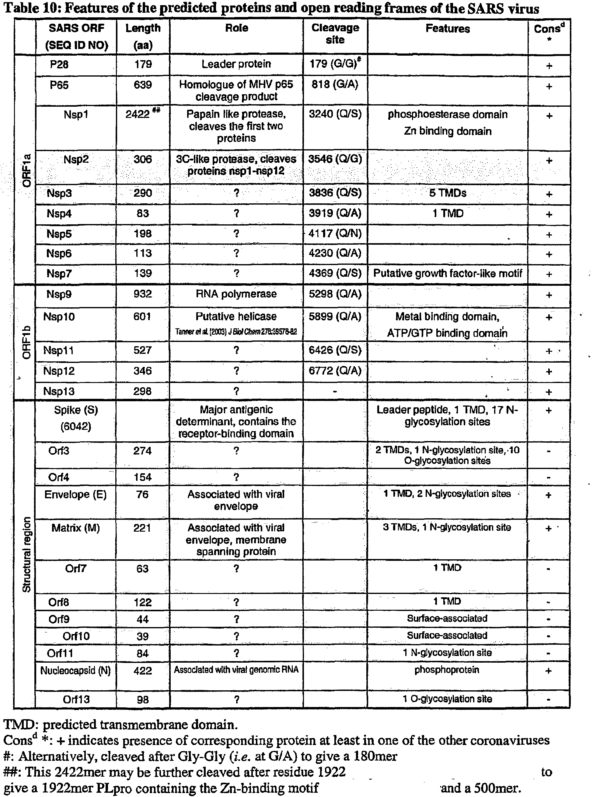
Table 11: Protein homologies between SARS and other coronaviruses Numbers indicate percentage of aminoacid identity between SARS proteins and corresponding gene products of other coronaviruses. More conserved pairs are in bold; more variable pairs are underlined. group 1group 2group 3Proteins 229E TGV PEDV MHV BCoV AIBV REPLICASE REGION leader protein p28 <20 <20 <20 27 <20 <20 p65 homologue <20 23 23 <20 20 <20 nsp1 (PLP protease) 25.5 25.8 25.4 29 30 25 nsp2 (3CL protease) 40.4 43.8 44.6 50 48.4 41 nsp3 30 27 29.4 34.2 35.5 28.5 nsp4 38.6 42.2 39.8 47.5 46.1 37.3 nsp5 48.2 42.9 43.9 46.8 47.3 38.7 nsp6 45.1 38.9 45.1 45.1 46.9 39.8 nsp7 53.8 54.5 56.1 56.2 55.4 58.3 nsp9 (polymerase) 59.8 59.6 60 67.3 66.9 62.4 nsp10 (helicase) 60.7 62 62.3 67.2 68.6 58.9 nsp11 52.3 53.7 52.3 57.6 57.6 52 nsp12 43.1 43 45.4 45.9 45 40.2 nsp13 56.4 54.4 55.3 63 65 53.4 STRUCTURAL REGION Spike (S) 28.8 31.6* 30.3 31.1 31 32.7* Envelope (E) 33* 27.9 20 23 26.5 23.2 Matrix glycoprotein (M) 30.6 32.5 34.8 40.8 41.9 32.5 Nucleocapsid (N) 26.9 30.1 29.5 37.3 37.4 31.5 * These three alignments were obtained only on a fragment of the whole protein. Table 12: Nucleotide and aminoacid differences between five SARS isolates FRA* TOR2* Urbani* CUHK* HKU* position° base/aminoacid base/aminoacid base/aminoacid base/aminoacid base/aminoacid ORF1a 2557 A/Thr G/Ala G/Ala G/Ala G/Ala 2601 T/Val C 7746 G/Pro T 7919 C/Ala T/Val 7930 G/Asp A/Asn 8387 G/Ser C/Thr 8416 G/Arg C/Thr 9404 T/Val C/Ala 9479 T/Val C/Ala 11448 T/Ile C C C C ORF1b 13494 GT/Val AG/Ser 16622 C/Ala T 17564 T/Asp C/Glu 17846 C/Arg T 18065 G/Lys A 18965 A/Ile T T T T 19064 A/Glu G G 19084 T/Ile C/Thr C/Thr C/Thr C/Thr spike 21721 G/Gly A/Asp 22222 T/Ile C/Thr 23220 T/Ser G/Ala 24872 T/Leu C 24933 T/Phe C/Leu C/Leu C/Leu C/Leu ORF3 25298 G/Gly A/Arg 25569 T/Met A/Lys matrix 26600 T/Val C/Ala C/Ala C/Ala 26857 T/Ser C/Pro ORF10 27827 T/Cys C/Arg nucleocapsid 28268 T/Ile C/Thr C/Thr C/Thr C/Thr * SARS coronavirus FRA (accession number AY310120)
SARS coronavirus TOR2 (accession number AY274119)
SARS coronavirus Urbani (accession number AY278741)
SARS coronavirus CUHK-W1 (accession number AY278554)
SARS coronavirus HKU-39849 (accession number AY278491)
° The position is based on the FRA sequence. - TABLES 13-25: T-epitope predictions for SEQ ID NO: 6042 Epitope predictions were performed at http://www.mpiib-berlin.mpg.de/MAPPP/binding.html using a minimum score of 0.5 and the BIMAS matrix, with a maximum of 20 results being selected. The analysis revealed 9mer and 10mer epitopes.
Table 16: Epitopes for SEQ ID NO: 6042 HLA A1 - 9 mers Maximum possible score using this molecule type 5625 Rank Start position Sequence % of max. score Score 1 846 SEQ ID NO: 8041 2.22 % 125 2 798 SEQ ID NO: 8042 1.6 % 90 3 787 SEQ ID NO: 8043 0.88 % 50 4 1178 SEQ ID NO: 8044 0.8 % 50 5 637 SEQ ID NO: 8045 0.8 % 45 6 557 SEQ ID NO: 8046 0.44 % 25 7 1020 SEQ ID NO: 8047 0.44 % 25 8 282 SEQ ID NO: 8048 0.32 % 18 9 1241 SEQ ID NO: 8049 0.24 % 13.5 10 466 SEQ ID NO: 8050 0.22 % 12.5 11 727 SEQ ID NO: 8051 0.2 % 11.25 12 706 SEQ ID NO: 8052 0.17 % 10 13 324 SEQ ID NO: 8053 0.16 % 9 14 752 SEQ ID NO: 8054 0.16 % 9 15 54 SEQ ID NO: 8055 0.13 % 7.5 16 554 SEQ ID NO: 8056 0.13 % 7.5 17 590 SEQ ID NO: 8057 0.12 % 6.75 18 569 SEQ ID NO: 8058 0.08 % 5 19 613 SEQ ID NO: 8059 0.08 % 5 20 90 SEQ ID NO: 8060 0.08 % 4.5 HLA A1 - 10 mers Maximum possible score using this molecule type 5625 Rank Start position Sequence % of max. score Score 1 1241 SEQ ID NO: 8061 4.8 % 270 2 967 SEQ ID NO: 8062 0.8 % 45 3 1010 SEQ ID NO: 8063 0.48 % 27 4 426 SEQ ID NO: 8064 0.44 % 25 5 809 SEQ ID NO: 8065 0.44 % 25 6 1178 SEQ ID NO: 8066 0.44 % 25 7 787 SEQ ID NO: 8067 0.22 % 12.5 8 958 SEQ ID NO: 8068 0.22 % 12.5 9 727 SEQ ID NO: 8069 0.2 % 11.25 10 610 SEQ ID NO: 8070 0.17 % 10 11 12 SEQ ID NO: 8071 0.13 % 7.5 12 1181 SEQ ID NO: 8072 0.12 % 6.75 13 373 SEQ ID NO: 8073 0.11 % 6.25 14 602 SEQ ID NO: 8074 0.11 % 6.25 15 20 SEQ ID NO: 8075 0.04 % 2.5 16 32 SEQ ID NO: 8076 0.04 % 2.5 17 53 SEQ ID NO: 8077 0.04 % 2.5 18 400 SEQ ID NO: 8078 0.04 % 2.5 19 557 SEQ ID NO: 8079 0.04 % 2.5 20 667 SEQ ID NO: 8080 0.04 % 2.5 HLA A3 - 9 mers Maximum possible score using this molecule type 12150 Rank Start position Sequence % of max. score Score 1 768 SEQ ID NO: 8081 0.82 % 100 2 808 SEQ ID NO: 8082 0.49 % 60 3 85 SEQ ID NO: 8083 0.24 % 30 4 663 SEQ ID NO: 8084 0.24 % 30 5 1245 SEQ ID NO: 8085 0.14 % 18 6 288 SEQ ID NO: 8086 0.09 % 12 7 50 SEQ ID NO: 8087 0.08 % 10 8 320 SEQ ID NO: 8088 0.07 % 9 9 402 SEQ ID NO: 8089 0.07 % 9 10 798 SEQ ID NO: 8090 0.07 % 9 11 902 SEQ ID NO: 8091 0.06 % 8.1 12 364 SEQ ID NO: 8092 0.05 % 6.75 13 297 SEQ ID NO: 8093 0.04 % 6 14 992 SEQ ID NO: 8094 0.04 % 6 15 38 SEQ ID NO: 8095 0.03 % 4.5 16 249 SEQ ID NO: 8096 0.03 % 4.5 17 706 SEQ ID NO: 8097 0.03 % 4.05 18 1204 SEQ ID NO: 8098 0.03 % 4.05 19 1178 SEQ ID NO: 8099 0.03 % 4 20 343 SEQ ID NO: 8100 0.02 % 3.6 HLA A3 - 10 mers Maximum possible score using this molecule type 12150 Rank Start position Sequence % of max. score Score 1 255 SEQ ID NO: 8101 1.48 % 180 2 180 SEQ ID NO: 8102 0.55 % 67.5 3 768 SEQ ID NO: 8103 0.49 % 60 4 1177 SEQ ID NO: 8104 0.49 % 60 5 380 SEQ ID NO: 8105 0.24 % 30 6 100 SEQ ID NO: 8106 0.18 % 22.5 7 786 SEQ ID NO: 8107 0.16 % 20 8 1217 SEQ ID NO: 8108 0.16 % 20 9 207 SEQ ID NO: 8109 0.14 % 18 10 1183 SEQ ID NO: 8110 0.14 % 18 11 38 SEQ ID NO: 8111 0.09 % 12 12 52 SEQ ID NO: 8112 0.09 % 12 13 8 SEQ ID NO: 8113 0.06 % 8 14 679 SEQ ID NO: 8114 0.06 % 8 15 73 SEQ ID NO: 8115 0.05 % 6.75 16 1204 SEQ ID NO: 8116 0.05 % 6.075 17 50 SEQ ID NO: 8117 0.04 % 6 18 774 SEQ ID NO: 8118 0.04 % 6 19 845 SEQ ID NO: 8119 0.04 % 6 20 214 SEQ ID NO: 8120 0.04 % 5.4 HLA A24 - 9 mers Maximum possible score using this molecule type 1596.672 Rank Start position Sequence % of max. score Score 1 1118 SEQ ID NO: 8121 19.84 % 316.8 2 51 SEQ ID NO: 8122 18.78 % 300 3 161 SEQ ID NO: 8123 18.78 % 300 4 434 SEQ ID NO: 8124 18.78 % 300 5 365 SEQ ID NO: 8125 13.77 % 220 6 736 SEQ ID NO: 8126 12.52 % 200 7 620 SEQ ID NO: 8127 7.51 % 120 8 1068 SEQ ID NO: 8128 7.51 % 120 9 817 SEQ ID NO: 8129 3.75 % 60 10 336 SEQ ID NO: 8130 3.44 % 55 11 687 SEQ ID NO: 8131 3.13 % 50 12 254 SEQ ID NO: 8132 2.34 % 37.5 13 627 SEQ ID NO: 8133 1.87 % 30 14 950 SEQ ID NO: 8134 1.75 % 28 15 28 SEQ ID NO: 8135 1.56 % 25 16 408 SEQ ID NO: 8136 1.56 % 25 17 159 SEQ ID NO: 8137 1.31 % 21 18 1166 SEQ ID NO: 8138 1.26 % 20.16 19 45 SEQ ID NO: 8139 1.25 % 20 20 185 SEQ ID NO: 8140 1.25 % 20 HLA A24 - 10 mers Maximum possible score using this molecule type 1596.672 Rank Start position Sequence % of max. score Score 1 438 SEQ ID NO: 8141 27.55 % 440 2 489 SEQ ID NO: 8142 22.54 % 360 3 254 SEQ ID NO: 8143 18.78 % 300 4 354 SEQ ID NO: 8144 11.27 % 180 5 406 SEQ ID NO: 8145 11.27 % 180 6 1047 SEQ ID NO: 8146 11.27 % 180 7 473 SEQ ID NO: 8147 7.51 % 120 8 350 SEQ ID NO: 8148 6.26 % 100 9 769 SEQ ID NO: 8149 6.26 % 100 10 193 SEQ ID NO: 8150 5.63 % 90 11 479 SEQ ID NO: 8151 3.13 % 50 12 0 SEQ ID NO: 8152 2.70 % 43.2 13 813 SEQ ID NO: 8153 1.87 % 30 14 739 SEQ ID NO: 8154 1.50 % 24 15 782 SEQ ID NO: 8155 1.50 % 24 16 1186 SEQ ID NO: 8156 1.31 % 21 17 910 SEQ ID NO: 8157 1.05 % 16.8 18 128 SEQ ID NO: 8158 0.93 % 15 19 183 SEQ ID NO: 8159 0.93 % 15 20 1069 SEQ ID NO: 8160 0.93 % 15 HLA A 0201 - 9 mers Maximum possible score using this molecule type 3925227.1 Rank Start position Sequence % of max. score Score 1 1041 SEQ ID NO: 8161 0.01 % 484.2379773 2 981 SEQ ID NO: 8162 0.00 % 382.536 3 957 SEQ ID NO: 8163 0.00 % 342.4606344 4 896 SEQ ID NO: 8164 0.00 % 232.6931712 5 1173 SEQ ID NO: 8165 0.00 % 201.447432 6 733 SEQ ID NO: 8166 0.00 % 171.86796 7 410 SEQ ID NO: 8167 0.00 % 135.45252 8 786 SEQ ID NO: 8168 0.00 % 119.463012 9 150 SEQ ID NO: 8169 0.00 % 102.17550222 10 1 SEQ ID NO: 8170 0.00 % 94.98737754 11 595 SEQ ID NO: 8171 0.00 % 93.239424 12 1095 SEQ ID NO: 8172 0.00 % 89.41779 13 1166 SEQ ID NO: 8173 0.00 % 87.58584 14 845 SEQ ID NO: 8174 0.00 % 79.642008 15 734 SEQ ID NO: 8175 0.00 % 73.47672 16 802 SEQ ID NO: 8176 0.00 % 71.872056 17 1213 SEQ ID NO: 8177 0.00 % 71.872056 18 105 SEQ ID NO: 8178 0.00 % 50.232 19 939 SEQ ID NO: 8179 0.00 % 49.13352 20 130 SEQ ID NO: 8180 0.00 % 48.732354 HLA A 0201 - 10 mers Maximum possible score using this molecule type 3925227.1 Rank Start position Sequence % of max. score Score 1 372 SEQ ID NO: 8181 0.04 % 1896.33528 2 410 SEQ ID NO: 8182 0.02 % 1134.00849744 3 162 SEQ ID NO: 8183 0.01 % 685.3897512 4 1076 SEQ ID NO: 8184 0.01 % 640.90320525 5 1196 SEQ ID NO: 8185 0.01 % 623.742666372 6 353 SEQ ID NO: 8186 0.01 % 446.7384576 7 50 SEQ ID NO: 8187 0.00 % 375.97824 8 733 SEQ ID NO: 8188 0.00 % 271.863864 9 130 SEQ ID NO: 8189 0.00 % 235.6873848 10 415 SEQ ID NO: 8190 0.00 % 185.679 11 297 SEQ ID NO: 8191 0.00 % 177.496704 12 1 SEQ ID NO: 8192 0.00 % 152.42160582 13 56 SEQ ID NO: 8193 0.00 % 110.013876 14 732 SEQ ID NO: 8194 0.00 % 101.0988 15 6 SEQ ID NO: 8195 0.00 % 98.26704 16 261 SEQ ID NO: 8196 0.00 % 91.60164 17 1040 SEQ ID NO: 8197 0.00 % 76.98537 18 928 SEQ ID NO: 8198 0.00 % 71.2908 19 1188 SEQ ID NO: 8199 0.00 % 69.81282 20 1094 SEQ ID NO: 8200 0.00 % 52.5987 HLA A 1101 - 9 mers Maximum possible score using this molecule type 36 Rank Start position Sequence % of max. Score 1 402 SEQ ID NO: 8201 25 % 9 2 902 SEQ ID NO: 8202 22.5 % 8.1 3 288 SEQ ID NO: 8203 11.11 % 4 4 85 SEQ ID NO: 8204 6.66 % 2.4 5 706 SEQ ID NO: 8205 6.66 % 2.4 6 456 SEQ ID NO: 8206 5.55 % 2 7 920 SEQ ID NO: 8207 5.55 % 2 8 535 SEQ ID NO: 8208 5 % 1.8 9 364 SEQ ID NO: 8209 3.33 % 1.2 10 438 SEQ ID NO: 8210 3.33 % 1.2 11 798 SEQ ID NO: 8211 3.33 % 1.2 12 808 SEQ ID NO: 8212 3.33 % 1.2 13 937 SEQ ID NO: 8213 3.33 % 1.2 14 956 SEQ ID NO: 8214 3.33 % 1.2 15 557 SEQ ID NO: 8215 2.77 % 1 16 1218 SEQ ID NO: 8216 2.77 % 1 17 784 SEQ ID NO: 8217 2.5 % 0.9 18 249 SEQ ID NO: 8218 2.22 % 0.8 19 768 SEQ ID NO: 8219 2.22 % 0.8 20 1178 SEQ ID NO: 8220 2.22 % 0.8 HLA A 1101 - 10 mers Maximum possible score using this molecule type 36 Rank Start position Sequence % of max. score Score 1 38 SEQ ID NO: 8221 13.33 % 4.8 2 807 SEQ ID NO: 8222 12.5 % 4.5 3 100 SEQ ID NO: 8223 11.11 % 4 4 380 SEQ ID NO: 8224 11.11 % 4 5 767 SEQ ID NO: 8225 10 % 3.6 6 533 SEQ ID NO: 8226 8.33 % 3 7 967 SEQ ID NO: 8227 6.66 % 2.4 8 919 SEQ ID NO: 8228 5.55 % 2 9 305 SEQ ID NO: 8229 5 % 1.8 10 211 SEQ ID NO: 8230 3.33 % 1.2 11 511 SEQ ID NO: 8231 3.33 % 1.2 12 1177 SEQ ID NO: 8232 3.33 % 1.2 13 429 SEQ ID NO: 8233 2.77 % 1 14 758 SEQ ID NO: 8234 2.77 % 1 15 797 SEQ ID NO: 8235 2.5 % 0.9 16 255 SEQ ID NO: 8236 2.22 % 0.8 17 986 SEQ ID NO: 8237 2.22 % 0.8 18 1157 SEQ ID NO: 8238 2.22 % 0.8 19 170 SEQ ID NO: 8239 1.66 % 0.6 20 893 SEQ ID NO: 8240 1.66 % 0.6 HLA B7 - 9 mers Maximum possible score using this molecule type 5400 Rank Start position Sequence % of max. score Score 1 200 SEQ ID NO: 8241 1.48 % 80 2 1243 SEQ ID NO: 8242 1.48 % 80 3 123 SEQ ID NO: 8243 0.74 % 40 4 248 SEQ ID NO: 8244 0.66 % 36 5 1036 SEQ ID NO: 8245 0.66 % 36 6 494 SEQ ID NO: 8246 0.37 % 20 7 495 SEQ ID NO: 8247 0.37 % 20 8 523 SEQ ID NO: 8248 0.37 % 20 9 842 SEQ ID NO: 8249 0.37 % 20 10 932 SEQ ID NO: 8250 0.37 % 20 11 274 SEQ ID NO: 8251 0.33 % 18 12 588 SEQ ID NO: 8252 0.22 % 12 13 656 SEQ ID NO: 8253 0.22 % 12 14 657 SEQ ID NO: 8254 0.22 % 12 15 767 SEQ ID NO: 8255 0.22 % 12 16 911 SEQ ID NO: 8256 0.22 % 12 17 939 SEQ ID NO: 8257 0.22 % 12 18 1007 SEQ ID NO: 8258 0.22 % 12 19 1170 SEQ ID NO: 8259 0.22 % 12 20 1206 SEQ ID NO: 8260 0.22 % 12 HLA B7 - 10 mers Maximum possible score using this molecule type 5400 Rank Start position Sequence % of max. score Score 1 505 SEQ ID NO: 8261 4.44 % 240 2 312 SEQ ID NO: 8262 3.70 % 200 3 141 SEQ ID NO: 8263 1.11 % 60 4 1006 SEQ ID NO: 8264 0.66 % 36 5 411 SEQ ID NO: 3265 0.44 % 24 6 122 SEQ ID NO: 8266 0.37 % 20 7 134 SEQ ID NO: 8267 0.37 % 20 8 184 SEQ ID NO: 8268 0.37 % 20 9 367 SEQ ID NO: 8269 0.37 % 20 10 402 SEQ ID NO: 8270 0.37 % 20 11 494 SEQ ID NO: 8271 0.37 % 20 12 560 SEQ ID NO: 8272 0.37 % 20 13 626 SEQ ID NO: 8273 0.37 % 20 14 931 SEQ ID NO: 8274 0.37 % 20 15 956 SEQ ID NO: 8275 0.37 % 20 16 1117 SEQ ID NO: 8276 0.37 % 20 17 1169 SEQ ID NO: 8277 0.37 % 20 18 1196 SEQ ID NO: 8278 0.37 % 20 19 247 SEQ ID NO: 8279 0.22 % 12 20 273 SEQ ID NO: 8280 0.22 % 12 TABLE 26: Cloned sequences for E.coli expression ORF DNA length bp Cloning pET pGEX P28 537 NdeI / XhoI P65 1917 NheI / HindIII Nsp1A 2495 NheI / XhoI Nsp1B 2153 NdeI / XhoI Nsp1C 2612 NdeI / XhoI Nsp2A 431 NdeI / XhoI BamHI / XhoI Nsp2B 426 NdeI / XhoI BamHI / XhoI Nsp3 870 NdeI / XhoI Nsp4 249 NdeI / XhoI BamHI / XhoI Nsp5 594 NheI / XhoI Nsp6 339 NdeI / XhoI BamHI / XhoI Nsp7 417 NdeI / XhoI BamHI / XhoI Nsp9A 1385 NheI /XhoI Nsp9B 1409 NdeI / XhoI Nsp10 1803 Nhel / XhoI Nsp11 1581 NdeI / XhoI Nsp12 1038 NdeI / HindIII Nsp1A3 897 NdeI / XhoI Spike (S1) 1946 NdeI / XhoI Spike (S2) 1598 NdeI / XhoI Spike (S1-S2) 3545 NdeI / XhoI HR1 287 NdeI / XhoI BamHI / XhoI HR2 146 NdeI / XhoI BamHI / XhoI ORF30100 525 NdeI / XhoI ORF4 465 NdeI / XhoI Envelope (E) 231 NdeI / XhoI BamHI / XhoI Matrix (M)Δ100 366 NdeI / XhoI BamHI / XhoI ORF7Δ18 137 NdeI / XhoI BamHI / XhoI ORF8 369 NdeI / XhoI BamHI / XhoI ORF9 135 NdeI / XhoI BamHI / XhoI ORF10 120 NheI / XhoI BamHI / XhoI ORF11 255 NdeI / XhoI BamHI / XhoI Nucleocapsid (N) 1269 NdeI / EcoRI ORF12 297 NdeI / EcoRI BamHI / EcoRI TABLE 29: Cloning, purification and expression in E.coli SARS CoV ORFs M.W Kd cloning Expr. purification as P28 19,7 + + his sol P65 70,3 + + his sol Nsp1A (N-term) 91,6 + + his ins Nsp1B (core) 80,8 + - Nsp1C (C-term) 95,3 + - Nsp2A (N-term) 15,8 + + his ins Nsp2B (C-term) 15,5 + + his sol Nsp3 31,9 + + Nsp4 9,1 + + his sol Nsp5 21,8 + + his sol Nsp6 12,4 + + his sol Nsp9A (N-term) 15,3 + - Nsp9B (C-term) 51,6 + + his ins Nsp10 66 - Nsp11 58 - - Nsp12 38 - Nsp13 32,7 + + his ins Spike (S1-his) 71,3 + + his ins Spike (S2-his) 58,6 + - Spike (S1S2-his) 130 + + his ins HR1 11 + + his ins HR2 5,4 + + his sol ORF3 Δ1001 19,1 + - ORF4 16,9 + + his ins (trimer) Envelope (E) 34,3 + + qst ins (IB) Matrix (M) Δ100 13,3 + + his ins ORF7Δ182 31 + + gst sol ORF8 39,5 + gst ins (IB) ORF9 30,8 + + gst sol ORF10 30,3 + + gst ins (IB) ORF11 35,2 + + gst ins (IB) Nucleocapsid (N) 43,6 + + his ins ORF12 36,7 + + his ins TABLE 30: E.coli expression, purity and yield Protein Tag Purity (%) Yield (mg/l) Nsp2A (N-term) His 95 1.7 Nsp2B (C-term) His 95 4.1 Nsp4 His 95 12.6 Nsp5 His 95 5.88 Nsp6 His 95 8.1 P28 His 95 1 P65 His 80 0.553 HR2 His 95 11.9 HR1 His 80 2.64 Nsp1A His 95 0.267 Spike S1-S2 His 80 0.381 Matrix M His 85 12.4 ORF7 GST 85 4.9 -
7188-7189 Component sequences of Figure 25 7194 Amino acids 901-1005 of SEQ ID NO: 6042 7196 Amino acids 1144-1196 of SEQ ID NO: 6042 7197-7199 Membrane fusion peptide regions 7207-7223 N-glycosylation sites within SEQ ID NO: 6042 7307-7308 Fragments of SEQ ID NO: 6042 7398-7399 Antigenic fragments of SEQ ID NO: 6042 9785-9798 Oligonucleotides used for S.cerevisiae expression 9921-9923 pCMVKm2 vectors 9962 Spike variant - <210> 6042
<211> 1255
<212> PRT
<213> SARS coronavirus - <400> 6042


- <210> 7188
<211> 2712
<212> PRT
<213> SARS coronavirus - <400> 7188

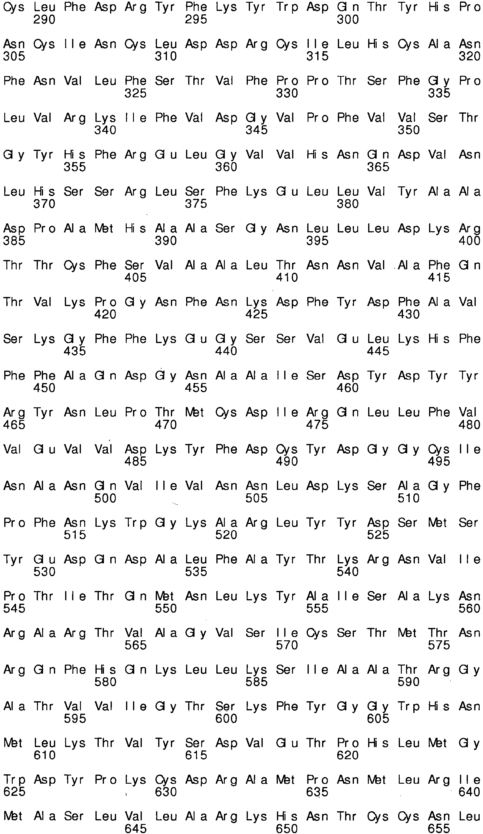


- <210> 7189
<211> 1257
<212> PRT
<213> SARS coronavirus - <400> 7189


- <210> 7193
<211> 102
<212> PRT
<213> SARS cor onavi r us - <400> 7193
Ile Asp Arg

Leu Ile Thr 100 - <210> 7194
<211> 105
<212> PRT
<213> SARS coronavi rus - <400> 7194

- <210> 7196
<211> 52
<212> PRT
<213> SARS cor onavi r us - <400> 7196

- <210> 7197
<211> 19
<212> PRT
<213> SARS coronavirus - <400> 7197

- <210> 7198
<211> 17
<212> PRT
<213> SARS coronavirus - <400> 7198

- <210> 7199
<211> 20
<212> PRT
<213> SARS coronavirus - <400> 7199

- <210> 7207
<211> 4
<212> PRT
<213> SARS coronavirus - <400> 7207

- <210> 7208
<211> 4
<212> PRT
<213> SARS coronavirus - <400> 7208

- <210> 7209
<211> 4
<212> PRT
<213> SARS coronavirus - <400> 7209

- <210> 7210
<211> 4
<212> PRT
<213> SARS coronavirus - <400> 7210

- <210> 7211
<211> 4
<212> PRT
<213> SARS coronavirus - <400> 7211

- <210> 7212
<211> 4
<212> PRT
<213> SARS cor onavi r us - <400> 7212

- <210> 7213
<211> 4
<212> PRT
<213> SARS cor onavi rus - <400> 7213

- <210> 7214
<211> 4
<212> PRT
<213> SARS coronavirus - <400> 7214

- <210> 7215
<211> 4
<212> PRT
<213> SARS coronavirus - <400> 7215

- <210> 7216
<211> 4
<212> PRT
<213> SARS coronavirus - <400> 7216

- <210> 7217
<211> 4
<212> PRT
<213> SARS coronavirus - <400> 7217

- <210> 7218
<211> 4
<212> PRT
<213> SARS coronavirus - <400> 7218

- <210> 7219
<211> 4
<212> PRT
<213> SARS coronavirus - <400> 7219


- <210> 7220
<211> 4
<212> PRT
<213> SARS coronavirus - <400> 7220

- <210> 7221
<211> 4
<212> PRT
<213> SARS coronavirus - <400> 7221

- <210> 7222
<211> 4
<212> PRT
<213> SARS coronavirus - <400> 7222

- <210> 7223
<211> 4
<212> PRT
<213> SARS coronavirus - <400> 7223

- <210> 7307
<211> 649
<212> PRT
<213> SARS coronavirus - <400> 7307


- <210> 7308
<211> 533
<212> PRT
<213> SARS coronavirus - <400> 7308


- <210> 7398
<211> 16
<212> PRT
<213> SARS coronavirus - <400> 7398

- <210> 7399
<211> 15
<212> PRT
<213> SARS coronavirus - <400> 7399

- <210> 8041
<211> 9
<212> PRT
<213> SARS coronavirus - <400> 8041

- <210> 8042
<211> 9
<212> PRT
<213> SARS coronavirus - <400> 8042

- <210> 8043
<211> 9
<212> PRT
<213> SARS coronavirus - <400> 8043

- <210> 8044
<211> 9
<212> PRT
<213> SARS coronavirus - <400> 8044

- <210> 8045
<211> 9
<212> PRT
<213> SARS coronavirus - <400> 8045

- <210> 8046
<211> 9
<212> PRT
<213> SARS coronavirus - <400> 8046

- <210> 8047
<211> 9
<212> PRT
<213> SARS coronavirus - <400> 8047

- <210> 8048
<211> 9
<212> PRT
<213> SARS coronavirus - <400> 8048

- <210> 8049
<211> 9
<212> PRT
<213> SARS coronavirus - <400> 8049

- <210> 8050
<211> 9
<212> PRT
<213> SARS coronavirus - <400> 8050

- <210> 8051
<211> 9
<212> PRT
<213> SARS coronavirus - <400> 8051

- <210> 8052
<211> 9
<212> PRT
<213> SARS cor onavi r us - <400> 8052

- <210> 8053
<211> 9
<212> PRT
<213> SARS cor onavi r us - <400> 8053

- <210> 8054
<211> 9
<212> PRT
<213> SARS coronavirus - <400> 8054

- <210> 8055
<211> 9
<212> PRT
<213> SARS coronavirus - <400> 8055

- <210> 8056
<211> 9
<212> PRT
<213> SARS coronavirus - <400> 8056

- <210> 8057
<211> 9
<212> PRT
<213> SARS cor onavi r us - <400> 8057


- <210> 8058
<211> 9
<212> PRT
<213> SARS coronavirus - <400> 8058

- <210> 8059
<211> 9
<212> PRT
<213> SARS coronavirus - <400> 8059

- <210> 8060
<211> 9
<212> PRT
<213> SARS cor onavi r us - <400> 8060

- <210> 8061
<211> 10
<212> PRT
<213> SARS coronavirus - <400> 8061

- <210> 8062
<211> 10
<212> PRT
<213> SARS coronavirus - <400> 8062

- <210> 8063
<211> 10
<212> PRT
<213> SARS coronavirus - <400> 8063

- <210> 8064
<211> 10
<212> PRT
<213> SARS coronavirus - <400> 8064

- <210> 8065
<211> 10
<212> PRT
<213> SARS cor onavi r us - <400> 8065

- <210> 8066
<211> 10
<212> PRT
<213> SARS coronavirus - <400> 8066

- <210> 8067
<211> 10
<212> PRT
<213> SARS cor onavi r us - <400> 8067

- <210> 8068
<211> 10
<212> PRT
<213> SARS coronavirus - <400> 8068

- <210> 8069
<211> 10
<212> PRT
<213> SARS coronavirus - <400> 8069

- <210> 8070
<211> 10
<212> PRT
<213> SARS coronavirus - <400> 8070

- <210> 8071
<211> 10
<212> PRT
<213> SARS coronavirus - <400> 8071

- <210> 8072
<211> 10
<212> PRT
<213> SARS coronavirus - <400> 8072

- <210> 8073
<211> 10
<212> PRT
<213> SARS coronavirus - <400> 8073

- <210> 8074
<211> 10
<212> PRT
<213> SARS coronavirus - <400> 8074

- <210> 8075
<211> 10
<212> PRT
<213> SARS coronavirus - <400> 8075

- <210> 8076
<211> 10
<212> PRT
<213> SARS coronavirus - <400> 8076

- <210> 8077
<211> 10
<212> PRT
<213> SARS coronavirus - <400> 8077

- <210> 8078
<211> 10
<212> PRT
<213> SARS coronavirus - <400> 8078

- <210> 8079
<211> 10
<212> PRT
<213> SARS coronavirus - <400> 8079

- <210> 8080
<211> 10
<212> PRT
<213> SARS coronavirus - <400> 8080


- <210> 8081
<211> 9
<212> PRT
<213> SARS coronavirus - <400> 8081

- <210> 8082
<211> 9
<212> PRT
<213> SARS coronavirus - <400> 8082

- <210> 8083
<211> 9
<212> PRT
<213> SARS coronavirus - <400> 8083

- <210> 8084
<211> 9
<212> PRT
<213> SARS coronavirus - <400> 8084

- <210> 8085
<211> 9
<212> PRT
<213> SARS coronavirus - <400> 8085

- <210> 8086
<211> 9
<212> PRT
<213> SARS coronavirus - <400> 8086

- <210> 8087
<211> 9
<212> PRT
<213> SARS coronavirus - <400> 8087

- <210> 8088
<211> 9
<212> PRT
<213> SARS coronavirus - <400> 8088

- <210> 8089
<211> 9
<212> PRT
<213> SARS coronavirus - <400> 8089

- <210> 8090
<211> 9
<212> PRT
<213> SARS coronavirus - <400> 8090

- <210> 8091
<211> 9
<212> PRT
<213> SARS coronavirus - <400> 8091

- <210> 8092
<211> 9
<212> PRT
<213> SARS cor onavi r us - <400> 8092

- <210> 8093
<211> 9
<212> PRT
<213> SARS cor onavi r us - <400> 8093

- <210> 8094
<211> 9
<212> PRT
<213> SARS coronavirus - <400> 8094

- <210> 8095
<211> 9
<212> PRT
<213> SARS cor onavi r us - <400> 8095

- <210> 8096
<211> 9
<212> PRT
<213> SARS cor onavi r us - <400> 8096

- <210> 8097
<211> 9
<212> PRT
<213> SARS cor onavi r us - <400> 8097

- <210> 8098
<211> 9
<212> PRT
<213> SARS cor onavi r us - <400> 8098

- <210> 8099
<211> 9
<212> PRT
<213> SARS coronavirus - <400> 8099

- <210> 8100
<211> 9
<212> PRT
<213> SARS cor onavi r us - <400> 8100

- <210> 8101
<211> 10
<212> PRT
<213> SARS coronavirus - <400> 8101

- <210> 8102
<211> 10
<212> PRT
<213> SARS coronavirus - <400> 8102

- <210> 8103
<211> 10
<212> PRT
<213> SARS cor onavi r us - <400> 8103


- <210> 8104
<211> 10
<212> PRT
<213> SARS coronavirus - <400> 8104

- <210> 8105
<211> 10
<212> PRT
<213> SARS cor onavi r us - <400> 8105

- <210> 8106
<211> 10
<212> PRT
<213> SARS cor onavi r us - <400> 8106

- <210> 8107
<211> 10
<212> PRT
<213> SARS cor onavi r us - <400> 8107

- <210> 8108
<211> 10
<212> PRT
<213> SARS coronavirus - <400> 8108

- <210> 8109
<211> 10
<212> PRT
<213> SARS coronavirus - <400> 8109

- <210> 8110
<211> 10
<212> PRT
<213> SARS coronavirus - <400> 8110

- <210> 8111
<211> 10
<212> PRT
<213> SARS coronavirus - <400> 8111

- <210> 8112
<211> 10
<212> PRT
<213> SARS coronavirus - <400> 8112

- <210> 8113
<211> 10
<212> PRT
<213> SARS coronavirus - <400> 8113

- <210> 8114
<211> 10
<212> PRT
<213> SARS coronavirus - <400> 8114

- <210> 8115
<211> 10
<212> PRT
<213> SARS coronavirus - <400> 8115

- <210> 8116
<211> 10
<212> PRT
<213> SARS coronavirus - <400> 8116

- <210> 8117
<211> 10
<212> PRT
<213> SARS coronavirus - <400> 8117

- <210> 8118
<211> 10
<212> PRT
<213> SARS cor onavi r us - <400> 8118

- <210> 8119
<211> 10
<212> PRT
<213> SARS cor onavi r us - <400> 8119

- <210> 8120
<211> 10
<212> PRT
<213> SARS cor onavi r us - <400> 8120

- <210> 8121
<211> 9
<212> PRT
<213> SARS cor onavi r us - <400> 8121

- <210> 8122
<211> 9
<212> PRT
<213> SARS coronavirus - <400> 8122

- <210> 8123
<211> 9
<212> PRT
<213> SARS coronavirus - <400> 8123

- <210> 8124
<211> 9
<212> PRT
<213> SARS coronavirus - <400> 8124

- <210> 8125
<211> 9
<212> PRT
<213> SARS coronavirus - <400> 8125

- <210> 8126
<211> 9
<212> PRT
<213> SARS coronavirus - <400> 8126


- <210> 8127
<211> 9
<212> PRT
<213> SARS coronavirus - <400> 8127

- <210> 8128
<211> 9
<212> PRT
<213> SARS coronavirus - <400> 8128

- <210> 8129
<211> 9
<212> PRT
<213> SARS coronavi rus - <400> 8129

- <210> 8130
<211> 9
<212> PRT
<213> SARS coronavirus - <400> 8130

- <210> 8131
<211> 9
<212> PRT
<213> SARS coronavirus - <400> 8131

- <210> 8132
<211> 9
<212> PRT
<213> SARS coronavirus - <400> 8132

- <210> 8133
<211> 9
<212> PRT
<213> SARS coronavirus - <400> 8133

- <210> 8134
<211> 9
<212> PRT
<213> SARS cor onavi r us - <400> 8134

- <210> 8135
<211> 9
<212> PRT
<213> SARS coronavi rus - <400> 8135

- <210> 8136
<211> 9
<212> PRT
<213> SARS coronavi rus - <400> 8136

- <210> 8137
<211> 9
<212> PRT
<213> SARS coronavi rus - <400> 8137

- <210> 8138
<211> 9
<212> PRT
<213> SARS coronavirus - <400> 8138

- <210> 8139
<211> 9
<212> PRT
<213> SARS coronavi rus - <400> 8139

- <210> 8140
<211> 9
<212> PRT
<213> SARS coronavi rus - <400> 8140

- <210> 8141
<211> 10
<212> PRT
<213> SARS coronavirus - <400> 8141

- <210> 8142
<211> 10
<212> PRT
<213> SARS coronavirus - <400> 8142

- <210> 8143
<211> 10
<212> PRT
<213> SARS coronavirus - <400> 8143

- <210> 8144
<211> 10
<212> PRT
<213> SARS coronavi rus - <400> 8144

- <210> 8145
<211> 10
<212> PRT
<213> SARS cor onavi r us - <400> 8145

- <210> 8146
<211> 10
<212> PRT
<213> SARS cor onavi r us - <400> 8146

- <210> 8147
<211> 10
<212> PRT
<213> SARS coronavirus - <400> 8147

- <210> 8148
<211> 10
<212> PRT
<213> SARS coronavirus - <400> 8148

- <210> 8149
<211> 10
<212> PRT
<213> SARS coronavirus - <400> 8149


- <210> 8150
<211> 10
<212> PRT
<213> SARS coronavirus - <400> 8150

- <210> 8151
<211> 10
<212> PRT
<213> SARS cor onavi r us - <400> 8151

- <210> 8152
<211> 10
<212> PRT
<213> SARS coronavirus - <400> 8152

- <210> 8153
<211> 10
<212> PRT
<213> SARS coronavirus - <400> 8153

- <210> 8154
<211> 10
<212> PRT
<213> SARS coronavirus - <400> 8154

- <210> 8155
<211> 10
<212> PRT
<213> SARS cor onavi r us - <400> 8155

- <210> 8156
<211> 10
<212> PRT
<213> SARS coronavirus - <400> 8156

- <210> 8157
<211> 10
<212> PRT
<213> SARS coronavirus - <400> 8157

- <210> 8158
<211> 10
<212> PRT
<213> SARS coronavirus - <400> 8158

- <210> 8159
<211> 10
<212> PRT
<213> SARS coronavirus - <400> 8159

- <210> 8160
<211> 10
<212> PRT
<213> SARS coronavirus - <400> 8160

- <210> 8161
<211> 9
<212> PRT
<213> SARS coronavirus - <400> 8161

- <210> 8162
<211> 9
<212> PRT
<213> SARS coronavirus - <400> 8162

- <210> 8163
<211> 9
<212> PRT
<213> SARS cor onavi r us - <400> 8163

- <210> 8164
<211> 9
<212> PRT
<213> SARS cor onavi r us - <400> 8164

- <210> 8165
<211> 9
<212> PRT
<213> SARS coronavirus - <400> 8165

- <210> 8166
<211> 9
<212> PRT
<213> SARS coronavirus - <400> 8166

- <210> 8167
<211> 9
<212> PRT
<213> SARS coronavi rus - <400> 8167

- <210> 8168
<211> 9
<212> PRT
<213> SARS coronavirus - <400> 8168

- <210> 8169
<211> 9
<212> PRT
<213> SARS coronavirus - <400> 8169

- <210> 8170
<211> 9
<212> PRT
<213> SARS coronavirus - <400> 8170

- <210> 8171
<211> 9
<212> PRT
<213> SARS coronavi rus - <400> 8171

- <210> 8172
<211> 9
<212> PRT
<213> SARS coronavirus - <400> 8172


- <210> 8173
<211> 9
<212> PRT
<213> SARS cor onavi r us - <400> 8173

- <210> 8174
<211> 9
<212> PRT
<213> SARS coronavi rus - <400> 8174

- <210> 8175
<211> 9
<212> PRT
<213> SARS coronavi rus - <400> 8175

- <210> 8176
<211> 9
<212> PRT
<213> SARS coronavirus - <400> 8176

- <210> 8177
<211> 9
<212> PRT
<213> SARS coronavirus - <400> 8177

- <210> 8178
<211> 9
<212> PRT
<213> SARS coronavirus - <400> 8178

- <210> 8179
<211> 9
<212> PRT
<213> SARS cor onavi r us - <400> 8179

- <210> 8180
<211> 9
<212> PRT
<213> SARS coronavi rus - <400> 8180

- <210> 8181
<211> 10
<212> PRT
<213> SARS coronavirus - <400> 8181

- <210> 8182
<211> 10
<212> PRT
<213> SARS coronavi rus - <400> 8182

- <210> 8183
<211> 10
<212> PRT
<213> SARS coronavirus - <400> 8183

- <210> 8184
<211> 10
<212> PRT
<213> SARS coronavirus - <400> 8184

- <210> 8185
<211> 10
<212> PRT
<213> SARS coronavirus - <400> 8185

- <210> 8186
<211> 10
<212> PRT
<213> SARS coronavirus - <400> 8186

- <210> 8187
<211> 10
<212> PRT
<213> SARS coronavirus - <400> 8187

- <210> 8188
<211> 10
<212> PRT
<213> SARS coronavirus - <400> 8188

- <210> 8189
<211> 10
<212> PRT
<213> SARS coronavirus - <400> 8189

- <210> 8190
<211> 10
<212> PRT
<213> SARS coronavirus - <400> 8190

- <210> 8191
<211> 10
<212> PRT
<213> SARS cor onavi r us - <400> 8191

- <210> 8192
<211> 10
<212> PRT
<213> SARS coronavi rus - <400> 8192

- <210> 8193
<211> 10
<212> PRT
<213> SARS coronavirus - <400> 8193

- <210> 8194
<211> 10
<212> PRT
<213> SARS coronavirus - <400> 8194

- <210> 8195
<211> 10
<212> PRT
<213> SARS coronavirus - <400> 8195


- <210> 8196
<211> 10
<212> PRT
<213> SARS coronavirus - <400> 8196

- <210> 8197
<211> 10
<212> PRT
<213> SARS coronavirus - <400> 8197

- <210> 8198
<211> 10
<212> PRT
<213> SARS cor onavi r us - <400> 8198

- <210> 8199
<211> 10
<212> PRT
<213> SARS coronavirus - <400> 8199

- <210> 8200
<211> 10
<212> PRT
<213> SARS cor onavi r us - <400> 8200

- <210> 8201
<211> 9
<212> PRT
<213> SARS cor onavi r us - <400> 8201

- <210> 8202
<211> 9
<212> PRT
<213> SARS cor onavi r us - <400> 8202

- <210> 8203
<211> 9
<212> PRT
<213> SARS coronavirus - <400> 8203

- <210> 8204
<211> 9
<212> PRT
<213> SARS coronavirus - <400> 8204

- <210> 8205
<211> 9
<212> PRT
<213> SARS coronavirus - <400> 8205

- <210> 8206
<211> 9
<212> PRT
<213> SARS cor onavi r us - <400> 8206

- <210> 8207
<211> 9
<212> PRT
<213> SARS coronavirus - <400> 8207

- <210> 8208
<211> 9
<212> PRT
<213> SARS coronavirus - <400> 8208

- <210> 8209
<211> 9
<212> PRT
<213> SARS coronavirus - <400> 8209

- <210> 8210
<211> 9
<212> PRT
<213> SARS coronavirus - <400> 8210

- <210> 8211
<211> 9
<212> PRT
<213> SARS coronavirus - <400> 8211

- <210> 8212
<211> 9
<212> PRT
<213> SARS coronavirus - <400> 8212

- <210> 8213
<211> 9
<212> PRT
<213> SARS coronavirus - <400> 8213

- <210> 8214
<211> 9
<212> PRT
<213> SARS cor onavi r us - <400> 8214

- <210> 8215
<211> 9
<212> PRT
<213> SARS coronavirus - <400> 8215

- <210> 8216
<211> 9
<212> PRT
<213> SARS coronavirus - <400> 8216

- <210> 8217
<211> 9
<212> PRT
<213> SARS coronavirus - <400> 8217

- <210> 8218
<211> 9
<212> PRT
<213> SARS coronavirus - <400> 8218


- <210> 8219
<211> 9
<212> PRT
<213> SARS coronavirus - <400> 8219

- <210> 8220
<211> 9
<212> PRT
<213> SARS coronavirus - <400> 8220

- <210> 8221
<211> 10
<212> PRT
<213> SARS coronavi rus - <400> 8221

- <210> 8222
<211> 10
<212> PRT
<213> SARS coronavirus - <400> 8222

- <210> 8223
<211> 10
<212> PRT
<213> SARS coronavirus - <400> 8223

- <210> 8224
<211> 10
<212> PRT
<213> SARS cor onavi r us - <400> 8224

- <210> 8225
<211> 10
<212> PRT
<213> SARS coronavirus - <400> 8225

- <210> 8226
<211> 10
<212> PRT
<213> SARS coronavirus - <400> 8226

- <210> 8227
<211> 10
<212> PRT
<213> SARS coronavirus - <400> 8227

- <210> 8228
<211> 10
<212> PRT
<213> SARS coronavirus - <400> 8228

- <210> 8229
<211> 10
<212> PRT
<213> SARS coronavirus - <400> 8229

- <210> 8230
<211> 10
<212> PRT
<213> SARS coronavirus - <400> 8230

- <210> 8231
<211> 10
<212> PRT
<213> SARS coronavirus - <400> 8231

- <210> 8232
<211> 10
<212> PRT
<213> SARS coronavirus - <400> 8232

- <210> 8233
<211> 10
<212> PRT
<213> SARS coronavirus - <400> 8233

- <210> 8234
<211> 10
<212> PRT
<213> SARS coronavirus - <400> 8234

- <210> 8235
<211> 10
<212> PRT
<213> SARS coronavirus - <400> 8235

- <210> 8236
<211> 10
<212> PRT
<213> SARS coronavirus - <400> 8236

- <210> 8237
<211> 10
<212> PRT
<213> SARS cor onavi r us - <400> 8237

- <210> 8238
<211> 10
<212> PRT
<213> SARS cor onavi r us - <400> 8238

- <210> 8239
<211> 10
<212> PRT
<213> SARS coronavirus - <400> 8239

- <210> 8240
<211> 10
<212> PRT
<213> SARS coronavirus - <400> 8240

- <210> 8241
<211> 9
<212> PRT
<213> SARS coronavirus - <400> 8241


- <210> 8242
<211> 9
<212> PRT
<213> SARS coronavirus - <400> 8242

- <210> 8243
<211> 9
<212> PRT
<213> SARS coronavirus - <400> 8243

- <210> 8244
<211> 9
<212> PRT
<213> SARS coronavirus - <400> 8244

- <210> 8245
<211> 9
<212> PRT
<213> SARS coronavirus - <400> 8245

- <210> 8246
<211> 9
<212> PRT
<213> SARS coronavirus - <400> 8246

- <210> 8247
<211> 9
<212> PRT
<213> SARS coronavirus - <400> 8247

- <210> 8248
<211> 9
<212> PRT
<213> SARS coronavirus - <400> 8248

- <210> 8249
<211> 9
<212> PRT
<213> SARS coronavirus - <400> 8249

- <210> 8250
<211> 9
<212> PRT
<213> SARS coronavirus - <400> 8250

- <210> 8251
<211> 9
<212> PRT
<213> SARS coronavirus - <400> 8251

- <210> 8252
<211> 9
<212> PRT
<213> SARS coronavirus - <400> 8252

- <210> 8253
<211> 9
<212> PRT
<213> SARS coronavirus - <400> 8253

- <210> 8254
<211> 9
<212> PRT
<213> SARS cor onavi r us - <400> 8254

- <210> 8255
<211> 9
<212> PRT
<213> SARS coronavirus - <400> 8255

- <210> 8256
<211> 9
<212> PRT
<213> SARS coronavirus - <400> 8256

- <210> 8257
<211> 9
<212> PRT
<213> SARS coronavi rus - <400> 8257

- <210> 8258
<211> 9
<212> PRT
<213> SARS coronavirus - <400> 8258

- <210> 8259
<211> 9
<212> PRT
<213> SARS coronavirus - <400> 8259

- <210> 8260
<211> 9
<212> PRT
<213> SARS coronavirus - <400> 8260

- <210> 8261
<211> 10
<212> PRT
<213> SARS coronavirus - <400> 8261

- <210> 8262
<211> 10
<212> PRT
<213> SARS coronavirus - <400> 8262

- <210> 8263
<211> 10
<212> PRT
<213> SARS coronavirus - <400> 8263

- <210> 8264
<211> 10
<212> PRT
<213> SARS coronavirus - <400> 8264


- <210> 8265
<211> 10
<212> PRT
<213> SARS coronavirus - <400> 8265

- <210> 8266
<211> 10
<212> PRT
<213> SARS coronavirus - <400> 8266

- <210> 8267
<211> 10
<212> PRT
<213> SARS coronavirus - <400> 8267

- <210> 8268
<211> 10
<212> PRT
<213> SARS coronavirus - <400> 8268

- <210> 8269
<211> 10
<212> PRT
<213> SARS coronavirus - <400> 8269

- <210> 8270
<211> 10
<212> PRT
<213> SARS coronavirus - <400> 8270

- <210> 8271
<211> 10
<212> PRT
<213> SARS coronavirus - <400> 8271

- <210> 8272
<211> 10
<212> PRT
<213> SARS coronavirus - <400> 8272

- <210> 8273
<211> 10
<212> PRT
<213> SARS coronavirus - <400> 8273

- <210> 8274
<211> 10
<212> PRT
<213> SARS coronavirus - <400> 8274

- <210> 8275
<211> 10
<212> PRT
<213> SARS coronavirus - <400> 8275

- <210> 8276
<211> 10
<212> PRT
<213> SARS coronavi rus - <400> 8276

- <210> 8277
<211> 10
<212> PRT
<213> SARS coronavirus - <400> 8277

- <210> 8278
<211> 10
<212> PRT
<213> SARS coronavi rus - <400> 8278

- <210> 8279
<211> 10
<212> PRT
<213> SARS coronavirus - <400> 8279

- <210> 8280
<211> 10
<212> PRT
<213> SARS coronavirus - <400> 8280

- <210> 9785
<211> 42
<212> DNA
<213> Artificial Sequence - <220>
<223> Primer - <400> 9785
agatcttcta gataaaagaa gtgaccttga ccggt gcaccac 42 - <210> 9786
<211> 50
<212> DNA
<213> Artificial Sequence - <220>
<223> Primer - <400> 9786
agatctccta ggcattcgcc atattgcttc atgaagccag catcagcgag 50 - <210> 9787
<211> 49
<212> DNA
<213> Artificial Sequence - <220>
<223> Primer - <400> 9787
agat ct cct a ggtgatatta atgctaggga cctcatttgt gcgcagaag 49 - <210> 9788
<211> 45
<212> DNA
<213> Artificial Sequence - <220>
<223> Primer - <400> 9788
agatctgtcg act cat t at g tgtaatgtaatttgacaccc t t gag 45 - <210> 9789
<211> 44
<212> DNA
<213> Artificial Sequence - <220>
<223> Oligonucleotide - <400> 9789
tgagcatgtt gatacttctt t at gagt gcga cat t cct at t ggag 44 - <210> 9790
<211> 39
<212> DNA
<213> Artificial Sequence - <220>
<223> Oligonucleotide - <400> 9790
ctggcatttg tgctagttac catacagttt ctttattac 39 - <210> 9791
<211> 58
<212> DNA
<213> Artificial Sequence - <220>
<223> Oligonucleotide - <400> 9791
gt agt act ag ccaaaaat ct attgtggctt atactatgtc tttaggtgct gat agt t c 58 - <210> 9792
<211> 52
<212> DNA
<213> Artificial Sequence - <220>
<223> Oligonucleotide - <400> 9792
aattgaacta tcagcaccta aagacat agt at aagccaca atagattttt gg 52 - <210> 9793
<211> 37
<212> DNA
<213> Artificial Sequence - <220>
<223> Oligonucleotide - <400> 9793
ct agt act ac gt aat aaaga aact gt at gg t aactag 37 - <210> 9794
<211> 53
<212> DNA
<213> Artificial Sequence - <220>
<223> Oligonucleotide - <400> 9794
cacaaat gcc agctccaata ggaatgtcgc act cat aaga agt at caaca t gc 53 - <210> 9795
<211> 29
<212> DNA
<213> Artificial Sequence - <220>
<223> Oligonucleotide - <400> 9795
agcttacaaa acaaaat gag tgacct t ga 29 - <210> 9796
<211> 29
<212> DNA
<213> Artificial Sequence - <220>
<223> Oligonucleotide - <400> 9796
ccggt caagg t cact cat t t tgt t t t g t a 29 - <210> 9797
<211> 68
<212> DNA
<213> Artificial Sequence - <220>
<223> Oligonucleotide - <400> 9797

- <210> 9798
<211> 72
<212> DNA
<213> Artificial Sequence - <220>
<223> Oligonucleotide - <400> 9798

- <210> 9921
<211> 8098
<212> DNA
<213> Artificial Sequence - <220>
<223> pCMVKm2 vect or - <400> 9921


- <210> 9922
<211> 7918
<212> DNA
<213> Artificial Sequence - <220>
<223> pCMVKm2 vect or - <400> 9922


- <210> 9923
<211> 4333
<212> DNA
<213> Artificial Sequence - <220>
<223> pCMVKm2 vect or - <400> 9923


- <210> 9962
<211> 1255
<212> PRT
<213> SARS coronavirus - <400> 9962


Claims (16)
- An immunogenic composition comprising an isolated or purified polypeptide comprisinga) the Severe Acute Respiratory Syndrome (SARS) virus Spike polypeptide amino acid sequence SEQ ID NO: 6042, orb) an amino acid sequence having >80% sequence identity to SEQ ID NO: 6042, orc) a fragment of at least 10 consecutive amino acids of SEQ ID NO: 6042;
with the proviso that the polypeptide is not MFIFLLFLTLTSGSDLDRCTTFDDVQAP. - An immunogenic composition comprising an isolated or purified polypeptide comprisinga) amino acid sequence SEQ ID NO: 6042, orb) an amino acid sequence having >80% sequence identity to SEQ ID NO: 6042, orc) a fragment of at least 10 consecutive amino acids of SEQ ID NO: 6042;
and further comprising an adjuvant. - The composition of claim 1 or claim 2, wherein the polypeptide comprises: (a) an amino acid sequence having >85%, or >90%, or >95% sequence identity to SEQ ID NO: 6042.
- The composition of claim 1 or claim 2, wherein the polypeptide comprises a fragment of at least 25, or at least 40, or at least 100 consecutive amino acids of SEQ ID NO: 6042.
- The composition of any preceding claim, wherein the polypeptide is isolated and purified from the SARS virus.
- The composition of any preceding claim, wherein the polypeptide is produced by recombinant expression.
- The composition of claim 6, wherein the fragment comprises any of the polypeptide sequences given in SEQ ID NOs: 7197, 7198, 7199 and 8041-8280.
- The composition of claim 6 wherein the polypeptide is a fusion protein.
- The composition of claim 8, wherein the fusion protein comprises one or more of the following:a) the S1 domain (SEQ ID NO: 7307),b) S2 domain (SEQ ID NO: 7308),c) the receptor binding region of the S1 domain,d) the oligomerization domain regions of the S2 domain,e) the leucine zipper region of the S2 domain,f) the membrane anchor region of the S2 domain,g) the hydrophobic domain region of the S2 domain,h) the cytoplasmic tail region of the S2 domain, and/ori) any of the polypeptide sequences given in SEQ ID NOs: 7193-7194, 7196-7199, 7207-7223, 7398, 7399 and 8041-8280.
- The composition of any preceding claim wherein the polypeptide is in oligomeric form.
- The composition of claim 10, wherein the oligomer is in trimeric form.
- The composition of any one of claims 8 or 9, wherein the fusion protein comprisesa) a tag sequence,b) a second SARS virus protein,c) a non-SARS virus protein,d) a bacterial protein and/ore) an adjuvant.
- The composition of claim 12, wherein the non-SARS virus protein is derived from a coronavirus, influenza virus, rhinovirus, parainfluenza virus (PIV), respiratory syncytial virus (RSV), adenovirus, and/or metapneumovirus.
- The composition of claim 12, wherein the bacterial protein is a bacterial adhesion protein or fragment thereof, such as NadA, YadA, UspA2 or a NadA-like protein.
- The composition of claim 2, or of claims 3-14 when dependent from claim 2, wherein the adjuvant is selected from: mineral salts, oil emulsions, saponin formulations, bacterial or microbial derivatives, human immunomodulators, bioadhesives or mucoadhesives, microparticels, liposomes, polyethylene ether and polyethylene ester formuations, PCPP, muramyl peptides, immidazoquinolone compounds, and/or virosomes or virus-like particles.
- The composition of claim 15, wherein the adjuvant is a detoxified bacterial ADP-ribosylating toxin, a non-toxic double mutant form of Bordella pertussis toxoids, chitosan, MF59, aluminum, an aluminum salt or a small molecule immunomodulator (SMIP).
Applications Claiming Priority (22)
| Application Number | Priority Date | Filing Date | Title |
|---|---|---|---|
| US46221803P | 2003-04-10 | 2003-04-10 | |
| US46246503P | 2003-04-11 | 2003-04-11 | |
| US46241803P | 2003-04-12 | 2003-04-12 | |
| US46274803P | 2003-04-13 | 2003-04-13 | |
| US46310903P | 2003-04-14 | 2003-04-14 | |
| US46346003P | 2003-04-15 | 2003-04-15 | |
| US46366803P | 2003-04-16 | 2003-04-16 | |
| US46398303P | 2003-04-17 | 2003-04-17 | |
| US46397103P | 2003-04-18 | 2003-04-18 | |
| US46483803P | 2003-04-22 | 2003-04-22 | |
| US46489903P | 2003-04-22 | 2003-04-22 | |
| US46527303P | 2003-04-23 | 2003-04-23 | |
| US46553503P | 2003-04-24 | 2003-04-24 | |
| US46831203P | 2003-05-05 | 2003-05-05 | |
| US47314403P | 2003-05-22 | 2003-05-22 | |
| US49502403P | 2003-08-14 | 2003-08-14 | |
| US50565203P | 2003-09-23 | 2003-09-23 | |
| US51078103P | 2003-10-11 | 2003-10-11 | |
| US52946403P | 2003-12-11 | 2003-12-11 | |
| US53617704P | 2004-01-12 | 2004-01-12 | |
| US56075704P | 2004-04-07 | 2004-04-07 | |
| PCT/US2004/011710 WO2004092360A2 (en) | 2003-04-10 | 2004-04-09 | The severe acute respiratory syndrome coronavirus |
Publications (2)
| Publication Number | Publication Date |
|---|---|
| EP1618127A2 EP1618127A2 (en) | 2006-01-25 |
| EP1618127B1 true EP1618127B1 (en) | 2014-12-10 |
Family
ID=33304326
Family Applications (1)
| Application Number | Title | Priority Date | Filing Date |
|---|---|---|---|
| EP04750188.7A Expired - Lifetime EP1618127B1 (en) | 2003-04-10 | 2004-04-09 | Immunogenic composition comprising a spike protein of the SARS coronavirus |
Country Status (7)
| Country | Link |
|---|---|
| US (1) | US20060257852A1 (en) |
| EP (1) | EP1618127B1 (en) |
| AU (2) | AU2004230485B2 (en) |
| CA (1) | CA2522379C (en) |
| ES (1) | ES2529736T3 (en) |
| NZ (1) | NZ543467A (en) |
| WO (1) | WO2004092360A2 (en) |
Cited By (2)
| Publication number | Priority date | Publication date | Assignee | Title |
|---|---|---|---|---|
| WO2021203044A3 (en) * | 2020-04-03 | 2022-02-17 | Icahn School Of Medicine At Mount Sinai | High-throughput assay for circulating antibodies against severe acute respiratory syndrome coronavirus 2 |
| AU2020403296B2 (en) * | 2019-12-13 | 2025-03-06 | Grand Theravac Life Science (Nanjing) Co., Ltd. | Immunostimulatory composition and use thereof |
Families Citing this family (176)
| Publication number | Priority date | Publication date | Assignee | Title |
|---|---|---|---|---|
| WO2004085650A1 (en) * | 2003-03-24 | 2004-10-07 | The University Of Hong Kong | A high-troughput diagnostic assay for the human virus causing severe acute respiratory syndrome (sars) |
| TW200508247A (en) * | 2003-04-08 | 2005-03-01 | Vironovative Bv | Sars |
| US7582740B2 (en) * | 2003-04-17 | 2009-09-01 | The Trustees Of Columbia University In The City Of New York | Methods and kits for detecting SARS-associated coronavirus |
| US7220852B1 (en) * | 2003-04-25 | 2007-05-22 | The United States Of America As Represented By The Secretary Of The Department Of Health And Human Services, Centers For Disease Control And Prevention | Coronavirus isolated from humans |
| WO2005019410A2 (en) * | 2003-04-25 | 2005-03-03 | Intradigm Corporation | Rnai agents for anti-sars coronavirus therapy |
| GB2401175A (en) * | 2003-05-02 | 2004-11-03 | Hong Kong Dna Chips Ltd | Detection of SARS virus by PCR |
| WO2005035556A2 (en) * | 2003-05-06 | 2005-04-21 | Iguazu Biosciences Corp. | Sars-coronavirus virus-like particles and methods of use |
| US8080642B2 (en) * | 2003-05-16 | 2011-12-20 | Vical Incorporated | Severe acute respiratory syndrome DNA compositions and methods of use |
| WO2004109289A1 (en) * | 2003-06-10 | 2004-12-16 | Agency For Science, Technology And Research | Method of diagnosing sars corona virus infection |
| AU2004269320B2 (en) * | 2003-06-20 | 2009-12-24 | Protein Sciences Corporation | Vectors expressing SARS immunogens, compositions containing such vectors or expression products thereof, methods and assays for making and using |
| US20070270361A1 (en) * | 2003-08-04 | 2007-11-22 | Shan Lu | Sars Nucleic Acids, Proteins, Vaccines, and Uses Thereof |
| KR20070008504A (en) * | 2003-08-22 | 2007-01-17 | 버치 바이오메디칼 리서치 엘엘씨 | Double Allele Detection of SARS-Related Coronaviruses |
| WO2005027963A2 (en) * | 2003-09-15 | 2005-03-31 | The United States Of America As Represented By Thesecretary Of Health And Human Services, Nih | METHODS AND COMPOSITIONS FOR THE GENERATION OF A PROTECTIVE IMMUNE RESPONSE AGAINTS SARS-CoV |
| US7622112B2 (en) | 2003-12-05 | 2009-11-24 | Jody Berry | Anti-SARS monoclonal antibodies |
| EP1701977A2 (en) * | 2003-12-10 | 2006-09-20 | Agency for Science, Technology and Research | Sars coronavirus s proteins and uses thereof |
| KR100994568B1 (en) * | 2003-12-12 | 2010-11-15 | 삼성전자주식회사 | SARS coronavirus detection primer, Sascoronavirus detection method and kit using the same |
| US20050171044A1 (en) * | 2003-12-24 | 2005-08-04 | Stein David A. | Oligonucleotide compound and method for treating nidovirus infections |
| TWI303249B (en) * | 2004-01-09 | 2008-11-21 | Nat Health Research Institutes | Receptor binding polypeptides |
| WO2006071250A2 (en) * | 2004-04-05 | 2006-07-06 | Government Of The United States Of America As Represented By The Secretary Of The Department Of Health And Human Services | Soluble fragments of the sars-cov spike glycoprotein |
| ES2647491T3 (en) | 2004-05-21 | 2017-12-21 | Novartis Vaccines And Diagnostics, Inc. | Alphavirus vectors for influenza virus vaccines |
| WO2006115509A2 (en) | 2004-06-24 | 2006-11-02 | Novartis Vaccines And Diagnostics Inc. | Small molecule immunopotentiators and assays for their detection |
| US20060199176A1 (en) * | 2004-07-15 | 2006-09-07 | Yeau-Ching Wang | Coronavirus S peptides |
| US20060165716A1 (en) | 2004-07-29 | 2006-07-27 | Telford John L | Immunogenic compositions for gram positive bacteria such as streptococcus agalactiae |
| SG119380A1 (en) * | 2004-08-04 | 2006-02-28 | Agency Science Tech & Res | Proteins encoded by the severe acute respiratory syndrome (sars) coronavirus and a role in apoptosis |
| EP1632564A1 (en) * | 2004-09-03 | 2006-03-08 | Consejo Superior De Investigaciones Cientificas | Vaccine against severe accute respiratory syndrome causing coronavirus (SARS-CoV) |
| US20060093616A1 (en) * | 2004-09-29 | 2006-05-04 | Ralf Altmeyer | Process for vaccinating eucaryotic hosts and for protecting against SARS-CoV infection |
| WO2006091610A2 (en) * | 2005-02-23 | 2006-08-31 | The Brigham And Women's Hospital, Inc. | Inhibitors of enveloped virus infectivity |
| US20070190065A1 (en) * | 2005-06-03 | 2007-08-16 | Ralf Altmeyer | Nucleic acids, polypeptides, methods of expression, and immunogenic compositions associated with SARS corona virus spike protein |
| CA2626253A1 (en) | 2005-10-18 | 2007-04-26 | Novartis Vaccines And Diagnostics, Inc. | Mucosal and systemic immunizations with alphavirus replicon particles |
| WO2007052163A2 (en) | 2005-11-01 | 2007-05-10 | Novartis Vaccines And Diagnostics Gmbh & Co Kg | Cell-derived viral vaccines with low levels of residual cell dna by beta-propiolactone treatment |
| AU2006335256B2 (en) | 2005-11-22 | 2012-10-18 | Novartis Vaccines And Diagnostics, Inc. | Norovirus and sapovirus antigens |
| US20080103746A1 (en) | 2005-11-30 | 2008-05-01 | Searete Llc, A Limited Liability Corporation | Systems and methods for pathogen detection and response |
| US20080241935A1 (en) | 2007-03-27 | 2008-10-02 | Searete Llc, A Limited Liability Corporation Of The State Of Delaware | Methods for pathogen detection |
| WO2007109813A1 (en) | 2006-03-23 | 2007-09-27 | Novartis Ag | Imidazoquinoxaline compounds as immunomodulators |
| EP2343385B1 (en) | 2006-04-11 | 2014-05-21 | Bio-Rad Innovations | HPV detection and quantification by real-time multiplex amplification |
| US20110053248A1 (en) * | 2006-10-23 | 2011-03-03 | Medimmune, Llc | Serum-Free Virus Propagation Platform For A Virus Vaccine Candidate |
| US10001496B2 (en) | 2007-01-29 | 2018-06-19 | Gearbox, Llc | Systems for allergen detection |
| US8617903B2 (en) * | 2007-01-29 | 2013-12-31 | The Invention Science Fund I, Llc | Methods for allergen detection |
| GB0711858D0 (en) * | 2007-06-19 | 2007-07-25 | Glaxosmithkline Biolog Sa | Vaccine |
| CA2699513C (en) | 2007-09-12 | 2018-03-13 | Novartis Ag | Gas57 mutant antigens and gas57 antibodies |
| JP2009109426A (en) * | 2007-10-31 | 2009-05-21 | Sysmex Corp | Analyte pretreatment liquid, virus measuring kit and virus detecting method |
| EP2217250A4 (en) | 2007-11-09 | 2011-01-05 | California Inst Of Techn | IMMUNOREGULATORY COMPOUNDS AND RELATED COMPOSITIONS AND METHODS |
| BRPI0821240B8 (en) | 2007-12-21 | 2022-10-04 | Novartis Ag | mutant forms of streptolysin o |
| ES2546213T3 (en) | 2008-03-03 | 2015-09-21 | Novartis Ag | Compounds and compositions as modulators of TLR activity |
| EP2286223B1 (en) * | 2008-05-02 | 2016-07-06 | University Of Rochester | Arrayed detector system for measurement of influenza immune response |
| US8450056B2 (en) | 2008-05-02 | 2013-05-28 | University Of Rochester | Arrayed imaging reflectometry (AIR) sensor chip comprising virus-like particles suitable for the detection of antiviral immune responses |
| WO2010039154A1 (en) * | 2008-10-05 | 2010-04-08 | Board Of Trustees Of Leland Stanford Junior University | Hepatitis c antibodies and uses thereof |
| EP2360173B1 (en) * | 2008-11-28 | 2016-01-13 | NOF Corporation | Cytotoxic t cell epitope peptide for sars coronavirus, and use thereof |
| GB0822001D0 (en) * | 2008-12-02 | 2009-01-07 | Glaxosmithkline Biolog Sa | Vaccine |
| CN102481349B (en) | 2009-01-12 | 2014-10-15 | 诺华股份有限公司 | Cna_B Domain in Vaccines Against Gram-Positive Bacteria |
| CA2765112A1 (en) | 2009-06-10 | 2010-12-16 | Novartis Ag | Benzonaphthyridine-containing vaccines |
| CN102482666B (en) | 2009-07-06 | 2017-02-08 | 变异生物技术公司 | Methods of making vesicles and formulations produced therefrom |
| CA2803282C (en) | 2009-07-06 | 2018-05-01 | David E. Anderson | Methods for preparing vesicles and formulations produced therefrom |
| JO3257B1 (en) | 2009-09-02 | 2018-09-16 | Novartis Ag | Vehicles and installations as TLR |
| AU2010290896B2 (en) | 2009-09-02 | 2014-07-03 | Glaxosmithkline Biologicals S.A. | Immunogenic compositions including TLR activity modulators |
| WO2011057148A1 (en) | 2009-11-05 | 2011-05-12 | Irm Llc | Compounds and compositions as tlr-7 activity modulators |
| US9408907B2 (en) | 2009-12-15 | 2016-08-09 | Glaxosmithkline Biologicals Sa | Homogenous suspension of immunopotentiating compounds and uses thereof |
| US20130028933A1 (en) * | 2009-12-28 | 2013-01-31 | Ligocyte Pharmaceuticals, Inc. | Methods for stabilizing influenza antigen enveloped virus-based virus-like particle solutions |
| WO2011114346A1 (en) | 2010-03-18 | 2011-09-22 | Chetan Balar | Chitin and related compounds for use in treating bacterial and viral infections |
| AU2011232421B2 (en) | 2010-03-23 | 2015-08-13 | Novartis Ag | Compounds (cystein based lipopeptides) and compositions as TLR2 agonists used for treating infections, inflammations, respiratory diseases etc. |
| HUE040658T2 (en) * | 2010-04-07 | 2019-03-28 | California Inst Of Techn | Vehicle for delivering a compound to a mucous membrane and related compositions, methods and systems |
| CN102933267B (en) | 2010-05-28 | 2015-05-27 | 泰特里斯在线公司 | Interactive hybrid asynchronous computer game infrastructure |
| CN103096922B (en) | 2010-07-06 | 2019-08-06 | 变异生物技术公司 | For treating the composition and method of influenza |
| US9192661B2 (en) | 2010-07-06 | 2015-11-24 | Novartis Ag | Delivery of self-replicating RNA using biodegradable polymer particles |
| WO2012054879A1 (en) * | 2010-10-22 | 2012-04-26 | Duke University | Compositions and methods for the treatment of septic arthritis, osteomyelitis, and bacteremia |
| ES2434737T3 (en) * | 2010-10-27 | 2013-12-17 | Lonza Biologics Plc. | Rapid targeted cell selection (cell lines) procedure |
| WO2012075379A2 (en) | 2010-12-02 | 2012-06-07 | Oncolytics Biotech Inc. | Liquid viral formulations |
| CN103269716B (en) | 2010-12-02 | 2015-05-27 | 昂科利蒂克斯生物科技公司 | Lyophilized viral formulations |
| SG191781A1 (en) | 2010-12-30 | 2013-08-30 | Lab Francais Du Fractionnement | Glycols as pathogen inactivating agents |
| BR112013017939B1 (en) * | 2011-01-13 | 2022-11-16 | Variation Biotechnologies Inc | LYOPHILIZED IMMUNOGENIC COMPOSITION, USE AND METHOD FOR PREPARING THE SAME |
| KR101939336B1 (en) * | 2011-02-18 | 2019-01-16 | 주식회사 엘지화학 | Composition for Detecting Virus Relating Respiratory Disease and Kit for Detecting Virus Relating Respiratory Disease Comprising the Same |
| EP2731617A4 (en) | 2011-07-12 | 2015-07-01 | Brigham & Womens Hospital | LIPID-CONTAINING PSA COMPOSITIONS, METHODS OF ISOLATION AND METHODS OF USING SAME |
| EP2802353A4 (en) | 2012-01-12 | 2015-12-02 | Variation Biotechnologies Inc | COMPOSITIONS AND METHODS FOR TREATING VIRAL INFECTIONS |
| CN104244984B (en) | 2012-01-27 | 2020-03-31 | 变异生物技术公司 | Methods and compositions for therapeutic agents |
| HK1205138A1 (en) | 2012-07-27 | 2015-12-11 | Institut National De La Santé Et De La Recherche Médicale (Inserm) | Cd147 as receptor for pilus-mediated adhesion of meningococci to vascular endothelia |
| TR201807340T4 (en) | 2013-02-01 | 2018-06-21 | Glaxosmithkline Biologicals Sa | Intradermal administration of immunological compositions comprising bell-like receptor agonists. |
| US9889194B2 (en) | 2013-03-01 | 2018-02-13 | New York Blood Center, Inc. | Immunogenic composition for MERS coronavirus infection |
| WO2015113102A1 (en) * | 2014-01-30 | 2015-08-06 | Adelaide Research & Innovation Pty Ltd | Self assembled carbon based structures and related methods |
| US11103575B2 (en) | 2014-02-28 | 2021-08-31 | The New York Blood Center, Inc. | Immunogenic composition for MERS coronavirus infection |
| JP2017515900A (en) * | 2014-05-09 | 2017-06-15 | スローン − ケタリング・インスティテュート・フォー・キャンサー・リサーチ | Naphtaquinone methyltransferase inhibitors and their use |
| WO2015179412A1 (en) * | 2014-05-19 | 2015-11-26 | Merial, Inc. | Recombinant spike protein subunit based vaccine for porcine epidemic diarrhea virus (pedv) |
| JO3701B1 (en) * | 2014-05-23 | 2021-01-31 | Regeneron Pharma | Human antibiotics for MERS-CoV-coronavirus |
| GB201413020D0 (en) * | 2014-07-23 | 2014-09-03 | Pribright The Inst | Coronavirus |
| KR20170103874A (en) * | 2015-01-23 | 2017-09-13 | 더 트러스티스 오브 더 유니버시티 오브 펜실바니아 | Immunogenicity of an optimized synthetic consensus DNA vaccine for swine-diarrheal virus |
| CN104762270A (en) * | 2015-01-30 | 2015-07-08 | 肇庆大华农生物药品有限公司 | Embryo-derived chicken infectious bronchitis virus purification method |
| US11331335B2 (en) | 2015-06-10 | 2022-05-17 | California Institute Of Technology | Sepsis treatment and related compositions methods and systems |
| JP6918365B2 (en) | 2015-08-19 | 2021-08-11 | プレジデント アンド フェローズ オブ ハーバード カレッジ | Lipidized PSA Compositions and Methods |
| WO2017059204A1 (en) * | 2015-10-02 | 2017-04-06 | Virginia Commonwealth University | Azolylacryloyl derivatives as therapeutic agents for sickle cell disease |
| CA3040110A1 (en) * | 2015-10-13 | 2017-04-20 | Daniel C. Carter | Nsp10 self-assembling fusion proteins for vaccines, therapeutics, diagnostics and other nanomaterial applications |
| US11491181B2 (en) | 2016-07-15 | 2022-11-08 | President And Fellows Of Harvard College | Glycolipid compositions and methods of use |
| US10707531B1 (en) | 2016-09-27 | 2020-07-07 | New Dominion Enterprises Inc. | All-inorganic solvents for electrolytes |
| AU2018226566B2 (en) | 2017-03-03 | 2025-01-23 | Treos Bio Limited | Population-based immunogenic peptide identification platform |
| JP7432506B2 (en) | 2017-10-30 | 2024-02-16 | 武田薬品工業株式会社 | Environmentally compatible surfactants for inactivating lipid enveloped viruses |
| PL3752182T3 (en) | 2018-02-12 | 2024-11-25 | Inimmune Corporation | Toll-like receptor ligands |
| CN108949787A (en) * | 2018-07-05 | 2018-12-07 | 上海海洋大学 | A kind of goldfish Tgf2 transposase and its preparation and store method |
| MA53543A (en) | 2018-09-04 | 2021-07-14 | Treos Bio Ltd | PEPTIDE VACCINES |
| WO2020198865A1 (en) * | 2019-04-03 | 2020-10-08 | The University Of British Columbia | Oligopeptides for quantitative viral proteomic analysis methods and uses |
| CN114126645B (en) | 2019-05-10 | 2025-04-25 | 勃林格殷格翰动物保健有限公司 | Modified S1 subunit of coronavirus spike protein |
| EP3966228A1 (en) | 2019-05-10 | 2022-03-16 | Boehringer Ingelheim Vetmedica GmbH | Modified s2 subunit of the coronavirus spike protein |
| US20220218818A1 (en) * | 2019-06-03 | 2022-07-14 | Immunolux International Corp. | Smallpox vaccine and stem cells for treatment of disease |
| CN110729022B (en) * | 2019-10-24 | 2023-06-23 | 江西中烟工业有限责任公司 | A method for establishing an early liver injury model in passive smoking rats and a method for screening related genes |
| EP4054627A4 (en) * | 2019-11-07 | 2023-12-06 | Cornell University | USE OF MEMBRANE INHIBITORS TO IMPROVE VACCINE DEVELOPMENT AGAINST ENCLOSED VIRUSES |
| BR112022014578A2 (en) * | 2020-01-23 | 2022-09-13 | Sirnaomics Inc | COMPOSITION AND METHODS OF PROPHYLATIC AND THERAPEUTIC RNAI FOR THE TREATMENT OF SEVERE ACUTE RESPIRATORY INFECTION CAUSED BY THE NEW CORONA VIRUS 2019 (2019-NCOV) |
| CA3167611A1 (en) | 2020-02-13 | 2021-08-19 | Etienne Simon-Loriere | Nucleic acid vaccine against the sars-cov-2 coronavirus |
| EP4103232A4 (en) * | 2020-02-14 | 2025-03-26 | Epivax, Inc. | REGULATORY T-CELL EPITOPES AND DETOLERATED SARS-CoV-2 ANTIGENS |
| WO2021168427A1 (en) * | 2020-02-20 | 2021-08-26 | The Trustees Of Columbia University In The City Of New York | Compositions and methods for rapid detection of sars-cov-2 |
| IL296071A (en) | 2020-03-01 | 2022-11-01 | Valneva Austria Gmbh | Sars-cov-2 virus vaccine with adjuvant cytidine-phosphoguanosine |
| WO2021178306A1 (en) * | 2020-03-01 | 2021-09-10 | Dynavax Technologies Corporation | Coronavirus vaccines comprising a tlr9 agonist |
| EP4114986A4 (en) * | 2020-03-04 | 2024-03-27 | Premas Biotech Pvt. Ltd | EXPRESSION OF SARS-COV PROTEINS, NUCLEIC ACID CONSTRUCTS, VIRUS-LIKE PROTEINS (VLPS) AND RELEVANT METHODS |
| CN111172327A (en) * | 2020-03-05 | 2020-05-19 | 杭州丹威生物科技有限公司 | Method and kit for detecting novel coronavirus nucleic acid without taking hands |
| US12605442B2 (en) | 2020-03-06 | 2026-04-21 | Colorado State University Research Foundation | Production of vaccines comprising inactivated SARS-CoV-2 viral particles |
| WO2021181390A1 (en) * | 2020-03-09 | 2021-09-16 | Vaxil Biotherapeutics Ltd. | Antigen specific epitope–based anti-infective vaccines |
| WO2021188906A1 (en) * | 2020-03-19 | 2021-09-23 | Nature's Toolbox, Inc. | Novel mrna-based covid-19 multi-valent vaccine and methods of scaled production of the same |
| WO2021188334A1 (en) * | 2020-03-19 | 2021-09-23 | Noveome Biotherapeutics, Inc. | Use of st266 to treat severe systemic inflammation and post-acute covid-19 syndrome |
| US20230172962A1 (en) * | 2020-03-20 | 2023-06-08 | University Of Virginia Patent Foundation | Nrtis, nrti metabolites, and nrti analogs for macular degeneration and viral infections |
| WO2021188931A1 (en) * | 2020-03-20 | 2021-09-23 | The Board Of Trustees Of The Leland Stanford Junior University | Anti-coronaviral compositions and methods of using the same |
| WO2021195136A2 (en) * | 2020-03-23 | 2021-09-30 | The Regents Of The University Of California | Devices for detecting antibodies to coronavirus antigens |
| US20230323353A1 (en) * | 2020-03-25 | 2023-10-12 | City Of Hope | Antisense rna for treatment of sars-associated coronavirus |
| US11149320B1 (en) * | 2020-03-31 | 2021-10-19 | Diasorin S.P.A. | Assays for the detection of SARS-CoV-2 |
| WO2021198999A1 (en) * | 2020-04-03 | 2021-10-07 | Axon Neuroscience Se | Epitope-based vaccines for treatment of coronavirus associated diseases |
| GB202004974D0 (en) * | 2020-04-03 | 2020-05-20 | Treos Bio Ltd | Coronavirus vaccine |
| WO2021209925A1 (en) * | 2020-04-17 | 2021-10-21 | Pfizer Inc. | Coronavirus serology assay |
| US11124497B1 (en) | 2020-04-17 | 2021-09-21 | Pardes Biosciences, Inc. | Inhibitors of cysteine proteases and methods of use thereof |
| JP2023523411A (en) * | 2020-04-20 | 2023-06-05 | ザ・トラスティーズ・オブ・インディアナ・ユニバーシティー | Recombinant rotavirus expression system and recombinant rotavirus |
| US20230165880A1 (en) * | 2020-04-24 | 2023-06-01 | University Of Maryland, Baltimore | Compositions and methods for treating lung injury |
| EP4142786A4 (en) * | 2020-04-27 | 2024-06-19 | Ohio State Innovation Foundation | LIVE ATTENATED MEASLES VIRUS VECTORIZED VACCINE FOR SARS-COV-2 |
| WO2021220137A2 (en) * | 2020-04-27 | 2021-11-04 | Sapir Pharmaceuticals Inc. | Pannexin-1 inhibitors for the treatment of sars-cov-2 infected covid-19 patients with or without an associated acute respiratory syndrome |
| WO2021222569A1 (en) * | 2020-04-29 | 2021-11-04 | LU, Shi-long | Viral testing in saliva |
| WO2021222772A2 (en) * | 2020-05-01 | 2021-11-04 | The Johns Hopkins University | Compositions and methods for coronavirus detection |
| WO2021226137A1 (en) * | 2020-05-04 | 2021-11-11 | Sigilon Therapeutics, Inc. | Compositions, devices and methods for inducing immune responses to infectious agents |
| WO2021228853A1 (en) * | 2020-05-11 | 2021-11-18 | Ose Immunotherapeutics | Vaccine against sars-cov virus |
| WO2021231274A1 (en) * | 2020-05-11 | 2021-11-18 | Ampel Biosolutions, Llc | Methods and systems for analyzing targetable pathologic processes in covid-19 via gene expression analysis |
| IL298171A (en) | 2020-05-14 | 2023-01-01 | Ampel Biosolutions Llc | Methods and systems for machine learning analysis of single nucleotide polymorphisms in lupus |
| WO2021229077A1 (en) * | 2020-05-15 | 2021-11-18 | Biomvis S.R.L. | Bacterial outer membrane vesicles carrying coronavirus polypeptides, method of preparation, compositions and use thereof |
| JP7104449B2 (en) * | 2020-05-20 | 2022-07-21 | シンテカバイオ インコーポレイティッド | Composition for prevention or treatment of coronavirus infection of type 2 severe acute respiratory syndrome |
| KR102169476B1 (en) | 2020-05-20 | 2020-10-23 | (주)신테카바이오 | Composition for preventing or treating sars coronavirus 2 infection disease |
| US11191824B1 (en) * | 2020-05-22 | 2021-12-07 | The Government of the United States of America, as represented by the Secretary of Homeland Security | Method of purifying virus-like-particles |
| GB202008250D0 (en) * | 2020-06-02 | 2020-07-15 | Emergex Vaccines Holding Ltd | Diagnosis, prevention and treatment of coronavirus infection |
| US20230218745A1 (en) * | 2020-06-04 | 2023-07-13 | New York University | Modified alphavirus for use as covid-19 vaccine |
| CN111518959A (en) * | 2020-06-05 | 2020-08-11 | 上海市计量测试技术研究院 | Digital PCR detection method and kit for novel coronavirus |
| EP3922262A1 (en) * | 2020-06-08 | 2021-12-15 | Consejo Superior De Investigaciones Científicas (CSIC) | Assay for the detection of the cys-like protease (mpro) of sars-cov-2 |
| AU2021286560A1 (en) * | 2020-06-09 | 2023-02-02 | Pardes Biosciences, Inc. | Inhibitors of cysteine proteases and methods of use thereof |
| CN116685576A (en) * | 2020-06-09 | 2023-09-01 | 美商帕迪斯生物科学公司 | Cysteine protease inhibitors and methods of use thereof |
| US11174231B1 (en) | 2020-06-09 | 2021-11-16 | Pardes Biosciences, Inc. | Inhibitors of cysteine proteases and methods of use thereof |
| US12403190B2 (en) * | 2020-06-10 | 2025-09-02 | Davinder Gill | Coronavirus vaccine and methods of use thereof |
| CN112063620A (en) * | 2020-06-16 | 2020-12-11 | 中国人民解放军陆军军医大学 | shRNA inhibiting M gene expression of porcine epidemic diarrhea virus |
| WO2021257841A1 (en) * | 2020-06-17 | 2021-12-23 | Meissa Vaccines, Inc. | Chimeric rsv and coronavirus proteins, immunogenic compositions, and methods of use |
| KR102272800B1 (en) * | 2020-06-30 | 2021-07-05 | 국방과학연구소 | Coronavirus specific siRNA and a composition for preventing and treating coronavirus infection-19 comprising the same |
| CA3186215A1 (en) * | 2020-07-17 | 2022-01-20 | David D. Ho | Monoclonal antibodies against sars-cov-2 nucleocapsid phosphoprotein and sandwich elisa method |
| US20240009297A1 (en) * | 2020-07-24 | 2024-01-11 | Soligenix, Inc. | Antibody/Adjuvant Compositions and Methods of Immune Response Generation against Coronaviruses |
| WO2022026921A1 (en) * | 2020-07-30 | 2022-02-03 | Repertoire Immune Medicines, Inc. | Identification and use of t cell epitopes in designing diagnostic and therapeutic approaches for covid-19 |
| EP4175654A4 (en) * | 2020-08-06 | 2024-10-16 | The Regents of the University of California | ANTISENSE OLIGONUCLEOTIDES TARGETING SARS-COV-2 |
| WO2022038642A1 (en) * | 2020-08-21 | 2022-02-24 | Bharat Biotech International Limited | Coronavirus vaccine and method for preparation thereof |
| CA3194032A1 (en) * | 2020-09-04 | 2022-03-10 | The University Of Chicago | Materials and methods of treating viral infection with amphiphilic block copolymers |
| CN113512609A (en) * | 2020-09-28 | 2021-10-19 | 上海仁度生物科技股份有限公司 | Novel coronavirus real-time fluorescent nucleic acid isothermal amplification detection kit and its special primers and probes |
| KR20230084513A (en) * | 2020-10-14 | 2023-06-13 | 잇판사이단호진한다이비세이부쯔뵤우겐큐우카이 | Beta Corona Virus Temperature Susceptibility Weeks and Vaccines |
| US11479582B2 (en) | 2020-10-16 | 2022-10-25 | King Faisal University | Anti-SARS-CoV-2 fusion peptides |
| US20220119812A1 (en) * | 2020-10-20 | 2022-04-21 | Duquesne University Of The Holy Spirit | Micro rna interactions as therapeutic targets for covid-19 and other viral infections |
| WO2022094139A1 (en) * | 2020-10-28 | 2022-05-05 | Ohio State Innovation Foundation | Peptide inhibitors for the treatment and prevention of coronavirus infections |
| CN112239501B (en) * | 2020-10-29 | 2022-01-07 | 东莞市朋志生物科技有限公司 | Antibody against novel coronavirus, reagent and kit for detecting novel coronavirus |
| WO2022094287A1 (en) * | 2020-10-30 | 2022-05-05 | Achelois Biopharma, Inc. | Multivalent particles compositions and methods of use |
| CA3198876A1 (en) | 2020-11-04 | 2022-05-12 | Eligo Bioscience | Cutibacterium acnes recombinant phages, method of production and uses thereof |
| CN112505330B (en) * | 2020-11-09 | 2024-03-29 | 昆明市妇幼保健院 | Kit for detecting novel coronavirus based on fusion protein of nucleocapsid protein |
| EP4244630A4 (en) * | 2020-11-12 | 2025-05-21 | Tscan Therapeutics, Inc. | SARS-CoV-2 IMMUNODOMINANT PEPTIDE CONSTRUCTS AND RELATED USES |
| CA3198118A1 (en) * | 2020-11-12 | 2022-05-19 | Martijn Alexander LANGEREIS | Recombinant vectors encoding chimeric coronavirus spike proteins and use thereof |
| WO2022144317A1 (en) * | 2020-12-30 | 2022-07-07 | Isar Bioscience Gmbh | Antigens and assays for detecting sars-cov-2 antibodies |
| US20220226465A1 (en) * | 2021-01-18 | 2022-07-21 | ConserV Bioscience | Coronavirus Immunogenic Compositions, Methods and Uses Thereof |
| CN112724209B (en) * | 2021-01-18 | 2022-03-08 | 广东华南疫苗股份有限公司 | Coronavirus recombinant protein capable of forming nano-particles and carrier and application thereof |
| JP7734197B2 (en) * | 2021-02-12 | 2025-09-04 | シーメンス・ヘルスケア・ダイアグノスティックス・インコーポレイテッド | Sample dissolution reagent composition, method of producing same and method of using same |
| JPWO2022176901A1 (en) * | 2021-02-18 | 2022-08-25 | ||
| WO2022216787A2 (en) * | 2021-04-08 | 2022-10-13 | City Of Hope | Lipid nanoparticles and methods of use |
| WO2022246116A2 (en) * | 2021-05-20 | 2022-11-24 | Achelois Biopharma, Inc. | Antibody-based antivirus compositions and methods of use |
| WO2022268916A2 (en) | 2021-06-23 | 2022-12-29 | Ose Immunotherapeutics | Pan-coronavirus peptide vaccine |
| CN113633763B (en) * | 2021-06-28 | 2023-04-28 | 南华大学 | Novel coronavirus S1-E vaccine and preparation method thereof |
| TW202320845A (en) * | 2021-08-14 | 2023-06-01 | 美商瓦辛尼帝股份有限公司 | Sars-cov-2 multitope peptide/protein vaccine for the prevention and treatment of coronavirus disease, 2019 (covid-19) |
| AR123532A1 (en) | 2021-09-16 | 2022-12-14 | Consejo Nacional De Investigaciones Cientificas Y Tecn Conicet | CORONAVIRUS VACCINE, YEAST STRAINS, DETECTION METHODS, TREATMENT METHODS AND USES |
Citations (1)
| Publication number | Priority date | Publication date | Assignee | Title |
|---|---|---|---|---|
| WO2004089983A2 (en) * | 2003-04-08 | 2004-10-21 | Coronovative B.V. | Severe acute respiratory syndrome (sars) causing coronavirus |
Family Cites Families (21)
| Publication number | Priority date | Publication date | Assignee | Title |
|---|---|---|---|---|
| US6207646B1 (en) | 1994-07-15 | 2001-03-27 | University Of Iowa Research Foundation | Immunostimulatory nucleic acid molecules |
| US6429199B1 (en) | 1994-07-15 | 2002-08-06 | University Of Iowa Research Foundation | Immunostimulatory nucleic acid molecules for activating dendritic cells |
| US6239116B1 (en) | 1994-07-15 | 2001-05-29 | University Of Iowa Research Foundation | Immunostimulatory nucleic acid molecules |
| AU753688B2 (en) | 1997-03-10 | 2002-10-24 | Ottawa Civic Loeb Research Institute | Use of nucleic acids containing unmethylated CpG dinucleotide as an adjuvant |
| GB9725084D0 (en) | 1997-11-28 | 1998-01-28 | Medeva Europ Ltd | Vaccine compositions |
| WO1999052549A1 (en) | 1998-04-09 | 1999-10-21 | Smithkline Beecham Biologicals S.A. | Adjuvant compositions |
| US6562798B1 (en) | 1998-06-05 | 2003-05-13 | Dynavax Technologies Corp. | Immunostimulatory oligonucleotides with modified bases and methods of use thereof |
| CN1227030C (en) | 1999-04-19 | 2005-11-16 | 史密丝克莱恩比彻姆生物有限公司 | Adjuvant compositions comprising saponins and immunostimulatory oligonucleotides |
| JP2003509473A (en) | 1999-09-24 | 2003-03-11 | スミスクライン ビーチャム バイオロジカルズ ソシエテ アノニム | vaccine |
| CO5200838A1 (en) | 1999-09-24 | 2002-09-27 | Smithkline Beecham Corp | VACCINES |
| US20010044416A1 (en) | 2000-01-20 | 2001-11-22 | Mccluskie Michael J. | Immunostimulatory nucleic acids for inducing a Th2 immune response |
| ATE384072T1 (en) | 2000-09-26 | 2008-02-15 | Idera Pharmaceuticals Inc | MODULATION OF THE IMMUNOSTIMULATIVE ACTIVITY OF IMMUNOSTIMULATIVE OLIGONUCLEOTIDE ANALOGS THROUGH POSITIONAL CHEMICAL CHANGES |
| WO2003035836A2 (en) | 2001-10-24 | 2003-05-01 | Hybridon Inc. | Modulation of immunostimulatory properties of oligonucleotide-based compounds by optimal presentation of 5' ends |
| US20060088909A1 (en) * | 2002-05-17 | 2006-04-27 | Compans Richard W | Virus-like particles, methods of preparation, and immunogenic compositions |
| WO2004085633A1 (en) * | 2003-03-24 | 2004-10-07 | The University Of Hong Kong | A novel human virus causing severe acute respiratory syndrome (sars) and uses thereof |
| WO2004085650A1 (en) * | 2003-03-24 | 2004-10-07 | The University Of Hong Kong | A high-troughput diagnostic assay for the human virus causing severe acute respiratory syndrome (sars) |
| US7267942B2 (en) * | 2003-03-24 | 2007-09-11 | The University Of Hong Kong | Diagnostic assay for the human virus causing severe acute respiratory syndrome (SARS) |
| WO2004091524A2 (en) * | 2003-04-14 | 2004-10-28 | Acambis Inc. | Respiratory virus vaccines |
| EP1618126B1 (en) * | 2003-04-15 | 2011-02-02 | Her Majesty The Queen In Right of Canada as represented by The Minister of Health | Sars-related proteins |
| US7897744B2 (en) * | 2003-04-28 | 2011-03-01 | The Public Health Agency Of Canada | SARS virus nucleotide and amino acid sequences and uses thereof |
| US20060240515A1 (en) * | 2003-07-21 | 2006-10-26 | Dimitrov Dimiter S | Soluble fragments of the SARS-CoV spike glycoprotein |
-
2004
- 2004-04-09 NZ NZ543467A patent/NZ543467A/en not_active IP Right Cessation
- 2004-04-09 US US10/822,303 patent/US20060257852A1/en not_active Abandoned
- 2004-04-09 WO PCT/US2004/011710 patent/WO2004092360A2/en not_active Ceased
- 2004-04-09 ES ES04750188.7T patent/ES2529736T3/en not_active Expired - Lifetime
- 2004-04-09 EP EP04750188.7A patent/EP1618127B1/en not_active Expired - Lifetime
- 2004-04-09 AU AU2004230485A patent/AU2004230485B2/en not_active Ceased
- 2004-04-09 CA CA2522379A patent/CA2522379C/en not_active Expired - Fee Related
-
2009
- 2009-04-15 AU AU2009201478A patent/AU2009201478B2/en not_active Ceased
Patent Citations (1)
| Publication number | Priority date | Publication date | Assignee | Title |
|---|---|---|---|---|
| WO2004089983A2 (en) * | 2003-04-08 | 2004-10-21 | Coronovative B.V. | Severe acute respiratory syndrome (sars) causing coronavirus |
Non-Patent Citations (2)
| Title |
|---|
| DATABASE EMBL 25 June 2003 (2003-06-25), CONG LM ET AL: "SARS coronavirus ZJ01 isolate spike glycoprotein" * |
| KSIAZEK T.G. ET AL.: "A new coronavirus associated with severe acute respiratory syndrome", NEW ENGLAND J MED, vol. 348, no. 20, 2003, pages 1953 - 1966 * |
Cited By (2)
| Publication number | Priority date | Publication date | Assignee | Title |
|---|---|---|---|---|
| AU2020403296B2 (en) * | 2019-12-13 | 2025-03-06 | Grand Theravac Life Science (Nanjing) Co., Ltd. | Immunostimulatory composition and use thereof |
| WO2021203044A3 (en) * | 2020-04-03 | 2022-02-17 | Icahn School Of Medicine At Mount Sinai | High-throughput assay for circulating antibodies against severe acute respiratory syndrome coronavirus 2 |
Also Published As
| Publication number | Publication date |
|---|---|
| EP1618127A2 (en) | 2006-01-25 |
| CA2522379A1 (en) | 2004-10-28 |
| AU2009201478B2 (en) | 2011-12-15 |
| WO2004092360A3 (en) | 2005-08-04 |
| AU2004230485B2 (en) | 2009-01-22 |
| AU2009201478A1 (en) | 2009-05-14 |
| AU2004230485A1 (en) | 2004-10-28 |
| HK1084132A1 (en) | 2006-07-21 |
| US20060257852A1 (en) | 2006-11-16 |
| NZ543467A (en) | 2008-07-31 |
| WO2004092360A2 (en) | 2004-10-28 |
| CA2522379C (en) | 2012-10-23 |
| ES2529736T3 (en) | 2015-02-25 |
Similar Documents
| Publication | Publication Date | Title |
|---|---|---|
| ES2529736T3 (en) | Immunogenic composition comprising a SARS coronavirus spicular protein | |
| US12280103B2 (en) | Coronavirus vaccine compositions, methods, and uses thereof | |
| US20230256085A1 (en) | Combination vaccine | |
| CN101952321B (en) | Recombinant RSV incorporate antigen | |
| CN115515627A (en) | SARS-CoV-2 vaccine | |
| JP2022101561A (en) | Stabilized soluble pre-fusion rsv f proteins | |
| CA2849471A1 (en) | Recombinant nanoparticle rsv f vaccine for respiratory syncytial virus | |
| JP2001511459A (en) | Recombinant dimeric envelope vaccine against flavivirus infection | |
| CA2982796A1 (en) | Vaccine compositions for treatment of zika virus | |
| WO2001089562A1 (en) | F-protein epitope-based vaccine for respiratory syncytial virus infection | |
| JP2023519837A (en) | Vaccine composition for treating coronavirus | |
| Perez-Velez et al. | Induction of neutralization antibodies in mice by Dengue-2 envelope DNA vaccines | |
| AU2021378483B2 (en) | Recombinant vectors encoding chimeric coronavirus spike proteins and use thereof | |
| WO2023179513A1 (en) | Coronavirus vaccine composition, method, and use thereof | |
| WO2023179514A1 (en) | Coronavirus vaccine composition, method, and use thereof | |
| HK1084132B (en) | Immunogenic composition comprising a spike protein of the sars coronavirus | |
| KR20240052044A (en) | Virus-like particles for treating or preventing infection by coronaviruses | |
| CN116685347A (en) | Recombinant vector encoding chimeric coronavirus spike protein and its use | |
| KR20240011134A (en) | Compositions and methods for preventing RSV and PIV3 infections | |
| HK40070461A (en) | Coronavirus vaccine compositions, methods, and uses thereof | |
| HK40070461B (en) | Coronavirus vaccine compositions, methods, and uses thereof | |
| EA045858B1 (en) | STABILIZED SOLUBLE RSV F-PROTEINS BEFORE FUSION |
Legal Events
| Date | Code | Title | Description |
|---|---|---|---|
| PUAI | Public reference made under article 153(3) epc to a published international application that has entered the european phase |
Free format text: ORIGINAL CODE: 0009012 |
|
| 17P | Request for examination filed |
Effective date: 20051110 |
|
| AK | Designated contracting states |
Kind code of ref document: A2 Designated state(s): AT BE BG CH CY CZ DE DK EE ES FI FR GB GR HU IE IT LI LU MC NL PL PT RO SE SI SK TR |
|
| AX | Request for extension of the european patent |
Extension state: AL HR LT LV MK |
|
| DAX | Request for extension of the european patent (deleted) | ||
| REG | Reference to a national code |
Ref country code: HK Ref legal event code: DE Ref document number: 1084132 Country of ref document: HK |
|
| RAP1 | Party data changed (applicant data changed or rights of an application transferred) |
Owner name: NOVARTIS VACCINES AND DIAGNOSTICS, INC. |
|
| 17Q | First examination report despatched |
Effective date: 20080414 |
|
| GRAP | Despatch of communication of intention to grant a patent |
Free format text: ORIGINAL CODE: EPIDOSNIGR1 |
|
| INTG | Intention to grant announced |
Effective date: 20140806 |
|
| GRAS | Grant fee paid |
Free format text: ORIGINAL CODE: EPIDOSNIGR3 |
|
| GRAA | (expected) grant |
Free format text: ORIGINAL CODE: 0009210 |
|
| AK | Designated contracting states |
Kind code of ref document: B1 Designated state(s): AT BE BG CH CY CZ DE DK EE ES FI FR GB GR HU IE IT LI LU MC NL PL PT RO SE SI SK TR |
|
| REG | Reference to a national code |
Ref country code: GB Ref legal event code: FG4D |
|
| REG | Reference to a national code |
Ref country code: CH Ref legal event code: EP |
|
| REG | Reference to a national code |
Ref country code: IE Ref legal event code: FG4D |
|
| REG | Reference to a national code |
Ref country code: AT Ref legal event code: REF Ref document number: 700597 Country of ref document: AT Kind code of ref document: T Effective date: 20150115 |
|
| REG | Reference to a national code |
Ref country code: DE Ref legal event code: R096 Ref document number: 602004046301 Country of ref document: DE Effective date: 20150122 |
|
| REG | Reference to a national code |
Ref country code: ES Ref legal event code: FG2A Ref document number: 2529736 Country of ref document: ES Kind code of ref document: T3 Effective date: 20150225 |
|
| REG | Reference to a national code |
Ref country code: AT Ref legal event code: MK05 Ref document number: 700597 Country of ref document: AT Kind code of ref document: T Effective date: 20141210 Ref country code: NL Ref legal event code: VDEP Effective date: 20141210 |
|
| REG | Reference to a national code |
Ref country code: NL Ref legal event code: VDEP Effective date: 20141210 |
|
| PG25 | Lapsed in a contracting state [announced via postgrant information from national office to epo] |
Ref country code: FI Free format text: LAPSE BECAUSE OF FAILURE TO SUBMIT A TRANSLATION OF THE DESCRIPTION OR TO PAY THE FEE WITHIN THE PRESCRIBED TIME-LIMIT Effective date: 20141210 |
|
| PG25 | Lapsed in a contracting state [announced via postgrant information from national office to epo] |
Ref country code: AT Free format text: LAPSE BECAUSE OF FAILURE TO SUBMIT A TRANSLATION OF THE DESCRIPTION OR TO PAY THE FEE WITHIN THE PRESCRIBED TIME-LIMIT Effective date: 20141210 Ref country code: GR Free format text: LAPSE BECAUSE OF FAILURE TO SUBMIT A TRANSLATION OF THE DESCRIPTION OR TO PAY THE FEE WITHIN THE PRESCRIBED TIME-LIMIT Effective date: 20150311 Ref country code: SE Free format text: LAPSE BECAUSE OF FAILURE TO SUBMIT A TRANSLATION OF THE DESCRIPTION OR TO PAY THE FEE WITHIN THE PRESCRIBED TIME-LIMIT Effective date: 20141210 |
|
| PG25 | Lapsed in a contracting state [announced via postgrant information from national office to epo] |
Ref country code: NL Free format text: LAPSE BECAUSE OF FAILURE TO SUBMIT A TRANSLATION OF THE DESCRIPTION OR TO PAY THE FEE WITHIN THE PRESCRIBED TIME-LIMIT Effective date: 20141210 |
|
| PG25 | Lapsed in a contracting state [announced via postgrant information from national office to epo] |
Ref country code: CZ Free format text: LAPSE BECAUSE OF FAILURE TO SUBMIT A TRANSLATION OF THE DESCRIPTION OR TO PAY THE FEE WITHIN THE PRESCRIBED TIME-LIMIT Effective date: 20141210 Ref country code: EE Free format text: LAPSE BECAUSE OF FAILURE TO SUBMIT A TRANSLATION OF THE DESCRIPTION OR TO PAY THE FEE WITHIN THE PRESCRIBED TIME-LIMIT Effective date: 20141210 Ref country code: SK Free format text: LAPSE BECAUSE OF FAILURE TO SUBMIT A TRANSLATION OF THE DESCRIPTION OR TO PAY THE FEE WITHIN THE PRESCRIBED TIME-LIMIT Effective date: 20141210 Ref country code: PT Free format text: LAPSE BECAUSE OF FAILURE TO SUBMIT A TRANSLATION OF THE DESCRIPTION OR TO PAY THE FEE WITHIN THE PRESCRIBED TIME-LIMIT Effective date: 20150410 Ref country code: RO Free format text: LAPSE BECAUSE OF FAILURE TO SUBMIT A TRANSLATION OF THE DESCRIPTION OR TO PAY THE FEE WITHIN THE PRESCRIBED TIME-LIMIT Effective date: 20141210 |
|
| PG25 | Lapsed in a contracting state [announced via postgrant information from national office to epo] |
Ref country code: PL Free format text: LAPSE BECAUSE OF FAILURE TO SUBMIT A TRANSLATION OF THE DESCRIPTION OR TO PAY THE FEE WITHIN THE PRESCRIBED TIME-LIMIT Effective date: 20141210 |
|
| REG | Reference to a national code |
Ref country code: DE Ref legal event code: R097 Ref document number: 602004046301 Country of ref document: DE |
|
| PLBE | No opposition filed within time limit |
Free format text: ORIGINAL CODE: 0009261 |
|
| STAA | Information on the status of an ep patent application or granted ep patent |
Free format text: STATUS: NO OPPOSITION FILED WITHIN TIME LIMIT |
|
| REG | Reference to a national code |
Ref country code: HK Ref legal event code: GR Ref document number: 1084132 Country of ref document: HK |
|
| PG25 | Lapsed in a contracting state [announced via postgrant information from national office to epo] |
Ref country code: DK Free format text: LAPSE BECAUSE OF FAILURE TO SUBMIT A TRANSLATION OF THE DESCRIPTION OR TO PAY THE FEE WITHIN THE PRESCRIBED TIME-LIMIT Effective date: 20141210 |
|
| 26N | No opposition filed |
Effective date: 20150911 |
|
| PG25 | Lapsed in a contracting state [announced via postgrant information from national office to epo] |
Ref country code: LU Free format text: LAPSE BECAUSE OF FAILURE TO SUBMIT A TRANSLATION OF THE DESCRIPTION OR TO PAY THE FEE WITHIN THE PRESCRIBED TIME-LIMIT Effective date: 20150409 Ref country code: MC Free format text: LAPSE BECAUSE OF FAILURE TO SUBMIT A TRANSLATION OF THE DESCRIPTION OR TO PAY THE FEE WITHIN THE PRESCRIBED TIME-LIMIT Effective date: 20141210 |
|
| REG | Reference to a national code |
Ref country code: CH Ref legal event code: PL |
|
| REG | Reference to a national code |
Ref country code: IE Ref legal event code: MM4A |
|
| PG25 | Lapsed in a contracting state [announced via postgrant information from national office to epo] |
Ref country code: CH Free format text: LAPSE BECAUSE OF NON-PAYMENT OF DUE FEES Effective date: 20150430 Ref country code: LI Free format text: LAPSE BECAUSE OF NON-PAYMENT OF DUE FEES Effective date: 20150430 |
|
| PG25 | Lapsed in a contracting state [announced via postgrant information from national office to epo] |
Ref country code: SI Free format text: LAPSE BECAUSE OF FAILURE TO SUBMIT A TRANSLATION OF THE DESCRIPTION OR TO PAY THE FEE WITHIN THE PRESCRIBED TIME-LIMIT Effective date: 20141210 |
|
| REG | Reference to a national code |
Ref country code: FR Ref legal event code: PLFP Year of fee payment: 13 |
|
| PG25 | Lapsed in a contracting state [announced via postgrant information from national office to epo] |
Ref country code: IE Free format text: LAPSE BECAUSE OF NON-PAYMENT OF DUE FEES Effective date: 20150409 |
|
| PGFP | Annual fee paid to national office [announced via postgrant information from national office to epo] |
Ref country code: FR Payment date: 20160331 Year of fee payment: 13 Ref country code: GB Payment date: 20160329 Year of fee payment: 13 |
|
| PGFP | Annual fee paid to national office [announced via postgrant information from national office to epo] |
Ref country code: ES Payment date: 20160404 Year of fee payment: 13 Ref country code: DE Payment date: 20160414 Year of fee payment: 13 |
|
| PGFP | Annual fee paid to national office [announced via postgrant information from national office to epo] |
Ref country code: IT Payment date: 20160418 Year of fee payment: 13 Ref country code: BE Payment date: 20160414 Year of fee payment: 13 |
|
| PG25 | Lapsed in a contracting state [announced via postgrant information from national office to epo] |
Ref country code: HU Free format text: LAPSE BECAUSE OF FAILURE TO SUBMIT A TRANSLATION OF THE DESCRIPTION OR TO PAY THE FEE WITHIN THE PRESCRIBED TIME-LIMIT; INVALID AB INITIO Effective date: 20040409 Ref country code: BG Free format text: LAPSE BECAUSE OF FAILURE TO SUBMIT A TRANSLATION OF THE DESCRIPTION OR TO PAY THE FEE WITHIN THE PRESCRIBED TIME-LIMIT Effective date: 20141210 |
|
| PG25 | Lapsed in a contracting state [announced via postgrant information from national office to epo] |
Ref country code: CY Free format text: LAPSE BECAUSE OF FAILURE TO SUBMIT A TRANSLATION OF THE DESCRIPTION OR TO PAY THE FEE WITHIN THE PRESCRIBED TIME-LIMIT Effective date: 20141210 |
|
| PG25 | Lapsed in a contracting state [announced via postgrant information from national office to epo] |
Ref country code: TR Free format text: LAPSE BECAUSE OF FAILURE TO SUBMIT A TRANSLATION OF THE DESCRIPTION OR TO PAY THE FEE WITHIN THE PRESCRIBED TIME-LIMIT Effective date: 20141210 |
|
| REG | Reference to a national code |
Ref country code: DE Ref legal event code: R119 Ref document number: 602004046301 Country of ref document: DE |
|
| GBPC | Gb: european patent ceased through non-payment of renewal fee |
Effective date: 20170409 |
|
| REG | Reference to a national code |
Ref country code: FR Ref legal event code: ST Effective date: 20171229 |
|
| PG25 | Lapsed in a contracting state [announced via postgrant information from national office to epo] |
Ref country code: DE Free format text: LAPSE BECAUSE OF NON-PAYMENT OF DUE FEES Effective date: 20171103 Ref country code: FR Free format text: LAPSE BECAUSE OF NON-PAYMENT OF DUE FEES Effective date: 20170502 |
|
| PG25 | Lapsed in a contracting state [announced via postgrant information from national office to epo] |
Ref country code: GB Free format text: LAPSE BECAUSE OF NON-PAYMENT OF DUE FEES Effective date: 20170409 |
|
| REG | Reference to a national code |
Ref country code: BE Ref legal event code: FP Effective date: 20150310 Ref country code: BE Ref legal event code: MM Effective date: 20170430 |
|
| PG25 | Lapsed in a contracting state [announced via postgrant information from national office to epo] |
Ref country code: IT Free format text: LAPSE BECAUSE OF NON-PAYMENT OF DUE FEES Effective date: 20170409 Ref country code: BE Free format text: LAPSE BECAUSE OF NON-PAYMENT OF DUE FEES Effective date: 20170430 |
|
| REG | Reference to a national code |
Ref country code: ES Ref legal event code: FD2A Effective date: 20180705 |
|
| PG25 | Lapsed in a contracting state [announced via postgrant information from national office to epo] |
Ref country code: ES Free format text: LAPSE BECAUSE OF NON-PAYMENT OF DUE FEES Effective date: 20170410 |