EP1590476B1 - Dosages de complementation de fragments proteiques pour criblage a rendement et contenu eleves - Google Patents
Dosages de complementation de fragments proteiques pour criblage a rendement et contenu eleves Download PDFInfo
- Publication number
- EP1590476B1 EP1590476B1 EP04708966A EP04708966A EP1590476B1 EP 1590476 B1 EP1590476 B1 EP 1590476B1 EP 04708966 A EP04708966 A EP 04708966A EP 04708966 A EP04708966 A EP 04708966A EP 1590476 B1 EP1590476 B1 EP 1590476B1
- Authority
- EP
- European Patent Office
- Prior art keywords
- protein
- pca
- cells
- assay
- assays
- Prior art date
- Legal status (The legal status is an assumption and is not a legal conclusion. Google has not performed a legal analysis and makes no representation as to the accuracy of the status listed.)
- Expired - Lifetime
Links
Images


Classifications
-
- G—PHYSICS
- G01—MEASURING; TESTING
- G01N—INVESTIGATING OR ANALYSING MATERIALS BY DETERMINING THEIR CHEMICAL OR PHYSICAL PROPERTIES
- G01N33/00—Investigating or analysing materials by specific methods not covered by groups G01N1/00 - G01N31/00
- G01N33/48—Biological material, e.g. blood, urine; Haemocytometers
- G01N33/50—Chemical analysis of biological material, e.g. blood, urine; Testing involving biospecific ligand binding methods; Immunological testing
- G01N33/5005—Chemical analysis of biological material, e.g. blood, urine; Testing involving biospecific ligand binding methods; Immunological testing involving human or animal cells
- G01N33/5008—Chemical analysis of biological material, e.g. blood, urine; Testing involving biospecific ligand binding methods; Immunological testing involving human or animal cells for testing or evaluating the effect of chemical or biological compounds, e.g. drugs, cosmetics
- G01N33/5011—Chemical analysis of biological material, e.g. blood, urine; Testing involving biospecific ligand binding methods; Immunological testing involving human or animal cells for testing or evaluating the effect of chemical or biological compounds, e.g. drugs, cosmetics for testing antineoplastic activity
-
- G—PHYSICS
- G01—MEASURING; TESTING
- G01N—INVESTIGATING OR ANALYSING MATERIALS BY DETERMINING THEIR CHEMICAL OR PHYSICAL PROPERTIES
- G01N33/00—Investigating or analysing materials by specific methods not covered by groups G01N1/00 - G01N31/00
- G01N33/48—Biological material, e.g. blood, urine; Haemocytometers
- G01N33/50—Chemical analysis of biological material, e.g. blood, urine; Testing involving biospecific ligand binding methods; Immunological testing
- G01N33/5005—Chemical analysis of biological material, e.g. blood, urine; Testing involving biospecific ligand binding methods; Immunological testing involving human or animal cells
- G01N33/5008—Chemical analysis of biological material, e.g. blood, urine; Testing involving biospecific ligand binding methods; Immunological testing involving human or animal cells for testing or evaluating the effect of chemical or biological compounds, e.g. drugs, cosmetics
- G01N33/502—Chemical analysis of biological material, e.g. blood, urine; Testing involving biospecific ligand binding methods; Immunological testing involving human or animal cells for testing or evaluating the effect of chemical or biological compounds, e.g. drugs, cosmetics for testing non-proliferative effects
- G01N33/5041—Chemical analysis of biological material, e.g. blood, urine; Testing involving biospecific ligand binding methods; Immunological testing involving human or animal cells for testing or evaluating the effect of chemical or biological compounds, e.g. drugs, cosmetics for testing non-proliferative effects involving analysis of members of signalling pathways
-
- G—PHYSICS
- G01—MEASURING; TESTING
- G01N—INVESTIGATING OR ANALYSING MATERIALS BY DETERMINING THEIR CHEMICAL OR PHYSICAL PROPERTIES
- G01N33/00—Investigating or analysing materials by specific methods not covered by groups G01N1/00 - G01N31/00
- G01N33/48—Biological material, e.g. blood, urine; Haemocytometers
- G01N33/50—Chemical analysis of biological material, e.g. blood, urine; Testing involving biospecific ligand binding methods; Immunological testing
- G01N33/53—Immunoassay; Biospecific binding assay; Materials therefor
- G01N33/536—Immunoassay; Biospecific binding assay; Materials therefor with immune complex formed in liquid phase
- G01N33/542—Immunoassay; Biospecific binding assay; Materials therefor with immune complex formed in liquid phase with steric inhibition or signal modification, e.g. fluorescent quenching
-
- G—PHYSICS
- G01—MEASURING; TESTING
- G01N—INVESTIGATING OR ANALYSING MATERIALS BY DETERMINING THEIR CHEMICAL OR PHYSICAL PROPERTIES
- G01N33/00—Investigating or analysing materials by specific methods not covered by groups G01N1/00 - G01N31/00
- G01N33/48—Biological material, e.g. blood, urine; Haemocytometers
- G01N33/50—Chemical analysis of biological material, e.g. blood, urine; Testing involving biospecific ligand binding methods; Immunological testing
- G01N33/68—Chemical analysis of biological material, e.g. blood, urine; Testing involving biospecific ligand binding methods; Immunological testing involving proteins, peptides or amino acids
- G01N33/6803—General methods of protein analysis not limited to specific proteins or families of proteins
- G01N33/6845—Methods of identifying protein-protein interactions in protein mixtures
-
- G—PHYSICS
- G01—MEASURING; TESTING
- G01N—INVESTIGATING OR ANALYSING MATERIALS BY DETERMINING THEIR CHEMICAL OR PHYSICAL PROPERTIES
- G01N2500/00—Screening for compounds of potential therapeutic value
- G01N2500/02—Screening involving studying the effect of compounds C on the interaction between interacting molecules A and B (e.g. A = enzyme and B = substrate for A, or A = receptor and B = ligand for the receptor)
-
- Y—GENERAL TAGGING OF NEW TECHNOLOGICAL DEVELOPMENTS; GENERAL TAGGING OF CROSS-SECTIONAL TECHNOLOGIES SPANNING OVER SEVERAL SECTIONS OF THE IPC; TECHNICAL SUBJECTS COVERED BY FORMER USPC CROSS-REFERENCE ART COLLECTIONS [XRACs] AND DIGESTS
- Y02—TECHNOLOGIES OR APPLICATIONS FOR MITIGATION OR ADAPTATION AGAINST CLIMATE CHANGE
- Y02A—TECHNOLOGIES FOR ADAPTATION TO CLIMATE CHANGE
- Y02A90/00—Technologies having an indirect contribution to adaptation to climate change
- Y02A90/10—Information and communication technologies [ICT] supporting adaptation to climate change, e.g. for weather forecasting or climate simulation
Definitions
- in vitro assays are the gold standard for pharmacology and studies of structure activity relationships, in vitro screening does not provide information about the biological availability or activity of the compound hit.
- Cell-based HTS and HCS assays could represent the fastest approach to screening poorly characterized targets.
- the increased numbers of drug targets that are derived from genomics approaches has driven the development of multiple 'gene to screen' approaches to interrogate poorly defined targets, many of which rely on cellular assay systems.
- cell-based screening approaches have been heavily employed for orphan receptors (those with no known ligand).
- These speculative targets are most easily screened in a format in which the target is expressed and regulated in the most physiologically relevant manner.
- These could include targets that regulate a biochemical pathway, targets that are themselves regulated by poorly understood partners implicated in such processes, or targets that require assembly of a transcriptional regulatory complex. It may be best to screen such targets in the biological context of a cell in which all of the necessary components are pre-assembled and regulated.
- US-B-6,294,330 concerns a DHFR PCA that is used to detect compounds which inhibit therapeutically relevant targets.
- US-A-6,063,578 discloses plasmid transporter systems for assaying transcription and replication.
- the plasmid comprise dual reporters for use in the independent evaluation of transcription and replication.
- WO-A-98/45704 describes a method for extracting quantitative information relating to an influence on a cellular response.
- the present invention concerns the construction and applications of Protein-fragment Complementation assays (PCAs) for high-throughput and high-content screening. Specific and broad applications to drug discovery are presented; specifically: (1) Screening of chemical compounds and chemical libraries to identify chemicals that alter the function of specific biochemical as claimed in claim 1.
- PCAs Protein-fragment Complementation assays
- PCAs are methods to measure protein-protein interactions in intact, living cells. However they have specific and unique features that make them particularly important tools in drug discovery: (a) The PCA strategy is the first and only direct and quantitative functional assay technology that is applicable to any cell of interest including human cells, (b) Unlike yeast two-hybrid or transcription reporter approaches, PCA does not rely on additional cellular machinery (such as the yeast transcription apparatus), on de-convolution of signals, or on secondary and tertiary experiments, (c) Genes are expressed in the relevant cellular context and the resulting proteins reflect the native biological state including the correct post-translational modifications, (d) Protein and drug function can be assessed within the appropriate sub-cellular context, (e) Quantitative high-throughput and high-content assays can readily be constructed with PCA using fluorescent or luminescent readouts, (f) PCA fragments can be synthesized and/or genetically engineered to create assays with
- Luminescence is a phenomenon in which energy is specifically channeled to a molecule to produce an excited state. Luminescence includes fluorescence, phosphorescence, chemiluminescence and bioluminescence.
- fluorescent proteins include the widely-used GFP derived from Aequorea Victoria and spectral variants thereof.
- the list includes a variety of fluorescent proteins derived from other marine organisms; bacteria; fungi; algae; dinoflagellates; and certain terrestrial species (See table I).
- These reporters have the advantage of not requiring any exogenous substrates or co-factors for the generation of a signal but do require an external source of radiation for excitation of the intrinsic fluorophore.
- the increasing availability of genes encoding a broad spectrum of fluorescent reporter proteins enables the construction of assays tailored for specific applications, cell types, and detection systems.
- Luciferases are proteins that catalyze the conversion of a natural substrate into a product that emits light in the visible spectrum and thus require no external radiation source.
- Table I Monomeric forms of luciferase have been cloned from firefly, Renilla, and other organisms.
- Firefly luciferase is the most common of the bioluminescent reporters and is a 61 kDa monomeric enzyme that catalyzes a two-step oxidation reaction to yield light.
- Renilla luciferase is a 31 kDa monomeric enzyme that catalyzes the oxidation of coelenterazine to yield coelenteramide and blue light of 480 nm.
- Substrates for luciferase are widely available from commercial suppliers such as Promega Corporation and Invitrogen Molecular Probes.
- a variety of useful enzymatic reporters are enzymes that either generate a fluorescent signal or are capable of binding small molecules that can be tagged with a fluorescent moiety to serve as a fluorescent probe.
- dihydrofolate reductase DHFR
- DHFR dihydrofolate reductase
- a methotrexate-fluorophore conjugate can serve as a quantitative fluorescent reagent for the measurement of the amount of DHFR within a cell.
- ligands and other probes can be tagged directly with fluorescein or another fluorophore for detection of binding to cellular proteins; or can be tagged with enzymes such as alkaline phosphatase or horseradish peroxidase to enable indirect detection and localization of signal.
- enzymes can be used to generate a fluorescent signal in live cells by using specific, cell-permeable substrate that either becomes fluorescent or shifts its fluorescence spectrum upon enzymatic cleavage.
- substrates for beta-lactamase exist whose fluorescence emission properties change in a measurable way upon cleavage of a beta-lactam core moiety to which fluorophores are attached. Changes include, shifts in fluorophore absorption or emission wavelengths, or cleavage of a covalent assembly of emmision-absorption-mathched fluorophore pairs that in the covalently- assembled form sustain resonance energy transfer between the two fluorophores that is lost when the two are separated.
- Membrane-permeant, fluorescent BLA substrates such as the widely-used CCF2/AM allow the measurement of gene expression in live mammalian cells in the absence or presence of compounds from a biologically active chemical library.
- Luminescent, fluorescent or bioluminescent signals are easily detected and quantified with any one of a variety of automated and/or high-throughput instrumentation systems including fluorescence multi-well plate readers, fluorescence activated cell sorters (FACS) and automated cell-based imaging systems that provide spatial resolution of the signal.
- FACS fluorescence activated cell sorters
- a variety of instrumentation systems have been developed to automate HCS including the automated fluorescence imaging and automated microscopy systems developed by Cellomics , Amersham , TTP, Q3DM, Evotec, Universal Imaging and Zeiss. Fluorescence recovery after photobleaching (FRAP) and time lapse fluorescence microscopy have also been used to study protein mobility in living cells.
- FRAP Fluorescence recovery after photobleaching
- time lapse fluorescence microscopy have also been used to study protein mobility in living cells.
- Cell-based reporters are often used to construct transcriptional reporter assays which allow monitoring of the cellular events associated with signal transduction and gene expression.
- Reporter gene assays couple the biological activity of a target to the expression of a readily detected enzyme or protein reporter. Based upon the fusion of transcriptional control elements to a variety of reporter genes, these systems "report" the effects of a cascade of signaling events on gene expression inside cells. Synthetic repeats of a particular response element can be inserted upstream of the reporter gene to regulate its expression in response to signaling molecules generated by activation of a specific pathway in a live cell.
- transcriptional reporter genes and their application is very broad and includes drug screening systems based on beta-galactosidase (beta-gal), luciferase, alkaline phosphatase (luminescent assay), GFP, aequorin, and a variety of newer bioluminescent or fluorescent reporters.
- transcription reporter assays have the capacity to provide information on the response of a pathway to natural or synthetic chemical agents on one or more biochemical pathways, however they only indirectly measure the effect of an agent on a pathway by measuring the consequence of pathway activation or inhibition, and not the site of action of the compound. For this reason, mammalian cell-based methods have been sought to directly quantitate protein-protein interactions that comprise the functional elements of cellular biochemical pathways and to develop assays for drug discovery based on these pathways.
- HCS High-content screening
- Fluorescent probes can be used in HCS; for example, receptor internalization can be measured using a fluorescently-labeled ligand that binds to the transferrin receptor.
- individual proteins are either expressed as fusion proteins - where the protein of interest is fused to a detectable moiety such as GFP - or are detected by immunocytochemistry after fixation, such as by the use of an antibody conjugated to Cy3 or another suitable dye.
- a detectable moiety such as GFP -
- Cy3 or another suitable dye such as an antibody conjugated to Cy3 or another suitable dye.
- a range of GFP assays have been developed to analyze key intracellular signaling pathways by following the redistribution of GFP fusion proteins in live cells.
- the objective is to identify therapeutic compounds that block disease pathways by inhibiting the movement of key signaling proteins to their site of action within the cell.
- Tagging a protein with a fluorophore or a luminophore enables tracking of that particular protein in response to cell stimuli or inhibitors.
- the activation of cell signaling by TNF can be detected by expressing the p65 subunit of the NFkB transcription complex as a GFP fusion and then following the redistribution of fluorescence from the cytosolic compartment to the nuclear compartment of the cell within minutes after TNF stimulation of live cells ( JA Schmid et al., 2000, Dynamics of NFkB and IkBa studied with green fluorescent protein (GFP) fusion proteins, J. Biol. Chem.. 275: 17035-17042 ).
- GFP green fluorescent protein
- a protein-protein interaction assay is capable of measuring the existence and quantity of complexes between two proteins.
- the classical yeast two-hybrid (Y2H) system has been a widely example of such assays, and has been adapted to mammalian two-hybrid systems. These assays have particularly been used in screening cDNA libraries to identify proteins that interact with some known protein. By virtue of being shown to interact with a known "bait" protein, a cDNA product can be inferred to potentially participate in the biochemical process in which the known protein participates. Although bait-versus-library screening with Y2H has been carried out in high throughput, several features of Y2H limit its utility for functional protein target validation and for screening of chemical libraries.
- Y2H often requires the expression of the proteins of interest within the nucleus of a cell such as the yeast cell, which is an unnatural context for most human proteins and cannot be used at all for human membrane proteins such as receptors.
- yeast do not contain the human biochemical pathways that are of interest for drug discovery, which obviates pathway-based discovery and validation of novel, potential drug target proteins.
- Y2H is not of use in identifying pharmacologically active molecules that disrupt mammalian biochemical pathways.
- cell based protein-protein interaction assays can be used to monitor the dynamic association and dissociation of proteins, both to monitor the activity of a biochemical pathway in the living cell and to directly study the effects of chemicals on the pathways.
- the information obtained by monitoring a protein-protein interaction is what is happening specifically in a particular branch or node of a cell signaling pathway, not its endpoint.
- FRET fluorescence resonance.energy transfer
- BRET bioluminescence resonance energy transfer
- the FRET pair fluoresces with a unique combination of excitation and emission wavelengths that can be distinguished from those of either fluorophore alone in living cells.
- GFP mutants have been used in FRET assays, including cyan, citrine, enhanced green and enhanced blue fluorescent proteins.
- a luminescent protein for example the enzyme Renilla luciferase (RLuc) is used as a donor and a green fluorescent protein (GFP) is used as an acceptor molecule.
- RLuc Renilla luciferase
- GFP green fluorescent protein
- the FRET signal is measured by comparing the amount of blue light emitted by Rluc to the amount of green light emitted by GFP. The ratio of green to blue increases as the two proteins are brought into proximity.
- FRET Fluorescence lifetime imaging microscopy
- Beta -gal is a multimeric enzyme which forms tetramers and octomeric complexes of up to 1 million Daltons. beta-gal subunits undergo self-oligomerization which leads to activity. This naturally-occurring phenomenon has been used to develop a variety of in vitro, homogeneous assays that are the subject of over 30 patents.
- Alpha- or omega-complementation of beta-gal which was first reported in 1965, has been utilized to develop assays for the detection of antibody-antigen, drug-protein, protein-protein, and other bio-molecular interactions.
- beta-gal complementation has been limited because the phenomenon occurs naturally, resulting in significant background activity.
- the background activity problem has been overcome in part by the development of low-affinity, mutant subunits with a diminished or negligible ability to complement naturally, enabling various assays including for example the detection of ligand-dependent activation of the EGF receptor in live cells.
- beta-gal is not suitable for high-content assays because the product of the beta-gal reaction diffuses throughout the cell.
- PCA protein-fragment complementation
- any reporter protein of interest can be used in PCA, including any of the reporters described above.
- reporters suitable for PCA include, but are not limited to, any of a number of enzymes and fluorescent, luminescent, or phosphorescent proteins.
- Small monomeric proteins are preferred for PCA, including monomeric enzymes and monomeric fluorescent proteins, resulting in small (-150 amino acid) fragments.
- any reporter protein can be fragmented using the principles established by Michnick et al., assays can be tailored to the particular demands of the cell type, target, signaling process, and instrumentation of choice.
- the ability to choose among a wide range of reporter fragments enables the construction of fluorescent, luminescent, phosphorescent, or otherwise detectable signals; and the choice of high-content or high-throughput assay formats.
- the fragments engineered for PCA are not individually fluorescent or luminescent. This feature of PCA distinguishes it from other inventions that involve tagging proteins with fluorescent molecules or luminophores, such as US 6,518,021 (Thastrup et al. ) in which proteins are tagged with GFP or other luminophores.
- a PCA fragment is not a luminophore and does not enable monitoring of the redistribution of an individual protein. In contrast, what is measured with PCA is the formation of a complex between two proteins.
- PCAs can be used in conjunction with a variety of existing, automated systems for drug discovery, including existing high-content instrumentation and software such as that described in US 5,989,835 .
- a further object of this invention was to teach methods for the construction of such assays based on any number of useful reporters that generate signals that can be detected in live cells.
- an object of the invention was to demonstrate that any reporter protein can be fragmented and used to generate a signal in live cells and to provide numerous reporters suitable for high-throughput and high-content assays.
- Another object of this invention was to enable the construction of both high-throughput assays and high-content assays to accelerate drug discovery for a variety of targets that may be difficult to screen by conventional methods.
- An additional object of the invention was to describe that the invention can be applied to detecting the effects of agonists, antagonists and inhibitors of biochemical pathways of therapeutic relevance.
- a further object of the invention was to describe vector constructions and elements useful in high-content screening and high-throughput screening.
- a still further object of the invention was to describe assays based on particular pathways, target classes, and target proteins useful for drug discovery.
- the application has the advantage of being broadly applicable to any pathway, gene, gene library, target class, reporter protein, detection mode, chemical library, automated format, automated instrumentation, vector design and cell type of interest.
- the present invention seeks to provide the above-mentioned needs for drug discovery.
- the present invention provides a general strategy for carrying out drug discovery based on protein-fragment complementation assays.
- the present invention teaches how these assays can be applied to screening compounds and chemical libraries in order to identify natural products, organic molecules, ligands, antibodies or other pharmacologically active agents that can inhibit or activate specific biochemical or disease pathways in live cells according to claim 1.
- HTS high-throughput screens
- HCS high-content/high-context screens
- Both types of assays utilize readouts that are optically detectable in live cell, fixed cell or lysed cell assays, such as fluorescence, bioluminescence, chemiluminescence or phosphorescence. Both types of assays are fully compatible with state-of-the-art instrumentation, data capture, software and automation.
- the bulk fluorescent or luminescent signal is detected, such as with fluorescence spectroscopy on a fluorescence microtiter plate reader, with a FACS analyzer, with a luminometer, or similar devices.
- individual cells are imaged and the PCA signal, and its sub-cellular location, is detected.
- the methods and assays provided herein may be performed in multiwell formats, in microtiter plates, in multispot formats, or in arrays, allowing flexibility in assay formatting and miniaturization.
- HTS or HCS formats are determined by the biology of the process and the functions of the proteins being screened. It should be noted that in either case the assays do not require special instrumentation. It will be understood by a person skilled in the art that the HTS and HCS assays that are the subject of the present invention can be read with any instrument that is suitable for detection of the signal that is generated by the chosen reporter. Many such instrument systems are commercially available.
- the present application also explains the rationale for selecting a particular reporter in a PCA.
- Reporters suitable for HTS and HCS with PCA are shown in Table I and their characteristics, and a variety of methods for fragmentation, have already been described by Michnick et al. (US 6,270,964 ).
- Examples of PCAs based on six such reporters are provided herein, including green fluorescent protein (GFP) and two variants thereof (YFP and IFP), dihydrofolate reductase (DHFR), beta-lactamase, and Renilla luciferase (RLuc).
- GFP green fluorescent protein
- DHFR dihydrofolate reductase
- beta-lactamase beta-lactamase
- RLuc Renilla luciferase
- the present invention teaches that any reporter generating a detectable signal can be utilized to create a protein-fragment complementation assay for a particular need in drug discovery.
- TABLE I EXAMPLES of PCA REPORTERS FOR THE PRESENT INVENTION Protein Nature of Signal Reference Aequorin monomeric calcium activated photoprotein Luminescence, requires cell permeable coelenterazine luciferin and calcium An automated aequorin luminescence-based functional calcium assay for G-protein-coupled receptors M.D. Ungrin et. al., Anal Biochem., 1999, 272, 34-42 ; Rapid changes of mitochondrial calcium revealed by specifically targeted recombinant aequorin, Rizzuto et.
- Dihydrofolate reductase DHFR Fluorescence, binding of fluorophore-methotrexate to reconstituted DHFR Remy, I. and Michnick, S.W. (2001). Visualization of Biochemical Networks in Living Cells. Proc Natl Acad Sci USA, 98: 7678-7683 . DsRed a tetrameric red fluorescent protein from discosoma coral Fluorescence Fluorescent proteins from nonbioluminescent anthozoa species. M. V. Matz et.
- the lipophilic substrate ImaGene Green C12 FDGlcU available from Molecular Probes; Catalog number I-2908 Reef coral Anthozoan derived GFPs Fluorescence Diversity and evolution of the green fluorescent protein family, Y. A. Labas et. al., Proc. Natl. Acad. Sci., USA, 2002, 99(7), 4256-4262 ,; Fluorescent proteins from nonbioluminescent anthozoa species. M. V. Matz et. al., Nature Biotechnology, 1999, 17 (10), 969-973 .
- Renilla and Ptilosarcus Green fluorescent proteins Fluorescence Luciferases, fluorescent proteins, nucleic acids encoding the luciferases and fluorescent proteins and the use thereof in diagnostics, high throughput screening and novelty items.
- Renilla luciferase engineered mutant protein (C152A) Mutant form of Renilla reniformis luciferase in which the cysteine at position 152 is mutated to alanine, showing a marked increase in bioluminescence due in part to enhanced stability of the mutant enzyme Improved assay sensitivity of an engineered secreted Renilla luciferase, J. Liu and A. Escher, Gene, 1999, 237: 153-159 SEAP (Secreted alkaline phosphatase) Fluorescence or luminescence Secreted placental alkaline phosphatase: a powerful new quantitative indicator of gene expression in eukaryotic cells.
- the invention further provides a method of screening chemical compounds, said method comprising: (A) selecting a chemical library; (B) constructing one or more protein-fragment complementation assay(s); (C) testing the effects of chemical compounds from said library on said assay(s); (C) using the results of said screen to identify specific compounds which alter the subcellular location of the signal generated in said assay(s) as defined in claim 1.
- Preferred embodiments are defined on dependent claims 2-7.
- the invention concerns the construction of an assay, said method comprising:
- the invention describes provides protein fragment complementation assays for drug discovery comprising a reassembly of separate fragments of a reporter molecule wherein reassembly of the reporter fragments generates an optically detectable signal. Additionally, the invention further describe protein fragment complementation assays for drug discovery wherein the assay signal is detected with automated instrumentation.
- the inventors worked on assay compositions for drug discovery comprising complementary fragments of a first reporter molecule, said complementary fragments exhibiting a detectable activity when associated, wherein each fragment is fused to a separate molecule.
- an assay composition for drug discovery comprising a product selected from the group consisting of:
- an assay composition for drug discovery comprising a nucleic acid molecule coding for a reporter fragment fusion product, which molecule comprises sequences coding for a product selected from the group consisting of:
- an assay composition for drug discovery comprising a nucleic acid molecule coding for a reporter fragment fusion product, which molecule comprises sequences coding for a product selected from the group consisting of:
- an assay composition for drug discovery comprising a product selected from the group consisting of:
- an assay composition for drug discovery comprising a nucleic acid molecule coding for a reporter fragment fusion product, which molecule comprises sequences coding for a product selected from the group consisting of:
- the invention is broadly enabling for drug discovery as it describes a large range of compositions, reporters, formats and assay properties suitable for high-throughput and high-content screening. These assays are straightforward to construct and perform and are cost-effective as well as being biologically relevant. None of these assays require purification of individual proteins, since the proteins of interest are simply expressed in a cell of interest in order to generate an assay.
- a wide range of the assays provided herein can be constructed by simply subcloning the genes of interest into acceptor sites in suitable expression vectors.
- Transient assays can be constructed in as little as 24-28 hours from the time of transfection, and renewable, stable cell lines can be created by including selectable markers in the vector cassettes. In sum, the methods, assays and compositions provided herein provide for drug discovery on an unprecedented scale in the biological context of the living cell.
- the genes to be used in the HTS or HCS assay may code either for known or for novel interacting proteins.
- the interacting proteins can be selected by one or methods that include bait-versus-library screening; pairwise (gene by gene) interaction mapping; and/or prior knowledge of a pathway or an interacting protein pair.
- proteins numbered 3 and 4 are known (or can be shown) to participate in a receptor-mediated cell signaling pathway and can be chosen to construct an HTS or HCS screen to identify compounds that block the pathway. It should be noted that not all protein-protein complexes will be responsive to agonists, antagonists, activators or inhibitors of pathways.
- PCA can be used to identify protein-protein pairs that serve as 'sentinels' capable of reporting out the activity of a pathway.
- the assays are constructed according to the following scheme: A reporter fragment pair F1/F2 is generated (a partial list of reporters is in Table 1). Using for example two genes of interest encoding the protein-protein pair denoted as (3,4) in Fig.
- two expression constructs are made, one comprising gene '3' fused in frame to a flexible linker and to the F1 reporter fragment, and the other comprising gene '4' fused in frame to a flexible linker and to the F2 reporter fragment, in such a way that the gene of interest, linker and reporter fragment are in frame and are operably linked to a promoter.
- Polycistronic vectors can also be used. A complete description of vector options is given in Example 12).
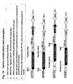
- Fig. 1 the genes are fused at the 5' end and the encoded proteins of interest will be at the N-terminus of the fusions; other combinations, and details of vector construction and vector elements, are shown in Figure 16 .
- Cells are co-transfected with complementary F1, F2 gene constructs such that proteins are expressed.
- Transient assays can be performed; also, stable cell lines can be constructed with "PCA Inside” by using selectable markers or by using a survival selection PCA to generate the stable cell line.
- the resulting cells or stable cell lines are used for HTS or HCS in conjunction with chemical libraries of interest.
- the inventors describes methods and compositions for quantitative, high-content and/or high-throughput assays using a wide range of different PCAs generating fluorescent or luminescent readouts, and specific examples for a GFP PCA and two variants thereof (YFP PCA and IFP PCA); a beta-lactamase PCA (BLA PCA); a luciferase PCA (RLuc PCA); and a dihydrofolate reductase PCA (DHFR PCA) are described.
- reporters can be selected that emit light of a specific wavelength and intensity that may be suitable for a range of protein expression levels, cell types, and detection modes.
- the flexibility is an important feature of the invention because of the wide range of biological processes and biochemical targets of interest for drug discovery.
- activation of a pathway - for example, by a receptor agonist or a drug - leads to an increase or decrease in the formation of protein-protein complexes without a change in the subcellular location of the complexes.
- An increase or decrease in the number of protein-protein complexes formed by the proteins leads to an increase or decrease, respectively, in the signal generated by the PCA.
- a high-throughput assay format can be used to measure the bulk fluorescent signal that reflects the amount of the complex of interest.
- Examples are shown herein for three different pathways in which the selected pathway sentinels were Chkl/p53, p53/p53, PKB/hFt1, PDK1/hFt1, FKBP/TOR (FRAP), IkBa/p65, and IkBa/Ubiquitin.
- NFkB p50/p65
- activation of a pathway leads to the change in the amount of a protein-protein complex from one subcellular compartment versus another (membrane vs. cytosol, cytosol vs. nucleus, etc).
- a high-content assay format is used to localize the fluorescent signal generated by the reassembled reporter at the site of the protein-protein complex within the cell.
- DHFR was used to construct a fluorescence assay based on the high-affinity binding of methotrexate (MTX) to the reassembled DHFR.
- MTX methotrexate
- the DHFR PCA was used to construct a high-content assay for NFkB translocation in order to identify agents that block the TNF pathway.
- the reporter used to construct a high-throughput assay is beta-lactamase (BLA).
- BLA PCA beta-lactamase
- the BLA PCA has been described previously and in the present invention it was used, in conjunction with a novel cephalosporin substrate, to construct a fluorescent high-throughput assay for inhibitors of the DNA damage response pathway acting on p53 and its upstream elements.
- the reporter used to construct a high throughput assay is luciferase.
- luciferase in PCA was first described by Michnick et al. (US 6,270,964 ).
- Renilla luciferase (RLuc) PCA was chosen to construct a high-throughput assay for inhibitors of the DNA damage response pathway, generating an assay with a robust signal that can be read in minutes with high throughput instrumentation. Mutant RLuc fragments are also provided for improved stability.
- intrinsically fluorescent proteins such as GFP are used to construct PCAs.
- a GFP PCA was first described by Michnick et al. (US 6,270,964 ).
- PCAs based on GFP or variants thereof are particularly suitable for HCS since the signal is located at the site of fragment complementation.
- the fluorescent proteins, including GFP, YFP, CFP and other variants as well as the newer reporters listed in Table 1 are particularly useful for the present invention, because no additional cofactors or substrates are needed for signal generation.
- PCAs based on these proteins are particularly useful for high-content assays, since the signal is localized at the site of the protein-protein complex.
- PCAs based on GFP and two mutants thereof are shown for PCAs based on GFP and two mutants thereof (YFP and 'IFP'). These assays can be read either with high-content instrumentation such as automated fluorescence microscopes or automated confocal imaging systems; or, in some cases where a particular assay pair results in an overall increase or decrease in fluorescence intensity, the change in bulk fluorescence can be read with high-throughput instrumentation as shown in Fig. 6 .
- Reporters generating a high quantum yield are often preferable for reasons of sensitivity; for example, the YFP PCA gives a brighter signal than the GFP PCA in the same way as the full-length YFP protein gives a brighter signal than the full-length GFP protein, and the mutant (IFP) fragments produce a brighter signal than the corresponding YFP fragments.
- the mutant (IFP) fragments produce a brighter signal than the corresponding YFP fragments.
- various useful PCA fragments can be created using the methods taught in US 6,270,964 (Michnick et al. ), and the fragments can be further engineered to generate a brighter signal upon fragment reassembly.
- protein fragments were generated either by PCR or were generated synthetically (by oligonucleotide synthesis) to create fragments with the desired assay properties.
- PCA fragments that reconstitute enzymes can be used in conjunction with various substrates or probes to generate assays with different spectral properties.
- a beta-lactamase PCA is used in conjunction with a cephalosporin substrate to generate a blue fluorescent product that can be read on a microtiter plate reader.
- a DHFR PCA is used in conjunction with a Texas Red-MTX probe to generate a red fluorescent signal that can be detected by automated microscopy.
- Multicolor PCAs allow the monitoring of more than one cellular process or pathway simultaneously, for example to determine if a compound of interest is affecting more than one pathway in the same cell or simply to multiplex assays for reasons of efficiency and cost savings.
- the ability to perform multicolor measurements enables the use of internal assay controls, for example where the controls give a red fluorescent signal while the proteins of interest give a yellow fluorescent signal.
- a multicolor PCA is demonstrated in which a DHFR PCA (red fluorescence) is combined in the same cells with a YFP PCA (yellow fluorescence) allowing the visualization of distinct protein-protein complexes with different subcellular locations.
- GFP global protein elastomer
- the wide range of forms of GFP including the yellow, cyan, citrine, SEYFP, Venus, and red homologues of GFP, are all suitable for PCA and can be further engineered to improve the signal intensity of the fragments used in the present invention.
- the numbers and kinds of assay readouts are limited only by the ability of the instrumentation to resolve different wavelengths of emitted light. Many other multicolor assays can be constructed using the principles and methods taught in the present invention.
- PCAs based on fragments of antigens or antibodies can be created and used in conjunction with simple detection schemes.
- PCAs based on fragments of a non-native antigen could be constructed such that a protein-protein interaction results in reconstitution of an epitope that can be detected with an antigen conjugated to a detectable moiety such as biotin or fluorescein.
- PCAs based on fragments of an antibody could be constructed such that a molecular interaction results in reconstitution of a functional antibody that binds to an antigen conjugated to a detectable moiety such a fluorophore. Any of these and similar reporters can be used, and modifications thereof, in conjunction with the present invention.
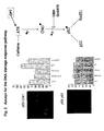
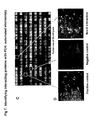
- Fig. 2 shows a scheme of the pathway and the results for an HTS assay based on a beta-lactamase PCA (BLA PCA) and a HCS assay based on a GFP PCA.
- Fig 3 shows an HTS assay based on Renilla luciferase (RLuc PCA).
- Fig 4 shows a high-content assay based on an IFP PCA (IFP is a variant of GFP).
- the proteins assayed are interacting pairs in the DNA damage response pathway, specifically, the checkpoint kinase Chk1 which interacts with the tumor suppressor p53 (Chk1/p53 PCA), or p53 itself which forms homodimers (p53/p53 PCA).
- the genes of interest - which were known to be involved in DNA damage response pathways - were fused to BLA reporter fragments, and co-transfected in pairs (in 6 replicates) into HEK293E cells. Specifically, interactions between two key proteins, p53 and the checkpoint kinase, Chk1, were evaluated for their response to the DNA damaging agent camptothecin (CPT) and various known drugs or compounds.
- CPT DNA damaging agent camptothecin
- HEK293E cells co-transfected with 250ng DNA (total) of BLA[1]-p53 and BLA[2]-Chk1 fusions (or with BLA[1]-p53 and BLA[2]-p53) were treated for two hours with 300nM camptothecin, followed by treatment with or without known inhibitors of the catalytic activity of Chk1 (e.g. 10 micromolar DBH and 50 micromolar Go6976), or an inhibitor of the upstream ATR kinase (2mM caffeine). After two hours (or up to 6 hours) the drugs were removed and a beta-lactamase substrate was added (Fig. 2B).
- the substrate was a derivative of a previously-described cephalosporin (Quante et al.; see References). Hydrolysis of the beta-lactam ring by reconstituted BLA releases free coumarin which has a blue fluorescence. After drug treatment, cells were washed with 200 microliters of PBS (plus calcium and magnesium), then covered with 25 microliters of PBS without calcium or magnesium. Freshly diluted BLA substrate was added to each well to a final concentration of 20 micromolar in 2% DMSO (in a final volume of 50 microliters). For each protein pair, the rate of hydrolysis of the substrate was determined immediately after addition of substrate by a kinetic assay on a Molecular Devices Gemini XS plate reader.
- a flexible 10-amino acid linker consisting of (GGGGS) 2 (SEQ ID No.1) separated the genes of interest and the YFP fragments.
- the use of a flexible linker between the gene of interest and the reporter fragment assures that the orientation and arrangement of the fusions is optimal to bring the protein fragments into close proximity ( J.N. Pelletier, F.-X. C.-Valois & S.W. Michnick, 1998, Proc Natl Acad Sci USA 95: 12141-12146 ).
- GFP[1] corresponds to amino acids 1 to 158 and GFP[2] corresponds to amino acids 159 to 239 of GFP and was amplified by PCR from pCMS-EGFP (Clontech).
- HEK293T cells were seeded at 10,000 cells/well in a 48-well cell culture dish (Costar). Cells were transfected with 150ng total DNA comprised of GFP[1]-p53 and GFP[2]- Chk1, or GFP[1]-p53 and GFP[2]-p53 using FuGene (Roche) as per the manufacturer's recommendations. After approximately 48 hours of expression; cells were rinsed once in PBS, then overlaid with 75 microliters of PBS (with no counterstain) for fluorescence microscopy. Live cells were imaged on an SP Nikon fluorescence microscope using a Chroma FITC filter (excitation: 460-500nm; emission: 505-560 nm; dichroic mirror: 505LP).
- the present invention describes not only F1 fragments that have a naturally occurring initiating methionine, but also the same F1 fragments that have been modified to remove the initiating methionine when the F2 fragment is to be at the 3' end of the construct. Similarly, the invention describes F2 fragments that naturally do not begin with an initiating methionine, but also those same F2 fragments that have been modified to include an initiating methionine when the F2 fragment is to be at the 5' end of the construct.
- RLuc PCA was based on wild-type Renilla luciferase and the fragments had the following sequences:
- Either the wild type RLuc F1 or the mutant RLuc F1(C124A) can be used in combination with the RLuc F2 fragment provided above to generate luminescent PCAs.
- the following examples below show the results obtained with the wild-type RLuc fragments.
- the F1 and F2 fragments described above were created by oligonucleotide synthesis (Blue Heron Biotechnology, Bothell, WA) and were designated RL[1] (aa 1-160) and RL[2] (aa 161-311).
- the synthetic fragments were amplified by PCR to incorporate restriction sites and a linker sequence encoding a flexible 10 amino acid peptide linker in configurations that would allow fusion of a gene of interest to either the 5'- or 3'-end of each reporter fragment sequence.
- the amplified fragments of RL[1] and RL[2] were then subcloned into a mammalian expression vector (pcDNA3.1Z, Invitrogen), creating 4 independent vectors (an N-terminal, and C-terminal fusion vector for each reporter fragment) as shown in Fig. 16 .
- STAT1/PDK2 served as a negative control PCA.
- the full coding sequences for p53, STAT1 and PDK2 were amplified by PCR from sequence verified full-length cDNAs.
- the resulting PCR products were desalted, digested with appropriate restriction enzymes to allow directional cloning, and fused in-frame to either the 5' or 3'-end of RL[1], RL[2], RL[3] or RL[4] through a linker encoding a flexible 10 amino acid peptide (GGGGS) 2 (SEQ ID No.1) to assure that the orientation/arrangement of the fusions in space is optimal to bring the fluorescent protein fragments into close proximity.
- GGGGS flexible 10 amino acid peptide
- DNAs from recombinant constructs were isolated using Qiagen Turbo BioRobot Prep kits (Qiagen, Chatsworth, CA) on a Beckman FX robotic workstation (Beckman Coulter, Fullerton, CA). Isolated DNAs were quantitated and then normalized to a concentration of 50 ng/microliter.
- the luciferase PCA was constructed to quantify the homo-dimerization of p53 (p53/p53 PCA) and compared to a negative control RLuc PCA (Pdk2/STAT1). The latter proteins do not interact. Twenty-four hours prior to transfection, HEK293T cells were plated (10,000 cells per well for 24 hr assay, 15,000 cells per well for 48 hr assay) in 96-well plates coated with poly-lysine. Cells were transfected with 0.1 microgram of total DNA (50ng of each reporter construct) using Fugene transfection reagent (Roche Diagnostics, Indianapolis, IN), as per the manufacturer's recommendations.
- Renilla Luciferase Assay Lysis Buffer Promega, Cat # E2810
- Renilla Luciferase Assay buffer containing a proprietary formulation of the Renilla luciferase substrate coelenterazine (Promega, Cat # E2810) was added by injector in a Thermo Lab Systems Luminoskan Ascent luminometer. For each sample, the luminescence released was captured over 10 seconds, with a 2 second delay after addition of substrate to the sample. Data are reported as relative luminescence units (RLU), and have not been normalized to protein content.
- RLU relative luminescence units
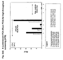
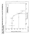
- Fig. 3(A) shows the luminescence generated from whole cell lysates of HEK293T cells expressing p53/p53 or Pdk2/STAT1 fused to fragments of Renilla luciferase after 24 and 48 hours of expression.
- the figure legend identifies the orientation of the encoded proteins relative to each reporter fragment. The results demonstrate that fragmentation of Renilla luciferase at E160 results in an efficient PCA; all four possible fusion pairs produced detectable luminescence at 24 and 48 hrs of expression. The signal was higher 48 hours after transfection than at 24 hours after transfection.
- the Pdk2/STAT1 PCA produced a negligible signal. This is a key point because it demonstrates that the PCA signal in the assay is absolutely dependent upon the presence of two interacting proteins fused to the complementary PCA fragments; the fragments themselves are incapable of reassembling into an active enzyme unless the complementation is assisted by the proteins fused to the complementary fragments.
- This key feature highlights the distinction between the present invention and alternative protein-protein interaction technologies such as FRET or BRET, where proteins of interest are expressed as fusions to active, full-length fluorescent or luminescent proteins.
- this feature highlights the distinction between the present invention and high-content assays based on single-protein tagging with a luminophore such as GFP. In the latter cases, individual proteins generate a signal, even in the absence of a protein-protein interaction.
- the p53/p53 complex produced a signal ranging from 20 RLU to over 200 RLU, depending on the gene-fragment orientations, resulting in a signal-to-background as high as 200:1 in the RLuc PCA.
- HEK293T cells were plated (15,000 cells per well) in a 96-well plate coated with poly-lysine. For each condition tested, cells were transfected in quadruplicate with 0.1 microgram of total DNA (50ng each of RL[1]-p53 and RL[2]-p53) using Fugene transfection reagent as above.
- the RLuc PCA can be applied to HTS for a large number of proteins and therapeutic targets in whole cell assays or cell lysates.
- Luciferase PCAs can be constructed in high-throughput and ultra-high-throughput formats due to the extraordinar sensitivity of the assay. These assays can be scaled up to 1536-well formats or even higher, and an entire plate can be read within minutes.
- mutant versions of luciferase PCAs can be created, taking advantage of genetic engineering to introduce mutations such as C152A which has been shown to increase the luminescent output of the Renilla luciferase holoenzyme (see Table 1 for references).
- the PCAs described here were first created by introducing the EYFP-specific mutations S65G, S72A and T203Y (24) into existing oligonucleotide fragments of EGFP, resulting in fragments designated YFP[1] and YFP[2] corresponding to amino acids 1-158 and 159-239 of the full-length EYFP (21, 25). Subsequently, assays were constructed by starting directly with synthetic oligonucleotides corresponding to YFP[1] and YFP[2] (Blue Heron). Fragments YFP[1] and YFP[2] had the following compositions:
- DNAs from recombinant constructs were isolated on a Beckman FX robotic workstation (Beckman Coulter, Fullerton, CA) using Qiagen Turbo BioRobot Prep kits or manually using Qiagen Midi Prep kits. Isolated DNAs were quantitated and then normalized to a concentration of 50 ng/microliter.
- CPT camptothecin
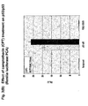
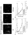
- Fig. 4 Representative images of drug effects on the p53:p53 PCA are shown in Fig. 4 .
- the left panel of three images corresponds to effects of geldanamycin or Trichostatin A on the PCA in the absence of CPT, while the right panel shows effects of the same drugs in the presence of 500nM camptothecin.
- DMSO top images is the vehicle used to resuspend the drugs.
- Geldanamycin is a known inhibitor of Hsp90, a chaperone protein for a number of cellular proteins, including wild type and mutant p53 (King et al. 2001). The expected effect of this drug would be to decrease the stability of p53, therefore decreasing the signal, as we observed.
- Hsp90 inhibitors can be detected by constructing a PCA in which at least one member of the PCA pair is an Hsp90 client protein. These assays will enable large-scale screening for additional Hsp90 client proteins, for example by constructing PCAs for a large number of protein-protein complexes and testing the PCA in the absence and presence of geldanamycin to identify proteins that are sensitive to Hsp90 inhibition.
- such assays can be used immemdiately in HTS to identify small-molecule inhibitors of Hsp90 activity. Since geldanamycin and its derivative, 17-AAG, have potent anti-tumor activity, the ability to construct assays that are sensitive to Hsp90 inhibition enables a new area of anti-cancer drug discovery.
- Trichostatin A is an inhibitor of histone deacetylase I (HDAC1). Inhibition of deacetylation in the presence of camptothecin should induce acetylation of p53, therefore stabilizing the protein and increasing its transcriptional activity.
- HDAC1 histone deacetylase 1
- PCA histone deacetylase 1
- the HDAC inhibitors Trichostatin A and MS-275 significantly stimulated the p53/p53 interacton above the control in the presence of CPT.
- a similar effect was seen with the BCL-2 inhibitor, HA14-1.
- the kinase inhibitors LY 294002 (PI3K) and SB 203580 (p38 MAPK) caused an increased association in both assays.
- Geldanamycin significantly inhibited both assays, as shown in the images and the associated histogram.
- the PCA strategy described in the invention and depicted in Fig. 1 was next used to identify novel protein-protein interactions in the PI-3-kinase and PKA/PKC-mediated pathways and then to carry out quantitative screens based on the novel interactions.
- cDNA library screening was performed with the GFP PCA in order to identify proteins interacting with PKB.
- a novel interaction between PKB and Ft1 was identified by the GFP PCA screen.
- the GFP PCA was used to construct fluorescent assays for PKB/Ft1 and PDK1/Ft1.
- the organization of the pathways and the position of the PKB/hFt1 interaction is shown in Figure 5(A) .
- the PKB-GFP[2] fusion was also inserted in a pMT3 vector where the ampicillin resistance gene has been replaced by a chloramphenicol resistance gene (pMT3-chloramphenicol) for the purpose of the cDNA library screen.
- a 10 amino acid flexible linker consisting of (GGGGS) 2 (SEQ ID No.1) was inserted between the cDNA and the GFP fragments to assure that the orientation/arrangement of the fusions in space is optimal to bring the protein fragments into close proximity.
- the GFP[1]-GCN4 and GCN4-GFP[2] constructs consist of fusions with GCN4 leucine zipper-forming sequences and are used as controls.
- a human brain cDNA library was excised from the vector pEXP1 (ClonCapture cDNA library, Clontech) using SfiI restriction sites and inserted into the pMT3 vector, 3' of the F[1] fragment of GFP and a 10 amino acids flexible linker.
- the PCA-cDNA library fusion expression vectors were divided into several pools (according to the size of the inserted cDNAs -from 0.5 to 4.6 kb) and amplified at 30°C in liquid medium.
- COS-1 cells were grown in DMEM (Life Technologies) supplemented with 10% fetal bovine serum (FBS, Hyclone).
- the human Tag-Jurkat T cell line expresses the SV40 large T antigen and harbor an integrated ⁇ -galactosidase reporter plasmid where three tandem copies of the NF-AT binding site directs transcription of the lacZ gene. They were grown in RPMI-1640 (Life Technologies) supplemented with 10% FBS, 1 mM sodium pyruvate and 10 mM Hepes.
- cDNA Library Screening with PCA to Identify Novel Protein-protein Interactions COS-1 cells were plated in 150-mm dishes 24 h before transfection.
- GFP[1]-cDNA library pMT3 vector harboring the human brain cDNA library fused to the F[1] fragment of GFP (GFP[1]-cDNA library) and pMT3-chloramphenicol vector containing the full-length PKB fused to the F[2] fragment of GFP (PKB-GFP[2]).
- the GFP[1]-cDNA library fusions were transfected in several pools, according to their size.
- 1]-cDNA fusions were extracted from individual clones and retransfected separately with PKB-GFP[2] or GFP[2] alone (negative control) to discard negative clones that enter the pool during the cell sorting. After this second round of selection, the DNA plasmids corresponding to the positive clones were submitted to sequence analysis.
- COS-1 cells were washed one time with PBS, gently trypsinized and resuspended in 200 microliters of PBS.
- Tag-Jurkat T cells were directly harvested and resuspended in 200 microliters of PBS.
- the relative amount of reconstituted GFP was detected by fluorometric analysis.
- the total cell suspensions were transferred to 96-well black microtiter plates (Dynex, VWR Scientific) and subjected to fluorometric analysis (Spectra MAX GEMINI XS, Molecular Devices).
- Tag-Jurkat T cells were treated for 90 min with 300 nM wortmannin or 30 min with 5 ⁇ g/ml anti-CD3 antibody or 5 ⁇ g/ml phytohemagglutinin (PHA) or 1 micromolar ionomycin or 10 micromolar forskolin or/and 500 nM phorbol-12-myristate-13-acetate (PMA) (all from Calbiochem) prior to fluorometric analysis. Afterward, the data were normalized to total protein concentration in cell lysates (Bio-Rad protein assay). The constitutive dimerization of GCN4 leucine zipper was used as a positive control.
- PHA phytohemagglutinin
- PMA phorbol-12-myristate-13-acetate
- the background fluorescence intensity corresponding to non-transfected cells was subtracted from the fluorescence intensities of all of the samples.
- the sub-cellular location of the hFtl/PKB and hFt1/PDK1 protein-protein complexes was also determined by fluorescence microscopy in live cells.
- fluorescence microscopy COS-1 cells were grown on 18-mm glass cover slips prior to transfection. Cells were washed two times with PBS and mounted on glass slides. Fluorescence microscopy was performed on live cells with a Zeiss Axiophot microscope (objective lens Zeiss Plan Neofluar 100X/1.30).
- Panel 1 of Fig. 5(B) shows the quantitative fluorescence results obtained with the PCAs in COS cells and panel 2 shows the results obtained in Jurkat cells.
- Panel 3 of Fig. 5(B) shows the images of protein-protein complexes and their subcellular locations. Agents that stimulated the pathway caused an increase in fluorescence, whereas compounds that inhibit the pathway caused a decrease in fluorescence of the protein-protein complexes in the pathway.
- PCAs can be used as quantitative assays providing relevant pharmacological information.
- FKBP the FK506 binding protein
- FRAP FKBP-Rapamycin-Associated-Protein
- HEK 293E Cells were seeded into a 96 well plates at a cell density of 13,000 per well. Cell media is MEM-alpha Growth medium. Total volume was 100 microliters. Cells were allowed to grow 20-24 hours prior to transfection - cells were 70-80% confluent at time of transfection. Cells were maintained at 37C, 5% CO2. Cells were transfected with a total of 0.1 micrograms of DNA per well using Fugene (Roche). HEK 293 cells expressing FKBP-YFP[1] and mTOR-YFP[2] were treated with increasing doses of rapamycin as follows.
- Figure 6 shows the results of the assay, demonstrating effects of rapamycin on the interaction of FKBP and mTOR
- mTOR is the murine equivalent of the human protein FKBP-rapamycin associated protein, FRAP.
- Rapamycin induced the formation of complex between FKBP and mTOR which could be seen by microscopy ( Fig. 6B ) and quantitated by fluorescence spectroscopy ( Fig. 6C ) in 96 well plates using excitation and emission wavelengths of 485 and 527 nm, respectively.
- Such assays can be used in combination with a variety of small-molecule, natural product, combinatorial, peptide or siRNA libraries to identify molecules that activate or inhibit the protein-protein complex, either by acting directly on the protein-protein interaction, or by acting upstream of the PCA sentinel.
- Fig. 1 shows that protein-protein interactions can be identified by various methods, including gene-by-gene interaction mapping.
- gene-by-gene interaction mapping provides an alternative to bait-vs.-library screening in cases where it is desirable to test defined sets of genes against each other, or for purposes of assay optimization.
- gene-by-gene interaction mapping enables testing of full-length proteins for interactions with other full-length proteins.
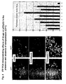
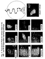
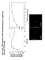
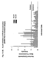
- Fig. 7 shows the results of two plates from the screen.
- Each plate contains 28 different PCAs representing different gene pairings, in addition to four sets of controls (one positive and three negative controls), all assayed in triplicate (represented on the x-axis).
- the y-axis shows the mean fluorescence intensity measurement for each PCA, with error measurements plotted as 95% confidence intervals.
- the positive control was p65/p50 and the negative control was PDK1/PDK1. For each plate, the negative controls are highlighted in red and the positive control in yellow.
- Interactions that are statistically different from the negative control are color-coded as in the legend, indicating the level of statistical significance associated with each measurement, as determined by the Student t-test of the mean fluorescence. Note that the y-axes in panels A and B are different, displaying the range of signal intensities that can be obtained in this assay relative to the positive control.
- the assay can be used to identify protein-protein complexes within pathways of interest for drug discovery in HTS or HCS formats or to optimize gene pair orientations for assay development.
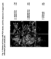
- Figure 7 (C) shows the images of cells in individual wells as acquired by automated microscopy.
- images were acquired from the same 96-well plates on the Discovery-1 imaging system (Universal Imaging).
- the Hoechst-stained cells of a control well (cells stained blue in Fig. 7D ) were used to establish the appropriate focal plane for image acquisition across the entire plate. Images were then acquired at two sites in each well, using a 10X objective at wavelengths appropriate for Hoechst and YFP, respectively.
- the merged view across an entire plate is visible in panel C. Examples of positive and negative controls, as well as a 'novel' positive are shown in panel D.
- the advantage of the present invention is therefore the ability to rapidly map protein-protein interactions and to simultaneously characterize the interactions in living cells in high-throughput and/or high-content assays; and subsequently, to use the same PCA constructs to develop robust, stable high-throughput screens for molecules that activate or inhibit the pathways for which the protein complexes represented in the PCAs.
- FIG. 8 illustrates the organization of the pathway leading from the TNF receptor to the nucleus, including the role of the NFkB transcription factor complex (p65/p50). Binding of TNF to its receptor leads to activation of the IKK complex, resulting in the phosphorylation and degradation of IkBa by the proteasome. Degradation of IkBa frees the NFkB transcription factor complex (p65/p50) to translocate from the cytoplasm into the nucleus, where it can turn on the transcription of pro-inflammatory genes.
- Proteasome inhibitors such as ALLN and epoxomicin (and the current anti-cancer drug, Velcade®) block the degradation of IkBa, resulting in the retention of NFkB in the cytosol.
- a series of PCAs were constructed with full-length cDNAs encoding known elements of the TNF pathway and using a DHFR PCA (red fluorescence) and/or the YFP PCA (yellow/green fluorescence) ( Figure 9 ).
- DHFR PCA red fluorescence
- YFP PCA yellow/green fluorescence
- open reading frames of p65, p50, CBP, CBPnt, TNFRI, TRAF2 and a single coding unit of Ubiquitin were PCR amplified, fused in-frame to complementary fragments of DHFR or YFP, and subcloned into pCDNA3.1zeo.
- the REFSEQ or GENBANK identifiers for the genes used are: NM009045 (p65/ReIA), NM003998 (NFkB1/p50), AY033600, NM004380 (CBP), NM003824 (FADD), NM003789 (TRADD), BC033810 (TRAF2), XM032491 (IKKbeta), BC000299 (IKKgamma), and Ubiquitin C ( BC039193 ).
- CBPnt [(S66385 (1..2313)] corresponds to the amino terminal 771 amino acids of CBP.
- Ubiquitin C corresponds to the 76 kDa ubiquitin monomer.
- DHFR fragments F[1,2] and F[3] correspond to murine DHFR residues 1 to 105 and 106-186, respectively (Pelletier, Campbell-Valois et al. 1998).
- the DNAs encoding the proteins of interest were ligated to either the 5' or 3' end of DHFR-F[1,2] and DHFR-F[3] to generate N or C terminal fusions, respectively.
- a flexible linker consisting of (GGGGS) 3 (SEQ ID No.30) separated the genes of interest and the DHFR fragments.
- DHFR PCA constructs 8x10e4 CHO DUKXB11 (DHFR-deficient) cells were seeded into 12 well plates and co-transfected 24 hours later with 1 microgram of DNA per well comprising a 1:1 molar ratio of the complementary pairs of fusion constructs, using Fugene (Boehringer Mannheim) according to the manufacturer's instructions. Forty-eight hours post-transfection, the cells were incubated with 4 micromolar Texas Red-Methotrexate (Molecular Probes/Invitrogen) for two hours at 37C in growth medium (alpha-MEM, 10% fetal bovine serum). When two proteins of interest interact, TxR-MTX binds to reconstituted DHFR.
- Texas Red-Methotrexate Molecular Probes/Invitrogen
- Unbound TxR-MTX was removed by rinsing followed by a 30-minute incubation in fresh medium.
- Cells were viewed and images acquired using a Nikon Eclipse TE1000 fluorescence microscope at excitation and emission wavelengths of 580 nm and 625 nm, respectively.
- HEK293T cells (Invitrogen) were seeded into poly-L-lysine coated 96-well plates at a density of 1.5x10e4 cells/well and transfected 24 hours later with 100 ng DNA per well comprising a 1:1 molar ratio of the complementary pairs of fusion constructs. Forty-eight hours post-transfection, cells were rinsed with PBS and viewed using a Discovery-1 automated microscope (Universal Imaging/Molecular Devices) at excitation and emission wavelengths of 485 nm and 527 nm, respectively.
- Discovery-1 automated microscope Universal Imaging/Molecular Devices
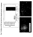
- Fig. 9 A number of proteins known to participate in the TNF signaling pathway formed protein-protein complexes in live cells that were readily detectable by PCA; some of these are shown in Fig. 9 . Fluorescent signals shown in yellow/green represent YFP PCAs whereas signals shown in red represent DHFR PCAs. Robust fluorescent signals and correct subcellular localization of selected protein-protein complexes could be detected by PCA in the transiently-transfected cells.
- TNF receptor upstream adaptor molecules TRADD, FADD, and TRAF2.
- TRADD upstream adaptor molecules
- FADD upstream adaptor molecules
- TRAF2 upstream adaptor molecules
- PCA Because p65 interacts with distinct proteins at sequential steps of the TNF signaling cascade, the use of PCA enables high-fidelity detection of TNF induced signal transduction. In addition, the ability to construct multi-color, multiparametric analyses with PCA provides a flexible approach enabling a wide range of basic research in cell biology, biochemistry and signal transduction; as well as an extraordinary degree of flexibility and efficiency in assay design and development.
- Fig. 1 the principle of these assays is that a pathway is actually a series of steps involving the physical association, dissociation or movement of proteins within complexes. These events occur in real time and within specific subcellular compartments in the living cell.
- the present invention enables the construction of assays to measure these dynamic events for any protein within any pathway.
- proteasome inhibitor blocks the degradation of IkBa such that the NFkB complex is retained in the cytosol.
- the latter assays can be read in high-content mode using PCAs capable of detecting the subcellular location of the complexes.
- proteasome inhibitors which block the degradation of IkBa lead to an accumulation of IkBa/Ubiquitin complexes.
- the latter assays can be read in high-content (automated microscopy or automated imaging) or high-throughput (bulk fluorescence) formats.
- any or all of these assays will be useful in screening for inhibitors of TNF signaling.
- a screening campaign based on a high-content assay for p50/p65 is described in detail below.
- these assays will be useful in identifying agents with anti-inflammatory activity and/or with anti-cancer activity.
- the three 'sentinel' PCAs studied in further detail all were sensitive detectors of proteasome inhibitors such as ALLN.
- the ability to detect ubiquitination of proteins enables large-scale screening for proteins that are degraded by ubiquitination. Sensitive and specific assays for such compounds are of particular interest in the pharmaceutical industry since the marketed drug Velcade®, which is a proteasome inhibitor, has potent anti-tumor activity.
- CHO DUKXB 11 cells were seeded into 96 well plates at a density of 8 x 10e3 cells/well and transfected 24 hours later with YFP[1] and YFP[2] fusion genes at a 1:1 molar ratio using Fugene (Boehringer Mannheim) according to manufacturer's directions. A total of 20 ng DNA per well was used for each sample. Thirty-six hours post-transfection, cells were serum starved by incubation in 0.25% FBS-supplemented aMEM for an additional 16-18 hours. For cytokine induction, certain cells were treated with 25 ng/ml mTNF (Boehringer Mannheim) for 30 min.
- the serum-starved cells were treated with 40 micrograms/ml ALLN (Calbiochem) for 1 hour prior to and during the mTNF alpha induction period.
- the cells were rinsed with PBS and the subcellular location of NFkB complexes was visualized and images acquired using a Nikon Eclipse TE2000 fluorescence microscope at excitation and emission wavelengths of 485 nm and 527 nm, respectively. Quantitative analysis of fluorescence intensities was performed using Metamorph software (Universal Imaging, Molecular Devices, Inc.)
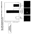
- Figure 10 shows results of a transient assay for NFkB (p65/p50) cytoplasmic-to-nuclear translocation in CHO cells based on YFP PCA.
- NFkB p65/p50
- cytoplasmic-to-nuclear translocation in CHO cells based on YFP PCA.
- the p65/p50 complexes were evenly distributed between the cytosol and nucleus.
- the ratio of nuclear:cytoplasmic fluourescence increased by an average of two-fold and the p65/p50 complexes could be visualized in the nucleus of live cells by fluorescence microscopy.
- ALLN the well-characterized proteasome inhibitor
- CHO cells transiently co-expressing complementary YFP fragment fusions of p50 and p65 were incubated in the absence or presence of TNF. Where indicated, cells were pre-treated with the proteasome inhibitor ALLN. Mean fluorescence intensities in the nucleus and cytoplasm of each cell were measured and expressed as a ratio. ALLN inhibited the TNF-induced cytoplasmic-to-nuclear translocation of NFkB complexes in the YFP PCA assay. While the effects of cytokine and inhibitor were readily apparent from the analysis of individual cells, the transient transfections resulted in significant cell-to-cell heterogeneity. Therefore we sought to establish stable cell lines with ⁇ PCA inside' for use in screening diverse small-molecule, known drug, and natural product libraries.
- Stable cell lines represent the gold standard for HTS since the assays can be reconstructed at any time from frozen stocks of cells.
- HEK293T cells were grown in MEM alpha medium (Invitrogen) supplemented with 10% FBS (Gemini Bio-Products), 1% penicillin, and 1% streptomycin and maintained in a 37°C incubator at 5% CO 2 .
- FBS Gelar Bio-Products
- penicillin 1%
- streptomycin 1%
- stable cell lines were selected using 100 micrograms/ml of Hygromycin B (Invitrogen). Selected cell lines were then transfected with YFP[1]-p50.
- Stable cell lines expressing YFP[1]-p50/YFP[2]-p65 were isolated following double antibiotic selection with 50 ⁇ g/ml Hygromycin B and 500 ⁇ g/ml Zeocin. Cell clones stably expressing the fusion genes were identified by immunoblot analysis and fluorescence microscopy. A single cell line of each transfectant was selected for further characterization. Fluorescence of these lines is stable over at least 25 passages (data not shown). A stable, MEK/ERK cell line - constructed as described below - was used as a control for TNF effects. Fugene 6 (Roche) was used for all the transfections according to manufacturer's directions.
- Cells stably expressing YFP[1]-p50/YFP[2]-p65 were seeded at 20,000 cells/well in black-walled poly-lysine coated 96 well plate (Greiner). Twenty-four hours later, the cells were incubated with human TNF-alpha (Roche) for 30 min. Nuclei were stained with Hoechst 33342 (Molecular Probes) at 10 micrograms/ml for 10 min. Cells were rinsed with HBSS (Invitrogen) and kept in the same buffer. Fluorescence was visualized and images were acquired using a Discovery-1 automated fluorescence imager (Molecular Devices, Inc.) equipped with excitation and emission filters 470/35 and 535/60, respectively.
- Discovery-1 automated fluorescence imager Molecular Devices, Inc.
- cells were treated with 25 micromolar ALLN (Calbiochem) for 60 min and induced with TNF in the continued presence of the inhibitor.
- ALLN Calbiochem
- cells were pretreated with compounds (10 micromolar) for 60 minutes and then stimulated with TNFalpha for 30 minutes in the presence of drugs.
- Cells were then fixed with 2% formaldehyde in HBSS and subsequently stained with Hoechst 33342. All liquid handling was done using a Biomek FX (Beckman) instrument and images were acquired as described above. Images were analyzed using Image J. Translocation is assessed by calculating the nuclear/cytoplasmic ratio of the mean fluorescence intensity for a population of cells (denoted as n) over several images for a given condition.
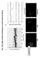
- FIG. 11 again illustrates an important distinction of PCA, which is that the fragments themselves do not generate a signal.
- the stable cell line with the single PCA fusion (p65-YFP[2]) produced no fluorescent signal.
- generation of a signal is dependent upon fragment complementation through the productive interaction of two molecules to which the complementary fragments are fused. Therefore the present invention is clearly distinct from other technologies that involve monitoring individual protein movements within cells.
- Fig. 12[A] The mean fluorescence of the nucleus and cytoplasm of individual cells was quantified, and the N:C fluorescence ratio was calculated.
- Treatment of the p50/p65 cell line with increasing doses of TNF resulted in an 3-fold increase in the N:C ratio, from 0.47 to 1.42, with a half-maximal response at 10ng/ml TNF.
- Analysis of the time course of the TNF response revealed that p50/p65 translocation into the nucleus occurred with a t 1/2 of 5 min. The maximal response was observed at 15 min., followed by a decrease at 60 min., consistent with feedback recovery of NFkB activation.
- the average NC ratio was derived by automated image analysis as described above, and compound-treated wells were compared to unstimulated and TNF-stimulated control wells. Results from this focused library screen and the plate-to-plate variability in TNF response is shown in Fig. 12(C) .
- the Z factor a commonly used metric for assay robustness, is not applicable for this subset of compounds due to the large number of known actives and fluorescent compounds.
- We utilized the Z' factor which measures the same statistical parameter across control wells to calculate assay quality. The Z' values averaged 0.627, with a median value of 0.67 across the 12 assay plates.
- NFkB pathway inhibitors In addition to the known inhibitors of this pathway in this compound set, we identified novel NFkB pathway inhibitors. For example, hit confirmation and 8-point dose-response analysis indicates that a compound we denoted as ODC0000160 inhibits the p50/65 PCA assay with an IC 50 of 1.1 micromolar; relatively potent for a screening hit in a cell-based assay (Fig. 12[D]). This compound has been used in human clinical trials, but has not been linked previously to the NFkB pathway. Clearly, its activity in this assay may have mechanistic significance, a concept supported by the fact that ODC0000160 can elicit apoptosis of human tumor cells (data not shown). The simultaneous exclusion of toxic compounds, enabled by the analysis of cell number and nuclear morphology via Hoechst staining in the standard protocol, provides added confidence to hits obtained in these assays.
- Figure 13 shows the results of the DHFR PCA.
- CHO DUKXB 11 cells transiently co-expressing DHFR-F(1,2)/p65 with DHFR-F(3)/p50 were co-transfected with I ⁇ B and incubated for 30 minutes with or without mTNFalpha as indicated in the bar graph.
- Co-transfection of the gene encoding IkBa induced the retention of p65/p50 complexes in the cytosol in the absence of TNF; treatment with TNF induced the translocation of the p65/p50 complex from the cytoplasm into the nucleus.
- FIG. 13 shows representative fluorescence images from samples co-expressing IkBa in which the NFkB complexes are located predominantly in the cytoplasm.
- the lower photomicrograph in Fig. 13 shows representative fluorescence images from samples co-expressing IkBa and induced with TNF, in which the NFkB complexes are located predominantly in the nucleus.
- the transiently transfected cells expressing the DHFR PCA were treated with 40 micrograms/ml ALLN (Calbiochem) for 1 hr prior to TxR-MTX labeling and for the subsequent duration of the experiment.
- the cells were rinsed with PBS and the subcellular location of NFkB complexes were visualized using a Nikon Eclipse TE2000 fluorescence microscope at excitation and emission wavelengths of 580nm and 625nm, respectively. Average fluorescence intensities in the nuclei and cytoplasm of cells were quantitated using NIH Image and/or OpenLab (Improvision).
- Figure 13(B) shows that the proteasome inhibitor ALLN inhibits the TNF-induced cytoplasmic-to-nuclear translocation of NFkB complexes in the DHFR PCA assay. In the presence of ALLN, the p50/p65 complexes are retained in the cytosol.
- Stable cell lines can be generated using a variety of methods known to those skilled in the art. With any PCA, stable cell lines can be generated using selectable markers, such as antibiotic resistance markers as described herein or any number of selectable markers that are known to those skilled in the art. With the DHFR PCA, stable cell lines can intrinsically be generated using survival-selection as previously described by Michnick et al. in DHFR- cells; alternatively, MTX selective pressure can be used with cells containing endogenous DHFR, such that only the cells expressing the DHFR PCA are capable of surviving under selective pressure.
- IkBa-NFkB complex TNF-induced degradation of IkBa, which is a consequence of ubiquitination and proteasomal degradation, frees bound NFkB and results in translocation of that transcription factor into the nucleus.
- disassembly of the IkBa-NFkB complex is a key step in NFkB-mediated gene regulation.
- ERK1/MEK1 was used as a control. ERK1 was ligated to the 5' end of YFP[1] while IkBa and MEK1 were appended to YFP[2] in N-terminal fusions.
- the fusion genes were subcloned into pCDNA3.1 expression vectors (Invitrogen) with Zeocin selectable marker for YFP[1]-p50, IkBa-YFP[1] and ERK1-YFP[1] and hygromycin marker for YFP[2]-p65 and ERK1-YFP[2].
- a linker consisting of (GGGGS) 2 (SEQ ID No.1) separated the genes of interest and the YFP fragments.
- the proteasome inhibitor ALLN was tested with the IkBa/p65 PCA. Cells were treated with 25 micromolar ALLN (Calbiochem) for 60 min and induced with TNF in the continued presence of the inhibitor.
- Ubiquitin is a highly conserved 76-amino acid polypeptide. Since its discovery in the mid-1970s, ubiquitin has been associated with cellular house-keeping functions such as eliminating damaged proteins. It has recently become clear that ubiquitin is involved in a variety of other vital processes at different subcellular locations ranging from the plasma membrane to the nucleus, including cell-cycle progression, signal transduction, transcriptional regulation, receptor down-regulation, and endocytosis.
- Ubiquitin is covalently attached to proteins through an isopeptide bond between its carboxy-terminal glycine and the epsilon-amino group of lysines in the target protein. This attachment is catalyzed by enzymes that activate and ultimately conjugate the ubiquitin moiety to a lysine residue in the substrate. This can be followed by further additions of ubiquitin to specific lysine residues within the linked ubiquitin itself, resulting in a poly-ubiquitin chain. This covalent modification can be reversed by unique proteases specific for the iso-peptide linkage.
- ubiquitin is the best-characterized polypeptide modifier
- other polypeptides (often referred to as Ubiquitin-like, or Ubl) are also conjugated to targets in analogous reactions.
- Ubiquitin-like, or Ubl polypeptides
- Ubiquitinated proteins are recognized by the 19S regulatory subunit of the proteasome, which removes the ubiquitin chain for recycling and denatures the doomed protein. The denatured protein is then fed into the core of the proteasome and reduced to short peptides (less than 22 residues). A number of proteins that are ubiquitinated have already been identified.
- cyclins and related proteins include cyclins A, B, D, E and cyclin-dependent kinase inhibitors); tumor suppressors, including p53; oncogenes, including c-fos, c-jun, c-myc and N-myc; inhibitory proteins, including IkappaBalpha and p130; and enzymes, including cdc25 phosphatase, tyrosine aminotransferase, and topoisomerases (I and IIalpha). Copies of two protein motifs - the F-box and the Ring finger, which are believed to identify targets for protein turnover - number in the hundreds in the eukaryotic genome suggesting a large number of proteins whose turnover is regulated by the ubiquitin system.
- ubiquitin-protein ligation requires the sequential action of three enzymes. Ubiquitin must first become attached to a member of the family of E2 ubiquitin-conjugating enzymes (an E1 ubiquitin-activating enzyme provides the initial ATP-dependent activation).
- the E2 enzyme itself or, more typically, an E3 ligase, provides the specificity for the transfer of ubiquitin onto the targeted protein (ligase substrate).
- ligase substrate Usually there is a single E1, but there are many species of E2s and multiple families of E3s or E3 multiprotein complexes.
- Specific E3s appear to be responsible mainly for the selectivity of ubiquitin-protein ligation (and thus, of protein degradation). They do so by binding specific protein substrates that contain specific recognition signals. In some cases, binding of the substrate protein to an E3 is indirect, via an adaptor protein.
- E3 ubiquitin ligases as proteins containing protein-protein interaction domains that couple to the ubiquitin-charged E2 (ubiquitin-conjugating) enzyme provided the link between substrate recognition and the catalytic steps for ubiquitin chain formation.
- IkBa phosphorylation-dependent ubiquitination of IkBa (IkappaBalpha), which targets the protein for rapid degradation by the proteasome and releases NF ⁇ B for translocation to the nucleus.
- TNF-induced ubiquitination of IkBa is essential for its proteolysis and subsequent activation of NFkB. Therefore, we sought to demonstrate the utility of PCA in identifying ubiquitinated proteins and inhibitors of the proteasome.
- IkBa-YFP[1] and YFP[2]-ubiquitin were constructed as described above and transiently expressed in HEK293T cells.
- Fig. 15 shows that mean fluorescence intensity was significantly increased in TNF induced cells treated with the proteasome inhibitor ALLN compared with control, vehicle treated cells.
- the present invention can be applied to the large-scale identification of proteins modified by ubiquitin and ubiquitin-like polypeptides; for example, using library screening as in Example 2 of the present invention where the 'bait' is ubiquitin or a ubiquitin-like molecule, or by using interaction mapping as in Example 4 where ubiquitin or a ubiquitin-like molecule is tested against individual cDNAs to identify ubiquitin-protein complexes.
- the assays that are the subject of the present invention can immediately be applied to high-throughput screening for novel therapeutic agents according to claim 1.
- vectors can be used in conjunction with the present invention.
- the elements of useful vectors can be varied as needed depending on the cell of interest, desired promoter, reporter choice, linker length, and cloning sites.
- the present invention is not limited to the vector sequence, its elements, or the way in which the genes are expressed. Plasmid, retroviral and adenoviral vectors are all compatible with the present invention. Several examples highlighting vector design and features specific to PCA are given below. These examples are not intended to be limiting for the applications of the present invention.
- linker length The use of a flexible linker between genes of interest and complementary fragments facilitates PCA.
- Linker lengths ranging from 5 amino acids to 30 amino acids have been used for PCA.
- the linker length can be varied as desired in order to control the intermolecular distance between interacting molecules required for complementation.
- Remy and Michnick showed that shortening the length of the flexible linker between the gene of interest and the PCA fragment allowed the precise detection of allosteric changes in erythropoietin receptor subunits upon ligand binding (see References).
- Assisted complementation - for example, between proteins that are indirectly associating as a result of their mutual binding to a third molecule or that are constitutively associated at a greater intermolecular distance - can also be investigated in detail by using longer linkers.
- a semi-standard linker of 10 to 15 amino acids- for example, as repeats of the 5-amino acid (GGGGS) (SEQ ID No.31) sequence used herein- facilitates fragment complementation and - as we have demonstrated in the present invention - is suitable for many applications of PCA.
- standard vectors can be constructed in which a fixed linker length is used and into which genes can be rapidly subcloned for assay construction as in Fig. 1 and Fig. 16 .
- PCAs based on survival-selection assays for example, using fragments of enzymes that act as drug resistance markers themselves, such as aminoglycoside kinase (AK PCA) or hygromycin phosphotransferase (HPT PCA), or where the PCA complements a metabolic pathway, such as DHFR PCA - no additional drug resistance genes need be incorporated in the expression plasmids. In those cases, reconstitution of the selectable marker upon fragment complementation allows cell survival under selective pressure.
- AK PCA aminoglycoside kinase
- HPT PCA hygromycin phosphotransferase
- an additional drug resistance or survival gene can be expressed to enable selection of cells expressing the proteins of interest.
- an additional drug resistance or survival gene can be expressed to enable selection of cells expressing the proteins of interest.
- different antibiotic resistance markers hygromycin and zeocin
- hygromycin and zeocin were used on the YFP[1] and YFP[2] plasmids to facilitate the generation of stable cell lines expressing the YFP PCA.
- the fluorescent or luminescent signal of the PCA can itself be used to select the stably expressing cells, for example by using FACS or bead-based selection methods to sort cells that have positive signals.
- FACS or similar methods can also be used in conjunction with antibodies to cell-surface PCAs, e.g. where the PCA reconstitutes a non-native cell surface marker that can be detected with a fluorescently tagged antibody.
- antigens or antibodies can also be used as selectable markers or detection probes in conjunction with PCA.
- antigens can be fragmented for PCA, such that the fragments reconstitute a protein that can be detected by a fluorescently-tagged antibody. If the reconstituted antigen represents a foreign protein in the transfected cells, there will be no background activity in the absence of a protein-protein interaction that reconstitutes the antigen.
- antigens (or antibodies) can be included as separate, non-operably-linked elements within vectors containing gene-fragment fusions.
- the co-expression of the gene-fragment fusions of interest can be detected by antibody-based cell selection using an antibody specific for the antigen element. Selection can be achieved by single-color or multi-color FACS sorting of antigen-expressing cells or by binding of antibodies linked to beads or a solid support.
- Dual PCAs combining a fluorescent or luminescent PCA with a survival-selection PCA, enabling the rapid selection of cell lines for HTS and HCS
- PCAs can be assembled on separate plasmids, as in the present invention, one or more polycistronic vectors can also be used in conjunction with PCA as shown in Fig. 17 .
- dual PCAs in which the construction of an HTS or HCS assay is linked to the generation of a stable cell line.
- Complementary bicistronic vectors are used to generate a stable cell line, such as with a leucine zipper-directed DHFR PCA, wherein the cell line also contains a fluorescent or luminescent PCA, where the fluorescent or luminescent signal is driven by the interaction of two proteins of interest.
- Bicistronic vectors contain an IRES (internal ribosomal entry sequence) that provides the ability to link the expression of one polypeptide to that of another, such as a selectable marker.
- IRES internal ribosomal entry sequence
- the creation of bicistronic vectors has made it possible to express a gene encoding a single polypeptide and the DHFR gene as a single mRNA, which is then translated into the two separate proteins ( Davies MV, Kaufman RJ. 1992. The sequence context of the initiation codon in the encephalomyocarditis virus leader modulates efficiency of internal translation initiation. J Virol 66:1924-1932 .) Expression of the DHFR gene and the recombinant gene as a single mRNA enriches for methotrexate amplification of both genes, and greatly enhances production of the molecule of interest.
- a fluorescent or luminescent PCA based on the present invention with the previously-described DHFR PCA, which enables rapid selection of stable cell lines through leucine zipper-directed reassembly of active DHFR ( Pelletier, J.N., C.-Valois, F.-X.
- a promoter drives the expression of the first PCA pair, comprising the genes of interest operably linked to the respective F1 and F2 fragments of an optically detectable reporter; while the IRES of each of the two vectors encodes the two halves of the second PCA, the latter comprising two oligomerization domains (such as the constitutively dimerizing GCN4 leucine zippers used in Example 2 of the present invention) operably linked to the respective F1 and F2 fragments of a selectable marker (such as the fragments of DHFR used in Example 9 of the present invention).
- oligomerization domains such as the constitutively dimerizing GCN4 leucine zippers used in Example 2 of the present invention
- the two bicistronic vectors are co-transfected into cells and subjected to selective pressure as previously described for the DHFR PCA such that co-expression of the second PCA pair enables the selection of cells that also co-express the first PCA pair.
- Cells are grown under conditions, described by us previously, in which the cells only survive if both fragments are expressed and DHFR is reconstituted though leucine zipper-directed complementation.
- stable cell lines can automatically be generated with PCA inside for use in HTS or HCS.
- the dual PCA provides an efficient means for generating a stable cell line without the need for antibiotic selection, and will speed the establishment of stable cell lines for a large number of screening applications.
- a constitutive promoter may be used in the present invention, such as the CMV promoter used in several examples provided herein.
- alternative vector and promoter schemes are suitable for use with the present invention and will be described here with particular reference to the use of pathway-specific and/or cell-specific promoters.
- Individual complementary fragment-fusion pairs can be put under the control of inducible promoters.
- the two complementary fragment fusions can be turned on and expression levels controlled by dose dependent expression with the inducer.
- inducible promoters e.g. the Tet or Ecdysone-responsive elements
- the present invention also provides for the novel use of cis -acting elements in conjunction with PCA.
- Combining inducible promoters with PCA provides a system in which the PCA response is enhanced or attenuated by the effect of a drug on a signaling pathway.
- full-length human genes operably linked to PCA fragment coding sequences are cloned into eukaryotic expression vectors.
- the fusion protein expression is controlled by the transcription regulatory elements of the human gene encoded by the fusion, or by another cis-acting regulatory element.
- transcriptional regulatory elements include cell cycle-regulated proteins such as cyclin D1 and other cyclins, kinases and phosphatases regulated during cell cycle progression such as polo-like kinases (PLKs). Transcription factors such as c-Fos, c-Myc, EGR-1, c-Jun, JunB, ATF-2, CREB, etc. are regulated in part at the transcriptional level. Other examples include cytokine and growth factor-induced proteins (such as matrix metalloproteinases, EGF and TGF-beta and their receptors), stress- or toxicity-induced proteins (e.g. heat shock proteins, ATF-3), and acute phase proteins (e.g.
- cytokine and growth factor-induced proteins such as matrix metalloproteinases, EGF and TGF-beta and their receptors
- stress- or toxicity-induced proteins e.g. heat shock proteins, ATF-3
- acute phase proteins e.g.
- promoter and enhancer cis-acting elements have been shown to be composed of multiple sequential and overlapping binding sites for the trans-acting transcription factors. The activity of these sites in directing the transcription of their cognate mRNA is generally considered to be independent of binding site orientation and distance from the start site of transcription. A large body of work has demonstrated that these cis -acting elements can be dissected such that individual transcription factor-binding sites are identified. Further, these sites can be engineered into gene expression vectors such that the activity of the expressed gene is dependent on the amount and activity of transcription factors bound to the isolated site.
- these individual transcription factor binding sites can be multimerized to increase the transcriptional induction of the expressed gene in response to specific stimuli.
- Examples of the engineering of single and multimerized transcriptional response elements to optimize the response to specific stimuli or pathway activation are provided in (Westwick et al., 1997, and references therein).
- Partial or full-length promoter enhancer sequences, or discrete cis -acting elements may be utilized, either singly or multimerized, to direct the expression of PCA fusions.
- inducible promoters for the TNF pathway is as follows.
- the IkBa gene is fused in-frame to a PCA reporter fragment-coding sequence.
- the IkBa fusion protein is expressed under the control of an IkBa promoter, which is controlled primarily by NFkB-dependent signals.
- This construct (or a cognate engineered cell line) is co-transfected with a vector encoding a binding partner of IkBa, such as the transcription factor p65. Cell stimulation resulting in NFkB pathway activation will result in an increase in IkBa-PCA fusion protein expression due to transcriptional induction of the fusion.
- NFkB pathway activation eventually leads to degradation of IkBa ( Fig. 14 ) and nuclear translocation of the p65 component of this complex ( Figs. 11-13 ).
- the interplay of transcriptional and post-transcriptional regulation of this pathway has previously been shown to result in cycles of IkBa protein levels (and activity) in the cell. Therefore, inducible promoter-PCA assays can be used to assess the complex biology of the TNF pathway and similar complex biological systems.
- gene-fragment fusions under the control of any inducible promoter(s) can be constructed, wherein the interacting pair of the PCA assay generates a constitutive activity (e.g. fluorescence or luminescence) when expressed.
- a constitutive activity e.g. fluorescence or luminescence
- PCA will only occur if both promoters are active. This will constitute a sensitive, live cell, real-time assay for transcriptional activity of one or two gene-regulating cis-acting elements. By fusing each PCA elements to a different promoter, an assay will yield a positive signal only in the instance that both promoters are active in the cell of interest.
- Universal vectors Once an expression vector, linker length, reporter fragments and promoter have been selected, vectors can be constructed for speed and ease in subcloning genes or libraries of interest for PCA. Briefly, for any given reporter, four universal vectors can be generated, encoding the reporter fragment of interest (shown as F1 and F2) fused in-frame to a flexible linker comprised of glycine and serine residues. A gene of interest is then fused to the reporter fragments, for example via a unique restriction site in the linker, either at the 5'- or 3'-end of the gene, to generate four possible fusion proteins, as shown in Fig. 1a and Fig. 16 .
- homologous recombination sites can be used in conjunction with recombination-based cloning methods.
- Construction of vectors suitable for the present invention can be accomplished with any suitable recombination method, for example, the Gateway system sold by Invitrogen Corp. and alternative rapid cloning systems are compatible with the present invention.
Landscapes
- Health & Medical Sciences (AREA)
- Life Sciences & Earth Sciences (AREA)
- Engineering & Computer Science (AREA)
- Immunology (AREA)
- Biomedical Technology (AREA)
- Molecular Biology (AREA)
- Chemical & Material Sciences (AREA)
- Hematology (AREA)
- Urology & Nephrology (AREA)
- Physics & Mathematics (AREA)
- Pathology (AREA)
- Biochemistry (AREA)
- General Physics & Mathematics (AREA)
- Cell Biology (AREA)
- General Health & Medical Sciences (AREA)
- Food Science & Technology (AREA)
- Medicinal Chemistry (AREA)
- Biotechnology (AREA)
- Analytical Chemistry (AREA)
- Microbiology (AREA)
- Bioinformatics & Cheminformatics (AREA)
- Toxicology (AREA)
- Tropical Medicine & Parasitology (AREA)
- Bioinformatics & Computational Biology (AREA)
- Biophysics (AREA)
- Proteomics, Peptides & Aminoacids (AREA)
- Measuring Or Testing Involving Enzymes Or Micro-Organisms (AREA)
- Investigating Or Analysing Biological Materials (AREA)
- Investigating Or Analysing Materials By The Use Of Chemical Reactions (AREA)
Claims (7)
- Procédé ex-vivo de criblage de composés chimiques, ledit procédé comprenant :(a) sélectionner une librairie chimique ;(b) construire un ou plusieurs dosages de complémentation de fragments protéiques qui utilisent des lectures qui sont détectables dans des cellules vivantes, des cellules fixes ou des cellules lysées,(c) tester les effets des composés chimiques de ladite librairie sur ledit ou lesdits dosages ;(d) utiliser les résultats dudit test pour identifier les composés spécifiques qui altèrent l'emplacement sous-cellulaire d'un signal de molécule rapportrice détectable optiquement généré dans ledit ou lesdits dosages par reconstitution d'une activité de molécule rapportrice en réassociant les fragments rapporteurs complémentaires, où le signal est fluorescence, bioluminescence, chimioluminescence ou phosphorescence, où un ou plusieurs emplacements sous-cellulaires sont surveillés et où ladite surveillance comprend le fait de détecter un changement dudit signal créé par l'activité de molécule rapportrice reconstituée d'un compartiment sous-cellulaire à un autre.
- Procédé selon la revendication 1, dans lequel lesdits composés chimiques sont sélectionnés parmi le groupe composé de produits naturels, liants, molécules, protéines, peptides, anticorps, acides nucléiques, oligonucléotides, hydrates de carbone, lipides et simples molécules inorganiques.
- Procédé selon l'une quelconque des revendications 1 à 2, dans lequel la molécule rapportrice est sélectionnée parmi le groupe composé d'une protéine fluorescente, d'une protéine luminescente, d'une protéine phosphorescente, d'une enzyme monomère, d'un antigène, ou d'un anticorps.
- Procédé selon l'une quelconque des revendications 1 à 3, dans lequel les un ou plusieurs dosages comprennent une détection effectuée par spectrométrie de fluorescence, spectrométrie de luminescence, analyse de cellules activées par fluorescence, tri de cellules activées par fluorescence, microscopie automatisée ou imagerie automatisée.
- Procédé selon l'une quelconque des revendications 1 à 4, dans lequel lesdits un ou plusieurs dosages de complémentation de protéines comprennent une molécule rapportrice sélectionnée parmi le groupe composé de : une protéine fluorescente verte (GFP) et des variantes de celle-ci, y compris des variantes de GFP : YPF et IFP;
une dihydrofolate réductase,
une béta lactamase, et
une luciférase renilla, une luciférase gaussia, une protéine mutante modifiée luciférase renilla (C1S2A) ou luciférase firefly. - Procédé selon l'une quelconque des revendications 1 à 5, dans lequel les fragmentes rapporteurs sont créés par synthèse d'oligonucléotides, par fragmentation d'une molécule rapportrice intacte, ou par amplification d'ADN d'une matrice.
- Procédé selon l'une quelconque des revendications1 à 6, dans lequel ledit dosage de complémentation de fragments de protéine comprend :- sélectionner des gènes codant des protéines qui interagissent,- sélectionner une molécule rapportrice appropriée,- effectuer une fragmentation de ladite molécule rapportice de sorte que ladite fragmentation résulte en une perte réversible de la fonction rapportrice,- fusionner ou attacher des fragments de ladite molécule rapportrice séparément à d'autres molécules,- réassocier lesdits fragments rapporteurs par interaction des composés qui sont fusionnés ou attachés auxdits fragments, et- mesurer l'activité desdites molécules rapportrices par des instruments automatisés.
Applications Claiming Priority (5)
| Application Number | Priority Date | Filing Date | Title |
|---|---|---|---|
| US44522503P | 2003-02-06 | 2003-02-06 | |
| US445225P | 2003-02-06 | ||
| US10/772,021 US7062219B2 (en) | 1997-01-31 | 2004-02-05 | Protein fragment complementation assays for high-throughput and high-content screening |
| US772021 | 2004-02-05 | ||
| PCT/US2004/002008 WO2004070351A2 (fr) | 2003-02-06 | 2004-02-06 | Dosages de complementation de fragments proteiques pour criblage a rendement et contenu eleves |
Publications (3)
| Publication Number | Publication Date |
|---|---|
| EP1590476A2 EP1590476A2 (fr) | 2005-11-02 |
| EP1590476A4 EP1590476A4 (fr) | 2006-07-05 |
| EP1590476B1 true EP1590476B1 (fr) | 2012-09-26 |
Family
ID=32853402
Family Applications (1)
| Application Number | Title | Priority Date | Filing Date |
|---|---|---|---|
| EP04708966A Expired - Lifetime EP1590476B1 (fr) | 2003-02-06 | 2004-02-06 | Dosages de complementation de fragments proteiques pour criblage a rendement et contenu eleves |
Country Status (6)
| Country | Link |
|---|---|
| US (3) | US7062219B2 (fr) |
| EP (1) | EP1590476B1 (fr) |
| JP (2) | JP5122129B2 (fr) |
| AU (2) | AU2004209398B2 (fr) |
| CA (1) | CA2514843A1 (fr) |
| WO (1) | WO2004070351A2 (fr) |
Families Citing this family (62)
| Publication number | Priority date | Publication date | Assignee | Title |
|---|---|---|---|---|
| US20020064769A1 (en) * | 2000-10-05 | 2002-05-30 | Watson Michnick Stephen William | Dynamic visualization of expressed gene networks in living cells |
| US7062219B2 (en) * | 1997-01-31 | 2006-06-13 | Odyssey Thera Inc. | Protein fragment complementation assays for high-throughput and high-content screening |
| US7166424B2 (en) * | 1998-02-02 | 2007-01-23 | Odyssey Thera Inc. | Fragments of fluorescent proteins for protein fragment complementation assays |
| US20040038317A1 (en) * | 1999-03-15 | 2004-02-26 | Kalobios, Inc. | Breakpoint fusion fragment complementation system |
| US8148110B2 (en) * | 1999-03-15 | 2012-04-03 | The Board Of Trustees Of The Leland Stanford Junior University | Detection of molecular interactions by β-lactamase reporter fragment complementation |
| US6673554B1 (en) * | 1999-06-14 | 2004-01-06 | Trellie Bioinformatics, Inc. | Protein localization assays for toxicity and antidotes thereto |
| US20060177416A1 (en) | 2003-10-14 | 2006-08-10 | Medivas, Llc | Polymer particle delivery compositions and methods of use |
| US7154002B1 (en) | 2002-10-08 | 2006-12-26 | Takeda San Diego, Inc. | Histone deacetylase inhibitors |
| WO2004078594A2 (fr) | 2003-03-03 | 2004-09-16 | The Trustees Of Columbia University In The City Of New York | Systemes de biomarquage ligand/partenaire de liaison |
| JP2006520796A (ja) | 2003-03-17 | 2006-09-14 | タケダ サン ディエゴ インコーポレイテッド | ヒストンデアセチラーゼインヒビター |
| WO2005001115A2 (fr) * | 2003-05-30 | 2005-01-06 | Odyssey Thera, Inc. | Surveillance de silençage genique et annotation de fonction genique dans des cellules vivantes |
| GB0327143D0 (en) * | 2003-11-21 | 2003-12-24 | Univ Belfast | Assay |
| US20060040338A1 (en) * | 2004-08-18 | 2006-02-23 | Odyssey Thera, Inc. | Pharmacological profiling of drugs with cell-based assays |
| EP1797427A4 (fr) * | 2004-09-22 | 2009-04-08 | Odyssey Thera Inc | Procedes pour identifier de nouveaux medicaments chefs de file et nouvelles utilisations therapeutiques pour des medicaments connus |
| US20060094059A1 (en) * | 2004-09-22 | 2006-05-04 | Odyssey Thera, Inc. | Methods for identifying new drug leads and new therapeutic uses for known drugs |
| US20060094682A1 (en) | 2004-10-29 | 2006-05-04 | Odyssey Thera, Inc. | Kinase inhibitors for the treatment of diabetes and obesity |
| US20070212677A1 (en) * | 2004-11-22 | 2007-09-13 | Odyssey Thera, Inc. | Identifying off-target effects and hidden phenotypes of drugs in human cells |
| US20060160109A1 (en) * | 2004-11-22 | 2006-07-20 | Odyssey Thera, Inc. | Harnessing network biology to improve drug discovery |
| WO2006062877A2 (fr) * | 2004-12-04 | 2006-06-15 | The Regents Of The University Of California | Bioessais de localisation d'elements subcellulaires de proteines utilisant des proteines fluorescentes scindees |
| WO2006066133A2 (fr) | 2004-12-16 | 2006-06-22 | Takeda San Diego, Inc. | Inhibiteurs d'histone desacetylase |
| US7642253B2 (en) | 2005-05-11 | 2010-01-05 | Takeda San Diego, Inc. | Histone deacetylase inhibitors |
| US20070015232A1 (en) * | 2005-06-21 | 2007-01-18 | Discoverx, Inc. | Mitotic index assay |
| KR20080032188A (ko) | 2005-07-14 | 2008-04-14 | 다케다 샌디에고, 인코포레이티드 | 히스톤 탈아세틸화 효소 억제제 |
| US20070037258A1 (en) * | 2005-08-09 | 2007-02-15 | The Board Of Trustees Operating Michigan State University | Methods and compositions for enhancing protein folding |
| JP5178520B2 (ja) * | 2005-09-22 | 2013-04-10 | メディバス エルエルシー | 固体ポリマー送達組成物およびその使用法 |
| EP1926780B1 (fr) | 2005-09-22 | 2013-08-14 | Medivas, LLC | Formules de poly(ester amide) et de poly(ester uréthane) contenant des diesters de bis-( -amino)-diol et méthodes d'emploi |
| EP1931787A4 (fr) * | 2005-09-30 | 2010-10-20 | Univ Montana State | Systeme de detection d'interactions proteines-proteines |
| WO2007067514A2 (fr) * | 2005-12-05 | 2007-06-14 | Boston Biomedical Research Institute | Procedes d'identification de modulateurs le l'auto-traitement de hedgehog |
| JP2009524584A (ja) * | 2005-12-07 | 2009-07-02 | メディバス エルエルシー | ポリマー−生物製剤送達組成物を構築するための方法 |
| US7601517B2 (en) | 2006-01-10 | 2009-10-13 | Stanford University | Split protein self complementing fragments, systems, and methods of use thereof |
| US20090170091A1 (en) * | 2006-01-17 | 2009-07-02 | Kenneth Giuliano | Method For Predicting Biological Systems Responses |
| US20070282011A1 (en) * | 2006-05-09 | 2007-12-06 | Medivas, Llc | Biodegradable water soluble polymers |
| US8114615B2 (en) | 2006-05-17 | 2012-02-14 | Cernostics, Inc. | Method for automated tissue analysis |
| US8178654B2 (en) | 2006-05-23 | 2012-05-15 | Stanford University | Estrogen receptor intramolecular folding systems, estrogen receptor intramolecular folding sensors, methods of use thereof, methods of detecting ER ligands, and methods of detecting ER agonists and antagonists |
| WO2008060483A2 (fr) * | 2006-11-10 | 2008-05-22 | Cellumen, Inc. | Biocapteurs d'interaction protéine-protéine et leurs procédés d'utilisation |
| US20080176225A1 (en) * | 2007-01-18 | 2008-07-24 | Steve Roffler | Membrane bound reporter gene system |
| US20100291543A1 (en) * | 2007-02-05 | 2010-11-18 | Rachel De Las Heras | Homogeneous in vitro fec assays and components |
| WO2008106087A1 (fr) * | 2007-02-28 | 2008-09-04 | The Brigham And Women's Hospital, Inc. | Compositions et procédés pour identifier des facteurs affectant la stabilité des protéines |
| CN101743329A (zh) * | 2007-05-11 | 2010-06-16 | 平移基因组学研究所 | 测定外部刺激对活细胞中生物通路影响的方法 |
| US20090029937A1 (en) * | 2007-07-24 | 2009-01-29 | Cornell University | Biodegradable cationic polymer gene transfer compositions and methods of use |
| CA2704226A1 (fr) * | 2007-11-01 | 2009-05-07 | The Arizona Board Of Regents On Behalf Of The University Of Arizona | Procedes acellulaires destines a detecter des interactions proteine-ligand |
| US20100081632A1 (en) * | 2008-03-06 | 2010-04-01 | Odyssey Thera, Inc. | High-content and high throughput assays for identification of lipid-regulating pathways, and novel therapeutic agents for lipid disorders |
| US9328172B2 (en) * | 2008-04-05 | 2016-05-03 | Single Cell Technology, Inc. | Method of obtaining antibodies of interest and nucleotides encoding same |
| WO2009151511A1 (fr) * | 2008-04-29 | 2009-12-17 | Therasis, Inc. | Systèmes et procédés pour identifier des combinaisons de composés d'intérêt thérapeutique |
| US20110154513A1 (en) * | 2008-08-12 | 2011-06-23 | University Of Toyama | Probe for visualizing neural activity |
| JP2012500207A (ja) * | 2008-08-13 | 2012-01-05 | メディバス エルエルシー | Aabb−ポリ(デプシペプチド)生分解性ポリマー及び使用方法 |
| CA2734895C (fr) * | 2008-08-18 | 2014-06-17 | Discoverx Corporation | Dosages de recepteurs tyrosine kinases |
| GB0900860D0 (en) * | 2009-01-19 | 2009-03-04 | Addex Pharmaceuticals Sa | Modulator assay |
| US8524457B2 (en) | 2009-09-22 | 2013-09-03 | William Patterson | Method for the selection of specific affinity binders by homogeneous noncompetitive assay |
| MX347613B (es) * | 2009-12-18 | 2017-05-04 | Sanofi Sa | Nuevos anticuerpos antagonistas y sus fragmentos fab contra gpvi y sus usos. |
| US20130052663A1 (en) * | 2010-05-05 | 2013-02-28 | Addex Pharma Sa | Chimeric Receptors and Methods for Identifying Agents Exhibiting an Activity on Type 1 Single Pass Transmembrane Receptors |
| US10018631B2 (en) | 2011-03-17 | 2018-07-10 | Cernostics, Inc. | Systems and compositions for diagnosing Barrett's esophagus and methods of using the same |
| US9873765B2 (en) | 2011-06-23 | 2018-01-23 | Dsm Ip Assets, B.V. | Biodegradable polyesteramide copolymers for drug delivery |
| EP4406558A3 (fr) | 2011-06-23 | 2024-10-23 | DSM IP Assets B.V. | Nouveaux copolymères de polyesteramide biodégradables pour l'administration de médicaments |
| US20170165230A1 (en) | 2014-04-09 | 2017-06-15 | Christopher Rudd | Use of gsk-3 inhibitors or activators which modulate pd-1 or t-bet expression to modulate t cell immunity |
| US10434071B2 (en) | 2014-12-18 | 2019-10-08 | Dsm Ip Assets, B.V. | Drug delivery system for delivery of acid sensitivity drugs |
| AU2020248020A1 (en) * | 2019-03-28 | 2021-10-28 | Eurofins Discoverx Products, Llc. | Promoter region analysis methods and cells for practicing same |
| CN110835641B (zh) * | 2019-10-23 | 2023-04-07 | 中山大学肿瘤防治中心(中山大学附属肿瘤医院、中山大学肿瘤研究所) | 基于nanoBRET的促蛋白泛素化降解药物筛选系统及方法 |
| WO2021094468A1 (fr) | 2019-11-12 | 2021-05-20 | Dsm Ip Assets B.V. | Copolymères de polyesteramides possédant une température de transition vitreuse élevée |
| EP3971294A1 (fr) * | 2020-09-17 | 2022-03-23 | Universite De Montpellier | Processus de surveillance des événements de trafic pendant une infection et réponse immunitaire innée |
| CN116451176B (zh) * | 2023-06-15 | 2024-01-12 | 武汉大学人民医院(湖北省人民医院) | 一种基于深度学习的药物光谱数据分析方法及装置 |
| WO2025156001A1 (fr) * | 2024-01-22 | 2025-07-31 | Kincon Biolabs Gmbh | Rapporteur de conformation pour la famille de protéines p53 |
Family Cites Families (11)
| Publication number | Priority date | Publication date | Assignee | Title |
|---|---|---|---|---|
| US5338843A (en) | 1992-01-30 | 1994-08-16 | Becton, Dickinson And Company | Fluorogenic and chromogenic β-lactamase substrates |
| US5989835A (en) | 1997-02-27 | 1999-11-23 | Cellomics, Inc. | System for cell-based screening |
| US7306914B2 (en) * | 1997-01-31 | 2007-12-11 | Odyssey Thera Inc. | Protein fragment complementation assays in whole animals applications to drug efficacy, ADME, cancer biology, immunology, infectious disease and gene therapy |
| US6294330B1 (en) * | 1997-01-31 | 2001-09-25 | Odyssey Pharmaceuticals Inc. | Protein fragment complementation assays for the detection of biological or drug interactions |
| US7062219B2 (en) * | 1997-01-31 | 2006-06-13 | Odyssey Thera Inc. | Protein fragment complementation assays for high-throughput and high-content screening |
| CA2196496A1 (fr) * | 1997-01-31 | 1998-07-31 | Stephen William Watson Michnick | Epreuve de complementation de fragments de proteines pour la detection d'interactions entre proteines |
| WO1998045704A2 (fr) * | 1997-04-07 | 1998-10-15 | Bioimage A/S | Procede permettant d'extraire des informations quantitatives ayant trait a une influence exercee sur une reponse cellulaire |
| US6828099B2 (en) * | 1998-02-02 | 2004-12-07 | Odyssey Thera Inc. | Protein fragment complementation assay (PCA) for the detection of protein-protein, protein-small molecule and protein nucleic acid interactions based on the E. coli TEM-1 β-Lactamase |
| US7166424B2 (en) * | 1998-02-02 | 2007-01-23 | Odyssey Thera Inc. | Fragments of fluorescent proteins for protein fragment complementation assays |
| US6063578A (en) | 1998-10-22 | 2000-05-16 | Signal Pharmaceuticals, Inc. | Dual reporter system and methods of use therefor |
| US7488583B2 (en) * | 2003-09-25 | 2009-02-10 | Odyssey Thera, Inc. | Fragment complementation assays for G-protein-coupled receptors and their signaling pathways |
-
2004
- 2004-02-05 US US10/772,021 patent/US7062219B2/en not_active Expired - Fee Related
- 2004-02-06 JP JP2006502989A patent/JP5122129B2/ja not_active Expired - Fee Related
- 2004-02-06 EP EP04708966A patent/EP1590476B1/fr not_active Expired - Lifetime
- 2004-02-06 AU AU2004209398A patent/AU2004209398B2/en not_active Ceased
- 2004-02-06 CA CA002514843A patent/CA2514843A1/fr not_active Abandoned
- 2004-02-06 WO PCT/US2004/002008 patent/WO2004070351A2/fr not_active Ceased
-
2006
- 2006-06-12 US US11/450,379 patent/US7935493B2/en not_active Expired - Fee Related
-
2010
- 2010-09-22 AU AU2010224363A patent/AU2010224363B2/en not_active Ceased
-
2011
- 2011-05-02 US US13/067,007 patent/US20120149597A1/en not_active Abandoned
-
2012
- 2012-07-23 JP JP2012163041A patent/JP2013015525A/ja active Pending
Also Published As
| Publication number | Publication date |
|---|---|
| WO2004070351A3 (fr) | 2005-03-10 |
| US7062219B2 (en) | 2006-06-13 |
| AU2004209398A1 (en) | 2004-08-19 |
| US20060224331A1 (en) | 2006-10-05 |
| WO2004070351A2 (fr) | 2004-08-19 |
| US20040161787A1 (en) | 2004-08-19 |
| AU2010224363B2 (en) | 2011-09-22 |
| CA2514843A1 (fr) | 2004-08-19 |
| EP1590476A4 (fr) | 2006-07-05 |
| AU2010224363A1 (en) | 2010-10-14 |
| US20120149597A1 (en) | 2012-06-14 |
| AU2004209398B2 (en) | 2010-06-24 |
| JP2013015525A (ja) | 2013-01-24 |
| JP5122129B2 (ja) | 2013-01-16 |
| EP1590476A2 (fr) | 2005-11-02 |
| US7935493B2 (en) | 2011-05-03 |
| JP2006518853A (ja) | 2006-08-17 |
Similar Documents
| Publication | Publication Date | Title |
|---|---|---|
| EP1590476B1 (fr) | Dosages de complementation de fragments proteiques pour criblage a rendement et contenu eleves | |
| JP7676487B2 (ja) | 細胞内生体発光共鳴エネルギ移動を用いた生物活性剤による細胞ターゲット結合の認知 | |
| Gonzalez et al. | Intracellular detection assays for high-throughput screening | |
| Hamdan et al. | Monitoring protein‐protein interactions in living cells by bioluminescence resonance energy transfer (BRET) | |
| EP1088233B9 (fr) | Systeme de transfert d'energie de resonance par bioluminescence et utilisation dudit systeme | |
| JP4955391B2 (ja) | Gタンパク質共役受容体およびそのシグナル伝達経路の断片相補アッセイ | |
| US7585636B2 (en) | Protein subcellular localization assays using split fluorescent proteins | |
| US20180209968A1 (en) | Drug discovery and protein-protein interaction assay using fluorescent protein exchange | |
| US7855167B2 (en) | In vivo screening of protein-protein interactions with protein-fragment complementation assays | |
| Wade et al. | Inhibition of protein-protein interactions: cell-based assays | |
| Prinz et al. | Fluorescence and bioluminescence procedures for functional proteomics | |
| Saca et al. | GPCR biosensors to study conformational dynamics and signaling in drug discovery | |
| WO2006036737A2 (fr) | Procedes pour identifier de nouveaux medicaments chefs de file et nouvelles utilisations therapeutiques pour des medicaments connus | |
| JP2009506784A (ja) | 生体分子間の細胞内相互作用の検出方法 | |
| JP2005527210A (ja) | 薬物スクリーニングのためのトランスロケーション依存的な相補性 | |
| Benz et al. | Identification of cellular protein–protein interactions | |
| Yunlong et al. | BiFC Magic: Shedding Light on Protein-Protein Interactions | |
| Auld et al. | Molecular sensors for transcriptional and posttranscriptional assays | |
| Kafi et al. | LUCIFERASES FOR THE STUDY OF PROTEIN–PROTEIN INTERACTIONS IN LIVE CELLS AND ANIMALS | |
| WO2024151471A1 (fr) | Dosage à haut débit basé sur une réponse de dynamique structurale polarisée par ligand | |
| Tenner | Development and Application of Genetically Encoded Biosensors for Studying Compartmentalized Signaling | |
| Massoud et al. | Engineered Split Reporter Systems for Molecular Imaging of Protein–Protein Interactions in Living Subjects | |
| Kunapuli | Aurora assays | |
| Leong et al. | Fluorescent labeling of proteins in living cells using the FKBP12 (F36V) tag | |
| AU2011256908A1 (en) | Methods for identifying new drug leads and new therapeutic uses for known drugs |
Legal Events
| Date | Code | Title | Description |
|---|---|---|---|
| PUAI | Public reference made under article 153(3) epc to a published international application that has entered the european phase |
Free format text: ORIGINAL CODE: 0009012 |
|
| 17P | Request for examination filed |
Effective date: 20050805 |
|
| AK | Designated contracting states |
Kind code of ref document: A2 Designated state(s): AT BE BG CH CY CZ DE DK EE ES FI FR GB GR HU IE IT LI LU MC NL PT RO SE SI SK TR |
|
| AX | Request for extension of the european patent |
Extension state: AL LT LV MK |
|
| DAX | Request for extension of the european patent (deleted) | ||
| A4 | Supplementary search report drawn up and despatched |
Effective date: 20060602 |
|
| 17Q | First examination report despatched |
Effective date: 20061117 |
|
| REG | Reference to a national code |
Ref country code: DE Ref legal event code: R079 Ref document number: 602004039449 Country of ref document: DE Free format text: PREVIOUS MAIN CLASS: C12Q0001000000 Ipc: G01N0033500000 |
|
| RIC1 | Information provided on ipc code assigned before grant |
Ipc: G01N 33/50 20060101AFI20120208BHEP |
|
| GRAP | Despatch of communication of intention to grant a patent |
Free format text: ORIGINAL CODE: EPIDOSNIGR1 |
|
| GRAS | Grant fee paid |
Free format text: ORIGINAL CODE: EPIDOSNIGR3 |
|
| GRAA | (expected) grant |
Free format text: ORIGINAL CODE: 0009210 |
|
| AK | Designated contracting states |
Kind code of ref document: B1 Designated state(s): AT BE BG CH CY CZ DE DK EE ES FI FR GB GR HU IE IT LI LU MC NL PT RO SE SI SK TR |
|
| REG | Reference to a national code |
Ref country code: GB Ref legal event code: FG4D |
|
| REG | Reference to a national code |
Ref country code: CH Ref legal event code: EP |
|
| REG | Reference to a national code |
Ref country code: AT Ref legal event code: REF Ref document number: 577280 Country of ref document: AT Kind code of ref document: T Effective date: 20121015 |
|
| REG | Reference to a national code |
Ref country code: IE Ref legal event code: FG4D |
|
| REG | Reference to a national code |
Ref country code: DE Ref legal event code: R096 Ref document number: 602004039449 Country of ref document: DE Effective date: 20121122 |
|
| PG25 | Lapsed in a contracting state [announced via postgrant information from national office to epo] |
Ref country code: FI Free format text: LAPSE BECAUSE OF FAILURE TO SUBMIT A TRANSLATION OF THE DESCRIPTION OR TO PAY THE FEE WITHIN THE PRESCRIBED TIME-LIMIT Effective date: 20120926 |
|
| REG | Reference to a national code |
Ref country code: AT Ref legal event code: MK05 Ref document number: 577280 Country of ref document: AT Kind code of ref document: T Effective date: 20120926 |
|
| REG | Reference to a national code |
Ref country code: NL Ref legal event code: VDEP Effective date: 20120926 |
|
| PG25 | Lapsed in a contracting state [announced via postgrant information from national office to epo] |
Ref country code: GR Free format text: LAPSE BECAUSE OF FAILURE TO SUBMIT A TRANSLATION OF THE DESCRIPTION OR TO PAY THE FEE WITHIN THE PRESCRIBED TIME-LIMIT Effective date: 20121227 Ref country code: SI Free format text: LAPSE BECAUSE OF FAILURE TO SUBMIT A TRANSLATION OF THE DESCRIPTION OR TO PAY THE FEE WITHIN THE PRESCRIBED TIME-LIMIT Effective date: 20120926 Ref country code: SE Free format text: LAPSE BECAUSE OF FAILURE TO SUBMIT A TRANSLATION OF THE DESCRIPTION OR TO PAY THE FEE WITHIN THE PRESCRIBED TIME-LIMIT Effective date: 20120926 |
|
| PG25 | Lapsed in a contracting state [announced via postgrant information from national office to epo] |
Ref country code: EE Free format text: LAPSE BECAUSE OF FAILURE TO SUBMIT A TRANSLATION OF THE DESCRIPTION OR TO PAY THE FEE WITHIN THE PRESCRIBED TIME-LIMIT Effective date: 20120926 Ref country code: BE Free format text: LAPSE BECAUSE OF FAILURE TO SUBMIT A TRANSLATION OF THE DESCRIPTION OR TO PAY THE FEE WITHIN THE PRESCRIBED TIME-LIMIT Effective date: 20120926 Ref country code: ES Free format text: LAPSE BECAUSE OF FAILURE TO SUBMIT A TRANSLATION OF THE DESCRIPTION OR TO PAY THE FEE WITHIN THE PRESCRIBED TIME-LIMIT Effective date: 20130106 Ref country code: RO Free format text: LAPSE BECAUSE OF FAILURE TO SUBMIT A TRANSLATION OF THE DESCRIPTION OR TO PAY THE FEE WITHIN THE PRESCRIBED TIME-LIMIT Effective date: 20120926 Ref country code: CZ Free format text: LAPSE BECAUSE OF FAILURE TO SUBMIT A TRANSLATION OF THE DESCRIPTION OR TO PAY THE FEE WITHIN THE PRESCRIBED TIME-LIMIT Effective date: 20120926 Ref country code: NL Free format text: LAPSE BECAUSE OF FAILURE TO SUBMIT A TRANSLATION OF THE DESCRIPTION OR TO PAY THE FEE WITHIN THE PRESCRIBED TIME-LIMIT Effective date: 20120926 |
|
| PGFP | Annual fee paid to national office [announced via postgrant information from national office to epo] |
Ref country code: IE Payment date: 20130228 Year of fee payment: 10 |
|
| PG25 | Lapsed in a contracting state [announced via postgrant information from national office to epo] |
Ref country code: SK Free format text: LAPSE BECAUSE OF FAILURE TO SUBMIT A TRANSLATION OF THE DESCRIPTION OR TO PAY THE FEE WITHIN THE PRESCRIBED TIME-LIMIT Effective date: 20120926 Ref country code: PT Free format text: LAPSE BECAUSE OF FAILURE TO SUBMIT A TRANSLATION OF THE DESCRIPTION OR TO PAY THE FEE WITHIN THE PRESCRIBED TIME-LIMIT Effective date: 20130128 Ref country code: CY Free format text: LAPSE BECAUSE OF FAILURE TO SUBMIT A TRANSLATION OF THE DESCRIPTION OR TO PAY THE FEE WITHIN THE PRESCRIBED TIME-LIMIT Effective date: 20120926 |
|
| PG25 | Lapsed in a contracting state [announced via postgrant information from national office to epo] |
Ref country code: AT Free format text: LAPSE BECAUSE OF FAILURE TO SUBMIT A TRANSLATION OF THE DESCRIPTION OR TO PAY THE FEE WITHIN THE PRESCRIBED TIME-LIMIT Effective date: 20120926 |
|
| PG25 | Lapsed in a contracting state [announced via postgrant information from national office to epo] |
Ref country code: BG Free format text: LAPSE BECAUSE OF FAILURE TO SUBMIT A TRANSLATION OF THE DESCRIPTION OR TO PAY THE FEE WITHIN THE PRESCRIBED TIME-LIMIT Effective date: 20121226 Ref country code: DK Free format text: LAPSE BECAUSE OF FAILURE TO SUBMIT A TRANSLATION OF THE DESCRIPTION OR TO PAY THE FEE WITHIN THE PRESCRIBED TIME-LIMIT Effective date: 20120926 |
|
| PLBE | No opposition filed within time limit |
Free format text: ORIGINAL CODE: 0009261 |
|
| STAA | Information on the status of an ep patent application or granted ep patent |
Free format text: STATUS: NO OPPOSITION FILED WITHIN TIME LIMIT |
|
| PG25 | Lapsed in a contracting state [announced via postgrant information from national office to epo] |
Ref country code: IT Free format text: LAPSE BECAUSE OF FAILURE TO SUBMIT A TRANSLATION OF THE DESCRIPTION OR TO PAY THE FEE WITHIN THE PRESCRIBED TIME-LIMIT Effective date: 20120926 |
|
| 26N | No opposition filed |
Effective date: 20130627 |
|
| PG25 | Lapsed in a contracting state [announced via postgrant information from national office to epo] |
Ref country code: MC Free format text: LAPSE BECAUSE OF NON-PAYMENT OF DUE FEES Effective date: 20130228 |
|
| REG | Reference to a national code |
Ref country code: DE Ref legal event code: R097 Ref document number: 602004039449 Country of ref document: DE Effective date: 20130627 |
|
| PGFP | Annual fee paid to national office [announced via postgrant information from national office to epo] |
Ref country code: DE Payment date: 20140829 Year of fee payment: 11 Ref country code: CH Payment date: 20140829 Year of fee payment: 11 |
|
| PGFP | Annual fee paid to national office [announced via postgrant information from national office to epo] |
Ref country code: FR Payment date: 20140829 Year of fee payment: 11 Ref country code: GB Payment date: 20140829 Year of fee payment: 11 |
|
| REG | Reference to a national code |
Ref country code: IE Ref legal event code: MM4A |
|
| PG25 | Lapsed in a contracting state [announced via postgrant information from national office to epo] |
Ref country code: IE Free format text: LAPSE BECAUSE OF NON-PAYMENT OF DUE FEES Effective date: 20140206 |
|
| PG25 | Lapsed in a contracting state [announced via postgrant information from national office to epo] |
Ref country code: TR Free format text: LAPSE BECAUSE OF FAILURE TO SUBMIT A TRANSLATION OF THE DESCRIPTION OR TO PAY THE FEE WITHIN THE PRESCRIBED TIME-LIMIT Effective date: 20120926 |
|
| PG25 | Lapsed in a contracting state [announced via postgrant information from national office to epo] |
Ref country code: LU Free format text: LAPSE BECAUSE OF NON-PAYMENT OF DUE FEES Effective date: 20130206 Ref country code: HU Free format text: LAPSE BECAUSE OF FAILURE TO SUBMIT A TRANSLATION OF THE DESCRIPTION OR TO PAY THE FEE WITHIN THE PRESCRIBED TIME-LIMIT; INVALID AB INITIO Effective date: 20040206 |
|
| REG | Reference to a national code |
Ref country code: DE Ref legal event code: R119 Ref document number: 602004039449 Country of ref document: DE |
|
| REG | Reference to a national code |
Ref country code: CH Ref legal event code: PL |
|
| GBPC | Gb: european patent ceased through non-payment of renewal fee |
Effective date: 20150206 |
|
| PG25 | Lapsed in a contracting state [announced via postgrant information from national office to epo] |
Ref country code: LI Free format text: LAPSE BECAUSE OF NON-PAYMENT OF DUE FEES Effective date: 20150228 Ref country code: CH Free format text: LAPSE BECAUSE OF NON-PAYMENT OF DUE FEES Effective date: 20150228 |
|
| REG | Reference to a national code |
Ref country code: FR Ref legal event code: ST Effective date: 20151030 |
|
| PG25 | Lapsed in a contracting state [announced via postgrant information from national office to epo] |
Ref country code: GB Free format text: LAPSE BECAUSE OF NON-PAYMENT OF DUE FEES Effective date: 20150206 Ref country code: DE Free format text: LAPSE BECAUSE OF NON-PAYMENT OF DUE FEES Effective date: 20150901 |
|
| PG25 | Lapsed in a contracting state [announced via postgrant information from national office to epo] |
Ref country code: FR Free format text: LAPSE BECAUSE OF NON-PAYMENT OF DUE FEES Effective date: 20150302 |