EP3971294A1 - Processus de surveillance des événements de trafic pendant une infection et réponse immunitaire innée - Google Patents
Processus de surveillance des événements de trafic pendant une infection et réponse immunitaire innée Download PDFInfo
- Publication number
- EP3971294A1 EP3971294A1 EP20306043.9A EP20306043A EP3971294A1 EP 3971294 A1 EP3971294 A1 EP 3971294A1 EP 20306043 A EP20306043 A EP 20306043A EP 3971294 A1 EP3971294 A1 EP 3971294A1
- Authority
- EP
- European Patent Office
- Prior art keywords
- protein
- cells
- fragment
- reporter
- reporter protein
- Prior art date
- Legal status (The legal status is an assumption and is not a legal conclusion. Google has not performed a legal analysis and makes no representation as to the accuracy of the status listed.)
- Pending
Links
- 230000015788 innate immune response Effects 0.000 title claims abstract description 63
- 238000000034 method Methods 0.000 title claims abstract description 45
- 230000008569 process Effects 0.000 title claims abstract description 34
- 238000012544 monitoring process Methods 0.000 title claims abstract description 30
- 230000032258 transport Effects 0.000 title claims description 30
- 208000015181 infectious disease Diseases 0.000 title abstract description 32
- 108090000623 proteins and genes Proteins 0.000 claims abstract description 215
- 102000004169 proteins and genes Human genes 0.000 claims abstract description 211
- 230000005945 translocation Effects 0.000 claims abstract description 18
- 208000036142 Viral infection Diseases 0.000 claims abstract description 8
- 230000009385 viral infection Effects 0.000 claims abstract description 8
- 210000004027 cell Anatomy 0.000 claims description 307
- 239000012634 fragment Substances 0.000 claims description 153
- 241000700605 Viruses Species 0.000 claims description 78
- 210000004940 nucleus Anatomy 0.000 claims description 60
- 102000040945 Transcription factor Human genes 0.000 claims description 35
- 108091023040 Transcription factor Proteins 0.000 claims description 35
- 238000001514 detection method Methods 0.000 claims description 32
- 230000003612 virological effect Effects 0.000 claims description 32
- 238000012216 screening Methods 0.000 claims description 30
- 230000019491 signal transduction Effects 0.000 claims description 24
- 239000000758 substrate Substances 0.000 claims description 22
- 108010067390 Viral Proteins Proteins 0.000 claims description 21
- 108010057466 NF-kappa B Proteins 0.000 claims description 18
- 102000003945 NF-kappa B Human genes 0.000 claims description 18
- 210000004492 nuclear pore Anatomy 0.000 claims description 18
- 230000000295 complement effect Effects 0.000 claims description 17
- 102100034343 Integrase Human genes 0.000 claims description 15
- 108010061833 Integrases Proteins 0.000 claims description 15
- 230000004913 activation Effects 0.000 claims description 14
- 108091054729 IRF family Proteins 0.000 claims description 13
- 230000000840 anti-viral effect Effects 0.000 claims description 13
- 102000034287 fluorescent proteins Human genes 0.000 claims description 12
- 108091006047 fluorescent proteins Proteins 0.000 claims description 12
- 230000004807 localization Effects 0.000 claims description 12
- 230000007377 viral translocation Effects 0.000 claims description 12
- 230000037361 pathway Effects 0.000 claims description 11
- 230000011664 signaling Effects 0.000 claims description 10
- 102000006830 Luminescent Proteins Human genes 0.000 claims description 9
- 108010047357 Luminescent Proteins Proteins 0.000 claims description 9
- 101150118334 Nup214 gene Proteins 0.000 claims description 8
- 210000002472 endoplasmic reticulum Anatomy 0.000 claims description 8
- FWMNVWWHGCHHJJ-SKKKGAJSSA-N 4-amino-1-[(2r)-6-amino-2-[[(2r)-2-[[(2r)-2-[[(2r)-2-amino-3-phenylpropanoyl]amino]-3-phenylpropanoyl]amino]-4-methylpentanoyl]amino]hexanoyl]piperidine-4-carboxylic acid Chemical compound C([C@H](C(=O)N[C@H](CC(C)C)C(=O)N[C@H](CCCCN)C(=O)N1CCC(N)(CC1)C(O)=O)NC(=O)[C@H](N)CC=1C=CC=CC=1)C1=CC=CC=C1 FWMNVWWHGCHHJJ-SKKKGAJSSA-N 0.000 claims description 6
- 210000003470 mitochondria Anatomy 0.000 claims description 6
- 206010061218 Inflammation Diseases 0.000 claims description 5
- 108010050904 Interferons Proteins 0.000 claims description 5
- 102000014150 Interferons Human genes 0.000 claims description 5
- 102000000887 Transcription factor STAT Human genes 0.000 claims description 5
- 108050007918 Transcription factor STAT Proteins 0.000 claims description 5
- 230000003213 activating effect Effects 0.000 claims description 5
- 230000004054 inflammatory process Effects 0.000 claims description 5
- 239000000556 agonist Substances 0.000 claims description 4
- 101150077352 NUP153 gene Proteins 0.000 claims description 3
- 101800000051 Nuclear pore complex protein Nup98 Proteins 0.000 claims description 3
- 102400000977 Nuclear pore complex protein Nup98 Human genes 0.000 claims description 3
- 102000003789 Nuclear pore complex proteins Human genes 0.000 claims description 3
- 108090000163 Nuclear pore complex proteins Proteins 0.000 claims description 3
- 229940079322 interferon Drugs 0.000 claims description 3
- 230000000638 stimulation Effects 0.000 claims description 3
- 125000003275 alpha amino acid group Chemical group 0.000 claims 4
- 102000016854 Interferon Regulatory Factors Human genes 0.000 claims 2
- 239000005090 green fluorescent protein Substances 0.000 description 62
- 241000713772 Human immunodeficiency virus 1 Species 0.000 description 45
- 239000013598 vector Substances 0.000 description 43
- 238000002474 experimental method Methods 0.000 description 36
- 230000014509 gene expression Effects 0.000 description 34
- 238000003556 assay Methods 0.000 description 32
- NQDJXKOVJZTUJA-UHFFFAOYSA-N nevirapine Chemical compound C12=NC=CC=C2C(=O)NC=2C(C)=CC=NC=2N1C1CC1 NQDJXKOVJZTUJA-UHFFFAOYSA-N 0.000 description 30
- 150000007523 nucleic acids Chemical class 0.000 description 26
- 102000039446 nucleic acids Human genes 0.000 description 25
- 108020004707 nucleic acids Proteins 0.000 description 25
- 150000001875 compounds Chemical class 0.000 description 21
- 230000005937 nuclear translocation Effects 0.000 description 20
- 238000000684 flow cytometry Methods 0.000 description 19
- 241000711408 Murine respirovirus Species 0.000 description 18
- 241000725303 Human immunodeficiency virus Species 0.000 description 17
- 239000013612 plasmid Substances 0.000 description 17
- 108010077850 Nuclear Localization Signals Proteins 0.000 description 16
- 230000012223 nuclear import Effects 0.000 description 16
- 108010043121 Green Fluorescent Proteins Proteins 0.000 description 15
- 102000004144 Green Fluorescent Proteins Human genes 0.000 description 15
- 229960000689 nevirapine Drugs 0.000 description 15
- 238000004020 luminiscence type Methods 0.000 description 13
- 238000003157 protein complementation Methods 0.000 description 13
- 230000000694 effects Effects 0.000 description 12
- 238000010361 transduction Methods 0.000 description 12
- 102000043138 IRF family Human genes 0.000 description 11
- 230000001413 cellular effect Effects 0.000 description 11
- 239000013642 negative control Substances 0.000 description 11
- 230000002103 transcriptional effect Effects 0.000 description 11
- 239000000243 solution Substances 0.000 description 10
- 230000026683 transduction Effects 0.000 description 10
- IGXWBGJHJZYPQS-SSDOTTSWSA-N D-Luciferin Chemical compound OC(=O)[C@H]1CSC(C=2SC3=CC=C(O)C=C3N=2)=N1 IGXWBGJHJZYPQS-SSDOTTSWSA-N 0.000 description 9
- 239000005089 Luciferase Substances 0.000 description 9
- 239000002158 endotoxin Substances 0.000 description 9
- 238000002372 labelling Methods 0.000 description 9
- 229920006008 lipopolysaccharide Polymers 0.000 description 9
- 239000013641 positive control Substances 0.000 description 9
- 238000001890 transfection Methods 0.000 description 9
- 230000029812 viral genome replication Effects 0.000 description 9
- 101001011382 Homo sapiens Interferon regulatory factor 3 Proteins 0.000 description 8
- 102100029843 Interferon regulatory factor 3 Human genes 0.000 description 8
- 108060001084 Luciferase Proteins 0.000 description 8
- 150000001413 amino acids Chemical group 0.000 description 8
- 238000003753 real-time PCR Methods 0.000 description 8
- CYCGRDQQIOGCKX-UHFFFAOYSA-N Dehydro-luciferin Natural products OC(=O)C1=CSC(C=2SC3=CC(O)=CC=C3N=2)=N1 CYCGRDQQIOGCKX-UHFFFAOYSA-N 0.000 description 7
- BJGNCJDXODQBOB-UHFFFAOYSA-N Fivefly Luciferin Natural products OC(=O)C1CSC(C=2SC3=CC(O)=CC=C3N=2)=N1 BJGNCJDXODQBOB-UHFFFAOYSA-N 0.000 description 7
- DDWFXDSYGUXRAY-UHFFFAOYSA-N Luciferin Natural products CCc1c(C)c(CC2NC(=O)C(=C2C=C)C)[nH]c1Cc3[nH]c4C(=C5/NC(CC(=O)O)C(C)C5CC(=O)O)CC(=O)c4c3C DDWFXDSYGUXRAY-UHFFFAOYSA-N 0.000 description 7
- YHIPILPTUVMWQT-UHFFFAOYSA-N Oplophorus luciferin Chemical class C1=CC(O)=CC=C1CC(C(N1C=C(N2)C=3C=CC(O)=CC=3)=O)=NC1=C2CC1=CC=CC=C1 YHIPILPTUVMWQT-UHFFFAOYSA-N 0.000 description 7
- 108091027967 Small hairpin RNA Proteins 0.000 description 7
- NOFOAYPPHIUXJR-APNQCZIXSA-N aphidicolin Chemical compound C1[C@@]23[C@@]4(C)CC[C@@H](O)[C@@](C)(CO)[C@@H]4CC[C@H]3C[C@H]1[C@](CO)(O)CC2 NOFOAYPPHIUXJR-APNQCZIXSA-N 0.000 description 7
- SEKZNWAQALMJNH-YZUCACDQSA-N aphidicolin Natural products C[C@]1(CO)CC[C@]23C[C@H]1C[C@@H]2CC[C@H]4[C@](C)(CO)[C@H](O)CC[C@]34C SEKZNWAQALMJNH-YZUCACDQSA-N 0.000 description 7
- 238000013459 approach Methods 0.000 description 7
- 230000015572 biosynthetic process Effects 0.000 description 7
- 210000000234 capsid Anatomy 0.000 description 7
- 210000000805 cytoplasm Anatomy 0.000 description 7
- 108090000765 processed proteins & peptides Proteins 0.000 description 7
- 238000011002 quantification Methods 0.000 description 7
- 230000035897 transcription Effects 0.000 description 7
- 238000013518 transcription Methods 0.000 description 7
- IAZDPXIOMUYVGZ-UHFFFAOYSA-N Dimethylsulphoxide Chemical compound CS(C)=O IAZDPXIOMUYVGZ-UHFFFAOYSA-N 0.000 description 6
- 239000013604 expression vector Substances 0.000 description 6
- 239000003112 inhibitor Substances 0.000 description 6
- 230000010354 integration Effects 0.000 description 6
- 238000004519 manufacturing process Methods 0.000 description 6
- 229920001184 polypeptide Polymers 0.000 description 6
- 102000004196 processed proteins & peptides Human genes 0.000 description 6
- 230000002441 reversible effect Effects 0.000 description 6
- 230000008685 targeting Effects 0.000 description 6
- 230000009466 transformation Effects 0.000 description 6
- XRILCFTWUCUKJR-INFSMZHSSA-N 2'-3'-cGAMP Chemical compound C([C@H]([C@H]1O)O2)OP(O)(=O)O[C@H]3[C@@H](O)[C@H](N4C5=NC=NC(N)=C5N=C4)O[C@@H]3COP(O)(=O)O[C@H]1[C@@H]2N1C=NC2=C1NC(N)=NC2=O XRILCFTWUCUKJR-INFSMZHSSA-N 0.000 description 5
- HTBLMRUZSCCOLL-UHFFFAOYSA-N 8-benzyl-2-(furan-2-ylmethyl)-6-phenylimidazo[1,2-a]pyrazin-3-ol Chemical compound OC1=C(CC2=CC=CO2)N=C2N1C=C(N=C2CC1=CC=CC=C1)C1=CC=CC=C1 HTBLMRUZSCCOLL-UHFFFAOYSA-N 0.000 description 5
- 108020004414 DNA Proteins 0.000 description 5
- HVLSXIKZNLPZJJ-TXZCQADKSA-N HA peptide Chemical compound C([C@@H](C(=O)N[C@@H](CC(O)=O)C(=O)N[C@@H](C(C)C)C(=O)N1[C@@H](CCC1)C(=O)N[C@@H](CC(O)=O)C(=O)N[C@@H](CC=1C=CC(O)=CC=1)C(=O)N[C@@H](C)C(O)=O)NC(=O)[C@H]1N(CCC1)C(=O)[C@@H](N)CC=1C=CC(O)=CC=1)C1=CC=C(O)C=C1 HVLSXIKZNLPZJJ-TXZCQADKSA-N 0.000 description 5
- 241000699666 Mus <mouse, genus> Species 0.000 description 5
- 102000007056 Recombinant Fusion Proteins Human genes 0.000 description 5
- 108010008281 Recombinant Fusion Proteins Proteins 0.000 description 5
- 102100035100 Transcription factor p65 Human genes 0.000 description 5
- 108010005774 beta-Galactosidase Proteins 0.000 description 5
- 230000029918 bioluminescence Effects 0.000 description 5
- 238000005415 bioluminescence Methods 0.000 description 5
- 238000010367 cloning Methods 0.000 description 5
- 239000013613 expression plasmid Substances 0.000 description 5
- 238000010166 immunofluorescence Methods 0.000 description 5
- 230000003834 intracellular effect Effects 0.000 description 5
- 238000005259 measurement Methods 0.000 description 5
- 238000003752 polymerase chain reaction Methods 0.000 description 5
- 230000010076 replication Effects 0.000 description 5
- 230000035945 sensitivity Effects 0.000 description 5
- AEQFSUDEHCCHBT-UHFFFAOYSA-M sodium valproate Chemical compound [Na+].CCCC(C([O-])=O)CCC AEQFSUDEHCCHBT-UHFFFAOYSA-M 0.000 description 5
- 108091005946 superfolder green fluorescent proteins Proteins 0.000 description 5
- 238000012360 testing method Methods 0.000 description 5
- 229940102566 valproate Drugs 0.000 description 5
- 208000030507 AIDS Diseases 0.000 description 4
- 102100026189 Beta-galactosidase Human genes 0.000 description 4
- 238000009010 Bradford assay Methods 0.000 description 4
- LFQSCWFLJHTTHZ-UHFFFAOYSA-N Ethanol Chemical compound CCO LFQSCWFLJHTTHZ-UHFFFAOYSA-N 0.000 description 4
- 102100040019 Interferon alpha-1/13 Human genes 0.000 description 4
- 108091028043 Nucleic acid sequence Proteins 0.000 description 4
- CZPWVGJYEJSRLH-UHFFFAOYSA-N Pyrimidine Chemical compound C1=CN=CN=C1 CZPWVGJYEJSRLH-UHFFFAOYSA-N 0.000 description 4
- 108010044012 STAT1 Transcription Factor Proteins 0.000 description 4
- 210000001744 T-lymphocyte Anatomy 0.000 description 4
- 125000000539 amino acid group Chemical group 0.000 description 4
- 238000004458 analytical method Methods 0.000 description 4
- 239000001506 calcium phosphate Substances 0.000 description 4
- 229910000389 calcium phosphate Inorganic materials 0.000 description 4
- 235000011010 calcium phosphates Nutrition 0.000 description 4
- 210000000170 cell membrane Anatomy 0.000 description 4
- 238000006243 chemical reaction Methods 0.000 description 4
- 238000010276 construction Methods 0.000 description 4
- 230000001086 cytosolic effect Effects 0.000 description 4
- 239000003814 drug Substances 0.000 description 4
- 238000005516 engineering process Methods 0.000 description 4
- 238000003780 insertion Methods 0.000 description 4
- 230000037431 insertion Effects 0.000 description 4
- 230000001404 mediated effect Effects 0.000 description 4
- 238000001000 micrograph Methods 0.000 description 4
- 210000000633 nuclear envelope Anatomy 0.000 description 4
- 239000002245 particle Substances 0.000 description 4
- 230000001105 regulatory effect Effects 0.000 description 4
- 238000010839 reverse transcription Methods 0.000 description 4
- 239000000523 sample Substances 0.000 description 4
- 239000004055 small Interfering RNA Substances 0.000 description 4
- UCSJYZPVAKXKNQ-HZYVHMACSA-N streptomycin Chemical compound CN[C@H]1[C@H](O)[C@@H](O)[C@H](CO)O[C@H]1O[C@@H]1[C@](C=O)(O)[C@H](C)O[C@H]1O[C@@H]1[C@@H](NC(N)=N)[C@H](O)[C@@H](NC(N)=N)[C@H](O)[C@H]1O UCSJYZPVAKXKNQ-HZYVHMACSA-N 0.000 description 4
- 238000003786 synthesis reaction Methods 0.000 description 4
- UMGDCJDMYOKAJW-UHFFFAOYSA-N thiourea Chemical compound NC(N)=S UMGDCJDMYOKAJW-UHFFFAOYSA-N 0.000 description 4
- 238000013519 translation Methods 0.000 description 4
- 230000014616 translation Effects 0.000 description 4
- QORWJWZARLRLPR-UHFFFAOYSA-H tricalcium bis(phosphate) Chemical compound [Ca+2].[Ca+2].[Ca+2].[O-]P([O-])([O-])=O.[O-]P([O-])([O-])=O QORWJWZARLRLPR-UHFFFAOYSA-H 0.000 description 4
- 108091032973 (ribonucleotides)n+m Proteins 0.000 description 3
- 102100038912 E3 SUMO-protein ligase RanBP2 Human genes 0.000 description 3
- 101710198453 E3 SUMO-protein ligase RanBP2 Proteins 0.000 description 3
- 101000998629 Homo sapiens Importin subunit beta-1 Proteins 0.000 description 3
- 101000852539 Homo sapiens Importin-5 Proteins 0.000 description 3
- 101000599445 Homo sapiens Importin-7 Proteins 0.000 description 3
- 101001032341 Homo sapiens Interferon regulatory factor 9 Proteins 0.000 description 3
- 101001057305 Homo sapiens Microtubule-associated protein 1S Proteins 0.000 description 3
- 101000648491 Homo sapiens Transportin-1 Proteins 0.000 description 3
- 101000648497 Homo sapiens Transportin-3 Proteins 0.000 description 3
- 101000835634 Homo sapiens Tubulin-folding cofactor B Proteins 0.000 description 3
- 108700020129 Human immunodeficiency virus 1 p31 integrase Proteins 0.000 description 3
- 102100033258 Importin subunit beta-1 Human genes 0.000 description 3
- 102100036340 Importin-5 Human genes 0.000 description 3
- 102100037963 Importin-7 Human genes 0.000 description 3
- 102100026720 Interferon beta Human genes 0.000 description 3
- 102100038251 Interferon regulatory factor 9 Human genes 0.000 description 3
- 108090000467 Interferon-beta Proteins 0.000 description 3
- 102100027228 Microtubule-associated protein 1S Human genes 0.000 description 3
- 102100029904 Signal transducer and activator of transcription 1-alpha/beta Human genes 0.000 description 3
- 102100028748 Transportin-1 Human genes 0.000 description 3
- 102100028746 Transportin-3 Human genes 0.000 description 3
- 102100026482 Tubulin-folding cofactor B Human genes 0.000 description 3
- 239000003443 antiviral agent Substances 0.000 description 3
- 230000022131 cell cycle Effects 0.000 description 3
- 238000004624 confocal microscopy Methods 0.000 description 3
- 229940079593 drug Drugs 0.000 description 3
- 230000004927 fusion Effects 0.000 description 3
- 108020001507 fusion proteins Proteins 0.000 description 3
- 102000037865 fusion proteins Human genes 0.000 description 3
- 239000001963 growth medium Substances 0.000 description 3
- -1 luciferin Chemical class 0.000 description 3
- 238000000386 microscopy Methods 0.000 description 3
- 238000003032 molecular docking Methods 0.000 description 3
- 230000030648 nucleus localization Effects 0.000 description 3
- 230000026731 phosphorylation Effects 0.000 description 3
- 238000006366 phosphorylation reaction Methods 0.000 description 3
- 238000001556 precipitation Methods 0.000 description 3
- 238000003908 quality control method Methods 0.000 description 3
- 230000004044 response Effects 0.000 description 3
- 238000007619 statistical method Methods 0.000 description 3
- 239000011550 stock solution Substances 0.000 description 3
- 102000040650 (ribonucleotides)n+m Human genes 0.000 description 2
- 241000894006 Bacteria Species 0.000 description 2
- 102100035875 C-C chemokine receptor type 5 Human genes 0.000 description 2
- 101710149870 C-C chemokine receptor type 5 Proteins 0.000 description 2
- 102100031650 C-X-C chemokine receptor type 4 Human genes 0.000 description 2
- 241000283707 Capra Species 0.000 description 2
- FCKYPQBAHLOOJQ-UHFFFAOYSA-N Cyclohexane-1,2-diaminetetraacetic acid Chemical compound OC(=O)CN(CC(O)=O)C1CCCCC1N(CC(O)=O)CC(O)=O FCKYPQBAHLOOJQ-UHFFFAOYSA-N 0.000 description 2
- 241001044073 Cypa Species 0.000 description 2
- 102000004127 Cytokines Human genes 0.000 description 2
- 108090000695 Cytokines Proteins 0.000 description 2
- 102000053602 DNA Human genes 0.000 description 2
- AOJJSUZBOXZQNB-TZSSRYMLSA-N Doxorubicin Chemical compound O([C@H]1C[C@@](O)(CC=2C(O)=C3C(=O)C=4C=CC=C(C=4C(=O)C3=C(O)C=21)OC)C(=O)CO)[C@H]1C[C@H](N)[C@H](O)[C@H](C)O1 AOJJSUZBOXZQNB-TZSSRYMLSA-N 0.000 description 2
- 208000031886 HIV Infections Diseases 0.000 description 2
- 208000037357 HIV infectious disease Diseases 0.000 description 2
- 102000003964 Histone deacetylase Human genes 0.000 description 2
- 108090000353 Histone deacetylase Proteins 0.000 description 2
- 101000922348 Homo sapiens C-X-C chemokine receptor type 4 Proteins 0.000 description 2
- 101001032342 Homo sapiens Interferon regulatory factor 7 Proteins 0.000 description 2
- VEXZGXHMUGYJMC-UHFFFAOYSA-N Hydrochloric acid Chemical compound Cl VEXZGXHMUGYJMC-UHFFFAOYSA-N 0.000 description 2
- 102100038070 Interferon regulatory factor 7 Human genes 0.000 description 2
- 101710175625 Maltose/maltodextrin-binding periplasmic protein Proteins 0.000 description 2
- 108091034117 Oligonucleotide Proteins 0.000 description 2
- 101001128814 Pandinus imperator Pandinin-1 Proteins 0.000 description 2
- 229930182555 Penicillin Natural products 0.000 description 2
- JGSARLDLIJGVTE-MBNYWOFBSA-N Penicillin G Chemical compound N([C@H]1[C@H]2SC([C@@H](N2C1=O)C(O)=O)(C)C)C(=O)CC1=CC=CC=C1 JGSARLDLIJGVTE-MBNYWOFBSA-N 0.000 description 2
- 108091036414 Polyinosinic:polycytidylic acid Proteins 0.000 description 2
- 101710149951 Protein Tat Proteins 0.000 description 2
- 239000012979 RPMI medium Substances 0.000 description 2
- XSQUKJJJFZCRTK-UHFFFAOYSA-N Urea Natural products NC(N)=O XSQUKJJJFZCRTK-UHFFFAOYSA-N 0.000 description 2
- 238000002835 absorbance Methods 0.000 description 2
- 239000002518 antifoaming agent Substances 0.000 description 2
- 239000000427 antigen Substances 0.000 description 2
- 108091007433 antigens Proteins 0.000 description 2
- 102000036639 antigens Human genes 0.000 description 2
- 229940121357 antivirals Drugs 0.000 description 2
- 239000012131 assay buffer Substances 0.000 description 2
- 238000000423 cell based assay Methods 0.000 description 2
- 230000002950 deficient Effects 0.000 description 2
- 238000011161 development Methods 0.000 description 2
- 238000007865 diluting Methods 0.000 description 2
- 238000009826 distribution Methods 0.000 description 2
- 231100000024 genotoxic Toxicity 0.000 description 2
- 230000001738 genotoxic effect Effects 0.000 description 2
- 208000033519 human immunodeficiency virus infectious disease Diseases 0.000 description 2
- 238000000338 in vitro Methods 0.000 description 2
- 230000002452 interceptive effect Effects 0.000 description 2
- 229940047124 interferons Drugs 0.000 description 2
- 230000010189 intracellular transport Effects 0.000 description 2
- 230000033001 locomotion Effects 0.000 description 2
- 239000000463 material Substances 0.000 description 2
- 239000000203 mixture Substances 0.000 description 2
- 239000003068 molecular probe Substances 0.000 description 2
- 230000035772 mutation Effects 0.000 description 2
- 125000003729 nucleotide group Chemical group 0.000 description 2
- 238000001543 one-way ANOVA Methods 0.000 description 2
- 238000007427 paired t-test Methods 0.000 description 2
- 244000052769 pathogen Species 0.000 description 2
- 229940049954 penicillin Drugs 0.000 description 2
- 229940115272 polyinosinic:polycytidylic acid Drugs 0.000 description 2
- 102000040430 polynucleotide Human genes 0.000 description 2
- 108091033319 polynucleotide Proteins 0.000 description 2
- 239000002157 polynucleotide Substances 0.000 description 2
- 230000001566 pro-viral effect Effects 0.000 description 2
- 239000000047 product Substances 0.000 description 2
- 230000000770 proinflammatory effect Effects 0.000 description 2
- 210000001938 protoplast Anatomy 0.000 description 2
- RXWNCPJZOCPEPQ-NVWDDTSBSA-N puromycin Chemical compound C1=CC(OC)=CC=C1C[C@H](N)C(=O)N[C@H]1[C@@H](O)[C@H](N2C3=NC=NC(=C3N=C2)N(C)C)O[C@@H]1CO RXWNCPJZOCPEPQ-NVWDDTSBSA-N 0.000 description 2
- 102000005962 receptors Human genes 0.000 description 2
- 108020003175 receptors Proteins 0.000 description 2
- 238000011160 research Methods 0.000 description 2
- 108091008146 restriction endonucleases Proteins 0.000 description 2
- 238000002741 site-directed mutagenesis Methods 0.000 description 2
- 239000002904 solvent Substances 0.000 description 2
- 210000000130 stem cell Anatomy 0.000 description 2
- 229960005322 streptomycin Drugs 0.000 description 2
- 230000001225 therapeutic effect Effects 0.000 description 2
- 238000004448 titration Methods 0.000 description 2
- 231100000419 toxicity Toxicity 0.000 description 2
- 230000001988 toxicity Effects 0.000 description 2
- 230000002463 transducing effect Effects 0.000 description 2
- 238000012546 transfer Methods 0.000 description 2
- 230000001052 transient effect Effects 0.000 description 2
- 238000003146 transient transfection Methods 0.000 description 2
- 210000003956 transport vesicle Anatomy 0.000 description 2
- 238000005199 ultracentrifugation Methods 0.000 description 2
- 238000011870 unpaired t-test Methods 0.000 description 2
- 238000011144 upstream manufacturing Methods 0.000 description 2
- 239000003981 vehicle Substances 0.000 description 2
- PRDFBSVERLRRMY-UHFFFAOYSA-N 2'-(4-ethoxyphenyl)-5-(4-methylpiperazin-1-yl)-2,5'-bibenzimidazole Chemical compound C1=CC(OCC)=CC=C1C1=NC2=CC=C(C=3NC4=CC(=CC=C4N=3)N3CCN(C)CC3)C=C2N1 PRDFBSVERLRRMY-UHFFFAOYSA-N 0.000 description 1
- 102100022289 60S ribosomal protein L13a Human genes 0.000 description 1
- 239000012103 Alexa Fluor 488 Substances 0.000 description 1
- 239000012114 Alexa Fluor 647 Substances 0.000 description 1
- 239000012099 Alexa Fluor family Substances 0.000 description 1
- 241000352333 Amegilla alpha Species 0.000 description 1
- NLXLAEXVIDQMFP-UHFFFAOYSA-N Ammonia chloride Chemical compound [NH4+].[Cl-] NLXLAEXVIDQMFP-UHFFFAOYSA-N 0.000 description 1
- 241000024188 Andala Species 0.000 description 1
- 102100037435 Antiviral innate immune response receptor RIG-I Human genes 0.000 description 1
- 101710127675 Antiviral innate immune response receptor RIG-I Proteins 0.000 description 1
- 241000537222 Betabaculovirus Species 0.000 description 1
- 238000010356 CRISPR-Cas9 genome editing Methods 0.000 description 1
- 108010056891 Calnexin Proteins 0.000 description 1
- 102100021868 Calnexin Human genes 0.000 description 1
- 102100026529 Cleavage and polyadenylation specificity factor subunit 6 Human genes 0.000 description 1
- 108091026890 Coding region Proteins 0.000 description 1
- 241000711573 Coronaviridae Species 0.000 description 1
- 102000000634 Cytochrome c oxidase subunit IV Human genes 0.000 description 1
- 108090000365 Cytochrome-c oxidases Proteins 0.000 description 1
- 108010037414 Cytoskeletal Proteins Proteins 0.000 description 1
- 102000010831 Cytoskeletal Proteins Human genes 0.000 description 1
- 241000238557 Decapoda Species 0.000 description 1
- 238000002965 ELISA Methods 0.000 description 1
- 102000004190 Enzymes Human genes 0.000 description 1
- 108090000790 Enzymes Proteins 0.000 description 1
- YQYJSBFKSSDGFO-UHFFFAOYSA-N Epihygromycin Natural products OC1C(O)C(C(=O)C)OC1OC(C(=C1)O)=CC=C1C=C(C)C(=O)NC1C(O)C(O)C2OCOC2C1O YQYJSBFKSSDGFO-UHFFFAOYSA-N 0.000 description 1
- 108700039887 Essential Genes Proteins 0.000 description 1
- 241000206602 Eukaryota Species 0.000 description 1
- 241000710831 Flavivirus Species 0.000 description 1
- 108091092584 GDNA Proteins 0.000 description 1
- 101710168592 Gag-Pol polyprotein Proteins 0.000 description 1
- 108090000288 Glycoproteins Proteins 0.000 description 1
- 102000003886 Glycoproteins Human genes 0.000 description 1
- 241000282412 Homo Species 0.000 description 1
- 101000681240 Homo sapiens 60S ribosomal protein L13a Proteins 0.000 description 1
- 101000855366 Homo sapiens Cleavage and polyadenylation specificity factor subunit 6 Proteins 0.000 description 1
- 101000598002 Homo sapiens Interferon regulatory factor 1 Proteins 0.000 description 1
- 101000578830 Homo sapiens Methionine aminopeptidase 1 Proteins 0.000 description 1
- 101001057324 Homo sapiens Microtubule-associated protein 1A Proteins 0.000 description 1
- 101000669447 Homo sapiens Toll-like receptor 4 Proteins 0.000 description 1
- 108010032038 Interferon Regulatory Factor-3 Proteins 0.000 description 1
- 102100036981 Interferon regulatory factor 1 Human genes 0.000 description 1
- 241000254158 Lampyridae Species 0.000 description 1
- 241001465754 Metazoa Species 0.000 description 1
- 102100028379 Methionine aminopeptidase 1 Human genes 0.000 description 1
- 231100000757 Microbial toxin Toxicity 0.000 description 1
- 101000574441 Mus musculus Alkaline phosphatase, germ cell type Proteins 0.000 description 1
- 241000699670 Mus sp. Species 0.000 description 1
- 229930193140 Neomycin Natural products 0.000 description 1
- 206010028980 Neoplasm Diseases 0.000 description 1
- 102100033819 Nuclear pore complex protein Nup214 Human genes 0.000 description 1
- 101710170839 Nuclear pore complex protein Nup214 Proteins 0.000 description 1
- 101710139297 Nucleoporin nup124 Proteins 0.000 description 1
- 108700026244 Open Reading Frames Proteins 0.000 description 1
- 101710129178 Outer plastidial membrane protein porin Proteins 0.000 description 1
- 238000012408 PCR amplification Methods 0.000 description 1
- 229930040373 Paraformaldehyde Natural products 0.000 description 1
- 108091005804 Peptidases Proteins 0.000 description 1
- 208000036758 Postinfectious cerebellitis Diseases 0.000 description 1
- 239000004365 Protease Substances 0.000 description 1
- 108010076504 Protein Sorting Signals Proteins 0.000 description 1
- 238000012228 RNA interference-mediated gene silencing Methods 0.000 description 1
- 241000242739 Renilla Species 0.000 description 1
- 102100037486 Reverse transcriptase/ribonuclease H Human genes 0.000 description 1
- 102000004389 Ribonucleoproteins Human genes 0.000 description 1
- 108010081734 Ribonucleoproteins Proteins 0.000 description 1
- 108091028664 Ribonucleotide Proteins 0.000 description 1
- 108010081691 STAT2 Transcription Factor Proteins 0.000 description 1
- 108010034546 Serratia marcescens nuclease Proteins 0.000 description 1
- 102100023978 Signal transducer and activator of transcription 2 Human genes 0.000 description 1
- 108010060804 Toll-Like Receptor 4 Proteins 0.000 description 1
- 102000008233 Toll-Like Receptor 4 Human genes 0.000 description 1
- 102100039360 Toll-like receptor 4 Human genes 0.000 description 1
- 102000004357 Transferases Human genes 0.000 description 1
- 108090000992 Transferases Proteins 0.000 description 1
- 108700019146 Transgenes Proteins 0.000 description 1
- 229920004890 Triton X-100 Polymers 0.000 description 1
- 239000013504 Triton X-100 Substances 0.000 description 1
- 241000907517 Usutu virus Species 0.000 description 1
- 241000711975 Vesicular stomatitis virus Species 0.000 description 1
- 108020005202 Viral DNA Proteins 0.000 description 1
- 102000004962 Voltage-dependent anion channels Human genes 0.000 description 1
- 108090001129 Voltage-dependent anion channels Proteins 0.000 description 1
- 102100037820 Voltage-dependent anion-selective channel protein 1 Human genes 0.000 description 1
- 241000710886 West Nile virus Species 0.000 description 1
- JLCPHMBAVCMARE-UHFFFAOYSA-N [3-[[3-[[3-[[3-[[3-[[3-[[3-[[3-[[3-[[3-[[3-[[5-(2-amino-6-oxo-1H-purin-9-yl)-3-[[3-[[3-[[3-[[3-[[3-[[5-(2-amino-6-oxo-1H-purin-9-yl)-3-[[5-(2-amino-6-oxo-1H-purin-9-yl)-3-hydroxyoxolan-2-yl]methoxy-hydroxyphosphoryl]oxyoxolan-2-yl]methoxy-hydroxyphosphoryl]oxy-5-(5-methyl-2,4-dioxopyrimidin-1-yl)oxolan-2-yl]methoxy-hydroxyphosphoryl]oxy-5-(6-aminopurin-9-yl)oxolan-2-yl]methoxy-hydroxyphosphoryl]oxy-5-(6-aminopurin-9-yl)oxolan-2-yl]methoxy-hydroxyphosphoryl]oxy-5-(6-aminopurin-9-yl)oxolan-2-yl]methoxy-hydroxyphosphoryl]oxy-5-(6-aminopurin-9-yl)oxolan-2-yl]methoxy-hydroxyphosphoryl]oxyoxolan-2-yl]methoxy-hydroxyphosphoryl]oxy-5-(5-methyl-2,4-dioxopyrimidin-1-yl)oxolan-2-yl]methoxy-hydroxyphosphoryl]oxy-5-(4-amino-2-oxopyrimidin-1-yl)oxolan-2-yl]methoxy-hydroxyphosphoryl]oxy-5-(5-methyl-2,4-dioxopyrimidin-1-yl)oxolan-2-yl]methoxy-hydroxyphosphoryl]oxy-5-(5-methyl-2,4-dioxopyrimidin-1-yl)oxolan-2-yl]methoxy-hydroxyphosphoryl]oxy-5-(6-aminopurin-9-yl)oxolan-2-yl]methoxy-hydroxyphosphoryl]oxy-5-(6-aminopurin-9-yl)oxolan-2-yl]methoxy-hydroxyphosphoryl]oxy-5-(4-amino-2-oxopyrimidin-1-yl)oxolan-2-yl]methoxy-hydroxyphosphoryl]oxy-5-(4-amino-2-oxopyrimidin-1-yl)oxolan-2-yl]methoxy-hydroxyphosphoryl]oxy-5-(4-amino-2-oxopyrimidin-1-yl)oxolan-2-yl]methoxy-hydroxyphosphoryl]oxy-5-(6-aminopurin-9-yl)oxolan-2-yl]methoxy-hydroxyphosphoryl]oxy-5-(4-amino-2-oxopyrimidin-1-yl)oxolan-2-yl]methyl [5-(6-aminopurin-9-yl)-2-(hydroxymethyl)oxolan-3-yl] hydrogen phosphate Polymers Cc1cn(C2CC(OP(O)(=O)OCC3OC(CC3OP(O)(=O)OCC3OC(CC3O)n3cnc4c3nc(N)[nH]c4=O)n3cnc4c3nc(N)[nH]c4=O)C(COP(O)(=O)OC3CC(OC3COP(O)(=O)OC3CC(OC3COP(O)(=O)OC3CC(OC3COP(O)(=O)OC3CC(OC3COP(O)(=O)OC3CC(OC3COP(O)(=O)OC3CC(OC3COP(O)(=O)OC3CC(OC3COP(O)(=O)OC3CC(OC3COP(O)(=O)OC3CC(OC3COP(O)(=O)OC3CC(OC3COP(O)(=O)OC3CC(OC3COP(O)(=O)OC3CC(OC3COP(O)(=O)OC3CC(OC3COP(O)(=O)OC3CC(OC3COP(O)(=O)OC3CC(OC3COP(O)(=O)OC3CC(OC3COP(O)(=O)OC3CC(OC3CO)n3cnc4c(N)ncnc34)n3ccc(N)nc3=O)n3cnc4c(N)ncnc34)n3ccc(N)nc3=O)n3ccc(N)nc3=O)n3ccc(N)nc3=O)n3cnc4c(N)ncnc34)n3cnc4c(N)ncnc34)n3cc(C)c(=O)[nH]c3=O)n3cc(C)c(=O)[nH]c3=O)n3ccc(N)nc3=O)n3cc(C)c(=O)[nH]c3=O)n3cnc4c3nc(N)[nH]c4=O)n3cnc4c(N)ncnc34)n3cnc4c(N)ncnc34)n3cnc4c(N)ncnc34)n3cnc4c(N)ncnc34)O2)c(=O)[nH]c1=O JLCPHMBAVCMARE-UHFFFAOYSA-N 0.000 description 1
- 230000002378 acidificating effect Effects 0.000 description 1
- 230000009056 active transport Effects 0.000 description 1
- 230000003321 amplification Effects 0.000 description 1
- 210000004102 animal cell Anatomy 0.000 description 1
- 239000004599 antimicrobial Substances 0.000 description 1
- 210000004507 artificial chromosome Anatomy 0.000 description 1
- 238000003149 assay kit Methods 0.000 description 1
- 230000001580 bacterial effect Effects 0.000 description 1
- 244000052616 bacterial pathogen Species 0.000 description 1
- WQZGKKKJIJFFOK-FPRJBGLDSA-N beta-D-galactose Chemical group OC[C@H]1O[C@@H](O)[C@H](O)[C@@H](O)[C@H]1O WQZGKKKJIJFFOK-FPRJBGLDSA-N 0.000 description 1
- 230000003115 biocidal effect Effects 0.000 description 1
- 230000031018 biological processes and functions Effects 0.000 description 1
- 230000033228 biological regulation Effects 0.000 description 1
- 210000004899 c-terminal region Anatomy 0.000 description 1
- 230000015556 catabolic process Effects 0.000 description 1
- 230000032823 cell division Effects 0.000 description 1
- 230000010307 cell transformation Effects 0.000 description 1
- 108091092356 cellular DNA Proteins 0.000 description 1
- 230000019522 cellular metabolic process Effects 0.000 description 1
- 230000005754 cellular signaling Effects 0.000 description 1
- 239000003795 chemical substances by application Substances 0.000 description 1
- 210000003483 chromatin Anatomy 0.000 description 1
- 230000002759 chromosomal effect Effects 0.000 description 1
- 239000012459 cleaning agent Substances 0.000 description 1
- 238000000975 co-precipitation Methods 0.000 description 1
- 238000012761 co-transfection Methods 0.000 description 1
- 239000002299 complementary DNA Substances 0.000 description 1
- 230000021615 conjugation Effects 0.000 description 1
- 239000008406 cosmetic ingredient Substances 0.000 description 1
- 125000004122 cyclic group Chemical group 0.000 description 1
- 230000009089 cytolysis Effects 0.000 description 1
- 238000004163 cytometry Methods 0.000 description 1
- 230000000120 cytopathologic effect Effects 0.000 description 1
- 239000005547 deoxyribonucleotide Substances 0.000 description 1
- 125000002637 deoxyribonucleotide group Chemical group 0.000 description 1
- 230000001419 dependent effect Effects 0.000 description 1
- 238000010586 diagram Methods 0.000 description 1
- 235000015872 dietary supplement Nutrition 0.000 description 1
- 201000010099 disease Diseases 0.000 description 1
- 208000037265 diseases, disorders, signs and symptoms Diseases 0.000 description 1
- 231100000673 dose–response relationship Toxicity 0.000 description 1
- 229960004679 doxorubicin Drugs 0.000 description 1
- 229940000406 drug candidate Drugs 0.000 description 1
- 241001493065 dsRNA viruses Species 0.000 description 1
- 239000000975 dye Substances 0.000 description 1
- 230000000797 effect on infection Effects 0.000 description 1
- 238000004520 electroporation Methods 0.000 description 1
- 210000001163 endosome Anatomy 0.000 description 1
- 108010048367 enhanced green fluorescent protein Proteins 0.000 description 1
- 239000003623 enhancer Substances 0.000 description 1
- 231100000317 environmental toxin Toxicity 0.000 description 1
- 230000002255 enzymatic effect Effects 0.000 description 1
- 238000006911 enzymatic reaction Methods 0.000 description 1
- 210000003527 eukaryotic cell Anatomy 0.000 description 1
- MHMNJMPURVTYEJ-UHFFFAOYSA-N fluorescein-5-isothiocyanate Chemical compound O1C(=O)C2=CC(N=C=S)=CC=C2C21C1=CC=C(O)C=C1OC1=CC(O)=CC=C21 MHMNJMPURVTYEJ-UHFFFAOYSA-N 0.000 description 1
- 238000000799 fluorescence microscopy Methods 0.000 description 1
- 238000005558 fluorometry Methods 0.000 description 1
- 239000002778 food additive Substances 0.000 description 1
- 235000013373 food additive Nutrition 0.000 description 1
- 108010027225 gag-pol Fusion Proteins Proteins 0.000 description 1
- 230000009368 gene silencing by RNA Effects 0.000 description 1
- 230000002068 genetic effect Effects 0.000 description 1
- BRZYSWJRSDMWLG-CAXSIQPQSA-N geneticin Natural products O1C[C@@](O)(C)[C@H](NC)[C@@H](O)[C@H]1O[C@@H]1[C@@H](O)[C@H](O[C@@H]2[C@@H]([C@@H](O)[C@H](O)[C@@H](C(C)O)O2)N)[C@@H](N)C[C@H]1N BRZYSWJRSDMWLG-CAXSIQPQSA-N 0.000 description 1
- RWSXRVCMGQZWBV-WDSKDSINSA-N glutathione Chemical compound OC(=O)[C@@H](N)CCC(=O)N[C@@H](CS)C(=O)NCC(O)=O RWSXRVCMGQZWBV-WDSKDSINSA-N 0.000 description 1
- 235000003969 glutathione Nutrition 0.000 description 1
- 229960003180 glutathione Drugs 0.000 description 1
- 210000002288 golgi apparatus Anatomy 0.000 description 1
- 230000035931 haemagglutination Effects 0.000 description 1
- 229910001385 heavy metal Inorganic materials 0.000 description 1
- 230000002363 herbicidal effect Effects 0.000 description 1
- 239000004009 herbicide Substances 0.000 description 1
- 229940121372 histone deacetylase inhibitor Drugs 0.000 description 1
- 239000003276 histone deacetylase inhibitor Substances 0.000 description 1
- 230000007062 hydrolysis Effects 0.000 description 1
- 238000006460 hydrolysis reaction Methods 0.000 description 1
- 238000010191 image analysis Methods 0.000 description 1
- 238000003384 imaging method Methods 0.000 description 1
- 230000028993 immune response Effects 0.000 description 1
- 238000003125 immunofluorescent labeling Methods 0.000 description 1
- 238000012744 immunostaining Methods 0.000 description 1
- 230000001976 improved effect Effects 0.000 description 1
- 238000001727 in vivo Methods 0.000 description 1
- 238000011534 incubation Methods 0.000 description 1
- 210000004263 induced pluripotent stem cell Anatomy 0.000 description 1
- 239000000411 inducer Substances 0.000 description 1
- 230000006698 induction Effects 0.000 description 1
- 230000001939 inductive effect Effects 0.000 description 1
- 230000002458 infectious effect Effects 0.000 description 1
- 208000027866 inflammatory disease Diseases 0.000 description 1
- 230000028709 inflammatory response Effects 0.000 description 1
- 230000002401 inhibitory effect Effects 0.000 description 1
- 239000002054 inoculum Substances 0.000 description 1
- 150000002484 inorganic compounds Chemical class 0.000 description 1
- 229910010272 inorganic material Inorganic materials 0.000 description 1
- 239000000138 intercalating agent Substances 0.000 description 1
- 108010018844 interferon type III Proteins 0.000 description 1
- 238000012417 linear regression Methods 0.000 description 1
- 150000002632 lipids Chemical class 0.000 description 1
- 239000002502 liposome Substances 0.000 description 1
- 239000007788 liquid Substances 0.000 description 1
- XIXADJRWDQXREU-UHFFFAOYSA-M lithium acetate Chemical compound [Li+].CC([O-])=O XIXADJRWDQXREU-UHFFFAOYSA-M 0.000 description 1
- 238000003670 luciferase enzyme activity assay Methods 0.000 description 1
- 230000002101 lytic effect Effects 0.000 description 1
- 239000003550 marker Substances 0.000 description 1
- 238000004949 mass spectrometry Methods 0.000 description 1
- 230000002503 metabolic effect Effects 0.000 description 1
- 239000011859 microparticle Substances 0.000 description 1
- 238000002941 microtiter virus yield reduction assay Methods 0.000 description 1
- 230000011278 mitosis Effects 0.000 description 1
- 238000002156 mixing Methods 0.000 description 1
- 239000002636 mycotoxin Substances 0.000 description 1
- 239000002105 nanoparticle Substances 0.000 description 1
- 229960004927 neomycin Drugs 0.000 description 1
- 210000005155 neural progenitor cell Anatomy 0.000 description 1
- 238000003199 nucleic acid amplification method Methods 0.000 description 1
- 239000002773 nucleotide Substances 0.000 description 1
- 210000003463 organelle Anatomy 0.000 description 1
- 150000002894 organic compounds Chemical class 0.000 description 1
- 239000003960 organic solvent Substances 0.000 description 1
- 230000008520 organization Effects 0.000 description 1
- 229920002866 paraformaldehyde Polymers 0.000 description 1
- 230000001717 pathogenic effect Effects 0.000 description 1
- 102000007863 pattern recognition receptors Human genes 0.000 description 1
- 108010089193 pattern recognition receptors Proteins 0.000 description 1
- 210000002824 peroxisome Anatomy 0.000 description 1
- 239000000575 pesticide Substances 0.000 description 1
- 230000004962 physiological condition Effects 0.000 description 1
- 101150086837 pic gene Proteins 0.000 description 1
- 101150088264 pol gene Proteins 0.000 description 1
- 229920001223 polyethylene glycol Polymers 0.000 description 1
- 230000034190 positive regulation of NF-kappaB transcription factor activity Effects 0.000 description 1
- 239000002243 precursor Substances 0.000 description 1
- 239000003755 preservative agent Substances 0.000 description 1
- 230000002335 preservative effect Effects 0.000 description 1
- 238000012545 processing Methods 0.000 description 1
- XJMOSONTPMZWPB-UHFFFAOYSA-M propidium iodide Chemical compound [I-].[I-].C12=CC(N)=CC=C2C2=CC=C(N)C=C2[N+](CCC[N+](C)(CC)CC)=C1C1=CC=CC=C1 XJMOSONTPMZWPB-UHFFFAOYSA-M 0.000 description 1
- 238000001498 protein fragment complementation assay Methods 0.000 description 1
- 230000004063 proteosomal degradation Effects 0.000 description 1
- 230000005180 public health Effects 0.000 description 1
- 150000003212 purines Chemical class 0.000 description 1
- 229950010131 puromycin Drugs 0.000 description 1
- 230000009467 reduction Effects 0.000 description 1
- 230000022532 regulation of transcription, DNA-dependent Effects 0.000 description 1
- 208000023504 respiratory system disease Diseases 0.000 description 1
- 239000002336 ribonucleotide Substances 0.000 description 1
- 125000002652 ribonucleotide group Chemical group 0.000 description 1
- 238000007423 screening assay Methods 0.000 description 1
- 238000011896 sensitive detection Methods 0.000 description 1
- 238000000926 separation method Methods 0.000 description 1
- 210000001082 somatic cell Anatomy 0.000 description 1
- 230000007399 subcellular translocation Effects 0.000 description 1
- 239000006228 supernatant Substances 0.000 description 1
- 230000001360 synchronised effect Effects 0.000 description 1
- 230000001960 triggered effect Effects 0.000 description 1
- 210000004881 tumor cell Anatomy 0.000 description 1
- 241000712461 unidentified influenza virus Species 0.000 description 1
- 241001515965 unidentified phage Species 0.000 description 1
- 241001430294 unidentified retrovirus Species 0.000 description 1
- 238000001262 western blot Methods 0.000 description 1
Images


Classifications
-
- C—CHEMISTRY; METALLURGY
- C07—ORGANIC CHEMISTRY
- C07K—PEPTIDES
- C07K14/00—Peptides having more than 20 amino acids; Gastrins; Somatostatins; Melanotropins; Derivatives thereof
- C07K14/435—Peptides having more than 20 amino acids; Gastrins; Somatostatins; Melanotropins; Derivatives thereof from animals; from humans
- C07K14/46—Peptides having more than 20 amino acids; Gastrins; Somatostatins; Melanotropins; Derivatives thereof from animals; from humans from vertebrates
- C07K14/47—Peptides having more than 20 amino acids; Gastrins; Somatostatins; Melanotropins; Derivatives thereof from animals; from humans from vertebrates from mammals
- C07K14/4701—Peptides having more than 20 amino acids; Gastrins; Somatostatins; Melanotropins; Derivatives thereof from animals; from humans from vertebrates from mammals not used
- C07K14/4702—Regulators; Modulating activity
-
- C—CHEMISTRY; METALLURGY
- C12—BIOCHEMISTRY; BEER; SPIRITS; WINE; VINEGAR; MICROBIOLOGY; ENZYMOLOGY; MUTATION OR GENETIC ENGINEERING
- C12N—MICROORGANISMS OR ENZYMES; COMPOSITIONS THEREOF; PROPAGATING, PRESERVING, OR MAINTAINING MICROORGANISMS; MUTATION OR GENETIC ENGINEERING; CULTURE MEDIA
- C12N15/00—Mutation or genetic engineering; DNA or RNA concerning genetic engineering, vectors, e.g. plasmids, or their isolation, preparation or purification; Use of hosts therefor
- C12N15/09—Recombinant DNA-technology
- C12N15/11—DNA or RNA fragments; Modified forms thereof; Non-coding nucleic acids having a biological activity
- C12N15/62—DNA sequences coding for fusion proteins
-
- C—CHEMISTRY; METALLURGY
- C12—BIOCHEMISTRY; BEER; SPIRITS; WINE; VINEGAR; MICROBIOLOGY; ENZYMOLOGY; MUTATION OR GENETIC ENGINEERING
- C12N—MICROORGANISMS OR ENZYMES; COMPOSITIONS THEREOF; PROPAGATING, PRESERVING, OR MAINTAINING MICROORGANISMS; MUTATION OR GENETIC ENGINEERING; CULTURE MEDIA
- C12N15/00—Mutation or genetic engineering; DNA or RNA concerning genetic engineering, vectors, e.g. plasmids, or their isolation, preparation or purification; Use of hosts therefor
- C12N15/09—Recombinant DNA-technology
- C12N15/63—Introduction of foreign genetic material using vectors; Vectors; Use of hosts therefor; Regulation of expression
- C12N15/79—Vectors or expression systems specially adapted for eukaryotic hosts
- C12N15/85—Vectors or expression systems specially adapted for eukaryotic hosts for animal cells
- C12N15/86—Viral vectors
-
- C—CHEMISTRY; METALLURGY
- C12—BIOCHEMISTRY; BEER; SPIRITS; WINE; VINEGAR; MICROBIOLOGY; ENZYMOLOGY; MUTATION OR GENETIC ENGINEERING
- C12N—MICROORGANISMS OR ENZYMES; COMPOSITIONS THEREOF; PROPAGATING, PRESERVING, OR MAINTAINING MICROORGANISMS; MUTATION OR GENETIC ENGINEERING; CULTURE MEDIA
- C12N7/00—Viruses; Bacteriophages; Compositions thereof; Preparation or purification thereof
-
- C—CHEMISTRY; METALLURGY
- C12—BIOCHEMISTRY; BEER; SPIRITS; WINE; VINEGAR; MICROBIOLOGY; ENZYMOLOGY; MUTATION OR GENETIC ENGINEERING
- C12N—MICROORGANISMS OR ENZYMES; COMPOSITIONS THEREOF; PROPAGATING, PRESERVING, OR MAINTAINING MICROORGANISMS; MUTATION OR GENETIC ENGINEERING; CULTURE MEDIA
- C12N2740/00—Reverse transcribing RNA viruses
- C12N2740/00011—Details
- C12N2740/10011—Retroviridae
- C12N2740/16011—Human Immunodeficiency Virus, HIV
- C12N2740/16041—Use of virus, viral particle or viral elements as a vector
- C12N2740/16043—Use of virus, viral particle or viral elements as a vector viral genome or elements thereof as genetic vector
-
- C—CHEMISTRY; METALLURGY
- C12—BIOCHEMISTRY; BEER; SPIRITS; WINE; VINEGAR; MICROBIOLOGY; ENZYMOLOGY; MUTATION OR GENETIC ENGINEERING
- C12Q—MEASURING OR TESTING PROCESSES INVOLVING ENZYMES, NUCLEIC ACIDS OR MICROORGANISMS; COMPOSITIONS OR TEST PAPERS THEREFOR; PROCESSES OF PREPARING SUCH COMPOSITIONS; CONDITION-RESPONSIVE CONTROL IN MICROBIOLOGICAL OR ENZYMOLOGICAL PROCESSES
- C12Q1/00—Measuring or testing processes involving enzymes, nucleic acids or microorganisms; Compositions therefor; Processes of preparing such compositions
- C12Q1/70—Measuring or testing processes involving enzymes, nucleic acids or microorganisms; Compositions therefor; Processes of preparing such compositions involving virus or bacteriophage
- C12Q1/701—Specific hybridization probes
- C12Q1/702—Specific hybridization probes for retroviruses
- C12Q1/703—Viruses associated with AIDS
-
- C—CHEMISTRY; METALLURGY
- C12—BIOCHEMISTRY; BEER; SPIRITS; WINE; VINEGAR; MICROBIOLOGY; ENZYMOLOGY; MUTATION OR GENETIC ENGINEERING
- C12Q—MEASURING OR TESTING PROCESSES INVOLVING ENZYMES, NUCLEIC ACIDS OR MICROORGANISMS; COMPOSITIONS OR TEST PAPERS THEREFOR; PROCESSES OF PREPARING SUCH COMPOSITIONS; CONDITION-RESPONSIVE CONTROL IN MICROBIOLOGICAL OR ENZYMOLOGICAL PROCESSES
- C12Q2600/00—Oligonucleotides characterized by their use
- C12Q2600/136—Screening for pharmacological compounds
Definitions
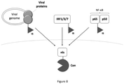
- the present invention relates to processes for monitoring viral infections and innate immune responses. More particularly, the present invention relates to processes for monitoring the translocation of proteins of interest to given subcellular components, wherein the translocated proteins are indicative of infection or sensing. Particularly, the processes of the invention allow to monitor the viral translocation of a given virus to a subcellular component of interest, particularly to the nucleus, and the translocation of transcription factors in the nucleus. These processes are particularly useful for screening antiviral candidate molecules and molecules able to stimulate, modulate or inhibit innate immunity.
- Protein movement between different subcellular compartments is an essential aspect of biological processes, including transcriptional and metabolic regulation, and immune response. Particularly, protein movement is a critical component of infection. Any intracellular infection, whether viral or bacterial, stimulates innate immunity signaling pathways triggered by the detection of pathogen-associated molecular motifs by endosomal or cytosolic surface receptors. These signaling pathways converge on the nuclear translocation of transcription factors (mainly IRF3 and NF- ⁇ B), which then stimulate the expression of type I, type II and type III interferons (hereafter, interferons or IFN) and proinflammatory cytokines.
- transcription factors mainly IRF3 and NF- ⁇ B
- Interferons can then bind to the IFN receptor at the surface of cells and trigger a second wave of signaling pathways that lead to the translocation of transcription factors STAT1/STAT2/IRF9 for IFN alpha, beta et lambda and STAT1/STAT1/IRF9 for IFN gamma.
- viral replication can be inferred from the measurement of virus cytopathic effects, but this readout is limited to lytic viruses.
- this method estimates viral replication only at the latest time point when the lysis of infected cells occurs. The methods used nowadays are therefore not efficient to screen antiviral molecules.
- the knowledge of the translocation of certain important molecules offers an attractive opportunity for the development of therapeutics.
- the knowledge of the translocation and trafficking of viruses and viral particles in the cell would be of interest to identify antiviral molecules, just as knowledge of the innate immunity signaling pathway would contribute to screen modulators of innate immunity.
- the intracellular trafficking of functional proteins or virus plays a key role in regulating gene expressions in response to extracellular signals in eukaryotic cells, particularly to activate the adequate inflammation pathways.
- the inventors have now developed specific tools allowing to evaluate with accuracy the translocation of virus or proteins of interest from a cellular compartment to another. More particularly, the inventors have developed new protein complementation assays (PCA) to monitor either virus trafficking or innate signaling in infected or stimulated cells.
- PCA protein complementation assays
- the present invention is based on the reconstitution of split fragments of a protein reporter (e.g., bioluminescent proteins) when a translocation of interest is achieved. According to the invention, a first fragment of the protein reporter is fused to a biological component of a cellular compartment of a given cell, whereas the complementary fragment of the protein reporter is fused to the virus of interest or to a transcription factor.
- the first fragment of the reporter protein is fused to a protein or motif conferring localization to the nucleus, such as SV40, or to a protein of the nuclear pore complex, such as a nucleoporin, preferably Nup214, Nup98 or Nup153.
- the first fragment of the reporter protein is fused to the protein of the nucleus via a flexible linker sequence and/or the second fragment of the reporter protein is fused to an interferon regulatory factor (IRF), a STAT protein or a subunit of NF- ⁇ B.
- IRF interferon regulatory factor
- the reporter protein is selected from the group consisting of a fluorescent protein, preferably a GFP-like fluorescent protein, and bioluminescent protein, preferably a NanoLuc-like protein.
- the first fragment is ⁇ -NanoLuc with the amino acid sequence set forth in SEQ ID NO: 2 and the second fragment is Cen-NanoLuc with the amino acid sequence set forth in SEQ ID NO: 3.
- Said process is particularly useful for screening candidate molecules able to stimulate and/or modulate innate immunity, wherein the stimulus consists in the candidate molecule(s) to be tested, wherein the detection of the reconstituted reporter protein in the cells is indicative that the candidate molecule is able to stimulate and/or to modulate the innate immunity.
- Said process is also useful for screening candidate molecules able to overstimulate innate immunity, wherein cells are submitted to a stimulus suitable to activate the innate immunity and to the candidate molecule(s) to be tested (before, during or after submitting the cells to the stimulus activating the innate immunity), wherein an increase of detection of the reconstituted reporter protein in the cells compared to reconstituted reporter protein in control cells, is indicative that the candidate molecule is able to over-activate the innate immunity.
- Said process is also useful for screening candidate molecules able to inhibit innate immunity, wherein the cells are submitted to a stimulus suitable to activate the innate immunity and to the candidate molecule(s) to be tested (before, during or after submitting the cells to the stimulus activating the innate immunity), wherein an absence of detection of the reconstituted reporter protein in the cells, or a decrease of detection of the reconstituted reporter protein in the cells compared to reconstituted reporter protein in control cells, is indicative that the candidate molecule is able to inhibit stimulation of the innate immunity.
- kit for screening candidate molecules acting on innate immunity pathway comprising at least one immortal cell line as described above, and optionally one or more agonist(s) of interferon and inflammation signaling adapted to the cell line provided in the kit.
- It is another object of the present invention to provide a process for monitoring the viral translocation of a virus of interest to a subcellular component of interest in a population of cells comprising:
- Said process can be used for screening antiviral candidate molecules, wherein the cells are subjected both to the virus and to at least one antiviral candidate molecule, wherein an absence of detection of the reconstituted reporter protein in the cells or a decrease of detection of the reconstituted reporter protein in the cells compared to reconstituted reporter protein in control cells, is indicative that the candidate molecule is able to inhibit the viral infection.
- the present invention further relates to the use of a split reporter protein for monitoring and/or evaluating and/or quantifying the viral trafficking of a virus of interest between subcellular compartments in a cell population, wherein a first fragment of the reporter protein is fused to a protein of a subcellular compartment of interest in the cell population, and the second fragment of the reporter protein is fused to a viral protein of the virus of interest.
- the reporter protein is selected from the group consisting of a fluorescent protein, preferably a GFP-like fluorescent protein, and bioluminescent protein, preferably a NanoLuc-like protein.
- the first fragment is ⁇ -NanoLuc with the amino acid sequence set forth in SEQ ID NO: 2 and the second fragment is Cen-NanoLuc with the amino acid sequence set forth in SEQ ID NO: 3.
- the first fragment of the reporter protein is fused to a protein or motif conferring localization to a subcellular compartment selected from the group consisting of the nucleus, the endoplasmic reticulum, the nuclear pore complex or the mitochondria and/or wherein the second fragment of the reporter protein is fused to an integrase protein of the virus.
- a "protein reporter” refers to a polypeptide molecule that can be detected in cells by ordinary means, such as spectroscopic means (e.g. fluorometry, mass spectrometry) or biochemical means (e.g. enzymatic reactions).
- spectroscopic means e.g. fluorometry, mass spectrometry
- biochemical means e.g. enzymatic reactions.
- a fluorescent protein and the like e.g. a green fluorescent protein (GFP) or the like
- GFP green fluorescent protein
- a bioluminescent protein such as luciferase, nanoluciferase or the like can be used as a protein reporter.
- bioluminescence refers to production or emission of light by a reaction catalyzed by, or enabled by, an enzyme, protein, protein complex, etc.
- a substrate for bioluminescent entity is converted into unstable form by the bioluminescent entity.
- the substrate subsequently emits a bioluminescent signal (i.e. light) that can be detected/measured/monitored.
- complementary fragment(s) when used in reference to a protein reporter refer to fragments of a protein reporter that are individually inactive (i.e., do not express the reporter phenotype), wherein binding of the complementing fragments restores reporter activity.
- subcellular compartment refers to various distinguishable part, components or organelles of a cell, including without limitation, the nucleus, cytoplasm, plasma membrane, endoplasmic reticulum, Golgi apparatus, endosome, peroxisome and mitochondria.
- linker refers to a molecule or group of molecules that connects two molecules, such as a fragment of a protein reporter and a protein or nucleic acid.
- the present invention is based on the use of a protein reporter and more particularly of a protein reporter that is split into two complementary fragments, which are linked to different compartments of a given cell and/or to a given virus, to study the intracellular trafficking of components of interest. Complementation of the protein reporter is indicative of trafficking of a protein or virus of interest within a given subcellular compartment of the cell.
- the protein reporter is a bioluminescent protein, such as a luciferase and nanoluciferase (NanoLuc), or the like.
- the protein reporter is a fluorescent protein or the like, such as a GFP and GFP-like protein.
- the protein reporter is split into two complementary fragments, which are each linked to a given protein located in a subcellular compartment or to a viral protein.
- the protein reporter is split into two fragments of unequal size, i.e. into a small fragment and a large fragment.
- the fragments are advantageously folded and soluble in the cellular environment.
- the small fragment is small enough to not perturb the solubility of the fused protein and/or to minimize the potential for interference with cellular processing and transport of the fusion protein.
- the protein reporter is split into two complementary fragments, called either 1 st fragment and second fragment, or large fragment and small fragment.
- the large fragment is fused to a protein expressed in a subcellular compartment of interest, whereas the small fragment is fused to a protein whom trafficking is studied (e.g. transcription factor, viral protein).
- a protein whom trafficking is studied e.g. transcription factor, viral protein.
- the protein reporter is a nanoluciferase (NanoLuc) split into a small fragment, called ⁇ fragment, and a large fragment, called Centauri fragment (or Cen).
- nanoLuc nanoluciferase
- the amino acid sequence of NanoLuc is wherein the ⁇ fragment corresponds to SEQ ID NO: 2: GVTGWRLCERILA and the Centauri fragment corresponds to SEQ ID NO: 3
- the protein reporter is a GFP split into a small fragment, called ⁇ fragment, and a large fragment, called Centauri fragment (or Cen).
- the amino acid sequence of GFP is wherein the ⁇ fragment corresponds to SEQ ID NO: 5: RDHMVLHEYVNAAGIT and the Centauri fragment corresponds to SEQ ID NO: 6:
- the fragments of the protein reporter are each fused to a given protein via a peptidic linker.
- the linker consists in a small peptide, comprising at most 20, at most 15 or at most 10 amino acid residues.
- the peptidic linker comprises between 6 and 8 amino acid residues.
- the linker comprises amino acid residues selected from small polar amino acid residues, such as Gly and Ala (allowing to confer flexibility to the linker).
- the small fragment is fused to a viral protein of a virus of interest.
- the small fragment is fused to a transcription factor.
- the small fragment is fused to an interferon regulatory factor (IRF), such as IRF1, IRF3, IRF7, IRF9, a STAT protein, such as STAT1, or a subunit of NF- ⁇ B, such as p65.
- IRF interferon regulatory factor
- the large fragment of the protein reporter is fused to a protein or motif conferring localization to the nucleus.
- the large fragment of the protein reporter is fused to a protein of nuclear pore complex (e.g., any nucleoporin such as Nup214, Nup98, Nup153).
- the large fragment of the protein reporter is fused to a protein carrying a nuclear localization protein (NLS) (e.g., SV40).
- NLS nuclear localization protein
- the large fragment of the protein reporter is fused to a protein of endoplasmic reticulum (e.g. Rab proteins, calnexin).
- a protein of endoplasmic reticulum e.g. Rab proteins, calnexin
- the large fragment of the protein reporter is fused to a protein of mitochondria (e.g. voltage-dependent anion channel VDAC, cytochrome c oxidase COX).
- a protein of mitochondria e.g. voltage-dependent anion channel VDAC, cytochrome c oxidase COX.
- a protein or motif conferring localization to the nucleus e.g. a protein of nuclear pore complex, a protein carrying a nuclear localization protein
- a protein of endoplasmic reticulum e.g. a protein of mitochondria.
- nucleic acid As used herein, the term "nucleic acid”, “nucleic sequence,” “polynucleotide”, “ oligonucleotide “ and “nucleotide sequence” are used interchangeably and refer to a sequence of deoxyribonucleotides and/or ribonucleotides.
- the nucleic acids can be DNA (cDNA or gDNA), RNA, or a mixture of the two. It can be in single stranded form or in duplex form or a mixture of the two. It can be of recombinant, artificial and/or synthetic origin and it can comprise modified nucleotides, comprising for example a modified bond, a modified purine or pyrimidine base, or a modified sugar.
- nucleic acids of the invention can be in isolated or purified form, and made, isolated and/or manipulated by techniques known per se in the art, e.g., enzymatic synthesis or recombinant technology.
- the nucleic acids can also be synthesized in vitro by well-known chemical synthesis techniques, as described in, e.g., Belousov (1997) Nucleic Acids Res. 25:3440-3444 .
- Nucleic acids of the invention may further comprise additional nucleotide sequences, such as regulatory regions, i.e., promoters, enhancers, silencers, terminators, signal peptides and the like that can be used to cause or regulate expression of the polypeptide in a selected host cell or system.
- nucleic acids of the invention may further comprise additional nucleotide sequences encoding fusion proteins, such as maltose binding protein (MBP) or glutathion S transferase (GST) that can be used to favor polypeptide expression and/or solubility.
- MBP maltose binding protein
- GST glutathion S transferase
- the present invention further relates to an expression cassette comprising a nucleic acid according to the invention operably linked to one or more control sequences that direct the expression of said nucleic acid in a suitable host cell.
- expression cassette denotes a nucleic acid construct comprising a coding region, i.e. a nucleic acid of the invention, and a regulatory region, i.e. comprising one or more control sequences, operably linked.
- the expression cassette comprises, or consists of, a nucleic acid according to the invention operably linked to a control sequence such as transcriptional promoter and/or transcription terminator.
- the control sequence may include a promoter that is recognized by a host cell or an in vitro expression system for expression of a nucleic acid encoding a protease of the present invention.
- the promoter contains transcriptional control sequences that mediate the expression of the recombinant protein.
- the promoter may be any polynucleotide that shows transcriptional activity in the host cell including mutant, truncated, and hybrid promoters, and may be obtained from genes encoding extracellular or intracellular polypeptides either homologous or heterologous to the host cell.
- the control sequence may also be a transcription terminator, which is recognized by a host cell to terminate transcription.
- the terminator is operably linked to the 3'-terminus of the nucleic acid encoding the recombinant protein. Any terminator that is functional in the host cell may be used in the present invention.
- the expression cassette comprises, or consists of, a nucleic acid according to the invention operably linked to a transcriptional promoter and a transcription terminator.
- the invention also relates to a vector comprising a nucleic acid or an expression cassette as defined above.
- vector refers to DNA molecule used as a vehicle to transfer recombinant genetic material into a host cell.
- the major types of vectors are plasmids, bacteriophages, viruses, fosmids, cosmids, and artificial chromosomes.
- the vector itself is generally a DNA sequence that consists of an insert (a heterologous nucleic acid sequence, transgene) and a larger sequence that serves as the "backbone" of the vector.
- the purpose of a vector which transfers genetic information to the host is typically to isolate, multiply, or express the insert in the target cell.
- Vectors called expression vectors are specifically adapted for the expression of the heterologous sequences in the target cell, and generally have a promoter sequence that drives expression of the heterologous sequences encoding a polypeptide.
- the regulatory elements that are present in an expression vector include a transcriptional promoter, a ribosome binding site, a terminator, and optionally present operator.
- an expression vector also contains an origin of replication for autonomous replication in a host cell, a selectable marker, a limited number of useful restriction enzyme sites, and a potential for high copy number.
- Examples of expression vectors are cloning vectors, modified cloning vectors, specifically designed plasmids and viruses. Expression vectors providing suitable levels of polypeptide expression in different hosts are well known in the art. The choice of the vector will typically depend on the compatibility of the vector with the host cell into which the vector is to be introduced.
- the recent emergence and re-emergence of viruses in the human population has highlighted the need for cell-based assays of viral replication.
- Viruses traffic between different subcellular compartments to replicate, including between the plasma membrane, endocytic vesicles, the cytoplasm, the nucleus and endoplasmic reticulum.
- the present invention allows to quantify viral translocation between subcellular compartments by protein complementation assays. More particularly, a fragment of the protein reporter is tethered to a subcellular compartment of interest whereas the complementary fragment of said protein reporter is tethered to a viral protein of the virus to study. A translocation within the subcellular compartment of interest brings together the two complementary fragments that do not emit any signal alone, creating a binary gain-of-signal.
- the invention allows to quantify a specific step of viral replication.
- the present invention relates to the use of a split protein reporter for monitoring / evaluating / quantifying the viral trafficking of a virus of interest between subcellular compartments in a cell population, wherein a 1 st fragment of the protein reporter is fused to a protein of a subcellular compartment of interest in the cell population, and the second fragment of the protein reporter is fused to a viral protein of the virus of interest.
- PCA protein complementation assay
- the PCA developed by the inventors can be used for screening for antivirals and for fundamental research to better understand viral replication, etc.
- It an object of the present invention to provide a recombinant cell that has been engineered to express a 1 st fragment, preferably a large fragment, of a protein reporter tethered to a subcellular component of interest of said cell.
- the 1 st fragment of the protein reporter can be tethered to any subcellular component of the cell.
- the 1 st fragment of the protein reporter can be tethered to a protein selected from the group consisting of proteins of the nucleus, the endoplasmic reticulum, the nuclear pore complex, the mitochondria, etc.
- recombinant cells are selected from the group consisting in Hela cells, HEK 293T cells, HT-1080 cells, A549 cells, HCT116 cells, THP-1 cells, CEM cells, MT4 cells.
- the present invention thus relates to the use of a nucleic acid, expression cassette or vector according to the invention to transform, transfect or transduce a host cell.
- the choice of the vector will typically depend on the compatibility of the vector with the host cell into which it must be introduced.
- the host cell may be transformed, transfected or transduced in a transient or stable manner.
- the expression cassette or vector of the invention is introduced into a host cell so that the cassette or vector is maintained as a chromosomal integrant or as a self-replicating extra-chromosomal vector.
- the term "host cell” also encompasses any progeny of a parent host cell that is not identical to the parent host cell due to mutations that occur during replication.
- the host cell may be any eukaryote cell useful in the production of a recombinant cell of the present invention.
- the nucleic acid, expression cassette or expression vector according to the invention may be introduced into the host cell by any method known by the skilled person, such as electroporation, conjugation, transduction, competent cell transformation, protoplast transformation, protoplast fusion, biolistic "gene gun” transformation, PEG-mediated transformation, lipid-assisted transformation or transfection, chemically mediated transfection, lithium acetate-mediated transformation, liposome-mediated transformation.
- virus Any virus may be used and engineered to express a fragment of the protein reporter.
- the virus is selected from the group consisting in human immunodeficiency viruses (HIV), coronaviruses, such as ⁇ -coronaviruses, Influenza viruses, flaviviruses such as West Nile Virus and Usutu virus.
- HAV human immunodeficiency viruses
- coronaviruses such as ⁇ -coronaviruses
- Influenza viruses flaviviruses such as West Nile Virus and Usutu virus.
- the virus is a retrovirus and the small fragment of the protein reporter is tethered to an integrase protein of said virus.
- the cells and virus described above may be used for monitoring the viral trafficking within cells as well as for screening for antiviral drug candidates.
- It is an object of the present invention to provide a process for monitoring the viral translocation of a virus of interest to a subcellular component of interest in cells comprising:
- the protein reporter is reconstituted only if or when the virus reaches the subcellular component of interest within the cells.
- the large fragment of the protein reporter is linked to a protein of the nucleus or the nuclear pore complex, in order to monitor the nuclear translocation of a viral protein.
- the protein reporter is a fluorescent protein, such as GFP or GFP-like.
- the protein reporter is a bioluminescent protein, such as NanoLuc or NanoLuc-like.
- the cells are subjected to a substrate (e.g. light-emitting compound such as luciferin, fumirazine or other coelenterazine analogues) prior to the step of detecting.
- a substrate e.g. light-emitting compound such as luciferin, fumirazine or other coelenterazine analogues
- the detection of reconstituted protein reporters comprises or consists of the detection of a fluorescent or luminescent signal emitted when the protein reporter is reconstituted.
- said signal may be further measured / quantified to evaluate with more accuracy the viral translocation to the target cellular compartment.
- Such PCA may be further implemented for screening antiviral candidate molecules, able to prevent the viral translocation.
- An absence of detection of reconstituted protein reporter or a decrease of detection of reconstituted protein reporter compared to detection of reconstituted protein reporter for control cells is indicative that the candidate molecule has an antiviral activity.
- the cells have been contacted with the candidate molecule before the step of detecting and/or quantifying the reconstituted protein reporter in the subcellular component of the cells.
- the cells are contacted simultaneously with the virus and the candidate molecule, or are contacted first with the virus and after with the candidate molecule.
- This process for screening may be implemented with any virus and any candidate molecule.
- kit ready to use for monitoring viral trafficking between subcellular compartments of cells.
- kit may comprise:
- the kit may further comprise a substrate for the protein reporter, such as an light-emitting compound (e.g. fumirazine).
- a substrate for the protein reporter such as an light-emitting compound (e.g. fumirazine).
- Activation of transcription factor NF- ⁇ B results in translocation of ubiquitously expressed NF- ⁇ B from the cytoplasm to the nucleus.
- NF- ⁇ B is associated with a number of diseases. There is thus an interest in identifying compounds that modulate or inhibit the nuclear translocation of activated NF- ⁇ B.
- the method of the present invention allows to identify and optionally quantify the nuclear translocation of NF- ⁇ B induced by the test compound(s) and thereby to determine the toxicity of the test compound(s) and their pro-inflammatory potential.
- the present invention relates to the use of a split protein reporter for monitoring and/or evaluating and/or quantifying the activation of innate immunity in cells.
- the present invention relates to the use of such split protein reporter for monitoring and/or evaluating and/or quantifying the nuclear translocation of transcription factors, such as interferon regulatory factors (IRF), STAT proteins or a subunit of NF- ⁇ B in a cell population.
- IRF interferon regulatory factors
- STAT proteins a subunit of NF- ⁇ B in a cell population.
- a large fragment of the protein reporter is fused to a protein of a subcellular compartment of interest in the cell population, and the small fragment of the protein reporter is fused to a transcription factor to monitor.
- PCA protein complementation assay
- the 1 st fragment of the protein reporter can be tethered to any protein or motif conferring localization to the nucleus, such as a protein carrying a nuclear localization protein (NLS) (ex.: SV40) or a protein of the nuclear pore (ex.: nucleoporin Nup124).
- NLS nuclear localization protein
- Nup124 nucleoporin Nup124
- the second fragment of the protein reporter is tethered to a of transcription factors, such as interferon regulatory factors (IRF), STAT proteins or a subunit of NF- ⁇ B, such as p65.
- IRF interferon regulatory factors
- STAT proteins or a subunit of NF- ⁇ B, such as p65.
- the cells may be selected from any type of cell, including any type of human or non-human animal cell, including mouse cells or rat cells.
- the cells may be stem cells or may be somatic cells such as primary cells, established cell lines such as immortal or immortalized cells, tumour cells, germs cells or their precursors, as well as cells derived or differentiated from stem cells, including derived or differentiated from induced pluripotent stem cells.
- recombinant cells are selected from the group consisting in Hela cells, HEK 293T cells, HT-1080 cells, A549 cells, HCT116 cells, THP-1 cells, CEM cells, MT4 cells.
- the recombinant cells described above, expressing both the small fragment of the protein reporter tethered to a transcription factor able to mediate the innate immune response and the large fragment of the protein reporter tethered to a protein or motif of the nucleus can be used for monitoring the innate immunity signal pathway during infection.
- the stimulus may be any extracellular stimulus able to infect the cells, such as, without limitation pathogens (e.g. viruses, bacteria), or to stimulate inflammation pathways, such as lipopolysaccharide (LPS).
- pathogens e.g. viruses, bacteria
- LPS lipopolysaccharide
- the protein reporter is reconstituted only if or when the innate immunity signaling pathway of interest is activated and the corresponding tagged transcription factor reaches within the nucleus of the cells.
- the protein reporter is a fluorescent protein, such as GFP or GFP-like.
- the protein reporter is a bioluminescent protein, such as NanoLuc or NanoLuc-like.
- the cells are subjected to a substrate (e.g. light-emitting compound such as luciferin, fumirazine or other coelenterazine analogues) prior to the step of detecting.
- a substrate e.g. light-emitting compound such as luciferin, fumirazine or other coelenterazine analogues
- the detection of reconstituted protein reporters consists in the detection of a fluorescent or luminescent signal emitted when the protein reporter is reconstituted.
- said signal may be further measured / quantified to evaluate with more accuracy the activation of the innate immunity pathway.
- the PCA developed by the inventors may be used for screening candidate molecule, either for evaluating their toxicity or their ability to enhance the innate immunity response.
- the recombinant cells of the invention are contacted with the candidate molecule.
- the contacting may be done by adding the candidate molecule to the culture medium in which the cells are cultured.
- the candidate molecule may be dissolved or dispersed in a liquid vehicle, such as a solvent or solution.
- the contacting may be done over a period of time, for example by incubating the candidate molecule that is to be tested with the cells in culture.
- the concentration of the candidate molecule to be used may be varied, and may depend on the compound that is to be tested.
- the candidate molecule may be any compound that is expected to come into contact with a subject, including being inhaled by, topically applied to, absorbed by, ingested by, administered to, or implanted into a subject.
- the test compound may be a pharmaceutical compound, an organic compound, an inorganic compound, a pesticide, a herbicide, an environmental toxin, a fungal toxin, a microbial toxin, a heavy metal-containing compound, an organic solvent, a cleaning agent, a preservative, a food additive, a dietary supplement, a herbal compound, an animal derived compound, an anti-microbial compound, a cosmetic ingredient, a microparticle or a nanoparticle.
- such PCA is used for screening candidate molecules able to stimulate and/or modulate innate immunity.
- the candidate molecule is used as the external stimulus in the above described process.
- detection of the reconstituted reporter protein in the cells is indicative that the candidate molecule is able to stimulate the innate immunity.
- the above described process further comprises the step of submitting the cells to a candidate molecule, before, after or simultaneously subjecting the cells to the external stimulus, in order to evaluate the impact of the candidate molecule on an already activated innate immunity pathway (i.e. the ability of the candidate compound to modulate the innate immunity).
- the amount of reconstituted protein reporter can be compared to the amount in negative control cells (subjected to the extern stimulus but not to the candidate molecule) to evaluate with more accuracy the ability of the candidate molecule to trigger the studied pathway.
- such PCA may be used for screening candidate molecules able to inhibit innate immunity.
- the above described process further comprises the step of submitting the cells to a candidate molecule, before, after or simultaneously subjecting the cells to the external stimulus.
- absence of detection of the reconstituted reporter protein in the cells is indicative that the candidate molecule is able to inhibit stimulation of the innate immunity.
- the amount of reconstituted protein reporter can be compared to the amount in negative control cells (subjected to the external stimulus but not to the candidate molecule) to evaluate with more accuracy the ability of the candidate molecule to inhibit the studied pathway.
- the negative control cells although not contacted with the candidate molecule, may be contacted with a negative control solution, for example the solvent or solution used to dissolve or disperse the candidate molecule.
- kit ready to use for monitoring innate immunity pathways in cells.
- such kit may comprise an immortal cell line (i.e. recombinant cells as described above) that expresses a large fragment of a protein reporter fused to a protein expressed in the nucleus and a small fragment of said protein reporter tethered to a transcription factor of interest, and optionally a substrate for the protein reporter, such as an light-emitting compound (e.g. fumirazine) and/or an agonist of interferon and inflammation signaling adapted to the cell line provided in the kit.
- an immortal cell line i.e. recombinant cells as described above
- a substrate for the protein reporter such as an light-emitting compound (e.g. fumirazine) and/or an agonist of interferon and inflammation signaling adapted to the cell line provided in the kit.
- the kit can further comprise candidate molecules and optionally a positive or a negative control.
- P4 TAR- ⁇ -gal indicator cells are HeLa CD4+ CXCR4+ CCR5+ carrying the LacZ gene under the control of the HIV-1 LTR promoter (AIDS Reagent Program).
- HEK 293T CL-11268), Hela (CCL-2) and A549 (CCL-185) were obtained from the ATCC.
- the MT4R5 1 and CEM CD4+ (NIH 117) T cell lines were grown in RPMI medium with 10% FCS, 100 IU/ml penicillin and 100 ⁇ g/ml streptomycin.
- NTP Nevirapine
- Aphidicolin APH, Sigma
- viruses were HIV-1 LAI, either full-length or ⁇ env and pseudotyped with the vesicular stomatitis virus glycoprotein (VSV-G).
- VSV-G vesicular stomatitis virus glycoprotein
- ⁇ HIV GFP , ⁇ HIV-225T GFP or ⁇ HIV Nluc viral constructs were generated by polymerase chain reaction (PCR) using a pBlueScript (pBS) plasmid containing a PstI-NcoI fragment of the HIV-1 LAI wt or mutant 225T molecular clone.
- oligonucleotides coding for ⁇ GFP or ⁇ Nluc flanked by EcoRI and NdeI restriction sites were used to amplify and add ⁇ GFP or ⁇ Nluc in C-ter of HIV-1 integrase.
- the forward primer was 5'-CCAGTACTACGGTTAAGGC-3' (SEQ ID NO:7).
- PCR products were digested with EcoRI/NdeI and cloned into pBS-LAI (PstI-NcoI). Finally, the PstI-NcoI fragment of LAI containing IN fused to ⁇ GFP or ⁇ Nluc was cloned back into a wild-type Env or ⁇ Env HIV-1 LAI molecular clone.
- ⁇ HIV-D116I Nluc was obtained by site-directed mutagenesis using the QuikChange II site-directed mutagenesis kit (Agilent) on the pBS-LAI containing IN fused to ⁇ Nluc, and was then cloned back into a wild-type Env or ⁇ Env HIV-1 LAI molecular clones.
- Lentiviral vectors (LV) coding for CenNLS or CenNup214 were obtained by cloning HA-NLS or HA-Nup214 downstream of Cen GFP or Cen Nluc by PCR amplification.
- Cen GFP NLS was generated by strand-overlap PCR first by generating Cen GFP -HA from a GFP1-10 plasmid using the following primers 5'- GATCGGATCCCGCCACCATG (SEQ ID NO:10) and 5'-AAGAGCGTAATCTGGAACATCGTATGGGTAGCCGGCGCCTTTCTCGTTTGGGTCT TTGCTCAGC-3' (SEQ ID NO:11)
- GFP-HA-NLS was generated by a second PCR reaction and cloned into a HIV-1 derived vector with BamH1/XhoI restriction enzymes.
- Cen NLuc NLS was synthesised by Genscript and cloned into a pcDNA3.1(+), followed by a HIV-1 derived vector.
- CenNup214 constructs were generated by amplifying Cen GFP -HA or Cen NLuc -HA with Agel and NotI overhangs and subcloning these at the place of EGFP upstream of Nup214 using a pEGFP-Nup214 plasmid (Euroscarf).
- viruses and vectors were produced by transient transfection of HEK 293T cells by calcium phosphate precipitation with the proviral or LV plasmid, co-transfected with VSV-G expression plasmid for ⁇ env viruses and vectors, and with an encapsidation plasmid (pCMV ⁇ R 8.74) for vectors.
- Viruses and vectors were harvested at 48 h after transfection. Viruses were concentrated using Lenti-X Concentrator (Clontech) and vectors by ultracentrifugation for 1 h at 64,000 ⁇ g (Beckman Coulter) at 4 °C.
- Cells were transduced with CenNLS GFP and CenNLS NLuc at MOI 10. Cells were used at 48 hours post-transduction (hpt), or stable cell lines were generated by selection and expansion of clones using Neomycin (1mg/ml). Cells were transfected CenNup214 GFP and CenNup214 NLuc plasmids using Fugene6 (Roche, HeLa) or calcium phosphate precipitation (HEK 293T) using 2 ⁇ g/ 10 6 cells. The efficiency of transduction and transfection was assessed by indirect immunofluorescence labeling of the HA tag that was inserted in the corresponding Centauri construct followed by flow cytometry or confocal microscopy. A threshold of 80% HA+ cells was set as a minimum value for performing ⁇ Centauri experiments.
- Virus yields were measured by p24 ELISA according to the manufacturer's instructions (Clontech). Multiplicities of infection were estimated by assuming that 1 ng of p24 corresponds to 5,000 transducing units (TU) 4 . Viruses were treated with after benzonase (Sigma, 15min, 37°C). Unless otherwise indicated, cells were infected at 2.5 TU/cell. Alternatively, viruses were titered by measuring Pol copy numbers by quantitative PCR (qPCR) at 6 hours post-infection (hpi) in HeLa cells and infections were performed at given Pol copy numbers/cell. Unless otherwise stated, cells were infected at 2 Pol copies/cell.
- qPCR quantitative PCR
- ⁇ Cen GFP reconstitution was assessed at 48 hpi by flow cytometry on fixed cells.
- cells were seeded in 96-well glass-bottomed Sensoplates (Greiner) at 10.000 cells/well at 24 hpi, and acquired on a ThermoCellomics at 48 hpi after addition of live Hoechst 33342 (Molecular Probes).
- CenNLS NLuc expressing cells were seeded in white opaque 96-well plates (Greiner) and infected the following day. ⁇ Cen Nuc reconstitution was assessed at 24 hpi by adding NanoGlo substrate (Promega) according to the manufacturer's instructions.
- Other NLuc substrates were prepared by diluting the stock solution 1:50 in assay buffer (100mM MES pH:6.0 adjusted with KOH, 1mM CDTA, 0.5% v/v Tergitol, 0.05% v/v antifoam, 150mM KCl, 1mM DTT et 35mM ThioUrea) and added to the cells 1:1. Luminescence was measured within 10 min using Tecan Infinite F200 Pro with 1000 ms integration and automatic attenuation.
- ⁇ -galactosidase assay was performed 48 hpi in indicator P4 cells according to the manufacturer's instructions (Roche Applied Science). Luciferase and ⁇ -Galactosidase activities were normalized for protein concentration by the Bradford assay. Luminescence and absorbance were acquired on a Tecan Infinite F200 Pro.
- the primary antibodies used were rat anti-HA tag (Roche 3F10), mouse monoclonal anti-p24 clone AG3.0 & 183-H12-5C (NIH AIDS Reagent Program). Secondary antibodies were goat anti-mouse and anti-rabbit HRP conjugates (Thermo Fisher Scientific, Rockford, IL) or Alexa Fluor 455 or 647 conjugates. Intracellular Gag was measured using KC57 antibody conjugated to FITC or PE (Beckman Coulter).
- Z' factors were calculated using 10 to 30 replicates per condition, randomly distributed on the plate.
- the Z-factor (Zhang et al., 1999) is a measure that quantifies the separation between the distribution of positive and negative controls.
- Z ⁇ factor 1 ⁇ 3 ⁇ p + ⁇ n ⁇ p ⁇ ⁇ n where ⁇ p and ⁇ p are the mean and standard deviation values of the positive control and ⁇ n and ⁇ n are those of the negative control.
- the Z-factor is calculated using robust estimates of location (median) and spread (mad).
- the signal/background ( ⁇ p - ⁇ n ) and signal/noise (( ⁇ p - ⁇ n )/ ⁇ n ) ratios are also provided.
- O-acetylated luciferin (1 mg) was dissolved in DMSO (0.2 mL) and then diluted by adding a solution of acidic ethanol (0.3 ml) made from the addition of 37 % hydrochloric acid (100 ⁇ l) on 100 % ethanol (12 mL). The 0.5 mL reaction solution was incubated at 50°C for 2 h to give a stock solution which was aliquoted and frozen at -80°C for later use.
- O-acetylated luciferins (hikarazines) were used in this work:
- Lentiviral vectors (LV) coding for shRNA against Pin1, CypA, RanBP2, TNPO1, TNPO3, CKAP1, WIRE, MAP1A, MAP1S, IPO5, IPO7 and KPNB1 were generated as previously published (Di Nunzio et al., 2012 ; Fernandez et al., 2015 ; Fernandez et al., 2019 ; Maarifi et al., 2019 ; Kaul et al., 2009). Transduction was performed at MOI 50.
- the HIV particle comprises an envelope, a capsid, and two copies of positive-strand RNA genome. Following the fusion of the HIV envelope with the target cell membrane, the capsid is released into the cytoplasm and transported towards the nucleus. Reverse transcription of the genome into double-stranded DNA produces a pre-integration complex (PIC), which enters the nucleus by active transport through the nuclear pore complex (NPC) and mediates integration of the HIV DNA into the host cell chromatin.
- PIC pre-integration complex
- NPC nuclear pore complex
- the ⁇ Cen reporter is expressed as two complementary, self-assembling fragments of sfGFP or NLuc.
- the small ⁇ fragment ( ⁇ GFP : 16aa, or ⁇ NLuc : 13aa) was fused into full-length and ⁇ env HIV-1 molecular clones (hereafter ⁇ HIV) in C-ter of HIV-1 integrase (IN) ( Fig. 1c ).
- Each incoming viral particle contains approximately 120 IN molecules bound to the RNA genome, based on the 20:1 synthesis ratio of Gag to Gag-Pol.
- IN molecules that bind to the viral DNA ends as a multimer accompany the PIC into the nucleus, while free cytoplasmic IN is inherently unstable and likely undergoes proteasomal degradation.
- Insertion of the ⁇ -tag within the Pol coding sequence did not disrupt particle production ( Fig.1d ) and ensured wild-type viral infectivity compared with non-tagged viruses ( Fig. 1e ).
- Cen GFP 52 kDa, or Cen Nluc 39 kDa was fused to the nucleoporin Nup214 for targeting to the NPC (CenNup214) or to the triple nuclear localization signal (NLS) of SV40 for tethering to the nucleus (CenNLS) ( Fig. 1f ).
- a HA tag was introduced in all Cen constructs to monitor their expression and localization ( Fig. 1g ).
- HeLa cells expressing CenNup214 GFP or CenNLS GFP were infected with ⁇ HIV GFP .
- Transduced cells did not emit any GFP fluorescence upon Centauri expression alone, however infection by ⁇ HIV led to gain-of-GFP signal close to nuclear pores in CenNup214 cells ( Fig. 2a ), and in nuclei in CenNLS cells (with 87% of all GFP signal localizing to the nucleus) ( Fig. 2b ).
- T cells which are the relevant target cells of HIV in vivo
- Complementation of sfGFP led to an approximately 5-10-fold increase in fluorescence in all tested cell types, while neither the ⁇ nor the Cen fragments emitted any detectable signal when expressed alone ( Fig. 2c ).
- Aphidicolin was used as control to block nuclear envelope breakdown during mitosis. Treatment did not reduce signal, confirming that ⁇ Centauri signal is generated following viral transport through NPCs, which is concordant with the HIV PIC entering the nucleus through nuclear pores ( Fig. 2d ).
- NVP Nevirapine
- the ⁇ Centauri signal intensity was directly proportional to the detection of the HIV-1 genome in the nucleus, confirming that the NLuc reporter was reconstituted upon HIV-1 PIC nuclear import ( Fig. 3e ).
- HIV-1 Gag and Gag-Pol polyproteins have been shown to traffic back to the nucleus and perinuclear area after translation in the cytoplasm.
- Infection with D116I virus still achieved ca. 10-fold increase in NLuc compared to infection in the presence of Nevirapine ( Fig. 3f ), confirming that ⁇ Centauri allows the detection of viral PICs trafficking into the nucleus.
- the ⁇ CentauriNLS assay reflects the combined efficiency of all the early steps of viral replication required to reach the nucleus, and is not specific to nuclear import.
- the system would allow to deconvolute successful trafficking to the NPC from HIV nuclear import and therefore enable the specific screening of nuclear import.
- the sfGFP approach was poorly quantitative for Nup214, we tested the NLuc readout.
- HeLa cells were transduced or transfected with CenNup214, then infected with ⁇ HIV. Infection resulted in a 10-fold increase in signal, indicating that the ⁇ CentauriNup214 assay is quantitative and could be used in parallel to ⁇ CentauriNLS to screen for specific inhibitors of nuclear import ( Fig. 3g ).
- LTR long-terminal repeat promoter
- Tat the HIV long-terminal repeat promoter
- LTR-reporter cell lines using LacZ, eGFP or Luciferase, have been used in the past to identify cellular co-factors of HIV infection.
- infected cells can be scored by immunolabelling of viral antigens.
- iGag intracellular Gag
- the assay also exhibited high sensitivity, virtually no background, and a linear dose-response over 2-log, which was superior to any other tested assay ( Fig. 4a ).
- This assay initially used the commercially available Nano-Glo bioluminescence-based reporting system which is made of the NanoLuc/NanoKAZ luciferase and uses furimazine as its substrate.
- a series of coelenterazine analogues including furimazine (Z01) that were previously characterized as NanoLuc substrates. Accordingly, the corresponding O-acetylated proluciferins (hikarazines 01, 03, 97, 103 and 108) were hydrolysed and the resulting solutions of these luciferin analogues assessed at a final concentration of 40-50 ⁇ M.
- Signal/background ratios were 2.53 for ⁇ HIV GFP , and 30.26 and 5.07 for ⁇ HIV NLuc and ⁇ HIV-D116I NLuc , respectively, which was considered acceptable since >2.
- Signal/noise ratios were 34.32 for ⁇ HIV GFP , and 280.77 and 39.07 for ⁇ HIV NLuc and ⁇ HIV-D116I NLuc , respectively, which was considered acceptable since >10.
- CypA has no effect in HeLa cells
- KPNB1 mediates Tat nuclear import independently of PIC entry
- ⁇ -karyopherins IPO5 and IPO7 and cytoskeletal proteins CKAP1 and WIRE have a low to moderate effect on infection.
- Hela-CenNLS NLuc cells were treated with previously validated shRNAs against these cellular co-factors, infected with ⁇ HIV NLuc , and NLuc signal was measured at 24 hpi.
- the knockdown of RanBP2 and TRN-1/TNPO1 had the greatest effect on HIV nuclear import, leading to ⁇ 10-fold decrease in ⁇ Centauri.
- Results confirm that ⁇ Centauri is a quantitative and reliable assay, applicable to the screening of shRNA/CRISPR-Cas9 libraries or small compound libraries.
- P4 TAR- ⁇ -gal indicator cells are HeLa CD4+ CXCR4+ CCR5+ carrying the LacZ gene under the control of the HIV-1 LTR promoter (AIDS Reagent Program).
- HEK 293T CL-11268), Hela (CCL-2), HT1080 (CCL-121) and A549 (CCL-185) were obtained from the ATCC.
- the MT4R5 Amara et al., J.Virol., 2003
- CEM CD4+ (AIDS Reagent Program) T cell lines were grown in RPMI medium with 10% FCS, 100 IU/ml penicillin and 100 ⁇ g/ml streptomycin. Vero E6 and HCT-116 are both from the ATCC repository.
- Puromycin, G418 and Hygromycin were obtained from Sigma and used at different concentration for cell selection.
- NanoLuc (SEQ ID NO:1) (based on Dixon et al., ACS Chem. Biol., 2016 ):
- CenNLS and IRF3-, IRF7- and p65- ⁇ -Flag expressing vectors were synthesised by GenScript into a pcDNA3.1(+) vector using NotI/XhoI cloning site. These constructions were also cloned into HIV-1 derived vectors using BamHI/XhoI cloning site.
- Vectors were produced by transient transfection of HEK 293T cells by calcium phosphate precipitation with LV plasmid, co-transfected with VSV-G expression plasmid and with an encapsidation plasmid (pCMV ⁇ R 8.74). Vectors were harvested at 48 h after transfection and concentrated by ultracentrifugation for 1 h at 64,000 ⁇ g (Beckman Coulter) at 4 °C.
- CenNLS expressing cells either transiently by plasmid transfection or HIV-1 derived vector transduction, or stably by selection under antibiotic, were seeded in 50 ⁇ l of complete growth medium in white opaque 96-well plates (Greiner) and stimulated the following day with defective interfering Sendai Virus (SdV, provided by D. Garcin (Department of Microbiology and Molecular Medicine, University of Geneva, Geneva, Switzerland) and used at 50 hemagglutination units (HAU)/ml , for 2 to 6 hours.
- SdV defective interfering Sendai Virus
- NanoGlo substrate Promega
- furimazine Coutant et al., 2019, 2020
- assay buffer 100mM MES pH:6.0 adjusted with KOH, 1mM CDTA, 0.5% v/v Tergitol, 0.05% v/v antifoam, 150mM KCl, 1mM DTT et 35mM ThioUrea
- Luminescence was measured within 10 min using Tecan Infinite F200 Pro with 1000 ms integration and automatic attenuation.
- ⁇ and Cen were assessed by immunolabelling of HA or Flag tag and analysed by confocal microscopy, flow cytometry or western-blot.
- the primary antibodies used were rat anti-HA tag (Roche 3F10) and mouse monoclonal anti-flag clone M2 (Sigma). Secondary antibodies were goat anti-mouse and anti-rat HRP conjugates (Thermo Fisher Scientific, Rockford, IL) or Alexa Fluor 488 or 647 conjugates.
- the luciferin solution was prepared as described above for "Viral trafficking".
- the present invention proposes innovative tools to characterise and quantify by HTS the nuclear translocation of transcription factors implicated in innate immune signalling pathways.
- This technique is based on the complementation of protein fragments to reconstitute a functional protein, an approach known as Protein-fragment complementation assay (PCA), which has been adapted to the measure of a nuclear translocation event.
- PCA Protein-fragment complementation assay
- NanoLuc is an engineered luciferase derived from a deep sea luminous shrimp that has a mass of 19kD, making it much smaller than Renilla (36kD) or firefly (61kD) luciferases, and therefore more appealing for fusion protein construction.
- Nluc is also approximately 150x brighter than other commercially available luciferase reporters allowing very sensitive detection ( Hall, M.P. et al. ACS Chem Biol 7, 1848-1857, (2012 )).
- the complementation of Nluc occurs via the self-association of the ⁇ and Cen fragments in the confined environment of the nucleus, and leads to a bioluminescent signal that is readily measured by plate luminometry.
- the assay has been termed ⁇ Centauri to underline its binary and very bright nature that is pronounced of the ⁇ Centauri star system.
- the ⁇ Centauri tools were originally developed for HTS to screen compounds by quantitatively measuring their impact on innate immune or inflammatory response signalling pathways, independently of the transcriptional and translational machineries.
- the system of the present invention should provide a fast and reliable read-out for any viral experimental infection.
- IFN- ⁇ is a cytokine secreted by cells downstream of the signaling cascade leading to nuclear translocation of the transcription factor IRF3.
- results show that the nuclear translocation of IRF3 ⁇ is only due to the infection of cells with SeV ( Fig. 6d ).
- Expression of the ⁇ and Centauri fragments was monitored by flow cytometry (data not shown) and confocal microscopy ( Fig. 6e ) upon detection of the Flag and HA tags that were inserted in the corresponding expression plasmids.
- the approach of the present invention has been compared side-by-side with alternatives commercialised by Invivogen (ISRE-Luciferase and ISRE-Secreted Embryonic Alkaline Phosphatase/SEAP cell lines). Since the ⁇ Centauri system does not rely on cellular transcription and translation machineries, it may be used at very early time points, 6 hpi by SeV compared to 24 hpi for the ISRE systems ( Fig. 7a ). In addition, the ⁇ Centauri system appeared to be more specific ( Fig.
- HDAC Histone Deacetylase
- the approach of the present invention provides a robust, versatile and sensitive read-out of innate signalling activation following viral infection. It is more reliable than competing commercial assays that monitor the activity of transcription factor-responsive promoter elements, which are hampered by a high number of false positives and negatives linked to the effect of transcriptional modulators and inducers of genotoxic stress, such as intercalating agents, or molecules interfering with cell cycle.
Landscapes
- Health & Medical Sciences (AREA)
- Life Sciences & Earth Sciences (AREA)
- Genetics & Genomics (AREA)
- Chemical & Material Sciences (AREA)
- Engineering & Computer Science (AREA)
- Organic Chemistry (AREA)
- Zoology (AREA)
- Bioinformatics & Cheminformatics (AREA)
- Biomedical Technology (AREA)
- Wood Science & Technology (AREA)
- Biotechnology (AREA)
- General Engineering & Computer Science (AREA)
- Molecular Biology (AREA)
- General Health & Medical Sciences (AREA)
- Biochemistry (AREA)
- Microbiology (AREA)
- Biophysics (AREA)
- Plant Pathology (AREA)
- Physics & Mathematics (AREA)
- Medicinal Chemistry (AREA)
- Virology (AREA)
- Toxicology (AREA)
- Gastroenterology & Hepatology (AREA)
- Proteomics, Peptides & Aminoacids (AREA)
- Immunology (AREA)
- Measuring Or Testing Involving Enzymes Or Micro-Organisms (AREA)
- Micro-Organisms Or Cultivation Processes Thereof (AREA)
Priority Applications (3)
| Application Number | Priority Date | Filing Date | Title |
|---|---|---|---|
| EP20306043.9A EP3971294A1 (fr) | 2020-09-17 | 2020-09-17 | Processus de surveillance des événements de trafic pendant une infection et réponse immunitaire innée |
| EP21777532.9A EP4214320A1 (fr) | 2020-09-17 | 2021-09-16 | Procédés pour le suivi des événements de trafic pendant une infection et une réponse immunitaire innée |
| PCT/EP2021/075509 WO2022058439A1 (fr) | 2020-09-17 | 2021-09-16 | Procédés pour le suivi des événements de trafic pendant une infection et une réponse immunitaire innée |
Applications Claiming Priority (1)
| Application Number | Priority Date | Filing Date | Title |
|---|---|---|---|
| EP20306043.9A EP3971294A1 (fr) | 2020-09-17 | 2020-09-17 | Processus de surveillance des événements de trafic pendant une infection et réponse immunitaire innée |
Publications (1)
| Publication Number | Publication Date |
|---|---|
| EP3971294A1 true EP3971294A1 (fr) | 2022-03-23 |
Family
ID=72840425
Family Applications (2)
| Application Number | Title | Priority Date | Filing Date |
|---|---|---|---|
| EP20306043.9A Pending EP3971294A1 (fr) | 2020-09-17 | 2020-09-17 | Processus de surveillance des événements de trafic pendant une infection et réponse immunitaire innée |
| EP21777532.9A Pending EP4214320A1 (fr) | 2020-09-17 | 2021-09-16 | Procédés pour le suivi des événements de trafic pendant une infection et une réponse immunitaire innée |
Family Applications After (1)
| Application Number | Title | Priority Date | Filing Date |
|---|---|---|---|
| EP21777532.9A Pending EP4214320A1 (fr) | 2020-09-17 | 2021-09-16 | Procédés pour le suivi des événements de trafic pendant une infection et une réponse immunitaire innée |
Country Status (2)
| Country | Link |
|---|---|
| EP (2) | EP3971294A1 (fr) |
| WO (1) | WO2022058439A1 (fr) |
Citations (4)
| Publication number | Priority date | Publication date | Assignee | Title |
|---|---|---|---|---|
| WO2004070351A2 (fr) * | 2003-02-06 | 2004-08-19 | Odyssey Thera, Inc. | Dosages de complementation de fragments proteiques pour criblage a rendement et contenu eleves |
| WO2005015161A2 (fr) * | 2003-08-06 | 2005-02-17 | Washington University In St. Louis | Interactions proteine-proteine regulees par imagerie dans des cellules et des animaux vivants au moyen de la complementation de fragments proteiques de luciferase |
| WO2005113838A2 (fr) * | 2004-05-18 | 2005-12-01 | The Board Of Trustees Of The Leland Stanford Junior University | Detection de translocation de proteine par complementation de fragment reporter de beta-galactosidase |
| WO2006062877A2 (fr) * | 2004-12-04 | 2006-06-15 | The Regents Of The University Of California | Bioessais de localisation d'elements subcellulaires de proteines utilisant des proteines fluorescentes scindees |
-
2020
- 2020-09-17 EP EP20306043.9A patent/EP3971294A1/fr active Pending
-
2021
- 2021-09-16 WO PCT/EP2021/075509 patent/WO2022058439A1/fr unknown
- 2021-09-16 EP EP21777532.9A patent/EP4214320A1/fr active Pending
Patent Citations (4)
| Publication number | Priority date | Publication date | Assignee | Title |
|---|---|---|---|---|
| WO2004070351A2 (fr) * | 2003-02-06 | 2004-08-19 | Odyssey Thera, Inc. | Dosages de complementation de fragments proteiques pour criblage a rendement et contenu eleves |
| WO2005015161A2 (fr) * | 2003-08-06 | 2005-02-17 | Washington University In St. Louis | Interactions proteine-proteine regulees par imagerie dans des cellules et des animaux vivants au moyen de la complementation de fragments proteiques de luciferase |
| WO2005113838A2 (fr) * | 2004-05-18 | 2005-12-01 | The Board Of Trustees Of The Leland Stanford Junior University | Detection de translocation de proteine par complementation de fragment reporter de beta-galactosidase |
| WO2006062877A2 (fr) * | 2004-12-04 | 2006-06-15 | The Regents Of The University Of California | Bioessais de localisation d'elements subcellulaires de proteines utilisant des proteines fluorescentes scindees |
Non-Patent Citations (4)
| Title |
|---|
| AMARA ET AL., J.VIROL., 2003 |
| BELOUSOV, NUCLEIC ACIDS RES., vol. 25, 1997, pages 3440 - 3444 |
| DIXON ET AL., ACS CHEM. BIOL., 2016 |
| HALL, M.P. ET AL., ACS CHEM BIOL, vol. 7, 2012, pages 1848 - 1857 |
Also Published As
| Publication number | Publication date |
|---|---|
| WO2022058439A1 (fr) | 2022-03-24 |
| EP4214320A1 (fr) | 2023-07-26 |
Similar Documents
| Publication | Publication Date | Title |
|---|---|---|
| Ozono et al. | Super-rapid quantitation of the production of HIV-1 harboring a luminescent peptide tag | |
| Di Nunzio et al. | Human nucleoporins promote HIV-1 docking at the nuclear pore, nuclear import and integration | |
| Le Tortorec et al. | Antagonism to and intracellular sequestration of human tetherin by the human immunodeficiency virus type 2 envelope glycoprotein | |
| Neil et al. | HIV-1 Vpu promotes release and prevents endocytosis of nascent retrovirus particles from the plasma membrane | |
| Jayappa et al. | Human immunodeficiency virus type 1 employs the cellular dynein light chain 1 protein for reverse transcription through interaction with its integrase protein | |
| Osei Kuffour et al. | USP18 (UBP43) abrogates p21-mediated inhibition of HIV-1 | |
| Edinger et al. | Cathepsin W is required for escape of influenza A virus from late endosomes | |
| Zhang et al. | MARCH8 targets cytoplasmic lysine residues of various viral envelope glycoproteins | |
| Sui et al. | Cytoplasmic‐translocated Ku70 senses intracellular DNA and mediates interferon‐lambda1 induction | |
| Galão et al. | TRIM25 and ZAP target the Ebola virus ribonucleoprotein complex to mediate interferon-induced restriction | |
| Bruce et al. | ZBTB2 represses HIV-1 transcription and is regulated by HIV-1 Vpr and cellular DNA damage responses | |
| Xu et al. | A high throughput screening assay for inhibitors of SARS-CoV-2 pseudotyped particle entry | |
| Kmiec et al. | SIVcol Nef counteracts SERINC5 by promoting its proteasomal degradation but does not efficiently enhance HIV-1 replication in human CD4+ T cells and lymphoid tissue | |
| Cubitt et al. | A cell-based, infectious-free, platform to identify inhibitors of lassa virus ribonucleoprotein (vRNP) activity | |
| Takahata et al. | Critical contribution of Tyr15 in the HIV-1 integrase (IN) in facilitating IN assembly and nonenzymatic function through the IN precursor form with reverse transcriptase | |
| Sudo et al. | Efavirenz enhances HIV-1 gag processing at the plasma membrane through Gag-Pol dimerization | |
| Zhao et al. | Rapid, reliable, and reproducible cell fusion assay to quantify SARS-Cov-2 spike interaction with hACE2 | |
| Patten et al. | Identification of potent inhibitors of SARS-CoV-2 infection by combined pharmacological evaluation and cellular network prioritization | |
| Guo et al. | The transmembrane nucleoporin Pom121 ensures efficient HIV-1 pre-integration complex nuclear import | |
| Ortega et al. | Novel roles of the Nipah virus attachment glycoprotein and its mobility in early and late membrane fusion steps | |
| Wang et al. | Keap1 recognizes EIAV early accessory protein Rev to promote antiviral defense | |
| Zhang et al. | Human LAMP1 accelerates Lassa virus fusion and potently promotes fusion pore dilation upon forcing viral fusion with non-endosomal membrane | |
| Moon et al. | Production and characterization of lentivirus vector-based SARS-CoV-2 pseudoviruses with dual reporters: evaluation of anti-SARS-CoV-2 viral effect of Korean red ginseng | |
| Chen et al. | Differential effects of actin cytoskeleton dynamics on equine infectious anemia virus particle production | |
| Lobritz et al. | Multifaceted mechanisms of HIV inhibition and resistance to CCR5 inhibitors PSC-RANTES and Maraviroc |
Legal Events
| Date | Code | Title | Description |
|---|---|---|---|
| PUAI | Public reference made under article 153(3) epc to a published international application that has entered the european phase |
Free format text: ORIGINAL CODE: 0009012 |
|
| STAA | Information on the status of an ep patent application or granted ep patent |
Free format text: STATUS: THE APPLICATION HAS BEEN PUBLISHED |
|
| AK | Designated contracting states |
Kind code of ref document: A1 Designated state(s): AL AT BE BG CH CY CZ DE DK EE ES FI FR GB GR HR HU IE IS IT LI LT LU LV MC MK MT NL NO PL PT RO RS SE SI SK SM TR |
|
| RIN1 | Information on inventor provided before grant (corrected) |
Inventor name: ARHEL, NATHALIE Inventor name: NISOLE, SEBASTIEN |
|
| STAA | Information on the status of an ep patent application or granted ep patent |
Free format text: STATUS: REQUEST FOR EXAMINATION WAS MADE |
|
| 17P | Request for examination filed |
Effective date: 20220923 |
|
| RBV | Designated contracting states (corrected) |
Designated state(s): AL AT BE BG CH CY CZ DE DK EE ES FI FR GB GR HR HU IE IS IT LI LT LU LV MC MK MT NL NO PL PT RO RS SE SI SK SM TR |
|
| RAP1 | Party data changed (applicant data changed or rights of an application transferred) |
Owner name: UNIVERSITE PARIS CITE |
|
| RAP1 | Party data changed (applicant data changed or rights of an application transferred) |
Owner name: UNIVERSITE PARIS CITE Owner name: CENTRE NATIONAL DE LA RECHERCHE SCIENTIFIQUE Owner name: UNIVERSITE DE MONTPELLIER |