EP1584289A2 - Appareil et procédé d'exploitation pour un appareil permettant l'évaluation non-invasive des fonctions hemodynamiques, y compris celles de l'endothélium - Google Patents
Appareil et procédé d'exploitation pour un appareil permettant l'évaluation non-invasive des fonctions hemodynamiques, y compris celles de l'endothélium Download PDFInfo
- Publication number
- EP1584289A2 EP1584289A2 EP05004826A EP05004826A EP1584289A2 EP 1584289 A2 EP1584289 A2 EP 1584289A2 EP 05004826 A EP05004826 A EP 05004826A EP 05004826 A EP05004826 A EP 05004826A EP 1584289 A2 EP1584289 A2 EP 1584289A2
- Authority
- EP
- European Patent Office
- Prior art keywords
- blood
- blood volume
- time
- arterial
- peripheral
- Prior art date
- Legal status (The legal status is an assumption and is not a legal conclusion. Google has not performed a legal analysis and makes no representation as to the accuracy of the status listed.)
- Granted
Links
- 230000006870 function Effects 0.000 title claims abstract description 21
- 230000000004 hemodynamic effect Effects 0.000 title claims description 9
- 230000003511 endothelial effect Effects 0.000 title description 8
- 238000011017 operating method Methods 0.000 title description 2
- MWUXSHHQAYIFBG-UHFFFAOYSA-N Nitric oxide Chemical compound O=[N] MWUXSHHQAYIFBG-UHFFFAOYSA-N 0.000 claims abstract description 136
- 210000004369 blood Anatomy 0.000 claims abstract description 114
- 239000008280 blood Substances 0.000 claims abstract description 114
- 238000000034 method Methods 0.000 claims abstract description 51
- 230000006835 compression Effects 0.000 claims abstract description 11
- 238000007906 compression Methods 0.000 claims abstract description 11
- 210000004204 blood vessel Anatomy 0.000 claims abstract description 10
- 230000008753 endothelial function Effects 0.000 claims abstract description 4
- 238000005259 measurement Methods 0.000 claims description 48
- 230000010349 pulsation Effects 0.000 claims description 41
- 230000010412 perfusion Effects 0.000 claims description 28
- 230000002093 peripheral effect Effects 0.000 claims description 28
- 210000003038 endothelium Anatomy 0.000 claims description 19
- 210000004177 elastic tissue Anatomy 0.000 claims description 12
- 230000000541 pulsatile effect Effects 0.000 claims description 12
- 230000009471 action Effects 0.000 claims description 10
- 238000011990 functional testing Methods 0.000 claims description 10
- 230000036772 blood pressure Effects 0.000 claims description 8
- 230000007704 transition Effects 0.000 claims description 7
- 101150075130 PNOC gene Proteins 0.000 claims description 6
- 230000001133 acceleration Effects 0.000 claims description 6
- 238000011524 similarity measure Methods 0.000 claims description 6
- 238000006213 oxygenation reaction Methods 0.000 claims description 5
- 238000013186 photoplethysmography Methods 0.000 claims description 5
- 230000008569 process Effects 0.000 claims description 5
- 239000003814 drug Substances 0.000 claims description 4
- 230000001404 mediated effect Effects 0.000 claims description 4
- 210000005259 peripheral blood Anatomy 0.000 claims description 4
- 239000011886 peripheral blood Substances 0.000 claims description 4
- 229940079593 drug Drugs 0.000 claims description 3
- 230000033228 biological regulation Effects 0.000 claims description 2
- 238000009795 derivation Methods 0.000 claims description 2
- 230000002123 temporal effect Effects 0.000 claims description 2
- 210000003414 extremity Anatomy 0.000 claims 2
- 230000005540 biological transmission Effects 0.000 claims 1
- 210000003141 lower extremity Anatomy 0.000 claims 1
- 210000001364 upper extremity Anatomy 0.000 claims 1
- 230000002792 vascular Effects 0.000 abstract description 23
- 238000012360 testing method Methods 0.000 abstract description 16
- 230000000747 cardiac effect Effects 0.000 abstract description 6
- 230000004087 circulation Effects 0.000 abstract description 4
- 239000003795 chemical substances by application Substances 0.000 abstract description 3
- 230000001435 haemodynamic effect Effects 0.000 abstract 1
- 238000010008 shearing Methods 0.000 abstract 1
- 239000000835 fiber Substances 0.000 description 50
- WQZGKKKJIJFFOK-GASJEMHNSA-N Glucose Natural products OC[C@H]1OC(O)[C@H](O)[C@@H](O)[C@@H]1O WQZGKKKJIJFFOK-GASJEMHNSA-N 0.000 description 22
- 206010048554 Endothelial dysfunction Diseases 0.000 description 21
- 230000008694 endothelial dysfunction Effects 0.000 description 21
- 239000008103 glucose Substances 0.000 description 21
- 235000021028 berry Nutrition 0.000 description 14
- 208000030831 Peripheral arterial occlusive disease Diseases 0.000 description 12
- 230000007423 decrease Effects 0.000 description 12
- NOESYZHRGYRDHS-UHFFFAOYSA-N insulin Chemical compound N1C(=O)C(NC(=O)C(CCC(N)=O)NC(=O)C(CCC(O)=O)NC(=O)C(C(C)C)NC(=O)C(NC(=O)CN)C(C)CC)CSSCC(C(NC(CO)C(=O)NC(CC(C)C)C(=O)NC(CC=2C=CC(O)=CC=2)C(=O)NC(CCC(N)=O)C(=O)NC(CC(C)C)C(=O)NC(CCC(O)=O)C(=O)NC(CC(N)=O)C(=O)NC(CC=2C=CC(O)=CC=2)C(=O)NC(CSSCC(NC(=O)C(C(C)C)NC(=O)C(CC(C)C)NC(=O)C(CC=2C=CC(O)=CC=2)NC(=O)C(CC(C)C)NC(=O)C(C)NC(=O)C(CCC(O)=O)NC(=O)C(C(C)C)NC(=O)C(CC(C)C)NC(=O)C(CC=2NC=NC=2)NC(=O)C(CO)NC(=O)CNC2=O)C(=O)NCC(=O)NC(CCC(O)=O)C(=O)NC(CCCNC(N)=N)C(=O)NCC(=O)NC(CC=3C=CC=CC=3)C(=O)NC(CC=3C=CC=CC=3)C(=O)NC(CC=3C=CC(O)=CC=3)C(=O)NC(C(C)O)C(=O)N3C(CCC3)C(=O)NC(CCCCN)C(=O)NC(C)C(O)=O)C(=O)NC(CC(N)=O)C(O)=O)=O)NC(=O)C(C(C)CC)NC(=O)C(CO)NC(=O)C(C(C)O)NC(=O)C1CSSCC2NC(=O)C(CC(C)C)NC(=O)C(NC(=O)C(CCC(N)=O)NC(=O)C(CC(N)=O)NC(=O)C(NC(=O)C(N)CC=1C=CC=CC=1)C(C)C)CC1=CN=CN1 NOESYZHRGYRDHS-UHFFFAOYSA-N 0.000 description 12
- 230000002526 effect on cardiovascular system Effects 0.000 description 11
- 230000008859 change Effects 0.000 description 10
- 206010012601 diabetes mellitus Diseases 0.000 description 10
- 238000011156 evaluation Methods 0.000 description 10
- 230000017531 blood circulation Effects 0.000 description 9
- 208000037265 diseases, disorders, signs and symptoms Diseases 0.000 description 9
- 230000036581 peripheral resistance Effects 0.000 description 8
- 230000009467 reduction Effects 0.000 description 8
- SNIOPGDIGTZGOP-UHFFFAOYSA-N Nitroglycerin Chemical compound [O-][N+](=O)OCC(O[N+]([O-])=O)CO[N+]([O-])=O SNIOPGDIGTZGOP-UHFFFAOYSA-N 0.000 description 7
- 238000001514 detection method Methods 0.000 description 7
- 210000001255 hallux Anatomy 0.000 description 7
- 239000000243 solution Substances 0.000 description 7
- 238000002604 ultrasonography Methods 0.000 description 7
- 230000024883 vasodilation Effects 0.000 description 7
- 102000004877 Insulin Human genes 0.000 description 6
- 108090001061 Insulin Proteins 0.000 description 6
- 229960003711 glyceryl trinitrate Drugs 0.000 description 6
- 229940125396 insulin Drugs 0.000 description 6
- 239000001301 oxygen Substances 0.000 description 6
- 229910052760 oxygen Inorganic materials 0.000 description 6
- 239000000126 substance Substances 0.000 description 6
- 208000037849 arterial hypertension Diseases 0.000 description 5
- QVGXLLKOCUKJST-UHFFFAOYSA-N atomic oxygen Chemical compound [O] QVGXLLKOCUKJST-UHFFFAOYSA-N 0.000 description 5
- 210000002302 brachial artery Anatomy 0.000 description 5
- 208000035475 disorder Diseases 0.000 description 5
- 230000000694 effects Effects 0.000 description 5
- 239000008151 electrolyte solution Substances 0.000 description 5
- 210000003989 endothelium vascular Anatomy 0.000 description 5
- 238000001802 infusion Methods 0.000 description 5
- 230000035479 physiological effects, processes and functions Effects 0.000 description 5
- 210000001519 tissue Anatomy 0.000 description 5
- 230000006442 vascular tone Effects 0.000 description 5
- 108010061951 Methemoglobin Proteins 0.000 description 4
- 230000008321 arterial blood flow Effects 0.000 description 4
- 201000010099 disease Diseases 0.000 description 4
- 230000003993 interaction Effects 0.000 description 4
- 230000010355 oscillation Effects 0.000 description 4
- 230000003647 oxidation Effects 0.000 description 4
- 238000007254 oxidation reaction Methods 0.000 description 4
- 229940124549 vasodilator Drugs 0.000 description 4
- 239000003071 vasodilator agent Substances 0.000 description 4
- 101710112752 Cytotoxin Proteins 0.000 description 3
- 108010064719 Oxyhemoglobins Proteins 0.000 description 3
- 230000002159 abnormal effect Effects 0.000 description 3
- 210000001367 artery Anatomy 0.000 description 3
- 210000004027 cell Anatomy 0.000 description 3
- 238000006243 chemical reaction Methods 0.000 description 3
- 230000001684 chronic effect Effects 0.000 description 3
- 208000029078 coronary artery disease Diseases 0.000 description 3
- 231100000599 cytotoxic agent Toxicity 0.000 description 3
- 239000002619 cytotoxin Substances 0.000 description 3
- 230000006378 damage Effects 0.000 description 3
- 238000011161 development Methods 0.000 description 3
- 238000001990 intravenous administration Methods 0.000 description 3
- 230000004089 microcirculation Effects 0.000 description 3
- 208000031225 myocardial ischemia Diseases 0.000 description 3
- 238000005457 optimization Methods 0.000 description 3
- 230000000241 respiratory effect Effects 0.000 description 3
- 230000000638 stimulation Effects 0.000 description 3
- 238000002560 therapeutic procedure Methods 0.000 description 3
- 208000031104 Arterial Occlusive disease Diseases 0.000 description 2
- 201000001320 Atherosclerosis Diseases 0.000 description 2
- 102000008186 Collagen Human genes 0.000 description 2
- 108010035532 Collagen Proteins 0.000 description 2
- 206010020565 Hyperaemia Diseases 0.000 description 2
- 229910002651 NO3 Inorganic materials 0.000 description 2
- NHNBFGGVMKEFGY-UHFFFAOYSA-N Nitrate Chemical compound [O-][N+]([O-])=O NHNBFGGVMKEFGY-UHFFFAOYSA-N 0.000 description 2
- 239000000006 Nitroglycerin Substances 0.000 description 2
- 206010047139 Vasoconstriction Diseases 0.000 description 2
- 238000010521 absorption reaction Methods 0.000 description 2
- 230000004913 activation Effects 0.000 description 2
- 210000000709 aorta Anatomy 0.000 description 2
- 230000015572 biosynthetic process Effects 0.000 description 2
- 210000000748 cardiovascular system Anatomy 0.000 description 2
- 229920001436 collagen Polymers 0.000 description 2
- 210000002808 connective tissue Anatomy 0.000 description 2
- 230000001419 dependent effect Effects 0.000 description 2
- 230000010339 dilation Effects 0.000 description 2
- 210000003743 erythrocyte Anatomy 0.000 description 2
- 230000003203 everyday effect Effects 0.000 description 2
- 238000002347 injection Methods 0.000 description 2
- 239000007924 injection Substances 0.000 description 2
- 239000007788 liquid Substances 0.000 description 2
- 230000035515 penetration Effects 0.000 description 2
- 201000011461 pre-eclampsia Diseases 0.000 description 2
- 230000009257 reactivity Effects 0.000 description 2
- 230000001105 regulatory effect Effects 0.000 description 2
- 230000000087 stabilizing effect Effects 0.000 description 2
- 230000002889 sympathetic effect Effects 0.000 description 2
- 210000000689 upper leg Anatomy 0.000 description 2
- 208000019553 vascular disease Diseases 0.000 description 2
- 230000025033 vasoconstriction Effects 0.000 description 2
- 230000003639 vasoconstrictive effect Effects 0.000 description 2
- 238000009423 ventilation Methods 0.000 description 2
- 206010060964 Arterial haemorrhage Diseases 0.000 description 1
- 206010003210 Arteriosclerosis Diseases 0.000 description 1
- 208000037260 Atherosclerotic Plaque Diseases 0.000 description 1
- 208000024172 Cardiovascular disease Diseases 0.000 description 1
- 206010058842 Cerebrovascular insufficiency Diseases 0.000 description 1
- 208000017667 Chronic Disease Diseases 0.000 description 1
- 208000002249 Diabetes Complications Diseases 0.000 description 1
- 238000004435 EPR spectroscopy Methods 0.000 description 1
- 229920002306 Glycocalyx Polymers 0.000 description 1
- 206010020772 Hypertension Diseases 0.000 description 1
- IOVCWXUNBOPUCH-UHFFFAOYSA-M Nitrite anion Chemical compound [O-]N=O IOVCWXUNBOPUCH-UHFFFAOYSA-M 0.000 description 1
- 208000005764 Peripheral Arterial Disease Diseases 0.000 description 1
- 208000018262 Peripheral vascular disease Diseases 0.000 description 1
- 241001499740 Plantago alpina Species 0.000 description 1
- 208000002787 Pregnancy Complications Diseases 0.000 description 1
- 208000001647 Renal Insufficiency Diseases 0.000 description 1
- 206010047141 Vasodilatation Diseases 0.000 description 1
- 206010058990 Venous occlusion Diseases 0.000 description 1
- 102400001284 Vessel dilator Human genes 0.000 description 1
- 208000027418 Wounds and injury Diseases 0.000 description 1
- 238000000862 absorption spectrum Methods 0.000 description 1
- YAJCHEVQCOHZDC-QMMNLEPNSA-N actrapid Chemical compound C([C@@H](C(=O)N[C@@H](CC(C)C)C(=O)N[C@H]1CSSC[C@H]2C(=O)N[C@H](C(=O)N[C@@H](CO)C(=O)N[C@H](C(=O)N[C@@H](C(N[C@@H](CO)C(=O)N[C@@H](CC(C)C)C(=O)N[C@@H](CC=3C=CC(O)=CC=3)C(=O)N[C@@H](CCC(N)=O)C(=O)N[C@@H](CC(C)C)C(=O)N[C@@H](CCC(O)=O)C(=O)N[C@@H](CC(N)=O)C(=O)N[C@@H](CC=3C=CC(O)=CC=3)C(=O)N[C@@H](CSSC[C@H](NC(=O)[C@H](C(C)C)NC(=O)[C@H](CC(C)C)NC(=O)[C@H](CC=3C=CC(O)=CC=3)NC(=O)[C@H](CC(C)C)NC(=O)[C@H](C)NC(=O)[C@H](CCC(O)=O)NC(=O)[C@H](C(C)C)NC(=O)[C@H](CC(C)C)NC(=O)[C@H](CC=3N=CNC=3)NC(=O)[C@H](CO)NC(=O)CNC1=O)C(=O)NCC(=O)N[C@@H](CCC(O)=O)C(=O)N[C@@H](CCCNC(N)=N)C(=O)NCC(=O)N[C@@H](CC=1C=CC=CC=1)C(=O)N[C@@H](CC=1C=CC=CC=1)C(=O)N[C@@H](CC=1C=CC(O)=CC=1)C(=O)N[C@@H]([C@H](C)O)C(=O)N1[C@@H](CCC1)C(=O)N[C@@H](CCCCN)C(=O)N[C@@H]([C@H](C)O)C(O)=O)C(=O)N[C@@H](CC(N)=O)C(O)=O)=O)CSSC[C@@H](C(N2)=O)NC(=O)[C@H](CCC(N)=O)NC(=O)[C@H](CCC(O)=O)NC(=O)[C@H](C(C)C)NC(=O)[C@@H](NC(=O)CN)[C@H](C)CC)[C@H](C)CC)[C@H](C)O)NC(=O)[C@H](CCC(N)=O)NC(=O)[C@@H](NC(=O)[C@@H](NC(=O)[C@@H](N)CC=1C=CC=CC=1)C(C)C)C(N)=O)C1=CNC=N1 YAJCHEVQCOHZDC-QMMNLEPNSA-N 0.000 description 1
- 230000001154 acute effect Effects 0.000 description 1
- 230000004075 alteration Effects 0.000 description 1
- 238000002266 amputation Methods 0.000 description 1
- 238000013459 approach Methods 0.000 description 1
- 230000006793 arrhythmia Effects 0.000 description 1
- 206010003119 arrhythmia Diseases 0.000 description 1
- 208000021328 arterial occlusion Diseases 0.000 description 1
- 210000005249 arterial vasculature Anatomy 0.000 description 1
- 208000011775 arteriosclerosis disease Diseases 0.000 description 1
- 210000003363 arteriovenous anastomosis Anatomy 0.000 description 1
- 108010090012 atrial natriuretic factor prohormone (31-67) Proteins 0.000 description 1
- 230000002567 autonomic effect Effects 0.000 description 1
- 230000004888 barrier function Effects 0.000 description 1
- 239000013060 biological fluid Substances 0.000 description 1
- 239000003990 capacitor Substances 0.000 description 1
- 230000036996 cardiovascular health Effects 0.000 description 1
- 210000001715 carotid artery Anatomy 0.000 description 1
- 210000003850 cellular structure Anatomy 0.000 description 1
- 208000026106 cerebrovascular disease Diseases 0.000 description 1
- 230000000052 comparative effect Effects 0.000 description 1
- 230000002860 competitive effect Effects 0.000 description 1
- 230000002354 daily effect Effects 0.000 description 1
- 238000013016 damping Methods 0.000 description 1
- 238000011982 device technology Methods 0.000 description 1
- 239000008121 dextrose Substances 0.000 description 1
- 230000035487 diastolic blood pressure Effects 0.000 description 1
- 230000004069 differentiation Effects 0.000 description 1
- 230000000916 dilatatory effect Effects 0.000 description 1
- 238000004090 dissolution Methods 0.000 description 1
- 238000013399 early diagnosis Methods 0.000 description 1
- 238000004870 electrical engineering Methods 0.000 description 1
- 230000008030 elimination Effects 0.000 description 1
- 238000003379 elimination reaction Methods 0.000 description 1
- 229940124642 endogenous agent Drugs 0.000 description 1
- 210000002889 endothelial cell Anatomy 0.000 description 1
- 238000004146 energy storage Methods 0.000 description 1
- 210000000245 forearm Anatomy 0.000 description 1
- 239000007789 gas Substances 0.000 description 1
- 210000004517 glycocalyx Anatomy 0.000 description 1
- 208000019622 heart disease Diseases 0.000 description 1
- 230000000870 hyperventilation Effects 0.000 description 1
- 208000000122 hyperventilation Diseases 0.000 description 1
- 230000002218 hypoglycaemic effect Effects 0.000 description 1
- 208000014674 injury Diseases 0.000 description 1
- 238000010253 intravenous injection Methods 0.000 description 1
- 238000011835 investigation Methods 0.000 description 1
- 230000009191 jumping Effects 0.000 description 1
- 201000006370 kidney failure Diseases 0.000 description 1
- 239000002655 kraft paper Substances 0.000 description 1
- 210000002414 leg Anatomy 0.000 description 1
- 238000011866 long-term treatment Methods 0.000 description 1
- 230000007257 malfunction Effects 0.000 description 1
- 238000013017 mechanical damping Methods 0.000 description 1
- 230000004060 metabolic process Effects 0.000 description 1
- 210000004088 microvessel Anatomy 0.000 description 1
- 239000000203 mixture Substances 0.000 description 1
- 208000010125 myocardial infarction Diseases 0.000 description 1
- 201000001119 neuropathy Diseases 0.000 description 1
- 230000007823 neuropathy Effects 0.000 description 1
- 229940093245 nitrolingual Drugs 0.000 description 1
- 238000010606 normalization Methods 0.000 description 1
- 210000000056 organ Anatomy 0.000 description 1
- 230000003534 oscillatory effect Effects 0.000 description 1
- 230000007310 pathophysiology Effects 0.000 description 1
- 210000000578 peripheral nerve Anatomy 0.000 description 1
- 208000033808 peripheral neuropathy Diseases 0.000 description 1
- 238000000554 physical therapy Methods 0.000 description 1
- 230000004962 physiological condition Effects 0.000 description 1
- 208000012113 pregnancy disease Diseases 0.000 description 1
- 230000002265 prevention Effects 0.000 description 1
- 230000002035 prolonged effect Effects 0.000 description 1
- 230000001737 promoting effect Effects 0.000 description 1
- 238000011002 quantification Methods 0.000 description 1
- 208000022064 reactive hyperemia Diseases 0.000 description 1
- 238000011160 research Methods 0.000 description 1
- 230000004044 response Effects 0.000 description 1
- 230000000284 resting effect Effects 0.000 description 1
- 230000002441 reversible effect Effects 0.000 description 1
- 238000012216 screening Methods 0.000 description 1
- 208000007056 sickle cell anemia Diseases 0.000 description 1
- 210000002460 smooth muscle Anatomy 0.000 description 1
- 238000001228 spectrum Methods 0.000 description 1
- 239000007921 spray Substances 0.000 description 1
- 230000035488 systolic blood pressure Effects 0.000 description 1
- 230000001225 therapeutic effect Effects 0.000 description 1
- 210000003371 toe Anatomy 0.000 description 1
- 238000012549 training Methods 0.000 description 1
- 230000001457 vasomotor Effects 0.000 description 1
- 239000004108 vegetable carbon Substances 0.000 description 1
- XLYOFNOQVPJJNP-UHFFFAOYSA-N water Substances O XLYOFNOQVPJJNP-UHFFFAOYSA-N 0.000 description 1
Images


Classifications
-
- A—HUMAN NECESSITIES
- A61—MEDICAL OR VETERINARY SCIENCE; HYGIENE
- A61B—DIAGNOSIS; SURGERY; IDENTIFICATION
- A61B5/00—Measuring for diagnostic purposes; Identification of persons
- A61B5/145—Measuring characteristics of blood in vivo, e.g. gas concentration or pH-value ; Measuring characteristics of body fluids or tissues, e.g. interstitial fluid or cerebral tissue
- A61B5/1455—Measuring characteristics of blood in vivo, e.g. gas concentration or pH-value ; Measuring characteristics of body fluids or tissues, e.g. interstitial fluid or cerebral tissue using optical sensors, e.g. spectral photometrical oximeters
- A61B5/14551—Measuring characteristics of blood in vivo, e.g. gas concentration or pH-value ; Measuring characteristics of body fluids or tissues, e.g. interstitial fluid or cerebral tissue using optical sensors, e.g. spectral photometrical oximeters for measuring blood gases
-
- A—HUMAN NECESSITIES
- A61—MEDICAL OR VETERINARY SCIENCE; HYGIENE
- A61B—DIAGNOSIS; SURGERY; IDENTIFICATION
- A61B5/00—Measuring for diagnostic purposes; Identification of persons
- A61B5/02—Detecting, measuring or recording for evaluating the cardiovascular system, e.g. pulse, heart rate, blood pressure or blood flow
- A61B5/02007—Evaluating blood vessel condition, e.g. elasticity, compliance
-
- A—HUMAN NECESSITIES
- A61—MEDICAL OR VETERINARY SCIENCE; HYGIENE
- A61B—DIAGNOSIS; SURGERY; IDENTIFICATION
- A61B5/00—Measuring for diagnostic purposes; Identification of persons
- A61B5/02—Detecting, measuring or recording for evaluating the cardiovascular system, e.g. pulse, heart rate, blood pressure or blood flow
- A61B5/021—Measuring pressure in heart or blood vessels
- A61B5/02108—Measuring pressure in heart or blood vessels from analysis of pulse wave characteristics
- A61B5/02125—Measuring pressure in heart or blood vessels from analysis of pulse wave characteristics of pulse wave propagation time
-
- A—HUMAN NECESSITIES
- A61—MEDICAL OR VETERINARY SCIENCE; HYGIENE
- A61B—DIAGNOSIS; SURGERY; IDENTIFICATION
- A61B5/00—Measuring for diagnostic purposes; Identification of persons
- A61B5/02—Detecting, measuring or recording for evaluating the cardiovascular system, e.g. pulse, heart rate, blood pressure or blood flow
- A61B5/021—Measuring pressure in heart or blood vessels
- A61B5/022—Measuring pressure in heart or blood vessels by applying pressure to close blood vessels, e.g. against the skin; Ophthalmodynamometers
- A61B5/02233—Occluders specially adapted therefor
-
- A—HUMAN NECESSITIES
- A61—MEDICAL OR VETERINARY SCIENCE; HYGIENE
- A61B—DIAGNOSIS; SURGERY; IDENTIFICATION
- A61B5/00—Measuring for diagnostic purposes; Identification of persons
- A61B5/02—Detecting, measuring or recording for evaluating the cardiovascular system, e.g. pulse, heart rate, blood pressure or blood flow
- A61B5/024—Measuring pulse rate or heart rate
- A61B5/02416—Measuring pulse rate or heart rate using photoplethysmograph signals, e.g. generated by infrared radiation
Definitions
- the invention relates to a device for the non-invasive determination of hemodynamic functions, including the endothelium and a working method for this.
- the metrological detection and thus the detection of early arteriosclerotic changes in the entire arterial cardiovascular system has become an essential task of clinical cardiovascular research.
- the prevention of vascular wall damage, especially the "inner lining" of the vessels, the endothelium is gaining in importance.
- endothelial dysfunction is the main cause of subsequent diabetes complications (including heart and vascular diseases to myocardial infarction or peripheral arterial occlusive disease with the outcome amputation, renal failure, cerebrovascular insufficiency with the outcome cerebrovascular insult), but also for the pre-eclampsia as one Endothelial disease , which is one of the most dangerous pregnancy complications for both mother and child [ Beinder, E. and W. Frobenius: Pre- eclampsia: An Endothelial Disease . Deutsches ⁇ videblatt Jg. 97, No. 41, October 13, 2000], currently only possible with special high-resolution ultrasound equipment and appropriately trained personnel, so hardly usable in everyday medical practice.
- Celermajer The method according to Celermajer is used, which consists in a measurement of the diameter of the brachial artery (upper arm artery) before and after 10-minute supra-systolic stasis with 250 mm Hg back pressure on the forearm [Celermajer DS, Sorensen KE, Gooch VM, Spiegelhalter DJ, Miller OI , Sullivan ID, Lloyd JK, Deanfield JE: Non-invasive detection of endothelial dysfunction in children and adults at risk of atherosclerosis. Lancet. 1992; 340 (8828): 1111-5, Celermajer DS: Testing endothelial function using ultrasound. J Cardiovasc Pharmacol. 1998; 32 Suppl 3: S29-32.].
- the stretchy gum is similar to the elastic fibers of the blood vessels, which are abundant in all types of vessels (except capillaries and arteriovenous anastomoses). Immediately below the endothelium and thus in constant interaction they form a layer, the elastic intima. They also occur z. T. in the Media and adventitia.
- the characteristic of these elastic fibers is their easy stretchability (about six times lighter than rubber, according to Burton ). Before they reach their elastic limit, they can be stretched many times their original length.
- the purpose of these fibers is to automatically apply an elastic tension and thus resist, without the expenditure of biochemical energy, the stretching force of the blood pressure, even if this task corresponds to the collagen fibers (in the media and adventitia) the stabilizing linen cover of the garden hose).
- the latter provide much more resistance to stretching than elastic fibers.
- its modulus of elasticity is about 100 times greater than that of elastic fibers, ie a relatively small number of collagen fibers in an arterial wall causes a high resistance to expansion. Consequently, they can only apply a back pressure when the vessel wall is stretched to a certain extent. As the extent of elongation gradually increases, more and more of these stiffer fibers reach their full length and then resist further stretching.
- dilating blood vessels provide more resistance the more they are stretched.
- FIG. 2 shows the basic method.
- wavelengths S1 and S2 can be indirectly monitored in real time by nitric oxide (NO) -mediated follow-up reactions, as increased NO release results in peripheral vascular dilatation associated with increased NO oxidation and thereby a pulsatile O. 2 reduction at the measuring location.
- NO nitric oxide
- An extension of the NIRP method with an additional "NO wavelength" S3 is intended to represent the solution approach for this, as will be shown below according to the invention.
- the wavelength of 411 nm represents an isosbestic point where the absorption characteristics of oxyhemoglobin and methemoglobin are identical, whereas at 401 nm the largest positive difference between the absorption spectra of oxy- and methemoglobin occurs. From the increasing absorption difference of 401 vs. 411 nm, the increase in the concentration of methemoglobin and thus also of NO can be calculated.
- a pulsation per cardiac action analogous to the microvascular blood volume at 840 nm can be derived with the wavelength of about 401 nm and - corresponding to the known oxygen saturation measurement by pulse oximeter - a relative (no absolute) NO change determined. This is sufficient for determining endothelial behavior and for medical practice. It also makes it possible to derive the relative NO changes occurring during therapy. From that of Kelm [ Kelm, M .: Cardiovascular effects of nitric oxide and their importance for arterial hypertension. Schattauer Stuttgart / New York 1996] determined difference spectrum of oxyhemoglobin vs. Methemoglobin can be used for NO evaluation of the wavelength range of 390 ... 405 or 415 ... 430 nm, with 411 nm was determined as isosbestscher point.
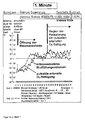
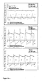
- FIG. 3 a shows, by way of example, a blood volume pulsation in the microvasculature area "finger-bladder" measured according to the non-invasive NIRP method in a normal patient with a resting period of 25 years under rest ventilation (measuring time 64 seconds).
- a signal is superimposed on the pulsations, which corresponds to the vascular tone (microcirculation, sympathetic activity, respiratory influence). If the sympathetic influence or the respiratory component is increased, the vascular tone is high.
- FIG. 3b shows the volume pulsations shown in FIG. 3a in a 4-second time window with characteristic time parameters, likewise with the pulse period of an associated heart action.
- the deciduous volume pulse of FIG. 3b shows a pronounced dicrotie and their position in about 2/3 of the total height of this pulse indicate a "normal" arterial system to the place of measurement. This is a dicrotie expression a more or less subdued occurring in the arterial system Oscillation of blood volume.
- a highly trained dicrotie is a reliable indicator of a powerful one and well-regulated arterial system. Such is a dicrotie particularly pronounced in young people, especially in high-performance athletes. It is also known that in a vessel dilatation occurring reinforced dicrotic wave and approaching the base. Conversely, one means Raising of the dicrotie with flattening of the falling pulse thigh oscillation occurring an arterial Engergna, so often a Dikrotie no longer visible.
- the first derivative represents the blood volume velocity
- the second the associated blood volume acceleration.
- the peripherally measured blood pulsation is a function of the blood volume currently in the illuminated area and the concentration of a substance to be determined contained in this blood volume
- the measurement signal is dominantly determined by the illuminated blood volume, so that it is possible in the physical-mathematical sense, the corresponding To make derivatives of a blood volume.
- the dependence of the measurement signal on the concentration of a substance allows in reverse the determination of the concentration of this substance as well as the measurement of the dynamics of the concentration over time.
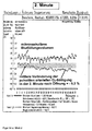
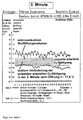
- FIG. 5 shows characteristic courses for microvascular blood volume pulsations and derived blood volume velocities including prominent ones Periods of characteristic cases at the respective measuring site adopted for microvascular blood volume pulsations and derived blood volume velocities including prominent ones Periods of characteristic cases at the respective measuring site adopted for microvascular blood volume pulsations and derived blood volume velocities including prominent ones Periods of characteristic cases at the respective measuring site adopted for zehenbeere (Figure 5a normal subject, Figure 5b patient with peripheral arterial Occlusive disease and Figure 5c with Mediasclerosis). Clearly different especially the shape of the blood volume velocities within a pulse period, so that both qualitative and quantitative measures can be derived are.
- the corresponding blood volume is ejected into the arterial blood vessel system, so that as the filling pressure increases, the lumen of the vessels widens during this time, when the pressure decreases, it returns to the initial state. Consequently, due to the interaction of the heart with the periphery of the vessel, the cardiovascular time interval "systole time" after a transit time has to map qualitatively in the vessel periphery, especially in the peak time T G. Prolonged systole time, as in arterial occlusive disease, correlates with increased peripheral peak time T G and vice versa.
- An increased fiber stretching FS as well as an increased inflection point time T W will indicate a reduction in blood flow in the broadest sense, so that according to the invention an arterial perfusion measure called "arterial perfusion quality aD" can be determined from these parameters: aD ⁇ 1 FS ⁇ T W
- Non-normal cases eg diabetes mellitus or coronary diseases
- z. T. extremely off eg diabetes mellitus or coronary diseases
- the filling volumes for the blood volume signal (840nm signal) as well as the oxymetric signal (640nm signal) accordingly previous work analogous volumes for the NO wavelength of abcr. 400 nm and related to each other: 400 to 840 nm, 400 to 640 nm.
- the filling volumes are at 840 and 640 nm almost congruent, while essential differences z. B. at adjust the vasoconstrictions that occur. The same applies with inclusion the NO wavelength.
- the filling volumes are to be determined when the sensor of different measurements, at different times (eg before, during and after one Therapy; during special functional tests, such as rest and subsequent hyperventilation), can be compared, so by dividing the volume pulse areas the current measurement and the pulse area average of the so-called “reference measurement” the occurring change both from the microvascular blood volume as well as the oxymetric signal or the NO pulsation signal be presented quantitatively.
- the resulting resistance is based on Schmid-Schönbein et al ultimately on the internal friction, so the viscosity of the liquid.
- the situation is different with turbulent flow, as may occur in the case of a mediasclerosis according to FIG. 5c.
- the pulsation of the pressure causes strong mixing of the boundary layer.
- High-speed zones thus reach in the immediate vicinity of the vessel wall, so that z. T. extreme shear stresses arise, as they occur especially in high-performance athletes. This leads to the abnormal release of NO and thus to massive vasodilation.
- Ritz points out that the resulting frictional resistance of the turbulent boundary layer can lead to the injury of the endothelium, which is so important for the arterial vascular system, which also leads to an endothelial dysfunction.
- system stability is known to be determined in such a way that a defined, temporally variable input variable (eg jump or surge function) is applied to the system input.
- the self-adjusting time-varying system output variable is analyzed . If it runs "unstable", ie up to a certain modulation limit, then there is a system instability [ Krauß, M. and E.-G. Woschni: Measurement Information Systems. Characteristic functions, quality criteria, optimization. 2nd ed. VEB Verlagtechnik 1975].
- the sudden opening of the applied blood pressure cuff greatly increases the shear stress of the flowing blood, in the normal case increases in the form of a temporal transition function, the release of the vasodilator NO compared to the time before congestion.
- a functional test must lead to a reduction of the pulsatile O 2 saturation in the normal case, at the time immediately after opening the fiber stretching must be increased and the arterial perfusion quality must be correspondingly reduced.
- Applying a compression and jumping open the tourniquet usually results in temporally variable transition functions for corresponding hemodynamic parameters.
- FIG. 1 Storage and Ent enteredungsfunktion ("Windkessel") in the arterial vascular system [According to Thews, G. and P. Vaupel: Vegetative physiology. 3rd edition Springer Berlin / Heidelberg / New York / Barcelona / Budapest / Hong Kong / London / Milan / Paris / Santa Clara / Singapore / Tokyo 1997].
- the ejection of the stroke volume leads to a wall expansion with subsequent expansion.
- 840 nm near infrared, Hb / HbO 2 emission isosbestscher range
- the degree of oxygenation of this detected by 840 nm blood or erythrocyte amount can be determined. Changes in both the microvascular and macrovascular blood flow, including oxygenation, can be derived from the backscatter signals which occur in the receiver E, but also with sufficient accuracy to conclude cardiovascular parameters, since the pulse periods coincide with those of the associated cardiac actions. Furthermore, additional parameters can be recorded with additional wavelengths (eg, with S3 ⁇ 400 nm, nitrogen monoxide can be deduced as a vessel dilator). To control the temperature ⁇ at the measuring location, a corresponding sensor is integrated in the clip.
- FIG. 3 a) Microvascular blood volume pulsation at the finger fingerboard location in a 25-year-old normal subject under rest ventilation, derived with the noninvasive NIRP method, b) associated blood volume pulsations in a 4-second time window including characteristic time parameters.
- FIG. 4 Microvascular blood volume pulsation and definition of the blood filling volume. From the blood volume pulsation derived blood volume velocity including there marked areas F 1 and F 2 and time periods in order to determine therefrom the elongation dimension "fiber stretching FS" and the arterial perfusion aD. Blood volume acceleration as the second derivative of microvascular blood volume pulsation.
- FIG. 5 Microvascular blood volume pulsations and blood volume velocities derived therefrom, including marked periods of time in characteristic cases at the respective measuring site of big-toe berry (FIG. 5a 22-year-old normal subject, FIG. 5b 63-year-old type II diabetic with peripheral arterial occlusive disease and FIG. 5c 68-year-old type II diabetic with mediasclerosis).
- FIG. 5a 22-year-old normal subject FIG. 5b 63-year-old type II diabetic with peripheral arterial occlusive disease
- FIG. 5c 68-year-old type II diabetic with mediasclerosis.
- the shape of the blood volume velocities differs within a pulse period, so that both qualitative and quantitative measures can be derived.
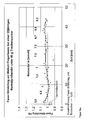
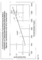
- FIG. 6 Dependence of the relative peripheral resistance RPW 2 on fiber stretching FS on finger and big-toe berries in 29 test persons with trained dicrotias [15 male, 14 female, mean age 23.5 years]. There are 11 cardiovascular health, 9 competitive athletes (about 12 hours after competition), 7 sickle cell patients, 2 type I diabetics.
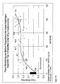
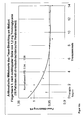
- FIG. 8 Dependence between the turning point time T W and the fiber stretching FS in test persons according to FIG. 6, ie in persons with relatively pronounced dicrotions.
- FIG. 9 Dependence between the turning point time T W and the fiber stretching FS in 50 PAOD patients, derived from the measuring sites of big toe berry.
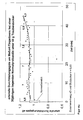
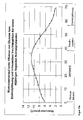
- FIG. 10 Comparison of the determined arterial perfusion qualities aD and aD *, derived from the 120 measurements of the 29 test persons according to FIG. 6.
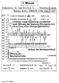
- FIG. 11 Pulsations derived from the peripheral measurement site of the finger of a 28-year-old reclining normal person using three-wavelength NIR-ROT-NO-reflectance photoplethysmography and the wavelengths 880, 635 and 405 nm, including 1st and 2nd derivative as well as time measurements (time offsets ) ⁇ 1 , ⁇ 2 and Fichtenweg 8 99198Erfurt-Kerspleben GmbH Software + Systeme Erfurt GmbHMB / KR / kh "Apparatus for non-invasive determination of hemodynamic functions, including the endothelium and working method therefor " M / SFW-014-DE / I ⁇ 3 ,
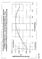
- FIG. 12 Arterial perfusion aD and fiber stretching FS, derived from the NIRP measuring site big toe berry, in 50 patients with peripheral arterial occlusive disease.
- FIG. 13 Shape of the blood volume pulsations, derived from the measuring site 2. Finger berry right. in a 27-year-old volunteer (cyclist professional), when a compressed at the upper arm compression of 250 mm Hg is suddenly opened.
- FIG. 14 shows courses of microvascular blood filling volume and pulsatile arterial O 2 saturation ( FIG. 14 a ), fiber stretching and inflection point time (FIG. 14 b), and shape and amplitude of blood volume flow and blood volume velocity derived therefrom (FIG. 14 c) in a 55-year normal subject at the measuring point, if a 250 mm Hg compression applied to the upper arm suddenly opens.
- FIG. 15 Characteristic courses of the peripheral fiber stretching in normal persons (FIG. 15a, b) and a type II diabetic (FIG. 15c), which were derived from the measurement location of the fingertip with the respective administration of 40 g dextrose as function or stimulation test.
- FIG. 16 Changes in the arterial perfusion aD at the measurement site of the fingertip, which occurred with the administration of 40 g of glucose in the test subjects according to FIG.
- FIG. 17 Relationship between fiber stretching, derived from the peripheral measuring location of the fingertip, and the cardiac cycle duration in normal persons and type II diabetics according to FIGS. 15 and 16 with the administration of 40 g glucose.
- FIG. 18 Quantitative changes of arterial vessel wall properties, specifically fiber stretching FS (FIG. 18 a) and arterial perfusion aD (FIG. 18 b), under a functional test "sublingual administration of 1.2 mg trinitroglycerin" in patients with chronic ischemic heart disease.
- FIG. 19 Changes in blood sugar, fiber stretching and arterial perfusion quality, as were established during a functional test "intravenous administration of glucose solution, insulin and electrolyte solution" at the 48-year-old subject (physician) at the measurement site fingerstick.
- FIG. 13 shows the shape of the blood volume pulsations, derived from the measuring location 2.
- Finger berry right. in a 27-year-old volunteer (cyclist professional) when a compressed at the upper arm compression of 250 mm Hg is suddenly opened.
- the pulsations develop, whereby the dicrotias are initially relatively weak, the associated fiber stretching is somewhat increased, and the arterial circulation is at the lower limit of the normal range.
- the dicroties are more distinct, the area under the pulsations, ie the microvascular volume of blood filling, increases, and vascular dilatation has occurred.
- FIG. 14b shows above all the behavior of the vessel expansion, the fiber stretching FS.
- FS increases to approximately 1.6 times the normal value (FS average of the entire 1st minute is 1).
- 2nd and 3rd minute FS decreases considerably in the form of a transition function (as respective mean values to 0.95).
- Figure 15 a, b and c shows characteristic courses of the peripheral fiber stretching in normal individuals and a type II diabetic who under each Giving 40 g of glucose as a function or stimulation test from the measurement site Finger berry were derived.
- FIG. 16 shows the changes in the arterial perfusion aD that occurred with the administration of 40 g of glucose in the test subjects according to FIG. 15 and that the measurement site was derived from the fingertip.
- FIGS. 19a, b and c parameter changes can be seen, as revealed by a functional test "intravenous administration of glucose solution, insulin and electrolyte solution”.
- Figure 19a shows the course of blood sugar, as he was in the 48-year-old subject (doctor) under intravenous administration of glucose solution, insulin and electrolyte solution.
- the infusion of 10% glucose solution at a rate of 500 ml / h leads to a continuous linear increase.
- After intravenous injection of 15 IU normal insulin (Actrapid R ) there is a clear decrease from baseline. Blood glucose levels are in the hypoglycemic range.
- the electrolyte solution (E153) is expected to have no effect on blood sugar.
Landscapes
- Health & Medical Sciences (AREA)
- Life Sciences & Earth Sciences (AREA)
- Cardiology (AREA)
- Physics & Mathematics (AREA)
- Heart & Thoracic Surgery (AREA)
- Medical Informatics (AREA)
- Veterinary Medicine (AREA)
- Public Health (AREA)
- Biophysics (AREA)
- Pathology (AREA)
- Engineering & Computer Science (AREA)
- Biomedical Technology (AREA)
- General Health & Medical Sciences (AREA)
- Animal Behavior & Ethology (AREA)
- Molecular Biology (AREA)
- Surgery (AREA)
- Physiology (AREA)
- Vascular Medicine (AREA)
- Dentistry (AREA)
- Ophthalmology & Optometry (AREA)
- Spectroscopy & Molecular Physics (AREA)
- Optics & Photonics (AREA)
- Measuring Pulse, Heart Rate, Blood Pressure Or Blood Flow (AREA)
- Measurement Of The Respiration, Hearing Ability, Form, And Blood Characteristics Of Living Organisms (AREA)
Applications Claiming Priority (4)
| Application Number | Priority Date | Filing Date | Title |
|---|---|---|---|
| DE102004013095 | 2004-03-17 | ||
| DE102004013095 | 2004-03-17 | ||
| DE102004016376 | 2004-04-02 | ||
| DE102004016376A DE102004016376A1 (de) | 2004-03-17 | 2004-04-02 | Vorrichtung zur nichtinvasiven Ermittlung hämodynamischer Funktionen, einschließlich des Endothels sowie Arbeitsverfahren hierfür |
Publications (3)
| Publication Number | Publication Date |
|---|---|
| EP1584289A2 true EP1584289A2 (fr) | 2005-10-12 |
| EP1584289A3 EP1584289A3 (fr) | 2005-10-19 |
| EP1584289B1 EP1584289B1 (fr) | 2010-01-06 |
Family
ID=34913356
Family Applications (1)
| Application Number | Title | Priority Date | Filing Date |
|---|---|---|---|
| EP05004826A Expired - Lifetime EP1584289B1 (fr) | 2004-03-17 | 2005-03-04 | Procédé d'exploitation pour un appareil permettant l'évaluation non-invasive des fonctions hemodynamiques, y compris celles de l'endothélium |
Country Status (3)
| Country | Link |
|---|---|
| EP (1) | EP1584289B1 (fr) |
| AT (1) | ATE454085T1 (fr) |
| DE (2) | DE102004016376A1 (fr) |
Cited By (6)
| Publication number | Priority date | Publication date | Assignee | Title |
|---|---|---|---|---|
| WO2009101140A1 (fr) * | 2008-02-15 | 2009-08-20 | Universite Paris Sud | Dispositif et méthode de calcul de nouveaux indices de la rigidité artérielle, et/ou de surveillance du volume d’éjection |
| DE102011051741A1 (de) | 2010-07-16 | 2012-01-19 | Optitron Systems Gmbh | Vorrichtung zur Lichtstimulation von Gewebe, insbesondere für den Augenbereich |
| US8657755B2 (en) | 2009-05-12 | 2014-02-25 | Angiologix, Inc. | System and method of measuring changes in arterial volume of a limb segment |
| WO2016055260A1 (fr) * | 2014-10-09 | 2016-04-14 | Koninklijke Philips N.V. | Capteur optique de signes vitaux |
| US10238306B2 (en) | 2006-02-20 | 2019-03-26 | Everist Genomics, Inc. | Method for non-evasively determining an endothelial function and a device for carrying out said method |
| US11911188B2 (en) | 2019-10-30 | 2024-02-27 | Samsung Electronics Co., Ltd. | Apparatus and method for monitoring health, and mobile device |
Families Citing this family (1)
| Publication number | Priority date | Publication date | Assignee | Title |
|---|---|---|---|---|
| DE102010038249A1 (de) | 2010-10-18 | 2012-04-19 | Manexco-Consult Ag | Verfahren und Vorrichtung zur physikalischen Stimulation von menschlichem Gewebe |
Family Cites Families (4)
| Publication number | Priority date | Publication date | Assignee | Title |
|---|---|---|---|---|
| DE3744538A1 (de) * | 1987-12-30 | 1989-07-13 | Nattermann A & Cie | Vorrichtung zur nichtinvasiven messung des blutvolumens in menschlichen extremitaeten |
| DE4238641C2 (de) * | 1992-11-16 | 1994-12-08 | Kraus Manfred | Vorrichtung und Arbeitsverfahren zum Bestimmen und Auswerten des physiologischen Zustandes von Gefäßsystemen |
| DE4322860A1 (de) * | 1993-07-08 | 1995-01-19 | Laumann Medizintech Gmbh | Verfahren und Vorrichtung zur Bestimmung und Auswertung des Zustandes von Gefäßsystemen |
| AU1421002A (en) * | 2000-10-23 | 2002-05-06 | Itamar Medical Ltd | Method and apparatus for non-invasively evaluating endothelial activity in a patient |
-
2004
- 2004-04-02 DE DE102004016376A patent/DE102004016376A1/de not_active Withdrawn
-
2005
- 2005-03-04 DE DE502005008806T patent/DE502005008806D1/de not_active Expired - Lifetime
- 2005-03-04 AT AT05004826T patent/ATE454085T1/de not_active IP Right Cessation
- 2005-03-04 EP EP05004826A patent/EP1584289B1/fr not_active Expired - Lifetime
Non-Patent Citations (1)
| Title |
|---|
| THEWS, G.; P. VAUPEL: "Vegetative Physiologie", 1997, SPRINGER |
Cited By (7)
| Publication number | Priority date | Publication date | Assignee | Title |
|---|---|---|---|---|
| US10238306B2 (en) | 2006-02-20 | 2019-03-26 | Everist Genomics, Inc. | Method for non-evasively determining an endothelial function and a device for carrying out said method |
| WO2009101140A1 (fr) * | 2008-02-15 | 2009-08-20 | Universite Paris Sud | Dispositif et méthode de calcul de nouveaux indices de la rigidité artérielle, et/ou de surveillance du volume d’éjection |
| US9107587B2 (en) | 2008-02-15 | 2015-08-18 | Universite Paris Sud | Device and process for calculating new indices of arterial stiffness, and/or for stroke volume monitoring |
| US8657755B2 (en) | 2009-05-12 | 2014-02-25 | Angiologix, Inc. | System and method of measuring changes in arterial volume of a limb segment |
| DE102011051741A1 (de) | 2010-07-16 | 2012-01-19 | Optitron Systems Gmbh | Vorrichtung zur Lichtstimulation von Gewebe, insbesondere für den Augenbereich |
| WO2016055260A1 (fr) * | 2014-10-09 | 2016-04-14 | Koninklijke Philips N.V. | Capteur optique de signes vitaux |
| US11911188B2 (en) | 2019-10-30 | 2024-02-27 | Samsung Electronics Co., Ltd. | Apparatus and method for monitoring health, and mobile device |
Also Published As
| Publication number | Publication date |
|---|---|
| ATE454085T1 (de) | 2010-01-15 |
| DE502005008806D1 (de) | 2010-02-25 |
| DE102004016376A1 (de) | 2005-10-06 |
| EP1584289B1 (fr) | 2010-01-06 |
| EP1584289A3 (fr) | 2005-10-19 |
Similar Documents
| Publication | Publication Date | Title |
|---|---|---|
| DE69934888T2 (de) | Nichtinvasive optische messung eines blutbestandteiles | |
| DE68927614T2 (de) | Verfahren zur nichtinvasiven periodischen und/oder kontinuierlichen Bestimmung von Hämoglobin , arteriellem Sauerstoffgehalt und Hämatokrit | |
| Sandroni et al. | Accuracy of plethysmographic indices as predictors of fluid responsiveness in mechanically ventilated adults: a systematic review and meta-analysis | |
| DE60035470T2 (de) | Verfahren und Vorrichtung zur Bestimmung des Herzzeitvolumens oder des totalen peripheren Widerstandes | |
| DE60223787T2 (de) | Verfahren und vorrichtung zur verbesserung der genauigkeit nichtinvasiver hematokritmessungen | |
| US20170079533A1 (en) | Diabetes and Hypertension Screening by Assessment of Arterial Stiffness and Autonomic Function | |
| MacLeod et al. | Development and validation of a cerebral oximeter capable of absolute accuracy | |
| Linder et al. | Using the morphology of photoplethysmogram peaks to detect changes in posture | |
| DE10392677T5 (de) | Verfahren und Vorrichtung zum Monitoring des autonomen Nervensystems | |
| Chalacheva et al. | Sickle cell disease subjects have a distinct abnormal autonomic phenotype characterized by peripheral vasoconstriction with blunted cardiac response to head-up tilt | |
| EP1584289B1 (fr) | Procédé d'exploitation pour un appareil permettant l'évaluation non-invasive des fonctions hemodynamiques, y compris celles de l'endothélium | |
| Langham | Ocular blood flow and visual loss in glaucomatous eyes | |
| EP2166933A1 (fr) | Procédé pour déterminer une modification quasi continue de la pression sanguine dans un flux sanguin pulsatile | |
| Kuznik et al. | Impact of fitness status on the optically measured hemodynamic indexes | |
| Picard et al. | Individual shear rate therapy (ISRT)—further development of external counterpulsation for decreasing blood pressure in patients with symptomatic coronary artery disease (CAD) | |
| DE4238641A1 (de) | Verfahren und Vorrichtung zur Bestimmung und Auswertung des Zustandes von Gefäßsystemen | |
| EP4132352A1 (fr) | Procédé et dispositif de détermination de l'état volémique et du tonus vasculaire | |
| Marthol et al. | Cardiovascular and cerebrovascular responses to lower body negative pressure in type 2 diabetic patients | |
| Barron et al. | DC photoplethysmography in the evaluation of sympathetic vasomotor responses | |
| EP0913120A1 (fr) | Appareil et dispositif destiné à la mesure non-invasive de paramètres de la circulation sanguine | |
| DE19645579A1 (de) | Vorrichtung und Arbeitsverfahren zum Betreiben der Vorrichtung zum Bestimmen und Auswerten von Funktionszuständen im Herz-Kreislauf-System | |
| Uguz | Design of a multipurpose photoplethysmography sensor to assist cardiovascular and respiratory diagnosis | |
| Fadil et al. | Respiratory Pump Contributions to Hemodynamic Responses in Lower-Body Negative Pressure: Preliminary Results | |
| Shali et al. | Correlation of blood pressure changes following postural change from sitting to standing with near infrared spectroscopy (NIRS) | |
| EP0600298A1 (fr) | Procédé de travail pour exploiter et dispositif pour déterminer et évaluer les conditions des systèmes vasculaires |
Legal Events
| Date | Code | Title | Description |
|---|---|---|---|
| PUAI | Public reference made under article 153(3) epc to a published international application that has entered the european phase |
Free format text: ORIGINAL CODE: 0009012 |
|
| PUAL | Search report despatched |
Free format text: ORIGINAL CODE: 0009013 |
|
| AK | Designated contracting states |
Kind code of ref document: A2 Designated state(s): AT BE BG CH CY CZ DE DK EE ES FI FR GB GR HU IE IS IT LI LT LU MC NL PL PT RO SE SI SK TR |
|
| AX | Request for extension of the european patent |
Extension state: AL BA HR LV MK YU |
|
| AK | Designated contracting states |
Kind code of ref document: A3 Designated state(s): AT BE BG CH CY CZ DE DK EE ES FI FR GB GR HU IE IS IT LI LT LU MC NL PL PT RO SE SI SK TR |
|
| AX | Request for extension of the european patent |
Extension state: AL BA HR LV MK YU |
|
| 17P | Request for examination filed |
Effective date: 20060327 |
|
| AKX | Designation fees paid |
Designated state(s): AT BE BG CH CY CZ DE DK EE ES FI FR GB GR HU IE IS IT LI LT LU MC NL PL PT RO SE SI SK TR |
|
| 17Q | First examination report despatched |
Effective date: 20080813 |
|
| GRAP | Despatch of communication of intention to grant a patent |
Free format text: ORIGINAL CODE: EPIDOSNIGR1 |
|
| RTI1 | Title (correction) |
Free format text: DEVICE OPERATING METHOD FOR NON-INVASIVE ASSESSING OF HEMODYNAMIC FUNCTIONS INCLUDING ENDOTHELIAL STATUS |
|
| GRAS | Grant fee paid |
Free format text: ORIGINAL CODE: EPIDOSNIGR3 |
|
| GRAA | (expected) grant |
Free format text: ORIGINAL CODE: 0009210 |
|
| AK | Designated contracting states |
Kind code of ref document: B1 Designated state(s): AT BE BG CH CY CZ DE DK EE ES FI FR GB GR HU IE IS IT LI LT LU MC NL PL PT RO SE SI SK TR |
|
| REG | Reference to a national code |
Ref country code: GB Ref legal event code: FG4D Free format text: NOT ENGLISH |
|
| REG | Reference to a national code |
Ref country code: CH Ref legal event code: EP |
|
| REG | Reference to a national code |
Ref country code: IE Ref legal event code: FG4D |
|
| REF | Corresponds to: |
Ref document number: 502005008806 Country of ref document: DE Date of ref document: 20100225 Kind code of ref document: P |
|
| REG | Reference to a national code |
Ref country code: NL Ref legal event code: VDEP Effective date: 20100106 |
|
| PG25 | Lapsed in a contracting state [announced via postgrant information from national office to epo] |
Ref country code: SI Free format text: LAPSE BECAUSE OF FAILURE TO SUBMIT A TRANSLATION OF THE DESCRIPTION OR TO PAY THE FEE WITHIN THE PRESCRIBED TIME-LIMIT Effective date: 20100106 |
|
| LTIE | Lt: invalidation of european patent or patent extension |
Effective date: 20100106 |
|
| PG25 | Lapsed in a contracting state [announced via postgrant information from national office to epo] |
Ref country code: NL Free format text: LAPSE BECAUSE OF FAILURE TO SUBMIT A TRANSLATION OF THE DESCRIPTION OR TO PAY THE FEE WITHIN THE PRESCRIBED TIME-LIMIT Effective date: 20100106 Ref country code: IS Free format text: LAPSE BECAUSE OF FAILURE TO SUBMIT A TRANSLATION OF THE DESCRIPTION OR TO PAY THE FEE WITHIN THE PRESCRIBED TIME-LIMIT Effective date: 20100506 Ref country code: ES Free format text: LAPSE BECAUSE OF FAILURE TO SUBMIT A TRANSLATION OF THE DESCRIPTION OR TO PAY THE FEE WITHIN THE PRESCRIBED TIME-LIMIT Effective date: 20100417 Ref country code: LT Free format text: LAPSE BECAUSE OF FAILURE TO SUBMIT A TRANSLATION OF THE DESCRIPTION OR TO PAY THE FEE WITHIN THE PRESCRIBED TIME-LIMIT Effective date: 20100106 Ref country code: PT Free format text: LAPSE BECAUSE OF FAILURE TO SUBMIT A TRANSLATION OF THE DESCRIPTION OR TO PAY THE FEE WITHIN THE PRESCRIBED TIME-LIMIT Effective date: 20100506 |
|
| PGFP | Annual fee paid to national office [announced via postgrant information from national office to epo] |
Ref country code: IE Payment date: 20100526 Year of fee payment: 6 |
|
| REG | Reference to a national code |
Ref country code: IE Ref legal event code: FD4D |
|
| PG25 | Lapsed in a contracting state [announced via postgrant information from national office to epo] |
Ref country code: FI Free format text: LAPSE BECAUSE OF FAILURE TO SUBMIT A TRANSLATION OF THE DESCRIPTION OR TO PAY THE FEE WITHIN THE PRESCRIBED TIME-LIMIT Effective date: 20100106 Ref country code: PL Free format text: LAPSE BECAUSE OF FAILURE TO SUBMIT A TRANSLATION OF THE DESCRIPTION OR TO PAY THE FEE WITHIN THE PRESCRIBED TIME-LIMIT Effective date: 20100106 |
|
| BERE | Be: lapsed |
Owner name: SOFTWARE + SYSTEME ERFURT G.M.B.H. Effective date: 20100331 |
|
| PG25 | Lapsed in a contracting state [announced via postgrant information from national office to epo] |
Ref country code: CY Free format text: LAPSE BECAUSE OF FAILURE TO SUBMIT A TRANSLATION OF THE DESCRIPTION OR TO PAY THE FEE WITHIN THE PRESCRIBED TIME-LIMIT Effective date: 20100106 Ref country code: MC Free format text: LAPSE BECAUSE OF NON-PAYMENT OF DUE FEES Effective date: 20100331 Ref country code: IE Free format text: LAPSE BECAUSE OF FAILURE TO SUBMIT A TRANSLATION OF THE DESCRIPTION OR TO PAY THE FEE WITHIN THE PRESCRIBED TIME-LIMIT Effective date: 20100106 Ref country code: RO Free format text: LAPSE BECAUSE OF FAILURE TO SUBMIT A TRANSLATION OF THE DESCRIPTION OR TO PAY THE FEE WITHIN THE PRESCRIBED TIME-LIMIT Effective date: 20100106 Ref country code: GR Free format text: LAPSE BECAUSE OF FAILURE TO SUBMIT A TRANSLATION OF THE DESCRIPTION OR TO PAY THE FEE WITHIN THE PRESCRIBED TIME-LIMIT Effective date: 20100407 Ref country code: SE Free format text: LAPSE BECAUSE OF FAILURE TO SUBMIT A TRANSLATION OF THE DESCRIPTION OR TO PAY THE FEE WITHIN THE PRESCRIBED TIME-LIMIT Effective date: 20100106 Ref country code: EE Free format text: LAPSE BECAUSE OF FAILURE TO SUBMIT A TRANSLATION OF THE DESCRIPTION OR TO PAY THE FEE WITHIN THE PRESCRIBED TIME-LIMIT Effective date: 20100106 |
|
| REG | Reference to a national code |
Ref country code: CH Ref legal event code: PL |
|
| PLBE | No opposition filed within time limit |
Free format text: ORIGINAL CODE: 0009261 |
|
| STAA | Information on the status of an ep patent application or granted ep patent |
Free format text: STATUS: NO OPPOSITION FILED WITHIN TIME LIMIT |
|
| PG25 | Lapsed in a contracting state [announced via postgrant information from national office to epo] |
Ref country code: CZ Free format text: LAPSE BECAUSE OF FAILURE TO SUBMIT A TRANSLATION OF THE DESCRIPTION OR TO PAY THE FEE WITHIN THE PRESCRIBED TIME-LIMIT Effective date: 20100106 Ref country code: SK Free format text: LAPSE BECAUSE OF FAILURE TO SUBMIT A TRANSLATION OF THE DESCRIPTION OR TO PAY THE FEE WITHIN THE PRESCRIBED TIME-LIMIT Effective date: 20100106 Ref country code: BG Free format text: LAPSE BECAUSE OF FAILURE TO SUBMIT A TRANSLATION OF THE DESCRIPTION OR TO PAY THE FEE WITHIN THE PRESCRIBED TIME-LIMIT Effective date: 20100406 |
|
| 26N | No opposition filed |
Effective date: 20101007 |
|
| PG25 | Lapsed in a contracting state [announced via postgrant information from national office to epo] |
Ref country code: DK Free format text: LAPSE BECAUSE OF FAILURE TO SUBMIT A TRANSLATION OF THE DESCRIPTION OR TO PAY THE FEE WITHIN THE PRESCRIBED TIME-LIMIT Effective date: 20100106 |
|
| PG25 | Lapsed in a contracting state [announced via postgrant information from national office to epo] |
Ref country code: LI Free format text: LAPSE BECAUSE OF NON-PAYMENT OF DUE FEES Effective date: 20100331 Ref country code: BE Free format text: LAPSE BECAUSE OF NON-PAYMENT OF DUE FEES Effective date: 20100331 Ref country code: CH Free format text: LAPSE BECAUSE OF NON-PAYMENT OF DUE FEES Effective date: 20100331 |
|
| PG25 | Lapsed in a contracting state [announced via postgrant information from national office to epo] |
Ref country code: IT Free format text: LAPSE BECAUSE OF FAILURE TO SUBMIT A TRANSLATION OF THE DESCRIPTION OR TO PAY THE FEE WITHIN THE PRESCRIBED TIME-LIMIT Effective date: 20100106 |
|
| PG25 | Lapsed in a contracting state [announced via postgrant information from national office to epo] |
Ref country code: AT Free format text: LAPSE BECAUSE OF NON-PAYMENT OF DUE FEES Effective date: 20100304 |
|
| PG25 | Lapsed in a contracting state [announced via postgrant information from national office to epo] |
Ref country code: LU Free format text: LAPSE BECAUSE OF NON-PAYMENT OF DUE FEES Effective date: 20100304 Ref country code: HU Free format text: LAPSE BECAUSE OF FAILURE TO SUBMIT A TRANSLATION OF THE DESCRIPTION OR TO PAY THE FEE WITHIN THE PRESCRIBED TIME-LIMIT Effective date: 20100707 |
|
| PG25 | Lapsed in a contracting state [announced via postgrant information from national office to epo] |
Ref country code: TR Free format text: LAPSE BECAUSE OF FAILURE TO SUBMIT A TRANSLATION OF THE DESCRIPTION OR TO PAY THE FEE WITHIN THE PRESCRIBED TIME-LIMIT Effective date: 20100106 |
|
| REG | Reference to a national code |
Ref country code: DE Ref legal event code: R082 Ref document number: 502005008806 Country of ref document: DE Representative=s name: MEISSNER, BOLTE & PARTNER GBR, DE |
|
| REG | Reference to a national code |
Ref country code: FR Ref legal event code: PLFP Year of fee payment: 11 |
|
| REG | Reference to a national code |
Ref country code: DE Ref legal event code: R082 Ref document number: 502005008806 Country of ref document: DE Representative=s name: MEISSNER, BOLTE & PARTNER GBR, DE Effective date: 20150324 Ref country code: DE Ref legal event code: R081 Ref document number: 502005008806 Country of ref document: DE Owner name: MANDLER, DIETRICH, DE Free format text: FORMER OWNER: SOFTWARE + SYSTEME ERFURT GMBH, 99198 KERSPLEBEN, DE Effective date: 20150324 Ref country code: DE Ref legal event code: R081 Ref document number: 502005008806 Country of ref document: DE Owner name: TIETZ, UWE-JENS, DE Free format text: FORMER OWNER: SOFTWARE + SYSTEME ERFURT GMBH, 99198 KERSPLEBEN, DE Effective date: 20150324 Ref country code: DE Ref legal event code: R082 Ref document number: 502005008806 Country of ref document: DE Representative=s name: MEISSNER BOLTE PATENTANWAELTE RECHTSANWAELTE P, DE Effective date: 20150324 |
|
| PGFP | Annual fee paid to national office [announced via postgrant information from national office to epo] |
Ref country code: GB Payment date: 20150331 Year of fee payment: 11 |
|
| REG | Reference to a national code |
Ref country code: GB Ref legal event code: 732E Free format text: REGISTERED BETWEEN 20150604 AND 20150610 |
|
| REG | Reference to a national code |
Ref country code: FR Ref legal event code: TQ Owner name: DIETRICH MANDLER, DE Effective date: 20150528 Ref country code: FR Ref legal event code: TQ Owner name: UWE JENS TIETZ, DE Effective date: 20150528 |
|
| PGFP | Annual fee paid to national office [announced via postgrant information from national office to epo] |
Ref country code: FR Payment date: 20150331 Year of fee payment: 11 |
|
| PGFP | Annual fee paid to national office [announced via postgrant information from national office to epo] |
Ref country code: DE Payment date: 20160330 Year of fee payment: 12 |
|
| GBPC | Gb: european patent ceased through non-payment of renewal fee |
Effective date: 20160304 |
|
| REG | Reference to a national code |
Ref country code: FR Ref legal event code: ST Effective date: 20161130 |
|
| PG25 | Lapsed in a contracting state [announced via postgrant information from national office to epo] |
Ref country code: FR Free format text: LAPSE BECAUSE OF NON-PAYMENT OF DUE FEES Effective date: 20160331 Ref country code: GB Free format text: LAPSE BECAUSE OF NON-PAYMENT OF DUE FEES Effective date: 20160304 |
|
| REG | Reference to a national code |
Ref country code: DE Ref legal event code: R119 Ref document number: 502005008806 Country of ref document: DE |
|
| PG25 | Lapsed in a contracting state [announced via postgrant information from national office to epo] |
Ref country code: DE Free format text: LAPSE BECAUSE OF NON-PAYMENT OF DUE FEES Effective date: 20171003 |