EP1570267B1 - Assay for identifying antibody producing cells - Google Patents
Assay for identifying antibody producing cells Download PDFInfo
- Publication number
- EP1570267B1 EP1570267B1 EP03778577A EP03778577A EP1570267B1 EP 1570267 B1 EP1570267 B1 EP 1570267B1 EP 03778577 A EP03778577 A EP 03778577A EP 03778577 A EP03778577 A EP 03778577A EP 1570267 B1 EP1570267 B1 EP 1570267B1
- Authority
- EP
- European Patent Office
- Prior art keywords
- antibody
- cells
- antigen
- cell
- assay
- Prior art date
- Legal status (The legal status is an assumption and is not a legal conclusion. Google has not performed a legal analysis and makes no representation as to the accuracy of the status listed.)
- Expired - Lifetime
Links
Images

Classifications
-
- C—CHEMISTRY; METALLURGY
- C07—ORGANIC CHEMISTRY
- C07K—PEPTIDES
- C07K16/00—Immunoglobulins [IG], e.g. monoclonal or polyclonal antibodies
- C07K16/18—Immunoglobulins [IG], e.g. monoclonal or polyclonal antibodies against material from animals or humans
- C07K16/24—Immunoglobulins [IG], e.g. monoclonal or polyclonal antibodies against material from animals or humans against cytokines, lymphokines or interferons
- C07K16/243—Colony Stimulating Factors
-
- C—CHEMISTRY; METALLURGY
- C07—ORGANIC CHEMISTRY
- C07K—PEPTIDES
- C07K16/00—Immunoglobulins [IG], e.g. monoclonal or polyclonal antibodies
- C07K16/18—Immunoglobulins [IG], e.g. monoclonal or polyclonal antibodies against material from animals or humans
- C07K16/24—Immunoglobulins [IG], e.g. monoclonal or polyclonal antibodies against material from animals or humans against cytokines, lymphokines or interferons
- C07K16/244—Interleukins [IL]
- C07K16/245—IL-1
-
- G—PHYSICS
- G01—MEASURING; TESTING
- G01N—INVESTIGATING OR ANALYSING MATERIALS BY DETERMINING THEIR CHEMICAL OR PHYSICAL PROPERTIES
- G01N33/00—Investigating or analysing materials by specific methods not covered by groups G01N1/00 - G01N31/00
- G01N33/48—Biological material, e.g. blood, urine; Haemocytometers
- G01N33/50—Chemical analysis of biological material, e.g. blood, urine; Testing involving biospecific ligand binding methods; Immunological testing
- G01N33/53—Immunoassay; Biospecific binding assay; Materials therefor
- G01N33/569—Immunoassay; Biospecific binding assay; Materials therefor for microorganisms, e.g. protozoa, bacteria, viruses
- G01N33/56966—Animal cells
- G01N33/56972—White blood cells
-
- G—PHYSICS
- G01—MEASURING; TESTING
- G01N—INVESTIGATING OR ANALYSING MATERIALS BY DETERMINING THEIR CHEMICAL OR PHYSICAL PROPERTIES
- G01N33/00—Investigating or analysing materials by specific methods not covered by groups G01N1/00 - G01N31/00
- G01N33/48—Biological material, e.g. blood, urine; Haemocytometers
- G01N33/50—Chemical analysis of biological material, e.g. blood, urine; Testing involving biospecific ligand binding methods; Immunological testing
- G01N33/58—Chemical analysis of biological material, e.g. blood, urine; Testing involving biospecific ligand binding methods; Immunological testing involving labelled substances
- G01N33/582—Chemical analysis of biological material, e.g. blood, urine; Testing involving biospecific ligand binding methods; Immunological testing involving labelled substances with fluorescent label
-
- G—PHYSICS
- G01—MEASURING; TESTING
- G01N—INVESTIGATING OR ANALYSING MATERIALS BY DETERMINING THEIR CHEMICAL OR PHYSICAL PROPERTIES
- G01N33/00—Investigating or analysing materials by specific methods not covered by groups G01N1/00 - G01N31/00
- G01N33/48—Biological material, e.g. blood, urine; Haemocytometers
- G01N33/50—Chemical analysis of biological material, e.g. blood, urine; Testing involving biospecific ligand binding methods; Immunological testing
- G01N33/68—Chemical analysis of biological material, e.g. blood, urine; Testing involving biospecific ligand binding methods; Immunological testing involving proteins, peptides or amino acids
- G01N33/6854—Immunoglobulins
Definitions
- the present invention relates generally to improved methods for producing antibodies and more specifically provides a homogeneous assay for obtaining antibodies.
- the selected lymphocyte antibody method (SLAM) for generating monoclonal antibodies overcomes the limitations of both hybridoma technology and bacterially expressed antibody libraries by enabling high affinity antibodies generated during in vivo immune responses to be isolated from any species ( Babcook et al., 1996, Proc.Natl.Acad.Sci, 93, 7843-7848 ).
- SLAM enables a single lymphocyte that is producing an antibody with a desired specificity or function to be identified within a large population of lymphoid cells and the genetic information that encodes the specificity of the antibody to be rescued from that lymphocyte.
- Antibody producing cells which produce antibodies which bind to selected antigens are detected using an adapted hemolytic plaque assay method ( Jerne and Nordin, 1963, Science, 140, 405 ).
- erythrocytes are coated with the selected antigen and incubated with the population of antibody producing cells and a source of complement.
- Single antibody producing cells are identified by the formation of hemolytic plaques. Plaques of lysed erythrocytes are identified using an inverted microscope and the single antibody producing cell of interest at the centre of the plaque is removed using micromanipulation techniques and the antibody genes from the cell are cloned by reverse transcription PCR.
- Other methods for detecting single antibody-producing cells of a desired function have already been described in International Patent Specification, WO 92/02551 .
- EP0286405 describes a homogeneous phase method of selecting cells that produce an anti-idiotypic antibody which comprises contacting a population of antibody-producing cells with a fluorescently-labelled antibody produced by immunisation of a host with an antigen from a selected pathogen and selecting those antibody-producing cells that bind the labelled antiserum.
- red blood cells are typically coated with antigen via a biotin/streptavidin coupling system that requires the antigen to be biotinylated.
- This method is therefore restricted to antigens that are available in a pure form and to those that can be biotinylated without affecting epitope presentation.
- This method clearly precludes the isolation of antibodies against a wide range of antigens.
- many proteins are difficult to purify, particularly cell surface expressed proteins, such as type III proteins.
- Many proteins alter their conformation and presentation of desirable eptiopes upon biotinylation, for example proteins that contain lysine groups in their active site.
- tumor cells may also be desirable to produce antibodies against unknown antigens, such as proteins expressed on the surface of cells, such as tumor cells.
- unknown antigens such as proteins expressed on the surface of cells, such as tumor cells.
- the direct use of tumor cells in the plaque assay instead of antigen coated erythrocytes is difficult to achieve given the requirement for cell lysis to occur in order for plaques containing antibody producing cells to be identified.
- Cell lysis is dependent on cell type, antigen and antibody concentration. Red blood cells coated with the desired antigen will bind large amounts of available antibody and will lyse readily in the presence of complement. Other cell types such as tumor cells will not lyse so readily, especially when the availability of antigen on the surface may be very low and hence antibody binding will be low.
- Manz et al discloses a method for identification of antibody secreting cells where the secreted product is retained on the cell surface of the screening cells.
- Required steps include labelling of the surface of the antibody producing cell with capture antibodies or antigen, artificially elevating the viscosity of the medium to stop diffusion and cross-feeding of secreted product between cells, wash steps, identification of cells by fluorescent activated cell sorting procedures.
- This improved assay has many advantages over the methods described above, allowing the identification of antibodies that bind to any antigen, including unknown antigens, cell surface antigens and antigens which cannot be biotinylated without altering the presentation of desirable epitopes. As a result, antibodies with binding specificities that were previously unidentifiable by conventional plaque assays can now be produced. In addition the assay is more facile than the hemolytic plaque assay and antibody producing cells can be identified more quickly.
- an in vitro homogeneous assay for identifying an antibody producing cell producing an antibody which binds to a selected antigen comprising:
- the term 'antibody' as used herein includes any recombinant or naturally occurring immunoglobulin molecule such as a member of the IgG class e.g. IgG1 and also any antigen binding immunoglobulin fragment, such as Fv, Fab' and F(ab') 2 fragments, and any derivatives thereof, such as single chain Fv fragments.
- antibody producing cell' as used herein means any cell capable of secreting an antibody, such as a B-lymphocyte, a plasma cell, a plasmablast, an activated B cell or a memory B cell.
- Antibody-producing cells for use in the invention may be obtained from an animal which has either been immunized with an antigen, or which has developed an immune response to an antigen as a result of disease.
- Other antibody producing cells for use in the present invention may include any transformed cell in particular, any mammalian cells which express immunoglobulin genes or parts thereof In one example the populations of antibody producing cells for use in the present invention produce a range of antibodies with different binding specificities.
- the assay of the present invention may also be used to identify high yielding antibody producing cells from a population of antibody producing cells which all produce the same antibody.
- the term 'high yielding' as used herein refers to antibody producing cells that produce antibodies of a known specificity but for which it would be desirable to identify those cells producing the antibody most efficiently. Identification of the high yielding cell will allow the cell to be isolated and clonally reproduced.
- the high yielding antibody producing cell is a hybridoma cell.
- the high yielding antibody producing cell is a transformed cell in particular, a mammalian cell which expresses immunoglobulin genes or parts thereof. Examples of such mammalian cells include but are not limited to NS0, CHO, COS and 293 cells.
- the term 'antigen' as used herein refers to any known or unknown substance that can be recognised by an antibody, including proteins, glycoproteins and carbohydrates.
- these antigens include biologically active proteins, such as hormones, cytokines, and their cell surface receptors, bacterial or parasitic cell membranes or purified components thereof, and viral antigens.
- the antigen is available in a pure form obtained either by direct purification from the native source or by recombinant expression and purification of said antigen.
- the purified antigen is coupled to erythrocytes or any other particle such as a bead for incorporation into the assay,.
- the antigen is one which is difficult to purify
- such antigens include but are not limited to cell surface expressed proteins such as receptors, particularly type III proteins.
- the presentation of desirable epitopes on the antigen is altered upon biotinylation, this includes but is not limited to proteins which contain lysines in their active site regions.
- the antigen may be expressed on the surface of a cell, either naturally or recombinantly.
- Such cells may include but are not limited to mammalian cells, immunomodulatory cells, lymphocytes, monocytes, polymorphs, T cells, tumor cells, yeast cells, bacterial cell, infectious agents, parasites, plant cells, transfected cells such as NS0, CHO, COS, 293 cells.
- the antigens expressed on the surface of said cells are antigens which are difficult to purify or antigens which lose desired epitopes upon biotinylation such as those antigens described above.
- the antigen is a cell or a population of cells for which it would be desirable to isolate antibodies to, such as mammalian cells, immunomodulatory cells, lymphocytes, monocytes, polymorphs, T cells, tumor cells, yeast cells, bacterial cell, infectious agents, parasites, and plant cells.
- the cell is a tumor cell.
- the term 'homogeneous assay' as used herein refers to an assay whereby all components of the assay are combined together to identify antibody producing cells without the need to remove unbound labeled anti-antibody antibodies.
- the term 'labeled anti-antibody antibody' refers to labeled antibodies which bind to any region of the antibodies produced by the antibody producing cells, regardless of the binding specificity of those antibodies.
- said anti-antibody antibodies are from one species while the antibody producing cells are from another.
- these antibodies bind to the Fc portion of the antibody produced by the antibody producing cell.
- the labeled anti-antibody antibodies are capable of distinguishing cells producing antibodies that bind to the selected antigen from those cells that do not.
- Appropriate labels are well known in the art and can include but are not limited to chemiluminescence, enzyme and fluorescent labels.
- the label is a fluorescent label.
- the fluorescent label conjugated to the anti-antibody antibodies can be any fluorescent label including but not limited to Aqua, Texas-Red, FITC, rhodamine, rhodamine derivative, fluorescein, fluorescein derivative, cascade blue, Cy5 and phycoerythrin.
- the fluorescent conjugate is FITC.
- the antibody producing cells are from rabbits and the labeled anti-antibody antibodies are fluorescent labeled goat anti-rabbit anti-Fc antibodies.
- the antibody producing cells producing antibodies which bind to the selected antigen are distinguished from those that do not by detecting the increased concentration of labeled anti-antibody antibodies surrounding said cells. This is achieved by visualising the labeled anti-antibody antibodies and hence the antibody producing cell surrounded by said antibodies. This is achieved using a microscope.
- the label is detected using an inverted microscope with a mercury vapour UV lamp and a filter set appropriate for the conjugate used.
- the filter set is a fluorescein filter set.
- the label is a fluorescent label
- the antibody producing cells producing antibodies which bind to the selected antigen are identified by a localised increase in fluorescence surrounding said cells.
- the present invention also provides a in vitro method for producing an antibody which binds to a selected antigen comprising:
- steps (d) and (e) can be repeated more than once to isolate more than one antibody producing cell and to synthesize more than one antibody.
- Antibody producing cells identified using the homogeneous assay described herein are isolated directly from the assay using micromanipulation techniques well known in the art.
- Antibodies can be synthesized from the isolated antibody producing cell either directly or indirectly. Direct synthesis can be achieved by culturing the isolated antibody producing cell in an appropriate medium. Indirect synthesis can be achieved by isolating the genes encoding the antibodies or parts thereof and expressing them in a host cell using methods well known in the art. A vector containing the antibody gene(s) is transfected into a host cell and the host cell cultured in an appropriate medium such that the antibody or antibody fragment with the desired specificity is produced in the host cell.
- Antibody-producing cells for use in the present invention may be obtained from any appropriate source, including an animal which has either been immunized with a selected antigen, or which has developed an immune response to an antigen as a result of disease.
- Animals may be immunized with a selected antigen using any of the techniques well known in the art suitable for generating an immune response (see Handbook of Experimental Immunology, D. M. Weir (ed.), Vol 4, Blackwell Scientific Publishers, Oxford, England, 1986 ).
- Many warm-blooded animals such as humans, rabbits, mice, rats, sheep, cows or pigs may be immunized in order to obtain antibody-producing cells.
- mice, rabbits and rats are generally preferred.
- antibody producing cells can be found in the spleen and lymph node of the immunised animal and once an immune response has been generated and the animal has been sacrificed, the spleen and lymph nodes are removed.
- a single cell suspension of antibody producing cells is prepared using techniques well known in the art.
- Antibody producing cells can also be obtained from an animal that has generated the cells during the course of a disease. For instance, antibody producing cells from a human with a disease of unknown cause, such as cancer, may be obtained and used to assist in the identification of antibodies which have an effect on the disease process or which may lead to identification of an agent or body component that is involved in the cause of the disease. Similarly, antibody-producing cells may be obtained from subjects with disease of known cause such as malaria or AIDS. These antibody producing cells may be derived from the blood or lymph nodes, as well as from other diseased or normal tissues.
- Antibody producing cells may also be obtained by culture techniques such as in vitro immunization. Examples of such methods are described by C. R. Reading in Methods in Enzymology 121:18-33 (J. J. Langone, H. H. van Vunakis (eds,), Academic Press Inc., N. Y .). Antibody producing cells may also be obtained from very early monoclonal or oligoclonal fusion cultures produced by conventional hybridoma technology.
- the population of antibody producing cells may be enriched for use in the assay by methods based upon the size or density of the antibody producing cells relative to other cells.
- An example of the use of Percoll to separate cells according to density is described by van Mourik and W. P. Zeizlmaker in Methods in Enzymology 121;174-182 (J. J. Langone, H. H. van Vunakis (eds.), Academic Press Inc., N.Y .). Gradients of varying density of solutions of bovine serum albumin can also be used to separate cells according to density. (See N. Moav and T. N. Harris, J. Immunol. 105, 1512, 1970 ; see also Raid, D. J. in Selected Methods in Cellular Immunology, B.
- fraction that is most enriched for desired antibody-producing cells can be determined in a preliminary procedure using ELISA based assays to select populations that may contain antibodies with the desired binding specificity. Alternatively or in addition, the fraction most enriched for the desired antibody can be determined by a functional assay.
- an appropriate medium for the assay will be one that provides at least the minimum requirements for short-term maintenance of cellular integrity and cellular structures, such as an isotonic buffer.
- this medium is immune cell medium comprising Roswell Park Memorial Institute medium (RPMI) + 10% foetal bovine serum; 50 ⁇ M 2- ⁇ -mercaptoethanol; 2mM glutamine; 20mM Hepes; and 1x Penicillin and Streptomycin.
- the antibody producing cells produce and secrete antibodies.
- Antibody producing cells are diluted within the medium to a density which allows selection of an individual or small number of antibody producing cells. If it is unclear which cell is responsible for the activity indicated by the assay, or in order to confirm the activity, the selected cell(s) may be retested for their ability to produce antibodies with the desired binding specificity.
- the antigen for use in the assay may be, as described above, any substance to which an antibody can be produced including proteins, glycoproteins, carbohydrates and whole cells, such as tumor cells or transfected cells expressing the antigen on the surface.
- the antigen is known and available in a pure form and is coated on the surface of erythrocytes or other particles such as beads for incorporation into the assay.
- a number of methods for coating particles with antigens are known to those skilled in the art. These include chromic chloride or water soluble carbodiimide.
- a biotin/streptavidin coupling system is used to couple antigen to erythrocytes, the methods for which are described in detail in WO92/02551 .
- the antigen is coupled to commercially available beads (for example as can be obtained from New England Biolabs).
- Antigen can be conjugated to beads using a number of different methods, preferably via direct conjugation to activated beads or via biotin to streptavidin-coupled beads. Preferably these beads are magnetic for ease of handling.
- the antigen is coupled to the surface of a particle via a polyclonal antibody that binds the antigen.
- a polyclonal antibody that binds the antigen.
- the polyclonal antibody is first conjugated to the surface of a particle, such as a bead using any suitable method, such as via biotin to streptavidin-coupled beads.
- the polyclonal antibody-particle conjugate is then incubated with an excess of antigen to allow binding of the polyclonal antibody to the antigen.
- the antigen-polyclonal antibody-particle conjugate is then separated from unbound antigen, for example by centrifugation, and incorporated into the assay.
- the polyclonal antibody for use in the conjugate may be produced using any suitable method known in the art, using the desired antigen as immunogen, in any suitable species.
- the polyclonal antibody may be a whole IgG or a fragment thereof such as a Fab', F(ab') 2 or Fab fragment. Fragments may be produced using any method known in the art, for example by pepsin or papain digestion. It is important that the polyclonal antibody used in the conjugate is not recognized by the labeled anti-antibody antibody used in the assay to detect the antibodies produced by the antibody producing cells.
- the labeled anti-antibody antibody may be an anti-rabbit anti-Fc antibody and the polyclonal antibody used in the conjugate should be an antibody from a species other than rabbit, for example goat, or if the antibody is from rabbit it should be a fragment lacking the Fc region, for example a Fab', F(ab') 2 or Fab fragment.
- the antigen is expressed on the surface of a cell.
- Such cells may be those that naturally express the antigen on their surface or a transfected cell expressing the antigen on its surface.
- Such cells may include but are not limited to mammalian cells, immunomodulatory cells, lymphocytes, monocytes, polymorphs, T cells, tumor cells, yeast cells, bacterial cell, infectious agents, parasites, plant cells, transfected cells such as NS0, CHO, COS, 293 cells.
- Transfection of cells such as NS0, CHO, COS and 293 cells can be achieved by any method known in the art including, electroporation and nucleofection.
- the antigen source is any cell that it would be desirable to isolate antibodies to.
- Such cells may include but are not limited to mammalian cells, immunomodulatory cells, lymphocytes, monocytes, polymorphs, T cells, tumor cells, yeast cells, bacterial cell, infectious agents, parasites and plant cells.
- the antibody producing cells and the antigen are incorporated into the assay at an appropriate concentration which can be determined empirically for example as described in the examples hereinafter.
- the antibody producing cells are at sufficiently low density that they are well separated allowing identification and isolation of the antibody producing cell producing antibodies of the desired specificity.
- the antigen will be present in excess and preferably the antigen is in a 10-1,000 fold excess over the antibody producing cells.
- labeled anti-antibody antibodies are incorporated into the assay. Said antibodies will bind to all antibodies produced by the antibody producing cells, regardless of their binding specificity. Such antibodies are easily produced by one skilled in the art or are readily available commercially.
- the anti-antibody antibodies are anti-Fc antibodies.
- the antibody producing cells are from rabbits and the labeled anti-Fc antibodies are goat anti-rabbit anti-Fc antibodies.
- the label conjugated to the anti-antibody antibodies is any label that can be detected in the assay by any suitable method known in the art. Many different conjugates are available for labeling the antibodies for example, chemiluminescent, enzyme and fluorescent labels. Such antibodies are easily produced by one skilled in the art or are readily available commercially.
- the label is one that can be detected by microscopy. In general in the various aspects of the invention described herein the label used is preferably a fluorescent label. Particular fluorescent labels are those which can be visualised by microscopy and can include but are not limited to Aqua, Texas-Red, FITC, rhodamine, rhodamine derivatives, fluorescein, fluorescein derivatives, cascade blue, Cy5 and phycoertythrin.
- said label is the fluorescent conjugate, fluorescein isothiocyanate (FITC).
- the labeled anti-antibody antibody is used in the assay at a concentration at which it is possible to distinguish cells producing antibodies that bind to the selected antigen from those cells that do not.
- the optimal concentration can be determined empirically by one skilled in the art by varying the concentration of labeled anti-antibody antibody.
- the labeled antibody is a fluorescent labeled antibody and is used at a concentration that is not so low that no fluorescence can be detected and not so high that there is high background fluorescence.
- the fluorescent labeled anti-antibody antibody is in excess such that it binds all antibodies produced by the antibody producing cell without causing excessive background fluorescence.
- the antigen and labeled anti-antibody antibody is incubated in the medium described above to allow binding to take place.
- Optimal incubation times and temperatures can be determined empirically by one skilled in the art. Incubation will take place in any suitable container such as a microscope slide at any suitable temperature for example between 4°C or about and 37°C or about, for any suitable length of time for example between 5 minutes or about and 5 hours or about. Preferably the incubation of the assay mixture takes place on a microscope slide at 37°C for up to 1 hour.
- the labeled anti-antibody antibody is detected using any appropriate method known in the art.
- the labeled anti-antibody antibody is detected using a microscope.
- the anti-antibody antibody is conjugated to a fluorescent label and the fluorescence is visualised using an inverted microscope equipped with a mercury vapour UV lamp with an appropriate filter set.
- the filter set is a fluorescein filter set.
- Antibody producing cells which produce an antibody which binds to the selected antigen are identified by the increased concentration of labeled anti-antibody antibodies surrounding the cell. Those antibody producing cells producing antibodies which do not bind to the antigen will not be surrounded by an increased concentration of labeled anti-antibody antibodies. High yielding antibody producing cells are identified as those where the localised increase in anti-antibody antibody concentration appears most quickly.
- the antibody producing cell may then be isolated directly from the assay using standard micromanipulation techniques such as a fine glass pipette and a micromanipulator.
- Antibodies can be synthesized directly or indirectly from the isolated antibody producing cell. Direct synthesis can be achieved by culturing the isolated antibody producing cell in an appropriate medium. If the assay is used to identify a high yielding antibody producing cell the cell will be cultured under appropriate conditions to clonally reproduce this high yielding cell.
- Indirect synthesis can be achieved by isolating the genes encoding the antibodies or parts thereof and expressing them in a host cell.
- the entire genes may be cloned or the variable regions or portions thereof which confer the desired specificity of the antibody may be cloned and used to produce recombinant antibodies.
- Recombinant antibodies can take several different forms and include intact immunoglobulins, chimeric antibodies, humanised antibodies and antigen binding fragments such as Fv, Fab, Fab' and F(ab') 2 fragments, and any derivatives thereof, such as single chain Fv fragments.
- the types of expression systems available to produce these antibody molecules include bacterial, yeast, insect and mammalian expression systems, the methods for which are well known in the art ( Verma et al., 1998, Journal of Immunological Methods, 216, 165-181 ).
- Antibodies obtained according to the present disclosure may be used without further modification, or if desired following modification including conjugation to one or more reporter or effector molecules, for any suitable diagnostic or therapeutic purpose.
- SRBC Sheep Red Blood Cells
- the coating of the SRBC was carried out by streptavidin linking the biotinylated antigen to the surface of biotin coated SRBC.
- the antigen coated SRBC were prepared on the day of use and stored 5% (v/v) in immune cell medium.
- the assay mix was set up in ICM and contained 10 ⁇ l of rabbit B cells containing 10-1,000 B cells from an ELISA positive population, 10 ⁇ l antigen coated SRBC (5% v/v) and 20 ⁇ l of Goat anti-Rabbit IgG Fc specific FITC conjugate (Jackson ImmunoResearch) at variable concentrations for each experiment (1:100, 1:200, 1:400 and 1:800).
- the experiments were set up to determine the optimal concentration of Goat anti-Rabbit IgG Fc specific FITC conjugate required for the identification of antibody producing cells without excessive background fluorescence.
- the B cells present within the fluorescent foci were then harvested into Eppendorf tubes using standard micro-manipulation apparatus, (Eppendorf Transferman and CellTram Vario) and the heavy and light chain variable regions of the antibody subsequently isolated by PCR.
- ICM immune cell medium
- B cells (plasma cells), secreting antigen specific IgG antibody were identified by a focal increase in fluorescence around the B cell. See Figure 2 .
- 1 ⁇ g of biotinylated antigen per 50 ⁇ l of bead stock was determined as optimal for signal generation for this particular antigen.
- Other B cells in the mixture which did not secrete antigen specific antibodies, did not show surrounding fluorescence. In controls, no B-cell localised fluorescence was observed when the beads were coated with another irrelevant antigen.
- the B cells present within the fluorescent foci were then harvested into Eppendorf tubes using standard micro-manipulation apparatus, (Eppendorf Transferman and CellTram Vario) and the heavy and light chain variable regions of the antibody from one of the cells subsequently isolated by PCR.
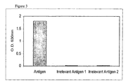
- a recombinant chimeric IgG (human constant regions) was produced by transient expression in CHO cells. Transfections of CHO cells were performed using the lipofectamine procedure according to manufacturer's instructions (InVitrogen, catalogue no. 18324). Specific binding of th IgG to antigen was confirmed by ELISA ( Figure 3 ).
- COS-1 cells transiently expressing the selected antigen were suspended in Immune cell media. Cell density was altered to 2x10 7 cells per ml.
- the assay mix was set up in ICM and contained 40 ⁇ l ELISA positive B cells, 40 ⁇ l of Goat anti-Rabbit IgG Fc specific FITC conjugate (Jackson ImmunoResearch) at 1:400 dilution and 40 ⁇ l of COS-1 cell suspension.
- This assay mix was then spotted (2-3ul per spot), onto Sigmacote treated 'chamber' slides and flooded with light paraffin oil. Slides were incubated for 40mins at 37°C and examined using an inverted microscope equipped with a mercury vapour UV lamp and a fluorescein filter set.
- B cells (plasma cells), secreting antigen specific IgG antibody were identified by a focal increase in fluorescence surrounding the B cells. See Figure 4 . Other B cells in the mixture, which did not secrete antigen specific antibodies, did not show surrounding fluorescence. In COS-1 cell controls where no antigen was present on the surface, no B-cell localised fluorescence was observed.
- the B cells present within the fluorescent foci were then harvested into Eppendorf tubes using standard micro-manipulation apparatus, (Eppendorf Transferman and CellTram Vario) and the heavy and light chain variable regions of the antibody subsequently isolated by PCR.
- CHO cells transiently expressing the selected antigen were suspended in Immune cell media. Cell density was altered to 2x10 7 cells per ml.
- the assay mix was set up in ICM and contained 40 ⁇ l ELISA positive B cells, 40 ⁇ l of Goat anti-Rabbit IgG Fc specific FITC conjugate (Jackson ImmunoResearch) at 1:400 dilution and 40 ⁇ l of CHO cell suspension.
- This assay mix was then spotted (2-3 ⁇ l per spot), onto Sigmacote treated 'chamber' slides and flooded with light paraffin oil. Slides were incubated for 40mins at 37°C and examined using an inverted microscope equipped with a mercury vapour UV lamp and a fluorescein filter set.
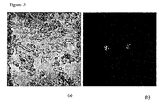
- B cells (plasma cells), secreting antigen specific IgG antibody were identified by a focal increase in fluorescence surrounding the B cells. See Figure 5 .
- Other B cells in the mixture which did not secrete antigen specific antibodies, did not show surrounding fluorescence.
- CHO cell controls where no antigen was present on the surface, no B-cell localised fluorescence was observed.
- the B cells present within the fluorescent foci were then harvested into Eppendorf tubes using standard micro-manipulation apparatus, (Eppendorf Transferman and CellTram Vario) and the heavy and light chain variable regions of the antibody subsequently isolated by PCR.
Landscapes
- Health & Medical Sciences (AREA)
- Life Sciences & Earth Sciences (AREA)
- Chemical & Material Sciences (AREA)
- Immunology (AREA)
- Engineering & Computer Science (AREA)
- Molecular Biology (AREA)
- Hematology (AREA)
- Urology & Nephrology (AREA)
- Biomedical Technology (AREA)
- General Health & Medical Sciences (AREA)
- Medicinal Chemistry (AREA)
- Cell Biology (AREA)
- Biochemistry (AREA)
- Organic Chemistry (AREA)
- Analytical Chemistry (AREA)
- Pathology (AREA)
- Proteomics, Peptides & Aminoacids (AREA)
- Microbiology (AREA)
- Biotechnology (AREA)
- Food Science & Technology (AREA)
- Physics & Mathematics (AREA)
- General Physics & Mathematics (AREA)
- Biophysics (AREA)
- Genetics & Genomics (AREA)
- Zoology (AREA)
- Tropical Medicine & Parasitology (AREA)
- Virology (AREA)
- Peptides Or Proteins (AREA)
- Preparation Of Compounds By Using Micro-Organisms (AREA)
- Measuring Or Testing Involving Enzymes Or Micro-Organisms (AREA)
- Micro-Organisms Or Cultivation Processes Thereof (AREA)
Abstract
Description
- The present invention relates generally to improved methods for producing antibodies and more specifically provides a homogeneous assay for obtaining antibodies.
- The selected lymphocyte antibody method (SLAM) for generating monoclonal antibodies overcomes the limitations of both hybridoma technology and bacterially expressed antibody libraries by enabling high affinity antibodies generated during in vivo immune responses to be isolated from any species (Babcook et al., 1996, Proc.Natl.Acad.Sci, 93, 7843-7848). SLAM enables a single lymphocyte that is producing an antibody with a desired specificity or function to be identified within a large population of lymphoid cells and the genetic information that encodes the specificity of the antibody to be rescued from that lymphocyte. Antibody producing cells which produce antibodies which bind to selected antigens are detected using an adapted hemolytic plaque assay method (Jerne and Nordin, 1963, Science, 140, 405). In this assay erythrocytes are coated with the selected antigen and incubated with the population of antibody producing cells and a source of complement. Single antibody producing cells are identified by the formation of hemolytic plaques. Plaques of lysed erythrocytes are identified using an inverted microscope and the single antibody producing cell of interest at the centre of the plaque is removed using micromanipulation techniques and the antibody genes from the cell are cloned by reverse transcription PCR. Other methods for detecting single antibody-producing cells of a desired function have already been described in International Patent Specification,
WO 92/02551 -
EP0286405 describes a homogeneous phase method of selecting cells that produce an anti-idiotypic antibody which comprises contacting a population of antibody-producing cells with a fluorescently-labelled antibody produced by immunisation of a host with an antigen from a selected pathogen and selecting those antibody-producing cells that bind the labelled antiserum. - In the hemolytic plaque assay described above the red blood cells are typically coated with antigen via a biotin/streptavidin coupling system that requires the antigen to be biotinylated. This method is therefore restricted to antigens that are available in a pure form and to those that can be biotinylated without affecting epitope presentation. This method clearly precludes the isolation of antibodies against a wide range of antigens. For example, many proteins are difficult to purify, particularly cell surface expressed proteins, such as type III proteins. Many proteins alter their conformation and presentation of desirable eptiopes upon biotinylation, for example proteins that contain lysine groups in their active site.
- It may also be desirable to produce antibodies against unknown antigens, such as proteins expressed on the surface of cells, such as tumor cells. The direct use of tumor cells in the plaque assay instead of antigen coated erythrocytes is difficult to achieve given the requirement for cell lysis to occur in order for plaques containing antibody producing cells to be identified. Cell lysis is dependent on cell type, antigen and antibody concentration. Red blood cells coated with the desired antigen will bind large amounts of available antibody and will lyse readily in the presence of complement. Other cell types such as tumor cells will not lyse so readily, especially when the availability of antigen on the surface may be very low and hence antibody binding will be low.
- Manz et al (PNAS, 92, 1921-1925) discloses a method for identification of antibody secreting cells where the secreted product is retained on the cell surface of the screening cells. Required steps include labelling of the surface of the antibody producing cell with capture antibodies or antigen, artificially elevating the viscosity of the medium to stop diffusion and cross-feeding of secreted product between cells, wash steps, identification of cells by fluorescent activated cell sorting procedures.
- The current invention addresses these difficulties by the subject-matter defined in the appended claims.
This improved assay has many advantages over the methods described above, allowing the identification of antibodies that bind to any antigen, including unknown antigens, cell surface antigens and antigens which cannot be biotinylated without altering the presentation of desirable epitopes. As a result, antibodies with binding specificities that were previously unidentifiable by conventional plaque assays can now be produced. In addition the assay is more facile than the hemolytic plaque assay and antibody producing cells can be identified more quickly. - We have demonstrated that it is possible to obtain antibody producing cells producing antibodies which bind to an antigen by incubating a population of antibody producing cells with an antigen source in the presence of labeled antibodies which bind to the antibodies produced by the antibody producing cells. Suprisingly, it is possible to distinguish those cells producing antibodies which bind to an antigen over those which do not without the need for wash steps to remove unbound label.
- Thus, according to the present invention, there is provided an in vitro homogeneous assay for identifying an antibody producing cell producing an antibody which binds to a selected antigen comprising:
- a) providing a population of antibody producing cells;
- b) incubating on a microscope slide said population of antibody producing cells with a selected antigen conjugated to a bead or coupled to an erythrocyte, or
antigen expressed on the surface of a cell, and
a labeled anti-antibody antibody, wherein said anti-antibody antibody is capable of distinguishing cells producing an antibody which binds to the selected antigen from those cells which do not and wherein the labeled anti-antibody antibody is labeled with a fluorescent conjugate ; and - c) without the need to remove unbound labeled anti-antibody antibodies, identifying an antibody producing cell capable of producing an antibody which binds to the selected antigen by detecting the increased concentration of labeled anti-antibody antibodies surrounding the cell using a microscope.
- The term 'antibody' as used herein includes any recombinant or naturally occurring immunoglobulin molecule such as a member of the IgG class e.g. IgG1 and also any antigen binding immunoglobulin fragment, such as Fv, Fab' and F(ab')2 fragments, and any derivatives thereof, such as single chain Fv fragments.
- The term 'antibody producing cell' as used herein means any cell capable of secreting an antibody, such as a B-lymphocyte, a plasma cell, a plasmablast, an activated B cell or a memory B cell. Antibody-producing cells for use in the invention may be obtained from an animal which has either been immunized with an antigen, or which has developed an immune response to an antigen as a result of disease. Other antibody producing cells for use in the present invention may include any transformed cell in particular, any mammalian cells which express immunoglobulin genes or parts thereof In one example the populations of antibody producing cells for use in the present invention produce a range of antibodies with different binding specificities.
- The assay of the present invention may also be used to identify high yielding antibody producing cells from a population of antibody producing cells which all produce the same antibody. The term 'high yielding' as used herein refers to antibody producing cells that produce antibodies of a known specificity but for which it would be desirable to identify those cells producing the antibody most efficiently. Identification of the high yielding cell will allow the cell to be isolated and clonally reproduced. In one example the high yielding antibody producing cell is a hybridoma cell. In another example the high yielding antibody producing cell is a transformed cell in particular, a mammalian cell which expresses immunoglobulin genes or parts thereof. Examples of such mammalian cells include but are not limited to NS0, CHO, COS and 293 cells.
- The term 'antigen' as used herein refers to any known or unknown substance that can be recognised by an antibody, including proteins, glycoproteins and carbohydrates. Preferably these antigens include biologically active proteins, such as hormones, cytokines, and their cell surface receptors, bacterial or parasitic cell membranes or purified components thereof, and viral antigens. In one example the antigen is available in a pure form obtained either by direct purification from the native source or by recombinant expression and purification of said antigen. Preferably the purified antigen is coupled to erythrocytes or any other particle such as a bead for incorporation into the assay,. In another example the antigen is one which is difficult to purify, such antigens include but are not limited to cell surface expressed proteins such as receptors, particularly type III proteins. In another example the presentation of desirable epitopes on the antigen is altered upon biotinylation, this includes but is not limited to proteins which contain lysines in their active site regions. In another example the antigen may be expressed on the surface of a cell, either naturally or recombinantly. Such cells may include but are not limited to mammalian cells, immunomodulatory cells, lymphocytes, monocytes, polymorphs, T cells, tumor cells, yeast cells, bacterial cell, infectious agents, parasites, plant cells, transfected cells such as NS0, CHO, COS, 293 cells. In one example the antigens expressed on the surface of said cells are antigens which are difficult to purify or antigens which lose desired epitopes upon biotinylation such as those antigens described above.
- In another example the antigen is a cell or a population of cells for which it would be desirable to isolate antibodies to, such as mammalian cells, immunomodulatory cells, lymphocytes, monocytes, polymorphs, T cells, tumor cells, yeast cells, bacterial cell, infectious agents, parasites, and plant cells. In one embodiment the cell is a tumor cell.
- The term 'homogeneous assay' as used herein refers to an assay whereby all components of the assay are combined together to identify antibody producing cells without the need to remove unbound labeled anti-antibody antibodies. The term 'labeled anti-antibody antibody' refers to labeled antibodies which bind to any region of the antibodies produced by the antibody producing cells, regardless of the binding specificity of those antibodies. Preferably said anti-antibody antibodies are from one species while the antibody producing cells are from another. Preferably these antibodies bind to the Fc portion of the antibody produced by the antibody producing cell.
- The labeled anti-antibody antibodies are capable of distinguishing cells producing antibodies that bind to the selected antigen from those cells that do not. Appropriate labels are well known in the art and can include but are not limited to chemiluminescence, enzyme and fluorescent labels. Preferably the label is a fluorescent label. The fluorescent label conjugated to the anti-antibody antibodies can be any fluorescent label including but not limited to Aqua, Texas-Red, FITC, rhodamine, rhodamine derivative, fluorescein, fluorescein derivative, cascade blue, Cy5 and phycoerythrin. Preferably the fluorescent conjugate is FITC. Thus in one particular example of an assay according to the present invention, the antibody producing cells are from rabbits and the labeled anti-antibody antibodies are fluorescent labeled goat anti-rabbit anti-Fc antibodies.
- In the assay of the present invention the antibody producing cells producing antibodies which bind to the selected antigen are distinguished from those that do not by detecting the increased concentration of labeled anti-antibody antibodies surrounding said cells. This is achieved by visualising the labeled anti-antibody antibodies and hence the antibody producing cell surrounded by said antibodies. This is achieved using a microscope. Preferably the label is detected using an inverted microscope with a mercury vapour UV lamp and a filter set appropriate for the conjugate used. Preferably the filter set is a fluorescein filter set. Thus, where the label is a fluorescent label the antibody producing cells producing antibodies which bind to the selected antigen are identified by a localised increase in fluorescence surrounding said cells.
- The present invention also provides a in vitro method for producing an antibody which binds to a selected antigen comprising:
- a) providing a population of antibody producing cells;
- b) incubating on a microscope slide said population of antibody producing cells with a selected antigen conjugated to a bead or coupled to an erythrocyte, or
antigen expressed on the surface of a cell, and
a labeled anti-antibody antibody, wherein said anti-antibody antibody is capable of distinguishing cells producing an antibody which binds to the selected antigen from those cells which do not and wherein the labeled anti-antibody antibody is labeled with a fluorescent conjugate; - c) without the need for wash steps, identifying an antibody producing cell producing an antibody which binds to the selected antigen by detecting the increased concentration of labeled anti-antibody antibodies surrounding the cell using a microscope;
- d) isolating the identified antibody producing cell; and
- e) synthesizing an antibody or antibody fragment therefrom.
- Where desired, steps (d) and (e) can be repeated more than once to isolate more than one antibody producing cell and to synthesize more than one antibody.
- Antibody producing cells identified using the homogeneous assay described herein are isolated directly from the assay using micromanipulation techniques well known in the art.
- Antibodies can be synthesized from the isolated antibody producing cell either directly or indirectly. Direct synthesis can be achieved by culturing the isolated antibody producing cell in an appropriate medium. Indirect synthesis can be achieved by isolating the genes encoding the antibodies or parts thereof and expressing them in a host cell using methods well known in the art. A vector containing the antibody gene(s) is transfected into a host cell and the host cell cultured in an appropriate medium such that the antibody or antibody fragment with the desired specificity is produced in the host cell.
- Antibody-producing cells for use in the present invention may be obtained from any appropriate source, including an animal which has either been immunized with a selected antigen, or which has developed an immune response to an antigen as a result of disease.
- Animals may be immunized with a selected antigen using any of the techniques well known in the art suitable for generating an immune response (see Handbook of Experimental Immunology, D. M. Weir (ed.), Vol 4, Blackwell Scientific Publishers, Oxford, England, 1986). Many warm-blooded animals, such as humans, rabbits, mice, rats, sheep, cows or pigs may be immunized in order to obtain antibody-producing cells. However, mice, rabbits and rats are generally preferred.
- High numbers of antibody producing cells can be found in the spleen and lymph node of the immunised animal and once an immune response has been generated and the animal has been sacrificed, the spleen and lymph nodes are removed. A single cell suspension of antibody producing cells is prepared using techniques well known in the art. Antibody producing cells can also be obtained from an animal that has generated the cells during the course of a disease. For instance, antibody producing cells from a human with a disease of unknown cause, such as cancer, may be obtained and used to assist in the identification of antibodies which have an effect on the disease process or which may lead to identification of an agent or body component that is involved in the cause of the disease. Similarly, antibody-producing cells may be obtained from subjects with disease of known cause such as malaria or AIDS. These antibody producing cells may be derived from the blood or lymph nodes, as well as from other diseased or normal tissues.
- Antibody producing cells may also be obtained by culture techniques such as in vitro immunization. Examples of such methods are described by C. R. Reading in Methods in Enzymology 121:18-33 (J. J. Langone, H. H. van Vunakis (eds,), Academic Press Inc., N. Y.). Antibody producing cells may also be obtained from very early monoclonal or oligoclonal fusion cultures produced by conventional hybridoma technology.
- The population of antibody producing cells may be enriched for use in the assay by methods based upon the size or density of the antibody producing cells relative to other cells. An example of the use of Percoll to separate cells according to density is described by van Mourik and W. P. Zeizlmaker in Methods in Enzymology 121;174-182 (J. J. Langone, H. H. van Vunakis (eds.), Academic Press Inc., N.Y.). Gradients of varying density of solutions of bovine serum albumin can also be used to separate cells according to density. (See N. Moav and T. N. Harris, J. Immunol. 105, 1512, 1970; see also Raid, D. J. in Selected Methods in Cellular Immunology, B. Misheli and S. Shiigi (eds.), W. H.Freeman and Co., San Francisco, 1987). Preferably separation is achieved by centrifugation with Ficoll-Hypaque (Pharmacia, Uppsula, Sweden). The fraction that is most enriched for desired antibody-producing cells can be determined in a preliminary procedure using ELISA based assays to select populations that may contain antibodies with the desired binding specificity. Alternatively or in addition, the fraction most enriched for the desired antibody can be determined by a functional assay.
- In the assay the population of antibody producing cells suspected of producing antibodies with the desired binding specificity are suspended in an appropriate medium before incorporation into the assay. An appropriate medium for the assay will be one that provides at least the minimum requirements for short-term maintenance of cellular integrity and cellular structures, such as an isotonic buffer. Preferably this medium is immune cell medium comprising Roswell Park Memorial Institute medium (RPMI) + 10% foetal bovine serum; 50µM 2-β-mercaptoethanol; 2mM glutamine; 20mM Hepes; and 1x Penicillin and Streptomycin.
- Under such conditions the antibody producing cells produce and secrete antibodies. Antibody producing cells are diluted within the medium to a density which allows selection of an individual or small number of antibody producing cells. If it is unclear which cell is responsible for the activity indicated by the assay, or in order to confirm the activity, the selected cell(s) may be retested for their ability to produce antibodies with the desired binding specificity.
- The antigen for use in the assay may be, as described above, any substance to which an antibody can be produced including proteins, glycoproteins, carbohydrates and whole cells, such as tumor cells or transfected cells expressing the antigen on the surface. In one example the antigen is known and available in a pure form and is coated on the surface of erythrocytes or other particles such as beads for incorporation into the assay. A number of methods for coating particles with antigens are known to those skilled in the art. These include chromic chloride or water soluble carbodiimide. In one embodiment, a biotin/streptavidin coupling system is used to couple antigen to erythrocytes, the methods for which are described in detail in
WO92/02551 - In another example the antigen is coupled to commercially available beads (for example as can be obtained from New England Biolabs). Antigen can be conjugated to beads using a number of different methods, preferably via direct conjugation to activated beads or via biotin to streptavidin-coupled beads. Preferably these beads are magnetic for ease of handling.
- In another example, particularly when the antigen loses desirable epitopes upon biotinylation, the antigen is coupled to the surface of a particle via a polyclonal antibody that binds the antigen. To prepare the antigen-polyclonal antibody-particle conjugate, the polyclonal antibody is first conjugated to the surface of a particle, such as a bead using any suitable method, such as via biotin to streptavidin-coupled beads. The polyclonal antibody-particle conjugate is then incubated with an excess of antigen to allow binding of the polyclonal antibody to the antigen. The antigen-polyclonal antibody-particle conjugate is then separated from unbound antigen, for example by centrifugation, and incorporated into the assay. The polyclonal antibody for use in the conjugate may be produced using any suitable method known in the art, using the desired antigen as immunogen, in any suitable species. The polyclonal antibody may be a whole IgG or a fragment thereof such as a Fab', F(ab')2 or Fab fragment. Fragments may be produced using any method known in the art, for example by pepsin or papain digestion. It is important that the polyclonal antibody used in the conjugate is not recognized by the labeled anti-antibody antibody used in the assay to detect the antibodies produced by the antibody producing cells. For example where the antibody producing cells are from rabbit, the labeled anti-antibody antibody may be an anti-rabbit anti-Fc antibody and the polyclonal antibody used in the conjugate should be an antibody from a species other than rabbit, for example goat, or if the antibody is from rabbit it should be a fragment lacking the Fc region, for example a Fab', F(ab')2 or Fab fragment.
- In another example, particularly when the antigen is difficult to purify or loses desired epitopes upon biotinylation the antigen is expressed on the surface of a cell. Such cells may be those that naturally express the antigen on their surface or a transfected cell expressing the antigen on its surface. Such cells may include but are not limited to mammalian cells, immunomodulatory cells, lymphocytes, monocytes, polymorphs, T cells, tumor cells, yeast cells, bacterial cell, infectious agents, parasites, plant cells, transfected cells such as NS0, CHO, COS, 293 cells. Transfection of cells such as NS0, CHO, COS and 293 cells can be achieved by any method known in the art including, electroporation and nucleofection.
- In a further example the antigen source is any cell that it would be desirable to isolate antibodies to. Such cells may include but are not limited to mammalian cells, immunomodulatory cells, lymphocytes, monocytes, polymorphs, T cells, tumor cells, yeast cells, bacterial cell, infectious agents, parasites and plant cells.
- The antibody producing cells and the antigen are incorporated into the assay at an appropriate concentration which can be determined empirically for example as described in the examples hereinafter. The antibody producing cells are at sufficiently low density that they are well separated allowing identification and isolation of the antibody producing cell producing antibodies of the desired specificity. The antigen will be present in excess and preferably the antigen is in a 10-1,000 fold excess over the antibody producing cells.
- In order to identify antibodies that bind to the selected antigen, labeled anti-antibody antibodies are incorporated into the assay. Said antibodies will bind to all antibodies produced by the antibody producing cells, regardless of their binding specificity. Such antibodies are easily produced by one skilled in the art or are readily available commercially. Preferably the anti-antibody antibodies are anti-Fc antibodies. In one embodiment of the present invention the antibody producing cells are from rabbits and the labeled anti-Fc antibodies are goat anti-rabbit anti-Fc antibodies.
- The label conjugated to the anti-antibody antibodies is any label that can be detected in the assay by any suitable method known in the art. Many different conjugates are available for labeling the antibodies for example, chemiluminescent, enzyme and fluorescent labels. Such antibodies are easily produced by one skilled in the art or are readily available commercially. Preferably the label is one that can be detected by microscopy. In general in the various aspects of the invention described herein the label used is preferably a fluorescent label. Particular fluorescent labels are those which can be visualised by microscopy and can include but are not limited to Aqua, Texas-Red, FITC, rhodamine, rhodamine derivatives, fluorescein, fluorescein derivatives, cascade blue, Cy5 and phycoertythrin. Preferably said label is the fluorescent conjugate, fluorescein isothiocyanate (FITC).
- The labeled anti-antibody antibody is used in the assay at a concentration at which it is possible to distinguish cells producing antibodies that bind to the selected antigen from those cells that do not. The optimal concentration can be determined empirically by one skilled in the art by varying the concentration of labeled anti-antibody antibody. In one example the labeled antibody is a fluorescent labeled antibody and is used at a concentration that is not so low that no fluorescence can be detected and not so high that there is high background fluorescence. Preferably the fluorescent labeled anti-antibody antibody is in excess such that it binds all antibodies produced by the antibody producing cell without causing excessive background fluorescence.
- To identify antibody producing cells producing antibodies which bind to the selected antigen the assay mixture comprising a population of antibody producing cells, the antigen and labeled anti-antibody antibody is incubated in the medium described above to allow binding to take place. Optimal incubation times and temperatures can be determined empirically by one skilled in the art. Incubation will take place in any suitable container such as a microscope slide at any suitable temperature for example between 4°C or about and 37°C or about, for any suitable length of time for example between 5 minutes or about and 5 hours or about. Preferably the incubation of the assay mixture takes place on a microscope slide at 37°C for up to 1 hour.
- The labeled anti-antibody antibody is detected using any appropriate method known in the art. Preferably the labeled anti-antibody antibody is detected using a microscope. More preferably the anti-antibody antibody is conjugated to a fluorescent label and the fluorescence is visualised using an inverted microscope equipped with a mercury vapour UV lamp with an appropriate filter set. Preferably the filter set is a fluorescein filter set.
- Antibody producing cells which produce an antibody which binds to the selected antigen are identified by the increased concentration of labeled anti-antibody antibodies surrounding the cell. Those antibody producing cells producing antibodies which do not bind to the antigen will not be surrounded by an increased concentration of labeled anti-antibody antibodies. High yielding antibody producing cells are identified as those where the localised increase in anti-antibody antibody concentration appears most quickly.
- The antibody producing cell may then be isolated directly from the assay using standard micromanipulation techniques such as a fine glass pipette and a micromanipulator.
- Antibodies can be synthesized directly or indirectly from the isolated antibody producing cell. Direct synthesis can be achieved by culturing the isolated antibody producing cell in an appropriate medium. If the assay is used to identify a high yielding antibody producing cell the cell will be cultured under appropriate conditions to clonally reproduce this high yielding cell.
- Indirect synthesis can be achieved by isolating the genes encoding the antibodies or parts thereof and expressing them in a host cell. The entire genes may be cloned or the variable regions or portions thereof which confer the desired specificity of the antibody may be cloned and used to produce recombinant antibodies. Recombinant antibodies can take several different forms and include intact immunoglobulins, chimeric antibodies, humanised antibodies and antigen binding fragments such as Fv, Fab, Fab' and F(ab')2 fragments, and any derivatives thereof, such as single chain Fv fragments. The methods for creating these antibody molecules are well known in the art (see for example,
Boss, US 4,816,397 ; Shrader,WO 92/02551 - Antibodies obtained according to the present disclosure may be used without further modification, or if desired following modification including conjugation to one or more reporter or effector molecules, for any suitable diagnostic or therapeutic purpose.
-
-
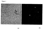
Figure 1 A homogeneous fluorescence assay comprising rabbit B cells, sheep red blood cells coated with antigen and Goat anti-Rabbit IgG Fc specific FITC conjugate. Assay visualised using an inverted microscope equipped with a mercury vapour UV lamp and fluorescein filter set. Magnification x8.- (a) Phase image of assay.
- (b) Fluorescence image of assay. Fluorescence localised around B cells secreting antigen specific antibodies
-
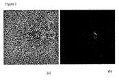
Figure 2 A homogeneous fluorescence assay comprising rabbit B cells, magnetic beads coated with antigen and Goat anti-Rabbit IgG Fc specific FITC conjugate. Assay visualised using an inverted microscope equipped with a mercury vapour UV lamp and fluorescein filter set. Magnification x20.- (a) Phase image of assay.
- (b) Fluorescence image of assay. Fluorescence localised around B cells secreting antigen specific antibodies
-
Figure 3 ELISA detection of specific antigen binding of antibodies produced in CHO cells. -
Figure 4 A homogeneous fluorescence assay comprising rabbit B cells, transfected COS-1 cells expressing antigen on their surface and Goat anti-Rabbit IgG Fc specific FITC conjugate. Assay visualised using an inverted microscope equipped with a mercury vapour UV lamp and fluorescein filter set. Magnification x8- (a) Phase image of assay.
- (b) Fluorescence image of assay. Fluorescence localised around B cells secreting antigen specific antibodies.
-
Figure 5 A homogeneous fluorescence assay comprising rabbit B cells, transfected CHO cells expressing antigen on their surface and Goat anti-Rabbit IgG Fc specific FITC conjugate. Assay visualised using an inverted microscope equipped with a mercury vapour UV lamp and fluorescein filter set. Magnification x8.- (a) Phase image of assay.
- (b) Fluorescence image of assay. Fluorescence localised around B cells secreting antigen specific antibodies
- The following examples are offered by way of illustration, and not by way of limitation. The following abbreviations are used in the examples:
- ICM - immune cell medium (RPMI +10% foetal bovine serum; 50µM 2-β-mercaptoethanol; 2mM glutamine; 20mM hepes; and 1x penicillin and streptomycin)
- RPMI - Roswell Park Memorial Institute medium
- PBS - Phosphate buffered saline
- The coating of the SRBC (obtained from TCS Biosciences) was carried out by streptavidin linking the biotinylated antigen to the surface of biotin coated SRBC. The antigen coated SRBC were prepared on the day of use and stored 5% (v/v) in immune cell medium.
- The assay mix was set up in ICM and contained 10µl of rabbit B cells containing 10-1,000 B cells from an ELISA positive population, 10µl antigen coated SRBC (5% v/v) and 20µl of Goat anti-Rabbit IgG Fc specific FITC conjugate (Jackson ImmunoResearch) at variable concentrations for each experiment (1:100, 1:200, 1:400 and 1:800). The experiments were set up to determine the optimal concentration of Goat anti-Rabbit IgG Fc specific FITC conjugate required for the identification of antibody producing cells without excessive background fluorescence.
- This assay mix was then spotted (2-3µl per spot), onto Sigmacote® treated 'chamber' slides and flooded with light paraffin oil. Slides were incubated for 20-30mins at 37°C and examined using an inverted microscope equipped with a mercury vapour UV lamp and a fluorescein filter set. B cells, (plasma cells), secreting antigen specific IgG antibody were identified by a focal increase in fluorescence surrounding said cells. See
Figure 1 . Using this method the optimal concentration for Goat anti-Rabbit IgG Fc specific FITC conjugate was found to be 1:400. Other B cells in the mixture, which did not secrete antigen specific antibodies, did not show surrounding fluorescence. In SRBC controls where no antigen was present on the surface, no B-cell localised fluorescence was observed. - The B cells present within the fluorescent foci were then harvested into Eppendorf tubes using standard micro-manipulation apparatus, (Eppendorf Transferman and CellTram Vario) and the heavy and light chain variable regions of the antibody subsequently isolated by PCR.
- 1µM magnetic streptavidin coated beads, (New England Biolabs), were used in all experiments.
- An aliquot of the bead stock was washed 3x in PBS to remove preservative, using a magnet, and resuspended in the same volume of immune cell medium, (ICM), (RPMI +10% foetal bovine serum; 50µM 2-β-mercaptoethanol; 2mM glutamine; 20mM Hepes; and 1x Penicillin and Streptomycin).
- Serial 2-fold dilutions of beads were prepared in ICM and used to determined a dilution with a bead density that produced an even monolayer when the beads were spotted (2-3ul per spot) onto Sigmacote treated slides and overlaid with light paraffin oil. A final dilution of washed beads of 1/8 was determined to be optimal.
- 50µl aliquots of streptavidin coated beads were washed and resuspended in 50µl PBS. Each aliquot was incubated with different amounts of stock biotinylated antigen (1mg/ml) ranging from 0.1 µg up to 25µg. These were incubated for 1h at room temperature with occasional manual shaking. The beads were then washed in PBS using a magnet and resuspended in 50µl of ICM.
- 20µl of the antigen loaded beads were then mixed with 40µl of ELISA positive B cells; 60µl of ICM; and 40µl of a 1/400 dilution of a Goat anti-Rabbit IgG Fc specific FITC conjugate (Jackson ImmunoResearch).
- This mixture was then spotted, (2-3µl per spot), onto Sigmacote treated 'chamber' slides and flooded with light paraffin oil. Slides were incubated for 20-30mins at 37°C and examined using an inverted microscope equipped with a mercury vapour UV lamp and a fluorescein filter set.
- B cells, (plasma cells), secreting antigen specific IgG antibody were identified by a focal increase in fluorescence around the B cell. See
Figure 2 . Using this method 1µg of biotinylated antigen per 50µl of bead stock was determined as optimal for signal generation for this particular antigen. Other B cells in the mixture, which did not secrete antigen specific antibodies, did not show surrounding fluorescence. In controls, no B-cell localised fluorescence was observed when the beads were coated with another irrelevant antigen. - The B cells present within the fluorescent foci were then harvested into Eppendorf tubes using standard micro-manipulation apparatus, (Eppendorf Transferman and CellTram Vario) and the heavy and light chain variable regions of the antibody from one of the cells subsequently isolated by PCR. A recombinant chimeric IgG (human constant regions) was produced by transient expression in CHO cells. Transfections of CHO cells were performed using the lipofectamine procedure according to manufacturer's instructions (InVitrogen, catalogue no. 18324). Specific binding of th IgG to antigen was confirmed by ELISA (
Figure 3 ). - COS-1 cells transiently expressing the selected antigen were suspended in Immune cell media. Cell density was altered to 2x10⁷ cells per ml.
- The assay mix was set up in ICM and contained 40µl ELISA positive B cells, 40µl of Goat anti-Rabbit IgG Fc specific FITC conjugate (Jackson ImmunoResearch) at 1:400 dilution and 40µl of COS-1 cell suspension.
- This assay mix was then spotted (2-3ul per spot), onto Sigmacote treated 'chamber' slides and flooded with light paraffin oil. Slides were incubated for 40mins at 37°C and examined using an inverted microscope equipped with a mercury vapour UV lamp and a fluorescein filter set.
- B cells, (plasma cells), secreting antigen specific IgG antibody were identified by a focal increase in fluorescence surrounding the B cells. See
Figure 4 . Other B cells in the mixture, which did not secrete antigen specific antibodies, did not show surrounding fluorescence. In COS-1 cell controls where no antigen was present on the surface, no B-cell localised fluorescence was observed. - The B cells present within the fluorescent foci were then harvested into Eppendorf tubes using standard micro-manipulation apparatus, (Eppendorf Transferman and CellTram Vario) and the heavy and light chain variable regions of the antibody subsequently isolated by PCR.
- CHO cells transiently expressing the selected antigen were suspended in Immune cell media. Cell density was altered to 2x10⁷ cells per ml.
- The assay mix was set up in ICM and contained 40µl ELISA positive B cells, 40µl of Goat anti-Rabbit IgG Fc specific FITC conjugate (Jackson ImmunoResearch) at 1:400 dilution and 40µl of CHO cell suspension.
- This assay mix was then spotted (2-3µl per spot), onto Sigmacote treated 'chamber' slides and flooded with light paraffin oil. Slides were incubated for 40mins at 37°C and examined using an inverted microscope equipped with a mercury vapour UV lamp and a fluorescein filter set.
- B cells, (plasma cells), secreting antigen specific IgG antibody were identified by a focal increase in fluorescence surrounding the B cells. See
Figure 5 . Other B cells in the mixture, which did not secrete antigen specific antibodies, did not show surrounding fluorescence. In CHO cell controls where no antigen was present on the surface, no B-cell localised fluorescence was observed. - The B cells present within the fluorescent foci were then harvested into Eppendorf tubes using standard micro-manipulation apparatus, (Eppendorf Transferman and CellTram Vario) and the heavy and light chain variable regions of the antibody subsequently isolated by PCR.
Claims (15)
- An in vitro homogeneous assay for identifying an antibody producing cell producing an antibody which binds to a selected antigen comprising:a) providing a population of antibody producing cells;b) incubating on a microscope slide said population of antibody producing cells with a selected antigen conjugated to a bead or coupled to an erythrocyte, or
antigen expressed on the surface of a cell, and
a labeled anti-antibody antibody, wherein said anti-antibody antibody is capable of distinguishing cells producing an antibody which binds to the selected antigen from those cells which do not and wherein the labeled anti-antibody antibody is labeled with a fluorescent conjugate ; andc) without the need to remove unbound labeled anti-antibody antibodies, identifying an antibody producing cell capable of producing an antibody which binds to the selected antigen by detecting the increased concentration of labeled anti-antibody antibodies surrounding the cell using a microscope. - The assay of claim 1 where the antigen is coupled to an erythrocyte.
- The assay of claim 1 where the antigen is coupled to a bead.
- The assay of claim 3 where the antigen is coupled to a bead via a polyclonal antibody
- The assay of claim 4 where the polyclonal antibody is an antibody fragment.
- The assay of claim 5 where the polyclonal antibody is an antibody Fab, Fab' or F(ab')2 fragment.
- The assay of claim 1 where the antigen expressing cell is a transfected cell.
- The assay of claim 1 where the antigen expressing cell is a tumor cell.
- The assay of claims 1 to 8 where the antigen is an infectious agent.
- The assay of claims 1 to 9 where the labeled anti-antibody antibody is an anti-Fc antibody.
- The assay of claim 10 where the fluorescent labeled anti-antibody antibody is labeled with FITC
- The assay of claim 11 where the FITC labeled anti-antibody antibody is an anti-Fc antibody
- The assay of claims 1 to 12 where the antibody producing cells are B cells, plasma cells, plasmablasts, activated B cells or memory B cells.
- The assay of claims 1 to 13 where the antibody producing cell is a hybridoma cell or a mammalian cell engineered to express antibodies.
- An in vitro method for producing an antibody which binds to a selected antigen comprising:a) providing a population of antibody producing cells;b) incubating on a microscope slide said population of antibody producing cells with a selected antigen conjugated to a bead or coupled to an erythrocyte, or
antigen expressed on the surface of a cell, and
a labeled anti-antibody antibody, wherein said anti-antibody antibody is capable of distinguishing cells producing an antibody which binds to the selected antigen from those cells which do not and wherein the labeled anti-antibody antibody is labeled with a fluorescent conjugate;c) without the need for wash steps, identifying an antibody producing cell producing an antibody which binds to the selected antigen by detecting the increased concentration of labeled anti-antibody antibodies surrounding the cell using a microscope;d) isolating the identified antibody producing cell; ande) synthesizing an antibody or antibody fragment therefrom.
Applications Claiming Priority (5)
| Application Number | Priority Date | Filing Date | Title |
|---|---|---|---|
| GB0228188 | 2002-12-03 | ||
| GB0228188A GB0228188D0 (en) | 2002-12-03 | 2002-12-03 | Biological products |
| GB0319587A GB0319587D0 (en) | 2003-08-20 | 2003-08-20 | Biological products |
| GB0319587 | 2003-08-20 | ||
| PCT/GB2003/005254 WO2004051268A1 (en) | 2002-12-03 | 2003-12-01 | Assay for identifying antibody producing cells |
Publications (2)
| Publication Number | Publication Date |
|---|---|
| EP1570267A1 EP1570267A1 (en) | 2005-09-07 |
| EP1570267B1 true EP1570267B1 (en) | 2011-10-12 |
Family
ID=32472152
Family Applications (1)
| Application Number | Title | Priority Date | Filing Date |
|---|---|---|---|
| EP03778577A Expired - Lifetime EP1570267B1 (en) | 2002-12-03 | 2003-12-01 | Assay for identifying antibody producing cells |
Country Status (9)
| Country | Link |
|---|---|
| US (1) | US7993864B2 (en) |
| EP (1) | EP1570267B1 (en) |
| JP (1) | JP4603894B2 (en) |
| AT (1) | ATE528648T1 (en) |
| AU (1) | AU2003285578B2 (en) |
| CA (1) | CA2507004A1 (en) |
| ES (1) | ES2374068T3 (en) |
| PT (1) | PT1570267E (en) |
| WO (1) | WO2004051268A1 (en) |
Cited By (11)
| Publication number | Priority date | Publication date | Assignee | Title |
|---|---|---|---|---|
| WO2017009473A1 (en) | 2015-07-16 | 2017-01-19 | Ucb Biopharma Sprl | Antibody molecules which bind cd45 |
| US10358493B2 (en) | 2014-05-29 | 2019-07-23 | Ucb Biopharma Sprl | Bispecific format suitable for use in high-through-put screening |
| US10618979B2 (en) | 2015-12-03 | 2020-04-14 | Ucb Biopharma Sprl | Multispecific antibodies |
| WO2020148554A1 (en) | 2019-01-18 | 2020-07-23 | UCB Biopharma SRL | Antibodies to ebola virus glycoprotein |
| US10774157B2 (en) | 2015-12-03 | 2020-09-15 | UCB Biopharma SRL | Multispecific antibodies |
| US10829566B2 (en) | 2015-12-03 | 2020-11-10 | UCB Biopharma SRL | Method employing bispecific antibodies |
| US10954312B2 (en) | 2015-12-03 | 2021-03-23 | UCB Biopharma SRL | Method employing bispecific protein complex |
| WO2021105669A1 (en) | 2019-11-29 | 2021-06-03 | Oxford University Innovation Limited | Antibodies |
| US11286312B2 (en) | 2015-12-03 | 2022-03-29 | UCB Biopharma SRL | Multispecific antibodies |
| EP3988936A1 (en) | 2015-06-18 | 2022-04-27 | UCB Biopharma SRL | Antibody epitope |
| WO2025120171A1 (en) | 2023-12-08 | 2025-06-12 | UCB Biopharma SRL | Antibodies |
Families Citing this family (155)
| Publication number | Priority date | Publication date | Assignee | Title |
|---|---|---|---|---|
| US20060263353A1 (en) | 2003-08-20 | 2006-11-23 | Lawson Alastair D G | Methods for obtaining antibodies |
| EP1658498A1 (en) * | 2003-08-20 | 2006-05-24 | Celltech R & D Limited | Methods for obtaining antibodies |
| CZ200755A3 (en) | 2004-07-26 | 2007-04-11 | Biogen Idec Ma Inc. | Anti-CD154 antibody peptides |
| GB0506912D0 (en) | 2005-04-05 | 2005-05-11 | Celltech R&D Ltd | Biological products |
| WO2007003041A1 (en) * | 2005-07-01 | 2007-01-11 | John Schrader | Methods of isolating cells and generating monoclonal antibodies |
| AU2006290567A1 (en) | 2005-09-14 | 2007-03-22 | Ucb Pharma S.A | Comb polymers |
| CN101356194B (en) | 2005-12-09 | 2013-06-12 | Ucb医药有限公司 | Antibody molecules specific to human IL-6 |
| WO2007122823A1 (en) * | 2006-04-20 | 2007-11-01 | Jichi Medical University | Tumor target cell capable of producing vector |
| CN101600793B (en) | 2006-05-19 | 2013-06-26 | 奥尔德生物制药公司 | Culture method for obtaining a clonal population of antigen-specific b cells |
| GB0614780D0 (en) | 2006-07-25 | 2006-09-06 | Ucb Sa | Biological products |
| GB0619291D0 (en) | 2006-09-29 | 2006-11-08 | Ucb Sa | Altered antibodies |
| GB0620729D0 (en) | 2006-10-18 | 2006-11-29 | Ucb Sa | Biological products |
| AU2007336243B2 (en) | 2006-12-19 | 2012-07-26 | Ablynx N.V. | Amino acid sequences directed against a metalloproteinase from the ADAM family and polypeptides comprising the same for the treatment of ADAM-related diseases and disorders |
| US9512236B2 (en) | 2006-12-19 | 2016-12-06 | Ablynx N.V. | Amino acid sequences directed against GPCRS and polypeptides comprising the same for the treatment of GPCR-related diseases and disorders |
| CA2673331A1 (en) | 2006-12-19 | 2008-06-26 | Ablynx N.V. | Amino acid sequences directed against gpcrs and polypeptides comprising the same for the treatment of gpcr-related diseases and disorders |
| ES2523194T3 (en) * | 2007-03-13 | 2014-11-21 | University Of Zurich | Specific human monoclonal antibody for each tumor |
| SG196697A1 (en) * | 2007-03-22 | 2014-02-13 | Biogen Idec Inc | Binding proteins, including antibodies, antibody derivatives and antibody fragments, that specifically bind cd154 and uses thereof |
| GB0717337D0 (en) | 2007-09-06 | 2007-10-17 | Ucb Pharma Sa | Method of treatment |
| US8629246B2 (en) | 2007-09-26 | 2014-01-14 | Ucb Pharma S.A. | Dual specificity antibody fusions |
| GB0721752D0 (en) * | 2007-11-06 | 2007-12-19 | Univ Southampton | Configurable electronic device and method |
| WO2009068631A1 (en) | 2007-11-27 | 2009-06-04 | Ablynx N.V. | Method for obtaining polypeptide constructs comprising two or more single domain antibodies |
| GB0800277D0 (en) | 2008-01-08 | 2008-02-13 | Imagination Tech Ltd | Video motion compensation |
| AU2009235467A1 (en) | 2008-04-07 | 2009-10-15 | Ablynx Nv | Single variable domains against the Notch pathways |
| GB0807413D0 (en) | 2008-04-23 | 2008-05-28 | Ucb Pharma Sa | Biological products |
| NZ589036A (en) | 2008-05-16 | 2012-07-27 | Ablynx Nv | AMINO ACID SEQUENCES DIRECTED AGAINST CXCR4 AND OTHER GPCRs AND COMPOUNDS COMPRISING THE SAME |
| HUE042053T2 (en) | 2008-06-05 | 2019-06-28 | Ablynx Nv | Amino acid sequences against viral envelope proteins and polypeptides containing them for treating viral diseases |
| AU2009329501B2 (en) | 2008-12-19 | 2015-11-26 | Ablynx N.V. | Genetic immunization for producing immunoglobulins against cell-associated antigens such as P2X7, CXCR7 or CXCR4 |
| GB0900425D0 (en) | 2009-01-12 | 2009-02-11 | Ucb Pharma Sa | Biological products |
| BRPI1008692B8 (en) | 2009-02-17 | 2021-05-25 | Ucb Biopharma Sprl | antagonist antibody having specificity for human ox40, isolated DNA sequence, cloning or expression vector, host cell, process for producing said antibody, pharmaceutical composition, use of said antibody and fusion protein |
| CA2744103C (en) | 2009-02-24 | 2018-01-02 | Esbatech, An Alcon Biomedical Research Unit Llc | Methods for identifying immunobinders of cell-surface antigens |
| GB0903207D0 (en) | 2009-02-25 | 2009-04-08 | Ucb Pharma Sa | Method for expressing multimeric proteins |
| ES2700442T3 (en) | 2009-02-25 | 2019-02-15 | Ucb Biopharma Sprl | Method to produce antibodies |
| GB0904214D0 (en) | 2009-03-11 | 2009-04-22 | Ucb Pharma Sa | Biological products |
| IN2012DN02401A (en) | 2009-09-24 | 2015-08-21 | Ucb Pharma Sa | |
| GB201005063D0 (en) | 2010-03-25 | 2010-05-12 | Ucb Pharma Sa | Biological products |
| US9008406B2 (en) * | 2009-10-09 | 2015-04-14 | Kawasaki Jukogyo Kabushiki Kaisha | Method and apparatus for discriminating undifferentiated pluripotent stem cells, and automated culture method and system |
| GB0922434D0 (en) | 2009-12-22 | 2010-02-03 | Ucb Pharma Sa | antibodies and fragments thereof |
| US9234037B2 (en) | 2009-10-27 | 2016-01-12 | Ucb Biopharma Sprl | Method to generate antibodies to ion channels |
| GB0922435D0 (en) | 2009-12-22 | 2010-02-03 | Ucb Pharma Sa | Method |
| CA2778673A1 (en) | 2009-10-27 | 2011-05-05 | Karen Margrete Miller | Function modifying nav 1.7 antibodies |
| US20120321640A1 (en) | 2009-12-01 | 2012-12-20 | Ablynx N.V. | Von willebrand factor specific binding agents and uses thereof |
| HRP20251657T1 (en) | 2009-12-14 | 2026-02-13 | Ablynx N.V. | SINGLE VARIABLE DOMAIN IMMUNOGLOBULIN ANTIBODIES AGAINST OX40L, CONSTRUCTS AND THEIR THERAPEUTIC USE |
| GB201000587D0 (en) | 2010-01-14 | 2010-03-03 | Ucb Pharma Sa | Bacterial hoist strain |
| GB201000591D0 (en) | 2010-01-14 | 2010-03-03 | Ucb Pharma Sa | Bacterial hoist strain |
| GB201000590D0 (en) | 2010-01-14 | 2010-03-03 | Ucb Pharma Sa | Bacterial host strain |
| US9120855B2 (en) | 2010-02-10 | 2015-09-01 | Novartis Ag | Biologic compounds directed against death receptor 5 |
| TW201134488A (en) | 2010-03-11 | 2011-10-16 | Ucb Pharma Sa | PD-1 antibodies |
| EP2545078A1 (en) | 2010-03-11 | 2013-01-16 | UCB Pharma, S.A. | Pd-1 antibody |
| GB201005064D0 (en) | 2010-03-25 | 2010-05-12 | Ucb Pharma Sa | Biological products |
| EP2550297B1 (en) | 2010-03-25 | 2019-01-23 | UCB Biopharma SPRL | Disulfide stabilized dvd-lg molecules |
| WO2011118579A1 (en) | 2010-03-25 | 2011-09-29 | 国立大学法人富山大学 | Fluorescent probe for plasma cell identification and isolation, and plasma cell identification or isolation method using the probe |
| EP2552962B1 (en) | 2010-03-26 | 2016-03-23 | Ablynx N.V. | Immunoglobulin single variable domains directed against cxcr7 |
| MX2012013490A (en) | 2010-05-20 | 2013-01-29 | Ablynx Nv | BIOLOGICAL MATERIALS RELATED TO HER3. |
| GB201012599D0 (en) | 2010-07-27 | 2010-09-08 | Ucb Pharma Sa | Process for purifying proteins |
| GB201014033D0 (en) | 2010-08-20 | 2010-10-06 | Ucb Pharma Sa | Biological products |
| EA037063B1 (en) | 2010-11-08 | 2021-02-01 | Аблинкс Н.В. | Cxcr2 binding polypeptides |
| ME02734B (en) | 2011-01-14 | 2017-10-20 | Ucb Biopharma Sprl | ANTIBODY MODULES FOR BINDING TO IL-17A AND IL-17F |
| EP2691418A1 (en) | 2011-03-28 | 2014-02-05 | Ablynx N.V. | Bispecific anti-cxcr7 immunoglobulin single variable domains |
| WO2012133572A1 (en) * | 2011-03-30 | 2012-10-04 | 国立大学法人富山大学 | Method for selecting plasma cells and plasmablasts, method for producing target antigen-specific antibody, and new monoclonal antibody |
| UA117218C2 (en) | 2011-05-05 | 2018-07-10 | Мерк Патент Гмбх | POLYPEPTIDE AGAINST IL-17A, IL-17F AND / OR IL17-A / F |
| EP4350345A3 (en) | 2011-06-23 | 2024-07-24 | Ablynx N.V. | Techniques for predicting, detecting and reducing aspecific protein interference in assays involving immunoglobin single variable domains |
| HUE031828T2 (en) | 2011-06-23 | 2017-08-28 | Ablynx Nv | Techniques for predicting, detecting and reducing aspecific protein interference in assays involving immunoglobulin single variable domains |
| EP2944654A1 (en) | 2011-06-23 | 2015-11-18 | Ablynx N.V. | Techniques for predicting, detecting and reducing aspecific protein interference in assays involving immunoglobin single variable domains |
| HUE035674T2 (en) | 2011-07-13 | 2018-05-28 | Ucb Biopharma Sprl | Bacterial host strains expressing recombinant DSBC |
| US20140234903A1 (en) | 2011-09-05 | 2014-08-21 | Eth Zurich | Biosynthetic gene cluster for the production of peptide/protein analogues |
| BR112014006175B1 (en) | 2011-09-16 | 2020-02-11 | Ucb Pharma S.A. | ANTIBODIES ABLE TO NEUTRALIZE THE MAIN EXOTOXINS TCDA AND TCDB OF CLOSTRIDIUM DIFFICILE |
| US9803004B2 (en) | 2011-11-11 | 2017-10-31 | Ucb Biopharma Sprl | Albumin binding antibodies and binding fragments thereof |
| US9175072B2 (en) | 2011-12-22 | 2015-11-03 | Elwha Llc | Compositions and methods including recombinant B lymphocyte cell line including an exogenously incorporated nucleic acid expressing an exogenous membrane immunoglobulin reactive to a first antigen and including an endogenous gene expressing an endogenous secreted immunoglobulin reactive to a second antigen |
| US10233424B2 (en) | 2011-12-22 | 2019-03-19 | Elwha Llc | Compositions and methods including cytotoxic B lymphocyte cell line expressing exogenous membrane immunoglobulin different from secreted immunoglobulin |
| US10745468B2 (en) | 2011-12-22 | 2020-08-18 | Kota Biotherapeutics, Llc | Compositions and methods for modified B cells expressing reassigned biological agents |
| US8962315B2 (en) | 2011-12-22 | 2015-02-24 | Elwha Llc | Compositions and methods including recombinant B lymphocyte cell line including at least one endogenous gene expressing at least one endogenous membrane immunoglobulin reactive to a first antigen and including at least one exogenously incorporated nucleic acid expressing at least one exogenous secreted immunoglobulin reactive to a second antigen |
| GB201201332D0 (en) | 2012-01-26 | 2012-03-14 | Imp Innovations Ltd | Method |
| GB201203071D0 (en) | 2012-02-22 | 2012-04-04 | Ucb Pharma Sa | Biological products |
| GB201203051D0 (en) | 2012-02-22 | 2012-04-04 | Ucb Pharma Sa | Biological products |
| FR2987627B1 (en) | 2012-03-05 | 2016-03-18 | Splicos | USE OF RBM39 AS A BIOMARKER |
| US9328174B2 (en) | 2012-05-09 | 2016-05-03 | Novartis Ag | Chemokine receptor binding polypeptides |
| GB201208367D0 (en) | 2012-05-14 | 2012-06-27 | Ucb Pharma Sa | Biological product |
| GB201208370D0 (en) | 2012-05-14 | 2012-06-27 | Ucb Pharma Sa | Antibodies |
| WO2014001557A1 (en) | 2012-06-28 | 2014-01-03 | Ucb Pharma S.A. | A method for identifying compounds of therapeutic interest |
| US8921055B2 (en) * | 2012-10-30 | 2014-12-30 | Berkeley Lights, Inc. | Detecting cells secreting a protein of interest |
| GB201223276D0 (en) | 2012-12-21 | 2013-02-06 | Ucb Pharma Sa | Antibodies and methods of producing same |
| GB201315487D0 (en) | 2013-08-30 | 2013-10-16 | Ucb Pharma Sa | Antibodies |
| SG11201602887QA (en) | 2013-10-25 | 2016-05-30 | Psioxus Therapeutics Ltd | Oncolytic adenoviruses armed with heterologous genes |
| GB201320066D0 (en) | 2013-11-13 | 2013-12-25 | Ucb Pharma Sa | Biological products |
| US8986694B1 (en) | 2014-07-15 | 2015-03-24 | Kymab Limited | Targeting human nav1.7 variants for treatment of pain |
| US9045545B1 (en) | 2014-07-15 | 2015-06-02 | Kymab Limited | Precision medicine by targeting PD-L1 variants for treatment of cancer |
| US8992927B1 (en) | 2014-07-15 | 2015-03-31 | Kymab Limited | Targeting human NAV1.7 variants for treatment of pain |
| US9067998B1 (en) | 2014-07-15 | 2015-06-30 | Kymab Limited | Targeting PD-1 variants for treatment of cancer |
| US9914769B2 (en) | 2014-07-15 | 2018-03-13 | Kymab Limited | Precision medicine for cholesterol treatment |
| GB201403775D0 (en) | 2014-03-04 | 2014-04-16 | Kymab Ltd | Antibodies, uses & methods |
| GB201406608D0 (en) | 2014-04-12 | 2014-05-28 | Psioxus Therapeutics Ltd | Virus |
| GB201411320D0 (en) | 2014-06-25 | 2014-08-06 | Ucb Biopharma Sprl | Antibody construct |
| US9139648B1 (en) | 2014-07-15 | 2015-09-22 | Kymab Limited | Precision medicine by targeting human NAV1.9 variants for treatment of pain |
| GB201412658D0 (en) | 2014-07-16 | 2014-08-27 | Ucb Biopharma Sprl | Molecules |
| GB201412659D0 (en) | 2014-07-16 | 2014-08-27 | Ucb Biopharma Sprl | Molecules |
| US9139653B1 (en) | 2015-04-30 | 2015-09-22 | Kymab Limited | Anti-human OX40L antibodies and methods of treatment |
| GB201506870D0 (en) | 2015-04-22 | 2015-06-03 | Ucb Biopharma Sprl | Method |
| GB201506869D0 (en) | 2015-04-22 | 2015-06-03 | Ucb Biopharma Sprl | Method |
| EP3288584A2 (en) | 2015-04-30 | 2018-03-07 | President and Fellows of Harvard College | Anti-ap2 antibodies and antigen binding agents to treat metabolic disorders |
| GB201508180D0 (en) | 2015-05-13 | 2015-06-24 | Ucb Biopharma Sprl | Antibodies |
| JP6851322B2 (en) | 2015-05-27 | 2021-03-31 | ユーシービー バイオファルマ エスアールエル | How to treat neurological disorders |
| GB201601077D0 (en) | 2016-01-20 | 2016-03-02 | Ucb Biopharma Sprl | Antibody molecule |
| GB201601075D0 (en) | 2016-01-20 | 2016-03-02 | Ucb Biopharma Sprl | Antibodies molecules |
| MY207225A (en) | 2015-10-27 | 2025-02-06 | UCB Biopharma SRL | Methods of treatment using anti-il-17a/f antibodies |
| EP3378488A4 (en) | 2015-11-18 | 2019-10-30 | Chugai Seiyaku Kabushiki Kaisha | METHOD FOR IMPROVING THE HUMORAL IMMUNE REACTION |
| GB201522394D0 (en) | 2015-12-18 | 2016-02-03 | Ucb Biopharma Sprl | Antibodies |
| GB201602413D0 (en) | 2016-02-10 | 2016-03-23 | Nascient Ltd | Method |
| US9395367B1 (en) | 2016-02-29 | 2016-07-19 | Rarecyte, Inc. | Method to identify antigen-specific B cells for antibody development |
| US10222373B2 (en) | 2016-02-29 | 2019-03-05 | Rarecyte, Inc. | Method to identify antigen-specific immune cells |
| US9442113B1 (en) | 2016-02-29 | 2016-09-13 | Rarecyte, Inc. | Method to identify antigen-specific immune cells for therapeutic development |
| WO2017214376A1 (en) | 2016-06-10 | 2017-12-14 | Elwha Llc | Compositions and methods including b lymphocyte cell line expressing membrane immunoglobulin different from secreted immunoglobulin and cytolytic function |
| GB201610198D0 (en) | 2016-06-10 | 2016-07-27 | Ucb Biopharma Sprl | Anti-ige antibodies |
| JP7461741B2 (en) | 2016-06-20 | 2024-04-04 | カイマブ・リミテッド | Anti-PD-L1 and IL-2 Cytokines |
| SG10201913858WA (en) | 2016-08-26 | 2020-03-30 | Agency Science Tech & Res | Macrophage stimulating protein receptor (or ron - recepteur d' origine nantais) antibodies and uses thereof |
| GB201616596D0 (en) | 2016-09-29 | 2016-11-16 | Nascient Limited | Epitope and antibodies |
| US11779604B2 (en) | 2016-11-03 | 2023-10-10 | Kymab Limited | Antibodies, combinations comprising antibodies, biomarkers, uses and methods |
| GB201621635D0 (en) | 2016-12-19 | 2017-02-01 | Ucb Biopharma Sprl | Crystal structure |
| WO2019084512A1 (en) * | 2017-10-26 | 2019-05-02 | Essenlix Corporation | Bacteria causing sexually-transmitted diseases and immune t-cell detection |
| GB201802486D0 (en) | 2018-02-15 | 2018-04-04 | Ucb Biopharma Sprl | Methods |
| SG11202012312UA (en) | 2018-06-18 | 2021-01-28 | UCB Biopharma SRL | Gremlin-1 antagonist for the prevention and treatment of cancer |
| MA53903A (en) | 2018-10-16 | 2021-08-25 | UCB Biopharma SRL | MYASTHENIA GRAVES TREATMENT METHOD |
| GB201817311D0 (en) | 2018-10-24 | 2018-12-05 | Ucb Biopharma Sprl | Antibodies |
| GB201817309D0 (en) | 2018-10-24 | 2018-12-05 | Ucb Biopharma Sprl | Antibodies |
| JP7410143B2 (en) | 2018-11-01 | 2024-01-09 | 山▲東▼新▲時▼代▲薬▼▲業▼有限公司 | Bispecific antibodies and their uses |
| WO2020171020A1 (en) | 2019-02-18 | 2020-08-27 | 株式会社エヌビィー健康研究所 | Method for selecting cells, method for producing nucleic acid, method for producing recombinant cells, method for producing target substance, method for producing pharmaceutical composition, and reagent |
| KR20220070220A (en) | 2019-09-30 | 2022-05-30 | 도오꾜오까고오교 가부시끼가이샤 | Method for screening secretion-producing cells, and screening kit for secretion-producing cells |
| GB201919058D0 (en) | 2019-12-20 | 2020-02-05 | Ucb Biopharma Sprl | Multi-specific antibodies |
| GB201919062D0 (en) | 2019-12-20 | 2020-02-05 | Ucb Biopharma Sprl | Antibody |
| GB201919061D0 (en) | 2019-12-20 | 2020-02-05 | Ucb Biopharma Sprl | Multi-specific antibody |
| GB202001447D0 (en) | 2020-02-03 | 2020-03-18 | Ucb Biopharma Sprl | Antibodies |
| EP4103612A1 (en) | 2020-02-13 | 2022-12-21 | UCB Biopharma SRL | Bispecific antibodies against cd9 |
| EP4103611B1 (en) | 2020-02-13 | 2024-03-27 | UCB Biopharma SRL | Bispecific antibodies binding hvem and cd9 |
| EP4103610A1 (en) | 2020-02-13 | 2022-12-21 | UCB Biopharma SRL | Anti cd44-ctla4 bispecific antibodies |
| EP4103609A1 (en) | 2020-02-13 | 2022-12-21 | UCB Biopharma SRL | Bispecific antibodies against cd9 and cd7 |
| EP4103608A1 (en) | 2020-02-13 | 2022-12-21 | UCB Biopharma SRL | Bispecific antibodies against cd9 and cd137 |
| US20230374158A1 (en) | 2020-10-13 | 2023-11-23 | Almirall, S.A. | Bispecific molecules and methods of treatment using the same |
| US20230374148A1 (en) | 2020-10-15 | 2023-11-23 | UCB Biopharma SRL | Binding molecules that multimerise cd45 |
| CA3197465A1 (en) | 2020-11-02 | 2022-05-05 | UCB Biopharma SRL | Use of anti-trem1 neutralizing antibodies for the treatment of motor neuron neurodegenerative disorders |
| MX2023006649A (en) | 2020-12-07 | 2023-06-21 | UCB Biopharma SRL | Antibodies against interleukin-22. |
| KR20230117588A (en) | 2020-12-07 | 2023-08-08 | 유씨비 바이오파마 에스알엘 | Multispecific antibodies and antibody combinations |
| CN117321076A (en) | 2021-02-19 | 2023-12-29 | 美国卫生及公众服务部代表 | Single domain antibodies that neutralize SARS-CoV-2 |
| EP4067381A1 (en) | 2021-04-01 | 2022-10-05 | Julius-Maximilians-Universität Würzburg | Novel tnfr2 binding molecules |
| JP7642857B2 (en) | 2021-04-22 | 2025-03-10 | 広東菲鵬制▲薬▼股▲ふん▼有限公司 | Bispecific multifunctional fusion polypeptides |
| EP4334355A1 (en) | 2021-05-03 | 2024-03-13 | UCB Biopharma SRL | Antibodies |
| WO2023285878A1 (en) | 2021-07-13 | 2023-01-19 | Aviation-Ophthalmology | Methods for detecting, treating, and preventing gpr68-mediated ocular diseases, disorders, and conditions |
| GB202111905D0 (en) | 2021-08-19 | 2021-10-06 | UCB Biopharma SRL | Antibodies |
| GB202205203D0 (en) | 2022-04-08 | 2022-05-25 | UCB Biopharma SRL | Combination with inhibitor |
| GB202205200D0 (en) | 2022-04-08 | 2022-05-25 | Ucb Biopharma Sprl | Combination with chemotherapy |
| CA3264339A1 (en) | 2022-08-31 | 2024-03-07 | Washington University | Alphavirus antigen binding antibodies and uses thereof |
| WO2024083843A1 (en) | 2022-10-18 | 2024-04-25 | Confo Therapeutics N.V. | Amino acid sequences directed against the melanocortin 4 receptor and polypeptides comprising the same for the treatment of mc4r-related diseases and disorders |
| CA3273660A1 (en) | 2022-11-28 | 2024-06-06 | UCB Biopharma SRL | Treatment of fibromyalgia |
| TW202540204A (en) | 2024-01-26 | 2025-10-16 | 西班牙商阿爾米雷爾有限公司 | Bispecific molecules and methods of treatment using the same |
| TW202547865A (en) | 2024-05-17 | 2025-12-16 | 比利時商Ucb生物製藥公司 | Antibody with binding specificity for il-11 |
| WO2025238133A1 (en) | 2024-05-17 | 2025-11-20 | UCB Biopharma SRL | Multispecific antibody with binding specificity for il-11 and il-17 |
| TW202606759A (en) | 2024-08-02 | 2026-02-16 | 比利時商Ucb生物製藥公司 | Formulations of anti-gremlin-1 antibodies |
Citations (1)
| Publication number | Priority date | Publication date | Assignee | Title |
|---|---|---|---|---|
| WO2003012449A2 (en) * | 2001-07-27 | 2003-02-13 | Lonza Group Ag | Method for selecting antibody expressing cells |
Family Cites Families (15)
| Publication number | Priority date | Publication date | Assignee | Title |
|---|---|---|---|---|
| US3865689A (en) * | 1972-11-09 | 1975-02-11 | Hoffmann La Roche | Method of producing carcinoembryonic antigens |
| GB8308235D0 (en) * | 1983-03-25 | 1983-05-05 | Celltech Ltd | Polypeptides |
| EP0286405A3 (en) * | 1987-04-08 | 1989-08-09 | Synbiotics Corporation | Method for the early selection of anti-idiotype-antibody internal images |
| US5264341A (en) * | 1989-08-30 | 1993-11-23 | Eli Lilly And Company | Selective cloning for high monoclonal antibody secreting hybridomas |
| CA2090126C (en) | 1990-08-02 | 2002-10-22 | John W. Schrader | Methods for the production of proteins with a desired function |
| US5256542A (en) * | 1992-03-09 | 1993-10-26 | Tanox Biosystems, Inc. | Selecting low frequency antigen-specific single B lymphocytes with correction for background noise |
| US5397703A (en) * | 1992-07-09 | 1995-03-14 | Cetus Oncology Corporation | Method for generation of antibodies to cell surface molecules |
| ES2191677T3 (en) | 1992-10-21 | 2003-09-16 | Stefan Miltenyi | DIRECT SELECTION OF CELLS THROUGH A SECRETION PRODUCT. |
| WO1999057311A2 (en) | 1998-04-30 | 1999-11-11 | MAX-PLANCK-Gesellschaft zur Förderung der Wissenschaften e.V. | Novel method for the identification of clones conferring a desired biological property from an expression library |
| JP4601166B2 (en) | 1998-05-11 | 2010-12-22 | ミルテニィ バイオテック ゲーエムベーハー | Method for directly selecting antigen-specific T cells |
| JP2000304750A (en) * | 1999-04-16 | 2000-11-02 | Fuji Photo Film Co Ltd | Luminous label immunoassay and its analysis element |
| CA2398748C (en) * | 2000-01-31 | 2010-09-14 | Emory University | Immunological assay system and method |
| AUPR177400A0 (en) | 2000-11-29 | 2000-12-21 | Cancerprobe Pty Ltd | Probes for identifying cancer-specific antigens |
| US20040240687A1 (en) * | 2003-05-30 | 2004-12-02 | Graetz Michael L. | Flat panel speaker |
| GB0412973D0 (en) * | 2004-06-10 | 2004-07-14 | Celltech R&D Ltd | Identification of antibody producing cells |
-
2003
- 2003-12-01 US US10/537,309 patent/US7993864B2/en not_active Expired - Lifetime
- 2003-12-01 ES ES03778577T patent/ES2374068T3/en not_active Expired - Lifetime
- 2003-12-01 EP EP03778577A patent/EP1570267B1/en not_active Expired - Lifetime
- 2003-12-01 CA CA002507004A patent/CA2507004A1/en not_active Abandoned
- 2003-12-01 WO PCT/GB2003/005254 patent/WO2004051268A1/en not_active Ceased
- 2003-12-01 JP JP2004570704A patent/JP4603894B2/en not_active Expired - Lifetime
- 2003-12-01 AU AU2003285578A patent/AU2003285578B2/en not_active Expired
- 2003-12-01 AT AT03778577T patent/ATE528648T1/en not_active IP Right Cessation
- 2003-12-01 PT PT03778577T patent/PT1570267E/en unknown
Patent Citations (1)
| Publication number | Priority date | Publication date | Assignee | Title |
|---|---|---|---|---|
| WO2003012449A2 (en) * | 2001-07-27 | 2003-02-13 | Lonza Group Ag | Method for selecting antibody expressing cells |
Non-Patent Citations (1)
| Title |
|---|
| MANZ R ET AL: "Analysis and sorting of live cells according to secreted molecules, relocated to a cell-surface affinity matrix.", PROCEEDINGS OF THE NATIONAL ACADEMY OF SCIENCES OF THE UNITED STATES OF AMERICA 14 MAR 1995 LNKD- PUBMED:7892200, vol. 92, no. 6, 14 March 1995 (1995-03-14), pages 1921 - 1925, XP002007404, ISSN: 0027-8424, DOI: doi:10.1073/pnas.92.6.1921 * |
Cited By (13)
| Publication number | Priority date | Publication date | Assignee | Title |
|---|---|---|---|---|
| EP3750915A1 (en) | 2014-05-29 | 2020-12-16 | UCB Biopharma SRL | New bispecific format suitable for use in high-through-put screening |
| US10358493B2 (en) | 2014-05-29 | 2019-07-23 | Ucb Biopharma Sprl | Bispecific format suitable for use in high-through-put screening |
| EP3995831A1 (en) | 2015-06-18 | 2022-05-11 | UCB Biopharma SRL | Antibody |
| EP3988936A1 (en) | 2015-06-18 | 2022-04-27 | UCB Biopharma SRL | Antibody epitope |
| WO2017009473A1 (en) | 2015-07-16 | 2017-01-19 | Ucb Biopharma Sprl | Antibody molecules which bind cd45 |
| US11286312B2 (en) | 2015-12-03 | 2022-03-29 | UCB Biopharma SRL | Multispecific antibodies |
| US10829566B2 (en) | 2015-12-03 | 2020-11-10 | UCB Biopharma SRL | Method employing bispecific antibodies |
| US10954312B2 (en) | 2015-12-03 | 2021-03-23 | UCB Biopharma SRL | Method employing bispecific protein complex |
| US10774157B2 (en) | 2015-12-03 | 2020-09-15 | UCB Biopharma SRL | Multispecific antibodies |
| US10618979B2 (en) | 2015-12-03 | 2020-04-14 | Ucb Biopharma Sprl | Multispecific antibodies |
| WO2020148554A1 (en) | 2019-01-18 | 2020-07-23 | UCB Biopharma SRL | Antibodies to ebola virus glycoprotein |
| WO2021105669A1 (en) | 2019-11-29 | 2021-06-03 | Oxford University Innovation Limited | Antibodies |
| WO2025120171A1 (en) | 2023-12-08 | 2025-06-12 | UCB Biopharma SRL | Antibodies |
Also Published As
| Publication number | Publication date |
|---|---|
| JP2006509217A (en) | 2006-03-16 |
| JP4603894B2 (en) | 2010-12-22 |
| US20060148012A1 (en) | 2006-07-06 |
| CA2507004A1 (en) | 2004-06-17 |
| ES2374068T3 (en) | 2012-02-13 |
| US7993864B2 (en) | 2011-08-09 |
| EP1570267A1 (en) | 2005-09-07 |
| PT1570267E (en) | 2012-01-03 |
| AU2003285578A1 (en) | 2004-06-23 |
| ATE528648T1 (en) | 2011-10-15 |
| WO2004051268A1 (en) | 2004-06-17 |
| AU2003285578B2 (en) | 2010-07-15 |
Similar Documents
| Publication | Publication Date | Title |
|---|---|---|
| US7993864B2 (en) | Assay for identifying antibody producing cells | |
| US20070243564A1 (en) | Identification of Antibody-Producing Cells | |
| JP4202251B2 (en) | Method for selecting antibody-expressing cells | |
| JP3731891B2 (en) | Direct selection of cells by secretory products | |
| US4420558A (en) | Bright field light microscopic method of enumerating and characterizing subtypes of white blood cells and their precursors | |
| EP2343363A1 (en) | Methods of isolating cells and generating monoclonal antibodies involving single epitope multiple staining | |
| US7166423B1 (en) | Direct selection of cells by secretion product | |
| US20140199319A1 (en) | Methods for increasing the efficiency of hybridoma generation | |
| JPH029366A (en) | Fast track method for preparing monoclonal specific immunoglobrin | |
| WO2019238137A1 (en) | ANTI-SFCεRIα MONOCLONAL ANTIBODY AND APPLICATION THEREOF | |
| US20060073095A1 (en) | Methods for screening antibody-producing cells on heterogeneous antigen substrates | |
| US20030044849A1 (en) | Methods for screening monoclonal antibodies on heterogeneous antigen substrates | |
| FR2599513A1 (en) | MONOCLONAL ANTIBODIES INHIBITING BINDING OF IGE SOLUBLE TO HUMAN LYMPHOCYTES, MONOCLONAL HYBRIDOMES PRODUCING SUCH ANTIBODIES, THEIR USE | |
| US7067314B2 (en) | Monoclonal antibody, its immunoreactive fragment and hybridoma | |
| Brams et al. | In vitro priming of human lymphocytes. II. Induction of antigen-specific IgG responses by repeated antigen stimulation | |
| JP2002209579A (en) | Anti-human hemoglobin monoclonal antibody, cell line producing the same, or human hemoglobin detection kit containing the same | |
| WO2003028625A2 (en) | Methods for screening monoclonal antibodies on heterogeneous antigen substrates | |
| CN119876016A (en) | Rabbit single B cell enrichment method and microfluidic screening method | |
| Assenmacher | Combined intracellular and surface staining | |
| CA1329545C (en) | Immunological complex, its preparation and its use | |
| CN120647759A (en) | Aldosterone antigen binding molecules and uses thereof | |
| JPH11504208A (en) | Repertoire cloning method, product obtained therefrom and use of the product | |
| Van Duijn et al. | Production and application of polyclonal and monoclonal antibodies | |
| JPH0257178A (en) | Monoclonal antibody forming cell line and monoclonal antibody | |
| JPH02131574A (en) | Miniclone |
Legal Events
| Date | Code | Title | Description |
|---|---|---|---|
| PUAI | Public reference made under article 153(3) epc to a published international application that has entered the european phase |
Free format text: ORIGINAL CODE: 0009012 |
|
| 17P | Request for examination filed |
Effective date: 20050704 |
|
| AK | Designated contracting states |
Kind code of ref document: A1 Designated state(s): AT BE BG CH CY CZ DE DK EE ES FI FR GB GR HU IE IT LI LU MC NL PT RO SE SI SK TR |
|
| AX | Request for extension of the european patent |
Extension state: AL LT LV MK |
|
| RAP1 | Party data changed (applicant data changed or rights of an application transferred) |
Owner name: UCB PHARMA, S.A. |
|
| 17Q | First examination report despatched |
Effective date: 20070830 |
|
| GRAJ | Information related to disapproval of communication of intention to grant by the applicant or resumption of examination proceedings by the epo deleted |
Free format text: ORIGINAL CODE: EPIDOSDIGR1 |
|
| GRAP | Despatch of communication of intention to grant a patent |
Free format text: ORIGINAL CODE: EPIDOSNIGR1 |
|
| GRAS | Grant fee paid |
Free format text: ORIGINAL CODE: EPIDOSNIGR3 |
|
| GRAA | (expected) grant |
Free format text: ORIGINAL CODE: 0009210 |
|
| AK | Designated contracting states |
Kind code of ref document: B1 Designated state(s): AT BE BG CH CY CZ DE DK EE ES FI FR GB GR HU IE IT LI LU MC NL PT RO SE SI SK TR |
|
| AX | Request for extension of the european patent |
Extension state: AL LT LV MK |
|
| REG | Reference to a national code |
Ref country code: GB Ref legal event code: FG4D |
|
| REG | Reference to a national code |
Ref country code: CH Ref legal event code: EP |
|
| REG | Reference to a national code |
Ref country code: IE Ref legal event code: FG4D |
|
| REG | Reference to a national code |
Ref country code: DE Ref legal event code: R096 Ref document number: 60338758 Country of ref document: DE Effective date: 20111201 |
|
| REG | Reference to a national code |
Ref country code: PT Ref legal event code: SC4A Free format text: AVAILABILITY OF NATIONAL TRANSLATION Effective date: 20111214 |
|
| REG | Reference to a national code |
Ref country code: NL Ref legal event code: VDEP Effective date: 20111012 |
|
| REG | Reference to a national code |
Ref country code: ES Ref legal event code: FG2A Ref document number: 2374068 Country of ref document: ES Kind code of ref document: T3 Effective date: 20120213 |
|
| LTIE | Lt: invalidation of european patent or patent extension |
Effective date: 20111012 |
|
| REG | Reference to a national code |
Ref country code: AT Ref legal event code: MK05 Ref document number: 528648 Country of ref document: AT Kind code of ref document: T Effective date: 20111012 |
|
| PG25 | Lapsed in a contracting state [announced via postgrant information from national office to epo] |
Ref country code: SI Free format text: LAPSE BECAUSE OF FAILURE TO SUBMIT A TRANSLATION OF THE DESCRIPTION OR TO PAY THE FEE WITHIN THE PRESCRIBED TIME-LIMIT Effective date: 20111012 Ref country code: NL Free format text: LAPSE BECAUSE OF FAILURE TO SUBMIT A TRANSLATION OF THE DESCRIPTION OR TO PAY THE FEE WITHIN THE PRESCRIBED TIME-LIMIT Effective date: 20111012 Ref country code: SE Free format text: LAPSE BECAUSE OF FAILURE TO SUBMIT A TRANSLATION OF THE DESCRIPTION OR TO PAY THE FEE WITHIN THE PRESCRIBED TIME-LIMIT Effective date: 20111012 Ref country code: GR Free format text: LAPSE BECAUSE OF FAILURE TO SUBMIT A TRANSLATION OF THE DESCRIPTION OR TO PAY THE FEE WITHIN THE PRESCRIBED TIME-LIMIT Effective date: 20120113 |
|
| PG25 | Lapsed in a contracting state [announced via postgrant information from national office to epo] |
Ref country code: CY Free format text: LAPSE BECAUSE OF FAILURE TO SUBMIT A TRANSLATION OF THE DESCRIPTION OR TO PAY THE FEE WITHIN THE PRESCRIBED TIME-LIMIT Effective date: 20111012 |
|
| PG25 | Lapsed in a contracting state [announced via postgrant information from national office to epo] |
Ref country code: EE Free format text: LAPSE BECAUSE OF FAILURE TO SUBMIT A TRANSLATION OF THE DESCRIPTION OR TO PAY THE FEE WITHIN THE PRESCRIBED TIME-LIMIT Effective date: 20111012 Ref country code: BG Free format text: LAPSE BECAUSE OF FAILURE TO SUBMIT A TRANSLATION OF THE DESCRIPTION OR TO PAY THE FEE WITHIN THE PRESCRIBED TIME-LIMIT Effective date: 20120112 Ref country code: SK Free format text: LAPSE BECAUSE OF FAILURE TO SUBMIT A TRANSLATION OF THE DESCRIPTION OR TO PAY THE FEE WITHIN THE PRESCRIBED TIME-LIMIT Effective date: 20111012 Ref country code: CZ Free format text: LAPSE BECAUSE OF FAILURE TO SUBMIT A TRANSLATION OF THE DESCRIPTION OR TO PAY THE FEE WITHIN THE PRESCRIBED TIME-LIMIT Effective date: 20111012 Ref country code: MC Free format text: LAPSE BECAUSE OF NON-PAYMENT OF DUE FEES Effective date: 20111231 Ref country code: DK Free format text: LAPSE BECAUSE OF FAILURE TO SUBMIT A TRANSLATION OF THE DESCRIPTION OR TO PAY THE FEE WITHIN THE PRESCRIBED TIME-LIMIT Effective date: 20111012 |
|
| PLBE | No opposition filed within time limit |
Free format text: ORIGINAL CODE: 0009261 |
|
| STAA | Information on the status of an ep patent application or granted ep patent |
Free format text: STATUS: NO OPPOSITION FILED WITHIN TIME LIMIT |
|
| PG25 | Lapsed in a contracting state [announced via postgrant information from national office to epo] |
Ref country code: RO Free format text: LAPSE BECAUSE OF FAILURE TO SUBMIT A TRANSLATION OF THE DESCRIPTION OR TO PAY THE FEE WITHIN THE PRESCRIBED TIME-LIMIT Effective date: 20111012 |
|
| 26N | No opposition filed |
Effective date: 20120713 |
|
| REG | Reference to a national code |
Ref country code: IE Ref legal event code: MM4A |
|
| PG25 | Lapsed in a contracting state [announced via postgrant information from national office to epo] |
Ref country code: IE Free format text: LAPSE BECAUSE OF NON-PAYMENT OF DUE FEES Effective date: 20111201 |
|
| REG | Reference to a national code |
Ref country code: DE Ref legal event code: R097 Ref document number: 60338758 Country of ref document: DE Effective date: 20120713 |
|
| PG25 | Lapsed in a contracting state [announced via postgrant information from national office to epo] |
Ref country code: AT Free format text: LAPSE BECAUSE OF FAILURE TO SUBMIT A TRANSLATION OF THE DESCRIPTION OR TO PAY THE FEE WITHIN THE PRESCRIBED TIME-LIMIT Effective date: 20111012 |
|
| PG25 | Lapsed in a contracting state [announced via postgrant information from national office to epo] |
Ref country code: LU Free format text: LAPSE BECAUSE OF NON-PAYMENT OF DUE FEES Effective date: 20111201 |
|
| PG25 | Lapsed in a contracting state [announced via postgrant information from national office to epo] |
Ref country code: FI Free format text: LAPSE BECAUSE OF FAILURE TO SUBMIT A TRANSLATION OF THE DESCRIPTION OR TO PAY THE FEE WITHIN THE PRESCRIBED TIME-LIMIT Effective date: 20111012 |
|
| PG25 | Lapsed in a contracting state [announced via postgrant information from national office to epo] |
Ref country code: TR Free format text: LAPSE BECAUSE OF FAILURE TO SUBMIT A TRANSLATION OF THE DESCRIPTION OR TO PAY THE FEE WITHIN THE PRESCRIBED TIME-LIMIT Effective date: 20111012 |
|
| PG25 | Lapsed in a contracting state [announced via postgrant information from national office to epo] |
Ref country code: HU Free format text: LAPSE BECAUSE OF FAILURE TO SUBMIT A TRANSLATION OF THE DESCRIPTION OR TO PAY THE FEE WITHIN THE PRESCRIBED TIME-LIMIT Effective date: 20111012 |
|
| REG | Reference to a national code |
Ref country code: FR Ref legal event code: PLFP Year of fee payment: 13 |
|
| REG | Reference to a national code |
Ref country code: FR Ref legal event code: PLFP Year of fee payment: 14 |
|
| REG | Reference to a national code |
Ref country code: FR Ref legal event code: PLFP Year of fee payment: 15 |
|
| PGFP | Annual fee paid to national office [announced via postgrant information from national office to epo] |
Ref country code: PT Payment date: 20221202 Year of fee payment: 20 Ref country code: IT Payment date: 20221111 Year of fee payment: 20 Ref country code: GB Payment date: 20221103 Year of fee payment: 20 Ref country code: FR Payment date: 20221110 Year of fee payment: 20 Ref country code: DE Payment date: 20221102 Year of fee payment: 20 |
|
| PGFP | Annual fee paid to national office [announced via postgrant information from national office to epo] |
Ref country code: BE Payment date: 20221118 Year of fee payment: 20 |
|
| PGFP | Annual fee paid to national office [announced via postgrant information from national office to epo] |
Ref country code: ES Payment date: 20230113 Year of fee payment: 20 Ref country code: CH Payment date: 20230101 Year of fee payment: 20 |
|
| REG | Reference to a national code |
Ref country code: DE Ref legal event code: R071 Ref document number: 60338758 Country of ref document: DE |
|
| REG | Reference to a national code |
Ref country code: CH Ref legal event code: PL |
|
| REG | Reference to a national code |
Ref country code: GB Ref legal event code: PE20 Expiry date: 20231130 |
|
| REG | Reference to a national code |
Ref country code: ES Ref legal event code: FD2A Effective date: 20231227 |
|
| REG | Reference to a national code |
Ref country code: BE Ref legal event code: MK Effective date: 20231201 |
|
| PG25 | Lapsed in a contracting state [announced via postgrant information from national office to epo] |
Ref country code: GB Free format text: LAPSE BECAUSE OF EXPIRATION OF PROTECTION Effective date: 20231130 |
|
| PG25 | Lapsed in a contracting state [announced via postgrant information from national office to epo] |
Ref country code: ES Free format text: LAPSE BECAUSE OF EXPIRATION OF PROTECTION Effective date: 20231202 |
|
| PG25 | Lapsed in a contracting state [announced via postgrant information from national office to epo] |
Ref country code: PT Free format text: LAPSE BECAUSE OF EXPIRATION OF PROTECTION Effective date: 20231214 Ref country code: GB Free format text: LAPSE BECAUSE OF EXPIRATION OF PROTECTION Effective date: 20231130 Ref country code: ES Free format text: LAPSE BECAUSE OF EXPIRATION OF PROTECTION Effective date: 20231202 |