EP4067381A1 - Novel tnfr2 binding molecules - Google Patents
Novel tnfr2 binding molecules Download PDFInfo
- Publication number
- EP4067381A1 EP4067381A1 EP21166756.3A EP21166756A EP4067381A1 EP 4067381 A1 EP4067381 A1 EP 4067381A1 EP 21166756 A EP21166756 A EP 21166756A EP 4067381 A1 EP4067381 A1 EP 4067381A1
- Authority
- EP
- European Patent Office
- Prior art keywords
- tnfr2
- antibody
- binding molecule
- binding
- scfv
- Prior art date
- Legal status (The legal status is an assumption and is not a legal conclusion. Google has not performed a legal analysis and makes no representation as to the accuracy of the status listed.)
- Pending
Links
- 230000027455 binding Effects 0.000 title claims abstract description 737
- 238000009739 binding Methods 0.000 title claims abstract description 737
- 101100425758 Mus musculus Tnfrsf1b gene Proteins 0.000 title description 4
- 102100033733 Tumor necrosis factor receptor superfamily member 1B Human genes 0.000 claims abstract description 459
- 101710187830 Tumor necrosis factor receptor superfamily member 1B Proteins 0.000 claims abstract description 458
- 101710187743 Tumor necrosis factor receptor superfamily member 1A Proteins 0.000 claims abstract description 32
- 102100033732 Tumor necrosis factor receptor superfamily member 1A Human genes 0.000 claims abstract description 32
- 239000000556 agonist Substances 0.000 claims abstract description 26
- 208000023275 Autoimmune disease Diseases 0.000 claims abstract description 17
- 210000004027 cell Anatomy 0.000 claims description 272
- 108090000765 processed proteins & peptides Proteins 0.000 claims description 113
- 108060008682 Tumor Necrosis Factor Proteins 0.000 claims description 87
- 102000000852 Tumor Necrosis Factor-alpha Human genes 0.000 claims description 87
- 238000000034 method Methods 0.000 claims description 85
- 230000004048 modification Effects 0.000 claims description 84
- 238000012986 modification Methods 0.000 claims description 84
- 102000007000 Tenascin Human genes 0.000 claims description 83
- 108010008125 Tenascin Proteins 0.000 claims description 83
- 102000004196 processed proteins & peptides Human genes 0.000 claims description 69
- 229920001184 polypeptide Polymers 0.000 claims description 62
- 210000003289 regulatory T cell Anatomy 0.000 claims description 42
- 208000037265 diseases, disorders, signs and symptoms Diseases 0.000 claims description 35
- 102000009109 Fc receptors Human genes 0.000 claims description 33
- 108010087819 Fc receptors Proteins 0.000 claims description 33
- 239000003446 ligand Substances 0.000 claims description 29
- 239000008194 pharmaceutical composition Substances 0.000 claims description 28
- MZOFCQQQCNRIBI-VMXHOPILSA-N (3s)-4-[[(2s)-1-[[(2s)-1-[[(1s)-1-carboxy-2-hydroxyethyl]amino]-4-methyl-1-oxopentan-2-yl]amino]-5-(diaminomethylideneamino)-1-oxopentan-2-yl]amino]-3-[[2-[[(2s)-2,6-diaminohexanoyl]amino]acetyl]amino]-4-oxobutanoic acid Chemical compound OC[C@@H](C(O)=O)NC(=O)[C@H](CC(C)C)NC(=O)[C@H](CCCN=C(N)N)NC(=O)[C@H](CC(O)=O)NC(=O)CNC(=O)[C@@H](N)CCCCN MZOFCQQQCNRIBI-VMXHOPILSA-N 0.000 claims description 27
- 208000035475 disorder Diseases 0.000 claims description 21
- 208000009329 Graft vs Host Disease Diseases 0.000 claims description 20
- 208000024908 graft versus host disease Diseases 0.000 claims description 20
- 210000001519 tissue Anatomy 0.000 claims description 20
- 230000011664 signaling Effects 0.000 claims description 19
- 108010003723 Single-Domain Antibodies Proteins 0.000 claims description 18
- 239000003814 drug Substances 0.000 claims description 18
- 238000005829 trimerization reaction Methods 0.000 claims description 18
- 210000000056 organ Anatomy 0.000 claims description 15
- -1 and an Fc region Proteins 0.000 claims description 14
- 210000002966 serum Anatomy 0.000 claims description 14
- 238000003745 diagnosis Methods 0.000 claims description 10
- 208000027866 inflammatory disease Diseases 0.000 claims description 10
- 210000001744 T-lymphocyte Anatomy 0.000 claims description 9
- 201000006417 multiple sclerosis Diseases 0.000 claims description 9
- 108010071390 Serum Albumin Proteins 0.000 claims description 8
- 102000007562 Serum Albumin Human genes 0.000 claims description 8
- 206010067584 Type 1 diabetes mellitus Diseases 0.000 claims description 8
- 230000035755 proliferation Effects 0.000 claims description 8
- 238000012360 testing method Methods 0.000 claims description 8
- 206010009900 Colitis ulcerative Diseases 0.000 claims description 7
- 208000011231 Crohn disease Diseases 0.000 claims description 7
- 208000022559 Inflammatory bowel disease Diseases 0.000 claims description 7
- 201000006704 Ulcerative Colitis Diseases 0.000 claims description 7
- 201000001320 Atherosclerosis Diseases 0.000 claims description 6
- 210000000265 leukocyte Anatomy 0.000 claims description 6
- 206010025135 lupus erythematosus Diseases 0.000 claims description 6
- 208000015943 Coeliac disease Diseases 0.000 claims description 5
- 239000003937 drug carrier Substances 0.000 claims description 5
- 108090000542 Lymphotoxin-alpha Proteins 0.000 claims description 4
- 102000004083 Lymphotoxin-alpha Human genes 0.000 claims description 4
- 206010034277 Pemphigoid Diseases 0.000 claims description 4
- 201000011152 Pemphigus Diseases 0.000 claims description 4
- 208000000594 bullous pemphigoid Diseases 0.000 claims description 4
- 230000004064 dysfunction Effects 0.000 claims description 4
- 206010028417 myasthenia gravis Diseases 0.000 claims description 4
- 201000001976 pemphigus vulgaris Diseases 0.000 claims description 4
- 230000004663 cell proliferation Effects 0.000 claims description 3
- 230000004936 stimulating effect Effects 0.000 claims description 3
- 241000083552 Oligomeris Species 0.000 claims description 2
- 239000011230 binding agent Substances 0.000 claims description 2
- 230000028993 immune response Effects 0.000 abstract description 10
- 230000001225 therapeutic effect Effects 0.000 abstract description 6
- 230000004968 inflammatory condition Effects 0.000 abstract description 2
- 239000000427 antigen Substances 0.000 description 79
- 102000036639 antigens Human genes 0.000 description 79
- 108091007433 antigens Proteins 0.000 description 79
- 108010047041 Complementarity Determining Regions Proteins 0.000 description 72
- 241000282414 Homo sapiens Species 0.000 description 55
- 210000004899 c-terminal region Anatomy 0.000 description 41
- 108090000623 proteins and genes Proteins 0.000 description 36
- 239000006228 supernatant Substances 0.000 description 36
- 230000035772 mutation Effects 0.000 description 35
- 235000018102 proteins Nutrition 0.000 description 34
- 102000004169 proteins and genes Human genes 0.000 description 34
- 150000001413 amino acids Chemical group 0.000 description 32
- 238000011282 treatment Methods 0.000 description 32
- 239000000203 mixture Substances 0.000 description 31
- 108090001007 Interleukin-8 Proteins 0.000 description 28
- 230000000694 effects Effects 0.000 description 27
- 230000030833 cell death Effects 0.000 description 25
- 241000699670 Mus sp. Species 0.000 description 24
- 235000001014 amino acid Nutrition 0.000 description 23
- 229940024606 amino acid Drugs 0.000 description 23
- 102000037865 fusion proteins Human genes 0.000 description 23
- 108020001507 fusion proteins Proteins 0.000 description 23
- 239000013641 positive control Substances 0.000 description 23
- 239000012636 effector Substances 0.000 description 21
- 230000021995 interleukin-8 production Effects 0.000 description 21
- 239000000463 material Substances 0.000 description 21
- 238000001262 western blot Methods 0.000 description 19
- 238000003556 assay Methods 0.000 description 18
- 241001529936 Murinae Species 0.000 description 17
- 238000002474 experimental method Methods 0.000 description 17
- 230000001404 mediated effect Effects 0.000 description 17
- 238000004458 analytical method Methods 0.000 description 16
- 230000009870 specific binding Effects 0.000 description 16
- 101100425757 Homo sapiens TNFRSF1B gene Proteins 0.000 description 14
- 201000010099 disease Diseases 0.000 description 14
- 230000006870 function Effects 0.000 description 14
- 102000005962 receptors Human genes 0.000 description 14
- 108020003175 receptors Proteins 0.000 description 14
- 239000013638 trimer Substances 0.000 description 14
- 241000699666 Mus <mouse, genus> Species 0.000 description 13
- 239000012634 fragment Substances 0.000 description 13
- 238000001727 in vivo Methods 0.000 description 13
- 238000012545 processing Methods 0.000 description 13
- 108010021625 Immunoglobulin Fragments Proteins 0.000 description 12
- 102000008394 Immunoglobulin Fragments Human genes 0.000 description 12
- 241001465754 Metazoa Species 0.000 description 12
- 230000006698 induction Effects 0.000 description 12
- 108060001084 Luciferase Proteins 0.000 description 11
- 239000005089 Luciferase Substances 0.000 description 11
- 230000004044 response Effects 0.000 description 11
- 230000015572 biosynthetic process Effects 0.000 description 10
- 239000003795 chemical substances by application Substances 0.000 description 10
- 239000013613 expression plasmid Substances 0.000 description 10
- 230000037361 pathway Effects 0.000 description 10
- NFGXHKASABOEEW-UHFFFAOYSA-N 1-methylethyl 11-methoxy-3,7,11-trimethyl-2,4-dodecadienoate Chemical compound COC(C)(C)CCCC(C)CC=CC(C)=CC(=O)OC(C)C NFGXHKASABOEEW-UHFFFAOYSA-N 0.000 description 9
- 102100036220 PC4 and SFRS1-interacting protein Human genes 0.000 description 9
- 238000009472 formulation Methods 0.000 description 9
- 210000000987 immune system Anatomy 0.000 description 9
- 210000003819 peripheral blood mononuclear cell Anatomy 0.000 description 9
- 239000013598 vector Substances 0.000 description 9
- 230000001270 agonistic effect Effects 0.000 description 8
- 230000003833 cell viability Effects 0.000 description 8
- 238000000684 flow cytometry Methods 0.000 description 8
- 239000007789 gas Substances 0.000 description 8
- 238000002523 gelfiltration Methods 0.000 description 8
- 238000003384 imaging method Methods 0.000 description 8
- 238000011534 incubation Methods 0.000 description 8
- 239000000243 solution Substances 0.000 description 8
- 239000000126 substance Substances 0.000 description 8
- 102000004190 Enzymes Human genes 0.000 description 7
- 108090000790 Enzymes Proteins 0.000 description 7
- 108060003951 Immunoglobulin Proteins 0.000 description 7
- 230000004913 activation Effects 0.000 description 7
- 238000013459 approach Methods 0.000 description 7
- 238000003235 crystal violet staining Methods 0.000 description 7
- 229940079593 drug Drugs 0.000 description 7
- 229940088598 enzyme Drugs 0.000 description 7
- 230000014509 gene expression Effects 0.000 description 7
- 102000018358 immunoglobulin Human genes 0.000 description 7
- 238000004519 manufacturing process Methods 0.000 description 7
- 238000002560 therapeutic procedure Methods 0.000 description 7
- 238000002054 transplantation Methods 0.000 description 7
- FWMNVWWHGCHHJJ-SKKKGAJSSA-N 4-amino-1-[(2r)-6-amino-2-[[(2r)-2-[[(2r)-2-[[(2r)-2-amino-3-phenylpropanoyl]amino]-3-phenylpropanoyl]amino]-4-methylpentanoyl]amino]hexanoyl]piperidine-4-carboxylic acid Chemical compound C([C@H](C(=O)N[C@H](CC(C)C)C(=O)N[C@H](CCCCN)C(=O)N1CCC(N)(CC1)C(O)=O)NC(=O)[C@H](N)CC=1C=CC=CC=1)C1=CC=CC=C1 FWMNVWWHGCHHJJ-SKKKGAJSSA-N 0.000 description 6
- 102000009027 Albumins Human genes 0.000 description 6
- 108010088751 Albumins Proteins 0.000 description 6
- PEDCQBHIVMGVHV-UHFFFAOYSA-N Glycerine Chemical compound OCC(O)CO PEDCQBHIVMGVHV-UHFFFAOYSA-N 0.000 description 6
- 101001057504 Homo sapiens Interferon-stimulated gene 20 kDa protein Proteins 0.000 description 6
- 101001055144 Homo sapiens Interleukin-2 receptor subunit alpha Proteins 0.000 description 6
- 102100027268 Interferon-stimulated gene 20 kDa protein Human genes 0.000 description 6
- 102000004393 TNF receptor-associated factor 2 Human genes 0.000 description 6
- 108090000925 TNF receptor-associated factor 2 Proteins 0.000 description 6
- 239000000443 aerosol Substances 0.000 description 6
- 230000029918 bioluminescence Effects 0.000 description 6
- 238000005415 bioluminescence Methods 0.000 description 6
- 238000001514 detection method Methods 0.000 description 6
- 210000002865 immune cell Anatomy 0.000 description 6
- 239000000546 pharmaceutical excipient Substances 0.000 description 6
- 238000002415 sodium dodecyl sulfate polyacrylamide gel electrophoresis Methods 0.000 description 6
- 238000012413 Fluorescence activated cell sorting analysis Methods 0.000 description 5
- FAPWRFPIFSIZLT-UHFFFAOYSA-M Sodium chloride Chemical compound [Na+].[Cl-] FAPWRFPIFSIZLT-UHFFFAOYSA-M 0.000 description 5
- 238000010171 animal model Methods 0.000 description 5
- 230000008901 benefit Effects 0.000 description 5
- 230000008859 change Effects 0.000 description 5
- 239000003153 chemical reaction reagent Substances 0.000 description 5
- DQLATGHUWYMOKM-UHFFFAOYSA-L cisplatin Chemical compound N[Pt](N)(Cl)Cl DQLATGHUWYMOKM-UHFFFAOYSA-L 0.000 description 5
- 230000021615 conjugation Effects 0.000 description 5
- XUJNEKJLAYXESH-UHFFFAOYSA-N cysteine Natural products SCC(N)C(O)=O XUJNEKJLAYXESH-UHFFFAOYSA-N 0.000 description 5
- 238000002347 injection Methods 0.000 description 5
- 239000007924 injection Substances 0.000 description 5
- 239000013642 negative control Substances 0.000 description 5
- 229920000642 polymer Polymers 0.000 description 5
- 239000000523 sample Substances 0.000 description 5
- 230000004083 survival effect Effects 0.000 description 5
- 208000002109 Argyria Diseases 0.000 description 4
- 241000283690 Bos taurus Species 0.000 description 4
- 241000282693 Cercopithecidae Species 0.000 description 4
- 108020004414 DNA Proteins 0.000 description 4
- AOJJSUZBOXZQNB-TZSSRYMLSA-N Doxorubicin Chemical compound O([C@H]1C[C@@](O)(CC=2C(O)=C3C(=O)C=4C=CC=C(C=4C(=O)C3=C(O)C=21)OC)C(=O)CO)[C@H]1C[C@H](N)[C@H](O)[C@H](C)O1 AOJJSUZBOXZQNB-TZSSRYMLSA-N 0.000 description 4
- 101000979342 Homo sapiens Nuclear factor NF-kappa-B p105 subunit Proteins 0.000 description 4
- 101000666340 Homo sapiens Tenascin Proteins 0.000 description 4
- 102000009490 IgG Receptors Human genes 0.000 description 4
- 108010073807 IgG Receptors Proteins 0.000 description 4
- 241000699660 Mus musculus Species 0.000 description 4
- 102100023050 Nuclear factor NF-kappa-B p105 subunit Human genes 0.000 description 4
- 241000283973 Oryctolagus cuniculus Species 0.000 description 4
- ATUOYWHBWRKTHZ-UHFFFAOYSA-N Propane Chemical compound CCC ATUOYWHBWRKTHZ-UHFFFAOYSA-N 0.000 description 4
- PXIPVTKHYLBLMZ-UHFFFAOYSA-N Sodium azide Chemical compound [Na+].[N-]=[N+]=[N-] PXIPVTKHYLBLMZ-UHFFFAOYSA-N 0.000 description 4
- RJURFGZVJUQBHK-UHFFFAOYSA-N actinomycin D Natural products CC1OC(=O)C(C(C)C)N(C)C(=O)CN(C)C(=O)C2CCCN2C(=O)C(C(C)C)NC(=O)C1NC(=O)C1=C(N)C(=O)C(C)=C2OC(C(C)=CC=C3C(=O)NC4C(=O)NC(C(N5CCCC5C(=O)N(C)CC(=O)N(C)C(C(C)C)C(=O)OC4C)=O)C(C)C)=C3N=C21 RJURFGZVJUQBHK-UHFFFAOYSA-N 0.000 description 4
- 239000013543 active substance Substances 0.000 description 4
- 230000001363 autoimmune Effects 0.000 description 4
- 235000018417 cysteine Nutrition 0.000 description 4
- 238000002825 functional assay Methods 0.000 description 4
- 230000004927 fusion Effects 0.000 description 4
- 239000000499 gel Substances 0.000 description 4
- 230000013595 glycosylation Effects 0.000 description 4
- 238000006206 glycosylation reaction Methods 0.000 description 4
- 210000003958 hematopoietic stem cell Anatomy 0.000 description 4
- 230000001965 increasing effect Effects 0.000 description 4
- 239000007788 liquid Substances 0.000 description 4
- 239000012528 membrane Substances 0.000 description 4
- 210000004985 myeloid-derived suppressor cell Anatomy 0.000 description 4
- 239000002245 particle Substances 0.000 description 4
- 238000002823 phage display Methods 0.000 description 4
- 229920001282 polysaccharide Polymers 0.000 description 4
- 239000005017 polysaccharide Substances 0.000 description 4
- 239000000843 powder Substances 0.000 description 4
- 230000001737 promoting effect Effects 0.000 description 4
- 239000003380 propellant Substances 0.000 description 4
- 238000000746 purification Methods 0.000 description 4
- 150000003839 salts Chemical class 0.000 description 4
- 241000894007 species Species 0.000 description 4
- 238000011830 transgenic mouse model Methods 0.000 description 4
- 230000035899 viability Effects 0.000 description 4
- STQGQHZAVUOBTE-UHFFFAOYSA-N 7-Cyan-hept-2t-en-4,6-diinsaeure Natural products C1=2C(O)=C3C(=O)C=4C(OC)=CC=CC=4C(=O)C3=C(O)C=2CC(O)(C(C)=O)CC1OC1CC(N)C(O)C(C)O1 STQGQHZAVUOBTE-UHFFFAOYSA-N 0.000 description 3
- 108091007065 BIRCs Proteins 0.000 description 3
- 108700003785 Baculoviral IAP Repeat-Containing 3 Proteins 0.000 description 3
- 102100021662 Baculoviral IAP repeat-containing protein 3 Human genes 0.000 description 3
- 102100023995 Beta-nerve growth factor Human genes 0.000 description 3
- 108010019670 Chimeric Antigen Receptors Proteins 0.000 description 3
- 108010092160 Dactinomycin Proteins 0.000 description 3
- BWGNESOTFCXPMA-UHFFFAOYSA-N Dihydrogen disulfide Chemical compound SS BWGNESOTFCXPMA-UHFFFAOYSA-N 0.000 description 3
- 238000008157 ELISA kit Methods 0.000 description 3
- LFQSCWFLJHTTHZ-UHFFFAOYSA-N Ethanol Chemical compound CCO LFQSCWFLJHTTHZ-UHFFFAOYSA-N 0.000 description 3
- 241001343649 Gaussia princeps (T. Scott, 1894) Species 0.000 description 3
- 241000282412 Homo Species 0.000 description 3
- 102000055031 Inhibitor of Apoptosis Proteins Human genes 0.000 description 3
- OKKJLVBELUTLKV-UHFFFAOYSA-N Methanol Chemical compound OC OKKJLVBELUTLKV-UHFFFAOYSA-N 0.000 description 3
- 101100046526 Mus musculus Tnf gene Proteins 0.000 description 3
- NWIBSHFKIJFRCO-WUDYKRTCSA-N Mytomycin Chemical compound C1N2C(C(C(C)=C(N)C3=O)=O)=C3[C@@H](COC(N)=O)[C@@]2(OC)[C@@H]2[C@H]1N2 NWIBSHFKIJFRCO-WUDYKRTCSA-N 0.000 description 3
- 108010025020 Nerve Growth Factor Proteins 0.000 description 3
- 229920002873 Polyethylenimine Polymers 0.000 description 3
- 241000700159 Rattus Species 0.000 description 3
- 230000003213 activating effect Effects 0.000 description 3
- 239000002671 adjuvant Substances 0.000 description 3
- 239000002160 alpha blocker Substances 0.000 description 3
- 125000000539 amino acid group Chemical group 0.000 description 3
- 230000000903 blocking effect Effects 0.000 description 3
- 239000000872 buffer Substances 0.000 description 3
- 239000000969 carrier Substances 0.000 description 3
- 238000004113 cell culture Methods 0.000 description 3
- 230000022534 cell killing Effects 0.000 description 3
- 230000001413 cellular effect Effects 0.000 description 3
- 239000002299 complementary DNA Substances 0.000 description 3
- 150000001875 compounds Chemical class 0.000 description 3
- STQGQHZAVUOBTE-VGBVRHCVSA-N daunorubicin Chemical compound O([C@H]1C[C@@](O)(CC=2C(O)=C3C(=O)C=4C=CC=C(C=4C(=O)C3=C(O)C=21)OC)C(C)=O)[C@H]1C[C@H](N)[C@H](O)[C@H](C)O1 STQGQHZAVUOBTE-VGBVRHCVSA-N 0.000 description 3
- 238000010586 diagram Methods 0.000 description 3
- 239000003085 diluting agent Substances 0.000 description 3
- 238000002866 fluorescence resonance energy transfer Methods 0.000 description 3
- 150000004676 glycans Chemical class 0.000 description 3
- 238000000338 in vitro Methods 0.000 description 3
- 230000002757 inflammatory effect Effects 0.000 description 3
- 230000003834 intracellular effect Effects 0.000 description 3
- 238000012417 linear regression Methods 0.000 description 3
- 239000002502 liposome Substances 0.000 description 3
- 210000004072 lung Anatomy 0.000 description 3
- 239000004337 magnesium citrate Substances 0.000 description 3
- 239000006199 nebulizer Substances 0.000 description 3
- 229940053128 nerve growth factor Drugs 0.000 description 3
- 229920001223 polyethylene glycol Polymers 0.000 description 3
- 230000019491 signal transduction Effects 0.000 description 3
- 239000011780 sodium chloride Substances 0.000 description 3
- 239000002904 solvent Substances 0.000 description 3
- 210000000952 spleen Anatomy 0.000 description 3
- 210000004988 splenocyte Anatomy 0.000 description 3
- 239000003381 stabilizer Substances 0.000 description 3
- 210000000130 stem cell Anatomy 0.000 description 3
- 230000000638 stimulation Effects 0.000 description 3
- 238000003860 storage Methods 0.000 description 3
- 239000004094 surface-active agent Substances 0.000 description 3
- 239000000725 suspension Substances 0.000 description 3
- 229940124597 therapeutic agent Drugs 0.000 description 3
- 239000003053 toxin Substances 0.000 description 3
- 231100000765 toxin Toxicity 0.000 description 3
- 108700012359 toxins Proteins 0.000 description 3
- YBJHBAHKTGYVGT-ZKWXMUAHSA-N (+)-Biotin Chemical compound N1C(=O)N[C@@H]2[C@H](CCCCC(=O)O)SC[C@@H]21 YBJHBAHKTGYVGT-ZKWXMUAHSA-N 0.000 description 2
- LVGUZGTVOIAKKC-UHFFFAOYSA-N 1,1,1,2-tetrafluoroethane Chemical compound FCC(F)(F)F LVGUZGTVOIAKKC-UHFFFAOYSA-N 0.000 description 2
- 102000007469 Actins Human genes 0.000 description 2
- 108010085238 Actins Proteins 0.000 description 2
- GUBGYTABKSRVRQ-XLOQQCSPSA-N Alpha-Lactose Chemical compound O[C@@H]1[C@@H](O)[C@@H](O)[C@@H](CO)O[C@H]1O[C@@H]1[C@@H](CO)O[C@H](O)[C@H](O)[C@H]1O GUBGYTABKSRVRQ-XLOQQCSPSA-N 0.000 description 2
- 241000615866 Antho Species 0.000 description 2
- CIWBSHSKHKDKBQ-JLAZNSOCSA-N Ascorbic acid Chemical compound OC[C@H](O)[C@H]1OC(=O)C(O)=C1O CIWBSHSKHKDKBQ-JLAZNSOCSA-N 0.000 description 2
- 101100110009 Caenorhabditis elegans asd-2 gene Proteins 0.000 description 2
- VTYYLEPIZMXCLO-UHFFFAOYSA-L Calcium carbonate Chemical compound [Ca+2].[O-]C([O-])=O VTYYLEPIZMXCLO-UHFFFAOYSA-L 0.000 description 2
- 102000014914 Carrier Proteins Human genes 0.000 description 2
- 241000251730 Chondrichthyes Species 0.000 description 2
- 102000004127 Cytokines Human genes 0.000 description 2
- 108090000695 Cytokines Proteins 0.000 description 2
- FBPFZTCFMRRESA-FSIIMWSLSA-N D-Glucitol Natural products OC[C@H](O)[C@H](O)[C@@H](O)[C@H](O)CO FBPFZTCFMRRESA-FSIIMWSLSA-N 0.000 description 2
- FBPFZTCFMRRESA-KVTDHHQDSA-N D-Mannitol Chemical compound OC[C@@H](O)[C@@H](O)[C@H](O)[C@H](O)CO FBPFZTCFMRRESA-KVTDHHQDSA-N 0.000 description 2
- FBPFZTCFMRRESA-JGWLITMVSA-N D-glucitol Chemical compound OC[C@H](O)[C@@H](O)[C@H](O)[C@H](O)CO FBPFZTCFMRRESA-JGWLITMVSA-N 0.000 description 2
- 108010039471 Fas Ligand Protein Proteins 0.000 description 2
- WQZGKKKJIJFFOK-GASJEMHNSA-N Glucose Natural products OC[C@H]1OC(O)[C@H](O)[C@@H](O)[C@@H]1O WQZGKKKJIJFFOK-GASJEMHNSA-N 0.000 description 2
- 102000004269 Granulocyte Colony-Stimulating Factor Human genes 0.000 description 2
- 108010017080 Granulocyte Colony-Stimulating Factor Proteins 0.000 description 2
- 108010017213 Granulocyte-Macrophage Colony-Stimulating Factor Proteins 0.000 description 2
- 102100039620 Granulocyte-macrophage colony-stimulating factor Human genes 0.000 description 2
- 206010020751 Hypersensitivity Diseases 0.000 description 2
- 206010061218 Inflammation Diseases 0.000 description 2
- 102000000589 Interleukin-1 Human genes 0.000 description 2
- 108010002352 Interleukin-1 Proteins 0.000 description 2
- 108010002350 Interleukin-2 Proteins 0.000 description 2
- 102000000588 Interleukin-2 Human genes 0.000 description 2
- 208000003456 Juvenile Arthritis Diseases 0.000 description 2
- ROHFNLRQFUQHCH-YFKPBYRVSA-N L-leucine Chemical compound CC(C)C[C@H](N)C(O)=O ROHFNLRQFUQHCH-YFKPBYRVSA-N 0.000 description 2
- GUBGYTABKSRVRQ-QKKXKWKRSA-N Lactose Natural products OC[C@H]1O[C@@H](O[C@H]2[C@H](O)[C@@H](O)C(O)O[C@@H]2CO)[C@H](O)[C@@H](O)[C@H]1O GUBGYTABKSRVRQ-QKKXKWKRSA-N 0.000 description 2
- ROHFNLRQFUQHCH-UHFFFAOYSA-N Leucine Natural products CC(C)CC(N)C(O)=O ROHFNLRQFUQHCH-UHFFFAOYSA-N 0.000 description 2
- GQYIWUVLTXOXAJ-UHFFFAOYSA-N Lomustine Chemical compound ClCCN(N=O)C(=O)NC1CCCCC1 GQYIWUVLTXOXAJ-UHFFFAOYSA-N 0.000 description 2
- 231100000002 MTT assay Toxicity 0.000 description 2
- 238000000134 MTT assay Methods 0.000 description 2
- 241000282567 Macaca fascicularis Species 0.000 description 2
- 229930195725 Mannitol Natural products 0.000 description 2
- 108091028043 Nucleic acid sequence Proteins 0.000 description 2
- 101150034459 Parpbp gene Proteins 0.000 description 2
- 239000001825 Polyoxyethene (8) stearate Substances 0.000 description 2
- 102000001708 Protein Isoforms Human genes 0.000 description 2
- 108010029485 Protein Isoforms Proteins 0.000 description 2
- 239000012980 RPMI-1640 medium Substances 0.000 description 2
- 108020004511 Recombinant DNA Proteins 0.000 description 2
- 206010039710 Scleroderma Diseases 0.000 description 2
- 238000012300 Sequence Analysis Methods 0.000 description 2
- BQCADISMDOOEFD-UHFFFAOYSA-N Silver Chemical compound [Ag] BQCADISMDOOEFD-UHFFFAOYSA-N 0.000 description 2
- 102100021996 Staphylococcal nuclease domain-containing protein 1 Human genes 0.000 description 2
- 241000282887 Suidae Species 0.000 description 2
- 108060008683 Tumor Necrosis Factor Receptor Proteins 0.000 description 2
- 102100031988 Tumor necrosis factor ligand superfamily member 6 Human genes 0.000 description 2
- VGQOVCHZGQWAOI-UHFFFAOYSA-N UNPD55612 Natural products N1C(O)C2CC(C=CC(N)=O)=CN2C(=O)C2=CC=C(C)C(O)=C12 VGQOVCHZGQWAOI-UHFFFAOYSA-N 0.000 description 2
- XSQUKJJJFZCRTK-UHFFFAOYSA-N Urea Chemical compound NC(N)=O XSQUKJJJFZCRTK-UHFFFAOYSA-N 0.000 description 2
- JXLYSJRDGCGARV-WWYNWVTFSA-N Vinblastine Natural products O=C(O[C@H]1[C@](O)(C(=O)OC)[C@@H]2N(C)c3c(cc(c(OC)c3)[C@]3(C(=O)OC)c4[nH]c5c(c4CCN4C[C@](O)(CC)C[C@H](C3)C4)cccc5)[C@@]32[C@H]2[C@@]1(CC)C=CCN2CC3)C JXLYSJRDGCGARV-WWYNWVTFSA-N 0.000 description 2
- RJURFGZVJUQBHK-IIXSONLDSA-N actinomycin D Chemical compound C[C@H]1OC(=O)[C@H](C(C)C)N(C)C(=O)CN(C)C(=O)[C@@H]2CCCN2C(=O)[C@@H](C(C)C)NC(=O)[C@H]1NC(=O)C1=C(N)C(=O)C(C)=C2OC(C(C)=CC=C3C(=O)N[C@@H]4C(=O)N[C@@H](C(N5CCC[C@H]5C(=O)N(C)CC(=O)N(C)[C@@H](C(C)C)C(=O)O[C@@H]4C)=O)C(C)C)=C3N=C21 RJURFGZVJUQBHK-IIXSONLDSA-N 0.000 description 2
- 239000004480 active ingredient Substances 0.000 description 2
- 230000009824 affinity maturation Effects 0.000 description 2
- 238000001261 affinity purification Methods 0.000 description 2
- 208000026935 allergic disease Diseases 0.000 description 2
- 230000007815 allergy Effects 0.000 description 2
- 229940124308 alpha-adrenoreceptor antagonist Drugs 0.000 description 2
- VGQOVCHZGQWAOI-HYUHUPJXSA-N anthramycin Chemical compound N1[C@@H](O)[C@@H]2CC(\C=C\C(N)=O)=CN2C(=O)C2=CC=C(C)C(O)=C12 VGQOVCHZGQWAOI-HYUHUPJXSA-N 0.000 description 2
- 230000006907 apoptotic process Effects 0.000 description 2
- 206010003246 arthritis Diseases 0.000 description 2
- WQZGKKKJIJFFOK-VFUOTHLCSA-N beta-D-glucose Chemical compound OC[C@H]1O[C@@H](O)[C@H](O)[C@@H](O)[C@@H]1O WQZGKKKJIJFFOK-VFUOTHLCSA-N 0.000 description 2
- 108091008324 binding proteins Proteins 0.000 description 2
- 239000013592 cell lysate Substances 0.000 description 2
- 238000005119 centrifugation Methods 0.000 description 2
- 238000010367 cloning Methods 0.000 description 2
- 238000012411 cloning technique Methods 0.000 description 2
- 238000012875 competitive assay Methods 0.000 description 2
- 238000012258 culturing Methods 0.000 description 2
- 230000001086 cytosolic effect Effects 0.000 description 2
- 231100000599 cytotoxic agent Toxicity 0.000 description 2
- 229960000640 dactinomycin Drugs 0.000 description 2
- 229960000975 daunorubicin Drugs 0.000 description 2
- 230000003247 decreasing effect Effects 0.000 description 2
- CFCUWKMKBJTWLW-UHFFFAOYSA-N deoliosyl-3C-alpha-L-digitoxosyl-MTM Natural products CC=1C(O)=C2C(O)=C3C(=O)C(OC4OC(C)C(O)C(OC5OC(C)C(O)C(OC6OC(C)C(O)C(C)(O)C6)C5)C4)C(C(OC)C(=O)C(O)C(C)O)CC3=CC2=CC=1OC(OC(C)C1O)CC1OC1CC(O)C(O)C(C)O1 CFCUWKMKBJTWLW-UHFFFAOYSA-N 0.000 description 2
- 238000002405 diagnostic procedure Methods 0.000 description 2
- 239000000539 dimer Substances 0.000 description 2
- 150000002016 disaccharides Chemical class 0.000 description 2
- 231100000673 dose–response relationship Toxicity 0.000 description 2
- 229960004679 doxorubicin Drugs 0.000 description 2
- 238000012377 drug delivery Methods 0.000 description 2
- 230000009977 dual effect Effects 0.000 description 2
- 210000002889 endothelial cell Anatomy 0.000 description 2
- 125000001495 ethyl group Chemical group [H]C([H])([H])C([H])([H])* 0.000 description 2
- 239000013604 expression vector Substances 0.000 description 2
- GNBHRKFJIUUOQI-UHFFFAOYSA-N fluorescein Chemical compound O1C(=O)C2=CC=CC=C2C21C1=CC=C(O)C=C1OC1=CC(O)=CC=C21 GNBHRKFJIUUOQI-UHFFFAOYSA-N 0.000 description 2
- 239000011888 foil Substances 0.000 description 2
- 238000010230 functional analysis Methods 0.000 description 2
- 210000001035 gastrointestinal tract Anatomy 0.000 description 2
- 239000008103 glucose Substances 0.000 description 2
- 230000036541 health Effects 0.000 description 2
- 102000057345 human TNC Human genes 0.000 description 2
- 210000004408 hybridoma Anatomy 0.000 description 2
- 229940072221 immunoglobulins Drugs 0.000 description 2
- 230000004054 inflammatory process Effects 0.000 description 2
- 238000001802 infusion Methods 0.000 description 2
- 239000004615 ingredient Substances 0.000 description 2
- 230000005764 inhibitory process Effects 0.000 description 2
- 238000003780 insertion Methods 0.000 description 2
- 230000037431 insertion Effects 0.000 description 2
- NOESYZHRGYRDHS-UHFFFAOYSA-N insulin Chemical compound N1C(=O)C(NC(=O)C(CCC(N)=O)NC(=O)C(CCC(O)=O)NC(=O)C(C(C)C)NC(=O)C(NC(=O)CN)C(C)CC)CSSCC(C(NC(CO)C(=O)NC(CC(C)C)C(=O)NC(CC=2C=CC(O)=CC=2)C(=O)NC(CCC(N)=O)C(=O)NC(CC(C)C)C(=O)NC(CCC(O)=O)C(=O)NC(CC(N)=O)C(=O)NC(CC=2C=CC(O)=CC=2)C(=O)NC(CSSCC(NC(=O)C(C(C)C)NC(=O)C(CC(C)C)NC(=O)C(CC=2C=CC(O)=CC=2)NC(=O)C(CC(C)C)NC(=O)C(C)NC(=O)C(CCC(O)=O)NC(=O)C(C(C)C)NC(=O)C(CC(C)C)NC(=O)C(CC=2NC=NC=2)NC(=O)C(CO)NC(=O)CNC2=O)C(=O)NCC(=O)NC(CCC(O)=O)C(=O)NC(CCCNC(N)=N)C(=O)NCC(=O)NC(CC=3C=CC=CC=3)C(=O)NC(CC=3C=CC=CC=3)C(=O)NC(CC=3C=CC(O)=CC=3)C(=O)NC(C(C)O)C(=O)N3C(CCC3)C(=O)NC(CCCCN)C(=O)NC(C)C(O)=O)C(=O)NC(CC(N)=O)C(O)=O)=O)NC(=O)C(C(C)CC)NC(=O)C(CO)NC(=O)C(C(C)O)NC(=O)C1CSSCC2NC(=O)C(CC(C)C)NC(=O)C(NC(=O)C(CCC(N)=O)NC(=O)C(CC(N)=O)NC(=O)C(NC(=O)C(N)CC=1C=CC=CC=1)C(C)C)CC1=CN=CN1 NOESYZHRGYRDHS-UHFFFAOYSA-N 0.000 description 2
- 230000003993 interaction Effects 0.000 description 2
- 238000001990 intravenous administration Methods 0.000 description 2
- NNPPMTNAJDCUHE-UHFFFAOYSA-N isobutane Chemical compound CC(C)C NNPPMTNAJDCUHE-UHFFFAOYSA-N 0.000 description 2
- 239000008101 lactose Substances 0.000 description 2
- 210000004698 lymphocyte Anatomy 0.000 description 2
- 239000000594 mannitol Substances 0.000 description 2
- 235000010355 mannitol Nutrition 0.000 description 2
- 238000005259 measurement Methods 0.000 description 2
- GLVAUDGFNGKCSF-UHFFFAOYSA-N mercaptopurine Chemical compound S=C1NC=NC2=C1NC=N2 GLVAUDGFNGKCSF-UHFFFAOYSA-N 0.000 description 2
- 229910052751 metal Inorganic materials 0.000 description 2
- 239000002184 metal Substances 0.000 description 2
- 229910021645 metal ion Inorganic materials 0.000 description 2
- 150000002739 metals Chemical class 0.000 description 2
- CFCUWKMKBJTWLW-BKHRDMLASA-N mithramycin Chemical compound O([C@@H]1C[C@@H](O[C@H](C)[C@H]1O)OC=1C=C2C=C3C[C@H]([C@@H](C(=O)C3=C(O)C2=C(O)C=1C)O[C@@H]1O[C@H](C)[C@@H](O)[C@H](O[C@@H]2O[C@H](C)[C@H](O)[C@H](O[C@@H]3O[C@H](C)[C@@H](O)[C@@](C)(O)C3)C2)C1)[C@H](OC)C(=O)[C@@H](O)[C@@H](C)O)[C@H]1C[C@@H](O)[C@H](O)[C@@H](C)O1 CFCUWKMKBJTWLW-BKHRDMLASA-N 0.000 description 2
- 229960004857 mitomycin Drugs 0.000 description 2
- 150000002772 monosaccharides Chemical class 0.000 description 2
- IJDNQMDRQITEOD-UHFFFAOYSA-N n-butane Chemical compound CCCC IJDNQMDRQITEOD-UHFFFAOYSA-N 0.000 description 2
- 210000002569 neuron Anatomy 0.000 description 2
- 108020004707 nucleic acids Proteins 0.000 description 2
- 102000039446 nucleic acids Human genes 0.000 description 2
- 150000007523 nucleic acids Chemical class 0.000 description 2
- 230000007170 pathology Effects 0.000 description 2
- 230000035515 penetration Effects 0.000 description 2
- 239000004033 plastic Substances 0.000 description 2
- 229920003023 plastic Polymers 0.000 description 2
- 229960003171 plicamycin Drugs 0.000 description 2
- 230000034190 positive regulation of NF-kappaB transcription factor activity Effects 0.000 description 2
- 230000003389 potentiating effect Effects 0.000 description 2
- 229940071643 prefilled syringe Drugs 0.000 description 2
- 238000002360 preparation method Methods 0.000 description 2
- 239000003755 preservative agent Substances 0.000 description 2
- 230000002335 preservative effect Effects 0.000 description 2
- 230000008569 process Effects 0.000 description 2
- 239000000047 product Substances 0.000 description 2
- AQHHHDLHHXJYJD-UHFFFAOYSA-N propranolol Chemical compound C1=CC=C2C(OCC(O)CNC(C)C)=CC=CC2=C1 AQHHHDLHHXJYJD-UHFFFAOYSA-N 0.000 description 2
- 238000000159 protein binding assay Methods 0.000 description 2
- RXWNCPJZOCPEPQ-NVWDDTSBSA-N puromycin Chemical compound C1=CC(OC)=CC=C1C[C@H](N)C(=O)N[C@H]1[C@@H](O)[C@H](N2C3=NC=NC(=C3N=C2)N(C)C)O[C@@H]1CO RXWNCPJZOCPEPQ-NVWDDTSBSA-N 0.000 description 2
- 230000002285 radioactive effect Effects 0.000 description 2
- 238000003127 radioimmunoassay Methods 0.000 description 2
- 230000007115 recruitment Effects 0.000 description 2
- 230000009467 reduction Effects 0.000 description 2
- 230000002829 reductive effect Effects 0.000 description 2
- 230000001105 regulatory effect Effects 0.000 description 2
- 230000000717 retained effect Effects 0.000 description 2
- 206010039073 rheumatoid arthritis Diseases 0.000 description 2
- 238000012216 screening Methods 0.000 description 2
- 229910052709 silver Inorganic materials 0.000 description 2
- 239000004332 silver Substances 0.000 description 2
- 239000000600 sorbitol Substances 0.000 description 2
- 235000010356 sorbitol Nutrition 0.000 description 2
- 238000007920 subcutaneous administration Methods 0.000 description 2
- 238000002198 surface plasmon resonance spectroscopy Methods 0.000 description 2
- 208000011580 syndromic disease Diseases 0.000 description 2
- 229920001059 synthetic polymer Polymers 0.000 description 2
- 239000003826 tablet Substances 0.000 description 2
- 230000008685 targeting Effects 0.000 description 2
- WYWHKKSPHMUBEB-UHFFFAOYSA-N tioguanine Chemical compound N1C(N)=NC(=S)C2=C1N=CN2 WYWHKKSPHMUBEB-UHFFFAOYSA-N 0.000 description 2
- 238000011200 topical administration Methods 0.000 description 2
- 238000012546 transfer Methods 0.000 description 2
- 230000009261 transgenic effect Effects 0.000 description 2
- 102000003298 tumor necrosis factor receptor Human genes 0.000 description 2
- 230000003827 upregulation Effects 0.000 description 2
- 231100000747 viability assay Toxicity 0.000 description 2
- 238000003026 viability measurement method Methods 0.000 description 2
- 229960003048 vinblastine Drugs 0.000 description 2
- JXLYSJRDGCGARV-XQKSVPLYSA-N vincaleukoblastine Chemical compound C([C@@H](C[C@]1(C(=O)OC)C=2C(=CC3=C([C@]45[C@H]([C@@]([C@H](OC(C)=O)[C@]6(CC)C=CCN([C@H]56)CC4)(O)C(=O)OC)N3C)C=2)OC)C[C@@](C2)(O)CC)N2CCC2=C1NC1=CC=CC=C21 JXLYSJRDGCGARV-XQKSVPLYSA-N 0.000 description 2
- OGWKCGZFUXNPDA-XQKSVPLYSA-N vincristine Chemical compound C([N@]1C[C@@H](C[C@]2(C(=O)OC)C=3C(=CC4=C([C@]56[C@H]([C@@]([C@H](OC(C)=O)[C@]7(CC)C=CCN([C@H]67)CC5)(O)C(=O)OC)N4C=O)C=3)OC)C[C@@](C1)(O)CC)CC1=C2NC2=CC=CC=C12 OGWKCGZFUXNPDA-XQKSVPLYSA-N 0.000 description 2
- 229960004528 vincristine Drugs 0.000 description 2
- OGWKCGZFUXNPDA-UHFFFAOYSA-N vincristine Natural products C1C(CC)(O)CC(CC2(C(=O)OC)C=3C(=CC4=C(C56C(C(C(OC(C)=O)C7(CC)C=CCN(C67)CC5)(O)C(=O)OC)N4C=O)C=3)OC)CN1CCC1=C2NC2=CC=CC=C12 OGWKCGZFUXNPDA-UHFFFAOYSA-N 0.000 description 2
- FDKXTQMXEQVLRF-ZHACJKMWSA-N (E)-dacarbazine Chemical compound CN(C)\N=N\c1[nH]cnc1C(N)=O FDKXTQMXEQVLRF-ZHACJKMWSA-N 0.000 description 1
- IAKHMKGGTNLKSZ-INIZCTEOSA-N (S)-colchicine Chemical compound C1([C@@H](NC(C)=O)CC2)=CC(=O)C(OC)=CC=C1C1=C2C=C(OC)C(OC)=C1OC IAKHMKGGTNLKSZ-INIZCTEOSA-N 0.000 description 1
- YFMFNYKEUDLDTL-UHFFFAOYSA-N 1,1,1,2,3,3,3-heptafluoropropane Chemical compound FC(F)(F)C(F)C(F)(F)F YFMFNYKEUDLDTL-UHFFFAOYSA-N 0.000 description 1
- 102000010400 1-phosphatidylinositol-3-kinase activity proteins Human genes 0.000 description 1
- OWEGMIWEEQEYGQ-UHFFFAOYSA-N 100676-05-9 Natural products OC1C(O)C(O)C(CO)OC1OCC1C(O)C(O)C(O)C(OC2C(OC(O)C(O)C2O)CO)O1 OWEGMIWEEQEYGQ-UHFFFAOYSA-N 0.000 description 1
- WUAPFZMCVAUBPE-NJFSPNSNSA-N 188Re Chemical compound [188Re] WUAPFZMCVAUBPE-NJFSPNSNSA-N 0.000 description 1
- ZENKESXKWBIZCV-UHFFFAOYSA-N 2,2,4,4-tetrafluoro-1,3-benzodioxin-6-amine Chemical group O1C(F)(F)OC(F)(F)C2=CC(N)=CC=C21 ZENKESXKWBIZCV-UHFFFAOYSA-N 0.000 description 1
- MJZJYWCQPMNPRM-UHFFFAOYSA-N 6,6-dimethyl-1-[3-(2,4,5-trichlorophenoxy)propoxy]-1,6-dihydro-1,3,5-triazine-2,4-diamine Chemical compound CC1(C)N=C(N)N=C(N)N1OCCCOC1=CC(Cl)=C(Cl)C=C1Cl MJZJYWCQPMNPRM-UHFFFAOYSA-N 0.000 description 1
- CJIJXIFQYOPWTF-UHFFFAOYSA-N 7-hydroxycoumarin Natural products O1C(=O)C=CC2=CC(O)=CC=C21 CJIJXIFQYOPWTF-UHFFFAOYSA-N 0.000 description 1
- 108010066676 Abrin Proteins 0.000 description 1
- 102000012440 Acetylcholinesterase Human genes 0.000 description 1
- 108010022752 Acetylcholinesterase Proteins 0.000 description 1
- 241000251468 Actinopterygii Species 0.000 description 1
- 108010000239 Aequorin Proteins 0.000 description 1
- 229920000936 Agarose Polymers 0.000 description 1
- 102000002260 Alkaline Phosphatase Human genes 0.000 description 1
- 108020004774 Alkaline Phosphatase Proteins 0.000 description 1
- 102000012936 Angiostatins Human genes 0.000 description 1
- 108010079709 Angiostatins Proteins 0.000 description 1
- 206010002556 Ankylosing Spondylitis Diseases 0.000 description 1
- 244000303258 Annona diversifolia Species 0.000 description 1
- 235000002198 Annona diversifolia Nutrition 0.000 description 1
- 239000004475 Arginine Substances 0.000 description 1
- DCXYFEDJOCDNAF-UHFFFAOYSA-N Asparagine Natural products OC(=O)C(N)CC(N)=O DCXYFEDJOCDNAF-UHFFFAOYSA-N 0.000 description 1
- 108090001008 Avidin Proteins 0.000 description 1
- 208000023328 Basedow disease Diseases 0.000 description 1
- 108010006654 Bleomycin Proteins 0.000 description 1
- COVZYZSDYWQREU-UHFFFAOYSA-N Busulfan Chemical compound CS(=O)(=O)OCCCCOS(C)(=O)=O COVZYZSDYWQREU-UHFFFAOYSA-N 0.000 description 1
- 210000001266 CD8-positive T-lymphocyte Anatomy 0.000 description 1
- 241000282832 Camelidae Species 0.000 description 1
- 241000282472 Canis lupus familiaris Species 0.000 description 1
- 241000283707 Capra Species 0.000 description 1
- DLGOEMSEDOSKAD-UHFFFAOYSA-N Carmustine Chemical compound ClCCNC(=O)N(N=O)CCCl DLGOEMSEDOSKAD-UHFFFAOYSA-N 0.000 description 1
- 241000700198 Cavia Species 0.000 description 1
- 208000031879 Chédiak-Higashi syndrome Diseases 0.000 description 1
- 108010035532 Collagen Proteins 0.000 description 1
- 102000008186 Collagen Human genes 0.000 description 1
- 102100031162 Collagen alpha-1(XVIII) chain Human genes 0.000 description 1
- 208000035473 Communicable disease Diseases 0.000 description 1
- 108020004635 Complementary DNA Proteins 0.000 description 1
- 241000557626 Corvus corax Species 0.000 description 1
- 241000699800 Cricetinae Species 0.000 description 1
- PMPVIKIVABFJJI-UHFFFAOYSA-N Cyclobutane Chemical class C1CCC1 PMPVIKIVABFJJI-UHFFFAOYSA-N 0.000 description 1
- LVZWSLJZHVFIQJ-UHFFFAOYSA-N Cyclopropane Chemical class C1CC1 LVZWSLJZHVFIQJ-UHFFFAOYSA-N 0.000 description 1
- UHDGCWIWMRVCDJ-CCXZUQQUSA-N Cytarabine Chemical compound O=C1N=C(N)C=CN1[C@H]1[C@@H](O)[C@H](O)[C@@H](CO)O1 UHDGCWIWMRVCDJ-CCXZUQQUSA-N 0.000 description 1
- IGXWBGJHJZYPQS-SSDOTTSWSA-N D-Luciferin Chemical compound OC(=O)[C@H]1CSC(C=2SC3=CC=C(O)C=C3N=2)=N1 IGXWBGJHJZYPQS-SSDOTTSWSA-N 0.000 description 1
- 229940123780 DNA topoisomerase I inhibitor Drugs 0.000 description 1
- XPDXVDYUQZHFPV-UHFFFAOYSA-N Dansyl Chloride Chemical compound C1=CC=C2C(N(C)C)=CC=CC2=C1S(Cl)(=O)=O XPDXVDYUQZHFPV-UHFFFAOYSA-N 0.000 description 1
- WEAHRLBPCANXCN-UHFFFAOYSA-N Daunomycin Natural products CCC1(O)CC(OC2CC(N)C(O)C(C)O2)c3cc4C(=O)c5c(OC)cccc5C(=O)c4c(O)c3C1 WEAHRLBPCANXCN-UHFFFAOYSA-N 0.000 description 1
- 102000010170 Death domains Human genes 0.000 description 1
- 108050001718 Death domains Proteins 0.000 description 1
- CYCGRDQQIOGCKX-UHFFFAOYSA-N Dehydro-luciferin Natural products OC(=O)C1=CSC(C=2SC3=CC(O)=CC=C3N=2)=N1 CYCGRDQQIOGCKX-UHFFFAOYSA-N 0.000 description 1
- 229920002307 Dextran Polymers 0.000 description 1
- 102000016607 Diphtheria Toxin Human genes 0.000 description 1
- 108010053187 Diphtheria Toxin Proteins 0.000 description 1
- 206010061818 Disease progression Diseases 0.000 description 1
- KCXVZYZYPLLWCC-UHFFFAOYSA-N EDTA Chemical compound OC(=O)CN(CC(O)=O)CCN(CC(O)=O)CC(O)=O KCXVZYZYPLLWCC-UHFFFAOYSA-N 0.000 description 1
- 238000012286 ELISA Assay Methods 0.000 description 1
- MBYXEBXZARTUSS-QLWBXOBMSA-N Emetamine Natural products O(C)c1c(OC)cc2c(c(C[C@@H]3[C@H](CC)CN4[C@H](c5c(cc(OC)c(OC)c5)CC4)C3)ncc2)c1 MBYXEBXZARTUSS-QLWBXOBMSA-N 0.000 description 1
- 108010079505 Endostatins Proteins 0.000 description 1
- 241000283086 Equidae Species 0.000 description 1
- 241000588724 Escherichia coli Species 0.000 description 1
- 108010008165 Etanercept Proteins 0.000 description 1
- OTMSDBZUPAUEDD-UHFFFAOYSA-N Ethane Chemical class CC OTMSDBZUPAUEDD-UHFFFAOYSA-N 0.000 description 1
- XZWYTXMRWQJBGX-VXBMVYAYSA-N FLAG peptide Chemical compound NCCCC[C@@H](C(O)=O)NC(=O)[C@H](CC(O)=O)NC(=O)[C@H](CC(O)=O)NC(=O)[C@H](CC(O)=O)NC(=O)[C@H](CC(O)=O)NC(=O)[C@H](CCCCN)NC(=O)[C@@H](NC(=O)[C@@H](N)CC(O)=O)CC1=CC=C(O)C=C1 XZWYTXMRWQJBGX-VXBMVYAYSA-N 0.000 description 1
- 108010020195 FLAG peptide Proteins 0.000 description 1
- 241000282326 Felis catus Species 0.000 description 1
- 101710189104 Fibritin Proteins 0.000 description 1
- BJGNCJDXODQBOB-UHFFFAOYSA-N Fivefly Luciferin Natural products OC(=O)C1CSC(C=2SC3=CC(O)=CC=C3N=2)=N1 BJGNCJDXODQBOB-UHFFFAOYSA-N 0.000 description 1
- GHASVSINZRGABV-UHFFFAOYSA-N Fluorouracil Chemical compound FC1=CNC(=O)NC1=O GHASVSINZRGABV-UHFFFAOYSA-N 0.000 description 1
- 241000287828 Gallus gallus Species 0.000 description 1
- 101100152737 Gallus gallus TNC gene Proteins 0.000 description 1
- 101000666343 Gallus gallus Tenascin Proteins 0.000 description 1
- 208000015872 Gaucher disease Diseases 0.000 description 1
- 108010026389 Gramicidin Proteins 0.000 description 1
- 208000015023 Graves' disease Diseases 0.000 description 1
- 229930195695 Halichondrin Natural products 0.000 description 1
- 101000690301 Homo sapiens Aldo-keto reductase family 1 member C4 Proteins 0.000 description 1
- 101000764535 Homo sapiens Lymphotoxin-alpha Proteins 0.000 description 1
- 101001116548 Homo sapiens Protein CBFA2T1 Proteins 0.000 description 1
- 101000611183 Homo sapiens Tumor necrosis factor Proteins 0.000 description 1
- 101000801232 Homo sapiens Tumor necrosis factor receptor superfamily member 1B Proteins 0.000 description 1
- 108010001336 Horseradish Peroxidase Proteins 0.000 description 1
- CPELXLSAUQHCOX-UHFFFAOYSA-N Hydrogen bromide Chemical class Br CPELXLSAUQHCOX-UHFFFAOYSA-N 0.000 description 1
- 102000004157 Hydrolases Human genes 0.000 description 1
- 108090000604 Hydrolases Proteins 0.000 description 1
- 108010067060 Immunoglobulin Variable Region Proteins 0.000 description 1
- 102000017727 Immunoglobulin Variable Region Human genes 0.000 description 1
- 108090001061 Insulin Proteins 0.000 description 1
- 102000004877 Insulin Human genes 0.000 description 1
- 238000012695 Interfacial polymerization Methods 0.000 description 1
- 102000006992 Interferon-alpha Human genes 0.000 description 1
- 108010047761 Interferon-alpha Proteins 0.000 description 1
- 102000003996 Interferon-beta Human genes 0.000 description 1
- 108090000467 Interferon-beta Proteins 0.000 description 1
- 102000004195 Isomerases Human genes 0.000 description 1
- 108090000769 Isomerases Proteins 0.000 description 1
- 206010059176 Juvenile idiopathic arthritis Diseases 0.000 description 1
- XUJNEKJLAYXESH-REOHCLBHSA-N L-Cysteine Chemical compound SC[C@H](N)C(O)=O XUJNEKJLAYXESH-REOHCLBHSA-N 0.000 description 1
- ODKSFYDXXFIFQN-BYPYZUCNSA-P L-argininium(2+) Chemical compound NC(=[NH2+])NCCC[C@H]([NH3+])C(O)=O ODKSFYDXXFIFQN-BYPYZUCNSA-P 0.000 description 1
- DCXYFEDJOCDNAF-REOHCLBHSA-N L-asparagine Chemical compound OC(=O)[C@@H](N)CC(N)=O DCXYFEDJOCDNAF-REOHCLBHSA-N 0.000 description 1
- CKLJMWTZIZZHCS-REOHCLBHSA-N L-aspartic acid Chemical compound OC(=O)[C@@H](N)CC(O)=O CKLJMWTZIZZHCS-REOHCLBHSA-N 0.000 description 1
- WHUUTDBJXJRKMK-VKHMYHEASA-N L-glutamic acid Chemical compound OC(=O)[C@@H](N)CCC(O)=O WHUUTDBJXJRKMK-VKHMYHEASA-N 0.000 description 1
- ZDXPYRJPNDTMRX-VKHMYHEASA-N L-glutamine Chemical compound OC(=O)[C@@H](N)CCC(N)=O ZDXPYRJPNDTMRX-VKHMYHEASA-N 0.000 description 1
- HNDVDQJCIGZPNO-YFKPBYRVSA-N L-histidine Chemical compound OC(=O)[C@@H](N)CC1=CN=CN1 HNDVDQJCIGZPNO-YFKPBYRVSA-N 0.000 description 1
- AGPKZVBTJJNPAG-WHFBIAKZSA-N L-isoleucine Chemical compound CC[C@H](C)[C@H](N)C(O)=O AGPKZVBTJJNPAG-WHFBIAKZSA-N 0.000 description 1
- KDXKERNSBIXSRK-YFKPBYRVSA-N L-lysine Chemical compound NCCCC[C@H](N)C(O)=O KDXKERNSBIXSRK-YFKPBYRVSA-N 0.000 description 1
- FFEARJCKVFRZRR-BYPYZUCNSA-N L-methionine Chemical compound CSCC[C@H](N)C(O)=O FFEARJCKVFRZRR-BYPYZUCNSA-N 0.000 description 1
- FBOZXECLQNJBKD-ZDUSSCGKSA-N L-methotrexate Chemical compound C=1N=C2N=C(N)N=C(N)C2=NC=1CN(C)C1=CC=C(C(=O)N[C@@H](CCC(O)=O)C(O)=O)C=C1 FBOZXECLQNJBKD-ZDUSSCGKSA-N 0.000 description 1
- COLNVLDHVKWLRT-QMMMGPOBSA-N L-phenylalanine Chemical compound OC(=O)[C@@H](N)CC1=CC=CC=C1 COLNVLDHVKWLRT-QMMMGPOBSA-N 0.000 description 1
- QIVBCDIJIAJPQS-VIFPVBQESA-N L-tryptophane Chemical compound C1=CC=C2C(C[C@H](N)C(O)=O)=CNC2=C1 QIVBCDIJIAJPQS-VIFPVBQESA-N 0.000 description 1
- OUYCCCASQSFEME-QMMMGPOBSA-N L-tyrosine Chemical compound OC(=O)[C@@H](N)CC1=CC=C(O)C=C1 OUYCCCASQSFEME-QMMMGPOBSA-N 0.000 description 1
- KZSNJWFQEVHDMF-BYPYZUCNSA-N L-valine Chemical compound CC(C)[C@H](N)C(O)=O KZSNJWFQEVHDMF-BYPYZUCNSA-N 0.000 description 1
- 239000012741 Laemmli sample buffer Substances 0.000 description 1
- NNJVILVZKWQKPM-UHFFFAOYSA-N Lidocaine Chemical compound CCN(CC)CC(=O)NC1=C(C)C=CC=C1C NNJVILVZKWQKPM-UHFFFAOYSA-N 0.000 description 1
- DDWFXDSYGUXRAY-UHFFFAOYSA-N Luciferin Natural products CCc1c(C)c(CC2NC(=O)C(=C2C=C)C)[nH]c1Cc3[nH]c4C(=C5/NC(CC(=O)O)C(C)C5CC(=O)O)CC(=O)c4c3C DDWFXDSYGUXRAY-UHFFFAOYSA-N 0.000 description 1
- 102000004317 Lyases Human genes 0.000 description 1
- 108090000856 Lyases Proteins 0.000 description 1
- 208000035809 Lymphohistiocytosis Diseases 0.000 description 1
- KDXKERNSBIXSRK-UHFFFAOYSA-N Lysine Natural products NCCCCC(N)C(O)=O KDXKERNSBIXSRK-UHFFFAOYSA-N 0.000 description 1
- 239000004472 Lysine Substances 0.000 description 1
- 239000004907 Macro-emulsion Substances 0.000 description 1
- GUBGYTABKSRVRQ-PICCSMPSSA-N Maltose Natural products O[C@@H]1[C@@H](O)[C@H](O)[C@@H](CO)O[C@@H]1O[C@@H]1[C@@H](CO)OC(O)[C@H](O)[C@H]1O GUBGYTABKSRVRQ-PICCSMPSSA-N 0.000 description 1
- 241000124008 Mammalia Species 0.000 description 1
- 201000011442 Metachromatic leukodystrophy Diseases 0.000 description 1
- VFKZTMPDYBFSTM-KVTDHHQDSA-N Mitobronitol Chemical compound BrC[C@@H](O)[C@@H](O)[C@H](O)[C@H](O)CBr VFKZTMPDYBFSTM-KVTDHHQDSA-N 0.000 description 1
- 229930192392 Mitomycin Natural products 0.000 description 1
- 208000002678 Mucopolysaccharidoses Diseases 0.000 description 1
- 206010056886 Mucopolysaccharidosis I Diseases 0.000 description 1
- 101100174574 Mus musculus Pikfyve gene Proteins 0.000 description 1
- 238000005481 NMR spectroscopy Methods 0.000 description 1
- 101100311235 Oryza sativa subsp. japonica STAR2 gene Proteins 0.000 description 1
- 206010031243 Osteogenesis imperfecta Diseases 0.000 description 1
- 108091007960 PI3Ks Proteins 0.000 description 1
- 229910019142 PO4 Inorganic materials 0.000 description 1
- 229930012538 Paclitaxel Natural products 0.000 description 1
- 241001494479 Pecora Species 0.000 description 1
- 102000035195 Peptidases Human genes 0.000 description 1
- 108091005804 Peptidases Proteins 0.000 description 1
- 108010004729 Phycoerythrin Proteins 0.000 description 1
- 108010038512 Platelet-Derived Growth Factor Proteins 0.000 description 1
- 102000010780 Platelet-Derived Growth Factor Human genes 0.000 description 1
- 241000276498 Pollachius virens Species 0.000 description 1
- 229920000954 Polyglycolide Polymers 0.000 description 1
- 241000288906 Primates Species 0.000 description 1
- XBDQKXXYIPTUBI-UHFFFAOYSA-N Propionic acid Chemical class CCC(O)=O XBDQKXXYIPTUBI-UHFFFAOYSA-N 0.000 description 1
- 108010076504 Protein Sorting Signals Proteins 0.000 description 1
- 102000002067 Protein Subunits Human genes 0.000 description 1
- 108010001267 Protein Subunits Proteins 0.000 description 1
- 101000762949 Pseudomonas aeruginosa (strain ATCC 15692 / DSM 22644 / CIP 104116 / JCM 14847 / LMG 12228 / 1C / PRS 101 / PAO1) Exotoxin A Proteins 0.000 description 1
- 208000020410 Psoriasis-related juvenile idiopathic arthritis Diseases 0.000 description 1
- OWPCHSCAPHNHAV-UHFFFAOYSA-N Rhizoxin Natural products C1C(O)C2(C)OC2C=CC(C)C(OC(=O)C2)CC2CC2OC2C(=O)OC1C(C)C(OC)C(C)=CC=CC(C)=CC1=COC(C)=N1 OWPCHSCAPHNHAV-UHFFFAOYSA-N 0.000 description 1
- 108010039491 Ricin Proteins 0.000 description 1
- 239000006146 Roswell Park Memorial Institute medium Substances 0.000 description 1
- AUVVAXYIELKVAI-UHFFFAOYSA-N SJ000285215 Natural products N1CCC2=CC(OC)=C(OC)C=C2C1CC1CC2C3=CC(OC)=C(OC)C=C3CCN2CC1CC AUVVAXYIELKVAI-UHFFFAOYSA-N 0.000 description 1
- 208000012658 Skin autoimmune disease Diseases 0.000 description 1
- 208000010346 Sphingolipidoses Diseases 0.000 description 1
- 201000001307 Sphingolipidosis Diseases 0.000 description 1
- 108010090804 Streptavidin Proteins 0.000 description 1
- ZSJLQEPLLKMAKR-UHFFFAOYSA-N Streptozotocin Natural products O=NN(C)C(=O)NC1C(O)OC(CO)C(O)C1O ZSJLQEPLLKMAKR-UHFFFAOYSA-N 0.000 description 1
- 229930006000 Sucrose Natural products 0.000 description 1
- CZMRCDWAGMRECN-UGDNZRGBSA-N Sucrose Chemical compound O[C@H]1[C@H](O)[C@@H](CO)O[C@@]1(CO)O[C@@H]1[C@H](O)[C@@H](O)[C@H](O)[C@@H](CO)O1 CZMRCDWAGMRECN-UGDNZRGBSA-N 0.000 description 1
- NINIDFKCEFEMDL-UHFFFAOYSA-N Sulfur Chemical compound [S] NINIDFKCEFEMDL-UHFFFAOYSA-N 0.000 description 1
- 239000005864 Sulphur Substances 0.000 description 1
- 201000009594 Systemic Scleroderma Diseases 0.000 description 1
- 206010042953 Systemic sclerosis Diseases 0.000 description 1
- 230000006052 T cell proliferation Effects 0.000 description 1
- 101150019088 TNFRSF1B gene Proteins 0.000 description 1
- 206010043391 Thalassaemia beta Diseases 0.000 description 1
- 102000003978 Tissue Plasminogen Activator Human genes 0.000 description 1
- 108090000373 Tissue Plasminogen Activator Proteins 0.000 description 1
- 108010087298 Tn receptor Proteins 0.000 description 1
- 239000000365 Topoisomerase I Inhibitor Substances 0.000 description 1
- 102000004357 Transferases Human genes 0.000 description 1
- 108090000992 Transferases Proteins 0.000 description 1
- QIVBCDIJIAJPQS-UHFFFAOYSA-N Tryptophan Natural products C1=CC=C2C(CC(N)C(O)=O)=CNC2=C1 QIVBCDIJIAJPQS-UHFFFAOYSA-N 0.000 description 1
- 102000018594 Tumour necrosis factor Human genes 0.000 description 1
- 108050007852 Tumour necrosis factor Proteins 0.000 description 1
- GBOGMAARMMDZGR-UHFFFAOYSA-N UNPD149280 Natural products N1C(=O)C23OC(=O)C=CC(O)CCCC(C)CC=CC3C(O)C(=C)C(C)C2C1CC1=CC=CC=C1 GBOGMAARMMDZGR-UHFFFAOYSA-N 0.000 description 1
- KZSNJWFQEVHDMF-UHFFFAOYSA-N Valine Natural products CC(C)C(N)C(O)=O KZSNJWFQEVHDMF-UHFFFAOYSA-N 0.000 description 1
- 241000700605 Viruses Species 0.000 description 1
- TVXBFESIOXBWNM-UHFFFAOYSA-N Xylitol Natural products OCCC(O)C(O)C(O)CCO TVXBFESIOXBWNM-UHFFFAOYSA-N 0.000 description 1
- 239000008351 acetate buffer Substances 0.000 description 1
- 150000001242 acetic acid derivatives Chemical class 0.000 description 1
- 229940022698 acetylcholinesterase Drugs 0.000 description 1
- 239000002253 acid Substances 0.000 description 1
- 230000002378 acidificating effect Effects 0.000 description 1
- 229930183665 actinomycin Natural products 0.000 description 1
- 230000009471 action Effects 0.000 description 1
- 229960002964 adalimumab Drugs 0.000 description 1
- 239000000654 additive Substances 0.000 description 1
- 230000000996 additive effect Effects 0.000 description 1
- 238000001042 affinity chromatography Methods 0.000 description 1
- 150000001298 alcohols Chemical class 0.000 description 1
- 150000001335 aliphatic alkanes Chemical class 0.000 description 1
- 229940100198 alkylating agent Drugs 0.000 description 1
- 239000002168 alkylating agent Substances 0.000 description 1
- 150000001408 amides Chemical group 0.000 description 1
- 239000004037 angiogenesis inhibitor Substances 0.000 description 1
- MWPLVEDNUUSJAV-UHFFFAOYSA-N anthracene Chemical compound C1=CC=CC2=CC3=CC=CC=C3C=C21 MWPLVEDNUUSJAV-UHFFFAOYSA-N 0.000 description 1
- 229940045799 anthracyclines and related substance Drugs 0.000 description 1
- 239000003242 anti bacterial agent Substances 0.000 description 1
- 230000003110 anti-inflammatory effect Effects 0.000 description 1
- 230000000340 anti-metabolite Effects 0.000 description 1
- 229940088710 antibiotic agent Drugs 0.000 description 1
- 230000010056 antibody-dependent cellular cytotoxicity Effects 0.000 description 1
- 229940100197 antimetabolite Drugs 0.000 description 1
- 239000002256 antimetabolite Substances 0.000 description 1
- 239000003080 antimitotic agent Substances 0.000 description 1
- 239000003963 antioxidant agent Substances 0.000 description 1
- 235000006708 antioxidants Nutrition 0.000 description 1
- 239000008135 aqueous vehicle Substances 0.000 description 1
- PYMYPHUHKUWMLA-WDCZJNDASA-N arabinose Chemical compound OC[C@@H](O)[C@@H](O)[C@H](O)C=O PYMYPHUHKUWMLA-WDCZJNDASA-N 0.000 description 1
- PYMYPHUHKUWMLA-UHFFFAOYSA-N arabinose Natural products OCC(O)C(O)C(O)C=O PYMYPHUHKUWMLA-UHFFFAOYSA-N 0.000 description 1
- ODKSFYDXXFIFQN-UHFFFAOYSA-N arginine Natural products OC(=O)C(N)CCCNC(N)=N ODKSFYDXXFIFQN-UHFFFAOYSA-N 0.000 description 1
- 125000003118 aryl group Chemical group 0.000 description 1
- 235000010323 ascorbic acid Nutrition 0.000 description 1
- 229960005070 ascorbic acid Drugs 0.000 description 1
- 239000011668 ascorbic acid Substances 0.000 description 1
- 229960001230 asparagine Drugs 0.000 description 1
- 235000009582 asparagine Nutrition 0.000 description 1
- 229940009098 aspartate Drugs 0.000 description 1
- FZCSTZYAHCUGEM-UHFFFAOYSA-N aspergillomarasmine B Natural products OC(=O)CNC(C(O)=O)CNC(C(O)=O)CC(O)=O FZCSTZYAHCUGEM-UHFFFAOYSA-N 0.000 description 1
- 208000006673 asthma Diseases 0.000 description 1
- 229940090047 auto-injector Drugs 0.000 description 1
- 230000005784 autoimmunity Effects 0.000 description 1
- 210000003719 b-lymphocyte Anatomy 0.000 description 1
- 210000004082 barrier epithelial cell Anatomy 0.000 description 1
- 230000009286 beneficial effect Effects 0.000 description 1
- 150000001558 benzoic acid derivatives Chemical class 0.000 description 1
- SRBFZHDQGSBBOR-UHFFFAOYSA-N beta-D-Pyranose-Lyxose Natural products OC1COC(O)C(O)C1O SRBFZHDQGSBBOR-UHFFFAOYSA-N 0.000 description 1
- 102000005936 beta-Galactosidase Human genes 0.000 description 1
- 108010005774 beta-Galactosidase Proteins 0.000 description 1
- GUBGYTABKSRVRQ-QUYVBRFLSA-N beta-maltose Chemical compound OC[C@H]1O[C@H](O[C@H]2[C@H](O)[C@@H](O)[C@H](O)O[C@@H]2CO)[C@H](O)[C@@H](O)[C@@H]1O GUBGYTABKSRVRQ-QUYVBRFLSA-N 0.000 description 1
- 230000008512 biological response Effects 0.000 description 1
- 229960002685 biotin Drugs 0.000 description 1
- 235000020958 biotin Nutrition 0.000 description 1
- 239000011616 biotin Substances 0.000 description 1
- JCXGWMGPZLAOME-RNFDNDRNSA-N bismuth-213 Chemical compound [213Bi] JCXGWMGPZLAOME-RNFDNDRNSA-N 0.000 description 1
- 229960001561 bleomycin Drugs 0.000 description 1
- OYVAGSVQBOHSSS-UAPAGMARSA-O bleomycin A2 Chemical compound N([C@H](C(=O)N[C@H](C)[C@@H](O)[C@H](C)C(=O)N[C@@H]([C@H](O)C)C(=O)NCCC=1SC=C(N=1)C=1SC=C(N=1)C(=O)NCCC[S+](C)C)[C@@H](O[C@H]1[C@H]([C@@H](O)[C@H](O)[C@H](CO)O1)O[C@@H]1[C@H]([C@@H](OC(N)=O)[C@H](O)[C@@H](CO)O1)O)C=1N=CNC=1)C(=O)C1=NC([C@H](CC(N)=O)NC[C@H](N)C(N)=O)=NC(N)=C1C OYVAGSVQBOHSSS-UAPAGMARSA-O 0.000 description 1
- 210000004369 blood Anatomy 0.000 description 1
- 239000008280 blood Substances 0.000 description 1
- RSIHSRDYCUFFLA-DYKIIFRCSA-N boldenone Chemical compound O=C1C=C[C@]2(C)[C@H]3CC[C@](C)([C@H](CC4)O)[C@@H]4[C@@H]3CCC2=C1 RSIHSRDYCUFFLA-DYKIIFRCSA-N 0.000 description 1
- 238000010322 bone marrow transplantation Methods 0.000 description 1
- 239000007853 buffer solution Substances 0.000 description 1
- 229960002092 busulfan Drugs 0.000 description 1
- 239000001273 butane Chemical class 0.000 description 1
- 229910000019 calcium carbonate Inorganic materials 0.000 description 1
- 229930195731 calicheamicin Natural products 0.000 description 1
- HGLDOAKPQXAFKI-OUBTZVSYSA-N californium-252 Chemical compound [252Cf] HGLDOAKPQXAFKI-OUBTZVSYSA-N 0.000 description 1
- 239000002775 capsule Substances 0.000 description 1
- 239000004202 carbamide Substances 0.000 description 1
- 235000013877 carbamide Nutrition 0.000 description 1
- 229960005243 carmustine Drugs 0.000 description 1
- 230000015556 catabolic process Effects 0.000 description 1
- 230000010261 cell growth Effects 0.000 description 1
- 238000003570 cell viability assay Methods 0.000 description 1
- 229960003115 certolizumab pegol Drugs 0.000 description 1
- NDAYQJDHGXTBJL-MWWSRJDJSA-N chembl557217 Chemical compound C1=CC=C2C(C[C@H](NC(=O)[C@@H](CC(C)C)NC(=O)[C@H](CC=3C4=CC=CC=C4NC=3)NC(=O)[C@@H](CC(C)C)NC(=O)[C@H](CC=3C4=CC=CC=C4NC=3)NC(=O)[C@@H](CC(C)C)NC(=O)[C@H](CC=3C4=CC=CC=C4NC=3)NC(=O)[C@@H](C(C)C)NC(=O)[C@H](C(C)C)NC(=O)[C@@H](C(C)C)NC(=O)[C@H](C)NC(=O)[C@H](NC(=O)CNC(=O)[C@@H](NC=O)C(C)C)CC(C)C)C(=O)NCCO)=CNC2=C1 NDAYQJDHGXTBJL-MWWSRJDJSA-N 0.000 description 1
- 238000010382 chemical cross-linking Methods 0.000 description 1
- 238000006243 chemical reaction Methods 0.000 description 1
- 229960004630 chlorambucil Drugs 0.000 description 1
- JCKYGMPEJWAADB-UHFFFAOYSA-N chlorambucil Chemical compound OC(=O)CCCC1=CC=C(N(CCCl)CCCl)C=C1 JCKYGMPEJWAADB-UHFFFAOYSA-N 0.000 description 1
- 238000004587 chromatography analysis Methods 0.000 description 1
- 208000019069 chronic childhood arthritis Diseases 0.000 description 1
- 229960004316 cisplatin Drugs 0.000 description 1
- 239000007979 citrate buffer Substances 0.000 description 1
- 238000005354 coacervation Methods 0.000 description 1
- 229920001436 collagen Polymers 0.000 description 1
- 230000000295 complement effect Effects 0.000 description 1
- 239000002131 composite material Substances 0.000 description 1
- 230000001268 conjugating effect Effects 0.000 description 1
- 239000000599 controlled substance Substances 0.000 description 1
- 230000001276 controlling effect Effects 0.000 description 1
- 229920001577 copolymer Polymers 0.000 description 1
- 210000004087 cornea Anatomy 0.000 description 1
- 239000006184 cosolvent Substances 0.000 description 1
- 239000007822 coupling agent Substances 0.000 description 1
- ZXJXZNDDNMQXFV-UHFFFAOYSA-M crystal violet Chemical compound [Cl-].C1=CC(N(C)C)=CC=C1[C+](C=1C=CC(=CC=1)N(C)C)C1=CC=C(N(C)C)C=C1 ZXJXZNDDNMQXFV-UHFFFAOYSA-M 0.000 description 1
- 125000000151 cysteine group Chemical group N[C@@H](CS)C(=O)* 0.000 description 1
- 229960000684 cytarabine Drugs 0.000 description 1
- GBOGMAARMMDZGR-TYHYBEHESA-N cytochalasin B Chemical compound C([C@H]1[C@@H]2[C@@H](C([C@@H](O)[C@@H]3/C=C/C[C@H](C)CCC[C@@H](O)/C=C/C(=O)O[C@@]23C(=O)N1)=C)C)C1=CC=CC=C1 GBOGMAARMMDZGR-TYHYBEHESA-N 0.000 description 1
- GBOGMAARMMDZGR-JREHFAHYSA-N cytochalasin B Natural products C[C@H]1CCC[C@@H](O)C=CC(=O)O[C@@]23[C@H](C=CC1)[C@H](O)C(=C)[C@@H](C)[C@@H]2[C@H](Cc4ccccc4)NC3=O GBOGMAARMMDZGR-JREHFAHYSA-N 0.000 description 1
- 231100000433 cytotoxic Toxicity 0.000 description 1
- 229940127089 cytotoxic agent Drugs 0.000 description 1
- 239000002254 cytotoxic agent Substances 0.000 description 1
- 230000001472 cytotoxic effect Effects 0.000 description 1
- 239000002619 cytotoxin Substances 0.000 description 1
- 230000006240 deamidation Effects 0.000 description 1
- 230000034994 death Effects 0.000 description 1
- 238000006731 degradation reaction Methods 0.000 description 1
- RSIHSRDYCUFFLA-UHFFFAOYSA-N dehydrotestosterone Natural products O=C1C=CC2(C)C3CCC(C)(C(CC4)O)C4C3CCC2=C1 RSIHSRDYCUFFLA-UHFFFAOYSA-N 0.000 description 1
- 230000008021 deposition Effects 0.000 description 1
- 230000001627 detrimental effect Effects 0.000 description 1
- 235000005911 diet Nutrition 0.000 description 1
- 230000037213 diet Effects 0.000 description 1
- 235000013681 dietary sucrose Nutrition 0.000 description 1
- 230000004069 differentiation Effects 0.000 description 1
- 230000005750 disease progression Effects 0.000 description 1
- 239000002270 dispersing agent Substances 0.000 description 1
- 238000004090 dissolution Methods 0.000 description 1
- 229930188854 dolastatin Natural products 0.000 description 1
- 239000008298 dragée Substances 0.000 description 1
- 239000000890 drug combination Substances 0.000 description 1
- 229960005501 duocarmycin Drugs 0.000 description 1
- 229930184221 duocarmycin Natural products 0.000 description 1
- 238000000804 electron spin resonance spectroscopy Methods 0.000 description 1
- 238000001962 electrophoresis Methods 0.000 description 1
- AUVVAXYIELKVAI-CKBKHPSWSA-N emetine Chemical compound N1CCC2=CC(OC)=C(OC)C=C2[C@H]1C[C@H]1C[C@H]2C3=CC(OC)=C(OC)C=C3CCN2C[C@@H]1CC AUVVAXYIELKVAI-CKBKHPSWSA-N 0.000 description 1
- 229960002694 emetine Drugs 0.000 description 1
- AUVVAXYIELKVAI-UWBTVBNJSA-N emetine Natural products N1CCC2=CC(OC)=C(OC)C=C2[C@H]1C[C@H]1C[C@H]2C3=CC(OC)=C(OC)C=C3CCN2C[C@H]1CC AUVVAXYIELKVAI-UWBTVBNJSA-N 0.000 description 1
- 239000003995 emulsifying agent Substances 0.000 description 1
- 239000000839 emulsion Substances 0.000 description 1
- 238000005516 engineering process Methods 0.000 description 1
- 230000002708 enhancing effect Effects 0.000 description 1
- HKSZLNNOFSGOKW-UHFFFAOYSA-N ent-staurosporine Natural products C12=C3N4C5=CC=CC=C5C3=C3CNC(=O)C3=C2C2=CC=CC=C2N1C1CC(NC)C(OC)C4(C)O1 HKSZLNNOFSGOKW-UHFFFAOYSA-N 0.000 description 1
- 230000004890 epithelial barrier function Effects 0.000 description 1
- 229930013356 epothilone Natural products 0.000 description 1
- HESCAJZNRMSMJG-KKQRBIROSA-N epothilone A Chemical class C/C([C@@H]1C[C@@H]2O[C@@H]2CCC[C@@H]([C@@H]([C@@H](C)C(=O)C(C)(C)[C@@H](O)CC(=O)O1)O)C)=C\C1=CSC(C)=N1 HESCAJZNRMSMJG-KKQRBIROSA-N 0.000 description 1
- 229960000403 etanercept Drugs 0.000 description 1
- 235000019441 ethanol Nutrition 0.000 description 1
- ZMMJGEGLRURXTF-UHFFFAOYSA-N ethidium bromide Chemical compound [Br-].C12=CC(N)=CC=C2C2=CC=C(N)C=C2[N+](CC)=C1C1=CC=CC=C1 ZMMJGEGLRURXTF-UHFFFAOYSA-N 0.000 description 1
- 229960005542 ethidium bromide Drugs 0.000 description 1
- VJJPUSNTGOMMGY-MRVIYFEKSA-N etoposide Chemical compound COC1=C(O)C(OC)=CC([C@@H]2C3=CC=4OCOC=4C=C3[C@@H](O[C@H]3[C@@H]([C@@H](O)[C@@H]4O[C@H](C)OC[C@H]4O3)O)[C@@H]3[C@@H]2C(OC3)=O)=C1 VJJPUSNTGOMMGY-MRVIYFEKSA-N 0.000 description 1
- 229960005420 etoposide Drugs 0.000 description 1
- 238000011156 evaluation Methods 0.000 description 1
- MHMNJMPURVTYEJ-UHFFFAOYSA-N fluorescein-5-isothiocyanate Chemical compound O1C(=O)C2=CC(N=C=S)=CC=C2C21C1=CC=C(O)C=C1OC1=CC(O)=CC=C21 MHMNJMPURVTYEJ-UHFFFAOYSA-N 0.000 description 1
- 229960002949 fluorouracil Drugs 0.000 description 1
- 230000037406 food intake Effects 0.000 description 1
- 238000004108 freeze drying Methods 0.000 description 1
- 230000033581 fucosylation Effects 0.000 description 1
- 238000001502 gel electrophoresis Methods 0.000 description 1
- 238000001415 gene therapy Methods 0.000 description 1
- 210000004602 germ cell Anatomy 0.000 description 1
- 239000003862 glucocorticoid Substances 0.000 description 1
- 229930195712 glutamate Natural products 0.000 description 1
- ZDXPYRJPNDTMRX-UHFFFAOYSA-N glutamine Natural products OC(=O)C(N)CCC(N)=O ZDXPYRJPNDTMRX-UHFFFAOYSA-N 0.000 description 1
- 208000007345 glycogen storage disease Diseases 0.000 description 1
- 229960001743 golimumab Drugs 0.000 description 1
- 239000003102 growth factor Substances 0.000 description 1
- 229940093915 gynecological organic acid Drugs 0.000 description 1
- 230000003394 haemopoietic effect Effects 0.000 description 1
- 150000008282 halocarbons Chemical class 0.000 description 1
- 150000005826 halohydrocarbons Chemical class 0.000 description 1
- 238000003306 harvesting Methods 0.000 description 1
- 210000002216 heart Anatomy 0.000 description 1
- 238000010438 heat treatment Methods 0.000 description 1
- 229930187626 hemiasterlin Natural products 0.000 description 1
- 239000000833 heterodimer Substances 0.000 description 1
- 238000004128 high performance liquid chromatography Methods 0.000 description 1
- HNDVDQJCIGZPNO-UHFFFAOYSA-N histidine Natural products OC(=O)C(N)CC1=CN=CN1 HNDVDQJCIGZPNO-UHFFFAOYSA-N 0.000 description 1
- 102000047270 human LTA Human genes 0.000 description 1
- 102000054751 human RUNX1T1 Human genes 0.000 description 1
- 102000057041 human TNF Human genes 0.000 description 1
- 150000004677 hydrates Chemical class 0.000 description 1
- 229930195733 hydrocarbon Natural products 0.000 description 1
- 150000002430 hydrocarbons Chemical class 0.000 description 1
- 150000003840 hydrochlorides Chemical class 0.000 description 1
- 230000001900 immune effect Effects 0.000 description 1
- 230000006028 immune-suppresssive effect Effects 0.000 description 1
- 238000002649 immunization Methods 0.000 description 1
- 230000005847 immunogenicity Effects 0.000 description 1
- 230000001506 immunosuppresive effect Effects 0.000 description 1
- 230000001976 improved effect Effects 0.000 description 1
- 238000011503 in vivo imaging Methods 0.000 description 1
- 238000011065 in-situ storage Methods 0.000 description 1
- 230000001939 inductive effect Effects 0.000 description 1
- 208000015181 infectious disease Diseases 0.000 description 1
- 208000000509 infertility Diseases 0.000 description 1
- 230000036512 infertility Effects 0.000 description 1
- 208000021267 infertility disease Diseases 0.000 description 1
- 229960000598 infliximab Drugs 0.000 description 1
- 229910052500 inorganic mineral Inorganic materials 0.000 description 1
- 229940125396 insulin Drugs 0.000 description 1
- 238000001361 intraarterial administration Methods 0.000 description 1
- 230000004068 intracellular signaling Effects 0.000 description 1
- 238000007918 intramuscular administration Methods 0.000 description 1
- 238000007912 intraperitoneal administration Methods 0.000 description 1
- 238000007913 intrathecal administration Methods 0.000 description 1
- 238000010253 intravenous injection Methods 0.000 description 1
- 238000007914 intraventricular administration Methods 0.000 description 1
- GKOZUEZYRPOHIO-IGMARMGPSA-N iridium-192 Chemical compound [192Ir] GKOZUEZYRPOHIO-IGMARMGPSA-N 0.000 description 1
- 239000001282 iso-butane Substances 0.000 description 1
- 229960000310 isoleucine Drugs 0.000 description 1
- AGPKZVBTJJNPAG-UHFFFAOYSA-N isoleucine Natural products CCC(C)C(N)C(O)=O AGPKZVBTJJNPAG-UHFFFAOYSA-N 0.000 description 1
- 238000006317 isomerization reaction Methods 0.000 description 1
- 201000002215 juvenile rheumatoid arthritis Diseases 0.000 description 1
- 210000003734 kidney Anatomy 0.000 description 1
- 230000002147 killing effect Effects 0.000 description 1
- 238000012004 kinetic exclusion assay Methods 0.000 description 1
- 238000011813 knockout mouse model Methods 0.000 description 1
- 229960004194 lidocaine Drugs 0.000 description 1
- 239000006193 liquid solution Substances 0.000 description 1
- 239000006194 liquid suspension Substances 0.000 description 1
- 230000004807 localization Effects 0.000 description 1
- 229960002247 lomustine Drugs 0.000 description 1
- 239000000314 lubricant Substances 0.000 description 1
- HWYHZTIRURJOHG-UHFFFAOYSA-N luminol Chemical compound O=C1NNC(=O)C2=C1C(N)=CC=C2 HWYHZTIRURJOHG-UHFFFAOYSA-N 0.000 description 1
- 239000006166 lysate Substances 0.000 description 1
- 229920001427 mPEG Polymers 0.000 description 1
- 229920002521 macromolecule Polymers 0.000 description 1
- 230000003211 malignant effect Effects 0.000 description 1
- 150000002690 malonic acid derivatives Chemical class 0.000 description 1
- 238000005621 mannosylation reaction Methods 0.000 description 1
- 239000003550 marker Substances 0.000 description 1
- 230000007246 mechanism Effects 0.000 description 1
- 229960004961 mechlorethamine Drugs 0.000 description 1
- HAWPXGHAZFHHAD-UHFFFAOYSA-N mechlorethamine Chemical compound ClCCN(C)CCCl HAWPXGHAZFHHAD-UHFFFAOYSA-N 0.000 description 1
- 239000002609 medium Substances 0.000 description 1
- 229960001924 melphalan Drugs 0.000 description 1
- SGDBTWWWUNNDEQ-LBPRGKRZSA-N melphalan Chemical compound OC(=O)[C@@H](N)CC1=CC=C(N(CCCl)CCCl)C=C1 SGDBTWWWUNNDEQ-LBPRGKRZSA-N 0.000 description 1
- 229960001428 mercaptopurine Drugs 0.000 description 1
- 210000002901 mesenchymal stem cell Anatomy 0.000 description 1
- HEBKCHPVOIAQTA-UHFFFAOYSA-N meso ribitol Natural products OCC(O)C(O)C(O)CO HEBKCHPVOIAQTA-UHFFFAOYSA-N 0.000 description 1
- 208000030159 metabolic disease Diseases 0.000 description 1
- 230000002503 metabolic effect Effects 0.000 description 1
- MYWUZJCMWCOHBA-VIFPVBQESA-N methamphetamine Chemical compound CN[C@@H](C)CC1=CC=CC=C1 MYWUZJCMWCOHBA-VIFPVBQESA-N 0.000 description 1
- VNWKTOKETHGBQD-UHFFFAOYSA-N methane Chemical class C VNWKTOKETHGBQD-UHFFFAOYSA-N 0.000 description 1
- 229930182817 methionine Natural products 0.000 description 1
- 229960000485 methotrexate Drugs 0.000 description 1
- 125000000956 methoxy group Chemical group [H]C([H])([H])O* 0.000 description 1
- 239000003094 microcapsule Substances 0.000 description 1
- 238000000386 microscopy Methods 0.000 description 1
- 239000004005 microsphere Substances 0.000 description 1
- 230000005012 migration Effects 0.000 description 1
- 238000013508 migration Methods 0.000 description 1
- 239000011707 mineral Substances 0.000 description 1
- 229960005485 mitobronitol Drugs 0.000 description 1
- 229960001156 mitoxantrone Drugs 0.000 description 1
- KKZJGLLVHKMTCM-UHFFFAOYSA-N mitoxantrone Chemical compound O=C1C2=C(O)C=CC(O)=C2C(=O)C2=C1C(NCCNCCO)=CC=C2NCCNCCO KKZJGLLVHKMTCM-UHFFFAOYSA-N 0.000 description 1
- 238000002156 mixing Methods 0.000 description 1
- 239000003607 modifier Substances 0.000 description 1
- 206010028093 mucopolysaccharidosis Diseases 0.000 description 1
- 239000003471 mutagenic agent Substances 0.000 description 1
- ZTLGJPIZUOVDMT-UHFFFAOYSA-N n,n-dichlorotriazin-4-amine Chemical compound ClN(Cl)C1=CC=NN=N1 ZTLGJPIZUOVDMT-UHFFFAOYSA-N 0.000 description 1
- OFBQJSOFQDEBGM-UHFFFAOYSA-N n-pentane Chemical class CCCCC OFBQJSOFQDEBGM-UHFFFAOYSA-N 0.000 description 1
- 239000002105 nanoparticle Substances 0.000 description 1
- 239000004764 new star Substances 0.000 description 1
- 238000010899 nucleation Methods 0.000 description 1
- 150000002482 oligosaccharides Polymers 0.000 description 1
- 238000011275 oncology therapy Methods 0.000 description 1
- 150000007524 organic acids Chemical class 0.000 description 1
- 235000005985 organic acids Nutrition 0.000 description 1
- 201000008482 osteoarthritis Diseases 0.000 description 1
- 208000002865 osteopetrosis Diseases 0.000 description 1
- 229960001592 paclitaxel Drugs 0.000 description 1
- 238000004091 panning Methods 0.000 description 1
- 230000005298 paramagnetic effect Effects 0.000 description 1
- 230000036961 partial effect Effects 0.000 description 1
- 230000001575 pathological effect Effects 0.000 description 1
- 239000008188 pellet Substances 0.000 description 1
- 230000000737 periodic effect Effects 0.000 description 1
- 239000000825 pharmaceutical preparation Substances 0.000 description 1
- COLNVLDHVKWLRT-UHFFFAOYSA-N phenylalanine Natural products OC(=O)C(N)CC1=CC=CC=C1 COLNVLDHVKWLRT-UHFFFAOYSA-N 0.000 description 1
- 235000021317 phosphate Nutrition 0.000 description 1
- 239000008363 phosphate buffer Substances 0.000 description 1
- 150000003904 phospholipids Chemical class 0.000 description 1
- 150000003013 phosphoric acid derivatives Chemical class 0.000 description 1
- 239000006187 pill Substances 0.000 description 1
- 239000013612 plasmid Substances 0.000 description 1
- 229920000747 poly(lactic acid) Polymers 0.000 description 1
- 229920002859 polyalkenylene Polymers 0.000 description 1
- 229920001281 polyalkylene Polymers 0.000 description 1
- 208000019764 polyarticular juvenile idiopathic arthritis Diseases 0.000 description 1
- 102000054765 polymorphisms of proteins Human genes 0.000 description 1
- 229920001451 polypropylene glycol Polymers 0.000 description 1
- 229920002451 polyvinyl alcohol Polymers 0.000 description 1
- 238000002600 positron emission tomography Methods 0.000 description 1
- 229960004919 procaine Drugs 0.000 description 1
- MFDFERRIHVXMIY-UHFFFAOYSA-N procaine Chemical compound CCN(CC)CCOC(=O)C1=CC=C(N)C=C1 MFDFERRIHVXMIY-UHFFFAOYSA-N 0.000 description 1
- 238000004393 prognosis Methods 0.000 description 1
- 230000000770 proinflammatory effect Effects 0.000 description 1
- 239000001294 propane Chemical class 0.000 description 1
- 230000000069 prophylactic effect Effects 0.000 description 1
- 229960003712 propranolol Drugs 0.000 description 1
- 230000004845 protein aggregation Effects 0.000 description 1
- 229940024999 proteolytic enzymes for treatment of wounds and ulcers Drugs 0.000 description 1
- 229950010131 puromycin Drugs 0.000 description 1
- 238000011002 quantification Methods 0.000 description 1
- ZAHRKKWIAAJSAO-UHFFFAOYSA-N rapamycin Natural products COCC(O)C(=C/C(C)C(=O)CC(OC(=O)C1CCCCN1C(=O)C(=O)C2(O)OC(CC(OC)C(=CC=CC=CC(C)CC(C)C(=O)C)C)CCC2C)C(C)CC3CCC(O)C(C3)OC)C ZAHRKKWIAAJSAO-UHFFFAOYSA-N 0.000 description 1
- 102000037983 regulatory factors Human genes 0.000 description 1
- 108091008025 regulatory factors Proteins 0.000 description 1
- 125000006853 reporter group Chemical group 0.000 description 1
- 230000000241 respiratory effect Effects 0.000 description 1
- 238000012552 review Methods 0.000 description 1
- OWPCHSCAPHNHAV-LMONGJCWSA-N rhizoxin Chemical compound C/C([C@H](OC)[C@@H](C)[C@@H]1C[C@H](O)[C@]2(C)O[C@@H]2/C=C/[C@@H](C)[C@]2([H])OC(=O)C[C@@](C2)(C[C@@H]2O[C@H]2C(=O)O1)[H])=C\C=C\C(\C)=C\C1=COC(C)=N1 OWPCHSCAPHNHAV-LMONGJCWSA-N 0.000 description 1
- PYWVYCXTNDRMGF-UHFFFAOYSA-N rhodamine B Chemical compound [Cl-].C=12C=CC(=[N+](CC)CC)C=C2OC2=CC(N(CC)CC)=CC=C2C=1C1=CC=CC=C1C(O)=O PYWVYCXTNDRMGF-UHFFFAOYSA-N 0.000 description 1
- 229930183944 roridin Natural products 0.000 description 1
- 238000007790 scraping Methods 0.000 description 1
- 230000035945 sensitivity Effects 0.000 description 1
- 208000002491 severe combined immunodeficiency Diseases 0.000 description 1
- 230000001568 sexual effect Effects 0.000 description 1
- 238000004904 shortening Methods 0.000 description 1
- 230000009450 sialylation Effects 0.000 description 1
- 208000007056 sickle cell anemia Diseases 0.000 description 1
- 229960002930 sirolimus Drugs 0.000 description 1
- QFJCIRLUMZQUOT-HPLJOQBZSA-N sirolimus Chemical compound C1C[C@@H](O)[C@H](OC)C[C@@H]1C[C@@H](C)[C@H]1OC(=O)[C@@H]2CCCCN2C(=O)C(=O)[C@](O)(O2)[C@H](C)CC[C@H]2C[C@H](OC)/C(C)=C/C=C/C=C/[C@@H](C)C[C@@H](C)C(=O)[C@H](OC)[C@H](O)/C(C)=C/[C@@H](C)C(=O)C1 QFJCIRLUMZQUOT-HPLJOQBZSA-N 0.000 description 1
- 239000002002 slurry Substances 0.000 description 1
- 150000003384 small molecules Chemical class 0.000 description 1
- 239000007787 solid Substances 0.000 description 1
- 238000001179 sorption measurement Methods 0.000 description 1
- 238000012358 sourcing Methods 0.000 description 1
- 230000003019 stabilising effect Effects 0.000 description 1
- 238000010186 staining Methods 0.000 description 1
- 238000010561 standard procedure Methods 0.000 description 1
- HKSZLNNOFSGOKW-FYTWVXJKSA-N staurosporine Chemical compound C12=C3N4C5=CC=CC=C5C3=C3CNC(=O)C3=C2C2=CC=CC=C2N1[C@H]1C[C@@H](NC)[C@@H](OC)[C@]4(C)O1 HKSZLNNOFSGOKW-FYTWVXJKSA-N 0.000 description 1
- 150000003431 steroids Chemical class 0.000 description 1
- 238000013517 stratification Methods 0.000 description 1
- 229960001052 streptozocin Drugs 0.000 description 1
- ZSJLQEPLLKMAKR-GKHCUFPYSA-N streptozocin Chemical compound O=NN(C)C(=O)N[C@H]1[C@@H](O)O[C@H](CO)[C@@H](O)[C@@H]1O ZSJLQEPLLKMAKR-GKHCUFPYSA-N 0.000 description 1
- 238000010254 subcutaneous injection Methods 0.000 description 1
- 239000007929 subcutaneous injection Substances 0.000 description 1
- 125000001424 substituent group Chemical group 0.000 description 1
- 239000000758 substrate Substances 0.000 description 1
- 229960004793 sucrose Drugs 0.000 description 1
- 235000000346 sugar Nutrition 0.000 description 1
- 150000005846 sugar alcohols Polymers 0.000 description 1
- 150000003467 sulfuric acid derivatives Chemical class 0.000 description 1
- 230000001629 suppression Effects 0.000 description 1
- FIAFUQMPZJWCLV-UHFFFAOYSA-N suramin Chemical compound OS(=O)(=O)C1=CC(S(O)(=O)=O)=C2C(NC(=O)C3=CC=C(C(=C3)NC(=O)C=3C=C(NC(=O)NC=4C=C(C=CC=4)C(=O)NC=4C(=CC=C(C=4)C(=O)NC=4C5=C(C=C(C=C5C(=CC=4)S(O)(=O)=O)S(O)(=O)=O)S(O)(=O)=O)C)C=CC=3)C)=CC=C(S(O)(=O)=O)C2=C1 FIAFUQMPZJWCLV-UHFFFAOYSA-N 0.000 description 1
- 229960005314 suramin Drugs 0.000 description 1
- 239000000375 suspending agent Substances 0.000 description 1
- 230000002459 sustained effect Effects 0.000 description 1
- 208000024891 symptom Diseases 0.000 description 1
- 239000006188 syrup Substances 0.000 description 1
- 235000020357 syrup Nutrition 0.000 description 1
- 229940037128 systemic glucocorticoids Drugs 0.000 description 1
- 201000000596 systemic lupus erythematosus Diseases 0.000 description 1
- RCINICONZNJXQF-MZXODVADSA-N taxol Chemical compound O([C@@H]1[C@@]2(C[C@@H](C(C)=C(C2(C)C)[C@H](C([C@]2(C)[C@@H](O)C[C@H]3OC[C@]3([C@H]21)OC(C)=O)=O)OC(=O)C)OC(=O)[C@H](O)[C@@H](NC(=O)C=1C=CC=CC=1)C=1C=CC=CC=1)O)C(=O)C1=CC=CC=C1 RCINICONZNJXQF-MZXODVADSA-N 0.000 description 1
- NRUKOCRGYNPUPR-QBPJDGROSA-N teniposide Chemical compound COC1=C(O)C(OC)=CC([C@@H]2C3=CC=4OCOC=4C=C3[C@@H](O[C@H]3[C@@H]([C@@H](O)[C@@H]4O[C@@H](OC[C@H]4O3)C=3SC=CC=3)O)[C@@H]3[C@@H]2C(OC3)=O)=C1 NRUKOCRGYNPUPR-QBPJDGROSA-N 0.000 description 1
- 229960001278 teniposide Drugs 0.000 description 1
- 229960002372 tetracaine Drugs 0.000 description 1
- GKCBAIGFKIBETG-UHFFFAOYSA-N tetracaine Chemical compound CCCCNC1=CC=C(C(=O)OCCN(C)C)C=C1 GKCBAIGFKIBETG-UHFFFAOYSA-N 0.000 description 1
- 230000001732 thrombotic effect Effects 0.000 description 1
- 229960003087 tioguanine Drugs 0.000 description 1
- 230000030968 tissue homeostasis Effects 0.000 description 1
- 229960000187 tissue plasminogen activator Drugs 0.000 description 1
- 230000017423 tissue regeneration Effects 0.000 description 1
- 230000000699 topical effect Effects 0.000 description 1
- 239000012096 transfection reagent Substances 0.000 description 1
- 230000006433 tumor necrosis factor production Effects 0.000 description 1
- WFKWXMTUELFFGS-RNFDNDRNSA-N tungsten-188 Chemical compound [188W] WFKWXMTUELFFGS-RNFDNDRNSA-N 0.000 description 1
- OUYCCCASQSFEME-UHFFFAOYSA-N tyrosine Natural products OC(=O)C(N)CC1=CC=C(O)C=C1 OUYCCCASQSFEME-UHFFFAOYSA-N 0.000 description 1
- ORHBXUUXSCNDEV-UHFFFAOYSA-N umbelliferone Chemical compound C1=CC(=O)OC2=CC(O)=CC=C21 ORHBXUUXSCNDEV-UHFFFAOYSA-N 0.000 description 1
- HFTAFOQKODTIJY-UHFFFAOYSA-N umbelliferone Natural products Cc1cc2C=CC(=O)Oc2cc1OCC=CC(C)(C)O HFTAFOQKODTIJY-UHFFFAOYSA-N 0.000 description 1
- 229960003824 ustekinumab Drugs 0.000 description 1
- 239000004474 valine Substances 0.000 description 1
- XLYOFNOQVPJJNP-UHFFFAOYSA-N water Substances O XLYOFNOQVPJJNP-UHFFFAOYSA-N 0.000 description 1
- 239000000811 xylitol Substances 0.000 description 1
- HEBKCHPVOIAQTA-SCDXWVJYSA-N xylitol Chemical compound OC[C@H](O)[C@@H](O)[C@H](O)CO HEBKCHPVOIAQTA-SCDXWVJYSA-N 0.000 description 1
- 235000010447 xylitol Nutrition 0.000 description 1
- 229960002675 xylitol Drugs 0.000 description 1
Images

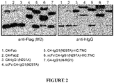


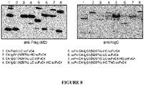

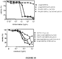

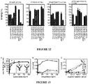


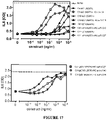

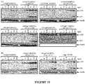


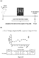


Classifications
-
- C—CHEMISTRY; METALLURGY
- C07—ORGANIC CHEMISTRY
- C07K—PEPTIDES
- C07K16/00—Immunoglobulins [IGs], e.g. monoclonal or polyclonal antibodies
- C07K16/18—Immunoglobulins [IGs], e.g. monoclonal or polyclonal antibodies against material from animals or humans
- C07K16/28—Immunoglobulins [IGs], e.g. monoclonal or polyclonal antibodies against material from animals or humans against receptors, cell surface antigens or cell surface determinants
- C07K16/2878—Immunoglobulins [IGs], e.g. monoclonal or polyclonal antibodies against material from animals or humans against receptors, cell surface antigens or cell surface determinants against the NGF-receptor/TNF-receptor superfamily, e.g. CD27, CD30, CD40, CD95
-
- A—HUMAN NECESSITIES
- A61—MEDICAL OR VETERINARY SCIENCE; HYGIENE
- A61P—SPECIFIC THERAPEUTIC ACTIVITY OF CHEMICAL COMPOUNDS OR MEDICINAL PREPARATIONS
- A61P37/00—Drugs for immunological or allergic disorders
-
- G—PHYSICS
- G01—MEASURING; TESTING
- G01N—INVESTIGATING OR ANALYSING MATERIALS BY DETERMINING THEIR CHEMICAL OR PHYSICAL PROPERTIES
- G01N33/00—Investigating or analysing materials by specific methods not covered by groups G01N1/00 - G01N31/00
- G01N33/48—Biological material, e.g. blood, urine; Haemocytometers
- G01N33/50—Chemical analysis of biological material, e.g. blood, urine; Testing involving biospecific ligand binding methods; Immunological testing
- G01N33/68—Chemical analysis of biological material, e.g. blood, urine; Testing involving biospecific ligand binding methods; Immunological testing involving proteins, peptides or amino acids
- G01N33/6893—Chemical analysis of biological material, e.g. blood, urine; Testing involving biospecific ligand binding methods; Immunological testing involving proteins, peptides or amino acids related to diseases not provided for elsewhere
-
- A—HUMAN NECESSITIES
- A61—MEDICAL OR VETERINARY SCIENCE; HYGIENE
- A61K—PREPARATIONS FOR MEDICAL, DENTAL OR TOILETRY PURPOSES
- A61K39/00—Medicinal preparations containing antigens or antibodies
- A61K2039/505—Medicinal preparations containing antigens or antibodies comprising antibodies
-
- C—CHEMISTRY; METALLURGY
- C07—ORGANIC CHEMISTRY
- C07K—PEPTIDES
- C07K2317/00—Immunoglobulins specific features
- C07K2317/30—Immunoglobulins specific features characterized by aspects of specificity or valency
-
- C—CHEMISTRY; METALLURGY
- C07—ORGANIC CHEMISTRY
- C07K—PEPTIDES
- C07K2317/00—Immunoglobulins specific features
- C07K2317/30—Immunoglobulins specific features characterized by aspects of specificity or valency
- C07K2317/33—Crossreactivity, e.g. for species or epitope, or lack of said crossreactivity
-
- C—CHEMISTRY; METALLURGY
- C07—ORGANIC CHEMISTRY
- C07K—PEPTIDES
- C07K2317/00—Immunoglobulins specific features
- C07K2317/30—Immunoglobulins specific features characterized by aspects of specificity or valency
- C07K2317/35—Valency
-
- C—CHEMISTRY; METALLURGY
- C07—ORGANIC CHEMISTRY
- C07K—PEPTIDES
- C07K2317/00—Immunoglobulins specific features
- C07K2317/50—Immunoglobulins specific features characterized by immunoglobulin fragments
- C07K2317/52—Constant or Fc region; Isotype
-
- C—CHEMISTRY; METALLURGY
- C07—ORGANIC CHEMISTRY
- C07K—PEPTIDES
- C07K2317/00—Immunoglobulins specific features
- C07K2317/50—Immunoglobulins specific features characterized by immunoglobulin fragments
- C07K2317/56—Immunoglobulins specific features characterized by immunoglobulin fragments variable (Fv) region, i.e. VH and/or VL
- C07K2317/569—Single domain, e.g. dAb, sdAb, VHH, VNAR or nanobody®
-
- C—CHEMISTRY; METALLURGY
- C07—ORGANIC CHEMISTRY
- C07K—PEPTIDES
- C07K2317/00—Immunoglobulins specific features
- C07K2317/60—Immunoglobulins specific features characterized by non-natural combinations of immunoglobulin fragments
-
- C—CHEMISTRY; METALLURGY
- C07—ORGANIC CHEMISTRY
- C07K—PEPTIDES
- C07K2317/00—Immunoglobulins specific features
- C07K2317/60—Immunoglobulins specific features characterized by non-natural combinations of immunoglobulin fragments
- C07K2317/62—Immunoglobulins specific features characterized by non-natural combinations of immunoglobulin fragments comprising only variable region components
- C07K2317/622—Single chain antibody (scFv)
-
- C—CHEMISTRY; METALLURGY
- C07—ORGANIC CHEMISTRY
- C07K—PEPTIDES
- C07K2317/00—Immunoglobulins specific features
- C07K2317/70—Immunoglobulins specific features characterized by effect upon binding to a cell or to an antigen
- C07K2317/73—Inducing cell death, e.g. apoptosis, necrosis or inhibition of cell proliferation
-
- C—CHEMISTRY; METALLURGY
- C07—ORGANIC CHEMISTRY
- C07K—PEPTIDES
- C07K2317/00—Immunoglobulins specific features
- C07K2317/70—Immunoglobulins specific features characterized by effect upon binding to a cell or to an antigen
- C07K2317/74—Inducing cell proliferation
-
- C—CHEMISTRY; METALLURGY
- C07—ORGANIC CHEMISTRY
- C07K—PEPTIDES
- C07K2317/00—Immunoglobulins specific features
- C07K2317/70—Immunoglobulins specific features characterized by effect upon binding to a cell or to an antigen
- C07K2317/75—Agonist effect on antigen
-
- C—CHEMISTRY; METALLURGY
- C07—ORGANIC CHEMISTRY
- C07K—PEPTIDES
- C07K2317/00—Immunoglobulins specific features
- C07K2317/90—Immunoglobulins specific features characterized by (pharmaco)kinetic aspects or by stability of the immunoglobulin
- C07K2317/92—Affinity (KD), association rate (Ka), dissociation rate (Kd) or EC50 value
-
- G—PHYSICS
- G01—MEASURING; TESTING
- G01N—INVESTIGATING OR ANALYSING MATERIALS BY DETERMINING THEIR CHEMICAL OR PHYSICAL PROPERTIES
- G01N2800/00—Detection or diagnosis of diseases
- G01N2800/24—Immunology or allergic disorders
-
- G—PHYSICS
- G01—MEASURING; TESTING
- G01N—INVESTIGATING OR ANALYSING MATERIALS BY DETERMINING THEIR CHEMICAL OR PHYSICAL PROPERTIES
- G01N2800/00—Detection or diagnosis of diseases
- G01N2800/24—Immunology or allergic disorders
- G01N2800/245—Transplantation related diseases, e.g. graft versus host disease
Definitions
- the present invention relates to binding molecules specific for TNFR2 (Tumor Necrosis Factor Receptor 2, also known as TNFRSF1B, and CD120B).
- the binding molecules are typically able to bind to TNFR2 and trigger TNFR2 signalling independent from FcyR (Fc gamma receptor) binding.
- the binding molecules may be used to target cells expressing TNFR2.
- the present invention also relates to the use of the binding molecules in treating conditions such as inflammatory disorders, and autoimmune disease.
- TNF ⁇ is a cytokine which plays a wide range of roles in inflammation, differentiation and tissue homeostasis and occurs in soluble and transmembrane forms. It is a key inflammatory mediator which is targeted in the clinic with TNF blocking reagents to suppress unwanted immune responses in inflammatory and autoimmune conditions.
- TNFR1 and TNFR2 are distinct receptors for Tumor Necrosis Factor-alpha (TNF ⁇ ) with signalling through each receptor eliciting different effects. Signalling via TNFR1 triggers strong and manifold pro-inflammatory effects, whilst signalling via TNFR2 has a wider range of effects ranging from immune stimulation, to suppression of immune responses, to tissue regeneration. The two receptors also differ from each other in terms of which cells they are expressed on.
- TNFR1 is ubiquitously expressed, whilst TNFR2 is found on a more restricted group of cells, in particular subsets of immune cells, endothelial cells, and neurons.
- Immune cells expressing TNFR2 include immune suppressive cell types such as T regulatory cells (Tregs), B regulatory cells (B regs), and MDSC (myeloid derived suppressor cells), as well as other cell types, such as CD8+ T cells.
- TNFR1 and TNFR2 have a extracellular domain containing four characteristic cystein rich domains (CRDs), a transmembrane segment, and an intracellular domain.
- CCDs characteristic cystein rich domains
- TNFR2 does not include a cytoplasmic death domain, but can still play a role in apoptosis by controlling cell death regulatory factors such as TRAF2 and cIAPs.
- TNFR2 molecules are able to associate with each other, forming dimeric and trimeric complexes of TNFR2 which can bind TNF ⁇ with high affinity, resulting in the formation of oligomers of trimeric TNF-TNFR2 complexes capable of intracellular signalling.
- TNFR2 is able to bind both soluble and membrane bound TNF ⁇ . Binding of membrane TNF ⁇ is able to activate TNFR2 effectively, but with soluble TNF ⁇ the oligomerisation of liganded TNFR2 trimers to hexamers or higher complexes is inadequate and thus fails to trigger effective TNFR2 activation. As well as TNF ⁇ , lymphotoxin-alpha (LT ⁇ ) is also thought to act as a ligand for TNFR2.
- LT ⁇ lymphotoxin-alpha
- TNFR2 When TNFR2 is activated by membrane TNF ⁇ , the intracellular domains of the receptor are thought to recruit existing cytoplasmic TRAF2 trimers, resulting in recruitment of the E3 ligases cIAP1 and cIAP2 to TNFR2 complexes, which leads to classical NE ⁇ B signalling. TRAF2 recruitment is also associated with activating the alternative NE ⁇ B signalling pathway. Binding of membrane TNF ⁇ to TNFR2 is also thought to trigger signalling through the PI3K/Akt pathway, maintaining survival and enhancing proliferation, as well as promoting other functions such as cellular adhesion and migration. TNFR2 knockout mice are more susceptible to inflammatory disease and, whilst they have Treg cells, they are less responsive. Naturally occurring polymorphisms of TNFR2 also exist and are associated with a wide variety of autoimmune diseases
- Treg cells make it a desirable target for modulating the immune system, especially as TNFR2 is found on a more limited subset of cells than TNFR1.
- the ability to stimulate Treg cells offers a way to control unwanted immune responses in a wide range of conditions where their ability to suppress immune responses may be useful including allergy, autoimmunity, transplantation, graft versus host disease (GVHD) and infectious disease.
- GVHD graft versus host disease
- Treg cells other cell types expressing TNFR2 may be targeted to bring about desired functions. There is therefore an ongoing need for agents that target and activate TNFR2.
- the present invention provides binding molecules which are able to bind the extracellular domain of TNFR2.
- a binding molecule of the present invention is able to bind TNFR2 and trigger signalling through the receptor thus acting as an agonist.
- the binding molecules may act as agonists of TNFR2 independent of FcyR binding.
- the binding molecules of the present invention are antibodies or antibody fragments.
- the binding molecules are selective in that they specifically bind TNFR2, but not TNFR1.
- the binding molecules can have a variety of uses from diagnostic applications to therapeutic uses. As the binding molecules have the ability to modulate the immune system they are particularly useful in treating autoimmune conditions, and inflammatory conditions. The binding molecules may be used to downregulate the immune system due to their action on Treg cells and so are especially useful in treating such conditions, including in particular Graft versus Host Disease (GvHD).
- GvHD Graft versus Host Disease
- the present invention provides a binding molecule that specifically binds TNFR2, but not TNFR1, where the binding molecule is an FcyR independent agonist of TNFR2 and has a valency of at least two for binding TNFR2.
- the present invention also provides:
- the present application includes a sequence listing in electronic form which forms part of the application as filed.
- the present invention provides binding molecules that bind TNFR2 and in particular which bind the extracellular domain of TNFR2.
- TNFR2 comprises a characteristic extracellular domain, a transmembrane segment, and an intracellular domain.
- the extracellular region contains four cysteine rich domains (CRD) that are repeated.
- the TNFR2 bound by a binding molecule of the present invention is typically a mammalian TNFR2.
- the TNFR2 bound by a binding molecule of the present invention is human TNFR2.
- the amino acid sequence of human TNFR2 is provided as SEQ ID NO: 1.
- the human TNFR2 amino acid sequence provided in SEQ ID NO: 1 is subdivided into the signal peptide comprising amino acids 1 to 22; the extracellular domain of amino acids 23 to 257 and the transmembrane region of amino acids 258 to 287.
- a binding molecule of the present invention typically binds the extracellular domain of TNFR2.
- a binding molecule of the present invention may bind human TNFR2, but also TNFR2 of a different species such as a mouse TNFR2 and/or a monkey TNFR2. In one embodiment, it binds human, mouse, and a monkey TNFR2.
- a binding molecule of the present invention will typically be used to bind a target cell, in particular a target cell expressing TNFR2.
- the target cell may be any cell type expressing TNFR2.
- the target cell is an immune cell, endothelial cell, mesenchymal stem cell, or neuron.
- the target cell is an immune cell.
- the target cell is a Treg cell.
- the immune cells expressing TNFR2 are selected from a T regulatory cells (Tregs), B regulatory cells (B regs), and MDSC (myeloid derived suppressor cells).
- the target cell may be a Treg cell which is a Foxp3+ CD25+ regulatory T cell.
- the present invention provides binding molecules that bind TNFR2.
- a binding molecule of the present invention will bind to TNFR2 and stimulate signalling through TNFR2.
- the binding molecules are agonistic antibodies or antibody fusion proteins or antibody fragments or antibody fragment fusion proteins against TNFR2 and so activate the receptor.
- the binding molecules are antibodies that specifically bind TNFR2, preferably they are antibodies that specifically bind TNFR2 and act as an agonist of it without FcgR binding.
- a binding molecule of the present invention will bind TNFR2 to a greater extent than to TNFR1.
- the strength of binding for TNFR2 compared to TNFR1 may be at least 10, 50, 100, 500, 1000 or more times higher. In one embodiment, the strength of binding for TNFR2 compared to TNFR1 may be at least 10,000, or at least100,000 times greater. In one embodiment, the binding molecule does not bind TNFR1 at all. In one preferred embodiment, a binding molecule, and in particular of an antibody or antibody fragment or fusion proteins thereof of the present invention, is said to specifically bind TNFR2 when the KD for the binding has a value below 10 -6 M, 10 -7 M, 10 -8 M, 10 -9 M, or 10 -10 M.
- the KD value is meaured using cells expressing TNFR2, for example HeLa transfectants expressing TNFR2.
- KD values are determined using the approach set out in Kums J, Nelke J, Rüth B,Schfer V, Siegmund D, Wajant H.MAbs. 2017 Apr;9(3):506-520 .
- a fusion of the binding molecule with Gaussia princeps luciferase as a reporter domain is employed to measure the KD value of the binding molecule as described in Kums et al., supra.
- cell lines expressing TNFR2 may be used in such measurement, for example HeLa or HEK293 cells that stably express TNFR2 on their surface.
- the binding molecules of the invention bind to TNFR2 and trigger TNFR2 signalling.
- the binding molecules, and in particular antibodies, provided are agonistic for TNFR2, but are not agonistic for TNFR1.
- a binding molecule of the present invention binds TNFR2, but does not bind any other TNFR super family members.
- the binding molecule of the present invention is an antibody.
- antibody typically refers to any functional antibody or antibody fusion protein or antibody fragment that is capable of specific binding to the antigen of interest, as generally outlined in chapter 7 of Paul, W.E. (Ed.).: Fundamental Immunology 2nd Ed. Raven Press, Ltd., New York 1989 , which is incorporated herein by reference or shown in the "Periodic Table of Antibodies” (https://absoluteantibody.com/periodic-table-of-antibodies/). Reference to an antibody herein includes antibody fragments unless specifically stated. An antibody of the present invention typically specifically binds TNFR2.
- an antibody of the present invention may be, or may comprise, a single chain antibody.
- An antibody of the present invention may also comprise non-antibody sequences such as a ligand based molecule that binds TNFR2.
- an antibody of the present invention may comprise polypeptide(s) which are a fusion of antibody sequences and non-antibody sequences, for example TNC peptide and TNF-alpha polypeptides (particularly TNFR2-specific mutants such as TNF80 (or TNF(143N/145R) as discussed herein.
- An antibody of the invention also includes a portion or fragment of an antibody.
- an antibody of the present invention may comprise just an antibody Fc region modified so that it does not bind Fc receptors and ligand based binding molecules joined to the Fc region.
- the ligand based molecules present in an antibody of the present invention is a TNF-based ligand for TNFR2, e.g. a TNF protomer or a mutant thereof. In one embodiment it is a single-chain version of a TNF trimer.
- An antibody of the present invention also includes antibody formats such as single domain antibodies, for example VHH domain antibodies.
- an antibody of the present invention may be a molecule that comprises such a single domain antibody or single domain antibodies where the overall valency of the binding molecule is at least two.
- an antibody of the present invention may comprise two such single domain antibodies joined together as part of the overall antibody.
- an antibody of the present invention may comprise an Fc region, but not the variable regions that would normally be found in a naturally occurring four chain antibody.
- such Fc regions may though comprise other sequences as part of the polypeptides comprising the heavy chains of the Fc region, in particular TNC peptide and/or TNF-alpha (particularly TNF80) and preferably both, in particular in the C-terminal portion of the polypeptides.
- the "valency" of a binding molecule as used herein denotes the number of binding sites that a binding molecule has, with the valency for TNFR2 denoting the number of binding sites for TNFR2 that a binding molecule has.
- the "specificity" of a binding site denotes what it binds to, for instance it may denote what target molecule the binding molecule binds, but it may also denote where on the target it binds. In respect of an antibody, the specificity of an antibody denotes what target the antibody binds, it may also be used to denote where on the antigen it binds in terms of the epitope bound on that target molecule.
- Binding molecules may have different specificities in the sense that they bind epitopes on different target molecules (antigens). However, binding molecules may have different specificities in the sense that they bind different epitopes on the same target molecule (antigen). For example, a bispecific binding molecule may be bispecific in the sense it can bind two different target molecules, but alternatively it may be bispecific in the sense it can bind two different epitopes of the same target molecule and in particular of TNFR2.
- all of the binding sites of a binding molecule of the present invention will bind the same target.
- the binding molecule of the present invention is an antibody
- it may be that all of the antigen-binding sites of the antibody bind the same epitope of TNFR2.
- a binding molecule of the present invention will comprise at least two different target binding sites that each bind a different target site on TNFR2, for example which each recognise a different epitope of TNFR2.
- a binding molecule of the present invention is an antibody which comprises at least two antigen-binding sites that bind a different epitope of TNFR2.
- a binding molecule of the present invention may include a binding site with a different specificity that recognises a target of a different cell type.
- the different specificity can be used as a homing entity to bring the target the cell expressing TNFR2, together with a different cell type.
- a binding molecule of the present invention may include at least one binding site that binds a target that alters the serum half-life of the binding molecule, for example in one embodiment it may bind serum albumin. In one embodiment, only one of the binding sites of the molecule will target something other than TNFR2.
- a binding molecule of the present invention will have a valency of at least two for TNFR2.
- a binding molecule of the present invention will have a valency of two, three, four, five, six, seven, eight, nine, ten, eleven or twelve, or at least those valencies for TNFR2.
- a binding molecule of the present invention will have a valency of two.
- it will have a valency of three.
- a binding molecule of the present invention is trispecific.
- a binding molecule of the present invention will have a valency of at least four.
- a binding molecule of the present invention is tetravalent.
- a binding molecule of the present invention will have a valency of four. In one embodiment, a binding molecule of the present invention will have a valency of at least six. In a preferred embodiment, it will have a valency of six. In one preferred embodiment, a binding molecule of the present invention will have a valency of twelve. In one embodiment, a binding molecule of the present invention is multi-specific, for instance as it has a valency of at least two, and preferably at least four.
- Figure 1 depicts antibodies with (N-terminal) unidirectional oriented antigen-binding sites, solely in what can be thought of as "North" orientation, for example in one embodiment such binding molecules may be thought of having the antigen-binding sites that bind TNFR2 at what would be considered the N-terminal portion of the heavy and light chains of a conventional four chain antibody and they may be located at the N-terminal portions of the polypeptides of the binding molecules.
- the orientation of the binding sites may also be defined by referring to binding sites in the same orientation as "parallel" binding sites, for example where all of the binding sites are at the N-terminal portions of the antibody chains or all at the C-terminal portion of the antibody chains, the binding sites may be considered as parallel. Such binding sites may also be considered as unidirectional orientated.
- Figure 7 of the present application depicts antibodies with dual N- and C-terminal antigen binding sites in what can be thought of as "North”+”South” orientations or "opposite” orientations. In one embodiment, such binding sites may be thought of as having antigen-binding sites in more than one plane or more than one direction.
- a construct of the invention is one where the antigen binding sites are solely in the "North” orientation, so the antigen-binding sites are N-terminal.
- a binding molecule of the present invention is a North construct with scFvs replacing the variable domains of the HC and LC, for example (scFv-IgG(N297A) and scFv-IgG(N297A)-HC:TNC).
- a binding molecule of the present invention may have binding sites in both the "North” and “South” orientation, so effectively have antigen-binding sites at both the N-terminal and C-terminal locations..
- "North" facing antigen-binding sites may be thought of as in the same orientation or plane as the binding site of a conventional monomeric IgG molecule, in particular what would be normally considered the N-terminal portion of the light and heavy chain variable regions of such a conventional IgG molecule.
- "South" facing antigen binding sites may be thought of those in a direction or a plane that a conventional monomeric IgG molecule does not have antigen binding sites.
- the orientation is the opposite from that of the Fab subdomains (VH+VL) domain in a conventional monomeric IgG molecule thus having the TNFR2 binding domain at the Fc domain C-terminus directing to neighboring cells of the antigen expressing cell.
- an antibody of the present invention comprises antigen binding sites at the N-terminal ends of the polypeptides making up the antibody.
- a binding molecule of the present invention is an antibody in the sense that it has antigen binding sites at both the N-terminus and C-terminus of polypeptide chains of the antibody.
- the binding molecule employed is a multimer of individual subunits.
- the binding molecule of the present invention is a dimer.
- the dimer is form of two scFv-Fab molecules.
- the binding molecule of the present invention is a trimer.
- a binding molecule of the present invention is a hexamer.
- the binding molecule employed may be an antibody.
- the binding molecule employed may be an antibody which is an antibody fusion protein hexamer.
- the heavy chains of the individual IgG molecules making up the hexamers comprise an RGY peptide motif and in particular may do so at their C-terminus.
- an antibody of the present invention comprises the mutations of the RGY motif which are E345R/E430G/S440Y in the heavy chain and in particular in the Fc tail.
- the RGY motif is described, for instance, by de Jong et al (2016) PLOSBiology IDOI:10. 1371/journal.pbio.1002344 which is incorporated by reference in its entirety, as well as specifically in relation to the RGF motif and its use in the formation of hexamers.
- a binding molecule of the invention comprises a multimer or assembly of smaller units it will still be considered a "binding molecule" or “antibody” of the present invention, for instance even though it may comprise several subunits that might individually be considered an antibody, such as an IgG and scFv. So, for instance, an “antibody” of the present invention may comprise an antibody fragment or fragments as part of the overall “antibody” of the present invention. As discussed below, it may comprise other entities such as ligands for TNFR2, such as TNF-alpha and in particular TNF80 and other TNFR2-specific TNF mutants.
- an antibody of the present invention will comprise at least a heavy chain modification at E345 and in particular E345R. In another embodiment, an antibody of the present invention may comprise at least a heavy chain modification at E430 and in particular E430G. In another embodiment, an antibody of the present invention may comprise at least a heavy chain modification at S440 and in particular S440Y. In another embodiment, an antibody of the present invention may comprise at least heavy chain modifications at E345 and E430 and in particular E345R and E430G. In a preferred embodiment, an antibody of the present invention may comprise at least heavy chain modifications at E345, E439, and S440, in particular E345R, E439G and S440Y. In a preferred embodiment, an antibody of the invention will comprise such modifications and comprise a hexamer of IgG units. In a further preferred embodiment, an antibody of the present invention will undergo on-target hexamerisation once it has bound to TNFR2.
- a binding molecule of the present invention may comprise sequences that bring about trimerization. Examples of such sequences include trimerisation domains from collagen, leucine zipper, T4 fibritin, and trimerization domains of TRAF family members.
- a binding molecule of the present invention in some embodiments may comprise such trimerization sequences.
- a binding molecule of the resent invention may comprise, or may be, a trimer due to the presence of such domains.
- the trimerization sequence may be that from tenascin-C (TNC).
- TNC trimerization sequence in general
- TNC any of the specific ones referred to herein and not just TNC, though in a preferred embodiment TNC is used.
- the TNC sequence used in from chicken TNC.
- it is from a mammalian TNC.
- the TNC is human TNC.
- the tenascin-C (TNC) trimerization domain may be used to assemble individual antibody or other subunits into a multimer.
- a binding molecule and in particular an antibody of the present invention, may comprise a TNC trimerization domain peptide such as that in SEQ ID NO: 2. It may comprise a variant TNC peptide, for example one with five, four, three, two, or one amino acid sequence change relative to SEQ ID NO:2, but which is still able to bring about the formation of multimers.
- the C-terminus of the heavy chains of the antibody comprise, or are linked to, the TNC peptide sequence.
- such an approach is used to bring together a trimer of individual TNFR2 binding moieties such as antibody subunits antibodies, and/or ligands.
- Any of the binding molecules set out herein, unless otherwise stated, may comprise a TNC peptide as a way to allow association of individual units and in particular as a way to form multimers.
- a binding molecule of the present invention is an FcyR independent agonist of TNFR2.
- the binding molecule in the case where the binding molecule is an antibody, it may be that the antibody lacks an Fc region and so does not bind FcyR for that region. Alternatively, in another embodiment, it may be that the antibody does have an Fc region, but that it has been modified so that it is unable to bind FcyR. Various Fc modifications are discussed herein that may be used to accomplish that.
- a binding molecule of the present invention will be an antibody selected from an IgG, IgM, IgA, IgE and IgD antibody or comprise such an antibody. It may comprise the Fc modifications discussed herein that render it Fc gamma Receptor independent. In one particularly preferred embodiment, it will be, or comprise, an IgG antibody or comprise such an antibody. In one particularly preferred embodiment it will be an IgG1 antibody or comprise such an antibody. The antibody may be an IgG2, IgG3, or IgG4 antibody or comprise such an antibody. In a particularly preferred embodiment, an antibody of the present invention may comprise single domain antibodies, for example VHH single domain antibodies. In one embodiment, an antibody of the present invention will comprise at least two such VHH single domain antibodies.
- the binding molecule is, or comprises, an antibody.
- an antibody encompasses antibodies in various formats, including antibodies that comprise, or have linked to them, ligand based molecules for binding TNFR2.
- the present invention provides binding molecules that specifically bind TNFR2, but not TNFR1.
- the binding molecule is an FcyR independent agonist of TNFR2.
- the binding molecule has a valency of at least two for binding TNFR2.
- Such a binding molecule of the present invention may be provided in a variety of different formats.
- the binding sites of a binding molecule of the present invention may be selected from antibody antigen-binding sites and ligands that bind to TNFR2.
- a binding molecule of the present invention is an antibody or comprises at least part of an antibody.
- an antibody includes various formats that include antigen-binding sites, but are not in a naturally occurring antibody format.
- An antibody does not necessarily have to be a whole antibody, for example an antibody of the present invention may be, or comprise, an antibody fragment, such as Fab2 antibody fragment.
- Antibodies of the present invention may comprise antibody fragments, for example in one preferred embodiment an antibody of the present invention may comprise an antigen-binding site provided by a scFv fragment or Fab as part of the antibody.
- An antibody of the present invention may be, or comprise, a single chain antibody.
- references to an antibody includes antibody molecules that comprise, are linked to, or conjugated to moieties that do not themselves originate from antibody based sequences. So, for instance, reference to an antibody herein includes an antibody which is linked or conjugated to a TNFR2 ligand or a modified version of a TNFR2 ligand. Examples of TNFR2 ligands that may be employed, or be modified to be employed, include TNF ⁇ and LT ⁇ , as well as variants thereof.
- a binding molecule, and in particular an antibody, of the present invention may comprise sequences that bring about multimerisation.
- an antibody of the present invention may comprise a heavy chain with a sequence that brings about multimerisation, for example by including the RGY mutations that bring about multimerisation.
- it is the Fc tailpiece which comprises the RGY modifications to bring about hexamerisation.
- an antibody of the present invention may comprise a tenascin-C trimerization domain peptide sequence (TNC) at the C-terminus of heavy chains.
- a binding molecule of the present invention particularly an antibody, may comprise a peptide sequence from Tenascin-C which is provided as SEQ ID NO: 2 or a variant TNC peptide which is still able to bring about multimerisation.
- it may comprise the human TNC sequence which brings about trimerization.
- it may comprise a trimerization domain, such as any of the specific ones mentioned herein.
- all of the binding sites of a TNFR2 binding molecule of the present invention may be antibody antigen-binding sites.
- a binding molecule of the present invention comprises at least one antibody antigen-binding site for TNFR2 and at least one ligand, or ligand derivative, that binds TNFR2, in other words it may comprise a mixture of such binding sites.
- a binding molecule of the present invention comprises an antibody which further comprises ligand based binding sites for TNFR2.
- a binding molecule, and in particular an antibody, of the present invention does not comprise ligand based TNFR2 binding sites.
- an antigen binding site will generally comprise six CDRs, three light chain variable region CDRs (LCDR1, LCDR2, and LCDR2) and three heavy chain CDRs (HCDR1, HCDR2, and HCDR3).
- the antigen binding sites of the antibody and the ligand based binding sites are in different orientations, so that the two may be in opposite orientations, for example the antigen-binding sites may be in "North” orientation and the ligand based binding sites in "South” or “opposite” orientation.
- the binding molecule may have antibody antigen-binding sites in both “North” and “South” orientations, but ligand based binding sites in only one orientation, for example solely in "South” orientation.
- Figure 1 of the present application shows illustrative examples of preferred TNFR2 binding molecules, as well as Fab which has a valency of one and so on its own is not a binding molecule of the present invention.
- the structures are indicated with a "CI” or "Clone Identifier” where an parental TNFR2-specific IgG molecule is referred to, with the Clone Identifier indicating from which clone discussed in the Examples the CDRs of the IgG antigen-binding sites originate.
- the structure is then not limited to reference to a specific clone, so is not limited to the CDRs being from a particular clone from the Examples, and the antigen-binding sites may be from any suitable TNFR2 antibody.
- reference to scFv-C4-IgG1(N297A) includes the clone identifier (CI) for Clone 4, but where scFv-IgG1(N297A) is referred to it indicates such a format binding molecule but without the antigen-binding site being limited to sequences from a particular clone.
- a binding molecule of the present invention is selected from the following:
- the binding molecule is one of those set out in (iii) to (vi) above.
- the binding molecule is a tetravalent binding molecule, for example that set out in (iv).
- the binding molecule is one with a valency of 12, for example selected from those set out in (v) and (vi) above.
- an antibody with both N-terminal and C-terminal placed binding sites on an antibody scaffold with parallel oriented chains (Fc, IgG, Fc-TNC, TNC-Fc, IgG-TNC) is employed.
- an antibody with "opposite" orientated binding sites is employed.
- Figure 7 of the present application shows illustrative examples of particularly preferred TNFR2 binding molecules which have binding sites which may be considered as in "opposing" orientations.
- a binding molecule of the present invention is selected from the group set out below:
- the formats discussed above shown in Figure 1 are provided where they are not limited to the sequences originating from a particular clone. In one embodiment, also provided are the specific formats where they do have the sequences from a specific clone provided in the Examples or variant sequences as discussed herein. In one embodiment for the formats with Fc regions, the heavy chains comprise heavy chain modifications as discussed herein, such as the DANA modifications discussed herein, and not just the N297A modification.
- the binding molecule of the present invention is an antibody selected from one of the following formats, with some of the advantages of the formats mentioned in brackets:
- IgG is referred to above IgG1 is employed.
- IgG1 is referred to in a particular format, the same format is also provided where the IgG may be any IgG subtype.
- a binding molecule of the present invention is an antibody that:
- the binding molecule comprises a scFv at the C-terminal portion of heavy chains of the binding molecule, where the scFv binds TNFR2.
- a binding molecule of the present invention comprises scFv at the C-terminal portion of the light chains in the molecule.
- a binding molecule of the present invention is an antibody that just comprises an Fc region which at the C-terminal portion of the heavy chains either has, or is linked to, or is conjugated to, ligands specific for TNFR2.
- each heavy chain has three such ligands present at the C terminal portion of the heavy chains.
- a binding molecule of the present invention is an antibody or Fc region with a C-terminal TNC trimerization followed by a TNFR2-specific TNF mutant, in particular TNF80.
- a polypeptide may comprise a single chain polypeptide comprising three TNF-alpha sequences that can tri-merise, such as three TNF80 units.
- a binding molecule of the present invention is bivalent with spatial movable C- and N-terminal located antigen-binding sites.
- an antibody of the present invention is a bivalent antibody which comprises an Fab specific for TNFR2, where the heavy or light chain of the Fab portion of the antibody comprises an scFv specific for TNFR2.
- the antibody may comprise the scFv joined to the light chain of the Fab, for example with the two being part of the same polypepide.
- the antibody is in the format of Fab-LC:scFv as shown by Figure 7 , but is not limited to sequences from the clones provided in the present Examples and may be any suitable sequences from an antibody for TNFR2.
- the Fab and scFv domains may, for instance, recognize in these embodiments the same or different TNFR2 epitopes.
- the binding molecule, and in particular an antibody, of the present invention is tetravalent.
- the binding molecule of the present invention is in the format Fab-HC-scFv, where the antibody comprises an Fc region modification eliminating or reducing Fc receptor binding, and where the antibody comprises a scFv at the C-terminal end of each heavy chain.
- the antibody is in the format Fab-HC-scFv where the antibody is bispecific in the sense that it has two different specificities for TNFR2.
- the scFvs present bind a different epitope than the antigen-binding sites of the IgG.
- an antibody of the present invention is in the format scFv:IgG, where the light and heavy chains of the antibody have been modified to include an scFv at the N-terminal portion, with the Fc region of the antibody modified so that it does not bind Fc receptors. Any of the modifications discussed herein that reduce or eliminate Fc binding may be employed.
- Such an antibody may be bispecific in the sense that the scFvs expressed on the heavy chain polypeptide may bind one epitope of TNFR2 and those of the light chains another specificity of TNFR2.
- all of the scFv regions may bind the same epitope of TNFR2.
- a binding molecule of the present invention is hexavalent.
- the antibody is in the format IgG-LC:scFv-HC:scFv and also has a modified Fc region so that it does not bind Fc receptors, for example having any of the Fc modifications discussed herein that eliminate or reduce binding to Fc ⁇ receptors.
- the IgG is IgG1.
- the antibody is hexavalent, where it comprises an antibody Fc region, with each of the heavy chains linked to a trimer of TNF- ⁇ , or a variant thereof, at the C-terminal portion.
- a binding molecule of the invention is 12-valent.
- a 12-valent binding molecule is a multimer of three individual antibody subunits where the heavy and light chain variable regions of the antibody are replaced with scFv molecules.
- the three antibody subunits are associated as the heavy chains for each antibody comprise a tenascin-C peptide at the C-terminus comprising the trimerization domain of TNC.
- the binding molecule will not bind Fc receptors and in a preferred embodiment comprises an Fc region with any of the modifications set out herein.
- a binding molecule of the present invention may be a hexabody comprising six IgG molecules.
- the binding molecule may be a hexabody of six IgG antibodies, where the Fc regions comprise RGY HexaBody mutations and also a modification that means they do not bind Fc receptors, such as any of the specific modifications set out herein.
- the IgG is IgG1.
- a binding molecule of the present invention is four or 12 valent for TNFR2.
- a binding molecule of the present invention is an antibody selected from the format scFv-IgG(*)-HC-TNC and scFv-IgG(*), where * indicates an Fc region modification which eliminates or reduces Fc receptor binding.
- the IgG portion of the binding molecule is IgG1.
- HC-TNC indicates that the heavy chains of the IgG comprise a tenascin peptide, for example that of SEQ ID NO:2 or a variant thereof.
- a binding molecule of the present invention is an antibody selected from the format scFv-IgG(*)-HC-TNC, scFv-IgG(*), and IgG(*)-HC:RGY.
- the IgG portion of the molecule is an IgG1 antibody.
- a binding molecule of the present invention will be able to bring about the formation of hexamers of TNFR2 molecules on the surface of the cell or at least hexamers.
- a binding molecule of the present invention may bind TNFR2 receptors in cis, that is bind a plurality of TNFR2 receptors on the surface of the same cell.
- the binding molecule is able to at least bring about the formation of hexamers of TNFR2 on the surface of the same cell.
- a binding molecule of the present invention is able to bind TNFR2 molecules in trans, that is the binding molecule binds at least one TNFR2 molecule on the surface of one cell and at least one TNFR2 molecule on the surface of another cell.
- the binding molecule is able to bind cis and trans to TNFR2.
- it may have the capability to do so and in another embodiment it may actually do so.
- a binding molecule of the present invention results in the formation of hexamers of TNFR2 on both cells when trans binding occurs.
- the orientation of the binding sites may influence whether an antibody binds to TNFR2 in cis and/or trans fashion.
- a binding molecule of the present invention is one that has at least two TNFR2 binding sites that have opposing orientation compared to each other preferably wherein the binding molecule has at least two such TNFR2 binding sites that are antibody antigen-binding sites.
- a binding molecule of the present invention is one that has at least two TNFR2 binding sites that are able to bind TNFR2 molecules on different cells allowing for trans-binding, preferably wherein the binding molecule has at least two such TNFR2 binding sites that are antibody antigen-binding sites.
- a binding molecule of the present invention has a valency of at least four and is able to bind a plurality of different molecules of TNFR2 on the surface of the same cell, preferably where the binding molecule has a valency of at least six.
- a binding molecule of the present invention may be a diabody.
- a diabody is typically a small bivalent antigen-binding antibody portion, and there are a variety of diabody formats, any of which may be used, for example a diabody may comprise a heavy chain variable domain linked to a light chain variable domain on the same polypeptide chain linked by a peptide linker that is too short to allow pairing between the two domains on the same chain. This results in pairing with the complementary domains of another chain and in the assembly of a dimeric molecule with two antigen binding sites for TNFR2.
- a binding molecule of the present invention has a valency of at least two.
- a binding molecule of the present invention has a valency of at least two spatially movable potentially cell-cell connecting TNFR2 binding sites or at least six "parallel” or “unidirectionally” oriented cis-acting binding sites.
- formats mentioned herein may be employed as part of an antibody of the present invention in such formats, rather than as the whole antibody.
- a binding molecule of the present may comprise VHH fragments. Organisms such as Camelids, sharks, and other cartilaginous fish produce heavy chain-only antibodies. The single-domain variable fragments of these heavy chain-only antibodies are termed VHHs or nanobodies or sdAb.
- VHHs retain the immunoglobulin fold shared by antibodies, using three hypervariable loops, CDR1, CDR2 and CDR3, to bind to their targets.
- a VHH fragment e.g., NANOBODY ®
- NANOBODY ® is a recombinant, antigen-specific, single-domain, variable fragment derived from camelid heavy chain antibodies.
- a binding molecule of the present invention preferably has a valency of at least two for TNFR2, in one embodiment where the binding molecule comprises VHH fragments it will comprise at least two such VHH fragments.
- VHH fragments may be joined together, for example through the use of a linker and hence, in one embodiment, may be expressed as a single polypeptide which overall comprises at least two VHH fragments which are each able to specifically bind TNFR2.
- VHH fragments do not have a Fc region and in one embodiment where VHH fragments are present as part of a binding molecule of the present invention, the binding molecule will lack an Fc region and so not bind Fc receptors for that reason.
- a binding molecule of the present invention will comprise, or at least comprise, three, four, five, six or more VHH fragments specific for TNFR2.
- a binding molecule of the present invention will comprise a single VHH domain antibody.
- a binding molecule of the present invention may comprise a variant binding site for TNFR2.
- a variant binding molecule for TNFR2 will be defined by its ability to cross-block one of the specific binding molecules for TNFR2 set out herein binding to TNFR2.
- the ability to cross-block may be assessed using a binding assay for measuring binding of a binding molecule to TNFR2 and the ability of a further binding molecule to cross-block such binding to TNFR2.
- the ability to cross-block is studied using a monovalent molecule that just comprises one copy of the binding site for TNFR2.
- a binding molecule of the present invention will be able to block, or reduce, the binding of TNF ⁇ to TNFR2.
- a binding molecule of the present invention will be able to reduce, or block the binding of lymphotoxin-alpha to TNFR2
- the affinity of the binding domain for TNFR2 in a binding molecule, in particular an antibody, of the present invention has a K D which is about 400 nM or smaller, 200 nM or smaller such as about 100 nM, 50 nM, 20 nM, 10 nM, 1 nM, 500 pM, 250 pM, 200 pM, 100 pM or smaller.
- the binding affinity is 50 pM or smaller.
- an individual TNFR2 binding site in a binding molecule of the present invention will show such affinity for TNFR2, rather than that being the overall affinity for the binding molecule.
- such affinity is the overall avidity of the antibody for TNFR2.
- the affinity of an individual antigen-binding site of a binding molecule of the present invention may be less than 1 ⁇ M, less than 750 nM, less than 500 nM, less than 250 nM, less than 200 nM, less than 150 nM, less than 100 nM, less than 75 nM, less than 50 nM, less than 10 nM, less than 1 nM, less than 0.1 nM, less than 10 pM, less than 1 pM, or less than 0.1 pM.
- the Kd is from about 0.1 pM to about 1 ⁇ M.
- an antibody of the present invention is a monoclonal antibody.
- an antibody of the present invention is an IgG1, IgG2 or IgG4 antibody or comprises heavy chain constant region of that class of antibody.
- an antibody of the present invention is a IgG1 isotype antibody or comprises such an antibody.
- the term "antibody” encompasses antibodies from any appropriate source species, including chicken, sharks and mammalian such as mouse, hamster, rabbit, goat, cow, llama, non-human primate and human.
- an antibody of the present invention is a humanised antibody.
- the binding molecule, and in particular antibody, of the present invention is wholly human.
- an antibody of the present invention is a human antibody.
- a binding molecule of the present invention and in particular an antibody, may comprise non-human sequences such as a tenascin-c peptide, but in one preferred embodiment all of the antibody based sequences of the antibody are human notwithstanding the presence of the tenascin peptide sequence.
- antibody encompasses monomeric antibodies (such as IgD, IgE, IgG) or oligomeric antibodies (such as IgA or IgM).
- antibody also encompasses - without particular limitations - isolated antibodies and modified antibodies such as genetically engineered antibodies, e.g. chimeric or humanized antibodies.
- a binding molecule of the present invention has only one specificity for TNFR2. Hence, in one preferred embodiment all of the antigen-binding sites of an antibody of the present invention which are specific for TNFR2 will be specific for the same epitope of TNFR2. In another embodiment, a binding molecule of the present invention will comprise more than one specificity for TNFR2. For example, that may be because the binding molecule that comprises both antibody antigen-binding sites, but also ligand based TNFR2 binding sites. In one preferred embodiment though, a binding molecule of the present invention is an antibody that comprises antigen binding sites that recognise more than one epitope of TNFR2.
- a binding molecule of the present invention is bispecific.
- a binding molecule of the present invention one specificity of the binding molecule for TNFR2 is provided by a pair of light and heavy chain variable regions whilst the other is provided by scFv fragments present as part of the overall binding molecule.
- all of the TNFR2 binding sites of a binding molecule, and in particular antibody are provided by scFvs with the binding molecule formed from two different polypeptides, each of which comprise a scFv with a different specificity.
- a binding molecule of the present invention may comprise an Fc region or an antibody molecule which has irrelevant specificity and the ability to bind to TNFR2 is provided by other portions of the binding molecule.
- a binding molecule of the present invention comprises an Fc region where at the C-terminal portion of the heavy chains are TNF-alpha molecules and in particular mutant TNF-alpha molecules that are selective for TNFR2, such as the TNF80 mutant.
- a binding molecule of the present invention may comprise an immunoglobulin of irrelevant specificity and at the C-terminal portion of the heavy chains may be TNF-alpha molecules, and in particular mutant TNF-alpha molecules such as TNF80.
- the heavy chains of the Fc or irrelevant antibody may comprise as part of the same polypeptide a TNF-alpha and in particular a TNF80 mutant. In one embodiment, they may comprise a single chain polyptide comprising three TNF-alpha polypeptides and in particular three TNF80 polypeptides. In another embodiment, before the TNF-alpha portion of the polypeptides the heavy chains may comprise a TNC peptide or variant thereof. In a further preferred embodiment, a binding molecule of the present invention may comprise an Fc region that, rather than have variable regions has the two heavy chain portions of the Fc region as part of a single polypeptide.
- a preferred binding molecule comprising a TNF ligand is Fc(DANA)-TNC-TNF80 as depicted in Figure 24 where DANA indicates the heavy chains of the Fc regions comprising D265A and N297A mutantions and TNC indicates the tenascin trimerization domain peptide sequence.
- a preferred binding molecule of the present invention comprises three Fc regions where each heavy chain has the DANA modifications of D265A and N297A.
- each of those heavy chain polypeptides comprises a TNC peptide.
- each heavy chain polypeptide of each Fc region comprises a TNC peptide sequence followed by a TNF-alpha sequence and in particular a TNF80 sequence.
- a further preferred binding molecule of the present invention is Fc(DANA)-TNF80 which comprises three Fc regions where each heavy chain polypeptide comprises the DANA modifications of D265A and N297A and a TNF-alpha, in particular a TNF80 sequence.
- the present invention also provides as binding molecules the Fc-TNC-TNF80 and Fc-TNF80 format molecules shown in Figure 24 .
- a binding molecule that specifically binds TNFR2, but not TNFR1, where the binding molecule is an FcyR independent agonist of TNFR2, has a valency of at least two for binding TNFR2, and comprises polypeptides comprising mutant TNF-alpha that bind TNFR2, but not TNFR1, and a tenascin peptide sequence that oligomerises the polypeptides.
- such a binding molecule does not include antibody sequences.
- the molecule comprises polypeptides that each comprise a mutant TNF-alpha sequence with a tenascin peptide, for example any of the tenascin polypeptides disclosed herein.
- the tenascin peptide sequences results in oligomerisation of such polypeptides.
- the mutant TNF-alpha has 143N/145R modifications resulting in selective binding for TNFR2 over TNFR1.
- TNF80 Such a TNF mutant may also be referred to as TNF80.
- a binding molecule of the present invention does not bind Fc receptors and in particular does not bind to FcyR receptors.
- the binding molecule of the present invention is an antibody and it does not bind to Fc rreceptors, either because it does not comprise an Fc region or alternatively as it is has an Fc region modified so that it does not bind Fc receptors.
- Fc domain as employed herein generally refers to -(CH 2 CH 3 ) 2 , unless the context clearly indicates otherwise, where CH2 is the heavy chain CH2 domain, CH3 is the heavy chain CH3 domain, and there are two CH 2 CH 3 with one from each heavy chain.
- an antibody of the present invention does not comprise a -CH2CH3 fragment.
- an antibody of the present invention does not comprise a CH 2 domain.
- an antibody of the present invention does not comprise a CH 3 domain.
- a binding molecule of the present invention binds to an Fc gamma receptor but to a substantially decreased extent relative to binding of an identical antibody comprising an unmodified Fc region to the FcgR (e.g., a decrease in binding to a FcyR by at least 70%, at least 80%, at least 90%, at least 95%, at least 98%, at least 99%, or 100% relative to binding of the identical antibody comprising an unmodified Fc region to the FcyR as measured).
- the binding molecule has no detectable binding to an FcyR at all.
- Binding can be determined using a variety of techniques known in the art, for example but not limited to, equilibrium methods (e.g., enzyme-linked immunoabsorbent assay (ELISA); KinExA, Rathanaswami et al. Analytical Biochemistry, Vol. 373:52-60, 2008 ; or radioimmunoassay (RIA)), or by a surface plasmon resonance assay or other mechanism of kinetics-based assay (e.g., BIACORE TM analysis or Octet TM analysis (forteBIO)), and other methods such as indirect binding assays, competitive binding assays fluorescence resonance energy transfer (FRET), gel electrophoresis and chromatography (e.g. gel filtration).
- equilibrium methods e.g., enzyme-linked immunoabsorbent assay (ELISA); KinExA, Rathanaswami et al. Analytical Biochemistry, Vol. 373:52-60, 2008 ; or radioimmunoassay (RIA)
- the Fc region employed is mutated, in particular comprising a mutation described herein.
- the mutation is to remove binding to Fc receptors and in particular FcyR.
- the antibody of the invention has been mutated so that it does not bind Fc receptors.
- an antibody of the present invention may comprise an aglycosyl IgG, for example to bring about reduced Fc function and in particular a nearly Fc-null phenotype.
- an antibody of the invention has a modification at N297 and in particular N297A.
- an antibody of the invention has modifications at F243 and/or F244, in particular ones that mean that the antibody comprises an aglycosyl IgG.
- an antibody of the present invention may comprise the F243A and/or F244A heavy chain modifications.
- one or more of F241, F243, V262 and V264 may be modified and particularly to amino acids that influence glycosylation.
- an antibody of the present invention may have modifications at F241A, F243A, V262E and V264E. Such modifications are discussed in Yu et al (2013) 135(26): 9723-9732, which is incorporated by reference in its entirety, particularly in relation to the modifications discussed therein. Such modifications provide a way to modulate, for example, Fc receptor binding.
- a modification which influences the glycosylation of the antibody may be present.
- an antibody of the invention may be produced in a cell type that influences glycosylation as a further approach for sugar engineering.
- the fucosylation, sialylation, galactosylation, and/or mannosylation of an antibody of the present invention may be altered either by sequence modifications and/or via the type of cell used to produce the antibody.
- an antibody of the present invention has modifications at position 297 and/or 299.
- an antibody of the present invention comprises a N297A modification in its heavy chains, preferably N297Q or mutation of Ser or Thr at 299 to other residues. In one embodiment it has both those modifications.
- a binding molecule of the present invention comprises two different heavy chains where the heavy chains comprise modifications that allow the different heavy chains to preferentially associate compared to heavy chains associating with identical heavy chains.
- the two different heavy chains comprise knob-in-hole mutations.
- the knob-in hole mutations are a T366W mutation in one heavy chain constant region and a T366S, L368A, and a Y407V mutation in the other domain.
- the modifications comprise charge-pair mutations.
- the charge-pair mutations are a T366K mutation in one of the heavy chain constant regions and a corresponding L351D mutation in the other domain.
- the heavy chains comprise modifications that mean a heterodimer comprising the two heavy chains can be purified preferentially from the homodimers only comprising one type of heavy chain.
- the modifications may alter affinity for Protein A, with one heavy chain still able to bind Protein A, whilst the modified heavy chain does not do so, meaning that heterodimers of the two different heavy chains can be purified based on their affinity for Protein A.
- heavy and light chains may comprise modifications that change whether or not a disulphide bridge is formed between them.
- the modifications comprise mutations that generate engineered disulfide bridges between light and heavy chains.
- engineered disulfide bridges are mutations that provide non-endogenous cysteine amino acids in two or more polypeptides such that a non-native disulfide bond forms when the two or more domains associate. Engineered disulfide bridges are described in greater detail in Merchant et al. (Nature Biotech (1998) 16:677-681 ), the entirety of which is hereby incorporated by reference.
- the mutations that generate engineered disulfide bridges are a K392C mutation in one of a first or second CH3 domains, and a D399C in the other CH3 domain.
- the mutations that generate engineered disulfide bridges are a S354C mutation in one of a first or second CH3 domains, and a Y349C in the other CH3 domain.
- the mutations that generate engineered disulfide bridges are a 447C mutation in both the first and second CH3 domains that are provided by extension of the C-terminus of a CH3 domain incorporating a KSC tripeptide sequence.
- binding molecules of the present invention may comprise modifications that alter serum half-life of the binding molecule.
- an antibody of the present invention has Fc region modification(s) that alter the half-life of the antibody. Such modifications may be present as well as those that alter Fc functions.
- an antibody of the present invention has modification(s) that alter the serum half-life of the antibody.
- an antibody of the present invention has modification(s) that alter serum half-life of the antibody compared to an antibody lacking such modifications.
- the modifications result in increased serum half-life.
- they result in decreased serum half-life.
- an antibody of the present invention comprises one or more modifications that collectively both silence the Fc region and decrease the serum half-life of the antibody compared to an antibody lacking such modifications.
- the binding molecules, in particular antibodies, of the present invention are mutated to provide improved affinity for a target antigen or antigens and in particular for TNFR2.
- affinity maturation protocols including mutating the CDRs ( Yang et al., J. Mol. Biol., 254, 392-403, 1995 ), chain shuffling ( Marks et al., Bio/Technology, 10, 779-783, 1992 ), use of mutator strains of E. coli ( Low et al J. Mol. Biol., 250, 359-368, 1996 ), DNA shuffling ( Patten et al Curr. Opin.
- Binding domains for use in the present invention may be generated by any suitable method known in the art, for example CDRs may be taken from non-human antibodies including commercially available antibodies and grafted into human frameworks or alternatively chimeric antibodies can be prepared with non-human variable regions and human constant regions etc.
- antibodies of the present invention may not be in a naturally occurring format, it may be that such screening is used to identify antibody antigen-binding sites with desirable properties for binding TNFR2 and then they are reformatted so that they are in one of the formats set out herein.
- Antigen polypeptides for use in generating antibodies for example for use to immunize a host or for use in panning, such as in phage display, may be prepared by processes well known in the art from genetically engineered host cells comprising expression systems or they may be recovered from natural biological sources.
- polypeptides includes peptides, polypeptides and proteins. These are used interchangeably unless otherwise specified.
- the antigen polypeptide may in some instances be part of a larger protein such as a fusion protein for example fused to an affinity tag or similar.
- the host may be immunised with a cell transfected with TNFR2, for instance expressing TNFR2 on its surface.
- Antibodies generated against an antigen polypeptide may be obtained, where immunisation of an animal is necessary, by administering the polypeptides to an animal, preferably a non-human animal, using well-known and routine protocols, see for example Handbook of Experimental Immunology, D. M. Weir (ed.), Vol 4, Blackwell Scientific Publishers, Oxford, England, 1986 ). Many warm-blooded animals, such as rabbits, mice, rats, sheep, cows, camels or pigs may be immunized. However, mice, rabbits, pigs and rats are generally most suitable.
- Monoclonal antibodies may be prepared by any method known in the art such as the hybridoma technique ( Kohler & Milstein, 1975, Nature, 256:495-497 ), the trioma technique, the human B-cell hybridoma technique ( Kozbor et al 1983, Immunology Today, 4:72 ) and the EBV-hybridoma technique ( Cole et al Monoclonal Antibodies and Cancer Therapy, pp77-96, Alan R Liss, Inc., 1985 ).
- Antibodies may also be generated using single lymphocyte antibody methods by cloning and expressing immunoglobulin variable region cDNAs generated from single lymphocytes selected for the production of specific antibodies by, for example, the methods described by Babcook, J. et al 1996, Proc. Natl. Acad. Sci. USA 93(15):7843-78481 ; WO92/02551 ; WO2004/051268 and WO2004/106377 .
- the antibodies for use in the present invention can also be generated using various phage display methods known in the art and include those disclosed by Brinkman et al. (in J. Immunol. Methods, 1995, 182: 41-50 ), Ames et al. (J. Immunol.
- an antibody of the present invention is fully human, in particular one or more of the variable domains are fully human.
- Fully human molecules are those in which the variable regions and the constant regions (where present) of both the heavy and the light chains are all of human origin, or substantially identical to sequences of human origin, not necessarily from the same antibody.
- Examples of fully human antibodies may include antibodies produced, for example by the phage display methods described above and antibodies produced by mice in which the murine immunoglobulin variable and optionally the constant region genes have been replaced by their human counterparts e.g.
- EP0546073 As described in general terms in EP0546073 , US5,545,806 , US5,569,825 , US5,625,126 , US5,633,425 , US5,661,016 , US5,770,429 , EP 0438474 and EP0463151 which are each incorporated by reference.
- the antigen-binding sites, and in particular the variable regions, of the antibodies according to the invention are humanised.
- Humanised which include CDR-grafted antibodies
- CDRs complementarity determining regions
- framework region from a human immunoglobulin molecule
- Humanised antibodies may optionally further comprise one or more framework residues derived from the non-human species from which the CDRs were derived.
- the term "humanised antibody molecule” refers to an antibody molecule wherein the heavy and/or light chain contains one or more CDRs (including, if desired, one or more modified CDRs) from a donor antibody (e.g. a murine monoclonal antibody) grafted into a heavy and/or light chain variable region framework of an acceptor antibody (e.g. a human antibody).
- a donor antibody e.g. a murine monoclonal antibody
- only one or more of the specificity determining residues from any one of the CDRs described herein above are transferred to the human antibody framework (see for example, Kashmiri et al., 2005, Methods, 36, 25-34 ).
- only the specificity determining residues from one or more of the CDRs described herein above are transferred to the human antibody framework.
- only the specificity determining residues from each of the CDRs described herein above are transferred to the human antibody framework.
- any appropriate acceptor variable region framework sequence may be used having regard to the class/type of the donor antibody from which the CDRs are derived, including mouse, primate and human framework regions.
- the humanised antibody according to the present invention has a variable domain comprising human acceptor framework regions as well as one or more of the CDRs provided herein.
- human frameworks which can be used in the present invention are KOL, NEWM, REI, EU, TUR, TEI, LAY and POM (Kabat et al supra).
- KOL and NEWM can be used for the heavy chain
- REI can be used for the light chain and EU
- LAY and POM can be used for both the heavy chain and the light chain.
- human germline sequences may be used; these are available at: http://www2.mrc-lmb.cam.ac.uk/vbase/list2.php.
- the acceptor heavy and light chains do not necessarily need to be derived from the same antibody and may, if desired, comprise composite chains having framework regions derived from different chains.
- the framework regions need not have exactly the same sequence as those of the acceptor antibody. For instance, unusual residues may be changed to more frequently-occurring residues for that acceptor chain class or type. Alternatively, selected residues in the acceptor framework regions may be changed so that they correspond to the residue found at the same position in the donor antibody (see Reichmann et al 1998, Nature, 332, 323-324 ). Such changes should be kept to the minimum necessary to recover the affinity of the donor antibody.
- a protocol for selecting residues in the acceptor framework regions which may need to be changed is set forth in WO 91/09967 .
- Derivatives of frameworks may have 1, 2, 3, 4, 5, 6, 7, 8, 9, 10 or more amino acids replaced with an alternative amino acid, for example with a donor residue.
- Donor residues are residues from the donor antibody, i.e. the antibody from which the CDRs were originally derived, in particular the residue in a corresponding location from the donor sequence is adopted.
- Donor residues may be replaced by a suitable residue derived from a human receptor framework (acceptor residues).
- the residues in antibody variable domains are conventionally numbered according to a system devised by Kabat et al. This system is set forth in Kabat el al., 1987, in Sequences of Proteins of Immunological Interest, US Department of Health and Human Services, NIH, USA (hereafter "Kabat et al. ( supra )”). This numbering system is used in the present specification except where otherwise indicated.
- the Kabat residue designations do not always correspond directly with the linear numbering of the amino acid residues.
- the actual linear amino acid sequence may contain fewer or additional amino acids than in the strict Kabat numbering corresponding to a shortening of, or insertion into, a structural component, whether framework or complementarity determining region (CDR), of the basic variable domain structure.
- the correct Kabat numbering of residues may be determined for a given antibody by alignment of residues of homology in the sequence of the antibody with a "standard" Kabat numbered sequence.
- the CDRs of the heavy chain variable domain are located at residues 31-35 (CDR-H1), residues 50-65 (CDR-H2) and residues 95-102 (CDR-H3) according to the Kabat numbering system.
- CDR-H1 Chothia, C. and Lesk, A.M. J. Mol. Biol., 196, 901-917 (1987 )
- the loop equivalent to CDR-H1 extends from residue 26 to residue 32.
- CDR-H1' as employed herein is intended to refer to residues 26 to 35, as described by a combination of the Kabat numbering system and Chothia's topological loop definition.
- the CDRs of the light chain variable domain are located at residues 24-34 (CDR-L1), residues 50-56 (CDR-L2) and residues 89-97 (CDR-L3) according to the Kabat numbering system.
- the invention extends to an antibody sequence disclosed herein. In another it extends to humanized versions of such antibodies.
- variant binding molecules may be identified by identifying such binding molecules that are able to cross-block specific binding molecules set out herein.
- Cross-blocking antibodies can be identified using any suitable method in the art, for example by using competition ELISA or BIAcore assays where binding of the cross blocking antibody to antigen (TNFR2) prevents the binding of an antibody of the present invention or vice versa.
- Such cross blocking assays may use cells expressing TNFR2 as a target.
- flow cytometry is used to assess binding to cells expressing TNFR2.
- Variant binding molecules may be employed where they still retain the desired properties of binding molecules of the present invention.
- binding molecules and in particular antibodies with degrees of sequence identity to specific ones set out herein are also provided.
- the sequence identity may be over the entire length of a polypeptide or it may be in relation to specific portions such as just over the variable regions or just over the CDR sequences. Degrees of identity and similarity can be readily calculated ( Computational Molecular Biology, Lesk, A.M., ed., Oxford University Press, New York, 1988 ; Biocomputing.
- the present invention also extends to novel polypeptide sequences disclosed herein and sequences at least 80% similar or identical thereto, for example 85% or greater, such 90% or greater, in particular 95%, 96%, 97%, 98% or 99% or greater similarity or identity.
- a sequence may have at least 99% sequence identity to at least one of the specific sequences provided herein.
- Identity indicates that at any particular position in the aligned sequences, the amino acid residue is identical between the sequences.
- similarity indicates that, at any particular position in the aligned sequences, the amino acid residue is of a similar type between the sequences.
- leucine may be substituted for isoleucine or valine.
- Other amino acids which can often be substituted for one another include but are not limited to:
- a variant may have from one to ten, such as one, two, three, four, five or up to those values of amino acid sequence changes or at least those values, or up to those values, so long as the variant is still able to specifically bind TNFR2 and preferably act as an agonist of it in the formats described previously.
- an antibody of the present invention may have at least five, six, seven, eight, nine, ten, eleven or twelve amino acid sequence changes compared to the CDRs of one of the specific antibodies set out herein, for example is may have that number of sequence changes in the six CDRs making up the antigen-binding site.
- An antibody of the present invention may have that number of sequence changes in the CDRs compared to the specific antibody set out herein. It may have up to that number of sequence changes. It may have at least that number of amino acid sequence changes.
- Such variant antibody molecules will typically retain the ability to specifically bind TNFR2. They may also retain out of the other functions set out herein.
- this aspect of the invention also extends to variants of the specific binding molecules, and in particular antibodies, including humanised versions and modified versions, including those in which amino acids have been mutated in the CDRs to remove one or more isomerisation, deamidation, glycosylation site or cysteine residue as described herein above.
- a binding molecule, and in particular an antibody, of the present invention may comprise specific sequences from those illustrative antibodies, or variants of them.
- Variant antibodies are also provided, including variants as defined here, particularly those that specifically bind TNFR2, but not TNFR1, in an FcyR independent manner.
- Table 3 of the present invention sets out the various pairs of expression constructs that were used to express a particular antibody.
- the Table also sets the polypeptides that are expressed by the pairs of vectors.
- the present invention provides a binding molecule that comprises one of the pairwise combinations of polypeptides set out by Table 3.
- the original antibody clones used to generate antibodies in the Examples are mouse antibodies.
- the present invention provides a binding molecule comprising one of the pairwise combinations of polypeptides set out in Table 3 where the mouse CDR sequences are retained but the rest of the antibody has been humanised.
- a binding molecule of the present invention comprises one of the pairwise combinations of polypeptides set out in the Table except the whole of the mouse sequences have been swapped for those of a human antibody that is able to specifically bind TNFR2.
- a binding molecule that cross-blocks the binding molecule that results from the pairwise combinations of polypeptides set out in Table 3 is employed.
- a binding molecule that cross-reacts with a binding molecule that results from the pairwise combinations of polypeptides set out in Table 3 is employed.
- a binding molecule of the present invention will comprise an antigen-binding site comprising a set of six CDRs from those of the pairwise combinations of heavy and light chains set out in Table 3 of the present application.
- a binding molecule and in particular an antibody of the present invention will comprise a pair of light and heavy chain variable regions pair from those that are set out in Table 3.
- a binding molecule of the present invention comprises at least one of those polypeptides.
- a binding molecule of the present invention comprises at least one of those polypeptides, where the polypeptide has been humanised, with the mouse CDR sequences retained, but the rest of the mouse sequences replaced by human ones.
- the binding molecule may comprise two of those polypeptides, or humanized versions where all but the mouse CDRs have been replaced by the corresponding sequences.
- a binding molecule of the present invention will comprise an antigen-binding site comprising a set of six CDRs from those of the pairwise combinations of heavy and light chains set out in Table 4 of the present application.
- a binding molecule and in particular an antibody of the present invention will comprise a pair of light and heavy chain variable regions pair from those that are set out in Table 4.
- Table 5 of the present application sets out the CDR sequences from the specific antibodies used as a starting point for the antibody formats set out in the present application.
- the three CDRs from a light chain are set out grouped together, as are the three CDRs from the heavy chains, with the CDRs from a light and heavy chains also grouped together.
- a binding molecule of the present invention will comprise a set of three light chain CDRs as set out in Table 5.
- it will comprise a set of three heavy chain CDRs as set out in Table 6, so both the light and heavy chain CDR sequences from one of the clones or functional variants of such sequences.
- a binding molecule of the present invention comprises the CDRs for more than one specificity, so the binding molecule may, for instance, comprise two polypeptides that collectively comprise the six CDRs from one of the antibodies as set out in Table 5, but the binding molecule may comprise a further set of six CDRs for another of the antibodies, for example present as a scFv which is part of the binding molecule.
- a binding molecule of the present invention is triparatopic for TNFR2 and so has three different specificities for TNFR2.
- at least one of the specificities is conferred by a set of six CDRs from Table 5 or variants of those CDRs.
- At least two of the specificities are conferred by sets of six CDRs from Table or variants thereof. In a further embodiment, all three of the specificities are.
- a binding molecule of the present invention may be tetraparatopic, for example with at least one, two, or three of the specifities being conferred by sets of six CDR set out in Table 5 or variants thereof. In one embodiment all four specificities may be conferred by sets of six CDRs from Table 5 or variants thereof.
- a binding molecule of the present invention comprises an antigen binding site comprising a set of six CDRs from the C40 clone.
- the binding molecule comprises an antigen binding site comprising a set of six CDRs that are from the C19 clone.
- an antibody has both an antigen binding site comprising a set of six CDRs from the C40 clone and also a further antigen-binding site comprising a set of six CDRs from the C19 clone.
- the CDRs are from the C4 clone.
- all of the antigen-binding sites of an antibody of the present invention comprise the same set of six CDRs.
- different antigen-binding sites of the antibody comprise a different set of six CDRs.
- a variant set of six CDRs may be employed, such as any of the variants discussed herein.
- any of the antibody formats discussed herein may be provided where the six CDRs of the antigen binding sites are those of C40. In another embodiment, they are those of C19. In another embodiment, they are those of C4. In another embodiment the CDRs are variants of those from C40, C19 or C4 which still retain the ability to bind to TNFR2.
- a variant may have from one, two, three, four, five, six or more amino acid sequence changes in the CDR sequences of an antigen-binding site compared to the six CDRs set out in Table 5 as a set, but still be able to act as an agonist of TNFR2.
- it may have six or more such amino acid changes, for example it may have from six to fifteen such changes, for instance six, seven, eight, nine, ten or more such changes.
- an antibody of the present invention may have at least the number of amino acid sequence changes specified. In another embodiment, it may have up to the value recited.
- sequence changes may be conservative amino acid sequence changes.
- sequence changes may include non-conservative sequence changes.
- a binding molecule may be provided, or employed, which is the antibody of that format from the Examples of the present application, but which has been modified to replace all but the mouse CDR sequences with the human CDR sequences.
- a functional assay may be employed to determine if a binding molecule of the present invention has a particular property or properties, for instance such as any of those mentioned herein.
- functional assays may be used in evaluating a binding molecule, an in particular an antibody, of the present invention.
- one or more of the assays described in the Examples of the present application may be employed to assess a particular binding molecule and whether it has a desired property or properties.
- one of the assays described in Example 1 may be employed.
- a binding molecule of the present invention is able to bind TNFR2.
- the ability of a binding molecule of the present invention, or a candidate binding molecule, to bind TNFR2 may be assessed in a variety of ways.
- the ability of the binding molecule to bind TNFR2 is assessed by employing TNFR2 protein, such as by using techniques like surface plasmon resonance using TNFR2, or a portion thereof, bound to a chip.
- the ability of a binding molecule to bind TNFR2 will be assessed using a cell expressing TNFR2 on its surface.
- candidate molecules are labelled and then screened for their ability to bind cells expressing TNFR2, using techniques such as ELISA or flow cytometry.
- candidate molecules may be incubated with cells expressing TNFR2 and then bound candidate molecules detected using secondary agents such as a labelled antibody specific for the species of the candidate molecules.
- a binding molecule of present invention is labelled, for example using luciferase-tagged (e.g. Gaussia princeps luciferase (GpL)) variants of a binding molecule, an in particular antibody or the fusion proteins, for example as described in Kums et al., MAbs. 2017 Apr; 9(3):506-520 ).
- GpL Gaussia princeps luciferase
- Such tagged binding molecules may also be used in competitive binding assays.
- a binding molecule of the present invention is able to: (a) oligomerise TNFR2 on the surface of a cell expressing TNFR2, preferably to form hexamers of TNFR2 or higher-order clusters of TNFR2 trimers on the cell surface; (b) trigger TNFR2 signalling; and/or (d) stimulate proliferation of leucocytes, preferably T cells, more preferably Treg cells.
- a binding molecule of the present invention may bind pre-existing higher order complexes of TNFR2 receptors on the cell surface and in particular trimers of TNFR2 on the cell surface. For instance, in one embodiment a binding molecule of the present invention may bring about the formation of a super-cluster of TNFR2 receptors either on the same cell, or bridging two cells, or both.
- the ability of a candidate molecule to bring about oligomerisation of individual TNFR2 molecules on the surface of a cell expressing TNFR2 is measured.
- a binding molecule of the present invention will be able to bring about such oligomerisation.
- a binding molecule of the present invention will be able to bring about the formation of hexamers or other oligomers of of TNFR2 trimers.
- the formation of such oligomers may be detected using high resolution microscopy.
- the formation of oligomers of TNFR2 may be detected using a technique such as PALM, dSTQRM, chemical cross-linking, or FRET.
- the ability of a binding molecule of the present invention to activate TNFR2 is assessed by measuring the downstream effects of TNFR2 activation, rather than directly measuring the binding itself. For example, in one embodiment, whether or not a binding molecule induces IL-8 production may be used as a way to establish if a given binding molecule can induce TNFR2 signaling. NE ⁇ B activation in response to TNFR2 signaling typically leads to the production of IL-8 and so that may be used as one way to assess whether a given binding molecule is able to activate TNFR2 signaling as in a desired embodiment, a binding molecule of the present invention is able to activate such signaling.
- a molecule known to activate TNFR2 signalling may be used as a positive control, such as a binding molecule of the present invention.
- An example of a preferred cell line that may be used to assess the ability of a binding molecule to bind to TNFR2 and stimulate IL-8 production is HT1080-Bcl2-TNFR2.
- the functional assay comprises contacting a binding molecule being assessed to cells expressing TNFR2, such as for instance HT1080-Bcl2-TNFR2 cells, incubating the two overnight, then analyzing a sample of the supernatant for the presence of IL-8.
- an ELISA is employed for such assessment, such as a BD OptEIA TM IL8 ELISA kit.
- such a method may also comprise a positive control known to stimulate TNFR2 signalling.
- such an assay may be used to screen one or more binding molecules for their ability to bind to and activate IL-8 production, such as to screen a library of potential binding molecules and to identify those activating IL-8 product.
- such an assay may be used to check that a variant of a binding molecule of the present invention is still as active, or more active, than a specific binding molecule of the present application.
- TNFR2 a viability assay as TNFR2 can induce in some cell lines/cell types cell death by inhibition of survival proteins (TRAF2, cIAP1, cIAP2) and concomitant upregulation of endogenously produced TNF triggering TNFR1.
- survival proteins TRAF2, cIAP1, cIAP2
- Kym-1 cells are used for such an assessment. For example, cells expressing TNFR2, in particular Kym-1 cells, seeded on a plate or in wells may be incubated with a test binding molecule, followed by crystal violet staining, then OD at 595 nm.
- a positive control may be employed that is known to induce cell killing, such as a "cell death"- such as a control mixture (an example of which is 200 ng/ml TNF, 200 ng/ml TRAIL, 200 ng/ml CD95L, 25 ⁇ g/ml CHX, 1 % (w/v) sodium azide).
- a control mixture an example of which is 200 ng/ml TNF, 200 ng/ml TRAIL, 200 ng/ml CD95L, 25 ⁇ g/ml CHX, 1 % (w/v) sodium azide.
- the values seen may be normalized compared to untreated cells and cells treated with the "death mixture". Again, such methods may be used to screen for binding molecules with the desired properties.
- a binding molecule of the present invention will both induce IL-8 production, for example as assessed in the assay discussed above, and also be able to induce cell death, for example as assessed in the assay discussed above.
- a binding molecule of the present invention will be able to bind to TNFR2 and initiate signaling through activation of the alternative NE ⁇ B pathway. In a particularly preferred embodiment, a binding molecule of the present invention will be able to activate the alternative NE ⁇ B pathway as shown by processing of p100 to p52.
- cells expressing TNFR2 are contacted with the binding molecule being assessed, the cells then harvested, and a lysate of the cells assessed to measure p100 and p52 levels.
- Western blotting is performed on the cell lysates using antibodies against p100 and p52 to study whether the former is processed to the latter.
- Any suitable cells may be used, but in a preferred emblodiment Kym-1 cells are used for the assessment.
- a positive control may be used, such as a binding molecule known to be active.
- a negative control may be also performed, such as cells that are incubated without a binding molecule and then assessed in the same way.
- a binding molecule of the present invention will be able to activate the classical NE ⁇ B pathway, for instance as measured by IL-8 production, and also be able to activate the alternative NE ⁇ B pathway, such as by assessing whether p100 is processed to p52.
- the binding molecule will also be able to activate cell killing, for example as assess using the cell viability assay as described above.
- binding molecules of the present invention do not bind FcyR.
- a candidate binding molecule is assessed both for its ability to bind and activate TNFR2, but also for its ability not to bind to and activate FcyR.
- the ability of a binding molecule of the present invention to bind Fc receptors and in particular FcyR is assessed.
- the lack of binding to Fc receptors may be assessed, for instance to determine whether or not CDC or ADCC activity is displayed and preferably neither will be by a binding molecule of the present invention.
- the ability of a binding molecule of the present invention to stimulate activation and/or expansion of cells will be assessed, for example to stimulate particular immune cells in that way, as a binding molecule of the present invention will be typically able to bring about activation and/or expansion of cells such as T cells.
- the ability of a binding molecule of the present invention to stimulate Treg cells from PBMC is assessed.
- the ability of a binding molecule to expand Tregs is assessed by a method comprising:
- a negative control is performed where the cells are cultured without contacting with a candidate binding molecule.
- the cells may be assessed using flow cytometry in particular staining for CD4+ CD25+ FoxP3+ cells.
- the number of FoxP3+ CD25+ cells within the CD3+ CD4+ cell population is measured.
- the cells may also be stained with antibodies specific for CD3 and/or CD8.
- a binding molecule of the present invention will give higher numbers of CD4+ CD25+ FoxP3+ cells compared to incubation without a binding molecule.
- a candidate binding molecule may also be compared to a specific binding molecule of the present invention, for example to assess whether a variant binding molecule is also able to expand Tregs to the same or greater degree than the specific binding molecule of the present invention.
- FoxP3-Luci mice are employed to study Treg cell expansion as the mice express luciferase under the control of the mouse FoxP3 promoter, which acts as a marker for Treg cells.
- such mice may be injected with a candidate TNFR2 agonist then bioluminescence imaging is used to image Treg cells.
- a positive control with a known TNFR2 agonist may be performed, as may be a negative control.
- a variant or candidate binding molecule will be compared to a known TNFR2 agonist set out herein and if it results in an equivalent or greater level of Tregs as assessed by the bioluminescence imaging in a preferred embodiment it itself is also classified as a binding molecule of the present invention.
- Such assessment may also be combined with ex vivo assessment, for example by subsequently sacrificing the animal, isolating cells, and then looking at Treg numbers.
- transgenic mice expressing human TNFR2, or in particular the extracellular domain of human TNFR2, for example obtained after random insertion of a human TNFR2 cDNA into the mouse genome or more specifically in the mouse TNFR2 gene locus are employed to study Treg levels and in particular expansion.
- Such transgenic mice can be crossbred with FoxP3-Luci transgenic mice for in vivo imaging of Treg expansion.
- separate tissues can also be processed via imaging for changed levels of Tregs, versus negative control animals.
- Treg expansion and Treg/Teff ratios can also be quantitated using flow cytometry, sourcing splenocytes, leukocytes in blood or other tissues.
- immunodeficient mice such as NSG mice can be injected with human PBMCs or human Tregs and the expansion of Tregs determined via flow cytometry.
- the efficacy of a particular binding molecule may be assessed in an in vivo system such as in animal models.
- various models of graft versus host disease (GvHD) may be employed, with a binding molecule given to such an animal model and then compared to a control animal which is the same animal model for GvHD but which has not been given the binding molecule.
- a binding molecule of the present invention will present or reduce the GvHD in the animal model.
- Other animal models may be used in the same way, for example models of conditions such as inflammatory bowel disease (IBD), lupus, multiple sclerosis, type 1 diabetes, and atherosclerosis. In vivo studies may also be used to see whether a given binding molecule is able to expand Tregs in vivo.
- IBD inflammatory bowel disease
- lupus multiple sclerosis
- type 1 diabetes type 1 diabetes
- atherosclerosis atherosclerosis
- a binding molecule, an in particular an antibody, of the invention may be conjugated to an effector molecule.
- a binding molecule, and in particular an antibody, for use in the present invention may be conjugated to one or more effector molecule(s).
- the effector molecule may comprise a single effector molecule or two or more such molecules so linked as to form a single moiety that can be attached to the binding molecules, an in particular antibodies, of the present invention.
- a binding molecule, and in particular an antibody, according to the present invention linked to an effector molecule this may be prepared by standard chemical or recombinant DNA procedures in which the binding molecule, and in particular antibody, is linked either directly or via a coupling agent to the effector molecule.
- Techniques for conjugating such effector molecules to antibodies are well known in the art (see, Hellstrom et al., Controlled Drug Delivery, 2nd Ed., Robinson et al., eds., 1987, pp. 623-53 ; Thorpe et al., 1982, Immunol. Rev., 62:119-58 and Dubowchik et al., 1999, Pharmacology and Therapeutics, 83, 67-123 ).
- the effector molecule is a protein or polypeptide the linkage may be achieved using recombinant DNA procedures, for example as described in WO 86/01533 and EP0392745 .
- the binding molecules, in particular antibodies, of the present invention may comprise an effector molecule.
- effector molecule as used herein includes, for example, drugs, toxins, biologically active proteins, for example enzymes, antibody or antibody fragments, synthetic or naturally occurring polymers, nucleic acids and fragments thereof e.g. DNA, RNA and fragments thereof, radionuclides, particularly radioiodide, radioisotopes, chelated metals, nanoparticles and reporter groups such as fluorescent compounds or compounds which may be detected by NMR or ESR spectroscopy.
- effector molecules may include cytotoxins or cytotoxic agents including any agent that is detrimental to (e.g. kills) cells.
- examples include combrestatins, dolastatins, epothilones, staurosporin, maytansinoids, spongistatins, rhizoxin, halichondrins, roridins, hemiasterlins, taxol, cytochalasin B, gramicidin D, ethidium bromide, emetine, mitomycin, etoposide, tenoposide, vincristine, vinblastine, colchicin, doxorubicin, daunorubicin, dihydroxy anthracin dione, mitoxantrone, mithramycin, actinomycin D, 1-dehydrotestosterone, glucocorticoids, procaine, tetracaine, lidocaine, propranolol, and puromycin and analogs or homologs
- Effector molecules also include, but are not limited to, antimetabolites (e.g. methotrexate, 6-mercaptopurine, 6-thioguanine, cytarabine, 5-fluorouracil decarbazine), alkylating agents (e.g. mechlorethamine, thioepa chlorambucil, melphalan, carmustine (BSNU) and lomustine (CCNU), cyclothosphamide, busulfan, dibromomannitol, streptozotocin, mitomycin C, and cis-dichlorodiamine platinum (II) (DDP) cisplatin), anthracyclines (e.g.
- antimetabolites e.g. methotrexate, 6-mercaptopurine, 6-thioguanine, cytarabine, 5-fluorouracil decarbazine
- alkylating agents e.g. mechlorethamine, thioepa chloramb
- daunorubicin (formerly daunomycin) and doxorubicin
- antibiotics e.g. dactinomycin (formerly actinomycin), bleomycin, mithramycin, anthramycin (AMC), calicheamicins or duocarmycins
- anti-mitotic agents e.g. vincristine and vinblastine
- effector molecules may include chelated radionuclides such as 111 In and 90 Y, Lu 177 Bismuth 213 , Californium 252 Iridium 192 and Tungsten 188 /Rhenium 188 ; or drugs such as but not limited to, alkylphosphocholines, topoisomerase I inhibitors, taxoids and suramin.
- Other effector molecules include proteins, peptides and enzymes. Enzymes of interest include, but are not limited to, proteolytic enzymes, hydrolases, lyases, isomerases, transferases.
- Proteins, polypeptides and peptides of interest include, but are not limited to, immunoglobulins, toxins such as abrin, ricin A, pseudomonas exotoxin, or diphtheria toxin, a protein such as insulin, tumour necrosis factor, ⁇ -interferon, ⁇ -interferon, nerve growth factor, platelet derived growth factor or tissue plasminogen activator, a thrombotic agent or an anti-angiogenic agent, e.g.
- angiostatin or endostatin or, a biological response modifier such as a lymphokine, interleukin-1 (IL-1), interleukin-2 (IL-2), granulocyte macrophage colony stimulating factor (GM-CSF), granulocyte colony stimulating factor (G-CSF), nerve growth factor (NGF) or other growth factor and immunoglobulins.
- IL-1 interleukin-1
- IL-2 interleukin-2
- GM-CSF granulocyte macrophage colony stimulating factor
- G-CSF granulocyte colony stimulating factor
- NGF nerve growth factor
- effector molecules may include detectable substances useful for example in diagnosis.
- detectable substances include various enzymes, prosthetic groups, fluorescent materials, luminescent materials, bioluminescent materials, radioactive nuclides, positron emitting metals (for use in positron emission tomography), and nonradioactive paramagnetic metal ions. See generally U.S. Patent No. 4,741,900 for metal ions which can be conjugated to antibodies for use as diagnostics.
- Suitable enzymes include horseradish peroxidase, alkaline phosphatase, beta-galactosidase, or acetylcholinesterase; suitable prosthetic groups include streptavidin, avidin and biotin; suitable fluorescent materials include umbelliferone, fluorescein, fluorescein isothiocyanate, rhodamine, dichlorotriazinylamine fluorescein, dansyl chloride and phycoerythrin; suitable luminescent materials include luminol; suitable bioluminescent materials include luciferase, luciferin, and aequorin; and suitable radioactive nuclides include 125 I, 131 I, 111 In and 99 Tc.
- the effector molecule may increase or decrease the half-life of the binding molecule, in particular antibody, in vivo, and/or reduce immunogenicity and/or enhance delivery across an epithelial barrier to the immune system.
- suitable effector molecules of this type include polymers, albumin, albumin binding proteins or albumin binding compounds such as those described in WO 05/117984 .
- the effector molecule is a polymer it may, in general, be a synthetic or a naturally occurring polymer, for example an optionally substituted straight or branched chain polyalkylene, polyalkenylene or polyoxyalkylene polymer or a branched or unbranched polysaccharide, e.g. a homo- or hetero- polysaccharide.
- Specific optional substituents which may be present on the above-mentioned synthetic polymers include one or more hydroxy, methyl or methoxy groups.
- Specific examples of synthetic polymers include optionally substituted straight or branched chain poly(ethyleneglycol), poly(propyleneglycol) poly(vinylalcohol) or derivatives thereof, especially optionally substituted poly(ethyleneglycol) such as methoxypoly(ethyleneglycol) or derivatives thereof.
- a binding molecule, in particular an antibody, of the present invention may be conjugated to a molecule that modulates or alters serum half-life.
- a binding molecule, in particular an antibody, of the invention may bind to albumin, for example in order to modulate the serum half-life.
- a binding molecule, in particular an antibody, of the invention may include a peptide linker which is an albumin binding peptide. Examples of albumin binding peptides are included in WO 2015/197772 and WO 2007/106120 the entirety of which are incorporated by reference.
- a binding molecule, in particular an antibody, of the invention is not conjugated to an effector molecule.
- a binding molecule, in particular an antibody, of the invention is not conjugated to a toxin.
- a binding molecule, in particular an antibody, of the invention is not conjugated to a radioisotope.
- a binding molecule, in particular an antibody, of the invention is not conjugated to an agent for imaging.
- conjugation to join an antibody molecule of the present invention it may be used to join different parts of the overall antibody or, for instance, to join ligand based TNFR2 binding sites to the antibody portion of the present molecule.
- a binding molecule of the present invention may be a fusion protein.
- a "fusion protein" as referred to herein is not limited to particular types of fusion proteins as long as the parts of the fusion protein are fused by covalent bonds.
- the parts of the fusion protein can be fused by expression in one or more single polypeptide chain(s), by one or more disulfide linkages, by chemical conjugation (preferably by chemical conjugation using click chemistry) and/or by any other covalent linkage which is known in the art as a suitable link for proteins.
- the parts of the fusion protein are fused by expression in one or more single polypeptide chain(s) and/or by one or more disulfide linkages.
- a binding molecule may comprise a polyptide which comprises a heavy or light chain constant region that is fusion to the polypeptide sequence for an scFv.
- the polypeptide may comprise a heavy chain constant region sequence which is fused to a tenascin peptide sequence, in particular with the tenascin peptide sequence at the C-terminus of the polypeptide.
- fusion may be via by one or more disulfide linkages, by chemical conjugation (preferably by chemical conjugation using click chemistry) and/or by any other covalent linkage which is known in the art as a suitable link for proteins.
- a binding molecule of the present invention comprises a moiety that influences the serum half-life of the molecule.
- a binding molecule of the present invention may comprise a Fc tail, serum albumin, and/or a moiety which is a binder of serum albumin, and PEG.
- a binding molecule of the invention will either comprise, or be conjugated to, or have a binding site for serum albumin.
- the binding molecule is conjugated to serum album or a variant thereof to alter the serum half-life of the binding molecule.
- the binding molecule is an antibody that comprises an antigen-binding site for serum albumin.
- the binding molecule, and in particular an antibody body, of the present invention may comprise a peptide sequence that binds to serum albumen.
- the present invention provides a pharmaceutical composition comprising: (a) a binding molecule, and in particular antibody, of the present invention; and (b) a pharmaceutically acceptable carrier, diluent, and/or excipient.
- the pharmaceutical composition comprises a binding molecule of the present invention which is an antibody.
- a pharmaceutical composition of the present invention comprises a binding molecule of the present invention as well as a carrier, a stabilizer, an excipient, a diluent, a solubilizer, a surfactant, an emulsifier, a preservative and/or adjuvant.
- a pharmaceutical composition of the present invention is in solid or liquid form.
- the pharmaceutical composition may be in the form of a powder, a tablet, a solution or an aerosol.
- a pharmaceutical composition of the present invention is provided in a frozen form.
- a composition of the present invention will usually be supplied as a sterile, pharmaceutical composition.
- a pharmaceutical composition of the present invention may additionally comprise a pharmaceutically-acceptable adjuvant. In another embodiment, no such adjuvant is present in a pharmaceutical composition of the present invention.
- the present invention also provides a process for preparation of a pharmaceutical or medicament composition comprising adding and mixing a binding molecule, in particular antibody, of the present invention together with one or more of a pharmaceutically acceptable excipient, diluent or carrier.
- Pharmaceutically acceptable carriers in therapeutic compositions may additionally contain liquids such as water, saline, glycerol and ethanol. Such carriers may be used, for example, so that the pharmaceutical compositions to be formulated as tablets, pills, dragees, capsules, liquids, gels, syrups, slurries and suspensions, for ingestion by the patient.
- pharmaceutically acceptable excipient typically refers to a pharmaceutically acceptable formulation carrier, solution or additive to enhance the desired characteristics of the compositions of the present invention.
- Excipients are well known in the art and include buffers (e.g., citrate buffer, phosphate buffer, acetate buffer and bicarbonate buffer), amino acids, urea, alcohols, ascorbic acid, phospholipids, proteins (e.g., serum albumin), EDTA, sodium chloride, liposomes, mannitol, sorbitol, and glycerol. Solutions or suspensions can be encapsulated in liposomes or biodegradable microspheres. Suitable carriers may be large, slowly metabolised macromolecules such as proteins, polypeptides, liposomes, polysaccharides, polylactic acids, polyglycolic acids, polymeric amino acids, amino acid copolymers and inactive virus particles.
- buffers e.g., citrate buffer, phosphate buffer, acetate buffer and bicarbonate buffer
- amino acids urea
- alcohols e.g., ascorbic acid
- proteins e.g., serum albumin
- EDTA
- salts can be used, for example mineral acid salts, such as hydrochlorides, hydrobromides, phosphates and sulphates, or salts of organic acids, such as acetates, propionates, malonates and benzoates.
- mineral acid salts such as hydrochlorides, hydrobromides, phosphates and sulphates
- organic acids such as acetates, propionates, malonates and benzoates.
- the pharmaceutical composition may contain formulation materials for the purpose of modifying, maintaining or preserving certain characteristics of the composition such as the pH, osmolarity, viscosity, clarity, color, isotonicity, odour, sterility, stability, rate of dissolution or release, adsorption or penetration.
- formulation materials for the purpose of modifying, maintaining or preserving certain characteristics of the composition such as the pH, osmolarity, viscosity, clarity, color, isotonicity, odour, sterility, stability, rate of dissolution or release, adsorption or penetration.
- Additional pharmaceutical compositions include formulations involving the binding molecule of the present invention in sustained or controlled delivery formulations. Techniques for formulating a variety of sustained- or controlled-delivery means are known to those skilled in the art.
- a binding molecule of the present invention may also be entrapped in microcapsules prepared, for example, by coacervation techniques or by interfacial polymerization, in colloidal drug delivery systems, or in macroemulsions. Such techniques are also disclosed in Remington's Pharmaceutical Sciences .
- a subject will be typically administered a therapeutically effective amount of a pharmaceutical composition and hence binding molecule of the present invention.
- therapeutically effective amount typically refers to an amount of a therapeutic agent needed to treat, ameliorate or prevent a targeted disease or condition, or to exhibit a detectable therapeutic or preventative effect.
- the precise therapeutically effective amount for a human subject will depend upon the severity of the disease state, the general health of the subject, the age, weight and gender of the subject, diet, time and frequency of administration, drug combination(s), reaction sensitivities and tolerance/response to therapy. This amount can be determined by routine experimentation and is within the judgement of the clinician.
- a therapeutically effective amount will be from 0.01 mg/kg to 50 mg/kg, for example 0.1 mg/kg to 20 mg/kg per day.
- the dose may be 1 to 500mg per day, such as 10 to 100, 200, 300 or 400mg per day.
- the amount in a given dose is at least enough to bring about a particular function.
- a binding molecule of the present invention may be given in combination with another treatment for the condition being treated and in particular with a TNF-alpha blocker.
- the second drug that a binding molecule of the present invention is used in conjunction with is an anti-inflammatory.
- a binding molecule of the present invention may be provided simultaneously, sequentially, or separately with such a further agent.
- a binding molecule of the present invention may be provided in the same pharmaceutical composition as a second therapeutic agent.
- drugs which may be provided in the same composition or used in conjunction with a binding molecule of the present invention include TNF-alpha blockers, rapamycin, ustekinumab, or agents which are used as standard-of-care in autoimmune or inflammatory diseases.
- TNF-alpha blockers which may be used in conjunction with a binding molecule of the present invention include, but are not limited to, infliximab, adalimumab, etanercept, golimumab, and certolizumab.
- the second drug is an antibody specific for TNF-alpha.
- the second drug administered may be a steroid.
- the therapeutic agent of the invention when in a pharmaceutical preparation, may be present in unit dose forms.
- suitable doses may be calculated for patients according to their weight, for example suitable doses may be in the range of 0.01 to 20mg/kg, for example 0.1 to 20mg/kg, for example 1 to 20mg/kg, for example 10 to 20mg/kg or for example 1 to 15mg/kg, for example 10 to 15mg/kg.
- suitable doses may be within the range of 0.01 to 1000 mg, for example 0.1 to 1000 mg, for example 0.1 to 500 mg, for example 500 mg, for example 0.1 to 100 mg, or 0.1 to 80 mg, or 0.1 to 60 mg, or 0.1 to 40 mg, or for example 1 to 100 mg, or 1 to 50 mg, of a dual targeting protein of this invention, which may be administered parenterally, for example subcutaneously, intravenously or intramuscularly.
- Such a dose may be, if necessary, repeated at appropriate time intervals selected as appropriate by a physician.
- a binding molecule of the present invention may be, for instance, lyophilized for storage and reconstituted in a suitable carrier prior to use. Lyophilization and reconstitution techniques can be employed.
- binding molecules and pharmaceutical compositions of this invention may be administered by any number of routes including, but not limited to, oral, intravenous, intramuscular, intra-arterial, intramedullary, intrathecal, intraventricular, transdermal, transcutaneous (for example, see WO 98/20734 ), subcutaneous, intraperitoneal, intranasal, enteral, topical, sublingual, intravaginal or rectal routes. Hyposprays may also be used to administer the pharmaceutical compositions of the invention. Direct delivery of the compositions will generally be accomplished by injection, subcutaneously, intraperitoneally, intravenously or intramuscularly, or delivered to the interstitial space of a tissue.
- administration is via intravenous administration, for example, in one preferred embodiment administration is via intravenous injection. In another preferred embodiment, administration is via subcutaneous administration, for example via subcutaneous injection.
- the compositions can also be administered into a specific tissue of interest.
- a binding molecule of the present invention is administered via site-specific or targeted local delivery techniques. Examples of site-specific or targeted local delivery techniques include various implantable depot sources of the bnding molecule or local delivery catheters, such as infusion catheters, indwelling catheters, or needle catheters, synthetic grafts, adventitial wraps, shunts and stents or other implantable devices, site specific carriers, direct injection, or direct application.
- Dosage treatment may be a single dose schedule or a multiple dose schedule.
- the product may take the form of a suspension, solution or emulsion in an oily or aqueous vehicle and it may contain formulatory agents, such as suspending, preservative, stabilising and/or dispersing agents.
- the binding molecule in particular the antibody, may be in dry form, for reconstitution before use with an appropriate sterile liquid.
- a pharmaceutical composition comprising a binding molecule of the present invention is provided in lyophilised form. If a composition is to be administered by a route using the gastrointestinal tract, the composition will need to contain agents which protect the binding protein from degradation but which release the binding molecule once it has been absorbed from the gastrointestinal tract.
- a nebulisable formulation according to the present invention may be provided, for example, as single dose units (e.g., sealed plastic containers or vials) packed in foil envelopes. Each vial contains a unit dose in a volume, e.g., 2 ml, of solvent/solution buffer.
- a pharmaceutical composition of the present invention may be provided in a receptacle that provides means for administration to a subject.
- a pharmaceutical composition of the present invention may be provided in a prefilled syringe.
- the present invention therefore provides such a loaded syringe. It also provides an auto-injector loaded with a pharmaceutical composition of the present invention.
- the formulation is provided as a formulation for topical administrations including inhalation.
- Suitable inhalable preparations include inhalable powders, metering aerosols containing propellant gases or inhalable solutions free from propellant gases.
- Inhalable powders according to the invention containing the active substance may consist solely of the abovementioned active substances or of a mixture of the abovementioned active substances with physiologically acceptable excipient.
- These inhalable powders may include monosaccharides (e.g. glucose or arabinose), disaccharides (e.g. lactose, saccharose, maltose), oligo- and polysaccharides (e.g. dextranes), polyalcohols (e.g.
- sorbitol e.g. sodium chloride, calcium carbonate
- salts e.g. sodium chloride, calcium carbonate
- Mono- or disaccharides are suitably used, the use of lactose or glucose, particularly but not exclusively in the form of their hydrates.
- Particles for deposition in the lung require a particle size less than 10 microns, such as 1-9 microns for example from 1 to 5 ⁇ m.
- the particle size of the active ingredient (such as the antibody or fragment) is of primary importance.
- the propellant gases which can be used to prepare the inhalable aerosols are known in the art. Suitable propellant gases are selected from among hydrocarbons such as n-propane, n-butane or isobutane and halohydrocarbons such as chlorinated and/or fluorinated derivatives of methane, ethane, propane, butane, cyclopropane or cyclobutane. The above mentioned propellent gases may be used on their own or in mixtures thereof.
- propellent gases are halogenated alkane derivatives selected from among TG 11, TG 12, TG 134a and TG227.
- halogenated hydrocarbons TG134a (1,1,1,2-tetrafluoroethane) and TG227 (1,1,1,2,3,3,3-heptafluoropropane) and mixtures thereof are particularly suitable.
- the propellent-gas-containing inhalable aerosols may also contain other ingredients such as cosolvents, stabilisers, surface-active agents (surfactants), antioxidants, lubricants and means for adjusting the pH. All these ingredients are known in the art.
- the propellant-gas-containing inhalable aerosols according to the invention may contain up to 5 % by weight of active substance. Aerosols according to the invention contain, for example, 0.002 to 5 % by weight, 0.01 to 3 % by weight, 0.015 to 2 % by weight, 0.1 to 2 % by weight, 0.5 to 2 % by weight or 0.5 to 1 % by weight of active ingredient.
- topical administrations to the lung may also be by administration of a liquid solution or suspension formulation, for example employing a device such as a nebulizer, for example, a nebulizer connected to a compressor (e.g., the Pari LC-Jet Plus(R) nebulizer connected to a Pari Master(R) compressor manufactured by Pari Respiratory Equipment, Inc., Richmond, Va.).
- a nebulizer for example, a nebulizer connected to a compressor (e.g., the Pari LC-Jet Plus(R) nebulizer connected to a Pari Master(R) compressor manufactured by Pari Respiratory Equipment, Inc., Richmond, Va.).
- Nebulizable formulation according to the present invention may be provided, for example, as single dose units (e.g., sealed plastic containers or vials) packed in foil envelopes. Each vial contains a unit dose in a volume, e.g., 2 mL, of solvent/solution buffer.
- the present invention also provides a syringe loaded with a composition comprising a binding molecule, in particular an antibody, of the invention.
- a pre-filled syringe loaded with a unit dose of a binding molecule, in particular of an antibody of the invention is provided.
- an auto injector loaded with binding molecule, in particular an antibody, of the invention is provided.
- an IV bag loaded with binding molecule, in particular an antibody, of the invention is provided.
- the binding molecule, in particular antibody, of the invention in lyophilised form in a vial or similar receptacle in lyophilized form.
- a binding molecule of the present invention may be administered by use of gene therapy.
- DNA sequences encoding the heavy and light chains of the antibody molecule under the control of appropriate DNA components are introduced into a patient such that the antibody chains are expressed from the DNA sequences and assembled in situ.
- compositions of the invention can be administered directly to the subject.
- the subjects to be treated can be animals. However, in one or more embodiments the compositions are adapted for administration to humans. In a particularly preferred embodiment the subject is human.
- the two may be given simultaneously, sequentially or separately. In one embodiment, the two may be given in the same pharmaceutical composition.
- a pharmaceutical composition of the present invention is administered before, at the same time, and/or a transplant.
- it may be so administered at the same time as the transplant of cells, tissues, or organs, such as any of those referred to herein.
- it may be that the material transplanted into a subject is treated ex vivo prior to transplantation.
- a binding molecule of the present invention is used to prevent, or reduce, rejection of transplanted material.
- it is used to prevent, treat, or ameliorate an immune response against transplanted material.
- it is used for that purpose in relation to graft versus host disease.
- kits comprising a binding moleculeof the invention.
- a kit comprising any of the binding molecules of the invention is provided, optionally with instructions for administration.
- the kit further comprises one or more reagents for performing one or more functional assays.
- a kit containing single-chambered or multichambered pre-filled syringes is provided.
- the invention also provides a kit for a singledose administration unit.
- a binding molecule of the present invention for use as a medicament.
- a binding molecule of the present invention is provided for use in a method of therapy of the human or animal body.
- a pharmaceutical composition comprising a binding molecule may also be employed and vice versa unless stated otherwise.
- a binding molecule of the present invention may also be used in diagnosis, including in both in vivo diagnosis and also in vitro diagnosis, for example such diagnosis performed on a sample from a subject.
- a binding molecule of the present invention may be employed to treat a condition.
- the terms “treat” or “treatment” refer to both therapeutic treatment and prophylactic or preventative measures, wherein the object is to prevent or slow down (lessen) an undesired physiological change or disorder.
- Beneficial or desired clinical results include, but are not limited to, alleviation of symptoms, diminishment of extent of disease, stabilized (i.e., not worsening) state of disease, delay or slowing of disease progression, amelioration or palliation of the disease state, and remission (whether partial or total), whether detectable or undetectable.
- “Treatment” can also mean prolonging survival as compared to expected survival if not receiving treatment.
- Those in need of treatment include those already with the condition or disorder as well as those prone to have the condition or disorder or those in which the condition or disorder is to be prevented.
- a binding molecule of the present invention may be used to modulate the immune system.
- a binding molecule of the present invention may be used to stimulate cells of the immune system, for instance activating particular cells of the immune system.
- the cells may be stimulated to proliferate.
- a binding molecule of the present invention is used to so activate cells expressing TNFR2 on their surface.
- the cells in question may be white blood cells and in particular T cells.
- a binding molecule of the present invention is used to activate T reg cells.
- a binding molecule of the present invention may be used to stimulate T reg cells which in turn suppress, reduce, or prevent an immune response.
- the ability of the present invention to modulate the immune system means that it represents a particular good way to target, for example, an autoimmune disorder, or an inflammatory disorder.
- the present invention provides for a binding molecule or pharmaceutical composition of the present invention for use in a method of treating or preventing an autoimmune disorder, or an inflammatory disorder.
- the present invention provides a binding molecule or pharmaceutical composition for use in such a method wherein:
- the present invention may be used in treating graft versus host disease (GVHD).
- the present invention is employed to promote Treg activity prior to a cell, tissue or organ transplant.
- the present invention is use to promote Treg activity before transplantation of cells, in particular prior to transplantation of stem cells, and preferably before the transplantation of hematopoietic stem cells.
- the invention is used to expand Tregs in the cells, tissue, or organ that is to be transplanted to the host. In a further embodiment, they are used as part of the treatment for non-malignant hematopietic diseases.
- the present invention may be used to reduce, prevent or treat an immune response against a transplant, for example against transplanted cells, tissue or an organ.
- the invention may be used to reduce, prevent or treat graft versus host disease (GvHD).
- the present invention may be used in that way where what is transplanted are cells such as a cell population.
- the transplanted material is, or comprises, haematopoietic stem cells (HSCs).
- the transplanted material may be an organ or tisse, such as the transplant of a heart, lung, kidney, cornea, or other organ.
- the transplanted material may be a graft, such as a skin graft.
- the present invention provides a method comprise administering a binding molecule of the present invention to treat, prevent, or ameliorate an unwanted immune response against transplanted cells, tissues or organs.
- the method may actually further comprise performing the transplant.
- the binding molecule of the present invention is given to the subject before, during, and/or after the transplant.
- the method comprises treating the material to be transplanted ex vivo with the binding molecule before it is transplanted.
- the invention is employed to help ensure that the treatment for the disease, namely the transplanted cells, tissue, or organ, is effective by preventing or reducing the severity of GvHD.
- the present invention may be employed in a variety of embodiments where a disease is treated by transplanting cells, tissue or organ.
- the condition may be one treated via a stem cell transplant, for example a hematopoietic stem cell (HSC) transplant.
- HSC hematopoietic stem cell
- the subject has or is otherwise affected by a metabolic storage disorder which is to be treated by a transplant.
- the subject may suffer or otherwise be affected by a metabolic disorder selected from the group consisting of glycogen storage diseases, mucopolysaccharidoses, Gaucher's Disease, Hurlers Disease, sphingolipidoses, metachromatic leukodystrophy, or any other diseases or disorders which may benefit from the treatments and therapies disclosed herein and including, without limitation, severe combined immunodeficiency, Wiscott-Aldrich syndrome, hyper immunoglobulin M (IgM) syndrome, Chediak-Higashi disease, hereditary lymphohistiocytosis, osteopetrosis, osteogenesis imperfecta, storage diseases, thalassemia major, sickle cell disease, systemic sclerosis, systemic lupus erythematosus, multiple sclerosis, juvenile rheumatoid arthritis and those diseases, or disorders described in " Bone Marrow Transplantation for Non-Malignant Disease," ASH Education Book, 1 :319-338 (2000 ), the disclosure of which is incorporated herein by reference in its entirety
- the transfer is of allogenic cells, tissues, or organs.
- the transferred cells may be cells expressing a chimeric antigen receptor (CAR).
- the subject is in need of chimeric antigen receptor T-cell (CART) therapy.
- CART chimeric antigen receptor T-cell
- the invention provides a method of promoting the engraftment of a cell population, tissue, or organ in a subject by treating, reducing, or preventing an immune response against said population, tissue, or organ.
- the subject to be treated has an autoimmune disorder.
- the autoimmune disorder is multiple sclerosis.
- the subject has ulcerative colitis.
- the condition is scleroderma.
- the condition to be treated is lupus. Further examples of autoimmune diseases include scleroderma, Crohn's disease, Type 1 diabetes, or another autoimmune pathology described herein.
- the autoimmune disease to be treated is selected from Ulcerative Colitis, Crohn's Disease, Celiac Disease, Inflammatory Bowel Disease, multiple sclerosis, lupus, Graves' disease and Type 1 Diabetes.
- the subject has Type 1 Diabetes and that is treated.
- the condition is atherosclerosis.
- the condition treated is a condition involving unwanted inflammation.
- the condition is arthritis.
- the present invention may be used to treat rheumatoid or osteoarthritis.
- diseases which may be treated include rheumatoid arthritis, polyarticular juvenile idiopathic arthritis, psoriatic arthritis, and pediatric arthritis.
- the condition to be treated is selected from multiple sclerosis, ankylosing spondylitis, Crohn's disease, and ulcerative colitis.
- the ability of the invention to stimulate Treg cells is employed as a way to treat allergy. In another embodiment, the ability to stimulate Treg cells may be employed as a way to treat asthma.
- a binding molecule of the present invention may be used to detect TNFR2.
- the present invention provides a method comprising contacting a binding molecule of the present invention with a test sample and detecting any binding of the binding molecule.
- a binding molecule of the present invention may be labelled or linked to an enzyme which allows the detection of the binding molecule and hence that the binding molecule has bound.
- detections methods may be, for instance, ELISA assays or flow cytometry as a way to detect whether or not cells in a test sample express TNFR2 on their surface.
- a binding molecule of the present invention may be used in in vitro detection, it may also be used in detection of TNFR2 that is in vivo.
- the present invention provides an in vivo method for detecting TNFR2 that comprises administering a labelled binding molecule of the present invention and then detecting the location of the antibody in the body of a subject.
- a binding molecule of the present invention may be used in the diagnosis of a condition, for example in identifying a reduction of cells expressing TNFR2, in particular a reduction of a particular subset of cells expressing TNFR2.
- the present invention provides a method of patient stratification comprising subdividing patients on the basis of the level of TNFR2, for example based on the level of a particular cell type expressing TNFR2.
- the present invention also provides a kit for detecting TNFR2 comprising a binding molecule of the present invention and optionally instructions for employing the binding molecule in a method of detecting TNFR2.
- the present invention provides a binding molecule of the present invention as a companion diagnostic, for instance to determine whether or not to administer a drug to a subject based on detection of TNFR2, such as levels of TNFR2, for instance the number of particular cell types expressing TNFR2 or their location.
- a binding molecule of the present invention as a companion diagnostic, for instance to determine whether or not to administer a drug to a subject based on detection of TNFR2, such as levels of TNFR2, for instance the number of particular cell types expressing TNFR2 or their location.
- the present invention may be used to treat a subject.
- subject or “individual” or “animal” or “patient” or “mammal,” is meant any subject, particularly a mammalian subject, for whom diagnosis, prognosis, or therapy is desired.
- Mammalian subjects include humans, domestic animals, farm animals, and zoo, sports, or pet animals such as dogs, cats, guinea pigs, rabbits, rats, mice, horses, cattle, cows, and so on.
- the subject is human.
- the binding molecules of the present invention may also be used to stimulate T cell proliferation and in particular T reg cell proliferation.
- such a method is performed in vitro or ex vivo, for example by contacting T reg cells with a binding molecule of the present invention.
- the present invention may further comprise then transferring the cells to a subject, for example after the T reg population has been expanded or after contact with the binding molecule so that the proliferation occurs in vivo after the cells have been transferred to the subject.
- the present invention also provides a method of treating or preventing an autoimmune disorder, or an inflammatory disord of the present invention to a subject suffering from, or at risk of, the disorder.
- the present invention also provides such a method wherein the method is for treating or preventing graft versus host disease (GvHD), with the method comprising administering the binding molecule to the subject and transplanting a cell, tissue, or organ to the subject, wherein the binding molecule is administered to the subject before, during, or after the transplant.
- GvHD graft versus host disease
- the present invention further provides such a method wherein:
- the present inventon also provides the use of a binding molecule of the present invention for manufacturing a medicament for use in a method of treating or preventing an autoimmune disorder, or an inflammatory disorder.
- the present invention further provides such a use wherein the medicament is for use in a method of treating:
- the present invention also provides a diagnostic method comprising administering a binding molecule of the present invention to a subject and detecting binding of the binding molecule to TNFR2, preferably wherein the binding molecule is labelled and the binding of the binding molecule to TNFR2 is detected via the label.
- HEK293T cells HeLa cells, HeLa-TNFR2 (Weiss et al., 1997) cells, HT1080-TNFR2 ( Gerspach et al., 2006, Cell Death Differ., 13(2): 273-284 ), and Kym-1 cells were cultured in RPMI 1640 media (Sigma-Aldrich, Irvine, United Kingdom) supplemented with 10 % FCS (GIBCO, EU Approved, South America). Cells were cultured at standard conditions (5 % CO 2 , 37 °C).
- TNFR2-specific antibodies Heavy and light chain encoding cDNA fragments of in-house generated TNFR2-specific antibodies (Medler et al., 2019) were generated, sequenced and subsequently cloned into the pCR3 expression vector (Invitrogen) using standard PCR and cloning techniques. Variants (mutants, fusion proteins) of antibody chains were generated using synthetic DNA fragments encoding the aa sequences of interest and standard cloning techniques.
- Expression plasmids for TNFR2-specific antibodies typically contain a FLAG-tag.
- Antibodies and antibody variants were produced in HEK293T cells by use of the transfection reagent PEI (polyethylenimine; Polyscience Inc., Warrington, USA) as described for example in Medler and Wajant (2021).
- PEI polyethylenimine
- SDS-PAGE and anti-FLAG western blotting with an in house produced FLAG-tagged standard protein was performed (ANTI-FLAG M2 antibody, Sigma-Aldrich, St. Louis, USA).
- the supernatant containing the produced TNFR2-specific antibodies were purified by anti-FLAG agarose affinity chromatography. After binding of the FLAG-tagged antibodies to anti-FLAG M2 affinity gel (Sigma-Aldrich, St. Louis, USA), the antibodies were eluated using an excess of FLAG-peptide (100 ⁇ g/ml) Sigma-Aldrich, St. Louis, USA) and fractionally collected. Purification was performed in a TBS-buffered system. To exchange TBS against PBS, proteins were afterwards dialyzed overnight at + 4 °C.
- Purity and concentration of purified proteins/antibodies were determined by SDS-PAGE and silverstaining in comparison to Amersham's "Low Molecular Weight Calibration Kit for SDS Electrophoresis” (GE Healthcare UK Limited, Little Chalfont, UK) containing defined amounts of proteins of known molecular weigth.
- the silver staining kit was purchased from Thermo Scientific (Rockford, USA).
- Figure 1 shows the antibody formats where the TNFR2 binding sites are unidirectionally orientated N-terminal, giving the nomenclature and domain architecture for each.
- Figure 7 shows the antibody formats where the TNFR2 binding sites are organized both N-terminal and C-terminal, with the nomenclature and domain architecture shown for each. The same abbreviations for clone identifier, the presence of a N297A modification, and the presence of tenascin trimerization peptide are used as in Figure 1 .
- Table 3 summarizes the binding molecules which are referred to in Figures 1 to 26 and discussed in the subsequent Examples, including the two expressions vectors used to express the polypeptides for each antibody.
- Table 3 Proteins used in figure 1 to 26 Amino acid sequences of the light and heavy chains encoded by the listed pDualyx expression plasmids Protein name Expression plasmid(s) Polypeptides expressed C4-IgG1(N297A) pDualyx-010pre pDualyx-011pre SEQ ID NO: 3 & SEQ ID NO: 28 C4-IgG1-LC:GpL pDualyx-030pre pDualyx-029pre SEQ ID NO: 4 & SEQ ID NO: 29 C4-IgG1(N297A)-HC:scFvC4 pDualyx-010pre pDualyx-012pre SEQ ID NO: 3 & SEQ ID NO: 30 C4-IgG1(
- Table 4 gives the amino acid sequences of the light and heavy chain polypeptides encoded by the expression constructs referred to in Table 3 above.
- Table 4 - Amino acid sequences of proteins encoded by the expression plasmids referred to in Table 3
- Table 5 provides the CDR sequences of the specific antibody clones whose light and heavy chain variable regions and CDR sequences were used as a starting point for the antigen-binding sites of the various antibody formats employed in the present Examples. The shading is included simply to help indicate light and heavy chain CDRs from the same clone.
- Table 6 provides further relevant amino acid sequences discussed herein, including the Accession number for them and the SEQ ID NO allocated to them. Table 6 - amino acid sequences of TNFR2, TNF ⁇ variants, LT ⁇ , and other protein SEQ ID NO: Protein SEQ ID NO: 1 Human TNFR2 - Accession No: NP_ 001057 SEQ ID NO: 2 Tenascin peptide - Amino acids 110 - 139 of chicken Tenascin: ACGCAAAPDIKDLLSRLEELEGLVSSLREQ SEQ ID NO: 132 Human TNF- ⁇ - Accession No.
- NP_ 000585 SEQ ID No: 133 Human Lymphotoxin-Alpha - Accession No: NP_ 000586 SEQ ID No: 134 TNC-sc(mu)TNF(143N/145R) SEQ ID No: 135 & SEQ ID 136 Irr-IgG1(N297A)-HC:scFvsc(mu)TNF(143N/145R)* Heavy chain SEQ ID No: 135: Light chain SEQ ID No: 136: SEQ ID No: 137 TNFR2(mu)-pCMV-SPORT6 (Tnfrsf1b) (Origene) cDNA clone IMAGE:3710793; GenBank: BC039127.1 SEQ ID No: 138 TNFR2-cyno; Macaca fascicularis; GenBank: EHH62644.1 SEQ ID No: 139 Human Tenascin peptide - Amino acids 110 - 139 of human Tenas
- Purified antibody fusion proteins (50-200 ⁇ g) were analyzed for their native weight and potential protein aggregation by gel filtration on a MabPac SEC-1 column (Thermo Fisher) using the UltiMate 3000 HPLC system (Thermo Fisher) and the aqueous SEC-1 column performance check standard (#AL0-3042, Phenomenex, Torrance, CA, USA).
- HEK293T cells were transiently transfected with human, cynomolgus or mouse TNFR2 encoding expression plasmids or an empty vector control (EV) using the PEI method. Aliquots of HEK293T transfectants were treated with increasing concentration of the indicated GpL ( Gaussia princeps luciferase)-tagged anti-TNFR2 antibodies, incubated for one hour at standard cell culture conditions and subsequently washed two times with ice-cold PBS.
- GpL Gaussia princeps luciferase
- TNFR2-specific antibodies and antibody variants were also evaluated with HeLa cells and HeLa cells stably expressing human TNFR2.
- HeLa and HeLa-TNFR2 cells were seeded out in 24-well plates. Next day, the cells were challenged with the indicated concentrations of GpL-labelled TNFR2 antibodies for 1 hour at 37 °C. To eliminate the unbound antibodies, the cells were washed 10 times with ice-cold PBS. The cells were then scratched and collected in 50 ⁇ l of medium (RPMI, 0.5% FCS) and transferred to black 96-well plates for measurement as mentioned above.
- medium RPMI, 0.5% FCS
- the specific binding of different TNFR2-specific antibodies/antibody variants was calculated by subtracting the unspecific binding (values obtained from HeLa cells) from total binding (obtained from HeLa-TNFR2). The resulting values were fitted using the non-linear regression analysis option of GraphPad Prism 5.
- Cytokine induction is a well-established readout for NF ⁇ B activation in response to TNFR2 signaling.
- TNFR2 can induce in some cell lines/cell types cell death by inhibition of survival proteins (TRAF2, cIAP1, cIAP2) and concomitant upregulation of TNF triggering TNFR1. This type of response has been demonstrated among others in Kym-1 cells (Schneider et al., 1999). To test whether the TNFR2-targeting constructs are able to trigger this TNFR2 response, Kym-1 cells were seeded in 96-well plates (20.000 cells/well) and were stimulated the next day with the TNFR2-targeting reagents of interest.
- TNF2 survival proteins
- Kym-1 cells were seeded in 96-well plates (20.000 cells/well) and were stimulated the next day with the TNFR2-targeting reagents of interest.
- p100 processing to p52 is the main characteristic of the activity of the alternative NF ⁇ B pathway.
- 1 x 10 6 Kym-1 cells were seeded per well of a 6-well tissue culture plate. Next day, cells were stimulated with the TNFR2-targeting reagent of interest.
- Example 2 Western blot analysis of variants of the anti-TNFR2 antibody C4 with parallel organized (unidirectionally orientated) N-terminal TNFR2 binding sites
- Flag-tagged variants of anti-TNFR2 antibody C4 with unidirectionally orientated (parallel organized) N-terminal TNFR2 binding sites were expressed in HEK293T cells as described in Example 1.
- the antibody formats assessed are those shown in Figure 1 , with the specific antibodies employed having the heavy and light chain variable regions from the clone 4 TNFR2 antibody, so that the "Clone Identifier" indicated in the Figure is "C4".
- Plasmids encoding the chains (HC, LC) of each of the various C4 construct were transfected into the cells.
- Antibody variant containing supernatants along with various amounts of an antibody standard of known concentration were analyzed by WB and the concentration of the antibody variants in the supernatants were concluded from the standard antibody signals.
- Example 3 TNFR2-mediated IL8 production after treatment with C4 variants with unidirectionally orientated (parallel organized) N-terminal TNFR2 binding sites
- HT1080-Bcl2-TNFR2 cells were stimulated with supernatants containing the concentrations of the various C4 constructs indicated in Figure 3 .
- supernatants were harvested and analyzed by ELISA for their IL8 content. The results obtained are shown in Figure 3 .
- the maximum response induced was with the highly active oligomeric form of the TNFR2-specific TNF mutant TNF(143N/145R) included as a positive control, with the results for that indicated by a dotted line in the graphs.
- the antibody variants that were most active in the assay were those with multiple unidirectionally orientated (parallel) organized N-terminal TNFR2 binding sites.
- the lowest level of IL-8 production was seen with the C4-Fab, C4-Fab2, and C4-IgG1(N297A) variants which have two or less TNFR2 binding sites.
- Example 4 TNFR2-mediated cell death in Kym-1 cells after treatment with C4 variants with unidirectionally orientated (parallel organized) N-terminal TNFR2 binding sites
- Kym-1 cells were stimulated with supernatants containing the concentrations of the various C4 constructs indicated in Figure 4 .
- cell viability was determined using crystal violet staining.
- the results obtained are shown in Figure 4 .
- the most effective antibody variants in inducing apoptosis were the scFv:C4-IgG1(N297A) and scFv:C4-IgG1-(N297A)-HC:TNC variants the results for which are showing in the middle panel of Figure 4 .
- Results for binding molecules based on the binding sites from the clones C19, C27, and C40 are given in Figure 6 .
- Example 5 TNFR2-mediated p100 processing in Kym-1 cells after treatment with C4 variants with unidirectionally orientated (parallel) organized N-terminal TNFR2 binding sites
- Example 6 Western blot analysis of C4 variants with N-terminal and C-terminal organized TNFR2 binding sites
- Example 2 The experiments described in Example 2 for antibody variants with unidirectionally orientated (parallel organized) N-terminal TNFR2 binding sites were also performed for the C4 antibody variants depicted in Figure 7 that have both N-terminal and C-terminal TNFR2 binding sites.
- the variants included FLAG tags in the heavy and light chains, except that the heavy chain of the C4-IgG1(N297A)-HC:scFvC4 variant which did not contain a Flag tag.
- Each C4 construct was transfected into the cells and the antibody variants purified from the supernatants of the cells. 200 ng of each antibody variant was used in Western blotting. The supernatants containing the Flag-tagged variants were analyzed by Western blotting with anti-human IgG and anti-Flag antibodies. The results obtained are shown in Figure 8 with all of the antibody formats successfully expressed and found in the supernatant recovered from the cells.
- Example 7 TNFR2-mediated IL8 production in HT1080-Bcl2-TNFR2 cells after treatment with C4 constructs with N-terminal and C-terminal organized TNFR2 binding sites
- HT1080-Bcl2-TNFR2 cells were stimulated with supernatants containing the concentrations of the various C4 constructs indicated in Figure 9 .
- supernatants were harvested and analyzed by ELISA for their IL8 content.
- the results obtained are shown in Figure 9 .
- the data shown are derived from parallel processed experiments which, for better resolution, are shown in three diagrams.
- the maximum response induced with a positive control highly active oligomeric form of the TNFR2-specific TNF mutant TNF(143N/145R) was indicated by a dotted line.
- the antibody variants with both N- and C-terminal TNFR2 binding sites tended to give the most positive results, with valency though also having an impact.
- the results in the middle panel show such antibody formats triggering a higher level of IL-8 production than the C4-IgG1(N297A) variant with just N-terminal TNFR2 binding sites.
- Example 8 TNFR2-mediated cell death in Kym-1 cells after treatment with C4 constructs with N-terminal and C-terminal organized TNFR2 binding sites.
- Example 4 Equivalent experiments to those performed in Example 4 were also carried out for C4 antibody variants where the antibody has both N and C-terminal TNFR2 binding sites.
- Kym-1 cells were stimulated with supernatants containing the concentrations of the various C4 constructs indicated in Figure 10 .
- cell viability was determined using the MTT assay.
- a highly active oligomeric form of the TNFR2-specific TNF mutant TNF(143N/145R) was used as a positive control. The results obtained are shown in Figure 10 .
- Example 9 Binding properties of TNFR2-specific antibodies to HeLa cells expressing human TNFR2
- the binding properties of a number of the TNFR2-specific antibodies derived from various clones was assessed using Hela cells expressing TNFR2. Equilibrium binding of the anti-TNFR2 antibody GpL fusion proteins to cells expressing TNFR2 at 37°C was assessed with the results obtained shown in Figure 11A . The level of specific, unspecific, and total binding is shown for each antibody.
- the first graph shows the results for a GpL-Flag-TNFwt positive control for binding to TNFR2.
- HeLa-TNFR2 cells were pretreated with the 3 - 10 ⁇ g of the anti-TNFR2 antibodies shown in Figure 12 or remained untreated. 50 ng/ml of the various ⁇ TNFR2-IgG1(N297A)-LC:GpL variants were added and binding was measured after 1 hour as discussed in the Materials & Methods Example 1. The results obtained are shown in Figure 12 .
- Example 11 Tetravalent mAb occupied TNFR2 molecules trigger much stronger IL8 induction than TNFR2 molecules occupied by the conventional bivalent mAb variant
- TNFR2-negative Hela cells and HeLa-TNFR2 cells were pairwise stimulated for 8 hours with the concentrations indicated in (A) and (B) of Figure 13 of the GpL fusion proteins of the bi- and tetravalent version of the TNFR2-specific mAb C4 (scheme see inset).
- Supernatants of HeLa-TNFR2 were then analyzed for TNFR2-induced IL8 production with the results shown in (B).
- Repeatedly washed cells (HeLa, and HeLa-TNFR2) were analyzed for cell-associated GpL activity. Specific binding to TNFR2, was calculated by subtraction of the unspecific binding values obtained from the HeLa cells from the total binding values derived of the HeLa-TNFR2 cells with the results obtained shown in (A).
- luciferase activity was normalized according to the ratio of the number of GpL reporter domains to the number of TNFR2-binding domains within the two mAb variants (1 versus 0.5).
- IL8 production by the mAb variants were directly plotted as a function of their specific binding.
- the latter was transformed into "occupied receptors per cells" by help of the number of cells in the assay and the measured specific activity (RLU/molecule) of the GpL constructs.
- RLU/molecule measured specific activity
- Example 12 Binding properties of TNFR2-specific antibodies to HEK293T cells expressing human TNFR2
- the binding properties of anti-TNFR2 antibodies from different clones were assessed.
- HEK293 cells were transiently transfected with human, murine and cynomolgus TNFR2 or empty vector. Equilibrium binding of the indicated anti-TNFR2 antibody GpL fusion proteins was then determined at 37°C. Binding to empty vector transfected cells was determined to obtain unspecific binding values. The results obtained are shown in Figure 14 .
- the antibody binding specifically to all three of human, murine and cynomolgus TNFR2 was the C19-IgG1-LC-GpL antibody.
- the C4-IgG1-LC-GpL antibody bound to human TNFR2, but not monkey or murine TNFR2.
- the C40-IgG1-LC-GpL antibody bound to human and monkey TNFR2 but not to murine TNFR2.
- Figure 15 shows the domain architecture of TNFR2, indicating the extracellular TNFR2 parts recognized by the anti-TNFR2 antibodies C4, C19, C27 and C40 and tetravalent CI-IgG1(N297A)-HC:scFvCI variants derived thereof.
- Example 13 Western blot analysis of mono- and bispecific TNFR2-specific CI-IgG-HC:scFvCI variants
- a number of different antibody variants were expressed and analyzed by Western blotting in an equivalent manner to the approach used in Examples 2 and 6.
- the eleven variants analysed had TNFR2 binding sites from different clones, different heavy chain modifications, and a number of them also had C-terminal TNFR2 binding sites as well, with the variants studied indicated in Figure 16 .
- the variants were expressed and analyzed in the equivalent way to that set out in Examples 2 and 6.
- the top panel of Figure 16 showing the results of blotting with the anti-FLAG antibody against the FLAG tag that the antibodies were labelled with. Please note that: i) the heavy chains of C4-IgG1(N297A), C4-IgG1(N297A)-HC:scFvC4 and C4-IgG1(N297A)-HC:scFvC19 do not contain a Flag tag; and ii) the conventional C19 variant used was in the murine IgG1(D265A) isoform.
- the bottom panel of Figure 16 shows the results of the Western blot with anti-hlgG.
- Example 14 TNFR2-mediated IL8 production in HT1080-Bcl2-TNFR2 cells
- the top panel shows that in each instance the antibody variant with both an N-terminal and C-terminal TNFR2 binding site stimulated a higher level of TNF production than the corresponding antibody variant with just N-terminal TNFR2 binding site.
- the results for the C40-IgG1(N297A)-HC:scFvC40 binding molecule were particularly positive in the results shown in the top panel.
- Example 15 TNFR2-mediated cell death in Kym-1 cells after treatment with mono-specific and bispecific TNFR2-specific CI-IgG-HC:scFvCI variants
- Example 16 TNFR2-mediated p100 processing in Kym-1 cells after treatment with monospecific and bispecific TNFR2-specific CI-IgG-HC:scFvCI variants
- Example 17 Purification and functional analysis of C4-IgG1(N297A)-HC:scFvC4, C4-IgG1(N297A)-LC:scFvC4 and C4-IgG1(N297A)-LC:scFvC4-HC:scFvC4
- FIG. 20 The constructs indicated in Figure 20 were assessed using the methods discussed in Example 1 and employed in the earlier Examples. The results obtained are shown in Figure 20 .
- A Silver stained SDS-PAGE gel.
- B Gelfiltration analysis.
- C IL8 induction in HT1080-Bcl2-TNFR2 cells.
- D Cell death induction in Kym-1 cells.
- E Induction of plOO processing in Kym-1 cells. ZVAD was added to prevent cell death.
- Table 7 summarises the activity of various antibody formats employed in earlier Examples produced as set out in the Materials and Methods Section in Example 1.
- the antibody formats assessed were those having antigen-binding sites derived from the C4 clone "Bs" in the table indicates the number of "North” and “South” antigen binding sites of the antibody.
- PBMCs were isolated from four donors, with the cells from each donor assessed separately. The PBMC were cultured for two days at a density of 10 7 cells/ml. The PBMCs were then harvested and seeded into multi-well plates at a density of 10 6 cells/ml, with 500,000 cells per well.
- the cells were then subdivided into: (a) untreated control cells; (b) cells which were treated with a STAR2 agonist molecule at 100 ng/ml; (c) cells which were treated with tetravalent antibodies with just one specificity (1000 - 0.3 ng/ml); and (d) cells which were treated with tetravalent antibodies with two specificities for TNFR2 (1000 - 0.3 ng/ml). Following culturing for four days the viability of the cells was assessed. The cells were stained with antibodies specific for CD3, CD8, CD4, CD25 and FoxP3. The cells were then assessed via flow cytometry.
- Figure 21 shows the results obtained in the top panel for Foxp3+ CD25+ cells within CD3+ CD4+ cells to measure Treg cells and their expansion.
- the bottom panel of Figure 21 shows Foxp3- cells within CD3+ CD4+ cells to measure Tcon cells. Both show percentage change compared to the number of cells prior to the four day incubation.
- TNFR2 agonists provided to expand Treg cells were also assessed using syngeneic FoxP3-Luci mice.
- FoxP3-Luci mice are transgenic mice that express a fusion protein of FoxP3 and luciferase. This effectively means that it is possible to image the sites of Treg cells in vivo.
- the mice were given a TNFR2 agonist with daily bioluminescence imaging. After four days the mice were sacrificed and ex vivo analysis performed on cells isolated from the spleens of the mice, with flow cytometry used to assess Foxp3, CD3, and CD4.
- the time line for the experiment is summarized in Figure 22 in the top panel (A).
- the results of the ex vivo analysis are shown in Figure 22 in the bottom panel (B).
- the experiment was repeated with multiple mice per treatment group, as shown in Figure 23 .
- the TNF-alpha based molecules shown in Figure 24 , part (A) were generated.
- the molecules all comprised the TNF80 molecule which represents a mutated form of TNF-alpha that lost TNFR1 binding and is thus specific for TNFR2.
- the structures shown from left to right in part (A) are:
- Murine soluble TNF80 based constructs for each of the above were used to express the molecules.
- the constructs were then purified by anti-Flag affinity purification utilizing a Flag tag contained in all constructs. They were then analyzed by SDS-PAGE and silver staining with the results obtained shown in Figure 24 , part (B).
- Gel filtration analysis of the purified proteins shown in Figure 24 , panel (B) was then performed and the results obtained are shown in Figure 24 panel (C).
- the gel filtration results show that the expressed binding molecules are not aggregated and represent a homogeneous protein species.
- the methodology used in this Example was as set out in the Materials & Methods in Example 1 above.
- Example 21 Agonistic activity of different formats of soluble murine TNF80
- Example 19 The ability of the constructs discussed in Example 19 to act as agonists of TNFR2 was next assessed using Kym-1 cells to determine the ability of the binding molecules to induce cell death and HT1080-TNFR2 cells to determine the ability of the binding molecules to induce IL-8 release.
- Kym-1 cells to determine the ability of the binding molecules to induce cell death
- HT1080-TNFR2 cells to determine the ability of the binding molecules to induce IL-8 release.
- the methodology set out above in the Materials & Methods of Example 1 was employed. The results obtained are shown in Figure 25 .
- the upper panel of Figure 25 shows the results for Kym-1 cells expressing TNFR2 stimulated with the indicated concentrations of the various muTNF80 variants and then assessed the next day using crystal violet staining to determine the ability of the constructs to induce cell death.
- the TNC- sc(mu)TNF80 binding molecule gave the biggest drop in cell viability followed by the Fc(DANA)-TNC-TNF80 binding molecule.
- the lower panel of Figure 25 shows the results for HT1080-Bcl2-TNFR2 cells.
- the HT1080-Bcl2-TNFR2 cells were stimulated with the indicated concentrations of the various TNF80 variants shown in figure 1 .
- supernatants were harvested and analyzed by ELISA for their IL8 content.
- the data shown are derived from four independent experiments. As can be seen, the basic murine TNF80 gave the lowest level of induction of IL8 with the other binding molecules stimulating a higher level of IL-8 release.
- HEK293 cells were transiently transfected with: (i) a TNFR2 expression plasmid; (ii) an expression plasmid encoding the extracellular domain of TNFR1 with a GPI anchor domain; or (iii) empty vector (ev).
- a TNFR2 expression plasmid a TNFR2 expression plasmid
- an expression plasmid encoding the extracellular domain of TNFR1 with a GPI anchor domain
- empty vector ev
Abstract
The present disclosure provides a binding molecule that specifically binds TNFR2, but not TNFR1. Preferably the binding molecule is an FcyR-independent agonist of TNFR2 and more preferably has a valency of at least two for binding TNFR2. The binding molecules have a variety of uses including diagnostic and therapeutic. The ability of the binding molecules to be able to modulate immune responses means that they are particularly suited for treating autoimmune diseases, or inflammatory conditions.
Description
- The present invention relates to binding molecules specific for TNFR2 (Tumor
Necrosis Factor Receptor 2, also known as TNFRSF1B, and CD120B). The binding molecules are typically able to bind to TNFR2 and trigger TNFR2 signalling independent from FcyR (Fc gamma receptor) binding. The binding molecules may be used to target cells expressing TNFR2. The present invention also relates to the use of the binding molecules in treating conditions such as inflammatory disorders, and autoimmune disease. - TNFα is a cytokine which plays a wide range of roles in inflammation, differentiation and tissue homeostasis and occurs in soluble and transmembrane forms. It is a key inflammatory mediator which is targeted in the clinic with TNF blocking reagents to suppress unwanted immune responses in inflammatory and autoimmune conditions. TNFR1 and TNFR2 are distinct receptors for Tumor Necrosis Factor-alpha (TNFα) with signalling through each receptor eliciting different effects. Signalling via TNFR1 triggers strong and manifold pro-inflammatory effects, whilst signalling via TNFR2 has a wider range of effects ranging from immune stimulation, to suppression of immune responses, to tissue regeneration. The two receptors also differ from each other in terms of which cells they are expressed on. TNFR1 is ubiquitously expressed, whilst TNFR2 is found on a more restricted group of cells, in particular subsets of immune cells, endothelial cells, and neurons. Immune cells expressing TNFR2 include immune suppressive cell types such as T regulatory cells (Tregs), B regulatory cells (B regs), and MDSC (myeloid derived suppressor cells), as well as other cell types, such as CD8+ T cells.
- TNFR1 and TNFR2 have a extracellular domain containing four characteristic cystein rich domains (CRDs), a transmembrane segment, and an intracellular domain. Unlike TNFR1, TNFR2 does not include a cytoplasmic death domain, but can still play a role in apoptosis by controlling cell death regulatory factors such as TRAF2 and cIAPs. TNFR2 molecules are able to associate with each other, forming dimeric and trimeric complexes of TNFR2 which can bind TNFα with high affinity, resulting in the formation of oligomers of trimeric TNF-TNFR2 complexes capable of intracellular signalling. Though both TNFR1 and TNFR2 may be expressed on the surface of the same cell type, the two do not form complexes with each other. TNFR2 is able to bind both soluble and membrane bound TNFα. Binding of membrane TNFα is able to activate TNFR2 effectively, but with soluble TNFα the oligomerisation of liganded TNFR2 trimers to hexamers or higher complexes is inadequate and thus fails to trigger effective TNFR2 activation. As well as TNFα, lymphotoxin-alpha (LTα) is also thought to act as a ligand for TNFR2.
- When TNFR2 is activated by membrane TNFα, the intracellular domains of the receptor are thought to recruit existing cytoplasmic TRAF2 trimers, resulting in recruitment of the E3 ligases cIAP1 and cIAP2 to TNFR2 complexes, which leads to classical NEκB signalling. TRAF2 recruitment is also associated with activating the alternative NEκB signalling pathway. Binding of membrane TNFα to TNFR2 is also thought to trigger signalling through the PI3K/Akt pathway, maintaining survival and enhancing proliferation, as well as promoting other functions such as cellular adhesion and migration. TNFR2 knockout mice are more susceptible to inflammatory disease and, whilst they have Treg cells, they are less responsive. Naturally occurring polymorphisms of TNFR2 also exist and are associated with a wide variety of autoimmune diseases
- The expression and function of TNFR2 on Treg cells make it a desirable target for modulating the immune system, especially as TNFR2 is found on a more limited subset of cells than TNFR1. The ability to stimulate Treg cells offers a way to control unwanted immune responses in a wide range of conditions where their ability to suppress immune responses may be useful including allergy, autoimmunity, transplantation, graft versus host disease (GVHD) and infectious disease. As well as Treg cells, other cell types expressing TNFR2 may be targeted to bring about desired functions. There is therefore an ongoing need for agents that target and activate TNFR2.
- The present invention provides binding molecules which are able to bind the extracellular domain of TNFR2. In a preferred embodiment, a binding molecule of the present invention is able to bind TNFR2 and trigger signalling through the receptor thus acting as an agonist. In a further particularly preferred embodiment the binding molecules may act as agonists of TNFR2 independent of FcyR binding. In a particularly preferred embodiment, the binding molecules of the present invention are antibodies or antibody fragments. In a further particularly preferred embodiment of the present invention the binding molecules are selective in that they specifically bind TNFR2, but not TNFR1.
- The binding molecules can have a variety of uses from diagnostic applications to therapeutic uses. As the binding molecules have the ability to modulate the immune system they are particularly useful in treating autoimmune conditions, and inflammatory conditions. The binding molecules may be used to downregulate the immune system due to their action on Treg cells and so are especially useful in treating such conditions, including in particular Graft versus Host Disease (GvHD).
- Accordingly, in one embodiment the present invention provides a binding molecule that specifically binds TNFR2, but not TNFR1, where the binding molecule is an FcyR independent agonist of TNFR2 and has a valency of at least two for binding TNFR2.
- The present invention also provides:
- The binding molecule of the invention for use as a medicament.
- The binding molecule of the invention for use in a method of treating or preventing an autoimmune disorder, or an inflammatory disorder.
- A method of treating or preventing an autoimmune disorder, or an inflammatory disorder comprising administering a binding molecule of the invention to a subject suffering from, or at risk of, the disorder.
- Use of a binding molecule of the invention for manufacturing a medicament for use in a method of treating or preventing an autoimmune disorder, or an inflammatory disorder.
- A method of stimulating cell proliferation comprising contacting a target cell expressing TNFR2 with a binding molecule of the invention.
- A pharmaceutical composition comprising a binding molecule of the invention and a pharmaceutically acceptable carrier.
- A method of detecting TNFR2 comprising contacting a test sample with a binding molecule of the invention and detecting binding of the binding molecule to TNFR2.
- A diagnostic method comprising administering a binding molecule of the invention to a subject and detecting binding of the binding molecule to TNFR2.
- A binding molecule of the invention for use in a method of diagnosis, the method comprising administering a binding molecule of the invention to a subject and detecting binding of the binding molecule to TNFR2.
- The present application includes a sequence listing in electronic form which forms part of the application as filed.
-
-
Figure 1 - Nomenclature and schemes of the domain architecture of exemplary anti-TNFR2 antibody variants with N-terminal organized TNFR2 binding sites (Fab or scFv domains). CI = clone identifier of parental anti-TNFR2 antibody, e.g. the mouse anti-human TNFR2 antibodies C4, C19, C9, C40 etc.; N297A refers to the corresponding mutation in the heavy chain of human IgGI which reduces FcyR interaction; N-RGY refers to the N297A mutation plus mutations promoting IgG1 hexamerization. -
Figure 2 - Western blot analysis of variants of the anti-TNFR2 antibody C4 with N-terminal unidirectionally orientated TNFR2 binding sites. Flag-tagged variants of the indicated C4 constructs (200 ng) were analyzed by Western blotting with anti-human IgG and anti-Flag antibodies. -
Figure 3 - TNFR2-mediated IL8 production in HT1080-Bcl2-TNFR2 cells after treatment with C4 variants with unidirectionally organized N-terminal TNFR2 binding sites. HT1080-Bcl2-TNFR2 cells, stably expressing human TNFR2, were stimulated with supernatants containing the indicated concentrations of the various C4 constructs. Next day, supernatants were harvested and analyzed by ELISA for their IL8 content. The data shown are derived from parallel processed experiments which for better resolution were shown in three diagrams. The maximum response induced with a highly active oligomeric form of the TNFR2-specific TNF mutant TNF(143N/145R) positive control was indicated by a dotted line. -
Figure 4 . TNFR2-mediated cell death in Kym-1 cells after treatment with C4 variants with unidirectionally orientated N-terminal TNFR2 binding sites. Kym-1 cells were stimulated with supernatants containing the indicated concentrations of the various C4 constructs. Next day, cell viability was determined using crystal violet staining. A highly active oligomeric form of the TNFR2-specific TNF mutant TNF(143N/145R) (oligo. TNF80) served as a positive control for TNFR2 engagement. -
Figure 5 - TNFR2-mediated p100 processing in Kym-1 cells after treatment with C4 variants with N-terminal unidirectionally orientated TNFR2 binding sites. Kym-1 cells were stimulated overnight with supernatants containing the indicated concentrations of the various C4 variants. Treatment was performed in the presence of 20 µM ZVAD to prevent TNFR2-induced cell death. A nonameric TNFR2-specific mutant of mouse TNF (oligo. TNF80), which potently activate human and murine TNFR2, was used as a positive control. -
Figure 6 . TNFR2-mediated IL8 production in HT1080-Bcl2-TNFR2 cells after treatment with variants of C19, C27 and C40 with N-terminal unidirectionally orientated TNFR2 binding sites. HT1080-Bcl2-TNFR2 cells were stimulated with supernatants containing the indicated concentrations of the various constructs. Next day, supernatants were harvested and analyzed by ELISA for their IL8 content. The maximum response induced with a highly active oligomeric form of the TNFR2-specific TNF mutant TNF(143N/145R) positive control was indicated by a dotted line. Note: even constructs derived of the lower affinity antibody C27 showed significant agonistic activity (lower panel). -
Figure 7 - Nomenclature and schemes of the domain architecture of anti-TNFR2 antibody variants with N-terminal and C-terminal organized TNFR2 binding sites (Fab or scFv domains). CI = clone identifier, e.g. C4, C19 etc.; N297A refers to the corresponding mutation in the heavy chain of human IgGI reducing FcyR binding. -
Figure 8 - Western blot analysis of C4 variants with N-terminal and C-terminally fused TNFR2 binding sites. Flag-tagged variants of the indicated C4 constructs (200 ng) were analyzed by Western blotting with anti-human IgG and anti-Flag antibodies. Note that the heavy chain of C4-IgG1(N297A)-HC:scFvC4 does not contain a Flag tag. -
Figure 9 - TNFR2-mediated IL8 production in HT1080-Bcl2-TNFR2 cells after treatment with C4 constructs with N-terminal and C-terminal TNFR2 binding sites. HT1080-Bcl2-TNFR2 cells were stimulated with supernatants containing the indicated concentrations of the various C4 constructs. Next day, supernatants were harvested and analyzed by ELISA for their IL8 content. The data shown are derived from parallel processed experiments which for better resolution were shown in three diagrams. The maximum response induced with a positive control (oligo. TNF80; highly active oligomeric form of the TNFR2-specific TNF mutant TNF(143N/145R)) was indicated by a dotted line. Corresponding constructs with only N-terminal binding sites were analyzed in parallel for comparison. -
Figure 10 - TNFR2-mediated cell death in Kym-1 cells after treatment with C4 constructs with N-terminal and C-terminal organized TNFR2 binding sites. Kym-1 cells were stimulated with supernatants from HEK293 transfected cells containing the indicated approximate concentrations of the various C4 constructs. Next day, cell viability was determined using the MTT assay. A highly active oligomeric form of the TNFR2-specific TNF mutant TNF(143N/145R) was used as a positive control. -
Figure 11 - Binding properties of TNFR2-specific antibodies. (A) Equilibrium binding of the indicated anti-TNFR2 antibody GpL fusion proteins to cell expressed human TNFR2 at 37°C. (B) HeLa-TNFR2 cells were pretreated with the indicated amounts of anti-TNFR2 antibodies or remained untreated. 5 ng/ml GpL-TNF were added and binding was measured after 1 hour incubation at 37°C. Binding of the various constructs to untransfected HeLa cells was analyzed to obtain unspecific binding values. -
Figure 12 - Competition of αTNFR2 antibodies. HeLa-TNFR2 cells were pretreated with 3 - 10 µg/ml of the indicated anti-TNFR2 antibodies or remained untreated. 50 ng/ml of the various αTNFR2-IgG1(N297A)-LC:GpL variants were added and binding was measured after 1 hour -
Figure 13 - Tetravalent mAb binding TNFR2 molecules trigger much stronger IL8 induction than TNFR2 molecules bound by the conventional bivalent mAb variant. (A,B) TNFR2-negative Hela cells and HeLa-TNFR2 cells were pairwise stimulated for 8 hours with the indicated concentrations of the GpL fusion proteins of the bi- and tetravalent version of the TNFR2-specific mAb C4 (see inset for scheme). Supernatants of HeLa-TNFR2 cells were then analyzed for TNFR2-induced IL8 production (B) and repeatedly washed cells (HeLa, and HeLa-TNFR2) were analyzed for cell-associated GpL activity. Specific binding to TNFR2, shown in (A) were calculated by subtraction of the unspecific binding values obtained from the HeLa cells from the total binding values derived of the HeLa-TNFR2 cells. Please note luciferase activity was normalized according to the ratio of the number of GpL reporter domains to the number of TNFR2-binding domains within the two mAb variants (1 versus 0.5). (C) IL8 production by the mAb variants were directly plotted as a function of their specific binding. The latter was transformed into "occupied receptors per cells" by help of the number of cells in the assay and the measured specific activity (RLU/molecule) of the GpL constructs. Maximum binding of the two constructs obtained from A is indicated by dashed vertical lines. The dotted lines indicate linear regression of the IL8 production as function of receptor occupancy. -
Figure 14 - Binding of TNFR2-specific antibodies to human, murine and cynomolgus TNFR2. HEK293 cells were transiently transfected with human, murine and cynomologus TNFR2 or empty vector. Equilibrium binding of the indicated anti-TNFR2 antibody GpL fusion proteins were then determined at 37°C. Binding to empty vector transfected cells was determined to obtain unspecific binding values. -
Figure 15 - Scheme of the domain architecture of TNFR2 and indication of the extracellular TNFR2 domains recognized by the anti-TNFR2 antibodies C4, C19, C27 and C40 and tetravalent CI-IgG1(N297A)-HC:scFvCI variants derived thereof. CI, anti-TNFR2 clone identifier; CRD, cysteine-rich domain. -
Figure 16 - Western blot analysis of mono- and bispecific TNFR2-specific CI-IgG-HC:scFvCI variants. Flag-tagged variants of the indicated antibody constructs (200 ng) were analyzed by Western blotting with anti-human IgG (lower panel) and anti-Flag antibodies (upper panel). Conventional IgG variants were included for comparison. Please note: i) The heavy chains of C4-IgG1(N297A), C4-IgG1(N297A)-HC:scFvC4 and C4-IgG1(N297A)-HC:scFvC19 do not contain a Flag tag, ii) the conventional C19 variant used was in the murine IgG1(D265A) isoform. -
Figure 17 - TNFR2-mediated IL8 production in HT1080-Bcl2-TNFR2 cells after treatment with mono- (upper level) and biparatopic (lower panel) TNFR2-specific CI-IgG-HC:scFvCI variants. The response induced by 200 ng/ml of a highly agonistic oligomerized TNFR2-specific TNF variant is indicated (dotted line). Please note, in the upper panel, the C27 antibody which has lower affinity still gave some agonistic activity in the tetravalent form. -
Figure 18 - TNFR2-mediated cell death in Kym-1 cells after treatment with mono-(upper level) and bispecific (lower panel) TNFR2-specific CI-IgG-HC:scFvCI variants. Kym-1 Cells were stimulated with supernatants containing the indicated approximate concentrations of the various constructs. Next day, cell viability was determined using crystal violet staining. Conventional antibodies (upper panel) were analyzed in parallel for comparison. -
Figure 19 - TNFR2-mediated p100 processing in Kym-1 cells after treatment with mono- (A) and bispecific (B) TNFR2-specific CI-IgG-HC:scFvCI variants. Cells were stimulated overnight with supernatants containing the indicated approximate concentrations of the various constructs. Treatment was performed in the presence of 20 µM ZVAD to prevent TNFR2-induced cell death. -
Figure 20 - Purification and functional analysis of C4-IgG1(N297A)-HC:scFvC4, C4-IgG1(N297A)-LC:scFvC4 and C4-IgG1(N297A)-LC:scFvC4-HC:scFvC4. (A) Silver stained SDS-PAGE gel. (B) Gelfiltration analysis. (C) IL8 induction in HT1080-Bcl2-TNFR2 cells. (D) Cell death induction in Kym-1 cells. (E) Induction of p100 processing in Kym-1 cells. ZVAD was added to prevent cell death. -
Figure 21 - Ex vivo assessment of Treg and Tcon cells after treatment with tetravalent TNFR2 agonists. PBMCs were collected from donors, cultured, harvested, then treated with tetravalent antibodies (mono- or bi-specific; Fab-HC-scFv format). Untreated negative control cells were also assessed as was a positive control with the Star2 (also named TNC-sc(mu)TNF(143N/145R) TNFR2 agonist (oligo TNF80), an oligomerized highly potent TNFR2-selective TNF variant (Chopra et al., 2016). The top panel shows percentage fold change in Tregs, the bottom in Tcon cells. -
Figure 22 - Dose-dependent increase of Tregs in syngeneic FoxP3-Luci mice by a human-mouse crossreactive tetravalent TNFR2 agonist. Transgenic FoxP3-Luci mice were administered 8 doses of a TNFR2 agonist (N=1) and then assessed daily by bioluminescence imaging. After four days the mice were sacrified and cells from the spleen analysed. Panel (A) shows the time line for the experiment. Panel (B) shows the results of the FACS analysis of splenocytes. -
Figure 23 - Dose-dependent increase of Tregs in syngeneic FoxP3-Luci mice by a human-mouse crossreactive tetravalent TNFR2 agonist. Transgenic FoxP3-Luci mice were administered three doses of a TNFR2 agonist and then assessed daily by bioluminescence imaging. After four days the mice were sacrified and cells from the spleen analysed. Panel (A) shows the time line for the experiment. Panel (B) shows the results of the FACS analysis of splenocytes. -
Figure 24 - Production and analysis of TNFR2-specific TNF constructs. Panel (A): Domain architecture of TNF-based molecules. TNF80 refers to a TNFR2-specific mutant of soluble TNF. Panel (B): murine soluble TNF80 based constructs were purified by anti-Flag affinity purification by help of a Flag tag contained in all constructs and analyzed by SDS-PAGE and silver staining. Panel (C): Gel filtration analysis of the purified protein shown in "B". -
Figure 25 - Agonistic activity of different formats of soluble murine TNF80. Panel (A): Kym-1 cells were stimulated with the indicated concentrations of the various muTNF80 variants. Next day, cell viability was determined using crystal violet staining. Panel (B): HT1080-Bcl2-TNFR2 cells were stimulated with the indicated concentrations of the various TNF80 variants shown inFigure 26 . Next day, supernatants were harvested and analyzed by ELISA for their IL8 content. The data shown are derived from four independent experiments. -
Figure 26 - In vivo Treg expansion by Fc(DANA)-TNC-muTNF80. Mice with luciferase under the control of the Foxp3 promoter were injected i.v with a single injection of the indicated amount of Fc(DANA)-TNC-muTNF80 and were analyzed daily by BLI (upper panel).Day 0 andday 4 BLI pictures are shown in the middle panel. The lower panel shows FACS analysis based quantification of Treg frequency onday 4. -
Figure 27 - Demonstration of selective binding of TNFR2, but not TNFR1, by binding molecules of the invention. HEK293 cells were transiently transfected with: (i) a TNFR2 expression plasmid; (ii) an expression plasmid encoding the extracellular domain of TNFR1 with a GPI anchor domain; or (iii) empty vector (ev). Next day, the indicated GpL tagged binding molecules were added at the indicated concentration to the cells. After incubation for 1 hour at 37°C, followed by two washes with PBS, the remaining cell bound GpL activity was determined. GpL-TNF served as positive control binding to both TN receptor types. - The present invention provides binding molecules that bind TNFR2 and in particular which bind the extracellular domain of TNFR2. TNFR2 comprises a characteristic extracellular domain, a transmembrane segment, and an intracellular domain. The extracellular region contains four cysteine rich domains (CRD) that are repeated. The TNFR2 bound by a binding molecule of the present invention is typically a mammalian TNFR2. In a particularly preferred embodiment, the TNFR2 bound by a binding molecule of the present invention is human TNFR2. The amino acid sequence of human TNFR2 is provided as SEQ ID NO: 1. The human TNFR2 amino acid sequence provided in SEQ ID NO: 1 is subdivided into the signal peptide comprising
amino acids 1 to 22; the extracellular domain ofamino acids 23 to 257 and the transmembrane region of amino acids 258 to 287. A binding molecule of the present invention typically binds the extracellular domain of TNFR2. In one embodiment, a binding molecule of the present invention may bind human TNFR2, but also TNFR2 of a different species such as a mouse TNFR2 and/or a monkey TNFR2. In one embodiment, it binds human, mouse, and a monkey TNFR2. - A binding molecule of the present invention will typically be used to bind a target cell, in particular a target cell expressing TNFR2. The target cell may be any cell type expressing TNFR2. In one embodiment the target cell is an immune cell, endothelial cell, mesenchymal stem cell, or neuron. In a particularly preferred embodiment, the target cell is an immune cell. In an especially preferred embodiment, the target cell is a Treg cell. In one embodiment the immune cells expressing TNFR2 are selected from a T regulatory cells (Tregs), B regulatory cells (B regs), and MDSC (myeloid derived suppressor cells). In one embodiment, the target cell may be a Treg cell which is a Foxp3+ CD25+ regulatory T cell.
- The present invention provides binding molecules that bind TNFR2. In one preferred embodiment, a binding molecule of the present invention will bind to TNFR2 and stimulate signalling through TNFR2. In one preferred embodiment, the binding molecules are agonistic antibodies or antibody fusion proteins or antibody fragments or antibody fragment fusion proteins against TNFR2 and so activate the receptor. In a preferred embodiment, the binding molecules are antibodies that specifically bind TNFR2, preferably they are antibodies that specifically bind TNFR2 and act as an agonist of it without FcgR binding. In one preferred embodiment, a binding molecule of the present invention will bind TNFR2 to a greater extent than to TNFR1. For instance, the strength of binding for TNFR2 compared to TNFR1 may be at least 10, 50, 100, 500, 1000 or more times higher. In one embodiment, the strength of binding for TNFR2 compared to TNFR1 may be at least 10,000, or at least100,000 times greater. In one embodiment, the binding molecule does not bind TNFR1 at all. In one preferred embodiment, a binding molecule, and in particular of an antibody or antibody fragment or fusion proteins thereof of the present invention, is said to specifically bind TNFR2 when the KD for the binding has a value below 10-6M, 10-7M, 10-8M, 10-9M, or 10-10M. In one embodiment the KD value is meaured using cells expressing TNFR2, for example HeLa transfectants expressing TNFR2. In one preferred embodiment, KD values are determined using the approach set out in Kums J, Nelke J, Rüth B, Schäfer V, Siegmund D, Wajant H.MAbs. 2017 Apr;9(3):506-520. Hence, in one embodiment, a fusion of the binding molecule with Gaussia princeps luciferase as a reporter domain is employed to measure the KD value of the binding molecule as described in Kums et al., supra. In one embodiment, cell lines expressing TNFR2 may be used in such measurement, for example HeLa or HEK293 cells that stably express TNFR2 on their surface. Typically, the binding molecules of the invention bind to TNFR2 and trigger TNFR2 signalling. In one embodiment, the binding molecules, and in particular antibodies, provided are agonistic for TNFR2, but are not agonistic for TNFR1. In one embodiment, a binding molecule of the present invention binds TNFR2, but does not bind any other TNFR super family members.
- In a particularly preferred embodiment, the binding molecule of the present invention is an antibody. The term "antibody" as used herein typically refers to any functional antibody or antibody fusion protein or antibody fragment that is capable of specific binding to the antigen of interest, as generally outlined in
chapter 7 of Paul, W.E. (Ed.).: Fundamental Immunology 2nd Ed. Raven Press, Ltd., New York 1989 - The "valency" of a binding molecule as used herein denotes the number of binding sites that a binding molecule has, with the valency for TNFR2 denoting the number of binding sites for TNFR2 that a binding molecule has. The "specificity" of a binding site denotes what it binds to, for instance it may denote what target molecule the binding molecule binds, but it may also denote where on the target it binds. In respect of an antibody, the specificity of an antibody denotes what target the antibody binds, it may also be used to denote where on the antigen it binds in terms of the epitope bound on that target molecule. Binding molecules may have different specificities in the sense that they bind epitopes on different target molecules (antigens). However, binding molecules may have different specificities in the sense that they bind different epitopes on the same target molecule (antigen). For example, a bispecific binding molecule may be bispecific in the sense it can bind two different target molecules, but alternatively it may be bispecific in the sense it can bind two different epitopes of the same target molecule and in particular of TNFR2.
- In one embodiment, all of the binding sites of a binding molecule of the present invention will bind the same target. In one embodiment, where the binding molecule of the present invention is an antibody, it may be that all of the antigen-binding sites of the antibody bind the same epitope of TNFR2. In another embodiment, it may be that a binding molecule of the present invention will comprise at least two different target binding sites that each bind a different target site on TNFR2, for example which each recognise a different epitope of TNFR2. In one embodiment, a binding molecule of the present invention is an antibody which comprises at least two antigen-binding sites that bind a different epitope of TNFR2. In one embodiment, at least one of the binding sites of the molecule will bind a target other than TNFR2. For example, in one embodiment a binding molecule of the present invention may include a binding site with a different specificity that recognises a target of a different cell type. For example, the different specificity can be used as a homing entity to bring the target the cell expressing TNFR2, together with a different cell type. In one embodiment, a binding molecule of the present invention may include at least one binding site that binds a target that alters the serum half-life of the binding molecule, for example in one embodiment it may bind serum albumin. In one embodiment, only one of the binding sites of the molecule will target something other than TNFR2.
- A binding molecule of the present invention will have a valency of at least two for TNFR2. In one embodiment, a binding molecule of the present invention will have a valency of two, three, four, five, six, seven, eight, nine, ten, eleven or twelve, or at least those valencies for TNFR2. In one preferred embodiment, a binding molecule of the present invention will have a valency of two. In another preferred embodiment, it will have a valency of three. In one embodiment, a binding molecule of the present invention is trispecific. In another preferred embodiment, a binding molecule of the present invention will have a valency of at least four. Hence, in a preferred embodiment, a binding molecule of the present invention is tetravalent. In one preferred embodiment, a binding molecule of the present invention will have a valency of four. In one embodiment, a binding molecule of the present invention will have a valency of at least six. In a preferred embodiment, it will have a valency of six. In one preferred embodiment, a binding molecule of the present invention will have a valency of twelve. In one embodiment, a binding molecule of the present invention is multi-specific, for instance as it has a valency of at least two, and preferably at least four.
- As well as the "valency" of a binding molecule of the present invention, a further point is the orientation/localization of the binding sites of the binding molecules. That may be best considered using the examples of the antibodies shown in
Figures 1 and7 .Figure 1 depicts antibodies with (N-terminal) unidirectional oriented antigen-binding sites, solely in what can be thought of as "North" orientation, for example in one embodiment such binding molecules may be thought of having the antigen-binding sites that bind TNFR2 at what would be considered the N-terminal portion of the heavy and light chains of a conventional four chain antibody and they may be located at the N-terminal portions of the polypeptides of the binding molecules. The orientation of the binding sites may also be defined by referring to binding sites in the same orientation as "parallel" binding sites, for example where all of the binding sites are at the N-terminal portions of the antibody chains or all at the C-terminal portion of the antibody chains, the binding sites may be considered as parallel. Such binding sites may also be considered as unidirectional orientated.Figure 7 of the present application depicts antibodies with dual N- and C-terminal antigen binding sites in what can be thought of as "North"+"South" orientations or "opposite" orientations. In one embodiment, such binding sites may be thought of as having antigen-binding sites in more than one plane or more than one direction. In one embodiment, a construct of the invention is one where the antigen binding sites are solely in the "North" orientation, so the antigen-binding sites are N-terminal. In one embodiment, a binding molecule of the present invention is a North construct with scFvs replacing the variable domains of the HC and LC, for example (scFv-IgG(N297A) and scFv-IgG(N297A)-HC:TNC). In another embodiment, a binding molecule of the present invention may have binding sites in both the "North" and "South" orientation, so effectively have antigen-binding sites at both the N-terminal and C-terminal locations.. - In one embodiment, "North" facing antigen-binding sites may be thought of as in the same orientation or plane as the binding site of a conventional monomeric IgG molecule, in particular what would be normally considered the N-terminal portion of the light and heavy chain variable regions of such a conventional IgG molecule. In one embodiment, "South" facing antigen binding sites may be thought of those in a direction or a plane that a conventional monomeric IgG molecule does not have antigen binding sites. In one embodiment the orientation is the opposite from that of the Fab subdomains (VH+VL) domain in a conventional monomeric IgG molecule thus having the TNFR2 binding domain at the Fc domain C-terminus directing to neighboring cells of the antigen expressing cell. Such binding sites can be thought of as in the opposite to the "normal" orientation of a Fab domain in an antibody. Whilst the hexameric IgG molecule depicted in
Figure 1 has antigen-binding sites in the top and bottom of the hexamer it is not considered to have "North" and "South" or "opposite" orientated antigen-binding sites, because all of the antigen-binding sites are in the same format as a conventional monomeric IgG molecules, for example in the sense that it does not have antigen-binding sites at what would be considered the C-terminal portion of a conventional monomeric IgG molecule. In one preferred embodiment, an antibody of the present invention comprises antigen binding sites at the N-terminal ends of the polypeptides making up the antibody. In one embodiment, it may be that a binding molecule of the present invention is an antibody in the sense that it has antigen binding sites at both the N-terminus and C-terminus of polypeptide chains of the antibody. - In one preferred embodiment, the binding molecule employed is a multimer of individual subunits. For example, in one preferred embodiment the binding molecule of the present invention is a dimer. In one embodiment of the present invention the dimer is form of two scFv-Fab molecules. In one preferred embodiment the binding molecule of the present invention is a trimer. In another preferred embodiment, a binding molecule of the present invention is a hexamer. In one embodiment, the binding molecule employed may be an antibody. In one embodiment the binding molecule employed may be an antibody which is an antibody fusion protein hexamer. In one preferred embodiment, the heavy chains of the individual IgG molecules making up the hexamers comprise an RGY peptide motif and in particular may do so at their C-terminus. In one particularly preferred embodiment an antibody of the present invention comprises the mutations of the RGY motif which are E345R/E430G/S440Y in the heavy chain and in particular in the Fc tail. The RGY motif is described, for instance, by de Jong et al (2016) PLOSBiology IDOI:10. 1371/journal.pbio.1002344 which is incorporated by reference in its entirety, as well as specifically in relation to the RGF motif and its use in the formation of hexamers.
- Typically, where a binding molecule of the invention comprises a multimer or assembly of smaller units it will still be considered a "binding molecule" or "antibody" of the present invention, for instance even though it may comprise several subunits that might individually be considered an antibody, such as an IgG and scFv. So, for instance, an "antibody" of the present invention may comprise an antibody fragment or fragments as part of the overall "antibody" of the present invention. As discussed below, it may comprise other entities such as ligands for TNFR2, such as TNF-alpha and in particular TNF80 and other TNFR2-specific TNF mutants.
- In one embodiment, an antibody of the present invention will comprise at least a heavy chain modification at E345 and in particular E345R. In another embodiment, an antibody of the present invention may comprise at least a heavy chain modification at E430 and in particular E430G. In another embodiment, an antibody of the present invention may comprise at least a heavy chain modification at S440 and in particular S440Y. In another embodiment, an antibody of the present invention may comprise at least heavy chain modifications at E345 and E430 and in particular E345R and E430G. In a preferred embodiment, an antibody of the present invention may comprise at least heavy chain modifications at E345, E439, and S440, in particular E345R, E439G and S440Y. In a preferred embodiment, an antibody of the invention will comprise such modifications and comprise a hexamer of IgG units. In a further preferred embodiment, an antibody of the present invention will undergo on-target hexamerisation once it has bound to TNFR2.
- In one preferred embodiment, a binding molecule of the present invention may comprise sequences that bring about trimerization. Examples of such sequences include trimerisation domains from collagen, leucine zipper, T4 fibritin, and trimerization domains of TRAF family members. A binding molecule of the present invention in some embodiments may comprise such trimerization sequences. In one embodiment, a binding molecule of the resent invention may comprise, or may be, a trimer due to the presence of such domains. In one preferred embodiment, the trimerization sequence may be that from tenascin-C (TNC). However, in any of the embodiments discussed herein where TNC is referred to a trimerization sequence in general may be employed, for example any of the specific ones referred to herein and not just TNC, though in a preferred embodiment TNC is used. In one preferred embodiment the TNC sequence used in from chicken TNC. In another preferred embodiment, it is from a mammalian TNC. In a further preferred embodiment, the TNC is human TNC.
- In another preferred embodiment, the tenascin-C (TNC) trimerization domain may be used to assemble individual antibody or other subunits into a multimer. Hence, a binding molecule, and in particular an antibody of the present invention, may comprise a TNC trimerization domain peptide such as that in SEQ ID NO: 2. It may comprise a variant TNC peptide, for example one with five, four, three, two, or one amino acid sequence change relative to SEQ ID NO:2, but which is still able to bring about the formation of multimers. In one preferred embodiment, the C-terminus of the heavy chains of the antibody comprise, or are linked to, the TNC peptide sequence. In one embodiment, such an approach is used to bring together a trimer of individual TNFR2 binding moieties such as antibody subunits antibodies, and/or ligands. Any of the binding molecules set out herein, unless otherwise stated, may comprise a TNC peptide as a way to allow association of individual units and in particular as a way to form multimers.
- In one embodiment, a binding molecule of the present invention is an FcyR independent agonist of TNFR2. For example, in the case where the binding molecule is an antibody, it may be that the antibody lacks an Fc region and so does not bind FcyR for that region. Alternatively, in another embodiment, it may be that the antibody does have an Fc region, but that it has been modified so that it is unable to bind FcyR. Various Fc modifications are discussed herein that may be used to accomplish that.
- In one embodiment, a binding molecule of the present invention will be an antibody selected from an IgG, IgM, IgA, IgE and IgD antibody or comprise such an antibody. It may comprise the Fc modifications discussed herein that render it Fc gamma Receptor independent. In one particularly preferred embodiment, it will be, or comprise, an IgG antibody or comprise such an antibody. In one particularly preferred embodiment it will be an IgG1 antibody or comprise such an antibody. The antibody may be an IgG2, IgG3, or IgG4 antibody or comprise such an antibody. In a particularly preferred embodiment, an antibody of the present invention may comprise single domain antibodies, for example VHH single domain antibodies. In one embodiment, an antibody of the present invention will comprise at least two such VHH single domain antibodies.
- In one particularly preferred embodiment of the present invention, the binding molecule is, or comprises, an antibody. As discussed further below, reference to an antibody encompasses antibodies in various formats, including antibodies that comprise, or have linked to them, ligand based molecules for binding TNFR2.
- In a preferred embodiment, the present invention provides binding molecules that specifically bind TNFR2, but not TNFR1. In a further preferred embodiment, the binding molecule is an FcyR independent agonist of TNFR2. The binding molecule has a valency of at least two for binding TNFR2. Such a binding molecule of the present invention may be provided in a variety of different formats.
- In a preferred embodiment, the binding sites of a binding molecule of the present invention may be selected from antibody antigen-binding sites and ligands that bind to TNFR2. In an especially preferred embodiment, a binding molecule of the present invention is an antibody or comprises at least part of an antibody. As defined herein an antibody includes various formats that include antigen-binding sites, but are not in a naturally occurring antibody format. An antibody does not necessarily have to be a whole antibody, for example an antibody of the present invention may be, or comprise, an antibody fragment, such as Fab2 antibody fragment. Antibodies of the present invention may comprise antibody fragments, for example in one preferred embodiment an antibody of the present invention may comprise an antigen-binding site provided by a scFv fragment or Fab as part of the antibody. An antibody of the present invention may be, or comprise, a single chain antibody.
- Reference to an antibody includes antibody molecules that comprise, are linked to, or conjugated to moieties that do not themselves originate from antibody based sequences. So, for instance, reference to an antibody herein includes an antibody which is linked or conjugated to a TNFR2 ligand or a modified version of a TNFR2 ligand. Examples of TNFR2 ligands that may be employed, or be modified to be employed, include TNFα and LTα, as well as variants thereof. In one embodiment, a binding molecule, and in particular an antibody, of the present invention may comprise sequences that bring about multimerisation. An example of one such sequence is that an antibody of the present invention may comprise a heavy chain with a sequence that brings about multimerisation, for example by including the RGY mutations that bring about multimerisation. In one preferred embodiment, it is the Fc tailpiece which comprises the RGY modifications to bring about hexamerisation. In another particularly preferred embodiment, an antibody of the present invention may comprise a tenascin-C trimerization domain peptide sequence (TNC) at the C-terminus of heavy chains. Hence, in one embodiment, a binding molecule of the present invention, particularly an antibody, may comprise a peptide sequence from Tenascin-C which is provided as SEQ ID NO: 2 or a variant TNC peptide which is still able to bring about multimerisation. In another embodiment, it may comprise the human TNC sequence which brings about trimerization. In another embodiment, it may comprise a trimerization domain, such as any of the specific ones mentioned herein.
- In one embodiment all of the binding sites of a TNFR2 binding molecule of the present invention may be antibody antigen-binding sites. In another embodiment, a binding molecule of the present invention comprises at least one antibody antigen-binding site for TNFR2 and at least one ligand, or ligand derivative, that binds TNFR2, in other words it may comprise a mixture of such binding sites. In one embodiment, a binding molecule of the present invention comprises an antibody which further comprises ligand based binding sites for TNFR2. In one preferred embodiment, a binding molecule, and in particular an antibody, of the present invention does not comprise ligand based TNFR2 binding sites. In embodiments where the binding molecule is an antibody, an antigen binding site will generally comprise six CDRs, three light chain variable region CDRs (LCDR1, LCDR2, and LCDR2) and three heavy chain CDRs (HCDR1, HCDR2, and HCDR3).
- In one preferred embodiment, the antigen binding sites of the antibody and the ligand based binding sites are in different orientations, so that the two may be in opposite orientations, for example the antigen-binding sites may be in "North" orientation and the ligand based binding sites in "South" or "opposite" orientation. In another embodiment, the binding molecule may have antibody antigen-binding sites in both "North" and "South" orientations, but ligand based binding sites in only one orientation, for example solely in "South" orientation.
- In one embodiment of the invention an antibody with only "North" orientated binding sites is employed.
Figure 1 of the present application shows illustrative examples of preferred TNFR2 binding molecules, as well as Fab which has a valency of one and so on its own is not a binding molecule of the present invention. - In the formats shown by the Figures, in particular
Figures 1 and7 , the structures are indicated with a "CI" or "Clone Identifier" where an parental TNFR2-specific IgG molecule is referred to, with the Clone Identifier indicating from which clone discussed in the Examples the CDRs of the IgG antigen-binding sites originate. Where reference is made herein to such structures, but without the Clone Identifier being used, the structure is then not limited to reference to a specific clone, so is not limited to the CDRs being from a particular clone from the Examples, and the antigen-binding sites may be from any suitable TNFR2 antibody. So, for instance, reference to scFv-C4-IgG1(N297A) includes the clone identifier (CI) forClone 4, but where scFv-IgG1(N297A) is referred to it indicates such a format binding molecule but without the antigen-binding site being limited to sequences from a particular clone. - In preferred embodiments, a binding molecule of the present invention is selected from the following:
- i. A Fab2 antibody where both specificities are specific for TNFR2.
- ii. An IgG antibody which comprises an Fc region that is not able to activate Fc receptors and in particular does not bind FcyR. Both valencies of the molecule are specific for TNFR2 and may be thought of as "North" orientated. In one embodiment, the Fc region of the antibody comprises a modification that means it does not bind to Fc receptors, such as any of the specific modifications set out herein. In one embodiment, the antibody is an IgG1 antibody. In a preferred embodiment it has the format IgG1(N297A) shown in
Figure 1 , but not limited to a specific clone. - iii. A tetravalent antibody molecule, comprising four antigen-binding sites specific for TNFR2 which are all "North" orientated, where the antibody has the basic structure of an IgG molecule modified so that each light and heavy chain has at its N terminus an antigen binding site linked to it. In one embodiment, each antigen-binding site comprises a pair of heavy and light chain variable regions (scFv domain) which are joined to a light or heavy chain of the basic four chain IgG structure. In one embodiment, the antibody comprises the heavy and light chain constant regions of a basic IgG molecule, where each N-terminus of an antibody chain has a scFv joined to it. The antibody typically comprises a Fc region that has been modified so that it does not bind FcyR. In one embodiment it comprises any of the modifications discussed herein that prevent or reduce FcyR binding. In one embodiment, the antibody has the format scFv-IgG1(N297A) as shown in
Figure 1 but is not limited to the sequences originating from a particular clone. - iv. An antibody with a valency of six which is a multimer of three IgG antibody subunits, the Fc regions of each individual IgG subunit being unable to bind FcyR. All six antigen-binding sites may be thought of as "North" orientated. In one embodiment, the Fc regions are unable to bind receptors of the Fcgamma-Receptor group because they comprise a modification, such as any of those disclosed herein. In another embodiment, the Fc regions comprise the N297A modification. Any suitable way may be used to multimerise the individual IgG subunits. In a particularly preferred embodiment, the subunits associate through the use of a tenascin-C peptide (TNC) and in particular the presence of a peptide derived from TNC such as the peptide sequence of SEQ ID NO: 2. For example, in one embodiment, the heavy chains of the IgG subunits comprise or are linked to a tenascin peptide, with the association of the tenascin peptides leading to assembly of the multimer. In a preferred embodiment the peptides have the sequence of SEQ ID NO: 2 or a variant sequence thereof still able to multimerise. In one embodiment, the antibody has the format IgG1(N297A)-HC:TNC as shown in
Figure 1 , but is not limited to sequences from a particular clone. - v. An antibody with a valency of 12 representing a multimer of three subunits. The subunits in one embodiment comprising a IgG structure where the variable regions have each been replaced with a scFv specific for TNFR2, where all of the antigen binding sites may be thought of as "North" facing, e.g. scFv-IgG1(297A)-HC:TNC. The antibody is unable to bind Fc receptors, in one embodiment it does not bind FcyR because the Fc region has been modified to render it unable to do so, for example any of the modifications discussed herein have been employed. In a preferred embodiment the N297A modification is employed. Any suitable way may be used to bring together the three subunits and in one embodiment the TNC trimerization domain is employed. In a preferred embodiment, the C terminus of each heavy chain either comprises, or is linked to, a TNC trimerization domain to bring about multimerisation of several Fab2 or IgGs, such as that of SEQ ID NO: 2. In one embodiment the antibody has the format scFv:IgG1(N297A)-HC:TNC shown in
Figure 1 , but is not limited to sequences from a particular clone. - vi. A hexameric IgG antibody. Typically, all the antigen binding sites of the antibody are specific for TNFR2 so that the antibody has a valency of twelve. The Fc regions of each individual IgG subunit have a modification preventing binding to FcyR. Any of the modifications discussed herein may be employed, in one preferred embodiment the modification being N297A. Any suitable way to achieve hexamerisation either on or off target may be employed. In one particularly preferred embodiment each heavy chain of the individual IgG subunit comprises the RGY mutations to allow for hexamerisation. In one embodiment, the antibody carries the RGY mutation plus the N297A mutation (IgG1(N-RGY) shown in
Figure 1 ) and so has both the mutation preventing Fc receptor binding and the mutations resulting in hexamerisation. Hence, in one embodiment the binding molecule has the format IgG1(N-RGY) as shown inFigure 1 but is not limited to sequences from a particular clone. - In one particularly preferred embodiment, the binding molecule is one of those set out in (iii) to (vi) above. In another preferred embodiment, the binding molecule is a tetravalent binding molecule, for example that set out in (iv). In a further preferred embodiment, the binding molecule is one with a valency of 12, for example selected from those set out in (v) and (vi) above.
- In one embodiment of the invention, an antibody with both N-terminal and C-terminal placed binding sites on an antibody scaffold with parallel oriented chains (Fc, IgG, Fc-TNC, TNC-Fc, IgG-TNC) is employed. Hence, an antibody with "opposite" orientated binding sites is employed.
Figure 7 of the present application shows illustrative examples of particularly preferred TNFR2 binding molecules which have binding sites which may be considered as in "opposing" orientations. In preferred embodiments, a binding molecule of the present invention is selected from the group set out below: - i. An antibody comprising a Fab region joined to a scFv, where the orientation of the binding sites of the Fab and the scFv for TNFR2 are in opposite orientation, thus the scFv domain is linked to the C-terminus of LC or the truncated HC. In a preferred embodiment, the scFv is joined to the C-terminus of the light chain of the Fab. Hence, in one embodiment an antibody of the present invention is in the format Fab-LC:scFv shown in
Figure 7 , but not limited to sequences from a particular clone. - ii. An antibody which is an IgG molecule which has scFv attached to the C-terminus of each heavy chain and an Fc region modified so that it does not bind to Fc receptors and in particular FcyR. The Fc region may comprise any of the modifications discussed herein that eliminate Fc binding and in particular the N297A heavy chain modification. In one embodiment, the antibody is of the format IgG1(N297A)-HC:scFv shown in
Figure 7 , but not limited to sequences from a particular clone. - iii. An antibody which is an IgG molecule which has scFv attached to the C-terminus of each light chain and an Fc region modified so that it does not bind to Fc receptors and in particular FcyR. The Fc region may comprise any of the modifications discussed herein that eliminate Fc binding and in particular the N297A heavy chain modification. In one embodiment, the antibody is of the format IgG1(N297A)-LC:scFv shown in
Figure 7 , but which is not limited to sequences from a particular clone. - iv. An antibody which is an IgG molecule which has scFv attached to the C-terminus of each light and heavy chain and an Fc region modified so that it does not bind to Fc receptors and in particular FcyR. The Fc region may comprise any of the modifications discussed herein that eliminate Fc binding and in particular the N297A heavy chain modification. In one embodiment, the antibody is of the format IgG1(N297A)-LC:scFv-HC:scFv shown in
Figure 7 . - v. An antibody which is an IgG molecule which has been modified so that the heavy and light chain variable regions have each been replaced with a TNFR2-specific scFv and where the heavy chains of the IgG molecule each have a TNFR2-specific scFv joined to them. Hence, such an antibody has a valency of six for TNFR2. The Fc region of the antibody having been modified so that it does not bind to Fc receptors and in particular FcyR. The Fc region may comprise any of the modifications discussed herein that eliminate Fc binding and in particular the N297A heavy chain modification. In one embodiment, the antibody is of the format LC:scFv-HC:scFv-IgG1(N297A)-HC:scFv shown in
Figure 7 , but not limited to sequences from a particular clone. - vi. An antibody which is an IgG molecule which has been modified so that the heavy and light chain variable regions have each been replaced with a scFv and where the light chains of the IgG molecule each have a scFv joined to them. Hence, such an antibody has a valency of six for TNFR2. The Fc region of the antibody having been modified so that it does not bind to Fc receptors and in particular FcyR. The Fc region may comprise any of the modifications discussed herein that eliminate Fc binding and in particular the N297A heavy chain modification. In one embodiment, the antibody is of the format LC:scFv-HC:scFv-IgG1(N297A)-LC:scFv shown in
Figure 7 , but not limited to sequences from a particular clone. - vii. An antibody which is an IgG molecule which has been modified so that the heavy and light chain variable regions have each been replaced with a scFv and where the light and heavy chains of the IgG molecule each have a scFv joined to them. Hence, such an antibody has a valency of eight for TNFR2. The Fc region of the antibody having been modified so that it does not bind to Fc receptors and in particular FcyR receptors. The Fc region may comprise any of the modifications discussed herein that eliminate Fc binding and in particular the N297A heavy chain modification. In one embodiment, the antibody is of the format LC:scFv-HC:scFv-IgG1(N297A)-LC:scFv-HC:scFv shown in
Figure 7 , but not limited to sequences from a particular clone. - viii. A multimer comprising three individual IgG antibody molecules that comprise at the C-terminus of the heavy chain a tenascin-C (TNC) trimerization peptide, with each tenascin peptide joined with its C-terminus to a scFv. The antibody molecule having a valency of twelve for TNFR2. The Fc region of the antibody having been modified so that it does not bind to Fc receptors and in particular FcyR. The Fc region may comprise any of the modifications discussed herein that eliminate Fc binding and in particular the N297A heavy chain modification. In one embodiment, the antibody is of the format IgG1(N297A)-HC:TNC-scFv shown in
Figure 7 , but not limited to sequences from a particular clone. - As discussed above, the formats discussed above shown in
Figure 1 are provided where they are not limited to the sequences originating from a particular clone. In one embodiment, also provided are the specific formats where they do have the sequences from a specific clone provided in the Examples or variant sequences as discussed herein. In one embodiment for the formats with Fc regions, the heavy chains comprise heavy chain modifications as discussed herein, such as the DANA modifications discussed herein, and not just the N297A modification. - In one particularly preferred embodiment, the binding molecule of the present invention is an antibody selected from one of the following formats, with some of the advantages of the formats mentioned in brackets:
- Fab 1-scFv (high tissue penetrance, short-lived);
- IgG-HC:scFv (tested for a variety of antibodies, easy generation of bispecific variants);
- scFv-IgG (easy generation of bispecific variants);
- IgG-TNC (potentially useful in pathway discrimination); and
- scFv-IgG-TNC (highly active, easy generation of bispecific variants).
- In one particularly preferred embodiment, where IgG is referred to above IgG1 is employed. Alternatively, in another embodiment, where IgG1 is referred to in a particular format, the same format is also provided where the IgG may be any IgG subtype.
- In one embodiment, a binding molecule of the present invention is an antibody that:
- (a) comprises a TNFR2 binding site at both the N-terminus and C-terminus of one or more heavy chain of the antibody; and/or
- (b) comprises a TNFR2 binding site at both the N-terminus and C-terminus of one or more light chain of the antibody.
- In one preferred embodiment, the binding molecule comprises a scFv at the C-terminal portion of heavy chains of the binding molecule, where the scFv binds TNFR2. In another embodiment, a binding molecule of the present invention comprises scFv at the C-terminal portion of the light chains in the molecule.
- In one particularly preferred embodiment, a binding molecule of the present invention is an antibody that just comprises an Fc region which at the C-terminal portion of the heavy chains either has, or is linked to, or is conjugated to, ligands specific for TNFR2. In one embodiment, each heavy chain has three such ligands present at the C terminal portion of the heavy chains.
- In one particularly preferred embodiment, a binding molecule of the present invention is an antibody or Fc region with a C-terminal TNC trimerization followed by a TNFR2-specific TNF mutant, in particular TNF80. In one embodiment, a polypeptide may comprise a single chain polypeptide comprising three TNF-alpha sequences that can tri-merise, such as three TNF80 units.
- In one preferred embodiment, a binding molecule of the present invention is bivalent with spatial movable C- and N-terminal located antigen-binding sites.
- In one embodiment, an antibody of the present invention is a bivalent antibody which comprises an Fab specific for TNFR2, where the heavy or light chain of the Fab portion of the antibody comprises an scFv specific for TNFR2. One advantage of such a small molecule is that it may show high tissue penetration. In one preferred embodiment, the antibody may comprise the scFv joined to the light chain of the Fab, for example with the two being part of the same polypepide. In one preferred embodiment, the antibody is in the format of Fab-LC:scFv as shown by
Figure 7 , but is not limited to sequences from the clones provided in the present Examples and may be any suitable sequences from an antibody for TNFR2. The Fab and scFv domains may, for instance, recognize in these embodiments the same or different TNFR2 epitopes. - In one preferred embodiment, the binding molecule, and in particular an antibody, of the present invention is tetravalent. In one embodiment, the binding molecule of the present invention is in the format Fab-HC-scFv, where the antibody comprises an Fc region modification eliminating or reducing Fc receptor binding, and where the antibody comprises a scFv at the C-terminal end of each heavy chain. In another preferred embodiment, the antibody is in the format Fab-HC-scFv where the antibody is bispecific in the sense that it has two different specificities for TNFR2. For example, in one preferred embodiment, the scFvs present bind a different epitope than the antigen-binding sites of the IgG. In an alternative embodiment, all of the TNFR2 binding sites recognize the same epitope of TNFR2. In a further preferred embodiment, an antibody of the present invention is in the format scFv:IgG, where the light and heavy chains of the antibody have been modified to include an scFv at the N-terminal portion, with the Fc region of the antibody modified so that it does not bind Fc receptors. Any of the modifications discussed herein that reduce or eliminate Fc binding may be employed. Such an antibody may be bispecific in the sense that the scFvs expressed on the heavy chain polypeptide may bind one epitope of TNFR2 and those of the light chains another specificity of TNFR2. In an alternative embodiment, all of the scFv regions may bind the same epitope of TNFR2.
- In one embodiment, a binding molecule of the present invention is hexavalent. In one embodiment the antibody is in the format IgG-LC:scFv-HC:scFv and also has a modified Fc region so that it does not bind Fc receptors, for example having any of the Fc modifications discussed herein that eliminate or reduce binding to Fcγ receptors. In a preferred embodiment, the IgG is IgG1. In another preferred embodiment, the antibody is hexavalent, where it comprises an antibody Fc region, with each of the heavy chains linked to a trimer of TNF-α, or a variant thereof, at the C-terminal portion.
- In another preferred embodiment, a binding molecule of the invention is 12-valent. In one preferred embodiment, a 12-valent binding molecule is a multimer of three individual antibody subunits where the heavy and light chain variable regions of the antibody are replaced with scFv molecules. In a preferred embodiment, the three antibody subunits are associated as the heavy chains for each antibody comprise a tenascin-C peptide at the C-terminus comprising the trimerization domain of TNC. The binding molecule will not bind Fc receptors and in a preferred embodiment comprises an Fc region with any of the modifications set out herein. In another embodiment, a binding molecule of the present invention may be a hexabody comprising six IgG molecules. For example, the binding molecule may be a hexabody of six IgG antibodies, where the Fc regions comprise RGY HexaBody mutations and also a modification that means they do not bind Fc receptors, such as any of the specific modifications set out herein. In a preferred embodiment the IgG is IgG1.
- In one preferred embodiment, a binding molecule of the present invention is four or 12 valent for TNFR2. In one preferred embodiment a binding molecule of the present invention is an antibody selected from the format scFv-IgG(*)-HC-TNC and scFv-IgG(*), where * indicates an Fc region modification which eliminates or reduces Fc receptor binding. In a particularly preferred embodiment, the IgG portion of the binding molecule is IgG1. HC-TNC indicates that the heavy chains of the IgG comprise a tenascin peptide, for example that of SEQ ID NO:2 or a variant thereof. In another embodiment, a binding molecule of the present invention is an antibody selected from the format scFv-IgG(*)-HC-TNC, scFv-IgG(*), and IgG(*)-HC:RGY. For the IgG(*)-HC:RGY format again preferable the IgG portion of the molecule is an IgG1 antibody.
- In an especially preferred embodiment, a binding molecule of the present invention will be able to bring about the formation of hexamers of TNFR2 molecules on the surface of the cell or at least hexamers. In one embodiment a binding molecule of the present invention may bind TNFR2 receptors in cis, that is bind a plurality of TNFR2 receptors on the surface of the same cell. In one embodiment, the binding molecule is able to at least bring about the formation of hexamers of TNFR2 on the surface of the same cell. In another embodiment, a binding molecule of the present invention is able to bind TNFR2 molecules in trans, that is the binding molecule binds at least one TNFR2 molecule on the surface of one cell and at least one TNFR2 molecule on the surface of another cell. In one embodiment, the binding molecule is able to bind cis and trans to TNFR2. For example, it may have the capability to do so and in another embodiment it may actually do so. In one embodiment a binding molecule of the present invention results in the formation of hexamers of TNFR2 on both cells when trans binding occurs. In one embodiment, the orientation of the binding sites may influence whether an antibody binds to TNFR2 in cis and/or trans fashion. In one embodiment, a binding molecule of the present invention is one that has at least two TNFR2 binding sites that have opposing orientation compared to each other preferably wherein the binding molecule has at least two such TNFR2 binding sites that are antibody antigen-binding sites. In one preferred embodiment, a binding molecule of the present invention is one that has at least two TNFR2 binding sites that are able to bind TNFR2 molecules on different cells allowing for trans-binding, preferably wherein the binding molecule has at least two such TNFR2 binding sites that are antibody antigen-binding sites. In another preferred embodiment a binding molecule of the present invention has a valency of at least four and is able to bind a plurality of different molecules of TNFR2 on the surface of the same cell, preferably where the binding molecule has a valency of at least six.
- In another embodiment, a binding molecule of the present invention may be a diabody. A diabody is typically a small bivalent antigen-binding antibody portion, and there are a variety of diabody formats, any of which may be used, for example a diabody may comprise a heavy chain variable domain linked to a light chain variable domain on the same polypeptide chain linked by a peptide linker that is too short to allow pairing between the two domains on the same chain. This results in pairing with the complementary domains of another chain and in the assembly of a dimeric molecule with two antigen binding sites for TNFR2.
- A wide variety of antibody formats exist and may be employed either as an antibody of the present invention or as part of it, for example Fab, Fv, scFv, (ScFv)2, (ScFv2), bispecific (scFv2), triabody, tetrabody, bispecific sc(Fv)2, tandem di-scFv, tandem tri-scFv, and tribodies all represent possible antibody formats of the present invention. As discussed above, a binding molecule of the present invention has a valency of at least two. In one preferred embodiment, a binding molecule of the present invention has a valency of at least two spatially movable potentially cell-cell connecting TNFR2 binding sites or at least six "parallel" or "unidirectionally" oriented cis-acting binding sites. In embodiments where formats mentioned herein have a valency of one they may be employed as part of an antibody of the present invention in such formats, rather than as the whole antibody. In another preferred embodiment, a binding molecule of the present may comprise VHH fragments. Organisms such as Camelids, sharks, and other cartilaginous fish produce heavy chain-only antibodies. The single-domain variable fragments of these heavy chain-only antibodies are termed VHHs or nanobodies or sdAb. VHHs retain the immunoglobulin fold shared by antibodies, using three hypervariable loops, CDR1, CDR2 and CDR3, to bind to their targets. A VHH fragment (e.g., NANOBODY®) is a recombinant, antigen-specific, single-domain, variable fragment derived from camelid heavy chain antibodies. As a binding molecule of the present invention preferably has a valency of at least two for TNFR2, in one embodiment where the binding molecule comprises VHH fragments it will comprise at least two such VHH fragments. The individual VHH fragments may be joined together, for example through the use of a linker and hence, in one embodiment, may be expressed as a single polypeptide which overall comprises at least two VHH fragments which are each able to specifically bind TNFR2. VHH fragments do not have a Fc region and in one embodiment where VHH fragments are present as part of a binding molecule of the present invention, the binding molecule will lack an Fc region and so not bind Fc receptors for that reason. In one embodiment, a binding molecule of the present invention will comprise, or at least comprise, three, four, five, six or more VHH fragments specific for TNFR2. In another embodiment, a binding molecule of the present invention will comprise a single VHH domain antibody.
- In one embodiment, rather than comprising one of the specific binding sites for TNFR2 set out herein, a binding molecule of the present invention may comprise a variant binding site for TNFR2. In one preferred embodiment, a variant binding molecule for TNFR2 will be defined by its ability to cross-block one of the specific binding molecules for TNFR2 set out herein binding to TNFR2. The ability to cross-block may be assessed using a binding assay for measuring binding of a binding molecule to TNFR2 and the ability of a further binding molecule to cross-block such binding to TNFR2. In one embodiment, the ability to cross-block is studied using a monovalent molecule that just comprises one copy of the binding site for TNFR2. For example, the ability of a scFv comprising the binding site to cross-block a scFv that only differs in having a binding site for TNFR2 from one of the specific binding molecules set out herein is studied. In another embodiment, the ability of the actual binding molecules to cross-block each other is studied without converting them into scFv molecules. In a further embodiment, a binding molecule of the present invention will be able to block, or reduce, the binding of TNFα to TNFR2. In another embodiment, a binding molecule of the present invention will be able to reduce, or block the binding of lymphotoxin-alpha to TNFR2
- In one embodiment, the affinity of the binding domain for TNFR2 in a binding molecule, in particular an antibody, of the present invention, has a KD which is about 400 nM or smaller, 200 nM or smaller such as about 100 nM, 50 nM, 20 nM, 10 nM, 1 nM, 500 pM, 250 pM, 200 pM, 100 pM or smaller. In one embodiment, the binding affinity is 50 pM or smaller. For example, in one embodiment an individual TNFR2 binding site in a binding molecule of the present invention will show such affinity for TNFR2, rather than that being the overall affinity for the binding molecule. In one embodiment, such affinity is the overall avidity of the antibody for TNFR2. In one embodiment, the affinity of an individual antigen-binding site of a binding molecule of the present invention may be less than 1 µM, less than 750 nM, less than 500 nM, less than 250 nM, less than 200 nM, less than 150 nM, less than 100 nM, less than 75 nM, less than 50 nM, less than 10 nM, less than 1 nM, less than 0.1 nM, less than 10 pM, less than 1 pM, or less than 0.1 pM. In some embodiments, the Kd is from about 0.1 pM to about 1 µM.
- In one embodiment, an antibody of the present invention is a monoclonal antibody. In one embodiment, an antibody of the present invention is an IgG1, IgG2 or IgG4 antibody or comprises heavy chain constant region of that class of antibody. In one especially preferred embodiment, an antibody of the present invention is a IgG1 isotype antibody or comprises such an antibody. Without particular limitation, the term "antibody" encompasses antibodies from any appropriate source species, including chicken, sharks and mammalian such as mouse, hamster, rabbit, goat, cow, llama, non-human primate and human. In one preferred embodiment, an antibody of the present invention is a humanised antibody. In antoher preferred embodiment, the binding molecule, and in particular antibody, of the present invention is wholly human. Nonlimiting examples for methods to generate humanized antibodies are known in the art, e.g. from Riechmann et al. (Nature. 1988 Mar 24; 332(6162):323-7) or Jones et al. (Nature. 1986 May 29-). In a further preferred embodiment, an antibody of the present invention is a human antibody. As discussed herein, a binding molecule of the present invention, and in particular an antibody, may comprise non-human sequences such as a tenascin-c peptide, but in one preferred embodiment all of the antibody based sequences of the antibody are human notwithstanding the presence of the tenascin peptide sequence. The term "antibody" encompasses monomeric antibodies (such as IgD, IgE, IgG) or oligomeric antibodies (such as IgA or IgM). The term "antibody" also encompasses - without particular limitations - isolated antibodies and modified antibodies such as genetically engineered antibodies, e.g. chimeric or humanized antibodies.
- In one preferred embodiment a binding molecule of the present invention has only one specificity for TNFR2. Hence, in one preferred embodiment all of the antigen-binding sites of an antibody of the present invention which are specific for TNFR2 will be specific for the same epitope of TNFR2. In another embodiment, a binding molecule of the present invention will comprise more than one specificity for TNFR2. For example, that may be because the binding molecule that comprises both antibody antigen-binding sites, but also ligand based TNFR2 binding sites. In one preferred embodiment though, a binding molecule of the present invention is an antibody that comprises antigen binding sites that recognise more than one epitope of TNFR2. For example, in one preferred embodiment a binding molecule of the present invention, and in particular an antibody, is bispecific. In one preferred embodiment, a binding molecule of the present invention one specificity of the binding molecule for TNFR2 is provided by a pair of light and heavy chain variable regions whilst the other is provided by scFv fragments present as part of the overall binding molecule. In another embodiment, all of the TNFR2 binding sites of a binding molecule, and in particular antibody, are provided by scFvs with the binding molecule formed from two different polypeptides, each of which comprise a scFv with a different specificity.
- In one embodiment, a binding molecule of the present invention may comprise an Fc region or an antibody molecule which has irrelevant specificity and the ability to bind to TNFR2 is provided by other portions of the binding molecule. For example, in one embodiment a binding molecule of the present invention comprises an Fc region where at the C-terminal portion of the heavy chains are TNF-alpha molecules and in particular mutant TNF-alpha molecules that are selective for TNFR2, such as the TNF80 mutant. In one embodiment, a binding molecule of the present invention may comprise an immunoglobulin of irrelevant specificity and at the C-terminal portion of the heavy chains may be TNF-alpha molecules, and in particular mutant TNF-alpha molecules such as TNF80. In one embodiment, the heavy chains of the Fc or irrelevant antibody may comprise as part of the same polypeptide a TNF-alpha and in particular a TNF80 mutant. In one embodiment, they may comprise a single chain polyptide comprising three TNF-alpha polypeptides and in particular three TNF80 polypeptides. In another embodiment, before the TNF-alpha portion of the polypeptides the heavy chains may comprise a TNC peptide or variant thereof. In a further preferred embodiment, a binding molecule of the present invention may comprise an Fc region that, rather than have variable regions has the two heavy chain portions of the Fc region as part of a single polypeptide.
- An example of a preferred binding molecule comprising a TNF ligand is Fc(DANA)-TNC-TNF80 as depicted in
Figure 24 where DANA indicates the heavy chains of the Fc regions comprising D265A and N297A mutantions and TNC indicates the tenascin trimerization domain peptide sequence. Hence, a preferred binding molecule of the present invention comprises three Fc regions where each heavy chain has the DANA modifications of D265A and N297A. In a further preferred embodiment, each of those heavy chain polypeptides comprises a TNC peptide. In a further preferred embodiment, each heavy chain polypeptide of each Fc region comprises a TNC peptide sequence followed by a TNF-alpha sequence and in particular a TNF80 sequence. - A further preferred binding molecule of the present invention is Fc(DANA)-TNF80 which comprises three Fc regions where each heavy chain polypeptide comprises the DANA modifications of D265A and N297A and a TNF-alpha, in particular a TNF80 sequence. The present invention also provides as binding molecules the Fc-TNC-TNF80 and Fc-TNF80 format molecules shown in
Figure 24 . - In another embodiment, a binding molecule is provided that specifically binds TNFR2, but not TNFR1, where the binding molecule is an FcyR independent agonist of TNFR2, has a valency of at least two for binding TNFR2, and comprises polypeptides comprising mutant TNF-alpha that bind TNFR2, but not TNFR1, and a tenascin peptide sequence that oligomerises the polypeptides. In one embodiment, such a binding molecule does not include antibody sequences. In one embodiment, the molecule comprises polypeptides that each comprise a mutant TNF-alpha sequence with a tenascin peptide, for example any of the tenascin polypeptides disclosed herein. In one preferred embodiment, the tenascin peptide sequences results in oligomerisation of such polypeptides. In one embodiment, the mutant TNF-alpha has 143N/145R modifications resulting in selective binding for TNFR2 over TNFR1. Such a TNF mutant may also be referred to as TNF80.
- In a preferred embodiment, a binding molecule of the present invention does not bind Fc receptors and in particular does not bind to FcyR receptors. In one preferred embodiment, the binding molecule of the present invention is an antibody and it does not bind to Fc rreceptors, either because it does not comprise an Fc region or alternatively as it is has an Fc region modified so that it does not bind Fc receptors. Fc domain as employed herein generally refers to -(CH2CH3)2, unless the context clearly indicates otherwise, where CH2 is the heavy chain CH2 domain, CH3 is the heavy chain CH3 domain, and there are two CH2CH3 with one from each heavy chain. In one embodiment, an antibody of the present invention does not comprise a -CH2CH3 fragment. In one embodiment, an antibody of the present invention does not comprise a CH2 domain. In one embodiment, an antibody of the present invention does not comprise a CH3 domain.
- In one embodiment, a binding molecule of the present invention binds to an Fc gamma receptor but to a substantially decreased extent relative to binding of an identical antibody comprising an unmodified Fc region to the FcgR (e.g., a decrease in binding to a FcyR by at least 70%, at least 80%, at least 90%, at least 95%, at least 98%, at least 99%, or 100% relative to binding of the identical antibody comprising an unmodified Fc region to the FcyR as measured). In a particularly preferred embodiment though the binding molecule has no detectable binding to an FcyR at all. Binding, including the presence or absence of binding, can be determined using a variety of techniques known in the art, for example but not limited to, equilibrium methods (e.g., enzyme-linked immunoabsorbent assay (ELISA); KinExA, Rathanaswami et al. Analytical Biochemistry, Vol. 373:52-60, 2008; or radioimmunoassay (RIA)), or by a surface plasmon resonance assay or other mechanism of kinetics-based assay (e.g., BIACORE™ analysis or Octet™ analysis (forteBIO)), and other methods such as indirect binding assays, competitive binding assays fluorescence resonance energy transfer (FRET), gel electrophoresis and chromatography (e.g. gel filtration).
- In one embodiment, where an Fc region is present, the Fc region employed is mutated, in particular comprising a mutation described herein. In one embodiment the mutation is to remove binding to Fc receptors and in particular FcyR. In one preferred embodiment the antibody of the invention has been mutated so that it does not bind Fc receptors.
- In one embodiment, an antibody of the present invention may comprise an aglycosyl IgG, for example to bring about reduced Fc function and in particular a nearly Fc-null phenotype. In one embodiment, an antibody of the invention has a modification at N297 and in particular N297A. In one embodiment an antibody of the invention has modifications at F243 and/or F244, in particular ones that mean that the antibody comprises an aglycosyl IgG. In one embodiment, an antibody of the present invention may comprise the F243A and/or F244A heavy chain modifications. In another embodiment, one or more of F241, F243, V262 and V264 may be modified and particularly to amino acids that influence glycosylation. In one embodiment, an antibody of the present invention may have modifications at F241A, F243A, V262E and V264E. Such modifications are discussed in Yu et al (2013) 135(26): 9723-9732, which is incorporated by reference in its entirety, particularly in relation to the modifications discussed therein. Such modifications provide a way to modulate, for example, Fc receptor binding. A modification which influences the glycosylation of the antibody may be present. Further, an antibody of the invention may be produced in a cell type that influences glycosylation as a further approach for sugar engineering. In one embodiment, the fucosylation, sialylation, galactosylation, and/or mannosylation of an antibody of the present invention may be altered either by sequence modifications and/or via the type of cell used to produce the antibody.
- In one embodiment, an antibody of the present invention has modifications at position 297 and/or 299. For example, in one embodiment, an antibody of the present invention comprises a N297A modification in its heavy chains, preferably N297Q or mutation of Ser or Thr at 299 to other residues. In one embodiment it has both those modifications.
- In one embodiment, a binding molecule of the present invention, and in particular an antibody, comprises two different heavy chains where the heavy chains comprise modifications that allow the different heavy chains to preferentially associate compared to heavy chains associating with identical heavy chains. Such an approach may be in particular employed where the antibody has more than one specificity for TNFR2, for instance where the antibody binds two different epitopes of TNFR2. In one embodiment, the two different heavy chains comprise knob-in-hole mutations. In certain aspects, the knob-in hole mutations are a T366W mutation in one heavy chain constant region and a T366S, L368A, and a Y407V mutation in the other domain. In certain aspects, the modifications comprise charge-pair mutations. In certain aspects, the charge-pair mutations are a T366K mutation in one of the heavy chain constant regions and a corresponding L351D mutation in the other domain. In an alternative embodiment, rather than have modifications that result in preferential pairing of different heavy chains, the heavy chains comprise modifications that mean a heterodimer comprising the two heavy chains can be purified preferentially from the homodimers only comprising one type of heavy chain. For example, the modifications may alter affinity for Protein A, with one heavy chain still able to bind Protein A, whilst the modified heavy chain does not do so, meaning that heterodimers of the two different heavy chains can be purified based on their affinity for Protein A.
- In other embodiments, heavy and light chains may comprise modifications that change whether or not a disulphide bridge is formed between them. In a variety of embodiments, the modifications comprise mutations that generate engineered disulfide bridges between light and heavy chains. As described herein, "engineered disulfide bridges" are mutations that provide non-endogenous cysteine amino acids in two or more polypeptides such that a non-native disulfide bond forms when the two or more domains associate. Engineered disulfide bridges are described in greater detail in Merchant et al. (Nature Biotech (1998) 16:677-681), the entirety of which is hereby incorporated by reference. In a particular embodiment, the mutations that generate engineered disulfide bridges are a K392C mutation in one of a first or second CH3 domains, and a D399C in the other CH3 domain. In a preferred embodiment, the mutations that generate engineered disulfide bridges are a S354C mutation in one of a first or second CH3 domains, and a Y349C in the other CH3 domain. In another preferred embodiment, the mutations that generate engineered disulfide bridges are a 447C mutation in both the first and second CH3 domains that are provided by extension of the C-terminus of a CH3 domain incorporating a KSC tripeptide sequence.
- In one embodiment, binding molecules of the present invention may comprise modifications that alter serum half-life of the binding molecule. Hence, in another embodiment, an antibody of the present invention has Fc region modification(s) that alter the half-life of the antibody. Such modifications may be present as well as those that alter Fc functions. In one preferred embodiment, an antibody of the present invention has modification(s) that alter the serum half-life of the antibody. In one particularly preferred embodiment, an antibody of the present invention has modification(s) that alter serum half-life of the antibody compared to an antibody lacking such modifications. In one embodiment, the modifications result in increased serum half-life. In another embodiment, they result in decreased serum half-life. In another preferred embodiment, an antibody of the present invention comprises one or more modifications that collectively both silence the Fc region and decrease the serum half-life of the antibody compared to an antibody lacking such modifications.
- In one embodiment, the binding molecules, in particular antibodies, of the present invention are mutated to provide improved affinity for a target antigen or antigens and in particular for TNFR2. Such variants can be obtained by a number of affinity maturation protocols including mutating the CDRs (Yang et al., J. Mol. Biol., 254, 392-403, 1995), chain shuffling (Marks et al., Bio/Technology, 10, 779-783, 1992), use of mutator strains of E. coli (Low et al J. Mol. Biol., 250, 359-368, 1996), DNA shuffling (Patten et al Curr. Opin. Biotechnol., 8, 724-733, 1997), phage display (Thompson et al., J. Mol. Biol., 256, 77-88, 1996) and sexual PCR (Crameri et al Nature, 391, 288-291, 1998). Vaughan et al (supra) discusses these methods of affinity maturation. Binding domains for use in the present invention may be generated by any suitable method known in the art, for example CDRs may be taken from non-human antibodies including commercially available antibodies and grafted into human frameworks or alternatively chimeric antibodies can be prepared with non-human variable regions and human constant regions etc. As the antibodies of the present invention may not be in a naturally occurring format, it may be that such screening is used to identify antibody antigen-binding sites with desirable properties for binding TNFR2 and then they are reformatted so that they are in one of the formats set out herein.
- The skilled person may generate antibodies for use in the antibodies of the invention using any suitable method known in the art. Antigen polypeptides, for use in generating antibodies for example for use to immunize a host or for use in panning, such as in phage display, may be prepared by processes well known in the art from genetically engineered host cells comprising expression systems or they may be recovered from natural biological sources. In the present application, the term "polypeptides" includes peptides, polypeptides and proteins. These are used interchangeably unless otherwise specified. The antigen polypeptide may in some instances be part of a larger protein such as a fusion protein for example fused to an affinity tag or similar. In one embodiment, the host may be immunised with a cell transfected with TNFR2, for instance expressing TNFR2 on its surface.
- Antibodies generated against an antigen polypeptide may be obtained, where immunisation of an animal is necessary, by administering the polypeptides to an animal, preferably a non-human animal, using well-known and routine protocols, see for example Handbook of Experimental Immunology, D. M. Weir (ed.), ). Many warm-blooded animals, such as rabbits, mice, rats, sheep, cows, camels or pigs may be immunized. However, mice, rabbits, pigs and rats are generally most suitable. Monoclonal antibodies may be prepared by any method known in the art such as the hybridoma technique (Kohler & Milstein, 1975, Nature, 256:495-497), the trioma technique, the human B-cell hybridoma technique (Kozbor et al 1983, Immunology Today, 4:72) and the EBV-hybridoma technique (Cole et al Monoclonal Antibodies and Cancer Therapy, pp77-96, Alan R Liss, Inc., 1985).
- Antibodies may also be generated using single lymphocyte antibody methods by cloning and expressing immunoglobulin variable region cDNAs generated from single lymphocytes selected for the production of specific antibodies by, for example, the methods described by Babcook, J. et al 1996, Proc. Natl. Acad. Sci. USA 93(15):7843-78481;
WO92/02551 WO2004/051268 andWO2004/106377 . The antibodies for use in the present invention can also be generated using various phage display methods known in the art and include those disclosed by Brinkman et al. (in J. Immunol. Methods, 1995, 182: 41-50), Ames et al. (J. Immunol. Methods, 1995, 184:177-186), Kettleborough et al. (Eur. J. Immunol. 1994, 24:952-958), Persic et al. (Gene, 1997 187 9-18), Burton et al. (Advances in Immunology, 1994, 57:191-280) andWO90/02809 WO91/10737 WO92/01047 WO92/18619 WO93/11236 WO95/15982 WO95/20401 US 5,698,426 ;5,223,409 ;5,403,484 ;5,580,717 ;5,427,908 ;5,750,753 ;5,821,047 ;5,571,698 ;5,427,908 ;5,516,637 ;5,780,225 ;5,658,727 ;5,733,743 ;5,969,108 , andWO20011/30305 . - In one example an antibody of the present invention is fully human, in particular one or more of the variable domains are fully human. Fully human molecules are those in which the variable regions and the constant regions (where present) of both the heavy and the light chains are all of human origin, or substantially identical to sequences of human origin, not necessarily from the same antibody. Examples of fully human antibodies may include antibodies produced, for example by the phage display methods described above and antibodies produced by mice in which the murine immunoglobulin variable and optionally the constant region genes have been replaced by their human counterparts e.g. as described in general terms in
EP0546073 ,US5,545,806 ,US5,569,825 ,US5,625,126 ,US5,633,425 ,US5,661,016 ,US5,770,429 ,EP 0438474 andEP0463151 which are each incorporated by reference. - In one example, the antigen-binding sites, and in particular the variable regions, of the antibodies according to the invention are humanised. Humanised (which include CDR-grafted antibodies) as employed herein refers to molecules having one or more complementarity determining regions (CDRs) from a non-human species and a framework region from a human immunoglobulin molecule (see, e.g.
US 5,585,089 ;WO91/09967 - When the CDRs or specificity determining residues are grafted, any appropriate acceptor variable region framework sequence may be used having regard to the class/type of the donor antibody from which the CDRs are derived, including mouse, primate and human framework regions. Suitably, the humanised antibody according to the present invention has a variable domain comprising human acceptor framework regions as well as one or more of the CDRs provided herein. Examples of human frameworks which can be used in the present invention are KOL, NEWM, REI, EU, TUR, TEI, LAY and POM (Kabat et al supra). For example, KOL and NEWM can be used for the heavy chain, REI can be used for the light chain and EU, LAY and POM can be used for both the heavy chain and the light chain. Alternatively, human germline sequences may be used; these are available at:
http://www2.mrc-lmb.cam.ac.uk/vbase/list2.php. - In a humanised antibody molecule of the present invention, the acceptor heavy and light chains do not necessarily need to be derived from the same antibody and may, if desired, comprise composite chains having framework regions derived from different chains. The framework regions need not have exactly the same sequence as those of the acceptor antibody. For instance, unusual residues may be changed to more frequently-occurring residues for that acceptor chain class or type. Alternatively, selected residues in the acceptor framework regions may be changed so that they correspond to the residue found at the same position in the donor antibody (see Reichmann et al 1998, Nature, 332, 323-324). Such changes should be kept to the minimum necessary to recover the affinity of the donor antibody. A protocol for selecting residues in the acceptor framework regions which may need to be changed is set forth in
WO 91/09967 - The residues in antibody variable domains are conventionally numbered according to a system devised by Kabat et al. This system is set forth in Kabat el al., 1987, in Sequences of Proteins of Immunological Interest, US Department of Health and Human Services, NIH, USA (hereafter "Kabat et al. (supra)"). This numbering system is used in the present specification except where otherwise indicated. The Kabat residue designations do not always correspond directly with the linear numbering of the amino acid residues. The actual linear amino acid sequence may contain fewer or additional amino acids than in the strict Kabat numbering corresponding to a shortening of, or insertion into, a structural component, whether framework or complementarity determining region (CDR), of the basic variable domain structure. The correct Kabat numbering of residues may be determined for a given antibody by alignment of residues of homology in the sequence of the antibody with a "standard" Kabat numbered sequence. The CDRs of the heavy chain variable domain are located at residues 31-35 (CDR-H1), residues 50-65 (CDR-H2) and residues 95-102 (CDR-H3) according to the Kabat numbering system. However, according to Chothia (Chothia, C. and Lesk, A.M. J. Mol. Biol., 196, 901-917 (1987)), the loop equivalent to CDR-H1 extends from
residue 26 to residue 32. Thus unless indicated otherwise 'CDR-H1' as employed herein is intended to refer toresidues 26 to 35, as described by a combination of the Kabat numbering system and Chothia's topological loop definition. The CDRs of the light chain variable domain are located at residues 24-34 (CDR-L1), residues 50-56 (CDR-L2) and residues 89-97 (CDR-L3) according to the Kabat numbering system. - In one embodiment, the invention extends to an antibody sequence disclosed herein. In another it extends to humanized versions of such antibodies.
- The skilled person is able to test variants of CDRs or humanised sequences in any suitable assay such as those described herein to confirm activity is maintained.
- In one embodiment, variant binding molecules may be identified by identifying such binding molecules that are able to cross-block specific binding molecules set out herein. Cross-blocking antibodies can be identified using any suitable method in the art, for example by using competition ELISA or BIAcore assays where binding of the cross blocking antibody to antigen (TNFR2) prevents the binding of an antibody of the present invention or vice versa. Such cross blocking assays may use cells expressing TNFR2 as a target. In one embodiment, flow cytometry is used to assess binding to cells expressing TNFR2.
- Variant binding molecules may be employed where they still retain the desired properties of binding molecules of the present invention. Hence, binding molecules and in particular antibodies with degrees of sequence identity to specific ones set out herein are also provided. The sequence identity may be over the entire length of a polypeptide or it may be in relation to specific portions such as just over the variable regions or just over the CDR sequences. Degrees of identity and similarity can be readily calculated (Computational Molecular Biology, Lesk, A.M., ed., Oxford University Press, New York, 1988; Biocomputing. Informatics and Genome Projects, Smith, D.W., ed., Academic Press, New York, 1993; Computer Analysis of Sequence Data, ; Sequence Analysis in Molecular Biology, von Heinje, G., Academic Press, 1987, Sequence Analysis Primer, Gribskov, M. and Devereux, J., eds., M Stockton Press, New York, 1991, the BLAST™ software available from NCBI (Altschul, S.F. et al., 1990, J. Mol. Biol. 215:403-410; Gish, W. & States, D.J. 1993, Nature Genet. 3:266-272. Madden, T.L. et al., 1996, Meth. Enzymol. 266:131-141; Altschul, S.F. et al., 1997, Nucleic Acids Res. 25:3389-3402; Zhang, J. & Madden, T.L. 1997, Genome Res. 7:649-656). The present invention also extends to novel polypeptide sequences disclosed herein and sequences at least 80% similar or identical thereto, for example 85% or greater, such 90% or greater, in particular 95%, 96%, 97%, 98% or 99% or greater similarity or identity. In one embodiment a sequence may have at least 99% sequence identity to at least one of the specific sequences provided herein. "Identity", as used herein, indicates that at any particular position in the aligned sequences, the amino acid residue is identical between the sequences. "Similarity", as used herein, indicates that, at any particular position in the aligned sequences, the amino acid residue is of a similar type between the sequences. For example, leucine may be substituted for isoleucine or valine. Other amino acids which can often be substituted for one another include but are not limited to:
- phenylalanine, tyrosine and tryptophan (amino acids having aromatic side chains);
- lysine, arginine and histidine (amino acids having basic side chains);
- aspartate and glutamate (amino acids having acidic side chains);
- asparagine and glutamine (amino acids having amide side chains); and
- cysteine and methionine (amino acids having sulphur-containing side chains).
- In one embodiment, a variant may have from one to ten, such as one, two, three, four, five or up to those values of amino acid sequence changes or at least those values, or up to those values, so long as the variant is still able to specifically bind TNFR2 and preferably act as an agonist of it in the formats described previously. In another embodiment, an antibody of the present invention may have at least five, six, seven, eight, nine, ten, eleven or twelve amino acid sequence changes compared to the CDRs of one of the specific antibodies set out herein, for example is may have that number of sequence changes in the six CDRs making up the antigen-binding site. An antibody of the present invention may have that number of sequence changes in the CDRs compared to the specific antibody set out herein. It may have up to that number of sequence changes. It may have at least that number of amino acid sequence changes. Such variant antibody molecules will typically retain the ability to specifically bind TNFR2. They may also retain out of the other functions set out herein.
- It will be appreciated that this aspect of the invention also extends to variants of the specific binding molecules, and in particular antibodies, including humanised versions and modified versions, including those in which amino acids have been mutated in the CDRs to remove one or more isomerisation, deamidation, glycosylation site or cysteine residue as described herein above.
- The Examples of the present application include various specific antibody molecules, including molecules with TNF ligands as part of their structure to help illustrate the invention and its advantages. The invention is not limited to those sequences. In one embodiment a binding molecule, and in particular an antibody, of the present invention may comprise specific sequences from those illustrative antibodies, or variants of them. Variant antibodies are also provided, including variants as defined here, particularly those that specifically bind TNFR2, but not TNFR1, in an FcyR independent manner.
- Table 3 of the present invention sets out the various pairs of expression constructs that were used to express a particular antibody. The Table also sets the polypeptides that are expressed by the pairs of vectors. In one embodiment, the present invention provides a binding molecule that comprises one of the pairwise combinations of polypeptides set out by Table 3. The original antibody clones used to generate antibodies in the Examples are mouse antibodies. In one embodiment, the present invention provides a binding molecule comprising one of the pairwise combinations of polypeptides set out in Table 3 where the mouse CDR sequences are retained but the rest of the antibody has been humanised. In another embodiment, a binding molecule of the present invention comprises one of the pairwise combinations of polypeptides set out in the Table except the whole of the mouse sequences have been swapped for those of a human antibody that is able to specifically bind TNFR2. In one embodiment, a binding molecule that cross-blocks the binding molecule that results from the pairwise combinations of polypeptides set out in Table 3 is employed. In another embodiment, a binding molecule that cross-reacts with a binding molecule that results from the pairwise combinations of polypeptides set out in Table 3 is employed. In one embodiment, a binding molecule of the present invention, and in particular an antibody, will comprise an antigen-binding site comprising a set of six CDRs from those of the pairwise combinations of heavy and light chains set out in Table 3 of the present application. In another embodiment, a binding molecule and in particular an antibody of the present invention will comprise a pair of light and heavy chain variable regions pair from those that are set out in Table 3.
- Table 4 of the present applications sets out the various polypeptides expressed by the expression vectors used in the Examples of the present application. In one embodiment, a binding molecule of the present invention comprises at least one of those polypeptides. In another embodiment, a binding molecule of the present invention comprises at least one of those polypeptides, where the polypeptide has been humanised, with the mouse CDR sequences retained, but the rest of the mouse sequences replaced by human ones. In a preferred embodiment, the binding molecule may comprise two of those polypeptides, or humanized versions where all but the mouse CDRs have been replaced by the corresponding sequences. In one embodiment, a binding molecule of the present invention, and in particular an antibody, will comprise an antigen-binding site comprising a set of six CDRs from those of the pairwise combinations of heavy and light chains set out in Table 4 of the present application. In another embodiment, a binding molecule and in particular an antibody of the present invention will comprise a pair of light and heavy chain variable regions pair from those that are set out in Table 4.
- Table 5 of the present application sets out the CDR sequences from the specific antibodies used as a starting point for the antibody formats set out in the present application. The three CDRs from a light chain are set out grouped together, as are the three CDRs from the heavy chains, with the CDRs from a light and heavy chains also grouped together. In one embodiment, a binding molecule of the present invention will comprise a set of three light chain CDRs as set out in Table 5. In another embodiment, it will comprise a set of three heavy chain CDRs as set out in Table 6, so both the light and heavy chain CDR sequences from one of the clones or functional variants of such sequences. In one embodiment, a binding molecule of the present invention comprises the CDRs for more than one specificity, so the binding molecule may, for instance, comprise two polypeptides that collectively comprise the six CDRs from one of the antibodies as set out in Table 5, but the binding molecule may comprise a further set of six CDRs for another of the antibodies, for example present as a scFv which is part of the binding molecule. In one embodiment, a binding molecule of the present invention is triparatopic for TNFR2 and so has three different specificities for TNFR2. In one embodiment, at least one of the specificities is conferred by a set of six CDRs from Table 5 or variants of those CDRs. In another embodiment, at least two of the specificities are conferred by sets of six CDRs from Table or variants thereof. In a further embodiment, all three of the specificities are. In another embodiment a binding molecule of the present invention may be tetraparatopic, for example with at least one, two, or three of the specifities being conferred by sets of six CDR set out in Table 5 or variants thereof. In one embodiment all four specificities may be conferred by sets of six CDRs from Table 5 or variants thereof.
- In one preferred embodiment, a binding molecule of the present invention comprises an antigen binding site comprising a set of six CDRs from the C40 clone. In another preferred embodiment the binding molecule comprises an antigen binding site comprising a set of six CDRs that are from the C19 clone. In a further preferred embodiment an antibody has both an antigen binding site comprising a set of six CDRs from the C40 clone and also a further antigen-binding site comprising a set of six CDRs from the C19 clone. In one preferred embodiment, the CDRs are from the C4 clone. In one embodiment, all of the antigen-binding sites of an antibody of the present invention comprise the same set of six CDRs. In another embodiment, different antigen-binding sites of the antibody comprise a different set of six CDRs. In one embodiment, rather than the six CDRs from C40, C19, or C4 a variant set of six CDRs may be employed, such as any of the variants discussed herein.
- In one embodiment any of the antibody formats discussed herein may be provided where the six CDRs of the antigen binding sites are those of C40. In another embodiment, they are those of C19. In another embodiment, they are those of C4. In another embodiment the CDRs are variants of those from C40, C19 or C4 which still retain the ability to bind to TNFR2.
- The present invention also provides variants of the above binding molecules and specific binding molecules employed in the Example. For example, in one embodiment a variant may have from one, two, three, four, five, six or more amino acid sequence changes in the CDR sequences of an antigen-binding site compared to the six CDRs set out in Table 5 as a set, but still be able to act as an agonist of TNFR2. In another embodiment, it may have six or more such amino acid changes, for example it may have from six to fifteen such changes, for instance six, seven, eight, nine, ten or more such changes. In one embodiment, an antibody of the present invention may have at least the number of amino acid sequence changes specified. In another embodiment, it may have up to the value recited. In a further embodiment, it may have the recited number of amino acid sequence changes. In one embodiment, the sequence changes may be conservative amino acid sequence changes. In another embodiment, the sequence changes may include non-conservative sequence changes. Various specific modifications are discussed herein and they may be present in such variant molecules. In any of the embodiments set out herein where a specific antibody format is referred to in a further embodiment a binding molecule may be provided, or employed, which is the antibody of that format from the Examples of the present application, but which has been modified to replace all but the mouse CDR sequences with the human CDR sequences.
- In one embodiment, a functional assay may be employed to determine if a binding molecule of the present invention has a particular property or properties, for instance such as any of those mentioned herein. Hence, functional assays may be used in evaluating a binding molecule, an in particular an antibody, of the present invention. In one embodiment, one or more of the assays described in the Examples of the present application may be employed to assess a particular binding molecule and whether it has a desired property or properties. In a particularly preferred embodiment, one of the assays described in Example 1 may be employed.
- A binding molecule of the present invention is able to bind TNFR2. The ability of a binding molecule of the present invention, or a candidate binding molecule, to bind TNFR2 may be assessed in a variety of ways. For example, in one embodiment the ability of the binding molecule to bind TNFR2 is assessed by employing TNFR2 protein, such as by using techniques like surface plasmon resonance using TNFR2, or a portion thereof, bound to a chip. In a particularly preferred embodiment the ability of a binding molecule to bind TNFR2 will be assessed using a cell expressing TNFR2 on its surface. In one embodiment, candidate molecules are labelled and then screened for their ability to bind cells expressing TNFR2, using techniques such as ELISA or flow cytometry. In another embodiment, candidate molecules may be incubated with cells expressing TNFR2 and then bound candidate molecules detected using secondary agents such as a labelled antibody specific for the species of the candidate molecules. In one embodiment, a binding molecule of present invention is labelled, for example using luciferase-tagged (e.g. Gaussia princeps luciferase (GpL)) variants of a binding molecule, an in particular antibody or the fusion proteins, for example as described in Kums et al., MAbs. 2017 Apr; 9(3):506-520). Such tagged binding molecules may also be used in competitive binding assays.
- In one embodiment, a binding molecule of the present invention is able to: (a) oligomerise TNFR2 on the surface of a cell expressing TNFR2, preferably to form hexamers of TNFR2 or higher-order clusters of TNFR2 trimers on the cell surface; (b) trigger TNFR2 signalling; and/or (d) stimulate proliferation of leucocytes, preferably T cells, more preferably Treg cells. In one embodiment, a binding molecule of the present invention may bind pre-existing higher order complexes of TNFR2 receptors on the cell surface and in particular trimers of TNFR2 on the cell surface. For instance, in one embodiment a binding molecule of the present invention may bring about the formation of a super-cluster of TNFR2 receptors either on the same cell, or bridging two cells, or both.
- In one embodiment, the ability of a candidate molecule to bring about oligomerisation of individual TNFR2 molecules on the surface of a cell expressing TNFR2 is measured. In one embodiment, a binding molecule of the present invention will be able to bring about such oligomerisation. For example, in one embodiment, a binding molecule of the present invention will be able to bring about the formation of hexamers or other oligomers of of TNFR2 trimers. In one embodiment, the formation of such oligomers may be detected using high resolution microscopy. In one embodiment the formation of oligomers of TNFR2 may be detected using a technique such as PALM, dSTQRM, chemical cross-linking, or FRET.
- In one preferred embodiment, the ability of a binding molecule of the present invention to activate TNFR2 is assessed by measuring the downstream effects of TNFR2 activation, rather than directly measuring the binding itself. For example, in one embodiment, whether or not a binding molecule induces IL-8 production may be used as a way to establish if a given binding molecule can induce TNFR2 signaling. NEκB activation in response to TNFR2 signaling typically leads to the production of IL-8 and so that may be used as one way to assess whether a given binding molecule is able to activate TNFR2 signaling as in a desired embodiment, a binding molecule of the present invention is able to activate such signaling. In one embodiment a molecule known to activate TNFR2 signalling may be used as a positive control, such as a binding molecule of the present invention. An example of a preferred cell line that may be used to assess the ability of a binding molecule to bind to TNFR2 and stimulate IL-8 production is HT1080-Bcl2-TNFR2. In one embodiment, the functional assay comprises contacting a binding molecule being assessed to cells expressing TNFR2, such as for instance HT1080-Bcl2-TNFR2 cells, incubating the two overnight, then analyzing a sample of the supernatant for the presence of IL-8. In one embodiment, an ELISA is employed for such assessment, such as a BD OptEIA™ IL8 ELISA kit. In one embodiment, such a method may also comprise a positive control known to stimulate TNFR2 signalling. In one embodiment, such an assay may be used to screen one or more binding molecules for their ability to bind to and activate IL-8 production, such as to screen a library of potential binding molecules and to identify those activating IL-8 product. In one embodiment, such an assay may be used to check that a variant of a binding molecule of the present invention is still as active, or more active, than a specific binding molecule of the present application.
- Another assay which may be used to assess a binding molecule of the present invention is a viability assay as TNFR2 can induce in some cell lines/cell types cell death by inhibition of survival proteins (TRAF2, cIAP1, cIAP2) and concomitant upregulation of endogenously produced TNF triggering TNFR1. In a preferred embodiment Kym-1 cells (Schneider et al., 1999) are used for such an assessment. For example, cells expressing TNFR2, in particular Kym-1 cells, seeded on a plate or in wells may be incubated with a test binding molecule, followed by crystal violet staining, then OD at 595 nm. A positive control may be employed that is known to induce cell killing, such as a "cell death"- such as a control mixture (an example of which is 200 ng/ml TNF, 200 ng/ml TRAIL, 200 ng/ml CD95L, 25 µg/ml CHX, 1 % (w/v) sodium azide). The values seen may be normalized compared to untreated cells and cells treated with the "death mixture". Again, such methods may be used to screen for binding molecules with the desired properties.
- In one preferred embodiment, a binding molecule of the present invention will both induce IL-8 production, for example as assessed in the assay discussed above, and also be able to induce cell death, for example as assessed in the assay discussed above.
- In one preferred embodiment, a binding molecule of the present invention will be able to bind to TNFR2 and initiate signaling through activation of the alternative NEκB pathway. In a particularly preferred embodiment, a binding molecule of the present invention will be able to activate the alternative NEκB pathway as shown by processing of p100 to p52. In one embodiment, cells expressing TNFR2 are contacted with the binding molecule being assessed, the cells then harvested, and a lysate of the cells assessed to measure p100 and p52 levels. In one embodiment, Western blotting is performed on the cell lysates using antibodies against p100 and p52 to study whether the former is processed to the latter. Any suitable cells may be used, but in a preferred emblodiment Kym-1 cells are used for the assessment. A positive control may be used, such as a binding molecule known to be active. A negative control may be also performed, such as cells that are incubated without a binding molecule and then assessed in the same way. In one preferred embodiment, a binding molecule of the present invention will be able to activate the classical NEκB pathway, for instance as measured by IL-8 production, and also be able to activate the alternative NEκB pathway, such as by assessing whether p100 is processed to p52. In another preferred embodiment, the binding molecule will also be able to activate cell killing, for example as assess using the cell viability assay as described above.
- Typically, binding molecules of the present invention do not bind FcyR. In one preferred embodiment, a candidate binding molecule is assessed both for its ability to bind and activate TNFR2, but also for its ability not to bind to and activate FcyR. In one embodiment, the ability of a binding molecule of the present invention to bind Fc receptors and in particular FcyR is assessed. The lack of binding to Fc receptors may be assessed, for instance to determine whether or not CDC or ADCC activity is displayed and preferably neither will be by a binding molecule of the present invention.
- In another embodiment, the ability of a binding molecule of the present invention to stimulate activation and/or expansion of cells will be assessed, for example to stimulate particular immune cells in that way, as a binding molecule of the present invention will be typically able to bring about activation and/or expansion of cells such as T cells. In one embodiment, the ability of a binding molecule of the present invention to stimulate Treg cells from PBMC is assessed. In one embodiment, the ability of a binding molecule to expand Tregs is assessed by a method comprising:
- isolating PBMC and then culturing the PBMC (for example at high density, such as at 10 million cells per ml);
- harvesting the cells and then seeding the PBMC (for example at a lower density of 1 million cells per ml);
- incubating the cells with a candidate binding molecule for four days; and
- performing analysis to determine the number of cells.
- In a preferred embodiment, a negative control is performed where the cells are cultured without contacting with a candidate binding molecule. The cells may be assessed using flow cytometry in particular staining for CD4+ CD25+ FoxP3+ cells. In a preferred embodiment the number of FoxP3+ CD25+ cells within the CD3+ CD4+ cell population is measured. The cells may also be stained with antibodies specific for CD3 and/or CD8. In one preferred embodiment, a binding molecule of the present invention will give higher numbers of CD4+ CD25+ FoxP3+ cells compared to incubation without a binding molecule. In another embodiment, a candidate binding molecule may also be compared to a specific binding molecule of the present invention, for example to assess whether a variant binding molecule is also able to expand Tregs to the same or greater degree than the specific binding molecule of the present invention.
- In another preferred embodiment, FoxP3-Luci mice are employed to study Treg cell expansion as the mice express luciferase under the control of the mouse FoxP3 promoter, which acts as a marker for Treg cells. For example, such mice may be injected with a candidate TNFR2 agonist then bioluminescence imaging is used to image Treg cells. A positive control with a known TNFR2 agonist may be performed, as may be a negative control. In one embodiment a variant or candidate binding molecule will be compared to a known TNFR2 agonist set out herein and if it results in an equivalent or greater level of Tregs as assessed by the bioluminescence imaging in a preferred embodiment it itself is also classified as a binding molecule of the present invention. Such assessment may also be combined with ex vivo assessment, for example by subsequently sacrificing the animal, isolating cells, and then looking at Treg numbers.
- In another preferred embodiment, for testing of human-specific molecules, transgenic mice expressing human TNFR2, or in particular the extracellular domain of human TNFR2, for example obtained after random insertion of a human TNFR2 cDNA into the mouse genome or more specifically in the mouse TNFR2 gene locus, are employed to study Treg levels and in particular expansion. Such transgenic mice can be crossbred with FoxP3-Luci transgenic mice for in vivo imaging of Treg expansion. Upon sacrifice, separate tissues can also be processed via imaging for changed levels of Tregs, versus negative control animals. Treg expansion and Treg/Teff ratios can also be quantitated using flow cytometry, sourcing splenocytes, leukocytes in blood or other tissues. Alternatively, immunodeficient mice such as NSG mice can be injected with human PBMCs or human Tregs and the expansion of Tregs determined via flow cytometry.
- The efficacy of a particular binding molecule may be assessed in an in vivo system such as in animal models. For example, various models of graft versus host disease (GvHD) may be employed, with a binding molecule given to such an animal model and then compared to a control animal which is the same animal model for GvHD but which has not been given the binding molecule. In one embodiment, a binding molecule of the present invention will present or reduce the GvHD in the animal model. Other animal models may be used in the same way, for example models of conditions such as inflammatory bowel disease (IBD), lupus, multiple sclerosis,
type 1 diabetes, and atherosclerosis. In vivo studies may also be used to see whether a given binding molecule is able to expand Tregs in vivo. - A binding molecule, an in particular an antibody, of the invention may be conjugated to an effector molecule. Hence, if desired a binding molecule, and in particular an antibody, for use in the present invention may be conjugated to one or more effector molecule(s). It will be appreciated that the effector molecule may comprise a single effector molecule or two or more such molecules so linked as to form a single moiety that can be attached to the binding molecules, an in particular antibodies, of the present invention. Where it is desired to obtain a binding molecule, and in particular an antibody, according to the present invention linked to an effector molecule, this may be prepared by standard chemical or recombinant DNA procedures in which the binding molecule, and in particular antibody, is linked either directly or via a coupling agent to the effector molecule. Techniques for conjugating such effector molecules to antibodies are well known in the art (see, Hellstrom et al., Controlled Drug Delivery, 2nd Ed., Robinson et al., eds., 1987, pp. 623-53; Thorpe et al., 1982, Immunol. Rev., 62:119-58 and Dubowchik et al., 1999, Pharmacology and Therapeutics, 83, 67-123). Particular chemical procedures include, for example, those described in
WO 93/06231 WO 92/22583 WO 89/00195 WO 89/01476 WO 03/031581 WO 86/01533 EP0392745 . In one embodiment the binding molecules, in particular antibodies, of the present invention may comprise an effector molecule. The term effector molecule as used herein includes, for example, drugs, toxins, biologically active proteins, for example enzymes, antibody or antibody fragments, synthetic or naturally occurring polymers, nucleic acids and fragments thereof e.g. DNA, RNA and fragments thereof, radionuclides, particularly radioiodide, radioisotopes, chelated metals, nanoparticles and reporter groups such as fluorescent compounds or compounds which may be detected by NMR or ESR spectroscopy. - Examples of effector molecules may include cytotoxins or cytotoxic agents including any agent that is detrimental to (e.g. kills) cells. Examples include combrestatins, dolastatins, epothilones, staurosporin, maytansinoids, spongistatins, rhizoxin, halichondrins, roridins, hemiasterlins, taxol, cytochalasin B, gramicidin D, ethidium bromide, emetine, mitomycin, etoposide, tenoposide, vincristine, vinblastine, colchicin, doxorubicin, daunorubicin, dihydroxy anthracin dione, mitoxantrone, mithramycin, actinomycin D, 1-dehydrotestosterone, glucocorticoids, procaine, tetracaine, lidocaine, propranolol, and puromycin and analogs or homologs thereof. Effector molecules also include, but are not limited to, antimetabolites (e.g. methotrexate, 6-mercaptopurine, 6-thioguanine, cytarabine, 5-fluorouracil decarbazine), alkylating agents (e.g. mechlorethamine, thioepa chlorambucil, melphalan, carmustine (BSNU) and lomustine (CCNU), cyclothosphamide, busulfan, dibromomannitol, streptozotocin, mitomycin C, and cis-dichlorodiamine platinum (II) (DDP) cisplatin), anthracyclines (e.g. daunorubicin (formerly daunomycin) and doxorubicin), antibiotics (e.g. dactinomycin (formerly actinomycin), bleomycin, mithramycin, anthramycin (AMC), calicheamicins or duocarmycins), and anti-mitotic agents (e.g. vincristine and vinblastine).
- Other effector molecules may include chelated radionuclides such as 111In and 90Y, Lu177 Bismuth213, Californium252 Iridium192 and Tungsten188/Rhenium188; or drugs such as but not limited to, alkylphosphocholines, topoisomerase I inhibitors, taxoids and suramin. Other effector molecules include proteins, peptides and enzymes. Enzymes of interest include, but are not limited to, proteolytic enzymes, hydrolases, lyases, isomerases, transferases. Proteins, polypeptides and peptides of interest include, but are not limited to, immunoglobulins, toxins such as abrin, ricin A, pseudomonas exotoxin, or diphtheria toxin, a protein such as insulin, tumour necrosis factor, α-interferon, β-interferon, nerve growth factor, platelet derived growth factor or tissue plasminogen activator, a thrombotic agent or an anti-angiogenic agent, e.g. angiostatin or endostatin, or, a biological response modifier such as a lymphokine, interleukin-1 (IL-1), interleukin-2 (IL-2), granulocyte macrophage colony stimulating factor (GM-CSF), granulocyte colony stimulating factor (G-CSF), nerve growth factor (NGF) or other growth factor and immunoglobulins.
- Other effector molecules may include detectable substances useful for example in diagnosis. Examples of detectable substances include various enzymes, prosthetic groups, fluorescent materials, luminescent materials, bioluminescent materials, radioactive nuclides, positron emitting metals (for use in positron emission tomography), and nonradioactive paramagnetic metal ions. See generally
U.S. Patent No. 4,741,900 for metal ions which can be conjugated to antibodies for use as diagnostics. Suitable enzymes include horseradish peroxidase, alkaline phosphatase, beta-galactosidase, or acetylcholinesterase; suitable prosthetic groups include streptavidin, avidin and biotin; suitable fluorescent materials include umbelliferone, fluorescein, fluorescein isothiocyanate, rhodamine, dichlorotriazinylamine fluorescein, dansyl chloride and phycoerythrin; suitable luminescent materials include luminol; suitable bioluminescent materials include luciferase, luciferin, and aequorin; and suitable radioactive nuclides include 125I, 131I, 111In and 99Tc. - In another embodiment, the effector molecule may increase or decrease the half-life of the binding molecule, in particular antibody, in vivo, and/or reduce immunogenicity and/or enhance delivery across an epithelial barrier to the immune system. Examples of suitable effector molecules of this type include polymers, albumin, albumin binding proteins or albumin binding compounds such as those described in
WO 05/117984 - A binding molecule, in particular an antibody, of the present invention may be conjugated to a molecule that modulates or alters serum half-life. A binding molecule, in particular an antibody, of the invention may bind to albumin, for example in order to modulate the serum half-life. In another embodiment, a binding molecule, in particular an antibody, of the invention may include a peptide linker which is an albumin binding peptide. Examples of albumin binding peptides are included in
WO 2015/197772 andWO 2007/106120 the entirety of which are incorporated by reference. - In another embodiment, a binding molecule, in particular an antibody, of the invention is not conjugated to an effector molecule. In one embodiment, a binding molecule, in particular an antibody, of the invention is not conjugated to a toxin. In another embodiment, a binding molecule, in particular an antibody, of the invention is not conjugated to a radioisotope. In another embodiment, a binding molecule, in particular an antibody, of the invention is not conjugated to an agent for imaging.
- As well as using conjugation to join an antibody molecule of the present invention it may be used to join different parts of the overall antibody or, for instance, to join ligand based TNFR2 binding sites to the antibody portion of the present molecule.
- In one embodiment, a binding molecule of the present invention may be a fusion protein. A "fusion protein" as referred to herein is not limited to particular types of fusion proteins as long as the parts of the fusion protein are fused by covalent bonds. For example, the parts of the fusion protein can be fused by expression in one or more single polypeptide chain(s), by one or more disulfide linkages, by chemical conjugation (preferably by chemical conjugation using click chemistry) and/or by any other covalent linkage which is known in the art as a suitable link for proteins. Preferably, the parts of the fusion protein are fused by expression in one or more single polypeptide chain(s) and/or by one or more disulfide linkages. For example, a binding molecule, and in particular an antibody, may comprise a polyptide which comprises a heavy or light chain constant region that is fusion to the polypeptide sequence for an scFv. In another embodiment, the polypeptide may comprise a heavy chain constant region sequence which is fused to a tenascin peptide sequence, in particular with the tenascin peptide sequence at the C-terminus of the polypeptide. In other embodiments, fusion may be via by one or more disulfide linkages, by chemical conjugation (preferably by chemical conjugation using click chemistry) and/or by any other covalent linkage which is known in the art as a suitable link for proteins.
- In one embodiment, a binding molecule of the present invention comprises a moiety that influences the serum half-life of the molecule. For example, a binding molecule of the present invention may comprise a Fc tail, serum albumin, and/or a moiety which is a binder of serum albumin, and PEG. In one preferred embodiment a binding molecule of the invention will either comprise, or be conjugated to, or have a binding site for serum albumin. In one embodiment, the binding molecule is conjugated to serum album or a variant thereof to alter the serum half-life of the binding molecule. In another embodiment, the binding molecule is an antibody that comprises an antigen-binding site for serum albumin. In another embodiment, the binding molecule, and in particular an antibody body, of the present invention may comprise a peptide sequence that binds to serum albumen.
- In one embodiment, the present invention provides a pharmaceutical composition comprising: (a) a binding molecule, and in particular antibody, of the present invention; and (b) a pharmaceutically acceptable carrier, diluent, and/or excipient. In one embodiment, the pharmaceutical composition comprises a binding molecule of the present invention which is an antibody. In one embodiment, a pharmaceutical composition of the present invention comprises a binding molecule of the present invention as well as a carrier, a stabilizer, an excipient, a diluent, a solubilizer, a surfactant, an emulsifier, a preservative and/or adjuvant. In one embodiment, a pharmaceutical composition of the present invention is in solid or liquid form. In one embodiment, the pharmaceutical composition may be in the form of a powder, a tablet, a solution or an aerosol. In one embodiment, a pharmaceutical composition of the present invention is provided in a frozen form.
- A composition of the present invention will usually be supplied as a sterile, pharmaceutical composition. A pharmaceutical composition of the present invention may additionally comprise a pharmaceutically-acceptable adjuvant. In another embodiment, no such adjuvant is present in a pharmaceutical composition of the present invention. The present invention also provides a process for preparation of a pharmaceutical or medicament composition comprising adding and mixing a binding molecule, in particular antibody, of the present invention together with one or more of a pharmaceutically acceptable excipient, diluent or carrier.
- Pharmaceutically acceptable carriers in therapeutic compositions may additionally contain liquids such as water, saline, glycerol and ethanol. Such carriers may be used, for example, so that the pharmaceutical compositions to be formulated as tablets, pills, dragees, capsules, liquids, gels, syrups, slurries and suspensions, for ingestion by the patient. The term "pharmaceutically acceptable excipient" as used herein typically refers to a pharmaceutically acceptable formulation carrier, solution or additive to enhance the desired characteristics of the compositions of the present invention. Excipients are well known in the art and include buffers (e.g., citrate buffer, phosphate buffer, acetate buffer and bicarbonate buffer), amino acids, urea, alcohols, ascorbic acid, phospholipids, proteins (e.g., serum albumin), EDTA, sodium chloride, liposomes, mannitol, sorbitol, and glycerol. Solutions or suspensions can be encapsulated in liposomes or biodegradable microspheres. Suitable carriers may be large, slowly metabolised macromolecules such as proteins, polypeptides, liposomes, polysaccharides, polylactic acids, polyglycolic acids, polymeric amino acids, amino acid copolymers and inactive virus particles. Pharmaceutically acceptable salts can be used, for example mineral acid salts, such as hydrochlorides, hydrobromides, phosphates and sulphates, or salts of organic acids, such as acetates, propionates, malonates and benzoates.
- In certain embodiments, the pharmaceutical composition may contain formulation materials for the purpose of modifying, maintaining or preserving certain characteristics of the composition such as the pH, osmolarity, viscosity, clarity, color, isotonicity, odour, sterility, stability, rate of dissolution or release, adsorption or penetration. A thorough discussion of pharmaceutically acceptable carriers is available in Remington's Pharmaceutical Sciences (Mack Publishing Company, N.J. 1991). Additional pharmaceutical compositions include formulations involving the binding molecule of the present invention in sustained or controlled delivery formulations. Techniques for formulating a variety of sustained- or controlled-delivery means are known to those skilled in the art. A binding molecule of the present invention may also be entrapped in microcapsules prepared, for example, by coacervation techniques or by interfacial polymerization, in colloidal drug delivery systems, or in macroemulsions. Such techniques are also disclosed in Remington's Pharmaceutical Sciences.
- A subject will be typically administered a therapeutically effective amount of a pharmaceutical composition and hence binding molecule of the present invention. The term "therapeutically effective amount" typically refers to an amount of a therapeutic agent needed to treat, ameliorate or prevent a targeted disease or condition, or to exhibit a detectable therapeutic or preventative effect. The precise therapeutically effective amount for a human subject will depend upon the severity of the disease state, the general health of the subject, the age, weight and gender of the subject, diet, time and frequency of administration, drug combination(s), reaction sensitivities and tolerance/response to therapy. This amount can be determined by routine experimentation and is within the judgement of the clinician. Generally, a therapeutically effective amount will be from 0.01 mg/kg to 50 mg/kg, for example 0.1 mg/kg to 20 mg/kg per day. Alternatively, the dose may be 1 to 500mg per day, such as 10 to 100, 200, 300 or 400mg per day. In one embodiment, the amount in a given dose is at least enough to bring about a particular function.
- In one embodiment, a binding molecule of the present invention may be given in combination with another treatment for the condition being treated and in particular with a TNF-alpha blocker. In one embodiment, the second drug that a binding molecule of the present invention is used in conjunction with is an anti-inflammatory. For example, a binding molecule of the present invention may be provided simultaneously, sequentially, or separately with such a further agent. In another embodiment, a binding molecule of the present invention may be provided in the same pharmaceutical composition as a second therapeutic agent. Examples of drugs which may be provided in the same composition or used in conjunction with a binding molecule of the present invention include TNF-alpha blockers, rapamycin, ustekinumab, or agents which are used as standard-of-care in autoimmune or inflammatory diseases. Examples of TNF-alpha blockers which may be used in conjunction with a binding molecule of the present invention include, but are not limited to, infliximab, adalimumab, etanercept, golimumab, and certolizumab. In one embodiment the second drug is an antibody specific for TNF-alpha. In one embodiment, the second drug administered may be a steroid.
- In one preferred embodiment, the therapeutic agent of the invention, when in a pharmaceutical preparation, may be present in unit dose forms. Suitable doses may be calculated for patients according to their weight, for example suitable doses may be in the range of 0.01 to 20mg/kg, for example 0.1 to 20mg/kg, for example 1 to 20mg/kg, for example 10 to 20mg/kg or for example 1 to 15mg/kg, for example 10 to 15mg/kg. To effectively treat conditions of use in the present invention in a human, suitable doses may be within the range of 0.01 to 1000 mg, for example 0.1 to 1000 mg, for example 0.1 to 500 mg, for example 500 mg, for example 0.1 to 100 mg, or 0.1 to 80 mg, or 0.1 to 60 mg, or 0.1 to 40 mg, or for example 1 to 100 mg, or 1 to 50 mg, of a dual targeting protein of this invention, which may be administered parenterally, for example subcutaneously, intravenously or intramuscularly. Such a dose may be, if necessary, repeated at appropriate time intervals selected as appropriate by a physician. A binding molecule of the present invention may be, for instance, lyophilized for storage and reconstituted in a suitable carrier prior to use. Lyophilization and reconstitution techniques can be employed.
- The binding molecules and pharmaceutical compositions of this invention may be administered by any number of routes including, but not limited to, oral, intravenous, intramuscular, intra-arterial, intramedullary, intrathecal, intraventricular, transdermal, transcutaneous (for example, see
WO 98/20734 - Dosage treatment may be a single dose schedule or a multiple dose schedule. Where the product is for injection or infusion, it may take the form of a suspension, solution or emulsion in an oily or aqueous vehicle and it may contain formulatory agents, such as suspending, preservative, stabilising and/or dispersing agents. Alternatively, the binding molecule, in particular the antibody, may be in dry form, for reconstitution before use with an appropriate sterile liquid. In one embodiment, a pharmaceutical composition comprising a binding molecule of the present invention is provided in lyophilised form. If a composition is to be administered by a route using the gastrointestinal tract, the composition will need to contain agents which protect the binding protein from degradation but which release the binding molecule once it has been absorbed from the gastrointestinal tract. In another embodiment, a nebulisable formulation according to the present invention may be provided, for example, as single dose units (e.g., sealed plastic containers or vials) packed in foil envelopes. Each vial contains a unit dose in a volume, e.g., 2 ml, of solvent/solution buffer.
- A pharmaceutical composition of the present invention may be provided in a receptacle that provides means for administration to a subject. In one embodiment, a pharmaceutical composition of the present invention may be provided in a prefilled syringe. The present invention therefore provides such a loaded syringe. It also provides an auto-injector loaded with a pharmaceutical composition of the present invention.
- In one embodiment the formulation is provided as a formulation for topical administrations including inhalation. Suitable inhalable preparations include inhalable powders, metering aerosols containing propellant gases or inhalable solutions free from propellant gases. Inhalable powders according to the invention containing the active substance may consist solely of the abovementioned active substances or of a mixture of the abovementioned active substances with physiologically acceptable excipient. These inhalable powders may include monosaccharides (e.g. glucose or arabinose), disaccharides (e.g. lactose, saccharose, maltose), oligo- and polysaccharides (e.g. dextranes), polyalcohols (e.g. sorbitol, mannitol, xylitol), salts (e.g. sodium chloride, calcium carbonate) or mixtures of these with one another. Mono- or disaccharides are suitably used, the use of lactose or glucose, particularly but not exclusively in the form of their hydrates.
- Particles for deposition in the lung require a particle size less than 10 microns, such as 1-9 microns for example from 1 to 5 µm. The particle size of the active ingredient (such as the antibody or fragment) is of primary importance. The propellant gases which can be used to prepare the inhalable aerosols are known in the art. Suitable propellant gases are selected from among hydrocarbons such as n-propane, n-butane or isobutane and halohydrocarbons such as chlorinated and/or fluorinated derivatives of methane, ethane, propane, butane, cyclopropane or cyclobutane. The above mentioned propellent gases may be used on their own or in mixtures thereof. Particularly suitable propellent gases are halogenated alkane derivatives selected from among
TG 11,TG 12, TG 134a and TG227. Of the abovementioned halogenated hydrocarbons, TG134a (1,1,1,2-tetrafluoroethane) and TG227 (1,1,1,2,3,3,3-heptafluoropropane) and mixtures thereof are particularly suitable. The propellent-gas-containing inhalable aerosols may also contain other ingredients such as cosolvents, stabilisers, surface-active agents (surfactants), antioxidants, lubricants and means for adjusting the pH. All these ingredients are known in the art. The propellant-gas-containing inhalable aerosols according to the invention may contain up to 5 % by weight of active substance. Aerosols according to the invention contain, for example, 0.002 to 5 % by weight, 0.01 to 3 % by weight, 0.015 to 2 % by weight, 0.1 to 2 % by weight, 0.5 to 2 % by weight or 0.5 to 1 % by weight of active ingredient. - Alternatively topical administrations to the lung may also be by administration of a liquid solution or suspension formulation, for example employing a device such as a nebulizer, for example, a nebulizer connected to a compressor (e.g., the Pari LC-Jet Plus(R) nebulizer connected to a Pari Master(R) compressor manufactured by Pari Respiratory Equipment, Inc., Richmond, Va.).
- Nebulizable formulation according to the present invention may be provided, for example, as single dose units (e.g., sealed plastic containers or vials) packed in foil envelopes. Each vial contains a unit dose in a volume, e.g., 2 mL, of solvent/solution buffer. The present invention also provides a syringe loaded with a composition comprising a binding molecule, in particular an antibody, of the invention. In one embodiment, a pre-filled syringe loaded with a unit dose of a binding molecule, in particular of an antibody of the invention, is provided. In another embodiment, an auto injector loaded with binding molecule, in particular an antibody, of the invention is provided. In a further embodiment, an IV bag loaded with binding molecule, in particular an antibody, of the invention is provided. Also provided, is the binding molecule, in particular antibody, of the invention in lyophilised form in a vial or similar receptacle in lyophilized form.
- It is also envisaged that a binding molecule of the present invention may be administered by use of gene therapy. In order to achieve this, DNA sequences encoding the heavy and light chains of the antibody molecule under the control of appropriate DNA components are introduced into a patient such that the antibody chains are expressed from the DNA sequences and assembled in situ.
- Once formulated, the compositions of the invention can be administered directly to the subject. The subjects to be treated can be animals. However, in one or more embodiments the compositions are adapted for administration to humans. In a particularly preferred embodiment the subject is human.
- In one embodiment, where a binding molecule of the present invention is administered together with another agent, the two may be given simultaneously, sequentially or separately. In one embodiment, the two may be given in the same pharmaceutical composition.
- In one embodiment, a pharmaceutical composition of the present invention is administered before, at the same time, and/or a transplant. For example, it may be so administered at the same time as the transplant of cells, tissues, or organs, such as any of those referred to herein. In another embodiment, it may be that the material transplanted into a subject is treated ex vivo prior to transplantation. In one embodiment, a binding molecule of the present invention is used to prevent, or reduce, rejection of transplanted material. In one embodiment, it is used to prevent, treat, or ameliorate an immune response against transplanted material. In one particularly preferred embodiment, it is used for that purpose in relation to graft versus host disease.
- The present invention also extends to a kit, comprising a binding moleculeof the invention. In one embodiment a kit comprising any of the binding molecules of the invention is provided, optionally with instructions for administration. In yet another embodiment, the kit further comprises one or more reagents for performing one or more functional assays. In another embodiment, a kit containing single-chambered or multichambered pre-filled syringes is provided. The invention also provides a kit for a singledose administration unit.
- Also provided is the use of a binding molecule of the present invention for use as a medicament. In another embodiment a binding molecule of the present invention is provided for use in a method of therapy of the human or animal body. Please note that, in the various therapeutic uses set out herein where reference is made to a binding molecule, a pharmaceutical composition comprising a binding molecule may also be employed and vice versa unless stated otherwise. A binding molecule of the present invention may also be used in diagnosis, including in both in vivo diagnosis and also in vitro diagnosis, for example such diagnosis performed on a sample from a subject.
- As discussed further below, a binding molecule of the present invention may be employed to treat a condition. As used herein, the terms "treat" or "treatment" refer to both therapeutic treatment and prophylactic or preventative measures, wherein the object is to prevent or slow down (lessen) an undesired physiological change or disorder. Beneficial or desired clinical results include, but are not limited to, alleviation of symptoms, diminishment of extent of disease, stabilized (i.e., not worsening) state of disease, delay or slowing of disease progression, amelioration or palliation of the disease state, and remission (whether partial or total), whether detectable or undetectable. "Treatment" can also mean prolonging survival as compared to expected survival if not receiving treatment. Those in need of treatment include those already with the condition or disorder as well as those prone to have the condition or disorder or those in which the condition or disorder is to be prevented.
- In one particularly preferred embodiment, a binding molecule of the present invention may be used to modulate the immune system. For example, a binding molecule of the present invention may be used to stimulate cells of the immune system, for instance activating particular cells of the immune system. In one embodiment the cells may be stimulated to proliferate. In one preferred embodiment, a binding molecule of the present invention is used to so activate cells expressing TNFR2 on their surface. For example, the cells in question may be white blood cells and in particular T cells. In a particularly preferred embodiment, a binding molecule of the present invention is used to activate T reg cells. For example, a binding molecule of the present invention may be used to stimulate T reg cells which in turn suppress, reduce, or prevent an immune response.
- The ability of the present invention to modulate the immune system means that it represents a particular good way to target, for example, an autoimmune disorder, or an inflammatory disorder. Hence, the present invention provides for a binding molecule or pharmaceutical composition of the present invention for use in a method of treating or preventing an autoimmune disorder, or an inflammatory disorder. The present invention provides a binding molecule or pharmaceutical composition for use in such a method wherein:
- (a) the disorder is graft versus host disease (GvHD), preferably where the binding molecule is for use in a method where it is administered prior to, at the same time, or after a transplant of a cell, tissue, or organ; or
- (b) the disorder is one involving dysfunction or unwanted proliferation of leukocytes, preferably of T cells, more preferably of Teff cells; such disorders may present with an imbalance of Tregs compared toTeff cells, for example due to increased numbers or activity of Teff which is not balance the the numbers and/or immunosuppressive properties of Tregs and in one embodiment the present invention promotes Tregs and in particular shifts the balance with effector cell to, or towards, normal.
- (c) the disorder is selected from Inflammatory Bowel Disease (such as Ulcerative Colitis, Crohn's Disease, and Celiac Disease), multiple sclerosis, myasthenia gravis, skin autoimmune diseases such as pemphigus vulgaris or bullous pemphigoid, and
Type 1 Diabetes, and atherosclerosis. - The present invention may be used in treating graft versus host disease (GVHD). In one embodiment, the present invention is employed to promote Treg activity prior to a cell, tissue or organ transplant. For example, in one embodiment the present invention is use to promote Treg activity before transplantation of cells, in particular prior to transplantation of stem cells, and preferably before the transplantation of hematopoietic stem cells. In another embodiment, rather than stimulate Tregs in the recipient prior to transplantation, the invention is used to expand Tregs in the cells, tissue, or organ that is to be transplanted to the host. In a further embodiment, they are used as part of the treatment for non-malignant hematopietic diseases.
- The present invention may be used to reduce, prevent or treat an immune response against a transplant, for example against transplanted cells, tissue or an organ. Hence, the invention may be used to reduce, prevent or treat graft versus host disease (GvHD). In one embodiment, the present invention may be used in that way where what is transplanted are cells such as a cell population. In one embodiment, the transplanted material is, or comprises, haematopoietic stem cells (HSCs). In another embodiment, the transplanted material may be an organ or tisse, such as the transplant of a heart, lung, kidney, cornea, or other organ. In another embodiment, the transplanted material may be a graft, such as a skin graft. In one embodiment, the present invention provides a method comprise administering a binding molecule of the present invention to treat, prevent, or ameliorate an unwanted immune response against transplanted cells, tissues or organs. In one embodiment, the method may actually further comprise performing the transplant. In another embodiment, the binding molecule of the present invention is given to the subject before, during, and/or after the transplant. In a further embodiment, rather than administration of the binding molecule to the subject the method comprises treating the material to be transplanted ex vivo with the binding molecule before it is transplanted.
- In one embodiment, rather than treat, prevent, or ameliorate the disease itself, the invention is employed to help ensure that the treatment for the disease, namely the transplanted cells, tissue, or organ, is effective by preventing or reducing the severity of GvHD. Hence, the present invention may be employed in a variety of embodiments where a disease is treated by transplanting cells, tissue or organ. In one preferred embodiment, the condition may be one treated via a stem cell transplant, for example a hematopoietic stem cell (HSC) transplant. In some embodiments, the subject has or is otherwise affected by a metabolic storage disorder which is to be treated by a transplant. The subject may suffer or otherwise be affected by a metabolic disorder selected from the group consisting of glycogen storage diseases, mucopolysaccharidoses, Gaucher's Disease, Hurlers Disease, sphingolipidoses, metachromatic leukodystrophy, or any other diseases or disorders which may benefit from the treatments and therapies disclosed herein and including, without limitation, severe combined immunodeficiency, Wiscott-Aldrich syndrome, hyper immunoglobulin M (IgM) syndrome, Chediak-Higashi disease, hereditary lymphohistiocytosis, osteopetrosis, osteogenesis imperfecta, storage diseases, thalassemia major, sickle cell disease, systemic sclerosis, systemic lupus erythematosus, multiple sclerosis, juvenile rheumatoid arthritis and those diseases, or disorders described in "Bone Marrow Transplantation for Non-Malignant Disease," ASH Education Book, 1 :319-338 (2000), the disclosure of which is incorporated herein by reference in its entirety as it pertains to pathologies that may be treated by administration of hematopoietic stem cell transplant therapy. In one embodiment, where the invention concerns transplantation, it may be that the transfer is of allogenic cells, tissues, or organs. In one embodiment, the transferred cells may be cells expressing a chimeric antigen receptor (CAR). In some embodiments, the subject is in need of chimeric antigen receptor T-cell (CART) therapy. For instance, such therapy may form part of a method of the present invention. In another preferred embodiment, the invention provides a method of promoting the engraftment of a cell population, tissue, or organ in a subject by treating, reducing, or preventing an immune response against said population, tissue, or organ.
- The ability of a binding molecule of the present invention to modulate the immune system is also makes it a particularly valuable approach for targeting autoimmune disease. Hence, in another embodiment, the subject to be treated has an autoimmune disorder. In one particularly preferred embodiment, the autoimmune disorder is multiple sclerosis. In a further particular preferred embodiment, the subject has ulcerative colitis. In another particularly preferred embodiment, the condition is scleroderma. In one embodiment, the condition to be treated is lupus. Further examples of autoimmune diseases include scleroderma, Crohn's disease,
Type 1 diabetes, or another autoimmune pathology described herein. In one embodiment, the autoimmune disease to be treated is selected from Ulcerative Colitis, Crohn's Disease, Celiac Disease, Inflammatory Bowel Disease, multiple sclerosis, lupus, Graves' disease andType 1 Diabetes. In one embodiment, the subject hasType 1 Diabetes and that is treated. In one embodiment, the condition is atherosclerosis. - In one preferred embodiment, the condition treated is a condition involving unwanted inflammation. In one preferred embodiment, the condition is arthritis. For example, the present invention may be used to treat rheumatoid or osteoarthritis. Nonlimiting types of Examples of diseases which may be treated include rheumatoid arthritis, polyarticular juvenile idiopathic arthritis, psoriatic arthritis, and pediatric arthritis. In another preferred embodiment, the condition to be treated is selected from multiple sclerosis, ankylosing spondylitis, Crohn's disease, and ulcerative colitis.
- In one embodiment, the ability of the invention to stimulate Treg cells is employed as a way to treat allergy. In another embodiment, the ability to stimulate Treg cells may be employed as a way to treat asthma.
- A binding molecule of the present invention may be used to detect TNFR2. For example the present invention provides a method comprising contacting a binding molecule of the present invention with a test sample and detecting any binding of the binding molecule. A binding molecule of the present invention may be labelled or linked to an enzyme which allows the detection of the binding molecule and hence that the binding molecule has bound. In one embodiment, such detections methods may be, for instance, ELISA assays or flow cytometry as a way to detect whether or not cells in a test sample express TNFR2 on their surface. A binding molecule of the present invention may be used in in vitro detection, it may also be used in detection of TNFR2 that is in vivo.
- In one embodiment, the present invention provides an in vivo method for detecting TNFR2 that comprises administering a labelled binding molecule of the present invention and then detecting the location of the antibody in the body of a subject. In another embodiment, a binding molecule of the present invention may be used in the diagnosis of a condition, for example in identifying a reduction of cells expressing TNFR2, in particular a reduction of a particular subset of cells expressing TNFR2. In one preferred embodiment, the present invention provides a method of patient stratification comprising subdividing patients on the basis of the level of TNFR2, for example based on the level of a particular cell type expressing TNFR2.
- The present invention also provides a kit for detecting TNFR2 comprising a binding molecule of the present invention and optionally instructions for employing the binding molecule in a method of detecting TNFR2.
- In one embodiment, the present invention provides a binding molecule of the present invention as a companion diagnostic, for instance to determine whether or not to administer a drug to a subject based on detection of TNFR2, such as levels of TNFR2, for instance the number of particular cell types expressing TNFR2 or their location.
- As discussed herein, the present invention may be used to treat a subject. By "subject" or "individual" or "animal" or "patient" or "mammal," is meant any subject, particularly a mammalian subject, for whom diagnosis, prognosis, or therapy is desired. Mammalian subjects include humans, domestic animals, farm animals, and zoo, sports, or pet animals such as dogs, cats, guinea pigs, rabbits, rats, mice, horses, cattle, cows, and so on. In an especially preferred embodiment, the subject is human.
- The binding molecules of the present invention may also be used to stimulate T cell proliferation and in particular T reg cell proliferation. In one embodiment, such a method is performed in vitro or ex vivo, for example by contacting T reg cells with a binding molecule of the present invention. In another embodiment, the present invention may further comprise then transferring the cells to a subject, for example after the T reg population has been expanded or after contact with the binding molecule so that the proliferation occurs in vivo after the cells have been transferred to the subject.
- The present invention also provides a method of treating or preventing an autoimmune disorder, or an inflammatory disord of the present invention to a subject suffering from, or at risk of, the disorder.
- The present invention also provides such a method wherein the method is for treating or preventing graft versus host disease (GvHD), with the method comprising administering the binding molecule to the subject and transplanting a cell, tissue, or organ to the subject, wherein the binding molecule is administered to the subject before, during, or after the transplant.
- The present invention further provides such a method wherein:
- (a) the disorder is one involving reduced numbers, dysfunction or unwanted proliferation of leukocytes, preferably of T cells, more preferably of T reg cells; or
- (b) the disorder is one that can benefit from increasing numbers and/or function of Tregs
- (b) the disorder is selected from Inflammatory Bowel Disease (such as Ulcerative Colitis, Crohn's Disease, and Celiac Disease), atherosclerosis, lupus, multiple sclerosis,
Type 1 Diabetes, myasthenia gravis, pemphigus vulgaris, and bullous pemphigoid. - The present inventon also provides the use of a binding molecule of the present invention for manufacturing a medicament for use in a method of treating or preventing an autoimmune disorder, or an inflammatory disorder.
- The present invention further provides such a use wherein the medicament is for use in a method of treating:
- (a) the disorder is one involving dysfunction or unwanted proliferation of leukocytes, preferably of T cells, more preferably of Treg cells; or
- (b) the disorder is selected from Inflammatory Bowel Disease (such as Ulcerative Colitis, Crohn's Disease, or Celiac Disease), lupus, multiple sclerosis,
Type 1 Diabetes, atherosclerosis, myasthenia gravis, pemphigus vulgaris, and bullous pemphigoid. - The present invention also provides a diagnostic method comprising administering a binding molecule of the present invention to a subject and detecting binding of the binding molecule to TNFR2, preferably wherein the binding molecule is labelled and the binding of the binding molecule to TNFR2 is detected via the label.
- All documents referred to herein are incorporated by reference. Reference herein to the singular, using terms such as "a" and "an" also encompasses the plural unless specifically stated otherwise. Where something is referred to herein as "comprising" in another embodiment what the invention may "consist essentially of' what is set out. In another embodiment, it may "consist of' what is set out. Unless defined otherwise, all technical and scientific terms used herein have the meaning commonly understood by a person skilled in the art to which this invention belongs.
- The following provides a summary of the materials and methods that were employed when performing the experiments described in the subsequent experiments.
- HEK293T cells, HeLa cells, HeLa-TNFR2 (Weiss et al., 1997) cells, HT1080-TNFR2 (Gerspach et al., 2006, Cell Death Differ., 13(2): 273-284), and Kym-1 cells were cultured in RPMI 1640 media (Sigma-Aldrich, Irvine, United Kingdom) supplemented with 10 % FCS (GIBCO, EU Approved, South America). Cells were cultured at standard conditions (5 % CO2, 37 °C).
- Heavy and light chain encoding cDNA fragments of in-house generated TNFR2-specific antibodies (Medler et al., 2019) were generated, sequenced and subsequently cloned into the pCR3 expression vector (Invitrogen) using standard PCR and cloning techniques. Variants (mutants, fusion proteins) of antibody chains were generated using synthetic DNA fragments encoding the aa sequences of interest and standard cloning techniques. Expression plasmids for TNFR2-specific antibodies typically contain a FLAG-tag.
- Antibodies and antibody variants were produced in HEK293T cells by use of the transfection reagent PEI (polyethylenimine; Polyscience Inc., Warrington, USA) as described for example in Medler and Wajant (2021). To quantify the amount of protein secreted into the supernatant, SDS-PAGE and anti-FLAG western blotting with an in house produced FLAG-tagged standard protein was performed (ANTI-FLAG M2 antibody, Sigma-Aldrich, St. Louis, USA).
- The supernatant containing the produced TNFR2-specific antibodies were purified by anti-FLAG agarose affinity chromatography. After binding of the FLAG-tagged antibodies to anti-FLAG M2 affinity gel (Sigma-Aldrich, St. Louis, USA), the antibodies were eluated using an excess of FLAG-peptide (100 µg/ml) Sigma-Aldrich, St. Louis, USA) and fractionally collected. Purification was performed in a TBS-buffered system. To exchange TBS against PBS, proteins were afterwards dialyzed overnight at + 4 °C.
- Purity and concentration of purified proteins/antibodies were determined by SDS-PAGE and silverstaining in comparison to Amersham's "Low Molecular Weight Calibration Kit for SDS Electrophoresis" (GE Healthcare UK Limited, Little Chalfont, UK) containing defined amounts of proteins of known molecular weigth. The silver staining kit was purchased from Thermo Scientific (Rockford, USA).
-
Figure 1 shows the antibody formats where the TNFR2 binding sites are unidirectionally orientated N-terminal, giving the nomenclature and domain architecture for each. The abbreviations used in the names given for the various antibody formats in the Figure are CI = clone identifier, e.g. C4, C19 etc.; N297A refers to the corresponding mutation in the heavy chain of human IgG1 reducing FcyR interaction; N-RGY refers to the N297A mutation plus mutations promoting IgG1 hexamerization; and TNC refers to a Tenascin-C trimeization domain peptide present at the C-terminus of the heavy chains act to allow three individual antibodies to be brought together in a trimeric form. The antibody formats and the number of N-terminal antigen bindings each is summarized in the Table below.Table 1 - Antibodies from Figure 1 and their valency Antibody format Valency for TNFR2 CI- Fab 1 CI- Fab2 2 CI-IgG1(N297A) 2 ScFv:CI-IgG1(297A) 4 CI-IgG1(N297A)-HC: TNC 6 ScFv:CI-IgG1(N297A)-HC: TNC 12 CI-IgG1(N-RGY) 12 -
Figure 7 shows the antibody formats where the TNFR2 binding sites are organized both N-terminal and C-terminal, with the nomenclature and domain architecture shown for each. The same abbreviations for clone identifier, the presence of a N297A modification, and the presence of tenascin trimerization peptide are used as inFigure 1 .Table 2 - Antibodies from Figure 1 and their valency Antibody format Number of N and C terminal antigen binding sites for TNFR2, as well as overall valency CI-Fab-LC:scFvCI 2 (1 N terminal and 1 C Terminal) CI-IgG1(N297A)-HC:scFvCI 4 (2 N terminal and 2 C Terminal) CI-IgG1(N297A)-LC:scFvCI 4 (2 N terminal and 2 C Terminal) CI-IgG1(N297A)-LC:scFvCI-HC:scFvCI 6 (2 N terminal and 4 C Terminal) LC:scFvCI-HC:scFvCI-IgG1(N297A)-HC:scFvCI 6 (4 N terminal and 2 C Terminal) LC:scFvCI-HC:scFvCI-IgG1 (N297A)-LC:scFvCI 6 (4 N terminal and 2 C Terminal) LC:scFvCI-HC:scFvCI-IgG1(N297A)-LC:scFvCI-HC:scFvCI 8 (4 N terminal and 4 C Terminal) CI-IgG1(297A)-HC:TNC-scFvCI 12 (6 N terminal and 6 C Terminal) - Table 3 below summarizes the binding molecules which are referred to in
Figures 1 to 26 and discussed in the subsequent Examples, including the two expressions vectors used to express the polypeptides for each antibody.Table 3 - Proteins used in figure 1 to 26 Amino acid sequences of the light and heavy chains encoded by the listed pDualyx expression plasmids Protein name Expression plasmid(s) Polypeptides expressed C4-IgG1(N297A) pDualyx-010pre pDualyx-011pre SEQ ID NO: 3 & SEQ ID NO: 28 C4-IgG1-LC:GpL pDualyx-030pre pDualyx-029pre SEQ ID NO: 4 & SEQ ID NO: 29 C4-IgG1(N297A)-HC:scFvC4 pDualyx-010pre pDualyx-012pre SEQ ID NO: 3 & SEQ ID NO: 30 C4-IgG1(N297A)-LC:scFvC4 pDualyx-013pre pDualyx-011pre SEQ ID NO: 5 & SEQ ID NO: 28 C4-IgG1(N297A)-LC:scFvC4-HC:scFvC4 pDualyx-013pre pDualyx-012pre SEQ ID NO: 5 & SEQ ID NO: 30 C4-Fab1 pDualyx-010pre pDualyx-019pre SEQ ID NO: 3 & SEQ ID NO: 31 C4-Fab1-LC:scFvC4 pDualyx-013pre pDualyx-019pre SEQ ID NO: 5 & SEQ ID NO: 31 C4-Fab2 pDualyx-010pre pDualyx-020pre SEQ ID NO: 3 & SEQ ID NO: 32 C4-Fab2-LC:scFvC4 pDualyx-013pre pDualyx-020pre SEQ ID NO: 5 & SEQ ID NO: 32 C4-IgG1 (N297A)-HC:TNC pDualyx-010pre pDualyx-044 SEQ ID NO: 3 & SEQ ID NO: 33 C4-IgG1 (N297A)-HC:RGY pDualyx-010pre pDualyx-118 SEQ ID NO: 3 & SEQ ID NO: 34 C4-IgG1 (N297A)-HC:TNC-scFvC4 pDualyx-010pre pDualyx-119 SEQ ID NO: 3 & SEQ ID NO: 35 C4(scFv)-IgG1 (N297A) pDualyx-022 pDualyx-021 SEQ ID NO: 6 & SEQ ID NO: 36 C4(scFv)-IgG1 (N297A)-LC:scFvC4 pDualyx-023 pDualyx-021 SEQ ID NO: 7 & SEQ ID NO: 36 C4(scFv)-IgG1 (N297A)-HC:scFvC4 pDualyx-022 pDualyx-024 SEQ ID NO: 6 & SEQ ID NO: 37 C4(scFv)-IgG1(N297A)-LC:scFvC4-HC:scFvC4 pDualyx-023 pDualyx-024 SEQ ID NO: 7 & SEQ ID NO: 37 C4(scFv)-IgG1(N297A)-HC:TNC pDualyx-022 pDualyx-051 SEQ ID NO: 6 & SEQ ID NO: 38 C4-IgG1(N297A)-HC:scFvC19 pDualyx-010pre pDualyx-080 SEQ ID NO: 3 & SEQ ID NO: 39 C4-IgG1 (N297A)-HC:scFvC27 pDualyx-010pre pDualyx-082 SEQ ID NO: 3 & SEQ ID NO: 40 C4-IgG1(N297A)-HC:scFvC40 pDualyx-010pre pDualyx-063 SEQ ID NO: 3 & SEQ ID NO: 41 C19-mulgG1(D265A) pDualyx-005 pDualyx-006 SEQ ID NO: 8 & SEQ ID NO: 42 C19-IgG1-LC:GpL pDualyx-032 pDualyx-031 SEQ ID NO: 9 & SEQ ID NO: 43 C27-IgG1(N297A) pDualyx-092 pDualyx-091 SEQ ID NO: 10 & SEQ ID NO: 44 C27-IgG1(N297A)-LC:GpL pDualyx-093 pDualyx-091 SEQ ID NO: 11 & SEQ ID NO: 44 C40-IgG1(N297A) pDualyx-077 pDualyx-075 SEQ ID NO: 12 & SEQ ID NO: 45 C40-IgG1 (N297A)-LC:GpL pDualyx-076 pDualyx-075 SEQ ID NO: 13 & SEQ ID NO: 45 C19-gG1 (N297A)-HC:scFvC19 pDualyx-016pre pDualyx-017pre SEQ ID NO: 14 & SEQ ID NO: 46 C27-IgG1(N297A)-HC:scFvC27 pDualyx-092 pDualyx-108 SEQ ID NO: 10 & SEQ ID NO: 47 C40-IgG1(N297A)-HC:scFvC40 pDualyx-077 pDualyx-111 SEQ ID NO: 12 & SEQ ID NO: 48 C1-IgG1-LC:GpL pDualyx-028 pDualyx-027 SEQ ID NO: 15 & SEQ ID NO: 49 C9-IgG1(N297A)-LC:GpL pDualyx-087 pDualyx-085 SEQ ID NO: 16 & SEQ ID NO: 50 C29-IgG1(N297A)-LC:GpL pDualyx-090 pDualyx-088 SEQ ID NO: 17 & SEQ ID NO: 51 C31-2-IgG1(N297A)-LC:GpL pDualyx-071 pDualyx-069 SEQ ID NO: 18 & SEQ ID NO: 52 68/69-IgG1(N297A)-LC:GpL pDualyx-040pre pDualyx-041pre SEQ ID NO: 19 & SEQ ID NO: 53 C19-IgG1(N297A)-HC:TNC pDualyx-016 pDualyx-149 SEQ ID NO: 14 & SEQ ID NO: 54 C19(scFv)-IgG1(N297A) pDualyx-151 pDualyx-150 SEQ ID NO: 25 & SEQ ID NO: 55 C19(scFv)-IgG1(N297A)-HC:TNC pDualyx-151 pDualyx-152 SEQ ID NO: 25 & SEQ ID NO: 56 C19(scFv)-Fab2-HC:TNC pDualyx-151 pDualyx-153 SEQ ID NO: 25 & SEQ ID NO: 57 C40-IgG1(N297A)-HC:TNC pDualyx-077 pDualyx-159 SEQ ID NO: 12 & SEQ ID NO: 62 C40(scFv)-IgG1(N297A) pDualyx-161 pDualyx-160 SEQ ID NO: 27 & SEQ ID NO: 63 C40(scFv)-IgG1(N297A)-HC:TNC pDualyx-161 pDualyx-162 SEQ ID NO: 27 & SEQ ID NO: 64 C40(scFv)-Fab2-HC:TNC pDualyx-161 pDualyx-163 SEQ ID NO: 27 & SEQ ID NO: 65 C27-IgG1(N297A)-HC:TNC pDualyx-092 pDualyx-154 SEQ ID NO: 10 & SEQ ID NO: 58 C27(scFv)-IgG1(N297A) pDualyx-156 pDualyx-155 SEQ ID NO: 26 & SEQ ID NO: 59 C27(scFv)-IgG1(N297A)-HC:TNC pDualyx-156 pDualyx-157 SEQ ID NO: 26 & SEQ ID NO: 60 C27(scFv)-Fab2-HC:TNC pDualyx-156 pDualyx-158 SEQ ID NO: 26 & SEQ ID NO: 61 C4-IgG1(N297A)-LC:GpL-HC:scFvC4 pDualyx-030 pDualyx-012 SEQ ID NO: 4 & SEQ ID NO: 30 C4-Fab1-LC:scFvC9 pDualyx-112 pDualyx-019 SEQ ID NO: 20 & SEQ ID NO: 31 C4-Fab1-LC:scFvC27 pDualyx-113 pDualyx-019 SEQ ID NO: 21 & SEQ ID NO: 31 C4-Fab1-LC:scFvC29 pDualyx-114 pDualyx-019 SEQ ID NO: 22 & SEQ ID NO: 31 C4-Fab1-HC:scFvC40 pDualyx-116 pDualyx-019 SEQ ID NO: 24 & SEQ ID NO: 31 C4-Fab1-HC:scFv31.1 pDualyx-115 pDualyx-019 SEQ ID NO: 23 & SEQ ID NO: 31 - Table 4 below gives the amino acid sequences of the light and heavy chain polypeptides encoded by the expression constructs referred to in Table 3 above.
Table 4 - Amino acid sequences of proteins encoded by the expression plasmids referred to in Table 3 Expression plasmid LC / HC SEQ ID NO: AA sequence (FASTA format) pDualyx-010pre LC SEQ ID NO: 3 
pDualyx-030pre LC SEQ ID NO: 4 
pDualyx-013pre LC SEQ ID NO: 5 
pDualyx-022 LC SEQ ID NO: 6 
pDualyx-023 LC SEQ ID NO: 7 

pDualyx-005 LC SEQ ID NO: 8 
pDualyx-032 LC SEQ ID NO: 9 
pDualyx-092 LC SEQ ID NO: 10 
pDualyx-093 LC SEQ ID NO: 11 

pDualyx-077 LC SEQ ID NO: 12 
pDualyx-076 LC SEQ ID NO: 13 
pDualyx-016pre LC SEQ ID NO: 14 
pDualyx-028 LC SEQ ID NO: 15 
pDualyx-087 LC SEQ ID NO: 16 
pDualyx-090 LC SEQ ID NO: 17 
pDualyx-071 LC SEQ ID NO: 18 
pDualyx-040pre LC SEQ ID NO: 19 
pDualyx-112 LC SEQ ID NO: 20 
pDualyx-113 LC SEQ ID NO: 21 

pDualyx-114 LC SEQ ID NO: 22 
pDualyx-115 LC SEQ ID NO: 23 
pDualyx-116 LC SEQ ID NO: 24 
pDualyx-151 LC SEQ ID NO: 25 
pDualyx-156 LC SEQ ID NO: 26 
pDualyx-161 LC SEQ ID NO: 27 
pDualyx-011pre HC SEQ ID NO: 28 
pDualyx-029pre HC SEQ ID NO: 29 

pDualyx-012pre HC SEQ ID NO: 30 
pDualyx-019pre HC SEQ ID NO: 31 
pDualyx-020pre HC SEQ ID NO: 32 
pDualyx-044 HC SEQ ID NO: 33 

pDualyx-118 HC SEQ ID NO: 34 
pDualyx-119 HC SEQ ID NO: 35 
pDualyx-021 HC SEQ ID NO: 36 

pDualyx-024 HC SEQ ID NO: 37 
pDualyx-051 HC SEQ ID NO: 38 
pDualyx-080 HC SEQ ID NO: 39 

pDualyx-082 HC SEQ ID NO: 40 
pDualyx-063 HC SEQ ID NO: 41 

pDualyx-006 HC SEQ ID NO: 42 
pDualyx-031 HC SEQ ID NO: 43 
pDualyx-091 HC SEQ ID NO: 44 
pDualyx-075 HC SEQ ID NO: 45 

pDualyx-017pre HC SEQ ID NO: 46 
pDualyx-108 HC SEQ ID NO: 47 
pDualyx-111 HC SEQ ID NO: 48 

pDualyx-027 HC SEQ ID NO: 49 
pDualyx-085 HC SEQ ID NO: 50 
pDualyx-088 HC SEQ ID NO: 51 

pDualyx-069 HC SEQ ID NO: 52 
pDualyx-041pre HC SEQ ID NO: 53 
pDualyx-149 HC SEQ ID NO: 54 
pDualyx-150 HC SEQ ID NO: 55 

pDualyx-152 HC SEQ ID NO: 56 
pDualyx1-153 HC SEQ ID NO: 57 
pDualyx-154 HC SEQ ID NO: 58 
pDualyx-155 HC SEQ ID NO: 59 
pDualyx-157 HC SEQ ID NO: 60 
pDualyx-158 HC SEQ ID NO: 61 
pDualyx-159 HC SEQ ID NO: 62 

pDualyx-160 HC SEQ ID NO: 63 
pDualyx-162 HC SEQ ID NO: 64 
pDualyx-163 HC SEQ ID NO: 65 

- Table 5 below provides the CDR sequences of the specific antibody clones whose light and heavy chain variable regions and CDR sequences were used as a starting point for the antigen-binding sites of the various antibody formats employed in the present Examples. The shading is included simply to help indicate light and heavy chain CDRs from the same clone.


- Table 6 provides further relevant amino acid sequences discussed herein, including the Accession number for them and the SEQ ID NO allocated to them.
Table 6 - amino acid sequences of TNFR2, TNFα variants, LTα, and other protein SEQ ID NO: Protein SEQ ID NO: 1 Human TNFR2 - Accession No: NP_ 001057 
SEQ ID NO: 2 Tenascin peptide - Amino acids 110 - 139 of chicken Tenascin: ACGCAAAPDIKDLLSRLEELEGLVSSLREQ SEQ ID NO: 132 Human TNF-α - Accession No. NP_ 000585 
SEQ ID No: 133 Human Lymphotoxin-Alpha - Accession No: NP_ 000586 
SEQ ID No: 134 TNC-sc(mu)TNF(143N/145R) 
SEQ ID No: 135 & SEQ ID 136 Irr-IgG1(N297A)-HC:scFvsc(mu)TNF(143N/145R)* Heavy chain SEQ ID No: 135: 
Light chain SEQ ID No: 136: 
SEQ ID No: 137 TNFR2(mu)-pCMV-SPORT6 (Tnfrsf1b) (Origene) cDNA clone IMAGE:3710793; GenBank: BC039127.1 
SEQ ID No: 138 TNFR2-cyno; Macaca fascicularis; GenBank: EHH62644.1 

SEQ ID No: 139 Human Tenascin peptide - Amino acids 110 - 139 of human Tenascin (NP_002151.2): Human ACGCAAAPDVKELLSRLEELENLVSSLREQ - Purified antibody fusion proteins (50-200 µg) were analyzed for their native weight and potential protein aggregation by gel filtration on a MabPac SEC-1 column (Thermo Fisher) using the UltiMate 3000 HPLC system (Thermo Fisher) and the aqueous SEC-1 column performance check standard (#AL0-3042, Phenomenex, Torrance, CA, USA).
- Affinity (KD-values) of TNFR2-targeting antibodies to human, cynomolgus monkey, and murine TNFR2 was evaluated by cellular equilibrium binding studies. Therefore, HEK293T cells were transiently transfected with human, cynomolgus or mouse TNFR2 encoding expression plasmids or an empty vector control (EV) using the PEI method. Aliquots of HEK293T transfectants were treated with increasing concentration of the indicated GpL (Gaussia princeps luciferase)-tagged anti-TNFR2 antibodies, incubated for one hour at standard cell culture conditions and subsequently washed two times with ice-cold PBS. Cells were finally resuspended in 50 µl RPMI 1640 media supplemented with 0.5 % FCS and transferred into a 96-black well plate. Cell associated luciferase activity (RLU; relative light units) was detected with LUmo luminometer (anthos Mikrosysteme GmbH, Friesoythe, Germany) directly after adding 1.5 µM of the substrate coelenterazin (Carl Roth, Karlsruhe, Germany).
- Affinity of TNFR2-specific antibodies and antibody variants were also evaluated with HeLa cells and HeLa cells stably expressing human TNFR2. HeLa and HeLa-TNFR2 cells were seeded out in 24-well plates. Next day, the cells were challenged with the indicated concentrations of GpL-labelled TNFR2 antibodies for 1 hour at 37 °C. To eliminate the unbound antibodies, the cells were washed 10 times with ice-cold PBS. The cells were then scratched and collected in 50 µl of medium (RPMI, 0.5% FCS) and transferred to black 96-well plates for measurement as mentioned above. The specific binding of different TNFR2-specific antibodies/antibody variants was calculated by subtracting the unspecific binding (values obtained from HeLa cells) from total binding (obtained from HeLa-TNFR2). The resulting values were fitted using the non-linear regression analysis option of
GraphPad Prism 5. - Cytokine induction is a well-established readout for NFκB activation in response to TNFR2 signaling. We analyzed the strength of NFκB activation by measuring the IL8 amount after TNFR2 stimulation with the help of a commercially available IL8 ELISA kit (IL8 ELISA Set, BD OptEIA™, San Diego, USA).
- HT1080-Bcl2-TNFR2 cells responding to TNFR2 stimulation with strong IL8 induction were seeded in 96-well plates (20.000 cells / well). The next day, cells were stimulated with the TNFR2-targeting reagents of interest and oligomerized TNF80 (= TNC-sc(mu)TNF(143N/145R) a highly potent TNFR2-selective TNF variant (Chopra et al., 2015) as a positive control. After
overnight incubation 50 µl of the supernatant was analyzed with the help of BD OptEIA™ IL8 ELISA kit as described in the manufacturer's manual. - TNFR2 can induce in some cell lines/cell types cell death by inhibition of survival proteins (TRAF2, cIAP1, cIAP2) and concomitant upregulation of TNF triggering TNFR1. This type of response has been demonstrated among others in Kym-1 cells (Schneider et al., 1999). To test whether the TNFR2-targeting constructs are able to trigger this TNFR2 response, Kym-1 cells were seeded in 96-well plates (20.000 cells/well) and were stimulated the next day with the TNFR2-targeting reagents of interest. To define complete cell killing an aliquot of cells were also treated with a "cell death"-control mixture ((200 ng/ml TNF, 200 ng/ml TRAIL, 200 ng/ml CD95L, 25 µg/ml CHX, 1 % (w/v) sodium azide)). Remaining plastic-attached cells were finally stained with 70 µl crystal-violet solution. After 20 minutes plates were washed once with
dH 20 and dried on air. After solving in methanol the OD at 595 nm was measured with a PHOmo photometer (anthos Mikrosysteme GmbH, Friesoythe, Germany) to quantify viability. Values were normalized according to untreated cells (100 % viability) and cells treated with the cytotoxic mixture (0 % viability). - p100 processing to p52 is the main characteristic of the activity of the alternative NFκB pathway. To evaluate the ability of anti-TNFR2 antibodies to induce p100 processing, 1 x 10⁶ Kym-1 cells were seeded per well of a 6-well tissue culture plate. Next day, cells were stimulated with the TNFR2-targeting reagent of interest. After overnight incubation at standard cell culture conditions tota cell lysates were prepared (1 x wash PBS, scraping cells in 1 ml PBS, centrifugation (2 min, 12000 rpm, cell pellet lysed in 4x Laemmli sample buffer, sonicated for 25 sec with maximal amplitude (UP100H Ultrasonic Processor, Hielscher, Germany), heating for 5 min at 95 °C and removal of debris by centrifugation (2 min, 12000 rpm). For evaluation of p100 processing western blotting with standard techniques and standard equipment were perforemed using the anti-p100/p52 antibody of from Sigma Aldrich (Darmstadt, Germany).
-
- Chopra M, Biehl M, Steinfatt T, Brandl A, Kums J, Amich J, Vaeth M, Kuen J, Holtappels R, Podlech J, Mottok A, Kraus S, Jordan-Garrote AL, Bäuerlein CA, Brede C, Ribechini E, Fick A, Seher A, Polz J, Ottmüller KJ, Baker J, Nishikii H, Ritz M, Mattenheimer K, Schwinn S, Winter T, Schäfer V, Krappmann S, Einsele H, Müller TD, Reddehase MJ, Lutz MB, Männel DN, Berberich-Siebelt F, Wajant H, Beilhack A. Exogenous TNFR2 activation protects from acute GvHD via host T reg cell expansion. J Exp Med. 2016 Aug 22;213(9):1881-900.
- Medler, J., Nelke, J., Weisenberger, D. et al. TNFRSF receptor-specific antibody fusion proteins with targeting controlled FcγR-independent agonistic activity.
- Medler J, Wajant H. Analysis of FcγR-Dependent Agonism of Antibodies Specific for Receptors of the Tumor Necrosis Factor (TNF) Receptor Superfamily (TNFRSF). Methods Mol Biol. 2021;2248:81-90.
- Schneider P, Schwenzer R, Haas E, Muhlenbeck F, Schubert G, Scheurich P et al. (1999). TWEAK can induce cell death via endogenous TNF and .
- Weiss, T. et al. Enhancement of TNF receptor p60-mediated cytotoxicity by TNF receptorp80: requirement of the TNF receptor-associated factor-2 binding site. J. Immunol. 158, 2398-2404 (1997).
- Flag-tagged variants of anti-TNFR2 antibody C4 with unidirectionally orientated (parallel organized) N-terminal TNFR2 binding sites were expressed in HEK293T cells as described in Example 1. The antibody formats assessed are those shown in
Figure 1 , with the specific antibodies employed having the heavy and light chain variable regions from theclone 4 TNFR2 antibody, so that the "Clone Identifier" indicated in the Figure is "C4". Plasmids encoding the chains (HC, LC) of each of the various C4 construct were transfected into the cells. Antibody variant containing supernatants along with various amounts of an antibody standard of known concentration were analyzed by WB and the concentration of the antibody variants in the supernatants were concluded from the standard antibody signals. A volume of supernant containing 200 ng for each antibody variant was then again subjected to Western blotting with anti-human IgG and anti-Flag antibodies. The results obtained are shown inFigure 2 with all of the antibody formats successfully expressed and found in the supernatant recovered from the cells. - The ability of antibody variants with unidirectionally orientated (parallel organized) N-terminal TNFR2 binding sites was assessed using the antibody variants comprising the heavy and light chain variable regions from the
original Clone 4 antibody specific for TNFR2. Again, the structure of the variants is that shown inFigure 1 , but with the "Clone Identifier" being C4 denoting the origin of the antigen-binding sites. A positive control was performed using a highly active oligomeric form of the TNFR2-specific TNF mutant TNF(143N/145R). - In order to perform the assessment, HT1080-Bcl2-TNFR2 cells were stimulated with supernatants containing the concentrations of the various C4 constructs indicated in
Figure 3 . Next day, supernatants were harvested and analyzed by ELISA for their IL8 content. The results obtained are shown inFigure 3 . The maximum response induced was with the highly active oligomeric form of the TNFR2-specific TNF mutant TNF(143N/145R) included as a positive control, with the results for that indicated by a dotted line in the graphs. - As can be seen from
Figure 3 , the antibody variants that were most active in the assay were those with multiple unidirectionally orientated (parallel) organized N-terminal TNFR2 binding sites. The lowest level of IL-8 production was seen with the C4-Fab, C4-Fab2, and C4-IgG1(N297A) variants which have two or less TNFR2 binding sites. - The ability of the antibody formats with N-terminal TNFR2 binding sites to trigger cell death in Kym-1 cells was then assessed, again using the
Clone 4 light and heavy chain variable regions to confer specificity against TNFR2. The results obtained are shown inFigure 4 with the abbreviations used again those fromFigure 1 apart from using "C4" to denote the origin of the antigen-binding sites. - Kym-1 cells were stimulated with supernatants containing the concentrations of the various C4 constructs indicated in
Figure 4 . Next day, cell viability was determined using crystal violet staining. The results obtained are shown inFigure 4 . A highly active oligomeric form of the TNFR2-specific TNF mutant TNF(143N/145R) again served as a positive control for TNFR2 engagement. The most effective antibody variants in inducing apoptosis were the scFv:C4-IgG1(N297A) and scFv:C4-IgG1-(N297A)-HC:TNC variants the results for which are showing in the middle panel ofFigure 4 . Results for binding molecules based on the binding sites from the clones C19, C27, and C40 are given inFigure 6 . - The ability of the C4 variants with unidirectionally orientated (parallel) organized N-terminal TNFR2 binding sites to trigger processing of p100 to p52 and hence indicate triggering of signal transduction through TNFR2 was assessed. Kym-1 cells were stimulated overnight with supernatants containing the concentrations of the various C4 variants indicated in
Figure 5 . The structures of the variants employed are shown inFigure 1 with "C4" denoting the "clone Identifier". Treatment was performed in the presence of 20 µM ZVAD to prevent TNFR2-induced cell death. A nonameric TNFR2-specific mutant of mouse TNF (oligo TNF80) was used as a positive control. Western blotting was performed using antibodies specific for the indicated proteins, p100, p52, and as a control β-actin. The results obtained are shown inFigure 5 . - The experiments described in Example 2 for antibody variants with unidirectionally orientated (parallel organized) N-terminal TNFR2 binding sites were also performed for the C4 antibody variants depicted in
Figure 7 that have both N-terminal and C-terminal TNFR2 binding sites. The variants included FLAG tags in the heavy and light chains, except that the heavy chain of the C4-IgG1(N297A)-HC:scFvC4 variant which did not contain a Flag tag. - Each C4 construct was transfected into the cells and the antibody variants purified from the supernatants of the cells. 200 ng of each antibody variant was used in Western blotting. The supernatants containing the Flag-tagged variants were analyzed by Western blotting with anti-human IgG and anti-Flag antibodies. The results obtained are shown in
Figure 8 with all of the antibody formats successfully expressed and found in the supernatant recovered from the cells. - Equivalent experiments to those set out in Example 3 were also performed with the C4 variants with both N-terminal and C-terminal organized TNFR2 binding sites shown in
Figure 7 . In order to provide a comparison, a number of the antibody variants with only N-terminal TNFR2 binding sites were also tested side-by-side with those with both N-terminal and C-terminal TNFR2 binding sites. - HT1080-Bcl2-TNFR2 cells were stimulated with supernatants containing the concentrations of the various C4 constructs indicated in
Figure 9 . Next day, supernatants were harvested and analyzed by ELISA for their IL8 content. The results obtained are shown inFigure 9 . The data shown are derived from parallel processed experiments which, for better resolution, are shown in three diagrams. The maximum response induced with a positive control (highly active oligomeric form of the TNFR2-specific TNF mutant TNF(143N/145R)) was indicated by a dotted line. - As shown in
Figure 9 , the antibody variants with both N- and C-terminal TNFR2 binding sites tended to give the most positive results, with valency though also having an impact. For instance, the results in the middle panel show such antibody formats triggering a higher level of IL-8 production than the C4-IgG1(N297A) variant with just N-terminal TNFR2 binding sites. - Equivalent experiments to those performed in Example 4 were also carried out for C4 antibody variants where the antibody has both N and C-terminal TNFR2 binding sites. Kym-1 cells were stimulated with supernatants containing the concentrations of the various C4 constructs indicated in
Figure 10 . Next day, cell viability was determined using the MTT assay. A highly active oligomeric form of the TNFR2-specific TNF mutant TNF(143N/145R) was used as a positive control. The results obtained are shown inFigure 10 . - The binding properties of a number of the TNFR2-specific antibodies derived from various clones was assessed using Hela cells expressing TNFR2. Equilibrium binding of the anti-TNFR2 antibody GpL fusion proteins to cells expressing TNFR2 at 37°C was assessed with the results obtained shown in
Figure 11A . The level of specific, unspecific, and total binding is shown for each antibody. The first graph shows the results for a GpL-Flag-TNFwt positive control for binding to TNFR2. - A further experiment was performed where rather than GpL labelled antibody GpL labelled TNF was used. HeLa-TNFR2 cells were pretreated with the indicated amounts of anti-TNFR2 antibodies or remained untreated. 5 ng/ml GpL-TNF was added and binding was measured after 1 hour incubation at 37°C. Binding to untransfected HeLa cells was then analyzed to obtain unspecific binding values. The results obtained are shown in
Figure 11B . - HeLa-TNFR2 cells were pretreated with the 3 - 10 µg of the anti-TNFR2 antibodies shown in
Figure 12 or remained untreated. 50 ng/ml of the various αTNFR2-IgG1(N297A)-LC:GpL variants were added and binding was measured after 1 hour as discussed in the Materials & Methods Example 1. The results obtained are shown inFigure 12 . - TNFR2-negative Hela cells and HeLa-TNFR2 cells were pairwise stimulated for 8 hours with the concentrations indicated in (A) and (B) of
Figure 13 of the GpL fusion proteins of the bi- and tetravalent version of the TNFR2-specific mAb C4 (scheme see inset). Supernatants of HeLa-TNFR2 were then analyzed for TNFR2-induced IL8 production with the results shown in (B). Repeatedly washed cells (HeLa, and HeLa-TNFR2) were analyzed for cell-associated GpL activity. Specific binding to TNFR2, was calculated by subtraction of the unspecific binding values obtained from the HeLa cells from the total binding values derived of the HeLa-TNFR2 cells with the results obtained shown in (A). Please note, luciferase activity was normalized according to the ratio of the number of GpL reporter domains to the number of TNFR2-binding domains within the two mAb variants (1 versus 0.5). - IL8 production by the mAb variants were directly plotted as a function of their specific binding. The latter was transformed into "occupied receptors per cells" by help of the number of cells in the assay and the measured specific activity (RLU/molecule) of the GpL constructs. The results are shown in (C). Maximum binding of the two constructs obtained from A is indicated by dashed vertical lines. The dotted lines indicate linear regression of the IL8 production as function of receptor occupancy.
- The binding properties of anti-TNFR2 antibodies from different clones were assessed. HEK293 cells were transiently transfected with human, murine and cynomolgus TNFR2 or empty vector. Equilibrium binding of the indicated anti-TNFR2 antibody GpL fusion proteins was then determined at 37°C. Binding to empty vector transfected cells was determined to obtain unspecific binding values. The results obtained are shown in
Figure 14 . The antibody binding specifically to all three of human, murine and cynomolgus TNFR2 was the C19-IgG1-LC-GpL antibody. The C4-IgG1-LC-GpL antibody bound to human TNFR2, but not monkey or murine TNFR2. The C40-IgG1-LC-GpL antibody bound to human and monkey TNFR2 but not to murine TNFR2. -
Figure 15 shows the domain architecture of TNFR2, indicating the extracellular TNFR2 parts recognized by the anti-TNFR2 antibodies C4, C19, C27 and C40 and tetravalent CI-IgG1(N297A)-HC:scFvCI variants derived thereof. - A number of different antibody variants were expressed and analyzed by Western blotting in an equivalent manner to the approach used in Examples 2 and 6. The eleven variants analysed had TNFR2 binding sites from different clones, different heavy chain modifications, and a number of them also had C-terminal TNFR2 binding sites as well, with the variants studied indicated in
Figure 16 . The variants were expressed and analyzed in the equivalent way to that set out in Examples 2 and 6. - The top panel of
Figure 16 showing the results of blotting with the anti-FLAG antibody against the FLAG tag that the antibodies were labelled with. Please note that: i) the heavy chains of C4-IgG1(N297A), C4-IgG1(N297A)-HC:scFvC4 and C4-IgG1(N297A)-HC:scFvC19 do not contain a Flag tag; and ii) the conventional C19 variant used was in the murine IgG1(D265A) isoform. The bottom panel ofFigure 16 shows the results of the Western blot with anti-hlgG. - Analysis of the ability of anti-TNFR2 antibodies to stimulate IL-8 production in HT1080-Bcl2-TNFR2 cells was performed in an equivalent manner to Examples 3 and 7. A number of monospecific antibody variants was assessed, as well as a number of bispecific antibodies with two different specificities for TNFR2. In order to perform the assessment, HT1080-Bcl2-TNFR2 cells were stimulated with supernatants containing the concentrations of the various constructs indicated in
Figure 17 . Next day, supernatants were harvested and analyzed by ELISA for their IL8 content. The results obtained are shown inFigure 17 , with the top panel showing the results for the monospecific antibody and the bottom panel the results for the bispecific antibody. - The top panel shows that in each instance the antibody variant with both an N-terminal and C-terminal TNFR2 binding site stimulated a higher level of TNF production than the corresponding antibody variant with just N-terminal TNFR2 binding site. The results for the C40-IgG1(N297A)-HC:scFvC40 binding molecule were particularly positive in the results shown in the top panel.
- The ability of monospecific and bispecific antibodies to induce cell death was assessed in an equivalent manner to the approach used in Examples 4 and 8. Kym-1 cells were stimulated with supernatants containing the concentrations of the various C4 constructs indicated in
Figure 18 . Next day, cell viability was determined using crystal violet staining. The results obtained are shown inFigure 18 . The results for the monospecific antibodies are shown in the top panel, with those for the biparatopic antibodies shown in the bottom panel. - The ability of monospecific and bispecific antibodies to bring about signal transduction through TNFR2 as assessed by looking at p100 processing in an equivalent manner to Examples 5 and 9. Kym-1 cells were stimulated overnight with supernatants containing the concentrations of the various C4 variants indicated in
Figure 19 . Treatment was performed in the presence of 20 µM ZVAD to prevent TNFR2-induced cell death. A nonameric TNFR2-specific mutant of mouse TNF (oligo. TNF80) was used as a positive control. Western blotting was performed using antibodies specific for the indicated proteins, p100, p52, and as a control β-actin. The results obtained are shown inFigure 19 . - The constructs indicated in
Figure 20 were assessed using the methods discussed in Example 1 and employed in the earlier Examples. The results obtained are shown inFigure 20 . (A) Silver stained SDS-PAGE gel. (B) Gelfiltration analysis. (C) IL8 induction in HT1080-Bcl2-TNFR2 cells. (D) Cell death induction in Kym-1 cells. (E) Induction of plOO processing in Kym-1 cells. ZVAD was added to prevent cell death. - Table 7 below summarises the activity of various antibody formats employed in earlier Examples produced as set out in the Materials and Methods Section in Example 1. The antibody formats assessed were those having antigen-binding sites derived from the C4 clone "Bs" in the table indicates the number of "North" and "South" antigen binding sites of the antibody. The Table summarises the activity seen in the IL-8 assay (Activity I - classical NFκB pathway > IL8) or Activity II (TRAF2 depletion > alternative NFκB pathway (= p100 processing) and Kym-1 killing)).
Table 7 - Summary of activity for C4 based antibody formats No. Ab format Bs Activity I (IL8) ng/ml Activity II (tox) ng/ml 1 Fab1 1+0 0 0 2 Fab1-scFv 1+1 10 20 3 Fab2 2+0 0 0 4 Fab2-scFv 2+2 50 5 IgG1 2 0 0 6 irrIgG1-HC:scFv 0+2 0 7 IgG1-HC:scFv 2+2 5-20 10-20 8 IgG1-LC:scFv 2+2 5-20 10-20 9 IgG1-LC:scFv-HC:scFv 2+4 5-20 10-20 10 IgG1-TNC 6+0 100 Very low 11 IgG1-RGY* 12+0 20-100 Low 12 LC:scFv-HC:scFv-IgG1 4**+0 10-20 20 13 L/H:scFv-IgG1-LC:scFv 4**+2 5-10 5-10 14 L/H:scFv-IgG1-LC:scFv 4**+2 5-10 5-10 15 L/H:scFv-IgG1-L/HC:scFv 4**+4 5-10 5-10 16 LC:scFv-HC:scFv-IgG1-TNC 12**+0 5-10 10 17 scFv-Fc-TNC 12+0 10 18 scFv-TNC-Fc 12+0 5-20 20 19 IgG1 - TNC-HC:scFv 6+6 5-10 10-20 Star2 9+0 5-20 100-200 - The ability of particular TNFR2 binding molecules to expand regulatory T cells was assessed. Tetravalent antibodies in the Fab-HC-scFv format, where the heavy chains of the Fc regions had the N297A amino acid mutation to eliminate Fc function, and the basic Fab with an IgG1 Fab. PBMCs were isolated from four donors, with the cells from each donor assessed separately. The PBMC were cultured for two days at a density of 107 cells/ml. The PBMCs were then harvested and seeded into multi-well plates at a density of 106 cells/ml, with 500,000 cells per well. The cells were then subdivided into: (a) untreated control cells; (b) cells which were treated with a STAR2 agonist molecule at 100 ng/ml; (c) cells which were treated with tetravalent antibodies with just one specificity (1000 - 0.3 ng/ml); and (d) cells which were treated with tetravalent antibodies with two specificities for TNFR2 (1000 - 0.3 ng/ml). Following culturing for four days the viability of the cells was assessed. The cells were stained with antibodies specific for CD3, CD8, CD4, CD25 and FoxP3. The cells were then assessed via flow cytometry.
Figure 21 shows the results obtained in the top panel for Foxp3+ CD25+ cells within CD3+ CD4+ cells to measure Treg cells and their expansion. The bottom panel ofFigure 21 shows Foxp3- cells within CD3+ CD4+ cells to measure Tcon cells. Both show percentage change compared to the number of cells prior to the four day incubation. - The ability of the TNFR2 agonists provided to expand Treg cells was also assessed using syngeneic FoxP3-Luci mice. FoxP3-Luci mice are transgenic mice that express a fusion protein of FoxP3 and luciferase. This effectively means that it is possible to image the sites of Treg cells in vivo. The mice were given a TNFR2 agonist with daily bioluminescence imaging. After four days the mice were sacrificed and ex vivo analysis performed on cells isolated from the spleens of the mice, with flow cytometry used to assess Foxp3, CD3, and CD4. The time line for the experiment is summarized in
Figure 22 in the top panel (A). The results of the ex vivo analysis are shown inFigure 22 in the bottom panel (B). The experiment was repeated with multiple mice per treatment group, as shown inFigure 23 . - The TNF-alpha based molecules shown in
Figure 24 , part (A) were generated. The molecules all comprised the TNF80 molecule which represents a mutated form of TNF-alpha that lost TNFR1 binding and is thus specific for TNFR2. The structures shown from left to right in part (A) are: - Fc-TNC-TNF80 - comprising three Fc regions where the C-terminal region of each polypeptide comprises a tenascin peptide (TNC) followed by a single TNF80 sequence (TNF80 corresponds to the TNFR2-specific mutant TNF(143N/145R). The tenascin peptides form trimers which in turns means that there are two trimers of the mutant TNF-alpha present.
- Fc-TNF80 - comprising three Fc regions where the C-terminal region of each polypeptide comprises a single TNF80 sequence, which results in two trimers of the mutant TNF-alpha being present in the molecule.
- Fc(DANA)-TNC-TNF80 - corresponding to Fc-TNC-TNF80 with the heavy chain modifications D265A and N297A (DANA).
- TNF80 - corresponding to the basic TNF80 mutant in trimeric form.
- TNC- scTNF80 - corresponding to single chain molecules comprising three TNF80 mutant molecules, where the chains further comprise a TNC peptide resulting in trimerisation of the three single chain polypeptides.
- Murine soluble TNF80 based constructs for each of the above were used to express the molecules. The constructs were then purified by anti-Flag affinity purification utilizing a Flag tag contained in all constructs. They were then analyzed by SDS-PAGE and silver staining with the results obtained shown in
Figure 24 , part (B). Gel filtration analysis of the purified proteins shown inFigure 24 , panel (B) was then performed and the results obtained are shown inFigure 24 panel (C). The gel filtration results show that the expressed binding molecules are not aggregated and represent a homogeneous protein species. The methodology used in this Example was as set out in the Materials & Methods in Example 1 above. - The ability of the constructs discussed in Example 19 to act as agonists of TNFR2 was next assessed using Kym-1 cells to determine the ability of the binding molecules to induce cell death and HT1080-TNFR2 cells to determine the ability of the binding molecules to induce IL-8 release. The methodology set out above in the Materials & Methods of Example 1 was employed. The results obtained are shown in
Figure 25 . - The upper panel of
Figure 25 shows the results for Kym-1 cells expressing TNFR2 stimulated with the indicated concentrations of the various muTNF80 variants and then assessed the next day using crystal violet staining to determine the ability of the constructs to induce cell death. As can be seen the TNC- sc(mu)TNF80 binding molecule gave the biggest drop in cell viability followed by the Fc(DANA)-TNC-TNF80 binding molecule. - The lower panel of
Figure 25 shows the results for HT1080-Bcl2-TNFR2 cells. The HT1080-Bcl2-TNFR2 cells were stimulated with the indicated concentrations of the various TNF80 variants shown infigure 1 . Next day, supernatants were harvested and analyzed by ELISA for their IL8 content. The data shown are derived from four independent experiments. As can be seen, the basic murine TNF80 gave the lowest level of induction of IL8 with the other binding molecules stimulating a higher level of IL-8 release. - In order to show the ability of the binding molecules comprising TNF80 to act as agonists of TNFR2 in vivo experiments were next performed in mice using the Fc(DANA)-TNC-muTNF80 binding molecule, as well as using Newstar which comprises an irrelevant IgG molecule with scTNF80 trimers at the C-terminal portion of the heavy chains of the antibody. The results obtained are shown in
Figure 26 . Mice with luciferase under the control of the Foxp3 promoter were injected i.v with a single injection of the indicated amount of Fc(DANA)-TNC-muTNF80 and were analyzed daily by bioluminescence imaging (BLI) and were also assessed onday 4 for Treg frequency using FACs analysis. The results of the BLI analysis are shown in the upper panel ofFigure 26 .Day 0 andday 4 BLI pictures are shown in the middle panel ofFigure 26 . Results for the FACs analysis performed onday 4 are shown in the lower panel ofFigure 26 . - An experiment was performed to illustrate the ability of the binding molecules provided to bind TNFR2, but not TNFR1. HEK293 cells were transiently transfected with: (i) a TNFR2 expression plasmid; (ii) an expression plasmid encoding the extracellular domain of TNFR1 with a GPI anchor domain; or (iii) empty vector (ev). Next day various binding molecules with a GpL tag were assessed for their ability to bind TNFR2, but not significantly bind TNFR1. The results obtained are shown in
Figure 27 . The proteins indicated inFigure 27 were added at the indicated concentration to the HEK293 cells. After incubation for 1 hour at 37°C, followed by two washes with PBS, the remaining cell bound GpL activity was determined. GpL-TNF served as positive control binding to both TNF receptor types.


Claims (15)
- A binding molecule that specifically binds TNFR2, but not TNFR1, where the binding molecule is an FcyR independent agonist of TNFR2 and has a valency of at least two for binding TNFR2.
- The binding molecule of claim 1, wherein the binding molecule is able to:(a) oligomerise TNFR2 on the surface of a cell expressing TNFR2, preferably to cluster six or more TNFR2 molecules on the cell surface;(b) trigger TNFR2 signalling; and/or(c) stimulate proliferation of leucocytes, preferably T cells, more preferably Treg cells.
- The binding molecule of claim 1 or 2, wherein:(a) the binding molecule comprises a moiety that alters serum half-life, preferably wherein the moiety is selected from the group consisting of a Fc tail, serum albumin, a moiety which is a binder of serum albumin, and PEG;(a) the binding molecule is bivalent for TNFR2 binding;(b) the binding molecule is trivalent for TNFR2 binding;(c) the binding molecule is tetravalent for TNFR2 binding; or(d) the binding molecule which has a valency of at least four for TNFR2 binding.
- The binding molecule of any one the preceding claims wherein:(a) the binding molecule is an antibody;(b) the binding molecule is an antibody which comprises a ligand for TNFR2;(c) the binding molecule comprises at least part of an antibody constant region, preferably an Fc region; or(d) the binding molecule comprises a ligand for TNFR2, preferably wherein the ligand is derived from TNFα or lymphotoxin-alpha,preferably where the binding molecule is:(a) an antibody that comprises a TNFR2 binding site at both the N-terminus and C-terminus of one or more heavy chain of the antibody; or(b) an antibody that comprises a TNFR2 binding site at both the N-terminus and C-terminus of one or more light chain of the antibody.
- The binding molecule of claim 4, wherein:(a) the antibody is a bivalent antibody;(b) the antibody is a trivalent antibody;(c) the antibody is tetravalent antibody;(d) the antibody is a hexavalent antibody;(e) the antibody is a heavy chain only antibody;(f) the antibody is a single domain antibody, such as a single domain Ab (sdAb), VHH, or a Nanobody;(g) the antibody is a multimer of at least two antibodies;(h) the antibody comprises, or is linked to, a multimerizing moiety preferably tenascin-c peptide (TNC), preferably where the antibody is a multimer of at least two antibodies associated through the tenascin-c peptide sequence;(i) the antibody is linked to one or more molecules derived from a ligand of TNFR2, preferably derived from TNF-α or lymphotoxin alpha.
- The binding molecule of claim 4 or 5 wherein:(a) the antibody lacks an Fc region; or(b) the antibody comprises a modified Fc region that is unable to bind Fc receptors.
- The binding molecule of any one of the preceding claims, wherein the binding molecule is an antibody in an antibody format selected from:- IgG with an Fc modification eliminating Fc receptor binding;- scFv:IgG where the IgG has an Fc modification eliminating Fc receptor binding;- IgG-HC:TNC, where the IgG has a modification eliminating Fc receptor binding;- scFv:IgG-HC:TNC where the IgG has a modification eliminating Fc receptor binding; and- hexameric IgG;wherein:- HC represents a heavy chain of an IgG;- LC represents a light chain of an IgG;- scFv:IgG represents a scFv either linked to, or forming part of an IgG molecule; and- HC:TNC indicates a heavy chain (HC) comprising, or linked to, tenascin-c trimerization domain peptide (TNC).
- The binding molecule of any one of claims 1 to 7, wherein the binding molecule is an antibody in an antibody format selected from the group consisting of:- Fab-LC:scFv;- IgG-HC:scFv where the IgG has a modification eliminating Fc receptor binding, preferably N297A;- IgG-LC:scFv where the IgG has a modification eliminating Fc receptor binding, preferably N297A;- IgG-LC: scFv-HC: scFv;- LC: scFv-HC: scFv-IgG-HC: scFv;- LC: scFv-HC: scFv-IgG-LC: scFv;- LC:scFv-HC:scFv-IgG-LC:scFv-HC:scFv; and- IgG-HC-TNC-scFv;- sdAb/VHHwherein:- HC represents a heavy chain of an IgG;- LC represents a light chain of an IgG;- LC:scFv indicates a scFv linked to, or forming part of, a light chain;- HC:scFv indicates a scFv linked to, or forming part of, a heavy chain;- HC:TNC indicates a heavy chain (HC) comprising, or linked to, tenascin (TNC);- TNC-scFv indicating tenascin linked or bound to a scFv;- sdAb indicates a single domain antibody; and- VHH indicates the variable region of a camelid antibody,preferably wherein the binding molecule is an antibody and is in an antibody format selected from:- a Fab-scFv;- IgG-HC:scFv;- LC:scFv-HC:scFv-IgG1; and- LC:scFv-HC:scFv-IgG1-TNC.
- A binding molecule that specifically binds TNFR2, but not TNFR1, where the binding molecule is an FcyR independent agonist of TNFR2, has a valency of at least two for binding TNFR2, and comprises polypeptides comprising mutant TNF-alpha that bind TNFR2, but not TNFR1, and an Fc region, or a tenascin peptide sequence, or both, that oligomerises the polypeptides,
preferably wherein the binding molecule is one:(a) which does not comprise antibody sequences;(b) which comprises polypeptides comprising mutant TNF-alpha that binds TNFR2, but not TNFR1, and a tenascin peptide;(c) wherein the TNF-alpha mutants are TNF80; and/or(d) has a valency for TNFR2 of at least three, preferably having a valency for TNFR2 of six - The binding molecule of any one of the preceding claims for use as a medicament.
- The binding molecule of any one of claims 1 to 9 for use in a method of treating or preventing an autoimmune disorder, or an inflammatory disorder,
preferably wherein:(a) the disorder is graft versus host disease (GvHD), preferably where the binding molecule is for use in a method where it is administered prior to, at the same time, or after a transplant of a cell, tissue, or organ; or(b) the disorder is one involving dysfunction or unwanted proliferation of leukocytes, preferably of T cells, more preferably of Treg cells; or(c) the disorder is selected from Inflammatory Bowel Disease (such as Ulcerative Colitis, Crohn's Disease, or Celiac Disease), atherosclerosis, lupus, multiple sclerosis, Type 1 Diabetes, myasthenia gravis, pemphigus vulgaris, and bullous pemphigoid. - A method of stimulating cell proliferation comprising contacting a target cell expressing TNFR2 with a binding molecule according to any one of claims 1 to 9.
- A pharmaceutical composition comprising a binding molecule according to any one of claims 1 to 9 and a pharmaceutically acceptable carrier.
- A method of detecting TNFR2 comprising contacting a test sample with a binding molecule according to any one of claims 1 to 9 and detecting binding of the binding molecule to TNFR2,
preferably wherein the binding molecule is labelled and the binding of the binding molecule to TNFR2 is detected via the label. - A binding molecule according to any one of claims 1 to 9 for use in a method of diagnosis, the method comprising administering a binding molecule according to any one of claims 1 to 15 to a subject and detecting binding of the binding molecule to TNFR2,
preferably wherein the binding molecule is labelled and the binding of the binding molecule to TNFR2 is detected via the label.
Priority Applications (10)
| Application Number | Priority Date | Filing Date | Title |
|---|---|---|---|
| EP21166756.3A EP4067381A1 (en) | 2021-04-01 | 2021-04-01 | Novel tnfr2 binding molecules |
| PCT/EP2022/058788 WO2022207921A1 (en) | 2021-04-01 | 2022-04-01 | Novel tnfr2 binding molecules |
| CA3213875A CA3213875A1 (en) | 2021-04-01 | 2022-04-01 | Novel tnfr2 binding molecules |
| KR1020237037803A KR20230166120A (en) | 2021-04-01 | 2022-04-01 | Novel TNFR2-binding molecule |
| CN202280039744.7A CN117836324A (en) | 2021-04-01 | 2022-04-01 | Novel TNFR2 binding molecules |
| BR112023020202A BR112023020202A2 (en) | 2021-04-01 | 2022-04-01 | BINDING MOLECULES, METHOD FOR STIMULATING CELL PROLIFERATION, PHARMACEUTICAL COMPOSITION AND TNFR2 DETECTION METHOD |
| JP2023560278A JP2024511642A (en) | 2021-04-01 | 2022-04-01 | Novel TNFR2 binding molecule |
| EP22713965.6A EP4314058A1 (en) | 2021-04-01 | 2022-04-01 | Novel tnfr2 binding molecules |
| IL307336A IL307336A (en) | 2021-04-01 | 2022-04-01 | Novel tnfr2 binding molecules |
| AU2022247162A AU2022247162A1 (en) | 2021-04-01 | 2022-04-01 | Novel tnfr2 binding molecules |
Applications Claiming Priority (1)
| Application Number | Priority Date | Filing Date | Title |
|---|---|---|---|
| EP21166756.3A EP4067381A1 (en) | 2021-04-01 | 2021-04-01 | Novel tnfr2 binding molecules |
Publications (1)
| Publication Number | Publication Date |
|---|---|
| EP4067381A1 true EP4067381A1 (en) | 2022-10-05 |
Family
ID=75377654
Family Applications (2)
| Application Number | Title | Priority Date | Filing Date |
|---|---|---|---|
| EP21166756.3A Pending EP4067381A1 (en) | 2021-04-01 | 2021-04-01 | Novel tnfr2 binding molecules |
| EP22713965.6A Pending EP4314058A1 (en) | 2021-04-01 | 2022-04-01 | Novel tnfr2 binding molecules |
Family Applications After (1)
| Application Number | Title | Priority Date | Filing Date |
|---|---|---|---|
| EP22713965.6A Pending EP4314058A1 (en) | 2021-04-01 | 2022-04-01 | Novel tnfr2 binding molecules |
Country Status (9)
| Country | Link |
|---|---|
| EP (2) | EP4067381A1 (en) |
| JP (1) | JP2024511642A (en) |
| KR (1) | KR20230166120A (en) |
| CN (1) | CN117836324A (en) |
| AU (1) | AU2022247162A1 (en) |
| BR (1) | BR112023020202A2 (en) |
| CA (1) | CA3213875A1 (en) |
| IL (1) | IL307336A (en) |
| WO (1) | WO2022207921A1 (en) |
Citations (45)
| Publication number | Priority date | Publication date | Assignee | Title |
|---|---|---|---|---|
| WO1986001533A1 (en) | 1984-09-03 | 1986-03-13 | Celltech Limited | Production of chimeric antibodies |
| US4741900A (en) | 1982-11-16 | 1988-05-03 | Cytogen Corporation | Antibody-metal ion complexes |
| WO1989000195A1 (en) | 1987-07-01 | 1989-01-12 | Novo Industri A/S | Process for immobilization of biological material by cross-linking with polyazetidine |
| WO1989001476A1 (en) | 1987-08-12 | 1989-02-23 | Celltech Limited | Tetra-aza macrocycles and metal complexes thereof |
| WO1990002809A1 (en) | 1988-09-02 | 1990-03-22 | Protein Engineering Corporation | Generation and selection of recombinant varied binding proteins |
| EP0392745A2 (en) | 1989-04-05 | 1990-10-17 | Celltech Limited | Immunoconjugates and prodrugs and their use in association for drug delivery |
| WO1991009967A1 (en) | 1989-12-21 | 1991-07-11 | Celltech Limited | Humanised antibodies |
| WO1991010737A1 (en) | 1990-01-11 | 1991-07-25 | Molecular Affinities Corporation | Production of antibodies using gene libraries |
| EP0438474A1 (en) | 1988-10-12 | 1991-07-31 | Medical Res Council | Production of antibodies from transgenic animals. |
| EP0463151A1 (en) | 1990-01-12 | 1992-01-02 | Cell Genesys Inc | Generation of xenogeneic antibodies. |
| WO1992001047A1 (en) | 1990-07-10 | 1992-01-23 | Cambridge Antibody Technology Limited | Methods for producing members of specific binding pairs |
| WO1992002551A1 (en) | 1990-08-02 | 1992-02-20 | B.R. Centre Limited | Methods for the production of proteins with a desired function |
| WO1992018619A1 (en) | 1991-04-10 | 1992-10-29 | The Scripps Research Institute | Heterodimeric receptor libraries using phagemids |
| WO1992022583A2 (en) | 1991-06-11 | 1992-12-23 | Celltech Limited | Tri- and tetra-valent monospecific antigen-binding proteins |
| WO1993006231A1 (en) | 1991-09-26 | 1993-04-01 | Celltech Limited | Anti-human milk fat globule humanised antibodies |
| WO1993011236A1 (en) | 1991-12-02 | 1993-06-10 | Medical Research Council | Production of anti-self antibodies from antibody segment repertoires and displayed on phage |
| EP0546073A1 (en) | 1990-08-29 | 1993-06-16 | Genpharm Int | Transgenic non-human animals capable of producing heterologous antibodies. |
| US5223409A (en) | 1988-09-02 | 1993-06-29 | Protein Engineering Corp. | Directed evolution of novel binding proteins |
| WO1995015982A2 (en) | 1993-12-08 | 1995-06-15 | Genzyme Corporation | Process for generating specific antibodies |
| US5427908A (en) | 1990-05-01 | 1995-06-27 | Affymax Technologies N.V. | Recombinant library screening methods |
| WO1995020401A1 (en) | 1994-01-31 | 1995-08-03 | Trustees Of Boston University | Polyclonal antibody libraries |
| US5516637A (en) | 1994-06-10 | 1996-05-14 | Dade International Inc. | Method involving display of protein binding pairs on the surface of bacterial pili and bacteriophage |
| US5545806A (en) | 1990-08-29 | 1996-08-13 | Genpharm International, Inc. | Ransgenic non-human animals for producing heterologous antibodies |
| US5585089A (en) | 1988-12-28 | 1996-12-17 | Protein Design Labs, Inc. | Humanized immunoglobulins |
| US5625126A (en) | 1990-08-29 | 1997-04-29 | Genpharm International, Inc. | Transgenic non-human animals for producing heterologous antibodies |
| US5633425A (en) | 1990-08-29 | 1997-05-27 | Genpharm International, Inc. | Transgenic non-human animals capable of producing heterologous antibodies |
| US5661016A (en) | 1990-08-29 | 1997-08-26 | Genpharm International Inc. | Transgenic non-human animals capable of producing heterologous antibodies of various isotypes |
| US5698426A (en) | 1990-09-28 | 1997-12-16 | Ixsys, Incorporated | Surface expression libraries of heteromeric receptors |
| US5733743A (en) | 1992-03-24 | 1998-03-31 | Cambridge Antibody Technology Limited | Methods for producing members of specific binding pairs |
| US5750753A (en) | 1996-01-24 | 1998-05-12 | Chisso Corporation | Method for manufacturing acryloxypropysilane |
| WO1998020734A1 (en) | 1996-11-14 | 1998-05-22 | The Government Of The United States Of America, As Represented By The Secretary Of The Army | Adjuvant for transcutaneous immunization |
| US5770429A (en) | 1990-08-29 | 1998-06-23 | Genpharm International, Inc. | Transgenic non-human animals capable of producing heterologous antibodies |
| US5780225A (en) | 1990-01-12 | 1998-07-14 | Stratagene | Method for generating libaries of antibody genes comprising amplification of diverse antibody DNAs and methods for using these libraries for the production of diverse antigen combining molecules |
| US5821047A (en) | 1990-12-03 | 1998-10-13 | Genentech, Inc. | Monovalent phage display |
| WO2003031581A2 (en) | 2001-10-09 | 2003-04-17 | Nektar Therapeutics Al, Corporation | Thioester-terminated water soluble polymers and method of modifying the n-terminus of a polypeptide therewith |
| WO2004051268A1 (en) | 2002-12-03 | 2004-06-17 | Celltech R & D Limited | Assay for identifying antibody producing cells |
| WO2004106377A1 (en) | 2003-05-30 | 2004-12-09 | Celltech R & D Limited | Methods for producing antibodies |
| WO2005117984A2 (en) | 2004-06-01 | 2005-12-15 | Celltech R & D Limited | Albumin-binding conjugates comprising a fatty acid and peg |
| WO2007106120A2 (en) | 2006-03-02 | 2007-09-20 | Genentech, Inc. | Serum albumin binding peptides for tumor targeting |
| WO2011000305A1 (en) | 2009-06-29 | 2011-01-06 | 华为技术有限公司 | Method, system and device for controlling access |
| WO2015197772A1 (en) | 2014-06-25 | 2015-12-30 | Ucb Biopharma Sprl | Multispecific antibody constructs |
| WO2019129644A1 (en) * | 2017-12-28 | 2019-07-04 | Julius-Maximilians-Universität Würzburg | TUMOR NECROSIS FACTOR (TNF) RECEPTOR SUPERFAMILY (TNFRSF) RECEPTOR-ACTIVATING ANTIBODY FUSION PROTEINS WITH FcγR-INDEPENDENT AGONISTIC ACTIVITY (TNFRSF RECEPTOR-ACTIVATING ANTIBODY FUSION PROTEINS WITH FCγR-INDEPENDENT AGONISTIC ACTIVITY; TRAAFFIAA) |
| WO2019179627A1 (en) * | 2018-03-22 | 2019-09-26 | Universität Stuttgart | Multivalent binding molecules |
| WO2020089473A2 (en) * | 2018-11-01 | 2020-05-07 | Bioinvent International Ab | Novel agonistic antibody molecules |
| WO2020102739A1 (en) * | 2018-11-15 | 2020-05-22 | The General Hospital Corporation | Agonistic tumor necrosis factor receptor superfamily polypeptides |
Family Cites Families (2)
| Publication number | Priority date | Publication date | Assignee | Title |
|---|---|---|---|---|
| US6387981B1 (en) | 1999-10-28 | 2002-05-14 | 3M Innovative Properties Company | Radiopaque dental materials with nano-sized particles |
| MX2021005150A (en) | 2018-11-01 | 2021-07-15 | Bioinvent Int Ab | Novel antagonistic anti tnfr2 antibody molecules. |
-
2021
- 2021-04-01 EP EP21166756.3A patent/EP4067381A1/en active Pending
-
2022
- 2022-04-01 JP JP2023560278A patent/JP2024511642A/en active Pending
- 2022-04-01 CN CN202280039744.7A patent/CN117836324A/en active Pending
- 2022-04-01 KR KR1020237037803A patent/KR20230166120A/en unknown
- 2022-04-01 EP EP22713965.6A patent/EP4314058A1/en active Pending
- 2022-04-01 CA CA3213875A patent/CA3213875A1/en active Pending
- 2022-04-01 IL IL307336A patent/IL307336A/en unknown
- 2022-04-01 AU AU2022247162A patent/AU2022247162A1/en active Pending
- 2022-04-01 BR BR112023020202A patent/BR112023020202A2/en unknown
- 2022-04-01 WO PCT/EP2022/058788 patent/WO2022207921A1/en active Application Filing
Patent Citations (51)
| Publication number | Priority date | Publication date | Assignee | Title |
|---|---|---|---|---|
| US4741900A (en) | 1982-11-16 | 1988-05-03 | Cytogen Corporation | Antibody-metal ion complexes |
| WO1986001533A1 (en) | 1984-09-03 | 1986-03-13 | Celltech Limited | Production of chimeric antibodies |
| WO1989000195A1 (en) | 1987-07-01 | 1989-01-12 | Novo Industri A/S | Process for immobilization of biological material by cross-linking with polyazetidine |
| WO1989001476A1 (en) | 1987-08-12 | 1989-02-23 | Celltech Limited | Tetra-aza macrocycles and metal complexes thereof |
| WO1990002809A1 (en) | 1988-09-02 | 1990-03-22 | Protein Engineering Corporation | Generation and selection of recombinant varied binding proteins |
| US5571698A (en) | 1988-09-02 | 1996-11-05 | Protein Engineering Corporation | Directed evolution of novel binding proteins |
| US5403484A (en) | 1988-09-02 | 1995-04-04 | Protein Engineering Corporation | Viruses expressing chimeric binding proteins |
| US5223409A (en) | 1988-09-02 | 1993-06-29 | Protein Engineering Corp. | Directed evolution of novel binding proteins |
| EP0438474A1 (en) | 1988-10-12 | 1991-07-31 | Medical Res Council | Production of antibodies from transgenic animals. |
| US5585089A (en) | 1988-12-28 | 1996-12-17 | Protein Design Labs, Inc. | Humanized immunoglobulins |
| EP0392745A2 (en) | 1989-04-05 | 1990-10-17 | Celltech Limited | Immunoconjugates and prodrugs and their use in association for drug delivery |
| WO1991009967A1 (en) | 1989-12-21 | 1991-07-11 | Celltech Limited | Humanised antibodies |
| WO1991010737A1 (en) | 1990-01-11 | 1991-07-25 | Molecular Affinities Corporation | Production of antibodies using gene libraries |
| US5780225A (en) | 1990-01-12 | 1998-07-14 | Stratagene | Method for generating libaries of antibody genes comprising amplification of diverse antibody DNAs and methods for using these libraries for the production of diverse antigen combining molecules |
| EP0463151A1 (en) | 1990-01-12 | 1992-01-02 | Cell Genesys Inc | Generation of xenogeneic antibodies. |
| US5580717A (en) | 1990-05-01 | 1996-12-03 | Affymax Technologies N.V. | Recombinant library screening methods |
| US5427908A (en) | 1990-05-01 | 1995-06-27 | Affymax Technologies N.V. | Recombinant library screening methods |
| US5969108A (en) | 1990-07-10 | 1999-10-19 | Medical Research Council | Methods for producing members of specific binding pairs |
| WO1992001047A1 (en) | 1990-07-10 | 1992-01-23 | Cambridge Antibody Technology Limited | Methods for producing members of specific binding pairs |
| WO1992002551A1 (en) | 1990-08-02 | 1992-02-20 | B.R. Centre Limited | Methods for the production of proteins with a desired function |
| US5770429A (en) | 1990-08-29 | 1998-06-23 | Genpharm International, Inc. | Transgenic non-human animals capable of producing heterologous antibodies |
| US5633425A (en) | 1990-08-29 | 1997-05-27 | Genpharm International, Inc. | Transgenic non-human animals capable of producing heterologous antibodies |
| US5545806A (en) | 1990-08-29 | 1996-08-13 | Genpharm International, Inc. | Ransgenic non-human animals for producing heterologous antibodies |
| US5569825A (en) | 1990-08-29 | 1996-10-29 | Genpharm International | Transgenic non-human animals capable of producing heterologous antibodies of various isotypes |
| EP0546073A1 (en) | 1990-08-29 | 1993-06-16 | Genpharm Int | Transgenic non-human animals capable of producing heterologous antibodies. |
| US5661016A (en) | 1990-08-29 | 1997-08-26 | Genpharm International Inc. | Transgenic non-human animals capable of producing heterologous antibodies of various isotypes |
| US5625126A (en) | 1990-08-29 | 1997-04-29 | Genpharm International, Inc. | Transgenic non-human animals for producing heterologous antibodies |
| US5698426A (en) | 1990-09-28 | 1997-12-16 | Ixsys, Incorporated | Surface expression libraries of heteromeric receptors |
| US5821047A (en) | 1990-12-03 | 1998-10-13 | Genentech, Inc. | Monovalent phage display |
| US5658727A (en) | 1991-04-10 | 1997-08-19 | The Scripps Research Institute | Heterodimeric receptor libraries using phagemids |
| WO1992018619A1 (en) | 1991-04-10 | 1992-10-29 | The Scripps Research Institute | Heterodimeric receptor libraries using phagemids |
| WO1992022583A2 (en) | 1991-06-11 | 1992-12-23 | Celltech Limited | Tri- and tetra-valent monospecific antigen-binding proteins |
| WO1993006231A1 (en) | 1991-09-26 | 1993-04-01 | Celltech Limited | Anti-human milk fat globule humanised antibodies |
| WO1993011236A1 (en) | 1991-12-02 | 1993-06-10 | Medical Research Council | Production of anti-self antibodies from antibody segment repertoires and displayed on phage |
| US5733743A (en) | 1992-03-24 | 1998-03-31 | Cambridge Antibody Technology Limited | Methods for producing members of specific binding pairs |
| WO1995015982A2 (en) | 1993-12-08 | 1995-06-15 | Genzyme Corporation | Process for generating specific antibodies |
| WO1995020401A1 (en) | 1994-01-31 | 1995-08-03 | Trustees Of Boston University | Polyclonal antibody libraries |
| US5516637A (en) | 1994-06-10 | 1996-05-14 | Dade International Inc. | Method involving display of protein binding pairs on the surface of bacterial pili and bacteriophage |
| US5750753A (en) | 1996-01-24 | 1998-05-12 | Chisso Corporation | Method for manufacturing acryloxypropysilane |
| WO1998020734A1 (en) | 1996-11-14 | 1998-05-22 | The Government Of The United States Of America, As Represented By The Secretary Of The Army | Adjuvant for transcutaneous immunization |
| WO2003031581A2 (en) | 2001-10-09 | 2003-04-17 | Nektar Therapeutics Al, Corporation | Thioester-terminated water soluble polymers and method of modifying the n-terminus of a polypeptide therewith |
| WO2004051268A1 (en) | 2002-12-03 | 2004-06-17 | Celltech R & D Limited | Assay for identifying antibody producing cells |
| WO2004106377A1 (en) | 2003-05-30 | 2004-12-09 | Celltech R & D Limited | Methods for producing antibodies |
| WO2005117984A2 (en) | 2004-06-01 | 2005-12-15 | Celltech R & D Limited | Albumin-binding conjugates comprising a fatty acid and peg |
| WO2007106120A2 (en) | 2006-03-02 | 2007-09-20 | Genentech, Inc. | Serum albumin binding peptides for tumor targeting |
| WO2011000305A1 (en) | 2009-06-29 | 2011-01-06 | 华为技术有限公司 | Method, system and device for controlling access |
| WO2015197772A1 (en) | 2014-06-25 | 2015-12-30 | Ucb Biopharma Sprl | Multispecific antibody constructs |
| WO2019129644A1 (en) * | 2017-12-28 | 2019-07-04 | Julius-Maximilians-Universität Würzburg | TUMOR NECROSIS FACTOR (TNF) RECEPTOR SUPERFAMILY (TNFRSF) RECEPTOR-ACTIVATING ANTIBODY FUSION PROTEINS WITH FcγR-INDEPENDENT AGONISTIC ACTIVITY (TNFRSF RECEPTOR-ACTIVATING ANTIBODY FUSION PROTEINS WITH FCγR-INDEPENDENT AGONISTIC ACTIVITY; TRAAFFIAA) |
| WO2019179627A1 (en) * | 2018-03-22 | 2019-09-26 | Universität Stuttgart | Multivalent binding molecules |
| WO2020089473A2 (en) * | 2018-11-01 | 2020-05-07 | Bioinvent International Ab | Novel agonistic antibody molecules |
| WO2020102739A1 (en) * | 2018-11-15 | 2020-05-22 | The General Hospital Corporation | Agonistic tumor necrosis factor receptor superfamily polypeptides |
Non-Patent Citations (45)
| Title |
|---|
| "ASH Education Book", vol. 1, 2000, article "Bone Marrow Transplantation for Non-Malignant Disease", pages: 319 - 338 |
| "Computational Molecular Biology", 1988, OXFORD UNIVERSITY PRESS |
| "Computer Analysis of Sequence Data", 1994, HUMANA PRESS |
| "Fundamental Immunology", 1989, RAVEN PRESS, LTD. |
| "Handbook of Experimental Immunology", vol. 4, 1986, BLACKWELL SCIENTIFIC PUBLISHERS |
| "Remington's Pharmaceutical Sciences", 1991, MACK PUBLISHING COMPANY |
| ALTSCHUL, S.F. ET AL., J. MOL. BIOL., vol. 215, 1990, pages 403 - 410 |
| ALTSCHUL, S.F. ET AL., NUCLEIC ACIDS RES., vol. 25, 1997, pages 3389 - 3402 |
| BABCOOK, J. ET AL., PROC. NATL. ACAD. SCI. USA, vol. 93, no. 15, 1996, pages 7843 - 78481 |
| BRINKMAN ET AL., J. IMMUNOL. METHODS, vol. 184, 1995, pages 177 - 186 |
| BURTON ET AL., ADVANCES IN IMMUNOLOGY, vol. 57, 1994, pages 191 - 280 |
| CHOPRA MBIEHL MSTEINFATT TBRANDL AKUMS JAMICH JVAETH MKUEN JHOLTAPPELS RPODLECH J: "Exogenous TNFR2 activation protects from acute GvHD via host T reg cell expansion", J EXP MED., vol. 213, no. 9, 22 August 2016 (2016-08-22), pages 1881 - 900 |
| CHOTHIA, C.LESK, A.M., J. MOL. BIOL., vol. 196, 1987, pages 901 - 917 |
| COLE ET AL.: "Monoclonal Antibodies and Cancer Therapy", 1985, ALAN R LISS, INC., pages: 77 - 96 |
| DE JONG ET AL., PLOSBIOLOGY, 2016 |
| DUBOWCHIK ET AL., PHARMACOLOGY AND THERAPEUTICS, vol. 83, 1999, pages 67 - 123 |
| GERSPACH ET AL., CELL DEATH DIFFER., vol. 13, no. 2, 2006, pages 273 - 284 |
| GISH, W.STATES, D.J., NATURE GENET., vol. 3, 1993, pages 266 - 272 |
| HELLSTROM ET AL.: "Sequence Analysis in Molecular Biology", 1987, ACADEMIC PRESS, pages: 623 - 53 |
| JONES ET AL., NATURE, vol. 321, no. 6069, 29 May 1986 (1986-05-29), pages 522 - 5 |
| KASHMIRI ET AL., METHODS, vol. 36, 2005, pages 25 - 34 |
| KETTLEBOROUGH ET AL., EUR. J. IMMUNOL., vol. 24, 1994, pages 952 - 958 |
| KOHLERMILSTEIN, NATURE, vol. 256, 1975, pages 495 - 497 |
| KOZBOR ET AL., IMMUNOLOGY TODAY, vol. 4, 1983, pages 72 |
| KUMS JNELKE JRUTH BSCHAFER VSIEGMUND DWAJANTH, MABS, vol. 9, no. 3, April 2017 (2017-04-01), pages 506 - 520 |
| MADDEN, T.L. ET AL., METH. ENZYMOL., vol. 266, 1996, pages 131 - 141 |
| MARKS ET AL., BIO/TECHNOLOGY, vol. 10, 1992, pages 779 - 783 |
| MEDLER JWAJANT H: "Analysis of FcyR-Dependent Agonism of Antibodies Specific for Receptors of the Tumor Necrosis Factor (TNF) Receptor Superfamily (TNFRSF", METHODS MOL BIOL., vol. 2248, 2021, pages 81 - 90 |
| MEDLER, J.NELKE, J.WEISENBERGER, D. ET AL.: "TNFRSF receptor-specific antibody fusion proteins with targeting controlled FcyR-independent agonistic activity", CELL DEATH DIS, vol. 10, 2019, pages 224, Retrieved from the Internet <URL:https://doi.org/10.1038/s41419-019-1456-x> |
| MERCHANT ET AL., NATURE BIOTECH, vol. 16, 1998, pages 677 - 681 |
| PATTEN ET AL., CURR. OPIN. BIOTECHNOL., vol. 8, 1997, pages 724 - 733 |
| PERIODIC TABLE OF ANTIBODIES, Retrieved from the Internet <URL:https://absoluteantibody.com/periodic-table-of-antibodies> |
| PERSIC ET AL., GENE, vol. 187, 1997, pages 9 - 18 |
| RATHANASWAMI ET AL., ANALYTICAL BIOCHEMISTRY, vol. 373, 2008, pages 52 - 60 |
| REICHMANN ET AL., NATURE, vol. 332, 1998, pages 323 - 324 |
| RIECHMANN ET AL., NATURE, vol. 332, no. 6162, 24 March 1988 (1988-03-24), pages 323 - 7 |
| ROMAN FISCHER ET AL: "A TNF Receptor 2 Selective Agonist Rescues Human Neurons from Oxidative Stress-Induced Cell Death", PLOS ONE, vol. 6, no. 11, 14 November 2011 (2011-11-14), pages e27621, XP055312078, DOI: 10.1371/journal.pone.0027621 * |
| SCHNEIDER PSCHWENZER RHAAS EMUHLENBECK FSCHUBERT GSCHEURICH P ET AL.: "TWEAK can induce cell death via endogenous TNF and TNF receptor 1", EUR J IMMUNOL, vol. 29, 1999, pages 1785 - 1792, XP001041876, DOI: 10.1002/(SICI)1521-4141(199906)29:06<1785::AID-IMMU1785>3.0.CO;2-U |
| THOMPSON ET AL., J. MOL. BIOL., vol. 256, 1996, pages 359 - 368 |
| THORPE ET AL., IMMUNOL. REV., vol. 62, 1982, pages 119 - 58 |
| TORREY HEATHER ET AL: "A novel TNFR2 agonist antibody expands highly potent regulatory T cells", SCIENCE SIGNALING, vol. 13, no. 661, 8 December 2020 (2020-12-08), US, pages eaba9600, XP055820627, ISSN: 1945-0877, Retrieved from the Internet <URL:http://dx.doi.org/10.1126/scisignal.aba9600> DOI: 10.1126/scisignal.aba9600 * |
| VAUGHAN ET AL., NATURE BIOTECHNOLOGY, vol. 16, 1998, pages 535 - 539 |
| WEISS, T. ET AL.: "Enhancement of TNF receptor p60-mediated cytotoxicity by TNF receptorp80: requirement of the TNF receptor-associated factor-2 binding site", J. IMMUNOL., vol. 158, 1997, pages 2398 - 2404 |
| YANG ET AL., J. MOL. BIOL., vol. 254, 1995, pages 392 - 403 |
| ZHANG, J.MADDEN, T.L., GENOME RES., vol. 7, 1997, pages 649 - 656 |
Also Published As
| Publication number | Publication date |
|---|---|
| WO2022207921A9 (en) | 2023-10-19 |
| JP2024511642A (en) | 2024-03-14 |
| WO2022207921A1 (en) | 2022-10-06 |
| KR20230166120A (en) | 2023-12-06 |
| BR112023020202A2 (en) | 2024-03-12 |
| CA3213875A1 (en) | 2022-10-06 |
| IL307336A (en) | 2023-11-01 |
| AU2022247162A1 (en) | 2023-10-19 |
| CN117836324A (en) | 2024-04-05 |
| EP4314058A1 (en) | 2024-02-07 |
Similar Documents
| Publication | Publication Date | Title |
|---|---|---|
| JP6703520B2 (en) | Bispecific antibodies against CD3 epsilon and BCMA | |
| US20220056136A1 (en) | Anti-pd-l1/anti-4-1bb bispecific antibodies and uses thereof | |
| WO2022042690A1 (en) | Ccr8 antibody and application thereof | |
| US11618776B2 (en) | Targeted heterodimeric Fc fusion proteins containing IL-15/IL-15RA and NKG2D antigen binding domains | |
| WO2015091853A2 (en) | Antibodies | |
| WO2020103836A1 (en) | Ox40 antibody, preparation method thereof and use thereof | |
| US10774144B2 (en) | Human programmed cell death 1 receptor antibody, method of preparing same, and use thereof | |
| TW201922784A (en) | 4-1bb antibody and preparation method and use thereof | |
| AU2014233503A1 (en) | Anti-CD25 antibodies and their uses | |
| WO2021223719A1 (en) | Antibody directed against cd19 antibody, and preparation therefor and application thereof | |
| JP7078237B1 (en) | Anti-TSPAN8-anti-CD3 bispecific antibody and anti-TSPAN8 antibody | |
| WO2022127844A1 (en) | Cd5 antibody and use thereof | |
| WO2023067194A1 (en) | Binding molecules targeting il-2 receptor | |
| EP4067381A1 (en) | Novel tnfr2 binding molecules | |
| KR20230042090A (en) | Anti-CD22 Single Domain Antibodies and Therapeutic Constructs | |
| CN115109156A (en) | Nanometer antibody targeting BCMA and application thereof | |
| WO2022218380A1 (en) | Multi-specific antibody targeting bcma | |
| US20230374148A1 (en) | Binding molecules that multimerise cd45 | |
| US20240124575A1 (en) | Human cd33 antibody and use thereof | |
| TWI835166B (en) | Specific binding protein targeting pd-1 and ox40 and application thereof | |
| WO2022247826A1 (en) | Specific binding protein targeting pd-l1 and cd73 | |
| WO2022121941A1 (en) | Anti-human msln antibody and application thereof | |
| KR20180089521A (en) | Antibody molecules that bind to TNF alpha | |
| WO2022171113A1 (en) | Human cd33 antibody and use thereof | |
| WO2024018242A1 (en) | Binding molecules targeting il-35r |
Legal Events
| Date | Code | Title | Description |
|---|---|---|---|
| PUAI | Public reference made under article 153(3) epc to a published international application that has entered the european phase |
Free format text: ORIGINAL CODE: 0009012 |
|
| STAA | Information on the status of an ep patent application or granted ep patent |
Free format text: STATUS: THE APPLICATION HAS BEEN PUBLISHED |
|
| AK | Designated contracting states |
Kind code of ref document: A1 Designated state(s): AL AT BE BG CH CY CZ DE DK EE ES FI FR GB GR HR HU IE IS IT LI LT LU LV MC MK MT NL NO PL PT RO RS SE SI SK SM TR |