EP1565883B1 - Fusion d'images avec mesure de confiance d'enregistrement - Google Patents
Fusion d'images avec mesure de confiance d'enregistrement Download PDFInfo
- Publication number
- EP1565883B1 EP1565883B1 EP03748269A EP03748269A EP1565883B1 EP 1565883 B1 EP1565883 B1 EP 1565883B1 EP 03748269 A EP03748269 A EP 03748269A EP 03748269 A EP03748269 A EP 03748269A EP 1565883 B1 EP1565883 B1 EP 1565883B1
- Authority
- EP
- European Patent Office
- Prior art keywords
- measure
- images
- confidence
- image
- transformation
- Prior art date
- Legal status (The legal status is an assumption and is not a legal conclusion. Google has not performed a legal analysis and makes no representation as to the accuracy of the status listed.)
- Expired - Lifetime
Links
Images


Classifications
-
- G—PHYSICS
- G06—COMPUTING OR CALCULATING; COUNTING
- G06T—IMAGE DATA PROCESSING OR GENERATION, IN GENERAL
- G06T3/00—Geometric image transformations in the plane of the image
- G06T3/14—Transformations for image registration, e.g. adjusting or mapping for alignment of images
- G06T3/153—Transformations for image registration, e.g. adjusting or mapping for alignment of images using elastic snapping
-
- G—PHYSICS
- G06—COMPUTING OR CALCULATING; COUNTING
- G06T—IMAGE DATA PROCESSING OR GENERATION, IN GENERAL
- G06T7/00—Image analysis
- G06T7/30—Determination of transform parameters for the alignment of images, i.e. image registration
- G06T7/33—Determination of transform parameters for the alignment of images, i.e. image registration using feature-based methods
Definitions
- the present invention relates to the registration of images, that is to say the process in which two different images are compared to find how they match each other, and are then displayed superimposed one on the other.
- the registration of different images is useful in a variety of fields.
- the images being compared and superimposed could be images of the same object acquired using different modalities, which thus show up different features of interest.
- the fact that different features of interest are shown by the two modalities is useful in itself, but the usefulness can be enhanced by displaying the two images in superimposition.
- this technique might be the fusion of an infrared image with a visible light image, for instance in a surveillance, mapping or medical situation, or, particularly in the medical field, the combination of two different modality images such as magnetic resonance images, nuclear medicine images; x-ray images, ultrasound images etc. In general this fusion of different images assists the interpretation of the images.
- the two images to be fused are taken at same time, or nearly the same time, but in other situations it is useful to fuse images taken at different times.
- the fusion of time-separated images arises also in many other fields, such as surveillance and mapping.
- a typical registration (or fusion) technique relies on identifying corresponding points in the two images and calculating a transformation which maps the pixels of one image to the pixels of another. This may use, for example, the well known block matching techniques in which pixels in a block in one image frame are compared with pixels in corresponding blocks in a search window in the other image frame and the transformation is calculated which minimises a similarity measure in the intensities in the blocks, such as the sum of square difference. Other techniques based on identification of corresponding shapes in the two images have also been proposed. Explanations of different registration techniques are found in, for example, US 5,672,877 (ADAC Laboratories), US 5,871,013 (Elscint Limited), and many other text books and published papers.
- the article " Automatic Registration and Mosaicking System for Remotely Sensed Imagery” by Federov et al., Image and Signal Processing for Remote sensing VIII, Procs. SPIE, vol. 4885 (2003 ) discloses a method as defined in the precharacterizing portion of claim 1.
- the document " VALIDATION OF REGISTRATION ACCURACY” by R.P. WOODS discloses a method for validating image registration procedures using the calculation and display of local errors relative to a gold standard.
- FIG. 1 of the accompanying drawings illustrates schematically a typical situation. An image feature 1 in image frame (a) is found to match an image feature 1' in another image frame (b).
- the present invention provides an image registration, or fusion, method in accordance with claim 1.
- the confidence measure may represent only the magnitude of the local deformation.
- the measure can also be based on the local change of volume implied by the mapping transformation from one image to the other.
- the measure may be selectively displayed in response to user input. It may be displayed as a visually distinguishable overlay on the display of the fused images. It may comprise a colour overlay with the colour or intensity (or both) indicating the measure of confidence, or the same could be achieved with a monochrome overlay whose grey, level represents the measure.
- a symbol such as a circle, can be displayed at any selected point in the fused image, whose size and/or shape, for instance the diameter of the circle, is measure of the confidence in the registration.
- Clearly a number or another symbol could be chosen and another attribute, e.g. colour, rather than size, used to indicate the confidence measure.
- the symbol is only displayed at a single point selected by the user, for example by setting the cursor at that position, possibly in response to the user "clicking" at the selected point on the screen.
- the confidence measure is displayed, it need not be on the fused image, but can be next to it, or in a separate display window, or on a copy of the fused image. For example an error bar or number corresponding to the confidence measure at the cursor position would be displayed alongside the fused image.
- the method is particularly applicable to fused medical images, though it is also applicable in other fields where images are registered, such as surveillance, mapping etc.
- the invention may conveniently be embodied as a computer program comprising program code means for executing the method, and the invention extends to a storage or transmission medium encoding the program and to an image processing and display apparatus which performs the method.
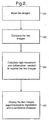
- a typical image fusion or registration process involves at step 21 the input of two images. These may be individual image frames in static imaging, or could be image sequences in a dynamic imaging application.
- the two images are compared in step 22 and the transformation which best maps features from one image onto corresponding features of the other is calculated.
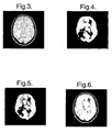
- Figures 3, 4, 5 and 6 illustrate an example of this method applied to brain imaging.
- Figures 3 and 4 illustrate respectively the original MR and PET images of the brain.
- Figure 5 illustrates the result of applying a non-rigid transformation to the PET image of Figure 4 so that it will register with the MR image of Figure 3
- Figure 6 illustrates the original MR image superimposed with the deformed PET image in registration with it.
- Figure 7 illustrates the result of superimposing a version of the PET image of Figure 4 onto the MR image of Figure 3 with only a rigid deformation..
- the size of the deformable part of the transformation DEF is regarded as a measure of the disagreement between the rigid registration ( RIG ) and the deformable registration ( RIG+DEF ).
- a confidence measure M is calculated from the deformable part of the transformation.
- the measure can be displayed over the fused image.
- One way of displaying it is, in response to the user "clicking" at a certain point on the display, to display a circle whose diameter represents the value of the confidence measure.
- Figure 8 illustrates this applied to the fused image of Figure 6 .
- the user can "point" to different positions on the image using the perpendicularly-intersecting cross-hairs (typically controlled by a pointing device such as a computer mouse), and “clicking" at the selected position causes a circle to be displayed as shown.
- the larger the circle the more non-rigid deformation has occurred and so the less agreement there is between the rigid and non-rigid registration.
- the registration has been basically a rigid movement, and thus the result of the fusion can be regarded as more reliable.
- Figure 9 shows an alternative way of displaying the confidence measure on the fused image of Figure 6 .
- the overlay is a transparent colour wash (though shown in black and white in Figure 9 ) whose colour and intensity are directly related to the value of the confidence measure. For example, green of low intensity may be used where the confidence is high (i.e. the amount of non-rigid deformation is low) whereas red, growing more intense, can be used as the amount of non-rigid deformation increases. It can be seen that the confidence decreases towards the left of the image where a high non-rigid deformation was required to register the two images.
Landscapes
- Engineering & Computer Science (AREA)
- Physics & Mathematics (AREA)
- General Physics & Mathematics (AREA)
- Theoretical Computer Science (AREA)
- Computer Vision & Pattern Recognition (AREA)
- Measuring And Recording Apparatus For Diagnosis (AREA)
- Magnetic Resonance Imaging Apparatus (AREA)
- Ultra Sonic Daignosis Equipment (AREA)
Abstract
Claims (18)
- Procédé d'affichage de deux images en enregistrement simultanément comportant les étapes consistant à comparer les deux images entre elles, calculer une transformation qui met en correspondance des caractéristiques d'une image avec des caractéristiques correspondantes de l'autre, et afficher les deux images en surimpression sur la base de la transformation, caractérisé par les étapes consistant à :calculer à partir du degré de transformation requis pour effectuer ladite mise en correspondance, à l'exclusion des déplacements rigides, une mesure de la confiance locale dans l'enregistrement, et afficher ladite mesure de confiance locale.
- Procédé selon la revendication 1, dans lequel ladite mesure de confiance est calculée à partir du degré de déformation non-rigide de ladite transformation calculée.
- Procédé selon la revendication 1 ou 2, dans lequel ladite mesure de confiance est calculée à partir de la grandeur de la déformation locale de ladite transformation.
- Procédé selon la revendication 1 ou 2, dans lequel ladite mesure de confiance est calculée à partir du changement local de volume impliqué par la transformation.
- Procédé selon l'une quelconque des revendications précédentes, dans lequel la mesure est affichée de manière sélective en réponse à une entrée de l'utilisateur.
- Procédé selon l'une quelconque des revendications précédentes, dans lequel la mesure de confiance est affichée superposée sur les deux images.
- Procédé selon l'une quelconque des revendications précédentes, dans lequel la mesure est affichée en tant que superposition pouvant être distinguée visuellement sur les deux images, les propriétés visuelles de la superposition à tout point quelconque étant basées sur ladite mesure.
- Procédé selon la revendication 7, dans lequel la couleur de la superposition pouvant être distinguée visuellement varie en fonction de ladite mesure.
- Procédé selon la revendication 7 ou 8, dans lequel l'intensité de la superposition pouvant être distinguée visuellement varie en fonction de ladite mesure.
- Procédé selon la revendication 7, dans lequel le niveau de gris de la superposition pouvant être distinguée visuellement varie en fonction de ladite mesure.
- Procédé selon la revendication 6, dans lequel la mesure de confiance est affichée à la suite des images superposées affichées.
- Procédé selon la revendication 6, 7 ou 11 dans lequel la superposition pouvant être distinguée visuellement comporte un symbole ayant une propriété qui dépend de la valeur de ladite mesure à un point d'affichage sélectionné.
- Procédé selon la revendication 12, dans lequel le symbole est l'un parmi un cercle et une barre d'erreur dont la taille dépend de la valeur de ladite mesure à un point d'affichage sélectionné.
- Procédé selon la revendication 12 ou 13, dans lequel le symbole est affiché à tout moment uniquement à un point d'affichage sélectionné unique.
- Procédé selon l'une quelconque des revendications précédentes, dans lequel les images sont des images médicales.
- Programme informatique comportant des moyens de code de programme pour mettre en oeuvre sur un ordinateur programmé le procédé selon l'une quelconque des revendications précédentes.
- Support de mémorisation lisible par ordinateur codant un programme informatique conformément à la revendication 16.
- Dispositif d'affichage d'image comportant un afficheur, et un processeur d'image adapté pour mettre en oeuvre le procédé selon l'une quelconque des revendications 1 à 15.
Applications Claiming Priority (3)
| Application Number | Priority Date | Filing Date | Title |
|---|---|---|---|
| GB0227887 | 2002-11-29 | ||
| GBGB0227887.7A GB0227887D0 (en) | 2002-11-29 | 2002-11-29 | Improvements in or relating to image registration |
| PCT/GB2003/004021 WO2004051571A2 (fr) | 2002-11-29 | 2003-09-18 | Ameliorations apportees ou associees a la superposition d'images |
Publications (2)
| Publication Number | Publication Date |
|---|---|
| EP1565883A2 EP1565883A2 (fr) | 2005-08-24 |
| EP1565883B1 true EP1565883B1 (fr) | 2008-10-29 |
Family
ID=9948779
Family Applications (1)
| Application Number | Title | Priority Date | Filing Date |
|---|---|---|---|
| EP03748269A Expired - Lifetime EP1565883B1 (fr) | 2002-11-29 | 2003-09-18 | Fusion d'images avec mesure de confiance d'enregistrement |
Country Status (6)
| Country | Link |
|---|---|
| US (1) | US20050238253A1 (fr) |
| EP (1) | EP1565883B1 (fr) |
| AU (1) | AU2003267577A1 (fr) |
| DE (1) | DE60324455D1 (fr) |
| GB (1) | GB0227887D0 (fr) |
| WO (1) | WO2004051571A2 (fr) |
Cited By (1)
| Publication number | Priority date | Publication date | Assignee | Title |
|---|---|---|---|---|
| CN116977387A (zh) * | 2023-09-22 | 2023-10-31 | 安徽大学 | 基于形变场融合的可变形医学图像配准方法 |
Families Citing this family (26)
| Publication number | Priority date | Publication date | Assignee | Title |
|---|---|---|---|---|
| US7713210B2 (en) * | 2004-11-23 | 2010-05-11 | St. Jude Medical, Atrial Fibrillation Division, Inc. | Method and apparatus for localizing an ultrasound catheter |
| US7295951B2 (en) | 2005-04-04 | 2007-11-13 | Siemens Medical Solutions Usa, Inc. | System and method for quantifying the quality of motion correction in image registration |
| DE102006013473B4 (de) * | 2006-03-23 | 2009-10-22 | Siemens Ag | Verfahren zur ortsaufgelösten Visualisierung der Rekonstruktionsqualität, insbesondere der Abdeckung, eines aufzunehmenden und in einer dreidimensionalen Rekonstruktionsvolumendarstellung wiederzugebenden dreidimensionalen Zielvolumens |
| US8195269B2 (en) * | 2006-07-07 | 2012-06-05 | Siemens Medical Solutions Usa, Inc. | System and method for automatic detection and measurement of malacia in the airways |
| US8548568B2 (en) * | 2006-09-08 | 2013-10-01 | General Electric Company | Methods and apparatus for motion compensation |
| US8223143B2 (en) | 2006-10-27 | 2012-07-17 | Carl Zeiss Meditec, Inc. | User interface for efficiently displaying relevant OCT imaging data |
| ITTO20070620A1 (it) * | 2007-08-31 | 2009-03-01 | Giancarlo Capaccio | Sistema e metodo per presentare dati visuali rilevati a distanza in immagini multi-spettrali, di fusione, ed a tre dimensioni spaziali. |
| EP2227783A1 (fr) * | 2007-10-19 | 2010-09-15 | Boston Scientific Scimed, Inc. | Affichage du résultat d'un classificateur et du niveau de confiance dans une image |
| US8965071B2 (en) * | 2008-05-30 | 2015-02-24 | Emory University | Assessing tumor response to therapy |
| US20100063400A1 (en) | 2008-09-05 | 2010-03-11 | Anne Lindsay Hall | Method and apparatus for catheter guidance using a combination of ultrasound and x-ray imaging |
| DE102010005265A1 (de) * | 2010-01-20 | 2011-07-21 | Testo AG, 79853 | Verfahren zur überlagerten Darstellung korrespondierender Bilder und Bildaufnahmevorrichtung |
| US9105115B2 (en) * | 2010-03-16 | 2015-08-11 | Honeywell International Inc. | Display systems and methods for displaying enhanced vision and synthetic images |
| US8582846B2 (en) * | 2010-06-18 | 2013-11-12 | Siemens Aktiengesellschaft | Method and system for validating image registration |
| CN102985946B (zh) * | 2010-07-09 | 2016-08-24 | 皇家飞利浦电子股份有限公司 | 呼吸运动估计的自动逐点验证 |
| US9996929B2 (en) * | 2010-10-27 | 2018-06-12 | Varian Medical Systems International Ag | Visualization of deformations using color overlays |
| US10152951B2 (en) | 2011-02-28 | 2018-12-11 | Varian Medical Systems International Ag | Method and system for interactive control of window/level parameters of multi-image displays |
| DE102011080905B4 (de) * | 2011-08-12 | 2014-03-27 | Siemens Aktiengesellschaft | Verfahren zur Visualisierung der Registrierungsqualität medizinischer Bilddatensätze |
| US8944597B2 (en) | 2012-01-19 | 2015-02-03 | Carl Zeiss Meditec, Inc. | Standardized display of optical coherence tomography imaging data |
| US9420945B2 (en) | 2013-03-14 | 2016-08-23 | Carl Zeiss Meditec, Inc. | User interface for acquisition, display and analysis of ophthalmic diagnostic data |
| CN105069768B (zh) * | 2015-08-05 | 2017-12-29 | 武汉高德红外股份有限公司 | 一种可见光图像与红外图像融合处理系统及融合方法 |
| WO2017102468A1 (fr) * | 2015-12-15 | 2017-06-22 | Koninklijke Philips N.V. | Systèmes et procédés de traitement d'images |
| CN114078102A (zh) * | 2020-08-11 | 2022-02-22 | 北京芯海视界三维科技有限公司 | 图像处理装置和虚拟现实设备 |
| CN116868233A (zh) * | 2020-12-31 | 2023-10-10 | 西安大医集团股份有限公司 | 图像配准评估方法、装置、电子设备及可读存储介质 |
| CN116831626A (zh) * | 2022-03-25 | 2023-10-03 | 深圳迈瑞生物医疗电子股份有限公司 | 超声波束合成方法及设备 |
| CN115170714A (zh) * | 2022-06-29 | 2022-10-11 | 先临三维科技股份有限公司 | 一种扫描图像渲染方法、装置、电子设备及存储介质 |
| CN121366156B (zh) * | 2025-12-18 | 2026-04-21 | 深圳市湃泊科技有限公司 | 一种预制焊料壳体缺陷检测方法、系统、设备及介质 |
Family Cites Families (7)
| Publication number | Priority date | Publication date | Assignee | Title |
|---|---|---|---|---|
| US5871013A (en) * | 1995-05-31 | 1999-02-16 | Elscint Ltd. | Registration of nuclear medicine images |
| US5672877A (en) * | 1996-03-27 | 1997-09-30 | Adac Laboratories | Coregistration of multi-modality data in a medical imaging system |
| US6016442A (en) * | 1998-03-25 | 2000-01-18 | Cardiac Pacemakers, Inc. | System for displaying cardiac arrhythmia data |
| US6944330B2 (en) * | 2000-09-07 | 2005-09-13 | Siemens Corporate Research, Inc. | Interactive computer-aided diagnosis method and system for assisting diagnosis of lung nodules in digital volumetric medical images |
| DE10141186A1 (de) * | 2001-08-22 | 2003-03-20 | Siemens Ag | Einrichtung zum Bearbeiten von Bildern, insbesondere medizinischen Bildern |
| DE10163813A1 (de) * | 2001-12-22 | 2003-07-03 | Philips Intellectual Property | Verfahren zur Darstellung von unterschiedlichen Bildern eines Untersuchungsobjektes |
| US7050615B2 (en) * | 2002-07-25 | 2006-05-23 | Ge Medical Systems Glogal Technology Company, Llc | Temporal image comparison method |
-
2002
- 2002-11-29 GB GBGB0227887.7A patent/GB0227887D0/en not_active Ceased
-
2003
- 2003-09-18 DE DE60324455T patent/DE60324455D1/de not_active Expired - Lifetime
- 2003-09-18 US US10/502,034 patent/US20050238253A1/en not_active Abandoned
- 2003-09-18 AU AU2003267577A patent/AU2003267577A1/en not_active Abandoned
- 2003-09-18 EP EP03748269A patent/EP1565883B1/fr not_active Expired - Lifetime
- 2003-09-18 WO PCT/GB2003/004021 patent/WO2004051571A2/fr not_active Ceased
Non-Patent Citations (1)
| Title |
|---|
| WOODS R.P.: "VALIDATION OF REGISTRATION ACCURACY", HANDBOOK OF MEDICAL IMAGING, 2000, pages 491 - 497, XP001152021 * |
Cited By (2)
| Publication number | Priority date | Publication date | Assignee | Title |
|---|---|---|---|---|
| CN116977387A (zh) * | 2023-09-22 | 2023-10-31 | 安徽大学 | 基于形变场融合的可变形医学图像配准方法 |
| CN116977387B (zh) * | 2023-09-22 | 2023-12-15 | 安徽大学 | 基于形变场融合的可变形医学图像配准方法 |
Also Published As
| Publication number | Publication date |
|---|---|
| DE60324455D1 (de) | 2008-12-11 |
| WO2004051571A3 (fr) | 2004-10-21 |
| GB0227887D0 (en) | 2003-01-08 |
| AU2003267577A1 (en) | 2004-06-23 |
| WO2004051571A2 (fr) | 2004-06-17 |
| AU2003267577A8 (en) | 2004-06-23 |
| EP1565883A2 (fr) | 2005-08-24 |
| US20050238253A1 (en) | 2005-10-27 |
Similar Documents
| Publication | Publication Date | Title |
|---|---|---|
| EP1565883B1 (fr) | Fusion d'images avec mesure de confiance d'enregistrement | |
| Cai et al. | Multi-modal vertebrae recognition using transformed deep convolution network | |
| Le Goualher et al. | Automated extraction and variability analysis of sulcal neuroanatomy | |
| EP3239930B1 (fr) | Procédé d'enregistrement d'images et appareil de traitement de données d'images médicales | |
| Lester et al. | A survey of hierarchical non-linear medical image registration | |
| Andriole et al. | Optimizing analysis, visualization, and navigation of large image data sets: one 5000-section CT scan can ruin your whole day | |
| EP2422318B1 (fr) | Quantification de données d'image médicale | |
| US7346199B2 (en) | Anatomic triangulation | |
| US20050289472A1 (en) | 3D display system and method | |
| US20070237372A1 (en) | Cross-time and cross-modality inspection for medical image diagnosis | |
| US20050285844A1 (en) | 3D display system and method | |
| CN101315700A (zh) | 一种多序列图像快速自动定位的方法 | |
| US20050285854A1 (en) | 3D display system and method | |
| An et al. | Part affinity fields and CoordConv for detecting landmarks of lumbar vertebrae and sacrum in X-ray images | |
| Wang et al. | Automatic vessel segmentation on fundus images using vessel filtering and fuzzy entropy | |
| Collignon et al. | Surface-based registration of 3D medical images | |
| Liu et al. | Multiple sclerosis medical image analysis and information management | |
| US20020122577A1 (en) | Method and system for tag detection and tracking in MRI tagged images | |
| Torres et al. | Deep learning-based detection of anthropometric landmarks in 3D infants head models | |
| Haase et al. | ToF/RGB sensor fusion for augmented 3D endoscopy using a fully automatic calibration scheme | |
| US7844132B2 (en) | Method for determining a transformation of coordinates of different images of an object | |
| EP1803099B1 (fr) | Alignement de grille virtuelle pour sous-volumes | |
| Andronache | Multi-modal non-rigid registration of volumetric medical images | |
| Temkin et al. | A high accuracy, landmark-based, sub-pixel level image registration method | |
| JP6692906B2 (ja) | 線形画像特徴に対する改良されたマーカー配置のための画像処理 |
Legal Events
| Date | Code | Title | Description |
|---|---|---|---|
| PUAI | Public reference made under article 153(3) epc to a published international application that has entered the european phase |
Free format text: ORIGINAL CODE: 0009012 |
|
| 17P | Request for examination filed |
Effective date: 20040702 |
|
| AK | Designated contracting states |
Kind code of ref document: A2 Designated state(s): AT BE BG CH CY CZ DE DK EE ES FI FR GB GR HU IE IT LI LU MC NL PT RO SE SI SK TR |
|
| AX | Request for extension of the european patent |
Extension state: AL LT LV MK |
|
| RIN1 | Information on inventor provided before grant (corrected) |
Inventor name: BEHRENBRUCH, CHRISTIAN PETER Inventor name: DECLERCK, JEROME, MARIE, JOSEPH |
|
| RIN1 | Information on inventor provided before grant (corrected) |
Inventor name: DECLERCK, JEROME, MARIE, JOSEPH Inventor name: BEHRENBRUCH, CHRISTIAN PETER |
|
| DAX | Request for extension of the european patent (deleted) | ||
| RBV | Designated contracting states (corrected) |
Designated state(s): DE FR GB |
|
| 17Q | First examination report despatched |
Effective date: 20050913 |
|
| GRAP | Despatch of communication of intention to grant a patent |
Free format text: ORIGINAL CODE: EPIDOSNIGR1 |
|
| GRAS | Grant fee paid |
Free format text: ORIGINAL CODE: EPIDOSNIGR3 |
|
| GRAA | (expected) grant |
Free format text: ORIGINAL CODE: 0009210 |
|
| RAP1 | Party data changed (applicant data changed or rights of an application transferred) |
Owner name: MIRADA SOLUTIONS LIMITED |
|
| AK | Designated contracting states |
Kind code of ref document: B1 Designated state(s): DE FR GB |
|
| REG | Reference to a national code |
Ref country code: GB Ref legal event code: FG4D |
|
| REF | Corresponds to: |
Ref document number: 60324455 Country of ref document: DE Date of ref document: 20081211 Kind code of ref document: P |
|
| RAP2 | Party data changed (patent owner data changed or rights of a patent transferred) |
Owner name: SIEMENS MEDICAL SOLUTIONS USA, INC. |
|
| REG | Reference to a national code |
Ref country code: GB Ref legal event code: 732E Free format text: REGISTERED BETWEEN 20090115 AND 20090121 |
|
| PLBE | No opposition filed within time limit |
Free format text: ORIGINAL CODE: 0009261 |
|
| STAA | Information on the status of an ep patent application or granted ep patent |
Free format text: STATUS: NO OPPOSITION FILED WITHIN TIME LIMIT |
|
| 26N | No opposition filed |
Effective date: 20090730 |
|
| PGFP | Annual fee paid to national office [announced via postgrant information from national office to epo] |
Ref country code: DE Payment date: 20101119 Year of fee payment: 8 |
|
| PGFP | Annual fee paid to national office [announced via postgrant information from national office to epo] |
Ref country code: GB Payment date: 20120917 Year of fee payment: 10 |
|
| PGFP | Annual fee paid to national office [announced via postgrant information from national office to epo] |
Ref country code: FR Payment date: 20121002 Year of fee payment: 10 |
|
| PG25 | Lapsed in a contracting state [announced via postgrant information from national office to epo] |
Ref country code: DE Free format text: LAPSE BECAUSE OF NON-PAYMENT OF DUE FEES Effective date: 20130403 |
|
| REG | Reference to a national code |
Ref country code: DE Ref legal event code: R119 Ref document number: 60324455 Country of ref document: DE Effective date: 20130403 |
|
| GBPC | Gb: european patent ceased through non-payment of renewal fee |
Effective date: 20130918 |
|
| REG | Reference to a national code |
Ref country code: FR Ref legal event code: ST Effective date: 20140530 |
|
| PG25 | Lapsed in a contracting state [announced via postgrant information from national office to epo] |
Ref country code: GB Free format text: LAPSE BECAUSE OF NON-PAYMENT OF DUE FEES Effective date: 20130918 |
|
| PG25 | Lapsed in a contracting state [announced via postgrant information from national office to epo] |
Ref country code: FR Free format text: LAPSE BECAUSE OF NON-PAYMENT OF DUE FEES Effective date: 20130930 |