EP1556072B1 - Ii-KEY/ANTIGENIC EPITOPE HYBRID PEPTIDE VACCINES - Google Patents
Ii-KEY/ANTIGENIC EPITOPE HYBRID PEPTIDE VACCINES Download PDFInfo
- Publication number
- EP1556072B1 EP1556072B1 EP03789700A EP03789700A EP1556072B1 EP 1556072 B1 EP1556072 B1 EP 1556072B1 EP 03789700 A EP03789700 A EP 03789700A EP 03789700 A EP03789700 A EP 03789700A EP 1556072 B1 EP1556072 B1 EP 1556072B1
- Authority
- EP
- European Patent Office
- Prior art keywords
- mhc class
- peptide
- epitopes
- epitope
- presented
- Prior art date
- Legal status (The legal status is an assumption and is not a legal conclusion. Google has not performed a legal analysis and makes no representation as to the accuracy of the status listed.)
- Expired - Lifetime
Links
- 108010028930 invariant chain Proteins 0.000 title claims description 306
- 230000000890 antigenic effect Effects 0.000 title claims description 305
- 229940023041 peptide vaccine Drugs 0.000 title description 15
- 108090000765 processed proteins & peptides Proteins 0.000 claims description 647
- 102000004196 processed proteins & peptides Human genes 0.000 claims description 217
- 150000001413 amino acids Chemical class 0.000 claims description 180
- 108091054438 MHC class II family Proteins 0.000 claims description 147
- 102000043131 MHC class II family Human genes 0.000 claims description 114
- 102000043129 MHC class I family Human genes 0.000 claims description 72
- 108091054437 MHC class I family Proteins 0.000 claims description 70
- 230000002708 enhancing effect Effects 0.000 claims description 68
- 230000028993 immune response Effects 0.000 claims description 50
- 102000018713 Histocompatibility Antigens Class II Human genes 0.000 claims description 46
- 229920001184 polypeptide Polymers 0.000 claims description 35
- 239000000203 mixture Substances 0.000 claims description 30
- 239000000126 substance Substances 0.000 claims description 24
- 210000004899 c-terminal region Anatomy 0.000 claims description 18
- 230000030741 antigen processing and presentation Effects 0.000 claims description 16
- 238000012217 deletion Methods 0.000 claims description 16
- 230000037430 deletion Effects 0.000 claims description 16
- 108010027412 Histocompatibility Antigens Class II Proteins 0.000 claims description 13
- 241000124008 Mammalia Species 0.000 claims description 9
- 230000004936 stimulating effect Effects 0.000 claims description 7
- 239000000816 peptidomimetic Substances 0.000 claims description 6
- 108700018351 Major Histocompatibility Complex Proteins 0.000 description 306
- 230000020382 suppression by virus of host antigen processing and presentation of peptide antigen via MHC class I Effects 0.000 description 306
- 235000001014 amino acid Nutrition 0.000 description 191
- 108090000623 proteins and genes Proteins 0.000 description 133
- 210000004027 cell Anatomy 0.000 description 117
- 102000004169 proteins and genes Human genes 0.000 description 102
- 235000018102 proteins Nutrition 0.000 description 101
- 206010028980 Neoplasm Diseases 0.000 description 99
- 108091007433 antigens Proteins 0.000 description 89
- 102000036639 antigens Human genes 0.000 description 89
- 239000000427 antigen Substances 0.000 description 88
- 210000001744 T-lymphocyte Anatomy 0.000 description 86
- 125000003275 alpha amino acid group Chemical group 0.000 description 85
- 238000000034 method Methods 0.000 description 85
- 230000004044 response Effects 0.000 description 65
- 210000001151 cytotoxic T lymphocyte Anatomy 0.000 description 63
- 102100025475 Carcinoembryonic antigen-related cell adhesion molecule 5 Human genes 0.000 description 61
- 108010022366 Carcinoembryonic Antigen Proteins 0.000 description 60
- 102100028389 Melanoma antigen recognized by T-cells 1 Human genes 0.000 description 58
- 108010072866 Prostate-Specific Antigen Proteins 0.000 description 53
- 102100038358 Prostate-specific antigen Human genes 0.000 description 52
- 201000011510 cancer Diseases 0.000 description 51
- 239000013566 allergen Substances 0.000 description 50
- 210000004443 dendritic cell Anatomy 0.000 description 46
- 101000578784 Homo sapiens Melanoma antigen recognized by T-cells 1 Proteins 0.000 description 42
- 229960005486 vaccine Drugs 0.000 description 42
- 108700028369 Alleles Proteins 0.000 description 39
- 201000010099 disease Diseases 0.000 description 39
- 208000037265 diseases, disorders, signs and symptoms Diseases 0.000 description 39
- 230000001225 therapeutic effect Effects 0.000 description 38
- 102100030086 Receptor tyrosine-protein kinase erbB-2 Human genes 0.000 description 35
- 201000001441 melanoma Diseases 0.000 description 35
- 108020004414 DNA Proteins 0.000 description 34
- 208000026935 allergic disease Diseases 0.000 description 34
- 150000001875 compounds Chemical class 0.000 description 34
- 230000004048 modification Effects 0.000 description 32
- 238000012986 modification Methods 0.000 description 32
- 101001012157 Homo sapiens Receptor tyrosine-protein kinase erbB-2 Proteins 0.000 description 31
- 230000014509 gene expression Effects 0.000 description 31
- 206010020751 Hypersensitivity Diseases 0.000 description 30
- 102100039094 Tyrosinase Human genes 0.000 description 30
- 108060008724 Tyrosinase Proteins 0.000 description 29
- 238000002255 vaccination Methods 0.000 description 29
- 241000282414 Homo sapiens Species 0.000 description 28
- 241000700605 Viruses Species 0.000 description 28
- 230000007815 allergy Effects 0.000 description 28
- 230000000694 effects Effects 0.000 description 25
- 210000000612 antigen-presenting cell Anatomy 0.000 description 24
- 101000716102 Homo sapiens T-cell surface glycoprotein CD4 Proteins 0.000 description 23
- 230000005867 T cell response Effects 0.000 description 23
- 102100036011 T-cell surface glycoprotein CD4 Human genes 0.000 description 23
- 239000013598 vector Substances 0.000 description 23
- 230000006870 function Effects 0.000 description 22
- 108010058597 HLA-DR Antigens Proteins 0.000 description 21
- 102000006354 HLA-DR Antigens Human genes 0.000 description 21
- 229960004784 allergens Drugs 0.000 description 21
- 230000003053 immunization Effects 0.000 description 21
- 125000006850 spacer group Chemical group 0.000 description 21
- 108010074328 Interferon-gamma Proteins 0.000 description 20
- 102100031413 L-dopachrome tautomerase Human genes 0.000 description 20
- 102100022430 Melanocyte protein PMEL Human genes 0.000 description 20
- 238000000338 in vitro Methods 0.000 description 20
- 210000003819 peripheral blood mononuclear cell Anatomy 0.000 description 20
- 238000011282 treatment Methods 0.000 description 19
- 102100037850 Interferon gamma Human genes 0.000 description 18
- 238000002649 immunization Methods 0.000 description 18
- 230000003612 virological effect Effects 0.000 description 18
- 101100314454 Caenorhabditis elegans tra-1 gene Proteins 0.000 description 17
- 238000009169 immunotherapy Methods 0.000 description 17
- 108010010995 MART-1 Antigen Proteins 0.000 description 16
- 239000003814 drug Substances 0.000 description 16
- 101800001271 Surface protein Proteins 0.000 description 15
- 230000015572 biosynthetic process Effects 0.000 description 15
- 230000004957 immunoregulator effect Effects 0.000 description 15
- 239000002502 liposome Substances 0.000 description 15
- 238000004519 manufacturing process Methods 0.000 description 15
- 230000007246 mechanism Effects 0.000 description 15
- 230000008569 process Effects 0.000 description 15
- 238000003786 synthesis reaction Methods 0.000 description 15
- 102000004127 Cytokines Human genes 0.000 description 14
- 108090000695 Cytokines Proteins 0.000 description 14
- 239000002131 composite material Substances 0.000 description 14
- 229940079593 drug Drugs 0.000 description 14
- 210000002443 helper t lymphocyte Anatomy 0.000 description 14
- 238000001727 in vivo Methods 0.000 description 14
- 208000015181 infectious disease Diseases 0.000 description 14
- 244000052769 pathogen Species 0.000 description 14
- 230000001717 pathogenic effect Effects 0.000 description 14
- 238000002560 therapeutic procedure Methods 0.000 description 14
- 108010041986 DNA Vaccines Proteins 0.000 description 13
- 229940021995 DNA vaccine Drugs 0.000 description 13
- 208000006673 asthma Diseases 0.000 description 13
- 125000005647 linker group Chemical group 0.000 description 13
- 210000004698 lymphocyte Anatomy 0.000 description 13
- 210000004881 tumor cell Anatomy 0.000 description 13
- LKKMLIBUAXYLOY-UHFFFAOYSA-N 3-Amino-1-methyl-5H-pyrido[4,3-b]indole Chemical compound N1C2=CC=CC=C2C2=C1C=C(N)N=C2C LKKMLIBUAXYLOY-UHFFFAOYSA-N 0.000 description 12
- 101710093778 L-dopachrome tautomerase Proteins 0.000 description 12
- 206010033128 Ovarian cancer Diseases 0.000 description 12
- 206010061535 Ovarian neoplasm Diseases 0.000 description 12
- 210000003719 b-lymphocyte Anatomy 0.000 description 12
- 230000001965 increasing effect Effects 0.000 description 12
- 230000001404 mediated effect Effects 0.000 description 12
- 230000037361 pathway Effects 0.000 description 12
- -1 polymethylene Polymers 0.000 description 12
- 239000000047 product Substances 0.000 description 12
- 241000282326 Felis catus Species 0.000 description 11
- 108010074032 HLA-A2 Antigen Proteins 0.000 description 11
- 102000025850 HLA-A2 Antigen Human genes 0.000 description 11
- 101710173694 Short transient receptor potential channel 2 Proteins 0.000 description 11
- 206010046865 Vaccinia virus infection Diseases 0.000 description 11
- 208000010668 atopic eczema Diseases 0.000 description 11
- 239000002299 complementary DNA Substances 0.000 description 11
- 238000011161 development Methods 0.000 description 11
- 210000001165 lymph node Anatomy 0.000 description 11
- 150000007523 nucleic acids Chemical group 0.000 description 11
- 210000003491 skin Anatomy 0.000 description 11
- 238000006467 substitution reaction Methods 0.000 description 11
- 208000007089 vaccinia Diseases 0.000 description 11
- 244000105624 Arachis hypogaea Species 0.000 description 10
- 206010006187 Breast cancer Diseases 0.000 description 10
- 208000026310 Breast neoplasm Diseases 0.000 description 10
- 125000000539 amino acid group Chemical group 0.000 description 10
- 238000013461 design Methods 0.000 description 10
- 238000009472 formulation Methods 0.000 description 10
- 210000000987 immune system Anatomy 0.000 description 10
- 230000002163 immunogen Effects 0.000 description 10
- 238000002347 injection Methods 0.000 description 10
- 239000007924 injection Substances 0.000 description 10
- 108010045069 keyhole-limpet hemocyanin Proteins 0.000 description 10
- 239000002245 particle Substances 0.000 description 10
- 241001674044 Blattodea Species 0.000 description 9
- 108010017213 Granulocyte-Macrophage Colony-Stimulating Factor Proteins 0.000 description 9
- 102100039620 Granulocyte-macrophage colony-stimulating factor Human genes 0.000 description 9
- 108010088729 HLA-A*02:01 antigen Proteins 0.000 description 9
- KDXKERNSBIXSRK-YFKPBYRVSA-N L-lysine Chemical compound NCCCC[C@H](N)C(O)=O KDXKERNSBIXSRK-YFKPBYRVSA-N 0.000 description 9
- KDXKERNSBIXSRK-UHFFFAOYSA-N Lysine Natural products NCCCCC(N)C(O)=O KDXKERNSBIXSRK-UHFFFAOYSA-N 0.000 description 9
- 230000004913 activation Effects 0.000 description 9
- 238000003556 assay Methods 0.000 description 9
- 230000003190 augmentative effect Effects 0.000 description 9
- 239000003659 bee venom Substances 0.000 description 9
- 238000002360 preparation method Methods 0.000 description 9
- 230000010076 replication Effects 0.000 description 9
- 210000001519 tissue Anatomy 0.000 description 9
- 102000005720 Glutathione transferase Human genes 0.000 description 8
- 108010070675 Glutathione transferase Proteins 0.000 description 8
- COLNVLDHVKWLRT-QMMMGPOBSA-N L-phenylalanine Chemical compound OC(=O)[C@@H](N)CC1=CC=CC=C1 COLNVLDHVKWLRT-QMMMGPOBSA-N 0.000 description 8
- 102100037611 Lysophospholipase Human genes 0.000 description 8
- 241001465754 Metazoa Species 0.000 description 8
- 241000699670 Mus sp. Species 0.000 description 8
- 108010058864 Phospholipases A2 Proteins 0.000 description 8
- 108091008874 T cell receptors Proteins 0.000 description 8
- 102000016266 T-Cell Antigen Receptors Human genes 0.000 description 8
- 239000002671 adjuvant Substances 0.000 description 8
- 230000000172 allergic effect Effects 0.000 description 8
- 238000013459 approach Methods 0.000 description 8
- 230000002349 favourable effect Effects 0.000 description 8
- 230000035772 mutation Effects 0.000 description 8
- 235000020232 peanut Nutrition 0.000 description 8
- 239000013612 plasmid Substances 0.000 description 8
- 230000009696 proliferative response Effects 0.000 description 8
- 230000001177 retroviral effect Effects 0.000 description 8
- 235000010777 Arachis hypogaea Nutrition 0.000 description 7
- PEDCQBHIVMGVHV-UHFFFAOYSA-N Glycerine Chemical compound OCC(O)CO PEDCQBHIVMGVHV-UHFFFAOYSA-N 0.000 description 7
- 108010086377 HLA-A3 Antigen Proteins 0.000 description 7
- 241000282412 Homo Species 0.000 description 7
- 102000004388 Interleukin-4 Human genes 0.000 description 7
- 108090000978 Interleukin-4 Proteins 0.000 description 7
- 206010027476 Metastases Diseases 0.000 description 7
- 241000746983 Phleum pratense Species 0.000 description 7
- 241000700618 Vaccinia virus Species 0.000 description 7
- 239000003795 chemical substances by application Substances 0.000 description 7
- 230000007423 decrease Effects 0.000 description 7
- 108010051081 dopachrome isomerase Proteins 0.000 description 7
- 210000002472 endoplasmic reticulum Anatomy 0.000 description 7
- 230000006698 induction Effects 0.000 description 7
- 230000001939 inductive effect Effects 0.000 description 7
- 125000002924 primary amino group Chemical group [H]N([H])* 0.000 description 7
- 230000009257 reactivity Effects 0.000 description 7
- 235000002639 sodium chloride Nutrition 0.000 description 7
- 230000004083 survival effect Effects 0.000 description 7
- 102100028976 HLA class I histocompatibility antigen, B alpha chain Human genes 0.000 description 6
- 108010002350 Interleukin-2 Proteins 0.000 description 6
- 102000000588 Interleukin-2 Human genes 0.000 description 6
- NKANXQFJJICGDU-QPLCGJKRSA-N Tamoxifen Chemical compound C=1C=CC=CC=1C(/CC)=C(C=1C=CC(OCCN(C)C)=CC=1)/C1=CC=CC=C1 NKANXQFJJICGDU-QPLCGJKRSA-N 0.000 description 6
- 230000003281 allosteric effect Effects 0.000 description 6
- 230000008901 benefit Effects 0.000 description 6
- 125000002091 cationic group Chemical group 0.000 description 6
- 125000003636 chemical group Chemical group 0.000 description 6
- 230000000875 corresponding effect Effects 0.000 description 6
- 230000003247 decreasing effect Effects 0.000 description 6
- 108700020302 erbB-2 Genes Proteins 0.000 description 6
- 239000012634 fragment Substances 0.000 description 6
- 230000002458 infectious effect Effects 0.000 description 6
- 238000003780 insertion Methods 0.000 description 6
- 230000037431 insertion Effects 0.000 description 6
- 150000002632 lipids Chemical class 0.000 description 6
- 206010061289 metastatic neoplasm Diseases 0.000 description 6
- 230000002018 overexpression Effects 0.000 description 6
- 230000003389 potentiating effect Effects 0.000 description 6
- 150000003839 salts Chemical class 0.000 description 6
- 241000894007 species Species 0.000 description 6
- 230000000638 stimulation Effects 0.000 description 6
- 208000024891 symptom Diseases 0.000 description 6
- 238000012360 testing method Methods 0.000 description 6
- 230000000699 topical effect Effects 0.000 description 6
- 239000003053 toxin Substances 0.000 description 6
- 231100000765 toxin Toxicity 0.000 description 6
- 108700012359 toxins Proteins 0.000 description 6
- 230000009261 transgenic effect Effects 0.000 description 6
- 241000701161 unidentified adenovirus Species 0.000 description 6
- 241001430294 unidentified retrovirus Species 0.000 description 6
- 235000017060 Arachis glabrata Nutrition 0.000 description 5
- 235000018262 Arachis monticola Nutrition 0.000 description 5
- 235000018185 Betula X alpestris Nutrition 0.000 description 5
- 235000018212 Betula X uliginosa Nutrition 0.000 description 5
- 208000035473 Communicable disease Diseases 0.000 description 5
- 229940046168 CpG oligodeoxynucleotide Drugs 0.000 description 5
- 238000011510 Elispot assay Methods 0.000 description 5
- 108010075704 HLA-A Antigens Proteins 0.000 description 5
- 102000011786 HLA-A Antigens Human genes 0.000 description 5
- 108010035452 HLA-A1 Antigen Proteins 0.000 description 5
- 108010058607 HLA-B Antigens Proteins 0.000 description 5
- 101001057504 Homo sapiens Interferon-stimulated gene 20 kDa protein Proteins 0.000 description 5
- 101001055144 Homo sapiens Interleukin-2 receptor subunit alpha Proteins 0.000 description 5
- 101000623901 Homo sapiens Mucin-16 Proteins 0.000 description 5
- 101001024605 Homo sapiens Next to BRCA1 gene 1 protein Proteins 0.000 description 5
- 241000725303 Human immunodeficiency virus Species 0.000 description 5
- 102100027268 Interferon-stimulated gene 20 kDa protein Human genes 0.000 description 5
- 102000003814 Interleukin-10 Human genes 0.000 description 5
- 108090000174 Interleukin-10 Proteins 0.000 description 5
- 102100023123 Mucin-16 Human genes 0.000 description 5
- 108091028043 Nucleic acid sequence Proteins 0.000 description 5
- 206010060862 Prostate cancer Diseases 0.000 description 5
- 208000000236 Prostatic Neoplasms Diseases 0.000 description 5
- 108010008281 Recombinant Fusion Proteins Proteins 0.000 description 5
- 102000007056 Recombinant Fusion Proteins Human genes 0.000 description 5
- 239000000556 agonist Substances 0.000 description 5
- 230000005875 antibody response Effects 0.000 description 5
- 229940023860 canarypox virus HIV vaccine Drugs 0.000 description 5
- 230000036755 cellular response Effects 0.000 description 5
- 239000012636 effector Substances 0.000 description 5
- 238000001415 gene therapy Methods 0.000 description 5
- PCHJSUWPFVWCPO-UHFFFAOYSA-N gold Chemical compound [Au] PCHJSUWPFVWCPO-UHFFFAOYSA-N 0.000 description 5
- 239000010931 gold Substances 0.000 description 5
- 229910052737 gold Inorganic materials 0.000 description 5
- 230000002209 hydrophobic effect Effects 0.000 description 5
- 230000003993 interaction Effects 0.000 description 5
- 210000005259 peripheral blood Anatomy 0.000 description 5
- 239000011886 peripheral blood Substances 0.000 description 5
- 229920000747 poly(lactic acid) Polymers 0.000 description 5
- 230000017854 proteolysis Effects 0.000 description 5
- 230000002829 reductive effect Effects 0.000 description 5
- 238000001890 transfection Methods 0.000 description 5
- 238000012546 transfer Methods 0.000 description 5
- 108091032973 (ribonucleotides)n+m Proteins 0.000 description 4
- 208000023275 Autoimmune disease Diseases 0.000 description 4
- 208000003174 Brain Neoplasms Diseases 0.000 description 4
- 210000001266 CD8-positive T-lymphocyte Anatomy 0.000 description 4
- 244000025254 Cannabis sativa Species 0.000 description 4
- 206010009944 Colon cancer Diseases 0.000 description 4
- 208000001333 Colorectal Neoplasms Diseases 0.000 description 4
- 102000053602 DNA Human genes 0.000 description 4
- 102000018651 Epithelial Cell Adhesion Molecule Human genes 0.000 description 4
- 108010066687 Epithelial Cell Adhesion Molecule Proteins 0.000 description 4
- 208000001382 Experimental Melanoma Diseases 0.000 description 4
- 102100036242 HLA class II histocompatibility antigen, DQ alpha 2 chain Human genes 0.000 description 4
- 108010013476 HLA-A24 Antigen Proteins 0.000 description 4
- 101000620359 Homo sapiens Melanocyte protein PMEL Proteins 0.000 description 4
- 125000000729 N-terminal amino-acid group Chemical group 0.000 description 4
- 102000035195 Peptidases Human genes 0.000 description 4
- 108091005804 Peptidases Proteins 0.000 description 4
- 108010033276 Peptide Fragments Proteins 0.000 description 4
- 102000007079 Peptide Fragments Human genes 0.000 description 4
- 101000750404 Phoneutria keyserlingi CRISP-1 Proteins 0.000 description 4
- 108010076504 Protein Sorting Signals Proteins 0.000 description 4
- 108700019146 Transgenes Proteins 0.000 description 4
- 125000004429 atom Chemical group 0.000 description 4
- 230000001363 autoimmune Effects 0.000 description 4
- 230000001580 bacterial effect Effects 0.000 description 4
- 238000004422 calculation algorithm Methods 0.000 description 4
- 230000032823 cell division Effects 0.000 description 4
- 238000006243 chemical reaction Methods 0.000 description 4
- 230000007812 deficiency Effects 0.000 description 4
- 238000003745 diagnosis Methods 0.000 description 4
- 239000002552 dosage form Substances 0.000 description 4
- 238000005516 engineering process Methods 0.000 description 4
- 238000003114 enzyme-linked immunosorbent spot assay Methods 0.000 description 4
- 238000002474 experimental method Methods 0.000 description 4
- 239000000499 gel Substances 0.000 description 4
- 108010008486 gp100 Melanoma Antigen Proteins 0.000 description 4
- 230000006872 improvement Effects 0.000 description 4
- 206010022000 influenza Diseases 0.000 description 4
- 230000010354 integration Effects 0.000 description 4
- 238000001990 intravenous administration Methods 0.000 description 4
- 208000030776 invasive breast carcinoma Diseases 0.000 description 4
- 210000002540 macrophage Anatomy 0.000 description 4
- 239000012528 membrane Substances 0.000 description 4
- 210000004379 membrane Anatomy 0.000 description 4
- 210000001616 monocyte Anatomy 0.000 description 4
- 238000004806 packaging method and process Methods 0.000 description 4
- 239000004626 polylactic acid Substances 0.000 description 4
- 238000012545 processing Methods 0.000 description 4
- 229940002612 prodrug Drugs 0.000 description 4
- 239000000651 prodrug Substances 0.000 description 4
- 230000035755 proliferation Effects 0.000 description 4
- 208000023958 prostate neoplasm Diseases 0.000 description 4
- 230000001681 protective effect Effects 0.000 description 4
- 102000005962 receptors Human genes 0.000 description 4
- 108020003175 receptors Proteins 0.000 description 4
- 206010039073 rheumatoid arthritis Diseases 0.000 description 4
- 230000028327 secretion Effects 0.000 description 4
- 210000002966 serum Anatomy 0.000 description 4
- 239000000243 solution Substances 0.000 description 4
- 230000009885 systemic effect Effects 0.000 description 4
- 230000008685 targeting Effects 0.000 description 4
- 229960000575 trastuzumab Drugs 0.000 description 4
- 239000003981 vehicle Substances 0.000 description 4
- QTBSBXVTEAMEQO-UHFFFAOYSA-M Acetate Chemical compound CC([O-])=O QTBSBXVTEAMEQO-UHFFFAOYSA-M 0.000 description 3
- 206010003402 Arthropod sting Diseases 0.000 description 3
- 206010003645 Atopy Diseases 0.000 description 3
- 241000193738 Bacillus anthracis Species 0.000 description 3
- 108010008629 CA-125 Antigen Proteins 0.000 description 3
- 102000007269 CA-125 Antigen Human genes 0.000 description 3
- 108010021064 CTLA-4 Antigen Proteins 0.000 description 3
- 229940045513 CTLA4 antagonist Drugs 0.000 description 3
- 102100039498 Cytotoxic T-lymphocyte protein 4 Human genes 0.000 description 3
- 238000002965 ELISA Methods 0.000 description 3
- LFQSCWFLJHTTHZ-UHFFFAOYSA-N Ethanol Chemical compound CCO LFQSCWFLJHTTHZ-UHFFFAOYSA-N 0.000 description 3
- 108010091938 HLA-B7 Antigen Proteins 0.000 description 3
- 108010088652 Histocompatibility Antigens Class I Proteins 0.000 description 3
- 108010050904 Interferons Proteins 0.000 description 3
- 102000014150 Interferons Human genes 0.000 description 3
- 206010027480 Metastatic malignant melanoma Diseases 0.000 description 3
- 108010014251 Muramidase Proteins 0.000 description 3
- 102000016943 Muramidase Human genes 0.000 description 3
- 108010062010 N-Acetylmuramoyl-L-alanine Amidase Proteins 0.000 description 3
- 102000048850 Neoplasm Genes Human genes 0.000 description 3
- 108700019961 Neoplasm Genes Proteins 0.000 description 3
- 239000004365 Protease Substances 0.000 description 3
- 108010016634 Seed Storage Proteins Proteins 0.000 description 3
- 108020004440 Thymidine kinase Proteins 0.000 description 3
- 206010052779 Transplant rejections Diseases 0.000 description 3
- 108020005202 Viral DNA Proteins 0.000 description 3
- 239000002253 acid Substances 0.000 description 3
- 239000004480 active ingredient Substances 0.000 description 3
- 239000013543 active substance Substances 0.000 description 3
- 230000002411 adverse Effects 0.000 description 3
- 239000000443 aerosol Substances 0.000 description 3
- 125000001931 aliphatic group Chemical group 0.000 description 3
- 230000004075 alteration Effects 0.000 description 3
- 230000036783 anaphylactic response Effects 0.000 description 3
- 208000003455 anaphylaxis Diseases 0.000 description 3
- 125000000129 anionic group Chemical group 0.000 description 3
- 230000000259 anti-tumor effect Effects 0.000 description 3
- 230000004071 biological effect Effects 0.000 description 3
- 210000004369 blood Anatomy 0.000 description 3
- 239000008280 blood Substances 0.000 description 3
- 210000000481 breast Anatomy 0.000 description 3
- 239000002775 capsule Substances 0.000 description 3
- 230000001413 cellular effect Effects 0.000 description 3
- 238000007385 chemical modification Methods 0.000 description 3
- 238000002512 chemotherapy Methods 0.000 description 3
- 238000003776 cleavage reaction Methods 0.000 description 3
- 201000010989 colorectal carcinoma Diseases 0.000 description 3
- 239000006071 cream Substances 0.000 description 3
- 230000003013 cytotoxicity Effects 0.000 description 3
- 231100000135 cytotoxicity Toxicity 0.000 description 3
- 230000006378 damage Effects 0.000 description 3
- 230000002950 deficient Effects 0.000 description 3
- 238000009826 distribution Methods 0.000 description 3
- 238000004520 electroporation Methods 0.000 description 3
- 239000000839 emulsion Substances 0.000 description 3
- 210000002615 epidermis Anatomy 0.000 description 3
- 210000000981 epithelium Anatomy 0.000 description 3
- 239000013604 expression vector Substances 0.000 description 3
- 230000004927 fusion Effects 0.000 description 3
- 235000011187 glycerol Nutrition 0.000 description 3
- 108010014242 gp100(17-25) peptide Proteins 0.000 description 3
- 229940022353 herceptin Drugs 0.000 description 3
- 230000002998 immunogenetic effect Effects 0.000 description 3
- 230000005847 immunogenicity Effects 0.000 description 3
- 230000001506 immunosuppresive effect Effects 0.000 description 3
- 239000012678 infectious agent Substances 0.000 description 3
- 239000004325 lysozyme Substances 0.000 description 3
- 229960000274 lysozyme Drugs 0.000 description 3
- 235000010335 lysozyme Nutrition 0.000 description 3
- 239000000463 material Substances 0.000 description 3
- 230000015654 memory Effects 0.000 description 3
- 108020004999 messenger RNA Proteins 0.000 description 3
- 230000001394 metastastic effect Effects 0.000 description 3
- 208000021039 metastatic melanoma Diseases 0.000 description 3
- 201000006417 multiple sclerosis Diseases 0.000 description 3
- 210000003205 muscle Anatomy 0.000 description 3
- 230000000869 mutational effect Effects 0.000 description 3
- 206010028417 myasthenia gravis Diseases 0.000 description 3
- 230000007935 neutral effect Effects 0.000 description 3
- 210000004940 nucleus Anatomy 0.000 description 3
- 235000014571 nuts Nutrition 0.000 description 3
- 210000000056 organ Anatomy 0.000 description 3
- 101800002712 p27 Proteins 0.000 description 3
- 230000001575 pathological effect Effects 0.000 description 3
- 125000001151 peptidyl group Chemical group 0.000 description 3
- 230000002093 peripheral effect Effects 0.000 description 3
- 239000000902 placebo Substances 0.000 description 3
- 229940068196 placebo Drugs 0.000 description 3
- 239000002243 precursor Substances 0.000 description 3
- 230000002265 prevention Effects 0.000 description 3
- 230000037452 priming Effects 0.000 description 3
- 230000002062 proliferating effect Effects 0.000 description 3
- 238000011084 recovery Methods 0.000 description 3
- 230000009467 reduction Effects 0.000 description 3
- 230000001105 regulatory effect Effects 0.000 description 3
- 230000007017 scission Effects 0.000 description 3
- 230000035945 sensitivity Effects 0.000 description 3
- 238000000926 separation method Methods 0.000 description 3
- 238000007920 subcutaneous administration Methods 0.000 description 3
- 230000001629 suppression Effects 0.000 description 3
- 238000013268 sustained release Methods 0.000 description 3
- 239000012730 sustained-release form Substances 0.000 description 3
- 229960001603 tamoxifen Drugs 0.000 description 3
- 230000001988 toxicity Effects 0.000 description 3
- 231100000419 toxicity Toxicity 0.000 description 3
- 238000010361 transduction Methods 0.000 description 3
- 230000026683 transduction Effects 0.000 description 3
- 230000032258 transport Effects 0.000 description 3
- 210000003171 tumor-infiltrating lymphocyte Anatomy 0.000 description 3
- 102000040650 (ribonucleotides)n+m Human genes 0.000 description 2
- QGZKDVFQNNGYKY-UHFFFAOYSA-N Ammonia Chemical compound N QGZKDVFQNNGYKY-UHFFFAOYSA-N 0.000 description 2
- 206010002198 Anaphylactic reaction Diseases 0.000 description 2
- 102100030346 Antigen peptide transporter 1 Human genes 0.000 description 2
- 102100030343 Antigen peptide transporter 2 Human genes 0.000 description 2
- 241000416162 Astragalus gummifer Species 0.000 description 2
- 241000238657 Blattella germanica Species 0.000 description 2
- 101100284398 Bos taurus BoLA-DQB gene Proteins 0.000 description 2
- FERIUCNNQQJTOY-UHFFFAOYSA-N Butyric acid Chemical compound CCCC(O)=O FERIUCNNQQJTOY-UHFFFAOYSA-N 0.000 description 2
- OYPRJOBELJOOCE-UHFFFAOYSA-N Calcium Chemical compound [Ca] OYPRJOBELJOOCE-UHFFFAOYSA-N 0.000 description 2
- 101710132601 Capsid protein Proteins 0.000 description 2
- 108010078791 Carrier Proteins Proteins 0.000 description 2
- 102000000844 Cell Surface Receptors Human genes 0.000 description 2
- 108010001857 Cell Surface Receptors Proteins 0.000 description 2
- 150000008574 D-amino acids Chemical class 0.000 description 2
- 241000702421 Dependoparvovirus Species 0.000 description 2
- AOJJSUZBOXZQNB-TZSSRYMLSA-N Doxorubicin Chemical compound O([C@H]1C[C@@](O)(CC=2C(O)=C3C(=O)C=4C=CC=C(C=4C(=O)C3=C(O)C=21)OC)C(=O)CO)[C@H]1C[C@H](N)[C@H](O)[C@H](C)O1 AOJJSUZBOXZQNB-TZSSRYMLSA-N 0.000 description 2
- 102000001301 EGF receptor Human genes 0.000 description 2
- 108060006698 EGF receptor Proteins 0.000 description 2
- 241000196324 Embryophyta Species 0.000 description 2
- 101710091045 Envelope protein Proteins 0.000 description 2
- 102000004190 Enzymes Human genes 0.000 description 2
- 108090000790 Enzymes Proteins 0.000 description 2
- 241001331845 Equus asinus x caballus Species 0.000 description 2
- 101000917793 Felis catus Major allergen I polypeptide chain 1 Proteins 0.000 description 2
- DHMQDGOQFOQNFH-UHFFFAOYSA-N Glycine Chemical compound NCC(O)=O DHMQDGOQFOQNFH-UHFFFAOYSA-N 0.000 description 2
- 102100040482 HLA class II histocompatibility antigen, DR beta 3 chain Human genes 0.000 description 2
- 102220378376 HLA-B*2705 Human genes 0.000 description 2
- 108010054198 HLA-B*27:05 antigen Proteins 0.000 description 2
- 108010086786 HLA-DQA1 antigen Proteins 0.000 description 2
- 108010046732 HLA-DR4 Antigen Proteins 0.000 description 2
- 241000700721 Hepatitis B virus Species 0.000 description 2
- 101100382122 Homo sapiens CIITA gene Proteins 0.000 description 2
- 101000930801 Homo sapiens HLA class II histocompatibility antigen, DQ alpha 2 chain Proteins 0.000 description 2
- 101000914484 Homo sapiens T-lymphocyte activation antigen CD80 Proteins 0.000 description 2
- 206010062016 Immunosuppression Diseases 0.000 description 2
- 102100034349 Integrase Human genes 0.000 description 2
- 102000008070 Interferon-gamma Human genes 0.000 description 2
- 206010022998 Irritability Diseases 0.000 description 2
- 235000010643 Leucaena leucocephala Nutrition 0.000 description 2
- 240000007472 Leucaena leucocephala Species 0.000 description 2
- 208000006552 Lewis Lung Carcinoma Diseases 0.000 description 2
- 241000209082 Lolium Species 0.000 description 2
- 240000004296 Lolium perenne Species 0.000 description 2
- 206010064912 Malignant transformation Diseases 0.000 description 2
- 108010023335 Member 2 Subfamily B ATP Binding Cassette Transporter Proteins 0.000 description 2
- 241000702623 Minute virus of mice Species 0.000 description 2
- 241001529936 Murinae Species 0.000 description 2
- 241000699666 Mus <mouse, genus> Species 0.000 description 2
- 241000699660 Mus musculus Species 0.000 description 2
- 208000003788 Neoplasm Micrometastasis Diseases 0.000 description 2
- 208000034530 PLAA-associated neurodevelopmental disease Diseases 0.000 description 2
- 229930012538 Paclitaxel Natural products 0.000 description 2
- GLUUGHFHXGJENI-UHFFFAOYSA-N Piperazine Chemical compound C1CNCCN1 GLUUGHFHXGJENI-UHFFFAOYSA-N 0.000 description 2
- 239000002202 Polyethylene glycol Substances 0.000 description 2
- 229920001710 Polyorthoester Polymers 0.000 description 2
- 101710188315 Protein X Proteins 0.000 description 2
- 108020004511 Recombinant DNA Proteins 0.000 description 2
- 241001068263 Replication competent viruses Species 0.000 description 2
- 241000293869 Salmonella enterica subsp. enterica serovar Typhimurium Species 0.000 description 2
- 244000082988 Secale cereale Species 0.000 description 2
- 235000007238 Secale cereale Nutrition 0.000 description 2
- 241000700584 Simplexvirus Species 0.000 description 2
- 208000001203 Smallpox Diseases 0.000 description 2
- FAPWRFPIFSIZLT-UHFFFAOYSA-M Sodium chloride Chemical compound [Na+].[Cl-] FAPWRFPIFSIZLT-UHFFFAOYSA-M 0.000 description 2
- 229920002472 Starch Polymers 0.000 description 2
- 102100027222 T-lymphocyte activation antigen CD80 Human genes 0.000 description 2
- 101800000849 Tachykinin-associated peptide 2 Proteins 0.000 description 2
- 102000006601 Thymidine Kinase Human genes 0.000 description 2
- 229920001615 Tragacanth Polymers 0.000 description 2
- 241000870995 Variola Species 0.000 description 2
- 241000700647 Variola virus Species 0.000 description 2
- 108700005077 Viral Genes Proteins 0.000 description 2
- 230000021736 acetylation Effects 0.000 description 2
- 238000006640 acetylation reaction Methods 0.000 description 2
- 125000003295 alanine group Chemical group N[C@@H](C)C(=O)* 0.000 description 2
- 230000009435 amidation Effects 0.000 description 2
- 238000007112 amidation reaction Methods 0.000 description 2
- 230000003321 amplification Effects 0.000 description 2
- 238000004458 analytical method Methods 0.000 description 2
- 238000010171 animal model Methods 0.000 description 2
- 230000006023 anti-tumor response Effects 0.000 description 2
- 230000010056 antibody-dependent cellular cytotoxicity Effects 0.000 description 2
- 230000014102 antigen processing and presentation of exogenous peptide antigen via MHC class I Effects 0.000 description 2
- 230000001640 apoptogenic effect Effects 0.000 description 2
- 230000002238 attenuated effect Effects 0.000 description 2
- 230000005784 autoimmunity Effects 0.000 description 2
- 239000011230 binding agent Substances 0.000 description 2
- 230000003115 biocidal effect Effects 0.000 description 2
- 230000008827 biological function Effects 0.000 description 2
- 238000010170 biological method Methods 0.000 description 2
- 230000008512 biological response Effects 0.000 description 2
- 239000013575 birch pollen allergen Substances 0.000 description 2
- 230000037396 body weight Effects 0.000 description 2
- GZUXJHMPEANEGY-UHFFFAOYSA-N bromomethane Chemical compound BrC GZUXJHMPEANEGY-UHFFFAOYSA-N 0.000 description 2
- 229910052791 calcium Inorganic materials 0.000 description 2
- 239000011575 calcium Substances 0.000 description 2
- 230000003197 catalytic effect Effects 0.000 description 2
- 229940030156 cell vaccine Drugs 0.000 description 2
- 230000008859 change Effects 0.000 description 2
- 239000003153 chemical reaction reagent Substances 0.000 description 2
- HVYWMOMLDIMFJA-DPAQBDIFSA-N cholesterol Chemical compound C1C=C2C[C@@H](O)CC[C@]2(C)[C@@H]2[C@@H]1[C@@H]1CC[C@H]([C@H](C)CCCC(C)C)[C@@]1(C)CC2 HVYWMOMLDIMFJA-DPAQBDIFSA-N 0.000 description 2
- 210000000349 chromosome Anatomy 0.000 description 2
- 239000000470 constituent Substances 0.000 description 2
- 230000000139 costimulatory effect Effects 0.000 description 2
- 230000016396 cytokine production Effects 0.000 description 2
- 230000009089 cytolysis Effects 0.000 description 2
- 231100000433 cytotoxic Toxicity 0.000 description 2
- 230000001472 cytotoxic effect Effects 0.000 description 2
- 231100000263 cytotoxicity test Toxicity 0.000 description 2
- 230000002939 deleterious effect Effects 0.000 description 2
- 230000006806 disease prevention Effects 0.000 description 2
- 239000002270 dispersing agent Substances 0.000 description 2
- 238000006073 displacement reaction Methods 0.000 description 2
- 238000010494 dissociation reaction Methods 0.000 description 2
- 230000005593 dissociations Effects 0.000 description 2
- 239000002359 drug metabolite Substances 0.000 description 2
- 239000003623 enhancer Substances 0.000 description 2
- 108700004025 env Genes Proteins 0.000 description 2
- 230000002255 enzymatic effect Effects 0.000 description 2
- 229940088598 enzyme Drugs 0.000 description 2
- 210000003979 eosinophil Anatomy 0.000 description 2
- 150000002148 esters Chemical class 0.000 description 2
- 238000000684 flow cytometry Methods 0.000 description 2
- 239000012530 fluid Substances 0.000 description 2
- 230000005714 functional activity Effects 0.000 description 2
- 108700004026 gag Genes Proteins 0.000 description 2
- RWSXRVCMGQZWBV-WDSKDSINSA-N glutathione Chemical compound OC(=O)[C@@H](N)CCC(=O)N[C@@H](CS)C(=O)NCC(O)=O RWSXRVCMGQZWBV-WDSKDSINSA-N 0.000 description 2
- 108010072094 gp100(280-288) melanoma antigen peptide Proteins 0.000 description 2
- 230000012010 growth Effects 0.000 description 2
- 125000005842 heteroatom Chemical group 0.000 description 2
- 239000000833 heterodimer Substances 0.000 description 2
- 210000003630 histaminocyte Anatomy 0.000 description 2
- 230000006801 homologous recombination Effects 0.000 description 2
- 238000002744 homologous recombination Methods 0.000 description 2
- 230000007062 hydrolysis Effects 0.000 description 2
- 238000006460 hydrolysis reaction Methods 0.000 description 2
- 230000001900 immune effect Effects 0.000 description 2
- 230000016784 immunoglobulin production Effects 0.000 description 2
- 230000001024 immunotherapeutic effect Effects 0.000 description 2
- 230000002779 inactivation Effects 0.000 description 2
- NOESYZHRGYRDHS-UHFFFAOYSA-N insulin Chemical compound N1C(=O)C(NC(=O)C(CCC(N)=O)NC(=O)C(CCC(O)=O)NC(=O)C(C(C)C)NC(=O)C(NC(=O)CN)C(C)CC)CSSCC(C(NC(CO)C(=O)NC(CC(C)C)C(=O)NC(CC=2C=CC(O)=CC=2)C(=O)NC(CCC(N)=O)C(=O)NC(CC(C)C)C(=O)NC(CCC(O)=O)C(=O)NC(CC(N)=O)C(=O)NC(CC=2C=CC(O)=CC=2)C(=O)NC(CSSCC(NC(=O)C(C(C)C)NC(=O)C(CC(C)C)NC(=O)C(CC=2C=CC(O)=CC=2)NC(=O)C(CC(C)C)NC(=O)C(C)NC(=O)C(CCC(O)=O)NC(=O)C(C(C)C)NC(=O)C(CC(C)C)NC(=O)C(CC=2NC=NC=2)NC(=O)C(CO)NC(=O)CNC2=O)C(=O)NCC(=O)NC(CCC(O)=O)C(=O)NC(CCCNC(N)=N)C(=O)NCC(=O)NC(CC=3C=CC=CC=3)C(=O)NC(CC=3C=CC=CC=3)C(=O)NC(CC=3C=CC(O)=CC=3)C(=O)NC(C(C)O)C(=O)N3C(CCC3)C(=O)NC(CCCCN)C(=O)NC(C)C(O)=O)C(=O)NC(CC(N)=O)C(O)=O)=O)NC(=O)C(C(C)CC)NC(=O)C(CO)NC(=O)C(C(C)O)NC(=O)C1CSSCC2NC(=O)C(CC(C)C)NC(=O)C(NC(=O)C(CCC(N)=O)NC(=O)C(CC(N)=O)NC(=O)C(NC(=O)C(N)CC=1C=CC=CC=1)C(C)C)CC1=CN=CN1 NOESYZHRGYRDHS-UHFFFAOYSA-N 0.000 description 2
- 230000017307 interleukin-4 production Effects 0.000 description 2
- 238000007912 intraperitoneal administration Methods 0.000 description 2
- 238000002955 isolation Methods 0.000 description 2
- 230000002147 killing effect Effects 0.000 description 2
- 239000006210 lotion Substances 0.000 description 2
- 239000000314 lubricant Substances 0.000 description 2
- 210000004072 lung Anatomy 0.000 description 2
- 206010025135 lupus erythematosus Diseases 0.000 description 2
- 239000006166 lysate Substances 0.000 description 2
- 230000002101 lytic effect Effects 0.000 description 2
- HQKMJHAJHXVSDF-UHFFFAOYSA-L magnesium stearate Chemical compound [Mg+2].CCCCCCCCCCCCCCCCCC([O-])=O.CCCCCCCCCCCCCCCCCC([O-])=O HQKMJHAJHXVSDF-UHFFFAOYSA-L 0.000 description 2
- 238000012423 maintenance Methods 0.000 description 2
- 230000014759 maintenance of location Effects 0.000 description 2
- 230000036212 malign transformation Effects 0.000 description 2
- 230000010534 mechanism of action Effects 0.000 description 2
- 210000002752 melanocyte Anatomy 0.000 description 2
- MYWUZJCMWCOHBA-VIFPVBQESA-N methamphetamine Chemical compound CN[C@@H](C)CC1=CC=CC=C1 MYWUZJCMWCOHBA-VIFPVBQESA-N 0.000 description 2
- 229920000609 methyl cellulose Polymers 0.000 description 2
- 239000001923 methylcellulose Substances 0.000 description 2
- 235000010981 methylcellulose Nutrition 0.000 description 2
- 239000004005 microsphere Substances 0.000 description 2
- 238000012544 monitoring process Methods 0.000 description 2
- 231100000252 nontoxic Toxicity 0.000 description 2
- 230000003000 nontoxic effect Effects 0.000 description 2
- 238000003199 nucleic acid amplification method Methods 0.000 description 2
- 102000039446 nucleic acids Human genes 0.000 description 2
- 108020004707 nucleic acids Proteins 0.000 description 2
- 239000003921 oil Substances 0.000 description 2
- 229960001592 paclitaxel Drugs 0.000 description 2
- 230000036961 partial effect Effects 0.000 description 2
- 230000008506 pathogenesis Effects 0.000 description 2
- 210000001539 phagocyte Anatomy 0.000 description 2
- 239000008194 pharmaceutical composition Substances 0.000 description 2
- 210000004180 plasmocyte Anatomy 0.000 description 2
- 229920001610 polycaprolactone Polymers 0.000 description 2
- 229920001223 polyethylene glycol Polymers 0.000 description 2
- 235000019419 proteases Nutrition 0.000 description 2
- 238000000746 purification Methods 0.000 description 2
- 230000006798 recombination Effects 0.000 description 2
- 238000005215 recombination Methods 0.000 description 2
- 230000003362 replicative effect Effects 0.000 description 2
- 238000011160 research Methods 0.000 description 2
- 239000000523 sample Substances 0.000 description 2
- 238000001338 self-assembly Methods 0.000 description 2
- 239000002453 shampoo Substances 0.000 description 2
- 239000012453 solvate Substances 0.000 description 2
- 230000002269 spontaneous effect Effects 0.000 description 2
- 230000007480 spreading Effects 0.000 description 2
- 238000003892 spreading Methods 0.000 description 2
- 238000010561 standard procedure Methods 0.000 description 2
- 239000008107 starch Substances 0.000 description 2
- 235000019698 starch Nutrition 0.000 description 2
- 239000000725 suspension Substances 0.000 description 2
- 239000006188 syrup Substances 0.000 description 2
- 235000020357 syrup Nutrition 0.000 description 2
- 239000003826 tablet Substances 0.000 description 2
- RCINICONZNJXQF-MZXODVADSA-N taxol Chemical compound O([C@@H]1[C@@]2(C[C@@H](C(C)=C(C2(C)C)[C@H](C([C@]2(C)[C@@H](O)C[C@H]3OC[C@]3([C@H]21)OC(C)=O)=O)OC(=O)C)OC(=O)[C@H](O)[C@@H](NC(=O)C=1C=CC=CC=1)C=1C=CC=CC=1)O)C(=O)C1=CC=CC=C1 RCINICONZNJXQF-MZXODVADSA-N 0.000 description 2
- 229960000814 tetanus toxoid Drugs 0.000 description 2
- 239000012049 topical pharmaceutical composition Substances 0.000 description 2
- 235000010487 tragacanth Nutrition 0.000 description 2
- 239000000196 tragacanth Substances 0.000 description 2
- 229940116362 tragacanth Drugs 0.000 description 2
- VZCYOOQTPOCHFL-UHFFFAOYSA-N trans-butenedioic acid Natural products OC(=O)C=CC(O)=O VZCYOOQTPOCHFL-UHFFFAOYSA-N 0.000 description 2
- 238000013518 transcription Methods 0.000 description 2
- 230000035897 transcription Effects 0.000 description 2
- 230000002463 transducing effect Effects 0.000 description 2
- 238000011830 transgenic mouse model Methods 0.000 description 2
- 230000001052 transient effect Effects 0.000 description 2
- 241000712461 unidentified influenza virus Species 0.000 description 2
- 230000029812 viral genome replication Effects 0.000 description 2
- XLYOFNOQVPJJNP-UHFFFAOYSA-N water Substances O XLYOFNOQVPJJNP-UHFFFAOYSA-N 0.000 description 2
- GHOKWGTUZJEAQD-ZETCQYMHSA-N (D)-(+)-Pantothenic acid Chemical compound OCC(C)(C)[C@@H](O)C(=O)NCCC(O)=O GHOKWGTUZJEAQD-ZETCQYMHSA-N 0.000 description 1
- GHPCICSQWQDZLM-UHFFFAOYSA-N 1-(4-chlorophenyl)sulfonyl-1-methyl-3-propylurea Chemical compound CCCNC(=O)N(C)S(=O)(=O)C1=CC=C(Cl)C=C1 GHPCICSQWQDZLM-UHFFFAOYSA-N 0.000 description 1
- IXPNQXFRVYWDDI-UHFFFAOYSA-N 1-methyl-2,4-dioxo-1,3-diazinane-5-carboximidamide Chemical compound CN1CC(C(N)=N)C(=O)NC1=O IXPNQXFRVYWDDI-UHFFFAOYSA-N 0.000 description 1
- ZENKESXKWBIZCV-UHFFFAOYSA-N 2,2,4,4-tetrafluoro-1,3-benzodioxin-6-amine Chemical group O1C(F)(F)OC(F)(F)C2=CC(N)=CC=C21 ZENKESXKWBIZCV-UHFFFAOYSA-N 0.000 description 1
- GOJUJUVQIVIZAV-UHFFFAOYSA-N 2-amino-4,6-dichloropyrimidine-5-carbaldehyde Chemical group NC1=NC(Cl)=C(C=O)C(Cl)=N1 GOJUJUVQIVIZAV-UHFFFAOYSA-N 0.000 description 1
- ALKYHXVLJMQRLQ-UHFFFAOYSA-M 3-carboxynaphthalen-2-olate Chemical compound C1=CC=C2C=C(C([O-])=O)C(O)=CC2=C1 ALKYHXVLJMQRLQ-UHFFFAOYSA-M 0.000 description 1
- FWMNVWWHGCHHJJ-SKKKGAJSSA-N 4-amino-1-[(2r)-6-amino-2-[[(2r)-2-[[(2r)-2-[[(2r)-2-amino-3-phenylpropanoyl]amino]-3-phenylpropanoyl]amino]-4-methylpentanoyl]amino]hexanoyl]piperidine-4-carboxylic acid Chemical compound C([C@H](C(=O)N[C@H](CC(C)C)C(=O)N[C@H](CCCCN)C(=O)N1CCC(N)(CC1)C(O)=O)NC(=O)[C@H](N)CC=1C=CC=CC=1)C1=CC=CC=C1 FWMNVWWHGCHHJJ-SKKKGAJSSA-N 0.000 description 1
- HBAQYPYDRFILMT-UHFFFAOYSA-N 8-[3-(1-cyclopropylpyrazol-4-yl)-1H-pyrazolo[4,3-d]pyrimidin-5-yl]-3-methyl-3,8-diazabicyclo[3.2.1]octan-2-one Chemical class C1(CC1)N1N=CC(=C1)C1=NNC2=C1N=C(N=C2)N1C2C(N(CC1CC2)C)=O HBAQYPYDRFILMT-UHFFFAOYSA-N 0.000 description 1
- 206010052747 Adenocarcinoma pancreas Diseases 0.000 description 1
- 208000003200 Adenoma Diseases 0.000 description 1
- BYXHQQCXAJARLQ-ZLUOBGJFSA-N Ala-Ala-Ala Chemical compound C[C@H](N)C(=O)N[C@@H](C)C(=O)N[C@@H](C)C(O)=O BYXHQQCXAJARLQ-ZLUOBGJFSA-N 0.000 description 1
- 208000035285 Allergic Seasonal Rhinitis Diseases 0.000 description 1
- 206010001742 Allergy to animal Diseases 0.000 description 1
- 244000144927 Aloe barbadensis Species 0.000 description 1
- 235000002961 Aloe barbadensis Nutrition 0.000 description 1
- 208000033399 Anaphylactic responses Diseases 0.000 description 1
- 239000004475 Arginine Substances 0.000 description 1
- 102000004580 Aspartic Acid Proteases Human genes 0.000 description 1
- 108010017640 Aspartic Acid Proteases Proteins 0.000 description 1
- 208000032116 Autoimmune Experimental Encephalomyelitis Diseases 0.000 description 1
- 102000019260 B-Cell Antigen Receptors Human genes 0.000 description 1
- 108010012919 B-Cell Antigen Receptors Proteins 0.000 description 1
- 238000011725 BALB/c mouse Methods 0.000 description 1
- 241000894006 Bacteria Species 0.000 description 1
- 206010004446 Benign prostatic hyperplasia Diseases 0.000 description 1
- GUBGYTABKSRVRQ-DCSYEGIMSA-N Beta-Lactose Chemical compound OC[C@H]1O[C@@H](O[C@H]2[C@H](O)[C@@H](O)[C@H](O)O[C@@H]2CO)[C@H](O)[C@@H](O)[C@H]1O GUBGYTABKSRVRQ-DCSYEGIMSA-N 0.000 description 1
- BTBUEUYNUDRHOZ-UHFFFAOYSA-N Borate Chemical compound [O-]B([O-])[O-] BTBUEUYNUDRHOZ-UHFFFAOYSA-N 0.000 description 1
- 206010055113 Breast cancer metastatic Diseases 0.000 description 1
- CPELXLSAUQHCOX-UHFFFAOYSA-M Bromide Chemical compound [Br-] CPELXLSAUQHCOX-UHFFFAOYSA-M 0.000 description 1
- 238000011740 C57BL/6 mouse Methods 0.000 description 1
- 238000011746 C57BL/6J (JAX™ mouse strain) Methods 0.000 description 1
- 210000004366 CD4-positive T-lymphocyte Anatomy 0.000 description 1
- 102100036008 CD48 antigen Human genes 0.000 description 1
- 210000001239 CD8-positive, alpha-beta cytotoxic T lymphocyte Anatomy 0.000 description 1
- 241000178270 Canarypox virus Species 0.000 description 1
- 108090000565 Capsid Proteins Proteins 0.000 description 1
- BVKZGUZCCUSVTD-UHFFFAOYSA-L Carbonate Chemical compound [O-]C([O-])=O BVKZGUZCCUSVTD-UHFFFAOYSA-L 0.000 description 1
- 229920002134 Carboxymethyl cellulose Polymers 0.000 description 1
- 101710190849 Carcinoembryonic antigen-related cell adhesion molecule 5 Proteins 0.000 description 1
- 102000014914 Carrier Proteins Human genes 0.000 description 1
- 102000005600 Cathepsins Human genes 0.000 description 1
- 108010084457 Cathepsins Proteins 0.000 description 1
- 102000006579 Chemokine CXCL10 Human genes 0.000 description 1
- 108010008978 Chemokine CXCL10 Proteins 0.000 description 1
- VEXZGXHMUGYJMC-UHFFFAOYSA-M Chloride anion Chemical compound [Cl-] VEXZGXHMUGYJMC-UHFFFAOYSA-M 0.000 description 1
- 108090000317 Chymotrypsin Proteins 0.000 description 1
- 108091062157 Cis-regulatory element Proteins 0.000 description 1
- KRKNYBCHXYNGOX-UHFFFAOYSA-K Citrate Chemical compound [O-]C(=O)CC(O)(CC([O-])=O)C([O-])=O KRKNYBCHXYNGOX-UHFFFAOYSA-K 0.000 description 1
- HZZVJAQRINQKSD-UHFFFAOYSA-N Clavulanic acid Natural products OC(=O)C1C(=CCO)OC2CC(=O)N21 HZZVJAQRINQKSD-UHFFFAOYSA-N 0.000 description 1
- 101710094648 Coat protein Proteins 0.000 description 1
- 108091026890 Coding region Proteins 0.000 description 1
- 206010052360 Colorectal adenocarcinoma Diseases 0.000 description 1
- 108020004635 Complementary DNA Proteins 0.000 description 1
- CMSMOCZEIVJLDB-UHFFFAOYSA-N Cyclophosphamide Chemical compound ClCCN(CCCl)P1(=O)NCCCO1 CMSMOCZEIVJLDB-UHFFFAOYSA-N 0.000 description 1
- 230000004544 DNA amplification Effects 0.000 description 1
- 241000450599 DNA viruses Species 0.000 description 1
- 206010011968 Decreased immune responsiveness Diseases 0.000 description 1
- 241000521299 Deinocerites cancer Species 0.000 description 1
- 201000004624 Dermatitis Diseases 0.000 description 1
- FEWJPZIEWOKRBE-JCYAYHJZSA-N Dextrotartaric acid Chemical compound OC(=O)[C@H](O)[C@@H](O)C(O)=O FEWJPZIEWOKRBE-JCYAYHJZSA-N 0.000 description 1
- 206010061818 Disease progression Diseases 0.000 description 1
- 101150029662 E1 gene Proteins 0.000 description 1
- KCXVZYZYPLLWCC-UHFFFAOYSA-N EDTA Chemical compound OC(=O)CN(CC(O)=O)CCN(CC(O)=O)CC(O)=O KCXVZYZYPLLWCC-UHFFFAOYSA-N 0.000 description 1
- 201000011001 Ebola Hemorrhagic Fever Diseases 0.000 description 1
- 241001115402 Ebolavirus Species 0.000 description 1
- 102100031780 Endonuclease Human genes 0.000 description 1
- 108010042407 Endonucleases Proteins 0.000 description 1
- 241000701867 Enterobacteria phage T7 Species 0.000 description 1
- 206010014950 Eosinophilia Diseases 0.000 description 1
- 101100377706 Escherichia phage T5 A2.2 gene Proteins 0.000 description 1
- 102100038595 Estrogen receptor Human genes 0.000 description 1
- PIICEJLVQHRZGT-UHFFFAOYSA-N Ethylenediamine Chemical compound NCCN PIICEJLVQHRZGT-UHFFFAOYSA-N 0.000 description 1
- 108010087819 Fc receptors Proteins 0.000 description 1
- 102000009109 Fc receptors Human genes 0.000 description 1
- 108700013752 Felis domesticus Fel d 1 Proteins 0.000 description 1
- 101710145505 Fiber protein Proteins 0.000 description 1
- 208000000666 Fowlpox Diseases 0.000 description 1
- VZCYOOQTPOCHFL-OWOJBTEDSA-N Fumaric acid Chemical compound OC(=O)\C=C\C(O)=O VZCYOOQTPOCHFL-OWOJBTEDSA-N 0.000 description 1
- 241000233866 Fungi Species 0.000 description 1
- 108010010803 Gelatin Proteins 0.000 description 1
- WQZGKKKJIJFFOK-GASJEMHNSA-N Glucose Natural products OC[C@H]1OC(O)[C@H](O)[C@@H](O)[C@@H]1O WQZGKKKJIJFFOK-GASJEMHNSA-N 0.000 description 1
- 108010024636 Glutathione Proteins 0.000 description 1
- 101710170470 Glycoprotein 42 Proteins 0.000 description 1
- 102100021181 Golgi phosphoprotein 3 Human genes 0.000 description 1
- 102100031618 HLA class II histocompatibility antigen, DP beta 1 chain Human genes 0.000 description 1
- 108010036972 HLA-A11 Antigen Proteins 0.000 description 1
- 108010018475 HLA-A31 antigen Proteins 0.000 description 1
- 102220391613 HLA-B*2702 Human genes 0.000 description 1
- 108010087480 HLA-B40 Antigen Proteins 0.000 description 1
- 108010037618 HLA-C*08 antigen Proteins 0.000 description 1
- 108010045483 HLA-DPB1 antigen Proteins 0.000 description 1
- 108010061311 HLA-DRB3 Chains Proteins 0.000 description 1
- 208000030836 Hashimoto thyroiditis Diseases 0.000 description 1
- 101100005713 Homo sapiens CD4 gene Proteins 0.000 description 1
- 101000716130 Homo sapiens CD48 antigen Proteins 0.000 description 1
- 101000856199 Homo sapiens Chymotrypsin-like protease CTRL-1 Proteins 0.000 description 1
- 101000864089 Homo sapiens HLA class II histocompatibility antigen, DP alpha 1 chain Proteins 0.000 description 1
- 101000930802 Homo sapiens HLA class II histocompatibility antigen, DQ alpha 1 chain Proteins 0.000 description 1
- 101000968032 Homo sapiens HLA class II histocompatibility antigen, DR beta 3 chain Proteins 0.000 description 1
- 101001040800 Homo sapiens Integral membrane protein GPR180 Proteins 0.000 description 1
- 101000796203 Homo sapiens L-dopachrome tautomerase Proteins 0.000 description 1
- 101000934346 Homo sapiens T-cell surface antigen CD2 Proteins 0.000 description 1
- 101000914514 Homo sapiens T-cell-specific surface glycoprotein CD28 Proteins 0.000 description 1
- 101000606090 Homo sapiens Tyrosinase Proteins 0.000 description 1
- VEXZGXHMUGYJMC-UHFFFAOYSA-N Hydrochloric acid Chemical compound Cl VEXZGXHMUGYJMC-UHFFFAOYSA-N 0.000 description 1
- CPELXLSAUQHCOX-UHFFFAOYSA-N Hydrogen bromide Chemical compound Br CPELXLSAUQHCOX-UHFFFAOYSA-N 0.000 description 1
- DGAQECJNVWCQMB-PUAWFVPOSA-M Ilexoside XXIX Chemical compound C[C@@H]1CC[C@@]2(CC[C@@]3(C(=CC[C@H]4[C@]3(CC[C@@H]5[C@@]4(CC[C@@H](C5(C)C)OS(=O)(=O)[O-])C)C)[C@@H]2[C@]1(C)O)C)C(=O)O[C@H]6[C@@H]([C@H]([C@@H]([C@H](O6)CO)O)O)O.[Na+] DGAQECJNVWCQMB-PUAWFVPOSA-M 0.000 description 1
- 101800000324 Immunoglobulin A1 protease translocator Proteins 0.000 description 1
- 102000016844 Immunoglobulin-like domains Human genes 0.000 description 1
- 108050006430 Immunoglobulin-like domains Proteins 0.000 description 1
- 206010061218 Inflammation Diseases 0.000 description 1
- 102000004877 Insulin Human genes 0.000 description 1
- 108090001061 Insulin Proteins 0.000 description 1
- 102100021244 Integral membrane protein GPR180 Human genes 0.000 description 1
- 108090000176 Interleukin-13 Proteins 0.000 description 1
- 108010002616 Interleukin-5 Proteins 0.000 description 1
- VQTUBCCKSQIDNK-UHFFFAOYSA-N Isobutene Chemical group CC(C)=C VQTUBCCKSQIDNK-UHFFFAOYSA-N 0.000 description 1
- AHLPHDHHMVZTML-BYPYZUCNSA-N L-Ornithine Chemical compound NCCC[C@H](N)C(O)=O AHLPHDHHMVZTML-BYPYZUCNSA-N 0.000 description 1
- 150000008575 L-amino acids Chemical class 0.000 description 1
- ODKSFYDXXFIFQN-BYPYZUCNSA-P L-argininium(2+) Chemical compound NC(=[NH2+])NCCC[C@H]([NH3+])C(O)=O ODKSFYDXXFIFQN-BYPYZUCNSA-P 0.000 description 1
- JVTAAEKCZFNVCJ-UHFFFAOYSA-M Lactate Chemical compound CC(O)C([O-])=O JVTAAEKCZFNVCJ-UHFFFAOYSA-M 0.000 description 1
- WHXSMMKQMYFTQS-UHFFFAOYSA-N Lithium Chemical compound [Li] WHXSMMKQMYFTQS-UHFFFAOYSA-N 0.000 description 1
- 239000004472 Lysine Substances 0.000 description 1
- 102100026371 MHC class II transactivator Human genes 0.000 description 1
- 108700002010 MHC class II transactivator Proteins 0.000 description 1
- FYYHWMGAXLPEAU-UHFFFAOYSA-N Magnesium Chemical compound [Mg] FYYHWMGAXLPEAU-UHFFFAOYSA-N 0.000 description 1
- 101710125418 Major capsid protein Proteins 0.000 description 1
- 108010031099 Mannose Receptor Proteins 0.000 description 1
- 241001115401 Marburgvirus Species 0.000 description 1
- 102000007557 Melanoma-Specific Antigens Human genes 0.000 description 1
- 108010071463 Melanoma-Specific Antigens Proteins 0.000 description 1
- 108050008953 Melanoma-associated antigen Proteins 0.000 description 1
- 108010052285 Membrane Proteins Proteins 0.000 description 1
- AFVFQIVMOAPDHO-UHFFFAOYSA-N Methanesulfonic acid Chemical compound CS(O)(=O)=O AFVFQIVMOAPDHO-UHFFFAOYSA-N 0.000 description 1
- 102000007474 Multiprotein Complexes Human genes 0.000 description 1
- 108010085220 Multiprotein Complexes Proteins 0.000 description 1
- 241000714177 Murine leukemia virus Species 0.000 description 1
- 101100293261 Mus musculus Naa15 gene Proteins 0.000 description 1
- 241000186359 Mycobacterium Species 0.000 description 1
- 241000204031 Mycoplasma Species 0.000 description 1
- ONXPDKGXOOORHB-BYPYZUCNSA-N N(5)-methyl-L-glutamine Chemical compound CNC(=O)CC[C@H](N)C(O)=O ONXPDKGXOOORHB-BYPYZUCNSA-N 0.000 description 1
- 230000004988 N-glycosylation Effects 0.000 description 1
- 101150082943 NAT1 gene Proteins 0.000 description 1
- 229910002651 NO3 Inorganic materials 0.000 description 1
- NHNBFGGVMKEFGY-UHFFFAOYSA-N Nitrate Chemical compound [O-][N+]([O-])=O NHNBFGGVMKEFGY-UHFFFAOYSA-N 0.000 description 1
- 101710141454 Nucleoprotein Proteins 0.000 description 1
- REYJJPSVUYRZGE-UHFFFAOYSA-N Octadecylamine Chemical compound CCCCCCCCCCCCCCCCCCN REYJJPSVUYRZGE-UHFFFAOYSA-N 0.000 description 1
- 206010030113 Oedema Diseases 0.000 description 1
- 108091034117 Oligonucleotide Proteins 0.000 description 1
- 108700020796 Oncogene Proteins 0.000 description 1
- 108700026244 Open Reading Frames Proteins 0.000 description 1
- AHLPHDHHMVZTML-UHFFFAOYSA-N Orn-delta-NH2 Natural products NCCCC(N)C(O)=O AHLPHDHHMVZTML-UHFFFAOYSA-N 0.000 description 1
- UTJLXEIPEHZYQJ-UHFFFAOYSA-N Ornithine Natural products OC(=O)C(C)CCCN UTJLXEIPEHZYQJ-UHFFFAOYSA-N 0.000 description 1
- 108010058846 Ovalbumin Proteins 0.000 description 1
- MUBZPKHOEPUJKR-UHFFFAOYSA-N Oxalic acid Chemical compound OC(=O)C(O)=O MUBZPKHOEPUJKR-UHFFFAOYSA-N 0.000 description 1
- 229910019142 PO4 Inorganic materials 0.000 description 1
- 241000721454 Pemphigus Species 0.000 description 1
- 229920002732 Polyanhydride Polymers 0.000 description 1
- 241001337451 Polyblastus cancer Species 0.000 description 1
- 229920001273 Polyhydroxy acid Polymers 0.000 description 1
- 241001505332 Polyomavirus sp. Species 0.000 description 1
- 239000004743 Polypropylene Substances 0.000 description 1
- ZLMJMSJWJFRBEC-UHFFFAOYSA-N Potassium Chemical compound [K] ZLMJMSJWJFRBEC-UHFFFAOYSA-N 0.000 description 1
- 229940124939 Pox vaccine Drugs 0.000 description 1
- 101710083689 Probable capsid protein Proteins 0.000 description 1
- 102100025803 Progesterone receptor Human genes 0.000 description 1
- 208000004403 Prostatic Hyperplasia Diseases 0.000 description 1
- 101710194807 Protective antigen Proteins 0.000 description 1
- 241000125945 Protoparvovirus Species 0.000 description 1
- 201000004681 Psoriasis Diseases 0.000 description 1
- 101710185720 Putative ethidium bromide resistance protein Proteins 0.000 description 1
- 238000010240 RT-PCR analysis Methods 0.000 description 1
- 241000700159 Rattus Species 0.000 description 1
- 230000010799 Receptor Interactions Effects 0.000 description 1
- 101710100968 Receptor tyrosine-protein kinase erbB-2 Proteins 0.000 description 1
- 108091081062 Repeated sequence (DNA) Proteins 0.000 description 1
- 241001068295 Replication defective viruses Species 0.000 description 1
- 206010039085 Rhinitis allergic Diseases 0.000 description 1
- 241000606701 Rickettsia Species 0.000 description 1
- 241000607142 Salmonella Species 0.000 description 1
- 206010039710 Scleroderma Diseases 0.000 description 1
- 206010070834 Sensitisation Diseases 0.000 description 1
- 206010040070 Septic Shock Diseases 0.000 description 1
- 108020004682 Single-Stranded DNA Proteins 0.000 description 1
- 206010040825 Skin depigmentation Diseases 0.000 description 1
- VMHLLURERBWHNL-UHFFFAOYSA-M Sodium acetate Chemical compound [Na+].CC([O-])=O VMHLLURERBWHNL-UHFFFAOYSA-M 0.000 description 1
- BCKXLBQYZLBQEK-KVVVOXFISA-M Sodium oleate Chemical compound [Na+].CCCCCCCC\C=C/CCCCCCCC([O-])=O BCKXLBQYZLBQEK-KVVVOXFISA-M 0.000 description 1
- 238000002105 Southern blotting Methods 0.000 description 1
- 229930182558 Sterol Natural products 0.000 description 1
- 101710172711 Structural protein Proteins 0.000 description 1
- QAOWNCQODCNURD-UHFFFAOYSA-L Sulfate Chemical compound [O-]S([O-])(=O)=O QAOWNCQODCNURD-UHFFFAOYSA-L 0.000 description 1
- 108010008038 Synthetic Vaccines Proteins 0.000 description 1
- 230000006044 T cell activation Effects 0.000 description 1
- 230000017274 T cell anergy Effects 0.000 description 1
- 230000024932 T cell mediated immunity Effects 0.000 description 1
- 230000006052 T cell proliferation Effects 0.000 description 1
- 102100025237 T-cell surface antigen CD2 Human genes 0.000 description 1
- 102100027213 T-cell-specific surface glycoprotein CD28 Human genes 0.000 description 1
- 210000000662 T-lymphocyte subset Anatomy 0.000 description 1
- 101710137500 T7 RNA polymerase Proteins 0.000 description 1
- 101150052863 THY1 gene Proteins 0.000 description 1
- 101710192266 Tegument protein VP22 Proteins 0.000 description 1
- 108010055044 Tetanus Toxin Proteins 0.000 description 1
- 206010044248 Toxic shock syndrome Diseases 0.000 description 1
- 231100000650 Toxic shock syndrome Toxicity 0.000 description 1
- 108090000631 Trypsin Proteins 0.000 description 1
- 102000004142 Trypsin Human genes 0.000 description 1
- 206010067584 Type 1 diabetes mellitus Diseases 0.000 description 1
- 206010053613 Type IV hypersensitivity reaction Diseases 0.000 description 1
- 101710196023 Vicilin Proteins 0.000 description 1
- 108010067390 Viral Proteins Proteins 0.000 description 1
- 208000036142 Viral infection Diseases 0.000 description 1
- 240000008042 Zea mays Species 0.000 description 1
- 235000005824 Zea mays ssp. parviglumis Nutrition 0.000 description 1
- 235000002017 Zea mays subsp mays Nutrition 0.000 description 1
- HCHKCACWOHOZIP-UHFFFAOYSA-N Zinc Chemical compound [Zn] HCHKCACWOHOZIP-UHFFFAOYSA-N 0.000 description 1
- 102100026497 Zinc finger protein 654 Human genes 0.000 description 1
- JLCPHMBAVCMARE-UHFFFAOYSA-N [3-[[3-[[3-[[3-[[3-[[3-[[3-[[3-[[3-[[3-[[3-[[5-(2-amino-6-oxo-1H-purin-9-yl)-3-[[3-[[3-[[3-[[3-[[3-[[5-(2-amino-6-oxo-1H-purin-9-yl)-3-[[5-(2-amino-6-oxo-1H-purin-9-yl)-3-hydroxyoxolan-2-yl]methoxy-hydroxyphosphoryl]oxyoxolan-2-yl]methoxy-hydroxyphosphoryl]oxy-5-(5-methyl-2,4-dioxopyrimidin-1-yl)oxolan-2-yl]methoxy-hydroxyphosphoryl]oxy-5-(6-aminopurin-9-yl)oxolan-2-yl]methoxy-hydroxyphosphoryl]oxy-5-(6-aminopurin-9-yl)oxolan-2-yl]methoxy-hydroxyphosphoryl]oxy-5-(6-aminopurin-9-yl)oxolan-2-yl]methoxy-hydroxyphosphoryl]oxy-5-(6-aminopurin-9-yl)oxolan-2-yl]methoxy-hydroxyphosphoryl]oxyoxolan-2-yl]methoxy-hydroxyphosphoryl]oxy-5-(5-methyl-2,4-dioxopyrimidin-1-yl)oxolan-2-yl]methoxy-hydroxyphosphoryl]oxy-5-(4-amino-2-oxopyrimidin-1-yl)oxolan-2-yl]methoxy-hydroxyphosphoryl]oxy-5-(5-methyl-2,4-dioxopyrimidin-1-yl)oxolan-2-yl]methoxy-hydroxyphosphoryl]oxy-5-(5-methyl-2,4-dioxopyrimidin-1-yl)oxolan-2-yl]methoxy-hydroxyphosphoryl]oxy-5-(6-aminopurin-9-yl)oxolan-2-yl]methoxy-hydroxyphosphoryl]oxy-5-(6-aminopurin-9-yl)oxolan-2-yl]methoxy-hydroxyphosphoryl]oxy-5-(4-amino-2-oxopyrimidin-1-yl)oxolan-2-yl]methoxy-hydroxyphosphoryl]oxy-5-(4-amino-2-oxopyrimidin-1-yl)oxolan-2-yl]methoxy-hydroxyphosphoryl]oxy-5-(4-amino-2-oxopyrimidin-1-yl)oxolan-2-yl]methoxy-hydroxyphosphoryl]oxy-5-(6-aminopurin-9-yl)oxolan-2-yl]methoxy-hydroxyphosphoryl]oxy-5-(4-amino-2-oxopyrimidin-1-yl)oxolan-2-yl]methyl [5-(6-aminopurin-9-yl)-2-(hydroxymethyl)oxolan-3-yl] hydrogen phosphate Polymers Cc1cn(C2CC(OP(O)(=O)OCC3OC(CC3OP(O)(=O)OCC3OC(CC3O)n3cnc4c3nc(N)[nH]c4=O)n3cnc4c3nc(N)[nH]c4=O)C(COP(O)(=O)OC3CC(OC3COP(O)(=O)OC3CC(OC3COP(O)(=O)OC3CC(OC3COP(O)(=O)OC3CC(OC3COP(O)(=O)OC3CC(OC3COP(O)(=O)OC3CC(OC3COP(O)(=O)OC3CC(OC3COP(O)(=O)OC3CC(OC3COP(O)(=O)OC3CC(OC3COP(O)(=O)OC3CC(OC3COP(O)(=O)OC3CC(OC3COP(O)(=O)OC3CC(OC3COP(O)(=O)OC3CC(OC3COP(O)(=O)OC3CC(OC3COP(O)(=O)OC3CC(OC3COP(O)(=O)OC3CC(OC3COP(O)(=O)OC3CC(OC3CO)n3cnc4c(N)ncnc34)n3ccc(N)nc3=O)n3cnc4c(N)ncnc34)n3ccc(N)nc3=O)n3ccc(N)nc3=O)n3ccc(N)nc3=O)n3cnc4c(N)ncnc34)n3cnc4c(N)ncnc34)n3cc(C)c(=O)[nH]c3=O)n3cc(C)c(=O)[nH]c3=O)n3ccc(N)nc3=O)n3cc(C)c(=O)[nH]c3=O)n3cnc4c3nc(N)[nH]c4=O)n3cnc4c(N)ncnc34)n3cnc4c(N)ncnc34)n3cnc4c(N)ncnc34)n3cnc4c(N)ncnc34)O2)c(=O)[nH]c1=O JLCPHMBAVCMARE-UHFFFAOYSA-N 0.000 description 1
- 230000002159 abnormal effect Effects 0.000 description 1
- 231100000230 acceptable toxicity Toxicity 0.000 description 1
- 238000009825 accumulation Methods 0.000 description 1
- 150000008043 acidic salts Chemical class 0.000 description 1
- 230000002378 acidificating effect Effects 0.000 description 1
- 150000007513 acids Chemical class 0.000 description 1
- 230000009471 action Effects 0.000 description 1
- 238000009098 adjuvant therapy Methods 0.000 description 1
- 238000001042 affinity chromatography Methods 0.000 description 1
- 230000009824 affinity maturation Effects 0.000 description 1
- 208000037883 airway inflammation Diseases 0.000 description 1
- 235000004279 alanine Nutrition 0.000 description 1
- 125000003158 alcohol group Chemical group 0.000 description 1
- 230000001476 alcoholic effect Effects 0.000 description 1
- 150000001298 alcohols Chemical class 0.000 description 1
- 150000003973 alkyl amines Chemical class 0.000 description 1
- 125000000217 alkyl group Chemical group 0.000 description 1
- 125000005233 alkylalcohol group Chemical group 0.000 description 1
- 125000002947 alkylene group Chemical group 0.000 description 1
- 108010016737 allergen Ara h3 Proteins 0.000 description 1
- 230000002009 allergenic effect Effects 0.000 description 1
- 230000009285 allergic inflammation Effects 0.000 description 1
- 201000010105 allergic rhinitis Diseases 0.000 description 1
- 230000000735 allogeneic effect Effects 0.000 description 1
- 235000011399 aloe vera Nutrition 0.000 description 1
- 229910052782 aluminium Inorganic materials 0.000 description 1
- XAGFODPZIPBFFR-UHFFFAOYSA-N aluminium Chemical compound [Al] XAGFODPZIPBFFR-UHFFFAOYSA-N 0.000 description 1
- 150000001408 amides Chemical class 0.000 description 1
- 229910021529 ammonia Inorganic materials 0.000 description 1
- 229960000510 ammonia Drugs 0.000 description 1
- 150000003863 ammonium salts Chemical class 0.000 description 1
- 230000002052 anaphylactic effect Effects 0.000 description 1
- 210000003484 anatomy Anatomy 0.000 description 1
- 210000004102 animal cell Anatomy 0.000 description 1
- 230000001093 anti-cancer Effects 0.000 description 1
- 230000002223 anti-pathogen Effects 0.000 description 1
- 230000005809 anti-tumor immunity Effects 0.000 description 1
- 230000005975 antitumor immune response Effects 0.000 description 1
- ODKSFYDXXFIFQN-UHFFFAOYSA-N arginine Natural products OC(=O)C(N)CCCNC(N)=N ODKSFYDXXFIFQN-UHFFFAOYSA-N 0.000 description 1
- 229960003121 arginine Drugs 0.000 description 1
- 238000003491 array Methods 0.000 description 1
- 208000010216 atopic IgE responsiveness Diseases 0.000 description 1
- 229940065181 bacillus anthracis Drugs 0.000 description 1
- 239000011324 bead Substances 0.000 description 1
- 230000006399 behavior Effects 0.000 description 1
- 230000009286 beneficial effect Effects 0.000 description 1
- UPABQMWFWCMOFV-UHFFFAOYSA-N benethamine Chemical compound C=1C=CC=CC=1CNCCC1=CC=CC=C1 UPABQMWFWCMOFV-UHFFFAOYSA-N 0.000 description 1
- 239000000440 bentonite Substances 0.000 description 1
- 229910000278 bentonite Inorganic materials 0.000 description 1
- SVPXDRXYRYOSEX-UHFFFAOYSA-N bentoquatam Chemical compound O.O=[Si]=O.O=[Al]O[Al]=O SVPXDRXYRYOSEX-UHFFFAOYSA-N 0.000 description 1
- JUHORIMYRDESRB-UHFFFAOYSA-N benzathine Chemical compound C=1C=CC=CC=1CNCCNCC1=CC=CC=C1 JUHORIMYRDESRB-UHFFFAOYSA-N 0.000 description 1
- 229940077388 benzenesulfonate Drugs 0.000 description 1
- SRSXLGNVWSONIS-UHFFFAOYSA-M benzenesulfonate Chemical compound [O-]S(=O)(=O)C1=CC=CC=C1 SRSXLGNVWSONIS-UHFFFAOYSA-M 0.000 description 1
- WPYMKLBDIGXBTP-UHFFFAOYSA-N benzoic acid Chemical compound OC(=O)C1=CC=CC=C1 WPYMKLBDIGXBTP-UHFFFAOYSA-N 0.000 description 1
- ZYXYTGQFPZEUFX-UHFFFAOYSA-N benzpyrimoxan Chemical compound O1C(OCCC1)C=1C(=NC=NC=1)OCC1=CC=C(C=C1)C(F)(F)F ZYXYTGQFPZEUFX-UHFFFAOYSA-N 0.000 description 1
- WQZGKKKJIJFFOK-VFUOTHLCSA-N beta-D-glucose Chemical compound OC[C@H]1O[C@@H](O)[C@H](O)[C@@H](O)[C@@H]1O WQZGKKKJIJFFOK-VFUOTHLCSA-N 0.000 description 1
- 229920002988 biodegradable polymer Polymers 0.000 description 1
- 239000004621 biodegradable polymer Substances 0.000 description 1
- 229920001400 block copolymer Polymers 0.000 description 1
- 230000036770 blood supply Effects 0.000 description 1
- 201000008274 breast adenocarcinoma Diseases 0.000 description 1
- 201000008275 breast carcinoma Diseases 0.000 description 1
- 230000010083 bronchial hyperresponsiveness Effects 0.000 description 1
- MBGOSTCRFOFESL-UHFFFAOYSA-N butane-1,4-diamine hydrate Chemical compound O.NCCCCN MBGOSTCRFOFESL-UHFFFAOYSA-N 0.000 description 1
- 239000006227 byproduct Substances 0.000 description 1
- AIYUHDOJVYHVIT-UHFFFAOYSA-M caesium chloride Chemical compound [Cl-].[Cs+] AIYUHDOJVYHVIT-UHFFFAOYSA-M 0.000 description 1
- MIOPJNTWMNEORI-UHFFFAOYSA-N camphorsulfonic acid Chemical compound C1CC2(CS(O)(=O)=O)C(=O)CC1C2(C)C MIOPJNTWMNEORI-UHFFFAOYSA-N 0.000 description 1
- 238000002619 cancer immunotherapy Methods 0.000 description 1
- 210000000234 capsid Anatomy 0.000 description 1
- 125000003178 carboxy group Chemical group [H]OC(*)=O 0.000 description 1
- 239000001768 carboxy methyl cellulose Substances 0.000 description 1
- 235000010948 carboxy methyl cellulose Nutrition 0.000 description 1
- 239000008112 carboxymethyl-cellulose Substances 0.000 description 1
- 229940105329 carboxymethylcellulose Drugs 0.000 description 1
- 239000012876 carrier material Substances 0.000 description 1
- 230000015556 catabolic process Effects 0.000 description 1
- 238000006555 catalytic reaction Methods 0.000 description 1
- 150000001768 cations Chemical class 0.000 description 1
- 230000020411 cell activation Effects 0.000 description 1
- 238000004113 cell culture Methods 0.000 description 1
- 230000007910 cell fusion Effects 0.000 description 1
- 230000022534 cell killing Effects 0.000 description 1
- 239000013592 cell lysate Substances 0.000 description 1
- 210000000170 cell membrane Anatomy 0.000 description 1
- 230000004663 cell proliferation Effects 0.000 description 1
- 238000001516 cell proliferation assay Methods 0.000 description 1
- 230000007969 cellular immunity Effects 0.000 description 1
- 230000008614 cellular interaction Effects 0.000 description 1
- 238000005119 centrifugation Methods 0.000 description 1
- 238000012512 characterization method Methods 0.000 description 1
- VDANGULDQQJODZ-UHFFFAOYSA-N chloroprocaine Chemical compound CCN(CC)CCOC(=O)C1=CC=C(N)C=C1Cl VDANGULDQQJODZ-UHFFFAOYSA-N 0.000 description 1
- 229960002023 chloroprocaine Drugs 0.000 description 1
- 235000012000 cholesterol Nutrition 0.000 description 1
- OEYIOHPDSNJKLS-UHFFFAOYSA-N choline Chemical compound C[N+](C)(C)CCO OEYIOHPDSNJKLS-UHFFFAOYSA-N 0.000 description 1
- 229960001231 choline Drugs 0.000 description 1
- 229960002376 chymotrypsin Drugs 0.000 description 1
- 229940001468 citrate Drugs 0.000 description 1
- 229940090805 clavulanate Drugs 0.000 description 1
- HZZVJAQRINQKSD-PBFISZAISA-N clavulanic acid Chemical compound OC(=O)[C@H]1C(=C/CO)/O[C@@H]2CC(=O)N21 HZZVJAQRINQKSD-PBFISZAISA-N 0.000 description 1
- 206010009887 colitis Diseases 0.000 description 1
- 210000001072 colon Anatomy 0.000 description 1
- 239000003086 colorant Substances 0.000 description 1
- 238000002648 combination therapy Methods 0.000 description 1
- 238000011284 combination treatment Methods 0.000 description 1
- 230000000052 comparative effect Effects 0.000 description 1
- 230000002153 concerted effect Effects 0.000 description 1
- 239000004020 conductor Substances 0.000 description 1
- 238000004624 confocal microscopy Methods 0.000 description 1
- 230000021615 conjugation Effects 0.000 description 1
- 238000010276 construction Methods 0.000 description 1
- 208000010247 contact dermatitis Diseases 0.000 description 1
- 238000013270 controlled release Methods 0.000 description 1
- 235000005822 corn Nutrition 0.000 description 1
- 230000002596 correlated effect Effects 0.000 description 1
- 230000004940 costimulation Effects 0.000 description 1
- 230000037029 cross reaction Effects 0.000 description 1
- 229960004397 cyclophosphamide Drugs 0.000 description 1
- 230000002380 cytological effect Effects 0.000 description 1
- 238000004163 cytometry Methods 0.000 description 1
- 210000000805 cytoplasm Anatomy 0.000 description 1
- 210000000172 cytosol Anatomy 0.000 description 1
- 230000001086 cytosolic effect Effects 0.000 description 1
- 230000007402 cytotoxic response Effects 0.000 description 1
- 238000002784 cytotoxicity assay Methods 0.000 description 1
- 230000034994 death Effects 0.000 description 1
- 231100000517 death Toxicity 0.000 description 1
- 230000006735 deficit Effects 0.000 description 1
- 238000006731 degradation reaction Methods 0.000 description 1
- 238000000326 densiometry Methods 0.000 description 1
- 230000001419 dependent effect Effects 0.000 description 1
- 230000035614 depigmentation Effects 0.000 description 1
- 238000009795 derivation Methods 0.000 description 1
- 239000007933 dermal patch Substances 0.000 description 1
- 201000001981 dermatomyositis Diseases 0.000 description 1
- 238000001514 detection method Methods 0.000 description 1
- 206010012601 diabetes mellitus Diseases 0.000 description 1
- ACYGYJFTZSAZKR-UHFFFAOYSA-J dicalcium;2-[2-[bis(carboxylatomethyl)amino]ethyl-(carboxylatomethyl)amino]acetate Chemical compound [Ca+2].[Ca+2].[O-]C(=O)CN(CC([O-])=O)CCN(CC([O-])=O)CC([O-])=O ACYGYJFTZSAZKR-UHFFFAOYSA-J 0.000 description 1
- ZBCBWPMODOFKDW-UHFFFAOYSA-N diethanolamine Chemical compound OCCNCCO ZBCBWPMODOFKDW-UHFFFAOYSA-N 0.000 description 1
- 229940043237 diethanolamine Drugs 0.000 description 1
- HPNMFZURTQLUMO-UHFFFAOYSA-N diethylamine Chemical compound CCNCC HPNMFZURTQLUMO-UHFFFAOYSA-N 0.000 description 1
- 230000004069 differentiation Effects 0.000 description 1
- 238000010790 dilution Methods 0.000 description 1
- 239000012895 dilution Substances 0.000 description 1
- SPCNPOWOBZQWJK-UHFFFAOYSA-N dimethoxy-(2-propan-2-ylsulfanylethylsulfanyl)-sulfanylidene-$l^{5}-phosphane Chemical compound COP(=S)(OC)SCCSC(C)C SPCNPOWOBZQWJK-UHFFFAOYSA-N 0.000 description 1
- 239000001177 diphosphate Substances 0.000 description 1
- XPPKVPWEQAFLFU-UHFFFAOYSA-J diphosphate(4-) Chemical compound [O-]P([O-])(=O)OP([O-])([O-])=O XPPKVPWEQAFLFU-UHFFFAOYSA-J 0.000 description 1
- 235000011180 diphosphates Nutrition 0.000 description 1
- 230000005750 disease progression Effects 0.000 description 1
- POULHZVOKOAJMA-UHFFFAOYSA-M dodecanoate Chemical compound CCCCCCCCCCCC([O-])=O POULHZVOKOAJMA-UHFFFAOYSA-M 0.000 description 1
- 231100000371 dose-limiting toxicity Toxicity 0.000 description 1
- 230000003828 downregulation Effects 0.000 description 1
- 229960004679 doxorubicin Drugs 0.000 description 1
- 229940009662 edetate Drugs 0.000 description 1
- 230000005684 electric field Effects 0.000 description 1
- 238000010892 electric spark Methods 0.000 description 1
- 230000009881 electrostatic interaction Effects 0.000 description 1
- 230000008030 elimination Effects 0.000 description 1
- 238000003379 elimination reaction Methods 0.000 description 1
- 231100000655 enterotoxin Toxicity 0.000 description 1
- 238000003920 environmental process Methods 0.000 description 1
- 210000003426 epidermal langerhans cell Anatomy 0.000 description 1
- 229950000206 estolate Drugs 0.000 description 1
- 229940011871 estrogen Drugs 0.000 description 1
- 239000000262 estrogen Substances 0.000 description 1
- 108010038795 estrogen receptors Proteins 0.000 description 1
- CCIVGXIOQKPBKL-UHFFFAOYSA-M ethanesulfonate Chemical compound CCS([O-])(=O)=O CCIVGXIOQKPBKL-UHFFFAOYSA-M 0.000 description 1
- 238000009093 first-line therapy Methods 0.000 description 1
- 235000003599 food sweetener Nutrition 0.000 description 1
- 102000037865 fusion proteins Human genes 0.000 description 1
- 108020001507 fusion proteins Proteins 0.000 description 1
- 101150098622 gag gene Proteins 0.000 description 1
- 229940044627 gamma-interferon Drugs 0.000 description 1
- 239000007789 gas Substances 0.000 description 1
- 239000008273 gelatin Substances 0.000 description 1
- 229920000159 gelatin Polymers 0.000 description 1
- 235000019322 gelatine Nutrition 0.000 description 1
- 235000011852 gelatine desserts Nutrition 0.000 description 1
- 238000002523 gelfiltration Methods 0.000 description 1
- 230000002068 genetic effect Effects 0.000 description 1
- 239000008103 glucose Substances 0.000 description 1
- 229960003180 glutathione Drugs 0.000 description 1
- 239000008187 granular material Substances 0.000 description 1
- 229940046528 grass pollen Drugs 0.000 description 1
- 239000013574 grass pollen allergen Substances 0.000 description 1
- 239000001307 helium Substances 0.000 description 1
- 229910052734 helium Inorganic materials 0.000 description 1
- SWQJXJOGLNCZEY-UHFFFAOYSA-N helium atom Chemical compound [He] SWQJXJOGLNCZEY-UHFFFAOYSA-N 0.000 description 1
- 108010021083 hen egg lysozyme Proteins 0.000 description 1
- IPCSVZSSVZVIGE-UHFFFAOYSA-M hexadecanoate Chemical compound CCCCCCCCCCCCCCCC([O-])=O IPCSVZSSVZVIGE-UHFFFAOYSA-M 0.000 description 1
- 229940088597 hormone Drugs 0.000 description 1
- 239000005556 hormone Substances 0.000 description 1
- 238000001794 hormone therapy Methods 0.000 description 1
- 230000005745 host immune response Effects 0.000 description 1
- 102000053076 human PMEL Human genes 0.000 description 1
- 102000007579 human kallikrein-related peptidase 3 Human genes 0.000 description 1
- 108010071652 human kallikrein-related peptidase 3 Proteins 0.000 description 1
- 230000028996 humoral immune response Effects 0.000 description 1
- 210000004408 hybridoma Anatomy 0.000 description 1
- XGIHQYAWBCFNPY-AZOCGYLKSA-N hydrabamine Chemical compound C([C@@H]12)CC3=CC(C(C)C)=CC=C3[C@@]2(C)CCC[C@@]1(C)CNCCNC[C@@]1(C)[C@@H]2CCC3=CC(C(C)C)=CC=C3[C@@]2(C)CCC1 XGIHQYAWBCFNPY-AZOCGYLKSA-N 0.000 description 1
- 239000000017 hydrogel Substances 0.000 description 1
- 239000001257 hydrogen Substances 0.000 description 1
- 229910052739 hydrogen Inorganic materials 0.000 description 1
- XMBWDFGMSWQBCA-UHFFFAOYSA-N hydrogen iodide Chemical compound I XMBWDFGMSWQBCA-UHFFFAOYSA-N 0.000 description 1
- 150000002433 hydrophilic molecules Chemical class 0.000 description 1
- 230000004046 hyporesponsiveness Effects 0.000 description 1
- 210000002865 immune cell Anatomy 0.000 description 1
- 230000006054 immunological memory Effects 0.000 description 1
- 238000010348 incorporation Methods 0.000 description 1
- 238000011534 incubation Methods 0.000 description 1
- 230000006882 induction of apoptosis Effects 0.000 description 1
- 230000008595 infiltration Effects 0.000 description 1
- 238000001764 infiltration Methods 0.000 description 1
- 210000004969 inflammatory cell Anatomy 0.000 description 1
- 208000027866 inflammatory disease Diseases 0.000 description 1
- 230000004054 inflammatory process Effects 0.000 description 1
- 238000001802 infusion Methods 0.000 description 1
- 230000000977 initiatory effect Effects 0.000 description 1
- 150000007529 inorganic bases Chemical class 0.000 description 1
- 229940125396 insulin Drugs 0.000 description 1
- 229960003130 interferon gamma Drugs 0.000 description 1
- 230000031261 interleukin-10 production Effects 0.000 description 1
- 229940028885 interleukin-4 Drugs 0.000 description 1
- 230000022023 interleukin-5 production Effects 0.000 description 1
- 230000003834 intracellular effect Effects 0.000 description 1
- 238000007917 intracranial administration Methods 0.000 description 1
- 238000007918 intramuscular administration Methods 0.000 description 1
- 238000007913 intrathecal administration Methods 0.000 description 1
- 230000002601 intratumoral effect Effects 0.000 description 1
- 238000010253 intravenous injection Methods 0.000 description 1
- 238000011835 investigation Methods 0.000 description 1
- 230000000622 irritating effect Effects 0.000 description 1
- 210000003292 kidney cell Anatomy 0.000 description 1
- 229940001447 lactate Drugs 0.000 description 1
- 229940099584 lactobionate Drugs 0.000 description 1
- JYTUSYBCFIZPBE-AMTLMPIISA-N lactobionic acid Chemical compound OC(=O)[C@H](O)[C@@H](O)[C@@H]([C@H](O)CO)O[C@@H]1O[C@H](CO)[C@H](O)[C@H](O)[C@H]1O JYTUSYBCFIZPBE-AMTLMPIISA-N 0.000 description 1
- 210000001821 langerhans cell Anatomy 0.000 description 1
- 229940070765 laurate Drugs 0.000 description 1
- 230000003902 lesion Effects 0.000 description 1
- 231100000518 lethal Toxicity 0.000 description 1
- 230000001665 lethal effect Effects 0.000 description 1
- 210000000265 leukocyte Anatomy 0.000 description 1
- 239000003446 ligand Substances 0.000 description 1
- 230000000670 limiting effect Effects 0.000 description 1
- 239000007788 liquid Substances 0.000 description 1
- 229910052744 lithium Inorganic materials 0.000 description 1
- 210000004185 liver Anatomy 0.000 description 1
- 230000004807 localization Effects 0.000 description 1
- 230000007774 longterm Effects 0.000 description 1
- 210000002751 lymph Anatomy 0.000 description 1
- 230000002934 lysing effect Effects 0.000 description 1
- 239000011777 magnesium Substances 0.000 description 1
- 229910052749 magnesium Inorganic materials 0.000 description 1
- 235000019359 magnesium stearate Nutrition 0.000 description 1
- VZCYOOQTPOCHFL-UPHRSURJSA-N maleic acid Chemical compound OC(=O)\C=C/C(O)=O VZCYOOQTPOCHFL-UPHRSURJSA-N 0.000 description 1
- 230000036210 malignancy Effects 0.000 description 1
- 230000003211 malignant effect Effects 0.000 description 1
- 230000005741 malignant process Effects 0.000 description 1
- 230000035800 maturation Effects 0.000 description 1
- 238000002483 medication Methods 0.000 description 1
- 210000001806 memory b lymphocyte Anatomy 0.000 description 1
- 210000003071 memory t lymphocyte Anatomy 0.000 description 1
- 230000004066 metabolic change Effects 0.000 description 1
- 230000002503 metabolic effect Effects 0.000 description 1
- 229910052751 metal Inorganic materials 0.000 description 1
- 239000002184 metal Substances 0.000 description 1
- 208000010658 metastatic prostate carcinoma Diseases 0.000 description 1
- 229940102396 methyl bromide Drugs 0.000 description 1
- LRMHVVPPGGOAJQ-UHFFFAOYSA-N methyl nitrate Chemical compound CO[N+]([O-])=O LRMHVVPPGGOAJQ-UHFFFAOYSA-N 0.000 description 1
- JZMJDSHXVKJFKW-UHFFFAOYSA-M methyl sulfate(1-) Chemical compound COS([O-])(=O)=O JZMJDSHXVKJFKW-UHFFFAOYSA-M 0.000 description 1
- 239000011859 microparticle Substances 0.000 description 1
- 230000003228 microsomal effect Effects 0.000 description 1
- 230000005012 migration Effects 0.000 description 1
- 238000013508 migration Methods 0.000 description 1
- 239000002480 mineral oil Substances 0.000 description 1
- 235000010446 mineral oil Nutrition 0.000 description 1
- 238000002156 mixing Methods 0.000 description 1
- 108091005601 modified peptides Proteins 0.000 description 1
- 102000035118 modified proteins Human genes 0.000 description 1
- 108091005573 modified proteins Proteins 0.000 description 1
- 238000010369 molecular cloning Methods 0.000 description 1
- 108700043516 mouse H-2Kb Proteins 0.000 description 1
- 229940078555 myristyl propionate Drugs 0.000 description 1
- 210000004897 n-terminal region Anatomy 0.000 description 1
- 229920001206 natural gum Polymers 0.000 description 1
- 230000010807 negative regulation of binding Effects 0.000 description 1
- 229910000069 nitrogen hydride Inorganic materials 0.000 description 1
- 239000002674 ointment Substances 0.000 description 1
- 231100000590 oncogenic Toxicity 0.000 description 1
- 230000002246 oncogenic effect Effects 0.000 description 1
- 238000011275 oncology therapy Methods 0.000 description 1
- 239000006186 oral dosage form Substances 0.000 description 1
- 238000003305 oral gavage Methods 0.000 description 1
- 239000007935 oral tablet Substances 0.000 description 1
- 150000007530 organic bases Chemical class 0.000 description 1
- 239000003960 organic solvent Substances 0.000 description 1
- 229960003104 ornithine Drugs 0.000 description 1
- 229940092253 ovalbumin Drugs 0.000 description 1
- 229910052760 oxygen Inorganic materials 0.000 description 1
- 125000004430 oxygen atom Chemical group O* 0.000 description 1
- 238000007427 paired t-test Methods 0.000 description 1
- 210000000496 pancreas Anatomy 0.000 description 1
- 201000002094 pancreatic adenocarcinoma Diseases 0.000 description 1
- 229940014662 pantothenate Drugs 0.000 description 1
- 235000019161 pantothenic acid Nutrition 0.000 description 1
- 239000011713 pantothenic acid Substances 0.000 description 1
- 244000045947 parasite Species 0.000 description 1
- 230000003071 parasitic effect Effects 0.000 description 1
- 238000007911 parenteral administration Methods 0.000 description 1
- 230000035515 penetration Effects 0.000 description 1
- 102000014187 peptide receptors Human genes 0.000 description 1
- 238000010647 peptide synthesis reaction Methods 0.000 description 1
- 239000008024 pharmaceutical diluent Substances 0.000 description 1
- 239000000546 pharmaceutical excipient Substances 0.000 description 1
- NBIIXXVUZAFLBC-UHFFFAOYSA-K phosphate Chemical compound [O-]P([O-])([O-])=O NBIIXXVUZAFLBC-UHFFFAOYSA-K 0.000 description 1
- 239000010452 phosphate Substances 0.000 description 1
- 150000008105 phosphatidylcholines Chemical class 0.000 description 1
- 230000004962 physiological condition Effects 0.000 description 1
- 239000006187 pill Substances 0.000 description 1
- 108700004029 pol Genes Proteins 0.000 description 1
- 239000013573 pollen allergen Substances 0.000 description 1
- 229920002627 poly(phosphazenes) Polymers 0.000 description 1
- 229920002721 polycyanoacrylate Polymers 0.000 description 1
- 238000003752 polymerase chain reaction Methods 0.000 description 1
- 229920006324 polyoxymethylene Polymers 0.000 description 1
- 229920001155 polypropylene Polymers 0.000 description 1
- 229910052700 potassium Inorganic materials 0.000 description 1
- 239000011591 potassium Substances 0.000 description 1
- 229940124606 potential therapeutic agent Drugs 0.000 description 1
- 239000000843 powder Substances 0.000 description 1
- 229940024231 poxvirus vaccine Drugs 0.000 description 1
- 238000009021 pre-vaccination Methods 0.000 description 1
- 238000001556 precipitation Methods 0.000 description 1
- 239000003755 preservative agent Substances 0.000 description 1
- 230000001566 pro-viral effect Effects 0.000 description 1
- MFDFERRIHVXMIY-UHFFFAOYSA-N procaine Chemical compound CCN(CC)CCOC(=O)C1=CC=C(N)C=C1 MFDFERRIHVXMIY-UHFFFAOYSA-N 0.000 description 1
- 229960004919 procaine Drugs 0.000 description 1
- 108090000468 progesterone receptors Proteins 0.000 description 1
- 230000000770 proinflammatory effect Effects 0.000 description 1
- 230000002035 prolonged effect Effects 0.000 description 1
- 230000001737 promoting effect Effects 0.000 description 1
- 239000003380 propellant Substances 0.000 description 1
- 125000004805 propylene group Chemical group [H]C([H])([H])C([H])([*:1])C([H])([H])[*:2] 0.000 description 1
- 210000005267 prostate cell Anatomy 0.000 description 1
- 235000019833 protease Nutrition 0.000 description 1
- 230000002685 pulmonary effect Effects 0.000 description 1
- 238000003908 quality control method Methods 0.000 description 1
- 230000002285 radioactive effect Effects 0.000 description 1
- 230000007115 recruitment Effects 0.000 description 1
- 230000001172 regenerating effect Effects 0.000 description 1
- 210000003289 regulatory T cell Anatomy 0.000 description 1
- 230000008844 regulatory mechanism Effects 0.000 description 1
- 230000027425 release of sequestered calcium ion into cytosol Effects 0.000 description 1
- 238000009877 rendering Methods 0.000 description 1
- 210000002345 respiratory system Anatomy 0.000 description 1
- 230000000717 retained effect Effects 0.000 description 1
- 230000003307 reticuloendothelial effect Effects 0.000 description 1
- 238000010839 reverse transcription Methods 0.000 description 1
- 230000002441 reversible effect Effects 0.000 description 1
- 238000012216 screening Methods 0.000 description 1
- 238000013515 script Methods 0.000 description 1
- 230000003248 secreting effect Effects 0.000 description 1
- 230000008313 sensitization Effects 0.000 description 1
- 238000012163 sequencing technique Methods 0.000 description 1
- 230000011664 signaling Effects 0.000 description 1
- 210000002027 skeletal muscle Anatomy 0.000 description 1
- 229910052708 sodium Inorganic materials 0.000 description 1
- 235000015424 sodium Nutrition 0.000 description 1
- 239000011734 sodium Substances 0.000 description 1
- 239000001632 sodium acetate Substances 0.000 description 1
- 235000017281 sodium acetate Nutrition 0.000 description 1
- 235000010413 sodium alginate Nutrition 0.000 description 1
- 239000000661 sodium alginate Substances 0.000 description 1
- 229940005550 sodium alginate Drugs 0.000 description 1
- WXMKPNITSTVMEF-UHFFFAOYSA-M sodium benzoate Chemical compound [Na+].[O-]C(=O)C1=CC=CC=C1 WXMKPNITSTVMEF-UHFFFAOYSA-M 0.000 description 1
- 235000010234 sodium benzoate Nutrition 0.000 description 1
- 239000004299 sodium benzoate Substances 0.000 description 1
- 239000011780 sodium chloride Substances 0.000 description 1
- 238000002415 sodium dodecyl sulfate polyacrylamide gel electrophoresis Methods 0.000 description 1
- RYYKJJJTJZKILX-UHFFFAOYSA-M sodium octadecanoate Chemical compound [Na+].CCCCCCCCCCCCCCCCCC([O-])=O RYYKJJJTJZKILX-UHFFFAOYSA-M 0.000 description 1
- 239000007787 solid Substances 0.000 description 1
- 238000001179 sorption measurement Methods 0.000 description 1
- 210000000952 spleen Anatomy 0.000 description 1
- 230000003393 splenic effect Effects 0.000 description 1
- 238000010186 staining Methods 0.000 description 1
- 239000007858 starting material Substances 0.000 description 1
- 150000003432 sterols Chemical class 0.000 description 1
- 235000003702 sterols Nutrition 0.000 description 1
- 235000000346 sugar Nutrition 0.000 description 1
- 150000008163 sugars Chemical class 0.000 description 1
- 230000002483 superagonistic effect Effects 0.000 description 1
- 238000001356 surgical procedure Methods 0.000 description 1
- 239000000375 suspending agent Substances 0.000 description 1
- 239000003765 sweetening agent Substances 0.000 description 1
- 230000002194 synthesizing effect Effects 0.000 description 1
- 230000009897 systematic effect Effects 0.000 description 1
- 238000007910 systemic administration Methods 0.000 description 1
- 201000000596 systemic lupus erythematosus Diseases 0.000 description 1
- 229940095064 tartrate Drugs 0.000 description 1
- 230000002123 temporal effect Effects 0.000 description 1
- 238000011191 terminal modification Methods 0.000 description 1
- 229940118376 tetanus toxin Drugs 0.000 description 1
- YRZGMTHQPGNLEK-UHFFFAOYSA-N tetradecyl propionate Chemical compound CCCCCCCCCCCCCCOC(=O)CC YRZGMTHQPGNLEK-UHFFFAOYSA-N 0.000 description 1
- 125000000383 tetramethylene group Chemical group [H]C([H])([*:1])C([H])([H])C([H])([H])C([H])([H])[*:2] 0.000 description 1
- 230000004797 therapeutic response Effects 0.000 description 1
- JOXIMZWYDAKGHI-UHFFFAOYSA-N toluene-4-sulfonic acid Chemical compound CC1=CC=C(S(O)(=O)=O)C=C1 JOXIMZWYDAKGHI-UHFFFAOYSA-N 0.000 description 1
- 238000011200 topical administration Methods 0.000 description 1
- 230000037317 transdermal delivery Effects 0.000 description 1
- 238000011269 treatment regimen Methods 0.000 description 1
- 230000001960 triggered effect Effects 0.000 description 1
- LENZDBCJOHFCAS-UHFFFAOYSA-N tris Chemical compound OCC(N)(CO)CO LENZDBCJOHFCAS-UHFFFAOYSA-N 0.000 description 1
- 239000012588 trypsin Substances 0.000 description 1
- 230000001810 trypsinlike Effects 0.000 description 1
- 208000035408 type 1 diabetes mellitus 1 Diseases 0.000 description 1
- 230000005951 type IV hypersensitivity Effects 0.000 description 1
- 208000027930 type IV hypersensitivity disease Diseases 0.000 description 1
- 241001529453 unidentified herpesvirus Species 0.000 description 1
- 239000002691 unilamellar liposome Substances 0.000 description 1
- VBEQCZHXXJYVRD-GACYYNSASA-N uroanthelone Chemical compound C([C@@H](C(=O)N[C@H](C(=O)N[C@@H](CS)C(=O)N[C@@H](CC(N)=O)C(=O)N[C@@H](CS)C(=O)N[C@H](C(=O)N[C@@H]([C@@H](C)CC)C(=O)NCC(=O)N[C@@H](CC=1C=CC(O)=CC=1)C(=O)N[C@@H](CO)C(=O)NCC(=O)N[C@@H](CC(O)=O)C(=O)N[C@@H](CCCNC(N)=N)C(=O)N[C@@H](CS)C(=O)N[C@@H](CCC(N)=O)C(=O)N[C@@H]([C@@H](C)O)C(=O)N[C@@H](CCCNC(N)=N)C(=O)N[C@@H](CC(O)=O)C(=O)N[C@@H](CC(C)C)C(=O)N[C@@H](CCCNC(N)=N)C(=O)N[C@@H](CC=1C2=CC=CC=C2NC=1)C(=O)N[C@@H](CC=1C2=CC=CC=C2NC=1)C(=O)N[C@@H](CCC(O)=O)C(=O)N[C@@H](CC(C)C)C(=O)N[C@@H](CCCNC(N)=N)C(O)=O)C(C)C)[C@@H](C)O)NC(=O)[C@H](CO)NC(=O)[C@H](CC(O)=O)NC(=O)[C@H](CC(C)C)NC(=O)[C@H](CO)NC(=O)[C@H](CCC(O)=O)NC(=O)[C@@H](NC(=O)[C@H](CC=1NC=NC=1)NC(=O)[C@H](CCSC)NC(=O)[C@H](CS)NC(=O)[C@@H](NC(=O)CNC(=O)CNC(=O)[C@H](CC(N)=O)NC(=O)[C@H](CC(C)C)NC(=O)[C@H](CS)NC(=O)[C@H](CC=1C=CC(O)=CC=1)NC(=O)CNC(=O)[C@H](CC(O)=O)NC(=O)[C@H](CC=1C=CC(O)=CC=1)NC(=O)[C@H](CO)NC(=O)[C@H](CO)NC(=O)[C@H]1N(CCC1)C(=O)[C@H](CS)NC(=O)CNC(=O)[C@H]1N(CCC1)C(=O)[C@H](CC=1C=CC(O)=CC=1)NC(=O)[C@H](CO)NC(=O)[C@@H](N)CC(N)=O)C(C)C)[C@@H](C)CC)C1=CC=C(O)C=C1 VBEQCZHXXJYVRD-GACYYNSASA-N 0.000 description 1
- 108700000403 vaccinia-CEA vaccine Proteins 0.000 description 1
- 229940070710 valerate Drugs 0.000 description 1
- NQPDZGIKBAWPEJ-UHFFFAOYSA-N valeric acid Chemical compound CCCCC(O)=O NQPDZGIKBAWPEJ-UHFFFAOYSA-N 0.000 description 1
- 238000010200 validation analysis Methods 0.000 description 1
- 230000009385 viral infection Effects 0.000 description 1
- 239000013603 viral vector Substances 0.000 description 1
- 235000019155 vitamin A Nutrition 0.000 description 1
- 239000011719 vitamin A Substances 0.000 description 1
- 235000019165 vitamin E Nutrition 0.000 description 1
- 239000011709 vitamin E Substances 0.000 description 1
- 238000005406 washing Methods 0.000 description 1
- 239000001993 wax Substances 0.000 description 1
- 230000003442 weekly effect Effects 0.000 description 1
- 239000000230 xanthan gum Substances 0.000 description 1
- 235000010493 xanthan gum Nutrition 0.000 description 1
- 229920001285 xanthan gum Polymers 0.000 description 1
- 229940082509 xanthan gum Drugs 0.000 description 1
- 229910052725 zinc Inorganic materials 0.000 description 1
- 239000011701 zinc Substances 0.000 description 1
- 229930195724 β-lactose Natural products 0.000 description 1
Classifications
-
- C—CHEMISTRY; METALLURGY
- C07—ORGANIC CHEMISTRY
- C07H—SUGARS; DERIVATIVES THEREOF; NUCLEOSIDES; NUCLEOTIDES; NUCLEIC ACIDS
- C07H21/00—Compounds containing two or more mononucleotide units having separate phosphate or polyphosphate groups linked by saccharide radicals of nucleoside groups, e.g. nucleic acids
- C07H21/04—Compounds containing two or more mononucleotide units having separate phosphate or polyphosphate groups linked by saccharide radicals of nucleoside groups, e.g. nucleic acids with deoxyribosyl as saccharide radical
-
- A—HUMAN NECESSITIES
- A61—MEDICAL OR VETERINARY SCIENCE; HYGIENE
- A61P—SPECIFIC THERAPEUTIC ACTIVITY OF CHEMICAL COMPOUNDS OR MEDICINAL PREPARATIONS
- A61P31/00—Antiinfectives, i.e. antibiotics, antiseptics, chemotherapeutics
- A61P31/12—Antivirals
-
- C—CHEMISTRY; METALLURGY
- C07—ORGANIC CHEMISTRY
- C07K—PEPTIDES
- C07K14/00—Peptides having more than 20 amino acids; Gastrins; Somatostatins; Melanotropins; Derivatives thereof
- C07K14/435—Peptides having more than 20 amino acids; Gastrins; Somatostatins; Melanotropins; Derivatives thereof from animals; from humans
- C07K14/705—Receptors; Cell surface antigens; Cell surface determinants
- C07K14/70503—Immunoglobulin superfamily
- C07K14/70539—MHC-molecules, e.g. HLA-molecules
Definitions
- the immune system responds to foreign pathogens, to tumor cells, to autoimmune disease-inducing processes, to allergens, to grafts, through the recognition of the 'foreign' or 'abnormal' structures, as antigens.
- Most of those antigens are proteins, which are synthesized either by cells of the host, or by a pathogen.
- Such antigens are processed (proteolytically digested) into peptide fragments which come to be presented to the responding lymphocytes of the immune system, in a peptide-presenting structure on the surface of the antigen presenting cell.
- Those peptide presenting structures are called major histocompatibility complex (MHC) molecules. They obtained that name since they were first recognized as products of polymorphic, allelic genes in the MHC locus, which genes control graft rejection among inbred strains of mice.
- MHC major histocompatibility complex
- the immune response to a specific antigen is mediated by T lymphocytes which recognize peptide fragments of those antigens in the MHC molecules.
- T lymphocytes which recognize peptide fragments of those antigens in the MHC molecules.
- APC antigen presenting cell
- peptide fragments of a proteolytically processed antigen become bound into the antigenic peptide binding site of major histocompatibility complex (MHC) molecules.
- MHC major histocompatibility complex
- T cell receptors on responding T lymphocytes Those T lymphocytes can have either immunoregulatory functions (to help or suppress an immune response) or effector functions (to clear the pathogen or tumor, for example, through a cytotoxic immune response).
- the antigen-specific recognition event initiates the immune response cascade which leads to a protective immune response, or in the case of autoimmune processes, a deleterious immune response.
- MHC class I molecules function as immune system presenters of antigenic peptides to T cells.
- MHC class I molecules receive peptides from endogenously synthesized proteins, such as an infectious virus, in the endoplasmic reticulum about the time of synthesis of the MHC class I molecules.
- the MHC class I-bound antigenic peptides are presented at the cell surface to CD8-positive cytotoxic T lymphocytes, which then become activated and can directly kill the virus-expressing cells.
- MHC class II molecules are synthesized in the endoplasmic reticulum with their antigenic peptide binding sites blocked by the invariant chain protein (Ii).
- Ii(77-92) One segment of the Ii protein, Ii(77-92), was found to act at an allosteric site outside the antigenic peptide binding site near the end of that site holding the N-terminus of the antigenic peptide.
- the referenced patents furthermore, disclosed novel therapeutic compounds and methods to control this initial regulatory, antigenic peptide recognizing event of the immune response by three classes of mechanisms. In the first mechanism, antigenic peptides are spilled from cell surface MHC class II molecules by the action of compounds of the invention.
- the charging of the antigenic peptide binding site on those molecules is promoted with compounds of the invention for binding of other, synthetic peptides.
- Such inserted peptide sequences can be either antigenic epitopes or nonantigenic peptide sequences which nevertheless bind tightly to block the antigenic peptide binding site.
- the third mechanism involves altering the rates of association/dissociation of antigenic peptides from those complexes and the nature of the interaction of components of the trimolecular MHC molecule/antigenic peptide/ T cell receptor complex, and furthermore the interaction of that trimolecular complex with auxiliary cell-to-cell interaction molecules, in a manner to regulate differentiation and function of the responding T lymphocytes.
- the present invention relates to a composition
- a composition comprising a mixture of:
- the N-terminal element is LRMK (SEQ ID NO:3). In a further embodiment of the invention, the N-terminal element is LRMKLPK (SEQ ID NO:4).
- the Ii-Key/MHC Class II hybrid polypeptide is an antigen presentation enhancing hybrid polypeptide which includes three elements.
- the first element is an N-terminal element consisting essentially of 4-16 residues of the mammalian Ii-Key peptide LRMKLPKPPKPVSKMR (SEQ ID NO: 1) and non-N-terminal deletion modifications thereof that retain antigen presentation enhancing activity.
- the second element is a chemical structure covalently linking the N-terminal element described above to the MHC Class II-presented epitope described below.
- the chemical structure is a covalently joined group of atoms which when arranged in a linear fashion forms a flexible chain which extends up to the length of 20 amino acids likewise arranged in a linear fashion, the chemical structure being selected from the group consisting of: i) immunologically neutral chemical structures, ii) a MHC Class I epitope or a portion thereof, and/or iii) an antibody-recognized determinant or a portion thereof.
- the enhancing antigen presentation enhancing hybrid polypeptide includes a C-terminal element comprising an antigenic epitope in the form of a polypeptide or peptidomimetic structure which binds to the antigenic peptide binding site of an MHC class II molecule.
- the C-terminal element further comprises an MHC Class I-presented epitope, or a portion thereof, the amino acid residues comprising the MHC Class I-presented epitope or portion thereof being constituent residues of the MAC Class II-presented epitope.
- Disclosed embodiments also include embodiments wherein the C-terminal, element further comprises an antibody-recognized determinant, or a portion thereof.
- the amino acid residues comprising the antibody-recognized determinant, or a portion thereof, are preferably constituent residues of the MHC Class II-presented epitope.
- the infectious pathogen is selected from the group consisting of anthrax, EBOLA, HIV and influenza.
- hybrid polypeptide disclosed was referred to as an "MHC Class II antigen presentation enhancing hybrid polypeptide", or more simply as an "enhancing hybrid”.
- enhancing hybrid such peptides have also been referred to as "Ii-Key/antigenic epitope hybrids" or “hybrid peptides”.
- short-hand designations based on functional elements may be used, particularly in the Exemplification section.
- Ii-Key/MHC Class II-presented antigenic epitope hybrids For example, Ii-Key/MHC Class II-presented antigenic epitope hybrids, Ii-Key/MHC Class II-presented antigenic epitope/MHC Class I-presented antigenic epitope hybrids, Ii-Key/MHC Class II-presented antigenic epitope/antibody-recognized determinant (ARD) hybrids.
- ARD determinant
- the '813 enhancing hybrid has an N-terminus comprised of a mammalian Ii-Key peptide, or a modification thereof, which retains antigen presentation enhancing activity.
- Covalently, but indirectly, linked to the Ii-Key peptide is the specific MAC Class II antigenic epitope to be presented.
- an intervening chemical structure which covalently links the other two components. This intervening chemical structure was referred to simply as a "spacer". Necessary parameters of the spacer were described in detail.
- the present disclosure specifically contemplates enhancing hybrid peptides containing antigenic epitopes/determinants in addition to the MHC Class II antigenic epitope disclosed in connection with the '813 enhancing hybrid.
- the enhancing hybrids may contain multiple MHC Class II epitopes.
- the inclusion of multiple MHC Class II epitopes enables a greater fraction of the human population to be immunized because the multiple epitopes are frequently presented by different alleles.
- the present disclosure also contemplates the inclusion of one or more MHC Class I epitopes and/or one or more ARDs ( A ntibody R ecognized D eterminants).
- epitopes and “determinants” are considered as synonyms by many skilled in the art.
- epitope/determinant is intended to encompass MHC Class II epitopes, MHC Class I epitopes and ARDs.
- the Exemplification section which follows provides numerous specific examples of experimentally-determined or predicted MHC Class II epitopes, MHC Class I epitopes and ARDs, which can be incorporated in enhancing hybrid peptides.
- the experimentally determined epitopes are preferred over algorithm-predicted epitopes for preclinical trials in animal models for human disease, in part, because a significant percentage of algorithm-predicted epitopes are not found to be biologically functional. Nevertheless, the "significant percentage" is sufficiently small that such epitopes are a source of sequences for the development of enhancing hybrids.
- the interaction of the '813 enhancing peptide with cells of the immune system greatly amplified a number of responsive cell types.
- Molecular input for a subset of these responsive cell types, in the form of the MAC Class II epitope component of the enhancing hybrid, were provided.
- large numbers of primed and responsive immune cell types were stimulated by the '813 peptide, but no provision for appropriate molecular inputs was provided.
- the enhancement of the T helper cell stimulation mediated by the Class II epitope of the '813 peptide is substantially augmented (i.e., about 250 times) by the effect of the Ii-Key moiety.
- the clonal expansion of an immunoregulatory cell type, such as an activated T cell has a cascading effect through the immune system. As discussed above, this can create an excess of immune capacity which has not been addressed in the prior art.
- an MHC Class II-presented antigen which is an element of the hybrid peptide (either an enhancing hybrid peptide of the present disclosure or an '813 enhancing hybrid peptide), exerts its influence through presentation by an MHC Class II molecule on the surface of an antigen presenting cell.
- An MHC Class II presenting cell Two particularly important classes of antigen presenting cells are dendritic cells and macrophages. These antigen presenting cells have on their respective surfaces, two types of special molecules that function in antigen presentation. These two types of molecules are MHC Class I and MHC Class II molecules.
- Antigenic peptides e.g., MHC Class I or MHC Class II epitopes
- peptides containing MHC Class I and/or MHC Class II epitopes may be displayed on the surface of an antigen presenting cell in association with the cognate display molecule (i.e., MHC Class I molecules or MHC Class II molecules) through at least two mechanisms. For example, following contact with an antigen presenting cell, such peptides may be internalized by the antigen presenting cell and processed through classical channels.
- the MHC Class I or MHC Class II-presented antigen portion of such a peptide may bind directly to an MHC Class I or MHC Class II molecule on the surface of an antigen presenting cell.
- the MHC Class I or MHC Class II-presented epitope of the peptide is displayed on the surface of an antigen presenting cell in association with its cognate MHC Class I or MHC Class II molecule.
- T helper cells stimulated in this manner, respond in a variety of ways.
- stimulated T helper cells function by releasing cytokines that provide various activation signals for B cells.
- B cells produce a surface immunoglobulin which can recognize and specifically bind to an ARD element which is present, for example, on a protein or peptide which contacts the cell surface.
- the protein or peptide is then internalized and any processed MHC Class I or MHC Class II-presented epitopes present are subsequently displayed on the B cell surface in association with MHC Class I or MHC Class II molecules, respectively.
- an ARD-containing molecule provided in the preceding paragraph was a protein or peptide.
- the ARD is provided as an element of an enhancing hybrid peptide.
- the enhancing hybrid peptide is internalized by the B cell and any MHC Class II epitopes present as an element of the enhancing hybrid are processed for display on the surface of the B cell in association with MHC Class II molecules. Such presentation further stimulates the helper T cell population resulting in proliferation and maturation of B lymphocytes to plasma cells which produce the antibody specific to the ARD.
- the enhancing hybrid polypeptide disclosed herein is comprised of 3 elements, as was the '813 enhancing hybrid.
- the first element is an N-terminal element consisting essentially of 4-16 residues of the mammalian Ii-Key peptide LRMKLPKPPKPVSKMR (SEQ ID NO: 1) and non-N-terminal deletion modifications thereof that retain antigen presentation enhancing activity
- the second element is c-terminal element comprising an MHC Class II-presented epitope in the form of a polypeptide or peptidomimetic structure which binds to the antigenic peptide binding site of an MHC Class II molecule.
- the third element is an intervening chemical structure covalently linking the N-terminal and C-terminal elements of the hybrid, the chemical structure being a covalently joined group of atoms which when arranged in a linear fashion forms a flexible chain which extends up to the length of 20 amino acids likewise arranged in a linear fashion.
- any included additional epitope(s) or determinant(s) which distinguish the enhancing hybrid disclosed herein from the '813 enhancing hybrid are preferably located within the C-terminal element or the linker element. Additionally, an epitope or determinant may overlap the C-terminal element and the linker element. In some circumstances it may be possible for an additional epitope or determinant to overlap between the linker element and the N-terminal Ii-Key moiety.
- MHC Class I and MHC Class II epitopes are comprised of from about 8 to about 12 amino acid residues.
- ARD elements are typically have a size range somewhat broader than MHC Class I and MHC Class II epitopes.
- a commonly cited size range for ARDs is from about 6 to about 16 amino acid residues.
- ARDs are recognized based on their 3-dimensional structure whereas MHC Class I and MHC Class II epitopes are recognized on the basis of their linear, primary amino acid structure.
- the linker sequence has been described as an intervening chemical structure covalently linking the N-terminal and C-terminal elements of the hybrid, the chemical structure being a covalently joined group of atoms which when arranged in a linear fashion forms a flexible chain which extends up to the length of 20 amino acids likewise arranged in a linear fashion.
- the linker sequence is comprised of amino acids (which is not a requirement)
- the disclosure of the present invention provides an additional functionality to the amino acid residues of the linker, above and beyond their required role as space occupiers.
- the specified linker length (up to 20 amino acids arranged in a linear fashion) is long enough to contain a second complete MHC Class II epitope, a first complete MHC Class I epitope, or a first complete ARD or segments of such additional epitopes. Additionally, such a sequence length can accommodate a plurality of non-overlapping epitopes selected from the group consisting of MHC Class I epitopes, MHC Class II epitopes and ARDs.
- MHC Class I epitopes MHC Class II epitopes and ARDs may be arranged in an overlapping manner while retaining full functionality of all represented epitopes.
- the respective functions of each epitope within a hybrid are not co-expressed at one point in time on a per peptide basis, because such peptides must be bound into MHC Class I or MHC Class II molecules and recognized in a folded structure by an antibody. Nevertheless, given a population of injected peptides with respective processing and/or binding to cell surface MHC molecules, all three classes of epitopes within any one Ii-Key enhancing hybrid can be effective immunogens within an immunized animal.
- the linker element may contain a plurality of epitopes which overlap one another (i.e., an individual amino acid residues may be a components of more than one epitope).
- the C-terminal element which includes an MHC Class II-presented epitope may also contain additional epitopes (MHC Class I, MHC Class II or ARD) in an overlapping or non-overlapping arrangement.
- modified versions of Ii key peptide which are known to retain high activity are LRMK (SEQ ID NO: 3), LRMKLPK (SEQ ID NO : 4), LRMKLPKS (SEQ ID NO: 5), LRMKLPKSAKP (SEQ ID NO: 6), and LRMKLPKSAKPVSK (SEQ ID NO: 7).
- LRMK SEQ ID NO: 3
- LRMKLPK SEQ ID NO: 4
- LRMKLPKS SEQ ID NO: 5
- LRMKLPKSAKP SEQ ID NO: 6
- LRMKLPKSAKPVSK SEQ ID NO: 7
- Other modifications and modified versions of the Ii-key peptide are described in U.S. Patent No. 5,919,639 , and U.S. Patent No. 5,559,028 .
- Ii-Key peptides were demonstrated by several experimental methods to bind to an allosteric site at the end of the antigenic peptide binding site of MHC Class II molecules holding the N-terminal end of an antigenic peptide. That process of binding to the allosteric site, facilitated the release and exchange of endogenously bound antigenic peptide with cell surface MHC Class II molecules.
- This property has great clinical utility in diagnosis, treatment monitoring and therapy of various diseases and conditions, as presented herein.
- This activity of the Ii-Key moiety within Ii-Key/antigenic epitope hybrids is found either in vitro or in vivo. This activity can be ascribed to interaction with cell surface MHC Class II molecules because the Ii-Key compounds were active in vitro with either living or paraformaldehyde-fixed antigen presenting cells ( Adams S. Eur J Immunol. 1995 25:1693-1702 ). However, since the compounds are potent in vivo, they may also be taken up by the pathway which processes exogenous antigens and bind to MHC Class II molecule sin the post-Golgi, antigen charging compartment.
- MHC Class I epitopes, MHC Class II epitopes and ARDs of the enhancing hybrid disclosed herein have been discussed above.
- Such epitopes/determinants selected for use in the generation of an enhancing hybrid may be further modified for use. That is to say, polypeptides of natural or modified sequence, peptidomimetic structures, and also chemical structures which are not natural or modified amino acids may be included in the epitope/determinant elements of the enhancing hybrids disclosed herein.
- various chemical modifications may be made to the antigenic epitope/determinant element of the enhancing hybrid.
- non-natural amino acids or of other backbone or side chain moieties, wherein the modifications preserve binding specificities of the antigenic epitope/determinant.
- modifications might bear moderate, little, or no apparent structural resemblance to any antigenic peptide which is derived from a natural protein sequence.
- modifications might or might not bear on recognition by T cell receptors. Modifications may increase recognition of the antigenic epitope (e.g. lead to recognition by previously non-recognizing subsets of T cell receptors).
- the intervening chemical structure has been discussed above. Where the intervening chemical structure comprises one or more epitopes/determinants, the overall length within defined limits is dictated to a large extent by the identity and of the epitope/determinant. In the case in which the intervening chemical structure is antigenically neutral, the teachings of U.S. Application NO. 09/396,813 , now U.S. Patent No. 6,432,409 , apply.
- the spacer is preferably less than the length of a peptidyl backbone of 9 amino acids linearly arranged. Optimally, spacer length is the length of a peptidyl backbone of between 4 and 6 amino acids, linearly arranged. Preferably, the spacer is unable to hydrogen bond in any spatially distinct manner to other distinct elements of the enhancing hybrid peptide.
- a spacer may be composed of alternating units, for example of hydrophobic, lipophilic, aliphatic and arylaliphatic sequences, optionally interrupted by heteroatoms such as O, N, or S.
- Such components of a spacer are preferably chosen from the following classes of compounds: sterols, alkyl alcohols, polyglycerides with varying alkyl functions, alkyl-phenols, alkylamines, amides, hydroxyphobic polyoxyalkylenes, and the like.
- hydrophobic polyanhydrides polyorthoesters, polyphosphazenes, polyhydroxy acids, polycaprolactones, polylactic, polyglycolic polyhydroxybutyric acids.
- a spacer may also contain repeating short aliphatic chains, such as polypropylene, isopropylene, butylene, isobutylene, pentamethlyene, and the like, separated by oxygen atoms.
- the spacer has a chemical group incorporated within which is subject to cleavage.
- a chemical group may be designed for cleavage catalyzed by a protease, by a chemical group, or by a catalytic monoclonal antibody.
- tryptic targets two amino acids with cationic side chains
- chymotryptic targets with a hydrophobic side chain
- cathepsin sensitivity B, D or S
- the term 'tryptic target' is used herein to describe sequences of amino acids which are recognized by trypsin and trypsin-like enzymes.
- 'chymotryptic target' is used herein to describe sequences of amino acids which are recognized by chymotrypsin and chymotrypsin-like enzymes.
- chemical targets of catalytic monoclonal antibodies, and other chemically cleaved groups are well known to persons skilled in the art of peptide synthesis, enzymic catalysis, and organic chemistry in general, and can be designed into the hybrid structure and synthesized, using routine.experimental methods.
- hybrid polypeptide includes immunogenic neutrality of the intervening chemical structure, or spacer. That is, the hybrid polypeptide includes embodiments in which the intervening chemical structure, or spacer, is selected from the group consisting of: 1) an MHC Class I epitope, or a portion thereof; and 2) an antibody-recognized determinant, or a portion thereof.
- the hybrids vary from totally peptide in character to substantially non-peptide in character. In view of the fact that some homologs are substantially reduced or non-peptide in character, they will be more likely to have favorable properties, for example, penetration through cellular membranes, solubility, resistance to proteolysis, resistance to inactivation by conjugation, oral bioavailability, and longer half life in vivo.
- pharmaceutically acceptable salts of the hybrid molecule when an acidic or basic group is present in the structure.
- the term 'pharmaceutically acceptable salt' is intended to include all acceptable salts such as acetate, ammonium salt, benzenesulfonate, benzoate, borate, bromide, calcium edetate, camsylate, carbonate, chloride/dihydrochloride, citrate, clavulanate, edetate, edisylate, estolate, esylate, fumarate, hexylresorcinate, hydrabamine, hydroxynaphthoate, iodide, isothionate, lactate, lactobionate, laurate, mesylate, methylbromide, methylnitrate, methylsulfate, mucate, napsylate, nitrate, N-methylglucamide, oleaste, oxalate, pamoate, palmitate, panoate
- the pharmaceutically acceptable salt can be used as a dosage form for modifying the solubility or hydrolysis characteristics, or can be used in a sustained release or pro-drug formulation.
- pharmaceutically acceptable salt's of the compounds may be formed from cations such as sodium, potassium, aluminum, calcium, lithium, magnesium, zinc and from bases such as ammonia, arginine, chloroprocaine, choline, diethanolamine, diethylamine, ethylenediamine, lysine, N-methyl-glutamine, ornithine, N,N'-dibenzylethylenediamine, N-benzylphenethylamine, piperazine, procaine, tris(hydroxymethyl)aminomethane, and tetramethylenediamine hydroxide, and the like.
- salts may be prepared by standard procedures, for example, by reacting a free acid with suitable organic or inorganic base.
- a basic group such as an amino, and acidic salt, i.e., acetate, hydrobromide, hydrochloride, pamoate, and the like, can be used as the dosage form.
- esters can be employed, for example, acetate, maleate, pivaloyloxymethyl, and the like and those esters known in the art for modifying solubility or hydrolysis characteristics for use as sustained release or prodrug formulations.
- hybrid molecules or components thereof may have chiral centers, and therefor may occur as racemates, racemic mixtures, and as individual enantiomers or diastereomers, with all such isomeric forms being included as well as mixtures thereof.
- some of the crystalline forms of hybrid compounds may exist as polymorphous and as such are intended to be included,
- some of the compounds may form solvates with water or common organic solvents. Such solvates are also encompassed withinthis disclosure.
- the enhancing hybrid may be composed of peptide or peptidomimetic or additional chemical groups which may be synthesized and selected by methods which have been developed for the synthesis and selection of antigenic peptides.
- Those methods and compounds are presented in the following patents: U.S. Patent No. 4,708,871 ; U.S. Patent No. 5,194,392 ; U.S. Patent No. 5,270,170 ; U.S. Patent No. 5,382,513 ; U.S. Patent No. 5,539,084 ; U.S. Patent No. 5,556,762 ; ( 1997) U.S. Patent No. 5,595,915 ; U.S. Patent No. 5,747,334 ; and U.S. Patent No. 5,874,214 ,
- the disclosure presented above relates primarily to antigen presentation enhancing hybrid peptides.
- the disclosure relates to nucleic acid sequences which encode such enhancing peptides. It is noted that the scope of the enhancing hybrid peptide disclosure is somewhat broader than the corresponding nucleic acid sequence disclosure in light of the fact that enhancing hybrid peptides produced using recombinant DNA techniques from an encoding nucleic acid sequence must be produced from one of the 20 naturally occurring amino acids. A much broader range of substitutions is available when an enhancing hybrid peptide is produced by chemical synthetic techniques.
- a wide variety of delivery systems are available for use in delivering the enhancing hybrid to a target cell in vitro and in vivo.
- Such delivery systems include, for example, viral and non-viral systems.
- suitable viral systems include, for example, adenoviral vectors, adeno-associated virus, retroviral vectors, vaccinia, herpes simplex virus, HIV, the minute virus of mice, hepatitis B virus and influenza virus.
- Non-viral delivery systems may also be used, for example using, uncomplexed DNA, DNA-liposome complexes, DNA-protein complexes and DNA-coated gold particles, bacterial vectors such as salmonella, and other technologies such as those involving VP22 transport protein, Co-X-gene, and replicon vectors.
- Adenovirus possesses a double-stranded DNA genome, and replicates independently of host cell division.
- Adenoviral vectors offer a variety of advantages relative to alternative methods for introducing expressible constructs into cells. For example, adenoviral vectors are capable of transducing a broad spectrum of human tissues and high levels of gene expression can be obtained in dividing.and nondividing cells. Adenoviral vectors are characterized by a relatively short duration of transgene expression due to immune system clearance and dilutional loss during target cell division.
- Several routes of administration can be used including intravenous, intrabiliary, intraperitoneal, intravesicular, intracranial and intrathecal injection, and direct injection of a target organ or tissue.
- routes of administration including intravenous, intrabiliary, intraperitoneal, intravesicular, intracranial and intrathecal injection, and direct injection of a target organ or tissue.
- the adenoviral genome encodes about 15 proteins and infection involves a fiber protein which binds to a cell surface receptor. This receptor interaction results in internalization of the virus. Viral DNA enters the nucleus of the infected cell and transcription is initiated in the absence of cell division. Expression and replication is under control of the E1A and E1B genes (see Horwitz, M. S., In Virology, 2.sup.nd ed., 1990, pp. 1723-1740 ). Removal of E1 genes renders the virus replication-incompetent.
- Adenoviral serotypes 2 and 5 have been extensively used for vector construction. Bett et al. (Proc. Nat. Acad. Sci. U.S.A., 1994, 91: 8802-8806 ) have used an adenoviral type 5 vector system with deletions of the E1 and E3 adenoviral genes.
- the 293 human embryonic kidney cell line has been engineered to express E1 proteins and can thus transcomplement the E1-deficient viral genome.
- the virus can be isolated from 293 cell media and purified by limiting dilution plaque assays ( Graham, F. L. and Prevek, L. In Methods in Molecular Biology: Gene Transfer and Expression Protocols, Humana Press 1991, pp. 109-128 ).
- Recombinant virus can be grown in 293 cell line cultures and isolated by lysing infected cells and purification by cesium chloride density centrifugation.
- a problem associated with the 293 cells for manufacture of recombinant adenovirus is that due to additional flanking regions of the E1 genes, they may give rise to replication competent adenovirus (RCA) during the viral particle production.
- RCA replication competent adenovirus
- this material is only wild-type adenovirus, and is not replication competent recombinant virus, it can have significant effects on the eventual yield of the desired adenoviral material and lead to increased manufacturing costs, quality control issues for the production runs and acceptance of batches for clinical use.
- Alternative cell lines such as the PER.C6 which have more defined E1 gene integration than 293 cells (i.e. contain no flanking viral sequence) have been developed which do not allow the recombination events which produce RCA and thus have the potential to overcome above viral production issues.
- Adeno-associated virus ( Kotin, R. M., Hum. Gene Ther., 1994, 5: 793-801 ) are single-stranded DNA, nonautonomous parvoviruses able to integrate into the genome of nondividing cells of a very broad host range. AAV has not been shown to be associated with human disease and does not elicit an immune response. AAV has two distinct life cycle phases. Wild-type virus will infect a host cell, integrate and remain latent. In the presence of adenovirus, the lytic phase of the virus is induced, which depends on the expression of early adenoviral genes, and leads to active virus replication.
- the AAV genome is composed of two open reading frames (called rep and cap) flanked by inverted terminal repeat (ITR) sequences.
- the rep region encodes four proteins which mediate AAV replication, viral DNA transcription, and endonuclease functions used in host genome integration.
- the rep genes are the only AAV sequences required for viral replication.
- the cap sequence encodes structural proteins that form the viral capsid.
- the ITRs contain the viral origins of replication, provide encapsidation signals, and participate in viral DNA integration.
- Recombinant, replication-defective viruses that have been developed for gene therapy lack rep and cap sequences.
- Replication-defective AAV can be produced by co-transfecting the separated elements necessary for AAV replication into a permissive 293 cell line.
- U.S. Pat. No. 4,797,368 contains relevant disclosure.
- Retroviral vectors are useful for infecting dividing cells, and are composed of an RNA genome that is packaged in an envelope derived from host cell membrane and viral proteins. Retroviral gene expression involves a reverse transcription step in which its positive-strand RNA genome is employed as a template to direct the synthesis of double-stranded DNA, which is then integrated into the host cell DNA. The integrated provirus is able to use host cell machinery for gene expression.
- Murine leukemia virus is a commonly employed retrovirus species ( Miller et al., Methods Enzymol., 1993, 217: 581-599 ). Retroviral vectors are typically constructed by deletion of the gag, pol and env genes. The deletion of these sequences provides capacity for insertion of nucleic acid sequences of interest, and eliminates the replicative functions of the virus. Genes encoding antibiotic resistance often are included as a means of selection. Promoter and enhancer functions also may be included, for example, to provide for tissue-specific expression following in vivo administration. Promoter and enhancer functions contained in long terminal repeats may also be used.
- the packaging cell line may be constructed by stably inserting the deleted viral genes (gag, pol and env) into the cell such that they reside on different chromosomes to prevent recombination.
- the packaging cell line is used to construct a producer cell line that will generate replication-defective retrovirus containing the nucleic acid sequence of interest by inserting the recombinant proviral DNA.
- Plasmid DNA containing the long terminal repeat sequences flanking a small portion of the gag gene that contains the encapsidation sequence and the genes of interest is transfected into the packaging cell line using standard techniques for DNA transfer and uptake (electroporation, calcium precipitation, etc.). Variants of this approach have been employed to decrease the likelihood of production of replication-competent virus ( Jolly, D., Cancer Gene Therapy, 1994, 1, 51-64 ).
- the host cell range of the virus is determined by the envelope gene (env) and substitution of env genes with different cell specificities can be employed. Incorporation of appropriate ligands into the envelope protein may also be used for targeting.
- Administration of recombinant retroviral vectors may be accomplished by any suitable technique.
- Such techniques include, for example, ex vivo transduction of patients' cells, direct injection of virus into tissue, and by the administration of the retroviral producer cells.
- ex vivo approaches require the isolation and maintenance in tissue culture of the patient's cells.
- a high ratio of viral particles to target cells can be achieved and thus improve the transduction efficiency (see, e.g., U.S. Pat. No. 5,399,346 ).
- U.S. Patent No. 4,650,764 contains disclosure relevant to the use of retroviral expression systems.
- Retroviruses have been used to treat brain tumors wherein the ability of a retrovirus to infect only dividing cells (tumor cells) may be particularly advantageous.
- the administration of a retrovirus producer cell line directly into a brain tumor in a patient has also been proposed (see e.g., Oldfield et al., Hum. Gene Ther., 1993, 4: 39-69 ). Such a producer cell would.survive within the brain tumor for a period of days, and would secrete retrovirus capable of transducing the surrounding brain tumor.
- Vaccinia for example, are large, enveloped DNA viruses that replicate in the cytoplasm of infected cells. Nondividing and dividing cells from many different tissues are infected, and gene expression from a nonintegrated genome is observed. Recombinant virus can be produced by inserting the transgene into a vaccinia-derived plasmid and transfecting this DNA into vaccinia-infected cells where homologous recombination leads to the virus production.
- a significant disadvantage is that it elicits a host immune response to the 150 to 200 virally encoded proteins making repeated administration problematic.
- the herpes simplex virus is a large, double-stranded DNA virus that replicates in the nucleus of infected cells. This virus is adaptable for use in connection with exogenous nucleic acid sequences (see Kennedy, P. G. E. and Steiner, I., Q. J. Med., 1993, 86: 697-702 ). Advantages include a broad host cell range, infection of dividing and nondividing cells, and large sequences of foreign DNA can be inserted into the viral genome by homologous recombination. Disadvantages are the difficulty in rendering viral preparations free of replication-competent virus and a potent immune response.
- thymidine kinase gene Deletion of the viral thymidine kinase gene renders the virus replication-defective in cells with low levels of thymidine kinase.
- Cells undergoing active cell division e.g., tumor cells
- Nonviral DNA delivery strategies are also applicable. These DNA delivery strategies relate to uncomplexed plasmid DNA, DNA-lipid complexes, DNA-liposome complexes, DNA-protein complexes, DNA-coated gold particles and DNA-coated polylactide coglycolide particles.
- Purified nucleic acid can be injected directly into tissues and results in transient gene expression for example in muscle tissue, particularly effective in regenerating muscle ( Wolff et al., Science, 1990, 247: 1465-1468 ). Davis et al. (Hum. Gene Ther., 1993, 4: 733-740 ) has published on direct injection of DNA into mature muscle (skeletal muscle is generally preferred).
- Plasmid DNA on gold particles can be "fired" into cells (e.g. epidermis or melanoma) using a gene-gun. DNA is coprecipitated onto the gold particle and then fired using an electric spark or pressurized gas as propellant ( Fynan et al., Proc. Natl. Acad. Sci. U.S.A., 1993, 90: 11478-11482 ). Electroporation has also been used to enable transfer of DNA into solid tumors using electroporation probes employing multi-needle arrays and pulsed, rotating electric fields ( Nishi et al., in Cancer Res., 1996, 56:1050-1055 ). High efficiency gene transfer to subcutaneous tumors has been claimed with significant cell transfection enhancement and better distribution characteristics over intra-tumoral injection procedures.
- Lipid-mediated transfections are preferred for both in vitro and in vivo transfections ( Horton et al., J. Immunology, 162:6378, 1999 ).
- Lipid-DNA complexes are formed by mixing DNA and lipid 1 to 5 minutes before injection, using commercially available lipids such as DMRIE-C reagent.
- Liposomes work by surrounding hydrophilic molecules with hydrophobic molecules to facilitate cell entry. Liposomes are unilamellar or multilamellar spheres made from lipids. Lipid composition and manufacturing processes affect liposome structure. Other molecules can be incorporated into the lipid membranes. Liposomes can be anionic or cationic. Nicolau et al. (Proc. Natl. Acad. Sci. U.S.A., 1983, 80: 1068-1072 ) has published work relating to insulin expression from anionic liposomes injected into rats. Anionic liposomes mainly target the reticuloendothelial cells of the liver, unless otherwise targeted.
- Molecules can be incorporated into the surface of liposomes to alter their behavior, for example cell-selective delivery ( Wu, G. Y. and Wu, C. H., J. Biol. Chem., 1987, 262: 4429-4432 ).
- Felgner et al. Proc. Nat. Acad. Sci. U.S.A., 1987, 84: 7413-7417 ) has published work relating to cationic liposomes, demonstrated their binding of nucleic acids by electrostatic interactions and shown cell entry. Intravenous injection of cationic liposomes leads to transgene expression in most organs on injection into the afferent blood supply to the organ. Cationic liposomes can be administered by aerosol to target lung epithelium ( Brigham et al., Am. J. Med. Sci., 1989, 298: 278-281 ).
- Microparticles are being studied as systems for delivery of DNA to phagocytic cells such approaches have been reported by Pangaea Pharmaceuticals.
- a DNA microencapsulation delivery system has been used to effect more efficient transduction of phagocytic cells, such as macrophages, which ingest the microspheres.
- the microspheres encapsulate plasmid DNA encoding potentially immunogenic peptides which, when expressed, lead to peptide display via MHC molecules on the cell surface which can stimulate immune response against such peptides and protein sequences which contain the same epitopes.
- This approach is presently aimed towards a potential role in anti-tumor and pathogen vaccine development but may have other possible gene therapy applications.
- Natural viral coat proteins which are capable of homogeneous self-assembly into virus-like particles (VLPs) have also been used to package DNA for delivery.
- the major structural coat protein (VP1) of human polyoma virus can be expressed as a recombinant protein and is able to package plasmid DNA during self-assembly into a VLP.
- the resulting particles can be subsequently used to transduce various cell lines.
- DNA vectors have also been made and are likely applicable to many of the non-viral delivery systems. These include the use of supercoiled minicircles (which do not have bacterial origins of replication nor antibiotic resistance genes and thus are potentially safer as they exhibit a high level of biological containment), episomal expression vectors (replicating episomal expression systems where the plasmid amplifies within the nucleus but outside the chromosome and thus avoids genome integration events) and T7 systems (a strictly a cytoplasmic expression vector in which the vector itself expresses phage T7 RNA polymerase and the therapeutic gene is driven from a second T7 promoter, using the polymerase generated by the first promoter).
- Other, more general improvements to DNA vector technology include use of cis-acting elements to effect high levels of expression, sequences derived from alphoid repeat DNA to supply once-per-cell-cycle replication and nuclear targeting sequences.
- the MHC Class II-restricted antigenic epitope is appropriately incorporated into the C-terminus of an enhancing hybrid described above.
- the produced enhancing hybrid is then contacted under physiological conditions to an MHC Class II expressing antigen presenting cell which is in contact with or is then contacted to a T cell which is responsive to the presentation of the antigenic epitope by an MHC Class II molecule of the antigen presenting cell.
- This method is suitable for use with all antigenic epitopes which conform to the above listed description of an antigenic epitope. Examples of methods to assay such enhancement in vitro are detailed in the Exemplification section below, and in U.S. Patents listed in the present disclosure.
- CD4+ immunoregulatory T cells can either augment or suppress the immune response to antigens of clinical interest in cancer, infectious disease, allergy, autoimmunity, graft rejection, and other clinical processes.
- Antigens of clinical interest in the treatment or modification of various diseases and conditions as presented herein, are recognized by the T cells of the immune system as small peptide fragments, which are presented by Major Histocompatibility Complex (MHC) molecules on the surfaces of antigen presenting cells.
- MHC Class I molecules present such antigenic peptides to CD8+ cytotoxic or killer T cells.
- Most cells of the body express cell surface MHC Class I-presented peptides which have been drawn from the repertoire of cellular proteins and bound into the MHC Class I molecules of those cells at the time of their synthesis in the endoplasmic reticulum (the "immunological survey of self").
- the CD8+, cytotoxic T cells recognize the novel or "foreign" endogenously derived peptides in the MHC Class I molecules and kill the presenting cells.
- MHC Class II molecules present antigenic peptides to CD4+ T immunoregulatory cells, which regulate the immune response by augmenting or suppressing various effector mechanisms of that response.
- effector mechanisms include, for example, cytotoxic T cell killing of target cells, antibody production by B cells and plasma cells, and dendritic cell activation. Because they regulate directly or indirectly almost all mechanisms in the immune response, CD4+ T immunoregulatory cells have been called the conductors of the immune response orchestra.
- MHC Class II molecules are expressed on only a subset of the cells of the body, such as macrophages, dendritic cells, and B-cells that have specialized mechanisms to internalize and process antigens of the environment.
- the antigenic peptide-binding site of MHC Class II molecules is filled with the Ii protein.
- the Ii protein is removed by proteases with the concerted insertion of antigenic peptides from foreign proteins, which have been internalized and processed by the antigen processing cells ( Cresswell P. Cell. 1996 84:505-7 ; Hudson AW. Exp Cell Res. 2002 272:1-7 ; Bryant PW. Adv Immunol. 2002 80:71-114 ).
- the Ii-Key segment of the Ii protein interacts with an allosteric site on the MHC Class II molecule to induce lability of the antigenic peptide binding site during release of the Ii protein and binding of a selected antigenic peptide.
- the antigenic peptide After dissociation/destruction of the Ii-Key segment, the antigenic peptide is tightly bound in the MHC Class II molecule, for extended expression in the antigenic peptide binding site of those molecules.
- MHC Class II-antigen peptide complexes are recognized by specialized receptors on CD4+ T immunoregulatory cells. Activation of those cells regulates the immune response in various ways, which are considered later in terms of individual therapeutic objectives.
- CD4+ cells may be activated along Th1, Th2, or Th2 pathways, which are characterized by differential induction of cytokines and other genes. Those regulatory cells either induce or suppress immune responses in an antigen-specific manner.
- CD4+ T cells can be induced to be a long-lived population of memory T cells.
- Ii-Key segment of the Ii protein interacts is accessible to the environment in cell surface-expressed MHC Class II molecules.
- Ii-Key/antigenic epitope hybrids peptides can be administered in a simple manner in a fluid phase, for example subcutaneously, intravenously, intrathecally, intraperitoneally, transmucosally and as an aerosol to the respiratory tract, and can contact the target MHC Class II molecules without traversing membranes or undergoing any special intracellular or metabolic processing or modification.
- the Ii-Key/antigenic epitope hybrid peptides can also be taken up in an antigen processing and presenting cell, such as a macrophage or dendritic cell, and contacted to MHC Class II molecules in the course of their transversing a post-Golgi, antigen charging compartment. Selective use of either these two, very different pathways for antigen to contact MHC Class II molecules is useful during the treatment of various diseases and conditions as described herein.
- intravenous administration at a low concentrations over a long period of time will favor epitope presentation in a manner yielding immunosuppression, which is favored for example in the case of peptide epitopes from antigens related to multiple sclerosis or rheumatoid arthritis.
- administration of an Ii-Key/antigenic epitope incorporating an epitope coded by the DNA vaccine with an adjuvant cytokine or other stimulant promotes development of a Th1-mediated response.
- the method of enhancing presentation of an MHC Class II-restricted antigenic epitope to a T lymphocyte finds wide application in the diagnosis and therapy of diseases.
- T cell responses to diagnostic antigenic epitopes are often measured in the diagnosis of diseases, particularly with respect to etiological infectious agents.'
- the use of enhancing hybrids which have such diagnostic antigenic epitopes incorporated will increase substantially the sensitivity of these in vitro diagnostic assays.
- antigenic epitopes which are identified as pathogen or cancer specific can be incorporated into an enhancing hybrid and the hybrid then used to initiate a Th response to a pathogen or cancer specific MHC Class II-presented antigenic epitope.
- T helper cells which in turn activate or 'licence' dendritic cells, to prime an effective MHC Class I restricted cytotoxic T lymphocyte response toward the invading organism.
- T helper cells which in turn activate or 'licence' dendritic cells
- MHC Class I restricted cytotoxic T lymphocyte response toward the invading organism.
- specific antigenic epitopes which trigger the pathogenic immune response are identified and then incorporated into an enhancing hybrid, The hybrid is then used to stimulate T cells in a manner leading to a Th2 response which will down regulate T cell responses.
- stimulation of a suppressor cell response is used to down regulate a pathogenic immune response.
- the Ii-Key antigenic epitope hybrids increase the repertoire of MHC Class II alleles, and therefore the reaction of individuals in the vaccinated population who can be immunized with any given MHC Class II-presented epitope. Since the potency of an antigenic epitope presented within an Ii-Key/antigenic epitope hybrid is much larger than that of the same epitope presented as a peptide, mammals with low responder MHC Class II alleles for that given epitope may be stimulated to a level equivalent to mammals with high responder MHC Class II alleles.
- Ii-Key/antigenic epitope hybrids enhance responses to DNA vaccines.
- Vaccines containing the cDNA sequence for one or more antigens from either a pathogen or a tumor specific or tumor-associated antigen are being tested clinically.
- high levels of protective antibodies, or long duration immunological memory, or maximal cytotoxic T cell responses are not found. This lack of potency has been ascribed to weak helper T cell responses to such immunization.
- T helper cells can therefore be primed with Ii-Key/antigenic epitope hybrids to MHC Class II-presented epitopes in the cDNA vaccine in a suitable temporal schedule to maximize immunization with the cDNA vaccine.
- Ii-Key-linker to each of a member of a library of peptides, overlapping through the sequence of an antigen of interest, increases the sensitivity of picking up MHC Class II epitopes. Given the increased potency of presentation of epitope in such hybrids, weakly antigenic epitopes, and epitopes with other limitations in inducing a particular pathway of biological response, for example those mediated by IgE, might be better recognized.
- the potency of peptides in such libraries can be enhanced by synthesizing the Ii-Key motif and linker at the N-terminus of such peptides.
- the fact that the synthesis of such peptides proceeds from the C- to the N-termini is favorable because either, sequentially ava, then K, then M, then R, then L (LRMK (SEQ ID NO: 8) in reverse) can be added, or Ac-LRMK-ava (SEQ ID NO: 9) can be added terminally as a unit.
- Ii-Key/antigenic epitope technology can be applied in the discovery, validation and use of cryptic antigenic epitopes.
- Cryptic antigenic epitopes have been defined empirically to be those epitopes, which are recognized upon immunization of a mammal with a peptide from an antigenic protein, but not upon immunization of a genetically identical mammal with the intact antigenic protein.
- Sercarz and colleagues a procedure was established to discover most cryptic epitopes in a given antigenic protein, with respect to a given strain of mice.
- a library of peptides for example each 15 amino acids in length with overlapping terminal segment of 6 amino acids, was created through the primary amino acid sequence of an antigen protein of interest, for example hen egg lysozyme.
- an antigen protein of interest for example hen egg lysozyme.
- One mouse of a given strain was immunized with lysozyme and the proliferative response of splenic T cells to each of the peptides in the lysozyme library was tested.
- the epitopes in peptides stimulating the proliferative response were termed dominant epitopes.
- Ii-Key hybrids Upon presentation of such cryptic epitopes within Ii-Key hybrids, therefore, a robust immune response can be developed, if the dose, route, schedule and adjuvants are designed toward that end. In the case of cancer, and even in the case of some infectious agents, tolerance can be developed to one or more epitopes, with the end result being that an effective immune response of the host is blocked.
- Cryptic epitope offer a novel repertoire of antigenic epitopes for such therapeutic purposes.
- epitopes from allergens offer targets to develop therapeutic Th1 response while an IgE-promoting Th2 response had been developed toward dominant epitopes of the allergen. In such cases, Ii-Key/antigenic epitope hybrids containing dominant epitopes might exacerbate the pathological allergic responses.
- the Ii-Key/antigenic epitope hybrids are favored in clinical diagnostic or therapeutic immunizations of patient for responses to epitopes in antigen of interest. That is, immunizing with Ii-Key/antigenic epitope hybrids as opposed to the epitope peptide, is favored because the dose required to obtain a clinically significant result is greatly reduced. Concomitantly the likelihood of a fatal anaphylactic response to the antigen, either in the case of an allergen, or otherwise, is reduced.
- Additional assay systems can be used to measure the effect of incorporating an antigenic epitope other than a single MHC Class II epitope into an enhancing hybrid of the present invention.
- Assays with alternative readouts include, without limitation, measuring efficacy of immunoglobulin production from B cells, measuring efficacy of cytotoxic T cell generation, and the use of native T cells from animals which are outbred, inbred, congenic, transgenic for a T cell receptor or another biologically relevant molecule.
- Methods for modulating the immune response of an individual finds application in the therapeutic treatment of an individual with a disease or condition.
- An antigenic epitope to which an enhanced immune response is considered to be beneficial in treatment of the patient is first selected.
- the molecule from which the antigenic epitope is derived plays a role in pathogenesis.
- the antigenic epitope may be an epitope found on a harmful agent such as a pathogen, or on a pathogen infected cell.
- the term 'therapeutic treatment' as used herein is intended to include ameliorating the signs or symptoms of disease, or arresting the progression of disease in an individual identified or considered to be suffering from a disease.
- the term 'prevention' as used herein is intended to include ameliorating the underlying cause to, or associated factor predisposing to, a disease, in an individual who might not have begun to experience recognizable signs or symptoms of a disease.
- the disease may be an infectious disease caused or associated with infection by a bacterium, a virus, a parasite, a fungus, a rickettsia, or other infectious agent, or combination of such agents.
- the therapy may be directed against the toxin of a disease or against a receptor for a toxin of a disease.
- Preferred toxins for epitope derivation include, without limitation, staphylococcal enterotoxins, toxic shock syndrome toxin, retroviral antigens (e.g. antigens derived from human immunodeficiency virus), streptococcal antigens, mycoplasma, mycobacterium, and herpes viruses.
- Highly preferred toxins are anthrax toxin (lethal factor, edema factor and protective antigen), SEA, SEB, SE 1-3 , SED and SEE .
- the disease or condition may be considered to be an autoimmune process, for example rheumatoid arthritis, multiple sclerosis, lupus erythematosus, diabetes mellitus, myasthenia gravis, autoimmune thyroiditis, scleroderma, dermatomyositis, pemphigus, and other similar processes.
- autoimmune process for example rheumatoid arthritis, multiple sclerosis, lupus erythematosus, diabetes mellitus, myasthenia gravis, autoimmune thyroiditis, scleroderma, dermatomyositis, pemphigus, and other similar processes.
- model systems for autoimmune diseases which can be used to evaluate the effects of the compounds and methods disclosed herein are systemic lupus erythematosus, myasthenia gravis, rheumatoid arthritis, insulin dependent diabetes mellitus, and experimental allergic encephalomyelitis.
- the disease or condition may be considered to be an allergic process, for example asthma, hayfever, allergic rhinitis, topical dermatitis, colitis, and other such processes initiated or associated with particular allergens or no defined allergen.
- allergens are plant, animal, bacterial, parasitic allergens and metal-based allergens that cause contact sensitivity.
- Preferred allergens are weed, grass, peanut, mite, flea and cat antigens.
- the disease or condition may be a proliferative or malignant process, for example cancer, benign prostatic hypertrophy, psoriasis, adenomas or other cellular proliferations of intrinsic origin, or in response to a viral or other infectious, irritative, or environmental process.
- a proliferative or malignant process for example cancer, benign prostatic hypertrophy, psoriasis, adenomas or other cellular proliferations of intrinsic origin, or in response to a viral or other infectious, irritative, or environmental process.
- the term 'mammal' as used herein is meant to encompass the human species as well as all other mammalian species.
- the compounds and methods may be applied in the treatment of diseases and conditions occurring in individuals of all mammalian species.
- the term 'individual' as used herein refers to one of any mammalian species, including the human species.
- the diseases and conditions occurring in individuals of the human species, and mentioned herein by way of example, shall include comparable diseases or conditions occurring in another species, whether caused by the same organism or pathogenic process, or by a related organism or pathogenic process, or by unknown or other known, organism and/or pathogenic process.
- the term 'physician' as used herein also encompasses veterinarians, or any individual participating in the diagnosis and/or treatment of an individual of a mammalian species.
- the compound, as a drug, a prodrug of the compound, or a drug-metabolite of the compound can be administered in a suitable pharmaceutical formulation.
- the terms 'administration of' or 'administering a' compound is understood to mean providing a compound as a drug, a prodrug of the compound, or a drug-metabolite of the compound, to an individual in need of treatment or prevention of a disease.
- a drug which contains one or more of the hybrid polypeptides as the principal or member active ingredient, for use in the treatment or prevention of one or more of the above-noted diseases and conditions can be administered in a wide variety of therapeutic dosage forms in the conventional vehicles for topical, oral, systemic, and parenteral administration.
- the routes and regimen of administration will vary defending upon the disease or condition to be treated, and is to be determined by the skilled practitioner.
- the compounds can be administered in such oral dosage forms for example as tablets, capsules (each including timed release and sustained release formulations), pills, powders, granules, elixirs, tinctures, solutions, suspensions, syrups and emulsions, or by injection.
- they may also be administered in intravenous (either by bolus or infusion methods), intraperitoneal, subcutaneous, topical with or without occlusion, or intramuscular form. All of these forms are well known to those of ordinary skill in the pharmaceutical arts.
- the daily dose of the products may be varied over a range from 0.001 to 1,000 mg per adult per day.
- the compositions are preferably provided in the form of tables containing from 0.001 to 1,000 mg, preferably 0.001, 0.01, 0.05, 0.1, 0.5, 1.0, 2.5, 10.0, 20.0, 50.0, 100.0 milligrams of active ingredient for the symptomatic adjustment of dosage according to signs and symptoms of the patient in the course of treatment.
- An effective amount of drug is ordinarily supplied at a dosage level of from about 0.0001 mg/kg to about 50 mg/kg of body weight per day.
- the range is more particular from about 0.0001 mg/kg to 7 mg/kg of body weight per day.
- suitable formulations of the present invention may be administered in a single daily dose, or the total daily dosage may be administered in divided doses for example of two, three, or four times daily.
- the enhancing hybrid polypeptide may be used to prepare a medicament or agent useful for the treatment of the diseases or conditions listed above.
- compounds can be administered in intranasal form via topical use of suitable intranasal vehicles, or via transdermal routes, using those forms of transdermal skin patches well known to those of ordinary skill in the art.
- the dosage administration will, or course, be continuous rather than intermittent throughout the dosage regimen.
- the hybrid polypeptide may be administered in a pharmaceutical composition comprising the active compound in combination with a pharmaceutically acceptable carried adopted for topical administration.
- Topical pharmaceutical compositions may be, for example, in the form of a solution, cream, ointment, gel, lotion, shampoo, or aerosol formulation adapted for application to the skin.
- These topical pharmaceutical composition containing the compounds ordinarily include about 0.005% to 5% by weight of the active compound in admixture with a pharmaceutically acceptable vehicle.
- the hybrid polypeptide may be used together with other agents know to be useful in treating such diseases and conditions.
- the active agents can be administered concurrently, the active agents can be administered concurrently, or they can be administered separately at staggered times.
- the dosage regimen utilizing the compositions is selected in accordance with a variety of factors, including for example type, species, age, weight, sex and medical condition of the patient, the severity of the condition to be treated, and the particular compound thereof employed.
- a physician of ordinary skill can readily determine and prescribe the effective amount of the drug required to prevent, counter, or arrest the progress of the disease or condition.
- Optimal precision in achieving concentration of drug with the range that yields efficacy either without toxicity or with acceptable toxicity. requires a regimen based on the kinetics of the drug's availability to target sites. This process involves a consideration of the distribution, equilibrium, and elimination of the drug, an is within the ability of the skilled practitioner.
- the compounds herein described in detail can form the active ingredient and are typically administered in admixture with suitable pharmaceutical diluents, excipients or carders (collectively referred to herein as 'carder materials') suitably selected with respect to the intended form of administration, that is, oral tablets, capsules, elixirs, syrups, and the like, and consistent with conventional pharmaceutical practices.
- suitable pharmaceutical diluents, excipients or carders collectively referred to herein as 'carder materials'
- suitable pharmaceutical diluents, excipients or carders (collectively referred to herein as 'carder materials') suitably selected with respect to the intended form of administration, that is, oral tablets, capsules, elixirs, syrups, and the like, and consistent with conventional pharmaceutical practices.
- the active drug component can be combined with an oral, non-toxic pharmaceutically acceptable inert carrier such as ethanol, glycerol, water and the like.
- Suitable binders include, without limitation, starch, gelatin, natural sugars such as glucose or beta-lactose, corn sweeteners, natural and synthetic gums such as acacia, tragacanth or sodium alginate, carboxymethyl cellulose, polyethylene glycol, waxes and the like.
- Lubricants used in these dosage forms include, without limitation, sodium oleate, sodium stearate, magnesium stearate, sodium benzoate, sodium acetate, sodium chloride and the like.
- Disintegrators include, without limitation, starch, methyl cellulose, aga, bentonite, xanthan gum and the like.
- the liquid forms may be suitably flavored suspending or dispersing agents such as the synthetic and natural gums, for example, tragacanth, acacia, methyl cellulose and the like.
- suspending or dispersing agents such as the synthetic and natural gums, for example, tragacanth, acacia, methyl cellulose and the like.
- Other dispersing agents which may be employed are glycerin and the like.
- sterile suspensions an solutions are desired.
- Isotonic predations which generally contain suitable preservatives are employed when intravenous administration is desired.
- Topical preparations containing the active drug component can be admixed with a variety of carrier materials well known in the art, such as, for example, alcohols, aloe vera gel, allatoin, glycerine, vitamins A or E oils, mineral oil, PPG2 myristyl propionate, and the like, to form, for example, alcoholic solutions, topical cleansers, cleansing creams, skin gels, skin lotions, and shampoos in cream or gel formulations.
- carrier materials well known in the art, such as, for example, alcohols, aloe vera gel, allatoin, glycerine, vitamins A or E oils, mineral oil, PPG2 myristyl propionate, and the like, to form, for example, alcoholic solutions, topical cleansers, cleansing creams, skin gels, skin lotions, and shampoos in cream or gel formulations.
- the hybrid polypeptide can also be administered in the form of liposome delivery systems, such as small unilamellar vesicles, large unilameller vesicles and multilamellar vesicles.
- liposomes can be formed from a variety of compounds, including for example cholesterol, stearylamine, and various phosphatidylcholines.
- the hybrid polypeptide or formulation thereof may be coupled to a class of biodegradable polymers useful in achieving controlled release of a drug, for example, polylactic acid, polyepsilon caprolactone, polyhydroxy butyric acid, polyorthoesters, polyacetals, polydihyrdo-pyrans, polycyanoacrylates, and cross-linked or amphipathic block copolymers of hydrogels.
- biodegradable polymers useful in achieving controlled release of a drug
- polylactic acid polyepsilon caprolactone
- polyhydroxy butyric acid polyorthoesters
- polyacetals polydihyrdo-pyrans
- polycyanoacrylates polycyanoacrylates
- cross-linked or amphipathic block copolymers of hydrogels for example, polylactic acid, polyepsilon caprolactone, polyhydroxy butyric acid, polyorthoesters, polyacetals, polydihyrdo-pyrans, polycyano
- hybrid polypeptides and formulations thereof can be prepared using readily available starting materials, reagents and conventional synthesis procedures. In these reactions, it is also possible to make use of variants which are themselves known to those of ordinary skill in this art, but are not mentioned in greater detail herein.
- a population of antigen presenting cells may be obtained from the individual and treated ex vivo with the enhancing hybrid.
- Enhancement of the immune response may have a favorable effect upon the cytotoxic response against, for example, either a cancer cell or an infectious organism. Alternately, enhancement of the T suppressor cell response may have the effect of suppressing the immune response to a specific molecule.
- Such suppression may have a therapeutic effect when utilizing antigenic epitopes from etiological antigens of autoimmune diseases, for example, rheumatoid arthritis, multiple sclerosis, myasthenia gravis, or lupus erythematosus.
- autoimmune diseases for example, rheumatoid arthritis, multiple sclerosis, myasthenia gravis, or lupus erythematosus.
- the methods and procedures for the ex vivo treatment of cells from a patient with the compounds and methods may be adapted from the following patents: Rosenberg (1998) U.S. Patent No. 5,126,132 ; Chada et al., (1997) U.S. Patent No. 5,693,522 ; Kriegler et al., (1998) U.S. Patent No. 5,849,586 ; Gruber et al., (1999) U.S. Patent No. 5,856,185 ; and Kriegler et al., (1999) U.S
- the compounds and methods disclosed herein can be used under ex vivo conditions to promote the generation of cytotoxic T lymphocytes, using the compounds and methods described in Celis et al., (1998) U.S. Patent No. 5,846,827 .
- this disclosure relates to a method to identify and exploit naturally occurring Ii-Key/MHC Class II antigenic epitopes which have in the sequence a primary sequence motif which functions during the processing and binding of such peptides to MHC Class II molecules in the classical exogenous pathway, as does the synthetic Ii-Key/antigenic epitope hybrids.
- Ii-Key motifs Given the identification of the presence or absence of such Ii-Key motifs comprising, one can modify the amino acid sequence of the protein in a manner to introduce such a motif when one was not present, or to delete such a motif when one was present. Such modifications are obtained for example trough manipulation of the genes coding of the antigenic protein in a manner to substitute a functionally accepted amino acid in the Ii-Key motif. In some instances a deletion or insertion of amino acids can obtain the same end, for example when the antigenic epitope occurs at or near the N-terminus of the protein. Such modifications to change the immunogenecity of the protein have favorable clinical properties. For example, vaccine promoters can behave increased potency. Certain therapeutic proteins can have decrease immunogenecity.
- the present disclosure relates to methods for selecting biologically active MHC Class II-presented epitopes and altering the immune response to such epitopes in antigenic proteins or polypeptides.
- this disclosure provides method to identify in the amino acid sequence of a protein the presence or absence of a Ii-Key immunoregulatory motif of 5 amino acids preceding an experimentally determined or algorithm-predicted, MHC Class II-presented, antigenic epitope. This immunoregulatory Ii-Key motif enhances charging of the antigenic epitope, which follows it into the antigenic peptide binding site of MHC Class II molecules.
- Adverse immunological responses to a therapeutic protein can limit the use of such a protein.
- Such adverse immunological responses can be lessened either by decreasing immunogenecity of some of the MHC Class II-presented epitopes or by inducing immunosuppression.
- insertion or alteration(s) at the location of an Ii-Key motif appropriately spaced before an MHC Class II epitope can achieve that endpoint without alteration of the MHC Class II epitope itself. It may not be possible to alter residues within the MHC Class II-epitope without loss of the biological function of the therapeutic protein.
- the following procedure is followed in designing sequence modifications in a therapeutic protein of interest to alter its immunogenecity and/or the immune response to that protein.
- the sequence of a protein, or a fragment thereof is established by one of several methods.
- the protein or fragment thereof can be experimentally sequenced, or the sequence can be deduced from either the sequence of either the gene coding for the protein or a cDNA created from the RNA coding for the protein.
- the experimentally determined or algorithm-predicted MHC Class II epitopes are specified.
- the experimentally determined epitopes are known from prior investigations.
- the algorithm-predicted epitopes are found by several methods, such as the ProPred MHC Class-II Binding Peptide Prediction Server ( Raghava GP. Nat Biotechnol. 1999 17:555-61 ); Singh, H.
- the immunoregulatory, Ii-Key motif is a segment of 5 contiguous amino acids containing at least two amino acids of the group comprising Leu, Ile, Val, Phe, and Met, and at least one of the group comprising His, Lys, and Arg, where that contiguous 5 amino acid segment is separated by 5 to 11 amino acids from the N-terminal residue of the MHC Class II-presented epitope.
- compositions and methods relate to non-naturally occurring proteins or polypeptides which contain: 1) a C-terminal element comprising an MHC Class II-presented epitope; 2) an N-terminal element comprising an Ii-key ⁇ motif; and 3) an intervening element comprising a sequence from about 4 to about 11 amino acid residues.
- non-naturally occurring is intended to require that the protein or polypeptide is modified.
- the modification is by recombinant DNA techniques, and the modification or modifications take place within elements 2) or 3) as defined above.
- the designations "N- and C-terminal" are meant to refer only to the relationship of these elements in the 3-part segment specifically recited.
- One of skill in the art will recognize that if such a 3-part segment is located within a protein, it is likely that additional residues will extend in the C-terminal direction from the C-terminal element, and in the N-terminal direction from the N-terminal element.
- the present application is also directed toward expressible nucleic acid sequences which encode such proteins or polypeptides.
- the non-naturally occurring protein or polypeptide is a modified form of a naturally occurring protein or polypeptide.
- Therapeutic proteins represent a particularly important class. Such modified proteins or polypeptides stimulate an immune response which differs from that induced by their non-modified, naturally-occurring counterparts.
- Such products include therapeutic proteins, such as hormones, cytokines, or other molecules interacting with cell surface receptors. Modifications of an Ii-Key motif can be made to eliminate its function, or a site N-terminal to a putative antigenic epitope can be modified to introduce an Ii-Key motif. Such modifications suppress a deleterious immune response to the therapeutic protein.
- Such products include the therapeutic protein, and fragments thereof, and genetic constructs leading to their expression.
- Modifications most likely not to disturb the biological function of a therapeutic protein to be engineered to alter immunogenecity include the following. Presence is scored of sequences, and even individual amino acid residues, which are known from the crystallographic structure of the protein to be superficially exposed on the protein, and thus more likely to accept a mutation without loss of function. In the case of therapeutic proteins for which the three dimensional structure has not been determined, various methods are applied to predicting acceptance of mutations to engineer an Ii-Key box appropriately spaced from an antigenic epitope. Distances from the N-terminus and from the C-terminus of the protein are determined. Upon modestly denaturing conditions, N-terminal and C-terminal antigenic epitopes can be presented by MHC Class II molecules.
- Epitopes at the N-terminus of the protein are favored over epitopes at the C-terminus, in part because the to-be-designed Ii-Key box is more distal.
- sequence alterations are more likely to be accepted in a sequence, which is predicted to be on the surface of the protein, preferably in relatively loose configuration.
- Such segments can be identified in homologous proteins with a relatively higher frequency of naturally occurring mutations. Segments are identified containing residues, which in site-directed mutational studies, have been shown to accept amino acid substitutions.
- Each experimentally determined or algorithm-predicted epitope in the protein of interest is examined for the presence in its primary sequence of a segment of 5 contiguous amino acids containing at least two amino acids of the group comprising Leu, Ile, Val, Phe, and Met, and at least one of the group comprising His, Lys, and Arg, where that contiguous 5 amino acid segment is separated by 4 to 12 amino acids from the N-terminal residue of a MHC Class II-presented epitope.
- the subset of all experimentally or algorithm-predicted epitopes meeting these criteria are preferred for developmental work, including for example synthesis of peptide vaccines, chemical modifications of those vaccines for a favorable therapeutic effect, experimental study in animals, and clinical studies.
- the standard single letter nomenclature of the International Union of Pure and Applied Chemists is used to identify amino acids within the sequence of proteins or peptides.
- More preferable is the set of experimentally or algorithm-predicted epitopes with a 4 to 8 amino acids separation from the N-terminal residue of the MHC Class II-presented epitope, of the segment of 5 contiguous amino acids containing at least two amino acids of the group comprising Leu, Ile, Val, Phe, and Met, and at least one of the group comprising His, Lys, and Arg.
- This set constitutes a subset of the set of experimentally or algorithm-predicted epitopes with a 0 to 12 amino acids separation from the N-terminal residue of the MHC Class II-presented epitope, of the segment of 5 contiguous amino acids containing at least two amino acids of the group comprising Leu, Ile, Val, Phe, and Met, and at least one of the group comprising His, Lys, and Arg.
- An important utility in this method is the reduction in the number of candidate epitopes subject to study toward therapeutic or diagnostic development objectives.
- Peptides which are chosen for synthesis have within the natural primary sequence of the protein, an MHC Class II-presented epitope and a segment of 5 contiguous amino acids containing at least two amino acids of the group comprising Leu, Ile, Val, Phe, and Met, and at least one of the group comprising His, Lys, and Arg, the intervening segment of 0 to 12 amino acids, and the antigenic epitope, comprising a total length of 12 to 34 amino acids.
- a synthetic peptide synthesized according to the primary sequence of the protein including the segment of 5 contiguous amino acids containing at least two amino acids of the group comprising Leu, Ile, Val, Phe, and Met, and at least one of the group comprising His, Lys, and Arg, the intervening segment of 4 to 11 amino acids, and the antigenic epitope, comprising a total length of 15 to 25 amino acids.
- the sequence of the synthetic peptide will usually be the natural sequence of a mammalian protein, but also the protein may be a non-natural sequence, such as that generated by methods using a combinatorial library with selections for a useful function.
- the sequence of the protein may include modifications of the sequence of natural protein, including for example substitution, insertion, or deletion of one or more amino acids, including the use of non-natural amino acids.
- the selected peptides including the segment of 5 contiguous amino acids containing at least two amino acids of the group comprising Leu, Ile, Val, Phe, and Met, and at least one of the group comprising His, Lys, and Arg, the intervening segment of 4 to 8 amino acids, and the antigenic epitope, comprising a total length of 12 to 34 amino acids will be modified to obtain favorable biological and pharmacokinetic properties.
- These medications are selected from the group consisting of: a) acetylation of the N-terminus, b) amidation of the C-terminus; c) replacement of an amino acid with another natural or synthetic amino acid, d) replacement of an L-amino acid with a D-amino acid, e) inversion of the amino acid sequence and use of D-amino acids in each residue positions, f) modifications to limit proteolysis or clearance (inactivation), and g) modifications to improve solubility, transport and half-life.
- Methods of chemical modification of therapeutic peptides for favorable therapeutic properties are presented, for example, in U.S. Patent No. 5,679,527 ,
- the method to design such modifications start with a list of identified epitopes, ranked according to each of several characteristics in order to identify-segments of the therapeutic protein, which are more likely to accept without loss of function, amino acid substitutions which create an Ii-Key box motif appropriately spaced from the N-terminus of an antigenic epitope.
- the characteristics by which the epitopes are ranked include, without limitation, the following. Presence is scored of sequences, and even individual amino acid residues, which are known from the crystallographic structure of the protein to be superficially exposed on the protein, and thus more likely to accept a mutation without loss of function.
- Segments are identified which in homologous proteins have a relatively higher frequency of naturally occurring mutations. Segments are identified containing residues, which in site-directed mutational studies have been shown to accept amino acid substitutions.
- amino acid substitutions at residue positions which create Ii-Key box motifs at appropriate N-terminal displacements from the N-terminus of an antigenic epitope which is highly ranked according to the following ranking scheme: epitopes known to be MHC Class II-presented, epitopes predicted to be MHC Class II-presented by MHC Class II alleles present either in the highest frequency among humans or in the animal strain of experimental interest.
- the Ii-Key box/spacer identifying algorithm is applied within the amino acid sequence of the protein to examine regions N-terminal to each of the above experimentally determined or predicted MHC Class II-presented epitopes, in a manner to identify three categories: a) presence of an Ii-Key box motif spaced by 4 to 8 amino acids, N-terminal to the antigenic epitope, b) presence of an Ii-Key box motif spaced by 4 to 8 amino acids, N-terminal to the antigenic epitope if one or more amino acids were exchanged for a member of the group Leu, Ile, Val, Phe, Met and/or one or more amino acids were exchanged for a member of the group His, Lys and Arg in the primary sequence.
- Ii-Key antigenic epitope hybrids can be described with respect to, individual antigenic proteins. Such uses are presented in the Examples, in varying degrees of detail. The concepts, which are presented in the context of one Example, apply nevertheless in the cases of all Examples when appropriate, even when they are not repeated in the context of each individual Example. While such specific examples well present methods to design and synthesize Ii-Key antigenic epitope hybrids of specific proteins by which such Ii-Key antigenic epitope hybrids can be created and used with respect to other proteins of interest, as the need might arise from time to time.
- this disclosure relates to the use of Ii-Key/antigenic epitope hybrids to enhance protective immune responses to a subsequently administered DNA vaccine or against an attenuated infectious pathogen vaccine.
- adjuvant vaccine preparations can be referred to as PreVaccines TM .
- Ii-Kear antigenic epitope hybrids in vaccination protocols to protect against variola. Uses in protecting against smallpox virus are considered in relatively greater detail in a corresponding section of the Exemplification section which follows. Considerations detailed herein also serve to model applications directed toward other pathogens.
- Ii-Key antigenic epitope hybrids are used to elicit a Th1 response to one or more MHC Class II-presented epitopes of the gp42 extracellular envelope protein coded by the B5R.
- viral gene of vaccinia Individuals so vaccinated will have an anamnestic response which is more rapid and of hither potency in terms of antibody titers and isotype an affinity maturation, CTL and memory responses to challenge by cDNA vaccines for the B5R gene, by vaccinia, or by variola.
- PreVaccines TM can be used before vaccination with recombinant vaccinia virus containing either Ii-RGC genes or CIITA plus Ii-RGC genes.
- the recombinant vaccinia virus containing an Ii-RGC gene upon infection within a professional antigen presenting cell such as a dendritic cell, will lead to MHC Class II-restricted T helper cell responses in those cells as described.
- a virus upon infecting cells which do not normally express MHC Class II molecules, such as dendritic cells will express MHC Class II molecules without Ii protein.
- Ii-Key antigenic epitope hybrids can thus be used either as a stand-alone protective vaccine or as a PreVaccine TM used in conjunction with vaccines for other viruses and infectious pathogens, for example, without limitation, HIV, Bacillus anthracis, EBOLA virus and Marburg virus.
- compositions of the invention for stimulating an immune response in a mammal directed toward a CTL MHC Class I epitope comprise a mixture of the Ii-Key/MHC Class II hybrid polypeptide and a peptide comprising a CTL MHC Class I epitope.
- Example 1 Ii-Key/Ara h 1 antigenic epitone hybrids.
- this invention relates to therapeutic modulation of pathological allergic responses of some humans to peanuts.and other edible nuts. Such responses include potentially fatal asthmatic or anaphylactic reactions.
- Good progress has been made in identifying and sequencing the principal protein allergens in peanuts and other nuts mediating these pathological responses.
- Crossed-radioimmunoelectrophoresis has identified 16 allergenic fractions in raw peanut and sodium dodecylsulfate polyacrylamide gel electrophoresis has revealed 32 protein bands ( Barnett D. J Allergy Clin Immunol. 1983 72:61-68 ).
- Three major allergens have been identified.
- Ara h 1 of 64.5 kDa is a member of the vicilin family of seed storage proteins ( Burks AW.
- Ara h 2 of 17.5 kDa is a member of the conglutin family of seed storage proteins ( Burks AW. J Allergy and Clin Immunol. 1992 90:962-9 ).
- Ara h 3 of 60 kDa, a preproglobulin, is a member of the glycinin-like seed storage proteins ( Rabjohn P. J Clin Invest. 1999 103:535-42 ).
- Ara h 2 10 IgE-recognized epitopes have been mapped, with 3 being dominant.
- IgE antibodies Development of allergy-inducing IgE antibodies is regulated by a subset of CD4+ T cells, the receptors of which recognize antigenic peptides presented by MHC Class II-molecules.
- the recognition of such epitopes by CD4+ T cells can lead either to a Th1 response, in which the responding T cells are characterized by synthesis of predominantly certain cytokines such as IFN- ⁇ , or to a Th2 response, in which the responding T cells are characterized by synthesis of predominantly other cytokines such as IL-4 and IL-10.
- Th1 response in which the responding T cells are characterized by synthesis of predominantly certain cytokines such as IFN- ⁇
- Th2 response in which the responding T cells are characterized by synthesis of predominantly other cytokines such as IL-4 and IL-10.
- a Th2 pattern of response enhances synthesis of IgE molecules recognizing many different surface epitopes of the offending allergen(s).
- Binding of IgE to such allergens activates a cascade of biological mediators resulting in the asthmatic symptoms.
- the compounds and methods of the invention can be applied to the modification of responses in a Th1 or Th2 pathway-specific manner to obtain clinically desired effects. Such modifications can be illustrated for the control of asthma.
- Affinity of the antigenic epitope peptide for a patient's MHC Class II alleles can be decreased by such structural manipulations.
- One significant advantage of the method of this invention is the ability to immunodeviate the pattern of Th subset activation from the Th2 pathway to the Th1 pathway, without changing the sequence of the antigenic epitope.
- Ii-Key/antigenic epitope hybrids demonstrate allele-specific preferences for some antigenic peptides and not for other antigenic peptides (which might nevertheless be well presented by other MHC Class II alleles), there is no issue of potentially decreased potency of Ii-Key/antigenic epitope hybrids. In fact, given the increase in potency of presentation of epitopes within Ii-Key/antigenic epitope hybrids, one can expect presentation by a wider range of MHC Class II alleles.
- Another clinically preferred characteristic of the Ii-Key/antigenic epitope hybrids over sequence-modified antigenic epitope peptides is that the dose required to achieve immunodeviation is much less (by a factor of 10 to 100) and therefore potentially fatal anaphylaxis is much less likely to occur.
- this invention relates to the design of Ii-Key/Ara h 1 antigenic epitope hybrids.
- Ii-Key/Ara h 1 antigenic epitope hybrids comprise the Ii-Key motif LRMK (SEQ ID NO: 3) and acceptable modifications, linked through a simple, flexible linker to a MHC Class II-presented epitopes of the Arachis hypogaea 1 (Ara h 1) major allergen protein found in peanuts and some additional edible nuts.
- the amino acid sequence of this allergen (626 amino acids) is presented in Table 1.1.
- the sequence of Ara h 1 was taken from GenBank entry gi/11683gi/ allergen Ara h 1.
- MHC Class II-presented epitopes within this protein sequence were identified with the Singh ProPred MHC Class-II Binding Peptide Prediction Server ( Raghava GP. Nat Biotechnol. 1999 17:555-61 ; Singh, H. Bioinformatics 2001 17:1236-7 (access via: http://www.imtech.res.in/raghava/propred/index.html)).
- the ProPred program evaluates sequences for presentation by many common MHC Class II alleles.
- An alternative program is the SYFPEITHI program ( Rammensee H-G. Immunogenetics 1999 50: 213-219 (access via: http://www.uni-tuebingen.de/uni/kxi/ )).
- Epitopes with highest scores were identified for their presentation by 51 HLA-DR alleles, that cover more than 90% of the MHC Class II alleles.
- the highest scoring epitopes predicted with the ProPred program are likely to be experimentally antigenic.
- the peptides listed in Table 1.2 have the highest scoring epitopes, in the ProPred program analysis for Ara h1.
- Ii-Key/Ara h 1 hybrids containing some of the predicted MHC Class II-presented Ara h 1 epitopes of Table 1.2 are listed in Table 1.3.
- Experimentally defined IgE-binding Ara h 1 epitopes which overlap with predicted MHC Class II-presented Ara h 1 epitopes are listed in Table 1.4.
- Ii-Key/Ara h 1 hybrids containing predicted MHC Class II Ara h 1 epitopes and experimentally determined IgE-binding Ara h 1 epitopes are listed in Table 1.5.
- PEPTIDES 1.2.1, 1.2.3, 1.2.6, 1.2.5, and 1.2.18 overlap to some degree with experimentally defined IgE-binding epitopes of Table 1.4.
- PEPTIDES 1.2.9, 1.2.10, 1.2.11, 1.2.12 are peptides with altered amino acid sequences in a recombinant, mutated Ara h 1 ( Burks AW. Eur J Immunol. 1997 245:334-9 ). IgE epitopes were defined further in the work of Shin et al. (J Biol Chem. 1998 273:13753-9 ). Table 1.3.
- PEPTIDE Pos. Sequence SEQ ID NO: 1.3.1 192 Ac-LRMK-ava-IRVLQRFDQ-NH 2 30 1.3.2 1 Ac-LRMK-ava-MRGRVSPLM-NH 2 31 1.3.3 1/8/10/14 Ac-LRMK-ava-MRGRVSPLML LLGILVLASV SAT-NH 2 32 1.3.4 205 Ac-LRMK-ava-FQNLQNHRI-NH 2 33 1.3.5 205/208 Ac-LRMK-ava-FQNLQNHRIVQI-NH 2 34 1.3.6 428 Ac-LRMK-ava-LDMMLTCVE-NH 3 35 1.3.7 263 Ac-LRMK-ava-LRIPSGFIS-NH 2 36 1.3.8 263/269°/274 Ac-LRMK-ava-LRIPSGFISYILNRHDNQNL-NH 2 37 Pos.
- Ii-Key is the number of residue positions intervening between an Ii-Key motif and the first residue of the antigenic epitope.
- Ii-Key/Ara h antigenic epitope hybrids are tested with one residue of ä-aminovaleric acid as a spacer because, in previous studies of a series of hybrids with systematic variation of spacer structures, the hybrid with one ava residue was no less active than any hybrid with a more complex spacer sequence.
- the Ii-Key-spacer (LRMK-ava) (SEQ ID NO: 9) sequence was linked to the first amino acid of the ProPred-identified peptide, which amino acid is thought to fit into pocket 1 of the antigenic peptide-binding site of the MHC Class II molecules.
- PEPTIDE 1.3.1 contains the ProPred-predicted MHC Class II-presented epitope
- PEPTIDE 1.2.2 contains the ProPred-predicted MHC Class II-presented epitope
- PEPTIDE 1.3.2 is a composite of the first two MHC Class II-presented epitopes (PEPTIDE 1.2.9; PEPTIDE 1.2.11), overlapping by two amino acids.
- PEPTIDE 1.3.3 is a composite of the first four MHC Class II-presented epitopes (PEPTIDE 1.2.11, PEPTIDE 1.2.9, PEPTIDE 1.2.10, PEPTIDE 1.2.12).
- PEPTIDES 1.3.2 and 1.3.3 are peptides with altered amino acid sequences in the recombinant, mutated Ara h 1 ( Burks AW.
- PEPTIDE 1.3.4 contains the ProPred-predicted MHC Class II-presented epitopes PEPTIDE 1.2.8.
- PEPTIDE 1.3.5 is the composite of two MHC Class II-predicted epitopes (PEPTIDE 1.2.7 and PEPTIDE 1.2.8), overlapping by six amino acids.
- PEPTIDE 1.3.6 contains the ProPred-predicted MHC Class II-presented epitope PEPTIDE 1.2.13.
- PEPTIDE 1.3.7 contains the ProPred-predicted MHC Class II- presented epitope PEPTIDE 1.2.14.
- PEPTIDE 1.3.8 is the composite of three MHC Class II-predicted epitopes (PEPTIDE 1.2.14, PEPTIDE 1.2.15 and PEPTIDE 1.2.16), overlapping by three and four amino acids, respectively.
- Table 1.4. Experimentally defined IgE-binding Ara h 1 epitopes which overlap with predicted MHC Class II-presented Ara h 1 epitopes.
- PEPTIDE Pos. Sequence SEQ ID NO: 1.4.1 409 NNFGKLFEVK 38 1.4.2 311 SYLQEFSRNT 39 1.4.3 498 RRYTARLKEG 40 1.4.4 325 FNAEFNEIRR 41 1.4.5 461 GTGNLELVAV 42 Pos. is the residue position in the primary sequence of the first amino acid in the antigenic epitope.
- Ii-Key is the number of residue positions intervening between an Ii-Key motif and the first residue of the antigenic epitope. Table 1.5. Ii-Key/Ara h 1 hybrids containing predicted MHC Class II Ara h 1 epitopes and experimentally determined IgE-binding Ara h 1 epitopes. PEPTIDE Pos.
- Ii-Key is the number of residue positions intervening between an Ii-Key motif and the first residue of the antigenic epitope.
- PEPTIDES 1.5.1, 1.5.6, and 1.5.10 include residues of an experimentally defined, IgE-binding epitope.
- PEPTIDES 1.5.1, 1.5.2, 1.5.4, 1.5.6, 1.5.9, and 1.5.10 have residues of a ProPred-predicted MHC Class II-presented epitopes.
- PEPTIDES 1.5.2, 1.5.3, 1.5.4, 1.5.5, 1.5.6, 1.5.7, 1.5.8 and 1.5.10 share amino acids between overlapping IgE binding and MHC Class II-presented epitopes.
- PEPTIDES 1.5.4, 1.5.5, 1.5.6, 1.5.8, 1.5.9, and 1.5.10 share amino acids between overlapping MHC Class II-presented epitopes.
- PEPTIDE 1.5.1 is the composite of MHC Class II-presented epitope with the highest ProPred predictive binding score (PEPTIDE 1.2.1) and IgE binding epitope (PEPTIDE 1.4.1), overlapping by 2 amino acids.
- PEPTIDE 1.5.2 is the composite of MHC Class II-presented epitope SEQ ID NO 44 and IgE binding epitope PEPTIDE 1.4.2, overlapping by 8 amino acids.
- PEPTIDE 1.5.3 is the composite of MHC Class II-presented epitope PEPTIDE 1.2.6 and IgE binding epitope PEPTIDE 1.4.3, overlapping by 8 amino acids.
- PEPTIDE 1.5.4 contains MHC Class II-predicted epitope PEPTIDE 1.2.4 and an IgE binding epitope PEPTIDE 1.4.5, overlapping by 1 amino acid.
- PEPTIDE 1.5.5 contains MHC Class II-predicted epitope PEPTIDE 1.5 and an IgE binding epitope PEPTIDE 1.4.5, overlapping by 5 amino acids.
- PEPTIDE 1.5.6 is the composite of the two MHC Class II-predicted epitopes, PEPTIDE 1.2.4 and PEPTIDE 1.2.5, overlapping by 5 amino acids. Additionally, there is a 5 amino acids overlap with IgE binding epitope (PEPTIDE 1.4.5).
- PEPTIDE 1.5.7 contains MHC Class II-predicted epitope PEPTIDE 1.2.17 and an IgE binding epitope PEPTIDE 1.4.4, overlapping by 9 amino acids.
- PEPTIDE 1.5.8 contains MHC Class II-predicted epitope PEPTIDE 1.18 and an IgE binding epitope PEPTIDE 1.4.4, overlapping by 6 amino acids.
- PEPTIDE 1.5.9 contains MHC Class II-predicted epitope PEPTIDE 1.2.19.
- PEPTIDE 1.5.10 is the composite of the three MHC Class II-predicted epitopes PEPTIDES 1.2.17, 1.2.18, and 1.2.19 and IgE binding epitope PEPTIDE 3.1.5.
- PEPTIDE 1.5.5 is the composite of three MHC Class II-predicted epitopes (PEPTIDES 1.2.17, 1.2.18 and 1.2.19), overlapping by 5 and 3 amino acids, respectively. Additionally, there is a 9 amino acid overlap with IgE binding epitope (PEPTIDE 1.4.4).
- this invention relates to the design of Ii-Key/Ara h 2 antigenic epitope hybrids.
- Sampson, WO 0052154 a series of Ara h 2 MHC Class II-presented epitopes, which had been experimentally identified by Burks AW. (J Allergy Clin Immunol. 1992 90:962-7 ).
- Ara h 2-specific T cell lines were established from the peripheral blood of 12 atopic and 4 nonatopic individuals. All of the T cell lines were predominantly CD 4+ T cells. Reactivity of each of these T cell lines was tested against individual peptides from a library of overlapping Ara h 2 peptides.
- T cell epitopes Four immunodominant T cell epitopes were identified for Ara h 2: epitope 1 (amino acids 18-28), epitope 2 (amino acids 45-55), epitope 3 (amino acids 95-108), and epitope 4 (amino acids 134-144).
- Epitopes 1, 2, and 4 have overlapping sequences with IgE antibody-recognized epitopes while epitope 3 does not overlap IgE binding epitopes. Bannon and colleagues suggested that such sequences provide for the possibility for the development of a non-anaphylactic, T cell-directed immunotherapeutic ( Bannon GA. Int Arch Allergy Immunol. 2001 124:70-72 ).
- the sequence of Ara h 2 in Table 2.1 was taken from GenBank gi/15418705/ allergen II [Arachis hypogaea].
- Experimentally defined MHC Class II-presented Ara h 2 epitopes are listed in Table 2.2.
- Ii-Key/Ara h 2 hybrids containing some of the experimentally defined MHC Class II-presented Ara h 2 epitopes of Table 2.2 are listed in Table 2.3.
- Predicted MHC Class II epitopes of Ara h 2 are listed in Table 2.4.
- Ii-Key/Ara h 2 hybrids containing some of the predicted MHC Class II-presented Ara h 2 epitopes of Table 2.4 are listed in Table 2.5.
- PEPTIDES 2.2.1, 2.2.2, 2.2.4, and 2.2.5 are ProPred-predicted MHC Class II-presented sequences.
- PEPTIDE 2.2.1 contains an IgE binding epitopes.
- PEPTIDES 2.2.1 and 2.2.2 have overlapping amino acids of the IgE binding epitope and MHC Class II-presented epitope.
- Pos. is the residue number in the primary amino acid sequence of the first amino acid of the epitope. Many of the experimentally predicted epitopes are also predicted with the FroPred algorithm, either entirely or partially. Table 2.3.
- PEPTIDE Pos. Sequence SEQ ID NO: 2.3.1 19 Ac-LRMK-ava-RQQWE LQGDRRCQSQ-NH 2 59 2.3.2 39 Ac-LRMK-ava-LRPCEQHLMQKIQRDEDSYE-NH 2 60 2.3.3 84 Ac-LRMK-ava-HQERCCNELN-NH 2 61 2.3.4 99 Ac-LRMK-ava-QRCMCEALQQ-NH 2 62 2.3.5 135 Ac-LRMK-ava-PQQCGLRAPQ-NH 2 63 Pos.
- Ii-Key is the number of residue positions intervening between an Ii-Key motif and the first residue of the antigenic epitope.
- PEPTIDES of Table 2.3 are characterized as follows.
- PEPTIDE 2.3.1 contains an experimentally defined, IgE-binding epitope.
- PEPTIDES 2.3.1 and 2.3.2 share amino acids between overlapping IgE binding and MHC Class II-presented epitopes.
- PEPTIDES 2.3.2 and 2.3.3 are peptides with altered amino acid sequences in the modified Ara h 1 of Burks and colleagues ( Burks AW. Eur Immunol. 1997 245:334-9 ).
- Pos. is the residue number in the primary amino acid sequence of the first amino acid of the epitope.
- Table 2.4 Predicted MHC Class II epitopes of Ara h 2.
- Ii-Key is the number of residue positions intervening between an Ii-Key motif and the first residue of the antigenic epitope.
- PEPTIDES of Table 2.4 are characterized as follows.
- PEPTIDES 2.4.1, 2.4.5, 2.4.6, 2.4.8, and 2.4.11 are peptides not preserved in an Ara h 2 modified to decrease allergic IgE binding.
- PEPTIDE 2.4.4 R54 is replaced by A.
- PEPTIDE 2.4.7 P43 and Q46 are each replaced by A.
- PEPTIDES 2.4.2, 2.4.4, 2.4.9, 2.4.10, and 2.4.13 are experimentally defined T cell epitopes.
- PEPTIDES 2.4.2, 2.4.4, 2.4.7, 2.4.10, and 2.4.11 have amino acids of an IgE binding epitope. Table 2.5.
- PEPTIDE Pos. Sequence SEQ ID NO: 2.5.1 5 Ac-LRMK-ava-ILVALALFL-NH 2 77 2.5.2 3 Ac-LRMK-ava-LTILVALAL-NH 2 78 2.5.3 6 Ac-LRMK-ava-LVALALFLL-NH 2 79 2.5.4 3/5/6 Ac-LRMK-ava-LTILVALALFLL-NH 2 80 2.5.5 132 Ac-LRMK-ava-LRNLPQQCG-NH 2 81 2.5.6 76 Ac-LRMK-ava-YDRRGAGSS-NH 2 82 2.5.7 97 Ac-LRMK-ava-FENNQRCMC-NH 2 83 Pos.
- Ii-Key is the number of residue positions intervening between an Ii-Key motif and the first residue of the antigenic epitope.
- PEPTIDES of Table 2.5 are characterized as follows.
- PEPTIDES 2.5.1, 2.5.2, 2.5.3, 2.5.4 are peptides not preserved in the modified Ara h 2.
- PEPTIDES 2.5.5 and 2.5.7 are experimentally defined CD4+ T cell epitopes.
- this invention provides for the immunodeviation of an allergic patient's antibody response from an IgE pattern to an IgG or IgG subtype pattern.
- the decrease synthesis of IgE antibodies to the allergen and/or the synthesis of IgG antibodies, which block the binding of IgE antibodies, has a desired therapeutic effect.
- MHC Class II epitopes of the allergen are joined with an IgE binding peptide sequence in an Ii-Key/MHC Class II epitope/IgE epitope hybrid peptide.
- the sequences so combined may be taken from different segments of the primary amino acid sequence of the allergen.
- a MHC Class II epitope with a high ProPred score can be coupled to a peptide from an IgE-recognized site on the allergen.
- those two respective MHC Class II-presented and IgE-recognized sites overlap in the primary sequence of the allergen.
- Such hybrids combining MHC Class II-presented Ara h2 2 epitopes from Table 2.4 and experimentally determined IgE binding epitopes of Table 2.6 are presented in Table 2.7. Table 2.6.
- Table 2.6 Experimentally defined IgE-binding Ara h 2 epitopes, which overlap with predicted MHC Class II-presented Ara h 2 epitopes from Table 2.4. PEPTIDE Pos.
- PEPTIDE Pos. Sequence SEQ ID NO: 2.7.1 26 Ac-LRMK-ava-LQGDRRCQS-NH 2 2.7.2 48 Ac-LRMK-ava-LMQKIQRDE-NH 2 91 2.7.3 41 Ac-LRMK-ava-LRPCEQHLM-NH 2 92 2.7.4 41/48 Ac-LRMK-ava-LRPCEQHLMOKIQRDE-NH 2 93 2.7.5 36 Ac-LRMK-ava-LERANLRPCEQHLMQ-NH 2 94 2.7.6 12 Ac-LRMK-ava-FLLAAHASARQQWEL-NH 2 95 Pos.
- Ii-Key is the number of residue positions intervening between an Ii-Key motif and the first residue of the antigenic epitope.
- PEPTIDES of Table 2.7 are characterized as follows.
- PEPTIDE 2.7.1 contains predicted and experimentally defined MHC Class II epitope PEPTIDE 7.2, which coincides with IgE binding epitope PEPTIDES 2.6.2 and 2.6.3.
- PEPTIDE 2.7.2 contains predicted and experimentally defined MHC Class II epitope PEPTIDE 7.4, which coincides with IgE binding epitope PEPTIDES 2.6.4 and 2.6.5.
- PEPTIDE 2.7.3 contains predicted MHC Class II epitope PEPTIDE 7.7, which coincides with IgE binding epitope PEPTIDE 2.6.4.
- PEPTIDE 2.7.4 contains MHC Class II epitopes PEPTIDE 7.4 and 7.7, and IgE binding epitopes PEPTIDE 2.6.4 and 2.6.5.
- PEPTIDE 2.7.5 contains MHC Class II epitope PEPTIDE 7.10 overlapping with IgE binding epitope PEPTIDE 2.6.4.
- PEPTIDE 2.7.6 contains MHC Class II epitope PEPTIDE 7.11, overlapping with IgE binding epitope PEPTIDE 2.6.1.
- this invention relates to the design of Ii-Key/Ara h 3 antigenic epitope hybrids.
- Rabjohn et al. reported the molecular cloning and T cell epitope analysis of the peanut allergen Ara h3 ( J Clin Invest. 1999 103:535-42 ).
- the sequence of Ara h 3 in Table 3.1 was taken from GenBank gi/3703107/glycin.
- Predicted MHC Class II epitopes of Ara h 3 are listed in Table 3.2.
- Ii-Key/Ara h 3 hybrids containing some of the MHC Class II-presented epitopes of Table 3.2 are listed in Table 3.3.
- Ii-Key is the number of residue positions intervening between an Ii-Key motif and the first residue of the antigenic epitope. Table 3.3. Ii-Key/Ara h 3 hybrids containing some of the MHC Class II-presented epitopes of Table 3.2. PEPTIDE Pos.
- PEPTIDE 3.3.1 contains the ProPred-predicted MHC Class II-presented epitope PEPTIDE 3.2.2.
- PEPTIDE 3.3.2 contains the ProPred-predicted MHC Class II-presented epitope PEPTIDE 3.2.1.
- PEPTIDE 3.3.3 is a composite of two ProPred-predicted MHC Class II-presented epitopes (PEPTIDES 3.2.1 and 3.2.2), overlapping by 7 amino acids.
- PEPTIDE 3.3.4 contains the ProPred-predicted MHC Class II-presented epitope PEPTIDE 3.2.3.
- PEPTIDE 3.3.5 contains the ProPred-predicted MHC Class II-presented epitope PEPTIDE 3.3.6.
- PEPTIDE 3.2.6 contains the ProPred-predicted MHC Class II-presented epitope PEPTIDE 3.2.8.
- PEPTIDE 3.3.7 is a composite of two ProPred-predicted MHC Class II-presented epitopes (PEPTIDES 3.2.9 and 3.2.8), overlapping by 7 amino acids.
- PEPTIDE 3.3.8 contains the ProPred-predicted MHC Class II-presented epitope PEPTIDE 3.2.7.
- PEPTIDE 3.3.9 contains the ProPred-predicted MHC Class II-presented epitope PEPTIDE 3.2.10.
- PEPTIDE 3.3.10 contains the ProPred-predicted MHC Class II-presented epitope PEPTIDE 3.2.11.
- PEPTIDE 3.3.11 contains the ProPred-predicted MHC Class II-presented epitope PEPTIDE 3.2.11.
- PEPTIDE 3.3.12 contains the ProPred-predicted MHC Class II-presented epitope PEPTIDE 3.2.13.
- PEPTIDE 3.3.13 is a composite of two ProPred-predicted MHC Class II-presented epitopes (PEPTIDES 3.2.12 and 3.2.13), overlapping by 7 amino acids.
- PEPTIDE 3.3.14 contains the ProPred-predicted MHC Class II-presented epitope PEPTIDE 3.2.14.
- PEPTIDES 3.5.1 - 3.5.4 share amino acids between IgE binding and MHC Class II-presented epitopes.
- PEPTIDE 3.5.1 contains the predicted MHC Class II epitope PEPTIDE 3.2.5, which coincides with experimentally defined IgE binding epitope PEPTIDE 13.1.
- PEPTIDE 3.5.2 contains the predicted MHC Class II epitope PEPTIDE 3.5.4, which coincides with experimentally defined IgE binding epitope PEPTIDE 3.4.1.
- PEPTIDE 3.4.3 contains the predicted MHC Class II epitope PEPTIDE 3.2.6, which coincides with experimentally defined IgE binding epitope PEPTIDE 3.4.1.
- PEPTIDE 3.4.4 is a composite of three ProPred-predicted MHC Class II-presented epitopes (PEPTIDE 3.2.4, 3.2.5 and 3.2.6), which coincide with IgE binding epitope PEPTIDE 3.4.1.
- Example 4 Ii-Key/Fel d 1 cat dander antigenic epitope hybrids.
- this invention relates to the design of Ii-Key/Fel d 1 antigenic epitope hybrids.
- Ii-Key/Fel d 1 antigenic epitope hybrids comprise the Ii-Key motif LRMK (SEQ ID NO: 3) and modifications, joined through a functionally acceptable linker to a MHC Class II-presented epitopes of the F1 d 1 major allergen protein of cat dander.
- the amino acid sequences of Fel d 1 chains 1 and 2 in Table 4.1 were taken from GenBank gi/1082945/chain 1 and gi/1082946/chain 2 ( Morgenstern JP. Proc Natl Acad Sci USA. 1991 88:9640-4 ).
- the MHC Class II-presented epitopes of Fel d 1 (chain 1) listed in Table 4.2 include those with the highest scores in the ProPred program analysis.
- Table 4.3 presents Ii-Key/Fel d 1 (chain 1) hybrids containing some of the predicted MHC Class II-presented epitopes of Table 2.
- Table 4.4 presents Fel d 1 (chain 1) MHC Class II-presented peptides which elicit allergic responses in cat dander-atopic humans ( Haselden BM. J Exp Med. 1999 189: 1885-94 ).
- Table 4.5 presents Ii-Key/Fel d 1 (chain 1) hybrids containing some of the experimentally defined MHC Class II-presented epitopes of Table 4.4.
- Table 4.6 presents predicted MHC Class II-presented epitopes of Fel d 1. (chain 2).
- Table 4.7 presents designed Ii-Key/Fel d 1 (chain 2) hybrids containing some of the MHC Class II-presented epitopes of Table 4.6.
- Table 4.8 presents Fel d 1 (chain 2) MHC Class II-presented peptides which elicit allergic responses in cat dander-atopic humans ( Haselden BM. J Exp Med. 1999 189: 1885-94 ).
- Table 4.9. presents designed Ii-Key/Fel d 1 (chain 2) hybrids containing some of the MHC Class II-presented epitopes of Table 4.8.
- Asthma is a complex inflammatory disease of the lung characterized by variable airflow obstruction, bronchial hyper-responsiveness, and airway inflammation.
- Inflammation in asthma consists of airway infiltration by mast cells, lymphocytes, and eosinophils.
- CD4+ cells with a Th2-cytokine pattern play a pivotal role in the pathogenesis of asthma.
- These cells orchestrate the recruitment and activation of the primary effector cells of the allergic response (mast cells and eosinophils), through the release of cytokines such as IL-4, IL-5, and IL-13.
- Allergic inflammation is also implicated in airway epithelium changes, although the mechanisms by which inflammatory cells and, in particular, T cells interact with the epithelium are not completely clarified.
- mice in an ovalbumin-induced asthmatic response with superagonistic Th1-skewing peptide 336E-A resultsed in a Th1-like cytokine profile and a significant decrease in airway eosinophilia and OVA-specific IL-4 and IL-5 production ( Janssen E. J Immunol. 2000 164:1580-8 ; Janssen EM. J Immunol. 2000 165:7207-14 ).
- the wild type sequence (ISQAVHAAHAEINEAGR) (SEQ ID NO: 131) was modified in homologs each with a single alanine substitutions at all non-alanine residue positions.
- Pene et al. examined the effects of immunotherapy with Fel d 1 peptides on the response to bronchial provocation tests with a standardized Fel d 1 cat extract on Fel d 1-specific serum IgE and IgG levels and in vitro IL-4 and IFN- ⁇ production ( Pene J. J Allergy Clin Immunol. 1998;102:571-8 ).
- Patients allergic to cats received 6 weekly injections of low dose, medium dose, or high dose of Fel d 1 peptides or a placebo.
- FC1P1 LFLTGTPDEYVEQVAQY (SEQ ID NO: 132); FC1P2, EQVAQYKALPWLENA (SEQ ID NO: 133); and FC1P3, KALPVVLENARILKNCV (SEQ ID NO: 134).
- the deduced amino acid sequence of Fel d 1 Chain 1 and Chain 2 are presented in Table 4.1 (>gi
- Predicted MHC Class-II-presented epitopes of Fel d 1 (chain 1) are listed in Table 4.3.
- Designed Ii-Key/Fel d 1 (chain 1) hybrids containing some of the predicted MHC Class II-presented epitopes of Table 4.2 are listed in Table 4.3.
- Fel d 1 chain 1 MHC Class II-presented epitopes are listed in Table 4.4.
- Designed Ii-Key/Fel d 1 (chain 1) hybrids containing some of the MHC Class II-presented epitopes of Table 4.4 are listed I Table 4.5.
- Predicted MHC Class II-presented epitopes of Fel d 1 (chain 2) are listed in Table 4.6.
- Designed Ii-Key/Fel d (chain 2) hybrids containing some of the MHC Class II-presented epitopes of Table 4.6 are listed in Table 4.7.
- Experimentally determined /Fel d 1 (chain 2) MHC Class II-presented epitopes are listed in Table 4.8.
- Ii-Key is the number of residue positions intervening between an Ii-Key motif and the first residue of the antigenic epitope.
- Ii-Key is the number of residue positions intervening between an Ii-Key motif and the first residue of the antigenic epitope. Table 4.4. Experimentally defined Fel d 1 chain 1 MHC Class II-presented epitopes. PEPTIDE Pos.
- Sequence Ii-Key SEQ ID NO: 4.4.1 20 EICPAVKRDVDLFLTGT - 158 4.4.2 30 LFLTGTPDEYVEQVAQY - 159 4.4.3 41 EQVAQYKALPVVLENA 10 160 4.4.4 47 KALPVVLENARILKNCV - 161 4.4.5 57 RILKNCVDAKMTEEDKE 5 162 4.4.6 66 KMTEEDKENALSLLDK 2 163 4.4.7 72 KENALSVLDKIYTSPL - 164 Pos. is the residue position in the primary sequence of the first amino acid in the antigenic epitope. Sequence is the amino acid sequence of certain peptides found to elicit responses in patients with allergy to cat dander ( Haselden BM.
- Ii-Key is the number of residue positions intervening between an Ii-Key motif and the first residue of the antigenic epitope.
- PEPTIDE 4.4.2 is from FC1P1; PEPTIDE 4.4.3 is from FC1P2; PEPTIDE 4.4.4 is from FC1P3 ( Haselden BM. J Exp Med. 1999 189:1885-94 ).
- Table 4.5 Designed Ii-Key/Fel d 1 (chain 1) hybrids containing some of the MHC Class II-presented epitopes of Table 4.4.
- Ii-Key is the number of residue positions intervening between an Ii-Key motif and the first residue of the antigenic epitope.
- PEPTIDE Pos. Sequence Score Ii-Key SEQ ID NO: 4.6.1 10 LLVTQALGV 6.10 3 170 4.6.2 40 LLLDLSLTK 4.60 - 171 4.6.3 79 LVMTTISSS 4.37 - 172 4.6.4 18 VKMAETCPI 4.80 11 173 4.6.5 5 LVLALLVTQ 4.50 - 174 4.6.6 4 LLVLALLVT 3.90 - 175 Pos. is the residue position in the primary sequence of the first amino acid in the antigenic epitope.
- Sequence is the amino acid sequence of the predicted MHC Class II-presented epitope. Score is the score reported by the ProPred program, for the relative likelihood of being presented by many common HLA-DR alleles.
- Ii-Key is the number of residue positions intervening between an Ii-Key motif and the first residue of the antigenic epitope. Table 4.7. Designed Ii-Key/Fel d (chain 2) hybrids containing some of the MAC Class II-presented epitopes of Table 4.6. PEPTIDE Pos.
- Ii-Key is the number of residue positions intervening between an Ii-Key motif and the first residue of the antigenic epitope.
- this invention relates to the design and use of Ii-Key/ Ph1 p 1 pollen antigenic epitope hybrids.
- Laffer and.colleagues obtained the cDNA for the major allergen Ph1 p I from timothy grass (Phleum pratense) and found that the recombinant protein Ph1 p I inhibits IgE binding to group I allergens prepared form eight different grass species ( Laffer S. J Allergy Clin Immunol. 1994 94:689-98 ).
- major pollen allergen of timothy grass Phleum pratense
- Ph1 p I allergen [Phleum pratense] in Table 5.1 was taken from GenBank 473360, Ph1 p I allergen.
- Predicted MHC class-II-presented epitopes of Ph1 p 1 are listed in Table 5.2.
- Ii-Key/Ph1 p 1 hybrids containing some of the MHC Class II-presented Ph1 p 1 epitopes of Table 5.2 are listed in Table 5.3.
- Experimentally defined MHC Class II-presented epitopes of Ph1 p 1 are listed in Table 5.4.
- Ii-Key/Ph1 p 1 hybrids containing some of the experimentally defined MHC Class II-presented epitopes of Table 5.4 are listed in Table 5.5.
- Sequence Score Ii-Key SEQ ID NO: 5.2.1 0 MASSSSVLL 6.30 - 194 5.2.2 220 WRIDTPDKL 5.86 5 195 5.2.3 9 VVVLFAVFL 5.80 - 196 5.2.4 10 VVLFAVLG 6.00 - 197 5.2.5 6 VLLVVVLFA 5.10 - 198 5.2.6 96 FEIKCTKPE 5.80 6 199 5.2.7 15 VFLGSAYGI 4.80 - 200 5.2.8 186 LVKYVNGDG 4.50 9 201 5.2.9 185 LLVKYVNGD 4.50 8 202 Pos. is the residue position in the primary sequence of the first amino acid in the antigenic epitope. Sequence is the amino acid sequence of the predicted MHC Class II-presented epitope.
- Ii-Key is the number of residue positions intervening between an Ii-Key motif and the first residue of the antigenic epitope. Table 5.3. Ii-Key/Ph1 p 1 hybrids containing some of the MHC Class II-presented Ph1 p 1 epitopes of Table 5.2. PEPTIDE Pos.
- Ii-Key is the number of residue positions intervening between an Ii-Key motif and the first residue of the antigenic epitope. Table 5.6.
- Ii-Key is the number of residue positions intervening between an Ii-Key motif and the first residue of the antigenic epitope.
- the peptides containing IgE-binding epitopes were defined by Focke and colleagues ( Focke M. FASEB J. 2001 15:2042-4 ).
- PEPTIDE 5.6.3 includes experimentally defined MHC Class II epitopes of Ph1 p 1 ( Schenk S. J Allergy Clin Immunol. 1995 96:986-96 ) overlapping with IgE epitope containing peptides ( Focke M. FASEB J. 2001 15:2042-4 ). Table 5.7.
- Ii-Key is the number of residue positions intervening between an Ii-Key motif and the first residue of the antigenic epitope.
- PEPTIDE 5.7.1 includes Ph1 p 1 experimentally defined IgE epitopes ( Focke M. FASEB J. 2001 15:2042-4 ) and experimentally defined MHC Class II epitopes of Ph1 p 1 ( Schenk S. J Allergy Clin Immunol. 1995 96:986-96 ).
- this invention relates to the design and use of Ii-Key/Ph1 p 5a birch pollen antigenic epitope hybrids.
- Multiple T cell epitopes on Bet v I the major birch pollen allergen, have been determined using specific T cell clones and overlapping peptides ( Ebner C. J Immunol. 1993 150:1047-54 ).
- Vrtala and colleagues found that the major birch pollen allergen, Bet v 1, can be divided into two fragments each of which contained nonanaphylactic T cell epitopes and are candidates for suppressive immunotherapy ( Vrtala S. Int Arch Allergy Immunol. 1997 113:246-8 ; Vrtala S. J Clin Invest.
- T cell clones While BP37-specific T cell clones were restricted by a HLA-DQA1*0301/DQB1*0603 heterodimer, BP21 was recognized in a highly promiscuous manner. T cell clones recognizing this sequence were restricted by HLA-DPB1*0201, a HLA-DQA1*0201/DQB1*0201 heterodimer, or HLA-DRB3*0101.
- Ph1 p 5a birch pollen protein in Table 6.1 was taken from GenBank 2851456 ( Bufe A. J Allergy Clin Immuno. 1994 94:173-81 ).
- Predicted MHC Class II-presented epitopes of Ph1 p 5a are listed in Table 6.2.
- Experimentally defined, cross reacting MHC Class II isoepitopes of Ph1 p 5a and Ph1 p 5b are listed in Table 6.3 ( Muller W. Clin Exp Allergy. 1998 28:1538-48 ).
- Designed Ii-Key/antigenic epitope hybrids containing Phl p 5 MHC Class II-presented epitopes are listed in Table 6.4. Table 6.2.
- Sequence Ii-Key SEQ ID NO: 6.3.1 209 Y K F I P A L E A A V K - 232 6.3.2 161 L Q V I E K V D A A F K 2 233 Pos. is the residue position in the primary sequence of the first amino acid in the antigenic epitope. Sequence is the amino acid sequence of an experimentally determined MHC Class II-presented epitope. Ii-Key is the number of residue positions intervening between an Ii-Key motif and the first residue of the antigenic epitope.
- PEPTIDE 6.3.1 corresponds to peptide Phl p 5b(184-195; YKCIPSLEAAVK) (SEQ ID NO: 234) and PEPTIDE 6.3.2 corresponds to peptide Phl p 5b(136-147; LQIIDKIDAAFK (SEQ ID NO: 235) ( Muller W. Clin Exp Allergy. 1998 28:1538-48 ).
- Table 6.4 Hybrids containing Ph1 p 5 MHC Class II-presented epitopes. A. Non-overlapping epitopes. PEPTIDE Pos.
- Example 7 Ii-Key/Phospholipase A-2 bee venom antigenic epitope hybrids.
- this invention relates to the design and use of Ii-Key/Phospholipase A-2 bee venom antigenic epitope hybrids.
- Muller and colleagues successful induced specific T-cell anergy in patients allergic to bee venom with immunotherapy with T-cell recognized peptides of bee venom phospholipase A2 ( Muller U. J Allergy Clin Immunol. 1998 101:747-54 ).
- Five patients with IgE-mediated systemic allergic reactions to bee stings were treated with a mixture of three T-cell epitope peptides of PLA.
- Ten patients allergic to BV receiving whole BV immunotherapy served as control subjects.
- MHC Class II-presented epitope is the residue position in the primary sequence of the first amino acid in the antigenic epitope. Sequence is the amino acid sequence of the predicted MHC Class II-presented epitope. Score is the score reported by the ProPred program, for the relative likelihood of being presented by many common HLA-DR alleles. Ii-Key is the number of residue positions intervening between an Ii-Key motif and the first residue of the antigenic epitope. Table 7.3. Experimentally defined MHC Class II-presented epitopes of the major bee venom allergen Phospholipase A-2. PEPTIDE Pos.
- Sequence Ii-Key SEQ ID NO: 7.3.1 113 K M Y F N L I D T K C Y K - 256 7.3.2 122 K C Y K L E H P V T G C G 4 257 7.3.3 109 Y F V G K M Y F N L I D T - 258 7.3.4 141 C L H Y T V D K S K P K 10 259 7.3.5 73 E S K H G L T N T A S H T RLSCD - 260 Pos. is the residue position in the primary sequence of the first amino acid in the antigenic epitope. Sequence is the amino acid sequence of an experimentally determined MHC Class II-presented epitope.
- Ii-Key is the number of residue positions intervening between an Ii-Key motif and the first residue of the antigenic epitope.
- the above epitopes were defined by Texier and colleagues and Carballido and colleagues ( Texier C. J Immunol. 2000 164:3177-84 ; Carballido J. J Immunol. 1993 150:3582-91 ).
- Table 7.4 Ii-Key/PHL A2 hybrids containing some of the NHC Class II-presented PHL A2 epitopes of Table 1 and 2.
- Example 8 Bla g 5 glutathione-S-transferase (GST) cockroach antigenic epitope hybrids.
- this invention relates to the design and use of Ii-Key/Bla g 5 glutathione-S-transferase (GST) cockroach antigenic epitope hybrids.
- GST glutathione-S-transferase
- Sensitization to cockroach allergens is associated with the development of asthma. This antigenic protein was found to induce positive skin test reactions in 70% of patients who are allergic to cockroaches.
- a 23-kDa allergen, glutathione S-transferase (EC 2.5.1.18; GST) was purified from German cockroach (Blattella germanica) by glutathione affinity chromatography ( Arruda LK. J Biol Chem. 1997 272:20907-12 ).
- the purified protein and recombinant protein had a high level of IgE antibody binding.activity. This GST caused positive immediate skin tests in cockroach-allergic patients using as little as 3 pg of recombinant protein.
- Bla g 1 and has a molecular structure composed of multiple tandem amino-acid repeats. Two consecutive repeats are not identical but form a duplex that constitutes a basic molecular unit of Bla g 1 ( Pomes A. Am J Respir Crit Care Med, 2002 165:391-7 ). The presence of two repeats was not a requirement for IgE antibody binding ( Pomes A. Eur J Biochem. 2002 269:3086-3092 ).
- Bla g 2 is one of the most potent cockroach allergens (prevalence of IgE responses of 60 to 80%) and shows homology to the aspartic proteinase family of enzymes. Mutational destruction of the enzymatic activity did not alter allergenicity ( Pomes A. Am J Respir Crit Care Med, 2002 165:391-7 ). Recombinant proteins of cockroach allergens Bla g 2, Bla g 4 and Bla g 5 and mite group 5 allergens were produced in bacterial expression vectors and demonstrated strong immediate skin and serum IgE antibody responses in cockroach-allergic patients ( Chapman MD.
- cockroach allergen BLA g 5 in Table 8.1 was taken from GenBank 6225491 ( Arruda LK. J Biol Chem. 1997 272:20907-12 ).
- Predicted MHC Class II epitopes of cockroach allergen Bla g 5 are presented in Table 8.2.
- Experimentally defined MHC Class II epitopes of Bla g 5 ( Papouchado BG. Tissue Antigens. 2000 55:303-11 ) are presented in Table 8.3.
- Table 8.2 Predicted MHC Class II-presented epitopes of cockroach allergen Bla g 5.
- Sequence is the amino acid sequence of the predicted MHC Class II-presented epitope. Score is the score reported by the ProPred program, for the relative likelihood of being presented by many common HLA-DR alleles.
- Ii-Key is the number of residue positions intervening between an Ii-Key motif and the first residue of the antigenic epitope. Table 8.3. Experimentally defined MHC Class II epitopes of Bla g 5. PEPTIDE NO: Pos. Sequence Ii-Key SEQ ID NO: 8.3.1 92 IVDTISDFRAAIANYHYDAD - 279 8.3.1 212 DYLNHMAKEDLVANQPNLKA - 280 Pos. is the residue position in the primary sequence of the first amino acid in the antigenic epitope. Sequence is the amino acid sequence of an experimentally determined MHC Class II-presented epitope. Ii-Key is the number of residue positions intervening between an Ii-Key motif and the first residue of the antigenic epitope.
- Carcinoembryonic antigen is a tumor associated antigen (TAA) that is expressed in tumors including colon, breast and pancreas.
- TAA tumor associated antigen
- the protein and the cDNA have been used for therapeutic tumor vaccines.
- a recombinant vaccinia-CEA vaccine has been used to generation cytotoxic T cells specific for human carcinoembryonic antigen epitopes ( Tsang KY. J Natl Cancer Inst. 1995 87:982-90 ).
- Ii-Key hybrids can be used to develop T helper cell responses to this tumor-associated antigen prior to DNA vaccines of any form.
- the clinical value of such a recombinant vaccinia-CEA construct can be enhanced substantially with the products and methods of this invention, as described in this disclosure. Additional CEA vaccination procedures, in which Ii-Key/CEA antigenic epitope hybrids can be applied, are presented below.
- DNA fusion vaccine including MHC Class II epitopes of tetanus toxoid along with a tumor antigen of interest (here CEA) induced cytotoxic T cell responses against defined peptides.
- Fusion of the fragment C of tetanus toxin to a CEA sequence promoted antibody and CD4+ T cell responses against tested B cell tumors.
- tetanus toxoid which contains a "universal" helper epitope, followed by two known CTL-recognized epitopes of CEA, they found strong CTL responses to each CTL-recognized peptide to be induced by the engineered construct.
- Diagnostic assays with Ii-Key/antigenic epitope hybrids can be used to monitor therapy and predict outcomes in patients with CEA-positive tumors, as indicated from the following study.
- PBMC from two CEA-based vaccine clinical trials were analyzed for T cell responses to the same CEA peptide and to an influenza (Flu) control peptide ( Arlen P. Cancer Immunol Immunother. 2000 49:517-29 ).
- the first trial consisted of three monthly vaccinations of CEA peptide (designated PPP) in adjuvant.
- the second trial consisted of cohorts receiving three monthly vaccinations of avipox-CEA recombinant (designated AAA) or cohorts receiving a primary vaccination with recombinant vaccinia-CEA followed by two monthly vaccinations with avipox-CEA (designated VAA). Few, if any, CEA-specific T cell responses were seen in patients receiving PPP vaccinations, while the majority of patients receiving the poxvirus CEA recombinants demonstrated increases in CEA-specific T cell responses and no increases in Flu-specific responses.
- CEA-specific IgG responses developed in patients following recombinant CEA poxvirus vaccinations.
- Clearly poxvirus recombinant-based vaccines are more potent in initiating tumor-antigen-specific T cell responses than are peptide vaccines. Their activity can be further enhanced, by prior vaccination with Ii-Key/CEA antigenic epitope hybrids.
- Ii-Key/MHC Class II-presented antigenic epitope hybrids create T helper cell responses that augment the development of immune responses to CEA MHC Class I epitopes, for example through dendritic cell licensing.
- CTL activation by MHC Class I epitopes can be generated also by incorporating such MHC Class I epitopes in an Ii-Key MHC Class II-presented hybrids.
- MHC Class I epitope of CEA have been experimentally determined ( Kawashima I. Hum Immunol. 1998 59:1-14 ; Nukaya I. Int J Cancer. 1999 80:92-7 ; Table 5). Such peptides have been discovered by various techniques.
- Nested deletions of the cDNA for the antigen of interest lead to protein products, which can be assayed for stimulation of CD8+ cells lines, which recognize the antigen.
- the localization of T cell epitopes can be approximated within the primary sequence, by analyzing the reactions of each T cell clone to the nested deletion cDNA constructs. Then a library of overlapping peptides through biologically active target regions can be assayed to define exactly the individual determinants.
- the binding of such peptides to immunopurified MHC Class I molecules can also be assayed, for example by inhibition of binding of a radiolabeled standard peptide to MHC molecules ( Kawashima I. Hum Immunol.
- the MHC Class I molecules can be immunopurified and bound into microtiter plates, in which the various components of the assay are added sequentially, with appropriate washings.
- the MHC Class I molecules can be detergent-solubilized without purification, for example from a microsomal membrane preparation of a cultured lymphoblastoid cell line, and the complexes separated in a gel filtration column, with the bound radioactive peptide being separated in the protein complexes from the unbound, free peptide.
- Kawashima initial studies were performed with HLA-A2.1 molecules.
- the highly reactive peptide 9.5.2 which induced vigorous anti-tumor CTL responses, also bound tightly to other common HLA alleles of the A2 supertype (A2.2, A2.3, A2.6 and A6802), thus demonstrating a potential in providing broad and not ethnically biased population coverage.
- CTL lines were used to identify peptides 9.5.4 and 9.5.6, which elicited CTL lines that lysed tumor cells expressing HLA-A24 and CEA.
- the cytotoxicity to tumor cells by the CTL lines was antigen-specific since it was inhibited by peptide-pulsed cold target cells as well as by monoclonal antibodies to MHC Class I and CD3 molecules. Similar methods can be used to characterize the biological responses induced by Ii-Key/MHC Class II epitope/MHC Class I epitope hybrids of this disclosure.
- such peptides are identified with algorithms for the prediction of MHC Class I and Class II T cell-recognized epitopes ( Lu J. Cancer Res. 2000 60:5223-7 ). These computer-based predictive algorithms, which are available on the Internet ( Parker KC. J Immunol. 1992 149:3580-7 ; Rammensee HG. Immunogenetics. 1995 41:178-228 ), were used to identify HL4-B7-restricted CTL epitopes for carcinoembryonic antigen (CEA).
- CEA carcinoembryonic antigen
- CEA9(632) IPQQHTQVL (SEQ ID NO: 281) induced primary CTL responses in lymphocytes from HLA-B7+ normal blood donors when dendritic cells were used as antigen-presenting cells. These CTLs were efficient in killing tumor cells that expressed HLA-B7 and produced CEA.
- CEA The amino acid sequence of CEA was obtained from GenBank as 11386171 carcinoembryonic antigen-related cell adhesion molecule 5; carcinoembryonic antigen [Homo sapiens] (Table 1).
- the carcinoembryonic antigen (CEA) contains multiple immunoglobulin-like domains ( Oikawa S. Biochem Biophys Res Commun. 1987 144:634-42 ).
- Predicted MHC Class II-presented epitopes of CEA are listed in Table 9.2.
- Designed Ii-Key/CEA hybrids containing some of the MHC Class II-presented epitopes of CEA in Table 9.2 are listed in Table 9.3.
- Predicted MHC Class I-presented CEA epitopes are listed in Table 9.4.
- Experimentally defined MHC Class I epitopes of CEA are listed in Table 9.5.
- Ii-Key/MHC Class II/MHC Class I CEA hybrids are listed in Table 9.6.
- Ii-Key is the number of residue positions intervening between an Ii-Key motif and the first residue of the antigenic epitope. Table 9.3. Designed Ii-Key/CEA hybrids containing some of the MAC Class II-presented epitopes of CEA in Table 9.2. A. Non-overlapping epitopes PEPTIDE Pos.
- Sequence is the amino acid sequence of the predicted MHC Class I-presented epitope. Score is the T 1/2 of disassociation of a peptide containing this subsequence ( Tsang KY. J Natl Cancer Inst. 1995 87:982-90 ).
- Peptide 9.3.1 are presented by HLA-A3.
- Peptides 9.3.2 and 9.3.2 are presented by HLA-A24.
- Peptides 9.3.4 and 9.3.5 are presented by HLA-A2.1.
- the MHC Class I-presented epitopes of this Table were predicted with the use of the online program at (http://bimas.dcrt.nih.gov/molbio/hla_bind/). Table 9.5. Experimentally defined MHC Class I epitopes of CEA. PEPTIDE Pos.
- Peptides 9.5.4 and 9.5.5 are presented by HLA-A24 ( Nukaya I. Int J Cancer. 1999 80:92-7 ).
- Peptide 9.5.2 is presented by HLA-A2.1 and it and LLTFWNPPV (SEQ ID NO: 308) are engineered CEA epitopes with respect to the wild type sequence LLTFWNPPT (SEQ ID NO: 309).
- Table 9.6 Ii-Key/MHC Class II/MHC Class I CEA hybrids. PEPTIDE Pos.
- Example 10 Ii-Key/Ca-125 cancer antigenic epitope hybrids.
- the ovarian cancer antigen CA-125 is used in immunotherapeutic vaccinations.
- vaccination with a mixed vaccine of autogenous and allogeneic breast cancer cells and tumor associated antigens including the breast cancer antigen CA15.3, the carcinoembryonic antigen (CEA) and the ovarian cancer antigen CA125 resulted in immune and clinical responses in breast cancer patients ( Jiang XP. Cancer Biother Radiopharm. 2000 15:495-505 ).
- the vaccine induced a significant increase in post-vaccination lymphocyte proliferative responses to AUTOC, CA15.3, CEA and CA125 but not ALLOC, compared to pre-vaccination (p ⁇ 0.05, p ⁇ 0.01, p ⁇ 0.05, p ⁇ 0.01 and p > 0.05, respectively, a paired t Test).
- the amino acid sequence of CA125 ovarian cancer antigen mucin 16 [Homo sa...[gi:14971110] as listed in Genebank is presented in Table 10.1.
- Predicted MHC Class II-presented epitopes of CA125, ovarian cancer antigen are listed in Table 10.2.
- Ii-Key/CA 125 hybrids containing some of the MHC Class II-presented epitopes of Table 10.2 are listed in Table 10.3.
- Predicted MHC Class I-presented epitopes of CA 125 are listed in Table 10.4.
- Ii-Key/MHC II epitope/MHC I epitope hybrids are listed in Table 10.5. Table 10.2.
- PEPTIDE Pos. Sequence Score Ii-Key SEQ ID NO: 10.2.1 1630 FRFCLVTNL 7.40 9 315 10.2.2 1018 VLFTLNFTI 7.10 - 316 10.2.3 1174 ILFTLNFTI 7.10 - 317 10.2.4 156 LRLDPTGPG 6.80 - 318 10.2.5 1017 LVLFTLNFT 6.20 - 319 10.2.6 861 LVLFTINFT 6.05 - 320 10.2.7 527 IVNLGTSGI 5.70 - 321 10.2.8 1318 LRYMADMGQ 5.70 - 322 10.2.9 1029 LRYEENMQH 5.68 - 323 10.2.10 873 LRYEENMHH 5.68 - 324 10.2.11 1663 VFLDKTLNA 5.60 10 325 10.2.12 1172 LLILFTLNF 5.60 - 326 10.2.13 936 YRPDPKSPG 5.57 2 327 10.2.14 393 LVPFTLNFT
- Ii-Key is the number of residue positions intervening between an Ii-Key motif and the first residue of the antigenic epitope. Because CA 125/MUC 16 is characterized by nine partially conserved tandem repeats (156 amino acids each) in an N-terminal region, similar predicted epitopes have different starting positions (e.g., start position 1017, 1173, 860, or 897, 1208, or 1184, 872, 1028). Table 10.3.
- Overlapping MHC II epitopes 10.3.7 1629 Ac-LRMK-ava-FRFCLVTNL-NH 2 342 10.3.8 1633 Ac-LRMK-ava-LVTNLTMDS-NH 2 343 10.3.9 1629 /1633 Ac-LRMK-ava-FRFCLVTNLTMDS-NH 2 344 Pos. is the residue position in the primary sequence of the first amino acid in the antigenic epitope. Sequence is the amino acid sequence of a hybrid peptide containing a MHC Class II epitope of Table 10.2. Table 10.4. Predicted MHC Class I-presented epitopes of CA 125. PEPTIDE Pos.
- Sequence Score SEQ ID NO: 10.4.1 1675 WLGSTYQLV 478 345 10.4.2 1018 VLFTLNFTI 381 346 10.4.3 1174 ILFTLNFTI 381 347 10.4.4 862 VLFTINFTI 381 348 10.4.5 344 LLDRGSLYV 260 349 10.4.6 1506 YLGCQLISL 226 350 10.4.7 1668 TLNASFHWL 223 351 10.4.8 1555 GVTQLGFYV 194 352 10.4.9 1845 GLLGLITCL 182 353 10.4.10 32 KLTRGIIEL 172 354 Pos. is the residue position in the primary sequence of the first amino acid in the antigenic epitope. Sequence is the amino acid sequence of the predicted HLA-A2.1-resented epitope.
- T cell specific epitopes within the coding sequence of PSA has led to the development of various vaccine strategies that target PSA in an attempt to treat established prostate cancer ( Kaufman HL. Expert Opin Biol Ther. 2002 2:395-408 ). These strategies have included HLA-restricted PSA peptides, dendritic cells pulsed with PSA, recombinant viruses expressing PSA and combinations with different cytokines and cell interaction molecules. Many of these methods are enhanced by use of the products and methods of this disclosure.
- PSA-recombinant pox vaccine constructs are immunogenic and induce antibody responses to a multitude of surface antigens on prostate tumor cell lines by epitope or determinant spreading after stimulation of the immune system by PSA immunization ( Cavacini LA. Clin Cancer Res. 2002 8:368-73 ). Determinant spreading in the antibody responses to prostate cell surface antigens was observed in patients immunized with prostate-specific antigen encoded by recombinant pox vectors.
- the serum IgG response to cell surface antigens expressed on LNCAP (PSA-positive) and PC-3 (PSA-negative) prostate cancer cell lines were analyzed in individuals with advanced disease receiving vaccinia- or fowlpox-expressed PSA (v-PSA or f-PSA, respectively).
- peripheral blood mononuclear cells spontaneously produced more IL-10 ex vivo than those of normal controls. After vaccination, recovery of zeta-chain expression was observed in 50% of patients in both clinical trials. Also, spontaneous IL-10 secretion by peripheral blood mononuclear cells decreased following immunotherapy in patients treated with PSA and GM-CSF. Such therapies will be greatly augmented by products and methods of this disclosure.
- PSA-m mannose receptor
- PSA-m mannose receptor
- PSA-m mannose receptor
- PSA-m mannose receptor
- PSA-m an anti-PSA antibody
- AR47.47 an anti-PSA antibody
- PSA and PSA-m are processed primarily through pathways that favor MHC Class II presentation, while the PSA/anti-PSA immune complexes are processed through both Class I and Class II pathways in monocyte-derived dendritic cells.
- PSA-3A an agonist epitope designated PSA-3A ("A" for agonist) of the PSA-3 CTL epitope which demonstrated enhanced binding to the HLA-A2 allele and enhanced stability of the peptide-MHC complex ( Terasawa H. Clin Cancer Res. 2002 8:41-53 ).
- T-cell lines generated with either the PSA-3 or the PSA-3A peptide showed higher levels of lysis of targets pulsed with the PSA-3A peptide than those targets pulsed with the PSA-3 peptide.
- T cells stimulated with dendritic cells (dendritic cells) pulsed with PSA-3A peptide produced higher levels of IFN-gamma than did dendritic cells pulsed with PSA-3 peptide.
- Dendritic cells infected with a recombinant vaccinia virus containing the agonist amino acid change within the entire PSA gene (designated rV-PSA-3A) were more effective than dendritic cells infected with the rV-PSA vector in enhancing IFN-gamma production by T cells.
- the PSA-3A agonist was shown to induce higher levels of T-cell activation, compared with the PSA-3 peptide, in an in vivo model using HLA-A2.1/K(b) transgenic mice.
- PSA proteins incorporating 6xHis (SEQ ID NO: 357) residues were synthesized for magnetic bead attachment allowing antigen isolation and delivery to APC for processing and presentation ( Turner MJ. J Immunol Meth. 1998 256:107-19 ).
- PSA deletion constructs were generated by amplifying 3' deletions of PSA using a constant 5' primer and five individual 3' primers starting at 736 bp, 610 bp, 505 bp, and 394 bp.
- the recombinant PSA proteins encoded 261, 231, 189, 154 and 117 amino acids.
- PSA-specific Class I- and II-restricted T cell hybridomas were generated by fusing Thy-1 + tumor infiltrating lymphocytes (TIL) isolated from BALB/c mice challenged with Line 1/PSA/IL-2 tumors to the T cell fusion partner BWZ.36.
- TIL Thy-1 + tumor infiltrating lymphocytes isolated from BALB/c mice challenged with Line 1/PSA/IL-2 tumors to the T cell fusion partner BWZ.36.
- TIL Thy-1 + tumor infiltrating lymphocytes isolated from BALB/c mice challenged with Line 1/PSA/IL-2 tumors to the T cell fusion partner BWZ.36.
- MHC Class I PSA 188-197
- Class II PSA 238-253
- PSA prostate specific antigen
- Ii-Key is the number of residue positions intervening between an Ii-Key motif and the first residue of the antigenic epitope. Table 11.3. Experimentally defined MAC Class 11-presented epitopes of PSA. PEPTIDE Pos. Sequence Ii-Key SEQ ID NO: 11.3.1 238 ERPSLYTKVVHYRKWI - 366 Pos. is the residue position in the primary sequence of the first amino acid in the antigenic epitope. Sequence is the amino acid sequence of the experimentally defined MHC Class II-presented epitope.
- Ii-Key is the number of residue positions intervening between an Ii-Key motif and the first residue of the antigenic epitope.
- Peptide 11.3.1 was experimentally defined ( Turner MJ. J Immunol Meth. 2001 256:107-19 ).
- Sequence is the amino acid sequence of a hybrid peptide containing a MHC Class II epitope of Table 11.2 and 11.3.
- Peptide 11.4.8 contains several MAC Class II-presented epitopes each beginning at residue positions 59, 67, 72 and 74. Table 11.5. Predicted MHC Class I-presented epitopes of PSA.
- PEPTIDE Pos. Sequence Score SEQ ID NO: 11.5.1 46 GRAVCGVL 2000 375 11.5.2 67 IRNKSVILL 2000 376 11.5.3 124 LRLSEPAEL 2000 377 11.5.4 18 APLILSRIV 660 378 11.5.5 7 FLTLSVTWI 607 379 11.5.6 249 YRKWIKDTI 600 380 Pos.
- Melanoma is a leading target in the development of therapeutic peptide and DNA vaccines because several specific tumor-associated antigens have been identified, efficiency of vaccinating mice with peptide or DNA vaccines in treating melanoma is proved, and use of comparable vaccines in the clinic has had occasionally promising results.
- the use of Ii-Key/melanoma antigenic epitope hybrids in melanoma vaccination is considered in respective Examples concerning melanocyte protein Pmel17, gp100, tyrosinase, and tyrosinase-s related protein.
- Ii-Key/antigenic epitope hybrids with appropriate immunization schemes and adjuvants can preferentially induce a Th1 or Th2 pattern of response thereby breaking tolerance.
- melanocyte protein Pmel 17 was obtained at NCBI, >gi
- melanocyte protein Pmel 17 [Homo sapiens] >gi
- Ii-Key is the number of residue positions intervening between an Ii-Key motif and the first residue of the antigenic epitope.
- DR*0401 best presented peptide 12.3.1 ( Storkus W. Forum (Genova). 2000 10:256-70 ) and peptide 12.3.2 ( Kierstead L. Brit J Cancer. 2001 85:1738-45 ). The remaining peptides of this Table were identified by Kobayashi H. (Cancer Res. 2001 61:7577-84 ).
- Sequence Score SEQ ID NO: 12.5.1 619 RLMKQDFSV 1495 416 12.5.2 520 LPKEACMEI 629 417 12.5.3 602 ILLVLMAVV 412 418 12.5.4 479 RYGSFSVTL 400 419 12.5.5 154 KTWGQYWQV 315 420 12.5.6 17 ALLAVGATK 45 421 12.5.7 614 LIYRRRLMK 20 422 Pos.
- Peptides 12.5.6 and 12.5.7 are presented by HLA-A3. Table 12.6. Experimentally defined MAC Class I-presented epitopes of gp100.
- PEPTIDE Pos. Sequence SEQ ID NO: 12.6.1 280 YLEPGPVTA 423 12.6.2 17 ALLAVGATK 424 12.6.3 209 ITDQVPFSV 425 12.6.4 614 LIYRRRLMK 426 12.6.5 619 RLMKQDFSV 427 12.6.6 639 RLPRIFCSC 428 12.6.7 154 KTWGQYWQV 429 12.6.8 177 AMLGTHTMEV 430 12.6.9 570 SLADTNSLAV 431 12.6.10 70 VSNDGPTLI 432 12.6.11 87 ALNFPGSQK 433 Pos.
- Peptide 12.6.1 is presented by HLA-A2 ( Slingluff C. Clin Cancer Res. 2001 7:3012-24 ).
- Peptide 12.6.2 is presented by HLA-A3 ( Yamshchikov G. Int J Cancer. 2001 92:703-11 ).
- Peptides 12.6.3, 12.6.5, 12.6.6 and 12.6.7 are presented by HLA-A*02012 ( Kawakami Y. Proc Natl Acad Sci USA 1998 ).
- Peptide 12.6.8 and 12.6.9 are presented by HLA-A*0201 ( Tsai V. J Immunol.
- Peptide 12.6.10 is presented by HLA-Cw8 ( Castelli C. J Immunol. 1999 162:1739-48 ).
- Peptide 12.6.11 is presented by HLA-A3 and HLA-A11.
- Table 12.7 Designed Ii-Key/gp 100 hybrids containing some of the MHC Class I- and Class II-presented epitopes of Tables 4 and 5.
- Example 13 Ii-Kev/tyrosinase-related protein 2 antigenic epitope hybrids.
- TRP-2 TRP2
- Table 131 The amino acid sequence of tyrosinase-related protein 2 as given in GenBank gi
- DT Dopachrome tautomerase precursor
- DCT Dopachrome delta-isomerase
- TRP-2 TRP-2
- Table 132 Predicted MHC Class II-presented epitopes of TRP-2 are listed in Table 13.2.
- Designed Ii-Key/TRP-2 antigenic epitope hybrids containing some of the MHC Class II-presented epitopes of Table 13.2 are listed in Table 13.3.
- Predicted MHC Class-I presented epitopes of TRP-2 are listed in Table 13.4.
- Ii-Key is the number of residue positions intervening between an Ii-Key motif and the first residue of the antigenic epitope. Table 13.3. Designed Ii-Key/TRP-2 antigenic epitope hybrids containing some of the MHC Class II-presented epitopes of Table 13.2. PEPTIDE Pos.
- Peptides 13.4.6, 13.4.7, 13.4.9 and 13.4.10 are presented by HLA-A*0201. Peptides 13.4.8 is presented by HLA-B*5102.
- Peptide 13.4.11 is presented by HLA-A*0205.
- Peptides 13.4.12 is presented by HLA-B5101 and HLA-B*5102.
- Peptide 13.4.13 is presented by HLA-B7.
- Peptides 13.4.14 is presented by Cw*0401.
- Peptide 13.4.15 is presented by HLA-B*2702.
- Peptide 13.4.16 is presented by HLA-A24.
- Peptide 13.4.17 is presented by HLA-B62. Table 13.5. Experimentally defined MHC Class I-presented TRP-2 epitopes. PEPTIDE Pos.
- Sequence SEQ ID NO: 13.5.1 180 SVYDFFVWL 473 13.5.2 360 TLDSQVMSL 474 13.5.3 288 SLDDYNHLV 475 13.5.4 455 YAIDLPVSV 476 Pos. is the residue position in the primary sequence of the first amino acid in the antigenic epitope. Sequence is the amino acid sequence of the experimentally defined MHC Class I-presented epitope.
- Peptide 13.5.1 is presented by HLA-A2 ( Parkhurst MR. Cancer Res. 1998 58:4895-901 ).
- Peptides 13.5.2 and 13.5.3 are presented by HLA-A2.1 ( Noppen C. Int J Cancer. 2000 87:241-6 ).
- Peptide 13.5.4 is presented by HLA-A2.1 ( Harada M. Cancer Res.
- Example 14 Ii-Key/melanoma tyrosinase antigenic epitope hybrids.
- Tyrosinase has many advantages as a target antigen for the immunotherapy of patients with melanoma because it is expressed in nearly all melanoma specimens with a high degree of cellular homogeneity, and its distribution in normal tissues is limited to melanocytes.
- MHC Class I-presented epitopes have been identified and used clinically, and MHC Class II-presented epitopes have been discovered.
- the following summaries of the current state-of-the-art in identification and use of peptide vaccines, DNA vaccines, and dendritic cell charging with peptide preparations (tumor cell lysates) are presented in part to illustrate the value of the products and methods of this Disclosure to improving these procedures.
- PBMC from 23 HLA-A2.1+ patients were stimulated in vitro with tyrosinase:8-17. Eleven bulk T-cell cultures demonstrated specific peptide recognition, and six of these also recognized HLA-A2.1+ tyrosinase+ melanoma cells.
- This epitope can be incorporated in an Ii-Key/MHC Class II-presented epitope/MHC Class I-presented epitope hybrid.
- HLA-A1 DAEKSDICTDEY
- HLA-A2 HLA-A2(YLEPGPVTA
- YMDGTMSQV SEQ ID NO: 485)
- HLA-A3 ALLAVGATK
- CTL responses to vaccinating peptides were found in a lymph node draining a vaccine site (sentinel immunized node, SIN) in 5/5 patients (100%) in PBLs of 2/5 patients (40%).
- Peptides restricted by HLA-A1 and -A3 and HLA-A2 restricted peptide, YMDGTMSQV (SEQ ID NO: 485), were immunogenic.
- Cytotoxic T lymphocytes against melanoma-associated antigens were induced by a recombinant vaccinia virus vector expressing multiple immunodominant epitopes and costimulatory molecules in vivo ( Oertli D. Hum Gene Ther. 2002 13:569-75 ).
- KLH keyhole limpet hemocyanin
- D5 melanoma exposed to similar levels of IFN-gamma results in substantial expression of MHC Class I molecules.
- Immunization with dendritic cells pulsed with KLH and mouse tyrosinase-related protein-2 peptide results in enhanced reduction of B16 melanoma metastases; the effect is most pronounced in a setting where tyrosinase-related protein-2 peptide-pulsed dendritic cells alone are completely ineffective.
- Therapeutic efficacy of a tumor cell-based vaccine against B16 melanoma requires disruption of either of two immunoregulatory mechanisms that control autoreactive T cell responses: the cytotoxic T lymphocyte-associated antigen (CTLA)-4 pathway or the CD25+ regulatory T cells. Combination of CTLA-4 blockade and depletion of CD25+ T cells results in maximal tumor rejection ( Sutmuller RP. J Exp Med. 2001 194:823-32 ). Efficacy of the antitumor therapy correlates with the extent of autoimmune skin depigmentation as well as with the frequency of tyrosinase-related protein 2(180-188)-specific CTLs detected in the periphery. Furthermore, tumor rejection is dependent on the CD8+ T cell subset.
- the CTL response against melanoma antigens is an important component of the therapeutic antitumor response, and the reactivity of these CTLs can be augmented through interference with immunoregulatory mechanisms.
- the synergism in the effects of CTLA-4 blockade and depletion of CD25+ T cells indicates that CD25+ T cells and CTLA-4 signaling represent two alternative pathways for suppression of autoreactive T cell immunity. Simultaneous intervention with both regulatory mechanisms is, therefore, a promising concept for the induction of therapeutic antitumor immunity.
- tyrosinase As given in GenBank 4507753
- MHC Class I-presented epitopes of tyrosinase are listed in Table 14.5.
- the experimental identification of MHC Class I-presented epitopes of tyrosinase (Pos. 240, 368, 146) is described in the gp100 example.
- Experimentally defined MHC Class I-presented epitopes of tyrosinase are listed in Table 14.6.
- Designed Ii-Key/tyrosinase hybrids containing some of the MHC Class I- and MHC Class II-presented epitopes of Tables 14.2, 14.3, 14.4 and 14.5 are listed in Table 14.7.
- Sequence Ii-Key SEQ ID NO: 14.3.1 56 QNILLSNAPLGPQFP - 500 14.3.2 365 ALHIYMNGTMSQVQGSA - 501 14.3.3 156 YGQMKNGSTPMFNDINIYDL - 502 Pos. is the residue position in the primary sequence of the first amino acid in the antigenic epitope. Sequence is the amino acid sequence of the experimentally defined MHC Class II-presented epitope. Ii-Key is the number of residue positions intervening between an Ii-Key motif and the first residue of the antigenic epitope. Peptide 14.3.1 is presented by HLA-DR*0401 ( Storkus W. Forum (Genova). 2000 10:256-270 ).
- Peptides 14.3.2 and 14.3.3 are presented by HLA-DR*0401 ( Kierstead L. Brit J Cancer. 2001 85:1738-45 ). Peptide 14.3.2 contains an N-glycosylation site. Table 14.4. Designed Ii-Key/tyrosinase hybrids containing some of the MHC Class II-presented epitopes of Table 14.2 and 14.3. Non-overlapping PEPTIDE Pos.
- Sequence Score SEQ ID NO: 14.5.1 243 KCDICTDEY 25.0 518 14.5.2 369 YMNGTMSQV 531.4 519 14.5.3 1 MLLAVLYCL 309.1 520 14.5.4 207 FLPWHRLFL 540.5 521 Pos. is the residue position in the primary sequence of the first amino acid in the antigenic epitope.
- Sequence is the amino acid sequence of the predicted MHC Class II-presented epitope.
- the MHC Class I-presented epitopes of Table 9.4 were predicted with the use of the online program at (http://bimas.dcrt.nih.gov/molbio/hla_bind/). Score is the T 1/2 of disassociation of a peptide containing this subsequence ( Tsang KY. J Natl Cancer Inst. 1995 87:982-90 ).
- Peptide 14.4.1 is presented by HLA-A1.
- Peptides 14.5.2, 14.5.3 and 14.5.4 are presented by HLA*A0201. Table 14.6.
- Peptide 14.6.1 is presented by HLA-A1 ( Yamshchikov G. Int J Cancer. 2001 92:703-11 ).
- Peptide 14.6.2 is presented by HLA-A2 ( Yamshchikov G. Int J Cancer. 2001 92:703-11 ).
- Peptide 14.6.3 is presented by HLA-A1 ( Kawakami Y. J Immunol.
- Example 15 Ii-Key/melanoma antigen MART-1 antigenic epitope hybrids.
- the DCs were administered four times at 3-week intervals.
- 2 x 10 8 DCs were administered in combination with interleukin-2 (720,000 IU/kg every 8 hours).
- the recovery of DCs after in vitro culture ranged from 3% to 35% (mean, 15%) of the original peripheral blood monocytes.
- Tumor infiltrating lymphocytes from a patient with melanoma were isolated, expanded in vitro in the presence of interleukin-2, and tested for cytotoxicity against HLA-A2 positive, MART-1 positive autologous tumor cells, an HLA-A2-positive, MART-1 positive melanoma cell line (Mel-501), and HLA-A2-negative melanoma cells.
- MART-1(27-35) peptide When 1 microM MART-1(27-35) peptide was incubated in plasma prior to pulsing on target cells, CTL reactivity was lost within 3 hr, and the calculated half-life of this peptide was 22 sec. This degradation was mediated by peptidases.
- the stability of MART-1(27-35) was markedly prolonged by C-terminal amidation and/or N-terminal acetylation (peptide crapping), or by polyethylene-glycol modification (PEGylation) of the C-terminus. These modified peptides were recognized by CTL.
- CpG is an efficient adjuvant for specific CTL induction against tumor antigen-derived peptide ( Miconnet I. J Immunol. 2002 168:1212-8 ).
- Mice transgenic for a chimeric MHC class I molecule were immunized with a peptide analog of MART-1/Melan-A(26-35) in the presence of CpG oligonucleotides alone or emulsified in IFA.
- the CTL response was monitored ex vivo by tetramer staining of lymphocytes.
- peptide mixed with CpG ODN alone was able to elicit a stronger systemic CTL response as compared with peptide emulsified in IFA.
- CpG ODN in combination with IFA further enhanced the CTL response in terms of the frequency of tetramer+CD8+ T cells ex vivo .
- the CTL induced in vivo against peptide analog in the presence of CpG ODN are functional, as they were able to recognize and kill melanoma cells in vitro.
- HLA-A201 cells infected with either viral construct to generate and to stimulate MART-1/Melan-A 27-35 specific cytotoxic T-lymphocytes (CTL), was comparatively characterized.
- the result obtained confirmed the capacity of vaccinia virus-encoded ER-minigene to generate a very strong antigenic signal.
- CTL cytotoxic T-lymphocytes
- recognition of target cells infected with high amounts of both recombinant viruses with activated specific CTL clones resulted in similar lytic activity.
- the immunogenic difference between the two formulations in their respective capacity to generate CTL from naive HLA-A2 peripheral blood mononuclear cells in vitro as measured by tetramer detection, was lower (2-to 3-fold).
- Recombinant vectors expressing complete antigens have demonstrated their capacity to generate specific responses and such vaccines might take advantage of a broader potential of presentation.
- nonreplicative vaccinia virus expressing ER-targeted minigenes appear to represent a significantly more immunogenic epitope vaccine formulation. Enhanced further with Ii-RGC.
- the vaccine resulted in increased IFN-gamma production by PBMCs to KLH in an ELISPOT assay.
- 3 of 7 patients' PBMCs displayed increased IFN-gamma production in response to autologous tumor lysate.
- One patient with melanoma also was observed to have an increased frequency of MART-1- and gp100-reactive CD8(+) T cells after vaccination.
- 8 of 9 and 4 of 10 patients demonstrated reactivity to KLH and autologous tumor, respectively.
- Ii-Key/antigenic epitope hybrids will improve the efficiency of this immunopriming technology.
- ALVAC ALVAC MART-1-infected DC
- uptake of apoptotic virally infected DC by uninfected DC and subsequent expression of tumor antigen in the latter were verified by flow cytometry analysis, image cytometry, and confocal microscopy.
- Functional activity was monitored in vitro by the stimulation of a MART-1-specific cytotoxic T cell clone. Heightened efficiency in Ag presentation was indicated by 2- to 3-fold increase in IFN-gamma production by the T cell clone, as compared with the ALVAC-infected DC alone.
- Cocultures of ALVAC MART-1-infected and uninfected DC are able to induce MART-1-specific T cell immune responses, as assessed by HLA class I/peptide tetramer binding, IFN-gamma ELISPOT assays, and cytotoxicity tests.
- A55253 melanoma antigen MART-1 - human is presented in Table 15.1.
- Predicted MHC Class II-presented epitopes of MART-1/Melan-A are listed in Table 15.2.
- Experimentally defined MHC Class II-presented epitopes of MART-1/Melan-A are listed in Table 15.3.
- Designed Ii-Key/MART-1/Melan-1 hybrids containing some of the MHC Class II-presented epitopes of Tables 15.2 and 15.3 are listed in Table 15.4.
- Predicted MHC Class I-presented epitopes of MART-1/Melan-A are listed in Table 15.5.
- Experimentally defined MHC Class I-presented epitopes of MART-1/Melan-A are listed in Table 15.6.
- Designed Ii-Key/MART-1 hybrids containing some of the MHC Class I- and Class II-presented epitopes of Tables 15.2, 15.3, 15.5, and 15.6 are listed in Table 15.7.
- Table 15.2 Predicted MHC Class II-presented epitopes of MART-1/Melan-A.
- Ii-Key is the number of residue positions intervening between an Ii-Key motif and the first residue of the antigenic epitope.
- Ii-Key is the number of residue positions intervening between an Ii-Key motif and the first residue of the antigenic epitope.
- Peptide 15.3.1 is presented by HLA-DR4 ( Zarour H.
- Sequence Score SEQ ID NO: 15.5.1 40 LLLIGCWYC 1289.01 545 15.5.2 56 ALMDKSLHV 1055.10 546 15.5.3 25 EEAAGIGIL 40.0 547 15.5.4 109
- AEQSPPPYS 12.0 548 Pos. is the residue position in the primary sequence of the first amino acid in the antigenic epitope. Sequence is the amino acid sequence of a hybrid peptide containing a MHC Class II epitope of Table 1.2. The MHC Class I-presented epitopes of Table 9.4 were predicted with the use of the online program at (http://bimas.dcrt.nih.gov/molbio/hla_bind/).
- Peptide 15.6.1 is presented by HLA-A*0201 ( Kawakami, Y. J Exp Med. 1994 180:347-52 ).
- Peptide 15.6.2 is presented by HLA-A*0201 ( Castelli C. J Exp Med. 1995 181:63-8 ).
- Peptide 15.6.3 is presented by HLA-B*4501 ( Schneider J. Intl J Cancer. 1998 75:451-8 ). Table 15.7.
- HER-2/neu is over-expressed on tumors in up to 30% of patients with invasive breast cancer and that over-expression is associated with poor clinical outcome.
- Carr et al. demonstrated in a retrospective consecutive series from 1995 to 1999 that the HER-2/neu gene was amplified in invasive breast carcinomas of 40 of 90 patients (43%) ( Carr JA. Arch Surg. 2000 135:1469-7420 ).
- HER-2/neu gene amplification is an independent prognostic indicator for a subset of breast cancer patients who are at high risk for early recurrence regardless of tumor grade, estrogen/progesterone receptor status, and lymph node status.
- lymph node-negative and node-positive disease as well as in women with metastatic disease, HER-2/neu overexpression is associated with worse survival.
- Women with tumors that overexpress HER-2/neu have a less favorable outcome despite adjuvant treatment with either hormonal therapy or chemotherapy.
- HER-2/neu-negative early stage patients in the Naples GUN trial, tamoxifen benefited overall survival.
- HER-2/neu over-expression is an independent predictor for tamoxifen failure.
- Over-expression of HER-2/neu is selective for tumor cells and is observed early in the course of malignant transformation. More importantly, the cytological characteristics of HER-2/neu over-expression (32%) in primary and metastatic lesions is nearly identical ( Masood S. Ann Clin Lab Sci. 2000 30:259-65 ).
- HER-2/neu is an excellent target for immunotherapy of patients with early disease, both to consolidate the anti-tumor response locally and to eradicate micrometastases.
- HER-2/neu should be targeted in conjunction with other major treatment regimens in patients who have relapsed following initial therapy.
- trastuzumab Herceptin®
- Herceptin® an FDA-approved humanized monoclonal antibody that binds to the extracellular domain of the HER-2/neu receptor for epidermal growth factor (EGF).
- This monoclonal antibody is indicated both as a single agent and in combination with classical chemotherapies.
- Slamon et al. evaluated Herceptin® in combination with doxorubicin and cyclophosphamide (AC), or paclitaxel in 496 women with metastatic breast carcinomas that over expressed HER-2/neu ( Vogel CL. J Clin Oncol. 2002 20:719-26 ; Slamon DJ. N Engl J Med. 2001 344:783-92 ).
- trastuzumab While clinical trials might proceed to alternate trastuzumab dosing regimens and combination therapies, one can suggest that the mechanism of action of trastuzumab will not lead to significantly increased efficacy. Specifically, Trastuzumab blocks the HER-2/neu EGF receptor and induces antibody dependent cellular cytotoxicity ( Sliwkowski MX. Semin Oncol. 1999 4 Supsl 12:60-70 ). ADCC does not lead to antigen-specific memory of T- or B-lymphocytes, nor does it induce proliferation of antigen-specific cytotoxic T-lymphocytes.
- HER-2/neu is also the target for several vaccine trials to induce an active specific immune response.
- the rationale is to enhance therapeutic efficacy and clinical ease of administration by inducing: (1) antigen-specific CD8 + and CD4 + lymphocytes; (2) autoantibodies against HER-2/neu with memory B-cells; and (3) memory helper T cells.
- peptide vaccination is preferred for several reasons. Specifically, peptide vaccines are: (1) easily constructed and manufactured; (2) chemically stable; (3) free of adventitious agents and other pathogens; and, (4) devoid of oncogenic potential. Until recently, most groups have focused on the use of MHC Class I peptide vaccines, which have triggered low-intensity CD8+ cytotoxic T cell responses.
- Peptide vaccines do enhance responses by CTL cells recognizing MHC Class I-presented peptides, but can be augmented by also immunizing T helper cells with MHC Class II-presented peptides.
- HER-2/neu-derived, MHC Class II-presented peptides are expressed by human breast, colorectal and pancreatic adenocarcinomas and are recognized by in vitro -induced, specific CD4 + T cell clones ( Perez S. Cancer Immunol Immunother. 2002 50:615-24 ; Sotiriadou R. Br J Cancer. 2001 85:1527-34 ).
- peptide vaccines are sought with either only a MHC Class II presented, CD4 + T-helper cell stimulating epitope or with a peptide in which a MHC Class II-presented, CD4+ T-helper cell stimulating epitope overlays a MHC Class I-presented, CD8 + T-cytotoxic cell stimulating epitope.
- Peripheral blood mononuclear cells from healthy donors and ovarian cancer patients do respond to Her-2/neu peptides ( Fisk B. Anticancer Res. 1997 17:45-53 ).
- Peptide sequences from Her-2/neu containing anchors for major human MHC-class II molecules induced proliferative and cytokine responses at a higher frequency in healthy donors than in ovarian cancer patients.
- Her-2/neu peptides of sequences: 396-406, 474-487, 777-789, and 884-899 stimulated proliferation of a larger number of healthy donors than three other distinct HER-2 peptides 449-464, 975-987 and 1086-1098.
- the pattern of responses of twenty-five ovarian cancer patients was different from that of healthy donors.
- T cell lines were developed by stimulation with peptides of peripheral blood mononuclear cells of an ovarian cancer patient who showed a stable response to all four Her-2/neu peptides over six months.
- lymphocytes isolated from lymph nodes of 7 women undergoing surgery for invasive breast cancer were stimulated with HER-2/neu peptides at 50 ⁇ gm/ml and with control antigens. IFN- ⁇ , IL-4, and IL-10 levels were determined at priming and at restimulation with HER-2/neu peptides. Lymphocytes isolated from the axillary lymph nodes of the patients responded to HER-2/neu peptides, proliferating and specific cytokine production.
- HER-2/neu peptides Proliferative responses to HER-2/neu peptides were seen in lymphocytes of patients with and without overexpression of HER-2/neu in the primary tumor. In some patients, the proliferative response to HER-2/neu peptides in lymphocytes from lymph nodes with metastases was absent or decreased compared to response in lymphocytes from lymph nodes without metastases from the same patient (p ⁇ 0.05). HER-2/neu peptides induced a predominantly T helper type 1 (Th1) pattern of cytokine response in nodal lymphocytes isolated from breast cancer patients. A Th1-specific cytokine production pattern was maintained at priming and restimulation with HER-2/neu peptides and was amplified with IL-12 costimulation. These results indicate that HER-2/neu peptides can activate T cells in draining lymph nodes from women with invasive breast cancer.
- Th1 T helper type 1
- Her-2/neu peptide developed only a low level and short-lived CTL response, in the absence of concurrent vaccination with a MHC Class II-presented epitope ( Ward RL. Hum Immunol. 1999 60:510-5 ).
- Six HLA-A2 patients with Her-2/neu-overexpressing cancers received 6 monthly vaccinations with a vaccine preparation consisting of 500 ⁇ g of Her-2/neu(369-377) peptide, admixed with 100 ⁇ g of GM-CSF. The patients had either stage III or IV breast or ovarian cancer. Immune responses to the Her-2/neu(369-377) peptide were examined using an IFN- ⁇ enzyme-linked immunosorbent spot assay.
- HER-2/neu MHC class I epitopes induced HER-2/neu peptide-specific IFN- ⁇ -producing CD8+ T cells, the magnitudes of the responses were low, as well as short-lived, indicating that CD4+ T-cell help is required for robust and lasting immunity to this epitope.
- HLA-A2 patients with HER-2/neu-overexpressing cancers received a vaccine preparation consisting of Her-2/neu(369-384), Her-2/neu(688-703), and Her-2/neu(971-984). Contained within these sequences are HLA-A2-binding motifs Her-2/neu(369-377), Her-2/neu(689-697), and Her-2/neu(971-979).
- the mean peptide-specific T-cell precursor frequency to the HLA-A2 peptides increased in the majority of patients.
- the peptide-specific T cells were able to lyse tumors.
- the responses were long-lived and detected for more than 1 year after the final vaccination in some patients.
- Immunization with the epitope-only peptide caused a weak immune response to the unmodified peptide in vitro of both type 1 (IL-2, IFN- ⁇ ) and type 2 (IL-4, IL-10) cytokine-producing cells analyzed by RT-PCR (qualitative and quantitative) and by limiting dilution assay. Comparatively, immunization with the chimeric construct elicited a potent immune response to the parent epitope with predominantly type 1 cytokine-producing cells.
- Her-2/neu degradation enhanced ovarian tumor recognition by CTL ( Castilleja A. Mol Cell Biochem. 2001 217:21-33 ).
- Her-2/neu degradation was enhanced in the ovarian tumor line, SKOV3.A2, that constitutively overexpressed Her-2/neu by the addition of geldanamycin, which down-modulated Her-2/neu from the cell surface and promoted its polyubiquitinylation and degradation.
- Presentation of the immunodominant cytotoxic T lymphocyte (CTL) epitope, Her-2/neu(369-377) from SKOV.A2 was inhibited by proteosome inhibitors, such as LLnL.
- N12 inhibited tumor cell proliferation
- N28 stimulated the proliferation of a subset of breast cancer cell lines over-expressing Her-2/neu.
- the peptide region recognized by N12, Her-2/neu(C531-A586) was used as an immunogen to selectively induce an inhibitory immune response in mice.
- such epitope can be synthesized within Ii-Key/Her-2/neu antigenic epitope hybrids for stimulation of a diagnostic or therapeutic immune response.
- Her-2/neu protein [Homo sapiens](gi
- GenBank GenBank
- An important consideration in the selection of peptides for cancer immunotherapy is the high degree of sequence homology between Her-2/neu and another member of the subclass I family of growth factor receptor (EGF-r) ( Lustgarten J. Hum Immunol. 1997 52:109-18 ).
- EGF-r growth factor receptor
- the EGF-r is widely expressed in the body.
- Peptide sequences identical between Her-2/neu and the mouse or human EGF-r were not selected for two reasons. First, it is likely that T-cell tolerance to such sequences would have eliminated from the repertoire high affinity T cells with specificity for such epitopes.
- MHC Class II-presented epitopes of Her-2/neu protein are presented in Table 16.2.
- Experimentally determined MHC Class II-restricted epitope of human Her-2/neu protein are listed in Table 16.3.
- Designed Ii-Key/Her-2/neu hybrids using some of the MHC Class II-presented epitopes of Tables 2 and 3 are listed in Table 16.4.
- Predicted MHC Class I-presented epitopes of Her-2/neu protein are listed in Table 16.5.
- Experimentally determined MHC Class I-presented epitopes of Her-2/neu protein are listed in Table 16.6.
- Designed Ii-key/MHC Class II epitope/MHC Class I epitope hybrids are listed in Table 16.7.
- Antibody-recognized determinants on Her-2/neu are listed in Table 16.8 Designed Ii-Key/Her-2/neu hybrids using some of the antibody-recognized determinants of Table 16.8 and MHC Class II-presented epitopes of Tables 2 and 3 are presented in Table 16.9.
- Sequence is the amino acid sequence of the predicted MHC Class II-presented epitope. Score is the score reported by the ProPred program, for high scoring selections with multiple common HLA-DR alleles.
- Ii-Key is the number of residue positions intervening between an Ii-Key motif and the first residue of the antigenic epitope. Table 16.3. Experimentally determined MHC Class II-restricted epitope of human Her-2/neu protein. PEPTIDE Pos. Sequence SEQ. ID. NO.
- Peptide 16.3.1 was reported by Perez S. et al. (Cancer Immunol Immunother. 2002 50:615-24 ).
- Peptide 16.3.2 was reported by Sotiriadou R. et al. (Br J Cancer. 2001 85:1527-34 ).
- Peptide 16.3.3 was reported by Fisk B. et al. (Anticancer Res. 1997 17:45-53 ).
- Peptides 16.3.8 - 16.3.16 are those reported in a Phase I clinical trial by Disis and colleagues ( Disis ML. J Clin Oncol 2002 20:2624-32 ).
- Peptide 16.3.9 contains a predicted HLA-DRB1-0101-presented motif LRIVRTGTQL (SEQ ID NO: 585) and PEPTIDE 16.3.16 contains a DRB1-0101-presented motif LVSEFSRMA (SEQ ID NO: 586); both stimulated lymphocytes from an immunized patients. Additional peptides in the series studied by Disis et al. might be found to containing MHC Class II-presented motifs when tested for additional HLA-DB alleles and to lower indices for scoring.
- Such epitopes are subject to being incorporated in Ii-Key/Her-2 antigenic epitope hybrids.
- PEPTIDE Pos. Sequence Score SEQ. ID. NO. 16.5.1 661 ILLVVVLGV 1006.2 599 16.5.1 369 KIFGSLAFL 481.2 600 16.5.1 167 ILWKDIFHK 450.0 601 16.5.1 63 TYLPTNASL 360.0 602 16.5.2 106 QLFEDNYAL 324.1 603 16.5.3 553 EYVNARHCL 300.0 604 16.5.4 440 AYSLTLQGL 240.0 605 16.5.5 907 SYGVTVWEL 220.0 606 16.5.6 1022 EYLVPQQGF 180.0 607 16.5.7 689 RLLQETELV 126.1 608 16.5.8 714 ILKETELRK 60.0 609 16.5.9 754 VLRENTSPK 30.0 610 16.5.10 673 ILIKRRQQK 30.0 611 Pos.
- Sequence is the amino acid sequence of the predicted MHC Class I-presented epitope.
- the MAC Class I-presented epitopes were predicted with the use of the online program at (http://bimas.dcrt.nih.gov/molbio/hla_bind/). Score is the T 1/2 of disassociation of a peptide containing this subsequence ( Tsang KY. J Natl Cancer Inst. 1995 87:982-90 ).
- Table 16.6 Experimentally determined MHC Class I-presented epitopes of Her-2/neu protein. PEPTIDE Pos. Sequence SEQ. ID. NO.
- Peptide 16.6.2 is presented by HLA-A2.1 ( Kono K. Int J Cancer. 1998 78:202-8 ), as confirmed by Rongcun Y., et al. (J Immunol. 1999 163:1037-44 ). It was also shown to be immunogenic in double transgenic mice expressing HLA-A2.1 and human CD8 ( Lustberg J. Hum Immunol. 1997 52:109-18 ). Peptide 16.6.3 is presented by HLA-A2.1 ( Kono, K. Int J Cancer. 1998 78:202-8 ; Rongcun Y. J Immunol. 1999 163:1037-44 ). It was nonimmunogenic in the study of Lustberg J. et al.
- Peptide 16.6.10 is presented by HLA-A3 ( Kawashima I. Cancer Res. 1999 59:431-5 ). Table 16.7. Designed Ii-key/MHC Class II epitope/MHC Class I epitope hybrids.
- PEPTIDE Pos. Sequence SEQ. ID. NO. 16.7.1 II:76, I:73 Ac-LRMK-ava-VMAGVGSPYVSRLLGICL-NH 2 622 16.7.2 II:396, I:369 Ac-LRMK-ava- QLQVFETLEEI-KIFGSLAFL-NH 2 623 16.7.3 II:670, I:673 Ac-LRMK-ava-FGILIKRRQQK-NH 2 624 Pos.
- PEPTIDE Pos. Sequence SEQ. ID. NO. 16.9.1 776; 216,220 Ac-LRMK-ava-GSPYVSRLLGICL-TRTVCAGGCARCKGPLPTD-NH 2 630 16.9.2 396;571 Ac-LRMK-ava-QLQVFETLEEI-NGSVTCFGPEADQCV-NH 2 631
- Overlapping PEPTIDE Pos. Sequence SEQ. ID. NO.
- Pos. is the residue position in the primary sequence of the first amino acid in the MHC Class II-presented epitope and after the semicolon is the first residue in the peptide reported to contain an antibody-recognized epitope. Sequence is the amino acid sequence of a hybrid peptide.
- Ii-Key/antigenic epitope hybrids can be applied as vaccines against anthrax and other bioterrorism agents.
- Ii-Key/antigenic epitope hybrids can be applied as vaccines against anthrax and other bioterrorism agents.
- the applications of Ii-Key/antigenic epitope hybrids as stand-alone vaccines or as components of a multivaccine protocol against anthrax, a review of the biology and pathogenesis of bacillus anthracis is useful.
- the currently available vaccines against anthrax are considered in light of improvements offered by the products and methods of this disclosure.
- the Ii-Key/antigenic epitope hybrid technology provides for enhanced antigen-specific T-helper cell responses, which enable existing vaccines and independently offer a significant degree of protection against anthrax infection.
- the Ii-Key/anthrax epitope peptide vaccine offers safety and effectiveness for use by both military and civilian populations.
- Anthrax is an infectious disease caused by the spores of the bacterium, Bacillus anthracis, a large gram-positive, non-motile, bacterial rod. Human anthrax disease has three major forms: cutaneous, inhalational, and gastrointestinal. If untreated, anthrax in all forms can lead to septicemia and death. Early treatment of cutaneous anthrax is usually curative. Patients with gastrointestinal anthrax have reported case fatalities of 25% to 75%. Case fatality rates for inhalation anthrax are 90% to 100%. Early treatment of all forms of anthrax with antibiotics is essential because antibiotics are ineffective once the bacteria grow densely enough to secrete anthrax toxin ( Leppla SH. Nature Medicine. 2001 7:659-660 ).
- Inhalational anthrax has two phases. During the first phase, which occurs within one to five days following exposure, the patient has flu-like symptoms (cough, malaise, fatigue and mild fever). The following phase includes sudden onset of severe respiratory distress, chest pain, and fever. Within a day, septic shock and death will likely occur. In the case of inhalational anthrax, antibiotic therapy is of limited benefit except when given immediately following exposure.
- Anthrax toxin the major virulence factor produced by B. anthracis, consists of three proteins. PA binds to human cells and forms a channel through which LF, the dominant virulence factor, enters the cytosol ( Leppla SH. Nature Medicine. 2001 7:659-660 ).
- LF is a metalloproteinase that cleaves mitogen-activated protein kinases (MEKs), resulting in cell death and a clinical picture resembling septic shock.
- MEKs mitogen-activated protein kinases
- Anthrax lethal toxin comprises two proteins: protective antigen (PA; MW 83 kDa) and lethal factor (LF; MW 87 kDa).
- PA protective antigen
- LF lethal factor
- the crystal structure of PA was determined in monomeric and heptameric forms ( Liddington R. J Appl Microbiol. 1999 87:282-290 ). It bears no resemblance to other bacterial toxins of known three-dimensional structure, and defines a new structural class, which includes homologous toxins from other Gram-positive bacteria.
- Membrane insertion involves the water-soluble heptamer undergoing a substantial pH-induced conformational change thereby creating a 14-stranded beta-barrel. Recent work by Collier's group lends support to this model of membrane insertion ( Benson EL.
- Lethal factor is the catalytic component of anthrax lethal toxin. It binds to the surface of the cell-bound PA heptamer and, following endocytosis and acidification of the endosome, translocates to the cytosol.
- Lethal factor is highly specific protease that cleaves members of the mitogen-activated protein kinase kinase (MAPKK) family near their amino termini, leading to the inhibition of one or more signaling pathways.
- MAPKK mitogen-activated protein kinase kinase
- the crystal structure of LF and its complex with the N terminus of MAPKK-2 was determined.
- LF comprises four domains: domain I binds the membrane-translocating component of anthrax toxin, the protective antigen (PA); domains II, III and IV together create a long deep groove that holds the 16-residue N-terminal tail of MAPKK-2 before cleavage.
- Domain II resembles the ADP-ribosylating toxin from Bacillus cereus, but the active site has been mutated and recruited to augment substrate recognition.
- Domain III is inserted into domain II, and seems to have arisen from a repeated duplication of a structural element of domain II.
- Domain IV is distantly related to the zinc metalloprotease family, and contains the catalytic center; it also resembles domain I. The structure thus reveals a protein that has evolved through a process of gene duplication, mutation and fusion, into an enzyme with high and unusual specificity.
- Proteasome activity is required for anthrax lethal toxin to kill macrophages ( Tang G. Infect Immun. 1999 67:3055-60 ).
- Anthrax lethal toxin consisting of protective antigen (PA) and lethal factor (LF), rapidly kills primary mouse macrophages and macrophage-like cell lines.
- LF is translocated by PA into the cytosol of target cells, where it cleaves mitogen-activated protein kinase kinase 1 (MEK1) and possibly other proteins.
- MEK1 mitogen-activated protein kinase kinase 1
- proteasome inhibitors such as acetyl-Leu-Leu-norleucinal, MG132, and lactacystin efficiently block LeTx cytotoxicity, whereas other protease inhibitors do not.
- Various data indicate that the proteasome mediates a toxic process initiated by LF in the cell cytosol. This process probably involves degradation of unidentified molecules that are essential for macrophage homeostasis.
- this proteasome-dependent process is an early step in LeTx intoxication, but it is downstream of the cleavage by LF of MEK1 or other putative substrates.
- PA63 protective antigen
- the receptor-bound PA63 oligomerizes to a heptamer and acts to translocate the catalytic moieties of the toxin, lethal factor (LF) and edema factor (EF), from endosomes to the cytosol.
- PA oligomerization in LF translocation was shown with PA protein cleaved at residues 313-314.
- the structure of the toxin proteins and the kinetics of proteolytic activation, LF binding, and internalization are balanced in a way that allows each PA63 subunit to internalize an LF molecule.
- anthrax infection Prior to its emergence as a potentially preferred bioterrorism weapon, anthrax infection was limited to animals and humans with occupations involving direct and extensive handling of animals or animal products. Approval of the current anthrax vaccine, Anthrax Vaccine Adsorbed was based on a clinical trial conducted by Philip S. Brachman in the 1950's involving U.S. mill workers who processed animal hides. Prior to the availability of this vaccine, the yearly average number of human anthrax cases was 1.2 per 100 employees in these mills.
- the Brachman study provided evidence for the efficacy of anthrax vaccination: (a) 26 patients developed anthrax during the study - 5 inhalation and 21 cutaneous; (b) of the 5 inhalation anthrax cases, 2 patients received placebo and 3 were in the observation group; (c) four of the 5 patients with inhalation anthrax died; (d) of the 21 cases of cutaneous anthrax, 15 individuals received placebo, three were in the observation group, and two individuals were partially immunized, and one individual was fully immunized; (e) the authors calculated vaccine efficacy level of 92.5% for fully vaccinated individuals ( Brachman PS. American Journal of Public Health. 1962 52:432-440 ).
- an Advisory Panel Review under the Public Health Service Act designated the anthrax vaccine produced by MDPH as a Category I product, that is safe, effective and not misbranded (Federal Register 1985 50:51002).
- the efficacy data from the Brachman study and the safety data from the CDC study were the basis for these findings.
- the Department of Defense (DOD) approved the prophylactic vaccination of US military personnel.
- therapeutic vaccination was also initiated in individuals previously exposed to anthrax spores (as a result of acts of bioterrorism in Florida, New York, and Washington, DC), and who were receiving prophylactic antibiotic therapy.
- the current AVA vaccine produced by BioPort (the successor to MDPH in anthrax vaccine manufacturing) is derived from a strain of B. anthracis that does not cause anthrax disease. It is a cell-free filtrate containing no whole bacteria.
- the vaccination protocol includes an initial dose of 0.5 ml s.c., followed by 0.5 ml s.c. booster doses at 2 and 4 weeks, and 6, 12 and 18 months, with yearly boosters thereafter.
- the manufacturing process is difficult, costly, time consuming, limited in scale, and laden with many biologics controls. Development of a nontoxinogenic and nonencapsulated recombinant B. anthracis spore vaccine and lethal factor DNA vaccine have been initiated recently ( Cohen S. Infect Immun.
- VAERS Vaccine Adverse Event Reporting System
- Ii-Key/LF(MHC Class II epitope) hybrids will induce strong Th1 immune responses that will in turn augment CTL activity, macrophage-mediated bacteria lysis, and B cell-mediated antibody production. The resulting immune responses will mediate destruction of the bacteria via enhanced macrophage activation.
- the hybrid will provide for augmented B cell activation, which, in the setting of concomitant or subsequent exposure to LF, will hasten and enhance the production of antibodies that block binding of LF to PA, thereby preventing internalization of anthrax toxin.
- Prophylactic vaccination with the Ii-Key/LF(MHC II epitope) hybrid peptide vaccine will induce memory T-helper cells that, upon subsequent exposure to B. anthracis, will activate macrophages more potently and more rapidly, thereby resulting in efficient lysis and clearance of bacteria.
- Priming with the Ii-Key/LF(MHC II epitope) hybrid peptide vaccine will lead to an expanded population of specific T-helper cells that will more quickly and efficiently activate B cells for antibody production upon vaccination with LF vaccine or exposure to B. anthracis.
- Boosting with the Ii-Key/LF(MHC Class II epitope) hybrid peptide vaccine in patients previously vaccinated or previously exposed to the disease will create a robust and rapid anamnestic response involving efficient activation of macrophages and B cells.
- Prior vaccination with the Ii-Key/LF(MHC II epitope) hybrid will result in more rapid stimulation of T-helper cells and activation of B-cells providing for augmented and more rapid antibody production, which is critical in the neutralization of anthrax toxin, upon exposure to a classical anthrax vaccine or the infection itself.
- Ii-Key/anthrax MHCC lass II epitope/anthrax ARD hybrids can be used to create an effective blocking antibody eliciting vaccine.
- Compound peptide constructs consisting of ARDs from PA binding sites on LF are designed with covalently linkage to the Ii-Key/antigenic epitope hybrids. In some instances the sequences of the MHC Class II epitope and an ARD overlap.
- These double hybrid constructs [Ii-Key/LF(MHC II epitope)/LF1-255(ARD)] trigger robust production of antibodies to LF1-255 via concomitant antigen-specific activation of T-helper cells and B-cells.
- the double hybrid construct focus and magnify the immune response on the most critical area, the PA63 binding site for LF.
- the antibodies produced following vaccination disrupt LF binding to PA63 and anthrax toxin internalization, thereby obviating the virulence of the disease.
- Methods of the process of developing immunization procedures with these Ii-Key/antigenic epitope hybrids for protection against anthrax and anthrax toxins include the following. 1.
- the most effective double hybrid(s) in terms of inducing the most potent CD4+ T cell immunity and blocking the binding of LF1-255 to PA63 and entry of LF1-255 into cells) are tested in vivo in animal infection models to evaluate inhibition of bacteria growth and the virulence of the lethal toxin. 2.
- Immunization formulations (different doses with or without adjuvants), roots of immunization (s.c. or i.v.), and immunization schedules (with or without boosts) are evaluated in animal models.
- dose, dosage schedule, formulation, cytokine adjuvant, and basic local and systemic toxicities are evaluated in a murine protective model.
- Activation of Th memory cells is tested in groups of immunized mice at 3, 6, 9 and 12 months for potency of CD4+ cell responses on a secondary challenge with the peptide, recombinant protein, or cDNA LF vaccine.
- the most potent human HLA-DR restricted LF epitopes are determined for human clinical application.
- the most potent epitope for certain HLA-DR alleles are predicted using the Rammensee program. In as much as LF MHC Class II epitope aa576-591 might be presented by both HLA-DR1 and HLA-DR4, efforts to identify other pan-DR allele binding epitopes are made.
- the predicted Ii-Key/LF(HLA-DR epitope) constructs are tested for activity in ex vivo human PBMC stimulation and re-stimulation studies. Th1 and Th2 responses (double staining for CD4 and IFN- ⁇ or CD4 and IL-4) are evaluated. 5.
- Double hybrids of the structure Ii-Key/LF(HLA-DR)/LF1-255(ARD) are synthesized using the most active Ii-Key/LF(HLA-DR epitope) and the most active antibody determinant (ARD). These are tested in animal toxicology and pharmacokinetics studies. 6.
- Clinical in vivo immunization and ex vivo PBMC re-stimulation studies in volunteers are performed with double hybrids to evaluate Th1 and Th2 responses. The several most promising double hybrids are evaluated in a subsequent clinical trial in which the induction of CD4+ T cell activation (double staining of PBMC for CD4 and IFN- ⁇ or IL-4) and blocking antibodies are evaluated.
- Ii-Key hybrid anthrax vaccines have significant advantages. (1) Safety. Since the Ii-Key hybrid vaccines are small peptides, as opposed to the full-length LF or PA protein, there is less risk of inducing unwanted immune responses against extraneous regions of the protein(s) which may be cross-reactive with normal host molecules, thereby resulting in autoimmune mediated toxicity. Peptide vaccine does not have reverse affect and thus can be safely used for large military and civilian populations; (2) Efficacy. To date, vaccines based on MHC Class II epitopes have not induced robust antigen specific immune responses primarily due to low binding efficiency.
- the Ii-Key hybrid technology enhances the charging efficiency of MHC Class II epitopes such that strong antigen-specific immune responses that are usually seen only in the context of concomitant IL-12 administration are observed. (3) Precise-targeting. Although current vaccines may induce high titers of polyclonal antibodies. However, these antibodies are not always against critical target, the LF binding site for PA.
- the Ii-Key double hybrid, Ii-Key/LF(MHC II epitope /LF1-255(ARD) will result in the production of antibodies specifically and precisely targeted to the LF binding sites for PA, thereby making efficient use of the resources brought to bear by the immune system.
- Ii-Key/LF(MHC II epitope) hybrids are designed to induce of LF-specific CD4+ T cell activation, which forms a major defense line to inhibit the growth of B. anthracis. Then the most potent Ii-Key/LF(MHC II epitope) hybrid are linked to putative ARDs of the PA63 binding site on LF to form double hybrids of the structure Ii-Key/LF(MHC II epitope)LF1-255(ARD).
- the ARDs are chosen from the published mapping of the sites on LF for binding to PA by mutation/binding assay ( Lacy DB. J Biol Chem. 2002 277:3005-10 ).
- Ii-Key/LF(MHC Class II epitope) hybrid to ARDs will offer strong CD4+ T cell help for the induction of antibodies to the covalently linked ARDs ( Golvano J. Eur J Immunol. 1990 20:2363-6 . These antibodies will bind to the surface of the PA binding sites on LF and block the binding of LF to PA63. The induction of high-titered antibodies against precisely targeted binding sites creates another line of defense, which abrogates the toxicity of B. anthracis LF, although the bacterial infection can be ongoing.
- MHC Class II-presented LF epitopes predicted with the SYFPEITHI program identifies three epitopes match perfectly the consensus sequence of the H-2E k motif: LF(91-106; HISLEALSDKKKIK) (SEQ ID NO: 634) LF(249-264; EQEINLSLEELKDQR) (SEQ ID NO: 635); LF(305-320; DDIIHSLSQEEKELL) (SEQ ID NO: 636).
- the activity of all hybrids in T cell activation studies will be compared with epitopes unlinked to Ii-Key.
- T cell activation is measured by two-color staining (anti-CD4 plus anti-IFN-• for Th1 and anti-CD4 plus anti-IL-4 for Th2).
- AKR or C3H mice (H-2K k ) are immunized (3 mice/group) with varying doses (0.8, 4, and 20 nmol) of the Ii-Key/LF(MHC II epitope) hybrids.
- concentration of 20 nmol used by Berzofsky and colleagues ( Berzofsky, J.A. J Clin Invest. 1991 88: 876-84 ), induced optimal T cell proliferation.
- a much lower concentration of hybrids will induce the same or higher levels of T cell response.
- the adjuvant emulsion consists of equal volumes of CFA containing 1 mg/ml of Mycobacterium tuberculosis and hybrid peptides dissolved in PBS. Mice are immunized s.c. on the left side at the base of the tail. The same amount of hybrid peptides in incomplete Freund's adjuvant (IFA) are injected into the right side at the base of the tail 9 days later. Hybrids are injected in saline intravenously according to the same schedule to test the requirement for CFA in the efficacy of hybrids.
- IFA incomplete Freund's adjuvant
- Ii-Key hybrids will interact directly with MHC Class II molecules on the cell surface of APCs, thereby bypassing classical MHC Class II epitope processing and rendering the adjuvant superfluous.
- the activation of lymphocytes from spleen, popliteal, inguinal, and para-aortic nodes of immunized mice are determined by established two color staining for CD4 and either IFN- ⁇ or IL-4 ( Varga SM. J Immunol. 2001 166:1554-61 ).
- Ii-Key/LF(MHC II epitope)/LF1-255(ARD) double hybrids will produce antibodies which inhibit binding of LF to PA.
- the binding of LF to PA and subsequent entry of LF into cells are essential for the principal toxicity of B. anthracis infections. Blocking the binding of LF to PA is thus an effective way to control the virulence of B. anthracis.
- Lacy et al. have identified the PA binding sites on the surface of LF 1-255 by mutation/binding assays. Nine overlapping ARDs from these sites are synthesized in Ii-Key/MHC Class II antigenic epitope/ARD hybrids.
- LF has been crystallized and its functional domains have been defined ( Pannifer AD. Nature 2001 414:229-33 ; Lacy DB. J Biol Chem. 2002 277:3005-10 ).
- Lacy et al. have mapped the PA63 binding sites on LF. Mutations clustered at two locations greatly abolish the binding of LF to PA63: aa182-188 and aa223-236. Because these two clusters are located on the surface of LF, at that exposed binding site ( Lacy DB. J Biol Chem. 2002 277:3005-10 ), they are logically good targets for developing antibodies to block the binding of LF to PA63.
- this disclosure relates to augmenting the immune response to DNA vaccines for PA or LF.
- the Ii-Key/anthrax antigenic epitope hybrids of this disclosure can be applied as a prevaccine given in advance of a DNA vaccine for an anthrax-coded protein. Several examples of such vaccines follow.
- Gold particles coated with either or both plasmids were gene-gun injected into mice three times at 2-week intervals.
- Antibody titers both PA and LF were five times greater than titers from mice immunized with either gene alone. All mice immunized with either or both plasmids survived an i.v. challenge with a lethal dose of PA + LF.
- Such protein antigens will further enhance antibody production to PA because although Ii-Key hybrids augment the MHC-Class II restricted response to antigen expressed from a DNA vaccine, there is presumably not enough PA protein available extracellularly to bind to B cells for internalization and processing of MHC Class Ii epitopes to activate those B cells to progress to plasma cells and soluble immunoglobulin production.
- Ii-Key/anthrax antigenic epitope hybrids in potentiating DNA and protein vaccines can be tested in guinea pigs, rabbits, and rhesus macaques against spore challenge by Bacillus anthracis isolates of diverse geographical origin ( Fellows PF. Vaccine 2001 19:3241-7 ).
- Ii-Key/anthrax MHC Class II epitope/anthrax ARD hybrids can be used to elicit antibodies which block the interaction of LF with PA required for the internalization of LF into cells. Examples of the creation and use of antibodies with such protective blocking effects follow.
- the entire antibody panel showed high serum, thermal, and denaturant stability.
- post-challenge protection of macrophages from the action of the holotoxin correlated with the K d of the scFv variants.
- Strong correlations among antibody construct affinity, serum half-life, and protection were also observed in a rat model of toxin challenge.
- High-affinity toxin-neutralizing antibodies can be of therapeutic value for alleviating the symptoms of anthrax toxin in infected individuals and for medium-term prophylaxis to infection.
- this disclosure relates to Ii-Key/anthrax MHC Class II epitope/ARD hybrids to generate protective antibodies to a segment of PA binding LF for internalization into cells.
- Varughese and colleagues identified two such potential sites in solvent-exposed loops of domain 4 of PA (aa 679 to 693 and 704 to 723) by mutagenesis and testing of the purified proteins for toxicity in the presence of LF ( Varughese M. Infect Immun. 1999 67:1860-5 ). Mutations were designed in these loops and were introduced by errors occurring during PCR. Substitutions within the large loop (aa 704 to 723) had no effect on PA activity.
- Anthrax lethal factor can be used either to draw other proteins into a cell or for its toxic activity to inactivate MAP-kinase-kinase ( Duesbery NS. Science 1998 280:734-7 ; Liu S. Cancer Res. 2000 60:6061-7 ; Liu S, J Biol Chem. 2001 276:17976-84 ).
- the hybrids of this disclosure will enhance responses to subsequently administered anthrax toxoid vaccine adsorbed to alum ( Pittman PR. Vaccine 2002 20:1412-20 ).
- the IM route of administering this is safe and has comparable peak anti-PA IgG antibody levels when two doses are administered 4 weeks apart compared to the licensed initial dose schedule of three doses administered 2 weeks apart.
- the Ii-Key/antigenic epitope hybrids of this disclosure can be assayed in a rabbit model of inhalational anthrax ( Pitt ML. Vaccine 2001 19:4768-73 ).
- a serological correlate of vaccine-induced immunity was identified in the rabbit model of inhalational anthrax.
- Animals are inoculated intramuscularly at 0 and 4 weeks with varying doses of Anthrax Vaccine Adsorbed ranging from a human dose to a 1:256 dilution in phosphate-buffered saline.
- both the quantitative anti-PA IgG ELISA and the toxin-neutralizing antibody assays were used to measure antibody levels to PA.
- Rabbits were aerosol-challenged at 10 weeks with a lethal dose of Bacillus anthracis spores. All the rabbits that received the undiluted and 1:4 dilution of vaccine survived, whereas those receiving the higher dilutions of vaccine (1:16, 1:64 and 1:256) had deaths in their groups. Results showed that antibody levels to PA at both 6 and 10 weeks were significant (P ⁇ 0.0001) predictors of survival.
- non-invasive nasal immunization can be used to vaccinate against anthrax ( Gaur R. Vaccine 2002 20:2836-9 ). Mice were inoculated intranasally, subcutaneously or through the skin on days 0, 15 and 28 with purified PA.
- Table 17.1 presents the deduced amino acid sequence of anthrax toxin lethal factor (GenBank gi
- Table 17.2 presents predicted MHC Class II-presented epitopes of anthrax toxin lethal factor.
- Table 17.3 presents predicted MHC Class I-presented epitopes of anthrax toxin lethal factor.
- Designed Ii-Key/MHC Class II epitope hybrids for anthrax lethal factor are presented in Table 17.4.
- Table 17.5 presents designed Ii-Key/MHC Class II epitope/ARD hybrids for anthrax lethal factor.
- Table 17.6 presents the deduced amino acid sequence of anthrax protective antigen (GenBank gi:9280533; Cohen,S. Infect Immun. 2000 68:4549-4558 ).
- Table 17.7 presents predicted MHC Class II-presented epitopes of anthrax protective antigen.
- Table 17.8 presents predicted MHC Class I-presented epitopes of anthrax protective antigen.
- Designed Ii-Key/MHC Class II epitope hybrids for anthrax protective antigen are presented in Table 17.9.
- Table 17.10 presents designed Ii-Key/anthrax protective antigen MHC Class II epitope/anthrax protective antigen ARD hybrids.
- Table 17.2. Predicted MHC Class II-presented epitopes of anthrax toxin lethal factor.
- Score is the score calculated by the ProPred program for the first of the given HLA-DRB*_01 alleles which were examined. The second listed allele is for exactly the same epitope or for an overlapping epitope for which the first amino acid position is given in parentheses. Table 17.3. Predicted MHC Class I-presented epitopes of anthrax toxin lethal factor. PEPTIDE Pos. Sequence Score SEQ. ID.
- Pos. is the first and last amino acids of the LF sequence, which is incorporated into the hybrid.
- the first amino acid of the predicted MHC Class II epitopes are listed after II:.
- the MHC Class II alleles predicted with high scores to present individual epitopes are the following: 170: HLA-DRB*1301. 191: HLA-DRB*0401. 203: HLA-DRB*0401. 215: HLA-DRB*0101. 230: HLA-DRB*0101. 239:HLA-DRB*0801. Only the _01 alleles were scored with the ProPred predicting program.
- PEPTIDE Pos. SEQUENCE Allele Score 17.7.1 404 YNVLPTTSL B1, B7(405) 1.6 17.7.2 7 LIPLMALST B1 1.4 17.7.3 395 YVNTGTAPI B1,B3(392),B4, B7, B13(392) 1.1, 3.9, 4.9, 7.3, 2.8, 17.7.4 717 YISNPNYKV B1 1.0 17.7.5.
- Allele is the HLA-DRB*_01 allele with a high score for presentation of the epitope. When a second allele is listed it predicts either exactly the same sequence or an overlaying sequence, the first amino acid residue position of which is given in parentheses. The score is the prediction score in the ProPred program for the given epitope and allele.
- Table 17.8 Predicted MHC Class I epitopes of anthrax protective antigen. (SEQ ID NOS 700-709 respectively, in order of appearance) PEPTIDE Pos.
- PEPTIDE Pos. Sequence SEQ. ID. NO: 17.10.1 173-200 II:173 Ac-LRMK-ava-VISSDNLQLPELKQKSSNSRKKRSTSA G-NH 2 720 17.10.2 212-232 II:221,223, 225 Ac-LRMK-PDSLEVEGYTVDVKNKRTFLS-NH 2 721 17.10.3 203-232 II:221,223, 225 Ac-LRMK-VPDRDNDGIPDSLEVEGYTVDVKNKRT FLS-NH 2 722 17.10.4 664-684 II:664,667, 672 Ac-LRMK-ava-IRKILSGYIVEIEDTEGLKEV-NH 2 723 17.10.5 685-705 II:690,696 Ac-LRMK-INDRYDMLNISSLRQDGKTFI -NH 2 724 Pos.
- the MHC Lass II alleles predicted with high scores to present individual epitopes are the following: 173: HLA-DRB0401, 0701, 1501. 221 HLA-DRB0301. 223: HLA-DRB1101. 225: HLA-DRB0301, 801, 1101, 1301. 664: HLA-DRB0101, 0301. 690:HLA-DRB0401, 1101. 696:HLA-DRB0301. Only the **01 alleles were scored with the ProPred predicting program.
- hybrids 17.10.2, .3, and .5 the intervening sequence is supplied by the natural sequence of PA, potentially contributing to the ARD structure.
- additional hybrids would be tested with systematic deletions/extensions of the epitope-containing peptide sequence.
- Peptides 17.10.1 and 17.10.2 were chosen from the region PA(197-222) shown by Collier and colleagues to be sensitive to LF binding with alanine substitutions at K197, R200, P205, I207, I210 and K214 ( Cunningham K. Proc Natl Acad Sci U S A 2002 99:7049-53 ).
- Peptides 17.10.4 and 17.10.5 were chosen from the smaller loop of PA(679-693) shown by Leppla and colleagues to contain interaction sites for binding to PA ( Varughese M. Infect Immun. 1999 67:1860-5 ). Upon identification of biological activity with any of these hybrids, additional hybrids would be tested with systematic deletions/extensions of the epitope-containing peptide sequence. Additional Ii-Key/PA MHC Class II epitope/ARD hybrids can be constructed with the peptides derived by phage display analyses to bind with PA-neutralizing antibodies. In these peptides the MHC Class II epitopes would be chosen from the best experimentally determined MHC class II-presented epitopes.
- epitope is the first amino acid of the MHC Class II-presented epitope, for which only one example is given. The best epitopes determined experimentally are favored. The sequences following that epitope are the ARD sequences discovered by Collier and colleagues by selection and sequencing of phages which interact with PA binding antibodies. Some of those antibodies inhibit internalization of LF.
- Example 18 Ii-Key/Variola B5R protein antigenic epitope hybrids.
- Ii-Key/smallpox antigenic epitope vaccines offer robust and relatively safe protection against smallpox, when used either alone or in combination with other vaccination methods.
- the potency and safety of certain other vaccines such as vaccinia virus are enhanced substantially, when preceded by one or more immunizations with an Ii-Key/smallpox antigenic epitope vaccine. Protection of a large population can be achieved with solely the use of the Ii-Key/smallpox antigenic epitope hybrid vaccine or preferably with such a vaccine in which the MHC Class II epitope is joined or overlapped in sequence with a MHC Class I-presented (cytotoxic T lymphocyte inducing) epitope and/or an antibody-recognized (virus neutralizing) epitope.
- Immunization with Ii-Key/smallpox antigenic epitope vaccines also improves clinical outlook for individuals infected with smallpox virus without prior vaccinia immunizations.
- the Ii-Key/antigenic epitope hybrid vaccines will enhance the protective responses of persons receiving a preventative vaccine with either vaccinia virus or a DNA for a smallpox or vaccinia viral protein.
- the efficacy of vaccinia virus vaccines given to individuals immediately upon exposure or potentially exposure to smallpox ("ring vaccination") will be accelerated in terms of the speed and potency of the protective response.
- the biology and clinical course of smallpox infections is reviewed in order to understand the substantial benefits brought to the prevention of smallpox by the products and methods of this Disclosure.
- the smallpox virus belongs to the family Poxviridae, subfamily Chordopoxvirinae, and genus orthopoxvirus, which includes vaccinia (the smallpox vaccine), monkey poxvirus, and several others animal poxviruses that cross-react serologically ( Breman JG. N Engl J Med. 2002 346:1300-8 ; Moss B. in Fields BN. Fields Virology. 1996: 2637-71 ; Fenner F. in Fields BN. Virology. 1996: 2673-83 ).
- the poxviruses are among the largest viruses known, containing one linear, double-stranded DNA molecule of 130 to 375 kb and replicating inn the cytoplasm.
- Variola major ordinary smallpox was responsible for 90% of cases in the pre-eradication era and is associated with an overall case-fatality rate of 30% (15% to 45%) in unvaccinated patients.
- Flat-type or malignant smallpox and hemorrhagic smallpox typically occur in patients with a defective immune system, and case fatality rates are 97% and 96% respectively.
- Smallpox in children is generally similar to smallpox in adults except the case fatality rate in infants is over 40%.
- Variola minor is the mildest form that predominated in outbreaks in the U.S. and Great Britain, with case fatality rates ⁇ 1% ( Fenner F. Bull WHO. 1988 1-68,121-208 ; Henderson DA. JAMA. 1999 281:2127-39 ).
- the smallpox virus enters through the respiratory tract, passing rapidly to lymph nodes to multiply in the reticuloendothelial system over 14 days.
- Mucous membranes in the oropharynx become infected, as well as the capillary epithelium of the dermis leading to skin lesions.
- Oropharyngx and skin lesions contain abundant viral particles; virus is also present in the urine and conjunctival secretions.
- Cytotoxic T-cells and B-cells arise to limit the infection; neutralizing antibodies appear in the first week of infection but are delayed if infection is severe ( Fenner F. in Fields BN. Virology. 1996: 2673-831996 ; Roberts JA. Br J Exp Pathol.
- the rash begins as small, reddish macules, which become papules with a diameter of 2 to 3 mm.
- the papules become vesicles with a diameter of 2 to 5 mm.
- Pustules of 4 to 6 mm diameter develop four to seven days after the rash.
- Smallpox lesions with a peripheral distribution generally are all at the same stage of development (in contrast to chicken pox lesions). Lesions on the palms and soles persist the longest. Death from smallpox is ascribed to toxemia, associated with immune complexes, and hypotension secondary to fluid and protein loss.
- Variola is transmitted predominantly from person to person by droplet inhalation, most commonly among those with close face-to-face contact ( Fenner F. Bull WHO. 1988 1-68,121-208 ). Airborne and fomite (laundry, bedding) transmission occurs (Dixon CW. Smallpox. London, 1962). Patients are infectious from the time of fever onset, immediately prior to rash development. Secondary attack rates range from 37% to >70% ( Rao AR. Indian J Med Res. 1968 56:1826-54 ; Arnt N. Am J Epidemiol. 1972 94:363-70 ; Heiner GG. Am J Epidemiol. 1971 94:316-26 ), with a primary case infecting 3.6 to 6 others ( Gani R. Nature.
- Cidofivir (Vistide®, approved for CMV retinitis) shows activity against orthopoxviruses, including variola (CIDRAP/IDSA. 2002).
- Smallpox vaccination began in China in 1000 AD with "variolation", administration of infectious material from an infected patient to uninfected individuals Edward Jenner discovered in the late 1700s that cowpox protected against smallpox.
- Vaccinia virus genetically distinct from cowpox, has replaced cowpox as a vaccine (CIDRAP/IDSA. 2002). Protection is afforded for 5 - 10 years after primary vaccination; neutralizing antibodies are detected up to 10 years in 75% of individuals receiving 2 doses of vaccine, and up to 30 years in those vaccinated with 3 doses ( Henderson DA. JAMA. 1999:281:2127-39 ). After an intensive worldwide campaign initiated in earnest in 1967, smallpox eradication was declared in 1980.
- the U.S. vaccinia vaccine since the 1970s, Dryvax, is a lyophilized live vaccinia virus preparation manufactured by Wyeth.
- the vaccine is administered on a bifurcated needle containing a droplet of the reconstituted product; the skin of the upper arm is poked approximately 15 times creating a wound producing a drop of blood.
- a "Jennerian pustule” must be induced.
- recent clinical trials have tested the protective effects of Dryvax at dilutions of 1:1, 1:5, 1:10, and 1:100 ( Frey SE. N Engl J Med. 2002 346:1265-75 ; Frey SE. N Engl J Med.
- Routine vaccination was discontinued in 1979 because the risk of complications from the vaccine outweighed the threat of endemic smallpox ( Fenner F. Bull WHO. 1988 1-68,121-208 ).
- a 10 state study indicated that there were 1254 complications per 1 million primary vaccinations including encephalitis, progressive vaccinia, eczema vaccinatum, generalized vaccinia, and erythema multiforme ( Lane JM. J Infect Dis. 1970 122:303-9 ).
- a nationwide survey showed that the case fatality rate was 1 per 1 million primary vaccinations ( Lane JM. N Engl J Med. 1969 281:1201-8 ).
- Certain groups of individuals are contraindicated to be vaccinated - those with conditions causing immunodeficiency (i.e., HIV infection, leukemia, lymphoma, generalized malignancy, agammaglobulinemia, organ transplant recipients, or therapy with alkylating agents, antimetabolites, radiation, or large doses of corticosteroids), persons with eczema, persons with household contacts who are immunodeficient or who have a history of eczema, and pregnant women.
- immunodeficiency i.e., HIV infection, leukemia, lymphoma, generalized malignancy, agammaglobulinemia, organ transplant recipients, or therapy with alkylating agents, antimetabolites, radiation, or large doses of corticosteroids
- the following individuals receive the vaccine following actual or potential release of variola virus: persons directly exposed to the release; persons with face-to-face or household contact with an infected patient or in close proximity (within 2m); personnel directly involved in the evaluation, care, or transport of infected patients; laboratory personnel involved in processing specimens; and others likely to have contact with infectious materials ( CDC Interim Smallpox Response Plan CDC Nov 2001 ; Vaccinia ACIP Morb Mortal Wkly Rep. 2001 50:1-25 ).
- Ring vaccination is clearly not optimal the following reasons.
- Pre-emptive voluntary vaccination eliminates the value of smallpox as a weapon, serving as an effective deterrent.
- Ring vaccination is effective only for the eradication of small, localized outbreaks in a population with widespread immunity. In a largely non-immune mobile population, epidemic control after multiple simultaneous exposures is a vastly different challenge.
- Ring vaccination requires prompt identification and vaccination of infected individuals within the 3-day post exposure period when the vaccination might be effective. A person might be infective for several days before smallpox is clinically obvious, therefore, identification of cases of exposure to an infected terrorist, for example, within a four-day period is logistically impossible.
- Ii-Key/antigenic epitope hybrid used alone or in combination with DNA vaccines will have the following preferred characteristics relative to Dryvax: (1) significantly reduced complication rate including death and debilitating side effects, (2) more rapid induction of protective antibodies and viral-specific cytotoxic T-cells (3) simpler vaccination method, (4) greater period of protection following primary vaccination, and (5) broader target population including use in immunocompromised individuals and in pregnancy.
- One preferred approach to protecting large populations is administration of one or more immunizations with an Ii-Key/smallpox antigenic epitope hybrid of this Disclosure, followed according to the ring immunization concept by vaccinia or similar viral vaccines in the population subset of exposed or potentially exposed individuals.
- vaccinia or similar viral vaccines in the population subset of exposed or potentially exposed individuals.
- significant protection is afforded by prior expansion and memory of CD4 + T helper cell clones, CD8 + cytotoxic T lymphocyte clones, and B cell immunoglobulin producing clones as the case might be.
- epitope spreading Such responses create a more rapid time frame for development of clinically protective responses frame to presentation of those same and other epitopes by the smallpox virus, than would be the case in individuals not immunized with the hybrids.
- epitope spreading The process of inducing responses to viral epitopes other than that in the immunizing Ii-Key/smallpox antigenic epitope hybrid, is referred to as epitope spreading.
- the Ii-Key/smallpox MHC Class II epitope hybrid vaccines can be used either alone or together with other approaches, including whole virus preparations, DNA and RNA vaccines, inactivated whole virus, and virus-like particles.
- the Ii-Key/antigenic epitope hybrid vaccines revealed in this Disclosure can be used in conjunction with diluted whole virus preparations, e.g., Dryvax, in order to improve the major reaction rate typically observed with diluted preparations and allow for decreased rates of complications ( Frey SE. N Engl J Med 2002 346:1265-75 ; Frey SE. N Engl J Med 2002 346:1275-80 ).
- Ii-Key/smallpox MHC Class II epitope hybrid vaccines can be used with attenuated virus strains that have been developed (Ankara MVA and Japanese strain LC16m8) in order to augment their efficacy ( Rosenthal SR. Emerg, Infect Dis 2001 7:920-6 ; Henderson DA JAMA. 1999:281:2127-39 ).
- Ii-Key/smallpox MHC Class II epitope hybrid vaccines can be used with DNA or RNA vaccines targeting gene products that are critical for viral pathogenicity and infectivity, for example, B5R and others ( Phillpotts RJ. Acta Virol 2000 44:151-6 ; Mathew EC. J Gen Virol 2001 82:1199-213 ).
- Ii-Key/smallpox antigenic epitope hybrids offer potent and safe vaccines against smallpox.
- One favored example uses Ii-Key/antigenic epitope hybrids containing the Ii-Key LRMK (SEQ ID NO: 3) motif and an MHC Class II epitope of the smallpox B5R gene product gp42.
- Such a construct can be further enhanced with a linked or overlapping MAC Class I epitope(s) and/or antibody-determined epitope(s).
- Th1 cells are recruited to elicit potent CTL and humoral responses with immunological memory.
- Addition of a MHC Class I epitope to the hybrid affords antigenic epitope-specific enhancement of the cytotoxic T lymphocyte response. Addition of an antibody-recognized epitope to the hybrid affords antigenic epitope-specific enhancement of the antibody-determined response.
- Smallpox gp42 is selected for several reasons.
- Gene B5R encodes a 42 kD glycoprotein that is expressed throughout the course of infection and forms part of the envelope of the extracellular virus.
- gp42 is required for the envelopment and egress of extracellular virus and virus virulence.
- gp42-specific IgG neutralizing antibodies are correlated with protection against orthopox inflection in humans ( Phillpotts RJ. Acta Virol 2000 44:151-6 ; Englestad M. Virology. 194:627-37 ; Mathew EC. J Gen Virol 2001 82:1199-213 ).
- Ii-Key/smallpox antigenic epitope hybrids In the course of routine experimentation to identify the biologically function and vaccine potential of additional proteins coded for or induced by the smallpox virus, additional candidates for the design, synthesis and use of Ii-Key/smallpox antigenic epitope hybrids will be targeted.
- the methods of this Disclosure can be applied without undue experimentation toward the development of additional Ii-Key/smallpox antigenic epitope hybrid vaccines.
- Other extracellular envelope proteins such as A33R, A34R, A36R, and A56R, can be used to produce Ii-Key/antigenic epitope hybrids.
- the Ii-Key/smallpox antigenic epitope hybrids can be used to enhance responses to DNA vaccines encoding B5R gp42.
- DNA vaccines can also be enhanced further by incorporating the Ii reverse gene construct in the same plasmid or delivery construct. Suppression of Ii protein expression allows for the presentation of endogenous gp42 epitopes.
- targeted Ii-suppressed antigen presenting cells will present an increased repertoire of novel, perhaps cryptic, B5R epitopes.
- This invention relates in part to the design of Ii-Key/Variola B5R protein antigenic epitope hybrids.
- the genes of the variola virus have been identified and sequenced principally by investigators in Russia ( Shchelkunov SN. FEBS Lett. 1993 319:80-83 ; Shchelkunov SN. Virus Res. 1994 34:207-236 ; Shchelkunov SN. Virus Genes 1995: 9:231-245 ; Shchelkunov SN. Virus Res. 1996 40:169-183 ).
- the sequence of Variola virus B5R protein (g510228) is presented in Table 18.1.
- Predicted MHC Class II-presented epitopes of the B5R protein are presented in Table 18.2.
- Table 18.3 lists Ii-Key/variola B5R protein epitope hybrids containing some of the MHC Class II-presented epitopes of Table 18.2. Predicted MHC Class I-presented epitopes of variola B5R protein are presented in Table 18.4. Table 18.5 lists Ii-Key/MHC Clasps II-presented/MHC Class I-presented B5R hybrids. Table 18.2. Predicted MHC Class II-presented epitopes of the B5R protein. PEPTIDE Pos.
- Ii-Key is the number of residue positions intervening between an Ii-Key motif and the first residue of the antigenic epitope. Table 18.3.
- Sequence is the amino acid sequence of a hybrid peptide containing a MHC Class II epitope of Table 18.2. Table 18.4. Predicted MHC Class I-presented epitopes of variola B5R protein. PEPTIDE Pos.
- Sequence is the amino acid sequence of the predicted MHC Class I-presented epitope. Score is the T 1/2 of disassociation of a peptide containing this subsequence ( Tsang KY. J Natl Cancer Inst. 1995 87:982-90 ).
- the MHC Class I-presented epitopes of this Table were predicted with the use of the online program at (http://bimas.dcrt.nih.gov/molbio/hla_bind/). Table 18.5. Ii-Key/MHC Class II-presented/MHC Class I-presented B5R hybrids. PEPTIDE Pos.
- Sequence is the amino acid sequence of a hybrid peptide containing a MHC Class II-presented epitope of Table 18.2 and a MHC Class I-presented epitope of Table 18.5.
- Example 19 Ii-Key/Ebola virus antigenic epitope hybrids.
- the Filoviruses which include the Marburg and Ebola viruses, are classified at biosafety level 4 due to the extreme pathogenicity of certain strains and the absence of a protective vaccine or effective antiviral drug ( Wilson JA. Cell Mol Life Sci. 2001 58:1826-41 ).
- Ebola virus causes a hemorrhagic fever, a severe, mostly fatal disease in humans and nonhuman primates, recognized in sporadic clusters since 1976.
- the natural reservoir for Ebola virus is an animal native to Africa ( Peters CJ. J Infect Dis. 1999 179(Suppl 1): ix-xvi ).
- the strain Ebola-Reston was isolated in the U.S. from imported cynomologous monkeys. Public concern over Ebola virus in non-African countries is derived from potential for spread of the viruses by international commerce, jet travel, and bioterrorism.
- Clusters of Ebola virus infections in humans appear to depend upon the first patient contacting an infected animal. After an index case-patient is infected, transmission occurs among humans by direct contact with (1) blood and/or secretions of an infected person and (2) objects, such as needles and syringes, that have been contaminated with infected secretions. All Ebola viruses can be transmitted in aerosols under research conditions.
- Ebola virus has caused a series of devastating hemorrhagic fever outbreaks, the first being reported in 1976 in Yambuku, Zaire where 318 people contracted Ebola hemorrhagic fever, with 88% dying. Disease spread by close personal contact and contaminated needles and syringes in hospitals and clinics. Also in 1976, in the Sudan (Nzara and Maridi), 284 people contracted Ebola hemorrhagic fever, with 53% dying and the disease being spread mainly through close personal contacts in hospitals ( Bowen ETW. Lancet 1977 1:571-3 ). In 1979, there was a recurrent outbreak in the Sudan, with 34 patients and 65% dying ( Baron RC. Bull WHO 1983 62:997-1003 ).
- DNA and RNA replicon vaccines encoding Ebola viral proteins NP (major nucleocapsid protein), VP35 (phosphoprotein), VP40 (membrane-associated matrix protein), GP (transmembrane glycoprotein), sGP (secreted glycoprotein), VP30 (ribonucleoprotein associated - minor), and VP24 (membrane-associated protein - minor) ( WO 99/32147 ; WO 00/00617 ; Wilson JA. Virology. 2001 286:384-90 ; Pushko P. Vaccine. 2000 19:142-53 ; and Vanderzanden L. Virology. 1998 246:134-44 ).
- NP major nucleocapsid protein
- VP35 phosphoprotein
- VP40 membrane-associated matrix protein
- GP transmembrane glycoprotein
- sGP secreted glycoprotein
- VP30 ribonucleoprotein associated - minor
- VP24 membrane-associated
- the NIAID plans to initiate clinical trials with an adenoviral vaccine encoding genes for Ebola glycoprotein and nucleoprotein within 2 years.
- This vaccine induces protective immunity in non-human primate studies ( Sullivan NJ, Nature 2000 408:605-9 ; Cheary M, Dutch Firm to Develop Ebola Vaccine with US, Reuters May 16, 2002 ).
- Another vaccine is being developed with Ebola-like particles which are nonreplicating due to the absence of Ebola genetic materials, but possessing proteins contained on the inner and outer membranes (UASAMRIID, Bavari S, J Exp Med. 2002 195:1-11 ).
- RNA replicon particles derived from attenuated strain of VEE expressing Ebola glycoprotein and nucleoprotein RNA replicon particles derived from attenuated strain of VEE expressing Ebola glycoprotein and nucleoprotein
- recombinant Vaccinia virus expressing Ebola glycoprotein liposomes containing lipid A and inactivated Ebola virus
- a concentrated inactivated whole Ebola virion preparation Geisbert TW. Emerg Infect Dis. 2002 8:503-7 ; Pushko P. J Virol. 2001 75:11677-85 ; and Pushko P. Vaccine. 2000 19:142-53 ).
- VEE Venezuelan equine encephalitis
- vaccines are generally regarded to be the best defense against Ebola virus, vaccines in development have not been demonstrated to be optimally protective.
- DNA vaccines whether presented in plasmids, in viral particles, or in another formulation, some of these developmental issues include: 1) delivery vector of formulation (cDNA as naked DNA, or in plasmid or bacterial vectors, or with lipid or other transfecting carrier, or on gold particles or in PLG particles), 2) route of administration (skin, mucosal (GI or respiratory tracts), or muscle) 3) choice or one or multiple EBOLA genes and promoters for those genes, 4) genetic or protein adjuvants for cytokines or the products of this Disclosure, 5) dose, dosage schedule and other pharmacokinetic and pharmacodynamic considerations.
- This example presents the design of a potent and relatively safe vaccine against Ebola virus VP24.
- the deduced amino acid sequence of Ebola VP24 is from GenBank g16751326 ( Leroy EM. J Gen Virol 2002 83: 67-73 ).
- the strain of this protein was the one present in deceased, surviving and asymptomatically infected individuals during the 1996 outbreak in Gabon.
- Sequences of GP, NP, VP24 and VP40 genes were obtained with comparative studies and phylogenetic characterization.
- Ii-Key/antigenic epitope hybrids Although experimentally determined MAC Class II epitopes are a more expeditious route to the construction of Ii-Key/antigenic epitope hybrids, such can be made with epitopes predicted with algorithms. Such epitopes predicted to be presented by multiple HLA-DR alleles are presented in Table 2. Ii-Key/Ebola MHC Class II antigenic epitope hybrids containing the Ii-Key LRMK (SEQ ID NO: 3) motif and single or significantly overlapping MHC Class II epitopes of VP24 are presented in Table 3. Such hybrids can be constructed with the fusion of MHC Class I-presented epitopes.
- Ii-Key/antigenic epitope hybrids composed of epitopes form VP24 can also be applied to similar vaccine hybrids with epitopes form other Ebola virus proteins, such as GP, NP, sGP, VP24, VP30, VP35 and VP40.
- the experimental validation of these hybrids can be accomplished in vaccination studies of mice by routine methods ( Wilson JA. Virology. 2001 286:384-90 ).
- Ebola virus membrane associated protein VP24 (GenBank # g16751326; Leroy EM. J Gen Virol 2002 83: 67-73 ) is presented in Table 1.
- Predicted MHC Class II-presented epitopes are presented in Table 2.
- Ii-Key/Ebola virus VP24 MHC class II epitope hybrids are presented in Table 3.
- Predicted Ebola virus VP24 MHC Class I-presented epitopes are presented in Table 4.
- Ii-Key/Ebola VP 24 MHC Class II-predicted epitope/Ebola VP 24 MHC Class I-predicted epitope hybrids are presented in Table 5.
- Table 19.2. Predicted MHC Class II-presented epitopes.
- EBOLA VP24 (GenBank g16751326) was subjected to HLA-DR epitope screening with the ProPred program. The 4 highest scoring epitopes of each allele was identified. Among that set, the first 14 unique epitopes were reported here, with the HLA-DR allele of their first occurrence. Many epitopes that are reported here, were in fact scored by the sequence algorithms of several alleles. Pos. is the amino acid residue position of the first amino acid of the epitope. Score is the score calculated by the ProPred program.
- Ii-Key is the number of amino acid residues intervening between the first amino acid of the epitope and N-terminally, a 5-amino acid-motif containing at least two amino acids of the group LIVFM (SEQ ID NO: 790) and at least one amino acid of the group HKR.
- Example 20 Ii-Key/Myelin basic protein MHC Class II-presented epitope hybrids.
- induction of suppressor T-immunoregulatory cells specific for autoantigens is a well-investigated and promising strategy for the control of these human autoimmune diseases.
- Administering peptide from myelin basic protein or collagen by oral or respiratory routes decreases antibodies to these proteins, suppresses cellular immune responses, and delays or inhibits development of experimental allergic encephalitis or collagen arthritis in animal models.
- certain hMBP peptides which bind to and neutralize anti-MBP antibodies, have been tested in the clinic.
- MBP75-85 peptide administered intrathecally neutralized anti-myelin basic protein antibodies intravenous administration of this peptide resulted in decreased titers of free and bound anti-myelin basic protein levels through an active immunotolerance-inducing mechanism.
- MS Multiple sclerosis
- MBP myelin basic protein
- PBP proteolipid protein
- MOG myelin oligodendrocyte glycoprotein
- MOBP myelin-associated oligodendrocyte basic protein
- MBP85-99 is immunodominant in humans, and several epitopes in this region induce EAE in mice (MBP87-98, MBP91-104, and MBP84-102).
- PLP139-151 and PLP178-191 are encephalitogenic epitopes in mice; when whole protein is used to immunize mice, lymph node cells respond to both of these epitopes indicating they are co-dominant.
- the encephalitogenic potential of several predicted T-cell epitopes from MOG (1-21, 35-55, 67-87, 104-117, and 202-218) were tested in mice; only MOG35-55 induced specific T-cell responses and EAE.
- This epitope stimulates specific T cell responses to MOG40-55 and T cell lines reactive to MOG40-55 were encephalitogenic upon transfer to syngeneic mice.
- MOBP37-60 is encephalitogenic in mice.
- Peripheral blood lymphocytes from a patient with MS mount a proliferative response to MOBP, especially MOBP21-39.
- the use of a DNA plasmid encoding multiple encephalitogenic epitopes derived from MBP (7-50, 83-106, and 142-168), MOG (1-25, 32-58, and 63-97), and PLP (30-60, 84-116, and 139-155) was shown to protect mice from developing EAE induced by PLP139-151.
- Ii-Key/antigenic epitope hybrids comprising MHC Class II-presented epitopes derived from autoantigenic peptides from MBP, PLP, MOG, and MOBP, as described above, will have many preferred characteristics as immunopharmacological therapeutics.
- Ii-Key/antigenic epitope hybrids in the treatment of MS, whether used as peptides or DNA vaccines include the following: (1) more rapid and potent immunosuppressive responses, (2) longer-duration of immunosuppressive responses and memory for later challenges, (3) decreased incidence of neo-reactivities as a result of intra- or intermolecular spread of autoimmunity, (4) greater breadth of response as a result of more potent presentation of epitopes on otherwise low-responding alleles, and (5) greater protection against the development, or slowing or reversal of, clinical manifestations of disease.
- hMBP human myelin basic protein
- Tolerance induction was monitored by titers of MBP-specific autoantibodies in the CSF. Intravenous but not intrathecal or subcutaneous injection induced tolerance to MBP.
- Four kinetic patterns of response were observed in 41 patients ( Warren KG. Mult Scler. 2000 6:300-11 ): Group A (15 patients) illustrated prolonged anti-BMP suppression into the normal range; Group B (10 patients) illustrated significant anti-MBP suppression into the normal range for shorter durations; Group C (eight patients) showed significant CSF anti-MBP suppression after the initial injection but lost the ability to suppress the autoantibody titer following subsequent injections; and Group D (eight patients) failed to show significant CSF anti-MBP suppression. In the control group, anti-MBP antibodies remained persistently elevated over the 2-year period. Tolerance duration depended on MHC Class II haplotypes of patients; tolerance was long-lived in all patients with disease-associated HLA-DR2. No neurological or systemic side effects were observed, regardless of the route of peptide administration.
- a synthetic gene was constructed to encode selected disease-relevant epitopes of myelin basic protein (MBP), proteolipid protein (PLP), and myelin oligodendrocyte glycoprotein (MOG).
- MBP myelin basic protein
- PLP proteolipid protein
- MOG myelin oligodendrocyte glycoprotein
- Anti-myelin antibodies can be found in some patients without MS ( Warren KG. Eur Neurol. 1999 42:95-104 ). Wucherpfenning, Catz, Warren and colleagues affinity-purified MBP autoantibodies from central nervous system lesions of 11 postmortem cases ( Wucherpfennig KW. J Clin Invest. 1997 100:1114-22 ).
- the MBP(83-97) peptide was immunodominant in all cases since it inhibited autoantibody binding to MBP > 95%.
- Residues contributing to autoantibody binding were located in a 10-amino acid segment (V86-T95) that also contained the MHC/T cell receptor contact residues of the T cell epitope. In the epitope center, the same residues were important for antibody binding and T cell recognition.
- Ii-Key Hybrids comprising MBP82-98 epitopes will increase the duration of anti-MBP suppressive responses, increase the proportion of patients developing sustained anti-MBP suppressive responses, increase the magnitude of the anti-MBP suppressive response, and decrease the number and frequency of doses needed to induce clinically effective anti-MBP suppressive responses. Because epitope charging with Ii-Key Hybrids is much greater than epitope alone, Ii-Key/MBP (85-96) Hybrids will facilitate binding to a greater repertoire of HLA DR alleles, thereby increasing the proportion of patients responding to treatment. Ii-Key/MBP (MBP85-96) Hybrids will also trigger a cell-mediated suppressive immune response to the MBP85-96 epitope.
- MHC Class II epitopes comprising this and/or other MHC Class II epitopes either alone, or in combination with MHC Class I or B-cell antibody recognized determinants (arranged linearly in tandem to - or imbedded within - the MHC Class II epitope) will also induce enhanced immunosuppressive responses the MBP that provide for clinically significant therapeutics for patients with multiple sclerosis.
- the MK16-selected antibodies recognized only complexes of DR2 and MBP and recognized intra- and extracellular HLA-DR2-MBP peptide complexes when antigen-presenting cells (APCs) had been pulsed with recombinant MBP.
- MK16 antibodies inhibited interleukin 2 secretion by two transfectants that expressed human MBP-specific T cell receptors.
- Analysis of the structural requirement for MK16 binding demonstrated that the two major HLA-DR2 anchor residues of MBP 85-99 and the COOH-terminal part of the peptide, in particular residues Val-96, Pro-98, and Arg-99, were important for binding.
- the antibody was used to determine if the HLA-DR2-MBP peptide complex was presented in MS lesions.
- the antibody stained APCs in MS lesions, in particular microglia/macrophages but also in some cases hypertrophic astrocytes. Staining of APCs was only observed in MS cases with the HLA-DR2 haplotype but not in cases with other haplotypes.
- HLA-DR2 DRA*0101/DRB1*1501
- MHC class II candidate MS susceptibility genes found in individuals of European descent
- TCR T-cell receptor
- MBP myelin basic protein
- transgenic mice Following administration of the MBP peptide, together with adjuvant and pertussis toxin, transgenic mice developed focal CNS inflammation and demyelination that led to clinical manifestations and disease courses resembling those seen in MS. Spontaneous disease was observed in 4% of mice.
- DR2 and TCR double-transgenic mice were backcrossed twice to Rag2 (for recombination-activating gene 2)-deficient mice, the incidence of spontaneous disease increased, demonstrating that T cells specific for the HLA-DR2 bound MBP peptide are sufficient and necessary for development of disease.
- This study provided evidence that HLA-DR2 can mediate both induced and spontaneous disease resembling MS by presenting an MBP self-peptide to T cells.
- HLA-DR2 human histocompatibility leukocyte antigen-DR2 (DRB1*1501) haplotype.
- Wiley and colleagues determined the structure of HLA-DR2 was determined with a bound peptide from human myelin basic protein (MBP) that was immunodominant for human MBP-specific T cells ( Smith KJ. J Exp Med. 1998 188:1511-20 ; Gauthier L. Proc Natl Acad Sci U S A. 1998 95:11828-33 ). Residues of MBP peptide that are important for T cell receptor recognition are prominent, solvent exposed residues in the crystal structure.
- MBP myelin basic protein
- a distinguishing feature of the HLA-DR2 peptide binding site is a large, primarily hydrophobic P4 pocket that accommodates a phenylalanine of the MBP peptide.
- the necessary space for this aromatic side chain is created by an alanine at the polymorphic DRbeta 71 position.
- the binding site orientation of Ii-Key/antigenic epitope hybrids can be proposed from analysis of the binding of TCR with hMBP/DR2 complexes.
- the structural basis for the specificity of ternary complex formation by the TCR and MHC/peptide complexes was examined for myelin basic protein (MBP)-specific T-cell clones restricted by different DR2 subtypes ( Wucherpfennig KW. Proc Natl Acad Sci U S A. 1995 92:8896-900 ).
- Table 20.1 presents the deduced amino acid sequence of human myelin basic protein (GenBank gi:17378805).
- Table 20.2 presents MHC Class II-presented epitopes of human myelin basic protein predicted with the SYFPEITHI program.
- Table 20.3 presents sequences identified experimentally to contain hMBP MHC Class II-presented epitopes ( Pette M. Proc Natl Acad Sci USA. 1998 87:7968 ).
- Table 20.4 presents hybrids incorporating epitopes of Table 20.2.
- Table 20.5 presents Ii-Key/antigenic epitope hybrids incorporating epitopes from peptides of Table 20.3.
- Table 20.6 presents the deduced amino acids sequence of human proteolipid protein 1 (GenBank gi 19923104).
- Table 20.7 presents MHC Class II-presented epitopes of proteolipid protein 1 predicted with the SYFPEITHI program.
- Table 20.8 presents sequences identified experimentally to contain proteolipid protein 1 MHC Class II-presented epitopes.
- Table 20.9 presents hybrids incorporating epitopes of Table 20.7.
- Table 20.10 presents Ii-Key/antigenic epitope hybrids incorporating epitopes within peptides of Table 20.8.
- Table 20.11 presents the deduced amino acids sequence of human myelin-oligodendrocyte glycoprotein precursor (GenEank gi: 2497312).
- Table 20.12 presents MHC Class II-presented epitopes of oligodendrocyte glycoprotein precursor predicted with the SYFPEITHI program.
- Table 20.13 presents sequences identified experimentally to contain oligodendrocyte glycoprotein precursor MHC Class II-presented epitopes.
- Table 20.14 presents hybrids incorporating epitopes of Table 20.12.
- Table 20.15 presents Ii-Key/antigenic epitope hybrids incorporating epitopes within peptides of Table 20.13.
- Table 20.2. Predicted MHC Class II-presented epitopes of human myelin basic protein. PEPTIDE Pos.
- Seq. is the sequence of the segment.
- Predicted epitope is the first amino acid of the epitopes predicted to be presented by respective MHC Class II alleles, using the ProPred algorithm.
- HMBP(91-98; FIRLFSRDA) (SEQ ID NO: 842) presented by HLA-DRB*0101, 1101, and 1301.
- hMBP(92-99; IRLFSRDAP) (SEQ ID NO: 843) presented by HLA-DRB*1301and 1501.
- hMBP(120-128; IQWDSAATA) (SEQ ID NO: 844) presented by HLA-DRB*03.
- hMBP(133-141; VMASQKRPS) (SEQ ID NO: 845) presented by HLA-DRB*0101, and 1301.
- hMPB(148-157; LATASTMDH) (SEQ ID NO: 846) presented by HLA-DRB*0401 and 1101.
- hMBP(162-170) presented by HLA-DRB*0801. Table 20.4.
- Ii-Key/human MBP antigenic epitope hybrids with MHC Class II-Presented epitopes of Table 20.2. (SEQ ID NOS 847-851 respectively, in order of appearance) PEPTIDE Pos.
- PGP proteolipid protein
- SJL/J mice Tuohy VK. J Immunol. 1989 142:1523-7 ; Greer JM. J Immunol. 1992 149:783-8 ).
- a principal MHC Class II-presented epitope was found in 139-154 HCLGKWLGHPDKFVGI (SEQ ID NO: 856), and in certain serine-substituted homologs. The sequence of the homologous human sequences are presented in Table 20.2.
- a second peptide murine 178-191 of PLP (Human homolog sequence: FNT 181 WTTCDSIAFP S) (SEQ ID NO: 857) was also identified to be encephalitogenic in SJL/J (h-2s) mice ( Greer JM. J Immunol. 192 149:783-8 ). Table 20.7. Predicted MHC Class II-presented epitopes of human myelin proteolipid protein. PEPTIDE Pos.
- Table 20.12. Predicted MHC Class II-presented epitopes of human myelin myelin-oligodendrocyte glycoprotein precursor.
- PEPTIDE Pos. Sequence Allele Ii-Key SEQ ID NO: 20.11.1 155 LVLLAVLPV 01, 03, 04 5 878 20.11.2 200 FLRVPCWKI 01, 07 3 879 20.11.3 31 FRVIGPRHP 01 .
- MHC Class II-presented epitope of the specified sequence is the first amino acid of the predicted MHC Class II-presented epitope of the specified sequence. Score is the score calculated by the ProPred program for the first of the given HLA-DRB*_01 alleles which were examined. The second listed allele is for exactly the same epitope or for an overlapping epitope for which the first amino acid position is given in parentheses). Table 20.13. Experimentally determined MHC Class II-presented epitopes of myelin-oligodendrocyte glycoprotein precursor. PEPTIDE Pos. Sequence SEQ ID NO: 20.12.1 h21-39 LLQVSSSYAG QFRVIGPRH 891 Table 20.14.
- Ii-Key/MOG epitope hybrids containing MHC Class II-presented epitopes of Table 20.12. (SEQ ID NOS 892-895 respectively, in order of appearance) PEPTIDE Pos. Sequence 20.13.1 155 Ac-LRMK-LVLLAVLPV-NH 2 20.13.2 200 Ac-LRMK-FLRVPCWKI 2 20.13.3 31 Ac-LRMK-FRVIGPRHP 2 20.13.4 211 Ac-LRMK-FVIVPVLGP 2 Table 20.15.
- Peptide 20.14.1 contains three overlapping MHC Class II-presented epitopes presented by alleles HLA-DRB 01, 03, 04, 07, 11, 13, and 15.
- Peptide 20.14.2 contains two overlapping MHC Class II-presented epitopes presented by alleles HLA_DRB 01, 08 and 11.
- Example 21 Identification and use of peptide sequences containing Ii-Key motifs appropriately placed from the N-terminal end of MHC Class II antigenic epitopes.
- this invention relates to methods to select a preferred set of biologically active MHC Class II-presented epitopes in antigenic proteins.
- this disclosure provides methods to identify in the amino acid sequence of a protein the presence or absence of a Ii-Key immunoregulatory motif of 5 amino acids preceding an experimentally determined or algorithm-predicted, MHC Class II-presented, antigenic epitope.
- This immunoregulatory Ii-Key motif enhances charging of the linked antigenic epitope into the antigenic peptide binding site of MHC Class II molecules.
- Given predictions of antigenic epitopes within a protein identifying the subset of those epitopes preceded by an Ii-Key motif improves greatly the efficiency of vaccine peptide selection.
- the immunological response to that protein can be altered.
- the immunoregulatory, Ii-Key motif is a segment of 5 contiguous amino acids containing at least two amino acids of the group comprising Leu, Ile, Val, Phe, and Met, and at least one of the group comprising His, Lys, and Arg, where that contiguous 5 amino acid segment is separated by 5 to 11 amino acids from the N-terminal residue of the MHC Class II-presented epitope.
- epitopes with the presence of an appropriately spaced Ii-Key motif are more potent than epitopes not preceded by such an Ii-Key motif in enhancing the potency of the CD4+ T cell immune response.
- Such epitopes are also more likely to be dominant or biologically active. Peptides with such epitopes are favored as vaccines to protect against infectious diseases and cancer, and as immunosuppressive vaccines for allergy, autoimmune disease, and graft rejection.
- this invention relates to therapeutic proteins with sequences which are modified in a manner to alter immune responses to the therapeutic proteins.
- Such proteins include therapeutic proteins, such as hormones, cytokines, or other molecules interacting with cell surface receptors, and enzymes. Modifications of an Ii-Key motif can be made to eliminate its function, or such an Ii-Key motif can be introduced before a putative antigenic epitope when such a motif is lacking. Such modifications can suppress a disadvantageous immune response to the therapeutic protein.
- Such products include the therapeutic protein, and fragments thereof, and genetic constructs leading to their expression.
- This invention is based in the discovery that a naturally occurring Ii-Key motif appropriately spaced before a potential antigenic epitope, selects for biological activity in a subset of MHC Class II binding motifs.
- the binding of radiolabeled, photo-crosslinking, antigenic peptides to MHC Class II molecules is more efficient during the cleavage and release of the Ii protein from MHC Class II alpha and beta chains in the presence of cathepsin B but not cathepsin D ( Daibata M. Mol Immunol. 1994 31:255-260 ).
- Ii-key motifs in antigenic proteins serve to catalyze insertion of closely following, MHC Class II-presented, antigenic epitopes into peptide binding sites of MHC Class II molecules. It is the Ii-Key motif appropriately spaced before a potential MHC Class II binding peptide, which makes that epitope immunogenic. From this discovery, comes a novel method to select a subset of epitopes identified by consensus motifs for their dominant role in antigen presentation. Such epitopes can be exploited in preventative and therapeutic vaccines. Therapeutic proteins can also be modified to either enhance or suppress immunogenicity.
- Ii-Key box was defined to be 5 contiguous residue positions containing at least two residues of the group L, I, V, F, and M and at least one residue of the group H, K, and R. This was the simplest model based on two concepts.
- the first critical concept in the design of the method of this invention was the discovery of the motif defining the Ii-Key "core segment".
- the core motif was defined to be L 77 RMK (SEQ ID NO: 3) on the basis of previous experimental studies showing retention by this peptide of at least half-maximal activity of the best Ii-Key peptide in a systematic series of hybrids ( Adams S. Arzneistoff-Forschung. 1997 47:1069-77 ; Humphreys RE. Vaccine 2000 18:2693-7 ).
- the core motif of four amino acids is contained within a previously defined segment of 7 amino acids (LRMKLPK) (SEQ ID NO: 4) studied through analysis of 84 homologs with 12 amino acid substitutions at each residue position ( Adams S. Arzneistoff-Forschung. 1997 47:1069-77 ; Xu M. Arzneistoff-Forschung. 1999 49:791-9 ). In those studies, biological activity was discovered not to require precisely alternating hydrophobic/cationic side chains, provided that at least two hydrophobic and one cationic residue were present within a segment of 4 or 5 residues.
- the second critical concept in the design of the method of this invention was the discovery of the functional equivalence of L, I, V, F, and M in one set, and H, K, and R in another respective set, in governing the structure of locally folded segments of proteins.
- This equivalency was discovered in a systematic survey of groups of amino acids in certain patterns either throughout proteins ( Vazquez S. Proc Natl Acad Sci USA. 1993 90:9100-4 ) or in geometrically defined positions around alpha helices ( Torgerson S. J Biol Chem. 1991 266:5521-24 ; Vazquez S. J Biol Chem. 1992 267:7406-10 ; Rennell D. J Biol Chem. 1992 267:17748-52 ).
- an Ii-Key box Given the definition of an Ii-Key box to contain 5 contiguous residue positions containing at least two residues of the group L, I, V, F, and M and at least one residue of the group H, K, and R, then the distance of such boxes from the N-terminus or C-terminus of known MHC Class II-presented epitopes within antigenic proteins were determined. A significant minimal spacing of such Ii-Key boxes from the N-terminus but not the C-terminus of such epitopes was anticipated because the biological effect was anticipated to be at the N- terminus and not the C-terminus. The predicted lack of biological effect of such boxes at the C-terminus (and thus the spacing form the C-terminus) was a useful null hypothesis for statistical analysis.
- Segments of 5 contiguous residues extending progressively in an N-terminal direction from a Rammensee-reported antigenic epitope were tested for the occurrence of an Ii-Key box.
- the box with the least number of intervening residue positions between the Ii-Key box and the Rammensee-reported antigenic epitope was scored in terms of the number of intervening residue positions (Table 1).
- a similar analysis, in the mirror image manner was scored distally from the C-terminus of the Rammensee-reported antigenic epitope.
- the least intervening residue positions to the first Ii-Key box were scored from the C-terminus of the antigenic epitope (Table 21.1).
- the response of T cells to a MHC Class II-restricted antigenic epitope is enhanced greatly when that epitope is presented to T cells in a synthetic hybrid peptide linking an Ii-Key sequence (such as Acetyl-Leu-Arg-Met-Lys (SEQ ID NO: 3) [Ac-LRMK-])(SEQ ID NO: 3) through a spacer 5-amino-pentanoic acid to the N-terminus of the MHC Class II-presented epitope.
- Ii-Key sequence such as Acetyl-Leu-Arg-Met-Lys (SEQ ID NO: 3) [Ac-LRMK-]
- Ii-Key/spacer/epitope motifs select for antigenic epitopes during processing of antigenic proteins.
- the frequency of one prototypic pattern was tested in a series of proteins with respect to placement about their experimentally determined MHC Class II epitopes.
- a non-empirical, N-terminal distribution (p ⁇ 0.025) was found for a prototypic Ii-Key/spacer pattern (a 5 amino acid segment containing at least two residues of the group Leu, Ile, Val, Phe, and Met [LIVFM] (SEQ ID NO: 790) and at least one of the group Arg, His, and Lys [RHK]), and spacer length of 4 to 8 residues.
- Ii-Key motif are found appropriately spaced to the N-terminal side of antigenically active MHC Class II-epitopes supports the view that such Ii-Key motifs are active in the selection of peptides to become bound to MHC Class II molecules in post-Golgi antigen charging compartment of antigen presenting cells.
- Ii-Key/MHC Class II antigenic epitope hybrid peptides are well presented after immunization in Freund's incomplete adjuvant indicates that the Ii-Key motif on such peptides is active in selection of the epitopes of those peptides for charging in the post-Golgi antigenic peptide charging compartment.
- Alteration can be introduced in therapeutic proteins to enhance a favorable characteristic.
- Use of many therapeutic proteins is limited because an immune response, as evidenced by neutralizing antibodies, developed against the protein, as has been observed with insulin, erythropoietin, and beta-interferon.
- the current invention may be used to prevent the immune response or mute the response in patients who have developed such response.
- the process includes the following steps. Examine the primary amino acid sequence. Define within the primary amino acid sequence motifs of MHC Class II-restricted epitopes.
- Ii-Key motifs upstream to selected MHC Class II-presented epitopes in clinically relevant antigens might lead to novel therapeutic vaccines.
- engineering an Ii-Key spacer motif can alter the immunogenicity of an antigenic epitope within a protein. Particularly for the most N-terminal antigenic epitopes, introduction of one or a few altered residues may be tolerated ( Rennell D. J Biol Chem. 1992 267:17748-52 ). Such manipulations could generate forms of therapeutic proteins amenable to induction of tolerance.
- This method of analysis can be extended to additional antigenic or therapeutic proteins of interest to which in vivo immunosuppressive responses are damaging to the host.
- an examination of antigenic epitopes in HIV reverse transcriptase shows that antigenic epitopes there may have such upstream Ii-Key like segments, perhaps governing biological potency to establish dominance in establishing tolerance.
- a conserved universal Th epitope in HIV-1 reverse transcriptase is preceded by an Ii-key-spacer motif.
- An HIV-1 reverse transcriptase epitope which was highly conserved among various HIV-1 isolates and was presented by at least four HLA-DR molecules, was discovered by Van der Burg and colleagues in a systematic survey of 20 amino acid peptides through the sequence of that enzyme ( van der Burg. J Immunol. 1999 162:152-160 ). This peptide and the upstream 15 amino acids are the following:
- Possible MHC Class II-presented epitopes are single underlined and the putative Ii-Key motifs is double underlined. This example illustrates how the presence of an Ii-Key motif appropriately spaced before a MHC Class II-presented epitope can enhance the presentation of that epitope. Further, the presence of the Ii-Key motif may be responsible for the development of a highly efficient immunosuppressive response.
- Example 22 Enhancement of antibody, T helper cell, and CTL responses to MHC Class I epitopes by immunizations with Ii-Key/MHC Class II epitope hybrids.
- mice immunized with epitopes presented in Ii-Key antigenic epitope hybrids were found in mice immunized with epitopes presented in Ii-Key antigenic epitope hybrids, than in antigenic epitope peptides alone.
- the immune responses were measured by titers of antibodies to individual antigenic epitopes, epitope-specific-CD4+/IFN- ⁇ + cells, and epitope-specific IFN- ⁇ release in the ELISPOT assay.
- the PGCC(95-104) epitope was presented in an Ii-Key/antigenic epitope hybrid peptide (Ac-LRMK- ava -IAYLKQATAK-NH 2 ; "Ii-Key/PGCC”; SEQ ID NO: 900) or in an antigenic epitope peptide (Ac-IAYLKQATAK-NH 2 ; ("PGCC”); SEQ ID NO: 901).
- the HIV gp160(843-855) epitope was presented in: 1) an Ii-Key/antigenic epitope hybrid peptide with two ava residues (Ac-LRMK-ava-ava-AYRAIRHIPR-NH 2 ; "Ii-Key/two- ava /gp160(843-855)”; SEQ ID NO: 902); 2) an Ii-Key/antigenic epitope hybrid peptide with one ava residue (Ac-LRMK- ava - AYRAIRHIPR-NH 2 ; "Ii-Key/one-ava/gp160(843-855)”; SEQ ID NO: 903); 3) an Ii-Key/antigenic epitope hybrid peptide with one ava residue (Ac-LRMK-ava-YRAIRHIPR-NH 2 ; alanine-843 is deleted for more precise measurement of space between epitope and Ii-Key; "Ii-Key/one-ava/gp160(844-855)”;
- ava is delta-aminovaleric acid, which is 5-aminopentanoic acid. Its maximal linear extent approximates the length of the backbone atoms of 2.5 amino acids in a peptidyl sequence. Thus, the two- ava linker bridges the Ii-Key motif from the antigenic epitope by about 5 amino acids. Five amino acids is the number of amino acids of an extended antigenic peptide occupying the antigenic peptide binding trough from the residue that lies in the P1 site to the N-terminally exposed end of a peptide that lies in that trough.
- An ELISA assay for antibody responses following immunization with the experimental peptides indicated above was performed as follows. Fifty microliters ( ⁇ l) of a solution of the coating peptide at 2 ⁇ g/well in 0.1 M carbonate buffer, pH 9.5 was added to each well of a 96-well Nunc-immunoplate (#442404) for an overnight incubation at 4°C. After aspiration, 250 ⁇ l of phosphate-buffer saline solution containing 3% fetal bovine serum (assay diluent) was added to each well for 2 hr at RT.
- mice On day 28 the mice were boosted intravenously with 40 nmole peptides dissolved in Hank's balanced salts solution (HBSS). On day 33 the mice were sacrificed and serum samples were assayed for antibody titers against the PGCC(95-104) epitope peptide. Table 22.1. Antibody induction after immunizations with PGCC(95-104) epitope with complete Freund's adjuvant. Immunogen Dilution -1 20 60 180 540 Ii-Key/PGCC 1.409 1.489 0.252 0.53 PGCC 0.128 0.057 0.016 0.004 None 0.105 0.72 0.049 0.036
- mice H-2 k were immunized with 10 nmole of peptides (50 ⁇ l) emulsified with equal amount of IFA, subcutaneously at the base of the tail.
- mice were boosted with 10 nmole of peptides (50 ⁇ l) emulsified with an equal volume of IFA, again subcutaneously at the base of the tail.
- mice were boosted intravenously with 40 nmole peptides dissolved in Hank's balanced salts solution (HBSS).
- HBSS Hank's balanced salts solution
- mice were sacrificed and serum samples were assayed for antibody titers against the PGCC(95-104) epitope peptide.
- mice On day 14 the mice were boosted intramuscularly with 40 nmole of peptides in 200 ⁇ l Hank's balanced salts solution intramuscularly in a rear leg. On day 30 the mice were boosted intravenously with 40 nmole peptides dissolved in Hank's balanced salts solution (HBSS). On day 35 the mice were sacrificed and serum samples were assayed for antibody titers against the HIV gp160(843-855) peptide. Table 22.3. Antibody induction after immunizations with HIV gp160(843-855) epitope peptide in saline solution.
- Ii-Key/antigenic epitope hybrids have benefit from two mechanisms for charging to MHC Class II molecules: at cell surface MHC Class II molecules, for example on paraformaldehyde-fixed cells ( Adams S. Eur J. Immunol 1995 25:1693-1702 ), and in the post-Golgi antigen charging compartment, after internalization.
- mice On day 10 the mice were boosted with 40 nmole of hybrid peptide or epitope peptide in saline by intravenous injection. On day 26 mice were sacrificed and splenic cells were obtained. 1x10 6 splenic mononuclear cells were stimulated overnight in the presence of 10 units of recombinant IL-2 and indicated peptides (10 ⁇ g/ml). During the last 3 hours of incubation, 2 ⁇ M monensin (Golgi-stop, Pharmingen) was added to the cultures, and cells were then stained for both cellular surface markers and intracellular IFN- ⁇ . The FACS assay to quantify antigen specific Th1 helper cell responses was performed as follows.
- the cells were incubated with 1 ⁇ g fluorescein blocking reagent (FC/block, Pharmingen) per 10 6 cells in 100 ⁇ l of staining buffer (Dulbecco's phosphate-buffered saline solution without magnesium or calcium). Those cells were stained at 10 6 cells/100 ⁇ l with either rat-anti-mouse CD3 or CD4 monoclonal antibodies for 30 min at 4°C. After washing, the cells were re-suspended, fixed with 4% formaldehyde, and permeablized with 0.5% saponin for 20 min at 4°C.
- the cells were suspended in staining buffer with 0.5% saponin, and stained with the appropriate anti-cytokine or isotype control antibody (IFN- ⁇ , XMG1.2, Pharmingen or IFN- ⁇ isotype control, R3-34, Pharmingen).
- the cells were incubated for 30 min at 4°C in the dark, washed and fixed for 10 min with 0.3% formaldehyde in 0.5% saponins in staining buffer for flow cytometric analysis.
- Flow cytometric analysis was performed as follows. First, CD3 + cells were gated using dual color dot plot of side scatter versus CD3 FITC as the T-gate population.
- the CD3 + cells were then analyzed for CD4 expression to target the CD3 + /CD4 + T-helper cell population.
- dual color dot plots were used to analyze intracellular interferon- ⁇ cytokine stained by phycoerthyrin (PE)-labeled antibody versus CD3 + /CD4 + cells stained with fluorescein-labeled antibody.
- PE phycoerthyrin
- the increase in CD4 + /IFN- ⁇ + cells is consistent with stimulation and expansion of antigen-specific Th-1 helper T cells.
- the Th-1 helper T cell subpopulation is characterized by predominant production of IFN- ⁇ while the Th-2 subpopulation of helper T cells is characterized by preferential production of IL-4 and IL-10.
- Anti-CD3 antibody which reacts with T cell receptors, measures all T cells, both resting and CD4 + and CD8 + subpopulations.
- the increase from about 1.0% to about 2.0% in the CD4+/IFN ⁇ + antigen-specific subpopulation is consistent with studies of others on mice immunization with various antigens ( Caraher EM.
- An ELISPOT assay for IFN- ⁇ cytokine responses was performed in order to titer more exactly splenic T lymphocyte subset responses to immunization with Ii-Key-ava-gp160(843-855)or gp160(843-855).
- the assay was performed as follows. A solution of the cytokine-specific capture antibody (100 ⁇ l at 6 ⁇ g/ml in phosphate-buffered saline solution, pH 7.2) was added to each well of a 96-well Immunospot plate (M200) for an overnight incubation at 4°C.
- phosphate-buffer saline solution 200 ⁇ l containing 10% fetal bovine serum and 1% penicillin-streptomycin-glutamine (mouse medium) was added to each well for 2 hr at RT.
- wash buffer 1 100 ⁇ l of single cell suspensions from the spleens of immunized mice at 10 6 cells/well were re-stimulated with 100 ⁇ l of peptide-epitope at 5 ⁇ g/well in mouse medium and incubated for 20-40 hr at 37°C, 5% CO 2 .
- wash buffer II 100 ⁇ l of 2 ⁇ g/ml biotinylated anti-mouse IFN- ⁇ in 1X phosphate buffer saline with 10% fetal bovine serum (dilution buffer) was added to each well for 2 hr at RT.
- dilution buffer 100 il of streptavidin-horse radish peroxidase conjugate (1:500) in dilution buffer was added to each well for 1 hr at RT.
- IFN- ⁇ /Th-1 responses were elicited to the HIV gp160(843-855) epitope from immunization with Ii-key/HIV gp160(844-855) hybrid and HIV gp160(843-855) peptide, both being administered in saline solution (Table 5).
- C3H/HeJ(H-2 k ) mice were immunized with 40 nmole of peptides (50 ⁇ l) emulsified with equal volume of incomplete Freund's adjuvant (IFA), subcutaneously at the base of the tail.
- IFA incomplete Freund's adjuvant
- mice were boosted subcutaneously at the base of the tail with 40 nmole of peptides (50 ⁇ l) emulsified with equal amount of incomplete Freund's adjuvant (IFA).
- IFA incomplete Freund's adjuvant
- mice were boosted intravenously with 40 nmole peptides dissolved in 100 ⁇ l Hank's balanced salts solution (HBSS).
- HBSS Hank's balanced salts solution
- mice were sacrificed and single cell suspensions from spleens were assayed for IFN- ⁇ responses to HIV gp160(843-855) peptide.
- Table 22.5 ELISPOT analysis of murine splenic T cells after vaccination with Ii-Key/HIV gp160(843-855) hybrid or epitope peptides.
- Ii-Key/gp160(844-855) 59 (+/-5) 0.028 (+/-0.006) gp160(843-855) 5 (+/-3.6) 0.019 (+/-0.01) None 0 0 Number is the mean number of spots (and standard deviation) in triplicate wells and Size is the mean spot size in mm 2 (and standard deviation).
- Ii-Key/gp160(844-855) has one ava spacer (Ii-key-ava-gp160(844-855)).
- Ii-Key/antigenic epitope hybrids are significantly more potent immunogens than the comparable antigenic peptides.
- antigenic epitopes in Ii-Key/antigenic epitope hybrids are well presented after subcutaneous immunization in PBS, without an adjuvant. This fact points to effective use of these peptides as vaccines in humans, for whom various other adjuvants, e.g., CFA or even IFA, are either contraindicated or not preferred.
- a cDNA sequence for an Ii-Key/antigenic epitope hybrid peptide can be constructed for delivery as a minigene DNA vaccine.
- a construct is either a minigene composed of one or several repeated gene constructs each encoding the Ii-Key/antigenic epitope, or as such one or more inserts into a DNA vaccine coding for expression of a protein from which the antigenic epitope of the minigene construct was derived.
- Standard molecular biology techniques are used to generate such minigene constructs ( Leifert JA. Hum Gene Ther. 2001 12:1881-92 ; Liu WJ. Virology. 2000 273:374-82 ).
- the DNA structure coding for such a minigene contains the codons for 1) a biologically active Ii-Key peptide such as LRMK or a biologically active homolog of the Ii-Key peptide as taught in U.S. Pat. No: 5,919,639 , 2) a spacer composed of ala-ala-ala or other biologically accepted functional equivalent of ava or ala-ala-ala, and 3) the antigenic epitope.
- a spacer composed of one delta-aminovaleric acid which is 5-aminopentanoic acid
- a spacer composed of two such residues or no spacer at all Since the linear extent of one delta-aminovaleric acid residue in an Ii-Key/antigenic epitope peptide approximates the linear extent along the backbone of about 2.5 amino acids, the length of the spacer-equivalent in the DNA construct of the minigene is preferably 2, 3 or 4 amino acids, but the length of that spacer can extend from one to 11 amino acids.
- the codons in the spacer-equivalent segment of the minigene can encode functionally accepted amino acids, but preferable are drawn from the group including small side chain amino acids such as alanine, glycine and serine.
- the amino acids of the antigenic epitope segment of the Ii-Key/antigenic epitope hybrid may be composed of amino acid sequences coding for one MHC Class II-presented antigenic epitope only, or for such an epitope with attached or overlapping sequence(s) coding for one or more of the following a) a second MHC Class II-presented epitope, b) a MHC Class I-presented epitope, and c) an antibody-recognized epitope.
- Immunization with Ii-Key/Class II epitope hybrids enhances CTL responses to MHC Class I epitopes activity by augmenting antigen-specific T helper cell responses. Improved potency of MHC Class II epitope presentation potentiates responses to activity of MHC class I epitopes.
- Mice were immunized with mixtures of Ii-Key/MHC Class II hybrid with CTL epitope, or MHC Class II epitope + CTL epitope, or CTL epitope alone.
- the ELISPOT assay showed that immunizing mice with Ii-Key/MHC Class II hybrid with CTL epitope produced enhanced CTL activity (Table 22.6).
- mice were immunized subcutaneously at the base of the tail with a mixture of either: 1) 40 nmole of Ii-key/HIV helper T epitope GP120(91-100)] & 20 nmole of HIV CTL epitope (p18) in IFA, 2) 40 nmole of HIV helper T epitope GP120(91-100) and 20 nmole HIV CTL epitope (p18) in IFA, 3) 20 nmole of HIV CTL epitope (p18) in IFA, or 4) No immunogen. On day 14, the mice were boosted with the same immunogens, as described above, at the base of the tail.
- mice were boosted one more time subcutaneously.
- Single cell suspensions from individual mouse spleens were challenged ex vivo five days following the last boost in cultures (10 6 cells/well) containing CTL epitope p18 (5 ig/ well), a non-specific epitope (5 ig/ well) and medium alone.
- Table 22.6 represents the mean spot values and SD calculated from data averaged from three mice per group in six to nine wells.
- Immunogen CTL reaction Non-specific peptide reaction
- Non-specific peptide reaction Medium Ii-Key/gp120 (91-100) + p18 27 6 0 Gp120 + p18 11 5 0 P18 7 0 0 naive 5 2 0
- mice immunized with Ii-Key/MHC Class II helper epitopes + CTL epitope exhibited a much greater antigen specific CTL response than mice immunized with CTL epitope alone or MHC Class II epitope + CTL epitope.
- Covalent coupling of Ii-Key/MHC Class II hybrids and CTL epitopes, or MHC Class II sequences within which CTL epitopes are resident, will also provide enhanced CTL responses.
- minigenes and DNA vaccines composed of Ii-Key/MHC Class II hybrids CTL sequences will also induce enhanced CTL reactions.
Landscapes
- Health & Medical Sciences (AREA)
- Chemical & Material Sciences (AREA)
- Life Sciences & Earth Sciences (AREA)
- Organic Chemistry (AREA)
- General Health & Medical Sciences (AREA)
- Molecular Biology (AREA)
- Biochemistry (AREA)
- Immunology (AREA)
- Medicinal Chemistry (AREA)
- Genetics & Genomics (AREA)
- Proteomics, Peptides & Aminoacids (AREA)
- Communicable Diseases (AREA)
- Gastroenterology & Hepatology (AREA)
- Zoology (AREA)
- Toxicology (AREA)
- Cell Biology (AREA)
- Engineering & Computer Science (AREA)
- Biotechnology (AREA)
- Virology (AREA)
- Biophysics (AREA)
- Oncology (AREA)
- Chemical Kinetics & Catalysis (AREA)
- General Chemical & Material Sciences (AREA)
- Nuclear Medicine, Radiotherapy & Molecular Imaging (AREA)
- Pharmacology & Pharmacy (AREA)
- Animal Behavior & Ethology (AREA)
- Public Health (AREA)
- Veterinary Medicine (AREA)
- Peptides Or Proteins (AREA)
- Medicines Containing Antibodies Or Antigens For Use As Internal Diagnostic Agents (AREA)
Description
- The immune system responds to foreign pathogens, to tumor cells, to autoimmune disease-inducing processes, to allergens, to grafts, through the recognition of the 'foreign' or 'abnormal' structures, as antigens. Most of those antigens are proteins, which are synthesized either by cells of the host, or by a pathogen. Such antigens are processed (proteolytically digested) into peptide fragments which come to be presented to the responding lymphocytes of the immune system, in a peptide-presenting structure on the surface of the antigen presenting cell. Those peptide presenting structures are called major histocompatibility complex (MHC) molecules. They obtained that name since they were first recognized as products of polymorphic, allelic genes in the MHC locus, which genes control graft rejection among inbred strains of mice.
- The immune response to a specific antigen is mediated by T lymphocytes which recognize peptide fragments of those antigens in the MHC molecules. Within an antigen presenting cell (APC), peptide fragments of a proteolytically processed antigen become bound into the antigenic peptide binding site of major histocompatibility complex (MHC) molecules. These peptide-MHC complexes are then transported to the cell surface for recognition (of both the foreign peptide and the adjacent surface of the presenting MHC molecule) by T cell receptors on responding T lymphocytes. Those T lymphocytes can have either immunoregulatory functions (to help or suppress an immune response) or effector functions (to clear the pathogen or tumor, for example, through a cytotoxic immune response). The antigen-specific recognition event initiates the immune response cascade which leads to a protective immune response, or in the case of autoimmune processes, a deleterious immune response.
- Two classes of MHC molecules function as immune system presenters of antigenic peptides to T cells. MHC class I molecules receive peptides from endogenously synthesized proteins, such as an infectious virus, in the endoplasmic reticulum about the time of synthesis of the MHC class I molecules. The MHC class I-bound antigenic peptides are presented at the cell surface to CD8-positive cytotoxic T lymphocytes, which then become activated and can directly kill the virus-expressing cells. In contrast, MHC class II molecules are synthesized in the endoplasmic reticulum with their antigenic peptide binding sites blocked by the invariant chain protein (Ii). These complexes of MHC class II molecules and Ii protein are transported from the endoplasmic reticulum to a post-Golgi compartment where Ii is released by proteolysis and a specific antigenic peptide becomes bound to the MHC class II molecule (Blum et al., Proc. Natl. Acad. Sci. USA 85: 3975 (1988); Riberdy et al., Nature 360: 474 (1992); Daibata et al., Mol. Immunol. 31: 255 (1994); Xu et al., Mol. Immunol. 31: 723 (1994); Xu et al., Antigen Processing and Presentation, Academic Press, NY p227 (1994); Kropshofer et al., Science 270: 1357 (1995); and Urban et al., J. Exp. Med. 180: 751 (1994)).
-
R. Humphreys (1996) U.S. Patent No. 5,559,028 , andHumphreys et al. (1999) U.S. Patent No. 5,919,639 revealed the mechanisms by which Ii protein is cleaved, releasing fragments in the course of cleavage to regulate the binding and locking in of antigenic peptides within the antigenic peptide binding site of MHC class II molecules (Adams et al., Eur. J. Immunol. 25: 1693 (1995); Adams et al., Arzneim. Forsch./ Drug Research 47: 1069 (1997); and Xu et al., Arzneim. Forsch./ Drug Research in press (1999)). One segment of the Ii protein, Ii(77-92), was found to act at an allosteric site outside the antigenic peptide binding site near the end of that site holding the N-terminus of the antigenic peptide. The referenced patents, furthermore, disclosed novel therapeutic compounds and methods to control this initial regulatory, antigenic peptide recognizing event of the immune response by three classes of mechanisms. In the first mechanism, antigenic peptides are spilled from cell surface MHC class II molecules by the action of compounds of the invention. - In the second, the charging of the antigenic peptide binding site on those molecules is promoted with compounds of the invention for binding of other, synthetic peptides. Such inserted peptide sequences can be either antigenic epitopes or nonantigenic peptide sequences which nevertheless bind tightly to block the antigenic peptide binding site. The third mechanism involves altering the rates of association/dissociation of antigenic peptides from those complexes and the nature of the interaction of components of the trimolecular MHC molecule/antigenic peptide/ T cell receptor complex, and furthermore the interaction of that trimolecular complex with auxiliary cell-to-cell interaction molecules, in a manner to regulate differentiation and function of the responding T lymphocytes.
- The identification of the mechanisms referred to above opens new avenues of therapeutic intervention. New methods and compositions based on these discoveries offer the promise of epitope-specific therapies.
- The present invention relates to a composition comprising a mixture of:
- a) an Li-Key/MHC Class II hybrid polypeptide comprising the following elements:
- i) an N-terminal element consisting of the mammalian Ii-Key peptide LRMKLPKPPKPVSKMR (SEQ TOD NO:1) or a modified version thereof resulting from deletion of one or more amino acids from the C-terminus that retains at least 4 contiguous amino acids of the original sequence, or an N-terminal element consisting of LRMKLPKS (SEQ ID NO:5), LRMKLPKSAKP (SEQ ID NO:6) or LRMKLPKSAKPVSK (SEQ ID NO:7), said N-terminal element retaining antigen presentation enhancing activity;
- ii) a C-terminal element comprising an antigenic epitope in the form of a polypeptide or peptidomimetic structure which binds to the antigenic peptide binding site of an MHC class II molecule; and
- iii) a chemical structure covalently linking the N-terminal element i) to the C-terminal element ii), the chemical structure being a covalently joined group of atoms which when arranged in a linear fashion forms a flexible chain which extends up to the length of 20 amino acids likewise arranged in a linear fashion; and
- b) a peptide comprising a CTL MHC Class I epitope,
- In one embodiment of the invention, the N-terminal element is LRMK (SEQ ID NO:3). In a further embodiment of the invention, the N-terminal element is LRMKLPK (SEQ ID NO:4).
- The Ii-Key/MHC Class II hybrid polypeptide is an antigen presentation enhancing hybrid polypeptide which includes three elements. In one aspect, the first element is an N-terminal element consisting essentially of 4-16 residues of the mammalian Ii-Key peptide LRMKLPKPPKPVSKMR (SEQ ID NO: 1) and non-N-terminal deletion modifications thereof that retain antigen presentation enhancing activity. The second element is a chemical structure covalently linking the N-terminal element described above to the MHC Class II-presented epitope described below. The chemical structure is a covalently joined group of atoms which when arranged in a linear fashion forms a flexible chain which extends up to the length of 20 amino acids likewise arranged in a linear fashion, the chemical structure being selected from the group consisting of: i) immunologically neutral chemical structures, ii) a MHC Class I epitope or a portion thereof, and/or iii) an antibody-recognized determinant or a portion thereof. Finally, the enhancing antigen presentation enhancing hybrid polypeptide includes a C-terminal element comprising an antigenic epitope in the form of a polypeptide or peptidomimetic structure which binds to the antigenic peptide binding site of an MHC class II molecule.
- In other embodiments, the C-terminal element further comprises an MHC Class I-presented epitope, or a portion thereof, the amino acid residues comprising the MHC Class I-presented epitope or portion thereof being constituent residues of the MAC Class II-presented epitope.
- Disclosed embodiments also include embodiments wherein the C-terminal, element further comprises an antibody-recognized determinant, or a portion thereof. The amino acid residues comprising the antibody-recognized determinant, or a portion thereof, are preferably constituent residues of the MHC Class II-presented epitope. Embodiments are disclosed in which the infectious pathogen is selected from the group consisting of anthrax, EBOLA, HIV and influenza.
- As discussed in the Background of the Intention section of the present disclosure,
U.S. Application No. 09/396,813 (nowU.S. Patent No. 6,432,409 ) discloses hybrid peptides useful in connection with modulation of the immune system (referred to herein as "the 813 enhancing hybrid peptide"). The disclosure was based on the discovery that an MHC Class II-restricted antigenic epitope which is covalently linked to a mammalian Ii key peptide by an appropriate intervening chemical structure, to form a hybrid polypeptide, is presented to T lymphocytes by antigen presenting cells with significantly higher efficacy than is the precursor antigenic epitope. - The.hybrid polypeptide disclosed was referred to as an "MHC Class II antigen presentation enhancing hybrid polypeptide", or more simply as an "enhancing hybrid". In this disclosure, such peptides have also been referred to as "Ii-Key/antigenic epitope hybrids" or "hybrid peptides". Alternatively, short-hand designations based on functional elements may be used, particularly in the Exemplification section. For example, Ii-Key/MHC Class II-presented antigenic epitope hybrids, Ii-Key/MHC Class II-presented antigenic epitope/MHC Class I-presented antigenic epitope hybrids, Ii-Key/MHC Class II-presented antigenic epitope/antibody-recognized determinant (ARD) hybrids. The preceding listing of alternative terminology may not be comprehensive, but reference to such enhancing hybrids will be clear in context.
- The '813 enhancing hybrid has an N-terminus comprised of a mammalian Ii-Key peptide, or a modification thereof, which retains antigen presentation enhancing activity. Covalently, but indirectly, linked to the Ii-Key peptide is the specific MAC Class II antigenic epitope to be presented. Between the Ii-Key peptide and the antigenic epitope is an intervening chemical structure which covalently links the other two components. This intervening chemical structure was referred to simply as a "spacer". Necessary parameters of the spacer were described in detail.
- The present disclosure specifically contemplates enhancing hybrid peptides containing antigenic epitopes/determinants in addition to the MHC Class II antigenic epitope disclosed in connection with the '813 enhancing hybrid. For example, the enhancing hybrids may contain multiple MHC Class II epitopes. The inclusion of multiple MHC Class II epitopes enables a greater fraction of the human population to be immunized because the multiple epitopes are frequently presented by different alleles. In addition to a plurality of MHC Class II epitopes, the present disclosure also contemplates the inclusion of one or more MHC Class I epitopes and/or one or more ARDs (Antibody Recognized Determinants). The expressions "epitopes" and "determinants" are considered as synonyms by many skilled in the art. The use of the expression "epitope/determinant", as used herein, is intended to encompass MHC Class II epitopes, MHC Class I epitopes and ARDs.
- The Exemplification section which follows provides numerous specific examples of experimentally-determined or predicted MHC Class II epitopes, MHC Class I epitopes and ARDs, which can be incorporated in enhancing hybrid peptides. The experimentally determined epitopes are preferred over algorithm-predicted epitopes for preclinical trials in animal models for human disease, in part, because a significant percentage of algorithm-predicted epitopes are not found to be biologically functional. Nevertheless, the "significant percentage" is sufficiently small that such epitopes are a source of sequences for the development of enhancing hybrids. In the context of a focus on a particular disease or condition, reference is made to the compounds and methods of use described in the corresponding Exemplification section which follows.
- As will be discussed below, the use of the '813 enhancing hybrid peptide to enhance or augment an MHC Class II-mediated immune response, created an untapped immune reservoir. As will be discussed in greater detail below, the interaction of the '813 enhancing peptide with cells of the immune system greatly amplified a number of responsive cell types. Molecular input for a subset of these responsive cell types, in the form of the MAC Class II epitope component of the enhancing hybrid, were provided. However, large numbers of primed and responsive immune cell types were stimulated by the '813 peptide, but no provision for appropriate molecular inputs was provided. Such additional molecule inputs, in the form of MHC Class I epitopes and ARDs, is provided herein.
- More specifically, the enhancement of the T helper cell stimulation mediated by the Class II epitope of the '813 peptide is substantially augmented (i.e., about 250 times) by the effect of the Ii-Key moiety. The clonal expansion of an immunoregulatory cell type, such as an activated T cell, has a cascading effect through the immune system. As discussed above, this can create an excess of immune capacity which has not been addressed in the prior art.
- Ultimately, an MHC Class II-presented antigen which is an element of the hybrid peptide (either an enhancing hybrid peptide of the present disclosure or an '813 enhancing hybrid peptide), exerts its influence through presentation by an MHC Class II molecule on the surface of an antigen presenting cell. Two particularly important classes of antigen presenting cells are dendritic cells and macrophages. These antigen presenting cells have on their respective surfaces, two types of special molecules that function in antigen presentation. These two types of molecules are MHC Class I and MHC Class II molecules. Antigenic peptides (e.g., MHC Class I or MHC Class II epitopes) are noncovalently bound to MHC Class I or MHC Class II molecules for subsequent presentation to antigen-specific receptors on T cells.
- While not wishing to be bound by theory, it is thought that peptides containing MHC Class I and/or MHC Class II epitopes may be displayed on the surface of an antigen presenting cell in association with the cognate display molecule (i.e., MHC Class I molecules or MHC Class II molecules) through at least two mechanisms. For example, following contact with an antigen presenting cell, such peptides may be internalized by the antigen presenting cell and processed through classical channels.
- Alternatively, the MHC Class I or MHC Class II-presented antigen portion of such a peptide may bind directly to an MHC Class I or MHC Class II molecule on the surface of an antigen presenting cell. Thus, in both cases, the MHC Class I or MHC Class II-presented epitope of the peptide is displayed on the surface of an antigen presenting cell in association with its cognate MHC Class I or MHC Class II molecule.
- Such an MHC Class II-associated display triggers a cascade of immune-mediated effects including the induction of T cells and the subsequent expansion of this induced population. T helper cells, stimulated in this manner, respond in a variety of ways. For example, stimulated T helper cells function by releasing cytokines that provide various activation signals for B cells. B cells produce a surface immunoglobulin which can recognize and specifically bind to an ARD element which is present, for example, on a protein or peptide which contacts the cell surface. The protein or peptide is then internalized and any processed MHC Class I or MHC Class II-presented epitopes present are subsequently displayed on the B cell surface in association with MHC Class I or MHC Class II molecules, respectively.
- The example of an ARD-containing molecule provided in the preceding paragraph was a protein or peptide. In connection with the present invention, the ARD is provided as an element of an enhancing hybrid peptide. As was the case in the previous example, the enhancing hybrid peptide is internalized by the B cell and any MHC Class II epitopes present as an element of the enhancing hybrid are processed for display on the surface of the B cell in association with MHC Class II molecules. Such presentation further stimulates the helper T cell population resulting in proliferation and maturation of B lymphocytes to plasma cells which produce the antibody specific to the ARD.
- The enhancing hybrid polypeptide disclosed herein is comprised of 3 elements, as was the '813 enhancing hybrid. In one aspect, the first element is an N-terminal element consisting essentially of 4-16 residues of the mammalian Ii-Key peptide LRMKLPKPPKPVSKMR (SEQ ID NO: 1) and non-N-terminal deletion modifications thereof that retain antigen presentation enhancing activity, The second element is c-terminal element comprising an MHC Class II-presented epitope in the form of a polypeptide or peptidomimetic structure which binds to the antigenic peptide binding site of an MHC Class II molecule. The third element is an intervening chemical structure covalently linking the N-terminal and C-terminal elements of the hybrid, the chemical structure being a covalently joined group of atoms which when arranged in a linear fashion forms a flexible chain which extends up to the length of 20 amino acids likewise arranged in a linear fashion.
- Any included additional epitope(s) or determinant(s) which distinguish the enhancing hybrid disclosed herein from the '813 enhancing hybrid are preferably located within the C-terminal element or the linker element. Additionally, an epitope or determinant may overlap the C-terminal element and the linker element. In some circumstances it may be possible for an additional epitope or determinant to overlap between the linker element and the N-terminal Ii-Key moiety.
- Generally speaking, MHC Class I and MHC Class II epitopes are comprised of from about 8 to about 12 amino acid residues., ARD elements are typically have a size range somewhat broader than MHC Class I and MHC Class II epitopes. A commonly cited size range for ARDs is from about 6 to about 16 amino acid residues. ARDs are recognized based on their 3-dimensional structure whereas MHC Class I and MHC Class II epitopes are recognized on the basis of their linear, primary amino acid structure.
- To provide specificity to the options outlined in the preceding paragraph, it is necessary to discuss the anatomy of the enhancing peptide disclosed herein in greater detail. The linker sequence has been described as an intervening chemical structure covalently linking the N-terminal and C-terminal elements of the hybrid, the chemical structure being a covalently joined group of atoms which when arranged in a linear fashion forms a flexible chain which extends up to the length of 20 amino acids likewise arranged in a linear fashion. Thus, to the extent that the linker sequence is comprised of amino acids (which is not a requirement), the disclosure of the present invention provides an additional functionality to the amino acid residues of the linker, above and beyond their required role as space occupiers.
- The specified linker length (up to 20 amino acids arranged in a linear fashion) is long enough to contain a second complete MHC Class II epitope, a first complete MHC Class I epitope, or a first complete ARD or segments of such additional epitopes. Additionally, such a sequence length can accommodate a plurality of non-overlapping epitopes selected from the group consisting of MHC Class I epitopes, MHC Class II epitopes and ARDs.
- It is known in the art that functional MHC Class I epitopes, MHC Class II epitopes and ARDs may be arranged in an overlapping manner while retaining full functionality of all represented epitopes. The respective functions of each epitope within a hybrid are not co-expressed at one point in time on a per peptide basis, because such peptides must be bound into MHC Class I or MHC Class II molecules and recognized in a folded structure by an antibody. Nevertheless, given a population of injected peptides with respective processing and/or binding to cell surface MHC molecules, all three classes of epitopes within any one Ii-Key enhancing hybrid can be effective immunogens within an immunized animal.
- Minimum sequences are preferred for several reasons. These include simplicity and cost of synthesis, less opportunity for proteolytic degradation, less opportunity for metabolic change leading to clearance or adsorption, Thus, the linker element may contain a plurality of epitopes which overlap one another (i.e., an individual amino acid residues may be a components of more than one epitope). Similarly, the C-terminal element which includes an MHC Class II-presented epitope may also contain additional epitopes (MHC Class I, MHC Class II or ARD) in an overlapping or non-overlapping arrangement.
- It is noted that the boundaries between the various elements of the enhancing hybrid peptide disclosed herein are, within certain stated limits, somewhat arbitrary. Epitopes spanning the junctions between the various elements are encompassed within the scope of the present invention. Thus, for example, where a claim specifies that a portion of an epitope is contained within one of the enhancing hybrid peptide elements or domains (e.g., the linker region), this necessarily implies that the remaining portion is found in a contiguous portion of a flanking portion or domain. Partial (i.e., non-functional epitopes are of no utility in connection with the present invention).
- Early work in this area demonstrated that the mammalian Ii key peptide LRMKLPKPPKPVSKMR (SEQ ID NO: 1), and a modified mammalian Ii-key peptide, YRMKLPKPPKPVSKMR (SEQ ID NO: 2), have the ability to alter presentation of certain MHC Class II-restricted, antigenic peptides to T lymphocyte-hybridomas which recognize those respective antigenic peptides (
U.S. Patent No. 5,559,028 ;U.S. Patent No. 5,919,639 ). - Previous experimentation with modified versions of the Ii-key peptide have indicated that a wide variety of modifications can be made to this polypeptide without detriment to activity. Indeed, modifications often enhanced antigen presentation activity of the polypeptide.
- Results detailed in the Exemplification section of
U.S. Application No. 09/396,813 , nowU.S. patent No. 6,432,409 , indicate that all modified Ii key peptides which retain antigen presentation enhancing activity will function in the enhancing hybrid disclosed herein when appropriately incorporated. Modifications of the Ii key peptide include deletion of one or more amino acids from the C-terminus, protection of the N-terminus and the amino acid substitutions represented by SEQ ID No:5, SEQ ID NO:6 and SEQ ID NO:7 below. Deletions of the Ii key peptide which retain at least 4 contiguous amino acids of the original sequence, or SEQ ID NO:5, SEQ ID NO:6 or SEQ ID NO:7, exhibit functional activity. - Examples of modified versions of Ii key peptide which are known to retain high activity are LRMK (SEQ ID NO: 3), LRMKLPK (SEQ ID NO : 4), LRMKLPKS (SEQ ID NO: 5), LRMKLPKSAKP (SEQ ID NO: 6), and LRMKLPKSAKPVSK (SEQ ID NO: 7). Other modifications and modified versions of the Ii-key peptide are described in
U.S. Patent No. 5,919,639 , andU.S. Patent No. 5,559,028 . - Such Ii-Key peptides were demonstrated by several experimental methods to bind to an allosteric site at the end of the antigenic peptide binding site of MHC Class II molecules holding the N-terminal end of an antigenic peptide. That process of binding to the allosteric site, facilitated the release and exchange of endogenously bound antigenic peptide with cell surface MHC Class II molecules.
- Peptide homologs of the Ii-Key peptide act on murine or human MHC Class II molecules to promote the release of bound antigenic peptides and their replacement with synthetic peptides (Adams S. Arneimittelforschung, 1997 47:1069-1077; Xu M. Arneimittelforschung. 1999 49:791-9). Hybrid constructs of the Ii-Key peptide linked to an antigenic epitope peptide through either a simple polymethylene linker or the extended, natural sequence of the Ii protein, have 500 to 2000 times the potency of presentation versus the antigenic peptides (Humphreys RE. Vaccine. 2000 18:2693-2697). This property has great clinical utility in diagnosis, treatment monitoring and therapy of various diseases and conditions, as presented herein. This activity of the Ii-Key moiety within Ii-Key/antigenic epitope hybrids is found either in vitro or in vivo. This activity can be ascribed to interaction with cell surface MHC Class II molecules because the Ii-Key compounds were active in vitro with either living or paraformaldehyde-fixed antigen presenting cells (Adams S. Eur J Immunol. 1995 25:1693-1702). However, since the compounds are potent in vivo, they may also be taken up by the pathway which processes exogenous antigens and bind to MHC Class II molecule sin the post-Golgi, antigen charging compartment.
- The MHC Class I epitopes, MHC Class II epitopes and ARDs of the enhancing hybrid disclosed herein have been discussed above. Such epitopes/determinants selected for use in the generation of an enhancing hybrid may be further modified for use. That is to say, polypeptides of natural or modified sequence, peptidomimetic structures, and also chemical structures which are not natural or modified amino acids may be included in the epitope/determinant elements of the enhancing hybrids disclosed herein. In addition, various chemical modifications may be made to the antigenic epitope/determinant element of the enhancing hybrid. For example, the addition, in whole or in part, of non-natural amino acids, or of other backbone or side chain moieties, wherein the modifications preserve binding specificities of the antigenic epitope/determinant. Such chemical structures might bear moderate, little, or no apparent structural resemblance to any antigenic peptide which is derived from a natural protein sequence. Such modifications might or might not bear on recognition by T cell receptors. Modifications may increase recognition of the antigenic epitope (e.g. lead to recognition by previously non-recognizing subsets of T cell receptors).
- The intervening chemical structure, or spacer, has been discussed above. Where the intervening chemical structure comprises one or more epitopes/determinants, the overall length within defined limits is dictated to a large extent by the identity and of the epitope/determinant. In the case in which the intervening chemical structure is antigenically neutral, the teachings of U.S. Application
NO. 09/396,813 U.S. Patent No. 6,432,409 , apply. As indicated, the spacer is preferably less than the length of a peptidyl backbone of 9 amino acids linearly arranged. Optimally, spacer length is the length of a peptidyl backbone of between 4 and 6 amino acids, linearly arranged. Preferably, the spacer is unable to hydrogen bond in any spatially distinct manner to other distinct elements of the enhancing hybrid peptide. - Again, with respect to antigenically neutral spacer elements, various chemical groups may be incorporated in the spacer segment instead of amino acids. Examples are described in
U.S. Patent No. 5,910,300 . - In a preferred embodiment the spacer is comprised of an aliphatic chain optimally interrupted by heteroatoms, for example a C2-C6 alkylene, or =N-(CH2)2-6-N= Alternatively, a spacer may be composed of alternating units, for example of hydrophobic, lipophilic, aliphatic and arylaliphatic sequences, optionally interrupted by heteroatoms such as O, N, or S. Such components of a spacer are preferably chosen from the following classes of compounds: sterols, alkyl alcohols, polyglycerides with varying alkyl functions, alkyl-phenols, alkylamines, amides, hydroxyphobic polyoxyalkylenes, and the like. Other examples are hydrophobic polyanhydrides, polyorthoesters, polyphosphazenes, polyhydroxy acids, polycaprolactones, polylactic, polyglycolic polyhydroxybutyric acids. A spacer may also contain repeating short aliphatic chains, such as polypropylene, isopropylene, butylene, isobutylene, pentamethlyene, and the like, separated by oxygen atoms.
- Additional peptidyl sequences which can be used in a spacer are described in
U.S. Patent No. 5,856,456 , - In one embodiment, the spacer has a chemical group incorporated within which is subject to cleavage. Without limitation, such a chemical group may be designed for cleavage catalyzed by a protease, by a chemical group, or by a catalytic monoclonal antibody. In the case of a protease-sensitive chemical group, tryptic targets (two amino acids with cationic side chains), chymotryptic targets (with a hydrophobic side chain), and cathepsin sensitivity (B, D or S) are favored. The term 'tryptic target' is used herein to describe sequences of amino acids which are recognized by trypsin and trypsin-like enzymes. The term 'chymotryptic target' is used herein to describe sequences of amino acids which are recognized by chymotrypsin and chymotrypsin-like enzymes. In addition, chemical targets of catalytic monoclonal antibodies, and other chemically cleaved groups are well known to persons skilled in the art of peptide synthesis, enzymic catalysis, and organic chemistry in general, and can be designed into the hybrid structure and synthesized, using routine.experimental methods.
- Not all embodiments of the hybrid polypeptide include immunogenic neutrality of the intervening chemical structure, or spacer. That is, the hybrid polypeptide includes embodiments in which the intervening chemical structure, or spacer, is selected from the group consisting of: 1) an MHC Class I epitope, or a portion thereof; and 2) an antibody-recognized determinant, or a portion thereof.
- The hybrids vary from totally peptide in character to substantially non-peptide in character. In view of the fact that some homologs are substantially reduced or non-peptide in character, they will be more likely to have favorable properties, for example, penetration through cellular membranes, solubility, resistance to proteolysis, resistance to inactivation by conjugation, oral bioavailability, and longer half life in vivo.
- Also included are pharmaceutically acceptable salts of the hybrid molecule, when an acidic or basic group is present in the structure. The term 'pharmaceutically acceptable salt' is intended to include all acceptable salts such as acetate, ammonium salt, benzenesulfonate, benzoate, borate, bromide, calcium edetate, camsylate, carbonate, chloride/dihydrochloride, citrate, clavulanate, edetate, edisylate, estolate, esylate, fumarate, hexylresorcinate, hydrabamine, hydroxynaphthoate, iodide, isothionate, lactate, lactobionate, laurate, mesylate, methylbromide, methylnitrate, methylsulfate, mucate, napsylate, nitrate, N-methylglucamide, oleaste, oxalate, pamoate, palmitate, panoate, pantothenate, phosphate/diphosphate, polygalacturonate, subacetate, sulfate, tartrate, tosylate, triethiodide, valerate, and the like. The pharmaceutically acceptable salt can be used as a dosage form for modifying the solubility or hydrolysis characteristics, or can be used in a sustained release or pro-drug formulation. Depending on the particular functionality for the compound, pharmaceutically acceptable salt's of the compounds may be formed from cations such as sodium, potassium, aluminum, calcium, lithium, magnesium, zinc and from bases such as ammonia, arginine, chloroprocaine, choline, diethanolamine, diethylamine, ethylenediamine, lysine, N-methyl-glutamine, ornithine, N,N'-dibenzylethylenediamine, N-benzylphenethylamine, piperazine, procaine, tris(hydroxymethyl)aminomethane, and tetramethylenediamine hydroxide, and the like. These salts may be prepared by standard procedures, for example, by reacting a free acid with suitable organic or inorganic base. When a basic group is present, such as an amino, and acidic salt, i.e., acetate, hydrobromide, hydrochloride, pamoate, and the like, can be used as the dosage form.
- Also in the case of an acid (-COOH) or alcohol group being present, pharmaceutically acceptable esters can be employed, for example, acetate, maleate, pivaloyloxymethyl, and the like and those esters known in the art for modifying solubility or hydrolysis characteristics for use as sustained release or prodrug formulations.
- The hybrid molecules or components thereof may have chiral centers, and therefor may occur as racemates, racemic mixtures, and as individual enantiomers or diastereomers, with all such isomeric forms being included as well as mixtures thereof. Furthermore, some of the crystalline forms of hybrid compounds may exist as polymorphous and as such are intended to be included, In addition, some of the compounds may form solvates with water or common organic solvents. Such solvates are also encompassed withinthis disclosure.
- The enhancing hybrid may be composed of peptide or peptidomimetic or additional chemical groups which may be synthesized and selected by methods which have been developed for the synthesis and selection of antigenic peptides. Those methods and compounds are presented in the following patents:
U.S. Patent No. 4,708,871 ;U.S. Patent No. 5,194,392 ;U.S. Patent No. 5,270,170 ;U.S. Patent No. 5,382,513 ;U.S. Patent No. 5,539,084 ;U.S. Patent No. 5,556,762 ; (1997) U.S. Patent No. 5,595,915 ;U.S. Patent No. 5,747,334 ; andU.S. Patent No. 5,874,214 , - The disclosure presented above relates primarily to antigen presentation enhancing hybrid peptides. In another aspect, the disclosure relates to nucleic acid sequences which encode such enhancing peptides. It is noted that the scope of the enhancing hybrid peptide disclosure is somewhat broader than the corresponding nucleic acid sequence disclosure in light of the fact that enhancing hybrid peptides produced using recombinant DNA techniques from an encoding nucleic acid sequence must be produced from one of the 20 naturally occurring amino acids. A much broader range of substitutions is available when an enhancing hybrid peptide is produced by chemical synthetic techniques.
- A wide variety of delivery systems are available for use in delivering the enhancing hybrid to a target cell in vitro and in vivo. Such delivery systems include, for example, viral and non-viral systems. Examples of suitable viral systems include, for example, adenoviral vectors, adeno-associated virus, retroviral vectors, vaccinia, herpes simplex virus, HIV, the minute virus of mice, hepatitis B virus and influenza virus. Non-viral delivery systems may also be used, for example using, uncomplexed DNA, DNA-liposome complexes, DNA-protein complexes and DNA-coated gold particles, bacterial vectors such as salmonella, and other technologies such as those involving VP22 transport protein, Co-X-gene, and replicon vectors.
- One option for expressing a nucleic acid sequence of interest in an animal cell is the adenovirus system. Adenovirus possesses a double-stranded DNA genome, and replicates independently of host cell division. Adenoviral vectors offer a variety of advantages relative to alternative methods for introducing expressible constructs into cells. For example, adenoviral vectors are capable of transducing a broad spectrum of human tissues and high levels of gene expression can be obtained in dividing.and nondividing cells. Adenoviral vectors are characterized by a relatively short duration of transgene expression due to immune system clearance and dilutional loss during target cell division. Several routes of administration can be used including intravenous, intrabiliary, intraperitoneal, intravesicular, intracranial and intrathecal injection, and direct injection of a target organ or tissue. Thus, it is recognized in the art that targeting based on anatomical boundaries is achievable.
- The adenoviral genome encodes about 15 proteins and infection involves a fiber protein which binds to a cell surface receptor. This receptor interaction results in internalization of the virus. Viral DNA enters the nucleus of the infected cell and transcription is initiated in the absence of cell division. Expression and replication is under control of the E1A and E1B genes (see Horwitz, M. S., In Virology, 2.sup.nd ed., 1990, pp. 1723-1740). Removal of E1 genes renders the virus replication-incompetent.
- Adenoviral serotypes 2 and 5 have been extensively used for vector construction. Bett et al. (Proc. Nat. Acad. Sci. U.S.A., 1994, 91: 8802-8806) have used an adenoviral type 5 vector system with deletions of the E1 and E3 adenoviral genes. The 293 human embryonic kidney cell line has been engineered to express E1 proteins and can thus transcomplement the E1-deficient viral genome. The virus can be isolated from 293 cell media and purified by limiting dilution plaque assays (Graham, F. L. and Prevek, L. In Methods in Molecular Biology: Gene Transfer and Expression Protocols, Humana Press 1991, pp. 109-128). Recombinant virus can be grown in 293 cell line cultures and isolated by lysing infected cells and purification by cesium chloride density centrifugation. A problem associated with the 293 cells for manufacture of recombinant adenovirus is that due to additional flanking regions of the E1 genes, they may give rise to replication competent adenovirus (RCA) during the viral particle production. Although this material is only wild-type adenovirus, and is not replication competent recombinant virus, it can have significant effects on the eventual yield of the desired adenoviral material and lead to increased manufacturing costs, quality control issues for the production runs and acceptance of batches for clinical use. Alternative cell lines such as the PER.C6 which have more defined E1 gene integration than 293 cells (i.e. contain no flanking viral sequence) have been developed which do not allow the recombination events which produce RCA and thus have the potential to overcome above viral production issues.
- Adeno-associated virus (AAV) (Kotin, R. M., Hum. Gene Ther., 1994, 5: 793-801) are single-stranded DNA, nonautonomous parvoviruses able to integrate into the genome of nondividing cells of a very broad host range. AAV has not been shown to be associated with human disease and does not elicit an immune response. AAV has two distinct life cycle phases. Wild-type virus will infect a host cell, integrate and remain latent. In the presence of adenovirus, the lytic phase of the virus is induced, which depends on the expression of early adenoviral genes, and leads to active virus replication. The AAV genome is composed of two open reading frames (called rep and cap) flanked by inverted terminal repeat (ITR) sequences. The rep region encodes four proteins which mediate AAV replication, viral DNA transcription, and endonuclease functions used in host genome integration. The rep genes are the only AAV sequences required for viral replication. The cap sequence encodes structural proteins that form the viral capsid. The ITRs contain the viral origins of replication, provide encapsidation signals, and participate in viral DNA integration. Recombinant, replication-defective viruses that have been developed for gene therapy lack rep and cap sequences. Replication-defective AAV can be produced by co-transfecting the separated elements necessary for AAV replication into a permissive 293 cell line.
U.S. Pat. No. 4,797,368 contains relevant disclosure. - Retroviral vectors are useful for infecting dividing cells, and are composed of an RNA genome that is packaged in an envelope derived from host cell membrane and viral proteins. Retroviral gene expression involves a reverse transcription step in which its positive-strand RNA genome is employed as a template to direct the synthesis of double-stranded DNA, which is then integrated into the host cell DNA. The integrated provirus is able to use host cell machinery for gene expression.
- Murine leukemia virus is a commonly employed retrovirus species (Miller et al., Methods Enzymol., 1993, 217: 581-599). Retroviral vectors are typically constructed by deletion of the gag, pol and env genes. The deletion of these sequences provides capacity for insertion of nucleic acid sequences of interest, and eliminates the replicative functions of the virus. Genes encoding antibiotic resistance often are included as a means of selection. Promoter and enhancer functions also may be included, for example, to provide for tissue-specific expression following in vivo administration. Promoter and enhancer functions contained in long terminal repeats may also be used.
- Such viruses, and modifications of such viruses which carry an exogenous nucleic acid sequence of interest, can only be produced in viral packaging cell lines. The packaging cell line may be constructed by stably inserting the deleted viral genes (gag, pol and env) into the cell such that they reside on different chromosomes to prevent recombination. The packaging cell line is used to construct a producer cell line that will generate replication-defective retrovirus containing the nucleic acid sequence of interest by inserting the recombinant proviral DNA. Plasmid DNA containing the long terminal repeat sequences flanking a small portion of the gag gene that contains the encapsidation sequence and the genes of interest is transfected into the packaging cell line using standard techniques for DNA transfer and uptake (electroporation, calcium precipitation, etc.). Variants of this approach have been employed to decrease the likelihood of production of replication-competent virus (Jolly, D., Cancer Gene Therapy, 1994, 1, 51-64). The host cell range of the virus is determined by the envelope gene (env) and substitution of env genes with different cell specificities can be employed. Incorporation of appropriate ligands into the envelope protein may also be used for targeting.
- Administration of recombinant retroviral vectors may be accomplished by any suitable technique. Such techniques include, for example, ex vivo transduction of patients' cells, direct injection of virus into tissue, and by the administration of the retroviral producer cells. ex vivo approaches require the isolation and maintenance in tissue culture of the patient's cells. In this context, a high ratio of viral particles to target cells can be achieved and thus improve the transduction efficiency (see, e.g.,
U.S. Pat. No. 5,399,346 ). -
U.S. Patent No. 4,650,764 contains disclosure relevant to the use of retroviral expression systems. - In some cases direct introduction of virus in vivo is necessary or preferred. Retroviruses have been used to treat brain tumors wherein the ability of a retrovirus to infect only dividing cells (tumor cells) may be particularly advantageous. The administration of a retrovirus producer cell line directly into a brain tumor in a patient has also been proposed (see e.g., Oldfield et al., Hum. Gene Ther., 1993, 4: 39-69). Such a producer cell would.survive within the brain tumor for a period of days, and would secrete retrovirus capable of transducing the surrounding brain tumor.
- Pox virus-based systems for expression have been described (Moss, B. and Flexner, C., Annu. Rev. Immunol., 1987, 5: 305-324; Moss, B., In virology, 1990, pp. 2079-2111). Vaccinia, for example, are large, enveloped DNA viruses that replicate in the cytoplasm of infected cells. Nondividing and dividing cells from many different tissues are infected, and gene expression from a nonintegrated genome is observed. Recombinant virus can be produced by inserting the transgene into a vaccinia-derived plasmid and transfecting this DNA into vaccinia-infected cells where homologous recombination leads to the virus production. A significant disadvantage is that it elicits a host immune response to the 150 to 200 virally encoded proteins making repeated administration problematic.
- The herpes simplex virus is a large, double-stranded DNA virus that replicates in the nucleus of infected cells. This virus is adaptable for use in connection with exogenous nucleic acid sequences (see Kennedy, P. G. E. and Steiner, I., Q. J. Med., 1993, 86: 697-702). Advantages include a broad host cell range, infection of dividing and nondividing cells, and large sequences of foreign DNA can be inserted into the viral genome by homologous recombination. Disadvantages are the difficulty in rendering viral preparations free of replication-competent virus and a potent immune response. Deletion of the viral thymidine kinase gene renders the virus replication-defective in cells with low levels of thymidine kinase. Cells undergoing active cell division (e.g., tumor cells) possess sufficient thymidine kinase activity to allow replication.
- A variety of other viruses, including HIV, the minute virus of mice, hepatitis B virus, and influenza virus, have been disclosed as vectors for gene transfer (see Jolly, D., Cancer Gene Therapy, 1994, 1: 51-64). Nonviral DNA delivery strategies are also applicable. These DNA delivery strategies relate to uncomplexed plasmid DNA, DNA-lipid complexes, DNA-liposome complexes, DNA-protein complexes, DNA-coated gold particles and DNA-coated polylactide coglycolide particles. Purified nucleic acid can be injected directly into tissues and results in transient gene expression for example in muscle tissue, particularly effective in regenerating muscle (Wolff et al., Science, 1990, 247: 1465-1468). Davis et al. (Hum. Gene Ther., 1993, 4: 733-740) has published on direct injection of DNA into mature muscle (skeletal muscle is generally preferred).
- Plasmid DNA on gold particles can be "fired" into cells (e.g. epidermis or melanoma) using a gene-gun. DNA is coprecipitated onto the gold particle and then fired using an electric spark or pressurized gas as propellant (Fynan et al., Proc. Natl. Acad. Sci. U.S.A., 1993, 90: 11478-11482). Electroporation has also been used to enable transfer of DNA into solid tumors using electroporation probes employing multi-needle arrays and pulsed, rotating electric fields (Nishi et al., in Cancer Res., 1996, 56:1050-1055). High efficiency gene transfer to subcutaneous tumors has been claimed with significant cell transfection enhancement and better distribution characteristics over intra-tumoral injection procedures.
- Lipid-mediated transfections are preferred for both in vitro and in vivo transfections (Horton et al., J. Immunology, 162:6378, 1999). Lipid-DNA complexes are formed by mixing DNA and lipid 1 to 5 minutes before injection, using commercially available lipids such as DMRIE-C reagent.
- Liposomes work by surrounding hydrophilic molecules with hydrophobic molecules to facilitate cell entry. Liposomes are unilamellar or multilamellar spheres made from lipids. Lipid composition and manufacturing processes affect liposome structure. Other molecules can be incorporated into the lipid membranes. Liposomes can be anionic or cationic. Nicolau et al. (Proc. Natl. Acad. Sci. U.S.A., 1983, 80: 1068-1072) has published work relating to insulin expression from anionic liposomes injected into rats. Anionic liposomes mainly target the reticuloendothelial cells of the liver, unless otherwise targeted. Molecules can be incorporated into the surface of liposomes to alter their behavior, for example cell-selective delivery (Wu, G. Y. and Wu, C. H., J. Biol. Chem., 1987, 262: 4429-4432).
- Felgner et al. (Proc. Nat. Acad. Sci. U.S.A., 1987, 84: 7413-7417) has published work relating to cationic liposomes, demonstrated their binding of nucleic acids by electrostatic interactions and shown cell entry. Intravenous injection of cationic liposomes leads to transgene expression in most organs on injection into the afferent blood supply to the organ. Cationic liposomes can be administered by aerosol to target lung epithelium (Brigham et al., Am. J. Med. Sci., 1989, 298: 278-281). In vivo studies with cationic liposome transgene delivery have been published (see, e.g., Nabel, G., Rev. Hum. Gene Ther., 1994, 5: 79-92; Hyde et al., Nature, 1993, 362: 250-255 and; Conary et al., J. Clin. Invest., 1994, 93: 1834-1840).
- Microparticles are being studied as systems for delivery of DNA to phagocytic cells such approaches have been reported by Pangaea Pharmaceuticals. Such a DNA microencapsulation delivery system has been used to effect more efficient transduction of phagocytic cells, such as macrophages, which ingest the microspheres. The microspheres encapsulate plasmid DNA encoding potentially immunogenic peptides which, when expressed, lead to peptide display via MHC molecules on the cell surface which can stimulate immune response against such peptides and protein sequences which contain the same epitopes. This approach is presently aimed towards a potential role in anti-tumor and pathogen vaccine development but may have other possible gene therapy applications.
- Natural viral coat proteins which are capable of homogeneous self-assembly into virus-like particles (VLPs) have also been used to package DNA for delivery. The major structural coat protein (VP1) of human polyoma virus can be expressed as a recombinant protein and is able to package plasmid DNA during self-assembly into a VLP. The resulting particles can be subsequently used to transduce various cell lines.
- Improvements in DNA vectors have also been made and are likely applicable to many of the non-viral delivery systems. These include the use of supercoiled minicircles (which do not have bacterial origins of replication nor antibiotic resistance genes and thus are potentially safer as they exhibit a high level of biological containment), episomal expression vectors (replicating episomal expression systems where the plasmid amplifies within the nucleus but outside the chromosome and thus avoids genome integration events) and T7 systems (a strictly a cytoplasmic expression vector in which the vector itself expresses phage T7 RNA polymerase and the therapeutic gene is driven from a second T7 promoter, using the polymerase generated by the first promoter). Other, more general improvements to DNA vector technology include use of cis-acting elements to effect high levels of expression, sequences derived from alphoid repeat DNA to supply once-per-cell-cycle replication and nuclear targeting sequences.
- Also disclosed herein are methods for enhancing presentation of an MHC Class II-presented antigenic peptide to a T-lymphocyte. As discussed in
U.S. Patent No. 6,432,409 , the MHC Class II-restricted antigenic epitope is appropriately incorporated into the C-terminus of an enhancing hybrid described above. The produced enhancing hybrid is then contacted under physiological conditions to an MHC Class II expressing antigen presenting cell which is in contact with or is then contacted to a T cell which is responsive to the presentation of the antigenic epitope by an MHC Class II molecule of the antigen presenting cell. This method is suitable for use with all antigenic epitopes which conform to the above listed description of an antigenic epitope. Examples of methods to assay such enhancement in vitro are detailed in the Exemplification section below, and in U.S. Patents listed in the present disclosure. - Also disclosed herein is a method to improve the potency of peptide vaccines containing MHC Class II-presented epitopes of antigens of interest to activate CD4+ immunoregulatory T cells for therapeutic or diagnostic purposes. A wide range of diseases and conditions in humans will benefit from the application of the compounds and methods of this invention to activate CD4+ immunoregulatory T cells. Such CD4+ immunoregulatory T cells can either augment or suppress the immune response to antigens of clinical interest in cancer, infectious disease, allergy, autoimmunity, graft rejection, and other clinical processes.
- Antigens of clinical interest in the treatment or modification of various diseases and conditions as presented herein, are recognized by the T cells of the immune system as small peptide fragments, which are presented by Major Histocompatibility Complex (MHC) molecules on the surfaces of antigen presenting cells. MHC Class I molecules present such antigenic peptides to CD8+ cytotoxic or killer T cells. Most cells of the body express cell surface MHC Class I-presented peptides which have been drawn from the repertoire of cellular proteins and bound into the MHC Class I molecules of those cells at the time of their synthesis in the endoplasmic reticulum (the "immunological survey of self"). After viral infection or malignant transformation, the CD8+, cytotoxic T cells recognize the novel or "foreign" endogenously derived peptides in the MHC Class I molecules and kill the presenting cells.
- MHC Class II molecules present antigenic peptides to CD4+ T immunoregulatory cells, which regulate the immune response by augmenting or suppressing various effector mechanisms of that response. Such effector mechanisms include, for example, cytotoxic T cell killing of target cells, antibody production by B cells and plasma cells, and dendritic cell activation. Because they regulate directly or indirectly almost all mechanisms in the immune response, CD4+ T immunoregulatory cells have been called the conductors of the immune response orchestra. MHC Class II molecules are expressed on only a subset of the cells of the body, such as macrophages, dendritic cells, and B-cells that have specialized mechanisms to internalize and process antigens of the environment. At the time of synthesis in the endoplasmic reticulum, the antigenic peptide-binding site of MHC Class II molecules is filled with the Ii protein. After transport of that complex to a post-Golgi, antigen charging compartment, the Ii protein is removed by proteases with the concerted insertion of antigenic peptides from foreign proteins, which have been internalized and processed by the antigen processing cells (Cresswell P. Cell. 1996 84:505-7; Hudson AW. Exp Cell Res. 2002 272:1-7; Bryant PW. Adv Immunol. 2002 80:71-114). The Ii-Key segment of the Ii protein interacts with an allosteric site on the MHC Class II molecule to induce lability of the antigenic peptide binding site during release of the Ii protein and binding of a selected antigenic peptide. After dissociation/destruction of the Ii-Key segment, the antigenic peptide is tightly bound in the MHC Class II molecule, for extended expression in the antigenic peptide binding site of those molecules. After transport to the cell surface, such MHC Class II-antigen peptide complexes are recognized by specialized receptors on CD4+ T immunoregulatory cells. Activation of those cells regulates the immune response in various ways, which are considered later in terms of individual therapeutic objectives. In brief, subsets of CD4+ cells may be activated along Th1, Th2, or Th2 pathways, which are characterized by differential induction of cytokines and other genes. Those regulatory cells either induce or suppress immune responses in an antigen-specific manner. Furthermore, CD4+ T cells can be induced to be a long-lived population of memory T cells.
- The allosteric site at which the Ii-Key segment of the Ii protein interacts is accessible to the environment in cell surface-expressed MHC Class II molecules. This fact is of considerable value clinically because Ii-Key/antigenic epitope hybrids peptides can be administered in a simple manner in a fluid phase, for example subcutaneously, intravenously, intrathecally, intraperitoneally, transmucosally and as an aerosol to the respiratory tract, and can contact the target MHC Class II molecules without traversing membranes or undergoing any special intracellular or metabolic processing or modification. Furthermore, the fact that the allosteric site of MHC Class II molecules is expressed on the surfaces of living, or even paraformaldehyde-fixed antigen presenting cells has facilitated in vitro studies of the mechanism of action of Ii-Key peptides and of Ii-Key/antigenic epitope hybrid peptides, as presented both herein and previously in
U.S. Pat. No. 5,559,028 (1996 ) andU.S. Pat. No. 5,919,639 (1999 ). - In addition to the favored property of contacting cell surface-expressed with MHC Class II molecules after a simple fluid phase administration, the Ii-Key/antigenic epitope hybrid peptides can also be taken up in an antigen processing and presenting cell, such as a macrophage or dendritic cell, and contacted to MHC Class II molecules in the course of their transversing a post-Golgi, antigen charging compartment. Selective use of either these two, very different pathways for antigen to contact MHC Class II molecules is useful during the treatment of various diseases and conditions as described herein. For example, intravenous administration at a low concentrations over a long period of time, will favor epitope presentation in a manner yielding immunosuppression, which is favored for example in the case of peptide epitopes from antigens related to multiple sclerosis or rheumatoid arthritis. Or, on the other hand, in the case of augmenting the immune response to a subsequently administered DNA vaccine for an antigen relevant to therapy of either a cancer or an infectious disease, administration of an Ii-Key/antigenic epitope incorporating an epitope coded by the DNA vaccine with an adjuvant cytokine or other stimulant promotes development of a Th1-mediated response.
- The method of enhancing presentation of an MHC Class II-restricted antigenic epitope to a T lymphocyte finds wide application in the diagnosis and therapy of diseases. T cell responses to diagnostic antigenic epitopes are often measured in the diagnosis of diseases, particularly with respect to etiological infectious agents.' The use of enhancing hybrids which have such diagnostic antigenic epitopes incorporated will increase substantially the sensitivity of these in vitro diagnostic assays. In the case of infectious diseases and cancer, antigenic epitopes which are identified as pathogen or cancer specific can be incorporated into an enhancing hybrid and the hybrid then used to initiate a Th response to a pathogen or cancer specific MHC Class II-presented antigenic epitope. This response leads to activation and expansion of T helper cells which in turn activate or 'licence' dendritic cells, to prime an effective MHC Class I restricted cytotoxic T lymphocyte response toward the invading organism. In the case of autoimmune diseases, allergy, and graft rejection, specific antigenic epitopes which trigger the pathogenic immune response are identified and then incorporated into an enhancing hybrid, The hybrid is then used to stimulate T cells in a manner leading to a Th2 response which will down regulate T cell responses. In this case, stimulation of a suppressor cell response is used to down regulate a pathogenic immune response. Methods for identifying enhancing hybrids which specifically stimulate a predetermined subset of T lymphocytes are described below. Additional methods and utilities of such hybrids in the therapy of disease are considered below.
- In another aspect, the Ii-Key antigenic epitope hybrids increase the repertoire of MHC Class II alleles, and therefore the reaction of individuals in the vaccinated population who can be immunized with any given MHC Class II-presented epitope. Since the potency of an antigenic epitope presented within an Ii-Key/antigenic epitope hybrid is much larger than that of the same epitope presented as a peptide, mammals with low responder MHC Class II alleles for that given epitope may be stimulated to a level equivalent to mammals with high responder MHC Class II alleles. The development of immunoregulatory T cell clones recognizing that epitope will lead to enhanced subsequent presentation of the same epitope from an antigen of interest, for example of a malignant or virus-infected cell. This expansion of the repertoire of MHC Class II alleles promoting a therapeutic response to any one epitope, leads to a greater portion of the population being protected by immunizing with any given epitope. Thus, a "basket of peptides" vaccine, i.e., one containing peptides with various epitopes, is not needed. That is, without the use of the Ii-Key/antigenic epitope hybrid, a much larger number of individual antigenic epitope peptides must be used in a T helper peptide vaccine.
- In another aspect, Ii-Key/antigenic epitope hybrids enhance responses to DNA vaccines. Vaccines containing the cDNA sequence for one or more antigens from either a pathogen or a tumor specific or tumor-associated antigen are being tested clinically. However, in many instances, high levels of protective antibodies, or long duration immunological memory, or maximal cytotoxic T cell responses, are not found. This lack of potency has been ascribed to weak helper T cell responses to such immunization. T helper cells can therefore be primed with Ii-Key/antigenic epitope hybrids to MHC Class II-presented epitopes in the cDNA vaccine in a suitable temporal schedule to maximize immunization with the cDNA vaccine.
- In another aspect, addition of the Ii-Key-linker to each of a member of a library of peptides, overlapping through the sequence of an antigen of interest, increases the sensitivity of picking up MHC Class II epitopes. Given the increased potency of presentation of epitope in such hybrids, weakly antigenic epitopes, and epitopes with other limitations in inducing a particular pathway of biological response, for example those mediated by IgE, might be better recognized. Furthermore, in the case of combinatorial libraries of peptides synthesized with homology to a given experimental antigenic epitope, or a sequence only partially identified, for example by HLPC separation and tandem mass spectrography, the potency of peptides in such libraries can be enhanced by synthesizing the Ii-Key motif and linker at the N-terminus of such peptides. The fact that the synthesis of such peptides proceeds from the C- to the N-termini is favorable because either, sequentially ava, then K, then M, then R, then L (LRMK (SEQ ID NO: 8) in reverse) can be added, or Ac-LRMK-ava (SEQ ID NO: 9) can be added terminally as a unit.
- In another aspect Ii-Key/antigenic epitope technology can be applied in the discovery, validation and use of cryptic antigenic epitopes. Cryptic antigenic epitopes have been defined empirically to be those epitopes, which are recognized upon immunization of a mammal with a peptide from an antigenic protein, but not upon immunization of a genetically identical mammal with the intact antigenic protein. In extensive experimental studies by Sercarz and colleagues, a procedure was established to discover most cryptic epitopes in a given antigenic protein, with respect to a given strain of mice. A library of peptides, for example each 15 amino acids in length with overlapping terminal segment of 6 amino acids, was created through the primary amino acid sequence of an antigen protein of interest, for example hen egg lysozyme. One mouse of a given strain was immunized with lysozyme and the proliferative response of splenic T cells to each of the peptides in the lysozyme library was tested. The epitopes in peptides stimulating the proliferative response were termed dominant epitopes. When additional mice of that strain were immunized with each of the respective peptides of the library of lysozyme peptides, all of the dominant epitopes were found to be immunogenic in isolated peptides, but additional epitopes were also discovered. These additional experiments were termed cryptic epitopes. Sercarz and colleagues demonstrated a series of mechanisms by which cryptic epitopes are not immunogenic when presented within the intact proteins. The clinical value of cryptic epitopes lies in part in the fact that a given individual is very unlikely to have previously recognized such epitope immunologically and therefore has not been tolerized to that epitope. Upon presentation of such cryptic epitopes within Ii-Key hybrids, therefore, a robust immune response can be developed, if the dose, route, schedule and adjuvants are designed toward that end. In the case of cancer, and even in the case of some infectious agents, tolerance can be developed to one or more epitopes, with the end result being that an effective immune response of the host is blocked. Cryptic epitope offer a novel repertoire of antigenic epitopes for such therapeutic purposes. Likewise such epitopes from allergens offer targets to develop therapeutic Th1 response while an IgE-promoting Th2 response had been developed toward dominant epitopes of the allergen. In such cases, Ii-Key/antigenic epitope hybrids containing dominant epitopes might exacerbate the pathological allergic responses.
- In another aspect, the Ii-Key/antigenic epitope hybrids are favored in clinical diagnostic or therapeutic immunizations of patient for responses to epitopes in antigen of interest. That is, immunizing with Ii-Key/antigenic epitope hybrids as opposed to the epitope peptide, is favored because the dose required to obtain a clinically significant result is greatly reduced. Concomitantly the likelihood of a fatal anaphylactic response to the antigen, either in the case of an allergen, or otherwise, is reduced.
- Additional assay systems can be used to measure the effect of incorporating an antigenic epitope other than a single MHC Class II epitope into an enhancing hybrid of the present invention. Assays with alternative readouts include, without limitation, measuring efficacy of immunoglobulin production from B cells, measuring efficacy of cytotoxic T cell generation, and the use of native T cells from animals which are outbred, inbred, congenic, transgenic for a T cell receptor or another biologically relevant molecule.
- Methods for modulating the immune response of an individual finds application in the therapeutic treatment of an individual with a disease or condition. An antigenic epitope to which an enhanced immune response is considered to be beneficial in treatment of the patient is first selected. In one embodiment, the molecule from which the antigenic epitope is derived plays a role in pathogenesis. Alternatively, the antigenic epitope may be an epitope found on a harmful agent such as a pathogen, or on a pathogen infected cell. The term 'therapeutic treatment' as used herein is intended to include ameliorating the signs or symptoms of disease, or arresting the progression of disease in an individual identified or considered to be suffering from a disease. The term 'prevention' as used herein is intended to include ameliorating the underlying cause to, or associated factor predisposing to, a disease, in an individual who might not have begun to experience recognizable signs or symptoms of a disease.
- The disease may be an infectious disease caused or associated with infection by a bacterium, a virus, a parasite, a fungus, a rickettsia, or other infectious agent, or combination of such agents. The therapy may be directed against the toxin of a disease or against a receptor for a toxin of a disease. Preferred toxins for epitope derivation include, without limitation, staphylococcal enterotoxins, toxic shock syndrome toxin, retroviral antigens (e.g. antigens derived from human immunodeficiency virus), streptococcal antigens, mycoplasma, mycobacterium, and herpes viruses. Highly preferred toxins are anthrax toxin (lethal factor, edema factor and protective antigen), SEA, SEB, SE1-3, SED and SEE.
- The disease or condition may be considered to be an autoimmune process, for example rheumatoid arthritis, multiple sclerosis, lupus erythematosus, diabetes mellitus, myasthenia gravis, autoimmune thyroiditis, scleroderma, dermatomyositis, pemphigus, and other similar processes. Examples of such model systems for autoimmune diseases which can be used to evaluate the effects of the compounds and methods disclosed herein are systemic lupus erythematosus, myasthenia gravis, rheumatoid arthritis, insulin dependent diabetes mellitus, and experimental allergic encephalomyelitis. The procedures for conducting these experiments are presented in
Clark et al., (1994) U.S. Patent No. 5,284,935 . - The disease or condition may be considered to be an allergic process, for example asthma, hayfever, allergic rhinitis, topical dermatitis, colitis, and other such processes initiated or associated with particular allergens or no defined allergen. Examples of such allergens are plant, animal, bacterial, parasitic allergens and metal-based allergens that cause contact sensitivity. Preferred allergens are weed, grass, peanut, mite, flea and cat antigens.
- Alternatively, the disease or condition may be a proliferative or malignant process, for example cancer, benign prostatic hypertrophy, psoriasis, adenomas or other cellular proliferations of intrinsic origin, or in response to a viral or other infectious, irritative, or environmental process.
- The term 'mammal' as used herein is meant to encompass the human species as well as all other mammalian species. The compounds and methods may be applied in the treatment of diseases and conditions occurring in individuals of all mammalian species. The term 'individual' as used herein refers to one of any mammalian species, including the human species. The diseases and conditions occurring in individuals of the human species, and mentioned herein by way of example, shall include comparable diseases or conditions occurring in another species, whether caused by the same organism or pathogenic process, or by a related organism or pathogenic process, or by unknown or other known, organism and/or pathogenic process. The term 'physician' as used herein also encompasses veterinarians, or any individual participating in the diagnosis and/or treatment of an individual of a mammalian species.
- The compound, as a drug, a prodrug of the compound, or a drug-metabolite of the compound, can be administered in a suitable pharmaceutical formulation. The terms 'administration of' or 'administering a' compound is understood to mean providing a compound as a drug, a prodrug of the compound, or a drug-metabolite of the compound, to an individual in need of treatment or prevention of a disease. Such a drug which contains one or more of the hybrid polypeptides as the principal or member active ingredient, for use in the treatment or prevention of one or more of the above-noted diseases and conditions, can be administered in a wide variety of therapeutic dosage forms in the conventional vehicles for topical, oral, systemic, and parenteral administration. The route and regimen of administration will vary defending upon the disease or condition to be treated, and is to be determined by the skilled practitioner. For example, the compounds can be administered in such oral dosage forms for example as tablets, capsules (each including timed release and sustained release formulations), pills, powders, granules, elixirs, tinctures, solutions, suspensions, syrups and emulsions, or by injection. Likewise, they may also be administered in intravenous (either by bolus or infusion methods), intraperitoneal, subcutaneous, topical with or without occlusion, or intramuscular form. All of these forms are well known to those of ordinary skill in the pharmaceutical arts.
- The daily dose of the products may be varied over a range from 0.001 to 1,000 mg per adult per day. For oral administration, the compositions are preferably provided in the form of tables containing from 0.001 to 1,000 mg, preferably 0.001, 0.01, 0.05, 0.1, 0.5, 1.0, 2.5, 10.0, 20.0, 50.0, 100.0 milligrams of active ingredient for the symptomatic adjustment of dosage according to signs and symptoms of the patient in the course of treatment. An effective amount of drug is ordinarily supplied at a dosage level of from about 0.0001 mg/kg to about 50 mg/kg of body weight per day. The range is more particular from about 0.0001 mg/kg to 7 mg/kg of body weight per day.
- Advantageously, suitable formulations of the present invention may be administered in a single daily dose, or the total daily dosage may be administered in divided doses for example of two, three, or four times daily. The enhancing hybrid polypeptide may be used to prepare a medicament or agent useful for the treatment of the diseases or conditions listed above. Furthermore, compounds can be administered in intranasal form via topical use of suitable intranasal vehicles, or via transdermal routes, using those forms of transdermal skin patches well known to those of ordinary skill in the art. To be administered in the form of a transdermal delivery system, the dosage administration will, or course, be continuous rather than intermittent throughout the dosage regimen.
- For treatment and prevention of disease, the hybrid polypeptide may be administered in a pharmaceutical composition comprising the active compound in combination with a pharmaceutically acceptable carried adopted for topical administration. Topical pharmaceutical compositions may be, for example, in the form of a solution, cream, ointment, gel, lotion, shampoo, or aerosol formulation adapted for application to the skin. These topical pharmaceutical composition containing the compounds ordinarily include about 0.005% to 5% by weight of the active compound in admixture with a pharmaceutically acceptable vehicle.
- For the treatment and prevention of disease and conditions, for example listed above, the hybrid polypeptide may be used together with other agents know to be useful in treating such diseases and conditions. For combination treatment with more than one active agent, where the active agents can be administered concurrently, the active agents can be administered concurrently, or they can be administered separately at staggered times.
- The dosage regimen utilizing the compositions is selected in accordance with a variety of factors, including for example type, species, age, weight, sex and medical condition of the patient, the severity of the condition to be treated, and the particular compound thereof employed. A physician of ordinary skill can readily determine and prescribe the effective amount of the drug required to prevent, counter, or arrest the progress of the disease or condition. Optimal precision in achieving concentration of drug with the range that yields efficacy either without toxicity or with acceptable toxicity.requires a regimen based on the kinetics of the drug's availability to target sites. This process involves a consideration of the distribution, equilibrium, and elimination of the drug, an is within the ability of the skilled practitioner.
- In the methods disclosed herein, the compounds herein described in detail can form the active ingredient and are typically administered in admixture with suitable pharmaceutical diluents, excipients or carders (collectively referred to herein as 'carder materials') suitably selected with respect to the intended form of administration, that is, oral tablets, capsules, elixirs, syrups, and the like, and consistent with conventional pharmaceutical practices. For instance, for oral administration in the form of a tablet or capsule, the active drug component can be combined with an oral, non-toxic pharmaceutically acceptable inert carrier such as ethanol, glycerol, water and the like. Moreover, when desired or necessary, suitable binders, lubricants, disintegrating agents and coloring agents can also be incorporated into the mixture. Suitable binders include, without limitation, starch, gelatin, natural sugars such as glucose or beta-lactose, corn sweeteners, natural and synthetic gums such as acacia, tragacanth or sodium alginate, carboxymethyl cellulose, polyethylene glycol, waxes and the like. Lubricants used in these dosage forms.include, without limitation, sodium oleate, sodium stearate, magnesium stearate, sodium benzoate, sodium acetate, sodium chloride and the like. Disintegrators include, without limitation, starch, methyl cellulose, aga, bentonite, xanthan gum and the like.
- The liquid forms may be suitably flavored suspending or dispersing agents such as the synthetic and natural gums, for example, tragacanth, acacia, methyl cellulose and the like. Other dispersing agents which may be employed are glycerin and the like. For parental administration, sterile suspensions an solutions are desired. Isotonic predations which generally contain suitable preservatives are employed when intravenous administration is desired.
- Topical preparations containing the active drug component can be admixed with a variety of carrier materials well known in the art, such as, for example, alcohols, aloe vera gel, allatoin, glycerine, vitamins A or E oils, mineral oil, PPG2 myristyl propionate, and the like, to form, for example, alcoholic solutions, topical cleansers, cleansing creams, skin gels, skin lotions, and shampoos in cream or gel formulations.
- The hybrid polypeptide can also be administered in the form of liposome delivery systems, such as small unilamellar vesicles, large unilameller vesicles and multilamellar vesicles. Liposomes can be formed from a variety of compounds, including for example cholesterol, stearylamine, and various phosphatidylcholines.
- The hybrid polypeptide or formulation thereof may be coupled to a class of biodegradable polymers useful in achieving controlled release of a drug, for example, polylactic acid, polyepsilon caprolactone, polyhydroxy butyric acid, polyorthoesters, polyacetals, polydihyrdo-pyrans, polycyanoacrylates, and cross-linked or amphipathic block copolymers of hydrogels.
- The hybrid polypeptides and formulations thereof can be prepared using readily available starting materials, reagents and conventional synthesis procedures. In these reactions, it is also possible to make use of variants which are themselves known to those of ordinary skill in this art, but are not mentioned in greater detail herein.
- As an alternative to administering the enhancing hybrid directly to an individual to enhance the MHC Class II presentation of an antigenic epitope to T lymphocytes of the individual, a population of antigen presenting cells may be obtained from the individual and treated ex vivo with the enhancing hybrid.
- These cells are treated with the enhancing hybrid under conditions appropriate for binding of the hybrid to an MHC Class II molecule of the antigen presenting cells. Once treated, the antigen presenting cells are administered to the individual under conditions which promote physical contact of the treated cells with T lymphocytes of the individual. As described above, the effect on the immune response, enhancement or suppression, will depend upon which subset of T cells are preferentially stimulated by the enhancing hybrid. Enhancement of the immune response may have a favorable effect upon the cytotoxic response against, for example, either a cancer cell or an infectious organism. Alternately, enhancement of the T suppressor cell response may have the effect of suppressing the immune response to a specific molecule. Such suppression may have a therapeutic effect when utilizing antigenic epitopes from etiological antigens of autoimmune diseases, for example, rheumatoid arthritis, multiple sclerosis, myasthenia gravis, or lupus erythematosus. The methods and procedures for the ex vivo treatment of cells from a patient with the compounds and methods may be adapted from the following patents:
Rosenberg (1998) U.S. Patent No. 5,126,132 ;Chada et al., (1997) U.S. Patent No. 5,693,522 ;Kriegler et al., (1998) U.S. Patent No. 5,849,586 ;Gruber et al., (1999) U.S. Patent No. 5,856,185 ; andKriegler et al., (1999) U.S. Patent No. 5,874,077 . - In another respect, the compounds and methods disclosed herein can be used under ex vivo conditions to promote the generation of cytotoxic T lymphocytes, using the compounds and methods described in
Celis et al., (1998) U.S. Patent No. 5,846,827 . - As discussed above, a non-comprehensive discussion of specific examples of epitopes/determinants useful as elements in the enhancing hybrids is provided in the Exemplification section. Also found in the corresponding Exemplification section is a discussion of methods for using an enhancing hybrid containing such an element. One skilled in the art, through the application of no more than routine experimentation, can incorporate experimentally-determined or predicted epitopes/determinants into an enhancing hybrid for application to a wide range of disease or conditions.
- In-another aspect this disclosure relates to a method to identify and exploit naturally occurring Ii-Key/MHC Class II antigenic epitopes which have in the sequence a primary sequence motif which functions during the processing and binding of such peptides to MHC Class II molecules in the classical exogenous pathway, as does the synthetic Ii-Key/antigenic epitope hybrids.
- Given the identification of the presence or absence of such Ii-Key motifs comprising, one can modify the amino acid sequence of the protein in a manner to introduce such a motif when one was not present, or to delete such a motif when one was present. Such modifications are obtained for example trough manipulation of the genes coding of the antigenic protein in a manner to substitute a functionally accepted amino acid in the Ii-Key motif. In some instances a deletion or insertion of amino acids can obtain the same end, for example when the antigenic epitope occurs at or near the N-terminus of the protein. Such modifications to change the immunogenecity of the protein have favorable clinical properties. For example, vaccine promoters can behave increased potency. Certain therapeutic proteins can have decrease immunogenecity.
- In another aspect, the present disclosure relates to methods for selecting biologically active MHC Class II-presented epitopes and altering the immune response to such epitopes in antigenic proteins or polypeptides. Specifically, this disclosure provides method to identify in the amino acid sequence of a protein the presence or absence of a Ii-Key immunoregulatory motif of 5 amino acids preceding an experimentally determined or algorithm-predicted, MHC Class II-presented, antigenic epitope. This immunoregulatory Ii-Key motif enhances charging of the antigenic epitope, which follows it into the antigenic peptide binding site of MHC Class II molecules. Given predictions of antigenic epitopes within a protein, identifying the subset of those epitopes preceded by an Ii-Key motif improves greatly the efficiency of vaccine peptide selection. Also, by modifying the sequence of a protein or polypeptide, for example, either to introduce or to eliminate an Ii-Key motif before selected MHC Class II-presented epitopes, the immunological response to that protein can be altered.
- Adverse immunological responses to a therapeutic protein can limit the use of such a protein. Such adverse immunological responses can be lessened either by decreasing immunogenecity of some of the MHC Class II-presented epitopes or by inducing immunosuppression. For either case, insertion or alteration(s) at the location of an Ii-Key motif appropriately spaced before an MHC Class II epitope can achieve that endpoint without alteration of the MHC Class II epitope itself. It may not be possible to alter residues within the MHC Class II-epitope without loss of the biological function of the therapeutic protein. The following procedure is followed in designing sequence modifications in a therapeutic protein of interest to alter its immunogenecity and/or the immune response to that protein.
- The sequence of a protein, or a fragment thereof, is established by one of several methods. The protein or fragment thereof can be experimentally sequenced, or the sequence can be deduced from either the sequence of either the gene coding for the protein or a cDNA created from the RNA coding for the protein. Given that primary amino acid sequence, the experimentally determined or algorithm-predicted MHC Class II epitopes are specified. The experimentally determined epitopes are known from prior investigations. The algorithm-predicted epitopes are found by several methods, such as the ProPred MHC Class-II Binding Peptide Prediction Server (Raghava GP. Nat Biotechnol. 1999 17:555-61); Singh, H. Bioinformatics 2001 17:1236-7 (access via: http://www.imtech.res.in/raghava/propred/index.html)). An alternative program is the SYFPEITHI program (Rammensee H-G. Immunogenetics 1999 50: 213-219 (access via: http://134.2.96.221/scripts/MHCServer.dll/Ep.html)). These epitopes are also characterized with respect to the MHC Class II alleles, which are either known or predicted to present them to the immune system of humans or an experimental animal such as the mouse. Thus, differing sets of predicted epitopes are obtained, according to the relevant presenting MHC Class II allele. Some epitopes are presented by multiple MHC Class II alleles and are, therefore, preferred.
- This disclosure presents a method for the identification of an Ii-Key immunoregulatory motif. Specifically, in the sequence of a protein, the immunoregulatory, Ii-Key motif is a segment of 5 contiguous amino acids containing at least two amino acids of the group comprising Leu, Ile, Val, Phe, and Met, and at least one of the group comprising His, Lys, and Arg, where that contiguous 5 amino acid segment is separated by 5 to 11 amino acids from the N-terminal residue of the MHC Class II-presented epitope.
- The subset of such antigenic epitopes with the presence of an appropriately spaced Ii-Key motif lead to vaccine peptides to enhance the potency of the CD4+ T cell immune response. Such epitopes are considered to be more likely to be dominant or biologically active. Peptides with such epitope are favored as vaccine protect against infectious diseases and cancer, and to immunosuppressive vaccines to allergy. The compositions and methods relate to non-naturally occurring proteins or polypeptides which contain: 1) a C-terminal element comprising an MHC Class II-presented epitope; 2) an N-terminal element comprising an Ii-key·motif; and 3) an intervening element comprising a sequence from about 4 to about 11 amino acid residues. The use of the term non-naturally occurring is intended to require that the protein or polypeptide is modified. Generally, the modification is by recombinant DNA techniques, and the modification or modifications take place within elements 2) or 3) as defined above. The designations "N- and C-terminal" are meant to refer only to the relationship of these elements in the 3-part segment specifically recited. One of skill in the art will recognize that if such a 3-part segment is located within a protein, it is likely that additional residues will extend in the C-terminal direction from the C-terminal element, and in the N-terminal direction from the N-terminal element. In addition to proteins or polypeptides as described above, the present application is also directed toward expressible nucleic acid sequences which encode such proteins or polypeptides.
- In preferred embodiments, the non-naturally occurring protein or polypeptide is a modified form of a naturally occurring protein or polypeptide. Therapeutic proteins represent a particularly important class. Such modified proteins or polypeptides stimulate an immune response which differs from that induced by their non-modified, naturally-occurring counterparts. Such products include therapeutic proteins, such as hormones, cytokines, or other molecules interacting with cell surface receptors. Modifications of an Ii-Key motif can be made to eliminate its function, or a site N-terminal to a putative antigenic epitope can be modified to introduce an Ii-Key motif. Such modifications suppress a deleterious immune response to the therapeutic protein. Such products include the therapeutic protein, and fragments thereof, and genetic constructs leading to their expression.
- Modifications most likely not to disturb the biological function of a therapeutic protein to be engineered to alter immunogenecity include the following. Presence is scored of sequences, and even individual amino acid residues, which are known from the crystallographic structure of the protein to be superficially exposed on the protein, and thus more likely to accept a mutation without loss of function. In the case of therapeutic proteins for which the three dimensional structure has not been determined, various methods are applied to predicting acceptance of mutations to engineer an Ii-Key box appropriately spaced from an antigenic epitope. Distances from the N-terminus and from the C-terminus of the protein are determined. Upon modestly denaturing conditions, N-terminal and C-terminal antigenic epitopes can be presented by MHC Class II molecules. Epitopes at the N-terminus of the protein are favored over epitopes at the C-terminus, in part because the to-be-designed Ii-Key box is more distal. Also sequence alterations are more likely to be accepted in a sequence, which is predicted to be on the surface of the protein, preferably in relatively loose configuration. Such segments can be identified in homologous proteins with a relatively higher frequency of naturally occurring mutations. Segments are identified containing residues, which in site-directed mutational studies, have been shown to accept amino acid substitutions. By the preceding and additional methods, one skilled in the art will predict segments of a protein that are more likely to accept without loss of function, amino acid substitutions at residue positions resulting in the creation of Ii-Key box motifs at appropriate N-terminal displacements from the N-terminus of an antigenic epitope. The following peptide sequences are targeted for Ii-Key box manipulations, in rank order: epitopes known to be MHC Class II-presented, epitopes predicted to be MHC Class II-presented by MHC Class II alleles present either in the highest frequency among humans or in the animal strain of experimental interest. Some of these methods are presented in
U.S. Pat. No. 5,679,527 (1997 ). - In addition to the above site-specific engineered replacements, one skilled in the art will use additional combinatorial molecular biological methods to generate mutations within sets of residue positions to create an Ii-Key box motif spaced 4 to 8 amino acids N-terminal to a selected, either known or putative antigenic epitope. Such methods may encompass the preparations of multiple products, which are screened for altered immunogenecity with or without retention of biological activity.
- Each experimentally determined or algorithm-predicted epitope in the protein of interest is examined for the presence in its primary sequence of a segment of 5 contiguous amino acids containing at least two amino acids of the group comprising Leu, Ile, Val, Phe, and Met, and at least one of the group comprising His, Lys, and Arg, where that contiguous 5 amino acid segment is separated by 4 to 12 amino acids from the N-terminal residue of a MHC Class II-presented epitope. The subset of all experimentally or algorithm-predicted epitopes meeting these criteria are preferred for developmental work, including for example synthesis of peptide vaccines, chemical modifications of those vaccines for a favorable therapeutic effect, experimental study in animals, and clinical studies. In this disclosure the standard single letter nomenclature of the International Union of Pure and Applied Chemists is used to identify amino acids within the sequence of proteins or peptides.
- More preferable is the set of experimentally or algorithm-predicted epitopes with a 4 to 8 amino acids separation from the N-terminal residue of the MHC Class II-presented epitope, of the segment of 5 contiguous amino acids containing at least two amino acids of the group comprising Leu, Ile, Val, Phe, and Met, and at least one of the group comprising His, Lys, and Arg. This set constitutes a subset of the set of experimentally or algorithm-predicted epitopes with a 0 to 12 amino acids separation from the N-terminal residue of the MHC Class II-presented epitope, of the segment of 5 contiguous amino acids containing at least two amino acids of the group comprising Leu, Ile, Val, Phe, and Met, and at least one of the group comprising His, Lys, and Arg. An important utility in this method is the reduction in the number of candidate epitopes subject to study toward therapeutic or diagnostic development objectives. In each of the Exemplification Tables of this disclosure presenting either experimentally determined or algorithm-predicted epitopes, the presence of an Ii-Key box separated by 4-10 amino acids from the N-terminus of the MHC Class II-presented epitope is indicated, with the number of intervening residue positions. Among these, preference is ranked according to the length of the separating interval, with shorter being better, among spacers of 4 or more amino acids.
- Peptides which are chosen for synthesis have within the natural primary sequence of the protein, an MHC Class II-presented epitope and a segment of 5 contiguous amino acids containing at least two amino acids of the group comprising Leu, Ile, Val, Phe, and Met, and at least one of the group comprising His, Lys, and Arg, the intervening segment of 0 to 12 amino acids, and the antigenic epitope, comprising a total length of 12 to 34 amino acids. More preferred is a synthetic peptide synthesized according to the primary sequence of the protein, including the segment of 5 contiguous amino acids containing at least two amino acids of the group comprising Leu, Ile, Val, Phe, and Met, and at least one of the group comprising His, Lys, and Arg, the intervening segment of 4 to 11 amino acids, and the antigenic epitope, comprising a total length of 15 to 25 amino acids. The sequence of the synthetic peptide will usually be the natural sequence of a mammalian protein, but also the protein may be a non-natural sequence, such as that generated by methods using a combinatorial library with selections for a useful function. Furthermore, the sequence of the protein may include modifications of the sequence of natural protein, including for example substitution, insertion, or deletion of one or more amino acids, including the use of non-natural amino acids.
- The selected peptides, including the segment of 5 contiguous amino acids containing at least two amino acids of the group comprising Leu, Ile, Val, Phe, and Met, and at least one of the group comprising His, Lys, and Arg, the intervening segment of 4 to 8 amino acids, and the antigenic epitope, comprising a total length of 12 to 34 amino acids will be modified to obtain favorable biological and pharmacokinetic properties. These medications are selected from the group consisting of: a) acetylation of the N-terminus, b) amidation of the C-terminus; c) replacement of an amino acid with another natural or synthetic amino acid, d) replacement of an L-amino acid with a D-amino acid, e) inversion of the amino acid sequence and use of D-amino acids in each residue positions, f) modifications to limit proteolysis or clearance (inactivation), and g) modifications to improve solubility, transport and half-life. Methods of chemical modification of therapeutic peptides for favorable therapeutic properties are presented, for example, in
U.S. Patent No. 5,679,527 , - The method to design such modifications start with a list of identified epitopes, ranked according to each of several characteristics in order to identify-segments of the therapeutic protein, which are more likely to accept without loss of function, amino acid substitutions which create an Ii-Key box motif appropriately spaced from the N-terminus of an antigenic epitope. The characteristics by which the epitopes are ranked include, without limitation, the following. Presence is scored of sequences, and even individual amino acid residues, which are known from the crystallographic structure of the protein to be superficially exposed on the protein, and thus more likely to accept a mutation without loss of function. In the case of therapeutic proteins for which the three dimensional structure has not been determined, various methods are applied to predicting acceptance of mutations to engineer an Ii-Key box appropriately spaced from an antigenic epitope. Distances from the N-terminus, and from the C-terminus of the protein are determined. Upon modestly denaturing conditions, N-terminal and C-terminal antigenic epitopes can be presented by MHC Class II molecules. Epitopes at the N-terminus of the protein are favored over epitopes at the C-terminus, in part because the to-be-designed Ii-Key box is more distal. Presence in a sequence motif, which is predicted to be on the surface of the protein, preferably in relatively loose configuration. Segments are identified which in homologous proteins have a relatively higher frequency of naturally occurring mutations. Segments are identified containing residues, which in site-directed mutational studies have been shown to accept amino acid substitutions. By the preceding and additional methods, one skilled in the art will predict segments of a protein which are more likely to accept without loss of function, amino acid substitutions at residue positions which create Ii-Key box motifs at appropriate N-terminal displacements from the N-terminus of an antigenic epitope which is highly ranked according to the following ranking scheme: epitopes known to be MHC Class II-presented, epitopes predicted to be MHC Class II-presented by MHC Class II alleles present either in the highest frequency among humans or in the animal strain of experimental interest. Some of these methods are presented in
U.S. Pat. No. 5,679,527 (1997 ). - The Ii-Key box/spacer identifying algorithm is applied within the amino acid sequence of the protein to examine regions N-terminal to each of the above experimentally determined or predicted MHC Class II-presented epitopes, in a manner to identify three categories: a) presence of an Ii-Key box motif spaced by 4 to 8 amino acids, N-terminal to the antigenic epitope, b) presence of an Ii-Key box motif spaced by 4 to 8 amino acids, N-terminal to the antigenic epitope if one or more amino acids were exchanged for a member of the group Leu, Ile, Val, Phe, Met and/or one or more amino acids were exchanged for a member of the group His, Lys and Arg in the primary sequence.
- In addition to the above site-specific engineered replacements, one skilled in the art will use additional combinatorial molecular biological methods to generate mutations within sets of residue positions to create an Ii-Key box motif spaced 4 to 8 amino acids N-terminal to a selected, either known or putative antigenic epitope. Such methods may encompass the preparations of multiple products, which are screened for altered immunogenecity with or without retention of biological activity.
- Many uses of Ii-Key antigenic epitope hybrids can be described with respect to, individual antigenic proteins. Such uses are presented in the Examples, in varying degrees of detail. The concepts, which are presented in the context of one Example, apply nevertheless in the cases of all Examples when appropriate, even when they are not repeated in the context of each individual Example. While such specific examples well present methods to design and synthesize Ii-Key antigenic epitope hybrids of specific proteins by which such Ii-Key antigenic epitope hybrids can be created and used with respect to other proteins of interest, as the need might arise from time to time.
- In another aspect, this disclosure relates to the use of Ii-Key/antigenic epitope hybrids to enhance protective immune responses to a subsequently administered DNA vaccine or against an attenuated infectious pathogen vaccine. Such adjuvant vaccine preparations can be referred to as PreVaccines™. One example is the use of Ii-Kear antigenic epitope hybrids in vaccination protocols to protect against variola. Uses in protecting against smallpox virus are considered in relatively greater detail in a corresponding section of the Exemplification section which follows. Considerations detailed herein also serve to model applications directed toward other pathogens. In the case of smallpox vaccination, Ii-Key antigenic epitope hybrids are used to elicit a Th1 response to one or more MHC Class II-presented epitopes of the gp42 extracellular envelope protein coded by the B5R. viral gene of vaccinia. Individuals so vaccinated will have an anamnestic response which is more rapid and of hither potency in terms of antibody titers and isotype an affinity maturation, CTL and memory responses to challenge by cDNA vaccines for the B5R gene, by vaccinia, or by variola. In a related application, such PreVaccines™ can be used before vaccination with recombinant vaccinia virus containing either Ii-RGC genes or CIITA plus Ii-RGC genes. The recombinant vaccinia virus containing an Ii-RGC gene, upon infection within a professional antigen presenting cell such as a dendritic cell, will lead to MHC Class II-restricted T helper cell responses in those cells as described. In the case of recombinant vaccinia virus containing both an Ii-RGC gene and a CIITA gene, such a virus upon infecting cells which do not normally express MHC Class II molecules, such as dendritic cells, will express MHC Class II molecules without Ii protein. A wide repertoire of MHC Class II-presented epitopes are thus represented and the response to those epitopes is further enhanced by prior expansion of responses to the MHC Class II epitope in the PreVaccine™. Such a use can be further augmented by prior immunization of mammals with Ii-Key antigenic epitope hybrids in an appropriate dose, vehicle, route and schedule. Ii-Key/antigenic epitope hybrids can thus be used either as a stand-alone protective vaccine or as a PreVaccine™ used in conjunction with vaccines for other viruses and infectious pathogens, for example, without limitation, HIV, Bacillus anthracis, EBOLA virus and Marburg virus.
- This section provides numerous specific examples of experimentally-determined or predicted MHC Class II epitopes, MHC Class I epitopes and ARDs, which can be incorporated in enhancing hybrid peptides. In accordance with the Summery of the Invention, the compositions of the invention for stimulating an immune response in a mammal directed toward a CTL MHC Class I epitope comprise a mixture of the Ii-Key/MHC Class II hybrid polypeptide and a peptide comprising a CTL MHC Class I epitope.
- In one aspect this invention relates to therapeutic modulation of pathological allergic responses of some humans to peanuts.and other edible nuts. Such responses include potentially fatal asthmatic or anaphylactic reactions. Good progress has been made in identifying and sequencing the principal protein allergens in peanuts and other nuts mediating these pathological responses. Crossed-radioimmunoelectrophoresis has identified 16 allergenic fractions in raw peanut and sodium dodecylsulfate polyacrylamide gel electrophoresis has revealed 32 protein bands (Barnett D. J Allergy Clin Immunol. 1983 72:61-68). Three major allergens have been identified. Ara h 1 of 64.5 kDa is a member of the vicilin family of seed storage proteins (Burks AW. J Allergy and Clin Immunol. 1991 88:172-9). Ara h 2 of 17.5 kDa is a member of the conglutin family of seed storage proteins (Burks AW. J Allergy and Clin Immunol. 1992 90:962-9). Ara h 3 of 60 kDa, a preproglobulin, is a member of the glycinin-like seed storage proteins (Rabjohn P. J Clin Invest. 1999 103:535-42). For Ara h 1, 23 IgE-recognized epitopes have been mapped, with 4 being dominant. For Ara h 2, 10 IgE-recognized epitopes have been mapped, with 3 being dominant. For Ara h 3, 4 IgE-recognized epitopes have been mapped, with 1 being dominant. For each of these three allergens, the respective cDNAs have been isolated and expressed. The deduced protein sequences are presented below (Tables 1.1, 2.1 and 3.1).
- Development of allergy-inducing IgE antibodies is regulated by a subset of CD4+ T cells, the receptors of which recognize antigenic peptides presented by MHC Class II-molecules. The recognition of such epitopes by CD4+ T cells can lead either to a Th1 response, in which the responding T cells are characterized by synthesis of predominantly certain cytokines such as IFN-γ, or to a Th2 response, in which the responding T cells are characterized by synthesis of predominantly other cytokines such as IL-4 and IL-10. In patients with allergen-induced asthma, a Th2 pattern of response enhances synthesis of IgE molecules recognizing many different surface epitopes of the offending allergen(s). Binding of IgE to such allergens activates a cascade of biological mediators resulting in the asthmatic symptoms. The compounds and methods of the invention can be applied to the modification of responses in a Th1 or Th2 pathway-specific manner to obtain clinically desired effects. Such modifications can be illustrated for the control of asthma.
- In animal studies of asthmatic allergic responses to protein antigens, it was discovered that substitution of one or more amino acids within the MHC Class II antigenic epitope leads to potential therapeutic agents inducing an altered T cell immune response. Specifically, such altered antigenic peptides modified a predominantly Th2 response, which promotes asthmatic responses, to a predominantly Th1 response (Janssen E. J Immunol. 2000 164:1580-8; Janssen EM. J Immunol. 2000 165:7207-14). Such immunodeviation from a Th2 to a Th1 pattern functionally suppresses the asthmatic response. However replacement of individual amino acids in a MHC Class II-presented epitope of an offending allergen is expected to alter potency of binding of the antigenic peptides in the antigenic peptide binding site as well as the repertoire of T cell receptors responding to the antigenic peptide. Affinity of the antigenic epitope peptide for a patient's MHC Class II alleles can be decreased by such structural manipulations. One significant advantage of the method of this invention is the ability to immunodeviate the pattern of Th subset activation from the Th2 pathway to the Th1 pathway, without changing the sequence of the antigenic epitope. Since MHC Class II molecules demonstrate allele-specific preferences for some antigenic peptides and not for other antigenic peptides (which might nevertheless be well presented by other MHC Class II alleles), there is no issue of potentially decreased potency of Ii-Key/antigenic epitope hybrids. In fact, given the increase in potency of presentation of epitopes within Ii-Key/antigenic epitope hybrids, one can expect presentation by a wider range of MHC Class II alleles. Another clinically preferred characteristic of the Ii-Key/antigenic epitope hybrids over sequence-modified antigenic epitope peptides is that the dose required to achieve immunodeviation is much less (by a factor of 10 to 100) and therefore potentially fatal anaphylaxis is much less likely to occur.
- In another aspect, this invention relates to the design of Ii-Key/Ara h 1 antigenic epitope hybrids. Such Ii-Key/Ara h 1 antigenic epitope hybrids comprise the Ii-Key motif LRMK (SEQ ID NO: 3) and acceptable modifications, linked through a simple, flexible linker to a MHC Class II-presented epitopes of the Arachis hypogaea 1 (Ara h 1) major allergen protein found in peanuts and some additional edible nuts. The amino acid sequence of this allergen (626 amino acids) is presented in Table 1.1. The sequence of Ara h 1 was taken from GenBank entry gi/11683gi/ allergen Ara h 1. MHC Class II-presented epitopes within this protein sequence were identified with the Singh ProPred MHC Class-II Binding Peptide Prediction Server (Raghava GP. Nat Biotechnol. 1999 17:555-61; Singh, H. Bioinformatics 2001 17:1236-7 (access via: http://www.imtech.res.in/raghava/propred/index.html)). The ProPred program evaluates sequences for presentation by many common MHC Class II alleles. An alternative program is the SYFPEITHI program (Rammensee H-G. Immunogenetics 1999 50: 213-219 (access via: http://www.uni-tuebingen.de/uni/kxi/)). Epitopes with highest scores were identified for their presentation by 51 HLA-DR alleles, that cover more than 90% of the MHC Class II alleles. The highest scoring epitopes predicted with the ProPred program are likely to be experimentally antigenic. The peptides listed in Table 1.2 have the highest scoring epitopes, in the ProPred program analysis for Ara h1. Ii-Key/Ara h 1 hybrids containing some of the predicted MHC Class II-presented Ara h 1 epitopes of Table 1.2 are listed in Table 1.3. Experimentally defined IgE-binding Ara h 1 epitopes which overlap with predicted MHC Class II-presented Ara h 1 epitopes are listed in Table 1.4. Ii-Key/Ara h 1 hybrids containing predicted MHC Class II Ara h 1 epitopes and experimentally determined IgE-binding Ara h 1 epitopes are listed in Table 1.5.
-
-

- In Table 1.2, PEPTIDES: 1.2.1, 1.2.3, 1.2.6, 1.2.5, and 1.2.18 overlap to some degree with experimentally defined IgE-binding epitopes of Table 1.4. PEPTIDES 1.2.9, 1.2.10, 1.2.11, 1.2.12 are peptides with altered amino acid sequences in a recombinant, mutated Ara h 1 (Burks AW. Eur J Immunol. 1997 245:334-9). IgE epitopes were defined further in the work of Shin et al. (J Biol Chem. 1998 273:13753-9).
Table 1.3. Ii-Key/Ara h 1 hybrids containing some of the predicted MHC Class II-presented Ara h 1 epitopes of Table 1.2. PEPTIDE Pos. Sequence SEQ ID NO: 1.3.1 192 Ac-LRMK-ava-IRVLQRFDQ-NH2 30 1.3.2 1 Ac-LRMK-ava-MRGRVSPLM-NH2 31 1.3.3 1/8/10/14 Ac-LRMK-ava-MRGRVSPLML LLGILVLASV SAT-NH2 32 1.3.4 205 Ac-LRMK-ava-FQNLQNHRI-NH2 33 1.3.5 205/208 Ac-LRMK-ava-FQNLQNHRIVQI-NH2 34 1.3.6 428 Ac-LRMK-ava-LDMMLTCVE-NH3 35 1.3.7 263 Ac-LRMK-ava-LRIPSGFIS-NH2 36 1.3.8 263/269°/274 Ac-LRMK-ava-LRIPSGFISYILNRHDNQNL-NH2 37 - The activity of additional Ii-Key/Ara h antigenic epitope hybrids are tested with one residue of ä-aminovaleric acid as a spacer because, in previous studies of a series of hybrids with systematic variation of spacer structures, the hybrid with one ava residue was no less active than any hybrid with a more complex spacer sequence. In the Ara h hybrids, the Ii-Key-spacer (LRMK-ava) (SEQ ID NO: 9) sequence was linked to the first amino acid of the ProPred-identified peptide, which amino acid is thought to fit into pocket 1 of the antigenic peptide-binding site of the MHC Class II molecules.
- The peptides of Table 1.3 are characterized as follows. PEPTIDE 1.3.1 contains the ProPred-predicted MHC Class II-presented epitope PEPTIDE 1.2.2. PEPTIDE 1.3.2 is a composite of the first two MHC Class II-presented epitopes (PEPTIDE 1.2.9; PEPTIDE 1.2.11), overlapping by two amino acids. PEPTIDE 1.3.3 is a composite of the first four MHC Class II-presented epitopes (PEPTIDE 1.2.11, PEPTIDE 1.2.9, PEPTIDE 1.2.10, PEPTIDE 1.2.12). PEPTIDES 1.3.2 and 1.3.3 are peptides with altered amino acid sequences in the recombinant, mutated Ara h 1 (Burks AW. Eur J Immunol. 1997 245:334-9). PEPTIDE 1.3.4 contains the ProPred-predicted MHC Class II-presented epitopes PEPTIDE 1.2.8. PEPTIDE 1.3.5 is the composite of two MHC Class II-predicted epitopes (PEPTIDE 1.2.7 and PEPTIDE 1.2.8), overlapping by six amino acids. PEPTIDE 1.3.6 contains the ProPred-predicted MHC Class II-presented epitope PEPTIDE 1.2.13. PEPTIDE 1.3.7 contains the ProPred-predicted MHC Class II- presented epitope PEPTIDE 1.2.14. PEPTIDE 1.3.8 is the composite of three MHC Class II-predicted epitopes (PEPTIDE 1.2.14, PEPTIDE 1.2.15 and PEPTIDE 1.2.16), overlapping by three and four amino acids, respectively.
Table 1.4. Experimentally defined IgE-binding Ara h 1 epitopes which overlap with predicted MHC Class II-presented Ara h 1 epitopes. PEPTIDE Pos. Sequence SEQ ID NO: 1.4.1 409 NNFGKLFEVK 38 1.4.2 311 SYLQEFSRNT 39 1.4.3 498 RRYTARLKEG 40 1.4.4 325 FNAEFNEIRR 41 1.4.5 461 GTGNLELVAV 42 Table 1.5. Ii-Key/Ara h 1 hybrids containing predicted MHC Class II Ara h 1 epitopes and experimentally determined IgE-binding Ara h 1 epitopes. PEPTIDE Pos. Sequence SEQ ID NO: 1.5.1 416 Ac-LRMK-ava-NNFGKLFEVKPDKKNPQ-NH2 43 1.5.2 312 Ac-LRMK-ava-LQGFSRNTL-NH2 44 1.5.3 496 Ac-LRMK-ava-VRRYTARLK-NH2 45 1.1.5.4 452 Ac-LRMK-ava-MVIVVVNKG-NH2 46 1.5.5 456 Ac-LRMK-ava-VVNKGTGNL-NH2 47 1.5.6 452 Ac-LRMK-ava-MVIVVVNKGTGNLELVAV-NH2 48 1.5.7 324 Ac-LRMK-ava-FNAEFNEIR-NH2 49 1.5.8 328 Ac-LRMK-ava-FNEIRRVLL-NH2 50 1.5.9 334 Ac-LRMK-ava-VLLEENAGG-NH2 51 1.5.10 324/328/334 Ac-LRMK-ava-FNAEFNEIRRVLLEENAGG-NH2 52 - The PEPTIDES of Table 1.5 are characterized as follows. PEPTIDES 1.5.1, 1.5.6, and 1.5.10 include residues of an experimentally defined, IgE-binding epitope. PEPTIDES 1.5.1, 1.5.2, 1.5.4, 1.5.6, 1.5.9, and 1.5.10 have residues of a ProPred-predicted MHC Class II-presented epitopes. PEPTIDES 1.5.2, 1.5.3, 1.5.4, 1.5.5, 1.5.6, 1.5.7, 1.5.8 and 1.5.10 share amino acids between overlapping IgE binding and MHC Class II-presented epitopes. PEPTIDES 1.5.4, 1.5.5, 1.5.6, 1.5.8, 1.5.9, and 1.5.10 share amino acids between overlapping MHC Class II-presented epitopes.
- The peptides of Table 1.5 are characterized as follows. PEPTIDE 1.5.1 is the composite of MHC Class II-presented epitope with the highest ProPred predictive binding score (PEPTIDE 1.2.1) and IgE binding epitope (PEPTIDE 1.4.1), overlapping by 2 amino acids. PEPTIDE 1.5.2 is the composite of MHC Class II-presented epitope SEQ ID NO 44 and IgE binding epitope PEPTIDE 1.4.2, overlapping by 8 amino acids. PEPTIDE 1.5.3 is the composite of MHC Class II-presented epitope PEPTIDE 1.2.6 and IgE binding epitope PEPTIDE 1.4.3, overlapping by 8 amino acids. PEPTIDE 1.5.4 contains MHC Class II-predicted epitope PEPTIDE 1.2.4 and an IgE binding epitope PEPTIDE 1.4.5, overlapping by 1 amino acid. PEPTIDE 1.5.5 contains MHC Class II-predicted epitope PEPTIDE 1.5 and an IgE binding epitope PEPTIDE 1.4.5, overlapping by 5 amino acids. PEPTIDE 1.5.6 is the composite of the two MHC Class II-predicted epitopes, PEPTIDE 1.2.4 and PEPTIDE 1.2.5, overlapping by 5 amino acids. Additionally, there is a 5 amino acids overlap with IgE binding epitope (PEPTIDE 1.4.5). PEPTIDE 1.5.7 contains MHC Class II-predicted epitope PEPTIDE 1.2.17 and an IgE binding epitope PEPTIDE 1.4.4, overlapping by 9 amino acids. PEPTIDE 1.5.8 contains MHC Class II-predicted epitope PEPTIDE 1.18 and an IgE binding epitope PEPTIDE 1.4.4, overlapping by 6 amino acids. PEPTIDE 1.5.9 contains MHC Class II-predicted epitope PEPTIDE 1.2.19. PEPTIDE 1.5.10 is the composite of the three MHC Class II-predicted epitopes PEPTIDES 1.2.17, 1.2.18, and 1.2.19 and IgE binding epitope PEPTIDE 3.1.5. PEPTIDE 1.5.5 is the composite of three MHC Class II-predicted epitopes (PEPTIDES 1.2.17, 1.2.18 and 1.2.19), overlapping by 5 and 3 amino acids, respectively. Additionally, there is a 9 amino acid overlap with IgE binding epitope (PEPTIDE 1.4.4).
- In another aspect, this invention relates to the design of Ii-Key/Ara h 2 antigenic epitope hybrids. Sampson,
WO 0052154 -

Table 2.2 Experimentally defined MHC Class II-presented Ara h 2 epitopes. PEPTIDE Pos. Sequence Ii-Key SEQ ID NO: 2.2.1 22 RQQWE LQGDRRCQSQ 3 54 2.2.2 42 LRPCEQHLMQKIQRDEDSYE - 55 2.2.3 7 HQERCCNELN - 56 2.2.4 102 QRCMCEALQQ - 57 2.2.5 137 PQQCGLRAPQ - 58 - The PEPTIDES of Table 2.2 are characterized as follows. PEPTIDES 2.2.1, 2.2.2, 2.2.4, and 2.2.5 are ProPred-predicted MHC Class II-presented sequences. PEPTIDE 2.2.1 contains an IgE binding epitopes. PEPTIDES 2.2.1 and 2.2.2 have overlapping amino acids of the IgE binding epitope and MHC Class II-presented epitope. Pos. is the residue number in the primary amino acid sequence of the first amino acid of the epitope. Many of the experimentally predicted epitopes are also predicted with the FroPred algorithm, either entirely or partially.
Table 2.3. Ii-Key/Ara h 2 hybrids containing some of the experimentally defined MHC Class II-presented Ara h 2 epitopes of Table 2.2. PEPTIDE Pos. Sequence SEQ ID NO: 2.3.1 19 Ac-LRMK-ava-RQQWE LQGDRRCQSQ-NH2 59 2.3.2 39 Ac-LRMK-ava-LRPCEQHLMQKIQRDEDSYE-NH2 60 2.3.3 84 Ac-LRMK-ava-HQERCCNELN-NH2 61 2.3.4 99 Ac-LRMK-ava-QRCMCEALQQ-NH2 62 2.3.5 135 Ac-LRMK-ava-PQQCGLRAPQ-NH2 63 - The PEPTIDES of Table 2.3 are characterized as follows. PEPTIDE 2.3.1 contains an experimentally defined, IgE-binding epitope. PEPTIDES 2.3.1 and 2.3.2 share amino acids between overlapping IgE binding and MHC Class II-presented epitopes. PEPTIDES 2.3.2 and 2.3.3 are peptides with altered amino acid sequences in the modified Ara h 1 of Burks and colleagues (Burks AW. Eur Immunol. 1997 245:334-9). Pos. is the residue number in the primary amino acid sequence of the first amino acid of the epitope.
Table 2.4. Predicted MHC Class II epitopes of Ara h 2. PEPTIDE Pos. Sequence Score Ii-key SEQ ID NO: 2.4.1 5 I L V A L A L F L 6.10 - 64 2.4.2 26 L Q G D R R C Q S 5.80 8 65 2.4.3 3 L T I L V A L A L 5.30 - 66 2.4.4 49 L M Q K I Q R D E 4.10 - 67 2.4.5 12 L F L L A A H A S 3.30 4 68 2.4.6 7 L V A L A L F L L 4.70 - 69 2.4.7 42 L R P C E Q H L M 3.60 - 70 2.4.8 10 L A L F L L A A H 3.30 2 71 2.4.9 133 L R N L P Q Q C G 2.70 - 72 2.4.10 37 L E R A N L R P C 2.20 - 73 2.4.11 13 F L L A A H A S A 1.90 5 74 2.4.12 77 Y D R R G A G S S 1.90 - 75 2.4.13 98 F E N N Q R C M C 1.70 - 76 - The PEPTIDES of Table 2.4 are characterized as follows. PEPTIDES 2.4.1, 2.4.5, 2.4.6, 2.4.8, and 2.4.11 are peptides not preserved in an Ara h 2 modified to decrease allergic IgE binding. In PEPTIDE 2.4.4 R54 is replaced by A. In PEPTIDE 2.4.7 P43 and Q46 are each replaced by A. PEPTIDES 2.4.2, 2.4.4, 2.4.9, 2.4.10, and 2.4.13 are experimentally defined T cell epitopes. PEPTIDES 2.4.2, 2.4.4, 2.4.7, 2.4.10, and 2.4.11 have amino acids of an IgE binding epitope.
Table 2.5. Ii-Key/Ara h 2 hybrids containing some of the predicted MHC Class II-presented Ara h 2 epitopes of Table 2.4. PEPTIDE Pos. Sequence SEQ ID NO: 2.5.1 5 Ac-LRMK-ava-ILVALALFL-NH2 77 2.5.2 3 Ac-LRMK-ava-LTILVALAL-NH2 78 2.5.3 6 Ac-LRMK-ava-LVALALFLL-NH2 79 2.5.4 3/5/6 Ac-LRMK-ava-LTILVALALFLL-NH2 80 2.5.5 132 Ac-LRMK-ava-LRNLPQQCG-NH2 81 2.5.6 76 Ac-LRMK-ava-YDRRGAGSS-NH2 82 2.5.7 97 Ac-LRMK-ava-FENNQRCMC-NH2 83 - The PEPTIDES of Table 2.5 are characterized as follows. PEPTIDES 2.5.1, 2.5.2, 2.5.3, 2.5.4 are peptides not preserved in the modified Ara h 2. PEPTIDES 2.5.5 and 2.5.7 are experimentally defined CD4+ T cell epitopes.
- In another aspect, this invention provides for the immunodeviation of an allergic patient's antibody response from an IgE pattern to an IgG or IgG subtype pattern. The decrease synthesis of IgE antibodies to the allergen and/or the synthesis of IgG antibodies, which block the binding of IgE antibodies, has a desired therapeutic effect. To this end MHC Class II epitopes of the allergen are joined with an IgE binding peptide sequence in an Ii-Key/MHC Class II epitope/IgE epitope hybrid peptide. The sequences so combined may be taken from different segments of the primary amino acid sequence of the allergen. For example, a MHC Class II epitope with a high ProPred score can be coupled to a peptide from an IgE-recognized site on the allergen. Preferably, however, those two respective MHC Class II-presented and IgE-recognized sites overlap in the primary sequence of the allergen. Such hybrids combining MHC Class II-presented Ara h2 2 epitopes from Table 2.4 and experimentally determined IgE binding epitopes of Table 2.6 are presented in Table 2.7.
Table 2.6. Experimentally defined IgE-binding Ara h 2 epitopes, which overlap with predicted MHC Class II-presented Ara h 2 epitopes from Table 2.4. PEPTIDE Pos. SEQUENCE Ii-Key SEQ ID NO: 2.6.1 18 HASARQQWEL 10 84 2.6.2 24 QWELQGDRRC 5 85 2.6.3 30 DRRCQSQLER 11 86 2.6.4 42 LRPCEQHLMQ - 87 2.6.5 52 KIQRDEDSYE - 88 2.6.6 130 KRELRNLPQQ - 89 Table 2.7. Hybrids containing predicted MHC Class II Ara h 2 epitopes and overlapping experimentally determined IgE-binding Ara h 2 epitopes. PEPTIDE Pos. Sequence SEQ ID NO: 2.7.1 26 Ac-LRMK-ava-LQGDRRCQS-NH2 2.7.2 48 Ac-LRMK-ava-LMQKIQRDE-NH2 91 2.7.3 41 Ac-LRMK-ava-LRPCEQHLM-NH2 92 2.7.4 41/48 Ac-LRMK-ava-LRPCEQHLMOKIQRDE-NH2 93 2.7.5 36 Ac-LRMK-ava-LERANLRPCEQHLMQ-NH2 94 2.7.6 12 Ac-LRMK-ava-FLLAAHASARQQWEL-NH2 95 - The PEPTIDES of Table 2.7 are characterized as follows. PEPTIDE 2.7.1 contains predicted and experimentally defined MHC Class II epitope PEPTIDE 7.2, which coincides with IgE binding epitope PEPTIDES 2.6.2 and 2.6.3. PEPTIDE 2.7.2 contains predicted and experimentally defined MHC Class II epitope PEPTIDE 7.4, which coincides with IgE binding epitope PEPTIDES 2.6.4 and 2.6.5. PEPTIDE 2.7.3 contains predicted MHC Class II epitope PEPTIDE 7.7, which coincides with IgE binding epitope PEPTIDE 2.6.4. PEPTIDE 2.7.4 contains MHC Class II epitopes PEPTIDE 7.4 and 7.7, and IgE binding epitopes PEPTIDE 2.6.4 and 2.6.5. PEPTIDE 2.7.5 contains MHC Class II epitope PEPTIDE 7.10 overlapping with IgE binding epitope PEPTIDE 2.6.4. PEPTIDE 2.7.6 contains MHC Class II epitope PEPTIDE 7.11, overlapping with IgE binding epitope PEPTIDE 2.6.1.
- In another aspect, this invention relates to the design of Ii-Key/Ara h 3 antigenic epitope hybrids. Rabjohn et al. reported the molecular cloning and T cell epitope analysis of the peanut allergen Ara h3 (J Clin Invest. 1999 103:535-42). The sequence of Ara h 3 in Table 3.1 was taken from GenBank gi/3703107/glycin. Predicted MHC Class II epitopes of Ara h 3 are listed in Table 3.2. Ii-Key/Ara h 3 hybrids containing some of the MHC Class II-presented epitopes of Table 3.2 are listed in Table 3.3. Experimentally defined IgE-binding Ara h 3 epitope, overlapping with a predicted MHC Class II-presented Ara h 3 epitope are listed in Table 3.4. Hybrids containing predicted MHC Class II Ara h 3 epitopes overlapping experimentally defined IgE-binding epitopes Table 3.5.
-

Table 3.2. Predicted MHC Class II epitopes of Ara h 3. PEPTIDE Pos. Sequence Score Ii-Key SEQ ID NO: 3.2.1. 395 Y R L R G R A H V 6.10 - 97 3.2.2 393 I I Y R L R G R A 4.70 6 98 3.2.3 446 F K T D S R P S I 5.70 - 99 3.2.4 278 V R G G L R I L S 5.40 - 100 3.2.5 274 I V T V R G G L R 5.00 10 101 3.2.6 282 L R I L S P D R K 4.70 - 102 3.2.7 252 F Q V D D R Q I V 5.20 - 103 3.2.8 364 L R W L G P S A E 5.00 - 104 3.2.9 362 L I L R W L G P S 4.80 - 105 3.2.10 173 F N L A G N T E Q 4.80 - 106 3.2.11 424 L V V P Q N F A V 4.70 - 107 3.2.12 403 V Q V V D S N G N 4.50 4 108 3.2.13 405 V V D S N G N R V 4.10 6 109 3.2.14 382 F V A H Y N T N A 4.40 - 110 Table 3.3. Ii-Key/Ara h 3 hybrids containing some of the MHC Class II-presented epitopes of Table 3.2. PEPTIDE Pos. Sequence SEQ ID NO: 3.3.1 393 Ac-LRMK-ava-IIYRLRGRA-NH2 111 3.3.2 395 Ac-LRMK-ava-YRLRGRAHV-NH2 112 3.3.3 393/395 Ac-LRMK-ava-IIYRLRGRAHV-NH2 113 3.3.4 446 Ac-LRMK-ava-FKTDSRPSI-NH2 114 3.3.5 362 Ac-LRMK-ava-LILRWLGPS-NH2 115 3.3.6 364 Ac-LRMK-ava-LRWLGPSAE-NH2 116 3.3.7 362/364 Ac-LRMK-ava-LILRWLGPSAE-NH2 117 3.3.8 252 Ac-LRMK-ava-FQVDDRQIV-NH2 118 3.3.9 173 Ac-LRMK-ava-FNLAGNTEQ-NH2 119 3.3.10 424 Ac-LRMK-ava-LVVPQNFAV-NH2 120 3.3.11 403 Ac-LRMK-ava-VQVVDSNGN-NH2 121 3.3.12 405 Ac-LRMIC-ava-VVDSNGNRV-NH2 122 3.3.13 403/405 Ac-LRMK-ava-VQVVDSNGNRV-NH2 123 3.3.14 382 Ac-LRMK-ava-FVAHYNTNA-NH2 124 - PEPTIDE 3.3.1 contains the ProPred-predicted MHC Class II-presented epitope PEPTIDE 3.2.2. PEPTIDE 3.3.2 contains the ProPred-predicted MHC Class II-presented epitope PEPTIDE 3.2.1. PEPTIDE 3.3.3 is a composite of two ProPred-predicted MHC Class II-presented epitopes (PEPTIDES 3.2.1 and 3.2.2), overlapping by 7 amino acids. PEPTIDE 3.3.4 contains the ProPred-predicted MHC Class II-presented epitope PEPTIDE 3.2.3. PEPTIDE 3.3.5 contains the ProPred-predicted MHC Class II-presented epitope PEPTIDE 3.3.6. PEPTIDE 3.2.6 contains the ProPred-predicted MHC Class II-presented epitope PEPTIDE 3.2.8. PEPTIDE 3.3.7 is a composite of two ProPred-predicted MHC Class II-presented epitopes (PEPTIDES 3.2.9 and 3.2.8), overlapping by 7 amino acids. PEPTIDE 3.3.8 contains the ProPred-predicted MHC Class II-presented epitope PEPTIDE 3.2.7. PEPTIDE 3.3.9 contains the ProPred-predicted MHC Class II-presented epitope PEPTIDE 3.2.10. PEPTIDE 3.3.10 contains the ProPred-predicted MHC Class II-presented epitope PEPTIDE 3.2.11. PEPTIDE 3.3.11 contains the ProPred-predicted MHC Class II-presented epitope PEPTIDE 3.2.11. PEPTIDE 3.3.12 contains the ProPred-predicted MHC Class II-presented epitope PEPTIDE 3.2.13. PEPTIDE 3.3.13 is a composite of two ProPred-predicted MHC Class II-presented epitopes (PEPTIDES 3.2.12 and 3.2.13), overlapping by 7 amino acids. PEPTIDE 3.3.14 contains the ProPred-predicted MHC Class II-presented epitope PEPTIDE 3.2.14.
Table 3.4 Experimentally defined IgE-binding Ara h 3 epitope, overlapping with a predicted MHC Class II-presented Ara h 3 epitope. PEPTIDE Pos. Sequence Ii-Key SEQ ID NO: 3.4.1 276 VTVRGGLRILSPDRK 11 125 Table 3.5 Hybrids containing predicted MHC Class II Ara h 3 epitopes overlapping experimentally defined IgE-binding epitopes. PEPTIDE POS. Sequence SEQ ID NO: 3.5.1 274 Ac-LRMK-ava-IVTVRGGLR-NH2 126 3.5.2 277 Ac-LRMK-ava-VRGGLRILS-NH2 127 3.5.3 281 Ac-LRMK-ava-IVTVRGGLRILSPDRK-NH2 128 3.5.4 274/277/281 Ac-LRMK-ava-IVTVRGGLRILSPDRK-NH2 129 - The PEPTIDES of Table 3.5 are characterized as follows. PEPTIDES 3.5.1 - 3.5.4 share amino acids between IgE binding and MHC Class II-presented epitopes. PEPTIDE 3.5.1 contains the predicted MHC Class II epitope PEPTIDE 3.2.5, which coincides with experimentally defined IgE binding epitope PEPTIDE 13.1. PEPTIDE 3.5.2 contains the predicted MHC Class II epitope PEPTIDE 3.5.4, which coincides with experimentally defined IgE binding epitope PEPTIDE 3.4.1. PEPTIDE 3.4.3 contains the predicted MHC Class II epitope PEPTIDE 3.2.6, which coincides with experimentally defined IgE binding epitope PEPTIDE 3.4.1. PEPTIDE 3.4.4 is a composite of three ProPred-predicted MHC Class II-presented epitopes (PEPTIDE 3.2.4, 3.2.5 and 3.2.6), which coincide with IgE binding epitope PEPTIDE 3.4.1.
- In another aspect, this invention relates to the design of Ii-Key/Fel d 1 antigenic epitope hybrids. Such Ii-Key/Fel d 1 antigenic epitope hybrids comprise the Ii-Key motif LRMK (SEQ ID NO: 3) and modifications, joined through a functionally acceptable linker to a MHC Class II-presented epitopes of the F1 d 1 major allergen protein of cat dander. The amino acid sequences of Fel d 1 chains 1 and 2 in Table 4.1 were taken from GenBank gi/1082945/chain 1 and gi/1082946/chain 2 (Morgenstern JP. Proc Natl Acad Sci USA. 1991 88:9640-4). The MHC Class II-presented epitopes of Fel d 1 (chain 1) listed in Table 4.2 include those with the highest scores in the ProPred program analysis. Table 4.3 presents Ii-Key/Fel d 1 (chain 1) hybrids containing some of the predicted MHC Class II-presented epitopes of Table 2. Table 4.4 presents Fel d 1 (chain 1) MHC Class II-presented peptides which elicit allergic responses in cat dander-atopic humans (Haselden BM. J Exp Med. 1999 189: 1885-94). Table 4.5 presents Ii-Key/Fel d 1 (chain 1) hybrids containing some of the experimentally defined MHC Class II-presented epitopes of Table 4.4. Table 4.6 presents predicted MHC Class II-presented epitopes of Fel d 1. (chain 2). Table 4.7 presents designed Ii-Key/Fel d 1 (chain 2) hybrids containing some of the MHC Class II-presented epitopes of Table 4.6. Table 4.8 presents Fel d 1 (chain 2) MHC Class II-presented peptides which elicit allergic responses in cat dander-atopic humans (Haselden BM. J Exp Med. 1999 189: 1885-94). Table 4.9. presents designed Ii-Key/Fel d 1 (chain 2) hybrids containing some of the MHC Class II-presented epitopes of Table 4.8. Since some of the epitope peptides alone can induce hyporesponsiveness to cat dander allergen challenge in the clinic (Oldfield WL. J Immunol. 2001 167:1734-9; Mazzarella G. Allergy 2000 61:6-9), the corresponding hybrids will be more potent and less susceptible to induce anaphylaxis during clinical testing or therapy. Methods for such analyses and therapy are presented in Larche M.
WO99/34826 U.S. Pat. No. 6,120,769 (2000 ), which are incorporated herein by reference. - Asthma is a complex inflammatory disease of the lung characterized by variable airflow obstruction, bronchial hyper-responsiveness, and airway inflammation. Inflammation in asthma consists of airway infiltration by mast cells, lymphocytes, and eosinophils. There is accumulating evidence that CD4+ cells with a Th2-cytokine pattern play a pivotal role in the pathogenesis of asthma. These cells orchestrate the recruitment and activation of the primary effector cells of the allergic response (mast cells and eosinophils), through the release of cytokines such as IL-4, IL-5, and IL-13. Allergic inflammation is also implicated in airway epithelium changes, although the mechanisms by which inflammatory cells and, in particular, T cells interact with the epithelium are not completely clarified.
- Treatment of mice in an ovalbumin-induced asthmatic response with superagonistic Th1-skewing peptide 336E-A (ISQAVHAAHAEINAAGR) (SEQ ID NO: 130) resulted in a Th1-like cytokine profile and a significant decrease in airway eosinophilia and OVA-specific IL-4 and IL-5 production (Janssen E. J Immunol. 2000 164:1580-8; Janssen EM. J Immunol. 2000 165:7207-14). In these studies the wild type sequence (ISQAVHAAHAEINEAGR) (SEQ ID NO: 131) was modified in homologs each with a single alanine substitutions at all non-alanine residue positions.
- In extension of the principles of this study Pene et al. examined the effects of immunotherapy with Fel d 1 peptides on the response to bronchial provocation tests with a standardized Fel d 1 cat extract on Fel d 1-specific serum IgE and IgG levels and in vitro IL-4 and IFN-ã production (Pene J. J Allergy Clin Immunol. 1998;102:571-8). Patients allergic to cats received 6 weekly injections of low dose, medium dose, or high dose of Fel d 1 peptides or a placebo. Six weeks after ending immunotherapy, posttreatment PD20Forced Expiratory Volume/1sec was not significantly different between the treated and placebo groups. However, in the medium- and high-dose groups there was a significant improvement between baseline and posttreatment days. IL-4 release was significantly reduced in the high dose-treated group whereas it was unchanged in the low or medium dose- and in the placebo-treated groups. In all groups, IFN-γ, IgE, and IgG levels remained unchanged. The investigators concluded there was no correlation between the improvement of PD20FEV1 and the decrease in IL-4 production. They suggested that peptide immunotherapy might act by shifting the Fel d 1-induced response of peripheral blood mononuclear cells in vitro from the Th2-like to the Th0-like phenotype. The Ii-Key/antigenic epitope hybrids presented below include some of the experimentally tested MHC Class II epitopes of these investigators and can be predicted to be of greater potency and have a wider margin of safety in diagnostic and therapeutic applications, for reasons presented previously.
- The preceding studies followed the preparation of ALLERVAX CAT, a peptide vaccine containing two peptides of 27 amino acids containing regions of multiple MHC Class II-presented epitopes from the Felis domesticus cat allergen Fel d1 chain I were produced FC1P1, LFLTGTPDEYVEQVAQY (SEQ ID NO: 132); FC1P2, EQVAQYKALPWLENA (SEQ ID NO: 133); and FC1P3, KALPVVLENARILKNCV (SEQ ID NO: 134).
- The deduced amino acid sequence of Fel d 1 Chain 1 and Chain 2 are presented in Table 4.1 (>gi|1082945|pir||B56413 major allergen Fel dI chain 1 short form - cat; >gi|1082946|pir||C56413 major allergen Fel dI chain 2 precursor - cat). Predicted MHC Class-II-presented epitopes of Fel d 1 (chain 1) are listed in Table 4.3. Designed Ii-Key/Fel d 1 (chain 1) hybrids containing some of the predicted MHC Class II-presented epitopes of Table 4.2 are listed in Table 4.3. Experimentally defined Fel d 1 chain 1 MHC Class II-presented epitopes are listed in Table 4.4. Designed Ii-Key/Fel d 1 (chain 1) hybrids containing some of the MHC Class II-presented epitopes of Table 4.4 are listed I Table 4.5. Predicted MHC Class II-presented epitopes of Fel d 1 (chain 2) are listed in Table 4.6. Designed Ii-Key/Fel d (chain 2) hybrids containing some of the MHC Class II-presented epitopes of Table 4.6 are listed in Table 4.7. Experimentally determined /Fel d 1 (chain 2) MHC Class II-presented epitopes are listed in Table 4.8. Designed Ii-Key/Fel d 1 (chain 2) hybrids containing some of the MHC Class II-presented epitopes of Table 4.8 are listed in Table 4.9.


Table 4.2. Predicted MHC ClassII-presented epitopes of Fel d 1 (chain 1) . PEPTIDE Pos. Sequence Score Ii-Key SEQ ID NO: 4.2.1 46 Y K A L P V V L E 4.20 - 137 4.2.2 51 V V L E N A R I L 4.80 - 138 4.2.3 43 V A Q Y K A L P V 4.70 - 139 4.2.4 39 Y V E Q V A Q Y K 3.60 8 140 4.2.5 24 V K R D V D L F L 3.4 - 141 4.2.6 20 I C P A V K R D V 3.1 - 142 4.2.7 76 L S L L D K I Y T 2.70 8 143 4.2.8 79 L D K I Y T S P L 2.52 11 144 4.2.9 30 L F L T G T P D E 2.40 - 145 Table 4.3. Designed Ii-Key/Fel d 1 (chain 1) hybrids containing some of the predicted MHC Class II-presented epitopes of Table 4.2. PEPTIDE Pos. Sequence SEQ ID NO: 4.3.1 43 Ac-LRMK-ava-VAQYKALPV-NH2 146 4.3.2 46 Ac-LRMK-ava-YKALPVVLE-NH2 147 4.3.3 51 Ac-LRMK-ava-VVLENARIL-NH2 148 4.3.4 39 Ac-LRMK-ava-YVEQVAQYK-NH2 149 4.3.5 39/43/46/51 Ac-LRMK-ava-YVEQVAQYKALPVVLENARIL-NH2 150 4.3.6 20 Ac-LRMK-ava-ICPAVKRDV-NH2 151 4.3.7 24 Ac-LRMK-ava-VKRDVDLFL-NH2 152 4.3.8 30 Ac-LRMK-ava-LFLTGTPDE-NH2 153 4.3.9 20/24/30 Ac-LRMK-ava-ICPAVKRDVDLFLTGTPDE-NH2 154 4.3.10 76 Ac-LRMK-ava-LSLLDKIYT-NH2 155 4.3.11 79 Ac-LRMK-ava-LDKIYTSPL-NH2 156 4.3.12 76/79 Ac-LRMK-ava-LSLLDKIYTSPL-NH2 157 Table 4.4. Experimentally defined Fel d 1 chain 1 MHC Class II-presented epitopes. PEPTIDE Pos. Sequence Ii-Key SEQ ID NO: 4.4.1 20 EICPAVKRDVDLFLTGT - 158 4.4.2 30 LFLTGTPDEYVEQVAQY - 159 4.4.3 41 EQVAQYKALPVVLENA 10 160 4.4.4 47 KALPVVLENARILKNCV - 161 4.4.5 57 RILKNCVDAKMTEEDKE 5 162 4.4.6 66 KMTEEDKENALSLLDK 2 163 4.4.7 72 KENALSVLDKIYTSPL - 164 Table 4.5. Designed Ii-Key/Fel d 1 (chain 1) hybrids containing some of the MHC Class II-presented epitopes of Table 4.4. PEPTIDE Pos. Sequence SEQ ID NO: 4.5.1 30 Ac-LRMK-ava-LFLTGTPDEYVEQVAQY-NH2 165 4.5.2 41 Ac-LRMK-ava-EQVAQYKALPWLENA-NE2 166 4.5.3 47 Ac-LRMK-ava-KALPVVLENARILKNCV-NH2 167 4.5.4 57 Ac-LRMK-ava-RILKNCVDAKMTEEDKE-NH2 168 4.5.5 41/47/57 Ac-LRMK-ava-QVAQYKALPVVLENARILKNCVDAKMTEED KE-NH2 169 Table 4.6. Predicted MHC Class II-presented epitopes of Fel d 1 (chain 2). PEPTIDE Pos. Sequence Score Ii-Key SEQ ID NO: 4.6.1 10 LLVTQALGV 6.10 3 170 4.6.2 40 LLLDLSLTK 4.60 - 171 4.6.3 79 LVMTTISSS 4.37 - 172 4.6.4 18 VKMAETCPI 4.80 11 173 4.6.5 5 LVLALLVTQ 4.50 - 174 4.6.6 4 LLVLALLVT 3.90 - 175 Table 4.7. Designed Ii-Key/Fel d (chain 2) hybrids containing some of the MAC Class II-presented epitopes of Table 4.6. PEPTIDE Pos. Sequence SEQ ID NO: 4.7.1 9 Ac-LRMK-ava-LLVTQALGV-NH2 176 4.7.2 5 Ac-LRMK-ava-LVLALLVTQ-NH2 177 4.7.3 4 Ac-LRMK-ava-LLVLALLVT-NH2 178 4.7.4 4/5/9 Ac-LRMK-ava-LLVLALLVTQALGV-NH2 179 4.7.5 17 Ac-LRMK-ava-VKMAETCPI-NH2 180 4.7.6 39 Ac-LRMK-ava-LLLDLSLTK-NH2 181 4.7.7 78 Ac-LRMK-ava-LVMTTISSS-NH2 182 Table 4.8. Experimentally determined Fel d 1 (chain 2) MHC Class II-presented epitopes. PEPTIDE Pos. Sequence Ii-Key SEQ ID NO: 4.8.1 46 LTKVNATEPERTAMKK - 183 4.8.2 57 TAMKKIQDCYVENGLI 6 184 4.8.3 65 CYVENGLISRVLDGLV - 185 4.8.4 84 ISSSKDCMGEAVQNTV 5 186 4.8.5 94 AVQNTVEDLKLNTLGR - 187 Table 4.9. Designed Ii-Key/Fel d 1 (chain 2) hybrids containing some of the MHC Class II-presented epitopes of Table 4.8. PEPTIDE Pos. Sequence SEQ ID NO: 4.9.1 46 Ac-LRMK-ava-LTKVNATEPERTAMKK-NH2 188 4.9.2 57 Ac-LRMK-ava-TAMKKIQDCYVENGLI-NH2 189 4.9.3 65 Ac-LRMK-ava-CYVENGLISRVLDGLV-NH2 190 4.9.4 84 Ac-LRMK-ava-ISSSKDCMGEAVQNTV-NH2 191 4.9.5 57/65 Ac-LRMK-ava-TAMKKIQDCYVENGLISRVLDGLV-NH2 192 - In another aspect this invention relates to the design and use of Ii-Key/ Ph1 p 1 pollen antigenic epitope hybrids. Laffer and.colleagues obtained the cDNA for the major allergen Ph1 p I from timothy grass (Phleum pratense) and found that the recombinant protein Ph1 p I inhibits IgE binding to group I allergens prepared form eight different grass species (Laffer S. J Allergy Clin Immunol. 1994 94:689-98). In a study of the T-cell epitopes of Ph1 p 1, major pollen allergen of timothy grass (Phleum pratense) Schenk S. and colleagues found evidence for crossreacting and non-crossreacting T-cell epitopes within grass group I allergens (Schenk S. J Allergy Clin Immunol. 1995 96:986-96). Immunological characterization of various purified recombinant timothy grass pollen (Phleum pratense) allergens (Ph1 p 1, Ph1 p2, Ph1 p 5) were characterized with respect to such cross reactions (Vrtala S. J Allergy Clin Immunol. 1996 97:781-7). Various nonanaphylactic synthetic peptides were obtained from antibody-recognized epitopes of the major grass pollen allergen, Phl p 1, for allergy vaccination (Focke M. FASEB J. 2001 15:2042-4). Some of these epitopes are incorporated in the Ii-Key/MHC Class II epitope/IgG epitope hybrids of Tables 5.7. In related work, Blaher et al. identified MHC Class II-presented epitopes of Lol p 9, a major allergen of ryegrass (Lolium perenne) pollen (Blaher B. J Allergy Clin Immunol. 1996 98:124-32).
- The sequence of Ph1 p I allergen [Phleum pratense] in Table 5.1 was taken from GenBank 473360, Ph1 p I allergen. Predicted MHC class-II-presented epitopes of Ph1 p 1 are listed in Table 5.2. Ii-Key/Ph1 p 1 hybrids containing some of the MHC Class II-presented Ph1 p 1 epitopes of Table 5.2 are listed in Table 5.3. Experimentally defined MHC Class II-presented epitopes of Ph1 p 1 are listed in Table 5.4. Ii-Key/Ph1 p 1 hybrids containing some of the experimentally defined MHC Class II-presented epitopes of Table 5.4 are listed in Table 5.5. Experimentally defined IgE-binding epitopes of Ph1 p 1 overlapping with MHC Class II-presented Ph1 p 1 epitopes are listed in Table 5.6. A hybrid peptide including an experimentally defined MHC Class II and IgE binding Php 1 epitope is listed in Table 5.7.

Table 5.2. Predicted MHC class II-presented epitopes of Ph1 p 1. PEPTIDE Pos. Sequence Score Ii-Key SEQ ID NO: 5.2.1 0 MASSSSVLL 6.30 - 194 5.2.2 220 WRIDTPDKL 5.86 5 195 5.2.3 9 VVVLFAVFL 5.80 - 196 5.2.4 10 VVLFAVLG 6.00 - 197 5.2.5 6 VLLVVVLFA 5.10 - 198 5.2.6 96 FEIKCTKPE 5.80 6 199 5.2.7 15 VFLGSAYGI 4.80 - 200 5.2.8 186 LVKYVNGDG 4.50 9 201 5.2.9 185 LLVKYVNGD 4.50 8 202 Table 5.3. Ii-Key/Ph1 p 1 hybrids containing some of the MHC Class II-presented Ph1 p 1 epitopes of Table 5.2. PEPTIDE Pos. Sequence SEQ ID NO: 5.3.1 0 Ac-LRMK-ava-MASSSSVLL-NH2 203 5.3.2 6 Ac-LRMK-ava-VLLVVVLFA-NH2 204 5.3.3 9 Ac-LRMK-ava-VVVLFAVFL-NH2 205 5.3.4 10 Ac-LRMK-ava-VVLFAVFLG-NH2 206 5.3.5 0/6/9/10 Ac-LRMK-ava-MASSSSVLLVVVLFAVFLG-NH2 207 5.3.6 219 Ac-LRMK-ava-WRIDTPDKL-NH2 208 5.3.7 95 Ac-LRMK-ava-FEIKCTKPE-NH2 209 5.3.8 15 Ac-LRMK-ava-VFLGSAYGI-NH2 210 5.3.9 184 Ac-LRMK-ava-LLVKYVNGD-NH2 211 5.3.10 185 Ac-LRMK-ava-LVKYVNGDG-NH2 212 5.3. 11 184/185 Ac-LRMK-ava-LLVKYVNGDG-NH2 213 Table 5.4. Experimentally defined MHC Class II-presented epitopes of Ph1 p 1. PEPTIDE Pos. Sequence Ii-Key SEQ ID NO: 5.4.1 96 F E I K C T K P E A C S 6 214 5.4.2 123 I A P Y H F D L S G H A 5 215 - The experimentally defined MHC Class II epitopes of Ph1 p 1, cross react within grass group I allergens - Lo1 p 1 (ryegrass, Lolium perenne), Sec c 1 (rye, secale cereale) (Schenk S. J Allergy Clin Immunol. 1995 96:986-96). Specifically the epitope of PEPTIDE 5.4.1 cross reacts with Lol p 1 (A97 is replaced by S97) and cross reacts with Sec c 1 (I89 is replaced by L89). The epitope of PEPTIDE 5.4.2 cross reacts with Lol p 1 and sec c 1 (A124 is replaced by D124).
Table 5.5. Ii-Key/Ph1 p 1 hybrids containing some of the experimentally defined MHC Class II-presented epitopes of Table 3. PEPTIDE Pos. Sequence SEQ ID NO: 5.4.1 96 Ac-LRMK-ava-FEIKCTKPEACS-NH2 216 5.4.2 123 Ac-LRMK-ava-IAPYHFDLSGHA-NH2 217 Table 5.6. Experimentally defined IgE-binding epitopes of Ph1 p 1 overlapping with MHC Class II-presented Ph1 p 1 epitopes. PEPTIDE Pos. Sequence SEQ ID NO: 5.6.1 24 IPKVPPG PNITATYGDK WLDAKSTWYG KPT 218 5.6.2 65 GYKDVD KPPFSGMTGC GNTPIFKSGR G 219 5.6.3 109 EP VVVHITDDNE EPIAPYHFDL SGHAFGAMA 220 5.6.4 173 HVEKGSNP NYLALLVKYV NGDGDVVAV 221 5.6.5 235 RYTTEG GTKTEAEDVI PEGWKADTSY ESK 222 Table 5.7. A hybrid peptide including an experimentally defined MHC Class II and IgE binding Php 1 epitope. PEPTIDE Pos. Sequence SEQ ID NO: 5.7.1 109 Ac-LRMK-ava-FEIKCTKPEACSGEPVVVHITDDNE EPIAPYHFDLSGHAFGAMA-NH2 223 - In another aspect this invention relates to the design and use of Ii-Key/Ph1 p 5a birch pollen antigenic epitope hybrids. Multiple T cell epitopes on Bet v I, the major birch pollen allergen, have been determined using specific T cell clones and overlapping peptides (Ebner C. J Immunol. 1993 150:1047-54). Vrtala and colleagues found that the major birch pollen allergen, Bet v 1, can be divided into two fragments each of which contained nonanaphylactic T cell epitopes and are candidates for suppressive immunotherapy (Vrtala S. Int Arch Allergy Immunol. 1997 113:246-8; Vrtala S. J Clin Invest. 1997 99:1673-81). Friedl-Hajek R. and colleagues characterized a highly promiscuous, HLA allele-specific T-cell epitope in the birch major allergen Bet v 1Five Bet v 1-specific T cell clones derived from two birch pollen-allergic individuals and specific for Bet v 1 (Friedl-Hajek R. Clin Exp Allergy. 1999 29:478-87). One of these T cell clones reacted with a Bet v 1 peptide containing amino acid residues 21-33 (BP21), the other two T cell clones reacted with a minimal peptide containing residues 37-45 (BP37). While BP37-specific T cell clones were restricted by a HLA-DQA1*0301/DQB1*0603 heterodimer, BP21 was recognized in a highly promiscuous manner. T cell clones recognizing this sequence were restricted by HLA-DPB1*0201, a HLA-DQA1*0201/DQB1*0201 heterodimer, or HLA-DRB3*0101.
- The sequence of Ph1 p 5a birch pollen protein in Table 6.1 was taken from GenBank 2851456 (Bufe A. J Allergy Clin Immuno. 1994 94:173-81). Predicted MHC Class II-presented epitopes of Ph1 p 5a are listed in Table 6.2. Experimentally defined, cross reacting MHC Class II isoepitopes of Ph1 p 5a and Ph1 p 5b are listed in Table 6.3 (Muller W. Clin Exp Allergy. 1998 28:1538-48). Designed Ii-Key/antigenic epitope hybrids containing Phl p 5 MHC Class II-presented epitopes are listed in Table 6.4.


Table 6.2. Predicted MHC Class II-presented epitopes of Phl p 5a. PEPTIDE Pos. Sequence Score Ii-Key SEQ ID NO: 6.2.1 126 Y V A T L S E A L 8.40 - 225 6.2.2 153 V K V I P A G E L 5.10 5 226 6.2.3 134 L R I I A G T L E 5.00 - 227 6.2.4 209 Y K F I P A L E A 4.80 - 228 6.2.5 206 Y E S Y K F I P A 4.00 - 229 6.2.6 171 F K V A A T A A N 4.10 2 230 6.2.7 64 Y R T F V A T F G 4.00 - 231 Table 6.3 Experimentally defined, cross reacting MHC Class II isoepitopes of Phl p 5a and Phl p 5b. PEPTIDE Pos. Sequence Ii-Key SEQ ID NO: 6.3.1 209 Y K F I P A L E A A V K - 232 6.3.2 161 L Q V I E K V D A A F K 2 233 Table 6.4 Hybrids containing Ph1 p 5 MHC Class II-presented epitopes. A. Non-overlapping epitopes. PEPTIDE Pos. Sequence SEQ ID NO: 6.4.1 126 Ac-LRMK-ava-YVATLSEAL-NH2 236 6.4.2 153 Ac-LRMK-ava-VKVIPAGEL-NH2 237 6.4.3 134 Ac-LRMK-ava-LRIIAGTLE-NH2 238 6.4.4 64 Ac-LRMK-ava-YRTFVATFG-NH2 239 B. Overlapping epitopes PEPTIDE Pos. Sequence SEQ ID NO: 6.4.5 209 Ac-LRMK-ava-YKFIPALEA-NH2 240 6.4.6 206 Ac-LRMK-ava-YESYKFIPA-NH2 241 6.4.7 206/209 Ac-LRMK-ava-YESYKFIPALEA-NH2 242 6.4.8 161 Ac-LRMK-ava-LQVIEKVDAAFK-NH2 243 6.4.9 171 Ac-LRMK-ava-FKVAATAAN-NH2 244 6.4.10 161/171 Ac-LRMK-ava-LQVIEKVDAAFKVAATAAN-NH2 245 - In another aspect this invention relates to the design and use of Ii-Key/Phospholipase A-2 bee venom antigenic epitope hybrids. Muller and colleagues successful induced specific T-cell anergy in patients allergic to bee venom with immunotherapy with T-cell recognized peptides of bee venom phospholipase A2 (Muller U. J Allergy Clin Immunol. 1998 101:747-54). Five patients with IgE-mediated systemic allergic reactions to bee stings were treated with a mixture of three T-cell epitope peptides of PLA. Ten patients allergic to BV receiving whole BV immunotherapy served as control subjects. Increasing doses of the peptide mixture, up to a maintenance dose of 100 micrograms, were administered subcutaneously within 2 months. The patients were then challenged with PLA and 1 week later with a bee sting. The cellular and humoral immune response was measured in vitro. No allergic side effects were caused by the peptide immunotherapy, and all patients tolerated the challenge with PLA without systemic allergic symptoms. Two patients developed mild systemic allergic reactions after the bee sting challenge. After peptide immunotherapy, specific proliferative responses to PLA and the peptides in peripheral blood mononuclear cells were decreased in successfully treated patients. The production of TH2 and TH1 cytokines was inhibited, and B cells were not affected in their capacity to produce specific IgE and IgG4 antibodies. Their levels increased after allergen challenge in favor of IgG4. The investigators concluded that immunotherapy of BV allergy with short T-cell peptides of PLA induces epitope-specific anergy in peripheral T cells and changes the specific isotype ratio in a fashion similar to that of conventional immunotherapy in successfully treated patients. Additional MHC Class II-presented candidate epitopes have been identified (Texier C. J Immunol 2000 164:3177-84).
- The sequence of bee venom phospholipase A-2 in Table 7.1 was taken from GenBank 129501 allergen Api m1 (Kuchler K. Eur J Biochem. 1989 184:249-54). Predicted MHC Class II-presented epitopes of the major bee venom allergen phospholipase A-2 are listed in Table 7.2. Table 7.3. Experimentally defined MHC Class II-presented epitopes of the major bee venom allergen phospholipase A-2 are listed in Table 7.3. Ii-Key/PHL A2 hybrids containing some of the MHC Class II-presented PHL A2 epitopes of Table 1 and 2 (nonoverlapping and overlapping epitopes)are listed in Table 7.4.

Table 7.2. Predicted MHC Class II-presented epitopes of the major bee venom allergen Phospholipase A-2. PEPTIDE Pos. Sequence Score Ii-Key SEQ ID NO: 7.2.1 14 W Q I R D R I G D 7.80 - 247 7.2.1 4 F L L L L S T S H 5.70 - 248 7.2.1 110 F V G K M Y F N L 5.50 - 249 7.2.1 118 L I D T K C Y K L 5.30 - 250 7.2.1 6 L L L S T S H G W 4.80 - 251 7.2.1 116 F N L I D T K C Y 4.50 - 252 7.2.1 5 L L L L S T S H G 4.40 - 253 7.2.1 52 F K H T D A C C R 4.10 - 254 7.2.1 124 Y K L E H P V T G 4.08 - 255 Table 7.3. Experimentally defined MHC Class II-presented epitopes of the major bee venom allergen Phospholipase A-2. PEPTIDE Pos. Sequence Ii-Key SEQ ID NO: 7.3.1 113 K M Y F N L I D T K C Y K - 256 7.3.2 122 K C Y K L E H P V T G C G 4 257 7.3.3 109 Y F V G K M Y F N L I D T - 258 7.3.4 141 C L H Y T V D K S K P K 10 259 7.3.5 73 E S K H G L T N T A S H T RLSCD - 260 Table 7.4. Ii-Key/PHL A2 hybrids containing some of the NHC Class II-presented PHL A2 epitopes of Table 1 and 2. Nonoverlapping epitopes PEPTIDE Pos. Sequence SEQ ID NO: 7.4.1 14 Ac-LRMK-ava-WQIRDRIGD-NH2 261 7.4.2 52 Ac-LRMK-ava-FKHTDACCR-NH2 262 Overlapping epitopes PEPTIDE Pos. Sequence SEQ ID NO: 7.4.3 4 Ac-LRMK-ava-FLLLLSTSH-NH2 263 7.4.4 5 Ac-LRMK-ava-LLLLSTSHG-NH2 264 7.4.5 6 Ac-LRMK-ava-LLLSTSHGW-NH2 265 7.4.5 4/5/6 Ac-LRMK-ava-FLLLLSTSHGW-NH2 266 7.4.7 110 Ac-LRMK-ava-FVGKMYFNL-NH2 267 7.4.8 116 Ac-LRMK-ava-FNLIDTKCY-NH2 268 7.4.9 118 Ac-LRMK-ava-LIDTKCYKL-NH2 269 7.4.10 110/116/118 Ac-LRMK-ava-FVGKMYFNLIDTKCYKL-NH2 270 - In another aspect this invention relates to the design and use of Ii-Key/Bla g 5 glutathione-S-transferase (GST) cockroach antigenic epitope hybrids. Sensitization to cockroach allergens is associated with the development of asthma. This antigenic protein was found to induce positive skin test reactions in 70% of patients who are allergic to cockroaches. A 23-kDa allergen, glutathione S-transferase (EC 2.5.1.18; GST) was purified from German cockroach (Blattella germanica) by glutathione affinity chromatography (Arruda LK. J Biol Chem. 1997 272:20907-12). The purified protein and recombinant protein had a high level of IgE antibody binding.activity. This GST caused positive immediate skin tests in cockroach-allergic patients using as little as 3 pg of recombinant protein. Bla g 1 and has a molecular structure composed of multiple tandem amino-acid repeats. Two consecutive repeats are not identical but form a duplex that constitutes a basic molecular unit of Bla g 1 (Pomes A. Am J Respir Crit Care Med, 2002 165:391-7). The presence of two repeats was not a requirement for IgE antibody binding (Pomes A. Eur J Biochem. 2002 269:3086-3092). That fact supports the view that single linear chains of one repeat in the antigen can be found to express IgE binding epitopes. Bla g 2 is one of the most potent cockroach allergens (prevalence of IgE responses of 60 to 80%) and shows homology to the aspartic proteinase family of enzymes. Mutational destruction of the enzymatic activity did not alter allergenicity (Pomes A. Am J Respir Crit Care Med, 2002 165:391-7). Recombinant proteins of cockroach allergens Bla g 2, Bla g 4 and Bla g 5 and mite group 5 allergens were produced in bacterial expression vectors and demonstrated strong immediate skin and serum IgE antibody responses in cockroach-allergic patients (Chapman MD. Int Arch Allergy Immunol. 1997 113:102-4). Each of the recombinant allergens retained biologic activity and the investigators suggested that cocktails of two to four recombinant allergens could be used for diagnostic or therapeutic purposes. No dominant linear IgE-binding epitopes have been reported, but when they become determined, Ii-Key/Bla g MHC Class II epitope/BLA g IgE-recognized hybrid peptides can be designed and tested.
- The sequence of cockroach allergen BLA g 5 in Table 8.1 was taken from GenBank 6225491 (Arruda LK. J Biol Chem. 1997 272:20907-12). Predicted MHC Class II epitopes of cockroach allergen Bla g 5 are presented in Table 8.2. Experimentally defined MHC Class II epitopes of Bla g 5 (Papouchado BG. Tissue Antigens. 2000 55:303-11) are presented in Table 8.3.
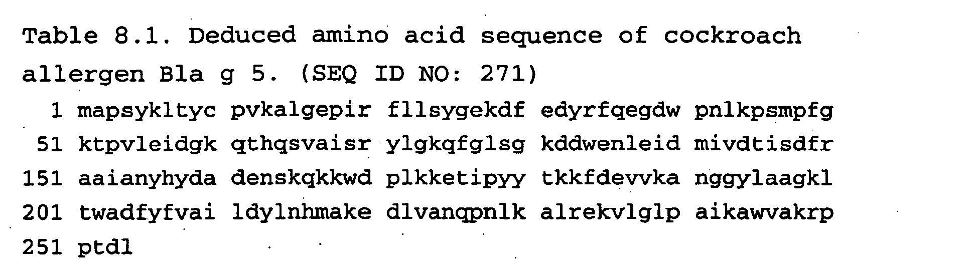
Table 8.2. Predicted MHC Class II-presented epitopes of cockroach allergen Bla g 5. PEPTIDE NO: Pos. Sequence 1 2 3 4 5 6 7 8 9 Score Ii-Key SEQ ID NO: 8.2.1 49 F G K T P V L E I 7.60 - 272 8.2.2 245 W V A K R P P T D 5.85 - 273 8.2.3 91 M I V D T I S D F 5.57 - 274 8.2.4 205 F Y F V A I L D Y 5.40 - 275 8.2.5 19 I R F L L S Y G E 5.10 2 276 8.2.6 55 L E I D G K Q T H 4.60 7 277 8.2.7 99 F R A A I A N Y H 4.20 - 278 Table 8.3. Experimentally defined MHC Class II epitopes of Bla g 5. PEPTIDE NO: Pos. Sequence Ii-Key SEQ ID NO: 8.3.1 92 IVDTISDFRAAIANYHYDAD - 279 8.3.1 212 DYLNHMAKEDLVANQPNLKA - 280 - Carcinoembryonic antigen (CEA) is a tumor associated antigen (TAA) that is expressed in tumors including colon, breast and pancreas. The protein and the cDNA have been used for therapeutic tumor vaccines. A recombinant vaccinia-CEA vaccine has been used to generation cytotoxic T cells specific for human carcinoembryonic antigen epitopes (Tsang KY. J Natl Cancer Inst. 1995 87:982-90). Ii-Key hybrids can be used to develop T helper cell responses to this tumor-associated antigen prior to DNA vaccines of any form. Thus, the clinical value of such a recombinant vaccinia-CEA construct can be enhanced substantially with the products and methods of this invention, as described in this disclosure. Additional CEA vaccination procedures, in which Ii-Key/CEA antigenic epitope hybrids can be applied, are presented below.
- Reisfeld and colleagues demonstrated that an oral DNA vaccine against human CEA prevented growth and dissemination of Lewis lung carcinoma in CEA transgenic mice (Niethammer AG. Vaccine 2001 20:421-9). A DNA vaccine encoding human CEA broke peripheral T-cell tolerance toward this antigen expressed by Lewis lung carcinoma stably transduced with CEA in C57BL/6J mice transgenic for CEA. The vaccine was delivered by oral gavage with an attenuated strain of Salmonella typhimurium (SL7207), and boosted with an antibody-IL2 fusion protein. Both CTL and antigen-presenting dendritic cells were activated as indicated by a decisive increase in their respective activation markers CD2, CD25, CD28 as well as CD48 and CD80.
- Stevenson and colleagues demonstrated that DNA fusion vaccine including MHC Class II epitopes of tetanus toxoid along with a tumor antigen of interest (here CEA) induced cytotoxic T cell responses against defined peptides. Fusion of the fragment C of tetanus toxin to a CEA sequence promoted antibody and CD4+ T cell responses against tested B cell tumors. Using only the first domain of tetanus toxoid, which contains a "universal" helper epitope, followed by two known CTL-recognized epitopes of CEA, they found strong CTL responses to each CTL-recognized peptide to be induced by the engineered construct.
- Diagnostic assays with Ii-Key/antigenic epitope hybrids can be used to monitor therapy and predict outcomes in patients with CEA-positive tumors, as indicated from the following study. PBMC from two CEA-based vaccine clinical trials were analyzed for T cell responses to the same CEA peptide and to an influenza (Flu) control peptide (Arlen P. Cancer Immunol Immunother. 2000 49:517-29). The first trial consisted of three monthly vaccinations of CEA peptide (designated PPP) in adjuvant. The second trial consisted of cohorts receiving three monthly vaccinations of avipox-CEA recombinant (designated AAA) or cohorts receiving a primary vaccination with recombinant vaccinia-CEA followed by two monthly vaccinations with avipox-CEA (designated VAA). Few, if any, CEA-specific T cell responses were seen in patients receiving PPP vaccinations, while the majority of patients receiving the poxvirus CEA recombinants demonstrated increases in CEA-specific T cell responses and no increases in Flu-specific responses. CEA-specific IgG responses developed in patients following recombinant CEA poxvirus vaccinations. T cell responses to the CEA peptide were significantly increased after immunization with the recombinant poxvirus vaccine, as compared with the peptide vaccine (p = 0.028). Clearly poxvirus recombinant-based vaccines are more potent in initiating tumor-antigen-specific T cell responses than are peptide vaccines. Their activity can be further enhanced, by prior vaccination with Ii-Key/CEA antigenic epitope hybrids.
- In the case of tumor antigens such as CEA, Ii-Key/MHC Class II-presented antigenic epitope hybrids create T helper cell responses that augment the development of immune responses to CEA MHC Class I epitopes, for example through dendritic cell licensing. Such CTL activation by MHC Class I epitopes can be generated also by incorporating such MHC Class I epitopes in an Ii-Key MHC Class II-presented hybrids. Several MHC Class I epitope of CEA have been experimentally determined (Kawashima I. Hum Immunol. 1998 59:1-14; Nukaya I. Int J Cancer. 1999 80:92-7; Table 5). Such peptides have been discovered by various techniques. Nested deletions of the cDNA for the antigen of interest lead to protein products, which can be assayed for stimulation of CD8+ cells lines, which recognize the antigen. Given various cell lines recognizing individual epitopes, the localization of T cell epitopes can be approximated within the primary sequence, by analyzing the reactions of each T cell clone to the nested deletion cDNA constructs. Then a library of overlapping peptides through biologically active target regions can be assayed to define exactly the individual determinants. The binding of such peptides to immunopurified MHC Class I molecules can also be assayed, for example by inhibition of binding of a radiolabeled standard peptide to MHC molecules (Kawashima I. Hum Immunol. 1998 59:1-14). The MHC Class I molecules can be immunopurified and bound into microtiter plates, in which the various components of the assay are added sequentially, with appropriate washings. Alternatively the MHC Class I molecules can be detergent-solubilized without purification, for example from a microsomal membrane preparation of a cultured lymphoblastoid cell line, and the complexes separated in a gel filtration column, with the bound radioactive peptide being separated in the protein complexes from the unbound, free peptide. In the work of Kawashima, initial studies were performed with HLA-A2.1 molecules. The highly reactive peptide 9.5.2, which induced vigorous anti-tumor CTL responses, also bound tightly to other common HLA alleles of the A2 supertype (A2.2, A2.3, A2.6 and A6802), thus demonstrating a potential in providing broad and not ethnically biased population coverage. CTL lines were used to identify peptides 9.5.4 and 9.5.6, which elicited CTL lines that lysed tumor cells expressing HLA-A24 and CEA. The cytotoxicity to tumor cells by the CTL lines was antigen-specific since it was inhibited by peptide-pulsed cold target cells as well as by monoclonal antibodies to MHC Class I and CD3 molecules. Similar methods can be used to characterize the biological responses induced by Ii-Key/MHC Class II epitope/MHC Class I epitope hybrids of this disclosure.
- Alternatively, such peptides are identified with algorithms for the prediction of MHC Class I and Class II T cell-recognized epitopes (Lu J. Cancer Res. 2000 60:5223-7). These computer-based predictive algorithms, which are available on the Internet (Parker KC. J Immunol. 1992 149:3580-7; Rammensee HG. Immunogenetics. 1995 41:178-228), were used to identify HL4-B7-restricted CTL epitopes for carcinoembryonic antigen (CEA). Of three candidate peptides, CEA9(632) (IPQQHTQVL) (SEQ ID NO: 281) induced primary CTL responses in lymphocytes from HLA-B7+ normal blood donors when dendritic cells were used as antigen-presenting cells. These CTLs were efficient in killing tumor cells that expressed HLA-B7 and produced CEA.
- Cell lines reflecting the natural T-cell response against MHC Class I epitopes of epithelial cell adhesion molecule, Her-2/neu, and carcinoembryonic antigen in patients with colorectal cancer have been used to identify the respective antigenic epitopes (Nagorsen D. Cancer Res. 2000 60:4850-4). Antigens of epithelial cell adhesion molecule (Ep-CAM), her-2/neu, and CEA were potential targets in antigen-specific vaccination-based cancer therapy. The investigators tested whether a natural specific T-cell response against these antigens already exists in patients with colorectal carcinoma. The IFN-gamma ELISPOT assay was used to detect circulating TAA-reactive T cells directly ex vivo in unstimulated peripheral blood mononuclear cells. They determined that seven of 22 patients, but none of the 8 healthy subjects, had T cells specifically secreting IFN-gamma in response to antigen peptides (n = 4, Ep-CAM; n = 5, her-2/neu; n = 6, CEA). T-cell responses occurred only in patients with metastatic disease (Dukes' stages C and D). The results of this study indicate that natural T-cell responses against tumor antigens occur in approximately one-half of colorectal carcinoma patients with involvement of lymph nodes or distant metastases, but not in colorectal carcinoma patients with disease confined to the bowel tract. Ii-Key/antigenic epitope hybrids containing MHC Class II epitopes can be used to vaccinate patients with localized and metastatic disease against their tumors.
- The amino acid sequence of CEA was obtained from GenBank as 11386171 carcinoembryonic antigen-related cell adhesion molecule 5; carcinoembryonic antigen [Homo sapiens] (Table 1). The primary sequence of human carcinoembryonic antigen (CEA) was deduced from cDNA sequence (Oikawa S. Biochem Biophys Res Commun. 1987 142:511-8). The carcinoembryonic antigen (CEA) contains multiple immunoglobulin-like domains (Oikawa S. Biochem Biophys Res Commun. 1987 144:634-42). Predicted MHC Class II-presented epitopes of CEA are listed in Table 9.2. Designed Ii-Key/CEA hybrids containing some of the MHC Class II-presented epitopes of CEA in Table 9.2 are listed in Table 9.3. Predicted MHC Class I-presented CEA epitopes are listed in Table 9.4. Experimentally defined MHC Class I epitopes of CEA are listed in Table 9.5. Ii-Key/MHC Class II/MHC Class I CEA hybrids are listed in Table 9.6.
-

Table 9.2 Predicted MHC Class II-presented epitopes of CEA. PEPTIDE Pos. Sequence Score Ii-Key SEQ ID NO: 9.2.1 427 YRPGVNLSL 6.40 - 283 9.2.2 535 WVNGQSLPV 6.30 - 284 9.2.3 179 WVNNQSLPV 5.50 - 285 357 6.30 9.2.4 627 WRINGIPQQ 5.80 - 286 9.2.5 249 YRSGENLNL 5.50 - 287 9.2.6 52 LLLVHNLPQ 5.40 - 288 9.2.7 449 WLIDGNIQQ 5.10 12 289 5.78 9.2.8 591 YGPDTPIIS 5.10 - 290 9.2.9 119 FYTLHVIK S 5.10 - 291 9.2.10 79 IIGYVIGTQ 5.00 - 292 Table 9.3. Designed Ii-Key/CEA hybrids containing some of the MAC Class II-presented epitopes of CEA in Table 9.2. A. Non-overlapping epitopes PEPTIDE Pos. Sequence SEQ ID NO: 9.3.1 179
357
535Ac-LRMK-ava-WVNNQSLPV-NH2 293 9.3.2 427 Ac-LRMK-ava-YRPGVNLSL-NH2 294 9.3.3 627 Ac-LRMK-ava-WRINGIPQQ-NH2 295 9.3.4 249 Ac-LRMK-ava-YRSGENLNL-NH2 296 Table 9.4. Predicted MHC Class I-presented CEA epitopes. PEPTIDE Pos. Sequence Score SEQ ID NO: 9.4.1 61 HLFGYSWYK 1350 297 9.4.2 425 TYYRPGVNL 200 298 9.4.3 652 TYACFVSNL 200 299 9.4.4 691 IMIGVLVGV 196 300 9.4.5 605 YLSGANLNL 98 301 Table 9.5. Experimentally defined MHC Class I epitopes of CEA. PEPTIDE Pos. Sequence SEQ ID NO: 9.5.1 691 IMIGVLVGV 302 9.5.2 24 LMTFWNPPV 303 9.5.3 605 YLSGANLNL 304 9.5.4 268 QYSWFVNGTF 305 9.5.5 652 TYACFVSNL 306 9.5.6 61 HLFYSWYK 307 Table 9.6. Ii-Key/MHC Class II/MHC Class I CEA hybrids. PEPTIDE Pos. Sequence SEQ ID NO: 9.6.1 II:179,357,535, I:691 Ac-LRMK-ava-WVNNQSLPV-IMIGVLVGV-NH2 310 9.6.2 II:427, I:425 Ac-LRMK-ava-TYYRPGVNLSL-NH2 311 9.6.3 II:249, I:268 Ac-LRMK-ava-YRSGENLNL-QYSWFVNGTF-NH2 312 9.6.4 II:52, I:61 Ac-LRMK-ava-LLLVHNLPQ-HLFYSWYK-NH2 313 - The ovarian cancer antigen CA-125 is used in immunotherapeutic vaccinations. In one case, vaccination with a mixed vaccine of autogenous and allogeneic breast cancer cells and tumor associated antigens including the breast cancer antigen CA15.3, the carcinoembryonic antigen (CEA) and the ovarian cancer antigen CA125, resulted in immune and clinical responses in breast cancer patients (Jiang XP. Cancer Biother Radiopharm. 2000 15:495-505). The vaccine induced a significant increase in post-vaccination lymphocyte proliferative responses to AUTOC, CA15.3, CEA and CA125 but not ALLOC, compared to pre-vaccination (p < 0.05, p < 0.01, p < 0.05, p < 0.01 and p > 0.05, respectively, a paired t Test).
- The amino acid sequence of CA125 ovarian cancer antigen mucin 16 [Homo sa...[gi:14971110] as listed in Genebank is presented in Table 10.1. Predicted MHC Class II-presented epitopes of CA125, ovarian cancer antigen are listed in Table 10.2. Ii-Key/CA 125 hybrids containing some of the MHC Class II-presented epitopes of Table 10.2 are listed in Table 10.3. Predicted MHC Class I-presented epitopes of CA 125 are listed in Table 10.4. Ii-Key/MHC II epitope/MHC I epitope hybrids are listed in Table 10.5.

Table 10.2. Predicted MHC Class II-presented epitopes of CA125, ovarian cancer antigen. PEPTIDE Pos. Sequence Score Ii-Key SEQ ID NO: 10.2.1 1630 FRFCLVTNL 7.40 9 315 10.2.2 1018 VLFTLNFTI 7.10 - 316 10.2.3 1174 ILFTLNFTI 7.10 - 317 10.2.4 156 LRLDPTGPG 6.80 - 318 10.2.5 1017 LVLFTLNFT 6.20 - 319 10.2.6 861 LVLFTINFT 6.05 - 320 10.2.7 527 IVNLGTSGI 5.70 - 321 10.2.8 1318 LRYMADMGQ 5.70 - 322 10.2.9 1029 LRYEENMQH 5.68 - 323 10.2.10 873 LRYEENMHH 5.68 - 324 10.2.11 1663 VFLDKTLNA 5.60 10 325 10.2.12 1172 LLILFTLNF 5.60 - 326 10.2.13 936 YRPDPKSPG 5.57 2 327 10.2.14 393 LVPFTLNFT 5.50 3 328 10.2.15 430 LRPLFKNTS 5.40 - 329 10.2.16 1185 LRYEENMWP 5.30 9 330 10.2.17 1634 LVTNLTMDS 5.30 - 331 10.2.18 360 FVPITSTPG 5.20 11 332 10.2.19 1209 LRPLFKNTS 5.10 - 333 10.2.20 898 LRPVFKNTS 5.10 - 334 10.2.21 1531 YHPDPVGPG 5.10 - 335 Table 10.3. Ii-Key/CA125 hybrids containing some of the MHC Class II-presented epitopes of Table 10.2. A. Conserved tandem-repeats epitopes PEPTIDE Pos. Sequence SEQ ID NO: 10.3.1 1017 Ac-LRMK-ava-VLFTLNFTI-NH2 336 10.3.2 1173 Ac-LRMK-ava-ILFTLNFTI-NH2 337 10.3.3 860 Ac-LRMK-ava-LVLFTINFT-NH2 338 10.3.4 1028 Ac-LRMK-ava-LRYEENMQH-NH2 339 10.3.5 872 Ac-LRMK-ava-LRYEENMHH-NH2 340 10.3.6 1184 Ac-LRMK-ava-LRYEENMWP-NH2 341 B. Overlapping MHC II epitopes 10.3.7 1629 Ac-LRMK-ava-FRFCLVTNL-NH2 342 10.3.8 1633 Ac-LRMK-ava-LVTNLTMDS-NH2 343 10.3.9 1629 /1633 Ac-LRMK-ava-FRFCLVTNLTMDS-NH2 344 Table 10.4. Predicted MHC Class I-presented epitopes of CA 125. PEPTIDE Pos. Sequence Score SEQ ID NO: 10.4.1 1675 WLGSTYQLV 478 345 10.4.2 1018 VLFTLNFTI 381 346 10.4.3 1174 ILFTLNFTI 381 347 10.4.4 862 VLFTINFTI 381 348 10.4.5 344 LLDRGSLYV 260 349 10.4.6 1506 YLGCQLISL 226 350 10.4.7 1668 TLNASFHWL 223 351 10.4.8 1555 GVTQLGFYV 194 352 10.4.9 1845 GLLGLITCL 182 353 10.4.10 32 KLTRGIIEL 172 354 Table 10.5. Ii-Key/MHC II epitope/MHC I epitope hybrids. Ii-Key/MHC Class PEPTIDE Pos. Sequence SEQ ID NO: 10.5.1 1630 Ac-LRMK-ava-FRFCLVTNL-NH2 355 10.5.2 II:392 1:394, 238, 82, 550 Ac-LRMK-ava-LVPFTLNFTI- NH2 356 - The identification of T cell specific epitopes within the coding sequence of PSA has led to the development of various vaccine strategies that target PSA in an attempt to treat established prostate cancer (Kaufman HL. Expert Opin Biol Ther. 2002 2:395-408). These strategies have included HLA-restricted PSA peptides, dendritic cells pulsed with PSA, recombinant viruses expressing PSA and combinations with different cytokines and cell interaction molecules. Many of these methods are enhanced by use of the products and methods of this disclosure.
- PSA-recombinant pox vaccine constructs are immunogenic and induce antibody responses to a multitude of surface antigens on prostate tumor cell lines by epitope or determinant spreading after stimulation of the immune system by PSA immunization (Cavacini LA. Clin Cancer Res. 2002 8:368-73). Determinant spreading in the antibody responses to prostate cell surface antigens was observed in patients immunized with prostate-specific antigen encoded by recombinant pox vectors. The serum IgG response to cell surface antigens expressed on LNCAP (PSA-positive) and PC-3 (PSA-negative) prostate cancer cell lines were analyzed in individuals with advanced disease receiving vaccinia- or fowlpox-expressed PSA (v-PSA or f-PSA, respectively). Sera from all seven patients in a Phase I study of v-PSA, collected prior to the third immunization, reacted with both prostate tumor cell lines. The majority of individuals (n = 12) in a Phase II trial of v-PSA and f-PSA developed sustainable antibody responses to cell surface antigens on the prostate tumor cell lines. The magnitude and kinetics of these responses depended on the immunization schedule.
- Whiteside and colleagues demonstrated recovery of zeta-chain expression and changes in spontaneous IL-10 production after PSA-based vaccines in patients with prostate cancer (Meidenbauer N. Br J Cancer. 2002 86:168-78). In order to determine a mechanism by which circulating T lymphocytes of patients with prostate cancer have been reported to have functional deficits, including low or absent zeta-chain expression, 10 patients treated with recombinant human prostate specific antigen plus GM-CSF and eight others receiving PSA plus oil emulsion were evaluated. Prior to therapy, the patients had significantly lower zeta-chain expression in circulating CD3+ cells and a higher percentage of zeta-chain negative CD3+ and CD4+ cells than normal donors. The patients' peripheral blood mononuclear cells spontaneously produced more IL-10 ex vivo than those of normal controls. After vaccination, recovery of zeta-chain expression was observed in 50% of patients in both clinical trials. Also, spontaneous IL-10 secretion by peripheral blood mononuclear cells decreased following immunotherapy in patients treated with PSA and GM-CSF. Such therapies will be greatly augmented by products and methods of this disclosure.
- Mann and colleagues demonstrated enhanced CD4+ and CD8+ T cell responses after exposure to PSA alone, PSA targeted to the mannose receptor (mannosylated PSA (PSA-m)), or PSA targeted to Fc receptors by combining PSA with an anti-PSA antibody (AR47.47) (Berlyn KA. Clin Immunol. 2001 101:276-83). PSA and PSA-m are processed primarily through pathways that favor MHC Class II presentation, while the PSA/anti-PSA immune complexes are processed through both Class I and Class II pathways in monocyte-derived dendritic cells.
- Gilboa and colleagues demonstrated that autologous dendritic cells transfected with PSA RNA stimulate CTL responses against metastatic prostate tumors (Heiser A. J Clin Invest. 2002 109:409-17). Autologous dendritic cells transfected with mRNA encoding prostate-specific antigen (PSA) stimulate potent, T cell-mediated antitumor immune responses in vitro. A phase I trial evaluated this strategy for safety, feasibility, and efficacy to induce T cell responses against the PSA in patients with metastatic prostate cancer. In 13 subjects, escalating doses of PSA mRNA-transfected dendritic cells were administered with no evidence of dose-limiting toxicity or adverse effects, including autoimmunity. Induction of PSA-specific T cell responses was consistently detected in all patients, suggesting in vivo bioactivity of the vaccine. Vaccination was further associated with a significant decrease in the log slope of serum PSA levels in six of seven subjects.
- Schlom and colleagues characterized an agonist epitope designated PSA-3A ("A" for agonist) of the PSA-3 CTL epitope which demonstrated enhanced binding to the HLA-A2 allele and enhanced stability of the peptide-MHC complex (Terasawa H. Clin Cancer Res. 2002 8:41-53). T-cell lines generated with either the PSA-3 or the PSA-3A peptide showed higher levels of lysis of targets pulsed with the PSA-3A peptide than those targets pulsed with the PSA-3 peptide. T cells stimulated with dendritic cells (dendritic cells) pulsed with PSA-3A peptide produced higher levels of IFN-gamma than did dendritic cells pulsed with PSA-3 peptide. Dendritic cells infected with a recombinant vaccinia virus containing the agonist amino acid change within the entire PSA gene (designated rV-PSA-3A) were more effective than dendritic cells infected with the rV-PSA vector in enhancing IFN-gamma production by T cells. Finally, the PSA-3A agonist was shown to induce higher levels of T-cell activation, compared with the PSA-3 peptide, in an in vivo model using HLA-A2.1/K(b) transgenic mice. These studies thus demonstrated the potential use of the PSA-3A agonist epitope in both peptide- and vector-mediated immunotherapy protocols for prostate cancer. Such results can be bettered with the products and methods of this disclosure.
- Recombinant PSA proteins incorporating 6xHis (SEQ ID NO: 357) residues were synthesized for magnetic bead attachment allowing antigen isolation and delivery to APC for processing and presentation (Turner MJ. J Immunol Meth. 1998 256:107-19). PSA deletion constructs were generated by amplifying 3' deletions of PSA using a constant 5' primer and five individual 3' primers starting at 736 bp, 610 bp, 505 bp, and 394 bp. The recombinant PSA proteins encoded 261, 231, 189, 154 and 117 amino acids. PSA-specific Class I- and II-restricted T cell hybridomas were generated by fusing Thy-1 + tumor infiltrating lymphocytes (TIL) isolated from BALB/c mice challenged with Line 1/PSA/IL-2 tumors to the T cell fusion partner BWZ.36. MHC Class I (PSA 188-197) and Class II (PSA 238-253) T cell epitopes were identified.
- The amino acid sequence of prostate specific antigen (PSA) as obtained from GenBank 45021731 kallikrein 3 is presented in Table 11.1. A cDNA vaccine for this antigen is available (Kim JJ. Oncogene. 1998 17:3125-35). Predicted MHC Class II-presented epitopes of PSA are listed in Table 11.2. Experimentally defined MHC Class II-presented epitopes of PSA are listed in Table 11.3. Ii-Key/PSA hybrids containing some of the MHC Class II-presented epitopes of Tables 11.2 and 11.3 are listed in Table 11.4. Predicted MHC Class I-presented epitopes of PSA are listed in Table 11.5. Experimentally defined MHC Class I-presented epitopes of PSA are listed in Table 11.6. Ii-Key/PSA MHC II-presented epitope/PSA MHC I-presented epitope hybrids are listed in Table 11.7.
-

Table 11.2. Predicted MHC Class II-presented epitopes of PSA. PEPTIDE Pos. Sequence Score Ii-Key SEQ ID NO: 11.2.1 59 WVLTAAHCI 8.80 - 359 11.2.2 2 WVPVVFLTL 8.10 - 360 11.2.3 124 LRLSEPAEL 6.60 - 361 11.2.4 67 IRNKSVILL 5.40 8 362 11.2.5 74 LLGRHSLFH 5.20 - 363 11.2.6 72 VILLGRHSL 4.70 - 364 11.2.7 223 LQGITSWGS 4.58 - 365 Table 11.3. Experimentally defined MAC Class 11-presented epitopes of PSA. PEPTIDE Pos. Sequence Ii-Key SEQ ID NO: 11.3.1 238 ERPSLYTKVVHYRKWI - 366 Table 11.4. Ii-Key/PSA hybrids containing some of the MHC Class II-presented epitopes of Tables 11.2 and 11.3. A. Non-overlapping epitopes PEPTIDE Pos. Sequence SEQ ID NO: 11.4.1 2 Ac-LRMK-ava-WVPVVFLTL-NH2 367 11.4.2 124 Ac-LRMK-ava-LRLSEPAEL-NH2 368 11.4.3 223 Ac-LRMK-ava-LQGITSWGS-NH2 369 B. Overlapping epitopes PEPTIDE Pos. Sequence SEQ ID NO: 11.4.4 59 Ac-LRMK-ava-WVLTAAHCI-NH2 370 11.4.5 67 Ac-LRMK-ava-IRNKSVILL-NH2 371 11.4.6 72 Ac-LRMK-ava-VILLGRHSL-NH2 372 11.4.7 74 Ac-LRMK-ava-LLGRHSLFH-NH2 373 11.4.8 59, 67, 72, 74 Ac-LRMK-ava-WVLTAAHCIRNKSVILLGR HSLFH-NH2 374 Table 11.5. Predicted MHC Class I-presented epitopes of PSA. PEPTIDE Pos. Sequence Score SEQ ID NO: 11.5.1 46 GRAVCGVL 2000 375 11.5.2 67 IRNKSVILL 2000 376 11.5.3 124 LRLSEPAEL 2000 377 11.5.4 18 APLILSRIV 660 378 11.5.5 7 FLTLSVTWI 607 379 11.5.6 249 YRKWIKDTI 600 380 Table 11.6. Experimentally defined MHC Class I-presented epitopes of PSA. PEPTIDE Pos. Sequence SEQ ID NO: 11.6.1 188 HPQKVTKFML 381 - Table 11.7. Ii-Key/PSA MHC II-presented epitope/PSA MHC I-presented epitope hybrids.
A Overlapping epitopes PEPTIDE Pos. Sequence SEQ ID NO: 11.7.1 2 Ac-LRMK-ava-WVPVVFLTLSVTWI-NH2 382 11.7.2 67 Ac-LRMK-ava-IRNKSVILL-NH2 383 11.7.3 124 Ac-LRMK-ava-LRLSEPAEL-NH2 384 - Melanoma is a leading target in the development of therapeutic peptide and DNA vaccines because several specific tumor-associated antigens have been identified, efficiency of vaccinating mice with peptide or DNA vaccines in treating melanoma is proved, and use of comparable vaccines in the clinic has had occasionally promising results. The use of Ii-Key/melanoma antigenic epitope hybrids in melanoma vaccination is considered in respective Examples concerning melanocyte protein Pmel17, gp100, tyrosinase, and tyrosinase-s related protein.
- Storkus and colleagues identified several MHC Class II-presented epitopes of gp 100/pmel17 and tyrosinase melanocyte-associated antigens and tested the response of tumor-reactive human CD4+ T cells from various melanoma patients against these peptides (Kierstead LS. Br J Cancer. 2001 85:1738-45). Two known and three novel CD4+ T cell epitopes were found using an IFN-gamma ELISPOT assay. Often freshly-isolated PBMC from HLA-DR4+ melanoma patients that are currently disease-free reveal elevated Th1-type CD4+ T-cells that recognize these peptides. Ii-Key/antigenic epitope hybrids incorporating these epitopes are presented in this Disclosure.
- One problem in tumor immunotherapy is the fact that hosts can be tolerized to self proteins of the tumor. Intracutaneous immunization of C57BL/6 mice with a human Pmel17/gp100 DNA vaccine, but not the murine DNA, induces T cell-mediated B16 melanoma protection in vivo (Wagner SN. J Invest Dermatol. 2000 115:1082-7). This state of unresponsiveness to the autoantigen Pmel17/gp100 was broken by immunization with a plasmid DNA construct encoding the autologous form of the molecule. Mice receiving of Pmel17/gp100 DNA mounted an antigen-specific cytotoxic T lymphocyte response to M3 melanoma. Furthermore M3 tumors growing in immunized mice lost expression of this melanoma-associated antigen whereas M3 melanomas appearing in control-vector-treated animals were still Pmel17/gp100-positive. Ii-Key/antigenic epitope hybrids with appropriate immunization schemes and adjuvants can preferentially induce a Th1 or Th2 pattern of response thereby breaking tolerance.
- The amino acid sequence of melanocyte protein Pmel 17 was obtained at NCBI, >gi|1125063|gb|AAB00386.1| melanocyte protein Pmel 17 [Homo sapiens] = >gi|639590|gb|AAC60634.1| gp100 [Homo sapiens].
-

Table 12.2. Predicted MHC Class II-presented epitopes of gp 100. PEPTIDE Pos. Sequence Score Ii-Key SEQ ID NO: 12.2.1 150 VYVWKTWGQ 6.80 - 386 12.2.2 423 WVETTREL 6.40 - 387 12.2.3 477 LYRYGSFSV 6.40 3 388 12.2.4 290 VVLQAAIPL 6.40 10 389 12.2.5 552 LVLHQILKG 6.10 - 390 12.2.6 596 VPLIVGILL 5.80 - 391 12.2.7 600 VGILLVLMA 5.80 - 392 12.2.8 605 VLMAVVLAS 5.80 - 393 12.2.9 604 LVLMAVVLA 5.70 - 394 12.2.10 3 LVLKRCLLH 5.40-7.20 - 395 12.2.11 615 IYRRRLMKQ 5.10-5.70 - 396 12.2.12 616 YRRRLMKQD 4.50 - 397 12.2.13 48 LYPEWTEAQ 4.30 8 398 Table 12.3. Experimentally defined MHC Class II-presented epitopes of gp100. PEPTIDE Pos. Sequence Ii-Key SEQ ID NO: 12.3.1 44 WNRQLYPEWTEAQRLD 4 399 12.3.2 615 IYRRRLMKQDFSVPQLPHS - 400 12.3.3 576 SLAVVSTQLIMPG - 401 12.3.4 175 GRAMLGTHTMEVTVY - 402 12.3.5 74 GPTLIGANASFSIALN - 403 Table 12.4. Ii-Key/gp 100 hybrids containing some of the MHC Class II-presented epitopes of Table 12.2 and 12.3. A. Non-overlapping epitopes PEPTIDE Pos. Sequence SEQ ID NO: 12.4.1 615 Ac-LRMK-ava-IYRRRLMKQDFSVPQLPHS-NH2 404 12.4.2 3 Ac-LRMK-ava-LVLKRCLLH-NH2 405 12.4.3 150 Ac-LRMK-ava-VYVWKTWGQ-NH2 406 12.4.5 423 Ac-LRMK-ava-WVETTAREL-NH2 407 12.4.6 477 Ac-LRMK-ava-LYRYGSFSV-NH2 408 B. Overlapping epitopes PEPTIDE Pos. Sequence SEQ ID NO: 12.4.7 44 Ac-LRMK-ava-WNRQLYPEWTEAQRLD-NH2 409 12.4.8 48 Ac-LRMK-ava-LYPEWTEAQ-NH2 410 12.4.9 44, 48 Ac-LRMK-ava-WNRQLYPEWTEAQRLD-NH2 411 12.4.10 596 Ac-LRMK-ava-VPLIVGILL-NH2 412 12.4.11 600 Ac-LRMK-ava-VGILLVLMA-NH2 413 12.4.12 605 Ac-LRMK-ava-VLMAVVLAS-NH2 414 12.4.13 596, 600, 605 Ac-LRMK-ava-VPLIVGILLVLMAVVLAS-NH2 415 Table 12.5. Predicted MHC Class I-presented epitopes of gp 100. PEPTIDE Pos. Sequence Score SEQ ID NO: 12.5.1 619 RLMKQDFSV 1495 416 12.5.2 520 LPKEACMEI 629 417 12.5.3 602 ILLVLMAVV 412 418 12.5.4 479 RYGSFSVTL 400 419 12.5.5 154 KTWGQYWQV 315 420 12.5.6 17 ALLAVGATK 45 421 12.5.7 614 LIYRRRLMK 20 422 Table 12.6. Experimentally defined MAC Class I-presented epitopes of gp100. PEPTIDE Pos. Sequence SEQ ID NO: 12.6.1 280 YLEPGPVTA 423 12.6.2 17 ALLAVGATK 424 12.6.3 209 ITDQVPFSV 425 12.6.4 614 LIYRRRLMK 426 12.6.5 619 RLMKQDFSV 427 12.6.6 639 RLPRIFCSC 428 12.6.7 154 KTWGQYWQV 429 12.6.8 177 AMLGTHTMEV 430 12.6.9 570 SLADTNSLAV 431 12.6.10 70 VSNDGPTLI 432 12.6.11 87 ALNFPGSQK 433 Table 12.7. Designed Ii-Key/gp 100 hybrids containing some of the MHC Class I- and Class II-presented epitopes of Tables 4 and 5. A. Non-overlapping epitopes PEPTIDE Pos. Sequence SEQ ID NO: 12.7.1 II:520, I:552 Ac-LRMK-ava-LPKEACMEI-LVLHQILKG-NH2 434 12.7.2 II:17, I:3 Ac-LRMK-ava-ALLAVGATK-LVLKRCLLH- NH2 435 B. Overlapping epitopes PEPTIDE Pos. Sequence SEQ ID NO: 12.7.3 II:570, I:576 Ac-LRMK-ava-SLADTNSLAVVSTQLIMPG-NH2 436 12.7.4 II:177, I: 175 Ac-LRMK-ava-GRAMLGTHTMEVTVY-NH2 437 12.7.5 II:70, II87, I:74 Ac-LRMK-ava-VSNDGPTLIGANASFSIALNFPGSQK-NH2 438 12.7.6 II:614 (619), I:615 Ac-LRMK-ava-LIYRRRLMKQDFSVPQLPHS-NH2 439 12.7.7 II:154, I:150 Ac-LRMK-ava-VYVKTWGQYWQV-NH2 440 12.7.8 II:479, I:477 Ac-LRMK-ava-LYRYGSFSVTL-NH2 441 - The amino acid sequence of tyrosinase-related protein 2 as given in GenBank gi|731026|sp|P40126|TYR2_HUMAN Dopachrome tautomerase precursor (DT) (DCT) (Dopachrome delta-isomerase) (Tyrosinase-related protein 2) (TRP-2) (TRP2) is presented in Table 13.1. Predicted MHC Class II-presented epitopes of TRP-2 are listed in Table 13.2. Designed Ii-Key/TRP-2 antigenic epitope hybrids containing some of the MHC Class II-presented epitopes of Table 13.2 are listed in Table 13.3. Predicted MHC Class-I presented epitopes of TRP-2 are listed in Table 13.4. Experimentally defined MHC Class I-presented TRP-2 epitopes are listed in Table 13.5. Designed Ii-Key/TRP-2 hybrids containing some of the MHC Class I- and II-presented epitopes of Tables 13.2, 13.3, 13.4 and 13.5 are listed in Table 13.6.

Table 13.2. Predicted MHC Class II-presented epitopes of TRP-2. PEPTIDE Pos. Sequence Score Ii-Key SEQ ID NO: 13.2.1 156 YVITTQHWL 8.20 9 443 13.2.2 451 LGYSYAIDL 7.60 - 444 13.2.3 64 VRADTRPWSG 6.60 - 445 13.2.4 483 LVGLFVLLA 6.10 - 446 13.2.5 272 LISRNSRFS 5.70 - 447 13.2.6 392 FVVLHSFTD 5.50 - 448 13.2.7 219 WHRYHLLCL 5.40 7 449 13.2.8 498 LRKGYTPLM 5.30 - 450 13.2.9 365 VMSLHNLVH 5.20 - 451 13.2.10 474 LVVMGTLVA 5.10 - 452 Table 13.3. Designed Ii-Key/TRP-2 antigenic epitope hybrids containing some of the MHC Class II-presented epitopes of Table 13.2. PEPTIDE Pos. Sequence Score SEQ ID NO: 13.3.11 156 Ac-LRMK-ava-YVITTQHWL-NH2 453 13.3.12 451 Ac-LRMK-ava-LGYSYAIDL-NH2 454 13.3.13 64 Ac-LRMK-ava-VRADTRPSG-NH2 455 Table 13.4. Predicted MHC Class-I presented epitopes of TRP-2. PEPTIDE Pos. Sequence Score SEQ ID NO: 13.4.1 277 SRFSSWETV 3000 456 13.4.2 408 KRFNPPADA 3000 457 13.4.3 325 IRDCLSLQK 2000 458 13.4.4 150 KRVHPDYVI 1800 459 13.4.5 427 NRMYNMVPF 1000 460 13.4.6 485 GLFVLLAFL 999 461 13.4.7 180 SVYDFFVWL 973 462 13.4.8 490 LAFLQYRRL 665 463 13.4.9 431 NMVPFFPPV 363 464 13.4.10 185 FVWLHYYSV 348 465 13.4.11 180 SVYDFFVWL 504 466 13.4.12 199 GPGRPYRAI 440 467 13.4.13 264 AARPDDPTL 360 468 13.4.14 353 GFDKADGTL 330 469 13.4.15 408 KRFNPPADA 300 470 13.4.16 189 HYYSVRDTL 280 471 13.4.17 331 LQKFDNPPF 240 472 Table 13.5. Experimentally defined MHC Class I-presented TRP-2 epitopes. PEPTIDE Pos. Sequence SEQ ID NO: 13.5.1 180 SVYDFFVWL 473 13.5.2 360 TLDSQVMSL 474 13.5.3 288 SLDDYNHLV 475 13.5.4 455 YAIDLPVSV 476 Table 13.6. Designed Ii-Key/TRP-2 hybrids containing some of the MHC Class I and II-presented epitopes of Tables 13.2, 13.3, 13.4 and 13.5. PEPTIDE Pos. Sequence SEQ ID NO: 13.6.1 I:180; II:156 Ac-LRMK-ava-YVITTQHWL-SVYDFFVWL-NH2 477 13.6.2 I:455; II: 451 Ac-LRMK-ava-LGYSYAIDLPVSV-NH2 478 13.6.3 I:360; II:365 Ac-LRMK-ava-TLDSQVMSLHNLVH-NH2 479 - Tyrosinase has many advantages as a target antigen for the immunotherapy of patients with melanoma because it is expressed in nearly all melanoma specimens with a high degree of cellular homogeneity, and its distribution in normal tissues is limited to melanocytes. Several MHC Class I-presented epitopes have been identified and used clinically, and MHC Class II-presented epitopes have been discovered. The following summaries of the current state-of-the-art in identification and use of peptide vaccines, DNA vaccines, and dendritic cell charging with peptide preparations (tumor cell lysates) are presented in part to illustrate the value of the products and methods of this Disclosure to improving these procedures.
- Rosenberg and colleagues identified a HLA-A2.1-presented restricted melanoma tyrosinase epitope (tyrosinase8-17; CLLWSFQTSA) (SEQ ID NO: 480) (Riley JP. J Immunother. 2001 24:212-20). In this study, the comparative binding to HLA-A2.1 of a series of algorithm-predicted peptides versus that of a standard peptide with an intermediate binding affinity was determined. Twelve peptides with binding affinities within 80% of that of the standard peptide stimulated peripheral blood mononuclear cells (PBMC) in vitro from three HLA-A2.1+ patients with metastatic melanoma. PBMC from 23 HLA-A2.1+ patients were stimulated in vitro with tyrosinase:8-17. Eleven bulk T-cell cultures demonstrated specific peptide recognition, and six of these also recognized HLA-A2.1+ tyrosinase+ melanoma cells. This epitope can be incorporated in an Ii-Key/MHC Class II-presented epitope/MHC Class I-presented epitope hybrid.
- Weber and colleagues found that patients with resected melanoma mounted an immune response against gp100(209-217)(210M) (IMDQVPSFV) (SEQ ID NO: 481) and tyrosinase(368-376)(370D) (YMDGTMSQV) (SEQ ID NO: 482), emulsified with incomplete Freund's adjuvant (Lee P. J Clin Oncol. 2001 19:3836-47). Patients received peptides/IFA with or without IL-12 (30 ng/kg) to evaluate the toxicities and immune responses. Immunizations were administered every 2 weeks for 8 weeks, then every 4 weeks for 12 weeks, and then once 8 weeks later. Thirty-four of 40 patients developed a positive skin test response to the gp100 peptide but none responded to the tyrosinase peptide. Immune responses were measured by release of gamma-interferon in an enzyme-linked immunosorbent assay (ELISA) by effector cells in the presence of peptide-pulsed antigen-presenting cells or by an antigen-specific tetramer flow cytometry assay. Thirty-three of 38 patients demonstrated an immune response by ELISA after vaccination, as did 37 of 42 patients by tetramer assay. Twenty-four of 48 patients relapsed with a median follow-up of 20 months, and 10 patients in this high-risk group have died.
- Slingluff and colleagues evaluated peptide vaccine immunogenicity of several peptides restricted to different HLA-A alleles in draining lymph nodes and peripheral blood of melanoma patients because vaccine trials have been limited mostly to those associated with HLA-A2, and immune responses have been detected inconsistently (Yamshchikov GV. Int J Cancer. 2001 92:703-11). They vaccinated stage IV melanoma patients with a mixture of gp100 and tyrosinase peptides restricted by HLA-A1 (DAEKSDICTDEY)(SEQ ID NO: 483), HLA-A2(YLEPGPVTA (SEQ ID NO: 484) and YMDGTMSQV (SEQ ID NO: 485)) and HLA-A3 (ALLAVGATK) (SEQ ID NO: 486) in an emulsion with GM-CSF and Montanide ISA-51 adjuvant. CTL responses to vaccinating peptides were found in a lymph node draining a vaccine site (sentinel immunized node, SIN) in 5/5 patients (100%) in PBLs of 2/5 patients (40%). Peptides restricted by HLA-A1 and -A3 and HLA-A2 restricted peptide, YMDGTMSQV (SEQ ID NO: 485), were immunogenic.
- Cytotoxic T lymphocytes against melanoma-associated antigens were induced by a recombinant vaccinia virus vector expressing multiple immunodominant epitopes and costimulatory molecules in vivo (Oertli D. Hum Gene Ther. 2002 13:569-75). Patients received psoralen-UV-treated and replication-incompetent recombinant vaccinia virus encoding the three immunodominant HLA-A*0201-restricted epitopes Melan-A(27-35), gp100(280-288), and tyrosinase(1-9) together with two costimulatory molecules, B7.1 and B7.2, in the context of systemic granulocyte-macrophage colony-stimulating factor (GM-CSF) treatment. Subsequent boosts used corresponding synthetic nona-peptides and GM-CSF. Within 12 days of injection of the recombinant vector, cytotoxic T cell responses specific for engineered epitopes were detected in three of three patients. During the vaccination treatment, antigen-specific CTL frequencies exceeding 1:10,000 peripheral CD8+ T cells could be observed.
- Two stage IV melanoma patients vaccinated with an HLA-A2- or HLA-A24-restricted tyrosinase peptide, and GM-CSF had long-term freedom from recurrence (Scheibenbogen C. Int J Cancer. 2002 99:403-8). While the patients had experienced 9 and 12 relapses (mostly subcutaneous), respectively, during the 3 years before vaccination, they experienced freedom from relapse for more than 2 years after vaccination. T-cell responses to the vaccine peptide were found in the peripheral blood of both patients using an IFN-gamma ELISPOT assay.
- Mule and colleagues found that addition of keyhole limpet hemocyanin (KLH) augmented the efficacy of both tumor lysate-pulsed dendritic cells and peptide-pulsed dendritic cells immunizations for immune priming and rejection of established metastases of the D5 subline of B16 melanoma in vivo (Shimizu K. Cancer Res. 2001 61:2618-24). Interleukin 2 further augmented the enhancement afforded by KLH, as measured by cure rates and overall survival, in the absence of autoimmune depigmentation. KLH added to dendritic cells immunizations markedly enhances tumor-specific T cell production of IFN-gamma. D5 melanoma exposed to similar levels of IFN-gamma results in substantial expression of MHC Class I molecules. Immunization with dendritic cells pulsed with KLH and mouse tyrosinase-related protein-2 peptide results in enhanced reduction of B16 melanoma metastases; the effect is most pronounced in a setting where tyrosinase-related protein-2 peptide-pulsed dendritic cells alone are completely ineffective.
- Therapeutic efficacy of a tumor cell-based vaccine against B16 melanoma requires disruption of either of two immunoregulatory mechanisms that control autoreactive T cell responses: the cytotoxic T lymphocyte-associated antigen (CTLA)-4 pathway or the CD25+ regulatory T cells. Combination of CTLA-4 blockade and depletion of CD25+ T cells results in maximal tumor rejection (Sutmuller RP. J Exp Med. 2001 194:823-32). Efficacy of the antitumor therapy correlates with the extent of autoimmune skin depigmentation as well as with the frequency of tyrosinase-related protein 2(180-188)-specific CTLs detected in the periphery. Furthermore, tumor rejection is dependent on the CD8+ T cell subset. The CTL response against melanoma antigens is an important component of the therapeutic antitumor response, and the reactivity of these CTLs can be augmented through interference with immunoregulatory mechanisms. The synergism in the effects of CTLA-4 blockade and depletion of CD25+ T cells indicates that CD25+ T cells and CTLA-4 signaling represent two alternative pathways for suppression of autoreactive T cell immunity. Simultaneous intervention with both regulatory mechanisms is, therefore, a promising concept for the induction of therapeutic antitumor immunity.
- The amino acid sequence of tyrosinase as given in GenBank 4507753|ref|NP_000363.1| tyrosinase (oculocutaneous albinism IA); Tyrosinase [Homo sapiens] is listed in Table 14.1. Predicted MHC Class II-presented epitopes of tyrosinase are listed in Table 14.2. Experimentally defined MHC Class II-presented epitopes of tyrosinase are listed in Table 14.3. Designed Ii-Key/tyrosinase hybrids containing some of the MHC Class II-presented epitopes of Table 14.2 and 14.3 are listed in Table 14.4. Predicted MHC Class I-presented epitopes of tyrosinase are listed in Table 14.5. The experimental identification of MHC Class I-presented epitopes of tyrosinase (Pos. 240, 368, 146) is described in the gp100 example. Experimentally defined MHC Class I-presented epitopes of tyrosinase are listed in Table 14.6. Designed Ii-Key/tyrosinase hybrids containing some of the MHC Class I- and MHC Class II-presented epitopes of Tables 14.2, 14.3, 14.4 and 14.5 are listed in Table 14.7.
-

Table 14.2. Predicted MHC Class II-presented epitopes of tyrosinase. PEPTIDE Pos. Sequence Score Ii-Key SEQ ID NO: 14.2.1 401 LRRHRPLQE 6.60 6 488 14.2.2 179 MHYYVSMDA 6.40 - 489 14.2.3 400 WLRRHRPLQ 6.25 5 490 14.2.4 118 LVRRNIFDL 5.70 9 491 14.2.5 366 LHIYMNGTM 5.50 - 492 14.2.6 368 IYMNGTMSQ 5.40 - 493 14.2.7 182 YVSMDALLG 5.50 - 494 14.2.8 150 VIPIGTYGQ 5.50 7 495 14.2.9 338 FSFRNTLEG 5.40 - 496 14.2.10 498 LLCRHKRKQ 5.30 - 497 14.2.11 1 MLLAVLYCL 5.20 - 498 14.2.12 167 FNDINIYDL 5.20 - 499 Table 14.3. Experimentally defined MHC Class II-presented epitopes of tyrosinase. PEPTIDE Pos. Sequence Ii-Key SEQ ID NO: 14.3.1 56 QNILLSNAPLGPQFP - 500 14.3.2 365 ALHIYMNGTMSQVQGSA - 501 14.3.3 156 YGQMKNGSTPMFNDINIYDL - 502 Table 14.4. Designed Ii-Key/tyrosinase hybrids containing some of the MHC Class II-presented epitopes of Table 14.2 and 14.3. Non-overlapping PEPTIDE Pos. Sequence SEQ ID NO: 14.4.1 56 Ac-LRMK-ava-QNILLSNAPLGPQFP-NH2 503 14.4.2 118 Ac-LRMK-ava-LVRRNIFDL-NH2 504 14.4.3 338 Ac-LRMK-ava-FSFRNTLEG-NH2 505 14.4.4 498 Ac-LRMK-ava-LLCRHKRKQ-NH2 506 Overlapping epitopes PEPTIDE Pos. Sequence SEQ ID NO: 14.4.5 365 Ac-LRMK-ava-ALHIYMNGTMSQVQGSA-NH2 507 14.4.6 366 Ac-LRMK-ava-LHIYMNGTM-NH2 508 14.4.7 368 Ac-LRMK-ava-IYMNGTMSQ-NH2 509 14.4.8 365 366 and 368 Ac-LRMK-ava-ALHIYMNGTMSQ-NH2 510 14.4.9 182 Ac-LRMK-ava-YVSMDALLG-NH2 511 14.4.10 179 Ac-LRMK-ava-MHYYVSMDA-NH2 512 14.4.11 179 and 182 Ac-LRMK-ava-MHYYVSMDALLG-NH2 513 14.4.12 150 Ac-LRMK-ava-VIPIGTYGQ-NH2 514 14.4.13 156 Ac-LRMK-ava-YGQMKNGSTPMFNDINIYDL-NH2 515 14.4.14 167 Ac-LRMK-ava-FNDINIYDL-NH2 516 14.4.15 150 156 and 167 Ac-LRMK-ava-VIPIGTYGQMKNGSTPMFNDINIYDL-NH2 517 Table 14.5. Predicted MHC Class I-presented epitopes of tyrosinase. PEPTIDE Pos. Sequence Score SEQ ID NO: 14.5.1 243 KCDICTDEY 25.0 518 14.5.2 369 YMNGTMSQV 531.4 519 14.5.3 1 MLLAVLYCL 309.1 520 14.5.4 207 FLPWHRLFL 540.5 521 Table 14.6. Experimentally defined MHC Class I-presented epitopes of tyrosinase. PEPTIDE Pos. Sequence SEQ ID NO: 14.6.1 240 DAEKSDICTDEY 522 14.6.2 368 YMDGTMSQV 523 14.6.3 146 SSDYVIPIGTY 524 Table 14.7. Designed Ii-Key/tyrosinase hybrids containing some of the MHC Class I- and MHC Class II-presented epitopes of Tables 14.2, 14.3, 14.5, and 14.6). Non-overlapping epitopes PEPTIDE Pos. Sequence SEQ ID NO: 14.7.1 240 and 56 Ac-LRMK-ava-DAEKSDICTDEY- QNILLSNAPLGPQFP-NH2 525 14.7.2 207 and 401 Ac-LRMK-ava-FLPWHRLFL -LRRHRPLQE-NH2 526 Overlapping epitopes PEPTIDE Pos. Sequence SEQ ID NO: 14.7.3 368 (371D) and 365 (366, 368) Ac-LRMK-ava-ALHIYMNGTMSQVQGSA-NH2 527 14.7.4 146 and 156 Ac-LRMK-ava-SSDYVIPIGTYGQMKNGSTPM FNDINIYDL-NH2 528 14.7.5 1 and 1 Ac-LRMK-ava-MLLAVLYCL-NH2 529 - Rosenberg and colleagues immunized metastatic melanoma patients with autologous dendritic cells presenting epitopes derived from the melanoma-associated antigens MART-1 and gp100 (Panelli MC. J Immunother. 2000 23:487-98). The DCs were generated by 5- to 7-day incubation in interleukin-4 (1,000 U/mL) and granulocyte-macrophage colony-stimulating factor (1,000 U/mL) of peripheral blood monocytes obtained by leukapheresis. Before administration, the DCs were pulsed separately with the HLA-A*0201-associated melanoma epitopes MART-1(27-35) and gp-100-209-2M. The DCs were administered four times at 3-week intervals. A first cohort of patients (n = 3) was treated with 6 x 10⁷ DCs and a second cohort (n = 5) with 2 x 10⁸ DCs (in either case, one half of the DCs were pulsed with MART-1(27-35) and the other half was pulsed with gp-100-209-2M). In a final cohort under accrual (n = 2) 2 x 10⁸ DCs were administered in combination with interleukin-2 (720,000 IU/kg every 8 hours). The recovery of DCs after in vitro culture ranged from 3% to 35% (mean, 15%) of the original peripheral blood monocytes. Administration of DCs caused no symptoms at any of the doses, and the concomitant administration of interleukin-2 did not cause toxicity other than that expected for interleukin-2 alone. Monitoring of patients' cytotoxic T lymphocyte reactivity before and after treatment revealed enhancement of cytotoxic T lymphocyte reactivity only in one of five patients tested. Of seven patients evaluated for response, one had a transient partial response with regression of pulmonary and cutaneous metastases.
- Ioannides and colleagues demonstrated reduced recognition of metastatic melanoma cells by autologous MART-1 specific CTL correlated to TAP deficiency (Murray JL. J Immunother. 2000 23:28-35). Class I expression in context with T-cell receptor expression is crucial for peptide presentation and induction of CD8+ cytotoxic T lymphocytes (CTL). Presentation of MHC class I bound peptides depends on transporter-associated proteins (TAP) expression and function. Tumor infiltrating lymphocytes from a patient with melanoma were isolated, expanded in vitro in the presence of interleukin-2, and tested for cytotoxicity against HLA-A2 positive, MART-1 positive autologous tumor cells, an HLA-A2-positive, MART-1 positive melanoma cell line (Mel-501), and HLA-A2-negative melanoma cells. Significant killing occurred against both A2-positive cell lines (63% and 65%, respectively), but not against the A2-negative line (18%) or A2-positive autologous tumor (1.5%). These CTL preferentially recognized the MART-1 peptide F119, 27-35, and gp100 peptide F125, 280-288, resulting in a 30% to 60% enhancement of lysis when autologous tumor or major histocompatibility complex class I "empty" T2 cells were pulsed with either peptide. To address whether the deficiency in autologous tumor recognition might be related to a deficiency in Ag presentation, screening for the presence of TAP1 and TAP2 transcripts by polymerase chain reaction, Southern blotting, and scanning densitometry using sequence-specific primers and probes. Both TAP1 and TAP2 expression levels in the autologous tumor were minimal, yet were upregulated 7- to 18-fold, respectively, by interferon-gamma. Despite this increase, a similar increase in cytotoxicity did not occur. In short, deficiencies in TAP presentation may have functional significance for tumor escape from immunosurveillance and with respect to impending vaccine trials.
- Slingluff and colleagues demonstrated terminal modifications inhibit proteolytic degradation of an immunogenic MART-1(27-35) peptide (Brinckerhoff LH. Int J Cancer 1999 Oct 29;83(3):326-34). The stability of the immunogenic peptide MART-1(27-35) in fresh normal human plasma (NHP) was tested to identify modifications protecting against enzymatic destruction without loss of immunogenicity. MART-1(27-35) peptide (AAGIGILTV) (SEQ ID NO: 530) and modified forms were incubated in plasma for varied time intervals and evaluated for their ability to reconstitute the epitope for MART-1(27-35)-reactive CTL. Loss of CTL reactivity signaled loss of immunoreactive peptide. When 1 microM MART-1(27-35) peptide was incubated in plasma prior to pulsing on target cells, CTL reactivity was lost within 3 hr, and the calculated half-life of this peptide was 22 sec. This degradation was mediated by peptidases. The stability of MART-1(27-35) was markedly prolonged by C-terminal amidation and/or N-terminal acetylation (peptide crapping), or by polyethylene-glycol modification (PEGylation) of the C-terminus. These modified peptides were recognized by CTL.
- Romero and colleagues demonstrated that CpG is an efficient adjuvant for specific CTL induction against tumor antigen-derived peptide (Miconnet I. J Immunol. 2002 168:1212-8). Mice transgenic for a chimeric MHC class I molecule were immunized with a peptide analog of MART-1/Melan-A(26-35) in the presence of CpG oligonucleotides alone or emulsified in IFA. The CTL response was monitored ex vivo by tetramer staining of lymphocytes. In blood, spleen, and lymph nodes, peptide mixed with CpG ODN alone was able to elicit a stronger systemic CTL response as compared with peptide emulsified in IFA. Moreover, CpG ODN in combination with IFA further enhanced the CTL response in terms of the frequency of tetramer+CD8+ T cells ex vivo. The CTL induced in vivo against peptide analog in the presence of CpG ODN are functional, as they were able to recognize and kill melanoma cells in vitro.
- Mitchell and colleagues demonstrated synthetic insertion of signal sequences enhance MHC Class I presentation of a peptide from the melanoma antigen MART-1 (Minev BR. Eur J Immunol. 2000 30:2115-24). Addition of synthetic signal sequences at the N terminus, but not at the C terminus, of an epitope from the human melanoma antigen MART-1 enhanced its presentation in both TAP-deficient and TAP-expressing cells. A peptide construct, composed of the epitope replacing the hydrophobic part of a natural signal sequence, was also very effective. Interestingly, an artificial signal sequence containing the same epitope was the most efficient construct for enhancing its presentation. These peptide constructs facilitated epitope presentation when loaded into the cytosol of TAP-deficient T2 cells, TAP-expressing melanoma cells and human dendritic cells.
- Zajac and colleagues demonstrated immunogenicity of nonreplicating recombinant vaccinia expressing HLA-A201 targeted or complete MART-1/Melan-A antigen (Schutz A. Cancer Gene Ther. 2001 8:655-61). The first recombinant virus expressed a minigene encoding a fusion product between an endoplasmic reticulum (ER)-targeting signal and the HLA-A201 binding MART-1/Melan-A 27-35 peptide. The second viral construct encoded the complete MART-1/Melan-A protein. The capacity of HLA-A201 cells infected with either viral construct to generate and to stimulate MART-1/Melan-A 27-35 specific cytotoxic T-lymphocytes (CTL), was comparatively characterized. The result obtained confirmed the capacity of vaccinia virus-encoded ER-minigene to generate a very strong antigenic signal. In cytotoxicity assays, recognition of target cells infected with high amounts of both recombinant viruses with activated specific CTL clones, resulted in similar lytic activity. With regard to calcium mobilization, TCR down-regulation, IFN-gamma release, and T cell proliferation assays, the targeted epitope elicited 10- to 1000-fold stronger responses. Remarkably, the immunogenic difference between the two formulations, in their respective capacity to generate CTL from naive HLA-A2 peripheral blood mononuclear cells in vitro as measured by tetramer detection, was lower (2-to 3-fold). Recombinant vectors expressing complete antigens have demonstrated their capacity to generate specific responses and such vaccines might take advantage of a broader potential of presentation. However, as demonstrated for the HLA-A201-restricted MART-1/Melan-A immunodominant epitope, nonreplicative vaccinia virus expressing ER-targeted minigenes appear to represent a significantly more immunogenic epitope vaccine formulation. Enhanced further with Ii-RGC.
- Falo and colleagues demonstrated direct transfection and activation of human cutaneous dendritic cells (Larregina AT. Gene Ther. 2001 8:608-17). A gene gun was used to transfect human skin organ cultures with a particular goal of expressing transgenic antigens in resident cutaneous dendritic cells. Gold particles delivered to human skin are observed primarily in the epidermis, even when high helium delivery pressures are used. Langerhans cells resident in the basal epidermis can be transfected, and gene gun delivery is sufficient to stimulate the activation and migration of skin dendritic cells. RT-PCR analysis of dendritic cells, which have migrated from transfected skin, demonstrates transgenic mRNA, indicating direct transfection of cutaneous dendritic cells. Transfected epidermal Langerhans cells can efficiently present a peptide derived from the transgenic melanoma antigen MART-1 to a MART-1-specific CTL.
- Mule and colleagues demonstrated that administration of tumor lysate-pulsed DCs is nontoxic and capable of inducing immunological response to tumor antigen (Chang AE. Clin Cancer Res. 2002 8:1021-32). Fourteen patients with stage IV solid malignancies were treated in cohorts that received 106, 107, and 108 dendtiric cells i.d. every 2 weeks for three vaccines. Each vaccine was composed of a mixture of half DCs pulsed with autologous tumor lysate and the other half with keyhole limpet hemocyanin (KLH). Local accumulation of CD4+ and CD8+ T cells were found at the vaccination sites. There was a significant proliferative response of PBMCs to KLH induced by the vaccine. In 5 of 6 patients, the vaccine resulted in increased IFN-gamma production by PBMCs to KLH in an ELISPOT assay. Using the same assay, 3 of 7 patients' PBMCs displayed increased IFN-gamma production in response to autologous tumor lysate. One patient with melanoma also was observed to have an increased frequency of MART-1- and gp100-reactive CD8(+) T cells after vaccination. By delayed-type hypersensitivity testing, 8 of 9 and 4 of 10 patients demonstrated reactivity to KLH and autologous tumor, respectively. Ii-Key/antigenic epitope hybrids will improve the efficiency of this immunopriming technology.
- Kourilsky and colleagues demonstrated cross-presentation by dendritic cells of tumor antigen expressed in apoptotic recombinant canarypox virus-infected dendritic cells (Motta I. J Immunol. 2001 167:1795-802). Recombinant canarypox virus (ALVAC) encoding the melanoma-associated Ag, Melan-A/MART-1 (MART-1), was tested in cancer immunotherapy, using a dendritic cell (DC)-based approach. ALVAC MART-1-infected DC express, and process and present, the antigen encoded by the viral vector. One consistent feature of infection by ALVAC was induction of apoptosis, and cross-presentation of Ag when uninfected DC are cocultured with ALVAC MART-1-infected DC. Uptake of apoptotic virally infected DC by uninfected DC and subsequent expression of tumor antigen in the latter were verified by flow cytometry analysis, image cytometry, and confocal microscopy. Functional activity was monitored in vitro by the stimulation of a MART-1-specific cytotoxic T cell clone. Heightened efficiency in Ag presentation was indicated by 2- to 3-fold increase in IFN-gamma production by the T cell clone, as compared with the ALVAC-infected DC alone. Cocultures of ALVAC MART-1-infected and uninfected DC are able to induce MART-1-specific T cell immune responses, as assessed by HLA class I/peptide tetramer binding, IFN-gamma ELISPOT assays, and cytotoxicity tests.
- The amino acid sequence of melanoma antigen MART-1 as given in GenBank as 1082589|pir||A55253 melanoma antigen MART-1 - human is presented in Table 15.1. Predicted MHC Class II-presented epitopes of MART-1/Melan-A are listed in Table 15.2. Experimentally defined MHC Class II-presented epitopes of MART-1/Melan-A are listed in Table 15.3. Designed Ii-Key/MART-1/Melan-1 hybrids containing some of the MHC Class II-presented epitopes of Tables 15.2 and 15.3 are listed in Table 15.4. Predicted MHC Class I-presented epitopes of MART-1/Melan-A are listed in Table 15.5. Experimentally defined MHC Class I-presented epitopes of MART-1/Melan-A are listed in Table 15.6. Designed Ii-Key/MART-1 hybrids containing some of the MHC Class I- and Class II-presented epitopes of Tables 15.2, 15.3, 15.5, and 15.6 are listed in Table 15.7.

Table 15.2. Predicted MHC Class II-presented epitopes of MART-1/Melan-A. PEPTIDE Pos. Sequence Score Ii-Key SEQ ID NO: 15.2.1 33 LTVILGVLL 5.00 - 532 15.2.2 35 VILGVLLLI 4.70 - 533 15.2.3 96 WPNAPPAY 4.10 6 534 15.2.4 30 IGILTVILG 4.28 - 535 15.2.5 9 IYGYPKKGH 4.10 - 5.10 - 536 Table 15.3. Experimentally defined MHC Class II-presented epitopes of MART-1/Melan-A. PEPTIDE Pos. Sequence Ii-Key SEQ ID NO: 15.3.1 51 RNGYRALMDKSLHVGTQ CALTRR - 537 Table 15.4. Designed Ii-Key/MART-1/Melan-1 hybrids containing some of the MHC Class II-presented epitopes of Table 1. A. Non-overlapping PEPTIDE Pos. Sequence SEQ ID NO: 15.4.1 95 Ac-LRMK-ava-VVPNAPPAY-NH2 538 15.4.2 8 Ac-LRMK-ava-IYGYPKKGH-NH2 539 15.4.3 51 Ac-LRMK-ava-RNGYRALMDKSLHVGTQCAL TRR-NH2 540 B. Overlapping PEPTIDE Pos. Sequence SEQ ID NO: 15.4.4 32 Ac-LRMK-ava-LTVILGVLL-NH2 541 15.4.5 34 Ac-LRMK-ava-VILGVLLLI-NH2 542 15.4.6 29 Ac-LRMK-ava-IGILTVILG-NH2 543 15.4.7 29 /32 /34 Ac-LRMK-ava-IGILTVILGVLLLI-NH2 544 Table 15.5. Predicted MHC Class I-presented epitopes of MART-1/Melan-A. PEPTIDE Pos. Sequence Score SEQ ID NO: 15.5.1 40 LLLIGCWYC 1289.01 545 15.5.2 56 ALMDKSLHV 1055.10 546 15.5.3 25 EEAAGIGIL 40.0 547 15.5.4 109 AEQSPPPYS 12.0 548 Table 15.6. Experimentally defined MHC Class I-presented epitopes of MART-1/Melan-A. PEPTIDE Pos. Sequence SEQ ID NO: 15.6.1 27 AAGIGILTV 549 15.6 2 32 ILTVILGVL 550 15.6.3 24 AEEAAGIGILT 551 Table 15.7. Designed Ii-Key/MART-1 hybrids containing some of the MHC Class I- and Class II-presented epitopes of 15.2, 15.3, 15.5, and 15.6). SEQ ID NO: Pos. Sequence SEQ ID NO: 15.7.1 27 and 51 Ac-LRMK-ava-AAGIGILTV-RNGYRALMDKSLHVGTQCALT RR-NH2 552 - Immunotherapy directed against the epidermal growth factor receptor which is overexpressed on some cancer cells can control the growth of those tumors. HER-2/neu is over-expressed on tumors in up to 30% of patients with invasive breast cancer and that over-expression is associated with poor clinical outcome. Carr et al. demonstrated in a retrospective consecutive series from 1995 to 1999 that the HER-2/neu gene was amplified in invasive breast carcinomas of 40 of 90 patients (43%) (Carr JA. Arch Surg. 2000 135:1469-7420). Following initial therapy, patients with HER-2/neu amplification had a shorter median disease-free interval (22 months) than did patients with breast cancers not amplifying that gene (40 months; p = .003). Disease recurred in seventy-two (72%) patients, with 18 (25%) recurring locally. HER-2/neu gene amplification is an independent prognostic indicator for a subset of breast cancer patients who are at high risk for early recurrence regardless of tumor grade, estrogen/progesterone receptor status, and lymph node status. In both early stage, lymph node-negative and node-positive disease, as well as in women with metastatic disease, HER-2/neu overexpression is associated with worse survival. Women with tumors that overexpress HER-2/neu have a less favorable outcome despite adjuvant treatment with either hormonal therapy or chemotherapy. Among HER-2/neu-negative, early stage patients in the Naples GUN trial, tamoxifen benefited overall survival. However, among patients with HER-2/neu-gene amplification, tamoxifen did not improve survival (De Placido S. Br J Cancer. 1990 62:643-6). HER-2/neu over-expression is an independent predictor for tamoxifen failure. Over-expression of HER-2/neu is selective for tumor cells and is observed early in the course of malignant transformation. More importantly, the cytological characteristics of HER-2/neu over-expression (32%) in primary and metastatic lesions is nearly identical (Masood S. Ann Clin Lab Sci. 2000 30:259-65). Inasmuch as micrometastases are the primary source of relapse following primary therapy and HER-2/neu is over-expressed in metastases, HER-2/neu is an excellent target for immunotherapy of patients with early disease, both to consolidate the anti-tumor response locally and to eradicate micrometastases. Likewise, HER-2/neu should be targeted in conjunction with other major treatment regimens in patients who have relapsed following initial therapy.
- Of many approaches to targeting HER-2/neu, the clinically most advanced approach is passive immunotherapy with trastuzumab (Herceptin®), an FDA-approved humanized monoclonal antibody that binds to the extracellular domain of the HER-2/neu receptor for epidermal growth factor (EGF). This monoclonal antibody is indicated both as a single agent and in combination with classical chemotherapies. Slamon et al. evaluated Herceptin® in combination with doxorubicin and cyclophosphamide (AC), or paclitaxel in 496 women with metastatic breast carcinomas that over expressed HER-2/neu (Vogel CL. J Clin Oncol. 2002 20:719-26; Slamon DJ. N Engl J Med. 2001 344:783-92). Patients receiving Herceptin®, as compared to patients randomized to chemotherapy alone (either paclitaxel or AC), had a significantly longer time to disease progression (7.4 mo vs. 4.6 mo; p<0.0001), a higher rate of objective response (50% vs. 32%; p<0.001), a longer duration of response (median 9.1 vs. 6.1; p<0.001), a higher 1 year survival rate (78% vs. 67%; p = 0.008), longer survival (median survival 25.1 mo vs. 20.3 mo; p = 0.046), and a 20% reduction in the risk of death.
- While clinical trials might proceed to alternate trastuzumab dosing regimens and combination therapies, one can suggest that the mechanism of action of trastuzumab will not lead to significantly increased efficacy. Specifically, Trastuzumab blocks the HER-2/neu EGF receptor and induces antibody dependent cellular cytotoxicity (Sliwkowski MX. Semin Oncol. 1999 4 Supsl 12:60-70). ADCC does not lead to antigen-specific memory of T- or B-lymphocytes, nor does it induce proliferation of antigen-specific cytotoxic T-lymphocytes.
- HER-2/neu is also the target for several vaccine trials to induce an active specific immune response. In the NCI PDQ, three current clinical trials use HER-2/neu protein, antigen-pulsed dendritic cells, liposome-encapsulated HER-2/neu MHC peptide epitopes, and a DNA vaccine (http://www.cancer.gov/cancer_information/doc.aspx?viewid =F2AFAEA4-64BD-4E44-B421-56026E252389). The rationale, of course, is to enhance therapeutic efficacy and clinical ease of administration by inducing: (1) antigen-specific CD8+ and CD4+ lymphocytes; (2) autoantibodies against HER-2/neu with memory B-cells; and (3) memory helper T cells.
- Compared to cell-based vaccines, DNA vaccines, and gene therapy approaches, peptide vaccination is preferred for several reasons. Specifically, peptide vaccines are: (1) easily constructed and manufactured; (2) chemically stable; (3) free of adventitious agents and other pathogens; and, (4) devoid of oncogenic potential. Until recently, most groups have focused on the use of MHC Class I peptide vaccines, which have triggered low-intensity CD8+ cytotoxic T cell responses. Shiku and colleagues have identified a novel human Her-2/neu2-derived peptide which is homologous to a mouse H-2Kd-restricted tumor antigen induces HLA-A24-restricted cytotoxic T lymphocytes in ovarian cancer patients and healthy individuals (Okugawa T. Eur J Immunol. 2000 30:3338-46; Ikuta Y. Int J Cancer. 2000 87: 553-8; Nagata Y. J Immunol. 1997 159:1336-43). In addition they have demonstrated presentation of a MHC Class I-binding peptide by monocyte-derived dendritic cells incorporating a hydrophobized polysaccharide-truncated Her-2/neu protein complex (Ikuta Y. Blood. 2002 99:3717-24; Araki H. Br J Haematol. 2001 114:681-9).
- Peptide vaccines do enhance responses by CTL cells recognizing MHC Class I-presented peptides, but can be augmented by also immunizing T helper cells with MHC Class II-presented peptides. HER-2/neu-derived, MHC Class II-presented peptides are expressed by human breast, colorectal and pancreatic adenocarcinomas and are recognized by in vitro-induced, specific CD4+ T cell clones (Perez S. Cancer Immunol Immunother. 2002 50:615-24; Sotiriadou R. Br J Cancer. 2001 85:1527-34). Murray et al. showed that the Her-2/neu(777-789) peptide induced peripheral blood mononuclear cells from patients with metastatic breast cancer to secrete IFN-γ (Murray JL. Semin Oncol. 2000 27 Suppl:71-5). This group also showed that Her-2/neu(369-377) induced strong CTL response in peripheral blood mononuclear cells from healthy donors (Anderson BW. Clin Cancer Res. 2000 6:4192-200; Anderson BW. Cancer Immunol Immunother. 2000 49:459-68), as well as the secretion of CXC chemokine IP-10 from peripheral blood mononuclear cells from breast cancer patients and healthy donors (Lee TV. J Interferon Cytokine Res. 2000 20:391-401). However, in a clinical trial with that MHC Class I peptide only 3/9 patients had lymphocyte proliferative responses that were above baseline following vaccination (Murray JL. Semin Oncol. 2000 27 Suppl:71-5). Increased CTL proliferation and IFN-ã levels were seen in stimulated cultures of peripheral blood mononuclear cells of only one vaccinated patient. In 3 of 5 patients, IFN-ã and CTL activity were increased significantly by IL-12 addition, indicating that weak antigen presentation leads to weak CTL induction, which is reversed partially in vitro with pro-inflammatory cytokines. However, MHC Class I peptide immunization does not induce helper CD4+ T cell responses. For this reason, peptide vaccines are sought with either only a MHC Class II presented, CD4+ T-helper cell stimulating epitope or with a peptide in which a MHC Class II-presented, CD4+ T-helper cell stimulating epitope overlays a MHC Class I-presented, CD8+ T-cytotoxic cell stimulating epitope.
- Peripheral blood mononuclear cells from healthy donors and ovarian cancer patients do respond to Her-2/neu peptides (Fisk B. Anticancer Res. 1997 17:45-53). Peptide sequences from Her-2/neu containing anchors for major human MHC-class II molecules induced proliferative and cytokine responses at a higher frequency in healthy donors than in ovarian cancer patients. Four Her-2/neu peptides of sequences: 396-406, 474-487, 777-789, and 884-899 stimulated proliferation of a larger number of healthy donors than three other distinct HER-2 peptides 449-464, 975-987 and 1086-1098. The pattern of responses of twenty-five ovarian cancer patients was different from that of healthy donors. T cell lines were developed by stimulation with peptides of peripheral blood mononuclear cells of an ovarian cancer patient who showed a stable response to all four Her-2/neu peptides over six months.
- Each T cell line differed in secretion of IFN-gamma and IL-10. These results demonstrate (a) that Her-2/neu peptides can stimulate expansion of T cells in both healthy donors and ovarian cancer patients, and (b) different peptides induce different cytokine secretion patterns. (J Interferon Cytokine Res. 2002 May;22(5):583-92)
- Ioannides and colleagues demonstrated axillary lymph nodes from patients with breast carcinoma respond to HER-2/neu peptides (Kuerer HM. J Interferon Cytokine Res. 2002 22:583-92). Freshly isolated lymphocytes from lymph nodes of 7 women undergoing surgery for invasive breast cancer were stimulated with HER-2/neu peptides at 50 µgm/ml and with control antigens. IFN-γ, IL-4, and IL-10 levels were determined at priming and at restimulation with HER-2/neu peptides. Lymphocytes isolated from the axillary lymph nodes of the patients responded to HER-2/neu peptides, proliferating and specific cytokine production. Proliferative responses to HER-2/neu peptides were seen in lymphocytes of patients with and without overexpression of HER-2/neu in the primary tumor. In some patients, the proliferative response to HER-2/neu peptides in lymphocytes from lymph nodes with metastases was absent or decreased compared to response in lymphocytes from lymph nodes without metastases from the same patient (p < 0.05). HER-2/neu peptides induced a predominantly T helper type 1 (Th1) pattern of cytokine response in nodal lymphocytes isolated from breast cancer patients. A Th1-specific cytokine production pattern was maintained at priming and restimulation with HER-2/neu peptides and was amplified with IL-12 costimulation. These results indicate that HER-2/neu peptides can activate T cells in draining lymph nodes from women with invasive breast cancer.
- Patients immunized with an HLA-A2-presented, Her-2/neu peptide developed only a low level and short-lived CTL response, in the absence of concurrent vaccination with a MHC Class II-presented epitope (Ward RL. Hum Immunol. 1999 60:510-5). Six HLA-A2 patients with Her-2/neu-overexpressing cancers received 6 monthly vaccinations with a vaccine preparation consisting of 500 µg of Her-2/neu(369-377) peptide, admixed with 100 µg of GM-CSF. The patients had either stage III or IV breast or ovarian cancer. Immune responses to the Her-2/neu(369-377) peptide were examined using an IFN-γ enzyme-linked immunosorbent spot assay. Although HER-2/neu MHC class I epitopes induced HER-2/neu peptide-specific IFN-γ-producing CD8+ T cells, the magnitudes of the responses were low, as well as short-lived, indicating that CD4+ T-cell help is required for robust and lasting immunity to this epitope.
- Disis and colleagues immunized with breast cancer patients a HER-2/neu helper peptide vaccine generating HER-2/neu CD8 T-cell immunity (Knutson KL. J Clin Invest. 2001 107:477-84). Nineteen HLA-A2 patients with HER-2/neu-overexpressing cancers received a vaccine preparation consisting of Her-2/neu(369-384), Her-2/neu(688-703), and Her-2/neu(971-984). Contained within these sequences are HLA-A2-binding motifs Her-2/neu(369-377), Her-2/neu(689-697), and Her-2/neu(971-979). After vaccination, the mean peptide-specific T-cell precursor frequency to the HLA-A2 peptides increased in the majority of patients. In addition, the peptide-specific T cells were able to lyse tumors. The responses were long-lived and detected for more than 1 year after the final vaccination in some patients. These results demonstrate that Her-2/neu MHC class II epitopes containing overlaying MHC Class I epitopes induce long-lasting Her-2/neu-specific IFN-γ-producing CD8+ T cells.
- Disis and colleagues immunized sixty-four patients with HER-2/neu-overexpressing breast, ovarian, or non-small-cell lung cancers with vaccines composed of peptides derived from potential T-helper epitopes of the HER-2/neu protein mixed with granulocyte-macrophage colony-stimulating factor and administered intradermally (Disis ML. J Clin Oncol. 2002 20:2624-32). Nine different epitopes were used: 3 derived from the intracellular domain of her-2/neu (p776-790, p927-941, and p1166-1180), 3 derived from the extracellular domain of her-2/neu (p42-56, p98-114, and p328-345), and 3 with helper epitopes that encompass in their natural sequence HLA-A2 binding motifs (p369-384, p688-703, and p971-984). Ninety-two percent of patients developed T-cell immunity to HER-2/neu peptides and 68% to a HER-2/neu protein domain. Epitope spreading was observed in 84% of patients and correlated with the generation of a HER-2/neu protein-specific T-cell immunity (P =.03). At 1-year follow-up, immunity to the HER-2/neu protein persisted in 38% of patients. No patient developed any detected autoimmune toxicity, particularly in organs known to express basal levels of her-2/neu protein including the liver, digestive tract, and skin. The incorporation of MHC Class II epitopes used in this study in Ii-Key hybrid molecules might lead to more rapid anti-her-2/neu immune responses with lower and fewer doses, greater epitope spreading, induction of higher affinity T-cells against tumor, more prolonged immune responses against epitopes and her-2/neu protein, and greater clinical efficacy.
- Finding tumor-reactive CTLs in tumor infiltrates and in the peripheral blood of cancer patients, raises the question that any anti-tumor immune response does not control disease spread (Anderson BW. Clin Cancer Res. 2000 6:4192-200). One might then question whether amplification of this response by peptide vaccines is useful during disease progression. Induction of tumor-reactive CTLs in healthy donors at risk, as well as in patients free of disease, has been proposed on the hypothesis that CTLs that recognize tumors early are more effective in containing their progression than CTLs that expand only when the disease progresses. Priming of cytolytic T cell activity in 10 healthy donors was tested with Her-2/neu(369-377) peptide as an immunogen and autologous peripheral blood mononuclear cell-derived dendritic cells as antigen presenting cells. Of those two responded at priming with Her-2/neu(369-377) peptide presented on autologous dendritic cells by induction of Her-2/neu(369-377) peptide-specific CTL activity. Three other responders were identified after two additional restimulations. Induction of cytolytic activity at priming was enhanced in responders by tumor necrosis factor-alpha and IL-12 but not in the non-responders.
- Determinant spreading and Th1 responses were induced by in vitro stimulation with Her-2/neu peptides (Anderson BW. Cancer Immunol Immunother 2000 49:459-68). The induction of a response to Her-2/neu(776-789) induced reactivity to other Her-2/neu peptides. Her-2/neu(776-789) expanded a response to Her-2/neu (884-899) in both an ovarian cancer patient with progressive disease and a healthy donor who shared HLA-DR11. This response was characterized mainly by increased IFN-γ secretion, and proliferation, but did not occur with another donor who shared only HLA-DR14 and HLA-DQ5 with the patient. Epitope spreading can also be enhanced by the coordinated use of Ii-Key/antigenic epitope hybrids immunizations with Ii reverse gene construct, Her-2/neu gene immunizations.
- Hess and colleagues found that a chimeric construct of an MHC class II binding peptide from Her-2/neu and the N-terminal flanking region of CLIP elicited potent antitumor activity against a Her-2/neu-positive tumor in a rat model system (Hess AD. Clin Immunol 2001 101:67-76). Induction of effective antitumor immunity required presentation of the chimeric peptide on irradiated tumor cells or in concert with a Her-2/neu MHC class I-restricted peptide from Her-2/neu. Adoptive transfer studies showed the need for CD4 T helper cells for protective antitumor immunity. Immunization with the epitope-only peptide caused a weak immune response to the unmodified peptide in vitro of both type 1 (IL-2, IFN-γ) and type 2 (IL-4, IL-10) cytokine-producing cells analyzed by RT-PCR (qualitative and quantitative) and by limiting dilution assay. Comparatively, immunization with the chimeric construct elicited a potent immune response to the parent epitope with predominantly type 1 cytokine-producing cells.
- Accelerated Her-2/neu degradation enhanced ovarian tumor recognition by CTL (Castilleja A. Mol Cell Biochem. 2001 217:21-33). In those studies, Her-2/neu degradation was enhanced in the ovarian tumor line, SKOV3.A2, that constitutively overexpressed Her-2/neu by the addition of geldanamycin, which down-modulated Her-2/neu from the cell surface and promoted its polyubiquitinylation and degradation. Presentation of the immunodominant cytotoxic T lymphocyte (CTL) epitope, Her-2/neu(369-377) from SKOV.A2 was inhibited by proteosome inhibitors, such as LLnL. Additional experiments indicated that the newly synthesized Her-2/neu in the presence of GA was the main source of epitopes recognized by CTL. Twenty-hour GA-treated SKOV3.A2 cells were better inducers of CTL activity directed to a number of Her-2/neu CTL epitopes, in peripheral blood mononuclear cells compared with control untreated SKOV3.A2 cells thereby promoting immunogenecity. Similarly geldanamycin and other compounds acting by a similar mechanism, are expected to enhance binding of MHC Class II epitopes in the ER in the absence of Ii protein.
- Ward and colleagues used phage-displayed ErbB-2 gene fragment libraries and synthetic peptides to epitope-map a panel of anti-Her-2/neu monoclonal antibodies (Yip YL. Cancer Immunol Immunother. 2002 50:569-87; Yip YL. J Immunol. 2001 166:5271-8). The epitopes of three monoclonal antibodies, N12, N28, and L87, were successfully located to Her-2/neu(C531-A586), Her-2/neu(T216-C235), and Her-2/neu(C220-C235) of Her-2/neu, respectively. It was found that while N12 inhibited tumor cell proliferation, N28 stimulated the proliferation of a subset of breast cancer cell lines over-expressing Her-2/neu. The peptide region recognized by N12, Her-2/neu(C531-A586), was used as an immunogen to selectively induce an inhibitory immune response in mice. Mice immunized with the GST fusion peptide, GST-Her-2/neu(C531-A586), recognized native Her-2/neu, the peptide Her-2/neu(531-586), three 15-amino acid peptides of Her-2/neu(533-548), Her-2/neu(545-5560), and Her-2/neu(571-586). More importantly, immunoglobulins purified from mouse sera were able to inhibit up to 85% of tumor cell proliferation.. This study supports the use of some of the potential antibody recognized determinants in the construction of Ii-Key/Her-2/neu MHC Class II-presented antigenic epitope/antibody-recognized determinant hybrids. The antibody recognized determinants are presented in Table 16.8 and hybrids containing those epitopes are presented in Table 16.9. Such hybrids containing antibody-recognized determinants might be preferred can be used for the development of both passive and active immunotherapies of Her-2/neu over-expressing tumors.
- Given the experimentally identified MHC Class II-presented epitopes (above) such epitope can be synthesized within Ii-Key/Her-2/neu antigenic epitope hybrids for stimulation of a diagnostic or therapeutic immune response.
- The amino acid sequence of human Her-2/neu protein [Homo sapiens](gi|19575768|) was obtained from GenBank (Table 16.1). An important consideration in the selection of peptides for cancer immunotherapy is the high degree of sequence homology between Her-2/neu and another member of the subclass I family of growth factor receptor (EGF-r) (Lustgarten J. Hum Immunol. 1997 52:109-18). Unlike Her-2/neu, the EGF-r is widely expressed in the body. Peptide sequences identical between Her-2/neu and the mouse or human EGF-r were not selected for two reasons. First, it is likely that T-cell tolerance to such sequences would have eliminated from the repertoire high affinity T cells with specificity for such epitopes. Second, it would be undesirable to target CTL against normal cell expressing EGF-r peptides. Predicted MHC Class II-presented epitopes of Her-2/neu protein are presented in Table 16.2. Experimentally determined MHC Class II-restricted epitope of human Her-2/neu protein are listed in Table 16.3. Designed Ii-Key/Her-2/neu hybrids using some of the MHC Class II-presented epitopes of Tables 2 and 3 are listed in Table 16.4. Predicted MHC Class I-presented epitopes of Her-2/neu protein are listed in Table 16.5. Experimentally determined MHC Class I-presented epitopes of Her-2/neu protein are listed in Table 16.6. Designed Ii-key/MHC Class II epitope/MHC Class I epitope hybrids are listed in Table 16.7. Antibody-recognized determinants on Her-2/neu are listed in Table 16.8 Designed Ii-Key/Her-2/neu hybrids using some of the antibody-recognized determinants of Table 16.8 and MHC Class II-presented epitopes of Tables 2 and 3 are presented in Table 16.9.
-


Table 16.2. Predicted MHC Class II-presented epitopes of Her-2/neu protein. PEPTIDE Pos. Sequence Score Ii-Key SEQ. ID. NO. 16.2.1 985 FVVIQNEDL 7.40 6 554 16.2.2 98 LRIVRGTQL 7.30 4 555 16.2.3 952 MIMVKCWMI 7.20 - 556 16.2.4 894 LRRRFTHQS 7.00 6 557 16.2.5 684 YTMRRLLQE 6.70 6 558 16.2.6 664 VVLGVVFGI 5.90 - 559 16.2.7 1041 MVHHRHRSS 5.60 - 560 16.2.8 421 LSVFQNLQV 5.50 - 561 16.2.9 180 LTLIDTNRS 5.40 4 562 16.2.10 670 FGILIKRRQ 5.40 - 563 16.2.11 396 LQVFETLEE 5.20 - 564 16.2.12 61 LTYLPTNAS 5.10 11 565 16.2.13 951 YMIMVKCWM 5.00 - 566 16.2.14 719 LRKVKVLGS 5.00 4 567 16.2.15 424 FQNLQVIRG 5.20 - 568 Table 16.3. Experimentally determined MHC Class II-restricted epitope of human Her-2/neu protein. PEPTIDE Pos. Sequence SEQ. ID. NO. 16.3.1 884 VPIKWMALESILRRR 569 16.3.2 776 GSPYVSRLLGICL 570 16.3.3 396 QLQVFETLEEI 571 16.3.4 474 LCFVHTVPWDQLF 572 16.3.5 450 GISWLGLRSLRE 573 16.3.6 975 EFSRMARDPQRF 574 16.3.7 1086 FDGDLGMAAKGL 575 16.3.8 42 HLDMLRHLYQGCQVV 576 16.3.9 98 LRIVRGTQLFEDNYAL 577 16.3.10 328 TQRCEKCSKPCARVCYGL 578 16.3.11 776 LGSGAFGTVYKGIWI 579 16.3.12 927 PAREIPDLLEKGERL 580 16.3.13 1166 TLERPKTLSPGKNGV 581 16.3.14 369 KKIFGSLAFLPESFDGD 582 16.3.15 688 RQQKIRKYTMRRLLQE 583 16.3.16 971 ELVSEFSRMARDPQ 584 Table 16.4. Designed Ii-Key/Her-2/neu hybrids using some of the MHC Class II-presented epitopes of Tables 2 and 3. A. Non-overlapping PEPTIDE Pos. Sequence SEQ. ID. NO. 16.4.1 776 Ac-LRMK-ava-GSPYVSRLLGICL-NH2 587 16.4.2 396 Ac-LRMK-ava-QLQVFETLEEI-NH2 588 16.4.3 985 Ac-LRMK-ava-FVVIQNEDL-NH2 589 16.4.4 98 Ac-LRMK-ava-LRIVRGTQL-NH2 590 16.4.5 894 Ac-LRMK-ava-LRRRFTHQS-NH2 591 16.4.6 684 Ac-LRMK-ava-YTMRRLLQE-NH2 592 16.4.7 1041 Ac-LRMK-ava-MVHHRHRSS-NH2 593 16.4.8 972 Ac-LRMK-ava-LVSEFSRMA-NH2 594 B. Overlapping PEPTIDE Pos. Sequence SEQ. ID. NO. 16.4.8 884, 894 Ac-LRMK-ava-VPIKWMALESILRRRFTHQS-NH2 595 16.4.9 664, 670 Ac-LRMK-ava-VVLGVVFGILIKRRQ-NH2 596 16.4.10 951, 952 Ac-LRMK-ava-YMIMVKCWMI-NH2 597 16.4.11 421, 424 Ac-LRMK-ava-LSVFQNLQVIRG-NH2 598 Table 16.5. Predicted MHC Class I-presented epitopes of Her-2/neu protein. PEPTIDE Pos. Sequence Score SEQ. ID. NO. 16.5.1 661 ILLVVVLGV 1006.2 599 16.5.1 369 KIFGSLAFL 481.2 600 16.5.1 167 ILWKDIFHK 450.0 601 16.5.1 63 TYLPTNASL 360.0 602 16.5.2 106 QLFEDNYAL 324.1 603 16.5.3 553 EYVNARHCL 300.0 604 16.5.4 440 AYSLTLQGL 240.0 605 16.5.5 907 SYGVTVWEL 220.0 606 16.5.6 1022 EYLVPQQGF 180.0 607 16.5.7 689 RLLQETELV 126.1 608 16.5.8 714 ILKETELRK 60.0 609 16.5.9 754 VLRENTSPK 30.0 610 16.5.10 673 ILIKRRQQK 30.0 611 Table 16.6. Experimentally determined MHC Class I-presented epitopes of Her-2/neu protein. PEPTIDE Pos. Sequence SEQ. ID. NO. 16.6.1 106 QLFEDNYAL 612 16.6.2 369 KIFGSLAFL 613 16.6.3 689 RLLQETELV 614 16.6.4 435 ILHNGAYSL 615 16.6.5 665 VVLGVVFGI 616 16.6.6 952 YMIMVKCWM 617 16.6.7 654 IISAVVGIL 618 16.6.8 654 FLSAVVGILV 619 16.6.9 773 VMAGVGSPYV 620 16.6.10 754 VLRENTSPK 621 Table 16.7. Designed Ii-key/MHC Class II epitope/MHC Class I epitope hybrids. PEPTIDE Pos. Sequence SEQ. ID. NO. 16.7.1 II:76, I:73 Ac-LRMK-ava-VMAGVGSPYVSRLLGICL-NH2 622 16.7.2 II:396, I:369 Ac-LRMK-ava- QLQVFETLEEI-KIFGSLAFL-NH2 623 16.7.3 II:670, I:673 Ac-LRMK-ava-FGILIKRRQQK-NH2 624 Table 16.8. Antibody-recognized determinants on Her-2/neu. Peptide Pos. Sequence SEQ ID NO: 16.8.1 216 TRTVCAGGCARCKGP 625 16.8.2 220 CAGGCARCKGPLPTD 626 16.8.3 533 QFLRQECVEECRVLQ 627 16.8.4 545 VLQGLPREYVNARHC 628 16.8.5 571 NGSVTCFGPEADQCV 629 Table 16.9. Designed Ii-Key/Her-2/neu hybrids using some of the antibody-recognized determinants of Table 16.8 and MHC Class II-presented epitopes of Tables 2 and 3. A. Non-overlapping (MHC Class II and antibody- recognized epitopes) PEPTIDE Pos. Sequence SEQ. ID. NO. 16.9.1 776; 216,220 Ac-LRMK-ava-GSPYVSRLLGICL-TRTVCAGGCARCKGPLPTD-NH2 630 16.9.2 396;571 Ac-LRMK-ava-QLQVFETLEEI-NGSVTCFGPEADQCV-NH2 631 B. Overlapping PEPTIDE Pos. Sequence SEQ. ID. NO. 16.9.3 534;533 Ac-LRMK-ava-SQFLRGQECVEECRVLQ-NH2 632 16.9.4 555;556 Ac-LRMK-ava-RVLQGLPREYVNARHC-NH2 633 - Ii-Key/antigenic epitope hybrids can be applied as vaccines against anthrax and other bioterrorism agents. In order to understand well the applications of Ii-Key/antigenic epitope hybrids as stand-alone vaccines or as components of a multivaccine protocol against anthrax, a review of the biology and pathogenesis of bacillus anthracis is useful. Likewise, the currently available vaccines against anthrax are considered in light of improvements offered by the products and methods of this disclosure. Specifically, the Ii-Key/antigenic epitope hybrid technology provides for enhanced antigen-specific T-helper cell responses, which enable existing vaccines and independently offer a significant degree of protection against anthrax infection. The Ii-Key/anthrax epitope peptide vaccine offers safety and effectiveness for use by both military and civilian populations.
- Anthrax is an infectious disease caused by the spores of the bacterium, Bacillus anthracis, a large gram-positive, non-motile, bacterial rod. Human anthrax disease has three major forms: cutaneous, inhalational, and gastrointestinal. If untreated, anthrax in all forms can lead to septicemia and death. Early treatment of cutaneous anthrax is usually curative. Patients with gastrointestinal anthrax have reported case fatalities of 25% to 75%. Case fatality rates for inhalation anthrax are 90% to 100%. Early treatment of all forms of anthrax with antibiotics is essential because antibiotics are ineffective once the bacteria grow densely enough to secrete anthrax toxin (Leppla SH. Nature Medicine. 2001 7:659-660). Inhalational anthrax has two phases. During the first phase, which occurs within one to five days following exposure, the patient has flu-like symptoms (cough, malaise, fatigue and mild fever). The following phase includes sudden onset of severe respiratory distress, chest pain, and fever. Within a day, septic shock and death will likely occur. In the case of inhalational anthrax, antibiotic therapy is of limited benefit except when given immediately following exposure.
- Anthrax toxin, the major virulence factor produced by B. anthracis, consists of three proteins. PA binds to human cells and forms a channel through which LF, the dominant virulence factor, enters the cytosol (Leppla SH. Nature Medicine. 2001 7:659-660). LF is a metalloproteinase that cleaves mitogen-activated protein kinases (MEKs), resulting in cell death and a clinical picture resembling septic shock. It is the binding of LF to PA63 .(an area on PA that is made available following cellular binding and furin catalysis of PA) that triggers LF-PA63 binding, oligomerization, heptamer formation, and cytosolic transport of LN (Leppla SH. Bacterial protein toxins (eds. Fehrenbach F. et al.) 111-112 Gustav Fischer, New York, 1988).
- Anthrax lethal toxin comprises two proteins: protective antigen (PA; MW 83 kDa) and lethal factor (LF; MW 87 kDa). The crystal structure of PA was determined in monomeric and heptameric forms (Liddington R. J Appl Microbiol. 1999 87:282-290). It bears no resemblance to other bacterial toxins of known three-dimensional structure, and defines a new structural class, which includes homologous toxins from other Gram-positive bacteria. Membrane insertion involves the water-soluble heptamer undergoing a substantial pH-induced conformational change thereby creating a 14-stranded beta-barrel. Recent work by Collier's group lends support to this model of membrane insertion (Benson EL. Biochemistry. 1998 37:3941-8). Lethal factor is the catalytic component of anthrax lethal toxin. It binds to the surface of the cell-bound PA heptamer and, following endocytosis and acidification of the endosome, translocates to the cytosol.
- Liddington and colleagues determined the crystal structure of the anthrax lethal factor (Pannifer AD. Nature 2001 414:229-33). Lethal factor (LF) is highly specific protease that cleaves members of the mitogen-activated protein kinase kinase (MAPKK) family near their amino termini, leading to the inhibition of one or more signaling pathways. The crystal structure of LF and its complex with the N terminus of MAPKK-2 was determined. LF comprises four domains: domain I binds the membrane-translocating component of anthrax toxin, the protective antigen (PA); domains II, III and IV together create a long deep groove that holds the 16-residue N-terminal tail of MAPKK-2 before cleavage. Domain II resembles the ADP-ribosylating toxin from Bacillus cereus, but the active site has been mutated and recruited to augment substrate recognition. Domain III is inserted into domain II, and seems to have arisen from a repeated duplication of a structural element of domain II. Domain IV is distantly related to the zinc metalloprotease family, and contains the catalytic center; it also resembles domain I. The structure thus reveals a protein that has evolved through a process of gene duplication, mutation and fusion, into an enzyme with high and unusual specificity.
- Proteasome activity is required for anthrax lethal toxin to kill macrophages (Tang G. Infect Immun. 1999 67:3055-60). Anthrax lethal toxin (LeTx), consisting of protective antigen (PA) and lethal factor (LF), rapidly kills primary mouse macrophages and macrophage-like cell lines. LF is translocated by PA into the cytosol of target cells, where it cleaves mitogen-activated protein kinase kinase 1 (MEK1) and possibly other proteins. Proteasome inhibitors such as acetyl-Leu-Leu-norleucinal, MG132, and lactacystin efficiently block LeTx cytotoxicity, whereas other protease inhibitors do not. Various data indicate that the proteasome mediates a toxic process initiated by LF in the cell cytosol. This process probably involves degradation of unidentified molecules that are essential for macrophage homeostasis. Moreover, this proteasome-dependent process is an early step in LeTx intoxication, but it is downstream of the cleavage by LF of MEK1 or other putative substrates.
- Leppla and colleagues found oligomerization of anthrax toxin protective antigen and binding of lethal factor during endocytic uptake into mammalian cells (Singh Y. Infect Immun. 1999 67:1853-9). The protective antigen (PA) protein of anthrax toxin binds to a cellular receptor and is cleaved by cell surface furin to produce a 63-kDa fragment (PA63). The receptor-bound PA63 oligomerizes to a heptamer and acts to translocate the catalytic moieties of the toxin, lethal factor (LF) and edema factor (EF), from endosomes to the cytosol. The essential role of PA oligomerization in LF translocation was shown with PA protein cleaved at residues 313-314. The structure of the toxin proteins and the kinetics of proteolytic activation, LF binding, and internalization are balanced in a way that allows each PA63 subunit to internalize an LF molecule.
- Leppla and colleagues identified three advances which point to possible therapies by inhibiting the toxin (Chaudry GJ. Trends Microbiol. 2002 10:58-62). Identification of the cell surface toxin receptor could lead to the design of binding competitors and receptor decoys. Determination of the crystal structure of the lethal factor protease will facilitate ongoing efforts to develop protease inhibitors as therapies. Finally, the susceptibility of certain inbred mice to anthrax lethal toxin was associated with mutations in the kinesin-like protein Kif1C, a discovery that could help to explain how anthrax toxin kills animals.
- Various vaccine strategies have been developed to protect humans against the pathological effects of PA and LF, which are released by Bacillus anthracis. In order to appreciate the usefulness of Ii-Key(MHC Class II and Ii-Key/MHC Class II epitope/ARD hybrids in augmenting those vaccines, it is useful to review the current state of vaccination against and treatment of anthrax infections.
- During the bioterrorism attacks of late 2001 in which thousands of people were potentially exposed to anthrax spores contained in letters to elected officials and employees of media outlets, more than 30,000 individuals received prophylactic antibiotic therapy (principally ciprofloxacin and doxycycline) for 60 days. Because anthrax spores can persist in the lungs of animals for more than 60 days, these potentially exposed individuals were then offered another 40-day course of antibiotics and therapeutic vaccination with the Anthrax Vaccine Adsorbed upon completion of the initial antibiotic regimen. However, since there is only a narrow time-window for effective antibiotic therapy following exposure to anthrax spores, using antibiotics on a mass scale is not a realistic option. Prophylactic and therapeutic vaccination against anthrax and anthrax toxin is thus the most promising form of mass intervention in case of an anthrax-bioterrorism event.
- Prior to its emergence as a potentially preferred bioterrorism weapon, anthrax infection was limited to animals and humans with occupations involving direct and extensive handling of animals or animal products. Approval of the current anthrax vaccine, Anthrax Vaccine Adsorbed was based on a clinical trial conducted by Philip S. Brachman in the 1950's involving U.S. mill workers who processed animal hides. Prior to the availability of this vaccine, the yearly average number of human anthrax cases was 1.2 per 100 employees in these mills. The Brachman study provided evidence for the efficacy of anthrax vaccination: (a) 26 patients developed anthrax during the study - 5 inhalation and 21 cutaneous; (b) of the 5 inhalation anthrax cases, 2 patients received placebo and 3 were in the observation group; (c) four of the 5 patients with inhalation anthrax died; (d) of the 21 cases of cutaneous anthrax, 15 individuals received placebo, three were in the observation group, and two individuals were partially immunized, and one individual was fully immunized; (e) the authors calculated vaccine efficacy level of 92.5% for fully vaccinated individuals (Brachman PS. American Journal of Public Health. 1962 52:432-440).
- In 1966 the CDC initiated a clinical study of a vaccine that was a modification of the vaccine used in the Brachman trial. Although both vaccines were based on immunity induced by protective antigen (PA), their methods of preparation differed. The IND trial used three lots of material produced by the Michigan Department of Public Health (MDPH). The data submitted to the Division of Biologics Standards described the CDC's experience with 16,000 doses of the anthrax vaccine administered to 7,000 study participants. Mild local reactions ranged between 3 to 36%, moderate reactions between 1 to 3%, and severe local reactions in less than 1%. Systemic reactions were reported in 4 cases over the 5-year period; these reactions included transient fever, chills, nausea, and general body aches. The vaccine was approved in 1970 for individuals who might contact animal products that might be contaminated with B. anthracis spores, individuals at high risk (including veterinarians), and those engaged in diagnostic or investigational activities that might bring them in contact with the spores.
- In 1985 an Advisory Panel Review under the Public Health Service Act designated the anthrax vaccine produced by MDPH as a Category I product, that is safe, effective and not misbranded (Federal Register 1985 50:51002). The efficacy data from the Brachman study and the safety data from the CDC study were the basis for these findings. In May 1988, the Department of Defense (DOD) approved the prophylactic vaccination of US military personnel. In December 2001, therapeutic vaccination was also initiated in individuals previously exposed to anthrax spores (as a result of acts of bioterrorism in Florida, New York, and Washington, DC), and who were receiving prophylactic antibiotic therapy.
- The current AVA vaccine produced by BioPort (the successor to MDPH in anthrax vaccine manufacturing) is derived from a strain of B. anthracis that does not cause anthrax disease. It is a cell-free filtrate containing no whole bacteria. The vaccination protocol includes an initial dose of 0.5 ml s.c., followed by 0.5 ml s.c. booster doses at 2 and 4 weeks, and 6, 12 and 18 months, with yearly boosters thereafter. The manufacturing process is difficult, costly, time consuming, limited in scale, and laden with many biologics controls. Development of a nontoxinogenic and nonencapsulated recombinant B. anthracis spore vaccine and lethal factor DNA vaccine have been initiated recently (Cohen S. Infect Immun. 2000 68:4549-58; Price BM. Infect Immun. 2002 69:4509-15). Also three new anthrax vaccines based on the PA protein are being studied (Friedlander AM. JAMA 1999 282:2104-6; Thomas LJ. 4th International Conference on Anthrax. Abstracts Book. June 10-13, 2001, Annapolis, MD, USA; Turnbull PCB. Curr Opin Infect Dis. 2001 13:11). Being products of biologic manufacturing, the process and controls are much more involved and wrought with regulatory issues than for simple peptides.
- The Anthrax Vaccine Expert Committee (AVEC) reviewed adverse events reported to the Vaccine Adverse Event Reporting System (VAERS) (Sever JL. Pharmacoepidemiol Drug Saf. 2002 11:189-202; Geier DA. Clin Exp Rheumatol. 2002 20:217-20). Nearly half the reports noted a local injection-site adverse effect, with more than one-third of these involving a moderate to large degree of inflammation. Six events qualified as serious adverse effects, and all were judged to be certain consequences of vaccination. Three-quarters of the reports cited a systemic adverse effect (most common: flu-like symptoms, malaise, rash, arthralgia, headache), but only six individual medically important events were judged possibly or probably due to vaccine (aggravation of spondyloarthropathy (2), anaphylactoid reaction, arthritis (2), bronchiolitis obliterans organizing pneumonia). They concluded, since some cases of local inflammation involved distal paresthesia, AVEC recommends giving subcutaneous injections of AVA over the inferior deltoid instead of the triceps to avoid compression injury to the ulnar nerve.
- Ii-Key/LF(MHC Class II epitope) hybrids will induce strong Th1 immune responses that will in turn augment CTL activity, macrophage-mediated bacteria lysis, and B cell-mediated antibody production. The resulting immune responses will mediate destruction of the bacteria via enhanced macrophage activation. In addition, the hybrid will provide for augmented B cell activation, which, in the setting of concomitant or subsequent exposure to LF, will hasten and enhance the production of antibodies that block binding of LF to PA, thereby preventing internalization of anthrax toxin.
- Prophylactic vaccination with the Ii-Key/LF(MHC II epitope) hybrid peptide vaccine will induce memory T-helper cells that, upon subsequent exposure to B. anthracis, will activate macrophages more potently and more rapidly, thereby resulting in efficient lysis and clearance of bacteria. Priming with the Ii-Key/LF(MHC II epitope) hybrid peptide vaccine will lead to an expanded population of specific T-helper cells that will more quickly and efficiently activate B cells for antibody production upon vaccination with LF vaccine or exposure to B. anthracis. Boosting with the Ii-Key/LF(MHC Class II epitope) hybrid peptide vaccine in patients previously vaccinated or previously exposed to the disease will create a robust and rapid anamnestic response involving efficient activation of macrophages and B cells. Prior vaccination with the Ii-Key/LF(MHC II epitope) hybrid will result in more rapid stimulation of T-helper cells and activation of B-cells providing for augmented and more rapid antibody production, which is critical in the neutralization of anthrax toxin, upon exposure to a classical anthrax vaccine or the infection itself.
- In another aspect Ii-Key/anthrax MHCC lass II epitope/anthrax ARD hybrids can be used to create an effective blocking antibody eliciting vaccine. Compound peptide constructs consisting of ARDs from PA binding sites on LF, are designed with covalently linkage to the Ii-Key/antigenic epitope hybrids. In some instances the sequences of the MHC Class II epitope and an ARD overlap. These double hybrid constructs [Ii-Key/LF(MHC II epitope)/LF1-255(ARD)] trigger robust production of antibodies to LF1-255 via concomitant antigen-specific activation of T-helper cells and B-cells. The double hybrid construct focus and magnify the immune response on the most critical area, the PA63 binding site for LF. The antibodies produced following vaccination disrupt LF binding to PA63 and anthrax toxin internalization, thereby obviating the virulence of the disease. Methods of the process of developing immunization procedures with these Ii-Key/antigenic epitope hybrids for protection against anthrax and anthrax toxins include the following. 1. The most effective double hybrid(s) (in terms of inducing the most potent CD4+ T cell immunity and blocking the binding of LF1-255 to PA63 and entry of LF1-255 into cells) are tested in vivo in animal infection models to evaluate inhibition of bacteria growth and the virulence of the lethal toxin. 2. Immunization formulations (different doses with or without adjuvants), roots of immunization (s.c. or i.v.), and immunization schedules (with or without boosts) are evaluated in animal models. Toward application in a human trial, dose, dosage schedule, formulation, cytokine adjuvant, and basic local and systemic toxicities are evaluated in a murine protective model. 3. Activation of Th memory cells is tested in groups of immunized mice at 3, 6, 9 and 12 months for potency of CD4+ cell responses on a secondary challenge with the peptide, recombinant protein, or cDNA LF vaccine. 4. The most potent human HLA-DR restricted LF epitopes are determined for human clinical application. The most potent epitope for certain HLA-DR alleles are predicted using the Rammensee program. In as much as LF MHC Class II epitope aa576-591 might be presented by both HLA-DR1 and HLA-DR4, efforts to identify other pan-DR allele binding epitopes are made. The predicted Ii-Key/LF(HLA-DR epitope) constructs are tested for activity in ex vivo human PBMC stimulation and re-stimulation studies. Th1 and Th2 responses (double staining for CD4 and IFN-γ or CD4 and IL-4) are evaluated. 5. Double hybrids of the structure Ii-Key/LF(HLA-DR)/LF1-255(ARD) are synthesized using the most active Ii-Key/LF(HLA-DR epitope) and the most active antibody determinant (ARD). These are tested in animal toxicology and pharmacokinetics studies. 6. Clinical in vivo immunization and ex vivo PBMC re-stimulation studies in volunteers are performed with double hybrids to evaluate Th1 and Th2 responses. The several most promising double hybrids are evaluated in a subsequent clinical trial in which the induction of CD4+ T cell activation (double staining of PBMC for CD4 and IFN-γ or IL-4) and blocking antibodies are evaluated. ex vivo studies of the induced antibodies are performed to evaluate inhibition of the binding of LF1-255 to PA63 and LF1-255 entry into cells. The optimal hybrid(s) are further developed as an anthrax vaccine in clinical trials involving greater numbers of individuals. Appropriate efficacy endpoints and immunological surrogates are selected based on extensive discussion with appropriate regulatory agencies.
- Ii-Key hybrid anthrax vaccines have significant advantages. (1) Safety. Since the Ii-Key hybrid vaccines are small peptides, as opposed to the full-length LF or PA protein, there is less risk of inducing unwanted immune responses against extraneous regions of the protein(s) which may be cross-reactive with normal host molecules, thereby resulting in autoimmune mediated toxicity. Peptide vaccine does not have reverse affect and thus can be safely used for large military and civilian populations; (2) Efficacy. To date, vaccines based on MHC Class II epitopes have not induced robust antigen specific immune responses primarily due to low binding efficiency. The Ii-Key hybrid technology enhances the charging efficiency of MHC Class II epitopes such that strong antigen-specific immune responses that are usually seen only in the context of concomitant IL-12 administration are observed. (3) Precise-targeting. Although current vaccines may induce high titers of polyclonal antibodies. However, these antibodies are not always against critical target, the LF binding site for PA. The Ii-Key double hybrid, Ii-Key/LF(MHC II epitope /LF1-255(ARD), will result in the production of antibodies specifically and precisely targeted to the LF binding sites for PA, thereby making efficient use of the resources brought to bear by the immune system. (4) Dual-action - the Ii-Key double hybrid will induce T-helper memory cells that will activate macrophages to effect cell-mediated bacterial lysis and clearing, as well as strong antibodies to the PA63 binding sites that will obviate the virulence of the anthrax toxin. Even in the setting of dense bacterial growth, the antibodies to PA binding sites on LF will protect from the virulent effects of the anthrax toxin. (5) Platform technology - once shown to be effective in the anthrax system, this approach is readily adaptable for use in other Category A (i.e., botulism, plague and smallpox), Category B, and Category C bioterrorism threats.
- Ii-Key/LF(MHC II epitope) hybrids are designed to induce of LF-specific CD4+ T cell activation, which forms a major defense line to inhibit the growth of B. anthracis. Then the most potent Ii-Key/LF(MHC II epitope) hybrid are linked to putative ARDs of the PA63 binding site on LF to form double hybrids of the structure Ii-Key/LF(MHC II epitope)LF1-255(ARD). The ARDs are chosen from the published mapping of the sites on LF for binding to PA by mutation/binding assay (Lacy DB. J Biol Chem. 2002 277:3005-10). The linkage of Ii-Key/LF(MHC Class II epitope) hybrid to ARDs will offer strong CD4+ T cell help for the induction of antibodies to the covalently linked ARDs (Golvano J. Eur J Immunol. 1990 20:2363-6. These antibodies will bind to the surface of the PA binding sites on LF and block the binding of LF to PA63. The induction of high-titered antibodies against precisely targeted binding sites creates another line of defense, which abrogates the toxicity of B. anthracis LF, although the bacterial infection can be ongoing. MHC Class II-presented LF epitopes predicted with the SYFPEITHI program identifies three epitopes match perfectly the consensus sequence of the H-2Ek motif: LF(91-106; HISLEALSDKKKIK) (SEQ ID NO: 634) LF(249-264; EQEINLSLEELKDQR) (SEQ ID NO: 635); LF(305-320; DDIIHSLSQEEKELL) (SEQ ID NO: 636). The activity of all hybrids in T cell activation studies will be compared with epitopes unlinked to Ii-Key. T cell activation is measured by two-color staining (anti-CD4 plus anti-IFN-• for Th1 and anti-CD4 plus anti-IL-4 for Th2). AKR or C3H mice (H-2Kk) are immunized (3 mice/group) with varying doses (0.8, 4, and 20 nmol) of the Ii-Key/LF(MHC II epitope) hybrids. The concentration of 20 nmol, used by Berzofsky and colleagues (Berzofsky, J.A. J Clin Invest. 1991 88: 876-84), induced optimal T cell proliferation. A much lower concentration of hybrids will induce the same or higher levels of T cell response. In the first experiment, the adjuvant emulsion consists of equal volumes of CFA containing 1 mg/ml of Mycobacterium tuberculosis and hybrid peptides dissolved in PBS. Mice are immunized s.c. on the left side at the base of the tail. The same amount of hybrid peptides in incomplete Freund's adjuvant (IFA) are injected into the right side at the base of the tail 9 days later. Hybrids are injected in saline intravenously according to the same schedule to test the requirement for CFA in the efficacy of hybrids. It should be noted that Ii-Key hybrids will interact directly with MHC Class II molecules on the cell surface of APCs, thereby bypassing classical MHC Class II epitope processing and rendering the adjuvant superfluous. Four days following the second injection, the activation of lymphocytes from spleen, popliteal, inguinal, and para-aortic nodes of immunized mice are determined by established two color staining for CD4 and either IFN-γ or IL-4 (Varga SM. J Immunol. 2001 166:1554-61).
- Ii-Key/LF(MHC II epitope)/LF1-255(ARD) double hybrids will produce antibodies which inhibit binding of LF to PA. The binding of LF to PA and subsequent entry of LF into cells are essential for the principal toxicity of B. anthracis infections. Blocking the binding of LF to PA is thus an effective way to control the virulence of B. anthracis. Lacy et al. have identified the PA binding sites on the surface of LF 1-255 by mutation/binding assays. Nine overlapping ARDs from these sites are synthesized in Ii-Key/MHC Class II antigenic epitope/ARD hybrids. Coupling to either a carrier or a MHC Class II epitope is required in order to induce antibodies against these short peptides (Golvano J. Eur J Immunol. 1990 20:2363-6). LF has been crystallized and its functional domains have been defined (Pannifer AD. Nature 2001 414:229-33; Lacy DB. J Biol Chem. 2002 277:3005-10). By LF mutation and PA/LF binding experiments, Lacy et al. have mapped the PA63 binding sites on LF. Mutations clustered at two locations greatly abolish the binding of LF to PA63: aa182-188 and aa223-236. Because these two clusters are located on the surface of LF, at that exposed binding site (Lacy DB. J Biol Chem. 2002 277:3005-10), they are logically good targets for developing antibodies to block the binding of LF to PA63.
- In another aspect this disclosure relates to augmenting the immune response to DNA vaccines for PA or LF. The Ii-Key/anthrax antigenic epitope hybrids of this disclosure can be applied as a prevaccine given in advance of a DNA vaccine for an anthrax-coded protein. Several examples of such vaccines follow.
- Galloway and colleagues developed protection against anthrax lethal toxin challenge by immunization with plasmids encoding LF(10-254) or PA(175-764) or both (Price BM. Infect Immun. 2001 69:4509-15). Gold particles coated with either or both plasmids were gene-gun injected into mice three times at 2-week intervals. Antibody titers both PA and LF were five times greater than titers from mice immunized with either gene alone. All mice immunized with either or both plasmids survived an i.v. challenge with a lethal dose of PA + LF.
- Gu and colleagues also studied comparable PA DNA vaccines (GU ML. Vaccine 1999 17:340-4). A 1:100 dilution of serum from mice immunized with PA DNA protected cells in vitro against cytotoxic concentrations of PA. 7 of 8 mice immunized three times with the PA DNA vaccine were protected against lethal challenge with a combination of anthrax protective antigen plus lethal factor. The augmentation of such immunizations with DNA vaccines for PA might be further augmented by a later boost with recombinant protective antigen. Such protein antigens will further enhance antibody production to PA because although Ii-Key hybrids augment the MHC-Class II restricted response to antigen expressed from a DNA vaccine, there is presumably not enough PA protein available extracellularly to bind to B cells for internalization and processing of MHC Class Ii epitopes to activate those B cells to progress to plasma cells and soluble immunoglobulin production.
- The efficacy of Ii-Key/anthrax antigenic epitope hybrids in potentiating DNA and protein vaccines can be tested in guinea pigs, rabbits, and rhesus macaques against spore challenge by Bacillus anthracis isolates of diverse geographical origin (Fellows PF. Vaccine 2001 19:3241-7).
- In another aspect the Ii-Key/anthrax MHC Class II epitope/anthrax ARD hybrids can be used to elicit antibodies which block the interaction of LF with PA required for the internalization of LF into cells. Examples of the creation and use of antibodies with such protective blocking effects follow.
- Georgiou and colleagues found protection against anthrax toxin by recombinant antibody fragments correlates with antigen affinity (Maynard JA. Nat Biotechnol. 2002 20:597-601). The tripartite toxin produced by Bacillus anthracis is the key determinant in the etiology of anthrax. They engineered a panel of toxin-neutralizing antibodies, including single-chain variable fragments (scFvs) and scFvs fused to a human constant kappa domain (scAbs), that bind to the protective antigen subunit of the toxin with equilibrium dissociation constants (K(d)) between 63 nM and 0.25 nM. The entire antibody panel showed high serum, thermal, and denaturant stability. in vitro, post-challenge protection of macrophages from the action of the holotoxin correlated with the Kd of the scFv variants. Strong correlations among antibody construct affinity, serum half-life, and protection were also observed in a rat model of toxin challenge. High-affinity toxin-neutralizing antibodies can be of therapeutic value for alleviating the symptoms of anthrax toxin in infected individuals and for medium-term prophylaxis to infection.
- In another aspect, this disclosure relates to Ii-Key/anthrax MHC Class II epitope/ARD hybrids to generate protective antibodies to a segment of PA binding LF for internalization into cells. Varughese and colleagues identified two such potential sites in solvent-exposed loops of domain 4 of PA (aa 679 to 693 and 704 to 723) by mutagenesis and testing of the purified proteins for toxicity in the presence of LF (Varughese M. Infect Immun. 1999 67:1860-5). Mutations were designed in these loops and were introduced by errors occurring during PCR. Substitutions within the large loop (aa 704 to 723) had no effect on PA activity. Comparisons among 28 mutant proteins showed that the large loop (aa 704 to 722) is not involved in receptor binding, whereas residues in and near the small loop (aa 679 to 693) are relevant to receptor interaction. Peptides through that small loop are good candidates for incorporation in Ii-Key/LF MHC Class II epitope/LF ARD hybrids.
- Anthrax lethal factor can be used either to draw other proteins into a cell or for its toxic activity to inactivate MAP-kinase-kinase (Duesbery NS. Science 1998 280:734-7; Liu S. Cancer Res. 2000 60:6061-7; Liu S, J Biol Chem. 2001 276:17976-84).
- The hybrids of this disclosure will enhance responses to subsequently administered anthrax toxoid vaccine adsorbed to alum (Pittman PR. Vaccine 2002 20:1412-20). The IM route of administering this is safe and has comparable peak anti-PA IgG antibody levels when two doses are administered 4 weeks apart compared to the licensed initial dose schedule of three doses administered 2 weeks apart.
- The Ii-Key/antigenic epitope hybrids of this disclosure can be assayed in a rabbit model of inhalational anthrax (Pitt ML. Vaccine 2001 19:4768-73). A serological correlate of vaccine-induced immunity was identified in the rabbit model of inhalational anthrax. Animals are inoculated intramuscularly at 0 and 4 weeks with varying doses of Anthrax Vaccine Adsorbed ranging from a human dose to a 1:256 dilution in phosphate-buffered saline. At 6 and 10 weeks, both the quantitative anti-PA IgG ELISA and the toxin-neutralizing antibody assays were used to measure antibody levels to PA. Rabbits were aerosol-challenged at 10 weeks with a lethal dose of Bacillus anthracis spores. All the rabbits that received the undiluted and 1:4 dilution of vaccine survived, whereas those receiving the higher dilutions of vaccine (1:16, 1:64 and 1:256) had deaths in their groups. Results showed that antibody levels to PA at both 6 and 10 weeks were significant (P<0.0001) predictors of survival. In addition non-invasive nasal immunization can be used to vaccinate against anthrax (Gaur R. Vaccine 2002 20:2836-9). Mice were inoculated intranasally, subcutaneously or through the skin on days 0, 15 and 28 with purified PA. Intranasal and subcutaneous immunization with PA resulted in high IgG ELISA liters. High titers of IgA were observed only in intranasally immunized mice. In a cytotoxicity assay these sera protected J774A.1 cells from lethal toxin challenge.
- Table 17.1 presents the deduced amino acid sequence of anthrax toxin lethal factor (GenBank gi|16974824; Pannifer AD. Nature 2001 414:229-233. (2001)). Table 17.2 presents predicted MHC Class II-presented epitopes of anthrax toxin lethal factor. Table 17.3 presents predicted MHC Class I-presented epitopes of anthrax toxin lethal factor. Designed Ii-Key/MHC Class II epitope hybrids for anthrax lethal factor are presented in Table 17.4. Table 17.5 presents designed Ii-Key/MHC Class II epitope/ARD hybrids for anthrax lethal factor. Table 17.6 presents the deduced amino acid sequence of anthrax protective antigen (GenBank gi:9280533; Cohen,S. Infect Immun. 2000 68:4549-4558). Table 17.7 presents predicted MHC Class II-presented epitopes of anthrax protective antigen. Table 17.8 presents predicted MHC Class I-presented epitopes of anthrax protective antigen. Designed Ii-Key/MHC Class II epitope hybrids for anthrax protective antigen are presented in Table 17.9. Table 17.10 presents designed Ii-Key/anthrax protective antigen MHC Class II epitope/anthrax protective antigen ARD hybrids.

Table 17.2. Predicted MHC Class II-presented epitopes of anthrax toxin lethal factor. Peptide Pos. Sequence Score Allele Ii-Key SEQ. ID. NO: 17.2.1 501 WRIQLSPDT 3.1 1, 4 0 638 17.2.2 542 YIRIDAKW 2.4 1 4 639 17.2.3 741 FRLMHSTDH 2.4 1, 3, 4 0 640 17.2.4 521 LQRNIGLEI 1.6 1, 8(519), 15,15(518) 0 641 17.2.5 341 LQIDIRDSL 5.4 3 0 642 17.2.6 404 INLDVRKQY 4.5 3, 13(407) 0 643 17.2.7 677 LRNDSEGFI 4.3 3 7 644 17.2.8 129 LVIQSSEDY 3.6 4, 11(124) 0 645 17.2.9 698 YLLDKNQSD 3.0 4 8 646 17.2.10 477 YSISSNYMI 7.2 7 4 647 17.2.11 398 LIDSPSINL 6.7 7 5 648 17.2.12 595 IVESAYLIL 6.0 7 9 649 17.2.13 475 FKYSISSNY 5.5 7 2 650 17.2.14 241 FNYMDKFNE 4.7 8 6 651 17.2.15 375 FLKKLKLDI 4.2 8, 11 0 652 17.2.16 549 WPKSKIDT 3.9 8 3 653 17.2.17 148 YYEIGKILS 3.4 11 0 654 17.2.18 416 IQNIDALLH 3.2 11 5 655 17.2.19 707 LVTNSKKFI 4.1 13 4 656 17.2.20 582 LITFNVHNR 3.9 13 5 657 17.2.21 527 LEIKDVQII 3.8 13 4 658 17.2.22 435 IYLYENMNI 7.5 15 7 659 17.2.23 71 LEMYKAIGG 4.7 15 3 660 Table 17.3. Predicted MHC Class I-presented epitopes of anthrax toxin lethal factor. PEPTIDE Pos. Sequence Score SEQ. ID. NO: 17.3.1 684 FIHEFGHAV 685.4 661 17.3.2 765 FINDQIKFI 342.2 662 17.3.3 147 VYYEIGKIL 336.0 663 17.3.4 277 HYQHWSDSL 300.0 664 17.3.5 113 LLHEHYVYA 285.7 665 17.3.6 331 STEEKEFLK 225.0 666 17.3.7 295 KLQIPIEPK 135.0 667 17.3.8 659 YVPESRSIL 126.0 668 Table 17.4. Designed Ii-Key/MHC Class II epitope hybrids for anthrax lethal factor. PEPTIDE Pos. Sequence SEQ. ID. NO: 17.4.1 501 Ac-LRMK-WRIQLSPDT-NH2 669 17.4.2 542 Ac-LRMK-YIRIDAKVV-NH2 670 17.4.3 741 Ac-LRMK-FRLMHSTDH-NH2 671 17.4.4 519 Ac-LRMK-LIQRNIGLEI-NH2 672 17.4.5 341 Ac-LRMK-LQIDIRDSL-NH2 673 17.4.6 404 Ac-LRMK-INLDVRKQYKRDI-NH2 674 17.4.7 677 Ac-LRMK-LRNDSEGFI-NH2 675 17.4.8 125 Ac-LRMK-YEPVQSSEDY-NH2 676 - These hybrids incorporate some for the predicted MHC Class II epitopes of Table 17.3.
Table 17.5. Designed Ii-Key/ anthrax lethal factor MHC Class II epitope/ARD hybrids. PEPTIDE Pos. Sequence SEQ ID NO: 17.5.1 166-184 II:170 Ac-LRMK-PYQKFLDVLNTIKNASDSD-NH2 677 17.5.2 190-213 II:191;203 Ac-LRMK-TNQLKEHPTDFSVEFLEQNSNEVQ-NH2 678 17.5.3 200-224 II:203,215 Ac-LRMK-DFSVEFLEQNSNEVQEVFAKAFAYYI-NH2 679 17.5.4 228-243 II:230 Ac-LRMK-QHRDVLQLYAPEAFN-NH2 680 - Pos. is the first and last amino acids of the LF sequence, which is incorporated into the hybrid. The first amino acid of the predicted MHC Class II epitopes are listed after II:. The MHC Class II alleles predicted with high scores to present individual epitopes are the following: 170: HLA-DRB*1301. 191: HLA-DRB*0401. 203: HLA-DRB*0401. 215: HLA-DRB*0101. 230: HLA-DRB*0101. 239:HLA-DRB*0801. Only the _01 alleles were scored with the ProPred predicting program. These peptides were chose from the segment of LF(182-236) containing interaction sites for binding to PA as indicated by loss of activity upon alanine substitutions at D182, D187, Y223, H229, L235 and Y236 (Lacy DB. J Biol Chem. 2002 277:3005-10). In these hybrids the intervening sequence is supplied by the natural sequence of LF, potentially contributing to the ARD structure. Upon identification of biological activity with any of these hybrids, additional hybrids would be tested with systematic deletions/extensions of the epitope-containing peptide sequence.


Table 17.7. Predicted MHC Class II-presented epitopes of anthrax protective antigen. (SEQ ID NOS 682-699 respectively, in order of appearance) PEPTIDE Pos. SEQUENCE Allele Score 17.7.1 404 YNVLPTTSL B1, B7(405) 1.6 17.7.2 7 LIPLMALST B1 1.4 17.7.3 395 YVNTGTAPI B1,B3(392),B4, B7, B13(392) 1.1, 3.9, 4.9, 7.3, 2.8, 17.7.4 717 YISNPNYKV B1 1.0 17.7.5. 697 LRQDGKTFI B3, B13(690) 6.3, 2.8 17.7.6 619 LIRDKRFHY B3, B8(617), B13(617) 5.9, 5.8, 4.7 17.7.7 610 IKLNAKMNI B3, B11(603),B13 5.3, 2.7, 4.1 17.7.8 625 FHYDRNNIA B4 4.9 17.7.9 298 IILSKNEDQ B4 3.9 17.7.10 174 VISSDNLQL B7, B15 6.8, 4.1 17.7.11 648 VINSSTEGL B7 6.8 17.7.12 161 FKLYWTDSQ B8 4.0 17.7.13 225 VKNKRTFLS B8, B13 3.5 17.7.14 96 FIKVKKSDE B8 2.7 17.7.15 752 ILIFSKKGY B13 4.9 17.7.16 47 LLGYYFSDL B15 4.2 17.7.17 663 IRKILSGYI B15 4.1 17.7.18 360 VAIDHSLSL B15 4.1 Table 17.8. Predicted MHC Class I epitopes of anthrax protective antigen. (SEQ ID NOS 700-709 respectively, in order of appearance) PEPTIDE Pos. Sequence 17.8.1 607 ILSGYIVEI 17.8.2 32 AIWSGFIKV 17.8.3 171 FLSPWISNI 17.8.4 328 RLNANIRYV 17.8.5 530 NIKNQLAEL 17.8.6 155 SLEVEGYTV 17.8.7 551 KLNAKMNIL 17.8.8 657 YISNPNYKV 17.8.9 225 VAAYPIVHV 17.8.10 352 LVLGKNQTL Table 17.9. Designed Ii-Key/anthrax protective antigen MHC Class II epitope hybrids. PEPTIDE Pos. Sequence SEQ. ID. NO: 17.9.1 404 Ac-LRMK-NVLPTTSL-NH2 710 17.9.2 7 Ac-LRMK-LIPLMALST-NH2 711 17.9.3 395 Ac-LRMK-VNTGTAPI-NH2 712 17.9.4 717 Ac-LRMK-YISNPNYKV-NH2 713 17.9.5 697 Ac-LRMK-LRQDGKTFI-NH2 714 17.9.6 619 Ac-LRMK-LIRDKRFHY-NH2 715 17.9.7 610 Ac-LRMK-IKLNAKMNI-NH2 716 17.9.8 625 Ac-LRMK-FHYDRNNIA-NH2 717 17.9.9 298 Ac-LRMK-IILSKNEDQ-NH2 718 17.9.10 174 Ac-LRMK-VISSDNLQL-NH2 719 Table 17.10. Designed Ii-Key/anthrax protective antigen MHC Class II epitope/anthrax protective antigen ARD hybrids. PEPTIDE Pos. Sequence SEQ. ID. NO: 17.10.1 173-200 II:173 Ac-LRMK-ava-VISSDNLQLPELKQKSSNSRKKRSTSA G-NH2 720 17.10.2 212-232 II:221,223, 225 Ac-LRMK-PDSLEVEGYTVDVKNKRTFLS-NH2 721 17.10.3 203-232 II:221,223, 225 Ac-LRMK-VPDRDNDGIPDSLEVEGYTVDVKNKRT FLS-NH2 722 17.10.4 664-684 II:664,667, 672 Ac-LRMK-ava-IRKILSGYIVEIEDTEGLKEV-NH2 723 17.10.5 685-705 II:690,696 Ac-LRMK-INDRYDMLNISSLRQDGKTFI -NH2 724 Table 17.11. Designed Ii-Key/anthrax protective antigen MHC Class II epitope/anthrax protective antigen ARD hybrids. PEPTIDE Pos. Sequence SEQ. ID. NO: 17.11.1 II:163 Ac-LRMK-YVNTGTAPI-NH2 725 17.11.2 209-230 II:222 Ac-LRMK-YVNTGTAPI-NH2 726 17.11.3 655-675 II:655,664,667 Ac-LRMK-ava-YVNTGTAPI-NH2 727 17.11.4 655-680 II:664,667 Ac-LRMK-ava-YVNTGTAPI-NH2 728 17.11.5 666-680 Ac-LRMK-ava-YVNTGTAPI- 729 II:667 NH2 17.11.6 693-706 II:693 Ac-LRMK-ava-YVNTGTAPI-NH2 730 17.11.7 688-706 II:693 Ac-LRMK-YVNTGTAPI-NH2 731 17.11.8 686-706 II:693 Ac-LRMK-NGIKKILIFSKKGYEIG-NH2 732 - Ii-Key/smallpox antigenic epitope vaccines offer robust and relatively safe protection against smallpox, when used either alone or in combination with other vaccination methods. The potency and safety of certain other vaccines such as vaccinia virus are enhanced substantially, when preceded by one or more immunizations with an Ii-Key/smallpox antigenic epitope vaccine. Protection of a large population can be achieved with solely the use of the Ii-Key/smallpox antigenic epitope hybrid vaccine or preferably with such a vaccine in which the MHC Class II epitope is joined or overlapped in sequence with a MHC Class I-presented (cytotoxic T lymphocyte inducing) epitope and/or an antibody-recognized (virus neutralizing) epitope. Immunization with Ii-Key/smallpox antigenic epitope vaccines also improves clinical outlook for individuals infected with smallpox virus without prior vaccinia immunizations. The Ii-Key/antigenic epitope hybrid vaccines will enhance the protective responses of persons receiving a preventative vaccine with either vaccinia virus or a DNA for a smallpox or vaccinia viral protein. The efficacy of vaccinia virus vaccines given to individuals immediately upon exposure or potentially exposure to smallpox ("ring vaccination"), will be accelerated in terms of the speed and potency of the protective response. The biology and clinical course of smallpox infections is reviewed in order to understand the substantial benefits brought to the prevention of smallpox by the products and methods of this Disclosure.
- Variola major, the smallpox virus, belongs to the family Poxviridae, subfamily Chordopoxvirinae, and genus orthopoxvirus, which includes vaccinia (the smallpox vaccine), monkey poxvirus, and several others animal poxviruses that cross-react serologically (Breman JG. N Engl J Med. 2002 346:1300-8; Moss B. in Fields BN. Fields Virology. 1996: 2637-71; Fenner F. in Fields BN. Virology. 1996: 2673-83). The poxviruses are among the largest viruses known, containing one linear, double-stranded DNA molecule of 130 to 375 kb and replicating inn the cytoplasm.
- There are five patterns of smallpox infections. Variola major (ordinary smallpox) was responsible for 90% of cases in the pre-eradication era and is associated with an overall case-fatality rate of 30% (15% to 45%) in unvaccinated patients. Flat-type or malignant smallpox and hemorrhagic smallpox typically occur in patients with a defective immune system, and case fatality rates are 97% and 96% respectively. Smallpox in children is generally similar to smallpox in adults except the case fatality rate in infants is over 40%. Variola minor is the mildest form that predominated in outbreaks in the U.S. and Great Britain, with case fatality rates <1% (Fenner F. Bull WHO. 1988 1-68,121-208; Henderson DA. JAMA. 1999 281:2127-39).
- The smallpox virus enters through the respiratory tract, passing rapidly to lymph nodes to multiply in the reticuloendothelial system over 14 days. Mucous membranes in the oropharynx become infected, as well as the capillary epithelium of the dermis leading to skin lesions. Oropharyngx and skin lesions contain abundant viral particles; virus is also present in the urine and conjunctival secretions. Cytotoxic T-cells and B-cells arise to limit the infection; neutralizing antibodies appear in the first week of infection but are delayed if infection is severe (Fenner F. in Fields BN. Virology. 1996: 2673-831996; Roberts JA. Br J Exp Pathol. 1962 43:451-61; Bedson HS. J Pathol Bacteriol. 1963 85:1-20; Buller RM. Microbiol Rev. 1991 55:80-122; Zaucha GM. Lab Invest. 2001 81:1581-600; Sarkar JK. Bull World Health Organ. 1973 48:517-22). The incubation period is 7 to 17 days (mean 10 to 12). The prodromal phase, which lasts for two to three days, is characterized by severe headache, backache, and fever, all beginning abruptly (Dixon CW. Smallpox. London, 1962). Enanthema of the tongue, mouth, and oropharynx precede the rash by a day. The rash begins as small, reddish macules, which become papules with a diameter of 2 to 3 mm. The papules become vesicles with a diameter of 2 to 5 mm. Pustules of 4 to 6 mm diameter develop four to seven days after the rash. Smallpox lesions with a peripheral distribution, generally are all at the same stage of development (in contrast to chicken pox lesions). Lesions on the palms and soles persist the longest. Death from smallpox is ascribed to toxemia, associated with immune complexes, and hypotension secondary to fluid and protein loss.
- Variola is transmitted predominantly from person to person by droplet inhalation, most commonly among those with close face-to-face contact (Fenner F. Bull WHO. 1988 1-68,121-208). Airborne and fomite (laundry, bedding) transmission occurs (Dixon CW. Smallpox. London, 1962). Patients are infectious from the time of fever onset, immediately prior to rash development. Secondary attack rates range from 37% to >70% (Rao AR. Indian J Med Res. 1968 56:1826-54; Arnt N. Am J Epidemiol. 1972 94:363-70; Heiner GG. Am J Epidemiol. 1971 94:316-26), with a primary case infecting 3.6 to 6 others (Gani R. Nature. 2001 414:748-51). In the 1970s outbreaks in Yugoslavia and Germany, there were 11 to 38 infected contacts per index case (Fenner F. Bull WHO. 1988 1-68,121-208). Thus in populations with low herd immunity, transmission rapidly creates outbreak cases before control measures take hold. Infectivity lasts until all lesions have scabbed over and the scabs have fallen off.
- Patients with smallpox are treated supportively - adequate fluid intake (which is difficult due to oropharyngeal enanthema), alleviation of pain and fever, keeping skin lesions clean to prevent bacterial superinfection. Although no antivirals are approved for smallpox by the U.S. FDA, many compounds have been screened for therapeutic activity. Cidofivir (Vistide®, approved for CMV retinitis) shows activity against orthopoxviruses, including variola (CIDRAP/IDSA. 2002).
- Smallpox vaccination began in China in 1000 AD with "variolation", administration of infectious material from an infected patient to uninfected individuals Edward Jenner discovered in the late 1700s that cowpox protected against smallpox. Vaccinia virus, genetically distinct from cowpox, has replaced cowpox as a vaccine (CIDRAP/IDSA. 2002). Protection is afforded for 5 - 10 years after primary vaccination; neutralizing antibodies are detected up to 10 years in 75% of individuals receiving 2 doses of vaccine, and up to 30 years in those vaccinated with 3 doses (Henderson DA. JAMA. 1999:281:2127-39). After an intensive worldwide campaign initiated in earnest in 1967, smallpox eradication was declared in 1980. With no natural reservoirs, variola has since existed only in laboratories. The WHO has sanctioned two depositories - The Center for Disease Control and Prevention (Atlanta, GA) and the State Research Center of Virology and Biotechnology (the Vektor Institute) in Novosibirsk, Russia. Inappropriately available variola virus could be a weapon of terrorists.
- Since less than 20% of 157 million individuals vaccinated before the early 1970s (when routine vaccination was discontinued in the US) are protected today and 119 million Americans have never been vaccinated, the need and problems of vaccinating against smallpox are being considered most carefully.
- The Working Group on Civilian Biodefense has identified a number of widely known organisms that could cause disease and deaths in sufficient numbers to cripple a city or region. Smallpox used as a biological weapon, is perhaps the most serious threat to civilian populations due to its ease of transmission, case-fatality rate of 30% or more among unvaccinated persons, and the absence of a specific therapy. Although smallpox has long been feared as the most terrible of all infectious diseases, its potential for devastation today is much greater than at any previous time. Routine vaccination throughout the US ceased 25 years ago. In a now highly susceptible, mobile population, smallpox would spread widely and rapidly throughout this country and the world (Henderson DA JAMA. 1999 281:2127-39; Fenner F. Bull WHO. 1988 1-68,121-208).
- The U.S. vaccinia vaccine since the 1970s, Dryvax, is a lyophilized live vaccinia virus preparation manufactured by Wyeth. The vaccine is administered on a bifurcated needle containing a droplet of the reconstituted product; the skin of the upper arm is poked approximately 15 times creating a wound producing a drop of blood. To elicit a protective response, a "Jennerian pustule" must be induced. In an effort to expand current supplies in light of bioterrorism threats, recent clinical trials have tested the protective effects of Dryvax at dilutions of 1:1, 1:5, 1:10, and 1:100 (Frey SE. N Engl J Med. 2002 346:1265-75; Frey SE. N Engl J Med. 2002 346:1275-80). A major response was observed in 95% with undiluted product, 70% with 1:10 diluted vaccine, and 15% with 1:100 diluted vaccine. One month after vaccination, 34 of the 36 subjects with major reactions developed antibody responses compared to 1 of 24 patients who did not develop Jennerian pustules (Frey SE. N Engl J Med. 2002 346:1275-80). Vigorous cytotoxic T-cell and IFN-ã responses occurred in 94% of subjects with major reactions and only 1 of 24 patients who did not develop Jennerian pustules.
- Routine vaccination was discontinued in 1979 because the risk of complications from the vaccine outweighed the threat of endemic smallpox (Fenner F. Bull WHO. 1988 1-68,121-208). A 10 state study indicated that there were 1254 complications per 1 million primary vaccinations including encephalitis, progressive vaccinia, eczema vaccinatum, generalized vaccinia, and erythema multiforme (Lane JM. J Infect Dis. 1970 122:303-9). A nationwide survey showed that the case fatality rate was 1 per 1 million primary vaccinations (Lane JM. N Engl J Med. 1969 281:1201-8). Certain groups of individuals are contraindicated to be vaccinated - those with conditions causing immunodeficiency (i.e., HIV infection, leukemia, lymphoma, generalized malignancy, agammaglobulinemia, organ transplant recipients, or therapy with alkylating agents, antimetabolites, radiation, or large doses of corticosteroids), persons with eczema, persons with household contacts who are immunodeficient or who have a history of eczema, and pregnant women.
- Based on the observed morbidity and mortality associated with vaccinia vaccination in the US from 1967 to 1979, a mass smallpox preventative vaccination campaign in the U.S. general public aged 1 to 65 could result in as many as 4,600 serious adverse events and 285 deaths (excluding high-risk persons and their immediate contacts) (Kemper AR. Eff Clin Pract. 2002 5:84-6). Indeed, dictating that everyone receives the Dryvax vaccine would sentence as many as 400 people to death and many others to seriously debilitating side effects (Grand Rapids Press April 10, 2002). Therefore, the CDC has recommended a "ring vaccination" or containment strategy. In this approach, the following individuals receive the vaccine following actual or potential release of variola virus: persons directly exposed to the release; persons with face-to-face or household contact with an infected patient or in close proximity (within 2m); personnel directly involved in the evaluation, care, or transport of infected patients; laboratory personnel involved in processing specimens; and others likely to have contact with infectious materials (CDC Interim Smallpox Response Plan CDC Nov 2001; Vaccinia ACIP Morb Mortal Wkly Rep. 2001 50:1-25).
- Compared to mass vaccination, ring vaccination is clearly not optimal the following reasons. (1) Pre-emptive voluntary vaccination eliminates the value of smallpox as a weapon, serving as an effective deterrent. (2) Ring vaccination is effective only for the eradication of small, localized outbreaks in a population with widespread immunity. In a largely non-immune mobile population, epidemic control after multiple simultaneous exposures is a vastly different challenge. (3) Ring vaccination requires prompt identification and vaccination of infected individuals within the 3-day post exposure period when the vaccination might be effective. A person might be infective for several days before smallpox is clinically obvious, therefore, identification of cases of exposure to an infected terrorist, for example, within a four-day period is logistically impossible. (4) The CDC is assuming that each infected person will infect only 2 to 3 others, however, as many as 38 secondary infections have been observed. (5) The logistical complexity of administering millions of vaccine doses in an acute emergency is daunting and likely to induce panic and collapse of the medical and public health service as was observed in the Dark Winter simulation exercise conducted by Johns Hopkins University in June 2001 (Bicknell WJ. N Engl J Med. 2002 346: 1323-25; Henderson DA. JAMA. 1999 281:2127-39; Millar JD. Public Health Policy Advisory Board. 2000; Fenner F. Bull WHO. 1988:1-68, 121-208; O'Toole T. Johns Hopkins Center for Civilian Biodefense Strategies. 2001). In contrast; pre-exposure vaccination does not pose the logistical difficulties of vaccination during an outbreak and is less expensive. In addition, pre-exposure vaccination reduces the risk of infection among immunocompromised persons (Rosenthal SR. Emerg Infect Dis. 2001 7:920-6).
- Improved vaccines capable of safely and rapidly eliciting long-lasting immunity against smallpox in all persons are clearly needed. Whether used in mass or ring vaccination strategies, greater safety and efficacy relative to Dryvax is required. The Ii-Key/antigenic epitope hybrid used alone or in combination with DNA vaccines will have the following preferred characteristics relative to Dryvax: (1) significantly reduced complication rate including death and debilitating side effects, (2) more rapid induction of protective antibodies and viral-specific cytotoxic T-cells (3) simpler vaccination method, (4) greater period of protection following primary vaccination, and (5) broader target population including use in immunocompromised individuals and in pregnancy.
- One preferred approach to protecting large populations is administration of one or more immunizations with an Ii-Key/smallpox antigenic epitope hybrid of this Disclosure, followed according to the ring immunization concept by vaccinia or similar viral vaccines in the population subset of exposed or potentially exposed individuals. However, in addition, when individuals who were not in the immunized ring, contract smallpox, significant protection is afforded by prior expansion and memory of CD4+ T helper cell clones, CD8+ cytotoxic T lymphocyte clones, and B cell immunoglobulin producing clones as the case might be. Such responses create a more rapid time frame for development of clinically protective responses frame to presentation of those same and other epitopes by the smallpox virus, than would be the case in individuals not immunized with the hybrids. The process of inducing responses to viral epitopes other than that in the immunizing Ii-Key/smallpox antigenic epitope hybrid, is referred to as epitope spreading.
- Although vaccination is generally regarded to be the best defense against smallpox virus, the approved vaccines and some in development are not optimally safe or potent. The Ii-Key/smallpox MHC Class II epitope hybrid vaccines can be used either alone or together with other approaches, including whole virus preparations, DNA and RNA vaccines, inactivated whole virus, and virus-like particles. The Ii-Key/antigenic epitope hybrid vaccines revealed in this Disclosure can be used in conjunction with diluted whole virus preparations, e.g., Dryvax, in order to improve the major reaction rate typically observed with diluted preparations and allow for decreased rates of complications (Frey SE. N Engl J Med 2002 346:1265-75; Frey SE. N Engl J Med 2002 346:1275-80). In addition, Ii-Key/smallpox MHC Class II epitope hybrid vaccines can be used with attenuated virus strains that have been developed (Ankara MVA and Japanese strain LC16m8) in order to augment their efficacy (Rosenthal SR. Emerg, Infect Dis 2001 7:920-6; Henderson DA JAMA. 1999:281:2127-39). Ii-Key/smallpox MHC Class II epitope hybrid vaccines can be used with DNA or RNA vaccines targeting gene products that are critical for viral pathogenicity and infectivity, for example, B5R and others (Phillpotts RJ. Acta Virol 2000 44:151-6; Mathew EC. J Gen Virol 2001 82:1199-213).
- Ii-Key/smallpox antigenic epitope hybrids offer potent and safe vaccines against smallpox. One favored example uses Ii-Key/antigenic epitope hybrids containing the Ii-Key LRMK (SEQ ID NO: 3) motif and an MHC Class II epitope of the smallpox B5R gene product gp42. Such a construct can be further enhanced with a linked or overlapping MAC Class I epitope(s) and/or antibody-determined epitope(s). By boosting the Th response >200 times to the MHC Class II epitope, Th1 cells are recruited to elicit potent CTL and humoral responses with immunological memory. Addition of a MHC Class I epitope to the hybrid affords antigenic epitope-specific enhancement of the cytotoxic T lymphocyte response. Addition of an antibody-recognized epitope to the hybrid affords antigenic epitope-specific enhancement of the antibody-determined response.
- Smallpox gp42 is selected for several reasons. (1) Gene B5R encodes a 42 kD glycoprotein that is expressed throughout the course of infection and forms part of the envelope of the extracellular virus. (2) gp42 is required for the envelopment and egress of extracellular virus and virus virulence. (3) gp42-specific IgG neutralizing antibodies are correlated with protection against orthopox inflection in humans (Phillpotts RJ. Acta Virol 2000 44:151-6; Englestad M. Virology. 194:627-37; Mathew EC. J Gen Virol 2001 82:1199-213). In the course of routine experimentation to identify the biologically function and vaccine potential of additional proteins coded for or induced by the smallpox virus, additional candidates for the design, synthesis and use of Ii-Key/smallpox antigenic epitope hybrids will be targeted. The methods of this Disclosure can be applied without undue experimentation toward the development of additional Ii-Key/smallpox antigenic epitope hybrid vaccines. Other extracellular envelope proteins such as A33R, A34R, A36R, and A56R, can be used to produce Ii-Key/antigenic epitope hybrids.
- In addition to the above vaccine methods, the Ii-Key/smallpox antigenic epitope hybrids can be used to enhance responses to DNA vaccines encoding B5R gp42. Such DNA vaccines can also be enhanced further by incorporating the Ii reverse gene construct in the same plasmid or delivery construct. Suppression of Ii protein expression allows for the presentation of endogenous gp42 epitopes. In the context of B5R DNA vaccination, targeted Ii-suppressed antigen presenting cells will present an increased repertoire of novel, perhaps cryptic, B5R epitopes.
- This invention relates in part to the design of Ii-Key/Variola B5R protein antigenic epitope hybrids. The genes of the variola virus have been identified and sequenced principally by investigators in Russia (Shchelkunov SN. FEBS Lett. 1993 319:80-83; Shchelkunov SN. Virus Res. 1994 34:207-236; Shchelkunov SN. Virus Genes 1995: 9:231-245; Shchelkunov SN. Virus Res. 1996 40:169-183). The sequence of Variola virus B5R protein (g510228) is presented in Table 18.1. Predicted MHC Class II-presented epitopes of the B5R protein are presented in Table 18.2. Table 18.3 lists Ii-Key/variola B5R protein epitope hybrids containing some of the MHC Class II-presented epitopes of Table 18.2. Predicted MHC Class I-presented epitopes of variola B5R protein are presented in Table 18.4. Table 18.5 lists Ii-Key/MHC Clasps II-presented/MHC Class I-presented B5R hybrids.

Table 18.2. Predicted MHC Class II-presented epitopes of the B5R protein. PEPTIDE Pos. Sequence Ii-Key Score Allele SEQ ID NO: 18.2.1 289 VIFLISVIV 6 2.2 01 734 18.2.2 290 IFLISVIVL 7 3.8 03,07,15 735 18.2.3 291 FLISVIVLV 8 6.3 07 736 18.2.4 51 YYSLDPNAV 4 2.2 01 737 18.2.5 229 WNPVLPICI 13 2.1 01,07 738 18.2.6 206 IHLSCKSGF 4 4.8 03 739 18.2.7 281 IVALTIMGV 0 4.2 03,11,13,15 740 18.2.8 279 IIIVALTIM 0 3.8 03 741 18.2.9 214 FILTGSPSS 0 3.8 04 742 18.2.10 175 WNVIPSCQQ 0 3.6 04 743 18.2.11 52 YSLDPNAVC 5 3.4 04,08,11 744 18.2.12 277 YHIIIVALT 11 3.5 08,11 745 18.2.13 284 LTIMGVIFL 0 2.3 08,07,13,15 746 18.2.14 6 VTLLCVLPA 0 3.8 06,01,13 747 18.2.15 84 LYNKPLYEV 6 2.5 14 748 18.2.16 289 VIFLISVIV 6 3.6 15 749 Table 18.3. Ii-Key/variola B5R epitope hybrids containing some of the MHC Class II-presented epitopes of Table 18.2. PEPTIDE Pos. Sequence SEQ ID NO: 18.3.1 289,290,291 Ac-LRMK-ava-VIFLISVIVLV-NH2 750 18.3.2 16 Ac-LRMK-ava-YYSLDPNAV-NH2 751 18.3.3 229 Ac-LRMK-ava-WNPVLPICI-NH2 752 18.3.4 206 Ac-LRMK-ava-IHLSCKSGF-NH2 753 18.3.5 279,281 Ac-LRMK-ava-IIIVALTIMGV-NH2 754 18.3.6 214 Ac-LRMK-ava-FILTGSPSS-NH2 755 18.3.7 175 Ac-LRMK-ava-WNVIPSCQQ-NH2 756 18.3.8 52 Ac-LRMK-ava-YHIIIVALT-NH2 757 18.3.9 277 Ac-LRMK-ava-YHIIIVALT-NH2 758 18.3.10 284 Ac-LRMK-ava-LTIMGVIFL-NH2 759 18.3.11 6 Ac-LRMK-ava-VTLLCVLPA-NH2 760 18.3.12 84 Ac-LRMK-ava-LYNKPLYEV-NH2 761 18.3.13 289 Ac-LRMK-ava-VIFLISVIV-NH2 762 Table 18.4. Predicted MHC Class I-presented epitopes of variola B5R protein. PEPTIDE Pos. Sequence Score SEQ ID NO: 18.4.1 292 FLISVIVLV 736 763 18.4.2 8 TLLCVLPAV 592 764 18.4.3 74 KMCTVSDYV 474 765 18.4.4 286 TIMGVIFLI 71 766 18.4.5 9 LLCVLPAVV 48 767 18.4.6 12 VLPAVVYST 29 768 18.4.7 290 VIFLISVIV 25 769 18.4.8 282 IVALTIMGV 24 770 18.4.9 77 TVSDYVSEL 18 771 18.4.10 195 LISGSTFSI 14 772 Table 18.5. Ii-Key/MHC Class II-presented/MHC Class I-presented B5R hybrids. PEPTIDE Pos. Sequence SEQ ID NO: 18.5.1 II:289,290,291 I:290,292 Ac-LRMK-ava-VIFLISVIVLV-NH2 773 18.5.2 II:286,289, 290,291 I:290,292 Ac-LRMK-ava-TIMGVIFLISVIVLV-NH2 774 18.5.3 II:277,279,284 I:286 Ac-LRMK-ava-YHIIIVALTIMGVIFLI-NH2 775 18.5.4 II:6 I:8,9 Ac-LRMK-ava-VTLLCVLPAVV-NH2 776 - Being among the most virulent infectious agents known, the Filoviruses, which include the Marburg and Ebola viruses, are classified at biosafety level 4 due to the extreme pathogenicity of certain strains and the absence of a protective vaccine or effective antiviral drug (Wilson JA. Cell Mol Life Sci. 2001 58:1826-41). Ebola virus causes a hemorrhagic fever, a severe, mostly fatal disease in humans and nonhuman primates, recognized in sporadic clusters since 1976. The natural reservoir for Ebola virus is an animal native to Africa (Peters CJ. J Infect Dis. 1999 179(Suppl 1): ix-xvi). The strain Ebola-Reston was isolated in the U.S. from imported cynomologous monkeys. Public concern over Ebola virus in non-African countries is derived from potential for spread of the viruses by international commerce, jet travel, and bioterrorism.
- Clusters of Ebola virus infections in humans appear to depend upon the first patient contacting an infected animal. After an index case-patient is infected, transmission occurs among humans by direct contact with (1) blood and/or secretions of an infected person and (2) objects, such as needles and syringes, that have been contaminated with infected secretions. All Ebola viruses can be transmitted in aerosols under research conditions.
- Within a few days of becoming infected with Ebola virus, most patients have high fever, headache, myalgia, abdominal pain, fatigue and diarrhea. Some patients have sore throat, hiccups, rash, red eyes, and bloody emesis and diarrhea. Within a week of becoming infected with Ebola virus, most patients have chest pain, shock, and death, while some experience blindness and bleeding (Gear HS. Reviews of Infectious Diseases. 1989 11(suppl 4):5777-5782). Why some patients recover from Ebola hemorrhagic fever is not understood, although those who do develop a significant immune response to the virus.
- Treatment of patients with Ebola hemorrhagic fever is supportive, consisting primarily in balancing the patient's fluids and electrolytes, maintaining oxygenation and blood pressure, and treating accompanying infections (CDC. Management of patients with suspected viral hemorrhagic fever. Morbidity and Mortality Weekly Report. 1988 37(suppl 3):1-16).
- Ebola virus has caused a series of devastating hemorrhagic fever outbreaks, the first being reported in 1976 in Yambuku, Zaire where 318 people contracted Ebola hemorrhagic fever, with 88% dying. Disease spread by close personal contact and contaminated needles and syringes in hospitals and clinics. Also in 1976, in the Sudan (Nzara and Maridi), 284 people contracted Ebola hemorrhagic fever, with 53% dying and the disease being spread mainly through close personal contacts in hospitals (Bowen ETW. Lancet 1977 1:571-3). In 1979, there was a recurrent outbreak in the Sudan, with 34 patients and 65% dying (Baron RC. Bull WHO 1983 62:997-1003). In 1994, 44 people in Gabon (Minkebe, Makokou, and gold-mining camps deep within the rain forest) developed Ebola hemorrhagic fever, with 63% dying. This outbreak was thought initially to be yellow fever, however in 1995 it was identified to be Ebola hemorrhagic fever (Georges AJ. J Infect Dis. 1999 179 Suppl 1:S65-75). In 1995, 315 people in Kikwit, Democratic Republic of the Congo (formerly Zaire) contracted Ebola hemorrhagic fever, with 81% dying (Le Geuenno B. Lancet. 1995 345:1271-4). This outbreak was traced to an index case-patient who worked in a forest adjoining the city; the outbreak spread through families and hospitals. In 1996 in Mayibout, Gabon, 37 people developed Ebola hemorrhagic fever, with 57% dying. A dead, infected chimpanzee, eaten by 19 people in the forest, initiated this outbreak. In the same year in Boue, Gabon, 60 patients were infected with Ebola-Zaire, with 75% dying. The index case-patient was a hunter who lived in a forest camp; a dead, infected chimpanzee was found nearby. Finally, in 2000 and 2001 in Uganda (Gulu, Masindi, and Mbarara) 425 people contracted Ebola hemorrhagic fever, with 53% dying. The three most important risk factors associated with infection were: attending funerals of Ebola hemorrhagic fever case-patients, contacting case-patients in one's family, and providing medical care to Ebola case-patients without using adequate personal protective measures and practices (CDC: SPB: Disease Information Fact Sheets: Ebola: Case Table 2001).
- Because the natural reservoir for Ebola virus is undetermined and human-to-human spread is documented, vaccines appear to be the best method to limit infectious spread (Nabel GL. Trans Am Clin Climatol Assoc. 2001 112:79-84). Antibodies isolated from serums of patients recovered from the 1995 Ebola infection Kikwit, Democratic Republic of the Congo, using recombinant phage display adsorption techniques, neutralized Ebola infectivity (Maruyama T. J Virol. 1999 73:6024-30). This finding coupled with the fact that dying patients do not mount an immunologically potent response offers hope that preventative vaccines will be effective. While no such vaccines are available, several vaccine approaches are under development including DNA and RNA replicon vaccines encoding Ebola viral proteins NP (major nucleocapsid protein), VP35 (phosphoprotein), VP40 (membrane-associated matrix protein), GP (transmembrane glycoprotein), sGP (secreted glycoprotein), VP30 (ribonucleoprotein associated - minor), and VP24 (membrane-associated protein - minor) (
WO 99/32147 WO 00/00617 - The NIAID plans to initiate clinical trials with an adenoviral vaccine encoding genes for Ebola glycoprotein and nucleoprotein within 2 years. This vaccine induces protective immunity in non-human primate studies (Sullivan NJ, Nature 2000 408:605-9; Cheary M, Dutch Firm to Develop Ebola Vaccine with US, Reuters May 16, 2002). Another vaccine is being developed with Ebola-like particles which are nonreplicating due to the absence of Ebola genetic materials, but possessing proteins contained on the inner and outer membranes (UASAMRIID, Bavari S, J Exp Med. 2002 195:1-11). A variety of vaccine strategies that protected mice and guinea pigs from lethal challenges with Ebola virus have been tested in non-human primates including: RNA replicon particles derived from attenuated strain of VEE expressing Ebola glycoprotein and nucleoprotein, recombinant Vaccinia virus expressing Ebola glycoprotein, liposomes containing lipid A and inactivated Ebola virus, and a concentrated inactivated whole Ebola virion preparation (Geisbert TW. Emerg Infect Dis. 2002 8:503-7; Pushko P. J Virol. 2001 75:11677-85; and Pushko P. Vaccine. 2000 19:142-53). Unfortunately, none of these approaches were successful in protecting non-human primates from lethal Ebola virus challenge.
- Vaccinating nice with Venezuelan equine encephalitis (VEE) virus replicons encoding Ebola virus nucleoprotein induced both antibodies and MHC Class I-restricted cytotoxic T-cells to an 11 amino-acid, Ebola virus NP(43-53). Passive transfer of polyclonal antibodies did not protect mice from a lethal challenge with Ebola virus; however, adoptive transfer of Ebola virus NP-specific CTLs did protect mice from an Ebola virus lethal challenge (Wilson JA. J Virol. 2001 75:2660-4). Protective recombinant antibodies have been identified to 5 unique epitopes of Ebola glycoprotein, with one of the epitopes being conserved among all strains known to be pathogenic for humans (Wilson JA. Science. 2000 287:1664-6). Some of those monoclonal antibodies were also therapeutically effective upon administration to mice 2 days following a lethal challenge with Ebola virus. These data support view that both antibody and cell-mediated responses are important for protection against Ebola virus and therefore vaccine strategies designed to promote both antibody and CTL responses are preferred.
- Although vaccines are generally regarded to be the best defense against Ebola virus, vaccines in development have not been demonstrated to be optimally protective. In the case of DNA vaccines, whether presented in plasmids, in viral particles, or in another formulation, some of these developmental issues include: 1) delivery vector of formulation (cDNA as naked DNA, or in plasmid or bacterial vectors, or with lipid or other transfecting carrier, or on gold particles or in PLG particles), 2) route of administration (skin, mucosal (GI or respiratory tracts), or muscle) 3) choice or one or multiple EBOLA genes and promoters for those genes, 4) genetic or protein adjuvants for cytokines or the products of this Disclosure, 5) dose, dosage schedule and other pharmacokinetic and pharmacodynamic considerations.
- This example presents the design of a potent and relatively safe vaccine against Ebola virus VP24. The deduced amino acid sequence of Ebola VP24 is from GenBank g16751326 (Leroy EM. J Gen Virol 2002 83: 67-73). The strain of this protein was the one present in deceased, surviving and asymptomatically infected individuals during the 1996 outbreak in Gabon. Sequences of GP, NP, VP24 and VP40 genes were obtained with comparative studies and phylogenetic characterization.
- Although experimentally determined MAC Class II epitopes are a more expeditious route to the construction of Ii-Key/antigenic epitope hybrids, such can be made with epitopes predicted with algorithms. Such epitopes predicted to be presented by multiple HLA-DR alleles are presented in Table 2. Ii-Key/Ebola MHC Class II antigenic epitope hybrids containing the Ii-Key LRMK (SEQ ID NO: 3) motif and single or significantly overlapping MHC Class II epitopes of VP24 are presented in Table 3. Such hybrids can be constructed with the fusion of MHC Class I-presented epitopes. Again in the absence of experimentally determined MHC Class I-presented epitopes, algorithm-predicted epitopes have been identified (Table 4). Such epitopes can be fused into Ii-Key/MHC Class II antigenic epitope hybrids, preferably when the highest degree in overlap of the MHC Class II and MHC Class I sequences are obtained. Examples of such products are presented in Table 5. When experimentally determined, antibody-recognized determinants have been identified experimentally or by prediction, additional hybrids composed of Ii-Key/MHC Class II-presented antigenic epitopes and such antibody-recognized epitopes can be designed by the methods presented herein without undue experimentation. Furthermore, the methods applied to the design and testing of Ii-Key/antigenic epitope hybrids composed of epitopes form VP24 can also be applied to similar vaccine hybrids with epitopes form other Ebola virus proteins, such as GP, NP, sGP, VP24, VP30, VP35 and VP40. The experimental validation of these hybrids can be accomplished in vaccination studies of mice by routine methods (Wilson JA. Virology. 2001 286:384-90). Among additional objectives in such murine studies is the testing of the concept that presentation of a MHC Class II-presented epitope in an Ii-Key/antigenic epitope hybrid will lead to presentation by a low responder allele, functionally converting the presentation to a promiscuous epitope, as discussed in the Background of the Invention. In the study of Wilson JA and colleagues, although immunization with VP24 was capable of stimulating a potent immune response in a BALB/c model, VP24 induced no protective effects in the C57BL/6 strain (Wilson JA. J Virol. 2001 75:2660-4). Thus, immunization of both BALB/c and C57BL/6 strains of mice with a MHC Class II-presented VP24 epitope will yield comparable immune responses as measure by antibody titers to the epitope in ELISAs, by induction of CD4+/IFNγ+ cells in the two-color FACS analysis, and by induction of CD4+/IFNγ+ cells in ELISPOT assays. Furthermore C57BL/6 mice will be protected against a lethal challenge against VEE.
- The sequence of Ebola virus membrane associated protein VP24 (GenBank # g16751326; Leroy EM. J Gen Virol 2002 83: 67-73) is presented in Table 1. Predicted MHC Class II-presented epitopes are presented in Table 2. Ii-Key/Ebola virus VP24 MHC class II epitope hybrids are presented in Table 3. Predicted Ebola virus VP24 MHC Class I-presented epitopes are presented in Table 4. Ii-Key/Ebola VP 24 MHC Class II-predicted epitope/Ebola VP 24 MHC Class I-predicted epitope hybrids are presented in Table 5.

Table 19.2. Predicted MHC Class II-presented epitopes. PEPTIDE POS. Sequence Score DR Ii-Key SEQ ID NO: 19.2.1 93 LRVILAAGI 2.90 0101 11 778 19.2.2 20 VVLSDLCNF 4.90 0301 3 779 19.2.3 151 LIRSNILKF 4.30 0301 5 780 19.2.4 146 LKMLSLIRS 4.20 0301 7 781 19.2.5 157 LKFINKLDA 3.90 0301 5 782 19.2.6 135 MTRQRVKEQ 3.60 0306 0 783 19.2.7 169 VNYNGLLSS 3.40 0306 5 784 19.2.8 220 LLHESTLKA 4.30 0401 0 785 19.2.9 124 WLLTTNTNH 3.98 0401 0 786 19.2.10 187 IIITRTNMG 3.80 0401 0 787 19.2.11 28 FLVSQTIQG 3.28 0401 4 788 19.2.12 34 IQGWKVYWA 4.80 0402 11 789 Table 19.3. Ii-Key/Ebola virus VP24 MHC class II epitope hybrids. PEPTIDE POS. Sequence SEQ ID NO: 19.3.1 20 Ac-LRMK-VVLSDLCNF-NH2 791 19.3.2 28 Ac-LRMK-FLVSQTIQG-NH2 792 19.3.3 34 Ac-LRMK-IQGWKVYWA-NH2 793 19.3.4 28; 34 Ac-LRMK-FLVSQTIQGWKVYWA-NH2 794 19.3.5 146 Ac-LRMK-LKMLSLIRS-NH2 795 19.3.6 151 Ac-LRMK-LIRSNILKF-NH2 796 19.3.7 157 Ac-LRMK-LKFINKLDA-NH2 797 19.3.8 146; 151 Ac-LRMK-LKMLSLIRSNILKF-NH2 798 19.3.9 169 AC-LRMK-VNYNGLLSS-NH2 799 19.3.10 220 Ac-LRMK-LLHESTLKA-NH2 800 19.3.11 Ac-LRMK-NH2 801 Table 19.4. Predicted Ebola virus VP24 MHC Class I-presented epitopes. PEPTIDE POS. Sequence Score SEQ ID NO: 19.4.1 22 VLSDLCNFL 819 802 19.4.2 241 LILEFNSSL 288 803 19.4.3 156 NILKFINKL 95 804 19.4.4 118 ALGLISDWL 58 805 19.4.5 32 SQTIQGWKV 53 806 19.4.6 144 QLSLKMLSL 49 807 19.4.7 110 SLIEPLAGA 47 808 19.4.8 221 LLHESTLKA 35 809 19.4.9 9 NLISPKKDL 21 810 19.4.10 149 MLSLIRSNI 18 811 19.4.11 121 LISDWLLTT 16 812 19.4.12 120 GLISDWLLT 13 813 Table 19.5. Ii-Key/Ebola VP 24 MHC Class II-predicted epitope/Ebola VP 24 MHC Class I-predicted epitope hybrids. PEPTIDE POS. Sequence SEQ ID NO: 19.5.1 II:20;I:22 Ac-LRMK-VVLSDLCNFL-NH2 814 19.5.2 II:28;I:22 Ac-LRMK-VLSDLCNFLVSQTIQG-NH2 815 19.5.3 II:124; I:120,121, Ac-LRMK-GLISDWLLTTNTNH-NH2 816 19.5.4 II:146;I:144 Ac-LRMK-QLSLKMLSIRS-NH2 817 19.5.5 II:146; I:146, 149 Ac-LRMK-QLSLKMLSLIRSNI-NH2 818 19.5.6 II:157;I:156 Ac-LRMK-NILKFINKLDA-NH2 819 19.5.7 II:151,157; I:149,156 Ac-LRMK-LIRSNILKFINKLDA-NH2 820 19.5.8 II:220;I:221 Ac-LRMK-LLHESTLKA-NH2 821 - In another aspect, induction of suppressor T-immunoregulatory cells specific for autoantigens, such as myelin basic protein in multiple sclerosis and collagen in arthritis, is a well-investigated and promising strategy for the control of these human autoimmune diseases. Administering peptide from myelin basic protein or collagen by oral or respiratory routes decreases antibodies to these proteins, suppresses cellular immune responses, and delays or inhibits development of experimental allergic encephalitis or collagen arthritis in animal models. In addition, certain hMBP peptides, which bind to and neutralize anti-MBP antibodies, have been tested in the clinic. MBP75-85 peptide administered intrathecally neutralized anti-myelin basic protein antibodies; intravenous administration of this peptide resulted in decreased titers of free and bound anti-myelin basic protein levels through an active immunotolerance-inducing mechanism. Various peptides ranging from 10 amino acids to 25 amino acids within the MBP sequence of 61 to 106 demonstrated this activity. Such peptides and methods of their use, which can be adapted for novel therapies with Ii-Key/antigenic epitope hybrids, have been described in
US 5,858,364: HL Weiner and DA Hafler, January 12 1999 - Pharmaceutical dosage form for treatment of multiple sclerosis; andUS 5,571,499: DA Hafler and HL Weiner, November 5, 1996 - Treatment of autoimmune disease by aerosol administration of autoantigens andUS 6,258,781: KG Warren and I Catz, July 10, 2001 - Peptide specificity of anti-myelin basic protein and the administration of myelin basic protein peptides to multiple sclerosis patients, the disclosures of which are incorporated herein by reference. These results have been considered in detail below with respect to the incorporation of such epitopes in to Ii-Key/antigenic epitope hybrids to increase the potency, safety, memory and Th subset preference of such therapeutic effects. - Multiple sclerosis (MS), a demyelinating apparently autoimmune disease of the central nervous system associated with inflammation and gliosis, demonstrates T lymphocytes and autoantibodies directed to myelin proteins. Immunosuppressive therapies of multiple sclerosis can be developed with peptide epitopes from several myelin proteins. Such epitopes incorporated into Ii-Key/antigenic epitope hybrids can be tested in experimental allergic encephalitis, the animal model of multiple sclerosis. These proteins include myelin basic protein (MBP), proteolipid protein (PLP), myelin oligodendrocyte glycoprotein (MOG), and myelin-associated oligodendrocyte basic protein (MOBP) (Zamvill SS. Nature. 1986 324:258-60; Kono DH. J Exp Med. 1988 168:213-27; Madsen LS. Nat Genet. 1999 23:343-7; Tuohy VK. J Immunol. 1989 142:1523-7; Greer JM. J Immunol. 1992 149:783-8; Mendel I. Eur J Immunol. 1995 25:1951-9). The MHC Class II-presented epitopes of particular therapeutic interest are summarized here and then the experimental data supporting their use in Ii-Key/antigenic epitope hybrids are reviewed in detail in part to consider methods for their use in both preclinical animal models and in the development and use of clinical therapies based on such studies. MBP85-99 is immunodominant in humans, and several epitopes in this region induce EAE in mice (MBP87-98, MBP91-104, and MBP84-102). PLP139-151 and PLP178-191 are encephalitogenic epitopes in mice; when whole protein is used to immunize mice, lymph node cells respond to both of these epitopes indicating they are co-dominant. The encephalitogenic potential of several predicted T-cell epitopes from MOG (1-21, 35-55, 67-87, 104-117, and 202-218) were tested in mice; only MOG35-55 induced specific T-cell responses and EAE. This epitope stimulates specific T cell responses to MOG40-55 and T cell lines reactive to MOG40-55 were encephalitogenic upon transfer to syngeneic mice. MOBP37-60 is encephalitogenic in mice. Peripheral blood lymphocytes from a patient with MS mount a proliferative response to MOBP, especially MOBP21-39. The use of a DNA plasmid encoding multiple encephalitogenic epitopes derived from MBP (7-50, 83-106, and 142-168), MOG (1-25, 32-58, and 63-97), and PLP (30-60, 84-116, and 139-155) was shown to protect mice from developing EAE induced by PLP139-151.
- Ii-Key/antigenic epitope hybrids comprising MHC Class II-presented epitopes derived from autoantigenic peptides from MBP, PLP, MOG, and MOBP, as described above, will have many preferred characteristics as immunopharmacological therapeutics. The useful effects of such Ii-Key/antigenic epitope hybrids in the treatment of MS, whether used as peptides or DNA vaccines include the following: (1) more rapid and potent immunosuppressive responses, (2) longer-duration of immunosuppressive responses and memory for later challenges, (3) decreased incidence of neo-reactivities as a result of intra- or intermolecular spread of autoimmunity, (4) greater breadth of response as a result of more potent presentation of epitopes on otherwise low-responding alleles, and (5) greater protection against the development, or slowing or reversal of, clinical manifestations of disease.
- Warren, Catz and colleagues have demonstrated that human myelin basic protein (hMBP) peptide-based tolerance induction might be an effective antigen-specific immunotherapy for MS (Warren KG. J Neurol Sci. 1995 133:85-94; Warren KG. J Neurol Sci. 1997 148:67-78; Warren KG. J Neurol Sci. 1997 152:31-8). Tolerance to myelin basic protein (MBP) was examined in a Phase I clinical trial in MS patients with chronic progressive disease using hMPB peptide P85VVHFFKNIVTP96 (SEQ ID NO: 822) that is immunodominant for MBP-specific T cells and B cells. Tolerance induction was monitored by titers of MBP-specific autoantibodies in the CSF. Intravenous but not intrathecal or subcutaneous injection induced tolerance to MBP. Four kinetic patterns of response were observed in 41 patients (Warren KG. Mult Scler. 2000 6:300-11): Group A (15 patients) illustrated prolonged anti-BMP suppression into the normal range; Group B (10 patients) illustrated significant anti-MBP suppression into the normal range for shorter durations; Group C (eight patients) showed significant CSF anti-MBP suppression after the initial injection but lost the ability to suppress the autoantibody titer following subsequent injections; and Group D (eight patients) failed to show significant CSF anti-MBP suppression. In the control group, anti-MBP antibodies remained persistently elevated over the 2-year period. Tolerance duration depended on MHC Class II haplotypes of patients; tolerance was long-lived in all patients with disease-associated HLA-DR2. No neurological or systemic side effects were observed, regardless of the route of peptide administration.
- Lees and colleagues identified several encephalitogenic determinants of myelin proteolipid protein active in SJL mice (Tuohy VK. J Immunol. 1989 142:1523-7; Greer JM. J Immunol. 1992 149:783-8). Immunization with PLP, the major protein constituent of central nervous system myelin, induces an acute form of EAE SJL/J (H-2s) mice. Immunization with PL139-154(HCLGKWLGHPDKFVGI) (SEQ ID NO: 823) induced severe clinical and histological EAE in 3 of 20 mice. In addition, PLP(178-191) also induced EAE in these mice. Two CD4+, peptide-specific, I-A(s)-restricted T cell lines, selected by stimulation of lymph node cells with either PLP 178-191 or 139-151, were each encephalitogenic in naive syngeneic mice.
- Ben-Nun and colleagues tested several peptides from pMOG, finding that a myelin oligodendrocyte glycoprotein peptide induces typical chronic experimental autoimmune encephalomyelitis in H-2b mice (Mendel I. Eur J Immunol. 1995 25:1951-9; Kaye JF. J Neuroimmunol. 2000 102:189-98). This group also tested the hypothesis that multiple potentially pathogenic antimyelin T cell reactivities could be inhibited by tolerogenic administration of an artificial "multiantigen/multiepitope" protein (Zhong MC. J Clin Invest. 2002 110:81-90). A synthetic gene was constructed to encode selected disease-relevant epitopes of myelin basic protein (MBP), proteolipid protein (PLP), and myelin oligodendrocyte glycoprotein (MOG). Systemic administration of hmTAP not only suppressed and treated experimental autoimmune encephalomyelitis (EAE) initiated by autoreactivity to a PLP epitope, but also abrogated complex EAE transferred by multispecific line T cells reactive against encephalitogenic epitopes of MBP, PLP, and MOG. In addition Oldstone and colleagues identified the MOBP37-60 epitope, which induced experimental allergic encephalomyelitis in mice with a severe clinical course (Holz A. J Immunol. 2000 164:1103-9). Also PBL from patients with relapsing/remitting multiple sclerosis mount a proliferative response to human MOBP, especially at amino acids 21-39.
- Anti-myelin antibodies can be found in some patients without MS (Warren KG. Eur Neurol. 1999 42:95-104). Wucherpfenning, Catz, Warren and colleagues affinity-purified MBP autoantibodies from central nervous system lesions of 11 postmortem cases (Wucherpfennig KW. J Clin Invest. 1997 100:1114-22). The MBP(83-97) peptide was immunodominant in all cases since it inhibited autoantibody binding to MBP > 95%. Residues contributing to autoantibody binding were located in a 10-amino acid segment (V86-T95) that also contained the MHC/T cell receptor contact residues of the T cell epitope. In the epitope center, the same residues were important for antibody binding and T cell recognition.
- Ii-Key Hybrids comprising MBP82-98 epitopes will increase the duration of anti-MBP suppressive responses, increase the proportion of patients developing sustained anti-MBP suppressive responses, increase the magnitude of the anti-MBP suppressive response, and decrease the number and frequency of doses needed to induce clinically effective anti-MBP suppressive responses. Because epitope charging with Ii-Key Hybrids is much greater than epitope alone, Ii-Key/MBP (85-96) Hybrids will facilitate binding to a greater repertoire of HLA DR alleles, thereby increasing the proportion of patients responding to treatment. Ii-Key/MBP (MBP85-96) Hybrids will also trigger a cell-mediated suppressive immune response to the MBP85-96 epitope. Ii-Key Hybrids comprising this and/or other MHC Class II epitopes either alone, or in combination with MHC Class I or B-cell antibody recognized determinants (arranged linearly in tandem to - or imbedded within - the MHC Class II epitope) will also induce enhanced immunosuppressive responses the MBP that provide for clinically significant therapeutics for patients with multiple sclerosis.
- Selected preclinical studies reveal mechanisms and specific structural (chemical) data that are useful in determining optimal epitope structure and methods of use of Ii-Key/antigenic epitope hybrids in the treatment of MS. Krogsgaard M, Wucherpfennig KW, Fugger L and colleagues used phage display technology to select HLA-DR2-peptide-specific antibodies from HLA-DR2-transgenic mice immunized with HLA-DR2 molecules complexed with MBP 85-99 (Krogsgaard M. J Exp Med. 2000 191:1395-412). The MK16-selected antibodies recognized only complexes of DR2 and MBP and recognized intra- and extracellular HLA-DR2-MBP peptide complexes when antigen-presenting cells (APCs) had been pulsed with recombinant MBP. MK16 antibodies inhibited interleukin 2 secretion by two transfectants that expressed human MBP-specific T cell receptors. Analysis of the structural requirement for MK16 binding demonstrated that the two major HLA-DR2 anchor residues of MBP 85-99 and the COOH-terminal part of the peptide, in particular residues Val-96, Pro-98, and Arg-99, were important for binding. Based on these results, the antibody was used to determine if the HLA-DR2-MBP peptide complex was presented in MS lesions. The antibody stained APCs in MS lesions, in particular microglia/macrophages but also in some cases hypertrophic astrocytes. Staining of APCs was only observed in MS cases with the HLA-DR2 haplotype but not in cases with other haplotypes. These results demonstrated that HLA-DR2 molecules in MS lesions present a myelin-derived self-peptide and indicate that microglia/macrophages rather than astrocytes are the predominant APCs in these lesions.
- Fugger and colleagues expressed in transgenic mice three human components involved in T-cell recognition of an MS-relevant autoantigen presented by the HLA-DR2 molecule: DRA*0101/DRB1*1501 (HLA-DR2), an MHC class II candidate MS susceptibility genes found in individuals of European descent; a T-cell receptor (TCR) from an MS-patient-derived T-cell clone specific for the HLA-DR2 bound immunodominant myelin basic protein (MBP) 4102 peptide; and the human CD4 co-receptor (Madsen LS. Nat Genet. 1999 23:258-9). The amino acid sequence of the MBP 84-102 peptide is the same in both human and mouse MBP. Following administration of the MBP peptide, together with adjuvant and pertussis toxin, transgenic mice developed focal CNS inflammation and demyelination that led to clinical manifestations and disease courses resembling those seen in MS. Spontaneous disease was observed in 4% of mice. When DR2 and TCR double-transgenic mice were backcrossed twice to Rag2 (for recombination-activating gene 2)-deficient mice, the incidence of spontaneous disease increased, demonstrating that T cells specific for the HLA-DR2 bound MBP peptide are sufficient and necessary for development of disease. This study provided evidence that HLA-DR2 can mediate both induced and spontaneous disease resembling MS by presenting an MBP self-peptide to T cells.
- Susceptibility to multiple sclerosis is associated with the human histocompatibility leukocyte antigen (HLA)-DR2 (DRB1*1501) haplotype. Wiley and colleagues determined the structure of HLA-DR2 was determined with a bound peptide from human myelin basic protein (MBP) that was immunodominant for human MBP-specific T cells (Smith KJ. J Exp Med. 1998 188:1511-20; Gauthier L. Proc Natl Acad Sci U S A. 1998 95:11828-33). Residues of MBP peptide that are important for T cell receptor recognition are prominent, solvent exposed residues in the crystal structure. A distinguishing feature of the HLA-DR2 peptide binding site is a large, primarily hydrophobic P4 pocket that accommodates a phenylalanine of the MBP peptide. The necessary space for this aromatic side chain is created by an alanine at the polymorphic DRbeta 71 position. These features make the P4 pocket of HLA-DR2 distinct from DR molecules associated with other autoimmune diseases.
- The binding site orientation of Ii-Key/antigenic epitope hybrids can be proposed from analysis of the binding of TCR with hMBP/DR2 complexes. The structural basis for the specificity of ternary complex formation by the TCR and MHC/peptide complexes was examined for myelin basic protein (MBP)-specific T-cell clones restricted by different DR2 subtypes (Wucherpfennig KW. Proc Natl Acad Sci U S A. 1995 92:8896-900). Conserved features of this system allowed a model for positioning of the TCR on DR2/peptide complexes to be developed: (i) The DR2 subtypes that presented the immunodominant MBP peptide differed only at a few polymorphic positions of the DR beta chain. (ii) TCR recognition of a polymorphic residue on the helical portion of the DR beta chain (position DR beta 67) was important in determining the MHC restriction. (iii) The TCR variable region (V) alpha 3.1 gene segment was used by all of the T-cell clones. TCR V beta usage was more diverse but correlated with the MHC restriction, i.e., with the polymorphic DR beta chains. (iv) Two clones with conserved TCR alpha chains but different TCR beta chains had a different MHC restriction but similar peptide specificity. The difference in MHC restriction between these T-cell clones appeared due to recognition of a cluster of polymorphic DR beta-chain residues (DR beta 67-71). MBP(85-99)-specific TCRs, therefore, appeared to be positioned on the DR2/peptide complex such that the TCR beta chain contacted the polymorphic DR beta-chain helix while the conserved TCR alpha chain contacted the nonpolymorphic DR alpha chain.
- Table 20.1 presents the deduced amino acid sequence of human myelin basic protein (GenBank gi:17378805). Table 20.2 presents MHC Class II-presented epitopes of human myelin basic protein predicted with the SYFPEITHI program. Table 20.3 presents sequences identified experimentally to contain hMBP MHC Class II-presented epitopes (Pette M. Proc Natl Acad Sci USA. 1998 87:7968). Table 20.4 presents hybrids incorporating epitopes of Table 20.2. Table 20.5 presents Ii-Key/antigenic epitope hybrids incorporating epitopes from peptides of Table 20.3. Table 20.6 presents the deduced amino acids sequence of human proteolipid protein 1 (GenBank gi 19923104). Table 20.7 presents MHC Class II-presented epitopes of proteolipid protein 1 predicted with the SYFPEITHI program. Table 20.8 presents sequences identified experimentally to contain proteolipid protein 1 MHC Class II-presented epitopes. Table 20.9 presents hybrids incorporating epitopes of Table 20.7. Table 20.10 presents Ii-Key/antigenic epitope hybrids incorporating epitopes within peptides of Table 20.8. Table 20.11 presents the deduced amino acids sequence of human myelin-oligodendrocyte glycoprotein precursor (GenEank gi: 2497312). Table 20.12 presents MHC Class II-presented epitopes of oligodendrocyte glycoprotein precursor predicted with the SYFPEITHI program. Table 20.13 presents sequences identified experimentally to contain oligodendrocyte glycoprotein precursor MHC Class II-presented epitopes. Table 20.14 presents hybrids incorporating epitopes of Table 20.12. Table 20.15 presents Ii-Key/antigenic epitope hybrids incorporating epitopes within peptides of Table 20.13.

Table 20.2. Predicted MHC Class II-presented epitopes of human myelin basic protein. PEPTIDE Pos. Sequence Allele Ii-Key SEQ ID NO: 20.2.1 284 LSKIFKLGG 01, 11(281) 4 825 20.2.2 88 RPHLIRLFS 01,03(89), 04(88) 0 826 20.2.3 4 HAGKRELNA 01 0 827 20.2.4 272 HKGFKGVDA 01, 02(273) 0 828 20.2.5 117 LQTIQEDSA 03 0 829 20.2.6 167 RDTGILDSI 03, 11(169) 3 830 20.2.7 66 DSKRTADPK 03 0 831 20.2.8 221 VHFFKNIVT 04 0 832 20.2.9 152 ASTMDHARH 04 0 833 20.2.10 29 KRNLGELSR 01, 04, 11 0 834 20.2.11 176 GRFFGGDRG 04, 11(175) 9 835 Table 20.3. Experimentally determined MHC Class II-presented epitopes of human myelin basic protein. (SEQ ID NOS 836-841 respectively, in order of appearance) Peptide Pos. Presentin g MHC II Sequence Predicted epitope 20.3.1 1-44 DR2a MGNHAGKREL NAEKASTNSE TNRGESEKKR NLGELSRTTS EDNE 20.3.2 76-91 DR2a AWQDA HPADPGSRPH LIRLFSRDAP GREDNTFKDR P 20.3.3 131-145 DR2a LDVMASQKRP SQRHG 132 20.3.4 139-153 DR2a; DR1 P SQRHGSKYLA TASTM 148 20.3.5 80-99 DR2b A HPADPGSRPH LIRLFSRDA 91,92 20.3.6 148-162 DR2b LA TASTMDHARH GF 148 Table 20.4. Ii-Key/human MBP antigenic epitope hybrids with MHC Class II-Presented epitopes of Table 20.2. (SEQ ID NOS 847-851 respectively, in order of appearance) PEPTIDE Pos. Sequence 20.4.1 284 Ac-LRMK- LSKIFKLGG -NH2 20.4.2 88 Ac-LRMK-RPHLIRLFS-NH2 20.4.3 4 Ac-LRMK-HAGKRELNA-NH2 20.4.4 272 Ac-LRMK-HKGFKGVDA-NH2 20.4.5 117 Ac-LRMK-HKGFKGVDA-NH2 Table 20.5. Ii-Key/human MBP antigenic epitope hybrids with MHC Class II-Presented epitopes of Table 20.3. (SEQ ID NOS 852-855 respectively, in order of appearance) PEPTIDE Pos. Sequence 20.5.1 91 Ac-LRMK-FIRLFSRDA-NH2 20.5.2 92 Ac-LRMK-IRLFSRDAP-NH2 20.5.3 133 Ac-LRMK-VMASQKRPS-NH2 20.5.4 148 Ac-LRMK-LATASTMDH-NH2 - Another major component of CNS myelin, the proteolipid protein (PLP), induces an acute form of EAE in SJL/J mice (Tuohy VK. J Immunol. 1989 142:1523-7; Greer JM. J Immunol. 1992 149:783-8). A principal MHC Class II-presented epitope was found in 139-154 HCLGKWLGHPDKFVGI (SEQ ID NO: 856), and in certain serine-substituted homologs. The sequence of the homologous human sequences are presented in Table 20.2. A second peptide murine 178-191 of PLP (Human homolog sequence: FNT 181 WTTCDSIAFP S) (SEQ ID NO: 857) was also identified to be encephalitogenic in SJL/J (h-2s) mice (Greer JM. J Immunol. 192 149:783-8).

Table 20.7. Predicted MHC Class II-presented epitopes of human myelin proteolipid protein. PEPTIDE Pos. Sequence Allele Ii-Key SEQ ID NO: 20.6.1 77 FFFLYGALL 01, 07(78), 08(78), 15(78) 8 859 20.6.2 243 FVGAAATLV 01, 04(244), 11(244) 4 860 20.6.3 236 FHLFIAAFV 01, 07(232), 11(232) 7 861 20.6.4 250 LVSLLTFMI 01, 03, 04, 13, 15 8 862 20.6.5 162 WLLVFACSA 01, 03(160), 07(156), 08, 11(160) 7 863 20.6.6 99 IFGDYKTTI 03 0 864 20.6.7 199 LCADARMYG 03 0 865 20.6.8 70 VIYGTASFF 03, 04(69), 08(69), 11(69), 13(69) 3 866 20.6.10 57 YEYLINVIH 04 8 867 20.6.11 152 VGITYALTV 08 6 868 Table 20.8. Experimentally determined MHC Class II-presented epitopes of human myelin proteolipid protein. PEPTIDE Pos. Sequence Ii-Key SEQ ID NO: 20.7.1 M139-151 139-154 HCLGKWLGHPDKFVGI 5 869 Table 20.9. Ii-Key/PLP epitope hybrids containing MHC Class II-presented epitopes of Table 20.7. (SEQ ID NOS 872-875 respectively, in order of appearance) PEPTIDE Pos. Sequence 20.8.1 77 Ac-LRMK-FFFLYGALL-NH2 20.8.2 243 Ac-LRMK-FVGAAATLV-NH2 20.8.3 236 Ac-LRMK-FHLFIAAFV-NH2 20.8.4 250 Ac-LRMK-LVSLLTFMI-NH2 Table 20.10. Ii-Key/PLP epitope hybrids containing MHC Class II-presented epitopes of Table 20.8. (SEQ ID NO: 876) PEPTIDE Pos. Sequence 20.9.1 152 Ac-LRMK-FVGITYALTVVWLLVFAC-NH2 - A third protein of myelin, human myelin-oligodendrocyte glycoprotein (MOG) has also been shown to be encephalitogenic in mice (Mendel I. Eur J Immunol. 1995 25:1951-9; Kaye JF. J Neuroimmunol. 2000 102:189-98).

Table 20.12. Predicted MHC Class II-presented epitopes of human myelin myelin-oligodendrocyte glycoprotein precursor. PEPTIDE Pos. Sequence Allele Ii-Key SEQ ID NO: 20.11.1 155 LVLLAVLPV 01, 03, 04 5 878 20.11.2 200 FLRVPCWKI 01, 07 3 879 20.11.3 31 FRVIGPRHP 01 . 0 880 20.11.4 217 LGPLVALII 01 5 881 20.11.5 211 FVIVPVLGP 01, 04, 11 6 882 20.11.6 15 FLLLLLLQV 01, 03(18), 04(18), 15(12) 0 883 20.11.7 43 LVGDEVELP 03 5 884 20.11.8 99 LLKDAIGEG 03 0 885 20.11.9 111 LRIRNVRFS 03, 08 6 886 20.11.10 164 LLLQITVGL 04, 07(166) 0 887 20.11.11 149 WVSPGVLVL 07 5 888 20.11.12 179 YRLRGKLRA 08 0 889 20.11.13 229 WLHRRLAGQ 08 0 890 Table 20.13. Experimentally determined MHC Class II-presented epitopes of myelin-oligodendrocyte glycoprotein precursor. PEPTIDE Pos. Sequence SEQ ID NO: 20.12.1 h21-39 LLQVSSSYAG QFRVIGPRH 891 Table 20.14. Ii-Key/MOG epitope hybrids containing MHC Class II-presented epitopes of Table 20.12. (SEQ ID NOS 892-895 respectively, in order of appearance) PEPTIDE Pos. Sequence 20.13.1 155 Ac-LRMK-LVLLAVLPV-NH2 20.13.2 200 Ac-LRMK-FLRVPCWKI2 20.13.3 31 Ac-LRMK-FRVIGPRHP2 20.13.4 211 Ac-LRMK-FVIVPVLGP2 Table 20.15. Ii-Key/MOG epitope hybrids containing MHC Class II-presented epitopes of Table 20.13. (SEQ ID NOS 896 & 897) PEPTIDE Pos. Sequence 20.14.1 16-27 Ac-LRMK-FLLLLLLQVSSSY2 20.14.2 13-23 Ac-LRMK-FRVIGPRHPIRA2 - In another aspect this invention relates to methods to select a preferred set of biologically active MHC Class II-presented epitopes in antigenic proteins. Specifically, this disclosure provides methods to identify in the amino acid sequence of a protein the presence or absence of a Ii-Key immunoregulatory motif of 5 amino acids preceding an experimentally determined or algorithm-predicted, MHC Class II-presented, antigenic epitope. This immunoregulatory Ii-Key motif enhances charging of the linked antigenic epitope into the antigenic peptide binding site of MHC Class II molecules. Given predictions of antigenic epitopes within a protein, identifying the subset of those epitopes preceded by an Ii-Key motif improves greatly the efficiency of vaccine peptide selection. Also, by modifying the sequence of a protein, either to introduce or to eliminate an Ii-Key motif before selected MHC Class II-presented epitopes, the immunological response to that protein can be altered.
- This disclosure presents a method to identify an Ii-Key immunoregulatory motif. Specifically, in the sequence of a protein, the immunoregulatory, Ii-Key motif is a segment of 5 contiguous amino acids containing at least two amino acids of the group comprising Leu, Ile, Val, Phe, and Met, and at least one of the group comprising His, Lys, and Arg, where that contiguous 5 amino acid segment is separated by 5 to 11 amino acids from the N-terminal residue of the MHC Class II-presented epitope. The subset of such antigenic epitopes with the presence of an appropriately spaced Ii-Key motif are more potent than epitopes not preceded by such an Ii-Key motif in enhancing the potency of the CD4+ T cell immune response. Such epitopes are also more likely to be dominant or biologically active. Peptides with such epitopes are favored as vaccines to protect against infectious diseases and cancer, and as immunosuppressive vaccines for allergy, autoimmune disease, and graft rejection.
- In another aspect, this invention relates to therapeutic proteins with sequences which are modified in a manner to alter immune responses to the therapeutic proteins. Such proteins include therapeutic proteins, such as hormones, cytokines, or other molecules interacting with cell surface receptors, and enzymes. Modifications of an Ii-Key motif can be made to eliminate its function, or such an Ii-Key motif can be introduced before a putative antigenic epitope when such a motif is lacking. Such modifications can suppress a disadvantageous immune response to the therapeutic protein. Such products include the therapeutic protein, and fragments thereof, and genetic constructs leading to their expression.
- This invention is based in the discovery that a naturally occurring Ii-Key motif appropriately spaced before a potential antigenic epitope, selects for biological activity in a subset of MHC Class II binding motifs. The binding of radiolabeled, photo-crosslinking, antigenic peptides to MHC Class II molecules is more efficient during the cleavage and release of the Ii protein from MHC Class II alpha and beta chains in the presence of cathepsin B but not cathepsin D (Daibata M. Mol Immunol. 1994 31:255-260). Mutants of putative cleavage sites in the Ii protein confirmed the role of residues in the R78-K86 region in the final cleavage and release of the avidin-labeled Ii fragments that are still immunoprecipitated with MHC Class II alpha and beta chains (Xu M. Mol Immunol. 1994 31:723-731). The biochemical mechanism of this "final cleavage" region was tested with synthetic Ii-L87-K, which contains six residues with cationic side chains, no anionic side chains and four spaced prolines. This Ii-Key peptide promoted binding or release of antigenic peptides in vitro (Adams S. Eur J Immunol. 1995 25:1693-1702). Structure-activity relationships were characterized with 160 homologs, in antigenic peptide presentation to murine T hybridomas (Adams S. Arzneimittel-Forschung. 1997 47:1069-1077), and with purified HLA-DR1 in a peptide binding/release assay (Xu M. Arzneimittel-Forschung. 1999 49:791-9). The Ii-Key segment hIi(77-92) of the Ii protein promotes the binding of synthetic peptides to MHC Class II molecules by acting at an allosteric site adjacent to one end of the antigenic peptide-binding trough. Furthermore, by coupling this Ii-Key segment through a simple polymethylene linker to an antigenic peptide, the potency of presentation of the epitope to a T hybridoma was enhanced 500 times, relative to only the antigenic epitope (Humphreys RE. Vaccine. 2000 18:2693-7). Thus, comparable, naturally occurring, appropriately spaced Ii-Key motifs can be expected to promote the selection of the subset of antigenic epitopes before which they occur at an appropriately spaced interval. Since synthetic hybrids containing linkers of either the natural sequence of Ii-protein extending from the LRMK motif, or 5-amino-pentanoic acid, were comparably active, no specific side chain interactions were required between the linker and the alpha helices of the antigen-binding site. Thus, the specific amino acids forming a spacer region appear not to be relevant. The hypothesis that naturally occurring Ii-Key-spacer motifs regulate selection of potential MHC Class II epitopes in vivo was tested for presence and spacing of a generic Ii-Key motif from both the N-terminus (active hypothesis) or the C-terminus (indifferent hypothesis) of defined MHC Class II-presented epitopes.
- Ii-key motifs in antigenic proteins serve to catalyze insertion of closely following, MHC Class II-presented, antigenic epitopes into peptide binding sites of MHC Class II molecules. It is the Ii-Key motif appropriately spaced before a potential MHC Class II binding peptide, which makes that epitope immunogenic. From this discovery, comes a novel method to select a subset of epitopes identified by consensus motifs for their dominant role in antigen presentation. Such epitopes can be exploited in preventative and therapeutic vaccines. Therapeutic proteins can also be modified to either enhance or suppress immunogenicity.
- The following method to identify Ii-Key motifs within the amino acid sequence of antigenic proteins was designed prior to examination of the data set, and tested without alteration. An Ii-Key box was defined to be 5 contiguous residue positions containing at least two residues of the group L, I, V, F, and M and at least one residue of the group H, K, and R. This was the simplest model based on two concepts.
- The first critical concept in the design of the method of this invention was the discovery of the motif defining the Ii-Key "core segment". In the natural sequence of the human Ii protein, the core motif was defined to be L77RMK (SEQ ID NO: 3) on the basis of previous experimental studies showing retention by this peptide of at least half-maximal activity of the best Ii-Key peptide in a systematic series of hybrids (Adams S. Arzneimittel-Forschung. 1997 47:1069-77; Humphreys RE. Vaccine 2000 18:2693-7). The core motif of four amino acids is contained within a previously defined segment of 7 amino acids (LRMKLPK) (SEQ ID NO: 4) studied through analysis of 84 homologs with 12 amino acid substitutions at each residue position (Adams S. Arzneimittel-Forschung. 1997 47:1069-77; Xu M. Arzneimittel-Forschung. 1999 49:791-9). In those studies, biological activity was discovered not to require precisely alternating hydrophobic/cationic side chains, provided that at least two hydrophobic and one cationic residue were present within a segment of 4 or 5 residues. For this reason, in defining the structure of an Ii-Key motif in the present invention, the presence of two hydrophobic side chains and at least one cationic side chain in any sequence within a stretch of 5 amino acids was considered to be sufficient for the function of Ii-Key in charging antigenic epitopes into MHC Class II molecules.
- The second critical concept in the design of the method of this invention was the discovery of the functional equivalence of L, I, V, F, and M in one set, and H, K, and R in another respective set, in governing the structure of locally folded segments of proteins. This equivalency was discovered in a systematic survey of groups of amino acids in certain patterns either throughout proteins (Vazquez S. Proc Natl Acad Sci USA. 1993 90:9100-4) or in geometrically defined positions around alpha helices (Torgerson S. J Biol Chem. 1991 266:5521-24; Vazquez S. J Biol Chem. 1992 267:7406-10; Rennell D. J Biol Chem. 1992 267:17748-52).
- The above method of identifying naturally occurring Ii-Key motifs appropriately spaced from the N-terminus of MHC Class II-presented epitopes was validated though the following analysis. All 36 of the antigenic epitopes reported by Rammensee and colleagues (Rammensee HG. Immunogenetics. 1995 41:178-228) in their analysis of motifs predicting MHC Class II-presented peptides were analyzed, excluding homologous epitopes (for example from difference MHC Class II alleles). The sequences for each of these reported proteins upstream and downstream with respect to the antigenic epitope were obtained from GenBank (http://ncbi.nlm.nih.gov/entrez/query.fcgi?db=Protein). Given the definition of an Ii-Key box to contain 5 contiguous residue positions containing at least two residues of the group L, I, V, F, and M and at least one residue of the group H, K, and R, then the distance of such boxes from the N-terminus or C-terminus of known MHC Class II-presented epitopes within antigenic proteins were determined. A significant minimal spacing of such Ii-Key boxes from the N-terminus but not the C-terminus of such epitopes was anticipated because the biological effect was anticipated to be at the N- terminus and not the C-terminus. The predicted lack of biological effect of such boxes at the C-terminus (and thus the spacing form the C-terminus) was a useful null hypothesis for statistical analysis. Segments of 5 contiguous residues extending progressively in an N-terminal direction from a Rammensee-reported antigenic epitope were tested for the occurrence of an Ii-Key box. The box with the least number of intervening residue positions between the Ii-Key box and the Rammensee-reported antigenic epitope was scored in terms of the number of intervening residue positions (Table 1). A similar analysis, in the mirror image manner was scored distally from the C-terminus of the Rammensee-reported antigenic epitope. The least intervening residue positions to the first Ii-Key box were scored from the C-terminus of the antigenic epitope (Table 21.1).
- The response of T cells to a MHC Class II-restricted antigenic epitope is enhanced greatly when that epitope is presented to T cells in a synthetic hybrid peptide linking an Ii-Key sequence (such as Acetyl-Leu-Arg-Met-Lys (SEQ ID NO: 3) [Ac-LRMK-])(SEQ ID NO: 3) through a spacer 5-amino-pentanoic acid to the N-terminus of the MHC Class II-presented epitope. In order to test whether homologous, naturally occurring, Ii-Key/spacer/epitope motifs select for antigenic epitopes during processing of antigenic proteins, the frequency of one prototypic pattern was tested in a series of proteins with respect to placement about their experimentally determined MHC Class II epitopes. A non-empirical, N-terminal distribution (p < 0.025) was found for a prototypic Ii-Key/spacer pattern (a 5 amino acid segment containing at least two residues of the group Leu, Ile, Val, Phe, and Met [LIVFM] (SEQ ID NO: 790) and at least one of the group Arg, His, and Lys [RHK]), and spacer length of 4 to 8 residues. This placement was significant in comparison to the empirical placement of the motif from the C-terminus (the indifferent hypothesis). This observation helps to explain the biological activity of only some epitopes among sets of predicted "MHC Class II consensus motifs" in antigenic proteins. It also leads to methods to modulate the immune response to certain antigens.
Table 21.1. Spacer length (residue positions) between Ii-Key boxes and terminus of the antigenic epitope. Spacer length N-terminus C-terminus 0 2 9 1 1 2 2 1 3 3 0 0 4 0 0 5 2 3 6 5 1 7 0 2 8 1 2 9 4 1 10 4 1 11 2 2 12 0 1 13 2 0 14 2 0 No box 9 7 Sum 29 29 - By inspection, the pattern for occurrence and placement of the Ii-Key box from the N-terminus of the epitope is very different from that on the C-terminal side, and the pattern of the C-terminal side is not different from that expected to be generated by the Monte Carlo model (random assignment of residue types at the frequency of occurrence in the data set). These observations were subjected to quantitative statistical analysis. The probabilities that the observed difference might have occurred as a random event for various groupings of spacer lengths are given in Table 21.2. Table 21.2. Analysis of distributions of Ii-Key motifs of certain spacer lengths.
Spacer length N(obs) C (obs) Chi square Vs 0-4 p ≤ 0 to 4 4 14 5 to 8 8 6 4.097 0.05 5 to 11 18 12 6.467 0.025 5 to 14 22 13 7.854 0.01 - Variations on the definition of a positive Ii-Key box and length and character of a spacer can be proposed. However, the testing of such alternatives must be performed on a new data set, to avoid a type I statistical error. Such an error did not occur in our present study since the scored motif was precisely and completely defined prior to examination of the current data set. We refrain from the suggestion of alternative Ii-Key box and spacer patterns, which might fit this data set better.
- The fact that Ii-Key motif are found appropriately spaced to the N-terminal side of antigenically active MHC Class II-epitopes supports the view that such Ii-Key motifs are active in the selection of peptides to become bound to MHC Class II molecules in post-Golgi antigen charging compartment of antigen presenting cells. Likewise, the fact that Ii-Key/MHC Class II antigenic epitope hybrid peptides are well presented after immunization in Freund's incomplete adjuvant indicates that the Ii-Key motif on such peptides is active in selection of the epitopes of those peptides for charging in the post-Golgi antigenic peptide charging compartment.
- Alteration can be introduced in therapeutic proteins to enhance a favorable characteristic. Use of many therapeutic proteins is limited because an immune response, as evidenced by neutralizing antibodies, developed against the protein, as has been observed with insulin, erythropoietin, and beta-interferon. Given a therapeutic protein to which an immunological response in some patients limits therapeutic use, the current invention may be used to prevent the immune response or mute the response in patients who have developed such response. The process includes the following steps. Examine the primary amino acid sequence. Define within the primary amino acid sequence motifs of MHC Class II-restricted epitopes. Choose epitope which are suitable to be altered at a few amino acids, in a manner to create an Ii-Key box-spacer motif N-terminal to the first residue of the antigenic motif. Synthesize the protein by recombinant molecular genetic methods or by peptide synthesis method. Test the synthetic variant for the induction of a suppressing immune response to the protein, for example by reduction of antibodies upon challenge with the parental therapeutic protein.
- Ii-Key motifs upstream to selected MHC Class II-presented epitopes in clinically relevant antigens might lead to novel therapeutic vaccines. Perhaps engineering an Ii-Key spacer motif can alter the immunogenicity of an antigenic epitope within a protein. Particularly for the most N-terminal antigenic epitopes, introduction of one or a few altered residues may be tolerated (Rennell D. J Biol Chem. 1992 267:17748-52). Such manipulations could generate forms of therapeutic proteins amenable to induction of tolerance.
- This method of analysis can be extended to additional antigenic or therapeutic proteins of interest to which in vivo immunosuppressive responses are damaging to the host. For example, an examination of antigenic epitopes in HIV reverse transcriptase shows that antigenic epitopes there may have such upstream Ii-Key like segments, perhaps governing biological potency to establish dominance in establishing tolerance. A conserved universal Th epitope in HIV-1 reverse transcriptase is preceded by an Ii-key-spacer motif. An HIV-1 reverse transcriptase epitope, which was highly conserved among various HIV-1 isolates and was presented by at least four HLA-DR molecules, was discovered by Van der Burg and colleagues in a systematic survey of 20 amino acid peptides through the sequence of that enzyme (van der Burg. J Immunol. 1999 162:152-160). This peptide and the upstream 15 amino acids are the following:

- Possible MHC Class II-presented epitopes are single underlined and the putative Ii-Key motifs is double underlined. This example illustrates how the presence of an Ii-Key motif appropriately spaced before a MHC Class II-presented epitope can enhance the presentation of that epitope. Further, the presence of the Ii-Key motif may be responsible for the development of a highly efficient immunosuppressive response.
- Substantially greater immune responses were found in mice immunized with epitopes presented in Ii-Key antigenic epitope hybrids, than in antigenic epitope peptides alone. The immune responses were measured by titers of antibodies to individual antigenic epitopes, epitope-specific-CD4+/IFN-γ+ cells, and epitope-specific IFN-γ release in the ELISPOT assay.
- Two different antigenic epitopes, from pigeon cytochrome C and from HIV gp160, were used in these comparative studies. The PGCC(95-104) epitope was presented in an Ii-Key/antigenic epitope hybrid peptide (Ac-LRMK-ava-IAYLKQATAK-NH2; "Ii-Key/PGCC"; SEQ ID NO: 900) or in an antigenic epitope peptide (Ac-IAYLKQATAK-NH2; ("PGCC"); SEQ ID NO: 901). The HIV gp160(843-855) epitope was presented in: 1) an Ii-Key/antigenic epitope hybrid peptide with two ava residues (Ac-LRMK-ava-ava-AYRAIRHIPR-NH2; "Ii-Key/two-ava/gp160(843-855)"; SEQ ID NO: 902); 2) an Ii-Key/antigenic epitope hybrid peptide with one ava residue (Ac-LRMK-ava- AYRAIRHIPR-NH2; "Ii-Key/one-ava/gp160(843-855)"; SEQ ID NO: 903); 3) an Ii-Key/antigenic epitope hybrid peptide with one ava residue (Ac-LRMK-ava-YRAIRHIPR-NH2; alanine-843 is deleted for more precise measurement of space between epitope and Ii-Key; "Ii-Key/one-ava/gp160(844-855)"; SEQ ID NO: 904); 4) an Ii-Key/antigenic epitope peptide with no "ava" (Ac-LRMK-AYRAIRHIPR-NH2; "gp160(843-855)"); SEQ ID NO: 905). "ava" is delta-aminovaleric acid, which is 5-aminopentanoic acid. Its maximal linear extent approximates the length of the backbone atoms of 2.5 amino acids in a peptidyl sequence. Thus, the two-ava linker bridges the Ii-Key motif from the antigenic epitope by about 5 amino acids. Five amino acids is the number of amino acids of an extended antigenic peptide occupying the antigenic peptide binding trough from the residue that lies in the P1 site to the N-terminally exposed end of a peptide that lies in that trough.
- An ELISA assay for antibody responses following immunization with the experimental peptides indicated above was performed as follows. Fifty microliters (µl) of a solution of the coating peptide at 2 µg/well in 0.1 M carbonate buffer, pH 9.5 was added to each well of a 96-well Nunc-immunoplate (#442404) for an overnight incubation at 4°C. After aspiration, 250 µl of phosphate-buffer saline solution containing 3% fetal bovine serum (assay diluent) was added to each well for 2 hr at RT. After washing three times with assay diluent, 50 µl of 20-times diluted mouse serum in 1:3 serial dilution in assay diluent was added to each well for 2 hr at RT. After washing three times with assay diluent, 50 µl of 1 µg/ml biotinylated goat anti-mouse IgG1 or IgG2a was added to each well and incubated for 1 hr at RT. After washing three times with assay diluent, 50 µl of streptavidin-horse radish peroxidase conjugate (1:1000) was added to each well and incubated for 30 min at RT. After washing three times with assay diluent, 100 µl of tetramethylbenzidine/H2O2 solution (Pharmingen 264KK) was added to each well for 15 min in the dark at room temperature. The reaction was stopped with 100 µl 1N H2SO4 in each well and the absorbance was read immediately at 450 nm.
- Significantly greater antibody titers against PGCC epitope were induced by immunizing with Ii-Key/PGCC(95-104) than with PGCC(95-104) peptide alone, either in CFA (Table 22.1) or in IFA (Table 22.2). C3H/HeJ mice (H-2k) were immunized with 10 nmole of peptides (50 µl) emulsified with an equal volume of complete Freund's adjuvant (CFA), subcutaneously at the base of the tail. On day 14 the mice were boosted subcutaneously at the base of the tail with 10 nmole of peptides (50 µl) emulsified with an equal volume of incomplete Freund's adjuvant (IFA). On day 28 the mice were boosted intravenously with 40 nmole peptides dissolved in Hank's balanced salts solution (HBSS). On day 33 the mice were sacrificed and serum samples were assayed for antibody titers against the PGCC(95-104) epitope peptide.
Table 22.1. Antibody induction after immunizations with PGCC(95-104) epitope with complete Freund's adjuvant. Immunogen Dilution-1 20 60 180 540 Ii-Key/PGCC 1.409 1.489 0.252 0.53 PGCC 0.128 0.057 0.016 0.004 None 0.105 0.72 0.049 0.036 - To vaccinate with IFA, C3H/HeJ mice (H-2k) were immunized with 10 nmole of peptides (50 µl) emulsified with equal amount of IFA, subcutaneously at the base of the tail. On day 14 the mice were boosted with 10 nmole of peptides (50 µl) emulsified with an equal volume of IFA, again subcutaneously at the base of the tail. On day 28 the mice were boosted intravenously with 40 nmole peptides dissolved in Hank's balanced salts solution (HBSS). On day 33 the mice were sacrificed and serum samples were assayed for antibody titers against the PGCC(95-104) epitope peptide.
Table 22.2. Antibody induction after immunizations with PGCC(95-104) epitope with incomplete Freund's adjuvant. Immunogen Dilution-1 20 60 180 540 1620 4860 Ii-Key/PGCC 3.503 2.995 0.782 0.205 0.071 0.024 PGCC 0.102 0.186 0.019 0.005 -0.003 0.003 None 0.042 0.004 -0.003 0.007 0.006 0.005 - Significantly greater antibody titers against the HIV gp160(843-855) epitope resulted from immunization with Ii-Key/HIV gp160(843-855) hybrid than with HIV gp160(843-855) peptide, both being administered in saline solution (Table 3). B10.A (5R) mice (H-2k/b) were immunized with 20 nmole of peptides in 50 µl phosphate-buffered saline solution, intramuscularly in right and left rear legs on days 1 and 2, respectively. On day 14 the mice were boosted intramuscularly with 40 nmole of peptides in 200 µl Hank's balanced salts solution intramuscularly in a rear leg. On day 30 the mice were boosted intravenously with 40 nmole peptides dissolved in Hank's balanced salts solution (HBSS). On day 35 the mice were sacrificed and serum samples were assayed for antibody titers against the HIV gp160(843-855) peptide.
Table 22.3. Antibody induction after immunizations with HIV gp160(843-855) epitope peptide in saline solution. Immunogen Dilution-1 20 60 180 540 Ii-Key/one-ava /gp160(843-855) 0.600 0.157 0.073 0.024 Ii-Key/two-ava /gp160(843-855) 0.131 0.039 0.027 0.003 Gp160(843-855) 0.052 0.023 0.000 -0.005 None 0.084 0.045 0.004 -0.003 - The above results demonstrate that presentation of the antigenic epitope in an Ii-Key/antigenic epitope hybrid greatly enhances induction of an antibody response regardless whether CFA or IFA is the vehicle for the first immunization. IFA is composed of bayol oil, and CFA is composed of IFA to which has been added heat-killed mycobacterium tuberculosis. In the experiments of Tables 1 and 2, IFA was the vehicle for the second immunization, a subcutaneous booster injection, and HBSS was the vehicle for the third immunization, an intravenous booster injection. Because CFA and IFA mediate phagocytosis of the peptides by professional antigen presenting cells, their use leads to charging of the epitope peptide in the post-Golgi, antigen charging compartment. Therefore, Ii-Key/antigenic epitope hybrids have benefit from two mechanisms for charging to MHC Class II molecules: at cell surface MHC Class II molecules, for example on paraformaldehyde-fixed cells (Adams S. Eur J. Immunol 1995 25:1693-1702), and in the post-Golgi antigen charging compartment, after internalization.
- The frequency of CD4+/IFN-γ+Th-1 helper T cells were greatly increased after immunization with Ii-Key/gp160(843-855) hybrid, as compared to immunization with gp160(843-855) peptide (Table 4). To test the mechanism for the much greater immunogenecity of Ii-Key/epitope hybrids epitope peptides, B10(A) 5R mice were immunized with 10 nmole of either gp160(843-855) peptide or Ii-Key/gp160(843-855) in CFA subcutaneously at the right side of the base of the tail. On day 10 the mice were boosted with 40 nmole of hybrid peptide or epitope peptide in saline by intravenous injection. On day 26 mice were sacrificed and splenic cells were obtained. 1x10⁶ splenic mononuclear cells were stimulated overnight in the presence of 10 units of recombinant IL-2 and indicated peptides (10 µg/ml). During the last 3 hours of incubation, 2 µM monensin (Golgi-stop, Pharmingen) was added to the cultures, and cells were then stained for both cellular surface markers and intracellular IFN-γ. The FACS assay to quantify antigen specific Th1 helper cell responses was performed as follows. The cells were incubated with 1 µg fluorescein blocking reagent (FC/block, Pharmingen) per 106 cells in 100 µl of staining buffer (Dulbecco's phosphate-buffered saline solution without magnesium or calcium). Those cells were stained at 106 cells/100 µl with either rat-anti-mouse CD3 or CD4 monoclonal antibodies for 30 min at 4°C. After washing, the cells were re-suspended, fixed with 4% formaldehyde, and permeablized with 0.5% saponin for 20 min at 4°C. The cells were suspended in staining buffer with 0.5% saponin, and stained with the appropriate anti-cytokine or isotype control antibody (IFN-γ, XMG1.2, Pharmingen or IFN-γ isotype control, R3-34, Pharmingen). The cells were incubated for 30 min at 4°C in the dark, washed and fixed for 10 min with 0.3% formaldehyde in 0.5% saponins in staining buffer for flow cytometric analysis. Flow cytometric analysis was performed as follows. First, CD3+ cells were gated using dual color dot plot of side scatter versus CD3 FITC as the T-gate population. The CD3+ cells were then analyzed for CD4 expression to target the CD3+/CD4+ T-helper cell population. Within this specific cell population, dual color dot plots were used to analyze intracellular interferon-γ cytokine stained by phycoerthyrin (PE)-labeled antibody versus CD3+/CD4+ cells stained with fluorescein-labeled antibody.
- The increase in CD4+/IFN-γ+ cells is consistent with stimulation and expansion of antigen-specific Th-1 helper T cells. The Th-1 helper T cell subpopulation is characterized by predominant production of IFN-γ while the Th-2 subpopulation of helper T cells is characterized by preferential production of IL-4 and IL-10. Anti-CD3 antibody, which reacts with T cell receptors, measures all T cells, both resting and CD4+ and CD8+ subpopulations. The increase from about 1.0% to about 2.0% in the CD4+/IFNγ+ antigen-specific subpopulation (as compared with naive mice) is consistent with studies of others on mice immunization with various antigens (Caraher EM. J Immunol Methods 2000 244:29; O'Hagan D. J Virol 2001 75:9037; Karulin AY. J Immunol. 2002 168:545-53; Targoni OS. J Immunol. 2001 166:4757-64; Hesse MD. J Immunol. 2001 167:1353-61; Heeger PS. J Immunol. 2000 165:1278-84; Helms T. J Immunol. 2000 164:3723-32; Yip HC. J Immunol. 1999 162:3942-9).
Table 22.4. Double color FACS analysis of murine splenic T cells after vaccination with Ii-Key/HIV gp160(843-855) hybrid or epitope peptides. Immunogen Percentage of cells CD4+ CD4+/IFN-γ+ CD3+/IFN-γ+ Naive 32.40 1.06 2.47 gp160(843-855) 31.03 0.94 2.05 Ii-Key/gp160 (843-855) 31.70 1.83 4.03 - An ELISPOT assay for IFN-γ cytokine responses was performed in order to titer more exactly splenic T lymphocyte subset responses to immunization with Ii-Key-ava-gp160(843-855)or gp160(843-855). The assay was performed as follows. A solution of the cytokine-specific capture antibody (100 µl at 6 µg/ml in phosphate-buffered saline solution, pH 7.2) was added to each well of a 96-well Immunospot plate (M200) for an overnight incubation at 4°C. After aspiration, phosphate-buffer saline solution 200 µl containing 10% fetal bovine serum and 1% penicillin-streptomycin-glutamine (mouse medium) was added to each well for 2 hr at RT. After washing four times with 1% Tween-20 in phosphate-buffered saline solution (wash buffer 1), 100 µl of single cell suspensions from the spleens of immunized mice at 106 cells/well were re-stimulated with 100 µl of peptide-epitope at 5 µg/well in mouse medium and incubated for 20-40 hr at 37°C, 5% CO2. After washing twice with phosphate-buffered saline solution (wash buffer II) and four times with wash buffer I, 100 µl of 2 µg/ml biotinylated anti-mouse IFN-γ in 1X phosphate buffer saline with 10% fetal bovine serum (dilution buffer) was added to each well for 2 hr at RT. After washing five times with wash buffer I, 100 il of streptavidin-horse radish peroxidase conjugate (1:500) in dilution buffer was added to each well for 1 hr at RT. After washing four times with wash buffer I and two times with wash buffer II, 100 µl of the 3-amino-9-ethylcarbazole/H2O2 substrate (Pharmingen 551951) was added and incubated for 30-60 min in the dark at RT. The reaction was stopped by washing three times with 200 µl of deionized water. ELISPOT data analysis was performed by using the Immunospot 1.7e software (Cellular Limited Technology). Digitized images of quadruplicate wells were analyzed for the presence of spots in which color density exceeds background based on comparison of control cells (naive splenocytes) and experimental cells (splenocytes of immunized mice) cells. Additional counting parameters for spot size and circularity were applied to gate out speckles caused by non-specific antibody binding. Each spot represents a single cell secreting IFN-γ.
- IFN-γ/Th-1 responses were elicited to the HIV gp160(843-855) epitope from immunization with Ii-key/HIV gp160(844-855) hybrid and HIV gp160(843-855) peptide, both being administered in saline solution (Table 5). C3H/HeJ(H-2k) mice were immunized with 40 nmole of peptides (50 µl) emulsified with equal volume of incomplete Freund's adjuvant (IFA), subcutaneously at the base of the tail. On day 13 the mice were boosted subcutaneously at the base of the tail with 40 nmole of peptides (50 µl) emulsified with equal amount of incomplete Freund's adjuvant (IFA). On day 31, the mice were boosted intravenously with 40 nmole peptides dissolved in 100 µl Hank's balanced salts solution (HBSS). On day 35, the mice were sacrificed and single cell suspensions from spleens were assayed for IFN-γ responses to HIV gp160(843-855) peptide.
Table 22.5. ELISPOT analysis of murine splenic T cells after vaccination with Ii-Key/HIV gp160(843-855) hybrid or epitope peptides. Immunogen Number Size Ii-Key/gp160(844-855) 59 (+/-5) 0.028 (+/-0.006) gp160(843-855) 5 (+/-3.6) 0.019 (+/-0.01) None 0 0 - The data of Tables 22.4 and 22.5 supports the view derived from the data of Tables 1-3 that Ii-Key/antigenic epitope hybrids are significantly more potent immunogens than the comparable antigenic peptides. Furthermore, antigenic epitopes in Ii-Key/antigenic epitope hybrids are well presented after subcutaneous immunization in PBS, without an adjuvant. This fact points to effective use of these peptides as vaccines in humans, for whom various other adjuvants, e.g., CFA or even IFA, are either contraindicated or not preferred.
- A cDNA sequence for an Ii-Key/antigenic epitope hybrid peptide can be constructed for delivery as a minigene DNA vaccine. Such a construct is either a minigene composed of one or several repeated gene constructs each encoding the Ii-Key/antigenic epitope, or as such one or more inserts into a DNA vaccine coding for expression of a protein from which the antigenic epitope of the minigene construct was derived. Standard molecular biology techniques are used to generate such minigene constructs (Leifert JA. Hum Gene Ther. 2001 12:1881-92; Liu WJ. Virology. 2000 273:374-82).
- The DNA structure coding for such a minigene contains the codons for 1) a biologically active Ii-Key peptide such as LRMK or a biologically active homolog of the Ii-Key peptide as taught in
U.S. Pat. No: 5,919,639 , 2) a spacer composed of ala-ala-ala or other biologically accepted functional equivalent of ava or ala-ala-ala, and 3) the antigenic epitope. - This disclosure reveals that a spacer composed of one delta-aminovaleric acid, which is 5-aminopentanoic acid, is preferred to a spacer composed of two such residues or no spacer at all. Since the linear extent of one delta-aminovaleric acid residue in an Ii-Key/antigenic epitope peptide approximates the linear extent along the backbone of about 2.5 amino acids, the length of the spacer-equivalent in the DNA construct of the minigene is preferably 2, 3 or 4 amino acids, but the length of that spacer can extend from one to 11 amino acids. The codons in the spacer-equivalent segment of the minigene can encode functionally accepted amino acids, but preferable are drawn from the group including small side chain amino acids such as alanine, glycine and serine.
- As with other examples presented in this disclosure, the amino acids of the antigenic epitope segment of the Ii-Key/antigenic epitope hybrid may be composed of amino acid sequences coding for one MHC Class II-presented antigenic epitope only, or for such an epitope with attached or overlapping sequence(s) coding for one or more of the following a) a second MHC Class II-presented epitope, b) a MHC Class I-presented epitope, and c) an antibody-recognized epitope.
- Immunization with Ii-Key/Class II epitope hybrids enhances CTL responses to MHC Class I epitopes activity by augmenting antigen-specific T helper cell responses. Improved potency of MHC Class II epitope presentation potentiates responses to activity of MHC class I epitopes. Mice were immunized with mixtures of Ii-Key/MHC Class II hybrid with CTL epitope, or MHC Class II epitope + CTL epitope, or CTL epitope alone. The ELISPOT assay showed that immunizing mice with Ii-Key/MHC Class II hybrid with CTL epitope produced enhanced CTL activity (Table 22.6). C3D2F1/J mice were immunized subcutaneously at the base of the tail with a mixture of either: 1) 40 nmole of Ii-key/HIV helper T epitope GP120(91-100)] & 20 nmole of HIV CTL epitope (p18) in IFA, 2) 40 nmole of HIV helper T epitope GP120(91-100) and 20 nmole HIV CTL epitope (p18) in IFA, 3) 20 nmole of HIV CTL epitope (p18) in IFA, or 4) No immunogen. On day 14, the mice were boosted with the same immunogens, as described above, at the base of the tail. On day 32, the mice were boosted one more time subcutaneously. Single cell suspensions from individual mouse spleens were challenged ex vivo five days following the last boost in cultures (106 cells/well) containing CTL epitope p18 (5 ig/ well), a non-specific epitope (5 ig/ well) and medium alone. Table 22.6 represents the mean spot values and SD calculated from data averaged from three mice per group in six to nine wells.
Table 22.6. ELISPOT analysis of CTL spots after immunization of mice with mixture of Ii-Key/gp120 (91-100) with CTL epitope p18 or mixture of gp120(91-100) with p18, or p18 alone. Immunogen CTL reaction Non-specific peptide reaction Medium Ii-Key/gp120 (91-100) + p18 27 6 0 Gp120 + p18 11 5 0 P18 7 0 0 naive 5 2 0 - Thus, mice immunized with Ii-Key/MHC Class II helper epitopes + CTL epitope exhibited a much greater antigen specific CTL response than mice immunized with CTL epitope alone or MHC Class II epitope + CTL epitope. Covalent coupling of Ii-Key/MHC Class II hybrids and CTL epitopes, or MHC Class II sequences within which CTL epitopes are resident, will also provide enhanced CTL responses. In addition, minigenes and DNA vaccines composed of Ii-Key/MHC Class II hybrids CTL sequences will also induce enhanced CTL reactions.
| PEPTIDE NO: | Pos. | Sequence | Score | Ii-Key | SEQ ID NO: |
| 1.2.1 | 417 | V K P D K K N P Q | 6.00 | - | 11 |
| 1.2.2 | 193 | I R V L Q R F D Q | 6.00 | - | 12 |
| 1.2.3 | 313 | L Q G F S R N T L | 6.00 | - | 13 |
| 1.2.4 | 453 | M V I V V V N K G | 6.00 | 3 | 14 |
| 1.2.5 | 457 | V V N K G T G N L | 5.20 | - | 15 |
| 1.2.6 | 498 | V R R Y T A R L K | 5.30 | - | 16 |
| 1.2.7 | 209 | L Q N H R I V Q I | 5.30 | 8 | 17 |
| 1.2.8 | 206 | F Q N L Q N H R I | 4.40 | 5 | 18 |
| 1.2.9 | 9 | M L L L G I L V L | 5.30 | 3 | 19 |
| 1.2.10 | 11 | L L G I L V L A S | 5.50 | 4 | 20 |
| 1.2.11 | 1 | M R G R V S P L M | 4.25 | - | 21 |
| 1.2.12 | 15 | L V L A S V S A T | 4.20 | - | 22 |
| 1.2.13 | 429 | L D M M L T C V E | 5.10 | 9 | 23 |
| 1.2.14 | 264 | L R I P S G F I S | 5.00 | 5 | 24 |
| 1.2.15 | 270 | F I S Y I L N R H | 4.48 | -/11 | 25 |
| 1.2.16 | 275 | L N R H D N Q N L | 4.10 | 6 | 26 |
| 1.2.17 | 325 | F N A E F N E I R | 4.30 | - | 27 |
| 1.2.18 | 329 | F N E I R R V L L | 4.60 | - | 28 |
| 1.2.19 | 335 | V L L E E N A G G | 4.20 | - | 29 |
Claims (3)
- A composition comprising a mixture of:a) an li-Key/MHC Class II hybrid polypeptide comprising the following elements:i) an N-terminal element consisting of the mammalian Ii-Key peptide LRMKLPKPPKPVSKMR (SEQ ID NO:1) or a modified version thereof resulting from deletion of one or more amino acids from the C-terminus that retains at least 4 contiguous amino acids of the original sequence, or an N-terminal element consisting of LRMKLPKS (SEQ ID NO:5), LRMKLPKSAKP (SEQ ID NO:6) or LRMKLPKSAKPVSK (SEQ ID NO:7), said N-terminal element retaining antigen presentation enhancing activity;ii) a C-tenninal element comprising an antigenic epitope in the form of a polypeptide or peptidomimetic structure which binds to the antigenic peptide binding site of an MHC class II molecule; and iii) a chemical structure covalently linking the N-terminal elementi) to the C-terminal element ii), the chemical structure being a covalently joined group of atoms which when arranged in a linear fashion forms a flexible chain which extends up to the length of 20 amino acids likewise arranged in a linear fashion; andb) a peptide comprising a CTL MHC Class I epitope,for stimulating an immune response in a mammal directed toward a CTL MHC Class I epitope.
- The composition of claim 1 wherein the N-terminal element is LRMK (SEQ ID NO:3).
- The composition of claim 1 wherein the N-terminal element is LRMKLPK (SEQ ID NO:4).
Applications Claiming Priority (5)
| Application Number | Priority Date | Filing Date | Title |
|---|---|---|---|
| US245871 | 1981-03-23 | ||
| US10/245,871 US20030235594A1 (en) | 1999-09-14 | 2002-09-17 | Ii-Key/antigenic epitope hybrid peptide vaccines |
| US10/253,286 US7179645B2 (en) | 2002-09-24 | 2002-09-24 | Ii-Key/antigenic epitope hybrid peptide vaccines |
| US253286 | 2002-09-24 | ||
| PCT/US2003/028574 WO2004030616A2 (en) | 2002-09-17 | 2003-09-12 | Ii-KEY/ANTIGENIC EPITOPE HYBRID PEPTIDE VACCINES |
Publications (3)
| Publication Number | Publication Date |
|---|---|
| EP1556072A2 EP1556072A2 (en) | 2005-07-27 |
| EP1556072A4 EP1556072A4 (en) | 2005-12-21 |
| EP1556072B1 true EP1556072B1 (en) | 2010-05-19 |
Family
ID=32072825
Family Applications (1)
| Application Number | Title | Priority Date | Filing Date |
|---|---|---|---|
| EP03789700A Expired - Lifetime EP1556072B1 (en) | 2002-09-17 | 2003-09-12 | Ii-KEY/ANTIGENIC EPITOPE HYBRID PEPTIDE VACCINES |
Country Status (5)
| Country | Link |
|---|---|
| EP (1) | EP1556072B1 (en) |
| JP (1) | JP2006515744A (en) |
| AU (1) | AU2003294220A1 (en) |
| CA (1) | CA2499123A1 (en) |
| WO (1) | WO2004030616A2 (en) |
Families Citing this family (15)
| Publication number | Priority date | Publication date | Assignee | Title |
|---|---|---|---|---|
| US7601351B1 (en) | 2002-06-26 | 2009-10-13 | Human Genome Sciences, Inc. | Antibodies against protective antigen |
| KR20070056042A (en) * | 2004-06-17 | 2007-05-31 | 맨카인드 코포레이션 | Epitope analogs |
| US7691396B2 (en) * | 2005-06-15 | 2010-04-06 | The Ohio State University Research Foundation | Chimeric peptides comprising HER-2 B-cell epitopes and measles virus fusion protein T-cell epitopes |
| US20080095798A1 (en) * | 2006-10-18 | 2008-04-24 | Robert Humphreys | Ii-key enhanced vaccine potency |
| GB0706070D0 (en) * | 2007-03-28 | 2007-05-09 | Scancell Ltd | Nucleic acids |
| US20100234283A1 (en) | 2009-02-04 | 2010-09-16 | The Ohio State University Research Foundation | Immunogenic epitopes, peptidomimetics, and anti-peptide antibodies, and methods of their use |
| JP2012050440A (en) * | 2011-09-16 | 2012-03-15 | Chiba Univ | Method of evaluating human th1/th2 differentiation induction |
| JP6608282B2 (en) * | 2012-10-30 | 2019-11-20 | アラヴァックス ピーティーワイ リミテッド | Novel immunotherapeutic molecules and uses thereof |
| RU2689552C2 (en) * | 2013-09-25 | 2019-05-28 | Аравакс Пти Лтд | Novel immunotherapeutic composition and use thereof |
| FI3273986T3 (en) * | 2015-03-27 | 2024-02-07 | Immatics Biotechnologies Gmbh | NEW PEPTIDES AND PEPTIDE COMBINATIONS FOR USE IN IMMUNOTHERAPY AGAINST DIFFERENT CANCER |
| GB201507030D0 (en) * | 2015-04-24 | 2015-06-10 | Immatics Biotechnologies Gmbh | Immunotherapy against lung cancers, in particular NSCLC |
| KR20220132023A (en) * | 2016-04-19 | 2022-09-29 | 임시스 에스에이 | Novel immunogenic cd1d binding peptides |
| CN108129558B (en) * | 2017-12-21 | 2021-10-12 | 中国医学科学院北京协和医院 | Extraction, separation and purification method and application of main allergenic protein Bet v8 of birch pollen |
| US20230000908A1 (en) * | 2019-09-11 | 2023-01-05 | Ohio State Innovation Foundation | Engineered cells and uses thereof |
| EP4536244A2 (en) * | 2022-06-10 | 2025-04-16 | Ayass Bioscience LLC | Food allergen processing and desensitization by aptamers |
Family Cites Families (23)
| Publication number | Priority date | Publication date | Assignee | Title |
|---|---|---|---|---|
| NZ207394A (en) | 1983-03-08 | 1987-03-06 | Commw Serum Lab Commission | Detecting or determining sequence of amino acids |
| CA1247080A (en) | 1983-03-08 | 1988-12-20 | Commonwealth Serum Laboratories Commission | Antigenically active amino acid sequences |
| US4650764A (en) | 1983-04-12 | 1987-03-17 | Wisconsin Alumni Research Foundation | Helper cell |
| DE3586772T2 (en) | 1984-07-24 | 1993-03-04 | Coselco Mimotopes Pty Ltd | METHOD FOR DETERMINING MIMOTOPES. |
| US4797368A (en) | 1985-03-15 | 1989-01-10 | The United States Of America As Represented By The Department Of Health And Human Services | Adeno-associated virus as eukaryotic expression vector |
| US6133029A (en) | 1988-03-21 | 2000-10-17 | Chiron Corporation | Replication defective viral vectors for infecting human cells |
| US5194425A (en) | 1988-06-23 | 1993-03-16 | Anergen, Inc. | Mhc-mediated toxic conjugates useful in ameliorating autoimmunity |
| DE69020143T2 (en) | 1989-02-17 | 1996-01-25 | Chiron Mimotopes Pty Ltd | METHOD FOR USE AND PRODUCTION OF PEPTIDES. |
| US5399346A (en) | 1989-06-14 | 1995-03-21 | The United States Of America As Represented By The Department Of Health And Human Services | Gene therapy |
| US5126132A (en) | 1989-08-21 | 1992-06-30 | The United States Of America As Represented By The Department Of Health And Human Services | Tumor infiltrating lymphocytes as a treatment modality for human cancer |
| EP0897987B1 (en) | 1989-10-24 | 2001-02-28 | Chiron Corporation | Secretion of human protein linked to gamma interferon signal peptide |
| US5747334A (en) | 1990-02-15 | 1998-05-05 | The University Of North Carolina At Chapel Hill | Random peptide library |
| EP0558671B1 (en) | 1990-11-21 | 1999-01-27 | Iterex Pharmaceuticals Ltd. Partnership | Synthesis of equimolar multiple oligomer mixtures, especially of oligopeptide mixtures |
| US5270170A (en) | 1991-10-16 | 1993-12-14 | Affymax Technologies N.V. | Peptide library and screening method |
| AU671971B2 (en) | 1991-11-29 | 1996-09-19 | Chiron Corporation | Anti-cancer immunotherapeutic vector constructs |
| US5679527A (en) | 1992-03-25 | 1997-10-21 | Antigen Express, Inc. | Modification of polypeptide structure |
| AU5670194A (en) | 1992-11-20 | 1994-06-22 | Enzon, Inc. | Linker for linked fusion polypeptides |
| US5559028A (en) * | 1993-05-19 | 1996-09-24 | Antigen Express, Inc. | Methods of enhancing or antigen presentation to T cells inhibiting |
| EP0726941B1 (en) | 1993-08-06 | 2002-04-03 | Epimmune Inc. | Methods for (ex vivo) therapy using peptide-loaded antigen presenting cells for the activation of ctl |
| US5874214A (en) | 1995-04-25 | 1999-02-23 | Irori | Remotely programmable matrices with memories |
| EP0800545B2 (en) | 1995-11-01 | 2011-03-02 | Bracco Research S.A. | Targeted magnetically labeled molecular marker systems for the nmr imaging |
| AU3307497A (en) | 1996-06-26 | 1998-01-14 | Antigen Express, Inc. | Immunotherapy by modulation of antigen presentation |
| US6432409B1 (en) * | 1999-09-14 | 2002-08-13 | Antigen Express, Inc. | Hybrid peptides modulate the immune response |
-
2003
- 2003-09-12 CA CA002499123A patent/CA2499123A1/en not_active Abandoned
- 2003-09-12 AU AU2003294220A patent/AU2003294220A1/en not_active Abandoned
- 2003-09-12 EP EP03789700A patent/EP1556072B1/en not_active Expired - Lifetime
- 2003-09-12 WO PCT/US2003/028574 patent/WO2004030616A2/en not_active Ceased
- 2003-09-12 JP JP2004541534A patent/JP2006515744A/en active Pending
Also Published As
| Publication number | Publication date |
|---|---|
| WO2004030616A2 (en) | 2004-04-15 |
| AU2003294220A1 (en) | 2004-04-23 |
| AU2003294220A8 (en) | 2004-04-23 |
| JP2006515744A (en) | 2006-06-08 |
| CA2499123A1 (en) | 2004-04-15 |
| EP1556072A4 (en) | 2005-12-21 |
| EP1556072A2 (en) | 2005-07-27 |
| WO2004030616A3 (en) | 2004-10-07 |
Similar Documents
| Publication | Publication Date | Title |
|---|---|---|
| US7179645B2 (en) | Ii-Key/antigenic epitope hybrid peptide vaccines | |
| US8815249B2 (en) | Ii-key/antigenic epitope hybrid peptide vaccines | |
| US20030235594A1 (en) | Ii-Key/antigenic epitope hybrid peptide vaccines | |
| US20230414735A1 (en) | Neoantigens and methods of their use | |
| ES2386411T3 (en) | Compositions and methods for the treatment of tumors that have survivin antigens | |
| EP1556072A2 (en) | Ii-KEY/ANTIGENIC EPITOPE HYBRID PEPTIDE VACCINES | |
| US20230218748A1 (en) | Artificial promiscuous t helper cell epitopes as immune stimulators for synthetic peptide immunogens | |
| HUP0302824A2 (en) | Vaccine compositions | |
| WO2015081155A1 (en) | Mers-cov vaccine | |
| CN107083391A (en) | Chikungunya virus albumen consensus sequence, the nucleic acid molecules and composition and its application method for encoding the chikungunya virus albumen consensus sequence | |
| JP2023523414A (en) | Capping compounds, compositions, and methods of use | |
| JP2024505274A (en) | Viral constructs used in enhancing T cell priming during vaccination | |
| DK2978445T3 (en) | A CANCERVACCINE FOR CAT | |
| MXPA04011527A (en) | Muc-1 antigen with reduced number of vntr repeat units. | |
| US20050033023A1 (en) | PTH-rP related peptide cancer therapeutics | |
| EP2134750B1 (en) | Ig-p consensus gene vaccination protects from antibody-dependent immune pathology in autoimmune disease | |
| JP2002501369A (en) | FIV vaccine | |
| Guo et al. | A novel peptide–nucleotide dual vaccine of human telomerase reverse transcriptase induces a potent cytotoxic T-cell response in vivo | |
| CN120712104A (en) | Peptide vaccine against glioma and its use | |
| WO2022174156A1 (en) | Prophylactic broad spectrum vaccine for sars-cov-2 | |
| EA049710B1 (en) | VACCINES AGAINST RECURRENT RESPIRATORY PAPILLOMATOSIS AND METHODS OF THEIR USE | |
| Hampton | Identification and characterization of tumor-associated antigens | |
| Leslie | DNA vaccination against the novel tumor antigen, MUC18, for the therapy of metastatic melanoma | |
| MXPA99012024A (en) | Use of mhc class ii ligands as adjuvant for vaccination and of lag-3 in cancer treatment | |
| HK1120722B (en) | The use of immunogenic composition in preparing a response and enhance the use of the reagent kit for immune responses |
Legal Events
| Date | Code | Title | Description |
|---|---|---|---|
| PUAI | Public reference made under article 153(3) epc to a published international application that has entered the european phase |
Free format text: ORIGINAL CODE: 0009012 |
|
| 17P | Request for examination filed |
Effective date: 20050418 |
|
| AK | Designated contracting states |
Kind code of ref document: A2 Designated state(s): AT BE BG CH CY CZ DE DK EE ES FI FR GB GR HU IE IT LI LU MC NL PT RO SE SI SK TR |
|
| AX | Request for extension of the european patent |
Extension state: AL LT LV MK |
|
| A4 | Supplementary search report drawn up and despatched |
Effective date: 20051108 |
|
| DAX | Request for extension of the european patent (deleted) | ||
| 17Q | First examination report despatched |
Effective date: 20060320 |
|
| GRAP | Despatch of communication of intention to grant a patent |
Free format text: ORIGINAL CODE: EPIDOSNIGR1 |
|
| GRAS | Grant fee paid |
Free format text: ORIGINAL CODE: EPIDOSNIGR3 |
|
| GRAA | (expected) grant |
Free format text: ORIGINAL CODE: 0009210 |
|
| AK | Designated contracting states |
Kind code of ref document: B1 Designated state(s): AT BE BG CH CY CZ DE DK EE ES FI FR GB GR HU IE IT LI LU MC NL PT RO SE SI SK TR |
|
| REG | Reference to a national code |
Ref country code: GB Ref legal event code: FG4D |
|
| REG | Reference to a national code |
Ref country code: CH Ref legal event code: EP |
|
| REG | Reference to a national code |
Ref country code: IE Ref legal event code: FG4D |
|
| REF | Corresponds to: |
Ref document number: 60332645 Country of ref document: DE Date of ref document: 20100701 Kind code of ref document: P |
|
| REG | Reference to a national code |
Ref country code: NL Ref legal event code: VDEP Effective date: 20100519 |
|
| PG25 | Lapsed in a contracting state [announced via postgrant information from national office to epo] |
Ref country code: ES Free format text: LAPSE BECAUSE OF FAILURE TO SUBMIT A TRANSLATION OF THE DESCRIPTION OR TO PAY THE FEE WITHIN THE PRESCRIBED TIME-LIMIT Effective date: 20100830 Ref country code: SE Free format text: LAPSE BECAUSE OF FAILURE TO SUBMIT A TRANSLATION OF THE DESCRIPTION OR TO PAY THE FEE WITHIN THE PRESCRIBED TIME-LIMIT Effective date: 20100519 |
|
| PG25 | Lapsed in a contracting state [announced via postgrant information from national office to epo] |
Ref country code: AT Free format text: LAPSE BECAUSE OF FAILURE TO SUBMIT A TRANSLATION OF THE DESCRIPTION OR TO PAY THE FEE WITHIN THE PRESCRIBED TIME-LIMIT Effective date: 20100519 Ref country code: FI Free format text: LAPSE BECAUSE OF FAILURE TO SUBMIT A TRANSLATION OF THE DESCRIPTION OR TO PAY THE FEE WITHIN THE PRESCRIBED TIME-LIMIT Effective date: 20100519 Ref country code: SI Free format text: LAPSE BECAUSE OF FAILURE TO SUBMIT A TRANSLATION OF THE DESCRIPTION OR TO PAY THE FEE WITHIN THE PRESCRIBED TIME-LIMIT Effective date: 20100519 |
|
| PG25 | Lapsed in a contracting state [announced via postgrant information from national office to epo] |
Ref country code: GR Free format text: LAPSE BECAUSE OF FAILURE TO SUBMIT A TRANSLATION OF THE DESCRIPTION OR TO PAY THE FEE WITHIN THE PRESCRIBED TIME-LIMIT Effective date: 20100820 Ref country code: CY Free format text: LAPSE BECAUSE OF FAILURE TO SUBMIT A TRANSLATION OF THE DESCRIPTION OR TO PAY THE FEE WITHIN THE PRESCRIBED TIME-LIMIT Effective date: 20100519 |
|
| PG25 | Lapsed in a contracting state [announced via postgrant information from national office to epo] |
Ref country code: PT Free format text: LAPSE BECAUSE OF FAILURE TO SUBMIT A TRANSLATION OF THE DESCRIPTION OR TO PAY THE FEE WITHIN THE PRESCRIBED TIME-LIMIT Effective date: 20100920 Ref country code: DK Free format text: LAPSE BECAUSE OF FAILURE TO SUBMIT A TRANSLATION OF THE DESCRIPTION OR TO PAY THE FEE WITHIN THE PRESCRIBED TIME-LIMIT Effective date: 20100519 Ref country code: EE Free format text: LAPSE BECAUSE OF FAILURE TO SUBMIT A TRANSLATION OF THE DESCRIPTION OR TO PAY THE FEE WITHIN THE PRESCRIBED TIME-LIMIT Effective date: 20100519 Ref country code: NL Free format text: LAPSE BECAUSE OF FAILURE TO SUBMIT A TRANSLATION OF THE DESCRIPTION OR TO PAY THE FEE WITHIN THE PRESCRIBED TIME-LIMIT Effective date: 20100519 |
|
| PG25 | Lapsed in a contracting state [announced via postgrant information from national office to epo] |
Ref country code: RO Free format text: LAPSE BECAUSE OF FAILURE TO SUBMIT A TRANSLATION OF THE DESCRIPTION OR TO PAY THE FEE WITHIN THE PRESCRIBED TIME-LIMIT Effective date: 20100519 Ref country code: CZ Free format text: LAPSE BECAUSE OF FAILURE TO SUBMIT A TRANSLATION OF THE DESCRIPTION OR TO PAY THE FEE WITHIN THE PRESCRIBED TIME-LIMIT Effective date: 20100519 Ref country code: SK Free format text: LAPSE BECAUSE OF FAILURE TO SUBMIT A TRANSLATION OF THE DESCRIPTION OR TO PAY THE FEE WITHIN THE PRESCRIBED TIME-LIMIT Effective date: 20100519 Ref country code: BE Free format text: LAPSE BECAUSE OF FAILURE TO SUBMIT A TRANSLATION OF THE DESCRIPTION OR TO PAY THE FEE WITHIN THE PRESCRIBED TIME-LIMIT Effective date: 20100519 |
|
| PLBE | No opposition filed within time limit |
Free format text: ORIGINAL CODE: 0009261 |
|
| STAA | Information on the status of an ep patent application or granted ep patent |
Free format text: STATUS: NO OPPOSITION FILED WITHIN TIME LIMIT |
|
| PG25 | Lapsed in a contracting state [announced via postgrant information from national office to epo] |
Ref country code: IT Free format text: LAPSE BECAUSE OF FAILURE TO SUBMIT A TRANSLATION OF THE DESCRIPTION OR TO PAY THE FEE WITHIN THE PRESCRIBED TIME-LIMIT Effective date: 20100519 |
|
| 26N | No opposition filed |
Effective date: 20110222 |
|
| PG25 | Lapsed in a contracting state [announced via postgrant information from national office to epo] |
Ref country code: MC Free format text: LAPSE BECAUSE OF NON-PAYMENT OF DUE FEES Effective date: 20100930 |
|
| REG | Reference to a national code |
Ref country code: CH Ref legal event code: PL |
|
| REG | Reference to a national code |
Ref country code: DE Ref legal event code: R097 Ref document number: 60332645 Country of ref document: DE Effective date: 20110221 |
|
| PG25 | Lapsed in a contracting state [announced via postgrant information from national office to epo] |
Ref country code: LI Free format text: LAPSE BECAUSE OF NON-PAYMENT OF DUE FEES Effective date: 20100930 Ref country code: IE Free format text: LAPSE BECAUSE OF NON-PAYMENT OF DUE FEES Effective date: 20100912 Ref country code: CH Free format text: LAPSE BECAUSE OF NON-PAYMENT OF DUE FEES Effective date: 20100930 |
|
| PG25 | Lapsed in a contracting state [announced via postgrant information from national office to epo] |
Ref country code: BG Free format text: LAPSE BECAUSE OF FAILURE TO SUBMIT A TRANSLATION OF THE DESCRIPTION OR TO PAY THE FEE WITHIN THE PRESCRIBED TIME-LIMIT Effective date: 20100519 Ref country code: HU Free format text: LAPSE BECAUSE OF FAILURE TO SUBMIT A TRANSLATION OF THE DESCRIPTION OR TO PAY THE FEE WITHIN THE PRESCRIBED TIME-LIMIT Effective date: 20101120 Ref country code: LU Free format text: LAPSE BECAUSE OF NON-PAYMENT OF DUE FEES Effective date: 20100912 |
|
| PG25 | Lapsed in a contracting state [announced via postgrant information from national office to epo] |
Ref country code: TR Free format text: LAPSE BECAUSE OF FAILURE TO SUBMIT A TRANSLATION OF THE DESCRIPTION OR TO PAY THE FEE WITHIN THE PRESCRIBED TIME-LIMIT Effective date: 20100519 |
|
| PG25 | Lapsed in a contracting state [announced via postgrant information from national office to epo] |
Ref country code: BG Free format text: LAPSE BECAUSE OF FAILURE TO SUBMIT A TRANSLATION OF THE DESCRIPTION OR TO PAY THE FEE WITHIN THE PRESCRIBED TIME-LIMIT Effective date: 20100819 |
|
| REG | Reference to a national code |
Ref country code: FR Ref legal event code: PLFP Year of fee payment: 12 |
|
| PGFP | Annual fee paid to national office [announced via postgrant information from national office to epo] |
Ref country code: DE Payment date: 20150226 Year of fee payment: 12 |
|
| PGFP | Annual fee paid to national office [announced via postgrant information from national office to epo] |
Ref country code: GB Payment date: 20150226 Year of fee payment: 12 Ref country code: FR Payment date: 20150217 Year of fee payment: 12 |
|
| REG | Reference to a national code |
Ref country code: DE Ref legal event code: R119 Ref document number: 60332645 Country of ref document: DE |
|
| GBPC | Gb: european patent ceased through non-payment of renewal fee |
Effective date: 20150912 |
|
| REG | Reference to a national code |
Ref country code: FR Ref legal event code: ST Effective date: 20160531 |
|
| PG25 | Lapsed in a contracting state [announced via postgrant information from national office to epo] |
Ref country code: DE Free format text: LAPSE BECAUSE OF NON-PAYMENT OF DUE FEES Effective date: 20160401 Ref country code: GB Free format text: LAPSE BECAUSE OF NON-PAYMENT OF DUE FEES Effective date: 20150912 |
|
| PG25 | Lapsed in a contracting state [announced via postgrant information from national office to epo] |
Ref country code: FR Free format text: LAPSE BECAUSE OF NON-PAYMENT OF DUE FEES Effective date: 20150930 |