EP1543146B1 - Interpretation model for the uv-vis spectra of microorganisms - Google Patents
Interpretation model for the uv-vis spectra of microorganisms Download PDFInfo
- Publication number
- EP1543146B1 EP1543146B1 EP03784997A EP03784997A EP1543146B1 EP 1543146 B1 EP1543146 B1 EP 1543146B1 EP 03784997 A EP03784997 A EP 03784997A EP 03784997 A EP03784997 A EP 03784997A EP 1543146 B1 EP1543146 B1 EP 1543146B1
- Authority
- EP
- European Patent Office
- Prior art keywords
- spectra
- sample
- microorganisms
- spectrum
- calculated
- Prior art date
- Legal status (The legal status is an assumption and is not a legal conclusion. Google has not performed a legal analysis and makes no representation as to the accuracy of the status listed.)
- Expired - Lifetime
Links
- 244000005700 microbiome Species 0.000 title claims abstract description 60
- 238000002371 ultraviolet--visible spectrum Methods 0.000 title description 3
- 238000001228 spectrum Methods 0.000 claims abstract description 89
- 238000000034 method Methods 0.000 claims abstract description 19
- 230000003287 optical effect Effects 0.000 abstract description 23
- 230000001413 cellular effect Effects 0.000 abstract description 19
- 210000004027 cell Anatomy 0.000 description 45
- 244000063299 Bacillus subtilis Species 0.000 description 16
- WJJMNDUMQPNECX-UHFFFAOYSA-N dipicolinic acid Chemical compound OC(=O)C1=CC=CC(C(O)=O)=N1 WJJMNDUMQPNECX-UHFFFAOYSA-N 0.000 description 16
- 239000000203 mixture Substances 0.000 description 16
- 239000000126 substance Substances 0.000 description 16
- 238000010521 absorption reaction Methods 0.000 description 13
- 230000003595 spectral effect Effects 0.000 description 11
- 235000014469 Bacillus subtilis Nutrition 0.000 description 9
- 241000588912 Pantoea agglomerans Species 0.000 description 9
- 241000588724 Escherichia coli Species 0.000 description 8
- 238000005259 measurement Methods 0.000 description 8
- 150000007523 nucleic acids Chemical class 0.000 description 7
- XLYOFNOQVPJJNP-UHFFFAOYSA-N water Chemical compound O XLYOFNOQVPJJNP-UHFFFAOYSA-N 0.000 description 7
- 230000000052 comparative effect Effects 0.000 description 6
- 239000008367 deionised water Substances 0.000 description 6
- 230000000694 effects Effects 0.000 description 6
- 102000039446 nucleic acids Human genes 0.000 description 6
- 108020004707 nucleic acids Proteins 0.000 description 6
- 239000002773 nucleotide Substances 0.000 description 6
- 125000003729 nucleotide group Chemical group 0.000 description 6
- 239000002245 particle Substances 0.000 description 6
- 108090000623 proteins and genes Chemical group 0.000 description 6
- 102000004169 proteins and genes Human genes 0.000 description 6
- 238000004611 spectroscopical analysis Methods 0.000 description 6
- 238000000411 transmission spectrum Methods 0.000 description 6
- 230000006870 function Effects 0.000 description 5
- 238000012545 processing Methods 0.000 description 5
- 239000012530 fluid Substances 0.000 description 4
- 210000001236 prokaryotic cell Anatomy 0.000 description 4
- 239000000725 suspension Substances 0.000 description 4
- 238000005406 washing Methods 0.000 description 4
- 150000001413 amino acids Chemical group 0.000 description 3
- 238000004364 calculation method Methods 0.000 description 3
- 210000002421 cell wall Anatomy 0.000 description 3
- 238000012512 characterization method Methods 0.000 description 3
- 239000002609 medium Substances 0.000 description 3
- 239000008188 pellet Substances 0.000 description 3
- 230000008569 process Effects 0.000 description 3
- 210000003705 ribosome Anatomy 0.000 description 3
- 238000010200 validation analysis Methods 0.000 description 3
- 241000894006 Bacteria Species 0.000 description 2
- 238000004458 analytical method Methods 0.000 description 2
- 230000008033 biological extinction Effects 0.000 description 2
- 239000006285 cell suspension Substances 0.000 description 2
- 238000010276 construction Methods 0.000 description 2
- 229910021641 deionized water Inorganic materials 0.000 description 2
- 238000009826 distribution Methods 0.000 description 2
- 238000001493 electron microscopy Methods 0.000 description 2
- 239000001963 growth medium Substances 0.000 description 2
- 238000000386 microscopy Methods 0.000 description 2
- 239000000049 pigment Chemical group 0.000 description 2
- 238000002360 preparation method Methods 0.000 description 2
- 230000035945 sensitivity Effects 0.000 description 2
- 239000003053 toxin Chemical group 0.000 description 2
- 231100000765 toxin Toxicity 0.000 description 2
- 108700012359 toxins Chemical group 0.000 description 2
- OYPRJOBELJOOCE-UHFFFAOYSA-N Calcium Chemical compound [Ca] OYPRJOBELJOOCE-UHFFFAOYSA-N 0.000 description 1
- 241001522878 Escherichia coli B Species 0.000 description 1
- OUYCCCASQSFEME-QMMMGPOBSA-N L-tyrosine Chemical compound OC(=O)[C@@H](N)CC1=CC=C(O)C=C1 OUYCCCASQSFEME-QMMMGPOBSA-N 0.000 description 1
- 241001465754 Metazoa Species 0.000 description 1
- 125000003047 N-acetyl group Chemical group 0.000 description 1
- 238000002835 absorbance Methods 0.000 description 1
- 125000000539 amino acid group Chemical group 0.000 description 1
- 125000003277 amino group Chemical group 0.000 description 1
- 238000013459 approach Methods 0.000 description 1
- 238000000149 argon plasma sintering Methods 0.000 description 1
- 230000001580 bacterial effect Effects 0.000 description 1
- 230000005540 biological transmission Effects 0.000 description 1
- 239000007853 buffer solution Substances 0.000 description 1
- 229910052791 calcium Inorganic materials 0.000 description 1
- 239000011575 calcium Substances 0.000 description 1
- 238000004422 calculation algorithm Methods 0.000 description 1
- 239000002775 capsule Substances 0.000 description 1
- 125000003178 carboxy group Chemical group [H]OC(*)=O 0.000 description 1
- 238000004113 cell culture Methods 0.000 description 1
- 210000000170 cell membrane Anatomy 0.000 description 1
- 210000003850 cellular structure Anatomy 0.000 description 1
- 210000000349 chromosome Anatomy 0.000 description 1
- 238000004590 computer program Methods 0.000 description 1
- 239000000470 constituent Substances 0.000 description 1
- 230000001086 cytosolic effect Effects 0.000 description 1
- 230000001419 dependent effect Effects 0.000 description 1
- 238000001514 detection method Methods 0.000 description 1
- 238000011161 development Methods 0.000 description 1
- 230000004069 differentiation Effects 0.000 description 1
- 238000010790 dilution Methods 0.000 description 1
- 239000012895 dilution Substances 0.000 description 1
- 230000008030 elimination Effects 0.000 description 1
- 238000003379 elimination reaction Methods 0.000 description 1
- 238000005516 engineering process Methods 0.000 description 1
- 125000004494 ethyl ester group Chemical group 0.000 description 1
- 238000011156 evaluation Methods 0.000 description 1
- 210000003495 flagella Anatomy 0.000 description 1
- 238000009472 formulation Methods 0.000 description 1
- 230000036541 health Effects 0.000 description 1
- 210000000987 immune system Anatomy 0.000 description 1
- 230000003993 interaction Effects 0.000 description 1
- 150000002632 lipids Chemical class 0.000 description 1
- 239000000463 material Substances 0.000 description 1
- 239000012528 membrane Substances 0.000 description 1
- 230000000813 microbial effect Effects 0.000 description 1
- 238000010606 normalization Methods 0.000 description 1
- 210000004940 nucleus Anatomy 0.000 description 1
- 239000013612 plasmid Substances 0.000 description 1
- 230000004044 response Effects 0.000 description 1
- 238000005070 sampling Methods 0.000 description 1
- 239000006228 supernatant Substances 0.000 description 1
- OUYCCCASQSFEME-UHFFFAOYSA-N tyrosine Natural products OC(=O)C(N)CC1=CC=C(O)C=C1 OUYCCCASQSFEME-UHFFFAOYSA-N 0.000 description 1
Images


Classifications
-
- C—CHEMISTRY; METALLURGY
- C12—BIOCHEMISTRY; BEER; SPIRITS; WINE; VINEGAR; MICROBIOLOGY; ENZYMOLOGY; MUTATION OR GENETIC ENGINEERING
- C12Q—MEASURING OR TESTING PROCESSES INVOLVING ENZYMES, NUCLEIC ACIDS OR MICROORGANISMS; COMPOSITIONS OR TEST PAPERS THEREFOR; PROCESSES OF PREPARING SUCH COMPOSITIONS; CONDITION-RESPONSIVE CONTROL IN MICROBIOLOGICAL OR ENZYMOLOGICAL PROCESSES
- C12Q1/00—Measuring or testing processes involving enzymes, nucleic acids or microorganisms; Compositions therefor; Processes of preparing such compositions
- C12Q1/02—Measuring or testing processes involving enzymes, nucleic acids or microorganisms; Compositions therefor; Processes of preparing such compositions involving viable microorganisms
- C12Q1/04—Determining presence or kind of microorganism; Use of selective media for testing antibiotics or bacteriocides; Compositions containing a chemical indicator therefor
-
- G—PHYSICS
- G01—MEASURING; TESTING
- G01N—INVESTIGATING OR ANALYSING MATERIALS BY DETERMINING THEIR CHEMICAL OR PHYSICAL PROPERTIES
- G01N21/00—Investigating or analysing materials by the use of optical means, i.e. using sub-millimetre waves, infrared, visible or ultraviolet light
- G01N21/17—Systems in which incident light is modified in accordance with the properties of the material investigated
- G01N21/25—Colour; Spectral properties, i.e. comparison of effect of material on the light at two or more different wavelengths or wavelength bands
- G01N21/31—Investigating relative effect of material at wavelengths characteristic of specific elements or molecules, e.g. atomic absorption spectrometry
- G01N21/33—Investigating relative effect of material at wavelengths characteristic of specific elements or molecules, e.g. atomic absorption spectrometry using ultraviolet light
-
- G—PHYSICS
- G01—MEASURING; TESTING
- G01N—INVESTIGATING OR ANALYSING MATERIALS BY DETERMINING THEIR CHEMICAL OR PHYSICAL PROPERTIES
- G01N15/00—Investigating characteristics of particles; Investigating permeability, pore-volume or surface-area of porous materials
- G01N15/10—Investigating individual particles
- G01N15/14—Optical investigation techniques, e.g. flow cytometry
- G01N15/1468—Optical investigation techniques, e.g. flow cytometry with spatial resolution of the texture or inner structure of the particle
-
- G—PHYSICS
- G01—MEASURING; TESTING
- G01N—INVESTIGATING OR ANALYSING MATERIALS BY DETERMINING THEIR CHEMICAL OR PHYSICAL PROPERTIES
- G01N15/00—Investigating characteristics of particles; Investigating permeability, pore-volume or surface-area of porous materials
- G01N15/10—Investigating individual particles
- G01N15/14—Optical investigation techniques, e.g. flow cytometry
- G01N2015/1477—Multiparameters
Definitions
- This invention relates to the identification of microbial bodies, and more particularly to the quantitative interpretation of the multiwavelength spectra of microorganisms and cells.
- Multiwavelength Uv-vis spectra of microorganisms and cell suspensions contain quantitative information on their number, size, shape, chemical composition, and internal structure. These properties constitute essential information for the identification and classification of microorganisms and cells.
- Characterization and classification of living organisms is a major objective in all branches of the biological sciences. There are many techniques used for the characterization of microorganisms such as microscopy, electron microscopy and ultraviolet microscopy, biomedical characteristics, chemical characterization and the like. Typical procedures include cell cultures which are time consuming and relatively expensive. For example, the specific information about the cell size and shape contained in the photomicrograph is quite limited and many cells have to be counted to obtain meaningful statistics.
- Microorganisms have differences in chemical composition at the molecular level. At the macroscopic level they also have differences in size, shape and cell morphology. Different molecular cell compositions result in distinct spectroscopic patterns. Accordingly, spectroscopy techniques permit the classification and identification of microorganisms by their spectra.
- Multiwavelength transmission spectra consist of combined absorption and scattering phenomena resulting from interaction of light with the microorganisms or cells typically suspended in a non-absorbing media.
- the distribution of intensities as a function of wavelength depends on the optical properties of the sample (see for example Y.D. Mattley et Al., Proceedings of the SPIE, vol. 4206, p.64-71, 2001 ).
- the complexity of microorganisms in terms of their chemical composition and internal structure makes the interpretation of their spectral signature a difficult task.
- spectral signatures may be used to identify an unknown sample.
- the sample parameters of the unknown must substantially match those used to generate the known signature.
- rapid identification of microorganisms cannot presently be achieved.
- the invention is defined in claim 1.
- the present invention comprises a model for the interpretation of the multiwavelength spectra of microorganisms.
- the model is based on light scattering theory, spectral deconvolution techniques, and on the approximation of the frequency dependent optical properties of the basic constituents of living organisms.
- the optical properties as functions of wavelength, and available literature data on size and chemical composition of E. Coli cells and B. globigii spores have been used to establish the sensitivity of the calculated spectra to the model parameters and to demonstrate that the model can reproduce the features of experimentally measured spectra.
- the model is used to deconvolute measured spectra in terms of critical parameters necessary for the detection and identification of cells, such as the size, dry mass, dipicolinic acid and the nucleotide concentration.
- the present invention has been showed to yield meaningful estimates of the size, dry mass, dipicolinic acid and the nucleotide concentration for E . Coli, P. agglomerans, B. subtilis spores, and the vegetative cells and spores of B. globigii from experimental data. Reliable estimates for cell size, number, chemical composition and indication of the characteristics of their internal structure and invention is applicable to a wide range of cell types found in diverse environments can be provided.
- a method for detecting and identifying microorganisms from a multiwavelength spectra including the steps of obtaining a specimen, resolving a sample spectrum for the specimen, calculating the average turbidity of the sample spectrum, normalizing the sample spectrum based on average turbidity, and comparing the sample spectrum to a library of known spectra to identify the specimen.
- the sample spectrum excludes wavelengths absorbed by a buffer solution in which the specimen is suspended.
- a plurality of cellular component values from an optical property database for a known microorganism are selected.
- the plurality of component values may include those for cell chromophores, cellular structures, or at least one value of each.
- a model turbidity spectrum is calculated based on the sum of the cellular component values and the sum is compared with the sample spectrum to determine whether the sample spectrum is indicative of the known microorganism.
- the present invention now models cellular component values that contribute to the spectrum in wavelengths less than 400 nm such as nucleic acid (approximately at 260 nm) and dipicolinic acid (approximately at 275 nm). Accordingly, modeling of cellular components is substantially achieved through a 180 to 1100 nm spectrum range. As a plurality of component values may be employed in the model, the number of quantitative markers is increased, and thus, the accuracy and precision of the microorganism identification.
- D is the effective particle diameter
- Q ext ( m ( ⁇ 0 ), D ) corresponds to the Mie extinction coefficient
- m ( ⁇ 0 ) is the complex refractive index
- N p is the number of particles per unit volume.
- the interpretation model divides the predetermined cell's structure into values for macrostructure, internal structure, and chromophores wherein the sum of the macrostructure, internal structure and chromophores values approximate the predetermined cell's turbidity spectrum.
- N is the number of populations.
- the biosensor includes a spectrometer means adapted to measure the spectrum of a fluid sample.
- a processing means is communicatively coupled to the spectrometer means and a database is communicatively coupled to the processing means.
- a plurality of stored cellular component values representative of a microorganism model is stored in the database.
- the stored cellular component values may include scattering components such as the body of the cell, the cell wall, ribosomes, nucleic acid, cell inclusions and the iike.
- the stored cellular component values may also include chromophoric groups such as nucleic acids, amino acids, proteins, pigments, and toxins.
- chromophoric groups such as nucleic acids and dipicolinic acid, can be considered to be part of a particular scattering element (i.e., nuclei, chromosomes), or consider as scattering elements by themselves (i.e., plasmids).
- a plurality of stored cellular component values are selected for the model and their sum is calculated to predict the expected turbidity spectrum.
- a signal output means is provided whereby a value for the at least one quantifiable parameter of the fluid sample is measured by the spectrometer means and passed to the processing means, the processing means accesses the database for a stored quantifiable parameter in correlation with the at least one quantifiable parameter, responsive to a positive correlation, a signal is generated by the signal output means.
- a storage means communicatively coupled to the processing means monitors changes in quantifiable parameters in the fluid sample over time.
- a first embodiment of the present invention is a method for detecting and identifying microorganisms from a multiwavelength spectra including the steps of obtaining a sample, acquiring the spectra from the sample, calculating the average turbidity of the sample spectra, normalizing the sample spectrum based on the average turbidity, and comparing the sample spectrum to a library of known spectra to identify the specimen.
- the multiwavelength spectra may utilize polarized light, non-polarized light, or a combination thereof.
- the present invention is based on Mie theory, where the volume of the microorganisms is expressed in terms of an equivalent sphere, and where the complex structure of microorganisms is approximated by dividing it into M groups or populations, each of which is characterized by its corresponding scattering and absorption components.
- Equation 1 can be calculated from the closest geometrical approximation to the shape of the microorganisms. Assuming volume additivity, the total concentration can be readily calculated in term of the concentration of each population or structure.
- Prokaryotic cells were used to validate the utility of the present invention.
- Prokaryotic cells are unicellular organisms with several architectural regions as shown in Fig. 1 . appendages (proteins attached to the cell surface) in the form of flagella and pili (extensions of the membrane that allow bacteria to stick to substances, and to each other); a cell envelope consisting of a capsule (which protects the cell from the immune system of its host); cell wall and plasma membrane; and a cytoplasmic region that contains the cell genome (total DNA content), ribosomes, and various sorts of inclusions.
- the main scattering elements that can be readily identified are the body of the microorganism, the surrounding cell wall, the ribosomes, the nucleic acid structures, and the inclusions.
- the absorption components include nucleic acids, nucleotides, and the amino acids present in proteins. For some microorganisms, specific pigments and/or toxins may be present
- the two populations are: (1) the body of the microorganism and its characteristic dimensions; and (2) an average representation of the internal structure through a characteristic dimension.
- the chemical composition of these structures will consist, as a first approximation, of nucleic acids, dipicolinic acid, and non-chromophoric substances such as proteins, lipids, and the like.
- Fig. 2 shows a schematic representation of the model dividing the macrostructure from the internal structure.
- Equations 3 and 4 constitute the initial equations for the interpretation of the spectra.
- E. Coli JM 109ATCC #53323
- B. globigii spores ATCC #9372
- ATCC American Type Culture Collection
- P . agglomerans and B. subtilis were provided by Los Alamos Rational Laboratory, Los Alamos, New Mexico.
- Free carboxyl and amino groups i.e., tyrosine
- tyrosine used for the representation of terminal amino acid groups
- n-acetyl/ethyl ester derivatives used to represent chromophores imbedded in protein molecules were purchased from Sigma Aldrich, Saint Louis Missouri.
- the dipicolinic acid was also from Sigma-Aldrich.
- the Uv-vis transmission spectra from the cells suspensions were recorded using a diode array spectrometer (HP 8443 Hewlett-Packard, Palo Alto, CA) having an acceptance angle smaller than 2°. All measurements were conducted at room temperature using a 1 cm pathlength cuvette. Prior to running each sample, the spectrometer was zeroed to account for any stray light. To avoid the effect of inhomogeneities in the suspending medium, the background spectrum was taken using the corresponding suspending media from the batch utilized in the preparation of the original sample (sterilized de-ionized water). To eliminate concentration and particle number effects, the transmission spectra were normalized with the average optical density between 230-900 nm. To illustrate better the composition information, the first derivative of the spectra was numerically evaluated.
- the washing process was repeated at least two times. Spectroscopy measurements were taken to ensure the elimination of the growth media. The washing process was repeated until no changes in the spectra were observed. After the last washing, the pellet of clean cells was resuspended in sterilized deionized water, which was also used to dilute the samples. The level of dilution was selected to yield optical density values below 1.2 Absorbance Units (Au). When samples of pure cells (spores and vegetative cells) were available, they were directly suspended and diluted in sterilized de-ionized water prior to spectroscopy measurements.
- the Mie scattering coefficients in Equation 4 were calculated with a computer program, which includes multiwavelength spectral calculations and has been adapted to calculate distributions of particle sizes. This program has been validated against published computer codes and tables.
- the refractive index of water n 0 ( ⁇ 0 ) in Equation 2 as a function of wavelength was calculated from the correlation reported by Thorzehlen.
- n ⁇ o 1.55 + 5900 ⁇ o 2
- equations 4-5 have been implemented within a standard Marquardt-Levenberg Least-Squares algorithm.
- the parameters estimated include the average size of the microorganism (macrostructure), the average size of the internal scattering structures, the volume fraction of the internal structure, and the chemical composition in terms of the total nucleotide and dipicolinic acid concentrations.
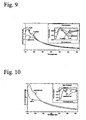
- Fig. 9 shows the normalized optical density spectra and the first derivative spectra (inset) measured for E. Coli, P. agglomerans, and The vegetative cells of B. globigii. Notice that although the spectra appear to be somewhat similar, they are not identical; this is probably due to differences in chemical composition, in particular DNA and RNA. The spectral differences can be appreciated particularly in the first derivative spectra, which enhance the differences in chemical composition (inset Fig. 10 ).
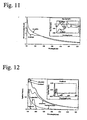
- Fig. 11 shows a comparison between the spores and vegetative cells of B. globigii. There is a marked difference between the spectra of the spores and the vegetative cells. These differences are expected since only the spores contain dipicolinic acid. Furthermore, the presence of high concentrations of calcium makes the spores highly refractive and as a result their scattering properties are affected. Comparison of the spectra from B. globigii, and B. subtilis spores show considerable differences ( Fig. 12 ). The first derivative spectra shown in the inset has the same absorption bands, but clearly with varying intensity. The experimental measurements of the multiwavelength transmission spectra show evidence of quantifiable differences between the bacterial cells and the spores analyzed.
- the transmission spectra from a sample is acquired 20.
- the wavelengths absorbed by the suspending media are excluded.
- the spectrum is normalized 40 based on the mean turbidity 30 excluding the range absorbed by the suspending media.
- a spectra fingerprint database 60 is accessed and the acquired spectra 20 is compared to known spectra fingerprints in the database 60 for a match.
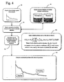
- Fig. 4 illustrates a comparative example using deconvolution to identify the acquired transmission spectra 20 wherein a plurality of cellular elements are selected 70 from an optical property database 80 of cellular element contributions. The sum of the contributions is calculated 90 from the selected absorptive and/or scattering elements selected. An appropriate scattering theory 100 may be applied such as Mie as known by those skilled in the art. The calculated spectrum 90 and the observed spectrum 20 are then compared 110 to determine the presence of the microorganism in the sample.
- Fig. 5 an alternative embodiment of Fig. 4 is presented wherein non-polarized 81 and polarized 82 values are provided to enable more precise deconvolution of the acquired spectra 20.
- a plurality of cellular elements are selected 70 from the optical property database 80 and the sum of contributions are calculated 90.
- the calculated and observed spectrums are compared 110 and evaluated 120 to determine whether a match is achieved under predetermined threshold tolerances. It is preferred that the spectrums be compared using a least-squares approach, but alternative methods known to those skilled in the art are acceptable. If an identification is achieved 130, the microorganism associated with the preselected model is deemed to be present in the sample. Alternatively, if the match is not achieved within threshold tolerates, a new combination of selected elements may be executed 140 iteratively and the process repeated.
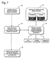
- Fig. 7 illustrates the process of employing preexisting models for microorganisms in the present invention wherein at least one suspected microorganism is established 150 for identification.
- An array of established sets of cellular elements is accessed 170 containing predetermined models for at least one microorganism.
- the established sets 170 are derived from the optical property database 80.
- a set of cellular elements is selected 160 which corresponds to the predetermined model for the suspected microorganism previously selected 150 and the sum of contributions is calculated 90 to determine if the suspected microorganism is present in a sample.
- At least one suspected microorganism is established 150 for identification.
- the cellular elements likely to absorb and/or scatter are established 180 based on known microbiology for the at least one suspected microorganism.
- the combination of cellular elements selected are then validated 190 against quantitative data. Upon validation, the combination forms a model 170 for the at least one microorganism. If validation is not achieved, a new combination of cellular elements are selected and the validation process is repeated.
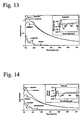
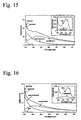
- Figs. 13-16 show typical comparisons between measured and calculated spectra for three vegetative cells and for B. globigii, and B. subtilis spores.
- the contribution to the calculated total optical density from each of the model components, the total absorption of the corresponding chromophoric groups calculated as Beer-Lambert absorption, and the residuals are also included in the figures. Notice the spectral features contributed by each component and the dramatic effect of the chromophoric groups to the total optical density.
- Extinction coefficient is a spectroscopic term applied to a molecular group for determining absorption at a particular wavelength. The two structures and the chromophoric groups contribute differently over different portions of the spectra. Although the residuals show some correlation, it is evident that the calculated spectra adequately represents the measured spectra of a large variety of prokaryotic microorganisms.
- Table II shows the literature data available for the volume of the microorganisms and the chromophore concentrations.
- the estimates of the particle volumes and concentrations are within the range of values reported in the literature that were obtained with completely different methods.
- the good agreement with literature values is surprising given the simplifying assumptions of the model. There are two outstanding values: the first one is the relatively small volume estimated for P. agglomerans, which could explain the low water content reported in Table I.
- the second value corresponds to the low DPA concentration estimate for B. subtilis.
- the total nucleotide concentration estimated is in excellent agreement with the literature for E. Coli.
- the DPA concentration for B. globigii obtained from the spectral deconvolution is in good agreement with the values reported in the literature for spores.
- the correlation observed in the residuals can be explained on the basis of the approximations and simplifications used.
- a spherical equivalent scattering approximation has been used. Only the average sizes for each population of microorganisms and for the internal structure have been estimated. Also, an average representation of the total nucleotide absorption has been utilized, and the contribution of chromophoric amino acids has been neglected. The last two may account for the correlation in the residuals apparent in the region between 230-300 nm.
- One may also include hypochromic effects, which are known to alter the spectral features of strong chromophoric groups such as DNA and RNA. This model is capable of representing the spectroscopy behavior of complex biological structures such as vegetative cells and spores, and as such, it provides quantitative information for analysis and differentiation.
Landscapes
- Chemical & Material Sciences (AREA)
- Health & Medical Sciences (AREA)
- Physics & Mathematics (AREA)
- Life Sciences & Earth Sciences (AREA)
- Organic Chemistry (AREA)
- General Health & Medical Sciences (AREA)
- Wood Science & Technology (AREA)
- Proteomics, Peptides & Aminoacids (AREA)
- Engineering & Computer Science (AREA)
- Biochemistry (AREA)
- Immunology (AREA)
- Zoology (AREA)
- Analytical Chemistry (AREA)
- Spectroscopy & Molecular Physics (AREA)
- Molecular Biology (AREA)
- Microbiology (AREA)
- Biotechnology (AREA)
- Bioinformatics & Cheminformatics (AREA)
- General Engineering & Computer Science (AREA)
- Biophysics (AREA)
- Genetics & Genomics (AREA)
- Toxicology (AREA)
- General Physics & Mathematics (AREA)
- Pathology (AREA)
- Measuring Or Testing Involving Enzymes Or Micro-Organisms (AREA)
- Investigating Or Analysing Materials By Optical Means (AREA)
- Micro-Organisms Or Cultivation Processes Thereof (AREA)
Applications Claiming Priority (3)
| Application Number | Priority Date | Filing Date | Title |
|---|---|---|---|
| US21426402A | 2002-08-07 | 2002-08-07 | |
| US214264 | 2002-08-07 | ||
| PCT/US2003/024728 WO2004015136A1 (en) | 2002-08-07 | 2003-08-07 | Interpretation model for the uv-vis spectra of microorganisms |
Publications (3)
| Publication Number | Publication Date |
|---|---|
| EP1543146A1 EP1543146A1 (en) | 2005-06-22 |
| EP1543146A4 EP1543146A4 (en) | 2009-10-21 |
| EP1543146B1 true EP1543146B1 (en) | 2011-12-21 |
Family
ID=31714240
Family Applications (1)
| Application Number | Title | Priority Date | Filing Date |
|---|---|---|---|
| EP03784997A Expired - Lifetime EP1543146B1 (en) | 2002-08-07 | 2003-08-07 | Interpretation model for the uv-vis spectra of microorganisms |
Country Status (6)
| Country | Link |
|---|---|
| EP (1) | EP1543146B1 (ja) |
| JP (1) | JP4476121B2 (ja) |
| AT (1) | ATE538211T1 (ja) |
| AU (1) | AU2003261438A1 (ja) |
| CA (1) | CA2494182A1 (ja) |
| WO (1) | WO2004015136A1 (ja) |
Families Citing this family (9)
| Publication number | Priority date | Publication date | Assignee | Title |
|---|---|---|---|---|
| WO2008147227A1 (en) * | 2007-05-31 | 2008-12-04 | Veritide Limited | Method for spore detection |
| EP2439536A1 (en) | 2010-10-01 | 2012-04-11 | Nederlandse Organisatie voor toegepast- natuurwetenschappelijk onderzoek TNO | New classification method for spectral data |
| JP6148986B2 (ja) * | 2011-03-01 | 2017-06-14 | トリネアン・ナムローゼ・フェンノートシャップTrinean NV | Uv−vis分光光度計データからのdna及び/又はrnaの決定 |
| WO2014014353A1 (en) | 2012-07-18 | 2014-01-23 | Nederlandse Organisatie Voor Toegepast-Natuurwetenschappelijk Onderzoek Tno | New classification method for spectral data |
| GB201215484D0 (en) | 2012-08-30 | 2012-10-17 | Trinean | Optical characterisation of DNA and/or RNA |
| FR3009387B1 (fr) * | 2013-07-31 | 2016-11-18 | Biomerieux Sa | Procede et dispositif d'analyse d'un echantillon biologique |
| WO2018195514A1 (en) | 2017-04-20 | 2018-10-25 | Biomerieux, Inc. | Method, apparatus, and computer program product for controlling components of a detection device |
| CN110850020B (zh) * | 2019-11-11 | 2022-03-29 | 中国药科大学 | 一种基于人工智能的中药识别方法 |
| CN111122484A (zh) * | 2019-12-30 | 2020-05-08 | 中国科学院合肥物质科学研究院 | 一种水体细菌的定性与定量方法 |
Family Cites Families (6)
| Publication number | Priority date | Publication date | Assignee | Title |
|---|---|---|---|---|
| US5164301A (en) * | 1990-06-22 | 1992-11-17 | Difco Laboratories | Process and kit for detecting microbial metabolism |
| US5784162A (en) * | 1993-08-18 | 1998-07-21 | Applied Spectral Imaging Ltd. | Spectral bio-imaging methods for biological research, medical diagnostics and therapy |
| US5616457A (en) * | 1995-02-08 | 1997-04-01 | University Of South Florida | Method and apparatus for the detection and classification of microorganisms in water |
| US6984526B2 (en) * | 1995-02-08 | 2006-01-10 | University Of South Florida | Spectrophotometric method for determining the viability of a sample containing platelets |
| GB2345754B (en) * | 1998-03-31 | 2001-02-07 | Zetatronics Ltd | Rapid method for detecting micro-organisms and evaluating antimicrobial activity |
| WO2002004947A2 (en) * | 2000-07-12 | 2002-01-17 | University Of South Florida | Spectrophotometric system and method for the identification and characterization of a particle in a bodily fluid |
-
2003
- 2003-08-07 WO PCT/US2003/024728 patent/WO2004015136A1/en active Application Filing
- 2003-08-07 AU AU2003261438A patent/AU2003261438A1/en not_active Abandoned
- 2003-08-07 AT AT03784997T patent/ATE538211T1/de active
- 2003-08-07 JP JP2004527837A patent/JP4476121B2/ja not_active Expired - Lifetime
- 2003-08-07 CA CA002494182A patent/CA2494182A1/en not_active Abandoned
- 2003-08-07 EP EP03784997A patent/EP1543146B1/en not_active Expired - Lifetime
Non-Patent Citations (1)
| Title |
|---|
| CATALINA ELENA ALUPOAEI: "Modeling of the Transmission Spectra of Microorganisms", THESIS,, 1 December 2001 (2001-12-01), pages 110PP, XP009128434 * |
Also Published As
| Publication number | Publication date |
|---|---|
| JP4476121B2 (ja) | 2010-06-09 |
| WO2004015136A1 (en) | 2004-02-19 |
| AU2003261438A1 (en) | 2004-02-25 |
| EP1543146A1 (en) | 2005-06-22 |
| ATE538211T1 (de) | 2012-01-15 |
| EP1543146A4 (en) | 2009-10-21 |
| CA2494182A1 (en) | 2004-02-19 |
| JP2005534336A (ja) | 2005-11-17 |
Similar Documents
| Publication | Publication Date | Title |
|---|---|---|
| US5589932A (en) | Spectrophotometric method and apparatus for the characterization of blood and blood types | |
| AU728462B2 (en) | Resonance raman spectroscopy for identifying and quantitating biomatter, organic, and inorganic analytes | |
| US7691642B1 (en) | Spectrophotometric method and apparatus for the cross-matching of platelets | |
| JP4745959B2 (ja) | 微生物の自動特徴づけおよび分類 | |
| DuRand et al. | Diel variations in optical properties of Micromonas pusilla (prasinophyceae) 1 | |
| US6421121B1 (en) | Method and apparatus for rapid particle identification utilizing scattered light histograms | |
| Zimmerman et al. | Analysis of allergenic pollen by FTIR microspectroscopy | |
| CN109001180B (zh) | 一种拉曼光谱结合人工智能高通量单细胞分析鉴定方法 | |
| EP1543146B1 (en) | Interpretation model for the uv-vis spectra of microorganisms | |
| JP2008510161A (ja) | 液体中の細菌の検出 | |
| CN113899702A (zh) | 一种基于量子傅里叶变换的疫苗多光谱快速检测方法 | |
| Alupoaei et al. | An interpretation model for the UV-VIS spectra of microorganisms | |
| Shirai et al. | Detection of fluorescence signals from ATP in the second derivative excitation–emission matrix of a pork meat surface for cleanliness evaluation | |
| Uitz et al. | Variations in the optical properties of a particle suspension associated with viral infection of marine bacteria | |
| US6330058B1 (en) | Spectrophotometric method and apparatus for blood typing | |
| Mattley et al. | Multiwavelength spectroscopy for the detection, identification, and quantification cells | |
| Callahan et al. | Use of multiwavelength transmission spectroscopy for the characterization of Cryptosporidium parvum oocysts: Quantitative interpretation | |
| Li et al. | Detection of the freshness of rice by chemiluminescence | |
| Mattley et al. | Blood characterization using uv/vis spectroscopy | |
| Garcia-Rubio et al. | A new spectroscopy method for in situ rapid detection and classification of micro-organisms | |
| US20220170839A1 (en) | Spectral diagnostic system | |
| Weber et al. | Raman Spectroscopy is Solving the Perpetual Problem of CSI: The Time of a Crime | |
| Bacon et al. | Quantitative classification of cryptosporidium oocysts and giardia cysts in water using UV/vis spectroscopy | |
| Vasilevskii et al. | Study of the absorption spectra of albumin and uric acid in the UV region | |
| Taniguchi et al. | Light-scattering in absorption spectra: a literature survey of examples and corrections |
Legal Events
| Date | Code | Title | Description |
|---|---|---|---|
| PUAI | Public reference made under article 153(3) epc to a published international application that has entered the european phase |
Free format text: ORIGINAL CODE: 0009012 |
|
| 17P | Request for examination filed |
Effective date: 20050219 |
|
| AK | Designated contracting states |
Kind code of ref document: A1 Designated state(s): AT BE BG CH CY CZ DE DK EE ES FI FR GB GR HU IE IT LI LU MC NL PT RO SE SI SK TR |
|
| AX | Request for extension of the european patent |
Extension state: AL LT LV MK |
|
| DAX | Request for extension of the european patent (deleted) | ||
| RIC1 | Information provided on ipc code assigned before grant |
Ipc: G01N 21/31 20060101ALI20090904BHEP Ipc: C12Q 1/00 20060101AFI20040302BHEP Ipc: G01N 21/47 20060101ALI20090904BHEP Ipc: G01N 21/49 20060101ALI20090904BHEP Ipc: G01N 15/02 20060101ALI20090904BHEP |
|
| A4 | Supplementary search report drawn up and despatched |
Effective date: 20090916 |
|
| 17Q | First examination report despatched |
Effective date: 20100210 |
|
| REG | Reference to a national code |
Ref country code: DE Ref legal event code: R079 Ref document number: 60339505 Country of ref document: DE Free format text: PREVIOUS MAIN CLASS: C12Q0001000000 Ipc: C12Q0001040000 |
|
| RIC1 | Information provided on ipc code assigned before grant |
Ipc: G01N 21/47 20060101ALI20110526BHEP Ipc: G01N 21/49 20060101ALI20110526BHEP Ipc: G01N 21/31 20060101ALI20110526BHEP Ipc: G01N 15/02 20060101ALI20110526BHEP Ipc: C12Q 1/00 20060101AFI20110526BHEP |
|
| RIC1 | Information provided on ipc code assigned before grant |
Ipc: G01N 21/33 20060101ALI20110527BHEP Ipc: C12Q 1/04 20060101AFI20110527BHEP |
|
| GRAP | Despatch of communication of intention to grant a patent |
Free format text: ORIGINAL CODE: EPIDOSNIGR1 |
|
| GRAS | Grant fee paid |
Free format text: ORIGINAL CODE: EPIDOSNIGR3 |
|
| GRAA | (expected) grant |
Free format text: ORIGINAL CODE: 0009210 |
|
| AK | Designated contracting states |
Kind code of ref document: B1 Designated state(s): AT BE BG CH CY CZ DE DK EE ES FI FR GB GR HU IE IT LI LU MC NL PT RO SE SI SK TR |
|
| REG | Reference to a national code |
Ref country code: GB Ref legal event code: FG4D |
|
| REG | Reference to a national code |
Ref country code: CH Ref legal event code: EP |
|
| REG | Reference to a national code |
Ref country code: AT Ref legal event code: REF Ref document number: 538211 Country of ref document: AT Kind code of ref document: T Effective date: 20120115 |
|
| REG | Reference to a national code |
Ref country code: IE Ref legal event code: FG4D |
|
| REG | Reference to a national code |
Ref country code: DE Ref legal event code: R096 Ref document number: 60339505 Country of ref document: DE Effective date: 20120223 |
|
| REG | Reference to a national code |
Ref country code: NL Ref legal event code: VDEP Effective date: 20111221 |
|
| PG25 | Lapsed in a contracting state [announced via postgrant information from national office to epo] |
Ref country code: GR Free format text: LAPSE BECAUSE OF FAILURE TO SUBMIT A TRANSLATION OF THE DESCRIPTION OR TO PAY THE FEE WITHIN THE PRESCRIBED TIME-LIMIT Effective date: 20120322 Ref country code: SE Free format text: LAPSE BECAUSE OF FAILURE TO SUBMIT A TRANSLATION OF THE DESCRIPTION OR TO PAY THE FEE WITHIN THE PRESCRIBED TIME-LIMIT Effective date: 20111221 Ref country code: SI Free format text: LAPSE BECAUSE OF FAILURE TO SUBMIT A TRANSLATION OF THE DESCRIPTION OR TO PAY THE FEE WITHIN THE PRESCRIBED TIME-LIMIT Effective date: 20111221 Ref country code: NL Free format text: LAPSE BECAUSE OF FAILURE TO SUBMIT A TRANSLATION OF THE DESCRIPTION OR TO PAY THE FEE WITHIN THE PRESCRIBED TIME-LIMIT Effective date: 20111221 |
|
| PG25 | Lapsed in a contracting state [announced via postgrant information from national office to epo] |
Ref country code: CY Free format text: LAPSE BECAUSE OF FAILURE TO SUBMIT A TRANSLATION OF THE DESCRIPTION OR TO PAY THE FEE WITHIN THE PRESCRIBED TIME-LIMIT Effective date: 20111221 Ref country code: BE Free format text: LAPSE BECAUSE OF FAILURE TO SUBMIT A TRANSLATION OF THE DESCRIPTION OR TO PAY THE FEE WITHIN THE PRESCRIBED TIME-LIMIT Effective date: 20111221 |
|
| PG25 | Lapsed in a contracting state [announced via postgrant information from national office to epo] |
Ref country code: SK Free format text: LAPSE BECAUSE OF FAILURE TO SUBMIT A TRANSLATION OF THE DESCRIPTION OR TO PAY THE FEE WITHIN THE PRESCRIBED TIME-LIMIT Effective date: 20111221 Ref country code: BG Free format text: LAPSE BECAUSE OF FAILURE TO SUBMIT A TRANSLATION OF THE DESCRIPTION OR TO PAY THE FEE WITHIN THE PRESCRIBED TIME-LIMIT Effective date: 20120321 Ref country code: CZ Free format text: LAPSE BECAUSE OF FAILURE TO SUBMIT A TRANSLATION OF THE DESCRIPTION OR TO PAY THE FEE WITHIN THE PRESCRIBED TIME-LIMIT Effective date: 20111221 Ref country code: EE Free format text: LAPSE BECAUSE OF FAILURE TO SUBMIT A TRANSLATION OF THE DESCRIPTION OR TO PAY THE FEE WITHIN THE PRESCRIBED TIME-LIMIT Effective date: 20111221 |
|
| PG25 | Lapsed in a contracting state [announced via postgrant information from national office to epo] |
Ref country code: PT Free format text: LAPSE BECAUSE OF FAILURE TO SUBMIT A TRANSLATION OF THE DESCRIPTION OR TO PAY THE FEE WITHIN THE PRESCRIBED TIME-LIMIT Effective date: 20120423 Ref country code: RO Free format text: LAPSE BECAUSE OF FAILURE TO SUBMIT A TRANSLATION OF THE DESCRIPTION OR TO PAY THE FEE WITHIN THE PRESCRIBED TIME-LIMIT Effective date: 20111221 |
|
| REG | Reference to a national code |
Ref country code: AT Ref legal event code: MK05 Ref document number: 538211 Country of ref document: AT Kind code of ref document: T Effective date: 20111221 |
|
| PLBE | No opposition filed within time limit |
Free format text: ORIGINAL CODE: 0009261 |
|
| STAA | Information on the status of an ep patent application or granted ep patent |
Free format text: STATUS: NO OPPOSITION FILED WITHIN TIME LIMIT |
|
| PG25 | Lapsed in a contracting state [announced via postgrant information from national office to epo] |
Ref country code: DK Free format text: LAPSE BECAUSE OF FAILURE TO SUBMIT A TRANSLATION OF THE DESCRIPTION OR TO PAY THE FEE WITHIN THE PRESCRIBED TIME-LIMIT Effective date: 20111221 |
|
| 26N | No opposition filed |
Effective date: 20120924 |
|
| PG25 | Lapsed in a contracting state [announced via postgrant information from national office to epo] |
Ref country code: IT Free format text: LAPSE BECAUSE OF FAILURE TO SUBMIT A TRANSLATION OF THE DESCRIPTION OR TO PAY THE FEE WITHIN THE PRESCRIBED TIME-LIMIT Effective date: 20111221 |
|
| REG | Reference to a national code |
Ref country code: DE Ref legal event code: R097 Ref document number: 60339505 Country of ref document: DE Effective date: 20120924 |
|
| PG25 | Lapsed in a contracting state [announced via postgrant information from national office to epo] |
Ref country code: AT Free format text: LAPSE BECAUSE OF FAILURE TO SUBMIT A TRANSLATION OF THE DESCRIPTION OR TO PAY THE FEE WITHIN THE PRESCRIBED TIME-LIMIT Effective date: 20111221 |
|
| REG | Reference to a national code |
Ref country code: CH Ref legal event code: PL |
|
| PG25 | Lapsed in a contracting state [announced via postgrant information from national office to epo] |
Ref country code: MC Free format text: LAPSE BECAUSE OF NON-PAYMENT OF DUE FEES Effective date: 20120831 |
|
| GBPC | Gb: european patent ceased through non-payment of renewal fee |
Effective date: 20120807 |
|
| PG25 | Lapsed in a contracting state [announced via postgrant information from national office to epo] |
Ref country code: ES Free format text: LAPSE BECAUSE OF FAILURE TO SUBMIT A TRANSLATION OF THE DESCRIPTION OR TO PAY THE FEE WITHIN THE PRESCRIBED TIME-LIMIT Effective date: 20120401 Ref country code: CH Free format text: LAPSE BECAUSE OF NON-PAYMENT OF DUE FEES Effective date: 20120831 Ref country code: LI Free format text: LAPSE BECAUSE OF NON-PAYMENT OF DUE FEES Effective date: 20120831 |
|
| REG | Reference to a national code |
Ref country code: FR Ref legal event code: ST Effective date: 20130430 |
|
| REG | Reference to a national code |
Ref country code: IE Ref legal event code: MM4A |
|
| PG25 | Lapsed in a contracting state [announced via postgrant information from national office to epo] |
Ref country code: FI Free format text: LAPSE BECAUSE OF FAILURE TO SUBMIT A TRANSLATION OF THE DESCRIPTION OR TO PAY THE FEE WITHIN THE PRESCRIBED TIME-LIMIT Effective date: 20111221 |
|
| PG25 | Lapsed in a contracting state [announced via postgrant information from national office to epo] |
Ref country code: DE Free format text: LAPSE BECAUSE OF NON-PAYMENT OF DUE FEES Effective date: 20130301 Ref country code: GB Free format text: LAPSE BECAUSE OF NON-PAYMENT OF DUE FEES Effective date: 20120807 Ref country code: IE Free format text: LAPSE BECAUSE OF NON-PAYMENT OF DUE FEES Effective date: 20120807 |
|
| PG25 | Lapsed in a contracting state [announced via postgrant information from national office to epo] |
Ref country code: FR Free format text: LAPSE BECAUSE OF NON-PAYMENT OF DUE FEES Effective date: 20120831 |
|
| REG | Reference to a national code |
Ref country code: DE Ref legal event code: R119 Ref document number: 60339505 Country of ref document: DE Effective date: 20130301 |
|
| PG25 | Lapsed in a contracting state [announced via postgrant information from national office to epo] |
Ref country code: TR Free format text: LAPSE BECAUSE OF FAILURE TO SUBMIT A TRANSLATION OF THE DESCRIPTION OR TO PAY THE FEE WITHIN THE PRESCRIBED TIME-LIMIT Effective date: 20111221 |
|
| PG25 | Lapsed in a contracting state [announced via postgrant information from national office to epo] |
Ref country code: LU Free format text: LAPSE BECAUSE OF NON-PAYMENT OF DUE FEES Effective date: 20120807 |
|
| PG25 | Lapsed in a contracting state [announced via postgrant information from national office to epo] |
Ref country code: HU Free format text: LAPSE BECAUSE OF FAILURE TO SUBMIT A TRANSLATION OF THE DESCRIPTION OR TO PAY THE FEE WITHIN THE PRESCRIBED TIME-LIMIT Effective date: 20030807 |