TECHNICAL FIELD
The present invention relates to a DNA micro-array
useful for quantitative analysis of a target
nucleic acid (target nucleic acid hereinafter) in a
sample. More specifically, the present invention
relates to a DNA micro-array that can improve
accuracy in a quantitative analysis using a DNA
micro-array of nucleic acid probes having a sequence
expected to be complementary to a target nucleic acid
sequence in a sample. The present invention also
relates to a detection kit using the DNA micro-array.
BACKGROUND ART
As a result of development in methods for
detecting and measuring genetic DNA on the basis of
hybridization process, rapid progress has been made
in studies of gene chip (DNA chip, micro-array)where
a plurality of nucleic acid probes are immobilized on
a substrate, allowing multiple detection tests on
genetic DNA in a sample concomitantly. The method of
detecting and measuring genetic DNA contained in a
sample using a gene chip (DNA chip, micro-array) is
expected to be applied in various fields such as
molecular biological researches, and gene disorder
and infectious disease diagnoses.
The basic form of a gene chip is that a
plurality of single-stranded nucleic acid fragments
expected to have a complementary base sequence to the
base sequence of a target gene are arranged in an
array and immobilized on the surface of a substrate
such as a glass substrate in order to detect target
genetic DNA by the hybridization process. Single-stranded
nucleic acid fragment to be used as a probe
having a complementary base sequence to a target gene
can be a chemically synthesized DNA oligomer called
"oligo-DNA" or a complementary strand DNA fragment
called "cDNA" enzymatically synthesized using a gene
derived from a biomaterial as a template. As for
immobilization of oligo-DNA on the surface of the
substrate, methods are roughly classified into two:
one is, as shown in US 5,474,796 B (or JP 9-500568 A,
ProtoGene Laboratories), to synthesize DNA molecules
on the substrate sequentially after immobilizing one
end to the substrate, the other is, as disclosed in
JP H11-187900 A (Canon), oligo-DNA are synthesized,
and then various bonding means are used to immobilize
these nucleic acid fragments on a substrate. As
described above, the immobilizing method is roughly
classified into above two methods.
As the means for immobilizing the separately
prepared nucleic acid fragments on the substrate,
various methods have been proposed, such as an
adsorption immobilizing method making use of the
charge of a substrate and the charge of a nucleic
acid fragment, and an immobilizing method which is
aimed to improve immobilization efficiency by using a
coating film formed by coating the substrate surface
with a poly-L-lysine or amino silane coupling agent.
Currently, DNA micro-arrays are not provided
with special probes for quantitative analysis of
nucleic acid having a target sequence (target nucleic
acid). Whether the DNA micro-array is a DNA micro-array
formed on the surface of a solid phase by a
sequential synthesis method or a DNA micro-array
produced by a pin method, there are variations caused
by the production process, for example, variation in
synthesis efficiency due to the nucleic acid sequence,
variation in the amount of the probe liquid dropped
from a pin spotter on a substrate or variation in
immobilization rate. Therefore, there are problems
in quantification accuracy and reproducibility. An
ink jet production method has been proposed (JP H11-187900
A) to solve the variation at the time of DNA
micro-array production.
On the other hand, a sample to be examined by
the DNA micro-array is subjected to an amplification
operation to amplify a target nucleic acid from the
viewpoint of sensitivity, as well as to incorporate a
detectable label to into the amplified product. The
basic technology of this amplifying operation is
called "PCR" and disclosed in US 4683195 B, US
4683202 B and US 4965188 B. However, PCR involves
hard-to-control factors in the amplification reaction
process. For example, it is well known that even
when the same samples are amplified at the same time,
the amplification yield may differ by several times
to several tens of times. Studies have been made to
solve this uncertainty in the amplification yield,
providing competitive PCR etc. The competitive PCR
is detailed in reports by P.D. Siebert et al. (Nature
359; 557-558 (1992), Bio Techniques 14; 244-249
(1993)). As an alternative method, a known amount of
a certain nucleic acid is added as an internal
standard to a sample taken from a patient, and the
internal standard nucleic acid and the target nucleic
acid are amplified in order to estimate the
amplification rate of the target nucleic acid from
the amplification rate of the internal standard
nucleic acid.
DISCLOSURE OF THE INVENTION
As described above, when a quantitative
analysis of a target nucleic acid is carried out
using a DNA micro-array, there exist various unstable
factors that affect the determination.
First of all, there is low reliability of
accuracy and reproducibility due to variation in the
amount of a probe immobilized on a substrate during
production of a DNA micro-array. There is also
variation in the amplification rate of a target
nucleic acid in a sample due to the uncertainty of
the amplification efficiency of the target nucleic
acid in PCR. Conventionally, variations in these
different factors are calibrated by various methods
separately, and then quantitative analysis is carried
out using a DNA micro-array, and the analytical data
are corrected on the basis of the calibration results
for quantitative determination.
Since these calibration procedures are very
complicated and time-consuming, they are inhibiting
factors in processing a large number of samples.
The present invention is to solve the above
problems. It is therefore an object of the present
invention to provide a DNA micro-array which allows
obtainment of correction data at the time of
quantitative determination of the amount of a target
nucleic acid in a sample, greatly simplifying the
calibration procedures, as well as an assay kit used
for the above purpose.
The inventors of the present invention have
studied intensively to solve the above problems and
have found that these assay works can be greatly
simplified if a DNA micro-array is prepared by
applying and immobilizing separately prepared nucleic
acid probes using an ink-jet method (JP H11-187900 A)
and at least either a probe for an internal standard
nucleic acid (internal standard probe hereinafter) or
a probe for an external standard nucleic acid
(external standard probe hereinafter) is immobilized
together with the nucleic acid probes for detecting
target nucleic acid. Based on the above findings,
the inventors of the present invention have also
devised a kit for carrying out these assay works
using the above DNA micro-array accurately and easily
to complete the present invention.
Thus, according to the present invention, there
is provided A DNA micro-array for detecting nucleic
acid molecules having target base sequences in a
sample comprising a substrate and nucleic acid probes
having base sequences substantially complementary to
the target base sequences immobilized on the
substrate, wherein the array contains additional
probes of one or several kinds selected from the
following probes:
Preferably, probes for internal standard
nucleic acids and probes for external standard
nucleic acids is immobilized on the substrate at four
probe concentrations or densities to form a group.
Preferably, the internal standard probe to be
immobilized on the DNA micro-array comprises at least
two kinds of probes corresponding to amplification
products of two different chain lengths obtained from
the internal standard nucleic acid. Also the
external standard probe comprises preferably at least
two kinds of probes of an appropriate base number
being complementary to the external standard nucleic
acid to be added.
More preferably, the internal standard probe
and the external standard probe are synthetic nucleic
acids immobilized on the substrate. The chain length
of the synthetic nucleic acid is 15 to 75 bases
preferably.
Further, when the DNA micro-array according to
the present invention contains an internal standard
probe, an internal standard nucleic acid is amplified
together with the target nucleic acid in one
amplification reaction to determine the amplification
efficiency of the reaction. The present invention
also provides a primer set for an amplification
reaction of an internal standard nucleic acid. That
is, the primer set of the present invention is for
amplification of an internal standard nucleic acid to
be amplified together with a target nucleic acid and
is designed to generate an amplified product having a
chain length similar to the amplification product of
the target nucleic acid.
The present invention also provides a detection
kit suitable for quantitative detection of a target
nucleic acid using a DNA micro-array of the present
invention, which includes a primer set for the
amplification reaction of the internal standard
nucleic acid to be amplified together with the target
nucleic acid. When the amplification product of the
target nucleic acid is of two or more chain lengths,
the kit includes at least two primer sets as claimed
in claim 7, corresponding to the chain lengths. The
primer sets include at least one primer set to obtain
an amplification product of 200 bp or less in chain
length, at least one primer set for an amplified
products of 200 to 500 bp, at least one primer set
for 500 to a 2,000 bp amplification product, and at
least one primer set for a 2,000 bp or more
amplification product.
Preferably, according to the present invention,
a kit for detecting a target base sequence includes
internal standard nucleic acid to be amplified
together with a target nucleic acid in an
amplification reaction for quantitative detection of
a target nucleic acid using the DNA micro-array,
where the internal standard nucleic acid is two or
more nucleic acids derived from a microorganism or a
virus having no homology with the target base
sequence to be detected.
The present invention also provides a detection
kit containing external standard nucleic acid,
suitably used for the quantitative detection of a
target nucleic acid using the DNA micro-array of the
present invention containing external standard probes.
That is, according to the present invention, there is
provided a kit for detecting a target base sequence,
including at least two external standard nucleic
acids to be added to a sample upon quantitatively
detecting a target nucleic acid using a DNA micro-array,
where the external standard nucleic acids are
synthetic nucleic acids labeled with a detectable
marker. The marker is preferably a fluorescent
material, a radioactive material or a luminescent
material.
In the DNA micro-array of the present invention,
the nucleic acid probe includes a single-stranded
nucleic acid, specifically DNA, RNA, PNA (peptide
nucleic acid), cDNA (complementary DNA) or cRNA
(complementary RNA). Further, the DNA micro-array
may comprise a single-stranded nucleic acid as a
nucleic acid probe and a target nucleic acid
hybridized to the nucleic acid probe.
BRIEF DESCRIPTION OF THE DRAWINGS
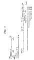
Fig. 1 shows an internal standard probe,
external standard probe, external standard nucleic
acid and internal standard nucleic acid, a primer set
for amplification of an internal standard nucleic
acid and amplified products thereof according to an
embodiment of the present invention;
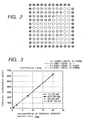
Fig. 2 schematically shows an arrangement of
internal standard probes in the DNA micro-array
according to an embodiment of the present invention;
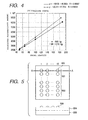
Fig. 3 is a graph showing calibration curves
for calculating the probe immobilization rate using
external standard probes in the DNA micro-array in
quantitative analysis using the DNA micro-array
according to the present invention;
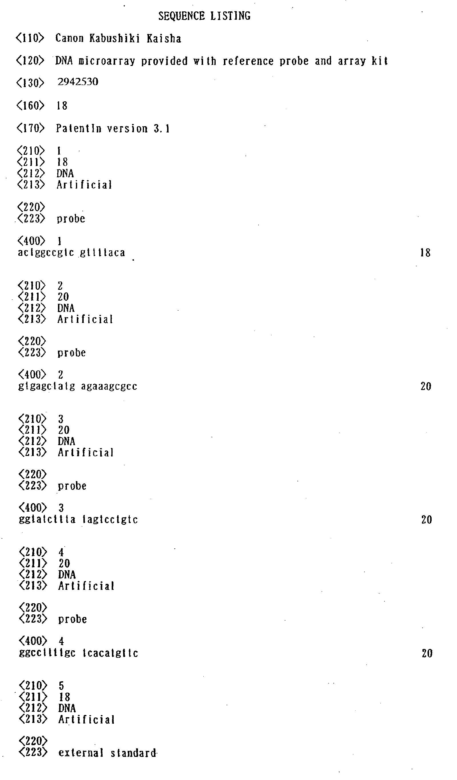
Fig. 4 is a graph showing calibration curves
for calculating the PCR amplification factor using
internal standard probes provided in the DNA micro-array
in quantitative analysis using the DNA micro-array
according to the present invention, the graph
also showing a difference in detection sensitivity
according to the chain length of the nucleic acid of
the amplified product;
Fig. 5 schematically shows the plane
arrangement and sectional form (taken along the line
A-A) of a biochip as a structural example according
to the present invention; and
Fig. 6 is a graph showing the relationship
between the measured secondary ion intensity and DNA
formation density (number of DNA molecules per probe
dot) of a biochip.
BEST MODE FOR CARRYING OUT THE INVENTION
According to a first aspect of the present
invention, there is provided a DNA micro-array having
an internal standard probe formed thereon.
In a broader sense, the internal standard probe
is a probe for detecting an internal standard nucleic
acid to be used to assist quantitative determination
of a target nucleic acid. This prove is a nucleic
acid probe having a base sequence complementary to an
internal standard nucleic acid or gene of which
amount in a sample is known and can bind to it, to
enable quantitative determination of a target nucleic
acid on the basis of the relative ratio of the
internal standard nucleic acid to the target nucleic
acid bound to the array. For example, a
transcription product of a gene encoding a
cytoskeleton (a house keeping gene) is often used as
the internal standard nucleic acid because its
expression amount hardly changes and is relatively
large. Specific examples of the transcription
product include mRNA of GAPDH or of β-actin and
ribosomal RNA.
In a narrower sense, the internal standard is a
nucleic acid of a known base sequence to be added in
a sample in a known amount when an amplification
reaction of nucleic acid (e.g., PCR) is carried to
increase the amount of nucleic acid or to label the
target nucleic acid with a detectable marker. Such
an internal standard nucleic acid to be added to the
sample is selected so as to be amplified or labeled
at the same efficiency as the target nucleic acid
when amplification or labeling of the target nucleic
acid is carried out. After the amplification or
labeling reaction, the amount of the internal
standard nucleic acid is determined to calculate the
amplification factor or labeling rate so that the
amount of the target nucleic acid can be obtained as
a relative value based on the amount of the internal
standard nucleic acid.
According to a second aspect of the present
invention, there is provided a DNA micro-array having
a probe for an external standard nucleic acid
(external standard probe) formed thereon.
Here, the external standard nucleic acid is a
nucleic acid having a known base sequence to be added
to a sample in order to compensate variation in the
DNA micro-array production step and/or in the
detection and measurement step. More specifically,
it is a nucleic acid prepared by introducing a
detectable and determinable marker into a highly
purified nucleic acid such as synthesized DNA or
plasmid DNA, of which labeling rate etc. are
determined in advance. This external standard
nucleic acid may be used for any type of DNA micro-arrays
not depending to the type of target genes so
long as it has no base sequence homology to the base
sequence of the target nucleic acid. The external
standard probe is a nucleic acid probe having a base
sequence complementary to the external standard
nucleic acid and can selectively bind to the external
standard.
Plural sets of external standard nucleic acids
and external standard probes may be used at the same
time to determine the performance of the DNA micro-array,
that is, quantification ability thereof. More
specifically, a DNA micro-array having not less than
three, preferably not less than five external
standard probes is prepared, and labeled external
standard nucleic acids corresponding to these probes
are added in known but different amounts when the
target nucleic acid is detected. Calibration curves
can be prepared by plotting the detected and added
amounts of these external standard nucleic acids.
The amount of a probe immobilized on a DNA micro-array
can also be corrected by using a set of
external standard nucleic acid and external standard
probe. That is, variation in probe immobilization
efficiency between DNA micro-arrays (substrates) can
be corrected using such a set. In this method, an
external standard probe is immobilized on a DNA
micro-array in not less than three concentrations,
preferably not less than five concentrations. By
reacting a sufficient amount of the labeled external
standard nucleic acid with this array and determining
the amounts of the bound external standard nucleic
acid, the ratio of the probe molecules actually
immobilized on the DNA micro-array can be determined.
According to a third aspect of the present
invention, there is provided a detection kit that
makes a detection and determination method utilizing
the DNA micro-array of the first or second aspect
more efficient and easy.
The detection kit that uses a DNA micro-array
having an internal standard probe comprises a primer
set for the amplification of an internal standard
nucleic acid. The detection kit using a DNA micro-array
having an external standard probe includes an
external standard nucleic acid labeled with a
detectable and determinable marker and whose labeling
rate has been determined. According to the internal
standard probes or external standard probes, at least
one, optionally several to ten or more primer sets or
external standard nucleic acids are included in the
detection kit.
The content of an primer set to amplify an
internal standard nucleic acid varies depending on
various requirements. First of all, the base
sequences (position) of the primer set must be
determined to produce an amplified product having a
complementary base sequence to the internal standard
probe in an amplification reaction using an internal
standard nucleic acid as a template. At the same
time, care must be taken not to generate undesirable
products by the amplification reaction using the
target nucleic acid as a template. Further, in order
to improve accuracy as an internal standard, it
should be avoided that the chain length of the
amplified product of the target nucleic acid greatly
differs from that of the internal standard amplified
with the primer set in this kit. More specifically,
it is desired that the several amplified products
differing in length are generated from the internal
standard nucleic acid so that one of them is equal in
length to the amplified product of the target nucleic
acid. For example, four primer sets are prepared to
obtain four amplified products of 200 bp or less, 200
to 500 bp, 500 to 2,000 bp, and 2000 bp or more, so
as to select one having the same chain length as the
amplified product of the target nucleic acid. Of
course, more primer sets may be prepared more finely
dividing the range of the chain length of the
amplified products.
Preferable internal standard nucleic acid may
be a nucleic acid of a known base sequence and high
purity. Examples of such nucleic acid are
commercially available nucleic acids for molecular
biological and medical use, such as plasmid vectors,
phage DNA and microbial genomes. In particular,
plasmid vectors and phage DNA are preferred because
they are available as a highly purified and
homogeneous sample relatively easily.
On the other hand, the external standard
nucleic acid labeled with a detectable and
determinable marker with a known labeling rate may be
also prepared using a nucleic acid having a known
base sequence and of high purity such as synthetic
DNA or a plasmid vector. Synthesized DNA is
particularly preferred because of its stable labeling
efficiency and high labeling rate per molecular
weight.
Fruthermore, the DNA micro-array of the present
invention preferably has preferably both the internal
standard probe and the external standard probe. In
this case, the detection kit preferably includes the
external standard nucleic acid as well as a primer
set for the amplification of the internal standard
nucleic acid.
As long as the external standard probe and the
internal standard probe are treated separately, the
same probes (sequence) can be used for them. When
they are used at the same time, probes of different
base sequences (different species) must be used
because competition occurs between them. Density
standard probes may serve as the external standard
probes.
[Analyzing method using a density standard probe
formed by the same method as the nucleic acid probe,
added for assaying the amount or density of the
nucleic acid probe]
The present invention further provides a method
of analyzing a DNA array itself. The following
embodiment is given to specifically illustrate the
present invention.
Conventional quantitative analysis, for example,
the analysis using TOF-SIMS has a problem that
detected secondary ion intensity differs according to
measurement conditions, namely, primary ion
application conditions (acceleration energy, incident
angle, ion species, and application amount) and
secondary ion detection conditions (energy width),
and when a sample is an insulator, elimination by an
electron beam pulse-applied for the correction of
charge and the degree of charge neutralization.
In particular, when the sample is an insulator,
charge conditions are not always the same between a
standard sample and a sample to be measured when
primary ions are applied to them. Therefore, to
achieve a high level quantitativity, measurement
conditions must be optimized precisely every time the
sample is changed. When optimization is
unsatisfactory, the accuracy of the measurement
results is not obtained. Creation of a calibration
curve requires standard samples as many as the
concentration levels to be measured, so that it
requires a troublesome work.
When standard samples for a nucleic acid chip
are prepared by spin coating as reported by P.
Lazzeri et al., high density reproducibility can be
obtained if averaged in a wide area, but uniformity
of the coating is not necessary satisfactory in view
of the minute area size of TOF-SIMS measurement as
small as several tens to several hundreds µm2, which
gives a serious problem to the reliability of
measurement results. To solve the above problem, the
present invention provides a method for highly
quantitative analysis of a so-called nucleic acid
chip where a plurality of nucleic acids (probes) are
arranged in a matrix pattern on a substrate, by using
time-of-flight type secondary ion mass spectrometry
(TOF-SIMS). The present invention also provides a
DNA micro array which allows easy determination of
the amount or density of nucleic acid of each probe
dot arranged on the nucleic acid chip, by using TOF-SIMS.
The inventors of the present invention have
conducted intensive studies to solve the above
problem. They have found that when nucleic acid
probe dots of known average fixation density as a
density standard and nucleic acid probe dots of
unknown fixation density are formed by the same
method on the surface of a substrate, and the density
standard dots are subjected to the secondary ion mass
spectrometry to detect secondary signals, it is
possible to determine with highly reproducible
quantitativity the nucleic acid concentration of
probe dots of unknown concentration on a substrate by
secondary ion mass spectrometry, on the basis of the
signal intensity of the density standard dots.
Further, the DNA micro-array of this embodiment,
can contain a probe of a single-stranded nucleic acid
hybridized with a target nucleic acid on the
substrate.
In addition, this embodiment provides a method
of producing a DNA micro-array having nucleic acid
probes having a plurality of nucleic acid-related
substances arranged in a matrix pattern on a
substrate, which comprises the step of forming
nucleic acid probe dots of known average nucleic acid
density per dot as density standards on part of the
surface of a substrate on which the nucleic acid
probe dots have been formed. In addition, this
method comprises the steps of: forming nucleic acid
probe dots in a matrix pattern on the substrate; and
forming nucleic acid dots of known average nucleic
acid density per dot as density standard on part of
the surface of the substrate, on which the nucleic
acid probe dots have been already formed, by the same
method with the probe dots. The DNA micro-array of
this embodiment is characterized in that it has an
area where dots of nucleic acid of known
concentrations are arranged in addition to the
nucleic acid probes arranged in a matrix pattern on
the substrate. A calibration curve is generated
based on the secondary ionic intensity by TOF-SIMS
from that area, and then the amount of the nucleic
acid probes (formed density) of a probe dot in
another area on the DNA micro-array substrate is
determined from its secondary ion intensity by using
the calibration curve.
That is, in the DNA micro-array of this
embodiment, dots of nucleic acid probe of which per
dot density is known are formed on part of a
substrate on which a plurality of nucleic acid-related
substances are arranged in a matrix pattern
(so-called DNA micro-array), and they are used as the
density standard for quantitative measurement by TOF-SIMS.
A calibration curve can be drawn instantly if
nucleic acid probe dots being step-wisely different
in concentration are formed on the DNA micro-array
substrate, enabling more quantitative measurement.
In general, the nucleic acid density per dot of the
nucleic acid probe dots of known density is
separately determined by chemically analyzing probe
dots prepared separately but in the same conditions.
The DNA micro-array of the present invention is
generally produced by using a flat substrate of a
size of 1 cm × 1 cm, 1 inch × 1 inch (25.4 mm × 25.4
mm) or of a slide glass size (for example, 26 mm × 76
mm) on which a matrix of probe dots are arranged. As
described above, the present invention allows highly
reliable TOF-SIMS determination of the density of the
nucleic acid probe immobilized on respective dots
arranged in a matrix pattern on the surface of a DNA
micro-array, utilizing the above density standards
formed on the same substrate.
In the DNA micro-array of this embodiment,
nucleic acid probes arranged on the substrate are
preferably immobilized on the substrate by covalent
bonding as described later. For this purpose, it is
effective to treat the surface of the substrate with
(N-β-(aminoethyl)-γ-aminopropyltrimethoxysilane), a
silane coupling agent having an amino group bonded
thereto, and N-maleimidocaproyloxysuccinimido (EMCS),
a crosslinking agent in order to form a coating film
in the process of treating the surface of the
substrate.
In chemical analysis for determining the amount
of nucleic acid contained per dot, a plurality of DNA
micro-arrays having a large number of nucleic acid
probe dots are prepared, by changing the nucleic acid
concentration stepwise, and then these micro-arrays
of a certain concentration are treated with an acid,
for example, to separate the nucleic acid probes from
the substrate, and the total amount of the nucleic
acid probes contained in this solution is determined
separately by analyzing the trace amounts of
phosphorus contained in the nucleic acid with
microanalysis means. The microanalysis means used to
determine the trace amounts of phosphorus contained
in the nucleic acid is not particularly limited but
preferably one having high sensitivity and high
accuracy, such as Inductively Coupled Plasma Mass Spectrometry
(ICP-MS). The detection limit of phosphorus by ICP-MS
is about 10 ppb. For determination, the amount of
phosphorus must be at least 2 to 3 times as much as
the above amount, that is, about 20 to 30 ppb. The
minimum amount of a sample (amount of the solution)
is 1 ml.
When nucleic acid probes are formed by an ink
jet method described later, it is not difficult to
form dots of nucleic acid probes in a matrix of 200 ×
300, for example, in an area of 1 inch × 3 inch. By
the ink jet method, the amount of one discharge is
highly reproducible and an average value thereof
(order of pl) can be estimated with high reliability.
The concentration of probe DNA (order of µM) in the
discharged solution used for the production of the
above density standard dots is predetermined and of
course known.
The total number of the nucleic acid probe dots
to be solubilized for the substrates to obtain the
sample amount required for the above ICP-MS can be
estimated to a certain extent by using, in addition
to the above values, the immobilized amount of the
probe DNA (the amount obtained by subtracting the
amount of probe DNA unimmobilized due to washing with
water etc. from the discharged amount, a level of
several tens of percents). More specifically, when a
nucleic acid chip is produced under the above typical
conditions, several tens of DNA micro-arrays having a
matrix size of about 200 rows × 300 columns and a
discharge rate per dot in the order of pl must be
used for each concentration determination (order of
µM).
As for dots formed as density standards on the
DNA micro-array, the dot number of one known
concentration is not particularly limited but it is
preferable to form plural dots.
In this embodiment, the nucleic acid density of
the probe dots of unknown density can be determined
by using a calibration curve prepared from the signal
intensities of secondary ions obtained from the
nucleic acid probe dots changing the density stepwise.
In the analyzing method of the present invention, it
is effective to use secondary ion intensity detected
by the time-of-flight type secondary ion mass
spectrometry for higher quantitativity.
The above secondary ion intensity is not a
counting rate but preferably integrated intensity
counted for a certain time under certain conditions.
To be exact, the dose of the primary ion is
preferably set to a certain value of 1 × 10¹²/cm2 or
less which is called "static conditions", and a count
value of the following secondary ions released from a
certain area is used as the secondary ion intensity.
The dose of the primary ion must be not more than 1 ×
1014/cm2.
Examples of the primary ion used for
measurement by TOF-SIMS include Cs+ ion, Ga+ ion and
Ar+ ion. Examples of the secondary ion species
include P-, PO-, PO2 - and PO3 - derived from phosphate
constituting the skeleton of a nucleic acid when the
probe nucleic acid and the target nucleic acid
introduced by hybridization are single-stranded
nucleic acids, specifically DNA, RNA, cDNA
(complementary DNA) or cRNA (complementary RNA).
Detectable secondary ions are bases from which a
proton has been eliminated, i.e., C5H4N5 - (134 a.m.u.)
in case of adenine, in the case of guanine, C5H9N5O-
(150 a.m.u.), in the case of cytosine, C4H4N3O- (110
a.m.u.) and in the case of thymine, C5H5N2O2 - (125
a.m.u.). When the nucleic acid probe is an RNA probe,
the same secondary ion as the DNA probe can be
detected except that C4H3N2O2 - (111 a.m.u.) derived
from uracil can be detected in place of thymine.
When an insulating substrate such as a glass
substrate is used as the substrate for the DNA micro-array,
suitable application conditions must be
determined for electron beam irradiation used in
combination with primary ion beam irradiation in
consideration of the pulse width and frequency of the
primary ion, the dielectric constant of the sample
and the thickness of the glass substrate.
The analyzing method of the DNA micro-array of
the present invention is also characterized in that
the secondary ion intensity derived from a nucleic
acid can be quantitatively displayed as a 2-D image
in accordance with the scanning using the primary ion
application.
Examples
The following examples are provided for the
purpose of further illustrating the present invention
in more detail. These examples are just examples of
the best embodiment according to the present
invention but are in no way to be taken as limiting.
Example 1
<Preparation of internal standard nucleic acid>
A plasmid DNA of a known base sequence, pUC118
(3,162 bp, a product of Takara), was used as an
internal standard nucleic acid. To be a PCR template,
the above plasmid pUC118 was cleaved by EcoRI
restriction enzyme, in accordance with a
predetermined method to prepare linear DNA. After
confirming the condition of digestion by using
agarose gel electrophoresis, the reaction solution
was treated with phenol, and the linear DNA of
interest was collected by precipitation with ethanol.
The collected linear DNA was dissolved in a TE buffer
solution (10 mM Tris-HCl (pH of 7.5), 1 mM EDTA) to a
final concentration of 10 ng/µl (2.4 pmol/ml) to
prepare a solution of internal standard nucleic acid.
Example 2
<Preparation of primers for the amplification of
internal standard nucleic acids>
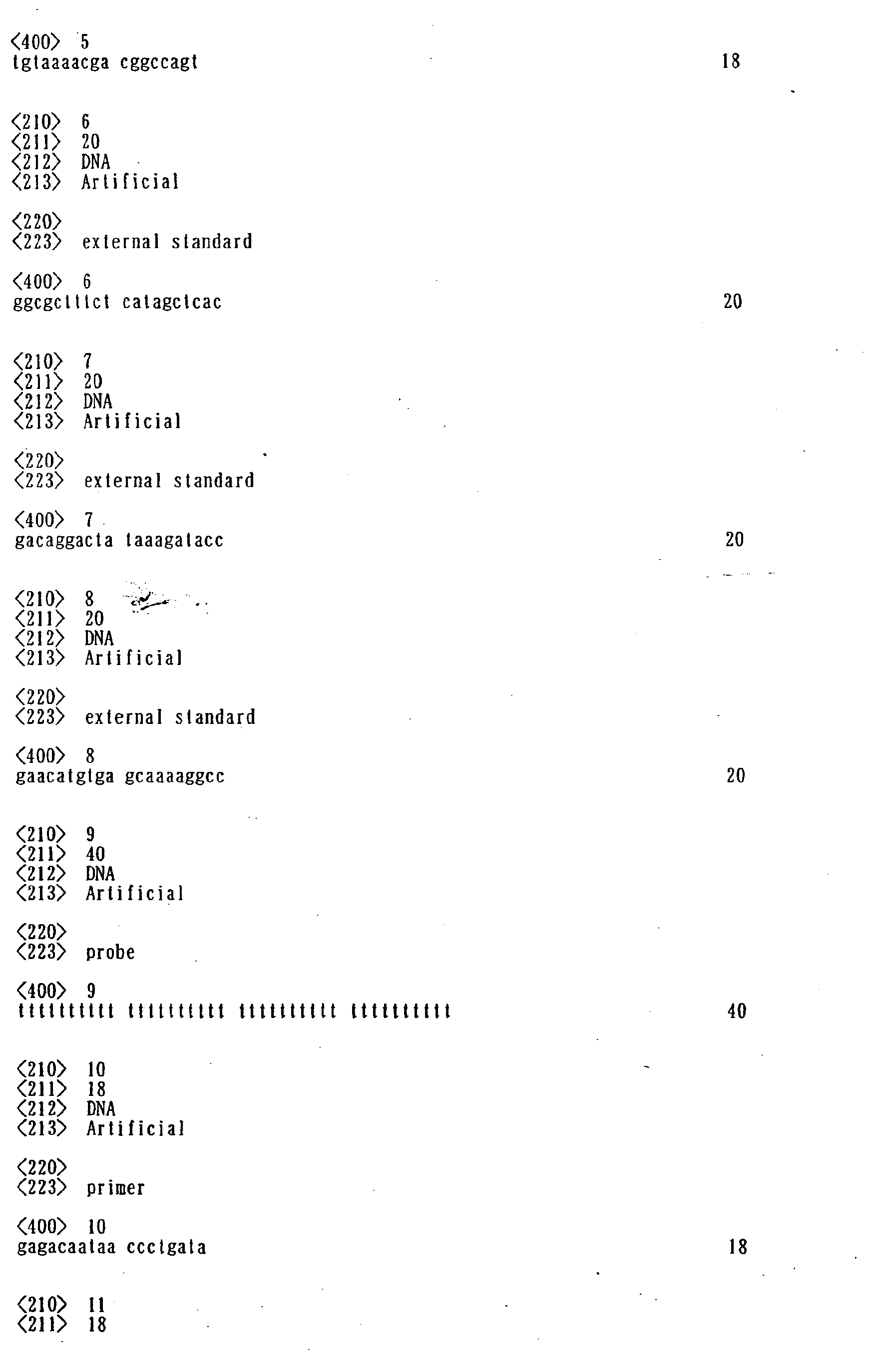
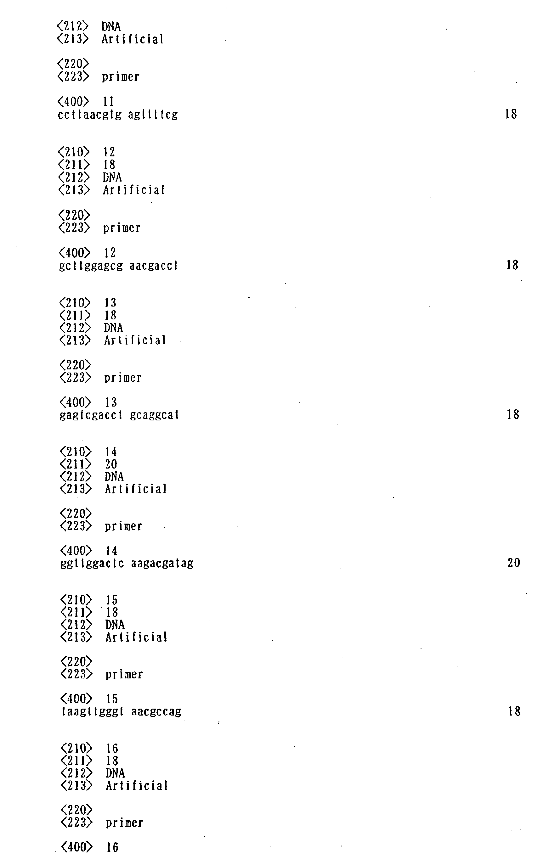
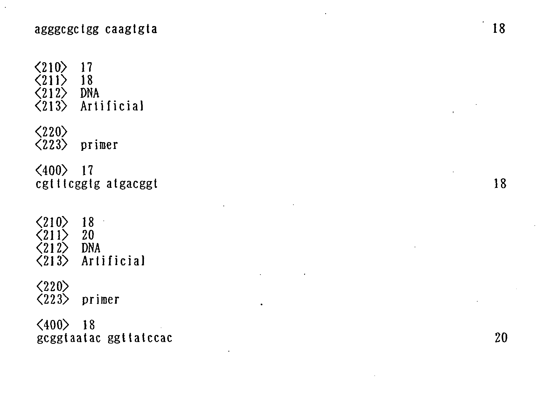
As shown in Table 1 below, five forward primers
P1 to P5 and four reverse primers RP1 to RP4 were
selected as PCR primers on the basis of information
on the base sequence of the linear pUC118 digested by
EcoRI prepared in Example 1.
The positions of the primers shown in Table 1
are those renumbered designating the first base of
the EcoRI cleavage site as position 1. As for the
reverse primers RP1 to RP4 in Table 1, the positions
are on the complementary chain.
Each primer having a base sequence shown in
Table 1 was synthesized and purified by high-performance
liquid chromatography (HPLC) and
dissolved in a TE buffer solution to a final
concentration of 10 pmol/µl to prepare a primer
solution.
Fig. 1 shows the chain lengths of amplified
products obtained by carrying out PCR amplification
reactions using primers shown in Table 1 and the
internal standard nucleic acid (EcoRI-digested pUC118
prepared in Example 1) as a template. Fig. 1 also
shows combinations of forward primers and reverse
primers used. Separately, 5'-fluorescent (rhodamine)
labeled reverse primers were prepared in the same
manner as with the above unlabeled primers to prepare
fluorescence-labeled primer solutions.
Example 3
<Preparation of internal standard probes>
Internal standard probes shown below were
prepared based on the information on the base
sequence of the linear plasmid DNA pUC118 EcoRI
digest prepared in Example 1.
The positions of the probes shown in Table 2
are those renumbered designating the first base of
the EcoRI cleavage site as position 1.
Four internal standard probes of which base
sequences are shown in Table 2 were synthesized and
then a sulfanyl group was introduced to the 5'
terminal of each nucleic acid as a functional group
to immobilize the probes in the DNA micro-array in
accordance with a conventional method. After the
introduction of the functional group, the internal
standard probes were purified and freeze-dried. The
freeze-dried internal standard probes were preserved
in a freezer at -30°C.
Fig. 1 shows the positions of the target base
sequence to which the four internal standard probes
would hybridize. The position of each PCR product on
the target base sequence is shown as well.
Example 4
<Preparation of external standard nucleic acids>
The following external standard nucleic acids
were prepared based on information on the sequence of
linear plasmid, pUC118 EcoR I Digest prepared in
Example 1.
The positions of the probes shown in Table 3
are those renumbered designating the first base of
the EcoRI cleavage site as position 1.
The four external standard nucleic acids are
complementary to the four internal standard probes
shown in Table 2.
A fluorescent dye (rhodamine) was introduced to
the 5' terminals of the four external standard
nucleic acids of which base sequences are shown in
Table 3 by a convention method after synthesis, as a
marker for detection and quantification. After the
introduction of the fluorescent marker, the external
standard nucleic acids were purified and dissolved in
a TE buffer solution to a final concentration of 5 µM
to prepare external standard nucleic acid solutions.
The external standard nucleic acid solution were put
in a light-shielded container and kept in a
refrigerator at -30°C.
Fig. 1 shows the positions of the base
sequences of the four external standard nucleic acids
and the above four internal standard probes having
complementary base sequences on the positions of the
target base sequence.
Example 5
<Preparation of DNA micro-array>
[1] Cleaning of glass substrate
A synthetic quartz glass substrate (size: 25 mm
× 75 mm × 1 mm, manufactured by Iiyama Tokushu Glass
Co., Ltd.) was placed in a heat-resistant and alkali-resistant
rack and immersed in a cleaning liquid of a
predetermined concentration for ultrasonic cleaning.
After overnight immersion in the cleaning liquid, it
was subjected to ultrasonic cleaning for 20 minutes.
Subsequently, the substrate was taken out from the
cleaning liquid and lightly rinsed with pure water,
and ultrasonic cleaning was carried out in ultra-pure
water for 20 minutes. The substrate was then
immersed in a 1 N sodium hydroxide aqueous solution
at 80°C for 10 minutes. After washing with pure
water and with ultra-pure water, a cleaned quartz
glass substrate for a DNA chip was obtained.
[2] Surface treatment
A silane coupling agent (KBM-603, a product of
Shin-Etsu Silicone Co., Ltd.) was dissolved in pure
water to a concentration of 1 wt% and stirred at room
temperature for 2 hours. Subsequently, the cleaned
glass substrate was immersed in the aqueous solution
of the silane coupling agent and kept at room
temperature for 20 minutes. Then the glass substrate
was pulled up, lightly washed with pure water on both
surfaces, and dried by blowing nitrogen gas onto both
sides. The dried substrate was then baked in an oven
heated at 120°C for 1 hour to complete the coupling
treatment, thereby amino groups were introduced onto
the surface of the substrate.
Next, N-(6-maleimidocaproyloxy)succimido
(EMCS)(a product of by Dojin Kagaku Kenkyujo Co.,
Ltd.) was dissolved in a solvent mixture of dimethyl
sulfoxide and ethanol (volume ratio of 1:1) to a
final concentration of 0.3 mg/ml to prepare an EMCS
solution. The glass substrate baked and cooled was
then immersed in the EMCS solution at room
temperature for 2 hours. By this treatment,
maleimido groups were introduced onto the substrate
surface through the reaction between the succimido
group of EMCS and the amino groups on the substrate
surface introduced by the silane coupling agent
treatment. Then the glass substrate was pulled up
from the EMCS solution and washed with the above
mixed solvent of dimethyl sulfoxide and ethanol.
After further washing with ethanol, the surface-treated
glass substrate was dried in a nitrogen gas
atmosphere.
[3] Probe DNA
The four internal standard probes prepared in
Example 3 were dissolved in pure water to prepare
solutions of various concentrations and compositions
as shown in Table 4 below. Each internal standard
probe solution was divided into aliquots and freeze-dried
to remove water.
| No. | Component | Concentration |
| 1 | B-SH | One probe | 1.50 D/ml |
| 2 | D-SH |
| 3 | F-SH |
| 4 | H-SH |
| 5 | B-SH | One probe | 0.750 D/ml |
| 6 | D-SH |
| 7 | F-SH |
| 8 | H-SH |
| 9 | B-SH | One probe | 0.50 D/ml |
| 10 | D-SH |
| 11 | F-SH |
| 12 | H=SH |
| 13 | B=SH | One probe | 0.3750 D/ml each |
| 14 | D-SH |
| 15 | F-SH |
| 16 | H-SH |
| 17 | B+D | Mixture of two probes | 0.750 D/ml each |
| 18 | B+F |
| 19 | B+H |
| 20 | D+F |
| 21 | D+H |
| 22 | F+H |
| 23 | B+D+F | Mixture of three probes | 0.50 D/ml each |
| 24 | B+D+H |
| 25 | B+F+H |
| 26 | D+F+H |
| 27 | B+D+F+H | Mixture of four probes | 0.3750 D/ml each |
An aqueous solution containing 7.5 wt% of
glycerin, 7.5 wt% of thiodiglycol, 7.5 wt% of urea
and 1.0 wt% of Acetylenol EH (produced by Kawaken
Fine Chemical Co., Ltd.) was prepared. Then, an
aliquot of each of 27 freeze-dried probe solutions
shown in Table 4 was dissolved in a specific amount
of the above mixed solvent to a specific
concentration. The obtained DNA probe solutions were
respectively charged into ink tanks for a bubble jet
printer (trade name: BJF-850, manufactured by Canon
Inc.), and the tank was mounted to a printing head.
The bubble jet printer used herein was modified
to enable printing on a plane surface. This bubble
jet printer can discharge ink droplets according to a
pattern inputted using a certain file creation method,
and can spot about 5 pico liter of the DNA solution
at a pitch of about 120 µm.
Subsequently, the 27 different probe solutions
were spotted onto a surface-treated glass substrate,
in accordance with the order shown in Fig. 2, using
the above modified bubble jet printer. After
confirming that intended spotting was carried out,
the glass substrate was left to stand in a humidified
chamber for 30 minutes to react the maleimido groups
on the surface of the glass substrate with the
sulfanyl groups at the 5' terminals of the DNA probes.
[5] Washing
After 30 minutes of reaction, the DNA solutions
remaining on the surface were washed away with a 10
mM phosphate buffer solution (pH of 7.0) containing
100 mM NaCl to obtain a gene chip (DNA probe array
substrate) having immobilized single-stranded DNAs
spotted in a matrix pattern on the surface of the
glass substrate.
Example 6
<Calibration of substrate using external standard
nucleic acid>
Eight DNA micro-arrays prepared in Example 5
were used. The eight DNA micro-arrays were immersed
in a blocking solution having a composition shown
below and left to stand at room temperature for 3
hours to carry out blocking treatment.
| Composition of blocking solution |
| Component | Content |
| Bovine serum albumin (produced by Sigma Co., Ltd.) | 2%(w/v) |
| NaCl | 100 mM |
| Phosphate buffer solution pH 7.0 | 10 mM |
Meanwhile eight different hybridization solutions containing external standard nucleic acid as shown in Table 6 were prepared. The basic solvent for these hybridization solutions was a 10 mM phosphate buffer solution (pH of 7.0) containing 100 mM NaCl (to be referred to as "basic buffer solution" hereinafter), and each of the external standard nucleic acids shown in Table 6 or a mixture thereof was dissolved in this basic buffer solution to a
total nucleic acid concentration of 30 nM.
| No. | External standard nucleic acid | Content |
| 1 | A | 30 nM each |
| 2 | G | 30 nM each |
| 3 | A+G | 15 nM each |
| 4 | C+G | 15 nM each |
| 5 | E+G | 15 nM each |
| 6 | A+C+G | 10 nM each |
| 7 | C+E+G | 10 nM each |
| 8 | A+C+E+G | 7.5 nM each |
In Table 6, abbreviations in the column of
external standard nucleic acids designate the
external standard nucleic acids shown in Table 3,
indicating that corresponding external standard
nucleic acids are contained.
DNA micro-arrays subjected to the blocking
treatment and rinsed with the basic buffer solution
were immersed in the eight hybridization solutions
respectively and sealed up in a vinyl pack. A
hybridization reaction was carried out in each DNA
micro-array at 45°C for 3 hours.
After the completion of the reaction, each DNA
micro-array was taken out from the vinyl pack and the
following cleaning was carried out.
The DNA micro-array was washed with 2 × SSC +
0.1 % sodium dodecylsulfate (SDS) at 55°C for 5
minutes for three times and 0.1 x SCC at room
temperature for 1 minute once. The composition of 2
× SSC was: NaCl 17.5 g/l, trisodium citrate · dihydrate
8.8 g/l, adjusted to pH 7.0 with sodium hydroxide.
After washing, extra liquid was blown off with
air to dry the DNA micro-array. After drying, the
fluorescence intensity at each spot was measured by
using a DNA array scanner (GenePix 4000B,
manufactured by Axon Co., Ltd.). The measurement
data was analyzed by using analytical software
(Genepix Pro 3.0) attached to the DNA array scanner.
Example 7
<Density calibration of immobilized probes>
The density ratio of the probes was calculated
from the measured fluorescence intensities. The
results are shown in Table 7.
| No. | Content | Concentration | Density ratio (%) |
| 1 | B-SH | One kind | 1.50 D/ml | 185 |
| 2 | D-SH | 189 |
| 3 | F-SH | 187 |
| 4 | H-SH | 189 |
| 5 | B-SH | One kind | 0.750 D/ml | 100 |
| 6 | D-SH | 100 |
| 7 | D-SH | 98 |
| 8 | H-SH | 101 |
| 9 | B-SH | One kind | 0.50 D/ml each | 67 |
| 10 | D-SH | 66 |
| 11 | F-SH | 66 |
| 12 | H-SH | 68 |
| 13 | B-SH | One kind | 0.3750 D/ml each | 51 |
| 14 | D-SH | 49 |
| 15 | F-SH | 51 |
| 16 | H-SH | 50 |
The density ratio was calculated as a relative
ratio making the immobilization density of the DNA
probe of Ink No. 5 to 100 %. The density ratios
shown in Table 7 were determined by first determining
the labeling rate of each external standard nucleic
acid separately and then correcting the density ratio
with the labeling rate.
In Table 7, the density ratios of Ink Nos. 1 to
4 (amounts of fluorescence) do not reach the
theoretical value of 200 %, because the immobilized
amount of the probe nucleic acid reached the
permissible amount (saturation amount) of the
substrate.
The density ratio of each probe in the mixed
probe ink Nos. 17 to 27 shown in Table 4 was similar
to the density ratio of the corresponding single
probe of a corresponding concentration.
Example 8
<Creation of calibration curve using external
standard nucleic acid>
The amount of each probe immobilized was
measured from the results of Example 7 and a
calibration curve was drawn using the correction data.
More specifically, hybridization experiments
were conducted in the same manner as in Example 6
using the DNA micro-array prepared in Example 5. The
external standard nucleic acid was added as a mixture
in amounts shown below.
Mixing ratio of the external standard nucleic acids:
- A-Rho:
- 66.7 % (20 nM)
- C-Rho:
- 25.0 % (7.5 nM)
- E-Rho:
- 6.7 % (2.0 nM)
- G-Rho:
- 1.7 % (0.5 nM)
The measurement and analysis were carried out
in the same manner as in Example 6.
Calibration curves were drawn using the
measurement results and the correction values of
probe density obtained in Example 7. Fig. 3 shows
the obtained calibration curves.
Example 9
<Creation of calibration curves using PCR products>
Calibration curves of PCR amplified products
were drawn using the internal standard nucleic acid
prepared in Example 1 and the internal standard
primers prepared in Example 2.
First, a PCR amplification reaction was carried
out using a combination of internal standard primers
shown in Table 8 in accordance with a predetermined
method. The composition of a PCR reaction solution
is as follows. The reverse primers used in this
example were primers whose 5' terminals were labeled
with fluorescence prepared in Example 3.
| -PCR reaction solution- |
| PCR premix reaction solution (2x) | 25 µl |
| pUC118 EcoR I Digest | 1 µl |
| Mixed primer (Table 8) | 6 µl |
| Pure water | 18 µl/50 µl in total |
Temperature cycle (24 cycles each including: 92°C for
10 sec; 62°C for 15 sec; and 72°C for 30 sec)
| No. | Forward primer | Reverse primer | Primer concentration | Chain length of amplified product |
| 1 | P-4 | PR-2 | 5 nmol/ml each | 337 bp |
| 2 | PR-3 | 895 bp |
| 3 | P-1 | PR-4 | 1776 bp |
| 4 | P-2 | 758 bp |
| |
After the reaction, gel filtration was carried
to purify the PCR amplified product. A TE buffer
solution was used to adjust the volume of the PCR
amplified product, and then the absorbance at 240 nm
and the amount of fluorescence were measured to
determine the amount of nucleic acid and the amount
of the product labeled by fluorescence in order to
obtain the labeling rate.
A hybridization reaction was carried out in the
same manner as in Example 8 using the internal
standard amplified product of known labeling rate to
measure the amount of fluorescence derived from the
internal standard amplified product bounding to the
probe.
Calibration curves were drawn from the
measurement results using the labeling rate of the
internal standard amplified product and the
correction value of probe density obtained in Example
7. Fig. 4 shows the obtained calibration curves.
When the calibration curves shown in Fig. 4 are
reviewed, there is a difference in the slope between
the calibration curves depending on the molecular
weight (chain length) of the amplified products.
That is, in order to determine the target nucleic
acid with higher reliability, it is necessary to
employ a combination of primers giving internal
standard amplified products having the same chain
length as the amplified product of the target nucleic
acid.
The following examples show use of density
standard probes formed by the same method as with the
nucleic acid probes to determine the amount or
density of the above nucleic acid probes.
Example 10
Fig. 5 schematically shows the plane
arrangement of an array of probe spots bonded to a
substrate and the sectional structure of the probe
array. Reference numeral 501 denotes a group of
probes prepared under arbitrary conditions; and 502,
a group of probes having a known nucleic acid
concentration. In the sectional view, reference
numeral 503 denotes a substrate; 504, a surface-treated
layer made from an organic material; and 500,
a nucleic acid probe. The process for producing this
probe array shown in Fig. 5 using a known method
(method 9 disclosed in JP H11-187900 A) will be
described hereinbelow.
The cleaning [1] and surface treatment [2] of
the glass substrate were carried out in the same
manner as in Example 5.
[3] Synthesis of probe DNA
Single-stranded nucleic acid having SEQ ID NO:
9 (40-mer of dT) was synthesized by a DNA
manufacturer (Bex Co., Ltd.). A (SH) group was
introduced into the 5' terminal of the single-stranded
DNA having SEQ ID NO: 1 by using a thiol
modifier (Glen Research Co., Ltd.). After the DNA
synthesis, deprotection and the collection of DNA
were carried out by conventional methods and HPLC was
used for purification. The manufacturer did the
steps from synthesis to purification.
A single-stranded DNA of SEQ ID NO: 1 described
in [3] was dissolved in a solution containing 7.5 wt%
of glycerin, 7.5 wt% of urea, 7.5 wt% of thioglycol
and 1 wt% of acetylene alcohol (trade name:
Acetylenol EH; produced by Kawaken Fine Chemical Co.,
Ltd.) to a final concentration of 8 µM.
The BC-50 printer head (manufactured by Canon
Inc.) for the BJF-850 bubble jet printer
(manufactured by Canon Inc.) using a bubble jet
method (a kind of thermal jet method) was modified to
discharge several hundreds µl of a solution. This
modified head was mounted to a modified discharge-drawing
machine so that ink could be discharged to a
flat quartz substrate. The above DNA solution was
injected in the modified tank of this head in an
amount of several hundred µl and the EMCS-treated
substrate was set in the drawing machine to spot the
solution on the EMCS-treated surface. The discharge
amount of the solution at one ejection was 4
pl/droplet and the spotted area was 20 mm × 30 mm on
the substrate at a pitch of 200 dpi, that is, 127 µm
intervals. Under the above conditions, the diameter
of dots spotted in a 157 × 236 matrix pattern (157
rows, 236 columns) was about 50 µm.
After spotting, the substrate was left to stand
in a humidifed chamber for 30 minuets for reaction of
the maleimido group on the surface of the substrate
and the sulfanyl group (-SH) at the 5' terminals of
the nucleic acid probes in order to immobilize the
DNA probes. Thereafter, the substrate was rinsed
with pure water and preserved in pure water. Just
before TOF-SIMS analysis, the DNA bonded substrate
(DNA chip) was dried by blowing nitrogen gas and
further dried in a vacuum desiccator. Under the
above conditions, 15 DNA chips were produced in total.
Further, 25 DNA chips and 35 DNA chips were
prepared by the same manner as above except that the
concentration of the above single-stranded DNA (SEQ
ID NO: 1) in the spotting solution was changed to 5
µM and 2.5 µM respectively. The above three types of
DNA chips were designated as standard DNA chips.
Thereafter, a DNA chip was produced under the
same conditions as above using a single-stranded DNA
solution of an ordinary concentration (about 8 µM).
In the above DNA chip, the first to third rows from
the end thereof were formed respectively using the
same three single-stranded DNA solutions as used in
the preparation of the standard DNA chips. That is,
probe spots were formed with the DNA solution having
a concentration of 10 µM in the first row at the
endmost position of the substrate, probe spots were
formed with the DNA solution having a concentration
of 5 µM in the second row, and probe spots were
formed with the DNA solution having a concentration
of 2.5 µM in the third row. The other rows (4th to
157th rows) were formed with a 8 µM solution of DNA
of another lot. The number of probe arrays can be
set to any value.
Example 11
<Determination of nucleic acid on nucleic acid chip>
The standard DNA chips produced in three
different DNA concentrations were washed with an acid
to dissolve DNA probes and the solution was
concentrated to 1 ml or less under conditions that
the acid solution did not scatter. Thereafter,
ultra-pure water was added to the concentrated
solution to a total volume of 1 ml. This solution
was introduced into an ICP-MS apparatus to measure
the mass of P (phosphorus). Based on this
measurement value, in consideration of the number of
dots used for the above measurement (number of spots),
the average value of the number of DNA molecules per
dot was determined for each DNA concentration. The
number of substrates of each DNA concentration may be
determined in consideration of the detection limit
concentration of P of the ICP-MS apparatus (about 10
ppb) and the number of DNA molecules on the substrate,
not limited to the number of substrates in Example 10.
Example 12
<TOF-SIMS measurement>
The DNA chip shown in Fig. 5 was conveyed to
the analysis chamber of a time-of-flight type
secondary ion mass spectrometer (TOFSIMS IV:
manufactured by ION TOF Co., Ltd.) and irradiated
with ions to the final primary ion dose density of 1
× 1012 atoms/cm2 under conditions shown in Table 1.
The secondary ion PO3 - (mass charge ratio of 78.958
amu (atom mass unit)) detected during this operation
was integrated to obtain cumulative intensity. Fig.
6 shows a relationship between the average value of
secondary ion intensity of the dots of one DNA
concentration formed on one of the first to third
rows and the DNA formation density (number of DNA
molecules per dot). The intensity of PO3 - measured
with the surface-treated substrate right before the
probe DNA immobilization was used as the background.
It was confirmed that the DNA formation density
and the secondary ion intensity of the dots of each
row were in proportion. When TOF-SIMS analysis was
carried out under the same measurement conditions for
a nucleic acid probe array prepared using a 8 µM
nucleic acid solution and an ink jet printer to
discharge the solution, the DNA formation density of
each dot (number of DNA molecules per dot) obtained
from a count value of PO
3 - was about 2.0 × 10
7 and the
variation in 10 dots selected at random was 20 % or
less.
| Measurement conditions of TOF-SIMS |
| Primary ion | Secondary ion |
| Ion species | Ga+ | Ion species | PO3 - |
| Acceleration voltage | 25 kV | Measurement area | 300µm × 300µm |
| Pulse | 2.5 kHz | Integrating circuit | 256 |
The same nucleic acid probe array as in Example
12 was prepared, and its surface was scanned with
primary ions to display the generated secondary ions
corresponding to each scan point, the intensity of P-
obtained at each scanning point was classified into a
plurality of intensity levels, and a dummy color was
set for each level to quantitatively compare the
intensity distribution of P-, that is, the
distribution of surface density or formation density
of nucleic acid (number of molecules of nucleic acid
per probe dot).
As described above, according to the present
invention, provided is a DNA micro-array which
enables easy and detailed analysis. More
specifically, detection of a nucleic acid which is
the actual object and an assay work can be carried
out concomitantly. Highly accurate assay results can
be obtained because quantitative assay is carried out
simultaneously with the detection operation.
Therefore, the determination accuracy of the
quantitative analysis of the molecules of a target
nucleic acid is greatly improved by using the DNA
micro-array according to the present invention. For
quantitative analysis, internal standard
amplification primers and external standard nucleic
acids are used as a detection kit in accordance with
the DNA micro-array according to the present
invention so that a user can make use of the present
invention very easily. Further, nucleic acid probe
dots for use as density standards whose average
nucleic acid density per dot has been determined and
which are formed by the same method as the formation
of the above nucleic acid probe dots are existent on
the chip substrate, whereby the nucleic acid
concentrations of nucleic acid probe dots having an
unknown concentration arranged on the substrate can
be determined by secondary ion mass spectrometry.
Therefore, reproducibility and quantitavity are
improved as compared with the prior art method.
Using the method of the present invention, the
reliability of DNA micro-array products can be
improved. It also enables to visualize the density
distribution of nucleic acid probe dots formed in a
matrix pattern in the DNA micro-array.