EP1519294B1 - Charakterisierung des Krankheitsverlaufs - Google Patents
Charakterisierung des Krankheitsverlaufs Download PDFInfo
- Publication number
- EP1519294B1 EP1519294B1 EP04253908.0A EP04253908A EP1519294B1 EP 1519294 B1 EP1519294 B1 EP 1519294B1 EP 04253908 A EP04253908 A EP 04253908A EP 1519294 B1 EP1519294 B1 EP 1519294B1
- Authority
- EP
- European Patent Office
- Prior art keywords
- progression
- disease
- uptake
- trajectory
- parkinson
- Prior art date
- Legal status (The legal status is an assumption and is not a legal conclusion. Google has not performed a legal analysis and makes no representation as to the accuracy of the status listed.)
- Expired - Lifetime
Links
Images



Classifications
-
- G—PHYSICS
- G06—COMPUTING OR CALCULATING; COUNTING
- G06T—IMAGE DATA PROCESSING OR GENERATION, IN GENERAL
- G06T7/00—Image analysis
- G06T7/0002—Inspection of images, e.g. flaw detection
- G06T7/0012—Biomedical image inspection
-
- G—PHYSICS
- G06—COMPUTING OR CALCULATING; COUNTING
- G06V—IMAGE OR VIDEO RECOGNITION OR UNDERSTANDING
- G06V10/00—Arrangements for image or video recognition or understanding
- G06V10/20—Image preprocessing
- G06V10/24—Aligning, centring, orientation detection or correction of the image
- G06V10/243—Aligning, centring, orientation detection or correction of the image by compensating for image skew or non-uniform image deformations
-
- G—PHYSICS
- G06—COMPUTING OR CALCULATING; COUNTING
- G06V—IMAGE OR VIDEO RECOGNITION OR UNDERSTANDING
- G06V10/00—Arrangements for image or video recognition or understanding
- G06V10/20—Image preprocessing
- G06V10/25—Determination of region of interest [ROI] or a volume of interest [VOI]
-
- G—PHYSICS
- G06—COMPUTING OR CALCULATING; COUNTING
- G06V—IMAGE OR VIDEO RECOGNITION OR UNDERSTANDING
- G06V10/00—Arrangements for image or video recognition or understanding
- G06V10/40—Extraction of image or video features
- G06V10/50—Extraction of image or video features by performing operations within image blocks; by using histograms, e.g. histogram of oriented gradients [HoG]; by summing image-intensity values; Projection analysis
- G06V10/507—Summing image-intensity values; Histogram projection analysis
-
- G—PHYSICS
- G06—COMPUTING OR CALCULATING; COUNTING
- G06V—IMAGE OR VIDEO RECOGNITION OR UNDERSTANDING
- G06V10/00—Arrangements for image or video recognition or understanding
- G06V10/40—Extraction of image or video features
- G06V10/56—Extraction of image or video features relating to colour
-
- G—PHYSICS
- G06—COMPUTING OR CALCULATING; COUNTING
- G06V—IMAGE OR VIDEO RECOGNITION OR UNDERSTANDING
- G06V2201/00—Indexing scheme relating to image or video recognition or understanding
- G06V2201/03—Recognition of patterns in medical or anatomical images
- G06V2201/031—Recognition of patterns in medical or anatomical images of internal organs
Definitions
- the present invention relates to the characterisation, i.e. monitoring and/or measurement of progressive dysfunction of a system. More particularly the invention relates to a method, which may be embodied in software for a software-controlled machine, for characterising such progressive dysfunction.
- Parkinson's disease is a disorder of the central nervous system that affects between one and one-and-a-half million US citizens. Parkinson's disease can appear at any age, but it is uncommon in people younger than 30, and the risk of developing it increases with age. It occurs in all parts of the world, and men are affected slightly more often than women. Patients with the disease suffer severe motor-neuro symptoms, including shaking, rigidity, slowness of movement and poor balance.
- Parkinson's disease can be monitored by nuclear medical imaging of the two striata of the brain (left and right).
- a characteristic of Parkinson's disease is the lack of dopamine in the striatum of the brain, caused by the loss of neurons in the substantia nigra.
- the amount of dopamine in the striatum can be measured by injecting a radioligand (for example ioflupane (FP-CIT/DaTSCAN) or iodobenzamide (IBZM)); after the radioactive labeled molecules arrive at the dopaminergic terminals in the striatum, they bind to pre-synaptic dopamine transporters molecules which enables imaging of functional dopaminergic neurons using Single Photon Emission Computed Tomography (SPECT).
- SPECT Single Photon Emission Computed Tomography
- the SPECT image of a normal person shows high uptake in the left and right striatum, as shown in Figures 1(a) and (b) of the accompanying drawings. These images are SPECT images with 123 I tracer (here DaTSCAN) showing the striatum as "hotspots".
- Figure 1(a) is a close-up of one of the striata in Figure 1(b) .
- the number of dopamine transporters in the striatum is substantially reduced.
- the SPECT image of a typical Parkinson's disease patient shows reduced uptake in the striatum, and in some patients there is asymmetry in the level of uptake between the left and right striatum. Such an image is shown in Figure 1(c) of the accompanying drawings.
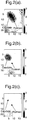
- Figures 2(a), (b) and (c) show typical disease progression for a single striatum. As the disease progresses, the tail of the striatum, the putamen, dies off first, followed finally by the upper region, the caudate.
- Figure 2(a) shows a healthy striatum, progressing in Figure 2(b) to a moderately degenerated one where the putamen tail has very reduced uptake, and finally in Figure 2(c) an extremely degenerated striatum with little uptake at all in either putamen or caudate.
- the present invention accordingly provides computer program and apparatus as defined in the appended claims.
- this embodiment consists of the following three steps:-
- a health measure is first computed for each striatum.
- the values of the health measure range from 1 to 0 where 1 indicates a healthy striatum and 0 indicates the most severe reduction in the level of uptake of the contrast agent. Although 1 and 0 are chosen in this example, obviously the range can be normalised between other values.
- These health measures, for the left and right striata, are then used to describe the disease progression. As Parkinson's disease progresses these two values will decrease in a characteristic way. This allows the calculation of the disease percentage.
- the first aim is to characterise the level of uptake of the contrast agent at different points in the striatum.
- a conventional 3-D SPECT data set (which may, for example, consist of 128 axial slices)
- the slices which include the striatum are found.
- the striatum is found in the 10 slices between slices 10 and 20.
- the slices including the striatum can be found manually by a clinician inspecting the slices, or alternatively can be found automatically by finding the brightest part of the SPECT scans (which is located in the striatum) and taking a predefined number of slices above and below that slice.
- the search area can be reduced, for example, by detecting the head outline and concentrating the search in the quadrant of the head where the striatum is known to be located.
- a single 2-D analysis slab is created by summing the layers together and normalising all the intensity values within the slab by dividing each pixel by an average background intensity value. This defines a very thick SPECT slice which includes all (or most) of the striatum. Normalisation is effective to ensure that factors such as variations in the amount of contrast agent given to the patient, total scan time or time between administering the contrast agent and performing the scan do not affect the overall brightness of the scan, thus confusing the analysis.
- a Region of Interest is positioned over the striatum such that the centre line of this ROI lies close to the central part of the striatum.
- ROI Region of Interest
- Figure 3(a) a rectangular ROI is used, though ROIs of other shape, for exampled based on the expected shape of the striatum, can be used.
- the Region of Interest is positioned by locating a short edge of the rectangle at the brightest part of the striatum (this may be located by positioning a larger search area over the striatum and searching for the brightest region within it) and orienting the long axis of the rectangle along the striatum.
- the ROI may be positioned differently as long as the position can be consistently found in the image).
- each point in the region of interest is projected onto a line defined through the region of interest (for example the centre line of the rectangle) by summing the intensity values (representing uptake of the contrast agent) over the width of the region of interest.
- This summed uptake value is graphed along the length of the striatum and Figures 4(a) to (c) illustrate three examples of regions of interest over SPECT scans with Figures 4(d) to (f) illustrating the corresponding summed values of intensity.
- the graphs begin at the short edge of the ROI which is positioned on the brightest point of the striatum and extend along the striatum towards the end of the putamen.
- Figure 4(a) shows a SPECT scan for a healthy striatum and Figure 4(d) the corresponding graphed intensity along the length of the striatum.
- Figures 4(b) and (e) show corresponding values for a striatum with a moderate to severe degeneration
- Figures 4(c) and (f) show values for a striatum with extreme degeneration. It can be seen that the values decrease into the putamen which is believed to be a characteristic of Parkinson's disease - the putamen dies off more quickly than the caudate.
- a collection of test scans is taken which show different stages in the progression of the disease from healthy to extreme degeneration.
- Each scan includes two striata (the left and right) and each of the striata can be classified into one of five stages:-
- test scans comprise the training set for the algorithm. Classifying each striatum into one of the stages is used to teach the algorithm the different stages in progression of the disease. Thus the algorithm can compute an optimal model to describe the reduction in uptake in the striatum as the disease progresses. For acceptable results a training set containing more than 100 scans should be used.
- the training of the algorithm involves taking the graphs which have been classified into the five stages above, and for each of the five stages computing an average graph to typify each stage.
- a set of five graphs for a typical training set is shown in Figure 5(a) .
- Stages 2 and 3 each show a progressive decrease in uptake along the putamen.
- Stage 4 Severe degeneration shows deterioration in the putamen and also some deterioration in the caudate.
- Stage 5 Extreme degeneration displays a marked drop in uptake in the caudate.
- a health measure between 0 and 1 is to be computed, indicating where the striatum lies on the continuum between Stage 1: Healthy and Stage 5: Extreme degeneration.
- This is done by embedding the five graphs in a 3-dimensional space, where the third dimension is the health measure.
- a confidence value for the health measure can also be obtained from the error between the uptake graph for the particular striatum under consideration and the most similar graph in the continuum of graphs defined by the surface in Figure 5(b) .
- a small error implies a high confidence in the measure.
- this confidence value can be used to warn the clinician when an accurate measurement is not possible, for example due to artefacts in the imaging process.
- a value between 0 and 1 for a health measure for each of the left and right striata can be obtained.
- This measure is useful because it is quantitative, and also it is consistent in that it is judged by a reference to the progression in many other data sets forming the training set. Thus it gives a consistent and quantitative measurement of the state of the striata for that patient.
- the left and right striata do not necessarily degenerate at the same rate and the asymmetry can be indicative of the degree of progression of the disease.
- the health measures for the left and right striata are plotted as a single point on a graph as is illustrated in Figure 6 , where the horizontal co-ordinate is given by the less active (sicker) striatum and the vertical co-ordinate is given by the more active (healthier) striatum, as illustrated below.
- a point in the top right-hand comer of this graph (1, 1) indicates that both striata are healthy.
- a point in the bottom left-hand comer (0, 0) indicates that both striata are extremely degenerated.
- a value of (0.3, 0.7) indicates severe degeneration in one striatum and mild degeneration in the other.
- PDF probability density function
- this disease progression percentage is a single score which indicates the progression of the bilateral disease (in this case Parkinson's disease) and is associated with a confidence measure.
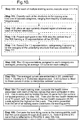
- Figure 9 illustrates in summary how a given scan is analysed to produce the two health measures and then the disease progression percentage and associated confidence values once the algorithm has already been set up with training data to compute the spline surface (shown in Figure 5(b) ) and best-fit curve on the progression graph (shown in Figure 8 ).
- Step 101 involves a series of steps in which the structures in a set of training scans are classified in step 111 into one of the categories mentioned above, and then in steps 112, 113 and 114 the one dimensional representations (such as shown in Figures 4 (d), (e) and (f) ) are calculated for each of the scans, and categorised according to the classification of step 111, thus linking the qualitative and quantitative assessments.
- step 102 the one dimensional representations are averaged in each category and in step 103 these are embedded in 3D positioned in the third dimension from 1: Healthy to 0: Extremely Degenerated and a hyper-surface is fitted to the curves to interpolate between them as shown in Figure 5(b) .
- step 104 the best fit of each individual training scan to the surface is found and the health measure between 0 and 1 is read off.
- the two health measures are then plotted against each other (as shown in Figure 7 ) and in step 105 the best fitting curve is found which passes through (0,0) and (1,1). This curve is then used for assessing disease progression as in steps 105-106 of Figure 9 .
Landscapes
- Engineering & Computer Science (AREA)
- Physics & Mathematics (AREA)
- General Physics & Mathematics (AREA)
- Theoretical Computer Science (AREA)
- Multimedia (AREA)
- General Health & Medical Sciences (AREA)
- Health & Medical Sciences (AREA)
- Medical Informatics (AREA)
- Nuclear Medicine, Radiotherapy & Molecular Imaging (AREA)
- Radiology & Medical Imaging (AREA)
- Quality & Reliability (AREA)
- Computer Vision & Pattern Recognition (AREA)
- Measuring And Recording Apparatus For Diagnosis (AREA)
- Measurement Of The Respiration, Hearing Ability, Form, And Blood Characteristics Of Living Organisms (AREA)
Claims (32)
- Computerprogramm, das Programmcodemittel zur Ausführung eines Verfahrens zur Messung des Verlaufs einer Parkinson-Erkrankung auf einem Computersystem umfasst, wobei das Computerprogramm Folgendes umfasst:- Messen der Aufnahme von Kontrastmittel in jeweiligen Striata eines Gehirns,- Bestimmen eines jeweiligen Gesundheitsmaßes innerhalb eines normalisierten Bereichs für jedes der Striata unter Verwendung gemessener Aufnahmewerte,- Definieren einer zweidimensionalen Raumdarstellung, wobei jede Dimension einem der Gesundheitsmaße entspricht,- Vordefinieren, in der zweidimensionalen Raumdarstellung, einer Trajektorie unter Verwendung von Gesundheitsmaßen aus einer Datenbank von Testfällen, die alle Phasen einer Parkinson-Erkrankung abdecken, und Punkten der Trajektorie, die bestimmten Phasen des Krankheitsverlaufs entsprechen,- Definieren eines Punkts in der zweidimensionalen Raumdarstellung, die die Werte der Gesundheitsmaße darstellt,- Definieren eines projizierten Punkts durch Projizieren des definierten Punkts auf die vordefinierte Trajektorie in der zweidimensionalen Raumdarstellung und- Bestimmen des Maßes der Parkinson-Erkrankung in dem Gehirn als den parametrischen Abstand des projizierten Punkts entlang der Trajektorie, wobei der parametrische Abstand des projizierten Punkts von einem ersten Ende einer Trajektorie, die ein gesundes Gehirn darstellt, in dem beide Striata gesund sind, berechnet wird.
- Computerprogramm nach Anspruch 1, wobei der definierte Punkt normal auf die vordefinierte Trajektorie projiziert wird.
- Computerprogramm nach Anspruch 1 oder 2, wobei ein Niveau der Konfidenz in die Messung des Verlaufs der Parkinson-Erkrankung aus dem Abstand des definierten Punkts von der vordefinierten Trajektorie berechnet wird.
- Computerprogramm nach Anspruch 1, 2 oder 3, wobei die vordefinierte Trajektorie auf dem gemessenen Verlauf der Parkinson-Erkrankung einer Mehrzahl von Gehirnen basiert.
- Computerprogramm nach Anspruch 4, wobei die vordefinierte Trajektorie erhalten wird durch statistische Analyse des gemessenen Verlaufs der Parkinson-Erkrankung in der Datenbank von Testfällen, die dadurch einen Trainingssatz für die Analyse bildet.
- Computerprogramm nach einem der vorhergehenden Ansprüche, wobei die Position des Punkts im Vergleich zu der vordefinierten Trajektorie als ein Prozentsatz des Abstands entlang der Trajektorie von dem ersten Ende der Trajektorie, das ein gesundes Gehirn darstellt, zu einem zweiten Ende der Trajektorie, das eine vollständig verlaufene Parkinson-Erkrankung darstellt, ausgedrückt wird.
- Computerprogramm nach einem der vorhergehenden Ansprüche, wobei die Dimensionen der zweidimensionalen Raumdarstellung den verschiedenen Gesundheitsmaßen in einer definierten Reihenfolge, die auf den relativen Werten der jeweiligen Gesundheitsmaße basiert, zugewiesen werden.
- Computerprogramm nach Anspruch 7, wobei die definierte Reihenfolge von der Darstellung des gesündesten Striatums zu der Darstellung kränksten Striatums ist.
- Computerprogramm nach Anspruch 1, wobei die Gesundheitsmaße durch Messen des Werts der Aufnahme, der sich mit der Zeit progressiv ändert, erhalten werden
und wobei das Verfahren die folgenden Schritte umfasst:
Vergleichen des gemessenen Werts der Aufnahme mit einem Satz von Werten, der eine Mehrzahl von Werten der Aufnahme umfasst, die von Messungen abgeleitet sind, die unterschiedliche Grade des Fortschreitens der Parkinson-Erkrankung zeigen, und Berechnen des durch den gemessenen Wert dargestellten Grads des Fortschreitens als das Gesundheitsmaß. - Computerprogramm nach Anspruch 9, wobei der Satz von Werten durch ein Verfahren erhalten wird, das Folgendes umfasst:Kategorisieren der Messungen, die unterschiedliche Grade des Fortschreitens zeigen, in eine Mehrzahl unterschiedlicher Kategorien des Grads des Fortschreitens,Berechnen eines typischen Werts für jede Kategorie aus den Mitgliedern der betreffenden Kategorie undwobei der gemessene Wert mit den typischen Werten verglichen wird.
- Computerprogramm nach Anspruch 10, wobei das Verfahren zum Erhalten des Satzes von Werten ferner Folgendes umfasst:
Interpolieren der erhaltenen typischen Werte, um eine effektiv kontinuierliche Darstellung des Verlaufs zu erhalten, mit der der gemessene Wert verglichen wird. - Computerprogramm nach Anspruch 11, wobei der Schritt des Interpolierens der typischen Werte das Anpassen einer Hyperfläche an die in einem Parameterraum dargestellten typischen Werte umfasst, wobei die Hyperfläche die effektiv kontinuierliche Darstellung bildet, und wobei der Schritt des Vergleichens des gemessenen Werts der Aufnahme das Finden der besten Anpassung des gemessenen Werts an die Hyperfläche umfasst.
- Computerprogramm nach einem der Ansprüche 9 bis 12, wobei das Gesundheitsmaß als ein Abstand entlang eines Pfads von einem Ausgangszustand der Aufnahme bis zu einem Endzustand der Aufnahme ausgedrückt wird.
- Computerprogramm nach einem der Ansprüche 9 bis 13, wobei der gemessene Wert der Aufnahme die zeitliche und/oder räumliche Variation der Aufnahme in dem menschlichen oder tierischen Körper ist.
- Computerprogramm nach einem der Ansprüche 9 bis 14, wobei die Aufnahme durch ein Bildattribut in einem medizinischen Bild dargestellt wird.
- Computerprogramm nach Anspruch 14 oder 15, wobei die Dimensionalität des gemessenen Werts durch Projektion und Summierung in einen Raum mit reduzierten Dimensionen reduziert wird.
- Vorrichtung, die dazu ausgelegt ist, ein Verfahren zur Messung des Verlaufs einer Parkinson-Krankheit auszuführen, das Folgendes umfasst:- Messen der Aufnahme von Kontrastmittel in jeweiligen Striata eines Gehirns,- Bestimmen eines jeweiligen Gesundheitsmaßes innerhalb eines normalisierten Bereichs für jedes der Striata unter Verwendung gemessener Aufnahmewerte,- Definieren einer zweidimensionalen Raumdarstellung, wobei jede Dimension einem der Gesundheitsmaße entspricht,- Vordefinieren, in der zweidimensionalen Raumdarstellung, einer Trajektorie unter Verwendung von Gesundheitsmaßen aus einer Datenbank von Testfällen, die alle Phasen einer Parkinson-Erkrankung abdecken, und Punkten der Trajektorie, die bestimmten Phasen des Krankheitsverlaufs entsprechen,- Definieren eines Punkts in der zweidimensionalen Raumdarstellung, die die Werte der Gesundheitsmaße darstellt,- Definieren eines projizierten Punkts durch Projizieren des definierten Punkts auf die vordefinierte Trajektorie in der zweidimensionalen Raumdarstellung und- Bestimmen des Maßes der Parkinson-Erkrankung in dem Gehirn als den parametrischen Abstand des projizierten Punkts entlang der Trajektorie, wobei der parametrische Abstand des projizierten Punkts von einem ersten Ende einer Trajektorie, die ein gesundes Gehirn darstellt, in dem beide Striata gesund sind, berechnet wird.
- Vorrichtung nach Anspruch 17, wobei der definierte Punkt normal auf die vordefinierte Trajektorie projiziert wird.
- Vorrichtung nach Anspruch 17 oder 18, wobei ein Niveau der Konfidenz in die Messung des Verlaufs der Parkinson-Erkrankung aus dem Abstand des definierten Punkts von der vordefinierten Trajektorie berechnet wird.
- Vorrichtung nach Anspruch 17, 18 oder 19, wobei die vordefinierte Trajektorie auf dem gemessenen Verlauf der Parkinson-Erkrankung einer Mehrzahl von Gehirnen basiert.
- Vorrichtung nach Anspruch 20, wobei die vordefinierte Trajektorie erhalten wird durch statistische Analyse des gemessenen Verlaufs der Parkinson-Erkrankung in der Datenbank von Testfällen, die dadurch einen Trainingssatz für die Analyse bildet.
- Vorrichtung nach einem der Ansprüche 17 bis 21, wobei die Position des Punkts im Vergleich zu der vordefinierten Trajektorie als ein Prozentsatz des Abstands entlang der Trajektorie von dem ersten Ende der Trajektorie zu einem zweiten Ende der Trajektorie, das eine vollständig verlaufene Parkinson-Erkrankung darstellt, ausgedrückt wird.
- Vorrichtung nach einem der Ansprüche 17 bis 22, wobei die Dimensionen der zweidimensionalen Raumdarstellung den verschiedenen Gesundheitsmaßen in einer definierten Reihenfolge, die auf den relativen Werten der jeweiligen Gesundheitsmaße basiert, zugewiesen werden.
- Vorrichtung nach einem der Ansprüche 17 bis 23, wobei die definierte Reihenfolge von der Darstellung des gesündesten Striatums zu der Darstellung des kränksten Striatums ist.
- Vorrichtung nach Anspruch 17, wobei die Gesundheitsmaße durch Messen des Werts der Aufnahme, der sich mit der Zeit progressiv ändert, erhalten werden
und wobei das Verfahren die folgenden Schritte umfasst:
Vergleichen des gemessenen Werts der Aufnahme mit einem Satz von Werten, der eine Mehrzahl von Werten der Aufnahme umfasst, die von Messungen abgeleitet sind, die unterschiedliche Grade des Fortschreitens der Parkinson-Erkrankung zeigen, und Berechnen des durch den gemessenen Wert dargestellten Grads des Fortschreitens als das Gesundheitsmaß. - Vorrichtung nach Anspruch 25, wobei der Satz von Werten durch ein Verfahren erhalten wird, das Folgendes umfasst:Kategorisieren der Messungen, die unterschiedliche Grade des Fortschreitens zeigen, in eine Mehrzahl unterschiedlicher Kategorien des Grads des Fortschreitens,Berechnen eines typischen Werts für jede Kategorie aus den Mitgliedern der betreffenden Kategorie undwobei der gemessene Wert mit den typischen Werten verglichen wird.
- Vorrichtung nach Anspruch 26, wobei das Verfahren zum Erhalten des Satzes von Werten ferner Folgendes umfasst: Interpolieren der erhaltenen typischen Werte, um eine effektiv kontinuierliche Darstellung des Verlaufs zu erhalten, mit der der gemessene Wert verglichen wird.
- Vorrichtung nach Anspruch 27, wobei der Schritt des Interpolierens der typischen Werte das Anpassen einer Hyperfläche an die in einem Parameterraum dargestellten typischen Werte umfasst, wobei die Hyperfläche die effektiv kontinuierliche Darstellung bildet, und wobei der Schritt des Vergleichens des gemessenen Werts der Aufnahme das Finden der besten Anpassung des gemessenen Werts an die Hyperfläche umfasst.
- Vorrichtung nach einem der Ansprüche 25 bis 28, wobei das Gesundheitsmaß als ein Abstand entlang eines Pfads von einem Ausgangszustand der Aufnahme bis zu einem Endzustand der Aufnahme ausgedrückt wird.
- Vorrichtung nach einem der Ansprüche 25 bis 29, wobei der gemessene Wert der Eigenschaft die zeitliche und/oder räumliche Variation der Eigenschaft in dem menschlichen oder tierischen Körper ist.
- Vorrichtung nach einem der Ansprüche 25 bis 29, wobei die Aufnahme durch ein Bildattribut in einem medizinischen Bild dargestellt wird.
- Vorrichtung nach Anspruch 31, wobei die Dimensionalität des gemessenen Werts durch Projektion und Summierung in einen Raum mit reduzierten Dimensionen reduziert wird.
Applications Claiming Priority (2)
| Application Number | Priority Date | Filing Date | Title |
|---|---|---|---|
| GB0321901 | 2003-09-18 | ||
| GBGB0321901.1A GB0321901D0 (en) | 2003-09-18 | 2003-09-18 | Characterisation of progressive system dysfunction |
Publications (3)
| Publication Number | Publication Date |
|---|---|
| EP1519294A2 EP1519294A2 (de) | 2005-03-30 |
| EP1519294A3 EP1519294A3 (de) | 2005-04-06 |
| EP1519294B1 true EP1519294B1 (de) | 2021-08-25 |
Family
ID=29266240
Family Applications (1)
| Application Number | Title | Priority Date | Filing Date |
|---|---|---|---|
| EP04253908.0A Expired - Lifetime EP1519294B1 (de) | 2003-09-18 | 2004-06-29 | Charakterisierung des Krankheitsverlaufs |
Country Status (3)
| Country | Link |
|---|---|
| US (1) | US7558680B2 (de) |
| EP (1) | EP1519294B1 (de) |
| GB (1) | GB0321901D0 (de) |
Families Citing this family (4)
| Publication number | Priority date | Publication date | Assignee | Title |
|---|---|---|---|---|
| GB0813372D0 (en) * | 2008-07-22 | 2008-08-27 | Siemens Medical Solutions | A confidence measure for comparing SUV between PET studies |
| US8687839B2 (en) * | 2009-05-21 | 2014-04-01 | Digimarc Corporation | Robust signatures derived from local nonlinear filters |
| JP6995642B2 (ja) * | 2018-01-19 | 2022-01-14 | 日本メジフィジックス株式会社 | 画像処理装置、画像処理方法およびプログラム |
| US11966822B2 (en) | 2020-09-29 | 2024-04-23 | International Business Machines Corporation | Feature processing for machine learning |
Citations (1)
| Publication number | Priority date | Publication date | Assignee | Title |
|---|---|---|---|---|
| WO1994010903A1 (en) * | 1992-11-18 | 1994-05-26 | Kunig Horst E | Measuring myocardial impairment, dysfunctions, and sufficiency/insufficiency |
Family Cites Families (6)
| Publication number | Priority date | Publication date | Assignee | Title |
|---|---|---|---|---|
| JPH02503760A (ja) | 1988-04-08 | 1990-11-08 | ケルトリンゲル ペーター | 多発神経病症候群の診断方法及び診断装置 |
| US5406502A (en) | 1993-06-29 | 1995-04-11 | Elbit Ltd. | System and method for measuring the operation of a device |
| US5810011A (en) | 1996-02-27 | 1998-09-22 | Kunig; Sabine Vivian | Method and apparatus for measuring myocardial impairment and dysfunctions from efficiency and performance diagrams |
| US7031843B1 (en) * | 1997-09-23 | 2006-04-18 | Gene Logic Inc. | Computer methods and systems for displaying information relating to gene expression data |
| US6560354B1 (en) * | 1999-02-16 | 2003-05-06 | University Of Rochester | Apparatus and method for registration of images to physical space using a weighted combination of points and surfaces |
| US6402707B1 (en) * | 2000-06-28 | 2002-06-11 | Denupp Corporation Bvi | Method and system for real time intra-orally acquiring and registering three-dimensional measurements and images of intra-oral objects and features |
-
2003
- 2003-09-18 GB GBGB0321901.1A patent/GB0321901D0/en not_active Ceased
-
2004
- 2004-06-29 EP EP04253908.0A patent/EP1519294B1/de not_active Expired - Lifetime
- 2004-08-19 US US10/921,473 patent/US7558680B2/en active Active
Patent Citations (1)
| Publication number | Priority date | Publication date | Assignee | Title |
|---|---|---|---|---|
| WO1994010903A1 (en) * | 1992-11-18 | 1994-05-26 | Kunig Horst E | Measuring myocardial impairment, dysfunctions, and sufficiency/insufficiency |
Also Published As
| Publication number | Publication date |
|---|---|
| US20050065734A1 (en) | 2005-03-24 |
| EP1519294A2 (de) | 2005-03-30 |
| GB0321901D0 (en) | 2003-10-22 |
| EP1519294A3 (de) | 2005-04-06 |
| US7558680B2 (en) | 2009-07-07 |
Similar Documents
| Publication | Publication Date | Title |
|---|---|---|
| US11443433B2 (en) | Quantification and staging of body-wide tissue composition and of abnormal states on medical images via automatic anatomy recognition | |
| KR101503940B1 (ko) | 신경퇴행성 질환의 진단을 돕는 수단 | |
| Chang et al. | Performance measure characterization for evaluating neuroimage segmentation algorithms | |
| NL1024869C2 (nl) | Werkwijze en systeem voor het meten van voor ziekte relevante weefselveranderingen. | |
| EP2661734B1 (de) | Automatische quantifizierung von asymmetrien | |
| US20090136107A1 (en) | Automatic Segmentation of the Heart and Aorta in Medical 3-D Scans Without Contrast Media Injections | |
| US20080069446A1 (en) | ROI-based assessment of abnormality using transformation invariant features | |
| US20110160546A1 (en) | System and method for volumetric analysis of medical images | |
| US20130251231A1 (en) | Medical image processing device | |
| WO2012151579A2 (en) | Systems and methods for analyzing in vivo tissue volumes using medical imaging data | |
| US20130156280A1 (en) | Processing system for medical scan images | |
| US8050734B2 (en) | Method and system for performing patient specific analysis of disease relevant changes of a disease in an anatomical structure | |
| EP4270305A1 (de) | Lernvorrichtung, -verfahren und -programm sowie vorrichtung zur verarbeitung medizinischer bilder | |
| KR101162599B1 (ko) | 흉부 방사선 이미지 분석을 통한 심비대 자동탐지 방법 및 그 기록매체 | |
| Moser et al. | Automated fetal brain extraction from clinical ultrasound volumes using 3D convolutional neural networks | |
| EP4113439B1 (de) | Bestimmung eines ortes, an dem ein bestimmtes merkmal in medizinischen bildgebungsdaten dargestellt ist | |
| US8077937B2 (en) | Reproducible objective quantification method to segment white matter structures | |
| US7684602B2 (en) | Method and system for local visualization for tubular structures | |
| CN1399763A (zh) | 使用统计曲率分析的三维测量 | |
| US7558680B2 (en) | Characterisation of progressive system dysfunction | |
| CN120299735A (zh) | 一种胸部肿瘤放疗后放射性肺炎预测系统及方法 | |
| Dovganich et al. | Automatic quality control in lung X-ray imaging with deep learning | |
| CN119151967A (zh) | 一种基于平扫ct数据的医学图像分析方法及系统 | |
| US20220067928A1 (en) | Method for locating and characterizing bifurcations of a cerebral vascular tree, associated methods and devices | |
| Alonso et al. | Composite index for the quantitative evaluation of image segmentation results |
Legal Events
| Date | Code | Title | Description |
|---|---|---|---|
| PUAI | Public reference made under article 153(3) epc to a published international application that has entered the european phase |
Free format text: ORIGINAL CODE: 0009012 |
|
| PUAL | Search report despatched |
Free format text: ORIGINAL CODE: 0009013 |
|
| 17P | Request for examination filed |
Effective date: 20040712 |
|
| AK | Designated contracting states |
Kind code of ref document: A2 Designated state(s): AT BE BG CH CY CZ DE DK EE ES FI FR GB GR HU IE IT LI LU MC NL PL PT RO SE SI SK TR |
|
| AX | Request for extension of the european patent |
Extension state: AL HR LT LV MK |
|
| AK | Designated contracting states |
Kind code of ref document: A3 Designated state(s): AT BE BG CH CY CZ DE DK EE ES FI FR GB GR HU IE IT LI LU MC NL PL PT RO SE SI SK TR |
|
| AX | Request for extension of the european patent |
Extension state: AL HR LT LV MK |
|
| AKX | Designation fees paid |
Designated state(s): DE FR GB |
|
| 17Q | First examination report despatched |
Effective date: 20060928 |
|
| RAP1 | Party data changed (applicant data changed or rights of an application transferred) |
Owner name: SIEMENS MEDICAL SOLUTIONS USA, INC. |
|
| STAA | Information on the status of an ep patent application or granted ep patent |
Free format text: STATUS: EXAMINATION IS IN PROGRESS |
|
| RAP1 | Party data changed (applicant data changed or rights of an application transferred) |
Owner name: SIEMENS MEDICAL SOLUTIONS USA, INC. |
|
| REG | Reference to a national code |
Ref country code: DE Ref legal event code: R079 Ref document number: 602004055013 Country of ref document: DE Free format text: PREVIOUS MAIN CLASS: G06F0019000000 Ipc: G06K0009460000 |
|
| GRAP | Despatch of communication of intention to grant a patent |
Free format text: ORIGINAL CODE: EPIDOSNIGR1 |
|
| STAA | Information on the status of an ep patent application or granted ep patent |
Free format text: STATUS: GRANT OF PATENT IS INTENDED |
|
| RIC1 | Information provided on ipc code assigned before grant |
Ipc: G06K 9/46 20060101AFI20201207BHEP Ipc: G06K 9/32 20060101ALI20201207BHEP Ipc: G06T 7/00 20170101ALI20201207BHEP |
|
| INTG | Intention to grant announced |
Effective date: 20210112 |
|
| GRAJ | Information related to disapproval of communication of intention to grant by the applicant or resumption of examination proceedings by the epo deleted |
Free format text: ORIGINAL CODE: EPIDOSDIGR1 |
|
| STAA | Information on the status of an ep patent application or granted ep patent |
Free format text: STATUS: EXAMINATION IS IN PROGRESS |
|
| INTC | Intention to grant announced (deleted) | ||
| GRAP | Despatch of communication of intention to grant a patent |
Free format text: ORIGINAL CODE: EPIDOSNIGR1 |
|
| STAA | Information on the status of an ep patent application or granted ep patent |
Free format text: STATUS: GRANT OF PATENT IS INTENDED |
|
| INTG | Intention to grant announced |
Effective date: 20210423 |
|
| GRAS | Grant fee paid |
Free format text: ORIGINAL CODE: EPIDOSNIGR3 |
|
| GRAA | (expected) grant |
Free format text: ORIGINAL CODE: 0009210 |
|
| STAA | Information on the status of an ep patent application or granted ep patent |
Free format text: STATUS: THE PATENT HAS BEEN GRANTED |
|
| REG | Reference to a national code |
Ref country code: DE Ref legal event code: R081 Ref document number: 602004055013 Country of ref document: DE Owner name: SIEMENS MEDICAL SOLUTIONS USA, INC., MALVERN, US Free format text: FORMER OWNER: MIRADA SOLUTIONS LTD., OXFORD, OXFORDSHIRE, GB |
|
| AK | Designated contracting states |
Kind code of ref document: B1 Designated state(s): DE FR GB |
|
| REG | Reference to a national code |
Ref country code: GB Ref legal event code: FG4D |
|
| REG | Reference to a national code |
Ref country code: DE Ref legal event code: R096 Ref document number: 602004055013 Country of ref document: DE |
|
| REG | Reference to a national code |
Ref country code: DE Ref legal event code: R079 Ref document number: 602004055013 Country of ref document: DE Free format text: PREVIOUS MAIN CLASS: G06K0009460000 Ipc: G06V0030180000 |
|
| REG | Reference to a national code |
Ref country code: DE Ref legal event code: R097 Ref document number: 602004055013 Country of ref document: DE |
|
| PLBE | No opposition filed within time limit |
Free format text: ORIGINAL CODE: 0009261 |
|
| STAA | Information on the status of an ep patent application or granted ep patent |
Free format text: STATUS: NO OPPOSITION FILED WITHIN TIME LIMIT |
|
| 26N | No opposition filed |
Effective date: 20220527 |
|
| PGFP | Annual fee paid to national office [announced via postgrant information from national office to epo] |
Ref country code: FR Payment date: 20230619 Year of fee payment: 20 Ref country code: DE Payment date: 20220630 Year of fee payment: 20 |
|
| PGFP | Annual fee paid to national office [announced via postgrant information from national office to epo] |
Ref country code: GB Payment date: 20230710 Year of fee payment: 20 |
|
| REG | Reference to a national code |
Ref country code: DE Ref legal event code: R071 Ref document number: 602004055013 Country of ref document: DE |
|
| PG25 | Lapsed in a contracting state [announced via postgrant information from national office to epo] |
Ref country code: GB Free format text: LAPSE BECAUSE OF EXPIRATION OF PROTECTION Effective date: 20240628 |
|
| REG | Reference to a national code |
Ref country code: GB Ref legal event code: PE20 Expiry date: 20240628 |
|
| PG25 | Lapsed in a contracting state [announced via postgrant information from national office to epo] |
Ref country code: GB Free format text: LAPSE BECAUSE OF EXPIRATION OF PROTECTION Effective date: 20240628 |