EP1516628B1 - Stable isotonic lyophilized protein formulation - Google Patents
Stable isotonic lyophilized protein formulation Download PDFInfo
- Publication number
- EP1516628B1 EP1516628B1 EP04022777.9A EP04022777A EP1516628B1 EP 1516628 B1 EP1516628 B1 EP 1516628B1 EP 04022777 A EP04022777 A EP 04022777A EP 1516628 B1 EP1516628 B1 EP 1516628B1
- Authority
- EP
- European Patent Office
- Prior art keywords
- protein
- formulation
- antibody
- lyophilized
- reconstituted
- Prior art date
- Legal status (The legal status is an assumption and is not a legal conclusion. Google has not performed a legal analysis and makes no representation as to the accuracy of the status listed.)
- Expired - Lifetime
Links
Images


Classifications
-
- A—HUMAN NECESSITIES
- A61—MEDICAL OR VETERINARY SCIENCE; HYGIENE
- A61K—PREPARATIONS FOR MEDICAL, DENTAL OR TOILETRY PURPOSES
- A61K39/00—Medicinal preparations containing antigens or antibodies
- A61K39/395—Antibodies; Immunoglobulins; Immune serum, e.g. antilymphocytic serum
- A61K39/39591—Stabilisation, fragmentation
-
- A—HUMAN NECESSITIES
- A61—MEDICAL OR VETERINARY SCIENCE; HYGIENE
- A61K—PREPARATIONS FOR MEDICAL, DENTAL OR TOILETRY PURPOSES
- A61K47/00—Medicinal preparations characterised by the non-active ingredients used, e.g. carriers or inert additives; Targeting or modifying agents chemically bound to the active ingredient
- A61K47/06—Organic compounds, e.g. natural or synthetic hydrocarbons, polyolefins, mineral oil, petrolatum or ozokerite
- A61K47/08—Organic compounds, e.g. natural or synthetic hydrocarbons, polyolefins, mineral oil, petrolatum or ozokerite containing oxygen, e.g. ethers, acetals, ketones, quinones, aldehydes, peroxides
- A61K47/12—Carboxylic acids; Salts or anhydrides thereof
-
- A—HUMAN NECESSITIES
- A61—MEDICAL OR VETERINARY SCIENCE; HYGIENE
- A61K—PREPARATIONS FOR MEDICAL, DENTAL OR TOILETRY PURPOSES
- A61K47/00—Medicinal preparations characterised by the non-active ingredients used, e.g. carriers or inert additives; Targeting or modifying agents chemically bound to the active ingredient
- A61K47/06—Organic compounds, e.g. natural or synthetic hydrocarbons, polyolefins, mineral oil, petrolatum or ozokerite
- A61K47/16—Organic compounds, e.g. natural or synthetic hydrocarbons, polyolefins, mineral oil, petrolatum or ozokerite containing nitrogen, e.g. nitro-, nitroso-, azo-compounds, nitriles, cyanates
- A61K47/18—Amines; Amides; Ureas; Quaternary ammonium compounds; Amino acids; Oligopeptides having up to five amino acids
- A61K47/183—Amino acids, e.g. glycine, EDTA or aspartame
-
- A—HUMAN NECESSITIES
- A61—MEDICAL OR VETERINARY SCIENCE; HYGIENE
- A61K—PREPARATIONS FOR MEDICAL, DENTAL OR TOILETRY PURPOSES
- A61K47/00—Medicinal preparations characterised by the non-active ingredients used, e.g. carriers or inert additives; Targeting or modifying agents chemically bound to the active ingredient
- A61K47/06—Organic compounds, e.g. natural or synthetic hydrocarbons, polyolefins, mineral oil, petrolatum or ozokerite
- A61K47/26—Carbohydrates, e.g. sugar alcohols, amino sugars, nucleic acids, mono-, di- or oligo-saccharides; Derivatives thereof, e.g. polysorbates, sorbitan fatty acid esters or glycyrrhizin
-
- A—HUMAN NECESSITIES
- A61—MEDICAL OR VETERINARY SCIENCE; HYGIENE
- A61K—PREPARATIONS FOR MEDICAL, DENTAL OR TOILETRY PURPOSES
- A61K9/00—Medicinal preparations characterised by special physical form
- A61K9/0012—Galenical forms characterised by the site of application
- A61K9/0019—Injectable compositions; Intramuscular, intravenous, arterial, subcutaneous administration; Compositions to be administered through the skin in an invasive manner
-
- A—HUMAN NECESSITIES
- A61—MEDICAL OR VETERINARY SCIENCE; HYGIENE
- A61K—PREPARATIONS FOR MEDICAL, DENTAL OR TOILETRY PURPOSES
- A61K9/00—Medicinal preparations characterised by special physical form
- A61K9/08—Solutions
-
- A—HUMAN NECESSITIES
- A61—MEDICAL OR VETERINARY SCIENCE; HYGIENE
- A61K—PREPARATIONS FOR MEDICAL, DENTAL OR TOILETRY PURPOSES
- A61K9/00—Medicinal preparations characterised by special physical form
- A61K9/14—Particulate form, e.g. powders, Processes for size reducing of pure drugs or the resulting products, Pure drug nanoparticles
- A61K9/19—Particulate form, e.g. powders, Processes for size reducing of pure drugs or the resulting products, Pure drug nanoparticles lyophilised, i.e. freeze-dried, solutions or dispersions
-
- A—HUMAN NECESSITIES
- A61—MEDICAL OR VETERINARY SCIENCE; HYGIENE
- A61P—SPECIFIC THERAPEUTIC ACTIVITY OF CHEMICAL COMPOUNDS OR MEDICINAL PREPARATIONS
- A61P1/00—Drugs for disorders of the alimentary tract or the digestive system
-
- A—HUMAN NECESSITIES
- A61—MEDICAL OR VETERINARY SCIENCE; HYGIENE
- A61P—SPECIFIC THERAPEUTIC ACTIVITY OF CHEMICAL COMPOUNDS OR MEDICINAL PREPARATIONS
- A61P11/00—Drugs for disorders of the respiratory system
-
- A—HUMAN NECESSITIES
- A61—MEDICAL OR VETERINARY SCIENCE; HYGIENE
- A61P—SPECIFIC THERAPEUTIC ACTIVITY OF CHEMICAL COMPOUNDS OR MEDICINAL PREPARATIONS
- A61P13/00—Drugs for disorders of the urinary system
-
- A—HUMAN NECESSITIES
- A61—MEDICAL OR VETERINARY SCIENCE; HYGIENE
- A61P—SPECIFIC THERAPEUTIC ACTIVITY OF CHEMICAL COMPOUNDS OR MEDICINAL PREPARATIONS
- A61P13/00—Drugs for disorders of the urinary system
- A61P13/10—Drugs for disorders of the urinary system of the bladder
-
- A—HUMAN NECESSITIES
- A61—MEDICAL OR VETERINARY SCIENCE; HYGIENE
- A61P—SPECIFIC THERAPEUTIC ACTIVITY OF CHEMICAL COMPOUNDS OR MEDICINAL PREPARATIONS
- A61P13/00—Drugs for disorders of the urinary system
- A61P13/12—Drugs for disorders of the urinary system of the kidneys
-
- A—HUMAN NECESSITIES
- A61—MEDICAL OR VETERINARY SCIENCE; HYGIENE
- A61P—SPECIFIC THERAPEUTIC ACTIVITY OF CHEMICAL COMPOUNDS OR MEDICINAL PREPARATIONS
- A61P15/00—Drugs for genital or sexual disorders; Contraceptives
-
- A—HUMAN NECESSITIES
- A61—MEDICAL OR VETERINARY SCIENCE; HYGIENE
- A61P—SPECIFIC THERAPEUTIC ACTIVITY OF CHEMICAL COMPOUNDS OR MEDICINAL PREPARATIONS
- A61P35/00—Antineoplastic agents
-
- A—HUMAN NECESSITIES
- A61—MEDICAL OR VETERINARY SCIENCE; HYGIENE
- A61P—SPECIFIC THERAPEUTIC ACTIVITY OF CHEMICAL COMPOUNDS OR MEDICINAL PREPARATIONS
- A61P37/00—Drugs for immunological or allergic disorders
-
- A—HUMAN NECESSITIES
- A61—MEDICAL OR VETERINARY SCIENCE; HYGIENE
- A61P—SPECIFIC THERAPEUTIC ACTIVITY OF CHEMICAL COMPOUNDS OR MEDICINAL PREPARATIONS
- A61P37/00—Drugs for immunological or allergic disorders
- A61P37/08—Antiallergic agents
-
- A—HUMAN NECESSITIES
- A61—MEDICAL OR VETERINARY SCIENCE; HYGIENE
- A61P—SPECIFIC THERAPEUTIC ACTIVITY OF CHEMICAL COMPOUNDS OR MEDICINAL PREPARATIONS
- A61P43/00—Drugs for specific purposes, not provided for in groups A61P1/00-A61P41/00
Definitions
- This invention is directed to a lyophilized protein formulation.
- it relates to a stable lyophilized protein formulation which can be reconstituted with a diluent to generate a stable reconstituted formulation suitable for subcutaneous admmistration.
- Chemical instability can result from deamidation, racemization, hydrolysis; oxidation, beta elimination or disulfide exchange.
- Physical instability can result from denaturation, aggregation, precipitation or adsorption, for example.
- the three most common protein degradation pathways are protein aggregation, deamidation and oxidation. Cleland et at. Critical Reviews in Therapeutic Drug Carrier Systems 10(4): 307-377 (1993 ).
- Freeze-drying is a commonly employed technique for preserving proteins which serves to remove water from the protein preparation of interest.
- Freeze-drying or lyophilization, is a process by which the material to be dried is first frozen and then the ice or frozen solvent is removed by sublimation in a vacuum environment.
- An excipient may be included in pre-lyophilized formulations to enhance stability during the freeze-drying process and/or to improve stability of the lyophilized product upon storage. Pikal, M. Biopharm. 3(9)26-30 (1990 ) and Arakawa et al. Pharm. Res. 8(3):285-291 (1991 ).
- EP-A-661060 is directed to stable, IV-tolerable immunoglobulin formulations having an Ig content of 13.5-17.5% w/v, with osmolarity from 250-600 mOs/l, and a viscosity of not more than 9 cP. It is stated that it can be used as a highly concentrated preparation without unduly high viscosity, which is a problem for IV administration. The concern is therefore with the viscosity of the resulting formulation, and not with the manufacture of the formulation itself. While there is mention of the possibility of lyophilisation, there is no exemplification of it, and any problems that might be associated with it are not recognised.
- a stable reconstituted protein formulation which is suitable for subcutaneous administration.
- it is an object to provide a multi-use formulation which is stable for at least the time over which it will be administered to a patient.
- a stable lyophilized protein formulation can be prepared using a lyoprotectant (preferably a sugar such as sucrose or trehalose), which lyophilized formulation can be reconstituted to generate a stable reconstituted formulation having a protein concentration which is significantly higher (e . g . from about 2-40 times higher, preferably 3-10 times higher and most preferably 3-6 times higher) than the protein concentration in the pre-lyophilized formulation.
- a lyoprotectant preferably a sugar such as sucrose or trehalose
- the protein concentration in the pre-lyophilized formulation may be 5 mg/mL or less
- the protein concentration in the reconstituted formulation is generally 50 mg/mL or more.
- Such high protein concentrations in the reconstituted formulation are considered to be particularly useful where the formulation is intended for subcutaneous administration.
- the reconstituted formulation is stable (i . e . fails to display significant or unacceptable levels of chemical or physical instability of the protein) at 2-8°C for at least about 30 days.
- the reconstituted formulation is isotonic.
- the protein in the lyophilized formulation essentially retains its physical and chemical stability and integrity upon lyophilization and storage.
- the reconstituted formulation When reconstituted with a diluent comprising a preservative (such as bacteriostatic water for injection, BWFI), the reconstituted formulation may be used as a multi-use formulation.
- a diluent comprising a preservative (such as bacteriostatic water for injection, BWFI)
- BWFI bacteriostatic water for injection
- the reconstituted formulation may be used as a multi-use formulation.
- a formulation is useful, for example, where the patient requires frequent subcutaneous administrations of the protein to treat a chronic medical condition.
- the advantage of a multi-use formulation is that it facilitates ease of use for the patient, reduces waste by allowing complete use of vial contents, and results in a significant cost savings for the manufacturer since several doses are packaged in a single vial (lower filling and shipping costs).
- a stable isotonic reconstituted formulation comprising a protein in an amount of at least about 50 mg/mL and a diluent, which reconstituted formulation has been prepared from a lyophilized mixture of a protein and a lyoprotectant, wherein the protein concentration in the reconstituted formulation is about 2-40 times greater than the protein concentration in the mixture before lyophilization.
- a stable reconstituted formulation comprising an antibody in an amount of at least about 50 mg/mL and a diluent, which reconstituted formulation has been prepared from a lyophilized mixture of an antibody and a lyoprotectant, wherein the antibody concentration in the reconstituted formulation is about 2-40 times greater than the antibody concentration in the mixture before lyophilization.
- the ratio of lyoprotectant:protein in the lyophilized formulation of the preceding paragraphs depends, for example, on both the protein and lyoprotectant of choice, as well as the desired protein concentration and isotonicity of the reconstituted formulation.
- the ratio may, for example, be 200-600 mole trehalose or sucrose:1 mole antibody.
- the pre-lyophilized formulation of the protein and lyoprotectant will further include a buffer which provides the formulation at a suitable pH, depending on the protein in the formulation.
- a buffer which provides the formulation at a suitable pH, depending on the protein in the formulation.
- the formulation may further include a surfactant (e.g. a polysorbate) in that it has been observed herein that this can reduce aggregation of the reconstituted protein and/or reduce the formation of particulates in the reconstituted formulation.
- a surfactant e.g. a polysorbate
- the surfactant can be added to the pre-lyophilized formulation, the lyophilized formulation and/or the reconstituted formulation (but preferably the pre-lyophilized formulation) as desired.
- Disclosed herein is a method for preparing a stable isotonic reconstituted formulation comprising reconstituting a lyophilized mixture of a protein and a lyoprotectant in a diluent such that the protein concentration in the reconstituted formulation is at least 50 mg/mL, wherein the protein concentration in the reconstituted formulation is about 2-40 times greater than the protein concentration in the mixture before lyophilization.
- a method for preparing a formulation comprising the steps of: (a) lyophilizing a mixture of a protein and a lyoprotectant; and (b) reconstituting the lyophilized mixture of step (a) in a diluent such that the reconstituted formulation is isotonic and stable and has a protein concentration of at least about 50 mg/mL.
- the protein concentration in the reconstituted formulation may be from about 80 mg/mL to about 300 mg/mL.
- the protein concentration in the reconstituted formulation is about 2-40 times greater than the protein concentration in the mixture before lyophilization.
- An article of manufacture is also disclosed herein which comprises: (a) a container which holds a lyophilized mixture of a protein and a lyoprotectant; and (b) instructions for reconstituting the lyophilized mixture with a diluent to a protein concentration in the reconstituted formulation of at least about 50 mg/mL.
- the article of manufacture may further comprise a second container which holds a diluent (e.g. bacteriostatic water for injection (BWFI) comprising an aromatic alcohol).
- BWFI bacteriostatic water for injection
- Disclosed herein is a method for treating a mammal comprising administering a therapeutically effective amount of a reconstituted formulation disclosed herein to a mammal, wherein the mammal has a disorder requiring treatment with the protein in the formulation.
- the formulation may be administered subcutaneously.
- anti-HER2 antibody pre-lyophilized formulation as discovered in the experiments detailed below was found to comprise anti-HER2 in amount from about 5-40 mg/mL ( e . g . 20-30 mg/mL) and sucrose or trehalose in an amount from about 10-100 mM ( e . g . 40-80 mM), a buffer ( e.g . histidine, pH 6 or succinate, pH 5) and a surfactant (e.g. a polysorbate).
- the lyophilized formulation was found to be stable at 40°C for at least 3 months and stable at 30°C for at least 6 months.
- This anti-HER2 formulation can be reconstituted with a diluent to generate a formulation suitable for intravenous administration comprising anti-HER2 in an amount from about 10-30 mg/mL which is stable at 2-8°C for at least about 30 days.
- the lyophilized formulation may be reconstituted to yield a stable reconstituted formulation having a protein concentration of 50 mg/mL or more.
- One desirable anti-IgE antibody pre-lyophilized formulation discovered herein has anti-IgE in amount from about 5-40 mg/mL ( e.g. 20-30 mg/mL) and sucrose or trehalose in an amount from about 60-300 mM ( e.g . 80-170 mM), a buffer (preferably histidine, pH 6) and a surfactant (such as a polysorbate).
- the lyophilized anti-IgE formulation is stable at 30°C for at least 1 year. This formulation can be reconstituted to yield a formulation comprising anti-IgE in an amount from about 15-45 mg/mL ( e.g.
- the lyophilized formulation can be reconstituted in order to generate a stable formulation having an anti-IgE concentration of ⁇ 50 mg/mL.
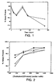
- Figure 1 shows the effect of reconstitution volume on the stability of lyophilized rhuMAb HER2.
- the lyophilized formulation was prepared from a pre-lyophilization formulation comprising 25 mg/mL protein, 60 mM trehalose, 5 mM sodium succinate, pH 5.0, and 0.01% Tween 20TM.
- the lyophilized cake was incubated at 40°C and then reconstituted with 4.0 (o) or 20.0 mL ( ⁇ ) of BWFI.
- the fraction of intact protein in the reconstituted formulation was measured by native size exclusion chromatography and defined as the peak area of the native protein relative to the total peak area including aggregates.
- Figure 2 illustrates the effect of trehalose concentration on the stability of lyophilized rhuMAb HER2.
- the protein was lyophilized at 25 mg/mL in 5 mM sodium succinate, pH 5.0 (circles) or 5 mM histidine, pH 6.0 (squares) and trehalose concentrations ranging from 60 mM (360 molar ratio) to 200 mM (1200 molar ratio).
- the lyophilized protein was incubated at 40°C for either 30 days (closed symbols) or 91 days (open symbols).
- the amount of intact protein was measured after reconstitution of the lyophilized protein with 20 mL BWFI.
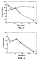
- Figure 3 demonstrates the effect of trehalose concentration on the long term stability of lyophilized rhuMAb HER2 stored at 40°C.
- the protein was lyophilized at either 25 mg/mL in 5 mM sodium succinate, pH 5.0, 0.0 1 % Tween 20TM, and 60 mM trehalose ( ⁇ ) or 5 mM histidine, pH 6.0, 0.01% Tween 20TM, and 60 mM trehalose ( ⁇ ) or 21 mg/mL in 10 mM sodium succinate, pH 5.0, 0.2% Tween 20TM and 250 mM trehalose ( ⁇ ).
- the lyophilized protein was incubated at 40°C and then reconstituted with 20 mL of BWFI. The amount of intact protein was measured after reconstitution.
- Figure 4 shows the stability of rhuMAb HER2 lyophilized in 38.4 mM mannitol (7 mg/mL), 20.4 mM sucrose (7 mg/mL), 5 mM histidine, pH 6.0, 0.01% Tween 20TM.
- the lyophilized protein was incubated at 40°C and then reconstituted with either 4.0 mL (o) or 20 mL ( ⁇ ) of BWFI. The amount of intact protein was measured after reconstitution.
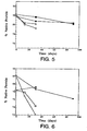
- Figure 5 demonstrates stability of reconstituted rhuMAb HER2 lyophilized in 5 mM sodium succinate, pH 5.0, 60 mM trehalose, 0.01% Tween 20TM.
- Samples were reconstituted with either 4.0 mL (squares) or 20.0 mL (circles) of BWFI (20 mL:0.9% benzyl alcohol; 4 mL:1.1% benzyl alcohol) and then stored at 5°C (solid symbols) or 25°C (open symbols).
- the % native protein was defined as the peak area of the native (not degraded) protein relative to the total peak area as measured by cation exchange chromatography.
- Figure 6 shows stability of reconstituted rhuMAb HER2 lyophilized in 5 mM histidine, pH 6.0, 60 mM trehalose, 0.01% Tween 20.
- Samples were reconstituted with either 4.0 mL (squares) or 20.0 mL (circles) of BWFI (20 mL:0.9% benzyl alcohol; 4 mL:1.1% benzyl alcohol) and then stored at 5°C (solid symbols) or 25 °C (open symbols).
- the % native protein was defined as the peak area of the native (not degraded) protein relative to the total peak area as measured by cation exchange chromatography.
- Figure 7 reveals stability of reconstituted rhuMAb HER2 lyophilized in 5 mM histidine, pH 6.0, 38.4 mM mannitol, 20.4 mM sucrose, 0.01% Tween 20.
- Samples were reconstituted with either 4.0 mL (squares) or 20.0 mL (circles) of BWFI (20 mL:0.9% benzyl alcohol; 4 mL:1.1% benzyl alcohol) and then stored at 5 °C (solid symbols) or 25 °C (open symbols).
- the % native protein was defined as the peak area of the native (not degraded) protein relative to the total peak area as measured by cation exchange chromatography.
- Figure 8 shows stability of reconstituted rhuMAb HER2 lyophilized in 10 mM sodium succinate, pH 5.0, 250 mM trehalose, 0.2% Tween 20.
- Samples were reconstituted with 20.0 mL of BWFI (0.9% benzyl alcohol) and then stored at 5°C ( ⁇ ) or 25 °C (o).
- the % native protein was defined as the peak area of the native (not degraded) protein relative to the total peak area as measured by cation exchange chromatography.
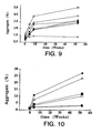
- Figure 9 shows aggregation of rhuMAb E25 formulated into buffers ranging from pH 5 to pH 7 at 10 mM buffer concentration and 5 mg/mL antibody concentration. Samples were lyophilized and assayed at time zero and after 4 weeks, 8 weeks, and 52 weeks of storage at 2-8°C.
- the buffers were: potassium phosphate pH 7.0 ( ⁇ ); sodium phosphate pH 7.0 ( ⁇ ); histidine pH 7.0 (o); sodium succinate pH 6.5 ( ⁇ ); sodium succinate pH 6.0 ( ⁇ ); sodium succinate pH 5.5 ( ⁇ ); and sodium succinate pH 5.0 ( ⁇ ).
- Figure 10 depicts aggregation of rhuMAb E25 lyophilized in 5 mM histidine buffer at both pH 6 and pH 7 and assayed following storage as follows.
- the buffer was at: pH 6.0 stored at 2-8°C ( ⁇ ); pH 6 stored at 25°C ( ⁇ ); pH 6 stored at 40° C( ⁇ ); pH 7 stored at 2-8°C ( ⁇ ); pH 7 stored at 25°C ( ⁇ ); and pH 7 stored at 40°C ( ⁇ ).
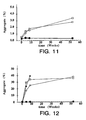
- Figure 11 illustrates aggregation of 5 mg/mL rhuMAb E25 formulated into 10 mM sodium succinate at pH 5.0 with lyoprotectant added at a concentration of 275 mM (isotonic).
- the lyoprotectants were: control, no lyoprotectant ( ⁇ ); mannitol ( ⁇ ); lactose ( ⁇ ); maltose ( ⁇ ); trehalose ( ⁇ ); and sucrose ( ⁇ ). Samples were lyophilized and assayed at time zero and after 4 weeks, 8 weeks, and 52 weeks of storage at 2-8° C.
- Figure 12 shows aggregation of 5 mg/mL rhuMAb E25 formulated into 10 mM sodium succinate at pH 5.0 with lyoprotectant added at a concentration of 275 mM (isotonic).
- the lyoprotectants were: control, no lyoprotectant ( ⁇ ); mannitol ( ⁇ ); lactose ( ⁇ ); maltose ( ⁇ ); trehalose ( ⁇ ); and sucrose ( ⁇ ). Samples were lyophilized and assayed at time zero and after 4 weeks, 8 weeks, and 52 weeks of storage at 40°C.
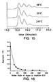
- Figure 13 depicts hydrophobic interaction chromatography of 20 mg/mL rhuMAb E25 lyophilized in histidine buffer at pH 6 with an isotonic concentration ( i . e . 275 mM) of lactose stored for 24 weeks at 2-8, 25 or 40°C and reconstituted to 20 mg/mL.
- Figure 14 shows hydrophobic interaction chromatography of 20 mg/mL rhuMAb E25 lyophilized in histidine buffer at pH 6 stored for 24 weeks at 2-8, 25 or 40°C and reconstituted to 20 mg/mL.
- Figure 15 illustrates hydrophobic interaction chromatography of 20 mg/mL rhuMAb E25 lyophilized in histidine buffer at pH 6 with an isotonic concentration (i . e . 275 mM) of sucrose and stored for 24 weeks at 2-8, 25 or 40°C and reconstituted to 20 mg/mL.
- Figure 16 illustrates the effect of sugar concentration on rhuMAb E25 formulated at 20 mg/mL in 5 mM histidine at pH 6.0.
- Sucrose ( ⁇ ) and trehalose ( ⁇ ) were added to the formulation at molar ratios ranging from 0 to 2010 (isotonic) (see Table 1 below). Samples were lyophilized and assayed after 12 weeks of storage at 50°C.
- FIG. 18 shows aggregation of rhuMAb E25 formulated at 25 mg/mL into 5 mM histidine at pH 6 with 85 mM sucrose ( ⁇ ); 85 mM trehalose ( ⁇ ); 161 mM sucrose ( ⁇ ) or 161 mM trehalose ( ⁇ ).
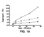
- FIG. 19 illustrates aggregation of rhuMAb E25 formulated at 25 mg/mL into 5 mM histidine at pH 6 with 85 mM sucrose ( ⁇ ); 85 mM trehalose ( ⁇ ); 161 mM sucrose ( ⁇ ) or 161 mM trehalose ( ⁇ ).
- Samples were lyophilized and stored at 50°C followed by reconstitution with 0.9% benzyl alcohol to 100 mg/mL antibody in 20 mM histidine at pH 6 with isotonic (340 mM) and hypertonic (644 mM) sugar concentration.
- protein is meant a sequence of amino acids for which the chain length is sufficient to produce the higher levels of tertiary and/or quaternary structure. This is to distinguish from “peptides” or other small molecular weight drugs that do not have such structure.
- the protein herein will have a molecular weight of at least about 15-20 kD, preferably at least about 20 kD.
- proteins encompassed within the definition herein include mammalian proteins, such as, e . g ., growth hormone, including human growth hormone and bovine growth hormone; growth hormone releasing factor; parathyroid hormone; thyroid stimulating hormone; lipoproteins; ⁇ -1-antitrypsin; insulin A-chain; insulin B-chain; proinsulin; follicle stimulating hormone; calcitonin; luteinizing hormone; glucagon; clotting factors such as factor VIIIC, factor IX, tissue factor, and von Willebrands factor; anti-clotting factors such as Protein C; atrial natriuretic factor; lung surfactant; a plasminogen activator, such as urokinase or tissue-type plasminogen activator (t-PA); bombazine; thrombin; tumor necrosis factor- ⁇ and - ⁇ ; enkephalinase; RANTES (regulated on activation normally T-cell expressed and secreted); human macrophage inflammatory protein (MIP-1- ⁇
- ILs interleukins
- T-cell receptors surface membrane proteins
- DAF decay accelerating factor
- a viral antigen such as, for example, a portion of the AIDS envelope; transport proteins; homing receptors; addressins; regulatory proteins; immunoadhesins; antibodies; and biologically active fragments or variants of any of the above-listed polypeptides.
- the protein which is formulated is preferably essentially pure and desirably essentially homogeneous (i.e. free from contaminating proteins etc).
- Essentially pure protein means a composition comprising at least about 90% by weight of the protein, based on total weight of the composition, preferably at least about 95% by weight.
- Essentially homogeneous protein means a composition comprising at least about 99% by weight of protein, based on total weight of the composition.
- the protein may be an antibody.
- the antibody may bind to any of the above-mentioned molecules, for example.
- Exemplary molecular targets for antibodies include CD proteins such as CD3, CD4, CD8, CD19, CD20 and CD34; members of the HER receptor family such as the EGF receptor, HER2, HER3 or HER4 receptor; cell adhesion molecules such as LFA-1, MoI, p150,95, VLA-4, ICAM-1, VCAM and ⁇ / ⁇ 3 integrin including either ⁇ or ⁇ subunits thereof ( e . g . anti-CD11a, anti-CD18 or anti-CD11b antibodies); growth factors such as VEGF; IgE; blood group antigens; flk2/flt3 receptor; obesity (OB) receptor; protein C etc.
- CD proteins such as CD3, CD4, CD8, CD19, CD20 and CD34
- members of the HER receptor family such as the EGF receptor, HER2, HER3 or HER4 receptor
- antibody is used in the broadest sense and specifically covers monoclonal antibodies (including full length antibodies which have an immunoglobulin Fc region), antibody compositions with polyepitopic specificity, bispecific antibodies, diabodies, and single-chain molecules, as well as antibody fragments ( e.g. , Fab, F(ab') 2 , and Fv).
- the term "monoclonal antibody” as used herein refers to an antibody obtained from a population of substantially homogeneous antibodies, i . e ., the individual antibodies comprising the population are identical except for possible naturally occurring mutations that may be present in minor amounts. Monoclonal antibodies are highly specific, being directed against a single antigenic site. Furthermore, in contrast to conventional (polyclonal) antibody preparations which typically include different antibodies directed against different determinants (epitopes), each monoclonal antibody is directed against a single determinant on the antigen. In addition to their specificity, the monoclonal antibodies are advantageous in that they are synthesized by the hybridoma culture, uncontaminated by other immunoglobulins.
- the modifier "monoclonal” indicates the character of the antibody as being obtained from a substantially homogeneous population of antibodies, and is not to be construed as requiring production of the antibody by any particular method.
- the monoclonal antibodies to be used in accordance with the present invention may be made by the hybridoma method first described by Kohler et al., Nature, 256: 495 (1975 ), or may be made by recombinant DNA methods (see, e.g. , U.S. Patent No. 4,816,567 ).
- the "monoclonal antibodies” may also be isolated from phage antibody libraries using the techniques described in Clackson et al., Nature, 352:624-628 (1991 ) and Marks et al., J. Mol. Biol., 222:581-597 (1991 ), for example.
- the monoclonal antibodies herein specifically include "chimeric" antibodies (immunoglobulins) in which a portion of the heavy and/or light chain is identical with or homologous to corresponding sequences in antibodies derived from a particular species or belonging to a particular antibody class or subclass, while the remainder of the chain(s) is identical with or homologous to corresponding sequences in antibodies derived from another species or belonging to another antibody class or subclass, as well as fragments of such antibodies, so long as they exhibit the desired biological activity ( U.S. Patent No. 4,816,567 ; Morrison et al., Proc. Natl. Acad. Sci. USA, 81:6851-6855 (1984 )).
- chimeric antibodies immunoglobulins in which a portion of the heavy and/or light chain is identical with or homologous to corresponding sequences in antibodies derived from a particular species or belonging to a particular antibody class or subclass, while the remainder of the chain(s) is identical with or homologous to corresponding sequence
- Humanized forms of non-human (e . g ., murine) antibodies are chimeric immunoglobulins, immunoglobulin chains or fragments thereof (such as Fv, Fab, Fab', F(ab') 2 or other antigen-binding subsequences of antibodies) which contain minimal sequence derived from non-human immunoglobulin.
- humanized antibodies are human immunoglobulins (recipient antibody) in which residues from a complementarity determining region (CDR) of the recipient are replaced by residues from a CDR of a non-human species (donor antibody) such as mouse, rat or rabbit having the desired specificity, affinity, and capacity.
- CDR complementarity determining region
- humanized antibodies may comprise residues which are found neither in the recipient antibody nor in the imported CDR or framework sequences. These modifications are made to further refine and optimize antibody performance.
- the humanized antibody will comprise substantially all of at least one, and typically two, variable domains, in which all or substantially all of the CDR regions correspond to those of a non-human immunoglobulin and all or substantially all of the FR regions are those of a human immunoglobulin sequence.
- the humanized antibody optimally also will comprise at least a portion of an immunoglobulin constant region (Fc), typically that of a human immunoglobulin.
- Fc immunoglobulin constant region
- the humanized antibody includes a PrimatizedTM antibody wherein the antigen-binding region of the antibody is derived from an antibody produced by immunizing macaque monkeys with the antigen of interest.
- a “stable” formulation is one in which the protein therein essentially retains its physical and chemical stability and integrity upon storage.
- Various analytical techniques for measuring protein stability are available in the art and are reviewed in Peptide and Protein Drug Delivery, 247-301, Vincent Lee Ed., Marcel Dekker, Inc., New York, New York, Pubs. (1991 ) and Jones, A. Adv. Drug Delivery Rev. 10: 29-90 (1993 ).
- Stability can be measured at a selected temperature for a selected time period. For rapid screening, the formulation may be kept at 40°C for 2 weeks to 1 month, at which time stability is measured.
- the formulation should be stable at 30°C or 40°C for at least I month and/or stable at 2-8°C for at least 2 years.
- the formulation should be stable for at least 2 years at 30°C and/or stable at 40°C for at least 6 months.
- the extent of aggregation following lyophilization and storage can be used as an indicator of protein stability (see Examples herein).
- a "stable" formulation may be one wherein less than about 10% and preferably less than about 5% of the protein is present as an aggregate in the formulation. In other embodiments, any increase in aggregate formation following lyophilization and storage of the lyophilized formulation can be determined.
- a "stable" lyophilized formulation may be one wherein the increase in aggregate in the lyophilized formulation is less than about 5% and preferably less than about 3%, when the lyophilized formulation is stored at 2-8°C for at least one year.
- stability of the protein formulation may be measured using a biological activity assay (see, e . g ., Example 2 below).
- a "reconstituted" formulation is one which has been prepared by dissolving a lyophilized protein formulation in a diluent such that the protein is dispersed in the reconstituted formulation.
- the reconstituted formulation in suitable for administration (e.g. parenteral administration) to a patient to be treated with the protein of interest and, may be one which is suitable for subcutaneous administration.
- isotonic is meant that the formulation of interest has essentially the same osmotic pressure as human blood. Isotonic formulations will generally have an osmotic pressure from about 250 to 350mOsm. Isotonicity can be measured using a vapor pressure or ice-freezing type osmometer, for example.
- a “lyoprotectant” is a molecule which, when combined with a protein of interest, significantly prevents or reduces chemical and/or physical instability of the protein upon lyophilization and subsequent storage.
- exemplary lyoprotectants include sugars such as sucrose or trehalose; an amino acid such as monosodium glutamate or histidine; a methylamine such as betaine; a lyotropic salt such as magnesium sulfate; a polyol such as trihydric or higher sugar alcohols, e . g .
- the preferred lyoprotectant is a non-reducing sugar, such as trehalose or sucrose.
- the lyoprotectant is added to the pre-lyophilized formulation in a "lyoprotecting amount" which means that, following lyophilization of the protein in the presence of the lyoprotecting amount of the lyoprotectant, the protein essentially retains its physical and chemical stability and integrity upon lyophilization and storage.
- the "diluent" of interest herein is one which is pharmaceutically acceptable (safe and non-toxic for administration to a human) and is useful for the preparation of a reconstituted formulation.
- exemplary diluents include sterile water, bacteriostatic water for injection (BWFI), a pH buffered solution (e.g. phosphate-buffered saline), sterile saline solution, Ringer's solution or dextrose solution.
- a "preservative" is a compound which can be added to the diluent to essentially reduce bacterial action in the reconstituted formulation, thus facilitating the production of a multi-use reconstituted formulation, for example.
- potential preservatives include octadecyldimethylbenzyl ammonium chloride, hexamethonium chloride, benzalkonium chloride (a mixture of alkylbenzyldimethylammonium chlorides in which the alkyl groups are long-chain compounds), and benzethonium chloride.
- preservatives include aromatic alcohols such as phenol, butyl and benzyl alcohol, alkyl parabens such as methyl or propyl paraben, catechol, resorcinol, cyclohexanol, 3-pentanol, and m-cresol.
- aromatic alcohols such as phenol, butyl and benzyl alcohol
- alkyl parabens such as methyl or propyl paraben
- catechol resorcinol
- cyclohexanol 3-pentanol
- m-cresol m-cresol
- a “bulking agent” is a compound which adds mass to the lyophilized mixture and contributes to the physical structure of the lyophilized cake (e.g. facilitates the production of an essentially uniform lyophilized cake which maintains an open pore structure).
- Exemplary bulking agents include mannitol, glycine, polyethylene glycol and xorbitol.
- Treatment refers to both therapeutic treatment and prophylactic or preventative measures. Those in need of treatment include those already with the disorder as well as those in which the disorder is to be prevented.
- mammal for purposes of treatment refers to any animal classified as a mammal, including humans, domestic and farm animals, and zoo, sports, or pet animals, such as dogs, horses, cats, cows, etc .
- the mammal is human.
- a “disorder” is any condition that would benefit from treatment with the protein. This includes chronic and acute disorders or diseases including those pathological conditions which predispose the mammal to the disorder in question.
- disorders to be treated herein include carcinomas and allergies.
- the protein to be formulated is prepared using techniques which are well established in the art including synthetic techniques (such as recombinant techniques and peptide synthesis or a combination of these techniques) or may be isolated from an endogenous source of the protein.
- the protein of choice is an antibody. Techniques for the production of antibodies follow.
- a protein that is immunogenic in the species to be immunized e.g., keyhole limpet hemocyanin, serum albumin, bovine thyrog
- Animals are immunized against the antigen, immunogenic conjugates, or derivatives by combining 1 mg or 1 ⁇ g of the peptide or conjugate (for rabbits or mice, respectively) with 3 volumes of Freund's complete adjuvant.
- the animals are boosted with 1/5 to 1/10 the original amount of peptide or conjugate in Freund's complete adjuvant by subcutaneous injection at multiple sites.
- the animals are bled and the serum is assayed for antibody titer. Animals are boosted until the titer plateaus.
- the animal is boosted with the conjugate of the same antigen, but conjugated to a different protein and/or through a different cross-linking reagent.
- Conjugates also can be made in recombinant cell culture as protein fusions. Also, aggregating agents such as alum are suitably used to enhance the immune response.
- Monoclonal antibodies are obtained from a population of substantially homogeneous antibodies, i . e ., the individual antibodies comprising the population are identical except for possible naturally occurring mutations that may be present in minor amounts. Thus, the modifier "monoclonal" indicates the character of the antibody as not being a mixture of discrete antibodies.
- the monoclonal antibodies may be made using the hybridoma method first described by Kohler et al., Nature, 256:495 (1975 ), or may be made by recombinant DNA methods ( U.S. Patent No. 4,816,567 ).
- a mouse or other appropriate host animal such as a hamster
- lymphocytes that produce or are capable of producing antibodies that will specifically bind to the protein used for immunization.
- lymphocytes may be immunized in vitro .
- Lymphocytes then are fused with myeloma cells using a suitable fusing agent, such as polyethylene glycol, to form a hybridoma cell ( Goding, Monoclonal Antibodies: Principles and Practice, pp.59-103 (Academic Press, 1986 )).
- the hybridoma cells thus prepared are seeded and grown in a suitable culture medium that preferably contains one or more substances that inhibit the growth or survival of the unfused, parental myeloma cells.
- a suitable culture medium that preferably contains one or more substances that inhibit the growth or survival of the unfused, parental myeloma cells.
- the culture medium for the hybridomas typically will include hypoxanthine, aminopterin, and thymidine (HAT medium), which substances prevent the growth of HGPRT-deficient cells.
- Preferred myeloma cells are those that fuse efficiently, support stable high-level production of antibody by the selected antibody-producing cells, and are sensitive to a medium such as HAT medium.
- preferred myeloma cell lines are murine myeloma lines, such as those derived from MOPC-21 and MPC-11 mouse tumors available from the Salk Institute Cell Distribution Center, San Diego, California USA, and SP-2 cells available from the American Type Culture Collection, Rockville, Maryland USA.
- Human myeloma and mouse-human heteromyeloma cell lines also have been described for the production of human monoclonal antibodies ( Kozbor, J. Immunol., 133:3001 (1984 ); Brodeur et al., Monoclonal Antibody Production Techniques and Applications, pp. 51-63 (Marcel Dekker, Inc., New York, 1987 )).
- Culture medium in which hybridoma cells are growing is assayed for production of monoclonal antibodies directed against the antigen.
- the binding specificity of monoclonal antibodies produced by hybridoma cells is determined by immunoprecipitation or by an in vitro binding assay, such as radioimmunoassay (RIA) or enzyme-linked immunoabsorbent assay (ELISA).
- RIA radioimmunoassay
- ELISA enzyme-linked immunoabsorbent assay
- the binding affinity of the monoclonal antibody can, for example, be determined by the Scatchard analysis of Muuson et al., Anal. Biochem., 107:220 (1980 ).
- the clones may be subcloned by limiting dilution procedures and grown by standard methods ( Coding, Monoclonal Antibodies: Principles and Practice, pp.59-103 (Academic Press, 1986 )). Suitable culture media for this purpose include, for example, D-MEM or RPMI-1640 medium.
- the hybridoma cells may be grown in vivo as ascites tumors in an animal.
- the monoclonal antibodies secreted by the subclones are suitably separated from the culture medium, ascites fluid, or serum by conventional immunoglobulin purification procedures such as, for example, protein A-Sepharose, hydroxylapatite chromatography, gel electrophoresis, dialysis, or affinity chromatography.
- DNA encoding the monoclonal antibodies is readily isolated and sequenced using conventional procedures (e.g., by using oligonucleotide probes that are capable of binding specifically to genes encoding the heavy and light chains of murine antibodies).
- the hybridoma cells serve as a preferred source of such DNA.
- the DNA may be placed into expression vectors, which are then transfected into host cells such as E. coli cells, simian COS cells, Chinese hamster ovary (CHO) cells, or myeloma cells that do not otherwise produce immunoglobulin protein, to obtain the synthesis of monoclonal antibodies in the recombinant host cells.
- antibodies can be isolated from antibody phage libraries generated using the techniques described in McCafferty et al., Nature, 348:552-554 (1990 ). Clackson et al., Nature, 352:624-628 (1991 ) and Marks et al., J. Mol. Biol., 222:581-597 (1991 ) describe the isolation of murine and human antibodies, respectively, using phage libraries.
- the DNA also may be modified, for example, by substituting the coding sequence for human heavy-and light-chain constant domains in place of the homologous murine sequences ( U.S. Patent No. 4,816,567 ; Morrison, et al., Proc. Natl Acad Sci. USA, 81:6851 (1984 )), or by covalently joining to the immunoglobulin coding sequence all or part of the coding sequence for a non-immunoglobulin polypeptide.
- non-immunoglobulin polypeptides are substituted for the constant domains of an antibody, or they are substituted for the variable domains of one antigen-combining site of an antibody to create a chimeric bivalent antibody comprising one antigen-combining site having specificity for an antigen and another antigen-combining site having specificity for a different antigen.
- Chimeric or hybrid antibodies also may be prepared in vitro using known methods in synthetic protein chemistry, including those involving crosslinking agents.
- immunotoxins may be constructed using a disulfide-exchange reaction or by forming a thioether bond.
- suitable reagents for this purpose include iminothiolate and methyl-4-mercaptobutyrimidate.
- a humanized antibody has one or more amino acid residues introduced into it from a source which is non-human. These non-human amino acid residues are often referred to as "import" residues, which are typically taken from an "import” variable domain. Humanization can be essentially performed following the method of Winter and co-workers ( Jones et al., Nature, 321:522-525 (1986 ); Riechmann et al., Nature, 332:323-327 (1988 ); Verhoeyen et al., Science, 239:1534-1536 (1988 )), by substituting rodent CDRs or CDR sequences for the corresponding sequences of a human antibody.
- humanized antibodies are chimeric antibodies ( U.S. Patent No. 4,816,567 ), wherein substantially less than an intact human variable domain has been substituted by the corresponding sequence from a non-human species.
- humanized antibodies are typically human antibodies in which some CDR residues and possibly some FR residues are substituted by residues from analogous sites in rodent antibodies.
- variable domains both light and heavy
- the choice of human variable domains, both light and heavy, to be used in making the humanized antibodies is very important to reduce antigenicity.
- the sequence of the variable domain of a rodent antibody is screened against the entire library of known human variable-domain sequences.
- the human sequence which is closest to that of the rodent is then accepted as the human framework (FR) for the humanized antibody ( Sims et al., J. Immunol., 151:2296 (1993 ); Chothia et al., J. Mol. Biol., 196:901 (1987 )).
- Another method uses a particular framework derived from the consensus sequence of all human antibodies of a particular subgroup of light or heavy chains.
- the same framework may be used for several different humanized antibodies ( Carter et al., Proc. Natl. Acad Sci. USA, 89:4285 (1992 ); Presta et al., J. Immnol., 151:2623 (1993 )).
- humanized antibodies are prepared by a process of analysis of the parental sequences and various conceptual humanized products using three-dimensional models of the parental and humanized sequences.
- Three-dimensional immunoglobulin models are commonly available and are familiar to those skilled in the art.
- Computer programs are available which illustrate and display probable three-dimensional conformational structures of selected candidate immunoglobulin sequences. Inspection of these displays permits analysis of the likely role of the residues in the functioning of the candidate immunoglobulin sequence, i . e ., the analysis of residues that influence the ability of the candidate immunoglobulin to bind its antigen.
- FR residues can be selected and combined from the recipient and import sequences so that the desired antibody characteristic, such as increased affinity for the target antigen(s), is achieved.
- the CDR residues are directly and most substantially involved in influencing antigen binding.
- transgenic animals e . g ., mice
- transgenic animals e . g ., mice
- J H antibody heavy-chain joining region
- Human antibodies can also be derived from phage-display libraries ( Hoogenboom et al., J. Mol. Biol., 227:381 (1991 ); Marks et al., J. Mol. Biol., 222:581-597 (1991 )).
- Bispecific antibodies are antibodies that have binding specificities for at least two different epitopes. Such antibodies can be derived from full length antibodies or antibody fragments ( e . g . F(ab') 2 bispecific antibodies).
- bispecific antibodies are known in the art. Traditional production of full length bispecific antibodies is based on the coexpression of two immunoglobulin heavy chain-light chain pairs, where the two chains have different specificities ( Millstein et al., Nature, 305:537-539 (1983 )). Because of the random assortment of immunoglobulin heavy and light chains, these hybridomas (quadromas) produce a potential mixture of 10 different antibody molecules, of which only one has the correct bispecific structure. Purification of the correct molecule, which is usually done by affinity chromatography steps, is rather cumbersome, and the product yields are low. Similar procedures are disclosed in WO 93/08829 and in Traunecker et al., EMBO J., 10:3655-3659 (1991 ).
- antibody variable domains with the desired binding specificities are fused to immunoglobulin constant domain sequences.
- the fusion preferably is with an immunoglobulin heavy chain constant domain, comprising at least part of the hinge, CH2, and CH3 regions. It is preferred to have the first heavy-chain constant region (CH1) containing the site necessary for light chain binding, present in at least one of the fusions.
- DNAs encoding the immunoglobulin heavy chain fusions and, if desired, the immunoglobulin light chain are inserted into separate expression vectors, and are co-transfected into a suitable host organism.
- the bispecific antibodies are composed of a hybrid immunoglobulin heavy chain with a first binding specificity in one arm, and a hybrid immunoglobulin heavy chain-light chain pair (providing a second binding specificity) in the other arm. It was found that this asymmetric structure facilitates the separation of the desired bispecific compound from unwanted immunoglobulin chain combinations, as the presence of an immunoglobulin light chain in only one half of the bispecific molecule provides for a facile way of separation. This approach is disclosed in WO 94/04690 published March 3, 1994 . For further details of generating bispecific antibodies see, for example, Suresh et al., Methods in Enzymology, 121:210 (1986 ).
- Bispecific antibodies include cross-linked or "heteroconjugate" antibodies.
- one of the antibodies in the heteroconjugate can be coupled to avidin, the other to biotin.
- Such antibodies have, for example, been proposed to target immune system cells to unwanted cells ( US Patent No. 4,676,980 ), and for treatment of HIV infection ( WO 91/00360 , WO 92/200373 ).
- Heteroconjugate antibodies may be made using any convenient cross-linking methods. Suitable cross-linking agents are well known in the art, and are disclosed in US Patent No. 4,676,980 , along with a number of cross-linking techniques.
- bispecific antibodies from antibody fragments
- the following techniques can also be used for the production of bivalent antibody fragments which are not necessarily bispecific.
- Fab' fragments recovered from E. coli can be chemically coupled in vitro to form bivalent antibodies. See, Shalaby et al., J. Exp. Med., 175:217-225 (1992 ).
- bivalent heterodimers have been produced using leucine zippers.
- the leucine zipper peptides from the Fos and Jun proteins were linked to the Fab' portions of two different antibodies by gene fusion.
- the antibody homodimers were reduced at the hinge region to form monomers and then re-oxidized to form the antibody heterodimers.
- the "diabody" technology described by Hollinger et al., Proc. Natl. Acad Sci.
- the fragments comprise a heavy-chain variable domain (V H ) connected to a light-chain variable domain (V L ) by a linker which is too short to allow pairing between the two domains on the same chain. Accordingly, the V H and V L domains of one fragment are forced to pair with the complementary V L and V H domains of another fragment, thereby forming two antigen-binding sites.
- V H and V L domains of one fragment are forced to pair with the complementary V L and V H domains of another fragment, thereby forming two antigen-binding sites.
- sFv single-chain Fv
- a "pre-lyophilized formulation” is produced.
- the amount of protein present in the pre-lyophilized formulation is determined taking into account the desired dose volumes, mode(s) of administration etc.
- the protein of choice is an intact antibody (such as an anti-IgE or anti-HER2 antibody)
- from about 2 mg/mL to about 50 mg/mL, preferably from about 5 mg/mL to about 40 mg/mL and most preferably from about 20-30 mg/mL is an exemplary starting protein concentration.
- the protein is generally present in solution.
- the protein may be present in a pH-buffered solution at a pH from about 4-8, and preferably from about 5-7.
- Exemplary buffers include histidine, phosphate, Tris, citrate, succinate and other organic acids.
- the buffer concentration can be from about 1 mM to about 20 mM, or from about 3 mM to about 15 mM, depending, for example, on the buffer and the desired isotonicity of the formulation ( e.g . of the reconstituted formulation).
- the preferred buffer is histidine in that, as demonstrated below, this can have lyoprotective properties. Succinate was shown to be another useful buffer.
- the lyoprotectant is added to the pre-lyophilized formulation.
- the lyoprotectant is a non-reducing sugar such as sucrose or trehalose.
- the amount of lyoprotectant in the pre-lyophilized formulation is generally such that, upon reconstitution, the resulting formulation will be isotonic. However, hypertonic reconstituted formulations may also be suitable. In addition, the amount of lyoprotectant must not be too low such that an unacceptable amount of degradation/aggregation of the protein occurs upon lyophilization.
- lyoprotectant concentrations in the pre-lyophilized formulation are from about 10 mM to about 400 mM, and preferably from about 30 mM to about 300 mM, and most preferably from about 50 mM to about 100 mM.
- the ratio of protein to lyoprotectant is selected for each protein and lyoprotectant combination.
- the molar ratio of lyoprotectant to antibody is from 200 to 600 moles of lyoprotectant to 1 mole antibody.
- surfactant it has been found to be desirable to add a surfactant to the pre-lyophilized formulation.
- the surfactant may be added to the lyophilized formulation and/or the reconstituted formulation.
- exemplary surfactants include nonionic surfactants such as polysorbates ( e.g . polysorbates 20 or 80); poloxamers ( e.g .
- poloxamer 188 Triton; sodium dodecyl sulfate (SDS); sodium laurel sulfate; sodium octyl glycoside; lauryl-, myristyl-, linoleyl-, or stearyl-sulfobetaine; lauryl-, myristyl-, linoleyl- or stearyl-sarcosine; linoleyl-, myristyl-, or cetyl-betaine; lauroamidopropyl-, cocamidopropyl-, linoleamidopropyl-, myristamidopropyl-, palmidopropyl-, or isostearamidopropyl-betaine ( e.g .
- the amount of surfactant added is such that it reduces aggregation of the reconstituted protein and minimizes the formation of particulates after reconstitution.
- the surfactant may be present in the pre-lyophilized formulation in an amount from about 0.001-0.5%, and preferably from about 0.005-0.05%.
- a mixture of the lyoprotectant (such as sucrose or trehalose) and a bulking agent (e.g . mannitol or glycine) is used in the preparation of the pre-lyophilization formulation.
- the bulking agent may allow for the production of a uniform lyophilized cake without excessive pockets therein etc.
- compositions such as those described in Remington's Pharmaceutical Sciences 16th edition, Osol, A. Ed. (1980 ) may be included in the pre-lyophilized formulation (and/or the lyophilized formulation and/or the reconstituted formulation) provided that they do not adversely affect the desired characteristics of the formulation.
- Acceptable carriers, excipients or stabilizers are nontoxic to recipients at the dosages and concentrations employed and include; additional buffering agents; preservatives; co-solvents; antioxidants including ascorbic acid and methionine; chelating agents such as EDTA; metal complexes ( e.g. Zn-protein complexes); biodegradable polymers such as polyesters; and/or salt-forming counterions such as sodium.
- the formulation herein may also contain more than one protein as necessary for the particular indication being treated, preferably those with complementary activities that do not adversely affect the other protein.
- it may be desirable to provide two or more antibodies which bind to the HER2 receptor or IgE in a single formulation.
- anti-HER2 and anti-VEGF antibodies may be combined in the one formulation.
- proteins are suitably present in combination in amounts that are effective for the purpose intended.
- the formulations to be used for in vivo administration must be sterile. This is readily accomplished by filtration through sterile filtration membranes, prior to, or following, lyophilization and reconstitution. Alternatively, sterility of the entire mixture may be accomplished by autoclaving the ingredients, except for protein, at about 120°C for about 30 minutes, for example.
- the formulation is lyophilized.
- freeze-dryers are available for this purpose such as Hull50TM (Hull, USA) or GT20TM (Leybold-Heraeus, Germany) freeze-dryers. Freeze-drying is accomplished by freezing the formulation and subsequently subliming ice from the frozen content at a temperature suitable for primary drying. Under this condition, the product temperature is below the eutectic point or the collapse temperature of the formulation.
- the shelf temperature for the primary drying will range from about -30 to 25°C (provided the product remains frozen during primary drying) at a suitable pressure, ranging typically from about 50 to 250mTorr.
- the formulation, size and type of the container holding the sample (e.g. , glass vial) and the volume of liquid will mainly dictate the time required for drying, which can range from a few hours to several days ( e.g . 40-60hrs).
- a secondary drying stage may be carried out at about 0-40°C, depending primarily on the type and size of container and the type of protein employed. However, it was found herein that a secondary drying step may not be necessary.
- the shelf temperature throughout the entire water removal phase of lyophilization may be from about 15-30° C ( e.g. , about 20°C).
- the time and pressure required for secondary drying will be that which produces a suitable lyophilized cake, dependent, e . g ., on the temperature and other parameters.
- the secondary drying time is dictated by the desired residual moisture level in the product and typically takes at least about 5 hours ( e . g . 10-15 hours).
- the pressure may be the same as that employed during the primary drying step. Freeze-drying conditions can be varied depending on the formulation and vial size.
- the container in which reconstitution of the protein is to be carried out may, for example, be a 3, 5, 10, 20, 50 or 100cc vial.
- lyophilization will result in a lyophilized formulation in which the moisture content thereof is less than about 5%, and preferably less than about 3%.
- the lyophilized formulation may be reconstituted with a diluent such that the protein concentration in the reconstituted formulation is at least 50 mg/mL, for example from about 50 mg/mL to about 400 mg/mL, more preferably from about 80 mg/mL to about 300 mg/mL, and most preferably from about 90 mg/mL to about 150 mg/mL.
- a diluent such that the protein concentration in the reconstituted formulation is at least 50 mg/mL, for example from about 50 mg/mL to about 400 mg/mL, more preferably from about 80 mg/mL to about 300 mg/mL, and most preferably from about 90 mg/mL to about 150 mg/mL.
- Such high protein concentrations in the reconstituted formulation are considered to be particularly useful where subcutaneous delivery of the reconstituted formulation is intended.
- the protein concentration in the reconstituted formulation is significantly higher than that in the pre-lyophilized formulation.
- the protein concentration in the reconstituted formulation may be about 2-40 times, preferably 3-10 times and most preferably 3-6 times ( e . g . at least three fold or at least four fold) that of the pre-lyophilized formulation.
- Reconstitution generally takes place at a temperature of about 25°C to ensure complete hydration, although other temperatures may be employed as desired.
- the time required for reconstitution will depend, e.g ., on the type of diluent, amount of excipient(s) and protein.
- Exemplary diluents include sterile water, bacteriostatic water for injection (BWFI), a pH buffered solution (e.g. phosphate-buffered saline), sterile saline solution, Ringer's solution or dextrose solution.
- BWFI bacteriostatic water for injection
- pH buffered solution e.g. phosphate-buffered saline
- sterile saline solution e.g. phosphate-buffered saline
- Ringer's solution or dextrose solution e.g., Ringer's solution or dextrose solution.
- the diluent optionally contains a preservative. Exemplary
- the amount of preservative employed is determined by assessing different preservative concentrations for compatibility with the protein and preservative efficacy testing. For example, if the preservative is an aromatic alcohol (such as benzyl alcohol), it can be present in an amount from about 0.1-2.0% and preferably from about 0.5-1.5%, but most preferably about 1.0-1.2%.
- aromatic alcohol such as benzyl alcohol
- the reconstituted formulation has less than 6000 particles per vial which are 10 ⁇ m in size.
- the reconstituted formulation is administered to a mammal in need of treatment with the protein, preferably a human, in accord with known methods, such as intravenous administration as a bolus or by continuous infusion over a period of time, by intramuscular, intraperitoneal, intracerobrospinal, subcutaneous, intra-articular, intrasynovial, intrathecal, oral, topical, or inhalation routes.
- known methods such as intravenous administration as a bolus or by continuous infusion over a period of time, by intramuscular, intraperitoneal, intracerobrospinal, subcutaneous, intra-articular, intrasynovial, intrathecal, oral, topical, or inhalation routes.
- the reconstituted formulation may be administered to the mammal by subcutaneous ( i.e. beneath the skin) administration.
- the formulation may be injected using a syringe.
- other devices for administration of the formulation are available such as injection devices ( e . g . the Inject-ease TM and Genject TM devices); injector pens (such as the GenPen TM ); needleless devices ( e.g . MediJector TM and BioJector TM ); and subcutaneous patch delivery systems.
- the appropriate dosage (therapeutically effective amount) of the protein will depend, for example, on the condition to be treated, the severity and course of the condition, whether the protein is administered for preventive or therapeutic purposes, previous therapy, the patient's clinical history and response to the protein, the type of protein used, and the discretion of the attending physician.
- the protein is suitably administered to the patient at one time or over a series of treatments and may be administered to the patient at any time from diagnosis onwards.
- the protein may be administered as the sole treatment or in conjunction with other drugs or therapies useful in treating the condition in question.
- the protein of choice is an antibody
- from about 0.1-20 mg/kg is an initial candidate dosage for administration to the patient, whether, for example, by one or more separate administrations.
- other dosage regimens may be useful. The progress of this therapy is easily monitored by conventional techniques.
- an anti-HER2 antibody In the case of an anti-HER2 antibody, a therapeutically effective amount of the antibody may be administered to treat or prevent cancer characterized by overexpression of the HER2 receptor. It is contemplated that a reconstituted formulation of the anti-HER2 antibody may be used to treat breast, ovarian, stomach, endometrial, salivary gland, lung, kidney, colon and/or bladder cancer. For example, the anti-HER2 antibody may be used to treat ductal carcinoma in situ (DCIS). Exemplary dosages of the anti-HER2 antibody are in the range 1-10 mg/kg by one or more separate administrations.
- an anti-IgE formulation include the treatment or prophylaxis of IgE-mediated allergic diseases, parasitic infections, interstitial cystitis and asthma, for example.
- a therapeutically effective amount e.g. from about 1-15 mg/kg
- the anti-IgE antibody is administered to the patient.
- An article of manufacture which contains the lyophilized formulation of the present invention and provides instructions for its reconstitution and/or use.
- the article of manufacture comprises a container. Suitable containers include, for example, bottles, vials ( e.g. dual chamber vials), syringes (such as dual chamber syringes) and test tubes.
- the container may be formed from a variety of materials such as glass or plastic.
- the container holds the lyophilized formulation and the label on, or associated with, the container may indicate directions for reconstitution and/or use.
- the label may indicate that the lyophilized formulation is reconstituted to protein concentrations as described above.
- the label may further indicate that the formulation is useful or intended for subcutaneous administration.
- the container holding the formulation may be a multi-use vial, which allows for repeat administrations ( e . g . from 2-6 administrations) of the reconstituted formulation.
- the article of manufacture may further comprise a second container comprising a suitable diluent (e.g . BWFI). Upon mixing ofthe diluent and the lyophilized formulation, the final protein concentration in the reconstituted formulation will generally be at least 50 mg/mL.
- the article of manufacture may further include other materials desirable from a commercial and user standpoint, including other buffers, diluents, filters, needles, syringes, and package inserts with instructions for use.
- muMAb4D5 is directed against the extracellular domain (ECD) of p185 HER2 .
- ECD extracellular domain
- the muMAb4D5 molecule has been humanized in an attempt to improve its clinical efficacy by reducing immunogenicity and allowing it to support human effector functions (see WO 92/22653 ).
- This example describes the development of a lyophilized formulation comprising full length humanized antibody huMAb4D5-8 described in WO 92/22653 .
- excipients and buffers are initially screened by measuring the stability of the protein after lyophilization and reconstitution.
- the lyophilized protein in each formulation is also subjected to accelerated stability studies to determine the potential stability of the protein over its shelf-life. These accelerated studies are usually performed at temperatures above the proposed storage conditions and the data are then used to estimate the activation energy for the degradation reactions assuming Arrhenius kinetics ( Cleland et al., Critical Reviews in Therapeutic Drug Carrier Systems 10(4): 307-377 (1993 )).
- the activation energy is then used to calculate the expected shelf-life of the protein formulation at the proposed storage conditions.
- rhuMAb HER2 humanized anti-HER2 antibody
- 5°C proposed storage condition
- 40°C accelerated stability condition
- rhuMAb HER2 was observed to degrade by deamidation (30Asn of light chain) and isoaspartate formation via a cyclic imide intermediate, succinimide (102Asp of heavy chain).
- the deamidation was minimized at pH 5.0 resulting in degradation primarily at the succinimide.
- pH 6.0 slightly greater deamidation was observed in the liquid protein formulation.
- the lyophilized formulations were therefore studied with: (a) 5 or 10 mM succinate buffer, pH 5.0 or (b) 5 or 10 mM histidine buffer, pH 6.0. Both buffers contained the surfactant, polysorbate 20 (Tween 20TM), which was employed to reduce the potential for aggregation of the reconstituted protein and minimize the formation of particulates after reconstitution. These buffers were used with and without various sugars.
- the protein was formulated in the buffer at 5.0, 21.0 or 25.0 mg/mL. These formulations were then lyophilized and assessed for protein stability after 2 weeks at 5°C and 40°C.
- the vials were frozen at a shelf temperature of -55°C for approximately 5 hours followed by primary drying at a shelf temperature of 5°C and 150 mTorr for 30 hours, and drying to 1-2% residual moisture was achieved with secondary drying at a shelf temperature of 20°C for 10 hours.
- the major degradation route for this protein upon lyophilization was aggregation, and therefore the protein stability was assessed by native size exclusion chromatography to measure the recovery of intact native protein (% intact protein in Table 2 below).
- formulations comprising mannitol, or mannitol in combination with sorbitol or glycine, contained aggregated protein after lyophilization and storage at both conditions.
- trehalose and sucrose prevented aggregation at both storage conditions.
- the 250 mM trehalose and 250 mM lactose formulations were assessed for long term stability. After 9 months at 40°C or 12 months at 5°C, there was no change in the % intact protein for the trehalose formulation. For the lactose formulation, the % intact protein remained constant (same as initial) after 3 months at 40°C or 6 months at 25°C.
- the trehalose formulation could be stored at controlled room temperature (15-30°C) for 2 years without a significant change in % intact protein.
- the 10 mM histidine, pH 6.0 formulation with mannitol contained less aggregated protein after storage at 40°C for 2 weeks than the 10 mM succinate formulation, pH 5.0 with mannitol. This result may be related to some stabilizing effect contributed by histidine alone. After storage at 40°C for 2 weeks, there was, however, significant aggregation for histidine alone or histidine/mannitol formulations. The addition of sucrose at an equal mass to mannitol (10 mg/mL of each) in the histidine formulation stabilized the protein against aggregation for both storage conditions.
- sucrose/glycine formulation provided the same stability as the sucrose/mannitol formulation.
- the fraction of intact protein was measured by native size exclusion HPLC and the peak area of the native protein relative to the total peak area including aggregates (TSK3000 SW XL, column, TosoHaas, with a flow rate of 1.0 mL/min; elution with phosphate buffered saline; detection at 214 and 280 nm).
- the protein formulations were analyzed before lyophilization (liquid, 5° C) and after lyophilization and storage at 5° C or 40° C for 2 weeks. b.
- Formulations containing 5 mg/mL protein were reconstituted with distilled water (20 mL, 5.0 mg/mL protein), and formulations containing 21 mg/mL protein were reconstituted with bacteriostatic water for injection (BWFI, 0.9% benzyl alcohol; 20 mL, 20 mg/mL protein).
- BWFI 0.9% benzyl alcohol
- the lyophilization process may provide a method to allow concentration of the protein.
- the protein is filled into vials at a volume (Vf) and then lyophilized.
- This process also results in the concentration of the buffers and excipients.
- the solution is desirably isotonic.
- the amount of trehalose in the lyophilized rhuMAb HER2 was reduced to produce an isotonic solution upon reconstitution to yield 100 mg/mL protein.
- the stabilizing effect of trehalose was determined as a function of concentration for 5 mM sodium succinate, pH 5.0 and 5 mM histidine, pH 6.0 at 25.0 mg/mL protein (Table 3). At trehalose concentrations from 60 to 200 mM, there was no significant aggregation after incubation of the lyophilized protein for 4 weeks at 40°C. These formulations were reconstituted with 20 mL of bacteriostatic water for injection (BWFI, USP, 0.9% benzyl alcohol).
- the fraction of intact protein was measured by native size exclusion HPLC and defined as the peak area of the native protein relative to the total peak area including aggregates (TSK3000 SW XL column, TosoHaas, with a flow rate of 1.0 mL/min; elution with phosphate buffered saline; detection at 214 and 280 nm).
- the protein formulations were analyzed before lyophilization (liquid, 5°C) and after lyophilization and storage at 5° C or 40° C for 4 weeks. Formulations were reconstituted with bacteriostatic water for injection (BWFI, USP, 0.9% w/w benzyl alcohol; 20 mL, 22 mg/mL protein).
- rhuMAb HER2 is under investigation as a therapeutic for the treatment of breast cancer.
- the protein is dosed to patients at 2 mg/kg on a weekly basis. Since the average weight of these patients is 65 kg, the average weekly dose is 130 mg of rhuMAb HER2.
- injection volumes of 1.5 mL or less are well tolerated and, therefore, the protein concentration for a weekly subcutaneous administration of rhuMAb HER2 may be approximately 100 mg/mL (130 mg average dose/1.5 mL). As mentioned above, this high protein concentration is difficult to manufacture and maintain in a stable form.
- rhuMAb HER2 formulated in: (a) 5 mM sodium succinate, pH 5.0 or (b) 5 mM histidine, pH 6.0, was lyophilized at 25 mg/mL protein in 60 mM trehalose, 0.01% Tween 20TM. The lyophilization was performed by filling 18 mL of the protein formulation into 50 cc vials. In the lyophilizer, the vials were frozen at a shelf temperature of -55°C for approximately 5 hours followed by primary drying at a shelf temperature of 5°C and 150 mTorr for 30 hours, and drying to 1-2% residual moisture was achieved with secondary drying at a shelf temperature of 20°C for 10 hours.
- the lyophilized protein was then reconstituted with either 4 or 20 mL of BWFI (0.9 or 1.1% benzyl alcohol) to yield concentrated protein solutions:
- the amount of aggregated protein appeared to increase slightly with decreasing trehalose concentration.
- the stability of the lyophilized protein was not affected by the volume of reconstitution.
- the amount of intact protein after incubation of the lyophilized protein at 40°C was the same for the 60 mM trehalose, 5 mM sodium succinate, pH 5.0, 0.01% Tween 20TM formulation reconstituted with either 4 or 20 mL of BWFI.
- the 250 mM trehalose formulation was unchanged after 6 months at 40°C while both the 60 mM trehalose formulations were less stable.
- the 60 mM trehalose formulations may then require refrigerated storage if the product specification at the end of its shelf-life is, for example, >98% intact protein by native size exclusion chromatography.
- sucrose was also observed to prevent aggregation of rhuMAb HER2 after lyophilization and subsequent storage.
- sucrose concentration must be reduced significantly.
- the equal mass concentration of sucrose and mannitol (bulking agent) used in the screening studies prevented aggregation of the protein.
- a lower concentration of sucrose and mannitol (equal mass concentrations) was chosen as a potential subcutaneous formulation of rhuMAb HER2.
- the protein solution (25 mg/mL protein, 5 mM histidine, pH 6.0, 38.4 mM (7 mg/mL) mannitol, 20.4 mM (7 mg/mL) sucrose, 0.01% Tween 20TM) was lyophilized in the same manner as the 60 mM trehalose formulation except that the primary drying cycle was extended to 54 hours. After 4 weeks at 40°C, there was a slight increase in the amount of aggregates after reconstitution with 4.0 or 20.0 mL of BWFI (Table 3). The amount of aggregated protein was the same for reconstitution at 22 or 100 mg/mL protein ( Figure 4 ).
- the mannitol/sucrose formulation yielded less intact protein over time at 40°C.
- the molar ratio of sucrose to protein for this formulation was 120 to 1, indicating that the mannitol/sucrose combination may be more effective than trehalose alone at the same molar ratio of stabilizing sugar ( Figures 2 and 4 ).
- the stability of the lyophilized rhuMAb HER2 formulations was determined as a function of temperature. These studies demonstrated that the trehalose and mannitol/sucrose formulations prevented degradation of the protein in the lyophilized state at high temperatures (40°C). However, these experiments did not address the stability of the protein after reconstitution and storage.
- the lyophilized rhuMAb HER2 formulations may be used for several administrations of the drug.
- the vial configuration 450 mg rhuMAb HER2 was designed to provide three doses to the average patient (130 mg rhuMAb HER2 per dose).
- the vial may be stored at least three weeks after reconstitution.
- stability studies on the reconstituted rhuMAb HER2 formulations were performed at 5°C and 25°C.
- the formulations were reconstituted to 100 mg/mL (4 mL BWFI). At this high protein concentration, the protein may be more susceptible to aggregation than the intravenous dosage form that was reconstituted to 22 mg/mL protein (20 mL BWFI).
- the four rhuMAb HER2 formulations from the previous example were assessed for aggregation (loss of intact protein). As shown in Tables 4 through 6, there was no difference in stability for formulations reconstituted at 22 and 100 mg/mL protein. Furthermore, these formulations maintained the protein completely intact for up to 90 days at 5 °C and 30 days at 25 ° C, indicating that the reconstituted protein could be stored refrigerated for at least 90 days.
- the samples were reconstituted with 4.0 or 20.0 mL of BWFI (1.1% or 0.9% benzyl alcohol), and then stored at 5°C or 25°C.
- the samples were reconstituted with 4.0 or 20.0 mL of BWFI (1.1% or 0.9% benzyl alcohol), and then stored at 5°C or 25 °C.
- the samples were reconstituted with 4.0 or 20.0 mL of BWFI (1.1% or 0.9% benzyl alcohol), and then stored at 5°C or 25 °C.
- rhuMAb HER2 the major degradation route for rhuMAb HER2 in aqueous solutions is deamidation or succinimide formation.
- the loss of native protein due to deamidation or succinimide formation was assessed for the four reconstituted rhuMAb HER2 formulations.
- Peak elution was monitored at 214 nm and 75 ⁇ g of protein was loaded for each analysis.
- Multi-use formulations should pass preservative efficacy testing as described by the US Pharmacopeia (USP) for use in the United States.
- USP US Pharmacopeia
- the rhuMAb HER2 lyophilized formulation consisting of 25 mg/mL protein, 5 mM histidine, pH 6.0, 60 mM trehalose, 0.01% Tween 20TM was reconstituted with 20 mL of benzyl alcohol at concentrations between 0.9 and 1.5% w/w. For concentrations at or above 1.3% w/w, the reconstituted solution became cloudy after overnight incubation at room temperature (-25 °C). Reconstitution with the standard BWFI solution (0.9% benzyl alcohol) resulted in a solution that did not consistently pass the preservative challenge tests.
- a single step lyophilization cycle for the rhuMAb HER2 formulation was developed .
- rhuMAb HER2 at 25 mg/mL, 60 mM trehalose, 5 mM histidine pH 6 and 0.01% polysorbate 20 was lyophilized at a shelf temperature of 20°C, and a pressure of 150 mTorr. After 47 hours, the residual moisture content of the lyophilized cake was less than 5%.
- This lyophilization cycle is considered to be useful in that it simplifies the manufacturing process, by eliminating the secondary drying step.
- IgE antibodies bind to specific high-affinity receptors on mast cells, leading to mast cell degranulation and release of mediators, such as histamine, which produce symptoms associated with allergy.
- mediators such as histamine
- anti-IgE antibodies that block binding of IgE to its high-affinity receptor are of potential therapeutic value in the treatment of allergy.
- These antibodies must also not bind to IgE once it is bound to the receptor because this would trigger histamine release.
- This example describes the development of a lyophilized formulation comprising full length humanized anti-IgE antibody MaE11 described in Presta et al. J. Immunology, 151: 2623-2632 (1993 ).
- rhuMAb E25 recombinant humanized anti-IgE antibody MaE II
- Tween 20TM was used in the formulations described below.
- Spectra/Por 7 dialysis membranes were purchased from Spectrum (Los Angeles, CA). All other reagents used in this study were obtained from commercial sources and were of analytical grade.
- Formulation buffers and chromatography mobile phase were prepared by mixing the appropriate amount of buffer and salt with Milli-Q water in a volumetric flask.
- E25 S Sepharose pool was dialyzed into formulation buffers as specified. Dialysis was accomplished by a minimum of 4 x 2L buffer exchanges over a 48 hour period at 2-8°C. Following dialysis, lyoprotectant was added at a isotonic concentration to some of the formulations as required. Protein concentration following dialysis was determined by UV spectroscopy using a molar absorptivity of 1.60. The dialyzed protein was diluted to the predetermined formulation concentration with an appropriate formulation buffer, sterile filtered using a 0.22 ⁇ m Millex-GV filter (Millipore) and dispensed into pre-washed and autoclaved glass vials.
- Millex-GV filter Millex-GV filter
- F(ab') 2 fragments of the E25 antibody were chromatographed using a TosoHaas Butyl-NPR column (3.5 x 4.6 mm) and a Hewlett Packard 1090L HPLC equipped with a diode array detector.
- Elution buffer A was: 20 mM Tris, 2 M ammonium sulfate, 20% (v/v) glycerol, pH 8.0 while elution buffer B was: 20 mM Tris, 20% (v/v) glycerol, pH 8.0.
- the column was equilibrated with 10% elution buffer B at a flow rate of 1.0 mL/min for a minimum of 20 minutes.
- the sample load was 5 ⁇ g and protein was detected by monitoring the UV absorption at 214 nm using Turbochrom 3 data acquisition software (PE Nelson, Inc). Following injection of the sample, the column was maintained at 10% buffer B for 1 minute followed by a linear gradient of from 10% to 62% buffer B in 20 minutes. The column was washed with 100% buffer B for 5 minutes and re-equilibrated with 10% buffer B for a minimum of 20 minutes between successive sample injections.
- IgE receptor binding inhibition assay (IE25:2) was carried out as described in Presta et al ., supra , on samples diluted to 20 pg/mL and 30 pg/mL in assay diluent (phosphate buffered saline, 0.5% BSA, 0.05% polysorbate 20, 0.01% Thimerosol). Each dilution was then assayed in triplicate and the results were multiplied by an appropriate dilution factor to yield an active concentration. The results from 6 assays were averaged.
- assay diluent phosphate buffered saline, 0.5% BSA, 0.05% polysorbate 20, 0.01% Thimerosol
- the assay measures the ability of rhuMAb E25 to competitively bind to IgE and thereby prevent IgE from binding to its high affinity receptor which is immobilized to an ELISA plate.
- the results are divided by the antibody concentration as determined by UV absorption spectroscopy and reported as a specific activity. Previous experiments have shown that this assay is stability indicating.
- Particulate Assay Reconstituted vials of lyophilized rhuMAb E25 were pooled to achieve a volume of approximately 7 mL. A count of the number of particles of size ranging from 2 to 80 ⁇ m present in 1 mL of sample was determined using a Hiac/Royco model 8000 counter. The counter was first washed with 1 mL of sample three times followed by the measurement of 1 mL of sample in triplicate. The instrument determines the number of particles per mL that are equal to or greater than 10 ⁇ m and the number of particles per mL that are equal to or greater than 25 ⁇ m.
- the first step in the development of a formulation for the anti-IgE antibody was to determine a suitable buffer and pH for lyophilization and storage of the product.
- Antibody at a concentration of 5.0 mg/mL was formulated into 10 mM succinate buffers ranging from pH 5.0 to pH 6.5 and into sodium phosphate, potassium phosphate and histidine buffers at pH 7.0.
- Figure 9 shows increased antibody aggregate was observed in the higher pH formulations both before and after lyophilization.
- An exception was the histidine formulation at pH 7, where no increase in aggregate was observed upon storage at 2-8°C.
- Figure 10 shows rhuMAb E25 lyophilized in 5 mM histidine buffer at both pH 6 and pH 7 and stored for 1 year at 2-8°C, 25°C, and 40°C. At each assay time point and storage temperature the pH 6 formulation had less aggregate than the antibody formulated at pH 7. These results show histidine at pH 6 is a particularly useful buffer system for preventing aggregation of the antibody.
- the anti-IgE antibody was formulated into sodium succinate at pH 5 with or without a lyoprotectant.
- Potential lyoprotectants, added at isotonic concentrations, were grouped into 3 categories:
- Hydrophobic interaction chromatography of the antibody formulated in histidine buffer at pH 6 with lactose shows the antibody is altered following storage for 6 months at 40°C ( Figure 13 ).
- the chromatography peaks are broadened and the retention time decreases. These changes are not observed with the buffer control and sucrose formulations stored under similar conditions as shown in Figures 14 and 15 , respectively.
- isoelectric focusing showed an acidic shift in the pI of the antibody formulated in lactose and stored at 25°C and 40°C. This indicates that reducing sugars are not suitable as lyoprotectants for the antibody.
- Isotonic formulation Anti-IgE at 25 mg/mL formulated in 5 mM histidine buffer at pH 6 with 500 moles of sugar per mole antibody which equals a sugar concentration of 85 mM. This formulation is reconstituted with BWFI (0.9% benzyl alcohol) at a volume which is four times less than was filled. This results in a 100 mg/mL of antibody in 20 mM histidine at pH 6 with an isotonic sugar concentration of 340 mM.
- BWFI 0.9% benzyl alcohol
- Hypertonic formulation Anti-IgE at 25 mg/mL formulated in 5 mM histidine buffer at pH 6 with 1000 moles of sugar per mole antibody which equals a sugar concentration of 161 mM. This formulation is reconstituted with BWFI (0.9% benzyl alcohol) at a volume which is four times less than was filled. This results in a 100 mg/mL of antibody in 20 mM histidine at pH 6 with a hypertonic sugar concentration of 644 mM.
- BWFI 0.9% benzyl alcohol
- the binding activity of the anti-IgE in the isotonic and hypertonic formulations was measured in an IgE receptor inhibition assay. It was discovered that the binding activity of the isotonic and hypertonic sucrose and trehalose formulations was essentially unchanged following storage at -70°C, 2-8°C, 30°C and 50°C for up to 36 weeks.
- Lyophilized formulations of proteins are known to contain insoluble aggregates or particulates ( Cleland et al., Critical Reviews in Therapeutic Drug Carrier Systems, 10 (4):307-377 (1993 )). Accordingly, a particulate assay of antibody lyophilized at a concentration of 25 mg/mL in 5 mM histidine, pH 6 with the addition of 85 mM and 161 mM sucrose and trehalose was performed. Polysorbate 20 was added to the formulations at a concentration of 0.005%, 0.01%, and 0.02%. Samples were lyophilized and assayed following reconstitution to 100 mg/mL antibody in 20 mM histidine, pH 6 with 340 mM and 644 mM sugar. The polysorbate 20 concentration following reconstitution was 0.02%, 0.04%, and 0.08%.
- Table 9 shows the number of particles of size equal to or greater than 10 ⁇ m and equal to or greater than 25 ⁇ m from the isotonic and hypertonic sucrose and trehalose formulations.
- Polysorbate 20 was added to the formulations at concentrations of 0.005%, 0.01%, and 0.02% prior to lyophilization. The results show that the addition of TweenTM to the formulation significantly reduces the number of particles in each size range tested.
- US Pharmacopeia (USP) specification for small volume injections are not more than 6,000 particles of greater than or equal to 10 ⁇ m and not more than 600 particles of greater than or equal to 25 ⁇ m per container (Cleland et al ., supra ).
- a 10 cc vial is filled with 5.7 mL of rhuMAb E25 at a concentration of 25 mg/mL formulated in 5 mM histidine at pH 6.0 with 0.01% polysorbate 20.
- Sucrose is added as a lyoprotectant at a concentration of 85 mM which corresponds to a molar ratio of sugar to antibody of 500 to 1.
- the vial is lyophilized and reconstituted with 0.9% benzyl alcohol to one quarter of the volume of the fill or 1.2 mL.
- the final concentration of components in the formulation is increased four fold to 100 mg/mL rhuMAb E25 in 20 mM histidine at pH 6 with 0.04% polysorbate 20 and 340 mM sucrose (isotonic) and 0.9% benzyl alcohol.
- the formulation contains histidine buffer at pH 6 because of its demonstrated protective effect on antibody aggregation.
- Sucrose was added as the lyoprotectant because of previous use in the pharmaceutical industry.
- the concentration of sugar was chosen to result in an isotonic formulation upon reconstitution.
- polysorbate 20 is added to prevent the formation of insoluble aggregates.
Landscapes
- Health & Medical Sciences (AREA)
- Chemical & Material Sciences (AREA)
- Life Sciences & Earth Sciences (AREA)
- Medicinal Chemistry (AREA)
- Veterinary Medicine (AREA)
- Public Health (AREA)
- General Health & Medical Sciences (AREA)
- Animal Behavior & Ethology (AREA)
- Pharmacology & Pharmacy (AREA)
- Engineering & Computer Science (AREA)
- Chemical Kinetics & Catalysis (AREA)
- General Chemical & Material Sciences (AREA)
- Bioinformatics & Cheminformatics (AREA)
- Organic Chemistry (AREA)
- Nuclear Medicine, Radiotherapy & Molecular Imaging (AREA)
- Epidemiology (AREA)
- Immunology (AREA)
- Oil, Petroleum & Natural Gas (AREA)
- Urology & Nephrology (AREA)
- Pulmonology (AREA)
- Molecular Biology (AREA)
- Dermatology (AREA)
- Biochemistry (AREA)
- Reproductive Health (AREA)
- Endocrinology (AREA)
- Microbiology (AREA)
- Mycology (AREA)
- Proteomics, Peptides & Aminoacids (AREA)
- Medicinal Preparation (AREA)
- Medicines Containing Antibodies Or Antigens For Use As Internal Diagnostic Agents (AREA)
- Medicines That Contain Protein Lipid Enzymes And Other Medicines (AREA)
- Peptides Or Proteins (AREA)
Abstract
Description
- This invention is directed to a lyophilized protein formulation. In particular, it relates to a stable lyophilized protein formulation which can be reconstituted with a diluent to generate a stable reconstituted formulation suitable for subcutaneous admmistration.
- In the past ten years, advances in biotechnology have made it possible to produce a variety of proteins for pharmaceutical applications using recombinant DNA techniques. Because proteins are larger and more complex than traditional organic and inorganic drugs (i.e. possessing multiple functional groups in addition to complex three-dimensional structures), the formulation of such proteins poses special problems. For a protein to remain biologically active, a formulation must preserve intact the conformational integrity of at least a core sequence of the protein's amino acids while at the same time protecting the protein's multiple functional groups from degradation. Degradation pathways for proteins can involve chemical instability (i.e. any process which involves modification of the protein by bond formation or cleavage resulting in a new chemical entity) or physical instability (i.e. changes in the higher order structure of the protein). Chemical instability can result from deamidation, racemization, hydrolysis; oxidation, beta elimination or disulfide exchange. Physical instability can result from denaturation, aggregation, precipitation or adsorption, for example. The three most common protein degradation pathways are protein aggregation, deamidation and oxidation. Cleland et at. Critical Reviews in Therapeutic Drug Carrier Systems 10(4): 307-377 (1993).
- Freeze-drying is a commonly employed technique for preserving proteins which serves to remove water from the protein preparation of interest. Freeze-drying, or lyophilization, is a process by which the material to be dried is first frozen and then the ice or frozen solvent is removed by sublimation in a vacuum environment. An excipient may be included in pre-lyophilized formulations to enhance stability during the freeze-drying process and/or to improve stability of the lyophilized product upon storage. Pikal, M. Biopharm. 3(9)26-30 (1990) and Arakawa et al. Pharm. Res. 8(3):285-291 (1991).
-
EP-A-661060 - It is an object of the present invention to provide a lyophilized protein formulation which is stable upon storage and delivery. Disclosed herein is a stable reconstituted protein formulation which is suitable for subcutaneous administration. In certain embodiments, it is an object to provide a multi-use formulation which is stable for at least the time over which it will be administered to a patient.
- This invention is based on the discovery that a stable lyophilized protein formulation can be prepared using a lyoprotectant (preferably a sugar such as sucrose or trehalose), which lyophilized formulation can be reconstituted to generate a stable reconstituted formulation having a protein concentration which is significantly higher (e.g. from about 2-40 times higher, preferably 3-10 times higher and most preferably 3-6 times higher) than the protein concentration in the pre-lyophilized formulation. In particular, while the protein concentration in the pre-lyophilized formulation may be 5 mg/mL or less, the protein concentration in the reconstituted formulation is generally 50 mg/mL or more. Such high protein concentrations in the reconstituted formulation are considered to be particularly useful where the formulation is intended for subcutaneous administration. Despite the very high protein concentration in the reconstituted formulation, it has been found that the reconstituted formulation is stable (i.e. fails to display significant or unacceptable levels of chemical or physical instability of the protein) at 2-8°C for at least about 30 days. In certain embodiments, the reconstituted formulation is isotonic. In spite of the use of lower concentrations of the lyoprotectant to achieve such isotonic formulations upon reconstitution, it was discovered herein that the protein in the lyophilized formulation essentially retains its physical and chemical stability and integrity upon lyophilization and storage.
- When reconstituted with a diluent comprising a preservative (such as bacteriostatic water for injection, BWFI), the reconstituted formulation may be used as a multi-use formulation. Such a formulation is useful, for example, where the patient requires frequent subcutaneous administrations of the protein to treat a chronic medical condition. The advantage of a multi-use formulation is that it facilitates ease of use for the patient, reduces waste by allowing complete use of vial contents, and results in a significant cost savings for the manufacturer since several doses are packaged in a single vial (lower filling and shipping costs).
- Disclosed herein is a stable isotonic reconstituted formulation comprising a protein in an amount of at least about 50 mg/mL and a diluent, which reconstituted formulation has been prepared from a lyophilized mixture of a protein and a lyoprotectant, wherein the protein concentration in the reconstituted formulation is about 2-40 times greater than the protein concentration in the mixture before lyophilization.
- Disclosed herein is a stable reconstituted formulation comprising an antibody in an amount of at least about 50 mg/mL and a diluent, which reconstituted formulation has been prepared from a lyophilized mixture of an antibody and a lyoprotectant, wherein the antibody concentration in the reconstituted formulation is about 2-40 times greater than the antibody concentration in the mixture before lyophilization.
- The ratio of lyoprotectant:protein in the lyophilized formulation of the preceding paragraphs depends, for example, on both the protein and lyoprotectant of choice, as well as the desired protein concentration and isotonicity of the reconstituted formulation. In the case of a full length antibody (as the protein) and trehalose or sucrose (as the lyoprotectant) for generating a high protein concentration isotonic reconstituted formulation, the ratio may, for example, be 200-600 mole trehalose or sucrose:1 mole antibody.
- Generally, the pre-lyophilized formulation of the protein and lyoprotectant will further include a buffer which provides the formulation at a suitable pH, depending on the protein in the formulation. For this purpose, it has been found to be desirable to use a histidine buffer in that, as demonstrated below, this appears to have lyoprotective properties.
- The formulation may further include a surfactant (e.g. a polysorbate) in that it has been observed herein that this can reduce aggregation of the reconstituted protein and/or reduce the formation of particulates in the reconstituted formulation. The surfactant can be added to the pre-lyophilized formulation, the lyophilized formulation and/or the reconstituted formulation (but preferably the pre-lyophilized formulation) as desired.
- Disclosed herein is a method for preparing a stable isotonic reconstituted formulation comprising reconstituting a lyophilized mixture of a protein and a lyoprotectant in a diluent such that the protein concentration in the reconstituted formulation is at least 50 mg/mL, wherein the protein concentration in the reconstituted formulation is about 2-40 times greater than the protein concentration in the mixture before lyophilization.
- Disclosed herein is a method for preparing a formulation comprising the steps of: (a) lyophilizing a mixture of a protein and a lyoprotectant; and (b) reconstituting the lyophilized mixture of step (a) in a diluent such that the reconstituted formulation is isotonic and stable and has a protein concentration of at least about 50 mg/mL. For example, the protein concentration in the reconstituted formulation may be from about 80 mg/mL to about 300 mg/mL. Generally, the protein concentration in the reconstituted formulation is about 2-40 times greater than the protein concentration in the mixture before lyophilization.
- An article of manufacture is also disclosed herein which comprises: (a) a container which holds a lyophilized mixture of a protein and a lyoprotectant; and (b) instructions for reconstituting the lyophilized mixture with a diluent to a protein concentration in the reconstituted formulation of at least about 50 mg/mL. The article of manufacture may further comprise a second container which holds a diluent (e.g. bacteriostatic water for injection (BWFI) comprising an aromatic alcohol).
- Disclosed herein is a method for treating a mammal comprising administering a therapeutically effective amount of a reconstituted formulation disclosed herein to a mammal, wherein the mammal has a disorder requiring treatment with the protein in the formulation. For example, the formulation may be administered subcutaneously.
- One useful anti-HER2 antibody pre-lyophilized formulation as discovered in the experiments detailed below was found to comprise anti-HER2 in amount from about 5-40 mg/mL (e.g. 20-30 mg/mL) and sucrose or trehalose in an amount from about 10-100 mM (e.g. 40-80 mM), a buffer (e.g. histidine, pH 6 or succinate, pH 5) and a surfactant (e.g. a polysorbate). The lyophilized formulation was found to be stable at 40°C for at least 3 months and stable at 30°C for at least 6 months. This anti-HER2 formulation can be reconstituted with a diluent to generate a formulation suitable for intravenous administration comprising anti-HER2 in an amount from about 10-30 mg/mL which is stable at 2-8°C for at least about 30 days. Where higher concentrations of the anti-HER2 antibody are desired (for example where subcutaneous delivery of the antibody is the intended mode of administration to the patient), the lyophilized formulation may be reconstituted to yield a stable reconstituted formulation having a protein concentration of 50 mg/mL or more.
- One desirable anti-IgE antibody pre-lyophilized formulation discovered herein has anti-IgE in amount from about 5-40 mg/mL (e.g. 20-30 mg/mL) and sucrose or trehalose in an amount from about 60-300 mM (e.g. 80-170 mM), a buffer (preferably histidine, pH 6) and a surfactant (such as a polysorbate). The lyophilized anti-IgE formulation is stable at 30°C for at least 1 year. This formulation can be reconstituted to yield a formulation comprising anti-IgE in an amount from about 15-45 mg/mL (e.g. 15-25 mg/mL) suitable for intravenous administration which is stable at 2-8°C for at least 1 year. Alternatively, where higher concentrations of anti-IgE in the formulation are desired, the lyophilized formulation can be reconstituted in order to generate a stable formulation having an anti-IgE concentration of ≥50 mg/mL.
-
Figure 1 shows the effect of reconstitution volume on the stability of lyophilized rhuMAb HER2. The lyophilized formulation was prepared from a pre-lyophilization formulation comprising 25 mg/mL protein, 60 mM trehalose, 5 mM sodium succinate, pH 5.0, and 0.01%Tween 20™. The lyophilized cake was incubated at 40°C and then reconstituted with 4.0 (o) or 20.0 mL (●) of BWFI. The fraction of intact protein in the reconstituted formulation was measured by native size exclusion chromatography and defined as the peak area of the native protein relative to the total peak area including aggregates.
Figure 2 illustrates the effect of trehalose concentration on the stability of lyophilized rhuMAb HER2. The protein was lyophilized at 25 mg/mL in 5 mM sodium succinate, pH 5.0 (circles) or 5 mM histidine, pH 6.0 (squares) and trehalose concentrations ranging from 60 mM (360 molar ratio) to 200 mM (1200 molar ratio). The lyophilized protein was incubated at 40°C for either 30 days (closed symbols) or 91 days (open symbols). The amount of intact protein was measured after reconstitution of the lyophilized protein with 20 mL BWFI.
Figure 3 demonstrates the effect of trehalose concentration on the long term stability of lyophilized rhuMAb HER2 stored at 40°C. The protein was lyophilized at either 25 mg/mL in 5 mM sodium succinate, pH 5.0, 0.0 1% Tween 20™, and 60 mM trehalose (■) or 5 mM histidine, pH 6.0, 0.01% Tween 20™, and 60 mM trehalose (□) or 21 mg/mL in 10 mM sodium succinate, pH 5.0, 0.2% Tween 20™ and 250 mM trehalose (●). The lyophilized protein was incubated at 40°C and then reconstituted with 20 mL of BWFI. The amount of intact protein was measured after reconstitution.
Figure 4 shows the stability of rhuMAb HER2 lyophilized in 38.4 mM mannitol (7 mg/mL), 20.4 mM sucrose (7 mg/mL), 5 mM histidine, pH 6.0, 0.01% Tween 20™. The lyophilized protein was incubated at 40°C and then reconstituted with either 4.0 mL (o) or 20 mL (●) of BWFI. The amount of intact protein was measured after reconstitution.
Figure 5 demonstrates stability of reconstituted rhuMAb HER2 lyophilized in 5 mM sodium succinate, pH 5.0, 60 mM trehalose, 0.01% Tween 20™. Samples were reconstituted with either 4.0 mL (squares) or 20.0 mL (circles) of BWFI (20 mL:0.9% benzyl alcohol; 4 mL:1.1% benzyl alcohol) and then stored at 5°C (solid symbols) or 25°C (open symbols). The % native protein was defined as the peak area of the native (not degraded) protein relative to the total peak area as measured by cation exchange chromatography.
Figure 6 shows stability of reconstituted rhuMAb HER2 lyophilized in 5 mM histidine, pH 6.0, 60 mM trehalose, 0.01% Tween 20. Samples were reconstituted with either 4.0 mL (squares) or 20.0 mL (circles) of BWFI (20 mL:0.9% benzyl alcohol; 4 mL:1.1% benzyl alcohol) and then stored at 5°C (solid symbols) or 25 °C (open symbols). The % native protein was defined as the peak area of the native (not degraded) protein relative to the total peak area as measured by cation exchange chromatography.
Figure 7 reveals stability of reconstituted rhuMAb HER2 lyophilized in 5 mM histidine, pH 6.0, 38.4 mM mannitol, 20.4 mM sucrose, 0.01% Tween 20. Samples were reconstituted with either 4.0 mL (squares) or 20.0 mL (circles) of BWFI (20 mL:0.9% benzyl alcohol; 4 mL:1.1% benzyl alcohol) and then stored at 5 °C (solid symbols) or 25 °C (open symbols). The % native protein was defined as the peak area of the native (not degraded) protein relative to the total peak area as measured by cation exchange chromatography.
Figure 8 shows stability of reconstituted rhuMAb HER2 lyophilized in 10 mM sodium succinate, pH 5.0, 250 mM trehalose, 0.2% Tween 20. Samples were reconstituted with 20.0 mL of BWFI (0.9% benzyl alcohol) and then stored at 5°C (●) or 25 °C (o). The % native protein was defined as the peak area of the native (not degraded) protein relative to the total peak area as measured by cation exchange chromatography.
Figure 9 shows aggregation of rhuMAb E25 formulated into buffers ranging frompH 5 to pH 7 at 10 mM buffer concentration and 5 mg/mL antibody concentration. Samples were lyophilized and assayed at time zero and after 4 weeks, 8 weeks, and 52 weeks of storage at 2-8°C. The buffers were: potassium phosphate pH 7.0 (○); sodium phosphate pH 7.0 (□); histidine pH 7.0 (o); sodium succinate pH 6.5 (●); sodium succinate pH 6.0 (■); sodium succinate pH 5.5 (◆); and sodium succinate pH 5.0 (▲).
Figure 10 depicts aggregation of rhuMAb E25 lyophilized in 5 mM histidine buffer at both pH 6 and pH 7 and assayed following storage as follows. The buffer was at: pH 6.0 stored at 2-8°C (○); pH 6 stored at 25°C (□); pH 6 stored at 40° C(◊); pH 7 stored at 2-8°C (●); pH 7 stored at 25°C (■); and pH 7 stored at 40°C (◆).
Figure 11 illustrates aggregation of 5 mg/mL rhuMAb E25 formulated into 10 mM sodium succinate at pH 5.0 with lyoprotectant added at a concentration of 275 mM (isotonic). The lyoprotectants were: control, no lyoprotectant (○); mannitol (□); lactose (◊); maltose (●); trehalose (■); and sucrose (◆). Samples were lyophilized and assayed at time zero and after 4 weeks, 8 weeks, and 52 weeks of storage at 2-8° C.
Figure 12 shows aggregation of 5 mg/mL rhuMAb E25 formulated into 10 mM sodium succinate at pH 5.0 with lyoprotectant added at a concentration of 275 mM (isotonic). The lyoprotectants were: control, no lyoprotectant (○); mannitol (□); lactose (◊); maltose (●); trehalose (■); and sucrose (◆). Samples were lyophilized and assayed at time zero and after 4 weeks, 8 weeks, and 52 weeks of storage at 40°C.
Figure 13 depicts hydrophobic interaction chromatography of 20 mg/mL rhuMAb E25 lyophilized in histidine buffer at pH 6 with an isotonic concentration (i.e. 275 mM) of lactose stored for 24 weeks at 2-8, 25 or 40°C and reconstituted to 20 mg/mL.
Figure 14 shows hydrophobic interaction chromatography of 20 mg/mL rhuMAb E25 lyophilized in histidine buffer at pH 6 stored for 24 weeks at 2-8, 25 or 40°C and reconstituted to 20 mg/mL.
Figure 15 illustrates hydrophobic interaction chromatography of 20 mg/mL rhuMAb E25 lyophilized in histidine buffer at pH 6 with an isotonic concentration (i.e. 275 mM) of sucrose and stored for 24 weeks at 2-8, 25 or 40°C and reconstituted to 20 mg/mL.
Figure 16 illustrates the effect of sugar concentration on rhuMAb E25 formulated at 20 mg/mL in 5 mM histidine at pH 6.0. Sucrose (●) and trehalose (□) were added to the formulation at molar ratios ranging from 0 to 2010 (isotonic) (see Table 1 below). Samples were lyophilized and assayed after 12 weeks of storage at 50°C.TABLE 1 Moles of sugar: E25 antibody Sugar conc. (mM) 0 0 260 34.4 380 51.6 510 68.8 760 103.1 1020 137.5 1530 206.3 2010 275
Figure 17 reveals aggregation of rhuMAb E25 formulated at 25 mg/mL into 5 mM histidine at pH 6 with 85 mM sucrose (○); 85 mM trehalose (□); 161 mM sucrose (◆) or 161 mM trehalose (▲). Samples were lyophilized and stored at 2-8°C followed by reconstitution with 0.9% benzyl alcohol to 100 mg/mL antibody in 20 mM histidine at pH 6 with isotonic (340 mM) and hypertonic (644 mM) sugar concentration.
Figure 18 shows aggregation of rhuMAb E25 formulated at 25 mg/mL into 5 mM histidine at pH 6 with 85 mM sucrose (○); 85 mM trehalose (□); 161 mM sucrose (◆) or 161 mM trehalose (▲). Samples were lyophilized and stored at 30°C followed by reconstitution with 0.9% benzyl alcohol to 100 mg/mL antibody in 20 mM histidine at pH 6 with isotonic (340 mM) and hypertonic (644 mM) sugar concentration.
Figure 19 illustrates aggregation of rhuMAb E25 formulated at 25 mg/mL into 5 mM histidine at pH 6 with 85 mM sucrose (○); 85 mM trehalose (□); 161 mM sucrose (◆) or 161 mM trehalose (▲). Samples were lyophilized and stored at 50°C followed by reconstitution with 0.9% benzyl alcohol to 100 mg/mL antibody in 20 mM histidine at pH 6 with isotonic (340 mM) and hypertonic (644 mM) sugar concentration. - By "protein" is meant a sequence of amino acids for which the chain length is sufficient to produce the higher levels of tertiary and/or quaternary structure. This is to distinguish from "peptides" or other small molecular weight drugs that do not have such structure. Typically, the protein herein will have a molecular weight of at least about 15-20 kD, preferably at least about 20 kD.
- Examples of proteins encompassed within the definition herein include mammalian proteins, such as, e.g., growth hormone, including human growth hormone and bovine growth hormone; growth hormone releasing factor; parathyroid hormone; thyroid stimulating hormone; lipoproteins; α-1-antitrypsin; insulin A-chain; insulin B-chain; proinsulin; follicle stimulating hormone; calcitonin; luteinizing hormone; glucagon; clotting factors such as factor VIIIC, factor IX, tissue factor, and von Willebrands factor; anti-clotting factors such as Protein C; atrial natriuretic factor; lung surfactant; a plasminogen activator, such as urokinase or tissue-type plasminogen activator (t-PA); bombazine; thrombin; tumor necrosis factor-α and -β; enkephalinase; RANTES (regulated on activation normally T-cell expressed and secreted); human macrophage inflammatory protein (MIP-1-α); serum albumin such as human serum albumin; mullerian-inhibiting substance; relaxin A-chain; relaxin B-chain; prorelaxin; mouse gonadotropin-associated peptide; DNase; inhibin; activin; vascular endothelial growth factor (VEGF); receptors for hormones or growth factors; an integrin; protein A or D; rheumatoid factors; a neurotrophic factor such as bone-derived neurotrophic factor (BDNF), neurotrophin-3, -4, -5, or -6 (NT-3, NT-4, NT-5, or NT-6), or a nerve growth factor such as NGF-β; platelet-derived growth factor (PDGF); fibroblast growth factor such as aFGF and bFGF; epidermal growth factor (EGF); transforming growth factor (TGF) such as TGF-α and TGF-β, including TGF-β1, TGF-β2, TGF-β3, TGF-β4, or TGF-β5; insulin-like growth factor-I and -II (IGF-I and IGF-II); des(I-3)-IGF-I (brain IGF-I); insulin-like growth factor binding proteins; CD proteins such as CD3, CD4, CD8, CD19 and CD20; erythropoietin (EPO); thrombopoietin (TPO); osteoinductive factors; immunotoxins; a bone morphogenetic protein (BMP); an interferon such as interferon-α, -β, and -γ; colony stimulating factors (CSFs), e.g., M-CSF, GM-CSF, and G-CSF; interleukins (ILs), e.g., IL-1 to IL-10; superoxide dismutase; T-cell receptors; surface membrane proteins; decay accelerating factor (DAF); a viral antigen such as, for example, a portion of the AIDS envelope; transport proteins; homing receptors; addressins; regulatory proteins; immunoadhesins; antibodies; and biologically active fragments or variants of any of the above-listed polypeptides.
- The protein which is formulated is preferably essentially pure and desirably essentially homogeneous (i.e. free from contaminating proteins etc). "Essentially pure" protein means a composition comprising at least about 90% by weight of the protein, based on total weight of the composition, preferably at least about 95% by weight. "Essentially homogeneous" protein means a composition comprising at least about 99% by weight of protein, based on total weight of the composition.
- The protein may be an antibody. The antibody may bind to any of the above-mentioned molecules, for example. Exemplary molecular targets for antibodies include CD proteins such as CD3, CD4, CD8, CD19, CD20 and CD34; members of the HER receptor family such as the EGF receptor, HER2, HER3 or HER4 receptor; cell adhesion molecules such as LFA-1, MoI, p150,95, VLA-4, ICAM-1, VCAM and αν/β3 integrin including either α or β subunits thereof (e.g. anti-CD11a, anti-CD18 or anti-CD11b antibodies); growth factors such as VEGF; IgE; blood group antigens; flk2/flt3 receptor; obesity (OB) receptor; protein C etc.
- The term "antibody" is used in the broadest sense and specifically covers monoclonal antibodies (including full length antibodies which have an immunoglobulin Fc region), antibody compositions with polyepitopic specificity, bispecific antibodies, diabodies, and single-chain molecules, as well as antibody fragments (e.g., Fab, F(ab')2, and Fv).
- The term "monoclonal antibody" as used herein refers to an antibody obtained from a population of substantially homogeneous antibodies, i.e., the individual antibodies comprising the population are identical except for possible naturally occurring mutations that may be present in minor amounts. Monoclonal antibodies are highly specific, being directed against a single antigenic site. Furthermore, in contrast to conventional (polyclonal) antibody preparations which typically include different antibodies directed against different determinants (epitopes), each monoclonal antibody is directed against a single determinant on the antigen. In addition to their specificity, the monoclonal antibodies are advantageous in that they are synthesized by the hybridoma culture, uncontaminated by other immunoglobulins. The modifier "monoclonal" indicates the character of the antibody as being obtained from a substantially homogeneous population of antibodies, and is not to be construed as requiring production of the antibody by any particular method. For example, the monoclonal antibodies to be used in accordance with the present invention may be made by the hybridoma method first described by Kohler et al., Nature, 256: 495 (1975), or may be made by recombinant DNA methods (see, e.g.,
U.S. Patent No. 4,816,567 ). The "monoclonal antibodies" may also be isolated from phage antibody libraries using the techniques described in Clackson et al., Nature, 352:624-628 (1991) and Marks et al., J. Mol. Biol., 222:581-597 (1991), for example. - The monoclonal antibodies herein specifically include "chimeric" antibodies (immunoglobulins) in which a portion of the heavy and/or light chain is identical with or homologous to corresponding sequences in antibodies derived from a particular species or belonging to a particular antibody class or subclass, while the remainder of the chain(s) is identical with or homologous to corresponding sequences in antibodies derived from another species or belonging to another antibody class or subclass, as well as fragments of such antibodies, so long as they exhibit the desired biological activity (
U.S. Patent No. 4,816,567 ; Morrison et al., Proc. Natl. Acad. Sci. USA, 81:6851-6855 (1984)). - "Humanized" forms of non-human (e.g., murine) antibodies are chimeric immunoglobulins, immunoglobulin chains or fragments thereof (such as Fv, Fab, Fab', F(ab')2 or other antigen-binding subsequences of antibodies) which contain minimal sequence derived from non-human immunoglobulin. For the most part, humanized antibodies are human immunoglobulins (recipient antibody) in which residues from a complementarity determining region (CDR) of the recipient are replaced by residues from a CDR of a non-human species (donor antibody) such as mouse, rat or rabbit having the desired specificity, affinity, and capacity. In some instances, Fv framework region (FR) residues of the human immunoglobulin are replaced by corresponding non-human residues. Furthermore, humanized antibodies may comprise residues which are found neither in the recipient antibody nor in the imported CDR or framework sequences. These modifications are made to further refine and optimize antibody performance. In general, the humanized antibody will comprise substantially all of at least one, and typically two, variable domains, in which all or substantially all of the CDR regions correspond to those of a non-human immunoglobulin and all or substantially all of the FR regions are those of a human immunoglobulin sequence. The humanized antibody optimally also will comprise at least a portion of an immunoglobulin constant region (Fc), typically that of a human immunoglobulin. For further details, see Jones et al., Nature, 321:522-525 (1986); Reichmann et al.. Nature, 332:323-329 (1988); and Presta, Curr. Op. Struct. Biol., 2:593-596 (1992). The humanized antibody includes a Primatized™ antibody wherein the antigen-binding region of the antibody is derived from an antibody produced by immunizing macaque monkeys with the antigen of interest.
- A "stable" formulation is one in which the protein therein essentially retains its physical and chemical stability and integrity upon storage. Various analytical techniques for measuring protein stability are available in the art and are reviewed in Peptide and Protein Drug Delivery, 247-301, Vincent Lee Ed., Marcel Dekker, Inc., New York, New York, Pubs. (1991) and Jones, A. Adv. Drug Delivery Rev. 10: 29-90 (1993). Stability can be measured at a selected temperature for a selected time period. For rapid screening, the formulation may be kept at 40°C for 2 weeks to 1 month, at which time stability is measured. Where the formulation is to be stored at 2-8°C, generally the formulation should be stable at 30°C or 40°C for at least I month and/or stable at 2-8°C for at least 2 years. Where the formulation is to be stored at 30°C, generally the formulation should be stable for at least 2 years at 30°C and/or stable at 40°C for at least 6 months. For example, the extent of aggregation following lyophilization and storage can be used as an indicator of protein stability (see Examples herein). For example, a "stable" formulation may be one wherein less than about 10% and preferably less than about 5% of the protein is present as an aggregate in the formulation. In other embodiments, any increase in aggregate formation following lyophilization and storage of the lyophilized formulation can be determined. For example, a "stable" lyophilized formulation may be one wherein the increase in aggregate in the lyophilized formulation is less than about 5% and preferably less than about 3%, when the lyophilized formulation is stored at 2-8°C for at least one year. In other embodiments, stability of the protein formulation may be measured using a biological activity assay (see, e.g., Example 2 below).
- A "reconstituted" formulation is one which has been prepared by dissolving a lyophilized protein formulation in a diluent such that the protein is dispersed in the reconstituted formulation. The reconstituted formulation in suitable for administration (e.g. parenteral administration) to a patient to be treated with the protein of interest and, may be one which is suitable for subcutaneous administration.
- By "isotonic" is meant that the formulation of interest has essentially the same osmotic pressure as human blood. Isotonic formulations will generally have an osmotic pressure from about 250 to 350mOsm. Isotonicity can be measured using a vapor pressure or ice-freezing type osmometer, for example.
- A "lyoprotectant" is a molecule which, when combined with a protein of interest, significantly prevents or reduces chemical and/or physical instability of the protein upon lyophilization and subsequent storage. Exemplary lyoprotectants include sugars such as sucrose or trehalose; an amino acid such as monosodium glutamate or histidine; a methylamine such as betaine; a lyotropic salt such as magnesium sulfate; a polyol such as trihydric or higher sugar alcohols, e.g. glycerin, erythritol, glycerol, arabitol, xylitol, sorbitol, and mannitol; propylene glycol; polyethylene glycol; Pluronics; and combinations thereof. The preferred lyoprotectant is a non-reducing sugar, such as trehalose or sucrose.
- The lyoprotectant is added to the pre-lyophilized formulation in a "lyoprotecting amount" which means that, following lyophilization of the protein in the presence of the lyoprotecting amount of the lyoprotectant, the protein essentially retains its physical and chemical stability and integrity upon lyophilization and storage.
- The "diluent" of interest herein is one which is pharmaceutically acceptable (safe and non-toxic for administration to a human) and is useful for the preparation of a reconstituted formulation. Exemplary diluents include sterile water, bacteriostatic water for injection (BWFI), a pH buffered solution (e.g. phosphate-buffered saline), sterile saline solution, Ringer's solution or dextrose solution.
- A "preservative" is a compound which can be added to the diluent to essentially reduce bacterial action in the reconstituted formulation, thus facilitating the production of a multi-use reconstituted formulation, for example. Examples of potential preservatives include octadecyldimethylbenzyl ammonium chloride, hexamethonium chloride, benzalkonium chloride (a mixture of alkylbenzyldimethylammonium chlorides in which the alkyl groups are long-chain compounds), and benzethonium chloride. Other types of preservatives include aromatic alcohols such as phenol, butyl and benzyl alcohol, alkyl parabens such as methyl or propyl paraben, catechol, resorcinol, cyclohexanol, 3-pentanol, and m-cresol. The most preferred preservative herein is benzyl alcohol.
- A "bulking agent" is a compound which adds mass to the lyophilized mixture and contributes to the physical structure of the lyophilized cake (e.g. facilitates the production of an essentially uniform lyophilized cake which maintains an open pore structure). Exemplary bulking agents include mannitol, glycine, polyethylene glycol and xorbitol.
- "Treatment" refers to both therapeutic treatment and prophylactic or preventative measures. Those in need of treatment include those already with the disorder as well as those in which the disorder is to be prevented.
- "Mammal" for purposes of treatment refers to any animal classified as a mammal, including humans, domestic and farm animals, and zoo, sports, or pet animals, such as dogs, horses, cats, cows, etc. Preferably, the mammal is human.
- A "disorder" is any condition that would benefit from treatment with the protein. This includes chronic and acute disorders or diseases including those pathological conditions which predispose the mammal to the disorder in question. Non-limiting examples of disorders to be treated herein include carcinomas and allergies.
- The protein to be formulated is prepared using techniques which are well established in the art including synthetic techniques (such as recombinant techniques and peptide synthesis or a combination of these techniques) or may be isolated from an endogenous source of the protein. The protein of choice is an antibody. Techniques for the production of antibodies follow.
- Polyclonal antibodies are generally raised in animals by multiple subcutaneous (sc) or intraperitoneal (ip) injections of the relevant antigen and an adjuvant. It may be useful to conjugate the relevant antigen to a protein that is immunogenic in the species to be immunized, e.g., keyhole limpet hemocyanin, serum albumin, bovine thyroglobulin, or soybean trypsin inhibitor using a bifunctional or derivatizing agent, for example, maleimidobenzoyl sulfosuccinimide ester (conjugation through cysteine residues), N-hydroxysuccinimide (through lysine residues), glutaraldehyde, succinic anhydride, SOCl2, or R1N=C=NR, where R and R1 are different alkyl groups.
- Animals are immunized against the antigen, immunogenic conjugates, or derivatives by combining 1 mg or 1 µg of the peptide or conjugate (for rabbits or mice, respectively) with 3 volumes of Freund's complete adjuvant. One month later the animals are boosted with 1/5 to 1/10 the original amount of peptide or conjugate in Freund's complete adjuvant by subcutaneous injection at multiple sites. Seven to 14 days later the animals are bled and the serum is assayed for antibody titer. Animals are boosted until the titer plateaus. Preferably, the animal is boosted with the conjugate of the same antigen, but conjugated to a different protein and/or through a different cross-linking reagent. Conjugates also can be made in recombinant cell culture as protein fusions. Also, aggregating agents such as alum are suitably used to enhance the immune response.
- Monoclonal antibodies are obtained from a population of substantially homogeneous antibodies, i.e., the individual antibodies comprising the population are identical except for possible naturally occurring mutations that may be present in minor amounts. Thus, the modifier "monoclonal" indicates the character of the antibody as not being a mixture of discrete antibodies.
- For example, the monoclonal antibodies may be made using the hybridoma method first described by Kohler et al., Nature, 256:495 (1975), or may be made by recombinant DNA methods (
U.S. Patent No. 4,816,567 ). - In the hybridoma method, a mouse or other appropriate host animal, such as a hamster, is immunized as hereinabove described to elicit lymphocytes that produce or are capable of producing antibodies that will specifically bind to the protein used for immunization. Alternatively, lymphocytes may be immunized in vitro. Lymphocytes then are fused with myeloma cells using a suitable fusing agent, such as polyethylene glycol, to form a hybridoma cell (Goding, Monoclonal Antibodies: Principles and Practice, pp.59-103 (Academic Press, 1986)).
- The hybridoma cells thus prepared are seeded and grown in a suitable culture medium that preferably contains one or more substances that inhibit the growth or survival of the unfused, parental myeloma cells. For example, if the parental myeloma cells lack the enzyme hypoxanthine guanine phosphoribosyl transferase (HGPRT or HPRT), the culture medium for the hybridomas typically will include hypoxanthine, aminopterin, and thymidine (HAT medium), which substances prevent the growth of HGPRT-deficient cells.
- Preferred myeloma cells are those that fuse efficiently, support stable high-level production of antibody by the selected antibody-producing cells, and are sensitive to a medium such as HAT medium. Among these, preferred myeloma cell lines are murine myeloma lines, such as those derived from MOPC-21 and MPC-11 mouse tumors available from the Salk Institute Cell Distribution Center, San Diego, California USA, and SP-2 cells available from the American Type Culture Collection, Rockville, Maryland USA. Human myeloma and mouse-human heteromyeloma cell lines also have been described for the production of human monoclonal antibodies (Kozbor, J. Immunol., 133:3001 (1984); Brodeur et al., Monoclonal Antibody Production Techniques and Applications, pp. 51-63 (Marcel Dekker, Inc., New York, 1987)).
- Culture medium in which hybridoma cells are growing is assayed for production of monoclonal antibodies directed against the antigen. Preferably, the binding specificity of monoclonal antibodies produced by hybridoma cells is determined by immunoprecipitation or by an in vitro binding assay, such as radioimmunoassay (RIA) or enzyme-linked immunoabsorbent assay (ELISA).
- The binding affinity of the monoclonal antibody can, for example, be determined by the Scatchard analysis of Muuson et al., Anal. Biochem., 107:220 (1980).
- After hybridoma cells are identified that produce antibodies of the desired specificity, affinity, and/or activity, the clones may be subcloned by limiting dilution procedures and grown by standard methods (Coding, Monoclonal Antibodies: Principles and Practice, pp.59-103 (Academic Press, 1986)). Suitable culture media for this purpose include, for example, D-MEM or RPMI-1640 medium. In addition, the hybridoma cells may be grown in vivo as ascites tumors in an animal.
- The monoclonal antibodies secreted by the subclones are suitably separated from the culture medium, ascites fluid, or serum by conventional immunoglobulin purification procedures such as, for example, protein A-Sepharose, hydroxylapatite chromatography, gel electrophoresis, dialysis, or affinity chromatography.
- DNA encoding the monoclonal antibodies is readily isolated and sequenced using conventional procedures (e.g., by using oligonucleotide probes that are capable of binding specifically to genes encoding the heavy and light chains of murine antibodies). The hybridoma cells serve as a preferred source of such DNA. Once isolated, the DNA may be placed into expression vectors, which are then transfected into host cells such as E. coli cells, simian COS cells, Chinese hamster ovary (CHO) cells, or myeloma cells that do not otherwise produce immunoglobulin protein, to obtain the synthesis of monoclonal antibodies in the recombinant host cells. Review articles on recombinant expression in bacteria of DNA encoding the antibody include Skerra et al., Curr. Opinion in Immunol., 5:256-262 (1993) and Plückthun, Immunol. Revs., 130:151-188 (1992).
- In a further embodiment, antibodies can be isolated from antibody phage libraries generated using the techniques described in McCafferty et al., Nature, 348:552-554 (1990). Clackson et al., Nature, 352:624-628 (1991) and Marks et al., J. Mol. Biol., 222:581-597 (1991) describe the isolation of murine and human antibodies, respectively, using phage libraries. Subsequent publications describe the production of high affinity (nM range) human antibodies by chain shuffling (Marks et al., Bio/Technology, 10:779-783 (1992)), as well as combinatorial infection and in vivo recombination as a strategy for constructing very large phage libraries (Waterhouse et al., Nuc. Acids. Res., 21:2265-2266 (1993)). Thus, these techniques are viable alternatives to traditional monoclonal antibody hybridoma techniques for isolation of monoclonal antibodies.
- The DNA also may be modified, for example, by substituting the coding sequence for human heavy-and light-chain constant domains in place of the homologous murine sequences (
U.S. Patent No. 4,816,567 ; Morrison, et al., Proc. Natl Acad Sci. USA, 81:6851 (1984)), or by covalently joining to the immunoglobulin coding sequence all or part of the coding sequence for a non-immunoglobulin polypeptide. - Typically such non-immunoglobulin polypeptides are substituted for the constant domains of an antibody, or they are substituted for the variable domains of one antigen-combining site of an antibody to create a chimeric bivalent antibody comprising one antigen-combining site having specificity for an antigen and another antigen-combining site having specificity for a different antigen.
- Chimeric or hybrid antibodies also may be prepared in vitro using known methods in synthetic protein chemistry, including those involving crosslinking agents. For example, immunotoxins may be constructed using a disulfide-exchange reaction or by forming a thioether bond. Examples of suitable reagents for this purpose include iminothiolate and methyl-4-mercaptobutyrimidate. (iii)
- Methods for humanizing non-human antibodies are well known in the art. Generally, a humanized antibody has one or more amino acid residues introduced into it from a source which is non-human. These non-human amino acid residues are often referred to as "import" residues, which are typically taken from an "import" variable domain. Humanization can be essentially performed following the method of Winter and co-workers (Jones et al., Nature, 321:522-525 (1986); Riechmann et al., Nature, 332:323-327 (1988); Verhoeyen et al., Science, 239:1534-1536 (1988)), by substituting rodent CDRs or CDR sequences for the corresponding sequences of a human antibody. Accordingly, such "humanized" antibodies are chimeric antibodies (
U.S. Patent No. 4,816,567 ), wherein substantially less than an intact human variable domain has been substituted by the corresponding sequence from a non-human species. In practice, humanized antibodies are typically human antibodies in which some CDR residues and possibly some FR residues are substituted by residues from analogous sites in rodent antibodies. - The choice of human variable domains, both light and heavy, to be used in making the humanized antibodies is very important to reduce antigenicity. According to the so-called "best-fit" method, the sequence of the variable domain of a rodent antibody is screened against the entire library of known human variable-domain sequences. The human sequence which is closest to that of the rodent is then accepted as the human framework (FR) for the humanized antibody (Sims et al., J. Immunol., 151:2296 (1993); Chothia et al., J. Mol. Biol., 196:901 (1987)). Another method uses a particular framework derived from the consensus sequence of all human antibodies of a particular subgroup of light or heavy chains. The same framework may be used for several different humanized antibodies (Carter et al., Proc. Natl. Acad Sci. USA, 89:4285 (1992); Presta et al., J. Immnol., 151:2623 (1993)).
- It is further important that antibodies be humanized with retention of high affinity for the antigen and other favorable biological properties. To achieve this goal, according to a preferred method, humanized antibodies are prepared by a process of analysis of the parental sequences and various conceptual humanized products using three-dimensional models of the parental and humanized sequences. Three-dimensional immunoglobulin models are commonly available and are familiar to those skilled in the art. Computer programs are available which illustrate and display probable three-dimensional conformational structures of selected candidate immunoglobulin sequences. Inspection of these displays permits analysis of the likely role of the residues in the functioning of the candidate immunoglobulin sequence, i.e., the analysis of residues that influence the ability of the candidate immunoglobulin to bind its antigen. In this way, FR residues can be selected and combined from the recipient and import sequences so that the desired antibody characteristic, such as increased affinity for the target antigen(s), is achieved. In general, the CDR residues are directly and most substantially involved in influencing antigen binding.
- Alternatively, it is now possible to produce transgenic animals (e.g., mice) that are capable, upon immunization, of producing a full repertoire of human antibodies in the absence of endogenous immunoglobulin production. For example, it has been described that the homozygous deletion of the antibody heavy-chain joining region (JH) gene in chimeric and germ-line mutant mice results in complete inhibition of endogenous antibody production. Transfer of the human germ-line immunoglobulin gene array in such germ-line mutant mice will result in the production of human antibodies upon antigen challenge. See, e.g., Jakobovits et al., Proc. Natl. Acad. Sci. USA, 90:2551 (1993); Jakobovits et al., Nature, 362:255-258 (1993); Bruggermann et al., Year in Immuno., 7:33 (1993). Human antibodies can also be derived from phage-display libraries (Hoogenboom et al., J. Mol. Biol., 227:381 (1991); Marks et al., J. Mol. Biol., 222:581-597 (1991)).
- Bispecific antibodies (BsAbs) are antibodies that have binding specificities for at least two different epitopes. Such antibodies can be derived from full length antibodies or antibody fragments (e.g. F(ab')2bispecific antibodies).
- Methods for making bispecific antibodies are known in the art. Traditional production of full length bispecific antibodies is based on the coexpression of two immunoglobulin heavy chain-light chain pairs, where the two chains have different specificities (Millstein et al., Nature, 305:537-539 (1983)). Because of the random assortment of immunoglobulin heavy and light chains, these hybridomas (quadromas) produce a potential mixture of 10 different antibody molecules, of which only one has the correct bispecific structure. Purification of the correct molecule, which is usually done by affinity chromatography steps, is rather cumbersome, and the product yields are low. Similar procedures are disclosed in
WO 93/08829 - According to a different approach, antibody variable domains with the desired binding specificities (antibody-antigen combining sites) are fused to immunoglobulin constant domain sequences. The fusion preferably is with an immunoglobulin heavy chain constant domain, comprising at least part of the hinge, CH2, and CH3 regions. It is preferred to have the first heavy-chain constant region (CH1) containing the site necessary for light chain binding, present in at least one of the fusions. DNAs encoding the immunoglobulin heavy chain fusions and, if desired, the immunoglobulin light chain, are inserted into separate expression vectors, and are co-transfected into a suitable host organism. This provides for great flexibility in adjusting the mutual proportions of the three polypeptide fragments in embodiments when unequal ratios of the three polypeptide chains used in the construction provide the optimum yields. It is, however, possible to insert the coding sequences for two or all three polypeptide chains in one expression vector when the expression of at least two polypeptide chains in equal ratios results in high yields or when the ratios are of no particular significance.
- In a preferred embodiment of this approach, the bispecific antibodies are composed of a hybrid immunoglobulin heavy chain with a first binding specificity in one arm, and a hybrid immunoglobulin heavy chain-light chain pair (providing a second binding specificity) in the other arm. It was found that this asymmetric structure facilitates the separation of the desired bispecific compound from unwanted immunoglobulin chain combinations, as the presence of an immunoglobulin light chain in only one half of the bispecific molecule provides for a facile way of separation. This approach is disclosed in
WO 94/04690 published March 3, 1994 - Bispecific antibodies include cross-linked or "heteroconjugate" antibodies. For example, one of the antibodies in the heteroconjugate can be coupled to avidin, the other to biotin. Such antibodies have, for example, been proposed to target immune system cells to unwanted cells (
US Patent No. 4,676,980 ), and for treatment of HIV infection (WO 91/00360 WO 92/200373 US Patent No. 4,676,980 , along with a number of cross-linking techniques. - Techniques for generating bispecific antibodies from antibody fragments have also been described in the literature. The following techniques can also be used for the production of bivalent antibody fragments which are not necessarily bispecific. For example, Fab' fragments recovered from E. coli can be chemically coupled in vitro to form bivalent antibodies. See, Shalaby et al., J. Exp. Med., 175:217-225 (1992).
- Various techniques for making and isolating bivalent antibody fragments directly from recombinant cell culture have also been described. For example, bivalent heterodimers have been produced using leucine zippers. Kostelny et al., J. Immunol., 148(5):1547-1553 (1992). The leucine zipper peptides from the Fos and Jun proteins were linked to the Fab' portions of two different antibodies by gene fusion. The antibody homodimers were reduced at the hinge region to form monomers and then re-oxidized to form the antibody heterodimers. The "diabody" technology described by Hollinger et al., Proc. Natl. Acad Sci. USA, 90:6444-6448 (1993) has provided an alternative mechanism for making bispecific/bivalent antibody fragments. The fragments comprise a heavy-chain variable domain (VH) connected to a light-chain variable domain (VL) by a linker which is too short to allow pairing between the two domains on the same chain. Accordingly, the VH and VL domains of one fragment are forced to pair with the complementary VL and VH domains of another fragment, thereby forming two antigen-binding sites. Another strategy for making bispecific/bivalent antibody fragments by the use of single-chain Fv (sFv) dimers has also been reported. See Gruber et al., J. Immunol., 152:5368 (1994).
- After preparation of the protein of interest as described above, a "pre-lyophilized formulation" is produced. The amount of protein present in the pre-lyophilized formulation is determined taking into account the desired dose volumes, mode(s) of administration etc. Where the protein of choice is an intact antibody (such as an anti-IgE or anti-HER2 antibody), from about 2 mg/mL to about 50 mg/mL, preferably from about 5 mg/mL to about 40 mg/mL and most preferably from about 20-30 mg/mL is an exemplary starting protein concentration. The protein is generally present in solution. For example, the protein may be present in a pH-buffered solution at a pH from about 4-8, and preferably from about 5-7. Exemplary buffers include histidine, phosphate, Tris, citrate, succinate and other organic acids. The buffer concentration can be from about 1 mM to about 20 mM, or from about 3 mM to about 15 mM, depending, for example, on the buffer and the desired isotonicity of the formulation (e.g. of the reconstituted formulation). The preferred buffer is histidine in that, as demonstrated below, this can have lyoprotective properties. Succinate was shown to be another useful buffer.
- The lyoprotectant is added to the pre-lyophilized formulation. In preferred embodiments, the lyoprotectant is a non-reducing sugar such as sucrose or trehalose. The amount of lyoprotectant in the pre-lyophilized formulation is generally such that, upon reconstitution, the resulting formulation will be isotonic. However, hypertonic reconstituted formulations may also be suitable. In addition, the amount of lyoprotectant must not be too low such that an unacceptable amount of degradation/aggregation of the protein occurs upon lyophilization. Where the lyoprotectant is a sugar (such as sucrose or trehalose) and the protein is an antibody, exemplary lyoprotectant concentrations in the pre-lyophilized formulation are from about 10 mM to about 400 mM, and preferably from about 30 mM to about 300 mM, and most preferably from about 50 mM to about 100 mM.
- The ratio of protein to lyoprotectant is selected for each protein and lyoprotectant combination. In the case of an antibody as the protein of choice and a sugar (e.g., sucrose or trehalose) as the lyoprotectant for generating an isotonic reconstituted formulation with a high protein concentration, the molar ratio of lyoprotectant to antibody is from 200 to 600 moles of lyoprotectant to 1 mole antibody.
- It has been found to be desirable to add a surfactant to the pre-lyophilized formulation. Alternatively, or in addition, the surfactant may be added to the lyophilized formulation and/or the reconstituted formulation. Exemplary surfactants include nonionic surfactants such as polysorbates (e.g.
polysorbates 20 or 80); poloxamers (e.g. poloxamer 188); Triton; sodium dodecyl sulfate (SDS); sodium laurel sulfate; sodium octyl glycoside; lauryl-, myristyl-, linoleyl-, or stearyl-sulfobetaine; lauryl-, myristyl-, linoleyl- or stearyl-sarcosine; linoleyl-, myristyl-, or cetyl-betaine; lauroamidopropyl-, cocamidopropyl-, linoleamidopropyl-, myristamidopropyl-, palmidopropyl-, or isostearamidopropyl-betaine (e.g. lauroamidopropyl); myristamidopropyl-, palmidopropyl-, or isostearamidopropyl-dimethylamine; sodium methyl cocoyl-, or disodium methyl oleyl-taurate; and the MONAQUAT™ series (Mona Industries, Inc., Paterson, New Jersey), polyethyl glycol, polypropyl glycol, and copolymers of ethylene and propylene glycol (e.g. Pluronics, PF68 etc). The amount of surfactant added is such that it reduces aggregation of the reconstituted protein and minimizes the formation of particulates after reconstitution. For example, the surfactant may be present in the pre-lyophilized formulation in an amount from about 0.001-0.5%, and preferably from about 0.005-0.05%. - In certain embodiments of the invention, a mixture of the lyoprotectant (such as sucrose or trehalose) and a bulking agent (e.g. mannitol or glycine) is used in the preparation of the pre-lyophilization formulation. The bulking agent may allow for the production of a uniform lyophilized cake without excessive pockets therein etc.
- Other pharmaceutically acceptable carriers, excipients or stabilizers such as those described in Remington's Pharmaceutical Sciences 16th edition, Osol, A. Ed. (1980) may be included in the pre-lyophilized formulation (and/or the lyophilized formulation and/or the reconstituted formulation) provided that they do not adversely affect the desired characteristics of the formulation. Acceptable carriers, excipients or stabilizers are nontoxic to recipients at the dosages and concentrations employed and include; additional buffering agents; preservatives; co-solvents; antioxidants including ascorbic acid and methionine; chelating agents such as EDTA; metal complexes (e.g. Zn-protein complexes); biodegradable polymers such as polyesters; and/or salt-forming counterions such as sodium.
- The formulation herein may also contain more than one protein as necessary for the particular indication being treated, preferably those with complementary activities that do not adversely affect the other protein. For example, it may be desirable to provide two or more antibodies which bind to the HER2 receptor or IgE in a single formulation. Furthermore, anti-HER2 and anti-VEGF antibodies may be combined in the one formulation. Such proteins are suitably present in combination in amounts that are effective for the purpose intended.
- The formulations to be used for in vivo administration must be sterile. This is readily accomplished by filtration through sterile filtration membranes, prior to, or following, lyophilization and reconstitution. Alternatively, sterility of the entire mixture may be accomplished by autoclaving the ingredients, except for protein, at about 120°C for about 30 minutes, for example.
- After the protein, lyoprotectant and other optional components are mixed together, the formulation is lyophilized. Many different freeze-dryers are available for this purpose such as Hull50™ (Hull, USA) or GT20™ (Leybold-Heraeus, Germany) freeze-dryers. Freeze-drying is accomplished by freezing the formulation and subsequently subliming ice from the frozen content at a temperature suitable for primary drying. Under this condition, the product temperature is below the eutectic point or the collapse temperature of the formulation. Typically, the shelf temperature for the primary drying will range from about -30 to 25°C (provided the product remains frozen during primary drying) at a suitable pressure, ranging typically from about 50 to 250mTorr. The formulation, size and type of the container holding the sample (e.g., glass vial) and the volume of liquid will mainly dictate the time required for drying, which can range from a few hours to several days (e.g. 40-60hrs). A secondary drying stage may be carried out at about 0-40°C, depending primarily on the type and size of container and the type of protein employed. However, it was found herein that a secondary drying step may not be necessary. For example, the shelf temperature throughout the entire water removal phase of lyophilization may be from about 15-30° C (e.g., about 20°C). The time and pressure required for secondary drying will be that which produces a suitable lyophilized cake, dependent, e.g., on the temperature and other parameters. The secondary drying time is dictated by the desired residual moisture level in the product and typically takes at least about 5 hours (e.g. 10-15 hours). The pressure may be the same as that employed during the primary drying step. Freeze-drying conditions can be varied depending on the formulation and vial size.
- In some instances, it may be desirable to lyophilize the protein formulation in the container in which reconstitution of the protein is to be carried out in order to avoid a transfer step. The container in this instance may, for example, be a 3, 5, 10, 20, 50 or 100cc vial.
- As a general proposition, lyophilization will result in a lyophilized formulation in which the moisture content thereof is less than about 5%, and preferably less than about 3%.
- At the desired stage, typically when it is time to administer the protein to the patient, the lyophilized formulation may be reconstituted with a diluent such that the protein concentration in the reconstituted formulation is at least 50 mg/mL, for example from about 50 mg/mL to about 400 mg/mL, more preferably from about 80 mg/mL to about 300 mg/mL, and most preferably from about 90 mg/mL to about 150 mg/mL. Such high protein concentrations in the reconstituted formulation are considered to be particularly useful where subcutaneous delivery of the reconstituted formulation is intended. However, for other routes of administration, such as intravenous administration, lower concentrations of the protein in the reconstituted formulation may be desired (for example from about 5-50 mg/mL, or from about 10-40 mg/mL protein in the reconstituted formulation). In certain embodiments, the protein concentration in the reconstituted formulation is significantly higher than that in the pre-lyophilized formulation. For example, the protein concentration in the reconstituted formulation may be about 2-40 times, preferably 3-10 times and most preferably 3-6 times (e.g. at least three fold or at least four fold) that of the pre-lyophilized formulation.
- Reconstitution generally takes place at a temperature of about 25°C to ensure complete hydration, although other temperatures may be employed as desired. The time required for reconstitution will depend, e.g., on the type of diluent, amount of excipient(s) and protein. Exemplary diluents include sterile water, bacteriostatic water for injection (BWFI), a pH buffered solution (e.g. phosphate-buffered saline), sterile saline solution, Ringer's solution or dextrose solution. The diluent optionally contains a preservative. Exemplary preservatives have been described above, with aromatic alcohols such as benzyl or phenol alcohol being the preferred preservatives. The amount of preservative employed is determined by assessing different preservative concentrations for compatibility with the protein and preservative efficacy testing. For example, if the preservative is an aromatic alcohol (such as benzyl alcohol), it can be present in an amount from about 0.1-2.0% and preferably from about 0.5-1.5%, but most preferably about 1.0-1.2%.
- Preferably, the reconstituted formulation has less than 6000 particles per vial which are 10 µm in size.
- The reconstituted formulation is administered to a mammal in need of treatment with the protein, preferably a human, in accord with known methods, such as intravenous administration as a bolus or by continuous infusion over a period of time, by intramuscular, intraperitoneal, intracerobrospinal, subcutaneous, intra-articular, intrasynovial, intrathecal, oral, topical, or inhalation routes.
- The reconstituted formulation may be administered to the mammal by subcutaneous (i.e. beneath the skin) administration. For such purposes, the formulation may be injected using a syringe. However, other devices for administration of the formulation are available such as injection devices (e.g. the Inject-ease™ and Genject™ devices); injector pens (such as the GenPen ™); needleless devices (e.g. MediJector™ and BioJector™); and subcutaneous patch delivery systems.
- The appropriate dosage ("therapeutically effective amount") of the protein will depend, for example, on the condition to be treated, the severity and course of the condition, whether the protein is administered for preventive or therapeutic purposes, previous therapy, the patient's clinical history and response to the protein, the type of protein used, and the discretion of the attending physician. The protein is suitably administered to the patient at one time or over a series of treatments and may be administered to the patient at any time from diagnosis onwards. The protein may be administered as the sole treatment or in conjunction with other drugs or therapies useful in treating the condition in question.
- Where the protein of choice is an antibody, from about 0.1-20 mg/kg is an initial candidate dosage for administration to the patient, whether, for example, by one or more separate administrations. However, other dosage regimens may be useful. The progress of this therapy is easily monitored by conventional techniques.
- In the case of an anti-HER2 antibody, a therapeutically effective amount of the antibody may be administered to treat or prevent cancer characterized by overexpression of the HER2 receptor. It is contemplated that a reconstituted formulation of the anti-HER2 antibody may be used to treat breast, ovarian, stomach, endometrial, salivary gland, lung, kidney, colon and/or bladder cancer. For example, the anti-HER2 antibody may be used to treat ductal carcinoma in situ (DCIS). Exemplary dosages of the anti-HER2 antibody are in the range 1-10 mg/kg by one or more separate administrations.
- Uses for an anti-IgE formulation include the treatment or prophylaxis of IgE-mediated allergic diseases, parasitic infections, interstitial cystitis and asthma, for example. Depending on the disease or disorder to be treated, a therapeutically effective amount (e.g. from about 1-15 mg/kg) of the anti-IgE antibody is administered to the patient.
- An article of manufacture is disclosed which contains the lyophilized formulation of the present invention and provides instructions for its reconstitution and/or use. The article of manufacture comprises a container. Suitable containers include, for example, bottles, vials (e.g. dual chamber vials), syringes (such as dual chamber syringes) and test tubes. The container may be formed from a variety of materials such as glass or plastic. The container holds the lyophilized formulation and the label on, or associated with, the container may indicate directions for reconstitution and/or use. For example, the label may indicate that the lyophilized formulation is reconstituted to protein concentrations as described above. The label may further indicate that the formulation is useful or intended for subcutaneous administration. The container holding the formulation may be a multi-use vial, which allows for repeat administrations (e.g. from 2-6 administrations) of the reconstituted formulation. The article of manufacture may further comprise a second container comprising a suitable diluent (e.g. BWFI). Upon mixing ofthe diluent and the lyophilized formulation, the final protein concentration in the reconstituted formulation will generally be at least 50 mg/mL. The article of manufacture may further include other materials desirable from a commercial and user standpoint, including other buffers, diluents, filters, needles, syringes, and package inserts with instructions for use.
- The invention will be more fully understood by reference to the following examples. They should not, however, be construed as limiting the scope of the invention.
- Overexpression of the HER2 proto-oncogene product (p185HER2) has been associated with a variety of aggressive human malignancies. The murine monoclonal antibody known as muMAb4D5 is directed against the extracellular domain (ECD) of p185HER2. The muMAb4D5 molecule has been humanized in an attempt to improve its clinical efficacy by reducing immunogenicity and allowing it to support human effector functions (see
WO 92/22653 WO 92/22653 - In the development of a lyophilized formulation, excipients and buffers are initially screened by measuring the stability of the protein after lyophilization and reconstitution. The lyophilized protein in each formulation is also subjected to accelerated stability studies to determine the potential stability of the protein over its shelf-life. These accelerated studies are usually performed at temperatures above the proposed storage conditions and the data are then used to estimate the activation energy for the degradation reactions assuming Arrhenius kinetics (Cleland et al., Critical Reviews in Therapeutic Drug Carrier Systems 10(4): 307-377 (1993)). The activation energy is then used to calculate the expected shelf-life of the protein formulation at the proposed storage conditions.
- In early screening studies, the stability of several lyophilized recombinant humanized anti-HER2 antibody (rhuMAb HER2) formulations was investigated after incubation at 5°C (proposed storage condition) and 40°C (accelerated stability condition). In the liquid state, rhuMAb HER2 was observed to degrade by deamidation (30Asn of light chain) and isoaspartate formation via a cyclic imide intermediate, succinimide (102Asp of heavy chain). The deamidation was minimized at pH 5.0 resulting in degradation primarily at the succinimide. At pH 6.0, slightly greater deamidation was observed in the liquid protein formulation. The lyophilized formulations were therefore studied with: (a) 5 or 10 mM succinate buffer, pH 5.0 or (b) 5 or 10 mM histidine buffer, pH 6.0. Both buffers contained the surfactant, polysorbate 20 (
Tween 20™), which was employed to reduce the potential for aggregation of the reconstituted protein and minimize the formation of particulates after reconstitution. These buffers were used with and without various sugars. The protein was formulated in the buffer at 5.0, 21.0 or 25.0 mg/mL. These formulations were then lyophilized and assessed for protein stability after 2 weeks at 5°C and 40°C. In the lyophilizer, the vials were frozen at a shelf temperature of -55°C for approximately 5 hours followed by primary drying at a shelf temperature of 5°C and 150 mTorr for 30 hours, and drying to 1-2% residual moisture was achieved with secondary drying at a shelf temperature of 20°C for 10 hours. The major degradation route for this protein upon lyophilization was aggregation, and therefore the protein stability was assessed by native size exclusion chromatography to measure the recovery of intact native protein (% intact protein in Table 2 below). - The stabilizing effects of various lyoprotectant sugars on lyophilized protein was measured in 10 mM sodium succinate, pH 5.0 (Table 2). At high sugar concentrations (250-275 mM) and low protein concentration (5.0 mg/mL), trehalose and lactose stabilized the protein against aggregation for the lyophilized protein stored for 2 weeks at 40°C. However, lactose, a reducing sugar, was observed to react with the protein over longer term storage at 40°C. The formulations at 5.0 mg/mL protein containing either sorbitol or mannitol yielded aggregated protein after storage at 40°C for 2 weeks. At the higher protein concentration (21.0 mg/mL), formulations comprising mannitol, or mannitol in combination with sorbitol or glycine, contained aggregated protein after lyophilization and storage at both conditions. In contrast, trehalose and sucrose prevented aggregation at both storage conditions.
- The 250 mM trehalose and 250 mM lactose formulations were assessed for long term stability. After 9 months at 40°C or 12 months at 5°C, there was no change in the % intact protein for the trehalose formulation. For the lactose formulation, the % intact protein remained constant (same as initial) after 3 months at 40°C or 6 months at 25°C. The trehalose formulation could be stored at controlled room temperature (15-30°C) for 2 years without a significant change in % intact protein.
- The 10 mM histidine, pH 6.0 formulation with mannitol contained less aggregated protein after storage at 40°C for 2 weeks than the 10 mM succinate formulation, pH 5.0 with mannitol. This result may be related to some stabilizing effect contributed by histidine alone. After storage at 40°C for 2 weeks, there was, however, significant aggregation for histidine alone or histidine/mannitol formulations. The addition of sucrose at an equal mass to mannitol (10 mg/mL of each) in the histidine formulation stabilized the protein against aggregation for both storage conditions. The use of glycine with mannitol did not improve the protein stability, while the sucrose/glycine formulation provided the same stability as the sucrose/mannitol formulation. These results further indicated that sucrose was useful for preventing aggregation of the lyophilized protein during storage.
TABLE 2 Composition Prior to Lyophilization % Intact Proteina [Protein]b (mg/mL) Formulation Liquid (5°C) Lyophilized (2 wk, 5° C) Lyophilized (2wk, 40° C) 10 mM sodium succinate pH 5.0 5.0 275 mM trehalose, 0.01% Tween 20™ 98.9 99.1 98.9 5.0 275 mM lactose, 0.01 % Tween 20™ 96.8 96.5 96.6 5.0 275 mM sorbitol, 0.01% Tween 20™ 99.4 99.3 95.4 5.0 250 mM mannitol, 0.01% Tween 20™ 100.0 99.9 98.8 5.0 250 mM trehalose, 0.01% Tween 20™ 100.0 99.9 100.0 5.0 250 mM lactose, 0.01% Tween 20™ 100.0 100.0 100.0 21.0 250 mM trehalose, 0.2% Tween 20™ 99.3 99.1 99.1 21.0 250 mM sucrose, 0.2% Tween 20™ 99.6 99.6 99.7 21.0 250 mM mannitol, 0.01% Tween 20™ 100.0 94.6 94.0 21.0 188 mM mannitol/63 mM sorbitol, 0.01% Tween 20™ 99.8 98.6 96.5 21.0 250 mM mannitol/25 mM glycine, 0.01% Tween 20™ 99.5 96.5 96.4 10 mM histidine pH 6.0 21.0 No sugar, 0.01% Tween 20™ 100.0 99.9 98.9 21.0 54.9 mM mannitol, 0.01% Tween 20™ 100.0 99.9 99.2 21.0 29.2 mM sucrose/266.4 mM glycine, 0.01% Tween 20™ 100.0 100.0 99.6 21.0 54.9 mM mannitol/266.4 mM glycine, 0.01 % Tween 20™ 100.0 99.8 98.9 21.0 54.9 mM mannitol/29.2 mM sucrose, 0.01% Tween 20™ 99.8 100.0 99.7 a. The fraction of intact protein was measured by native size exclusion HPLC and the peak area of the native protein relative to the total peak area including aggregates (TSK3000 SW XL, column, TosoHaas, with a flow rate of 1.0 mL/min; elution with phosphate buffered saline; detection at 214 and 280 nm). The protein formulations were analyzed before lyophilization (liquid, 5° C) and after lyophilization and storage at 5° C or 40° C for 2 weeks.
b. Formulations containing 5 mg/mL protein were reconstituted with distilled water (20 mL, 5.0 mg/mL protein), and formulations containing 21 mg/mL protein were reconstituted with bacteriostatic water for injection (BWFI, 0.9% benzyl alcohol; 20 mL, 20 mg/mL protein). - The delivery of a high protein concentration is often required for subcutaneous administration due to the volume limitations (s 1.5 mL) and dosing requirements (≥100 mg). However, high protein concentrations (≥ 50 mg/mL) are often difficult to achieve in the manufacturing process since at high concentrations, the protein has a tendency to aggregate during processing and becomes difficult to manipulate (e.g. pump) and sterile filter. Alternatively, the lyophilization process may provide a method to allow concentration of the protein. For example, the protein is filled into vials at a volume (Vf) and then lyophilized. The lyophilized protein is then reconstituted with a smaller volume (Vr) of water or preservative (e.g. BWFI) than the original volume (e.g. Vr = 0.25Vf) resulting in a higher protein concentration in the reconstituted solution. This process also results in the concentration of the buffers and excipients. For subcutaneous administration, the solution is desirably isotonic.
- The amount of trehalose in the lyophilized rhuMAb HER2 was reduced to produce an isotonic solution upon reconstitution to yield 100 mg/mL protein. The stabilizing effect of trehalose was determined as a function of concentration for 5 mM sodium succinate, pH 5.0 and 5 mM histidine, pH 6.0 at 25.0 mg/mL protein (Table 3). At trehalose concentrations from 60 to 200 mM, there was no significant aggregation after incubation of the lyophilized protein for 4 weeks at 40°C. These formulations were reconstituted with 20 mL of bacteriostatic water for injection (BWFI, USP, 0.9% benzyl alcohol). Reconstitution of the 50 mM trehalose formulation (5 mM sodium succinate) with 4 mL of BWFI (100 mg/mL protein) after incubation for 4 weeks at 40°C yielded a slight increase in aggregate formation. The preserved reconstituted formulations provided the advantage of multiple withdrawals from the same vial without sterility concerns. When sterile needles are used, these formulations would then allow for several doses from a single vial.
TABLE 3 Composition Prior to Lyophilization % Intact Proteina [Protein] (mg/mL) Formulation Liquid (5°C) Lyophilized (4 wk, 5°C) Lyophilized (4 wk, 40°C) 5 mM sodium succinate pH 5.0 25.0 50 mM trehalose, 0.01 % Tween 20™ b100.00 100.0 99.5 25.0 60 mM trehalose, 0.01 % Tween 20™100.0 100.0 99.9 25.0 60 mM trehalose, 0.01 % Tween 20™100.0 100.0 99.2 25.0 100 mM trehalose, 0.01 % Tween 20™100.0 100.0 99.7 25.0 150 mM trehalose, 0.01 % Tween 20™100.0 100.0 99.8 25.0 200 mM trehalose, 0.01 % Tween 20™100.0 100.0 100.0 5 mM histidine pH 6.0 25.0 38.4 mM mannitol/20.4 mM sucrose, 0.01 % Tween 20™100.0 100.0 99.3 25.0 38.4 mM mannitol/20.4 mM sucrose, 0.01 % Tween 20™ c100.0 100.0 99.4 25.0 60 mM trehalose, 0.01 % Tween 20™ d100.0 100.0 99.8 25.0 60 mM trehalose, 0.01 % Tween 20™100.0 100.0 99.4 25.0 100 mM trehalose, 0.01 % Tween 20™100.0 100.0 99.6 25.0 150 mM trehalose, 0.01 % Tween 20™100.0 100.0 100.0 25.0 200 mM trehalose, 0.01 % Tween 20™100.0 100.0 100.0 a. The fraction of intact protein was measured by native size exclusion HPLC and defined as the peak area of the native protein relative to the total peak area including aggregates (TSK3000 SW XL column, TosoHaas, with a flow rate of 1.0 mL/min; elution with phosphate buffered saline; detection at 214 and 280 nm). The protein formulations were analyzed before lyophilization (liquid, 5°C) and after lyophilization and storage at 5° C or 40° C for 4 weeks. Formulations were reconstituted with bacteriostatic water for injection (BWFI, USP, 0.9% w/w benzyl alcohol; 20 mL, 22 mg/mL protein).
b. Reconstituted with 4 mL of BWFI (0.9% benzyl alcohol) to yield 100 mg/mL protein.
c. Reconstituted with 4 mL of BWFI (1.1% benzyl alcohol) to yield 100 mg/mL protein.
d. Sample incubated for 2 weeks at 5°C or 40°C and then reconstituted with 20 mL of BWFI (0.9% benzyl alcohol) to yield 22 mg/mL protein. - Currently, rhuMAb HER2 is under investigation as a therapeutic for the treatment of breast cancer. The protein is dosed to patients at 2 mg/kg on a weekly basis. Since the average weight of these patients is 65 kg, the average weekly dose is 130 mg of rhuMAb HER2. For subcutaneous administration, injection volumes of 1.5 mL or less are well tolerated and, therefore, the protein concentration for a weekly subcutaneous administration of rhuMAb HER2 may be approximately 100 mg/mL (130 mg average dose/1.5 mL). As mentioned above, this high protein concentration is difficult to manufacture and maintain in a stable form. To achieve this high protein concentration, rhuMAb HER2 formulated in: (a) 5 mM sodium succinate, pH 5.0 or (b) 5 mM histidine, pH 6.0, was lyophilized at 25 mg/mL protein in 60 mM trehalose, 0.01
% Tween 20™. The lyophilization was performed by filling 18 mL of the protein formulation into 50 cc vials. In the lyophilizer, the vials were frozen at a shelf temperature of -55°C for approximately 5 hours followed by primary drying at a shelf temperature of 5°C and 150 mTorr for 30 hours, and drying to 1-2% residual moisture was achieved with secondary drying at a shelf temperature of 20°C for 10 hours. Thermocouples placed in vials containing the placebo (formulation without protein) indicated that the product in the vials was maintained below -10°C throughout primary drying. Sequential stoppering studies during the lyophilization revealed that the residual moisture after primary drying was usually less than 10%. - The lyophilized protein was then reconstituted with either 4 or 20 mL of BWFI (0.9 or 1.1% benzyl alcohol) to yield concentrated protein solutions:
- (a) 4 mL: 102 mg/mL rhuMAb HER2, 245 mM trehalose, 21 mM sodium succinate, pH 5.0 or 21 mM histidine, pH 6.0, 0.04
% Tween 20™; - (b) 20 mL: 22 mg/mL rhuMAb HER2, 52 mM trehalose, 4 mM sodium succinate, pH 5.0 or 4 mM histidine, pH 6.0, 0.009
% Tween 20™. - After storage of the lyophilized formulations for 4 weeks at 40°C and reconstitution to 22 mg/mL protein, the amount of aggregated protein appeared to increase slightly with decreasing trehalose concentration. The stability of the lyophilized protein was not affected by the volume of reconstitution. As shown in
Figure 1 , the amount of intact protein after incubation of the lyophilized protein at 40°C was the same for the 60 mM trehalose, 5 mM sodium succinate, pH 5.0, 0.01% Tween 20™ formulation reconstituted with either 4 or 20 mL of BWFI. - The results shown in Table 3 suggested that there may be a relationship between the trehalose concentration and the protein stability. To further assess this relationship, the formulations containing different concentrations of trehalose formulated in either sodium succinate or histidine were incubated for 91 days at 40° C. The stability was then measured as a function of the trehalose to protein molar ratio for each concentration of trehalose. As shown in
Figure 2 , the protein stability clearly decreased with decreasing trehalose concentration for both formulations. There was no apparent difference between the two buffers, succinate and histidine, in these formulations suggesting that the primary stabilizer under these conditions is trehalose. In addition, the observed decrease in intact protein for both these formulations would be acceptable even at the low trehalose concentration for a formulation that is stored at 2-8°C throughout its shelf-life. However, if controlled room temperature (30°C maxmimum temperature) stability is required, higher trehalose concentrations (≥ 600:1 trehalose:protein) may be needed depending on the stability specifications for the product (i.e. the specification for the amount of intact protein remaining after 2 years of storage). Typically, a controlled room temperature storage condition would require stability for 6 months at 40°C which is equivalent to storage at 30°C for 2 years. - As shown in
Figure 3 , the 250 mM trehalose formulation was unchanged after 6 months at 40°C while both the 60 mM trehalose formulations were less stable. The 60 mM trehalose formulations may then require refrigerated storage if the product specification at the end of its shelf-life is, for example, >98% intact protein by native size exclusion chromatography. - In the previous screening study, sucrose was also observed to prevent aggregation of rhuMAb HER2 after lyophilization and subsequent storage. To achieve isotonic solutions after reconstitution for subcutaneous administration (approximately four-fold concentration of formulation components and protein), the sucrose concentration must be reduced significantly. The equal mass concentration of sucrose and mannitol (bulking agent) used in the screening studies prevented aggregation of the protein. A lower concentration of sucrose and mannitol (equal mass concentrations) was chosen as a potential subcutaneous formulation of rhuMAb HER2. The protein solution (25 mg/mL protein, 5 mM histidine, pH 6.0, 38.4 mM (7 mg/mL) mannitol, 20.4 mM (7 mg/mL) sucrose, 0.01
% Tween 20™) was lyophilized in the same manner as the 60 mM trehalose formulation except that the primary drying cycle was extended to 54 hours. After 4 weeks at 40°C, there was a slight increase in the amount of aggregates after reconstitution with 4.0 or 20.0 mL of BWFI (Table 3). The amount of aggregated protein was the same for reconstitution at 22 or 100 mg/mL protein (Figure 4 ). Like the 60 mM trehalose formulations, the mannitol/sucrose formulation yielded less intact protein over time at 40°C. The molar ratio of sucrose to protein for this formulation was 120 to 1, indicating that the mannitol/sucrose combination may be more effective than trehalose alone at the same molar ratio of stabilizing sugar (Figures 2 and4 ). - In the previous examples, the stability of the lyophilized rhuMAb HER2 formulations was determined as a function of temperature. These studies demonstrated that the trehalose and mannitol/sucrose formulations prevented degradation of the protein in the lyophilized state at high temperatures (40°C). However, these experiments did not address the stability of the protein after reconstitution and storage. Once reconstituted with BWFI, the lyophilized rhuMAb HER2 formulations may be used for several administrations of the drug. In particular, the vial configuration (450 mg rhuMAb HER2) was designed to provide three doses to the average patient (130 mg rhuMAb HER2 per dose). Since the drug is dosed weekly, the vial may be stored at least three weeks after reconstitution. To assure that the rhuMAb HER2 remained stable after reconstitution, stability studies on the reconstituted rhuMAb HER2 formulations were performed at 5°C and 25°C.
- For subcutaneous administration, the formulations were reconstituted to 100 mg/mL (4 mL BWFI). At this high protein concentration, the protein may be more susceptible to aggregation than the intravenous dosage form that was reconstituted to 22 mg/mL protein (20 mL BWFI). The four rhuMAb HER2 formulations from the previous example were assessed for aggregation (loss of intact protein). As shown in Tables 4 through 6, there was no difference in stability for formulations reconstituted at 22 and 100 mg/mL protein. Furthermore, these formulations maintained the protein completely intact for up to 90 days at 5 °C and 30 days at 25 ° C, indicating that the reconstituted protein could be stored refrigerated for at least 90 days. Unlike the lyophilized protein stability in the previous example, the trehalose concentration in the formulation did not affect the protein stability (Table 7).
TABLE 4 Stability of the reconstituted formulations for rhuMAb HER2 lyophilized at 25 mg/mL protein in 5 mM sodium succinate, pH 5.0,60 mM trehalose, 0.01 % Tween 20™Time (days) % Intact Protein 22 mg/ mL protein 100 mg/ mL protein 5° C 25° C 5° C 25° C 0 99.9 99.9 99.7 99.7 14 ND 100.0 ND 100.0 30 100.0 100.0 100.0 100.0 91 99.8 ND 100 ND - The samples were reconstituted with 4.0 or 20.0 mL of BWFI (1.1% or 0.9% benzyl alcohol), and then stored at 5°C or 25°C. The % intact protein was defined as the fraction of native peak area as measured by native size exclusion chromatography. ND = not determined.
TABLE 5 Stability of the reconstituted formulations for rhuMAb HER2 lyophilized at 25 mg/mL protein in 5 mM histidine, pH 6.0, 60 mM trehalose, 0.01 % Tween 20™Time (days) % Intact Protein 22 mg/ mL protein 100 mg/ mL protein 5° C 25° C 5° C 25° C 0 100.0 100.0 100.0 100.0 14 ND 100.0 ND 100.0 31 99.3 99.7 100.0 100.0 61 100.0 ND ND ND - The samples were reconstituted with 4.0 or 20.0 mL of BWFI (1.1% or 0.9% benzyl alcohol), and then stored at 5°C or 25 °C. The % intact protein was defined as the fraction of native peak area as measured by native size exclusion chromatography. ND = not determined.
TABLE 6 Stability of the reconstituted formulations for rhuMAb HER2 lyophilized at 25 mg/mL protein in 5 mM histidine, pH 6.0, 38.4 mM mannitol, 20.4 mM sucrose, 0.01 % Tween 20™Time (days) % Intact Protein 22 mg/ mL protein 100 mg/ mL protein 5° C 25° C 5° C 25° C 0 99.7 99.7 99.8 99.8 14 ND 100.0 ND 99.8 31 100.0 100.0 100.0 100.0 92 100.0 ND 100.0 ND - The samples were reconstituted with 4.0 or 20.0 mL of BWFI (1.1% or 0.9% benzyl alcohol), and then stored at 5°C or 25 °C. The % intact protein was defined as the fraction of native peak area as measured by native size exclusion chromatography. ND = not determined.
TABLE 7 Stability of the reconstituted formulations for rhuMAb HER2 lyophilized at 21 mg/mL protein in 10 mM sodium succinate, pH 5.0, 250 mM trehalose, 0.2 % Tween 20™Time (days) % Intact Protein 21 mg/ mL protein 5° C 25° C 0 99.8 99.8 14 ND 100.0 31 99.9 99.4 92 99.8 ND - As mentioned previously, the major degradation route for rhuMAb HER2 in aqueous solutions is deamidation or succinimide formation. The loss of native protein due to deamidation or succinimide formation was assessed for the four reconstituted rhuMAb HER2 formulations.
- Analysis of rhuMAb HER2 deamidation and succinimide formation was performed using cation exchange chromatography. A Bakerbond Wide-Pore Carboxy Sulfon (CSX) column (4.6 x 250 mm) was operated at a flow rate of 1 mL/min. The mobile phase buffers were (A) 0.02 M sodium phosphate, pH 6.9, and (B) 0.02 M sodium phosphate, pH 6.9, 0.2 M NaCl. The chromatography was then performed at 40°C as follows:
TABLE 8 Time (min) % Buffer B 0 10 55 45 57 100 62 100 62.1 10 63 10 - Peak elution was monitored at 214 nm and 75 µg of protein was loaded for each analysis.
- Again, there were no differences in stability of the formulations reconstituted at 22 and 100 mg/mL protein (
Figures 5 through 7 ). The protein degradation was more rapid at 25°C than 5°C for each formulation, and the rate of degradation was comparable for all the formulations stored at 5°C. The formulations containing histidine underwent a slightly greater rate of degradation at 25°C than the succinate formulations. The amount of trehalose in the formulation did not affect the degradation rate at either temperature (Figures 5 and8 ). These results indicated that these four formulations provide an acceptable rate of degradation under refrigerated storage conditions (5°C) for the intended period of use (30 days after reconstitution with BWFI). - Multi-use formulations should pass preservative efficacy testing as described by the US Pharmacopeia (USP) for use in the United States. The rhuMAb HER2 lyophilized formulation consisting of 25 mg/mL protein, 5 mM histidine, pH 6.0, 60 mM trehalose, 0.01
% Tween 20™ was reconstituted with 20 mL of benzyl alcohol at concentrations between 0.9 and 1.5% w/w. For concentrations at or above 1.3% w/w, the reconstituted solution became cloudy after overnight incubation at room temperature (-25 °C). Reconstitution with the standard BWFI solution (0.9% benzyl alcohol) resulted in a solution that did not consistently pass the preservative challenge tests. However, reconstitution with 1.0 or 1.1% benzyl alcohol was both compatible with the formulation and passed the preservative challenge testing. The manufacturer's specifications for the solution required a range of ± 10%, and therefore, the lyophilized formulations are reconstituted with 1.1% benzyl alcohol (1.1 ± 0.1%). - A single step lyophilization cycle for the rhuMAb HER2 formulation was developed . In the single step lyophilization cycle, rhuMAb HER2 at 25 mg/mL, 60 mM trehalose, 5 mM histidine pH 6 and 0.01
% polysorbate 20 was lyophilized at a shelf temperature of 20°C, and a pressure of 150 mTorr. After 47 hours, the residual moisture content of the lyophilized cake was less than 5%. This lyophilization cycle is considered to be useful in that it simplifies the manufacturing process, by eliminating the secondary drying step. - IgE antibodies bind to specific high-affinity receptors on mast cells, leading to mast cell degranulation and release of mediators, such as histamine, which produce symptoms associated with allergy. Hence, anti-IgE antibodies that block binding of IgE to its high-affinity receptor are of potential therapeutic value in the treatment of allergy. These antibodies must also not bind to IgE once it is bound to the receptor because this would trigger histamine release. This example describes the development of a lyophilized formulation comprising full length humanized anti-IgE antibody MaE11 described in Presta et al. J. Immunology, 151: 2623-2632 (1993).
- Materials: Highly purified rhuMAb E25 (recombinant humanized anti-IgE antibody MaE II) which did not contain
Tween 20™ was used in the formulations described below. Spectra/Por 7 dialysis membranes were purchased from Spectrum (Los Angeles, CA). All other reagents used in this study were obtained from commercial sources and were of analytical grade. Formulation buffers and chromatography mobile phase were prepared by mixing the appropriate amount of buffer and salt with Milli-Q water in a volumetric flask. - Formulation: E25 S Sepharose pool was dialyzed into formulation buffers as specified. Dialysis was accomplished by a minimum of 4 x 2L buffer exchanges over a 48 hour period at 2-8°C. Following dialysis, lyoprotectant was added at a isotonic concentration to some of the formulations as required. Protein concentration following dialysis was determined by UV spectroscopy using a molar absorptivity of 1.60. The dialyzed protein was diluted to the predetermined formulation concentration with an appropriate formulation buffer, sterile filtered using a 0.22 µm Millex-GV filter (Millipore) and dispensed into pre-washed and autoclaved glass vials. The vials were fitted with siliconized teflon lyophilization stoppers and lyophilized using the following conditions: the E25 formulation was frozen to -55°C at 80°C/hour and the vial content was kept frozen for 4 hours. The shelf temperature was ramped to 25°C at 10°C/hour for primary drying. Primary drying was carried out at 25°C, 50 µ chamber vacuum pressure for 39 hours such that the residual moisture of the lyophilized cake was 1-2%. Following lyophilization, a vial of each formulation was removed for t=0 analysis and the remaining vials were held at various temperatures which include -70°C, 2-8°C, 25°C, 30°C (controlled room temperature) 40°C and 50°C.
- Chromatography: Native size exclusion chromatography was carried out on a Bio-Rad Bio-Select™ SEC 250-5 column (300 x 7.8 mm). The column was equilibrated and ran in PBS at a flow rate of 0.5 mL/min using a Hewlett Packard 1090L HPLC equipped with a diode array detector. Molecular weight standards (Bio-Rad, Inc.) consisting of thyroglobulin (670 kd), gamma-globulin (158 kd), ovalbumin (44 kd), and cyanocobalamin (1.35 kd) were used to calibrate the column. The sample load was 25 µg and protein was detected by monitoring the UV absorption at 214 nm using Turbochrom 3 software (PE Nelson, Inc).
- Hydrophobic Interaction Chromatography: F(ab')2 fragments of the E25 antibody were chromatographed using a TosoHaas Butyl-NPR column (3.5 x 4.6 mm) and a Hewlett Packard 1090L HPLC equipped with a diode array detector. Elution buffer A was: 20 mM Tris, 2 M ammonium sulfate, 20% (v/v) glycerol, pH 8.0 while elution buffer B was: 20 mM Tris, 20% (v/v) glycerol, pH 8.0. The column was equilibrated with 10% elution buffer B at a flow rate of 1.0 mL/min for a minimum of 20 minutes. The sample load was 5 µg and protein was detected by monitoring the UV absorption at 214 nm using Turbochrom 3 data acquisition software (PE Nelson, Inc). Following injection of the sample, the column was maintained at 10% buffer B for 1 minute followed by a linear gradient of from 10% to 62% buffer B in 20 minutes. The column was washed with 100% buffer B for 5 minutes and re-equilibrated with 10% buffer B for a minimum of 20 minutes between successive sample injections.
- Antibody Binding Activity: IgE receptor binding inhibition assay (IE25:2) was carried out as described in Presta et al., supra, on samples diluted to 20 pg/mL and 30 pg/mL in assay diluent (phosphate buffered saline, 0.5% BSA, 0.05
% polysorbate 20, 0.01% Thimerosol). Each dilution was then assayed in triplicate and the results were multiplied by an appropriate dilution factor to yield an active concentration. The results from 6 assays were averaged. The assay measures the ability of rhuMAb E25 to competitively bind to IgE and thereby prevent IgE from binding to its high affinity receptor which is immobilized to an ELISA plate. The results are divided by the antibody concentration as determined by UV absorption spectroscopy and reported as a specific activity. Previous experiments have shown that this assay is stability indicating. - Particulate Assay: Reconstituted vials of lyophilized rhuMAb E25 were pooled to achieve a volume of approximately 7 mL. A count of the number of particles of size ranging from 2 to 80 µm present in 1 mL of sample was determined using a Hiac/Royco model 8000 counter. The counter was first washed with 1 mL of sample three times followed by the measurement of 1 mL of sample in triplicate. The instrument determines the number of particles per mL that are equal to or greater than 10 µm and the number of particles per mL that are equal to or greater than 25 µm.
- The first step in the development of a formulation for the anti-IgE antibody was to determine a suitable buffer and pH for lyophilization and storage of the product. Antibody at a concentration of 5.0 mg/mL was formulated into 10 mM succinate buffers ranging from pH 5.0 to pH 6.5 and into sodium phosphate, potassium phosphate and histidine buffers at pH 7.0.
Figure 9 shows increased antibody aggregate was observed in the higher pH formulations both before and after lyophilization. An exception was the histidine formulation at pH 7, where no increase in aggregate was observed upon storage at 2-8°C.Figure 10 shows rhuMAb E25 lyophilized in 5 mM histidine buffer at both pH 6 and pH 7 and stored for 1 year at 2-8°C, 25°C, and 40°C. At each assay time point and storage temperature the pH 6 formulation had less aggregate than the antibody formulated at pH 7. These results show histidine at pH 6 is a particularly useful buffer system for preventing aggregation of the antibody. - To facilitate screening of lyoprotectants, the anti-IgE antibody was formulated into sodium succinate at
pH 5 with or without a lyoprotectant. Potential lyoprotectants, added at isotonic concentrations, were grouped into 3 categories: - (a) non-reducing monosaccharide (i.e. mannitol);
- (b) reducing disaccharides (i.e. lactose and maltose); and
- (c) non-reducing disaccharides (i.e. trehalose and sucrose).
- Aggregation of the formulations following storage at 2-8°C and 40°C for one year is shown in
Figures 11 and 12 . With storage at 2-8°C, the monosaccharide formulation (mannitol) aggregated at a rate similar to the buffer control, while formulations containing the disaccharides were equally effective in controlling aggregation (Figure 11 ). The results following storage at 40°C where similar with the exception of the sucrose formulation which rapidly aggregated (which correlated with a browning of the freeze-dried cake (Figure 12 )). This was later shown to be caused by degradation of sucrose following storage at both acidic pH and high temperature. - Hydrophobic interaction chromatography of the antibody formulated in histidine buffer at pH 6 with lactose shows the antibody is altered following storage for 6 months at 40°C (
Figure 13 ). The chromatography peaks are broadened and the retention time decreases. These changes are not observed with the buffer control and sucrose formulations stored under similar conditions as shown inFigures 14 and15 , respectively. Furthermore, isoelectric focusing showed an acidic shift in the pI of the antibody formulated in lactose and stored at 25°C and 40°C. This indicates that reducing sugars are not suitable as lyoprotectants for the antibody. - Aggregation of lyophilized formulations of anti-IgE at a concentration of 20 mg/mL in 5 mM histidine buffer at pH 6 with various concentrations of sucrose and trehalose following storage for 12 weeks at 50°C is shown in
Figure 16 . Both sugars have a similar protective effect on aggregation when the sugar concentration is greater than 500 moles of sugar per mole of antibody. From these results, isotonic and hypertonic formulations of both sucrose and trehalose were identified for further development. The formulations are designed to be filled prior to lyophilization at a relatively low concentration of antibody and the lyophilized product is reconstituted with less volume than was filled with bacteriostatic water for injection (BWFI) comprising 0.9% benzyl alcohol. This allows the concentration of the antibody immediately prior to subcutaneous delivery and includes a preservative for a potential multi-use formulation while avoiding interactions between the protein and preservative upon long-term storage. - Isotonic formulation: Anti-IgE at 25 mg/mL formulated in 5 mM histidine buffer at pH 6 with 500 moles of sugar per mole antibody which equals a sugar concentration of 85 mM. This formulation is reconstituted with BWFI (0.9% benzyl alcohol) at a volume which is four times less than was filled. This results in a 100 mg/mL of antibody in 20 mM histidine at pH 6 with an isotonic sugar concentration of 340 mM.
- Hypertonic formulation: Anti-IgE at 25 mg/mL formulated in 5 mM histidine buffer at pH 6 with 1000 moles of sugar per mole antibody which equals a sugar concentration of 161 mM. This formulation is reconstituted with BWFI (0.9% benzyl alcohol) at a volume which is four times less than was filled. This results in a 100 mg/mL of antibody in 20 mM histidine at pH 6 with a hypertonic sugar concentration of 644 mM.
- Comparisons of the antibody aggregation following storage of the isotonic and hypertonic formulations for up to 36 weeks are shown in
Figures 17 to 19 . No change in aggregation is observed in either the hypertonic or isotonic formulations with storage at 2-8°C (Figure 17 ). With storage at controlled room temperature (30°C) increased aggregation is not observed in the hypertonic formulations while an increase in aggregate of from 1 to 2% occurs in the isotonic formulations (Figure 18 ). Finally, following storage at 50°C a minimal increase in aggregate is observed with the hypertonic formulations, a 4% increase in aggregate occurs with the isotonic trehalose formulation and a 12% increase in aggregate occurs with the isotonic sucrose formulation (Figure 19 ). These results show the isotonic formulation contains the minimum amount of sugar necessary to maintain the stability of the antibody with storage at a temperature up to 30°C. - The binding activity of the anti-IgE in the isotonic and hypertonic formulations was measured in an IgE receptor inhibition assay. It was discovered that the binding activity of the isotonic and hypertonic sucrose and trehalose formulations was essentially unchanged following storage at -70°C, 2-8°C, 30°C and 50°C for up to 36 weeks.
- Lyophilized formulations of proteins are known to contain insoluble aggregates or particulates (Cleland et al., Critical Reviews in Therapeutic Drug Carrier Systems, 10 (4):307-377 (1993)). Accordingly, a particulate assay of antibody lyophilized at a concentration of 25 mg/mL in 5 mM histidine, pH 6 with the addition of 85 mM and 161 mM sucrose and trehalose was performed.
Polysorbate 20 was added to the formulations at a concentration of 0.005%, 0.01%, and 0.02%. Samples were lyophilized and assayed following reconstitution to 100 mg/mL antibody in 20 mM histidine, pH 6 with 340 mM and 644 mM sugar. Thepolysorbate 20 concentration following reconstitution was 0.02%, 0.04%, and 0.08%. - Table 9 below shows the number of particles of size equal to or greater than 10 µm and equal to or greater than 25 µm from the isotonic and hypertonic sucrose and trehalose formulations.
Polysorbate 20 was added to the formulations at concentrations of 0.005%, 0.01%, and 0.02% prior to lyophilization. The results show that the addition of Tween™ to the formulation significantly reduces the number of particles in each size range tested. The US Pharmacopeia (USP) specification for small volume injections are not more than 6,000 particles of greater than or equal to 10 µm and not more than 600 particles of greater than or equal to 25 µm per container (Cleland et al., supra). With the addition ofpolysorbate 20, both the hypertonic and isotonic formulations pass this specification.TABLE 9 Formulation Polysorbate 20 Particles per mL ≥ 10 µm ≥ 25 µM Isotonic Sucrose None 16,122 28 0.005% 173 2 0.01% 224 5 0.02% 303 6 Hypertonic Sucrose None 14,220 84 0.005% 73 6 0.01% 51 0 0.02% 6 Isotonic Trehalose None 33,407 24 0.005% 569 4 0.01% 991 16 0.02% 605 9 Hypertonic Trehalose None 24,967 28 0.005% 310 11 0.01% 209 6 0.02% 344 6 - One formulation developed for the anti-IgE antibody (i.e. 143 mg vial isotonic formulation of rhuMAb E25) which is considered to be useful for subcutaneous delivery of this antibody is shown in Table 10 below. A 10 cc vial is filled with 5.7 mL of rhuMAb E25 at a concentration of 25 mg/mL formulated in 5 mM histidine at pH 6.0 with 0.01
% polysorbate 20. Sucrose is added as a lyoprotectant at a concentration of 85 mM which corresponds to a molar ratio of sugar to antibody of 500 to 1. The vial is lyophilized and reconstituted with 0.9% benzyl alcohol to one quarter of the volume of the fill or 1.2 mL. The final concentration of components in the formulation is increased four fold to 100 mg/mL rhuMAb E25 in 20 mM histidine at pH 6 with 0.04% polysorbate 20 and 340 mM sucrose (isotonic) and 0.9% benzyl alcohol. The formulation contains histidine buffer at pH 6 because of its demonstrated protective effect on antibody aggregation. Sucrose was added as the lyoprotectant because of previous use in the pharmaceutical industry. The concentration of sugar was chosen to result in an isotonic formulation upon reconstitution. Finally,polysorbate 20 is added to prevent the formation of insoluble aggregates.TABLE 10 Pre-lyophilized Formulation (Fill 5.7 mL into 10 cc vial) Reconstituted Formulation (1.2 mL 0.9% Benzyl Alcohol) 25 mg/ mL rhuMAb E25 100 mg/ mL rhuMAb E25 5 mM Histidine, pH 6.0 20 mM Histidine, pH 6.0 85 mM Sucrose 340 mM Sucrose 0.01 % Polysorbate 200.04% Polysorbate 20 - 0.9% Benzyl Alcohol
Claims (11)
- A formulation comprising a lyophilized mixture of a lyoprotectant and an antibody, wherein the molar ratio of lyoprotectant : antibody is 200-600 moles lyoprotectant : 1 mole antibody, wherein the lyoprotectant is trehalose or sucrose and wherein the antibody is a monoclonal antibody.
- A formulation according to claim 1, wherein the antibody is an anti-HER2 antibody or an anti-IgE antibody.
- A formulation according to claim 2, wherein the antibody is humanized.
- A formulation according to any one of the preceding claims, wherein the formulation comprises a surfactant.
- A formulation according to claim 4, wherein the surfactant is polysorbate 20.
- A formulation according to any one of the preceding claims, wherein the formulation comprises a buffer.
- A formulation according to claim 6, wherein the buffer is a histidine or a succinate buffer.
- A method for preparing a formulation, the method comprising the steps of:(a) lyophilising a mixture of a lyoprotectant and an antibody, wherein the molar ratio of lyoprotectant : antibody is 200-600 moles lyoprotectant : 1 mole antibody, wherein the lyoprotectant is trehalose or sucrose and wherein the antibody is a monoclonal antibody; and(b) reconstituting the lyophilised mixture of step (a) in a diluent such that the reconstituted formulation is isotonic and stable and has an antibody concentration of at least 50 mg/mL, wherein the antibody concentration in the reconstituted formulation is about 2-40 times greater than the antibody concentration in the mixture before lyophilisation.
- A method according to claim 8, wherein the antibody is an anti-HER2 antibody or an anti-IgE antibody.
- A method according to claim 8 or 9, wherein the formulation comprises a buffer.
- A method according to claim 10, wherein the buffer is a histidine or succinate buffer.
Priority Applications (3)
| Application Number | Priority Date | Filing Date | Title |
|---|---|---|---|
| DK10178416.3T DK2275119T3 (en) | 1995-07-27 | 1996-07-23 | Stable, isotonic lyophilized protein formulation |
| SI9630779T SI1516628T1 (en) | 1995-07-27 | 1996-07-23 | Stable isotonic lyophilized protein formulation |
| EP10178416.3A EP2275119B1 (en) | 1995-07-27 | 1996-07-23 | Stable isotonic lyophilized protein formulation |
Applications Claiming Priority (5)
| Application Number | Priority Date | Filing Date | Title |
|---|---|---|---|
| US50801495A | 1995-07-27 | 1995-07-27 | |
| US508014 | 1995-07-27 | ||
| US08/615,369 US6267958B1 (en) | 1995-07-27 | 1996-03-14 | Protein formulation |
| US615369 | 1996-03-14 | ||
| EP96925497A EP0845997A1 (en) | 1995-07-27 | 1996-07-23 | Stabile isotonic lyophilized protein formulation |
Related Parent Applications (2)
| Application Number | Title | Priority Date | Filing Date |
|---|---|---|---|
| EP96925497A Division EP0845997A1 (en) | 1995-07-27 | 1996-07-23 | Stabile isotonic lyophilized protein formulation |
| EP96925497.8 Division | 1997-02-13 |
Related Child Applications (1)
| Application Number | Title | Priority Date | Filing Date |
|---|---|---|---|
| EP10178416.3 Division-Into | 2010-09-22 |
Publications (2)
| Publication Number | Publication Date |
|---|---|
| EP1516628A1 EP1516628A1 (en) | 2005-03-23 |
| EP1516628B1 true EP1516628B1 (en) | 2013-08-21 |
Family
ID=27056058
Family Applications (3)
| Application Number | Title | Priority Date | Filing Date |
|---|---|---|---|
| EP10178416.3A Expired - Lifetime EP2275119B1 (en) | 1995-07-27 | 1996-07-23 | Stable isotonic lyophilized protein formulation |
| EP04022777.9A Expired - Lifetime EP1516628B1 (en) | 1995-07-27 | 1996-07-23 | Stable isotonic lyophilized protein formulation |
| EP96925497A Withdrawn EP0845997A1 (en) | 1995-07-27 | 1996-07-23 | Stabile isotonic lyophilized protein formulation |
Family Applications Before (1)
| Application Number | Title | Priority Date | Filing Date |
|---|---|---|---|
| EP10178416.3A Expired - Lifetime EP2275119B1 (en) | 1995-07-27 | 1996-07-23 | Stable isotonic lyophilized protein formulation |
Family Applications After (1)
| Application Number | Title | Priority Date | Filing Date |
|---|---|---|---|
| EP96925497A Withdrawn EP0845997A1 (en) | 1995-07-27 | 1996-07-23 | Stabile isotonic lyophilized protein formulation |
Country Status (17)
| Country | Link |
|---|---|
| EP (3) | EP2275119B1 (en) |
| JP (7) | JPH11510170A (en) |
| CN (3) | CN102416176A (en) |
| AR (2) | AR003969A1 (en) |
| AU (1) | AU716785B2 (en) |
| BR (1) | BR9609743A (en) |
| CA (2) | CA2745743A1 (en) |
| DK (2) | DK1516628T3 (en) |
| ES (2) | ES2434840T3 (en) |
| IL (1) | IL122910A (en) |
| MX (1) | MX9800684A (en) |
| NO (2) | NO323557B3 (en) |
| NZ (2) | NZ500539A (en) |
| PT (2) | PT1516628E (en) |
| RU (2) | RU2497500C2 (en) |
| SI (2) | SI1516628T1 (en) |
| WO (1) | WO1997004801A1 (en) |
Cited By (2)
| Publication number | Priority date | Publication date | Assignee | Title |
|---|---|---|---|---|
| US9345661B2 (en) | 2009-07-31 | 2016-05-24 | Genentech, Inc. | Subcutaneous anti-HER2 antibody formulations and uses thereof |
| US10280227B2 (en) | 2009-09-11 | 2019-05-07 | Genentech, Inc. | Highly concentrated pharmaceutical formulations |
Families Citing this family (373)
| Publication number | Priority date | Publication date | Assignee | Title |
|---|---|---|---|---|
| US6267958B1 (en) | 1995-07-27 | 2001-07-31 | Genentech, Inc. | Protein formulation |
| PT1516628E (en) * | 1995-07-27 | 2013-09-24 | Genentech Inc | Stable isotonic lyophilized protein formulation |
| GB9610992D0 (en) * | 1996-05-24 | 1996-07-31 | Glaxo Group Ltd | Concentrated antibody preparation |
| US7371376B1 (en) | 1996-10-18 | 2008-05-13 | Genentech, Inc. | Anti-ErbB2 antibodies |
| EP0852951A1 (en) * | 1996-11-19 | 1998-07-15 | Roche Diagnostics GmbH | Stable lyophilized monoclonal or polyclonal antibodies containing pharmaceuticals |
| US6991790B1 (en) | 1997-06-13 | 2006-01-31 | Genentech, Inc. | Antibody formulation |
| US6171586B1 (en) | 1997-06-13 | 2001-01-09 | Genentech, Inc. | Antibody formulation |
| DK0999853T3 (en) * | 1997-06-13 | 2003-04-22 | Genentech Inc | Stabilized antibody formulation |
| WO1999055310A1 (en) | 1998-04-27 | 1999-11-04 | Altus Biologics Inc. | Stabilized protein crystals, formulations containing them and methods of making them |
| SK285284B6 (en) * | 1998-03-26 | 2006-10-05 | Schering Corporation | Aqueous formulation permitting stabilization of PEG-interferon alpha conjugates, method for producing a lyophilized powder and article of manufacture comprising thereof |
| ES2292682T3 (en) * | 1998-05-06 | 2008-03-16 | Genentech, Inc. | PURIFICATION OF ANTIBODIES THROUGH IONIC EXCHANGE CHROMATOGRAPHY. |
| US20030166525A1 (en) | 1998-07-23 | 2003-09-04 | Hoffmann James Arthur | FSH Formulation |
| CN1053590C (en) * | 1998-10-19 | 2000-06-21 | 卫生部长春生物制品研究所 | Frozen dried heptitis A toxicity-reduced bio-vaccine and protective agent thereof |
| DK2319928T3 (en) | 1998-10-23 | 2013-06-24 | Kirin Amgen Inc | Dimeric thrombopoietic peptidomimetics that bind to MPL receptor and have thrombopoietic activity |
| EP1157041B1 (en) | 1999-03-01 | 2005-06-01 | Genentech, Inc. | Antibodies for cancer therapy and diagnosis |
| US6914128B1 (en) | 1999-03-25 | 2005-07-05 | Abbott Gmbh & Co. Kg | Human antibodies that bind human IL-12 and methods for producing |
| JP4557098B2 (en) * | 1999-03-31 | 2010-10-06 | 味の素株式会社 | Stable 6-amidino-2-naphthyl 4-guanidinobenzoate acid addition salt preparation and method for producing the same |
| CN100448482C (en) * | 1999-05-31 | 2009-01-07 | 三菱化学株式会社 | HGF lyophilized preparation |
| EP1183047B1 (en) | 1999-06-12 | 2003-08-27 | bitop Aktiengesellschaft für biotechnische Optimierung | Pharmaceutical preparation |
| RU2270029C2 (en) | 1999-06-25 | 2006-02-20 | Джинентех, Инк. | HUMANIZED ANTIBODY THAT SHOWS CAPACITY TO BIND ErbB2 AND BLOCK ACTIVATION OF ErbB2 RECEPTOR BY LIGAND (VARIANTS) AND COMPOSITION FOR USING IN CANCER TREATMENT COMPRISING THIS ANTIBODY |
| US7041292B1 (en) | 1999-06-25 | 2006-05-09 | Genentech, Inc. | Treating prostate cancer with anti-ErbB2 antibodies |
| US6949245B1 (en) | 1999-06-25 | 2005-09-27 | Genentech, Inc. | Humanized anti-ErbB2 antibodies and treatment with anti-ErbB2 antibodies |
| ES2466715T3 (en) | 1999-06-25 | 2014-06-11 | Immunogen, Inc. | Treatment methods using anti-ErbB-maitansinoid antibody conjugates |
| EP1064934A1 (en) | 1999-06-30 | 2001-01-03 | Applied Research Systems ARS Holding N.V. | GRF-containing lyophilized pharmaceutical composition |
| US6627196B1 (en) | 1999-08-27 | 2003-09-30 | Genentech, Inc. | Dosages for treatment with anti-ErbB2 antibodies |
| HK1049787B (en) | 1999-10-01 | 2014-07-25 | Immunogen, Inc. | Compositions and methods for treating cancer using immunoconjugates and chemotherapeutic agents |
| ATE464062T1 (en) * | 1999-10-04 | 2010-04-15 | Novartis Vaccines & Diagnostic | STABILIZED LIQUID PHARMACEUTICAL COMPOSITION CONTAINING TFPI |
| ES2307553T3 (en) * | 1999-12-02 | 2008-12-01 | Novartis Vaccines And Diagnostics, Inc. | COMPOSITIONS AND PROCEDURES TO STABILIZE BIOLOGICAL MOLECULES AFTER LIOPHILIZATION. |
| CN1286969C (en) | 1999-12-14 | 2006-11-29 | 热生物之星公司 | Stabilizing diluent for polypeptides and antigens |
| GB9930882D0 (en) * | 1999-12-30 | 2000-02-23 | Nps Allelix Corp | GLP-2 formulations |
| US6632979B2 (en) | 2000-03-16 | 2003-10-14 | Genentech, Inc. | Rodent HER2 tumor model |
| US7097840B2 (en) | 2000-03-16 | 2006-08-29 | Genentech, Inc. | Methods of treatment using anti-ErbB antibody-maytansinoid conjugates |
| PT1282443E (en) | 2000-05-19 | 2009-12-04 | Genentech Inc | GENE DETECTION ANALYSIS TO IMPROVE THE LIKELIHOOD OF AN EFFECTIVE RESPONSE TO CANCER THERAPY WITH AN ERBB ANTAGONIST |
| EP1299128A2 (en) | 2000-06-20 | 2003-04-09 | Idec Pharmaceuticals Corporation | Cold anti-cd20 antibody/radiolabeled anti-cd22 antibody combination |
| US8632778B2 (en) | 2000-08-11 | 2014-01-21 | Chugai Seiyaku Kabushiki Kaisha | Stabilized anti-interleukin-6 antibody-containing preparations |
| US20020058029A1 (en) | 2000-09-18 | 2002-05-16 | Nabil Hanna | Combination therapy for treatment of autoimmune diseases using B cell depleting/immunoregulatory antibody combination |
| US8703126B2 (en) | 2000-10-12 | 2014-04-22 | Genentech, Inc. | Reduced-viscosity concentrated protein formulations |
| PT1324776E (en) † | 2000-10-12 | 2009-12-23 | Genentech Inc | Reduced-viscosity concentrated protein formulations |
| JP2005519848A (en) | 2000-11-02 | 2005-07-07 | スローン−ケッタリング・インスティテュート・フォー・キャンサー・リサーチ | Small molecule composition for binding to HSP90 |
| US20020159996A1 (en) | 2001-01-31 | 2002-10-31 | Kandasamy Hariharan | Use of CD23 antagonists for the treatment of neoplastic disorders |
| CA2436180C (en) | 2001-01-31 | 2011-11-08 | Idec Pharmaceutical Corporation | Immunoregulatory antibodies and uses thereof |
| EP2067486A1 (en) | 2001-01-31 | 2009-06-10 | Biogen Idec Inc. | Use of CD23 antagonists for the treatment of neoplastic disorders |
| WO2002094196A2 (en) | 2001-05-23 | 2002-11-28 | Sloan Kettering Institute For Cancer Research | Method of treatment for cancers associated with elevated |
| GB0113179D0 (en) * | 2001-05-31 | 2001-07-25 | Novartis Ag | Organic compounds |
| CA2385745C (en) | 2001-06-08 | 2015-02-17 | Abbott Laboratories (Bermuda) Ltd. | Methods of administering anti-tnf.alpha. antibodies |
| DE10133394A1 (en) * | 2001-07-13 | 2003-01-30 | Merck Patent Gmbh | Liquid formulation containing cetuximab |
| WO2003009817A2 (en) * | 2001-07-25 | 2003-02-06 | Protein Design Labs, Inc. | Stable lyophilized pharmaceutical formulation of igg antibodies |
| IL161677A0 (en) * | 2001-11-08 | 2004-09-27 | Protein Design Labs | Stable liquid pharmaceutical formulation of igg antibodies |
| CN100415769C (en) * | 2002-02-07 | 2008-09-03 | 中国科学院过程工程研究所 | Method for separation and purification of oligomeric or multimeric subunit proteins |
| WO2003068260A1 (en) * | 2002-02-14 | 2003-08-21 | Chugai Seiyaku Kabushiki Kaisha | Antibody-containing solution pharmaceuticals |
| DE10211227A1 (en) * | 2002-03-13 | 2003-10-02 | Aventis Behring Gmbh | Process for the reconstitution of lyophilized proteins |
| CA2481515C (en) | 2002-04-10 | 2013-10-01 | Genentech, Inc. | Anti-her2 antibody variants |
| US20050276812A1 (en) | 2004-06-01 | 2005-12-15 | Genentech, Inc. | Antibody-drug conjugates and methods |
| US20060246060A1 (en) * | 2002-07-02 | 2006-11-02 | Nesta Douglas P | Novel stable formulation |
| EP2263691B1 (en) | 2002-07-15 | 2012-08-29 | F.Hoffmann-La Roche Ag | Treatment of cancer with the recombinant humanized monoclonal anti-erbb2 antibody 2C4 (rhuMAb 2C4) |
| US20040033228A1 (en) | 2002-08-16 | 2004-02-19 | Hans-Juergen Krause | Formulation of human antibodies for treating TNF-alpha associated disorders |
| CA2496060C (en) | 2002-09-11 | 2015-08-04 | Genentech, Inc. | Protein purification by ion exchange chromatography |
| MY150740A (en) | 2002-10-24 | 2014-02-28 | Abbvie Biotechnology Ltd | Low dose methods for treating disorders in which tnf? activity is detrimental |
| NO346070B1 (en) * | 2003-02-10 | 2022-01-31 | Biogen Ma Inc | Stable, aqueous, pharmaceutical formulation and process for its preparation. |
| US20050142139A1 (en) * | 2003-03-21 | 2005-06-30 | Norbert Schulke | CD4-IgG2 formulations |
| MXPA05010635A (en) | 2003-04-02 | 2005-12-12 | Ares Trading Sa | Liquid pharmaceutical formulations of fsh and lh together with a non-ionic surfactant. |
| HRP20050934B1 (en) | 2003-04-04 | 2014-09-26 | Genentech, Inc. | FORMULATIONS WITH HIGH CONCENTRATION OF ANTIBODIES AND PROTEINS |
| AU2012200957B2 (en) * | 2003-04-04 | 2014-10-23 | Genentech, Inc. | High concentration antibody and protein formulations |
| FR2853551B1 (en) * | 2003-04-09 | 2006-08-04 | Lab Francais Du Fractionnement | STABILIZING FORMULATION FOR IMMUNOGLOBULIN G COMPOSITIONS IN LIQUID FORM AND LYOPHILIZED FORM |
| EP2062916A3 (en) | 2003-04-09 | 2009-08-19 | Genentech, Inc. | Therapy of autoimmune disease in a patient with an inadequate response to a TNF-Alpha inhibitor |
| JP4703566B2 (en) * | 2003-05-14 | 2011-06-15 | イムノゲン インコーポレーティッド | Drug complex composition |
| US8088387B2 (en) | 2003-10-10 | 2012-01-03 | Immunogen Inc. | Method of targeting specific cell populations using cell-binding agent maytansinoid conjugates linked via a non-cleavable linker, said conjugates, and methods of making said conjugates |
| KR20160014775A (en) | 2003-05-30 | 2016-02-11 | 제넨테크, 인크. | Treatment with anti-VEGF antibodies |
| AU2004256042A1 (en) | 2003-06-05 | 2005-01-20 | Genentech, Inc. | BlyS antagonists and uses thereof |
| WO2004112826A1 (en) | 2003-06-20 | 2004-12-29 | Ares Trading Sa | Freeze-dried fsh / lh formulations |
| KR101159559B1 (en) | 2003-08-05 | 2012-06-26 | 노보 노르디스크 에이/에스 | Novel insulin derivatives |
| AU2004269196B2 (en) | 2003-09-03 | 2010-03-04 | Shmuel Bukshpan | Methods and apparatus for rapid crystallization of biomolecules |
| US8277810B2 (en) | 2003-11-04 | 2012-10-02 | Novartis Vaccines & Diagnostics, Inc. | Antagonist anti-CD40 antibodies |
| ES2672640T3 (en) | 2003-11-05 | 2018-06-15 | Roche Glycart Ag | Antigen binding molecules with increased Fc receptor binding affinity and effector function |
| DE10355251A1 (en) * | 2003-11-26 | 2005-06-23 | Merck Patent Gmbh | Water-based pharmaceutical preparation for treatment of tumors has active ingredient effective against receptor of endothelial growth factor receptor |
| WO2005072772A1 (en) * | 2004-01-30 | 2005-08-11 | Suomen Punainen Risti Veripalvelu | Pharmaceutical compositions |
| WO2005089503A2 (en) * | 2004-03-19 | 2005-09-29 | Progenics Pharmaceuticals, Inc. | Cd4-igg2 formulations |
| US7319032B2 (en) | 2004-04-22 | 2008-01-15 | Medtox | Non-sugar sweeteners for use in test devices |
| US7611709B2 (en) | 2004-05-10 | 2009-11-03 | Boehringer Ingelheim Pharma Gmbh And Co. Kg | 1,4 O-linked saccharose derivatives for stabilization of antibodies or antibody derivatives |
| US7727962B2 (en) | 2004-05-10 | 2010-06-01 | Boehringer Ingelheim Pharma Gmbh & Co. Kg | Powder comprising new compositions of oligosaccharides and methods for their preparation |
| US7723306B2 (en) | 2004-05-10 | 2010-05-25 | Boehringer Ingelheim Pharma Gmbh & Co. Kg | Spray-dried powder comprising at least one 1,4 O-linked saccharose-derivative and methods for their preparation |
| DE102004022927A1 (en) * | 2004-05-10 | 2005-12-15 | Boehringer Ingelheim Pharma Gmbh & Co. Kg | 1,4 O-linked sucrose derivatives for the stabilization of antibodies or antibody derivatives |
| WO2006014965A2 (en) * | 2004-07-27 | 2006-02-09 | Human Genome Sciences, Inc. | Pharmaceutical formulation and process |
| US20060051347A1 (en) | 2004-09-09 | 2006-03-09 | Winter Charles M | Process for concentration of antibodies and therapeutic products thereof |
| KR101270829B1 (en) | 2004-09-23 | 2013-06-07 | 제넨테크, 인크. | Cystein engineered antibodies and conjugates |
| US20100111856A1 (en) | 2004-09-23 | 2010-05-06 | Herman Gill | Zirconium-radiolabeled, cysteine engineered antibody conjugates |
| JO3000B1 (en) * | 2004-10-20 | 2016-09-05 | Genentech Inc | Antibody Formulations. |
| EP1817059A2 (en) | 2004-12-01 | 2007-08-15 | Genentech, Inc. | Conjugates of 1,8-bis-naphthalimides with an antibody |
| MX2007006822A (en) | 2004-12-15 | 2007-07-24 | Amgen Inc | Therapeutic formulations of keratinocyte growth factor. |
| UA94899C2 (en) | 2005-01-21 | 2011-06-25 | Дженентек, Инк. | Fixed dosing of her antibodies |
| CN1313161C (en) * | 2005-01-27 | 2007-05-02 | 北京大学临床肿瘤学院 | Preparation of radiative immune guiding operating medicine of colorectal disease |
| US9403828B2 (en) | 2005-02-01 | 2016-08-02 | Sloan-Kettering Institute For Cancer Research | Small-molecule Hsp90 inhibitors |
| US7834181B2 (en) | 2005-02-01 | 2010-11-16 | Slaon-Kettering Institute For Cancer Research | Small-molecule Hsp90 inhibitors |
| BRPI0607315B1 (en) | 2005-02-07 | 2022-05-17 | Roche Glycart Ag | Humanized anti-egfr antigen binding molecule and its uses, pharmaceutical composition, and method for detecting the presence of egfr in a sample |
| DK1850874T3 (en) | 2005-02-23 | 2013-11-11 | Genentech Inc | Extension of time to disease progression or survival for ovarian cancer using pertuzumab |
| EP2586459B1 (en) * | 2005-03-25 | 2017-05-24 | Regeneron Pharmaceuticals, Inc. | Vegf antagonist formulations |
| JP2009503105A (en) * | 2005-08-03 | 2009-01-29 | イミュノジェン・インコーポレーテッド | Immune complex preparation |
| TWI478940B (en) | 2005-08-26 | 2015-04-01 | Roche Glycart Ag | Modified antigen binding molecules with altered cell signaling activity |
| JP2009516692A (en) * | 2005-11-22 | 2009-04-23 | ワイス | Immunoglobulin fusion protein preparation |
| TW200738261A (en) * | 2005-12-20 | 2007-10-16 | Bristol Myers Squibb Co | Stable protein formulations |
| US9309316B2 (en) | 2005-12-20 | 2016-04-12 | Bristol-Myers Squibb Company | Stable subcutaneous protein formulations and uses thereof |
| AU2006330922B2 (en) | 2005-12-20 | 2012-07-26 | Bristol-Myers Squibb Company | Compositions and methods for producing a composition |
| AR058568A1 (en) | 2005-12-20 | 2008-02-13 | Bristol Myers Squibb Co | METHODS TO PRODUCE A COMPOSITION WITH CTLA4-IG MOLECULES FROM A CROP MEANS |
| US20090074882A1 (en) | 2005-12-28 | 2009-03-19 | Novo Nordisk A/S | Insulin compositions and method of making a composition |
| AU2007212021B2 (en) * | 2006-02-07 | 2013-06-06 | Takeda Pharmaceutical Company Limited | Stabilized compositions of proteins having a free thiol moiety |
| AR060040A1 (en) | 2006-03-21 | 2008-05-21 | Genentech Inc | VEFG AND ALFA5 BETA 1 ANTAGONISTS |
| EP2389946A1 (en) | 2006-03-23 | 2011-11-30 | Novartis AG | Anti-tumor cell antigen antibody therapeutics |
| MX2008012896A (en) | 2006-04-05 | 2008-10-14 | Abbott Biotech Ltd | Antibody purification. |
| WO2007120656A2 (en) | 2006-04-10 | 2007-10-25 | Abbott Biotechnology Ltd. | Uses and compositions for treatment of rheumatoid arthritis |
| JO3324B1 (en) * | 2006-04-21 | 2019-03-13 | Amgen Inc | Lyophilized Therapeutic Peptibody Formulations |
| AU2011265555B2 (en) * | 2006-04-21 | 2016-03-10 | Amgen Inc. | Lyophilized therapeutic peptibody formulations |
| EP3753548A1 (en) | 2006-06-16 | 2020-12-23 | Regeneron Pharmaceuticals, Inc. | Vegf antagonist formulations suitable for intravitreal administration |
| AR062223A1 (en) | 2006-08-09 | 2008-10-22 | Glycart Biotechnology Ag | MOLECULES OF ADHESION TO THE ANTIGEN THAT ADHER TO EGFR, VECTORS THAT CODE THEM, AND THEIR USES OF THESE |
| CN101199483B (en) * | 2006-12-14 | 2011-01-26 | 上海中信国健药业股份有限公司 | Stable anti-HER2 humanized antibody preparation |
| CN101199845B (en) * | 2006-12-14 | 2012-05-23 | 上海国健生物技术研究院 | A stable anti-IgE humanized monoclonal antibody preparation |
| EP2114451A2 (en) * | 2007-01-09 | 2009-11-11 | Wyeth a Corporation of the State of Delaware | Anti-il-13 antibody formulations and uses thereof |
| AU2008205512B2 (en) | 2007-01-16 | 2014-06-12 | Abbvie Inc. | Methods for treating psoriasis |
| MX2009008692A (en) * | 2007-02-16 | 2009-08-21 | Wyeth Corp | Use of sucrose to suppress mannitol-induced protein aggregation. |
| BRPI0808418A2 (en) | 2007-03-02 | 2014-07-22 | Genentech, Inc | PREDICTION OF RESPONSE TO A HER INHIBITOR |
| MX2009010361A (en) | 2007-03-29 | 2009-10-16 | Abbott Lab | Crystalline anti-human il-12 antibodies. |
| WO2008154543A2 (en) | 2007-06-11 | 2008-12-18 | Abbott Biotechnology Ltd. | Methods for treating juvenile idiopathic arthritis |
| WO2008152106A1 (en) | 2007-06-13 | 2008-12-18 | Novo Nordisk A/S | Pharmaceutical formulation comprising an insulin derivative |
| TW200920406A (en) | 2007-08-24 | 2009-05-16 | Oncotherapy Science Inc | EBI3, DLX5, NPTX1 and CDKN3 for target genes of lung cancer therapy and diagnosis |
| JP2010536844A (en) | 2007-08-24 | 2010-12-02 | オンコセラピー・サイエンス株式会社 | DKK1 oncogene as a therapeutic target and diagnostic marker for cancer |
| KR20090056543A (en) * | 2007-11-30 | 2009-06-03 | 주식회사 녹십자 | Pharmaceutical preparations comprising hepatitis V virus (HV) neutralizing human antibodies |
| US8883146B2 (en) | 2007-11-30 | 2014-11-11 | Abbvie Inc. | Protein formulations and methods of making same |
| US11197916B2 (en) | 2007-12-28 | 2021-12-14 | Takeda Pharmaceutical Company Limited | Lyophilized recombinant VWF formulations |
| TW201731488A (en) | 2007-12-28 | 2017-09-16 | 巴克斯歐塔公司 | Recombinant VWF formulations |
| SG189769A1 (en) | 2008-01-03 | 2013-05-31 | Scripps Research Inst | Antibody targeting through a modular recognition domain |
| US8454960B2 (en) | 2008-01-03 | 2013-06-04 | The Scripps Research Institute | Multispecific antibody targeting and multivalency through modular recognition domains |
| US8574577B2 (en) | 2008-01-03 | 2013-11-05 | The Scripps Research Institute | VEGF antibodies comprising modular recognition domains |
| US8557243B2 (en) | 2008-01-03 | 2013-10-15 | The Scripps Research Institute | EFGR antibodies comprising modular recognition domains |
| US8557242B2 (en) | 2008-01-03 | 2013-10-15 | The Scripps Research Institute | ERBB2 antibodies comprising modular recognition domains |
| ES2382058T3 (en) | 2008-01-17 | 2012-06-04 | Philogen S.P.A. | Combination of a fusion protein of an antibody directed against the fibronectin-IL-2 EDB, and a B-lymphocyte-binding molecule, progenitors of B-lymphocytes and / or their cancerous counterparts |
| AR070141A1 (en) | 2008-01-23 | 2010-03-17 | Glenmark Pharmaceuticals Sa | SPECIFIC HUMANIZED ANTIBODIES FOR VON WILLEBRAND FACTOR |
| TWI472339B (en) | 2008-01-30 | 2015-02-11 | Genentech Inc | Composition comprising antibody that binds to domain ii of her2 and acidic variants thereof |
| ES2759075T3 (en) | 2008-03-14 | 2020-05-07 | Biocon Ltd | A monoclonal antibody and a method thereof |
| CY1112212T1 (en) * | 2008-04-24 | 2015-12-09 | Immatics Biotechnologies Gmbh | NEW VEGETABLE FORMATS OF VOLUME CONNECTED WITH PARTICULARS OF ANTI-HUMAN LEVEL CELLS (HLA) CLASS I OR II |
| SI2113253T1 (en) * | 2008-04-30 | 2010-06-30 | Immatics Biotechnologies Gmbh | Novel formulations of tumour-associated peptides binding to human leukocyte antigen (HLA) class I or II molecules for vaccines |
| US8093018B2 (en) | 2008-05-20 | 2012-01-10 | Otsuka Pharmaceutical Co., Ltd. | Antibody identifying an antigen-bound antibody and an antigen-unbound antibody, and method for preparing the same |
| JP6067222B2 (en) | 2008-07-15 | 2017-01-25 | ジェネンテック, インコーポレイテッド | Anthracycline derivative conjugates, methods for their preparation and their use as antitumor compounds |
| TW201438738A (en) | 2008-09-16 | 2014-10-16 | 建南德克公司 | Method for treating progressive multiple sclerosis |
| HRP20130414T1 (en) | 2008-10-21 | 2013-06-30 | Baxter International Inc. | Lyophilized recombinant vwf formulations |
| KR20110079693A (en) | 2008-10-29 | 2011-07-07 | 와이어쓰 엘엘씨 | Method for Purification of Single Domain Antigen Binding Molecules |
| EP2362767B1 (en) * | 2008-10-29 | 2017-12-06 | Ablynx N.V. | Formulations of single domain antigen binding molecules |
| CA2738615A1 (en) | 2008-10-30 | 2010-05-06 | Novo Nordisk A/S | Treating diabetes melitus using insulin injections with less than daily injection frequency |
| AR074203A1 (en) | 2008-11-22 | 2010-12-29 | Genentech Inc | ANTI-ANGIOGENESIS THERAPY FOR THE TREATMENT OF BREAST CANCER. KIT USE. |
| EP2196476A1 (en) | 2008-12-10 | 2010-06-16 | Novartis Ag | Antibody formulation |
| JO3672B1 (en) | 2008-12-15 | 2020-08-27 | Regeneron Pharma | High Affinity Human Antibodies to PCSK9 |
| FR2940617B1 (en) | 2008-12-30 | 2012-04-20 | Fractionnement Et Des Biotechonologies Lab Franc | IMMUNOGLOBULIN G COMPOSITION |
| AU2010221156A1 (en) | 2009-03-06 | 2011-09-22 | Genentech, Inc. | Antibody formulation |
| AU2010229479B2 (en) | 2009-03-25 | 2013-03-28 | Genentech, Inc. | Novel anti-alpha5beta1 antibodies and uses thereof |
| CA2754646A1 (en) | 2009-03-31 | 2010-10-07 | Roche Glycart Ag | Treatment of cancer with a humanized anti-egfr igg1 antibody and irinotecan |
| EP2413967A1 (en) | 2009-04-01 | 2012-02-08 | F. Hoffmann-La Roche AG | Treatment of insulin-resistant disorders |
| WO2010146059A2 (en) | 2009-06-16 | 2010-12-23 | F. Hoffmann-La Roche Ag | Biomarkers for igf-1r inhibitor therapy |
| NZ713967A (en) | 2009-07-28 | 2017-01-27 | Shire Human Genetic Therapies | Compositions and methods for treating gaucher disease |
| BR112012003346A2 (en) | 2009-08-15 | 2016-11-16 | Genentech Inc | treatment method of a previously treated metastatic breast cancer diagnostic patient previously treated metastatic breast cancer treatment kit in a human patient method to instruct a human cancer patient promotional method and commercial method |
| CA2809819A1 (en) | 2009-09-09 | 2011-03-17 | Centrose, Llc | Extracellular targeted drug conjugates |
| CN103980346A (en) | 2009-10-01 | 2014-08-13 | 弗·哈夫曼-拉罗切有限公司 | Multistep final filtration |
| PT3721904T (en) * | 2009-11-20 | 2021-11-15 | Biocon Ltd | Formulations of t1h antibody |
| MX2012006406A (en) | 2009-12-04 | 2012-07-25 | Genentech Inc | Multispecific antibodies, antibody analogs, compositions, and methods. |
| DK2512450T3 (en) | 2009-12-15 | 2018-04-23 | Ascendis Pharma Endocrinology Div A/S | Dry growth hormone composition transiently bound to a polymer carrier |
| AU2010338305A1 (en) * | 2009-12-29 | 2012-05-24 | F. Hoffmann-La Roche Ag | Antibody formulation |
| TWI505838B (en) | 2010-01-20 | 2015-11-01 | 中外製藥股份有限公司 | Stabilized antibody |
| US20110200595A1 (en) | 2010-02-18 | 2011-08-18 | Roche Glycart | TREATMENT WITH A HUMANIZED IgG CLASS ANTI EGFR ANTIBODY AND AN ANTIBODY AGAINST INSULIN LIKE GROWTH FACTOR 1 RECEPTOR |
| WO2011101796A1 (en) | 2010-02-19 | 2011-08-25 | Cadila Pharmaceuticals Limited | A pharmaceutical composition of killed cells with substantially retained immunogenicity |
| CN110227154A (en) | 2010-02-23 | 2019-09-13 | 霍夫曼-拉罗奇有限公司 | Antiangiogenic therapy for ovarian cancer |
| SG183443A1 (en) | 2010-03-01 | 2012-09-27 | Bayer Healthcare Llc | Optimized monoclonal antibodies against tissue factor pathway inhibitor (tfpi) |
| BR112012022223B1 (en) * | 2010-03-01 | 2022-08-09 | Cytodyn Inc | CONCENTRATED PROTEIN FORMULATION, USE AND METHOD OF PREPARING IT |
| DK2552410T3 (en) | 2010-03-31 | 2019-02-18 | Stabilitech Biopharma Ltd | PROCEDURE FOR THE CONSERVATION OF ALUNADUVANCES AND VACCINES WITH ALUNADUVANCES |
| CA2795047C (en) | 2010-03-31 | 2019-04-09 | Stabilitech Ltd. | Stabilised liquid formulations |
| EP2898890B1 (en) | 2010-03-31 | 2019-08-21 | Stabilitech Biopharma Ltd | Stabilisation of viral particles |
| AU2011247659B2 (en) * | 2010-04-27 | 2014-07-24 | Scil Technology Gmbh | Stable aqueous MIA/CD-RAP formulations |
| WO2011146568A1 (en) | 2010-05-19 | 2011-11-24 | Genentech, Inc. | Predicting response to a her inhibitor |
| WO2011153243A2 (en) | 2010-06-02 | 2011-12-08 | Genentech, Inc. | Anti-angiogenesis therapy for treating gastric cancer |
| MY184736A (en) | 2010-06-24 | 2021-04-20 | Genentech Inc | Compositions and methods containing alkylglycosides for stabilizing protein-containing formulations |
| CA2803998A1 (en) * | 2010-07-02 | 2012-01-05 | Medimmune, Llc | Antibody formulations |
| US20120100166A1 (en) | 2010-07-15 | 2012-04-26 | Zyngenia, Inc. | Ang-2 Binding Complexes and Uses Thereof |
| EP2596128B1 (en) | 2010-07-22 | 2015-04-22 | President and Fellows of Harvard College | Multiple input biologic classifier circuits for cells |
| WO2012012750A1 (en) | 2010-07-23 | 2012-01-26 | Trustees Of Boston University | ANTI-DEsupR INHIBITORS AS THERAPEUTICS FOR INHIBITION OF PATHOLOGICAL ANGIOGENESIS AND TUMOR CELL INVASIVENESS AND FOR MOLECULAR IMAGING AND TARGETED DELIVERY |
| PL2624865T3 (en) * | 2010-10-06 | 2018-11-30 | Regeneron Pharmaceuticals, Inc. | Stabilized preparations containing anti-interleukin-4 receptor (IL-4R) antibodies |
| JP6049625B2 (en) | 2010-10-27 | 2016-12-21 | ノヴォ ノルディスク アー/エス | Treatment of diabetes mellitus using insulin injections given at various injection intervals |
| AR083847A1 (en) | 2010-11-15 | 2013-03-27 | Novartis Ag | FC VARIANTS (CONSTANT FRAGMENT) SILENCERS OF ANTI-CD40 ANTIBODIES |
| CA2819246C (en) | 2010-12-02 | 2023-04-25 | Oncolytics Biotech Inc. | Liquid viral formulations |
| US9044498B2 (en) | 2010-12-02 | 2015-06-02 | Oncolytics Biotech Inc. | Lyophilized viral formulations |
| KR101574864B1 (en) | 2010-12-21 | 2015-12-11 | 에프. 호프만-라 로슈 아게 | Isoform enriched antibody preparation and method for obtaining it |
| CN102028661B (en) * | 2010-12-31 | 2012-05-23 | 山东新时代药业有限公司 | Pegylated recombinant human granulocyte colony stimulating factor freeze-dried powder/injection and preparation method thereof |
| KR20200077622A (en) | 2011-01-13 | 2020-06-30 | 리제너론 파아마슈티컬스, 인크. | Use of a vegf antagonist to treat angiogenic eye disorders |
| BR112013018740A2 (en) | 2011-01-28 | 2019-01-08 | Sanofi Sa | human antibodies to pcsk9 for use in treatment methods of specific groups of individuals |
| CA2830806C (en) | 2011-03-31 | 2020-05-12 | Merck Sharp & Dohme Corp. | Stable formulations of antibodies to human programmed death receptor pd-1 and related treatments |
| EP2702077A2 (en) | 2011-04-27 | 2014-03-05 | AbbVie Inc. | Methods for controlling the galactosylation profile of recombinantly-expressed proteins |
| EP2714738B1 (en) | 2011-05-24 | 2018-10-10 | Zyngenia, Inc. | Multivalent and monovalent multispecific complexes and their uses |
| TWI527590B (en) * | 2011-06-17 | 2016-04-01 | 艾瑞斯貿易公司 | Lyophilized formulation of FGF-18 |
| WO2013012022A1 (en) | 2011-07-19 | 2013-01-24 | 中外製薬株式会社 | Stable protein-containing preparation containing argininamide or analogous compound thereof |
| US20130022551A1 (en) | 2011-07-22 | 2013-01-24 | Trustees Of Boston University | DEspR ANTAGONISTS AND AGONISTS AS THERAPEUTICS |
| AR087305A1 (en) | 2011-07-28 | 2014-03-12 | Regeneron Pharma | STABILIZED FORMULATIONS CONTAINING ANTI-PCSK9 ANTIBODIES, PREPARATION METHOD AND KIT |
| GB201117233D0 (en) | 2011-10-05 | 2011-11-16 | Stabilitech Ltd | Stabilisation of polypeptides |
| MY194408A (en) | 2011-10-14 | 2022-11-30 | Genentech Inc | Uses for and Article of Manufacture Including Her2 Dimerization Inhibitor Pertuzumab |
| PE20191242A1 (en) * | 2011-10-25 | 2019-09-16 | Prothena Biosciences Ltd | ANTIBODY FORMULATIONS AND METHODS |
| WO2013091903A1 (en) | 2011-12-22 | 2013-06-27 | Novo Nordisk A/S | Anti-crac channel antibodies |
| US20130195851A1 (en) | 2011-12-23 | 2013-08-01 | Genentech, Inc. | Articles of manufacture and methods for co-administration of antibodies |
| CN102512384B (en) * | 2011-12-29 | 2014-11-26 | 嘉和生物药业有限公司 | Novel freeze-dried preparation-type protein composition and preparation method thereof |
| BR112014018964A2 (en) | 2012-03-13 | 2020-05-12 | F. Hoffmann-La Roche Ag | ANTI-VEGF ANTIBODY AND USE OF ANTI-VEGF ANTIBODY |
| WO2013148315A1 (en) | 2012-03-27 | 2013-10-03 | Genentech, Inc. | Diagnosis and treatments relating to her3 inhibitors |
| US9505833B2 (en) | 2012-04-20 | 2016-11-29 | Abbvie Inc. | Human antibodies that bind human TNF-alpha and methods of preparing the same |
| US9181572B2 (en) | 2012-04-20 | 2015-11-10 | Abbvie, Inc. | Methods to modulate lysine variant distribution |
| US9334319B2 (en) | 2012-04-20 | 2016-05-10 | Abbvie Inc. | Low acidic species compositions |
| US9067990B2 (en) | 2013-03-14 | 2015-06-30 | Abbvie, Inc. | Protein purification using displacement chromatography |
| US20140004131A1 (en) | 2012-05-04 | 2014-01-02 | Novartis Ag | Antibody formulation |
| AU2013262611B2 (en) * | 2012-05-18 | 2018-03-01 | Genentech, Inc. | High-concentration monoclonal antibody formulations |
| WO2013176754A1 (en) | 2012-05-24 | 2013-11-28 | Abbvie Inc. | Novel purification of antibodies using hydrophobic interaction chromatography |
| US20140017318A1 (en) * | 2012-07-10 | 2014-01-16 | Kevin O'Connell | Method to produce a medicinal product comprising a biologically active protein and the resulting product |
| SG11201500903XA (en) | 2012-08-07 | 2015-03-30 | Genentech Inc | Combination therapy for the treatment of glioblastoma |
| FR2994390B1 (en) | 2012-08-10 | 2014-08-15 | Adocia | METHOD FOR LOWERING THE VISCOSITY OF HIGH CONCENTRATION PROTEIN SOLUTIONS |
| US9592297B2 (en) | 2012-08-31 | 2017-03-14 | Bayer Healthcare Llc | Antibody and protein formulations |
| US8613919B1 (en) | 2012-08-31 | 2013-12-24 | Bayer Healthcare, Llc | High concentration antibody and protein formulations |
| US9512214B2 (en) | 2012-09-02 | 2016-12-06 | Abbvie, Inc. | Methods to control protein heterogeneity |
| HK1211981A1 (en) | 2012-09-02 | 2016-06-03 | Abbvie Inc. | Methods to control protein heterogeneity |
| WO2014045081A1 (en) | 2012-09-18 | 2014-03-27 | Adocia | Stable pharmaceutical composition, comprising an aqueous solution of an antibody-derived therapeutically active protein |
| US20150216977A1 (en) | 2012-09-18 | 2015-08-06 | Adocia | Stable pharmaceutical composition, comprising an aqueous solution of an antibody-derived therapeutically active protein |
| JP6348115B2 (en) | 2012-10-26 | 2018-06-27 | ザ ユニバーシティー オブ クイーンズランド | Use of endocytosis inhibitors and antibodies for cancer therapy |
| WO2014068029A1 (en) | 2012-10-31 | 2014-05-08 | Takeda Gmbh | Lyophilized formulation comprising gm-csf neutralizing compound |
| UA117466C2 (en) | 2012-12-13 | 2018-08-10 | Мерк Шарп Енд Доме Корп. | STABLE COMPOSITION IN THE VIEW OF AN ANTIBODY ANTIBODY TO IL-23p19 |
| PL2934582T3 (en) * | 2012-12-21 | 2020-08-10 | Ichnos Sciences SA | Anti her2 antibody formulation |
| US9017687B1 (en) | 2013-10-18 | 2015-04-28 | Abbvie, Inc. | Low acidic species compositions and methods for producing and using the same using displacement chromatography |
| WO2014151878A2 (en) | 2013-03-14 | 2014-09-25 | Abbvie Inc. | Methods for modulating protein glycosylation profiles of recombinant protein therapeutics using monosaccharides and oligosacharides |
| WO2014159579A1 (en) | 2013-03-14 | 2014-10-02 | Abbvie Inc. | MUTATED ANTI-TNFα ANTIBODIES AND METHODS OF THEIR USE |
| EP3424530A1 (en) | 2013-03-15 | 2019-01-09 | Zyngenia, Inc. | Multivalent and monovalent multispecific complexes and their uses |
| US11433029B2 (en) | 2013-03-15 | 2022-09-06 | Takeda Pharmaceutical Company Limited | Formulation of an antibody and use thereof |
| WO2014177623A1 (en) | 2013-04-30 | 2014-11-06 | Novo Nordisk A/S | Novel administration regime |
| PT3024485T (en) | 2013-07-23 | 2021-01-25 | Biocon Ltd | USE OF A CD6 LINK PARTNER AND METHOD BASED ON THE SAME |
| CN105705518A (en) | 2013-08-30 | 2016-06-22 | 塔科达有限责任公司 | Antibodies neutralizing GM-CSF for use in the treatment of rheumatoid arthritis or as analgesics |
| AR097762A1 (en) | 2013-09-27 | 2016-04-13 | Intervet Int Bv | DRY FORMULATIONS OF VACCINES THAT ARE STABLE AT ENVIRONMENTAL TEMPERATURE |
| WO2015051293A2 (en) | 2013-10-04 | 2015-04-09 | Abbvie, Inc. | Use of metal ions for modulation of protein glycosylation profiles of recombinant proteins |
| US9181337B2 (en) | 2013-10-18 | 2015-11-10 | Abbvie, Inc. | Modulated lysine variant species compositions and methods for producing and using the same |
| US9085618B2 (en) | 2013-10-18 | 2015-07-21 | Abbvie, Inc. | Low acidic species compositions and methods for producing and using the same |
| US8946395B1 (en) | 2013-10-18 | 2015-02-03 | Abbvie Inc. | Purification of proteins using hydrophobic interaction chromatography |
| US20150139988A1 (en) | 2013-11-15 | 2015-05-21 | Abbvie, Inc. | Glycoengineered binding protein compositions |
| MX377841B (en) | 2013-11-21 | 2025-03-11 | Genmab As | LYOPHILIZED FORMULATION OF ANTIBODY-DRUG CONJUGATE. |
| CN104707146B (en) * | 2013-12-16 | 2019-04-16 | 浙江海正药业股份有限公司 | A pharmaceutical composition containing adalimumab |
| JP2017506640A (en) | 2014-02-14 | 2017-03-09 | セントローズ, エルエルシー | Extracellular targeted drug conjugates |
| CN103893135B (en) * | 2014-03-28 | 2017-01-11 | 中国医学科学院输血研究所 | Lyophilized stabilizer composition of human plasma protein C and use of composition |
| GB201406569D0 (en) | 2014-04-11 | 2014-05-28 | Stabilitech Ltd | Vaccine compositions |
| RU2689145C2 (en) * | 2014-04-16 | 2019-05-24 | Биокон Лтд. | Stable protein preparations containing molar excess of sorbitol |
| AU2015257798C1 (en) * | 2014-05-07 | 2020-10-22 | Takeda Pharmaceutical Company Limited | Liquid formulation comprising GM-CSF neutralizing compound |
| CN114057895A (en) * | 2014-05-28 | 2022-02-18 | 诺诺公司 | Chloride salt of TAT-NR2B9c |
| EP3148581B1 (en) | 2014-05-30 | 2019-10-09 | Henlius Biotech Co., Ltd. | Anti-epidermal growth factor receptor (egfr) antibodies |
| HK1231496A1 (en) | 2014-06-20 | 2017-12-22 | 豪夫迈.罗氏有限公司 | Chagasin-based scaffold compositions, methods, and uses |
| EP4285930A3 (en) * | 2014-06-26 | 2024-02-28 | Amgen Inc. | Protein formulations |
| WO2016019969A1 (en) | 2014-08-08 | 2016-02-11 | Ludwig-Maximilians-Universität München | Subcutaneously administered bispecific antibodies for use in the treatment of cancer |
| EP3567056A1 (en) | 2014-09-10 | 2019-11-13 | F. Hoffmann-La Roche AG | Galactoengineered immunoglobulin 1 antibodies |
| ES2848376T3 (en) | 2014-11-19 | 2021-08-09 | Axon Neuroscience Se | Humanized tau antibodies in Alzheimer's disease |
| AR103173A1 (en) | 2014-12-22 | 2017-04-19 | Novarits Ag | PHARMACEUTICAL PRODUCTS AND STABLE LIQUID COMPOSITIONS OF ANTIBODIES IL-17 |
| EP3250927B1 (en) | 2015-01-28 | 2020-02-19 | H. Hoffnabb-La Roche Ag | Gene expression markers and treatment of multiple sclerosis |
| RU2021124437A (en) | 2015-04-03 | 2021-09-29 | Еурека Терапьютикс, Инк. | CONSTRUCTIONS AIMED AT THE AFP / MHC PEPTIDE COMPLEXES AND THEIR USES |
| WO2016169453A1 (en) | 2015-04-21 | 2016-10-27 | 舒泰神(北京)生物制药股份有限公司 | Nerve growth factor composition and powder injection |
| DK3287140T3 (en) | 2015-04-21 | 2021-07-19 | Beijing Staidson Medical Tech Co Ltd | Nerve growth factor composition and powder injection |
| KR102668727B1 (en) | 2015-04-24 | 2024-05-28 | 제넨테크, 인크. | Multispecific antigen-binding protein |
| PE20221007A1 (en) | 2015-06-24 | 2022-06-15 | Hoffmann La Roche | ANTI-TRANSFERRIN RECEPTOR ANTIBODIES WITH ENGINEERED AFFINITY |
| JP2018523684A (en) | 2015-08-18 | 2018-08-23 | リジェネロン・ファーマシューティカルズ・インコーポレイテッドRegeneron Pharmaceuticals, Inc. | Anti-PCSK9 inhibitory antibody for treating hyperlipidemic patients undergoing lipoprotein apheresis |
| WO2017055542A1 (en) | 2015-10-02 | 2017-04-06 | F. Hoffmann-La Roche Ag | Bispecific anti-human cd20/human transferrin receptor antibodies and methods of use |
| AR106189A1 (en) | 2015-10-02 | 2017-12-20 | Hoffmann La Roche | BIESPECTIFIC ANTIBODIES AGAINST HUMAN A-b AND THE HUMAN TRANSFERRINE RECEIVER AND METHODS OF USE |
| JP2018529747A (en) | 2015-10-06 | 2018-10-11 | ジェネンテック, インコーポレイテッド | Methods for treating multiple sclerosis |
| CN114712497B (en) | 2015-12-03 | 2024-03-22 | 雷杰纳荣制药公司 | Use of anti-VEGF agents for the preparation of a medicament for the treatment of patients suffering from neovascular age-related macular degeneration |
| NZ743488A (en) | 2016-01-08 | 2023-02-24 | Ascendis Pharma Growth Disorders As | Controlled-release cnp agonists with low initial npr-b activity |
| US11224661B2 (en) | 2016-01-08 | 2022-01-18 | Ascendis Pharma Growth Disorders A/S | Controlled-release CNP agonists with increased NEP stability |
| EP4162955A1 (en) | 2016-01-08 | 2023-04-12 | Ascendis Pharma Growth Disorders A/S | Cnp prodrugs with carrier attachment at the ring moiety |
| US11389511B2 (en) | 2016-01-08 | 2022-07-19 | Ascendis Pharma Growth Disorders A/S | Controlled-release CNP agonists with reduced side-effects |
| AU2017208117B2 (en) * | 2016-01-12 | 2022-07-14 | Intron Biotechnology, Inc. | Freeze-dried formulations of antibacterial protein |
| UA126022C2 (en) | 2016-01-12 | 2022-08-03 | Інтрон Байотекнолоджі, Інк. | ANTIBACTERIAL COMPOSITION AND METHOD OF TREATMENT OF STAPHYLOCOCCIC INFECTIONS WITH ANTIBACTERIAL COMPOSITION |
| JP2019505520A (en) | 2016-01-13 | 2019-02-28 | ゲンマブ エー/エス | Preparation of antibodies and drug conjugates thereof |
| WO2017148880A1 (en) | 2016-03-01 | 2017-09-08 | F. Hoffmann-La Roche Ag | Obinutuzumab variants having altered cell death induction |
| WO2017160835A1 (en) * | 2016-03-14 | 2017-09-21 | The Regents Of The University Of Michigan | A surface tension mediated lyo-processing technique for preservation of biologics |
| MX2018013683A (en) | 2016-05-10 | 2019-06-17 | Genentech Inc | Methods of decreasing trisulfide bonds during recombinant production of polypeptides. |
| WO2018017190A2 (en) | 2016-06-01 | 2018-01-25 | Baxalta Incorporated | Formulations of polyalkylene oxide-asparaginase and methods of making and using the same |
| GB201610198D0 (en) | 2016-06-10 | 2016-07-27 | Ucb Biopharma Sprl | Anti-ige antibodies |
| WO2018049275A1 (en) | 2016-09-09 | 2018-03-15 | Genentech, Inc. | Selective peptide inhibitors of frizzled |
| ES2977137T3 (en) | 2016-09-16 | 2024-08-19 | Shanghai Henlius Biotech Inc | Anti-PD-1 antibodies |
| CN110022855B (en) * | 2016-10-07 | 2024-07-16 | 瑞泽恩制药公司 | Room temperature stable lyophilized protein |
| EP3529274B1 (en) | 2016-10-21 | 2024-04-17 | Biocon Limited | A monoclonal antibody and a method of use for the treatment of lupus |
| WO2018094414A1 (en) | 2016-11-21 | 2018-05-24 | Obi Pharma, Inc. | Conjugated biological molecules, pharmaceutical compositions and methods |
| EP3556385A4 (en) | 2016-12-15 | 2020-12-30 | Talengen International Limited | METHOD FOR REDUCING HEART DISEASE |
| CN110191718A (en) | 2016-12-15 | 2019-08-30 | 泰伦基国际有限公司 | A method of preventing and treating histoorgan fibrosis |
| US20190365871A1 (en) | 2016-12-15 | 2019-12-05 | Talengen International Limited | Drug for preventing and treating osteoporosis and uses thereof |
| CN110167582A (en) | 2016-12-15 | 2019-08-23 | 泰伦基国际有限公司 | A method for preventing and treating drug-induced kidney injury |
| JP7168990B2 (en) | 2016-12-15 | 2022-11-10 | タレンゲン インターナショナル リミテッド | Methods and drugs for preventing and treating obesity |
| WO2018107690A1 (en) | 2016-12-15 | 2018-06-21 | 深圳瑞健生命科学研究院有限公司 | Method for preventing and treating obesity |
| WO2018107703A1 (en) | 2016-12-15 | 2018-06-21 | 深圳瑞健生命科学研究院有限公司 | METHOD FOR PROMOTING PANCREATIC β-CELL INJURY REPAIR AND REDUCING PANCREATIC ISLET FIBROSIS |
| JP6914336B2 (en) | 2016-12-28 | 2021-08-04 | ジェネンテック, インコーポレイテッド | Treatment of advanced HER2-expressing cancer |
| US11274157B2 (en) | 2017-01-12 | 2022-03-15 | Eureka Therapeutics, Inc. | Constructs targeting histone H3 peptide/MHC complexes and uses thereof |
| CN117752782A (en) | 2017-01-17 | 2024-03-26 | 豪夫迈·罗氏有限公司 | Subcutaneous HER2 antibody formulation |
| CN116531511A (en) | 2017-03-02 | 2023-08-04 | 豪夫迈·罗氏有限公司 | Adjuvant therapy of HER2 positive breast cancer |
| US20210330801A1 (en) | 2017-03-02 | 2021-10-28 | Cadila Healthcare Limited | Novel protein drug conjugate formulation |
| MX420947B (en) | 2017-04-26 | 2025-02-10 | Eureka Therapeutics Inc | CONSTRUCTS THAT SPECIFICALLY RECOGNIZE GLYPICAN 3 AND THEIR USES. |
| AU2018263868A1 (en) | 2017-05-02 | 2019-12-12 | Merck Sharp & Dohme Llc | Formulations of anti-LAG3 antibodies and co-formulations of anti-LAG3 antibodies and anti-PD-1 antibodies |
| JOP20190260A1 (en) | 2017-05-02 | 2019-10-31 | Merck Sharp & Dohme | Stable formulations of programmed death receptor 1 (pd-1) antibodies and methods of use thereof |
| GB2562241B (en) | 2017-05-08 | 2022-04-06 | Stabilitech Biopharma Ltd | Vaccine compositions |
| MY195465A (en) * | 2017-05-16 | 2023-01-25 | Jiangsu Hengrui Medicine Co | PD-L1 Antibody Pharmaceutical Composition and use Thereof |
| JP2020531514A (en) | 2017-08-22 | 2020-11-05 | モナシュ・ユニヴァーシティ | Screening assay, modulator and regulation of advanced glycation end product receptor (RAGE) activation |
| KR102831164B1 (en) | 2017-10-30 | 2025-07-07 | 에프. 호프만-라 로슈 아게 | Method for in vivo production of multispecific antibodies from monospecific antibodies |
| IL274711B2 (en) | 2017-11-30 | 2024-10-01 | Regeneron Pharma | Use of VEGF antagonist to treat angiogenic eye disorders |
| JP7214245B2 (en) | 2017-12-15 | 2023-01-30 | タレンゲン インターナショナル リミテッド | Methods and agents for preventing or treating osteoarthritis |
| TWI841554B (en) | 2018-03-21 | 2024-05-11 | 丹麥商珍美寶股份有限公司 | Methods of treating cancer with a combination of a platinum-based agent and an anti-tissue factor antibody-drug conjugate |
| RU2020135052A (en) | 2018-03-28 | 2022-04-29 | Аксон Ньюросайенс Се | METHODS FOR DETECTION AND TREATMENT OF ALZHEIMER'S DISEASE BASED ON ANTIBODIES |
| CR20250325A (en) | 2018-04-13 | 2025-08-29 | Genentech Inc | STABLE ANTI-CD79B IMMUNOCONJUGATE FORMULATIONS (DIVISIONAL FILE 2020-0550) |
| CA3099547A1 (en) | 2018-05-07 | 2019-11-14 | Reshma Abdulla RANGWALA | Methods of treating cancer with a combination of an anti-pd-1 antibody and an anti-tissue factor antibody-drug conjugate |
| SG11202010684YA (en) | 2018-05-10 | 2020-11-27 | Regeneron Pharma | High concentration vegf receptor fusion protein containing formulations |
| US11519020B2 (en) | 2018-05-25 | 2022-12-06 | Regeneron Pharmaceuticals, Inc. | Methods of associating genetic variants with a clinical outcome in patients suffering from age-related macular degeneration treated with anti-VEGF |
| WO2019227490A1 (en) | 2018-06-01 | 2019-12-05 | Tayu Huaxia Biotech Medical Group Co., Ltd. | Compositions and methods for imaging |
| EP3818085A4 (en) | 2018-06-01 | 2022-03-09 | Tayu Huaxia Biotech Medical Group Co., Ltd. | COMPOSITIONS AND THEIR USES FOR TREATING A DISEASE OR CONDITION |
| EP3806904A4 (en) | 2018-06-18 | 2022-04-27 | Eureka Therapeutics, Inc. | PROSTATE-SPECIFIC MEMBRANE ANTIGEN (PSMA)-TARGETING CONSTRUCTS AND THEIR USES |
| US10335464B1 (en) | 2018-06-26 | 2019-07-02 | Novo Nordisk A/S | Device for titrating basal insulin |
| WO2020037258A1 (en) | 2018-08-17 | 2020-02-20 | Ab Studio Inc. | Catabodies and methods of use thereof |
| WO2020061374A1 (en) | 2018-09-20 | 2020-03-26 | Mandalmed, Inc. | Methods and compositions for preventing, treating, and reversing liver fibrosis |
| JP7467438B2 (en) | 2018-10-18 | 2024-04-15 | メルク・シャープ・アンド・ドーム・エルエルシー | Anti-RSV antibody formulations and methods of use thereof |
| TWI844571B (en) | 2018-10-30 | 2024-06-11 | 丹麥商珍美寶股份有限公司 | Methods of treating cancer with a combination of an anti-vegf antibody and an anti-tissue factor antibody-drug conjugate |
| CN113015748B (en) | 2018-10-31 | 2025-12-12 | 默沙东有限责任公司 | Anti-human PD-1 antibody crystals and their usage |
| WO2020089743A1 (en) * | 2018-11-02 | 2020-05-07 | Cadila Healthcare Limited | Pharmaceutical composition of pegylated l-asparaginase |
| CN113316458B (en) | 2018-11-07 | 2024-08-02 | 默沙东有限责任公司 | Co-formulations of anti-LAG 3 antibodies and anti-PD-1 antibodies |
| EP4640698A3 (en) | 2018-11-16 | 2026-02-25 | Memorial Sloan Kettering Cancer Center | Antibodies to mucin-16 and methods of use thereof |
| CA3183309A1 (en) | 2018-11-27 | 2020-06-04 | Staidson (Beijing) Biopharmaceuticals Co., Ltd. | Antibodies specifically recognizing granulocyte-macrophage colony stimulating factor receptor alpha and uses thereof |
| GB201820554D0 (en) | 2018-12-17 | 2019-01-30 | Univ Oxford Innovation Ltd | BTLA antibodies |
| GB201820547D0 (en) | 2018-12-17 | 2019-01-30 | Oxford Univ Innovation | Modified antibodies |
| CN111375057B (en) * | 2018-12-28 | 2024-06-21 | 上海复宏汉霖生物技术股份有限公司 | Pharmaceutical formulation comprising anti-Her 2 monoclonal antibody |
| CN113795511B (en) | 2019-01-23 | 2024-07-23 | 大有华夏生物医药集团有限公司 | Anti-PD-L1 double antibody and its use |
| CN113423384B (en) | 2019-02-11 | 2024-01-05 | 阿森迪斯药物生长障碍股份有限公司 | Dry pharmaceutical formulations of CNP conjugates |
| CN112121150A (en) * | 2019-06-24 | 2020-12-25 | 杭州生物医药创新研究中心 | Fibroblast growth factor 10 freeze-dried powder |
| WO2021004446A1 (en) | 2019-07-09 | 2021-01-14 | Staidson (Beijing) Biopharmaceuticals Co., Ltd. | Antibodies specifically recognizing pseudomonas pcrv and uses thereof |
| CN114096269B (en) | 2019-07-12 | 2024-06-11 | 诺和诺德股份有限公司 | High-concentration insulin preparations |
| CN112300279A (en) | 2019-07-26 | 2021-02-02 | 上海复宏汉霖生物技术股份有限公司 | Methods and compositions directed to anti-CD 73 antibodies and variants |
| EP3808777A1 (en) | 2019-10-16 | 2021-04-21 | Glenmark Specialty S.A. | Stable liquid antibody formulations |
| CN114867488A (en) | 2020-01-17 | 2022-08-05 | 泰伦基国际有限公司 | A method for treating nerve injury and related diseases |
| CA3167202A1 (en) | 2020-02-06 | 2021-08-12 | Jinan Li | Method and drug for preventing and treating multiple sclerosis |
| CN115151573A (en) | 2020-02-28 | 2022-10-04 | 上海复宏汉霖生物技术股份有限公司 | anti-CD 137 constructs, multispecific antibodies, and uses thereof |
| BR112022016491A2 (en) | 2020-02-28 | 2022-10-11 | Shanghai Henlius Biotech Inc | ANTI-CD137 CONSTRUCTION AND USES THEREOF |
| TWI868333B (en) | 2020-03-24 | 2025-01-01 | 大陸商深圳瑞健生命科學研究院有限公司 | A method and drug for treating Alzheimer's disease |
| TW202144000A (en) | 2020-03-24 | 2021-12-01 | 大陸商深圳瑞健生命科學研究院有限公司 | Method and drug for treating parkinson's disease |
| EP4122481B1 (en) | 2020-03-24 | 2025-06-18 | Talengen International Limited | Method and medicine for treating huntington's disease |
| KR20220156934A (en) | 2020-03-24 | 2022-11-28 | 탈렌젠 인터내셔널 리미티드 | Methods and drugs for promoting degradation of misfolded proteins and their aggregates |
| EP4157881A4 (en) | 2020-05-27 | 2024-10-09 | Staidson (Beijing) Biopharmaceuticals Co., Ltd. | Antibodies specifically recognizing nerve growth factor and uses thereof |
| CN115697404A (en) | 2020-05-29 | 2023-02-03 | 中外制药株式会社 | Antibody-containing preparation |
| CN116529260A (en) | 2020-06-02 | 2023-08-01 | 当康生物技术有限责任公司 | anti-CD 93 constructs and uses thereof |
| BR112022024629A2 (en) | 2020-06-02 | 2023-02-23 | Dynamicure Biotechnology Llc | ANTI-CD93 CONSTRUCTS AND THEIR USES |
| GB202008860D0 (en) | 2020-06-11 | 2020-07-29 | Univ Oxford Innovation Ltd | BTLA antibodies |
| EP4164594B1 (en) * | 2020-06-12 | 2025-11-19 | IGI Therapeutics SA | Antibody formulation diluent |
| CN113827717A (en) * | 2020-06-23 | 2021-12-24 | 三生国健药业(上海)股份有限公司 | Anti-HER2 monoclonal antibody freeze-dried preparation and preparation method |
| KR20250151568A (en) | 2020-06-29 | 2025-10-21 | 제넨테크, 인크. | Pertuzumab plus trastuzumab fixed dose combination |
| TWI896700B (en) | 2020-07-10 | 2025-09-11 | 大陸商上海濟煜醫藥科技有限公司 | An anti-ige engineered antibody and the use thereof |
| EP4188550A1 (en) | 2020-07-29 | 2023-06-07 | Dynamicure Biotechnology LLC | Anti-cd93 constructs and uses thereof |
| JP2023537751A (en) | 2020-08-14 | 2023-09-05 | エフ・ホフマン-ラ・ロシュ・アクチェンゲゼルシャフト | Methods for treating multiple sclerosis with ocrelizumab |
| CN114246944A (en) * | 2020-09-24 | 2022-03-29 | 盛禾(中国)生物制药有限公司 | Pharmaceutical composition of bispecific antibody and application thereof |
| CA3193569A1 (en) | 2020-09-28 | 2022-03-31 | Xiaofeng Liu | Anti-sclerostin constructs and uses thereof |
| WO2022109087A1 (en) * | 2020-11-20 | 2022-05-27 | Genentech, Inc. | Reusable cartridges for detecting analytes in solution |
| CN112684177B (en) * | 2020-12-17 | 2024-05-28 | 北京维德维康生物技术有限公司 | Milk product multi-factor rapid detection kit and detection method thereof |
| CA3204307A1 (en) * | 2020-12-17 | 2022-06-23 | Astrazeneca Ab | Anti-il5r antibody formulations |
| WO2022187863A1 (en) | 2021-03-05 | 2022-09-09 | Dynamicure Biotechnology Llc | Anti-vista constructs and uses thereof |
| TW202300648A (en) | 2021-03-25 | 2023-01-01 | 美商當康生物科技有限公司 | Anti-igfbp7 constructs and uses thereof |
| US12227574B2 (en) | 2021-06-17 | 2025-02-18 | Amberstone Biosciences, Inc. | Anti-CD3 constructs and uses thereof |
| US20250011448A1 (en) | 2021-07-08 | 2025-01-09 | Staidson Biopharma Inc. | Antibodies specifically recognizing tnfr2 and uses thereof |
| WO2023284714A1 (en) | 2021-07-14 | 2023-01-19 | 舒泰神(北京)生物制药股份有限公司 | Antibody that specifically recognizes cd40 and application thereof |
| WO2023019556A1 (en) * | 2021-08-20 | 2023-02-23 | 齐鲁制药有限公司 | High-concentration anti-her2 antibody preparation and use thereof |
| EP4445911A4 (en) | 2021-12-06 | 2025-10-22 | Beijing Solobio Genetechnology Co Ltd | BISPECIFIC ANTIBODY WITH SPECIFIC BINDING TO KLEBSIELLA PNEUMONIAE O2 AND O1 ANTIGENS AND COMPOSITION |
| CN119487065A (en) | 2022-05-09 | 2025-02-18 | 舒泰神(北京)生物制药股份有限公司 | Antibodies specifically recognizing GDF15 and their applications |
| WO2024020407A1 (en) | 2022-07-19 | 2024-01-25 | Staidson Biopharma Inc. | Antibodies specifically recognizing b- and t-lymphocyte attenuator (btla) and uses thereof |
| JP2025526877A (en) | 2022-08-19 | 2025-08-15 | エバイブ バイオテクノロジー (シャンハイ) リミテッド | Preparations containing G-CSF and uses thereof |
| US20260078185A1 (en) | 2022-09-07 | 2026-03-19 | Dynamicure Biotechnology Llc | Anti-vista constructs and uses thereof |
| WO2024067344A1 (en) | 2022-09-27 | 2024-04-04 | 舒泰神(北京)生物制药股份有限公司 | Antibody for specifically recognizing light and use thereof |
| KR20250093362A (en) | 2022-10-20 | 2025-06-24 | 베이징 솔로바이오 진테크놀로지 컴퍼니 리미티드 | Combinations of antibodies and bispecific antibodies that specifically bind to TRAIL or FasL |
| CN115746082B (en) * | 2022-12-02 | 2024-12-10 | 大连工业大学 | A method for regulating the structure of glycosylated cod protein and its application in preparing high internal phase emulsion |
| CN121311247A (en) | 2023-06-09 | 2026-01-09 | 舒泰神(北京)生物制药股份有限公司 | Antibodies that specifically bind to MASP3 and multispecific antibodies that specifically bind to both MASP3 and MASP2 |
| CN121358781A (en) | 2023-06-16 | 2026-01-16 | 江苏贝捷泰生物科技有限公司 | Antibodies that specifically recognize factor XIIa and their applications |
| TW202517294A (en) | 2023-07-11 | 2025-05-01 | 美商建南德克公司 | Compositions and methods for treating multiple sclerosis |
| WO2026050244A1 (en) | 2024-08-26 | 2026-03-05 | Angitia Incorporated Limited | Methods of treating or preventing osteoporosis and low bone mass |
Citations (4)
| Publication number | Priority date | Publication date | Assignee | Title |
|---|---|---|---|---|
| WO1987000196A1 (en) * | 1985-07-09 | 1987-01-15 | Quadrant Bioresources Limited | Protection of proteins and the like |
| WO1989011297A1 (en) * | 1988-05-27 | 1989-11-30 | Centocor, Inc. | Freeze-dried formulation for antibody products |
| WO1992022653A1 (en) * | 1991-06-14 | 1992-12-23 | Genentech, Inc. | Method for making humanized antibodies |
| WO1995033488A1 (en) * | 1994-06-02 | 1995-12-14 | Quadrant Holdings Cambridge Limited | Method of preventing aggregation of various substances upon rehydration or thawing and compositions obtained thereby |
Family Cites Families (27)
| Publication number | Priority date | Publication date | Assignee | Title |
|---|---|---|---|---|
| JPS5896028A (en) * | 1981-11-30 | 1983-06-07 | Takeda Chem Ind Ltd | Preparation of human ige |
| US4482483A (en) * | 1983-04-06 | 1984-11-13 | Armour Pharmceutical Company | Composition of intravenous immune globulin |
| US4816567A (en) | 1983-04-08 | 1989-03-28 | Genentech, Inc. | Recombinant immunoglobin preparations |
| US4676980A (en) | 1985-09-23 | 1987-06-30 | The United States Of America As Represented By The Secretary Of The Department Of Health And Human Services | Target specific cross-linked heteroantibodies |
| JPH0827283B2 (en) * | 1986-03-10 | 1996-03-21 | 財団法人化学及血清療法研究所 | Composition for immunoaggregation reaction |
| US4806343A (en) * | 1986-03-13 | 1989-02-21 | University Of Southwestern Louisiana | Cryogenic protectant for proteins |
| JPS62289523A (en) * | 1986-06-09 | 1987-12-16 | Green Cross Corp:The | Heat treatment of immunoglobulin for intravenous administration |
| WO1989006692A1 (en) * | 1988-01-12 | 1989-07-27 | Genentech, Inc. | Method of treating tumor cells by inhibiting growth factor receptor function |
| EP0365685B1 (en) * | 1988-03-30 | 1995-01-18 | Toray Industries, Inc. | Freeze-dried composition comprising a horseradish peroxidase-labelled Fab' fragment of an anti-human interferon-beta monclonal antibody and trehalose; EIA kit containing the composition |
| JPH01268645A (en) * | 1988-04-18 | 1989-10-26 | Teijin Ltd | Agent for suppressing schwartzman reaction |
| CN1047342A (en) * | 1989-05-13 | 1990-11-28 | 杭州市中心血站 | The production of Factor IX and virus inactivating method thereof |
| DE69026380T2 (en) * | 1989-06-23 | 1996-10-17 | The Liposome Co.,Inc., Princeton, N.J. | TARGET LIPOSOMES AND METHOD FOR COUPLING LIPOSOME PROTEINS |
| ES2096590T3 (en) | 1989-06-29 | 1997-03-16 | Medarex Inc | BI-SPECIFIC REAGENTS FOR AIDS THERAPY. |
| US5011676A (en) * | 1990-03-27 | 1991-04-30 | Thomas Jefferson University | Method to directly radiolabel antibodies for diagnostic imaging and therapy |
| AU662311B2 (en) * | 1991-02-05 | 1995-08-31 | Novartis Ag | Recombinant antibodies specific for a growth factor receptor |
| JPH0565233A (en) * | 1991-03-08 | 1993-03-19 | Mitsui Toatsu Chem Inc | Monoclonal antibody-containing lyophilized preparation |
| JPH06507398A (en) | 1991-05-14 | 1994-08-25 | リプリジェン コーポレーション | Heterogeneous conjugate antibody for treatment of HIV infection |
| JP3290660B2 (en) * | 1991-10-11 | 2002-06-10 | アボツト・ラボラトリーズ | Unit-of-use reagent composition for specific binding assay |
| WO1993008829A1 (en) | 1991-11-04 | 1993-05-13 | The Regents Of The University Of California | Compositions that mediate killing of hiv-infected cells |
| US5290764A (en) * | 1992-01-14 | 1994-03-01 | The Dupont Merck Pharmaceutical Company | Stabilization of active plasminogen activator inhibitor-1 |
| DE69308573T2 (en) | 1992-08-17 | 1997-08-07 | Genentech Inc | SPECIFIC IMMUNOADHESINE |
| FR2700471B1 (en) * | 1993-01-21 | 1995-04-07 | Pasteur Merieux Serums Vacc | Use of anti-LFA-1 monoclonal antibodies for the preparation of a medicament intended to prevent rejection of solid organ transplants and medicaments obtained. |
| DK0686045T3 (en) * | 1993-02-23 | 2001-03-05 | Genentech Inc | Stabilization of organic solvent-treated polypeptides with an adjuvant |
| FR2708467B1 (en) * | 1993-07-30 | 1995-10-20 | Pasteur Merieux Serums Vacc | Stabilized immunoglobulin preparations and process for their preparation. |
| DE4344824C1 (en) * | 1993-12-28 | 1995-08-31 | Immuno Ag | Highly concentrated immunoglobulin preparation and process for its preparation |
| FR2719479B1 (en) * | 1994-05-04 | 1996-07-26 | Sanofi Elf | Stable lyophilized formulation comprising a protein: assay kit. |
| PT1516628E (en) * | 1995-07-27 | 2013-09-24 | Genentech Inc | Stable isotonic lyophilized protein formulation |
-
1996
- 1996-07-23 PT PT40227779T patent/PT1516628E/en unknown
- 1996-07-23 MX MX9800684A patent/MX9800684A/en active IP Right Grant
- 1996-07-23 NZ NZ500539A patent/NZ500539A/en not_active IP Right Cessation
- 1996-07-23 SI SI9630779T patent/SI1516628T1/en unknown
- 1996-07-23 IL IL12291096A patent/IL122910A/en active Protection Beyond IP Right Term
- 1996-07-23 EP EP10178416.3A patent/EP2275119B1/en not_active Expired - Lifetime
- 1996-07-23 CN CN2011104102814A patent/CN102416176A/en active Pending
- 1996-07-23 DK DK04022777.9T patent/DK1516628T3/en active
- 1996-07-23 PT PT101784163T patent/PT2275119E/en unknown
- 1996-07-23 EP EP04022777.9A patent/EP1516628B1/en not_active Expired - Lifetime
- 1996-07-23 CA CA2745743A patent/CA2745743A1/en not_active Abandoned
- 1996-07-23 WO PCT/US1996/012251 patent/WO1997004801A1/en not_active Ceased
- 1996-07-23 CN CNB961958308A patent/CN1151842C/en not_active Expired - Lifetime
- 1996-07-23 NZ NZ313503A patent/NZ313503A/en not_active IP Right Cessation
- 1996-07-23 EP EP96925497A patent/EP0845997A1/en not_active Withdrawn
- 1996-07-23 SI SI9630780T patent/SI2275119T1/en unknown
- 1996-07-23 RU RU98103237/15K patent/RU2497500C2/en not_active IP Right Cessation
- 1996-07-23 CN CNB2004100302563A patent/CN100360184C/en not_active Expired - Lifetime
- 1996-07-23 CA CA2226575A patent/CA2226575C/en not_active Expired - Lifetime
- 1996-07-23 DK DK10178416.3T patent/DK2275119T3/en active
- 1996-07-23 ES ES04022777T patent/ES2434840T3/en not_active Expired - Lifetime
- 1996-07-23 AU AU65992/96A patent/AU716785B2/en not_active Expired
- 1996-07-23 ES ES10178416T patent/ES2435462T3/en not_active Expired - Lifetime
- 1996-07-23 JP JP9507749A patent/JPH11510170A/en not_active Withdrawn
- 1996-07-23 BR BR9609743A patent/BR9609743A/en not_active Application Discontinuation
- 1996-07-25 AR ARP960103742A patent/AR003969A1/en not_active Application Discontinuation
-
1998
- 1998-01-26 NO NO19980335A patent/NO323557B3/en not_active IP Right Cessation
-
2007
- 2007-05-22 JP JP2007135910A patent/JP5043506B2/en not_active Expired - Lifetime
- 2007-05-22 JP JP2007135911A patent/JP5043507B2/en not_active Expired - Lifetime
- 2007-11-05 NO NO2007012C patent/NO2007012I1/en unknown
-
2008
- 2008-04-09 AR ARP080101466A patent/AR074517A2/en not_active Application Discontinuation
-
2011
- 2011-09-16 JP JP2011202957A patent/JP2011256205A/en not_active Withdrawn
- 2011-09-16 JP JP2011202958A patent/JP2011256206A/en not_active Withdrawn
-
2013
- 2013-09-06 RU RU2013141049/15A patent/RU2013141049A/en not_active Application Discontinuation
-
2015
- 2015-11-27 JP JP2015231653A patent/JP2016033156A/en active Pending
-
2016
- 2016-11-30 JP JP2016232371A patent/JP2017039778A/en active Pending
Patent Citations (4)
| Publication number | Priority date | Publication date | Assignee | Title |
|---|---|---|---|---|
| WO1987000196A1 (en) * | 1985-07-09 | 1987-01-15 | Quadrant Bioresources Limited | Protection of proteins and the like |
| WO1989011297A1 (en) * | 1988-05-27 | 1989-11-30 | Centocor, Inc. | Freeze-dried formulation for antibody products |
| WO1992022653A1 (en) * | 1991-06-14 | 1992-12-23 | Genentech, Inc. | Method for making humanized antibodies |
| WO1995033488A1 (en) * | 1994-06-02 | 1995-12-14 | Quadrant Holdings Cambridge Limited | Method of preventing aggregation of various substances upon rehydration or thawing and compositions obtained thereby |
Cited By (6)
| Publication number | Priority date | Publication date | Assignee | Title |
|---|---|---|---|---|
| US9345661B2 (en) | 2009-07-31 | 2016-05-24 | Genentech, Inc. | Subcutaneous anti-HER2 antibody formulations and uses thereof |
| US9968676B2 (en) | 2009-07-31 | 2018-05-15 | Genentech, Inc. | Subcutaneous anti-HER2 antibody formulations and uses thereof |
| US12427193B2 (en) | 2009-07-31 | 2025-09-30 | Genentech, Inc. | Subcutaneous anti-HER2 antibody formulations and uses thereof |
| US10280227B2 (en) | 2009-09-11 | 2019-05-07 | Genentech, Inc. | Highly concentrated pharmaceutical formulations |
| US10377831B2 (en) | 2009-09-11 | 2019-08-13 | Genentech, Inc. | Highly concentrated pharmaceutical formulations |
| US10752696B2 (en) | 2009-09-11 | 2020-08-25 | Genentech, Inc. | Highly concentrated pharmaceutical formulations |
Also Published As
Similar Documents
| Publication | Publication Date | Title |
|---|---|---|
| EP1516628B1 (en) | Stable isotonic lyophilized protein formulation | |
| US9283273B2 (en) | Protein formulation | |
| US7060268B2 (en) | Protein formulation | |
| RU2229288C2 (en) | Stable isotonic lyophilized protein composition | |
| MXPA98000684A (en) | Formulation of isotonic protocols isotonic s | |
| PT1771208E (en) | Use of thioflavin radiolabeled derivatives in amyloid imaging for assessing anti-amyloid therapies | |
| JP2014148555A (en) | Protein formulation | |
| AU746668B2 (en) | Protein formulation | |
| HK1070283A1 (en) | Stable isotonic lyophilized protein formulation | |
| HK1070283B (en) | Stable isotonic lyophilized protein formulation | |
| HK1152876B (en) | Stable isotonic lyophilized protein formulation |
Legal Events
| Date | Code | Title | Description |
|---|---|---|---|
| PUAI | Public reference made under article 153(3) epc to a published international application that has entered the european phase |
Free format text: ORIGINAL CODE: 0009012 |
|
| 17P | Request for examination filed |
Effective date: 20041012 |
|
| AC | Divisional application: reference to earlier application |
Ref document number: 0845997 Country of ref document: EP Kind code of ref document: P |
|
| AK | Designated contracting states |
Kind code of ref document: A1 Designated state(s): AT BE CH DE DK ES FI FR GB GR IE IT LI LU MC NL PT SE |
|
| AX | Request for extension of the european patent |
Extension state: AL LT LV SI |
|
| RIN1 | Information on inventor provided before grant (corrected) |
Inventor name: YANG, JANET YU-FENG Inventor name: OVERCASHIER, DAVID E. Inventor name: CLELAND, JEFFREY L. Inventor name: WU, SYLVIA SAU-YAN Inventor name: SHIRE, STEVEN J. Inventor name: ANDYA, JAMES Inventor name: HSU, CHUNG C. Inventor name: LAM, XANTHE M. |
|
| REG | Reference to a national code |
Ref country code: HK Ref legal event code: DE Ref document number: 1070283 Country of ref document: HK |
|
| AKX | Designation fees paid |
Designated state(s): AT BE CH DE DK ES FI FR GB GR IE IT LI LU MC NL PT SE |
|
| AXX | Extension fees paid |
Extension state: AL Payment date: 20041012 Extension state: SI Payment date: 20041012 Extension state: LV Payment date: 20041012 Extension state: LT Payment date: 20041012 |
|
| 17Q | First examination report despatched |
Effective date: 20100208 |
|
| TPAC | Observations filed by third parties |
Free format text: ORIGINAL CODE: EPIDOSNTIPA |
|
| TPAA | Information related to observations by third parties modified |
Free format text: ORIGINAL CODE: EPIDOSCTIPA |
|
| GRAP | Despatch of communication of intention to grant a patent |
Free format text: ORIGINAL CODE: EPIDOSNIGR1 |
|
| GRAS | Grant fee paid |
Free format text: ORIGINAL CODE: EPIDOSNIGR3 |
|
| INTG | Intention to grant announced |
Effective date: 20130611 |
|
| GRAA | (expected) grant |
Free format text: ORIGINAL CODE: 0009210 |
|
| AC | Divisional application: reference to earlier application |
Ref document number: 0845997 Country of ref document: EP Kind code of ref document: P |
|
| AK | Designated contracting states |
Kind code of ref document: B1 Designated state(s): AT BE CH DE DK ES FI FR GB GR IE IT LI LU MC NL PT SE |
|
| AX | Request for extension of the european patent |
Extension state: AL LT LV SI |
|
| REG | Reference to a national code |
Ref country code: GB Ref legal event code: FG4D |
|
| REG | Reference to a national code |
Ref country code: CH Ref legal event code: EP Ref country code: CH Ref legal event code: NV Representative=s name: F. HOFFMANN-LA ROCHE AG, CH |
|
| REG | Reference to a national code |
Ref country code: DK Ref legal event code: T3 |
|
| REG | Reference to a national code |
Ref country code: AT Ref legal event code: REF Ref document number: 627666 Country of ref document: AT Kind code of ref document: T Effective date: 20130915 |
|
| REG | Reference to a national code |
Ref country code: PT Ref legal event code: SC4A Free format text: AVAILABILITY OF NATIONAL TRANSLATION Effective date: 20130918 Ref country code: SE Ref legal event code: TRGR |
|
| REG | Reference to a national code |
Ref country code: IE Ref legal event code: FG4D |
|
| REG | Reference to a national code |
Ref country code: DE Ref legal event code: R096 Ref document number: 69638609 Country of ref document: DE Effective date: 20131017 |
|
| REG | Reference to a national code |
Ref country code: NL Ref legal event code: T3 Ref country code: GR Ref legal event code: EP Ref document number: 20130401866 Country of ref document: GR Effective date: 20131015 |
|
| REG | Reference to a national code |
Ref country code: ES Ref legal event code: FG2A Ref document number: 2434840 Country of ref document: ES Kind code of ref document: T3 Effective date: 20131217 |
|
| REG | Reference to a national code |
Ref country code: HK Ref legal event code: GR Ref document number: 1070283 Country of ref document: HK |
|
| PLBI | Opposition filed |
Free format text: ORIGINAL CODE: 0009260 |
|
| 26 | Opposition filed |
Opponent name: TAYLOR WESSING LLP Effective date: 20140520 |
|
| PLAX | Notice of opposition and request to file observation + time limit sent |
Free format text: ORIGINAL CODE: EPIDOSNOBS2 |
|
| REG | Reference to a national code |
Ref country code: DE Ref legal event code: R026 Ref document number: 69638609 Country of ref document: DE Effective date: 20140520 |
|
| REG | Reference to a national code |
Ref country code: GB Ref legal event code: S75Z Free format text: APPLICATION OPEN FOR OPPOSITION; PATENT COURT ACTION NUMBER: HP13 B03843 TITLE OF PATENT: STABLE ISOTONIC LYOPHILIZED PROTEIN FORMULATION INTERNATIONAL CLASSIFICATION: A61K NAME OF PATENT PROPRIETOR: GENENTECH, INC. PROPRIETOR'S ADDRESS FOR SERVICE: MARKS & CLERK SOLICITORS LLP 90 LONG ACRE LONDON WC2E 9RA THESE AMENDMENTS MAY BE VIEWED ON OUR WEBSITE. |
|
| PLAF | Information modified related to communication of a notice of opposition and request to file observations + time limit |
Free format text: ORIGINAL CODE: EPIDOSCOBS2 |
|
| PLBB | Reply of patent proprietor to notice(s) of opposition received |
Free format text: ORIGINAL CODE: EPIDOSNOBS3 |
|
| REG | Reference to a national code |
Ref country code: FR Ref legal event code: PLFP Year of fee payment: 20 |
|
| PGFP | Annual fee paid to national office [announced via postgrant information from national office to epo] |
Ref country code: GB Payment date: 20150624 Year of fee payment: 20 Ref country code: DK Payment date: 20150624 Year of fee payment: 20 Ref country code: MC Payment date: 20150629 Year of fee payment: 20 |
|
| PLAB | Opposition data, opponent's data or that of the opponent's representative modified |
Free format text: ORIGINAL CODE: 0009299OPPO |
|
| PGFP | Annual fee paid to national office [announced via postgrant information from national office to epo] |
Ref country code: FR Payment date: 20150624 Year of fee payment: 20 Ref country code: GR Payment date: 20150629 Year of fee payment: 20 Ref country code: IE Payment date: 20150626 Year of fee payment: 20 |
|
| R26 | Opposition filed (corrected) |
Opponent name: TAYLOR WESSING LLP Effective date: 20140520 |
|
| PGFP | Annual fee paid to national office [announced via postgrant information from national office to epo] |
Ref country code: LU Payment date: 20150813 Year of fee payment: 20 Ref country code: NL Payment date: 20150708 Year of fee payment: 20 |
|
| PGFP | Annual fee paid to national office [announced via postgrant information from national office to epo] |
Ref country code: FI Payment date: 20150708 Year of fee payment: 20 Ref country code: DE Payment date: 20150731 Year of fee payment: 20 Ref country code: ES Payment date: 20150709 Year of fee payment: 20 Ref country code: PT Payment date: 20150701 Year of fee payment: 20 Ref country code: CH Payment date: 20150724 Year of fee payment: 20 |
|
| PGFP | Annual fee paid to national office [announced via postgrant information from national office to epo] |
Ref country code: BE Payment date: 20150708 Year of fee payment: 20 Ref country code: AT Payment date: 20150624 Year of fee payment: 20 Ref country code: SE Payment date: 20150708 Year of fee payment: 20 |
|
| PGFP | Annual fee paid to national office [announced via postgrant information from national office to epo] |
Ref country code: IT Payment date: 20150716 Year of fee payment: 20 |
|
| PLCK | Communication despatched that opposition was rejected |
Free format text: ORIGINAL CODE: EPIDOSNREJ1 |
|
| REG | Reference to a national code |
Ref country code: DE Ref legal event code: R100 Ref document number: 69638609 Country of ref document: DE |
|
| REG | Reference to a national code |
Ref country code: DE Ref legal event code: R071 Ref document number: 69638609 Country of ref document: DE |
|
| REG | Reference to a national code |
Ref country code: NL Ref legal event code: MK Effective date: 20160722 |
|
| REG | Reference to a national code |
Ref country code: CH Ref legal event code: PL |
|
| REG | Reference to a national code |
Ref country code: DK Ref legal event code: EUP Effective date: 20160723 |
|
| PLBN | Opposition rejected |
Free format text: ORIGINAL CODE: 0009273 |
|
| STAA | Information on the status of an ep patent application or granted ep patent |
Free format text: STATUS: OPPOSITION REJECTED |
|
| REG | Reference to a national code |
Ref country code: GB Ref legal event code: PE20 Expiry date: 20160722 |
|
| REG | Reference to a national code |
Ref country code: IE Ref legal event code: MK9A |
|
| 27O | Opposition rejected |
Effective date: 20160512 |
|
| REG | Reference to a national code |
Ref country code: AT Ref legal event code: MK07 Ref document number: 627666 Country of ref document: AT Kind code of ref document: T Effective date: 20160723 |
|
| REG | Reference to a national code |
Ref country code: GR Ref legal event code: MA Ref document number: 20130401866 Country of ref document: GR Effective date: 20160724 |
|
| REG | Reference to a national code |
Ref country code: ES Ref legal event code: FD2A Effective date: 20161028 |
|
| PG25 | Lapsed in a contracting state [announced via postgrant information from national office to epo] |
Ref country code: GB Free format text: LAPSE BECAUSE OF EXPIRATION OF PROTECTION Effective date: 20160722 Ref country code: IE Free format text: LAPSE BECAUSE OF EXPIRATION OF PROTECTION Effective date: 20160723 |
|
| PG25 | Lapsed in a contracting state [announced via postgrant information from national office to epo] |
Ref country code: PT Free format text: LAPSE BECAUSE OF EXPIRATION OF PROTECTION Effective date: 20160802 |
|
| PG25 | Lapsed in a contracting state [announced via postgrant information from national office to epo] |
Ref country code: ES Free format text: LAPSE BECAUSE OF EXPIRATION OF PROTECTION Effective date: 20160724 |