EP1464710A2 - Infectious etiologic agent detection probe and probe set, carrier, and genetic screening method - Google Patents
Infectious etiologic agent detection probe and probe set, carrier, and genetic screening method Download PDFInfo
- Publication number
- EP1464710A2 EP1464710A2 EP04251980A EP04251980A EP1464710A2 EP 1464710 A2 EP1464710 A2 EP 1464710A2 EP 04251980 A EP04251980 A EP 04251980A EP 04251980 A EP04251980 A EP 04251980A EP 1464710 A2 EP1464710 A2 EP 1464710A2
- Authority
- EP
- European Patent Office
- Prior art keywords
- seq
- nos
- sequences
- base sequences
- complementary sequences
- Prior art date
- Legal status (The legal status is an assumption and is not a legal conclusion. Google has not performed a legal analysis and makes no representation as to the accuracy of the status listed.)
- Granted
Links
- 239000000523 sample Substances 0.000 title claims abstract description 133
- 208000015181 infectious disease Diseases 0.000 title claims abstract description 101
- 238000001514 detection method Methods 0.000 title claims abstract description 86
- 230000002458 infectious effect Effects 0.000 title claims abstract description 39
- 238000000034 method Methods 0.000 title claims description 37
- 238000010448 genetic screening Methods 0.000 title claims description 13
- 108090000623 proteins and genes Proteins 0.000 claims abstract description 69
- 230000000295 complement effect Effects 0.000 claims abstract description 64
- 108091034117 Oligonucleotide Proteins 0.000 claims abstract description 27
- 239000003795 chemical substances by application Substances 0.000 claims description 63
- 238000006243 chemical reaction Methods 0.000 claims description 58
- 108020004465 16S ribosomal RNA Proteins 0.000 claims description 33
- 230000003321 amplification Effects 0.000 claims description 27
- 238000003199 nucleic acid amplification method Methods 0.000 claims description 27
- 241000588697 Enterobacter cloacae Species 0.000 claims description 13
- 241000588724 Escherichia coli Species 0.000 claims description 11
- 241000588747 Klebsiella pneumoniae Species 0.000 claims description 11
- 238000012408 PCR amplification Methods 0.000 claims description 10
- 241000589517 Pseudomonas aeruginosa Species 0.000 claims description 10
- 230000008569 process Effects 0.000 claims description 10
- 241000194032 Enterococcus faecalis Species 0.000 claims description 9
- 241000607715 Serratia marcescens Species 0.000 claims description 9
- 241000191967 Staphylococcus aureus Species 0.000 claims description 9
- 241000191963 Staphylococcus epidermidis Species 0.000 claims description 9
- 241000193998 Streptococcus pneumoniae Species 0.000 claims description 9
- 229940032049 enterococcus faecalis Drugs 0.000 claims description 9
- 229940031000 streptococcus pneumoniae Drugs 0.000 claims description 9
- 239000003298 DNA probe Substances 0.000 claims description 8
- 210000004369 blood Anatomy 0.000 claims description 8
- 239000008280 blood Substances 0.000 claims description 8
- 241000606768 Haemophilus influenzae Species 0.000 claims description 6
- 229940047650 haemophilus influenzae Drugs 0.000 claims description 6
- 108020003215 DNA Probes Proteins 0.000 claims description 5
- 241000606790 Haemophilus Species 0.000 claims description 3
- 108020004414 DNA Proteins 0.000 description 46
- 239000000243 solution Substances 0.000 description 44
- 238000000018 DNA microarray Methods 0.000 description 38
- 241000894007 species Species 0.000 description 27
- 239000000758 substrate Substances 0.000 description 26
- 239000000203 mixture Substances 0.000 description 24
- 229910001868 water Inorganic materials 0.000 description 22
- 241000894006 Bacteria Species 0.000 description 21
- 238000009396 hybridization Methods 0.000 description 21
- 238000000746 purification Methods 0.000 description 18
- 239000011521 glass Substances 0.000 description 17
- 108020005187 Oligonucleotide Probes Proteins 0.000 description 15
- 239000002751 oligonucleotide probe Substances 0.000 description 15
- XLYOFNOQVPJJNP-UHFFFAOYSA-N water Substances O XLYOFNOQVPJJNP-UHFFFAOYSA-N 0.000 description 15
- 244000005700 microbiome Species 0.000 description 13
- 238000002372 labelling Methods 0.000 description 12
- 238000002360 preparation method Methods 0.000 description 11
- 239000000047 product Substances 0.000 description 10
- 238000004140 cleaning Methods 0.000 description 9
- 238000005558 fluorometry Methods 0.000 description 9
- VLARLSIGSPVYHX-UHFFFAOYSA-N (2,5-dioxopyrrolidin-1-yl) 6-(2,5-dioxopyrrol-1-yl)hexanoate Chemical compound O=C1CCC(=O)N1OC(=O)CCCCCN1C(=O)C=CC1=O VLARLSIGSPVYHX-UHFFFAOYSA-N 0.000 description 8
- LFQSCWFLJHTTHZ-UHFFFAOYSA-N Ethanol Chemical compound CCO LFQSCWFLJHTTHZ-UHFFFAOYSA-N 0.000 description 8
- FAPWRFPIFSIZLT-UHFFFAOYSA-M Sodium chloride Chemical compound [Na+].[Cl-] FAPWRFPIFSIZLT-UHFFFAOYSA-M 0.000 description 8
- 125000005439 maleimidyl group Chemical group C1(C=CC(N1*)=O)=O 0.000 description 8
- 150000007523 nucleic acids Chemical group 0.000 description 8
- 108090000790 Enzymes Proteins 0.000 description 7
- 102000004190 Enzymes Human genes 0.000 description 7
- 108020004711 Nucleic Acid Probes Proteins 0.000 description 7
- 229940088598 enzyme Drugs 0.000 description 7
- 239000012634 fragment Substances 0.000 description 7
- 125000000524 functional group Chemical group 0.000 description 7
- 239000002853 nucleic acid probe Substances 0.000 description 7
- 238000012360 testing method Methods 0.000 description 7
- 230000015572 biosynthetic process Effects 0.000 description 6
- 239000000872 buffer Substances 0.000 description 6
- 235000019557 luminance Nutrition 0.000 description 6
- 239000006228 supernatant Substances 0.000 description 6
- 238000003786 synthesis reaction Methods 0.000 description 6
- 108091026890 Coding region Proteins 0.000 description 5
- 239000006087 Silane Coupling Agent Substances 0.000 description 5
- 210000003050 axon Anatomy 0.000 description 5
- 239000007850 fluorescent dye Substances 0.000 description 5
- 238000001215 fluorescent labelling Methods 0.000 description 5
- 239000006249 magnetic particle Substances 0.000 description 5
- 238000002493 microarray Methods 0.000 description 5
- 241001465754 Metazoa Species 0.000 description 4
- DBMJMQXJHONAFJ-UHFFFAOYSA-M Sodium laurylsulphate Chemical compound [Na+].CCCCCCCCCCCCOS([O-])(=O)=O DBMJMQXJHONAFJ-UHFFFAOYSA-M 0.000 description 4
- 125000003277 amino group Chemical group 0.000 description 4
- 210000004027 cell Anatomy 0.000 description 4
- 210000000349 chromosome Anatomy 0.000 description 4
- 239000012530 fluid Substances 0.000 description 4
- 230000014509 gene expression Effects 0.000 description 4
- 238000004128 high performance liquid chromatography Methods 0.000 description 4
- 108020004707 nucleic acids Proteins 0.000 description 4
- 102000039446 nucleic acids Human genes 0.000 description 4
- 108091008146 restriction endonucleases Proteins 0.000 description 4
- 238000000926 separation method Methods 0.000 description 4
- 239000011780 sodium chloride Substances 0.000 description 4
- 125000003396 thiol group Chemical group [H]S* 0.000 description 4
- KCXVZYZYPLLWCC-UHFFFAOYSA-N EDTA Chemical compound OC(=O)CN(CC(O)=O)CCN(CC(O)=O)CC(O)=O KCXVZYZYPLLWCC-UHFFFAOYSA-N 0.000 description 3
- 108700005443 Microbial Genes Proteins 0.000 description 3
- 239000012807 PCR reagent Substances 0.000 description 3
- VYPSYNLAJGMNEJ-UHFFFAOYSA-N Silicium dioxide Chemical compound O=[Si]=O VYPSYNLAJGMNEJ-UHFFFAOYSA-N 0.000 description 3
- HEMHJVSKTPXQMS-UHFFFAOYSA-M Sodium hydroxide Chemical compound [OH-].[Na+] HEMHJVSKTPXQMS-UHFFFAOYSA-M 0.000 description 3
- 239000007984 Tris EDTA buffer Substances 0.000 description 3
- 230000000903 blocking effect Effects 0.000 description 3
- 239000006227 byproduct Substances 0.000 description 3
- 238000003759 clinical diagnosis Methods 0.000 description 3
- 239000002299 complementary DNA Substances 0.000 description 3
- 238000007796 conventional method Methods 0.000 description 3
- 238000000605 extraction Methods 0.000 description 3
- 239000002609 medium Substances 0.000 description 3
- 238000002844 melting Methods 0.000 description 3
- 230000008018 melting Effects 0.000 description 3
- 230000000813 microbial effect Effects 0.000 description 3
- 238000009629 microbiological culture Methods 0.000 description 3
- 230000035945 sensitivity Effects 0.000 description 3
- 239000007787 solid Substances 0.000 description 3
- 238000004506 ultrasonic cleaning Methods 0.000 description 3
- IJGRMHOSHXDMSA-UHFFFAOYSA-N Atomic nitrogen Chemical compound N#N IJGRMHOSHXDMSA-UHFFFAOYSA-N 0.000 description 2
- 208000035473 Communicable disease Diseases 0.000 description 2
- 238000007400 DNA extraction Methods 0.000 description 2
- AHCYMLUZIRLXAA-SHYZEUOFSA-N Deoxyuridine 5'-triphosphate Chemical compound O1[C@H](COP(O)(=O)OP(O)(=O)OP(O)(O)=O)[C@@H](O)C[C@@H]1N1C(=O)NC(=O)C=C1 AHCYMLUZIRLXAA-SHYZEUOFSA-N 0.000 description 2
- IAZDPXIOMUYVGZ-UHFFFAOYSA-N Dimethylsulphoxide Chemical compound CS(C)=O IAZDPXIOMUYVGZ-UHFFFAOYSA-N 0.000 description 2
- 206010016952 Food poisoning Diseases 0.000 description 2
- 208000019331 Foodborne disease Diseases 0.000 description 2
- ZHNUHDYFZUAESO-UHFFFAOYSA-N Formamide Chemical compound NC=O ZHNUHDYFZUAESO-UHFFFAOYSA-N 0.000 description 2
- PEDCQBHIVMGVHV-UHFFFAOYSA-N Glycerine Chemical compound OCC(O)CO PEDCQBHIVMGVHV-UHFFFAOYSA-N 0.000 description 2
- 206010062717 Increased upper airway secretion Diseases 0.000 description 2
- TWRXJAOTZQYOKJ-UHFFFAOYSA-L Magnesium chloride Chemical compound [Mg+2].[Cl-].[Cl-] TWRXJAOTZQYOKJ-UHFFFAOYSA-L 0.000 description 2
- 206010028980 Neoplasm Diseases 0.000 description 2
- 239000007864 aqueous solution Substances 0.000 description 2
- 230000001580 bacterial effect Effects 0.000 description 2
- 239000011324 bead Substances 0.000 description 2
- 210000001124 body fluid Anatomy 0.000 description 2
- 239000010839 body fluid Substances 0.000 description 2
- 210000003756 cervix mucus Anatomy 0.000 description 2
- 238000011109 contamination Methods 0.000 description 2
- 239000007822 coupling agent Substances 0.000 description 2
- 229910001873 dinitrogen Inorganic materials 0.000 description 2
- 108010060371 endo-N-acetylmuramidase Proteins 0.000 description 2
- 230000029142 excretion Effects 0.000 description 2
- 238000002474 experimental method Methods 0.000 description 2
- 210000003608 fece Anatomy 0.000 description 2
- 239000001963 growth medium Substances 0.000 description 2
- 230000008676 import Effects 0.000 description 2
- 244000144972 livestock Species 0.000 description 2
- 238000005259 measurement Methods 0.000 description 2
- 210000003097 mucus Anatomy 0.000 description 2
- 208000026435 phlegm Diseases 0.000 description 2
- 239000008363 phosphate buffer Substances 0.000 description 2
- NLJMYIDDQXHKNR-UHFFFAOYSA-K sodium citrate Chemical compound O.O.[Na+].[Na+].[Na+].[O-]C(=O)CC(O)(CC([O-])=O)C([O-])=O NLJMYIDDQXHKNR-UHFFFAOYSA-K 0.000 description 2
- 238000003756 stirring Methods 0.000 description 2
- 210000002784 stomach Anatomy 0.000 description 2
- 238000004381 surface treatment Methods 0.000 description 2
- 239000000725 suspension Substances 0.000 description 2
- 150000003573 thiols Chemical class 0.000 description 2
- 229910021642 ultra pure water Inorganic materials 0.000 description 2
- 239000012498 ultrapure water Substances 0.000 description 2
- 210000002700 urine Anatomy 0.000 description 2
- 206010046901 vaginal discharge Diseases 0.000 description 2
- 238000012800 visualization Methods 0.000 description 2
- QKNYBSVHEMOAJP-UHFFFAOYSA-N 2-amino-2-(hydroxymethyl)propane-1,3-diol;hydron;chloride Chemical compound Cl.OCC(N)(CO)CO QKNYBSVHEMOAJP-UHFFFAOYSA-N 0.000 description 1
- HCXJFMDOHDNDCC-UHFFFAOYSA-N 5-$l^{1}-oxidanyl-3,4-dihydropyrrol-2-one Chemical group O=C1CCC(=O)[N]1 HCXJFMDOHDNDCC-UHFFFAOYSA-N 0.000 description 1
- 229920000936 Agarose Polymers 0.000 description 1
- 208000031729 Bacteremia Diseases 0.000 description 1
- 108091003079 Bovine Serum Albumin Proteins 0.000 description 1
- 108020004635 Complementary DNA Proteins 0.000 description 1
- 102000053602 DNA Human genes 0.000 description 1
- 108010014251 Muramidase Proteins 0.000 description 1
- 102000016943 Muramidase Human genes 0.000 description 1
- 108010062010 N-Acetylmuramoyl-L-alanine Amidase Proteins 0.000 description 1
- 108091028043 Nucleic acid sequence Proteins 0.000 description 1
- XUIMIQQOPSSXEZ-UHFFFAOYSA-N Silicon Chemical compound [Si] XUIMIQQOPSSXEZ-UHFFFAOYSA-N 0.000 description 1
- 108020004682 Single-Stranded DNA Proteins 0.000 description 1
- 108010006785 Taq Polymerase Proteins 0.000 description 1
- XSQUKJJJFZCRTK-UHFFFAOYSA-N Urea Chemical compound NC(N)=O XSQUKJJJFZCRTK-UHFFFAOYSA-N 0.000 description 1
- 238000002835 absorbance Methods 0.000 description 1
- 238000010521 absorption reaction Methods 0.000 description 1
- 230000002730 additional effect Effects 0.000 description 1
- 239000012298 atmosphere Substances 0.000 description 1
- 230000003115 biocidal effect Effects 0.000 description 1
- 229940098773 bovine serum albumin Drugs 0.000 description 1
- 239000004202 carbamide Substances 0.000 description 1
- 239000000969 carrier Substances 0.000 description 1
- 230000015556 catabolic process Effects 0.000 description 1
- 210000002421 cell wall Anatomy 0.000 description 1
- 239000003153 chemical reaction reagent Substances 0.000 description 1
- 238000012258 culturing Methods 0.000 description 1
- NHVNXKFIZYSCEB-XLPZGREQSA-N dTTP Chemical compound O=C1NC(=O)C(C)=CN1[C@@H]1O[C@H](COP(O)(=O)OP(O)(=O)OP(O)(O)=O)[C@@H](O)C1 NHVNXKFIZYSCEB-XLPZGREQSA-N 0.000 description 1
- 238000000354 decomposition reaction Methods 0.000 description 1
- 238000006731 degradation reaction Methods 0.000 description 1
- 238000013461 design Methods 0.000 description 1
- 238000007599 discharging Methods 0.000 description 1
- 238000004090 dissolution Methods 0.000 description 1
- 229940079593 drug Drugs 0.000 description 1
- 239000003814 drug Substances 0.000 description 1
- 238000009509 drug development Methods 0.000 description 1
- 238000001962 electrophoresis Methods 0.000 description 1
- 125000003700 epoxy group Chemical group 0.000 description 1
- QFXZANXYUCUTQH-UHFFFAOYSA-N ethynol Chemical compound OC#C QFXZANXYUCUTQH-UHFFFAOYSA-N 0.000 description 1
- 238000011156 evaluation Methods 0.000 description 1
- 238000010195 expression analysis Methods 0.000 description 1
- 239000012091 fetal bovine serum Substances 0.000 description 1
- 239000012847 fine chemical Substances 0.000 description 1
- 238000004108 freeze drying Methods 0.000 description 1
- 238000001502 gel electrophoresis Methods 0.000 description 1
- 235000011187 glycerol Nutrition 0.000 description 1
- XLYOFNOQVPJJNP-ZSJDYOACSA-N heavy water Substances [2H]O[2H] XLYOFNOQVPJJNP-ZSJDYOACSA-N 0.000 description 1
- 229960000274 lysozyme Drugs 0.000 description 1
- 235000010335 lysozyme Nutrition 0.000 description 1
- 239000004325 lysozyme Substances 0.000 description 1
- 229910001629 magnesium chloride Inorganic materials 0.000 description 1
- 238000004519 manufacturing process Methods 0.000 description 1
- 239000000463 material Substances 0.000 description 1
- PHQOGHDTIVQXHL-UHFFFAOYSA-N n'-(3-trimethoxysilylpropyl)ethane-1,2-diamine Chemical compound CO[Si](OC)(OC)CCCNCCN PHQOGHDTIVQXHL-UHFFFAOYSA-N 0.000 description 1
- 238000001821 nucleic acid purification Methods 0.000 description 1
- 229920001296 polysiloxane Polymers 0.000 description 1
- 108700022487 rRNA Genes Proteins 0.000 description 1
- 238000011160 research Methods 0.000 description 1
- 239000011347 resin Substances 0.000 description 1
- 229920005989 resin Polymers 0.000 description 1
- FZHAPNGMFPVSLP-UHFFFAOYSA-N silanamine Chemical compound [SiH3]N FZHAPNGMFPVSLP-UHFFFAOYSA-N 0.000 description 1
- 229910052710 silicon Inorganic materials 0.000 description 1
- 239000010703 silicon Substances 0.000 description 1
- 239000000377 silicon dioxide Substances 0.000 description 1
- 239000011734 sodium Substances 0.000 description 1
- 239000001509 sodium citrate Substances 0.000 description 1
- AJPJDKMHJJGVTQ-UHFFFAOYSA-M sodium dihydrogen phosphate Chemical compound [Na+].OP(O)([O-])=O AJPJDKMHJJGVTQ-UHFFFAOYSA-M 0.000 description 1
- 229910000162 sodium phosphate Inorganic materials 0.000 description 1
- 239000007790 solid phase Substances 0.000 description 1
- 238000005507 spraying Methods 0.000 description 1
- KZNICNPSHKQLFF-UHFFFAOYSA-N succinimide Chemical group O=C1CCC(=O)N1 KZNICNPSHKQLFF-UHFFFAOYSA-N 0.000 description 1
- 238000011282 treatment Methods 0.000 description 1
- DGVVWUTYPXICAM-UHFFFAOYSA-N β‐Mercaptoethanol Chemical compound OCCS DGVVWUTYPXICAM-UHFFFAOYSA-N 0.000 description 1
Classifications
-
- C—CHEMISTRY; METALLURGY
- C12—BIOCHEMISTRY; BEER; SPIRITS; WINE; VINEGAR; MICROBIOLOGY; ENZYMOLOGY; MUTATION OR GENETIC ENGINEERING
- C12Q—MEASURING OR TESTING PROCESSES INVOLVING ENZYMES, NUCLEIC ACIDS OR MICROORGANISMS; COMPOSITIONS OR TEST PAPERS THEREFOR; PROCESSES OF PREPARING SUCH COMPOSITIONS; CONDITION-RESPONSIVE CONTROL IN MICROBIOLOGICAL OR ENZYMOLOGICAL PROCESSES
- C12Q1/00—Measuring or testing processes involving enzymes, nucleic acids or microorganisms; Compositions therefor; Processes of preparing such compositions
- C12Q1/68—Measuring or testing processes involving enzymes, nucleic acids or microorganisms; Compositions therefor; Processes of preparing such compositions involving nucleic acids
- C12Q1/6876—Nucleic acid products used in the analysis of nucleic acids, e.g. primers or probes
- C12Q1/6888—Nucleic acid products used in the analysis of nucleic acids, e.g. primers or probes for detection or identification of organisms
- C12Q1/689—Nucleic acid products used in the analysis of nucleic acids, e.g. primers or probes for detection or identification of organisms for bacteria
-
- C—CHEMISTRY; METALLURGY
- C12—BIOCHEMISTRY; BEER; SPIRITS; WINE; VINEGAR; MICROBIOLOGY; ENZYMOLOGY; MUTATION OR GENETIC ENGINEERING
- C12Q—MEASURING OR TESTING PROCESSES INVOLVING ENZYMES, NUCLEIC ACIDS OR MICROORGANISMS; COMPOSITIONS OR TEST PAPERS THEREFOR; PROCESSES OF PREPARING SUCH COMPOSITIONS; CONDITION-RESPONSIVE CONTROL IN MICROBIOLOGICAL OR ENZYMOLOGICAL PROCESSES
- C12Q1/00—Measuring or testing processes involving enzymes, nucleic acids or microorganisms; Compositions therefor; Processes of preparing such compositions
- C12Q1/68—Measuring or testing processes involving enzymes, nucleic acids or microorganisms; Compositions therefor; Processes of preparing such compositions involving nucleic acids
- C12Q1/6844—Nucleic acid amplification reactions
- C12Q1/686—Polymerase chain reaction [PCR]
-
- C—CHEMISTRY; METALLURGY
- C12—BIOCHEMISTRY; BEER; SPIRITS; WINE; VINEGAR; MICROBIOLOGY; ENZYMOLOGY; MUTATION OR GENETIC ENGINEERING
- C12Q—MEASURING OR TESTING PROCESSES INVOLVING ENZYMES, NUCLEIC ACIDS OR MICROORGANISMS; COMPOSITIONS OR TEST PAPERS THEREFOR; PROCESSES OF PREPARING SUCH COMPOSITIONS; CONDITION-RESPONSIVE CONTROL IN MICROBIOLOGICAL OR ENZYMOLOGICAL PROCESSES
- C12Q1/00—Measuring or testing processes involving enzymes, nucleic acids or microorganisms; Compositions therefor; Processes of preparing such compositions
- C12Q1/68—Measuring or testing processes involving enzymes, nucleic acids or microorganisms; Compositions therefor; Processes of preparing such compositions involving nucleic acids
- C12Q1/6813—Hybridisation assays
- C12Q1/6834—Enzymatic or biochemical coupling of nucleic acids to a solid phase
- C12Q1/6837—Enzymatic or biochemical coupling of nucleic acids to a solid phase using probe arrays or probe chips
-
- C—CHEMISTRY; METALLURGY
- C12—BIOCHEMISTRY; BEER; SPIRITS; WINE; VINEGAR; MICROBIOLOGY; ENZYMOLOGY; MUTATION OR GENETIC ENGINEERING
- C12R—INDEXING SCHEME ASSOCIATED WITH SUBCLASSES C12C - C12Q, RELATING TO MICROORGANISMS
- C12R2001/00—Microorganisms ; Processes using microorganisms
- C12R2001/01—Bacteria or Actinomycetales ; using bacteria or Actinomycetales
- C12R2001/185—Escherichia
- C12R2001/19—Escherichia coli
-
- Y—GENERAL TAGGING OF NEW TECHNOLOGICAL DEVELOPMENTS; GENERAL TAGGING OF CROSS-SECTIONAL TECHNOLOGIES SPANNING OVER SEVERAL SECTIONS OF THE IPC; TECHNICAL SUBJECTS COVERED BY FORMER USPC CROSS-REFERENCE ART COLLECTIONS [XRACs] AND DIGESTS
- Y02—TECHNOLOGIES OR APPLICATIONS FOR MITIGATION OR ADAPTATION AGAINST CLIMATE CHANGE
- Y02A—TECHNOLOGIES FOR ADAPTATION TO CLIMATE CHANGE
- Y02A50/00—TECHNOLOGIES FOR ADAPTATION TO CLIMATE CHANGE in human health protection, e.g. against extreme weather
- Y02A50/30—Against vector-borne diseases, e.g. mosquito-borne, fly-borne, tick-borne or waterborne diseases whose impact is exacerbated by climate change
Definitions
- the present invention relates to detection and/or identification of an infectious etiologic agent as an etiologic agent of an infectious disease and, more particularly, to a probe and probe set originated in an infectious etiologic agent, a carrier, and a genetic screening method, which are useful for detection and identification of the etiologic agent of an infectious disease.
- the present invention also relates to a PCR amplification process of an infectious etiologic agent, which is suitable for detection and/or identification of an infectious etiologic agent.
- DNA chips also referred to as DNA microarrays hereinafter
- Different specimen DNAs are made to react with a DNA microarray in which various kinds of gene sets (probes) are arranged.
- Gene dosages which exist in the respective specimens are compared.
- Genes which are present in high dosages (the expression amounts are large) or inactive genes (the expression amounts are small) at each stage are classified and analyzed in association with functions.
- An example is an infectious etiologic agent test.
- Ezaki et al have proposed a microorganism identification method using, as a DNA probe, a DNA chip on which chromosome DNAs are immobilized. According to this method, a plurality of chromosome DNAs originated in a plurality of known microorganisms with different GC contents are made to react with chromosome DNAs originated in an unknown microorganism in a specimen. When the resultant hybridization complex is detected, the unknown microorganism in the specimen can be detected.
- Ono et al have proposed a bacterial detection probe using restriction enzyme fragments in Japanese Patent Laid-Open No. 6-133798, a Pseudomonas aeruginosa detection probe in Japanese Patent Laid-Open No. 10-304896, and a detection probe using restriction enzyme fragments of Escherichia coli, klebsiella pneumoniae, and Enterobacter cloacae in Japanese Patent Laid-Open No. 10-304897.
- a microarray using stamping called a Stanford method As a microarray, for example, a microarray using stamping called a Stanford method is known. For example, DNA chips on which cDNA fragments of known genes of human origin, which are related to cancers, are applied by spotting or stamping and chips prepared by bonding cDNA fragments of 1,000 kinds of known genes of human origin to slide glasses are commercially available from TAKARA SHUZO.
- a chip available from Affymetrix is prepared by designing an oligonucleotide probe set on the basis of the known gene cDNAs, and probes are laid out by synthesis on a substrate. Oligoprobes are laid out on one chip at a high density so that the expression levels of 10,000 or more genes can be analyzed at once.
- the DNA chips of the prior arts described above use DNA probes such as chromosome DNAs or restriction enzyme fragments. DNAs directly extracted from microorganisms are used as materials. For this reason, the chips can hardly be mass-prepared at a time and are not suitable for clinical diagnosis. For application to clinical diagnosis, mass production of inexpensive and uniform DNA chips is necessary. For this purpose, mass preparation of uniform DNAs as probe solutions is essential. However, mass preparation of DNA probes is impossible. Even for DNA probes, when PCR amplification reaction is used, the number of DNAs can gradually be increased. However, mass preparation at a time using the PCR reaction is difficult, and the DNA chips are difficult to use for clinical diagnosis.
- a DNA probe since the base length of a DNA probe is large, it is difficult to identify one species in similar species. Such a DNA probe is therefore not suitable for, e.g., infection detection. In treating an infection, the species must be specified, and antibiotic drugs corresponding to it must be selected and administered. For this purpose, an infection detection probe is required to have a function capable of detecting a species while discriminating similar species, although bacteria belonging to the same species need not accurately be discriminated (that is, bacteria in the same species can be detected all together).
- the DNA chip using restriction enzyme fragments of Escherichia coli, klebsiella pneumoniae, and Enterobacter cloacae which is disclosed in Japanese Patent Laid-Open No. 10-304897, cross reaction occurs between the three species because of the large base length of the probe. Since similar species cannot individually be discriminated, the DNA chip can hardly be used for infection detection.
- a 16s rRNA gene arrangement contains a sequence unique to the species in the information of about 1,700 base pairs. When the sequence is used, classification can be done to some extent. In detecting/identifying bacteria, 16s rRNA parts in the DNA base sequences of bacteria are preferably used. Hence, it is demanded to amplify the 16s rRNA parts.
- the present invention has been made in consideration of the above situation, and has as its object to provide an infection detection probe which allows mass preparation at a time and identification of a species in similar species.
- an infection detection probe which can suitably be used to classify a plurality of kinds of etiologic agents of an infection on the basis of the species.
- the present invention provides a primer set characterized by amplifying all species without amplifying genes originated in human genomes by causing PCR reaction for a human blood specimen by using all the primer sets simultaneously. More specifically, a primer set having a sequence which is different from the base sequence of human genome genes by three or more bases is proposed.
- an oligonucleotide probe used to identify the etiologic agent of an infection and, more specifically, a probe used to detect one or some of Staphylococcus aureus, Staphylococcus epidermidis, Escherichia coli, Klebsiella pneumoniae, Pseudomonas aeruginosa, Serratia marcescens, Streptococcus pneumoniae, Haemophilus influenzae, Enterobacter cloacae, and Enterococcus faecalis will be described. That is, a nucleic acid probe or nucleic acid probe set, which is used to detect rRNA gene arrangements in the genes of the above 10 infectious etiologic agents in proper quantities, is disclosed.
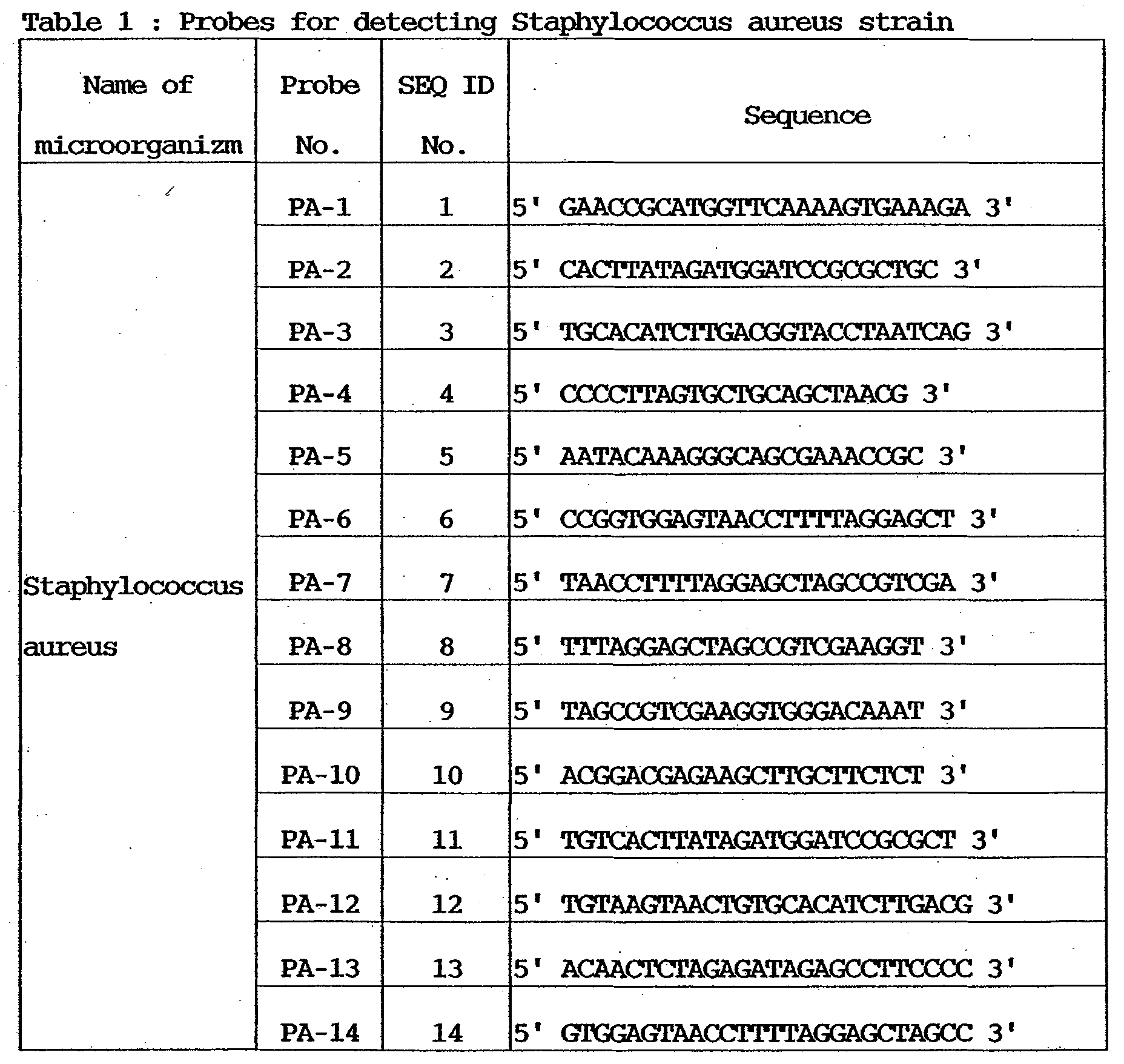
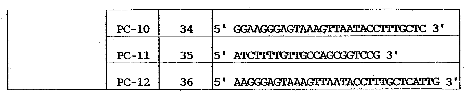
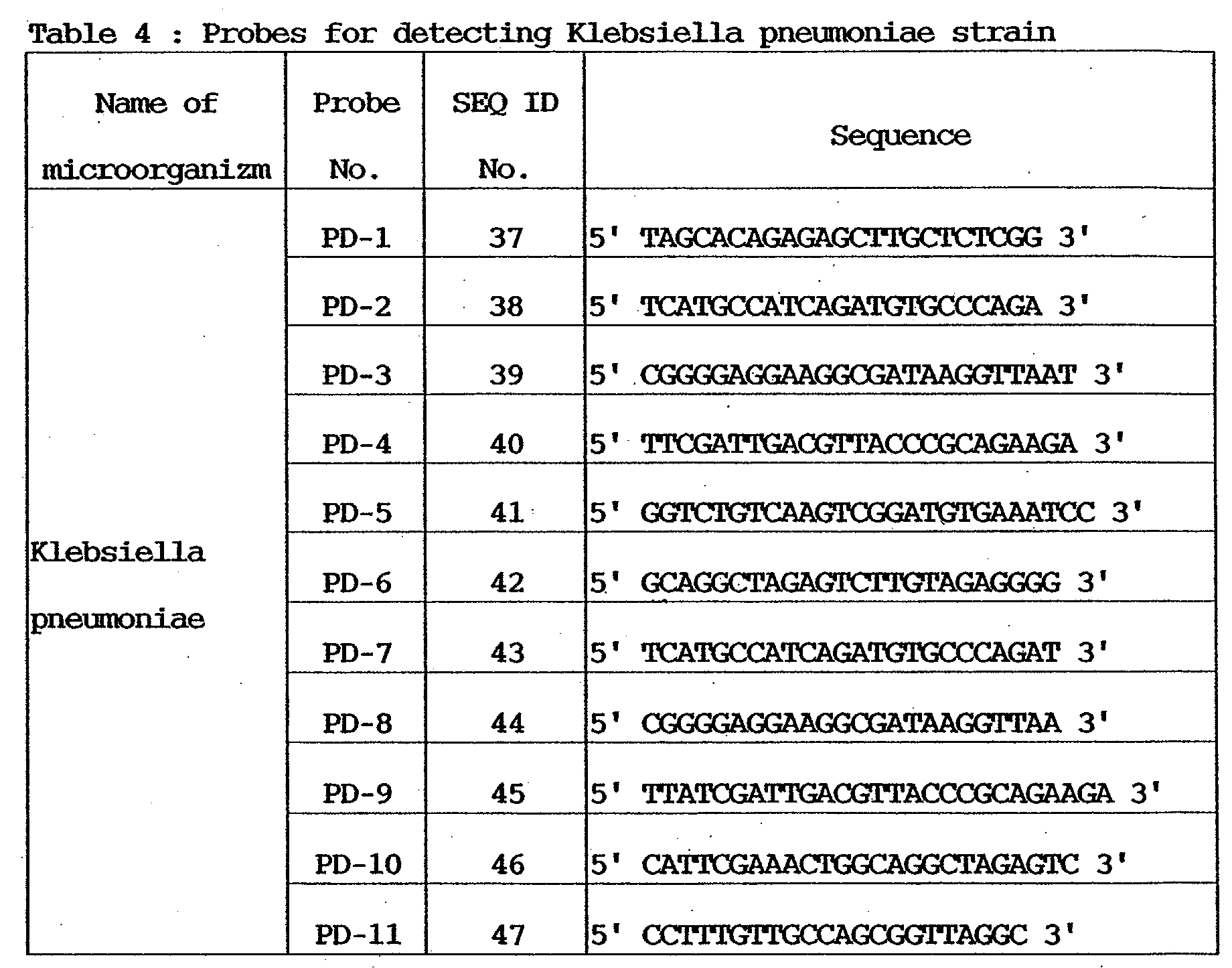
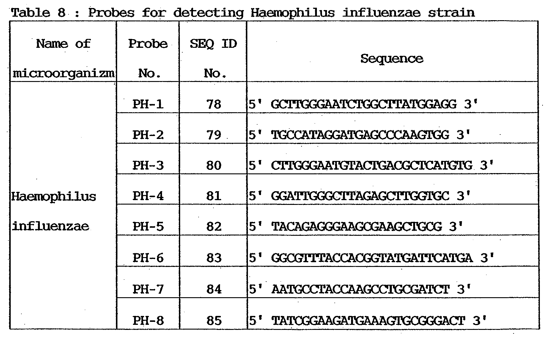
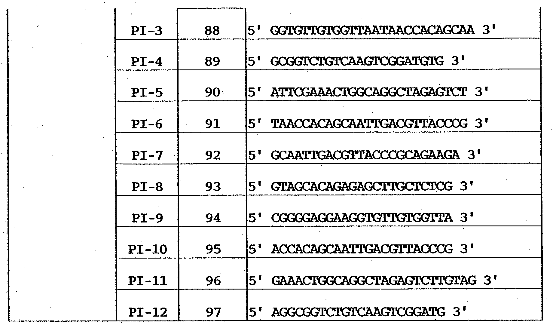
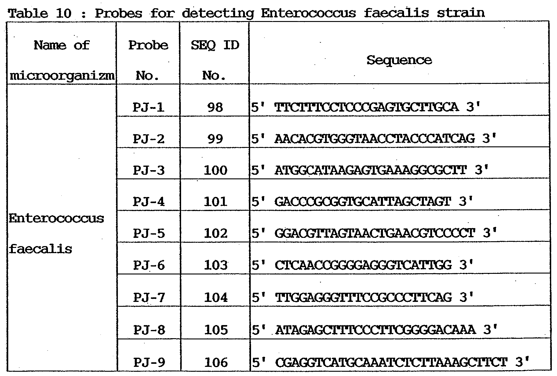
- the oligonucleotide probe to be reacted with a specimen solution containing the nucleic acid sequence of the genes of the infectious etiologic agents contains one base sequence which belongs to one of the first group (SEQ ID Nos. 1 to 14 in the attached sequence table) shown in Table 1, the second group (SEQ ID Nos. 15 to 24) shown in Table 2, the third group (SEQ ID Nos. 25 to 36) shown in Table 3, the fourth group (SEQ ID Nos. 37 to 47) shown in Table 4, the fifth group (SEQ ID Nos. 48 to 57) shown in Table 5, the sixth group (SEQ ID Nos. 58 to 68) shown in Table 6, the seventh group (SEQ ID Nos.
- An oligonucleotide probe having a base sequence selected from the first group detects Staphylococcus aureus.
- An oligonucleotide probe having a base sequence selected from the second group detects Staphylococcus epidermidis.
- An oligonucleotide probe having a base sequence selected from the third group detects Escherichia coli.
- An oligonucleotide probe having a base sequence selected from the fourth group detects Klebsiella pneumoniae.
- An oligonucleotide probe having a base sequence selected from the fifth group detects Pseudomonas aeruginosa.
- An oligonucleotide probe having a base sequence selected from the sixth group detects Serratia marcescens.
- An oligonucleotide probe having a base sequence selected from the seventh group detects Streptococcus pneumoniae.
- An oligonucleotide probe having a base sequence selected from the eighth group detects Haemophilus influenzae.
- An oligonucleotide probe having a base sequence selected from the ninth group detects Enterobacter cloacae .
- An oligonucleotide probe having a base sequence selected from the 10th group detects Enterococcus faecalis.
- Complementary sequences of these probe sequences can also be used as effective probe sequences because they have the same functions as those of the above probe sequences (the complementary sequences of the first group are indicated by SEQ ID Nos. 113 to 126 in the attached sequence table, the complementary sequences of the second group are indicated by SEQ ID Nos. 127 to 136, the complementary sequences of the third group are indicated by SEQ ID Nos. 137 to 148, the complementary sequences of the fourth group are indicated by SEQ ID Nos. 149 to 159, the complementary sequences of the fifth group are indicated by SEQ ID Nos. 160 to 169, the complementary sequences of the sixth group are indicated by SEQ ID Nos. 170 to 180, the complementary sequences of the seventh group are indicated by SEQ ID Nos.
- the complementary sequences of the eighth group are indicated by SEQ ID Nos. 190 to 197
- the complementary sequences of the ninth group are indicated by SEQ ID Nos. 198 to 209
- the complementary sequences of the 10th group are indicated by SEQ ID Nos. 210 to 218).
- the probes for the respective bacteria were designed from the genome parts coding the 16s rRNAs such that they could have a very high specificity with respect to the corresponding bacteria, any variation between the probe base sequences could be prevented, and a sufficient hybridization sensitivity could be expected.
- oligonucleotide probes are designed such that a stable hybrid body is formed by hybridization reaction between a specimen and two or more kinds of probes bonded onto a carrier, and a satisfactory result can be obtained.
- the carrier according to the present invention on which the infection detection probe of the present invention is immobilized, is prepared by discharging oligonucleotide by using a BJ printer and chemically bonding it to the carrier.
- the probe hardly peels off.
- An additional effect for increasing the sensitivity is also obtained.
- a DNA chip is produced by stamping called a Stanford method that is generally widely used (for example, TAKARA SHUZO produces DNA chips by applying cDNA fragments of known genes of human origin, which are related to cancers, by spotting or stamping), the applied DNA readily peels off.
- the carrier according to the present invention is prepared also in consideration of these problems.
- the probe is stably immobilized and hardly peels off, as compared to the prior arts, and highly sensitive and accurate detection can be executed. The preferred embodiment of the present invention will be described below in detail.
- the DNA chip of this embodiment can be applied to any specimen in which bacteria may be present, and for example, body fluids originated in animals such as human and livestock, including blood, spinal fluid, phlegm, stomach fluid, vaginal discharge, and intraoral mucus, and excretion such as urine and feces. All media which can be contaminated by bacteria can also be subjected to a test using the DNA chip, including food, drink water and hot spring water in the natural environment, which may cause food poisoning by contamination, filters from air and water cleaners, and so forth. Animals and plants which should be quarantined in import/export are also used as specimens.
- the specimens used for the DNA chip of this embodiment include not only an extracted nucleic acid itself but also specimens prepared by various methods, such as an amplified specimen prepared by using an PCR reaction primer designed for 16s rRNA detection, a specimen prepared by causing PCR reaction on the basis of a PCR amplified product, a specimen prepared by an amplification method other than PCR, and a specimen labeled by various labeling methods for visualization.
- the carrier used for the DNA chip of this embodiment includes all sorts of carriers including flat substrates such as a glass substrate, a plastic substrate, and a silicon wafer, a three-dimensional structure having a three-dimensional pattern, a spherical body such as a bead, and rod-, cord-, and thread-shaped structures.
- the carrier also includes a substrate whose surface is processed such that a probe DNA can be immobilized.
- a carrier prepared by introducing a functional group to its surface to make chemical reaction possible has a preferable form from the viewpoint of reproducibility because the probe is stably bonded in the process of hybridization reaction.
- a combination of a maleimide group and a thiol (-SH) group is used. More specifically, a thiol (-SH) group is bonded to the terminal of a nucleic acid probe, and a process is executed make the solid surface have a maleimide group. Accordingly, the thiol group of the nucleic acid probe supplied to the solid surface reacts with the maleimide group on the solid surface to immobilize the nucleic acid probe.
- an aminosilane coupling agent is caused to react on a glass substrate.
- the maleimide group is introduced by reaction between the amino group and an EMCS reagent (N-(6-Maleimidocaproyloxy)succinimide: available from Dojin).
- EMCS reagent N-(6-Maleimidocaproyloxy)succinimide: available from Dojin.
- Introduction of the SH group to a DNA can be done by using 5'-Thiol-ModifierC6 (available from Glen Research) when the DNA is synthesized by an automatic DNA synthesizer.
- a combination of, e.g., an epoxy group (in the solid phase) and an amino group (nucleic acid probe terminal) can also be used as a combination of functional groups to be used for immobilization.
- Surface treatments using various kinds of silane coupling agents are also effective.
- Oligonucleotide in which a functional group which can react with a functional group introduced by a silane coupling agent is introduced is used.
- a method of applying a resin having a functional group can also be used.
- the present invention will be described below in more detail on the basis of examples using the infectious etiologic agent detection probes to be used to detect the 10 etiologic agents described above.
- Example 1 Microorganism Detection Using 1-Step PCR
- Nucleic acid sequences shown in Tables 1 to 10 were designed as probes to be used for detection of the 10 etiologic agents. More specifically, the following probe base sequences were selected from the genome parts coding the 16s rRNAs of the respective bacteria. These probe base sequences were designed such that they could have a very high specificity with respect to the corresponding bacteria, any variation between the probe base sequences could be prevented, and a sufficient hybridization sensitivity could be expected (The probe base sequences need not always completely match those shown in Tables 1 to 10. Probe base sequences having base lengths of 20 to 30, including the probe base sequences, are also included in the probe base sequence shown in the tables. As described above, complementary sequences (complementary strands) of the base sequences shown in the tables may also be used).
- a thiol group was introduced to the 5' terminal of the nucleic acid after synthesis in accordance with a conventional method. After introduction of the functional group, purification and freeze-drying were executed. The freeze-dried probes were stored in a freezer at -30°C.
- 16s rRNA gene (target gene) amplification PCR primers for etiologic agent detection nucleic acid sequences shown in Table 11 were designed. More specifically, probe sets which specifically amplify the genome parts coding the 16s rRNAs, i.e., primers for which the specific melting points were made uniform as much as possible at the two end portions of the 16s rRNA coding region of a base length of 1,400 to 1,700 were designed. In order to simultaneously amplify variants or a plurality of 16s rRNA coding regions on genomes, a plurality of kinds of primers were designed. Note that a primer set is not limited to primer sets shown in the table 11. A primer set which is available in common to a plural kinds of etiologic agents and amplify almost entire length of 16s rRNA coding region of the etiologic agents can also be employed.
- the primers shown in Table 11 were purified by HPLC (High Performance Liquid Chromatography) after synthesis. Three forward primers and three reverse primers were mixed and dissolved in a TE buffer solution such that each primer concentration had an ultimate concentration of 10 pmol/ ⁇ l.
- microbial culture media were produced by culturing type strains of the etiologic agents (Staphylococcus aureus type strain (ATCC12600), Staphylococcus epidermidis type strain (ATCC14990), Escherichia coli type strain (ATCC11775), Klebsiella pneumoniae type strain (ATCC13883), Pseudomonas aeruginosa type strain (ATCC10145), Serratia marcescens strain, Streptococcus pneumoniae type strain, Haemophilus influenzaestrain, Enterobacter Cloacae type strain (ATCC13047), and Enterococcus faecalis type strain (ATCC19433)) in accordance with a conventional method.
- Staphylococcus aureus type strain ATCC12600
- Staphylococcus epidermidis type strain ATCC14990
- Escherichia coli type strain ATCC11775
- Klebsiella pneumoniae type strain ATCC13883
- the cells were collected by centrifuge (8,500 rpm, 5 min, 4°C). After the supernatant was removed, a 300- ⁇ l enzyme buffer (50 mM Tris-HCl: p.H. 8.0, 25 mM EDTA) was added, and the broth was re-suspended by using a mixer. From the re-suspended broth, the cells were collected again by centrifuge (8,500 rpm, 5 min, 4°C). After the supernatant was removed, the following enzyme solution was added to the collected cells, and the broth was re-suspended by using the mixer. Lysozyme 50 ⁇ l (20 mg/ml in Enzyme Buffer) N-Acetylmuramidase SG 50 ⁇ l (0.2 mg/ml in Enzyme Buffer)
- the broth added with the enzyme solution and re-suspended was left stand still in an incubator at 37°C for 30 min to melt cell walls.
- Microbial genome DNA extraction to be described below was done by using a nucleic acid purification kit (MagExtractor-Genome: available from TOYOBO).
- a 750- ⁇ l melting/absorption solution and a 40- ⁇ l magnetic beads were added to the preprocessed microbial suspension.
- the suspension was intensely stirred for 10 min by using a tube mixer (step 1).
- the microtube was set in a separation stand (Magical Trapper) and left stand still for 30 sec to gather magnetic particles to the wall surface of the tube. The supernatant was removed while the microtube was kept set in the stand (step 2).
- the microtube was set in the separation stand (Magical Trapper) and left stand still for 30 sec to gather magnetic particles to the wall surface of the tube. The supernatant was removed while the microtube was kept set in the stand (step 4).
- Steps 3 and 4 were repeated to execute the second cleaning process (step 5). After that, 900- ⁇ l 70% ethanol was added. The solution was re-suspended by stirring it for about 5 sec by a mixer (step 6).
- the microtube was set in the separation stand (Magical Trapper) and left stand still for 30 sec to gather magnetic particles to the wall surface of the tube. The supernatant was removed while the microtube was kept set in the stand (step 7).
- Steps 6 and 7 were repeated to execute the second cleaning process by using 70% ethanol (step 8). After that, 100- ⁇ l pure water was added to the collected magnetic particles. The solution was stirred for 10 min by a tube mixer (step 9).
- the microtube was set in the separation stand (Magical Trapper) and left stand still for 30 sec to gather magnetic particles to the wall surface of the tube. The supernatant was collected to a new tube while the microtube was kept set in the stand.
- the collected genome DNAs of microorganisms underwent agarose electrophoresis and 260/280-nm absorbance determination in accordance with the conventional method so that the quality (the admixture amount of low molecular nucleic acid and the degree of decomposition) and collection amount were tested.
- the quality the admixture amount of low molecular nucleic acid and the degree of decomposition
- collection amount were tested.
- about 9 to 10- ⁇ g genome DNAs were collected in each bacterium. No degradation of genome DNAs or admixture of rRNA was observed.
- the collected genome DNAs were dissolved in a TE buffer solution at an ultimate concentration of 50 ng/ ⁇ l and used in the following examples.
- the DNA Microarray was prepared according to Japanese Patent Application Laid-Open No. 11-187900.[4-1] Cleaning of Glass Substrate
- a glass substrate (size: 25 mm ⁇ 75 mm ⁇ 1 mm, available from Iiyama Tokushu Glass) made of synthetic silica was placed in a heat- and alkali-resisting rack and dipped in a cleaning solution for ultrasonic cleaning, which was prepared to a predetermined concentration.
- the glass substrate was kept dipped in the cleaning solution for a night and cleaned by ultrasonic cleaning for 20 min.
- the substrate was picked up, lightly rinsed by pure water, and cleaned by ultrasonic cleaning in ultrapure water for 20 min.
- the substrate was dipped in a 1N aqueous sodium hydroxide solution heated to 80°C for 10 min. Pure water cleaning and ultrapure water cleaning were executed again.
- a silica glass substrate for a DNA microchip was thus prepared.
- a silane coupling agent KBM-603 (available from Shinetsu Silicone) was dissolved in pure water at a concentration of 1% and stirred at room temperature for 2 hrs.
- the cleaned glass substrate was dipped in the aqueous solution of the silane coupling agent and left stand still at room temperature for 20 min.
- the glass substrate was picked up.
- the surface was lightly rinsed by pure water and dried by spraying nitrogen gas to both surfaces of the substrate.
- the dried substrate was baked in an oven at 120°C for 1 hr to complete the coupling agent treatment, thereby introducing an amino group to the substrate surface.
- N-(6-Maleimidocaproyloxy)succinimido) available from Dojindo Laboratories was dissolved in a 1 : 1 medium mixture of dimethyl sulfoxide and ethanol such that an ultimate concentration of 0.3 mg/ml was obtained, thereby preparing an EMCS solution.
- the baked glass substrate was left stand and cooled and dipped in the prepared EMCS solution at room temperature for 2 hrs. With this process, the amino group introduced to the surface by the silane coupling agent reacted with the succinimide group in the EMCS to introduce the maleimide group to the surface of the glass substrate.
- the glass substrate picked up from the EMCS solution was cleaned by using the above-described medium mixture in which the EMCS was dissolved. The glass substrate was further cleaned by ethanol and dried in a nitrogen gas atmosphere.
- the microorganism detection probe prepared in Example 1 was dissolved in pure water. The solution was dispensed such that the ultimate concentration (at ink dissolution) became 10 ⁇ M. Then, the solution was freeze-dried to remove water.
- An aqueous solution containing 7.5-wt% glycerin, 7.5-wt% thioglycol, 7.5-wt% urea, and 1.0-wt% Acetylenol EH (available from Kawaken Fine Chemicals) was prepared.
- Each of the seven probes (Table 1) prepared in advance was dissolved in the medium mixture at a specific concentration.
- An ink tank for an inkjet printer (tradename: BJF-850, available from Canon) is filled with the resultant DNA solution and attached to the printhead.
- the inkjet printer used here was modified in advance to allow printing on a flat plate.
- an about 5-picoliter DNA solution can be spotted at a pitch of about 120 ⁇ m.
- the printing operation was executed for one glass substrate by using the modified inkjet printer, thereby preparing a DNA microarray. After confirming that printing was reliably executed, the glass substrate was left stand still in a humidified chamber for 30 min to make the maleimide group on the glass substrate surface react with the thiol group at the nucleic acid probe terminal.
- DNA solution remaining on the surface was cleaned by using a 10-mM phosphate buffer (pH 7.0) containing 100-mM NaCl, thereby obtaining a gene chip (DNA microarray) in which single-stranded DNAs were immobilized on the glass substrate surface.
- Amplification reaction of the reaction solution having the above composition was caused by using a commercially available thermal cycler in accordance with the following protocol.
- the primers were removed (purified) by using a purification column (QIAquick PCR Purification Kit available from QIAGEN). Then, determination of the amplified products was executed to obtain labeled specimens.
- Detection reaction was performed by using the gene chips prepared by [4. Preparation of DNA Microarray] and the labeled specimen prepared by [5. Amplification and Labeling of Specimen (PCR Amplification & Fluorescent Labeling)].
- BSA fetal bovine serum albumin, Fraction V: available from Sigma
- the gene chips prepared by [4. Preparation of DNA Microarray] were dipped in the solution at room temperature for 2 hrs to execute blocking. After the end of blocking, the chips were cleaned by using a 2xSSC solution (NaCl 300 mM, Sodium Citrate (trisodium citrate dihydrate, C 6 H 5 Na 3 ⁇ 2H 2 O) 30 mM, pH 7.0) containing 0.1-wt% SDS (Sodium Dodecyl Sulfate), rinsed by pure water, and hydro-extracted by a spin dryer.
- 2xSSC solution NaCl 300 mM, Sodium Citrate (trisodium citrate dihydrate, C 6 H 5 Na 3 ⁇ 2H 2 O) 30 mM, pH 7.0
- SDS Sodium Dodecyl Sulfate
- the hydro-extracted gene chips were set in a hybridization apparatus (Hybridization Station available from Genomic Solutions Inc). Hybridization reaction was caused in a hybridization solution under conditions to be described below.
- the gene chips after the end of hybridization reaction were subjected to fluorometry by using a gene chip fluorescent detector (GenePix 4000B available from Axon). As a result, the respective bacteria could be detected with sufficient signals at a high reproducibility, as shown in Tables 12 to 21. No hybrid bodies for other bacteria were detected.
- the numerical values of florescent luminances(photomultiplier voltage: 400 V) in Tables 12 to 21 indicate average pixel luminances (resolution: 5 u m).
- the S/N ratios indicate values obtained by dividing the fluorescent luminances by background average values measured by analysis software (GenePix Pro Ver.3.0 available from Axon) attached to the measuring device.
- the respective etiologic agents can be detected with sufficient signals at a high reproducibility.
- Example 2 Microorganism Detection Using 2-Step PCR
- probe DNAs, specimen amplification PCR primers, the genome DNAs of etiologic agents, and DNA microarrays were prepared, and the following experiments were conducted.
- Premix PCR reagent 25 ⁇ l Template Genome DNA 2 ⁇ l (10ng) Forward Primer mix 2 ⁇ l (20pmol/tube each) Reverse Primer mix 2 ⁇ l (20pmol/tube each) H 2 0 19 ⁇ l Total 50 ⁇ l
- Amplification reaction of the reaction solution having the above composition was caused by using a commercially available thermal cycler in accordance with the following protocol.
- Amplification reaction of the reaction solution having the above composition was caused by using a commercially available thermal cycler in accordance with the following protocol.
- purification was performed by using a purification column (QIAquick PCR Purification Kit available from QIAGEN) to obtain labeled specimens.
- the numerical values of florescent luminances(photomultiplier voltage: 400 V) in Tables 22 to 31 indicate average pixel luminances (resolution: 5 ⁇ m).
- the S/N ratios indicate values obtained by dividing the fluorescent luminances by background average values measured by analysis software (GenePix Pro Ver.3.0 available from Axon) attached to the measuring device.
- probe DNAs As in Examples 1 and 2, probe DNAs, specimen amplification PCR primers, the genome DNAs of etiologic agents, and DNA microarrays were prepared, and the following experiments were conducted.
- AmpliTaq Gold LD(5U/ ⁇ L) 0.5 ⁇ L Template DNA variable dNTP mis(2.5mM/each) 4.0 ⁇ L x10 PCR buffer 5.0 ⁇ L 25mM MgCl 2 7.0 ⁇ L Forward Primer Mix(10 ⁇ M/each) 0.25 ⁇ L Reverse Primer Mix(10 ⁇ M/each) 0.25pL H 2 O variable Total 50 ⁇ L
- Amplification reaction of the reaction solution having the above composition was caused by using a commercially available thermal cycler in accordance with the following protocol.
- Amplification reaction of the reaction solution having the above composition was caused by using a commercially available thermal cycler in accordance with the following protocol.
- purification was performed by using a purification column (QIAquick PCR Purification Kit available from QIAGEN) to obtain labeled specimens.
- the respective etiologic agents can be detected with sufficient signals at a high reproducibility.
- an infectious etiologic agent can be identified by using microarrays on which probe sets capable of detecting the 10 bacteria, i.e., Staphylococcus aureus, Staphylococcus epidermidis, Escherichia coli, Klebsiella pneumoniae, Pseudomonas aeruginosa, Serratia marcescens, Streptococcus pneumoniae, Haemophilus influenzae, Enterobacter cloacae, and Enterococcus faecalis are separately immobilized or combined.
- the problems of the DNA probe of microbial origin are solved.
- oligonucleotide probes can chemically be mass-produced, and purification and concentration control are possible.
- a probe set can be provided, which allows to detect bacteria in the same species all together and discriminatingly detect bacteria in other species for the purpose of classifying the bacteria depending on the species.
- a probe set can be provided, which also considers the stability of a hybrid body between a probe and a specimen so that the difference between the species can accurately be evaluated on a DNA microarray.
- a carrier on which the probe DNAs are immobilized to make the probe DNAs react with specimens can also be provided.
- a carrier can be provided, on which the probe DNAs are chemically immobilized so that the probe DNAs are stably immobilized on the carrier, and a detection result with high reproducibility can be obtained in the process of reaction between a specimen solution and the probes and probe sets.
- 16s rRNA gene arrangements in the genes of infectious etiologic agents can be detected in proper quantities. Hence, the presence of an infectious etiologic agent can efficiently and accurately be determined.
- the primer sets of this example are designed to give a satisfactory amplification result in PCR reaction executed to identify an infectious etiologic agent. "Satisfactory" means not only that the target 16s rRNAs are sufficiently amplified but also that no products other than the 16s rRNAs are generated.
- “Satisfactory” also means that only the 16s rRNAs of an infectious etiologic agent are amplified without amplifying human genome genes originated in a specimen, which are contained in the specimen.
- any specimen in which bacteria may be present and for example, body fluids originated in animals such as human and livestock, including blood, spinal fluid, phlegm, stomach fluid, vaginal discharge, and intraoral mucus, and excretion such as urine and feces are used in this example. All media which can be contaminated by bacteria can also be used in this example, including food , drink water and hot spring water in the natulal environment, which may cause food poisoning by contamination, filters from air and water cleaners , and so forth. Animals and plants which should be quarantined in import/export are also used as specimens.
- the PCR reaction used in this example includes PCR reaction which uses an extracted nucleic acid itself as a template, asymmetrical PCR reaction which uses primers on one side of SEQ ID Nos. 107 to 109 (F1 to F3 in Table 11) or SEQ ID Nos. 110 to 112 (R1 to R3 in Table 11), and PCR which executes labeling for visualization.
- probe sets which specifically amplify the genome parts coding the 16s rRNAs, i.e., primers for which the specific melting points were made uniform as much as possible at the two end portions of the 16s rRNA coding region of a base length of 1,500 were designed.
- primers for which the specific melting points were made uniform as much as possible at the two end portions of the 16s rRNA coding region of a base length of 1,500 were designed.
- a plurality of kinds of primers were designed.
- the primers shown in Table 11 were purified by HPLC (High Performance Liquid Chromatography) after synthesis. All of three forward primers and three reverse primers were mixed and dissolved in a TE buffer solution such that each primer concentration had an ultimate concentration of 10 pmol/ ⁇ l. In this example, all the forward primers and reverse primers were used. Alternatively, one to three forward primers and one to three reverse primers may be used.
- the primers were removed by using a purification column (QIAquick PCR Purification Kit available from QIAGEN). Then, the amplified products were examined by gel electrophoresis. One band was detected in 1,500 base pair regions, and it was confirmed that satisfactory PCR reaction was executed. No byproducts were generated.
- Bacteremia model systems were prepared by adding 10 3 10 4 , and 10 5 Enterobacter cloacae, which was cultured in accordance with the procedures described in Example 1, to 200- ⁇ l human blood (collected EDTA blood). An N-acetyl muramidase solution (0.2 mg/ml in Enzyme Buffer) was added to each solution. The solutions were heated to 37°C for 30 min. After that, DNAs were extracted by using Qiamp Blood mini Kit (available from QIAGEN) to prepare templates for PCR reaction.
- Qiamp Blood mini Kit available from QIAGEN
- Example 4 As a result, as in Example 4, one band was detected in 1,500 base pair regions, and it was confirmed that satisfactory PCR reaction was executed. No byproducts were generated. The amount of PCR amplified products obtained from the band was proportional to the added cell amount. This indicates that when the primer sets were used, only the 16s rRNAs of Enterobacter cloacae were amplified without generating any PCR byproduct of human genome.
- the 16s rRNA parts in the genes of a plurality of kinds of infectious etiologic agents can efficiently be amplified at a high purity.
- the 16s rRNAs of an infectious etiologic agent can efficiently be amplified.
- an infection detection probe which allows mass preparation at a time and identification of a species in similar species can be provided. More specifically, an infection detection probe which can suitably be used to classify a plurality of kinds of etiologic agents of an infection on the basis of the species can be provided.
- an infection detection probe suitable for detection of, e.g., the above-described 10 bacteria as the etiologic agents of infections can be provided.
- a probe set can also be provided, which also considers the stability of a hybrid body between an infection detection probe and a specimen so that the difference between similar species can accurately be evaluated on a DNA chip.
- a carrier on which the infection detection probe is immobilized to make the infection detection probe react with the specimen can be provided.
- a carrier can be provided, on which the infection detection probes are chemically immobilized so that the infection detection probes are stably immobilized on the carrier, and a detection result with high reproducibility can be obtained in the process of reaction with a specimen solution.
- a PCR reaction primer which amplifies the 16s rRNAs of an etiologic agent in a specimen in order to detect and/or identify an infectious etiologic agent.
- a primer set which can commonly be used for a plurality of species and effectively amplify the 16s rRNAs of an etiologic agent even when the species is unknown.
- a primer set which can amplify the 16s rRNAs of a plurality of kinds of etiologic agents under the same PCR conditions.
Landscapes
- Chemical & Material Sciences (AREA)
- Life Sciences & Earth Sciences (AREA)
- Organic Chemistry (AREA)
- Proteomics, Peptides & Aminoacids (AREA)
- Analytical Chemistry (AREA)
- Zoology (AREA)
- Wood Science & Technology (AREA)
- Health & Medical Sciences (AREA)
- Engineering & Computer Science (AREA)
- Microbiology (AREA)
- Bioinformatics & Cheminformatics (AREA)
- Molecular Biology (AREA)
- Biotechnology (AREA)
- Biophysics (AREA)
- Physics & Mathematics (AREA)
- Biochemistry (AREA)
- Immunology (AREA)
- General Engineering & Computer Science (AREA)
- General Health & Medical Sciences (AREA)
- Genetics & Genomics (AREA)
- Chemical Kinetics & Catalysis (AREA)
- Measuring Or Testing Involving Enzymes Or Micro-Organisms (AREA)
- Apparatus Associated With Microorganisms And Enzymes (AREA)
Abstract
Description
- The present invention relates to detection and/or identification of an infectious etiologic agent as an etiologic agent of an infectious disease and, more particularly, to a probe and probe set originated in an infectious etiologic agent, a carrier, and a genetic screening method, which are useful for detection and identification of the etiologic agent of an infectious disease.
- The present invention also relates to a PCR amplification process of an infectious etiologic agent, which is suitable for detection and/or identification of an infectious etiologic agent.
- In recent years, gene expression analysis using DNA chips (also referred to as DNA microarrays hereinafter) is done in various fields including drug development. Different specimen DNAs are made to react with a DNA microarray in which various kinds of gene sets (probes) are arranged. Gene dosages which exist in the respective specimens are compared. Genes which are present in high dosages (the expression amounts are large) or inactive genes (the expression amounts are small) at each stage are classified and analyzed in association with functions.
- An example is an infectious etiologic agent test. In Japanese Patent Laid-Open No. 2001-299396, Ezaki et al have proposed a microorganism identification method using, as a DNA probe, a DNA chip on which chromosome DNAs are immobilized. According to this method, a plurality of chromosome DNAs originated in a plurality of known microorganisms with different GC contents are made to react with chromosome DNAs originated in an unknown microorganism in a specimen. When the resultant hybridization complex is detected, the unknown microorganism in the specimen can be detected.
- As probes used for DNA chips for infectious etiologic agent tests, Ono et al have proposed a bacterial detection probe using restriction enzyme fragments in Japanese Patent Laid-Open No. 6-133798, a Pseudomonas aeruginosa detection probe in Japanese Patent Laid-Open No. 10-304896, and a detection probe using restriction enzyme fragments of Escherichia coli, klebsiella pneumoniae, and Enterobacter cloacae in Japanese Patent Laid-Open No. 10-304897.
- As a microarray, for example, a microarray using stamping called a Stanford method is known. For example, DNA chips on which cDNA fragments of known genes of human origin, which are related to cancers, are applied by spotting or stamping and chips prepared by bonding cDNA fragments of 1,000 kinds of known genes of human origin to slide glasses are commercially available from TAKARA SHUZO.
- On the other hand, a chip available from Affymetrix is prepared by designing an oligonucleotide probe set on the basis of the known gene cDNAs, and probes are laid out by synthesis on a substrate. Oligoprobes are laid out on one chip at a high density so that the expression levels of 10,000 or more genes can be analyzed at once.
- However, the DNA chips of the prior arts described above use DNA probes such as chromosome DNAs or restriction enzyme fragments. DNAs directly extracted from microorganisms are used as materials. For this reason, the chips can hardly be mass-prepared at a time and are not suitable for clinical diagnosis. For application to clinical diagnosis, mass production of inexpensive and uniform DNA chips is necessary. For this purpose, mass preparation of uniform DNAs as probe solutions is essential. However, mass preparation of DNA probes is impossible. Even for DNA probes, when PCR amplification reaction is used, the number of DNAs can gradually be increased. However, mass preparation at a time using the PCR reaction is difficult, and the DNA chips are difficult to use for clinical diagnosis.
- In addition, since the base length of a DNA probe is large, it is difficult to identify one species in similar species. Such a DNA probe is therefore not suitable for, e.g., infection detection. In treating an infection, the species must be specified, and antibiotic drugs corresponding to it must be selected and administered. For this purpose, an infection detection probe is required to have a function capable of detecting a species while discriminating similar species, although bacteria belonging to the same species need not accurately be discriminated (that is, bacteria in the same species can be detected all together). However, in, e.g., the DNA chip using restriction enzyme fragments of Escherichia coli, klebsiella pneumoniae, and Enterobacter cloacae, which is disclosed in Japanese Patent Laid-Open No. 10-304897, cross reaction occurs between the three species because of the large base length of the probe. Since similar species cannot individually be discriminated, the DNA chip can hardly be used for infection detection.
- As an application purpose of microarrays, infectious etiologic agent tests have received a great deal of attention. Some probe sets aiming at testing infectious etiologic agents have also been proposed.
- As an important point of bacterial tests using microarrays, detection must be possible even when the number of infectious etiologic agents is small. To do this, it is effective to amplify specific parts in the base sequences of the DNAs of infectious etiologic agents by, e.g., PCR reaction using primers. For example, a 16s rRNA gene arrangement contains a sequence unique to the species in the information of about 1,700 base pairs. When the sequence is used, classification can be done to some extent. In detecting/identifying bacteria, 16s rRNA parts in the DNA base sequences of bacteria are preferably used. Hence, it is demanded to amplify the 16s rRNA parts.
- For various kinds of bacteria, however, the gene arrangements are only partially clarified, and the 16s rRNAs are not totally known. For this reason, it is not easy to design primers for PCR amplification reaction.
- The present invention has been made in consideration of the above situation, and has as its object to provide an infection detection probe which allows mass preparation at a time and identification of a species in similar species.
- More specifically, it is an object of the present invention to provide an infection detection probe which can suitably be used to classify a plurality of kinds of etiologic agents of an infection on the basis of the species.
- It is another object of the present invention to provide a probe set which also considers the stability of a hybrid body between an infection detection probe and a specimen so that the difference between similar species can accurately be evaluated on a DNA chip.
- It is still another object of the present invention to provide a carrier on which the infection detection probe is immobilized to make the infection detection probe react with the specimen.
- It is still another object of the present invention to provide a carrier on which the infection detection probes are chemically immobilized so that the infection detection probes are stably immobilized on the carrier, and a detection result with high reproducibility can be obtained in the process of reaction with a specimen solution.
- It is still another object of the present invention to provide a PCR reaction primer which amplifies the 16s rRNAs of an etiologic agent in a specimen in order to detect and/or identify an infectious etiologic agent.
- It is still another object of the present invention to provide a primer set which can commonly be used for a plurality of species and effectively amplify the 16s rRNAs of an etiologic agent even when the species is unknown. '
- It is still another object of the present invention to provide a primer set which can amplify the 16s rRNAs of a plurality of kinds of etiologic agents under the same PCR conditions.
- The present invention provides a primer set characterized by amplifying all species without amplifying genes originated in human genomes by causing PCR reaction for a human blood specimen by using all the primer sets simultaneously. More specifically, a primer set having a sequence which is different from the base sequence of human genome genes by three or more bases is proposed.
- Other features and advantages of the present invention will be apparent from the following description.
- A preferred embodiment of the present invention will now be described in.
- In the following embodiment, an oligonucleotide probe used to identify the etiologic agent of an infection and, more specifically, a probe used to detect one or some of Staphylococcus aureus, Staphylococcus epidermidis, Escherichia coli, Klebsiella pneumoniae, Pseudomonas aeruginosa, Serratia marcescens, Streptococcus pneumoniae, Haemophilus influenzae, Enterobacter cloacae, and Enterococcus faecalis will be described. That is, a nucleic acid probe or nucleic acid probe set, which is used to detect rRNA gene arrangements in the genes of the above 10 infectious etiologic agents in proper quantities, is disclosed.
- According to this embodiment, the oligonucleotide probe to be reacted with a specimen solution containing the nucleic acid sequence of the genes of the infectious etiologic agents contains one base sequence which belongs to one of the first group (SEQ ID Nos. 1 to 14 in the attached sequence table) shown in Table 1, the second group (SEQ ID Nos. 15 to 24) shown in Table 2, the third group (SEQ ID Nos. 25 to 36) shown in Table 3, the fourth group (SEQ ID Nos. 37 to 47) shown in Table 4, the fifth group (SEQ ID Nos. 48 to 57) shown in Table 5, the sixth group (SEQ ID Nos. 58 to 68) shown in Table 6, the seventh group (SEQ ID Nos. 69 to 77) shown in Table 7, the eighth group (SEQ ID Nos. 78 to 85) shown in Table 8, the ninth group (SEQ ID Nos. 86 to 97) shown in Table 9, and the 10th group (SEQ ID Nos. 98 to 106) shown in Table 10 (to be described later). An oligonucleotide probe having a base sequence selected from the first group detects Staphylococcus aureus. An oligonucleotide probe having a base sequence selected from the second group detects Staphylococcus epidermidis. An oligonucleotide probe having a base sequence selected from the third group detects Escherichia coli. An oligonucleotide probe having a base sequence selected from the fourth group detects Klebsiella pneumoniae. An oligonucleotide probe having a base sequence selected from the fifth group detects Pseudomonas aeruginosa. An oligonucleotide probe having a base sequence selected from the sixth group detects Serratia marcescens. An oligonucleotide probe having a base sequence selected from the seventh group detects Streptococcus pneumoniae. An oligonucleotide probe having a base sequence selected from the eighth group detects Haemophilus influenzae. An oligonucleotide probe having a base sequence selected from the ninth group detects Enterobacter cloacae . An oligonucleotide probe having a base sequence selected from the 10th group detects Enterococcus faecalis.
- Complementary sequences of these probe sequences can also be used as effective probe sequences because they have the same functions as those of the above probe sequences (the complementary sequences of the first group are indicated by SEQ ID Nos. 113 to 126 in the attached sequence table, the complementary sequences of the second group are indicated by SEQ ID Nos. 127 to 136, the complementary sequences of the third group are indicated by SEQ ID Nos. 137 to 148, the complementary sequences of the fourth group are indicated by SEQ ID Nos. 149 to 159, the complementary sequences of the fifth group are indicated by SEQ ID Nos. 160 to 169, the complementary sequences of the sixth group are indicated by SEQ ID Nos. 170 to 180, the complementary sequences of the seventh group are indicated by SEQ ID Nos. 181 to 189, the complementary sequences of the eighth group are indicated by SEQ ID Nos. 190 to 197, the complementary sequences of the ninth group are indicated by SEQ ID Nos. 198 to 209, and the complementary sequences of the 10th group are indicated by SEQ ID Nos. 210 to 218).
- The probes for the respective bacteria were designed from the genome parts coding the 16s rRNAs such that they could have a very high specificity with respect to the corresponding bacteria, any variation between the probe base sequences could be prevented, and a sufficient hybridization sensitivity could be expected.
- These oligonucleotide probes are designed such that a stable hybrid body is formed by hybridization reaction between a specimen and two or more kinds of probes bonded onto a carrier, and a satisfactory result can be obtained.
- As a characteristic feature, the carrier according to the present invention, on which the infection detection probe of the present invention is immobilized, is prepared by discharging oligonucleotide by using a BJ printer and chemically bonding it to the carrier. As compared to the prior arts, the probe hardly peels off. An additional effect for increasing the sensitivity is also obtained. When a DNA chip is produced by stamping called a Stanford method that is generally widely used (for example, TAKARA SHUZO produces DNA chips by applying cDNA fragments of known genes of human origin, which are related to cancers, by spotting or stamping), the applied DNA readily peels off. In addition, when a probe is laid out on a DNA chip by synthesis, as in the prior art (e.g., the DNA chip available from Affymetrix), accurate evaluation is impossible because the synthesis yield changes between probe sequences. The carrier according to the present invention is prepared also in consideration of these problems. As its characteristic features, the probe is stably immobilized and hardly peels off, as compared to the prior arts, and highly sensitive and accurate detection can be executed. The preferred embodiment of the present invention will be described below in detail.
- The DNA chip of this embodiment can be applied to any specimen in which bacteria may be present, and for example, body fluids originated in animals such as human and livestock, including blood, spinal fluid, phlegm, stomach fluid, vaginal discharge, and intraoral mucus, and excretion such as urine and feces. All media which can be contaminated by bacteria can also be subjected to a test using the DNA chip, including food, drink water and hot spring water in the natural environment, which may cause food poisoning by contamination, filters from air and water cleaners, and so forth. Animals and plants which should be quarantined in import/export are also used as specimens.
- The specimens used for the DNA chip of this embodiment include not only an extracted nucleic acid itself but also specimens prepared by various methods, such as an amplified specimen prepared by using an PCR reaction primer designed for 16s rRNA detection, a specimen prepared by causing PCR reaction on the basis of a PCR amplified product, a specimen prepared by an amplification method other than PCR, and a specimen labeled by various labeling methods for visualization.
- The carrier used for the DNA chip of this embodiment includes all sorts of carriers including flat substrates such as a glass substrate, a plastic substrate, and a silicon wafer, a three-dimensional structure having a three-dimensional pattern, a spherical body such as a bead, and rod-, cord-, and thread-shaped structures. The carrier also includes a substrate whose surface is processed such that a probe DNA can be immobilized. Especially, a carrier prepared by introducing a functional group to its surface to make chemical reaction possible has a preferable form from the viewpoint of reproducibility because the probe is stably bonded in the process of hybridization reaction.
- As an example of the immobilization method used in the present invention, a combination of a maleimide group and a thiol (-SH) group is used. More specifically, a thiol (-SH) group is bonded to the terminal of a nucleic acid probe, and a process is executed make the solid surface have a maleimide group. Accordingly, the thiol group of the nucleic acid probe supplied to the solid surface reacts with the maleimide group on the solid surface to immobilize the nucleic acid probe.
- To introduce the maleimide group, first, an aminosilane coupling agent is caused to react on a glass substrate. Next, the maleimide group is introduced by reaction between the amino group and an EMCS reagent (N-(6-Maleimidocaproyloxy)succinimide: available from Dojin). Introduction of the SH group to a DNA can be done by using 5'-Thiol-ModifierC6 (available from Glen Research) when the DNA is synthesized by an automatic DNA synthesizer.
- Instead of the above-described combination of a thiol group and a maleimide group, a combination of, e.g., an epoxy group (in the solid phase) and an amino group (nucleic acid probe terminal) can also be used as a combination of functional groups to be used for immobilization. Surface treatments using various kinds of silane coupling agents are also effective. Oligonucleotide in which a functional group which can react with a functional group introduced by a silane coupling agent is introduced is used. A method of applying a resin having a functional group can also be used.
- The present invention will be described below in more detail on the basis of examples using the infectious etiologic agent detection probes to be used to detect the 10 etiologic agents described above.
- Nucleic acid sequences shown in Tables 1 to 10 were designed as probes to be used for detection of the 10 etiologic agents. More specifically, the following probe base sequences were selected from the genome parts coding the 16s rRNAs of the respective bacteria. These probe base sequences were designed such that they could have a very high specificity with respect to the corresponding bacteria, any variation between the probe base sequences could be prevented, and a sufficient hybridization sensitivity could be expected (The probe base sequences need not always completely match those shown in Tables 1 to 10. Probe base sequences having base lengths of 20 to 30, including the probe base sequences, are also included in the probe base sequence shown in the tables. As described above, complementary sequences (complementary strands) of the base sequences shown in the tables may also be used).
- In the following tables, "Probe No." is assigned for convenience. SEQ ID Nos. coincide with those in the attached sequence tables. As described above, the complementary strand sequences of the base sequences with SEQ ID Nos. 1 to 106 have SEQ ID Nos. 113 to 218.





- For each probe shown in the tables, as a functional group to immobilize the probe to a DNA microarray, a thiol group was introduced to the 5' terminal of the nucleic acid after synthesis in accordance with a conventional method. After introduction of the functional group, purification and freeze-drying were executed. The freeze-dried probes were stored in a freezer at -30°C.
- As 16s rRNA gene (target gene) amplification PCR primers for etiologic agent detection, nucleic acid sequences shown in Table 11 were designed. More specifically, probe sets which specifically amplify the genome parts coding the 16s rRNAs, i.e., primers for which the specific melting points were made uniform as much as possible at the two end portions of the 16s rRNA coding region of a base length of 1,400 to 1,700 were designed. In order to simultaneously amplify variants or a plurality of 16s rRNA coding regions on genomes, a plurality of kinds of primers were designed. Note that a primer set is not limited to primer sets shown in the table 11. A primer set which is available in common to a plural kinds of etiologic agents and amplify almost entire length of 16s rRNA coding region of the etiologic agents can also be employed.


- The primers shown in Table 11 were purified by HPLC (High Performance Liquid Chromatography) after synthesis. Three forward primers and three reverse primers were mixed and dissolved in a TE buffer solution such that each primer concentration had an ultimate concentration of 10 pmol/µl.
- First, microbial culture media were produced by culturing type strains of the etiologic agents (Staphylococcus aureus type strain (ATCC12600), Staphylococcus epidermidis type strain (ATCC14990), Escherichia coli type strain (ATCC11775), Klebsiella pneumoniae type strain (ATCC13883), Pseudomonas aeruginosa type strain (ATCC10145), Serratia marcescens strain, Streptococcus pneumoniae type strain, Haemophilus influenzaestrain, Enterobacter Cloacae type strain (ATCC13047), and Enterococcus faecalis type strain (ATCC19433)) in accordance with a conventional method. Each of the microbial culture media was sampled 1.0 ml (OD600 = 0.7) into a 1.5-ml microtube. The cells were collected by centrifuge (8,500 rpm, 5 min, 4°C). After the supernatant was removed, a 300-µl enzyme buffer (50 mM Tris-HCl: p.H. 8.0, 25 mM EDTA) was added, and the broth was re-suspended by using a mixer. From the re-suspended broth, the cells were collected again by centrifuge (8,500 rpm, 5 min, 4°C). After the supernatant was removed, the following enzyme solution was added to the collected cells, and the broth was re-suspended by using the mixer.
Lysozyme 50 µl (20 mg/ml in Enzyme Buffer) N-Acetylmuramidase SG 50 µl (0.2 mg/ml in Enzyme Buffer) - The broth added with the enzyme solution and re-suspended was left stand still in an incubator at 37°C for 30 min to melt cell walls.
- Microbial genome DNA extraction to be described below was done by using a nucleic acid purification kit (MagExtractor-Genome: available from TOYOBO).
- More specifically, a 750-µl melting/absorption solution and a 40-µl magnetic beads were added to the preprocessed microbial suspension. The suspension was intensely stirred for 10 min by using a tube mixer (step 1).
- Next, the microtube was set in a separation stand (Magical Trapper) and left stand still for 30 sec to gather magnetic particles to the wall surface of the tube. The supernatant was removed while the microtube was kept set in the stand (step 2).
- Next, a 900-µl cleaning solution was added. The solution was re-suspended by stirring it for about 5 sec by a mixer (step 3).
- The microtube was set in the separation stand (Magical Trapper) and left stand still for 30 sec to gather magnetic particles to the wall surface of the tube. The supernatant was removed while the microtube was kept set in the stand (step 4).
- Steps 3 and 4 were repeated to execute the second cleaning process (step 5). After that, 900-µl 70% ethanol was added. The solution was re-suspended by stirring it for about 5 sec by a mixer (step 6).
- The microtube was set in the separation stand (Magical Trapper) and left stand still for 30 sec to gather magnetic particles to the wall surface of the tube. The supernatant was removed while the microtube was kept set in the stand (step 7).
- Steps 6 and 7 were repeated to execute the second cleaning process by using 70% ethanol (step 8). After that, 100-µl pure water was added to the collected magnetic particles. The solution was stirred for 10 min by a tube mixer (step 9).
- The microtube was set in the separation stand (Magical Trapper) and left stand still for 30 sec to gather magnetic particles to the wall surface of the tube. The supernatant was collected to a new tube while the microtube was kept set in the stand.
- The collected genome DNAs of microorganisms (etiologic agent strain) underwent agarose electrophoresis and 260/280-nm absorbance determination in accordance with the conventional method so that the quality (the admixture amount of low molecular nucleic acid and the degree of decomposition) and collection amount were tested. In this embodiment, about 9 to 10-µg genome DNAs were collected in each bacterium. No degradation of genome DNAs or admixture of rRNA was observed. The collected genome DNAs were dissolved in a TE buffer solution at an ultimate concentration of 50 ng/µl and used in the following examples.
- The DNA Microarray was prepared according to Japanese Patent Application Laid-Open No. 11-187900.[4-1] Cleaning of Glass Substrate
- A glass substrate (size: 25 mm × 75 mm × 1 mm, available from Iiyama Tokushu Glass) made of synthetic silica was placed in a heat- and alkali-resisting rack and dipped in a cleaning solution for ultrasonic cleaning, which was prepared to a predetermined concentration. The glass substrate was kept dipped in the cleaning solution for a night and cleaned by ultrasonic cleaning for 20 min. The substrate was picked up, lightly rinsed by pure water, and cleaned by ultrasonic cleaning in ultrapure water for 20 min. The substrate was dipped in a 1N aqueous sodium hydroxide solution heated to 80°C for 10 min. Pure water cleaning and ultrapure water cleaning were executed again. A silica glass substrate for a DNA microchip was thus prepared.
- A silane coupling agent KBM-603 (available from Shinetsu Silicone) was dissolved in pure water at a concentration of 1% and stirred at room temperature for 2 hrs. The cleaned glass substrate was dipped in the aqueous solution of the silane coupling agent and left stand still at room temperature for 20 min. The glass substrate was picked up. The surface was lightly rinsed by pure water and dried by spraying nitrogen gas to both surfaces of the substrate. The dried substrate was baked in an oven at 120°C for 1 hr to complete the coupling agent treatment, thereby introducing an amino group to the substrate surface. Next, N-(6-Maleimidocaproyloxy)succinimido) (to be abbreviated as EMCS hereinafter) available from Dojindo Laboratories was dissolved in a 1 : 1 medium mixture of dimethyl sulfoxide and ethanol such that an ultimate concentration of 0.3 mg/ml was obtained, thereby preparing an EMCS solution.
- The baked glass substrate was left stand and cooled and dipped in the prepared EMCS solution at room temperature for 2 hrs. With this process, the amino group introduced to the surface by the silane coupling agent reacted with the succinimide group in the EMCS to introduce the maleimide group to the surface of the glass substrate. The glass substrate picked up from the EMCS solution was cleaned by using the above-described medium mixture in which the EMCS was dissolved. The glass substrate was further cleaned by ethanol and dried in a nitrogen gas atmosphere.
- The microorganism detection probe prepared in Example 1 was dissolved in pure water. The solution was dispensed such that the ultimate concentration (at ink dissolution) became 10 µM. Then, the solution was freeze-dried to remove water.
- An aqueous solution containing 7.5-wt% glycerin, 7.5-wt% thioglycol, 7.5-wt% urea, and 1.0-wt% Acetylenol EH (available from Kawaken Fine Chemicals) was prepared. Each of the seven probes (Table 1) prepared in advance was dissolved in the medium mixture at a specific concentration. An ink tank for an inkjet printer (tradename: BJF-850, available from Canon) is filled with the resultant DNA solution and attached to the printhead.
- The inkjet printer used here was modified in advance to allow printing on a flat plate. When the inkjet printer inputs a printing pattern in accordance with a predetermined file creation method, an about 5-picoliter DNA solution can be spotted at a pitch of about 120 µm.
- The printing operation was executed for one glass substrate by using the modified inkjet printer, thereby preparing a DNA microarray. After confirming that printing was reliably executed, the glass substrate was left stand still in a humidified chamber for 30 min to make the maleimide group on the glass substrate surface react with the thiol group at the nucleic acid probe terminal.
- After reaction for 30 min, the DNA solution remaining on the surface was cleaned by using a 10-mM phosphate buffer (pH 7.0) containing 100-mM NaCl, thereby obtaining a gene chip (DNA microarray) in which single-stranded DNAs were immobilized on the glass substrate surface.
- Amplification of microbial genes as specimens and labeling reaction will be described below
Premix PCR reagent ( TAKARA ExTaq) 25 µl Template Genome DNA 2 µl (100ng) Forward Primer mix 2 µl (20pmol/tube each) Reverse Primer mix 2 µl (20pmol/tube each) Cy-3 dUTP (1mM) 2 µl (2nmol/tube) H2O 17 µl Total 50 µl - Amplification reaction of the reaction solution having the above composition was caused by using a commercially available thermal cycler in accordance with the following protocol.

- After the end of reaction, the primers were removed (purified) by using a purification column (QIAquick PCR Purification Kit available from QIAGEN). Then, determination of the amplified products was executed to obtain labeled specimens.
- Detection reaction was performed by using the gene chips prepared by [4. Preparation of DNA Microarray] and the labeled specimen prepared by [5. Amplification and Labeling of Specimen (PCR Amplification & Fluorescent Labeling)].
- BSA (fetal bovine serum albumin, Fraction V: available from Sigma) was dissolved in a 100-mM NaCl/10-mM phosphate buffer such that a 1 wt% solution was obtained. The gene chips prepared by [4. Preparation of DNA Microarray] were dipped in the solution at room temperature for 2 hrs to execute blocking. After the end of blocking, the chips were cleaned by using a 2xSSC solution (NaCl 300 mM, Sodium Citrate (trisodium citrate dihydrate, C6H5Na3·2H2O) 30 mM, pH 7.0) containing 0.1-wt% SDS (Sodium Dodecyl Sulfate), rinsed by pure water, and hydro-extracted by a spin dryer.
- The hydro-extracted gene chips were set in a hybridization apparatus (Hybridization Station available from Genomic Solutions Inc). Hybridization reaction was caused in a hybridization solution under conditions to be described below.
-
- 6xSSPE: NaCl 900 mM, NaH2PO4·H2O 60 mM, EDTA 6 mM, pH, 7.4)
-
- 65°C 3 min → 92°C 2 min → 45°C 3 hrs → Wash 2xSSC/0.1% SDS at 25°C → Wash 2xSSC at 20°C → (Rinse with H2O: Manual) → Spin dry (The hybridization reaction was caused at 65°C for 3 min, at 92°C for 2 min, and at 45°C for 3 hrs. The gene chips were cleaned by using 2xSSC/0.1% SDS at 25°C and 2xSSC at 20°C, rinsed by pure water, and spin-dried).
- The gene chips after the end of hybridization reaction were subjected to fluorometry by using a gene chip fluorescent detector (GenePix 4000B available from Axon). As a result, the respective bacteria could be detected with sufficient signals at a high reproducibility, as shown in Tables 12 to 21. No hybrid bodies for other bacteria were detected.
- In this example, fluorometry was executed twice for each gene chip. The results are shown below.

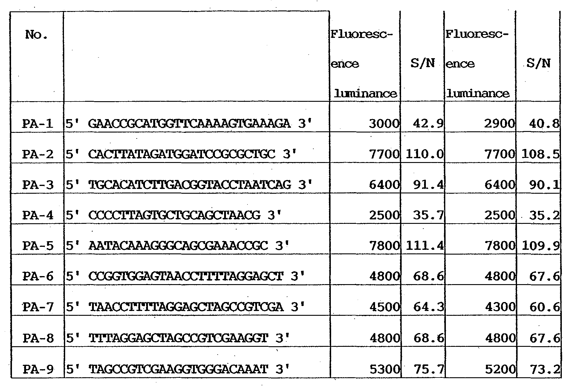
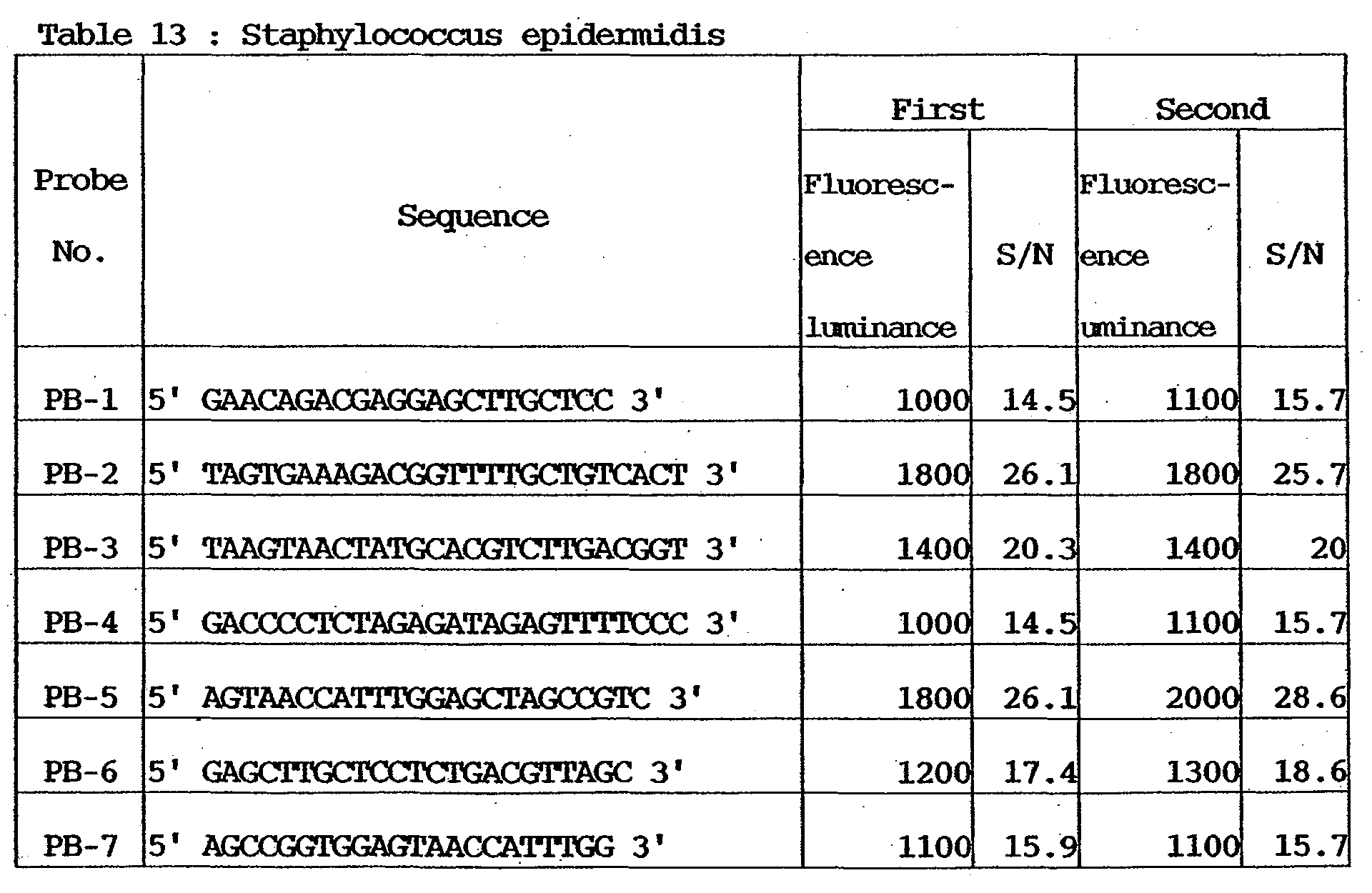

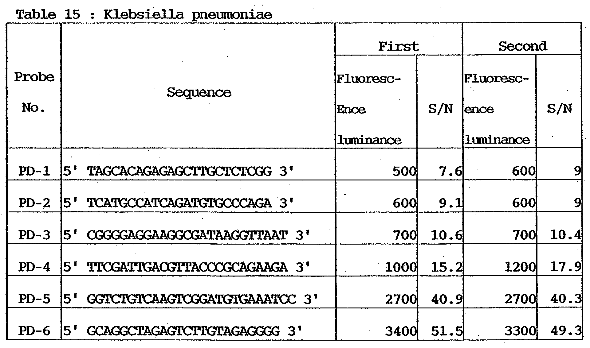


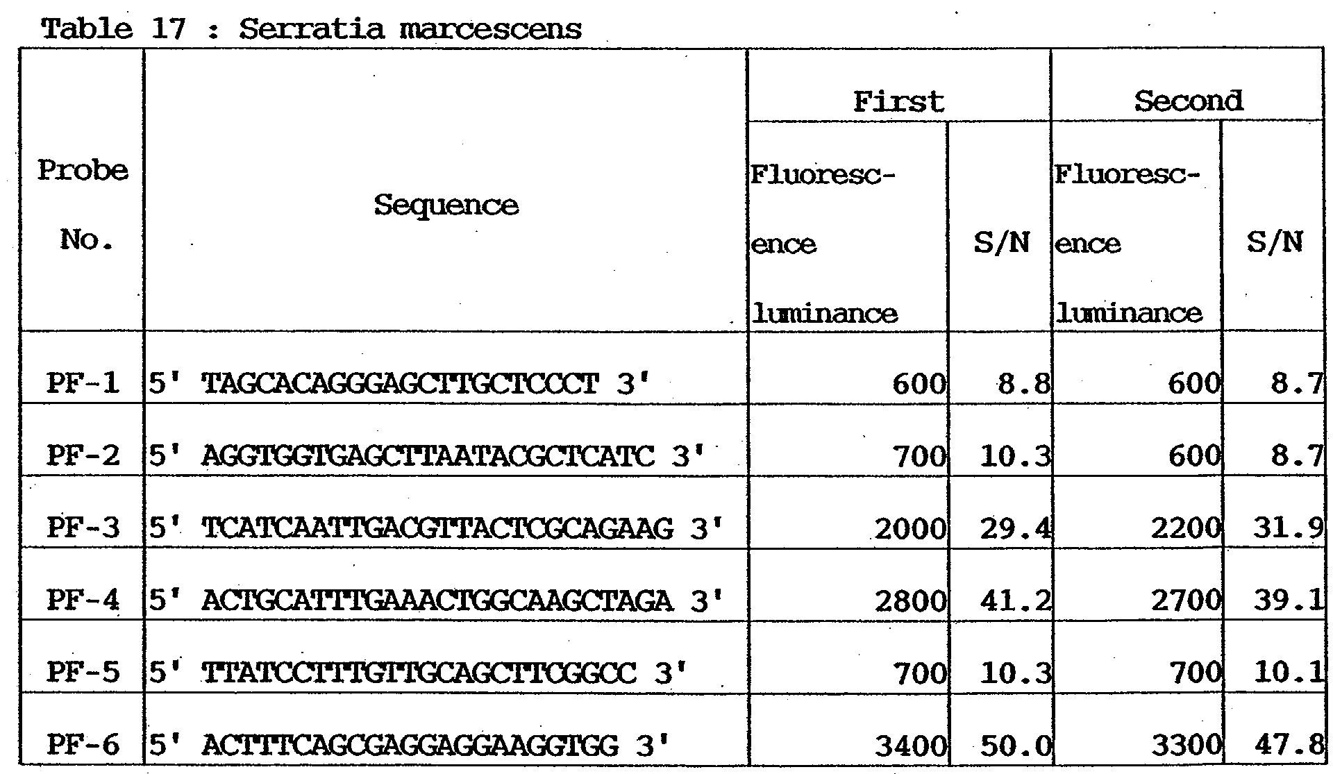


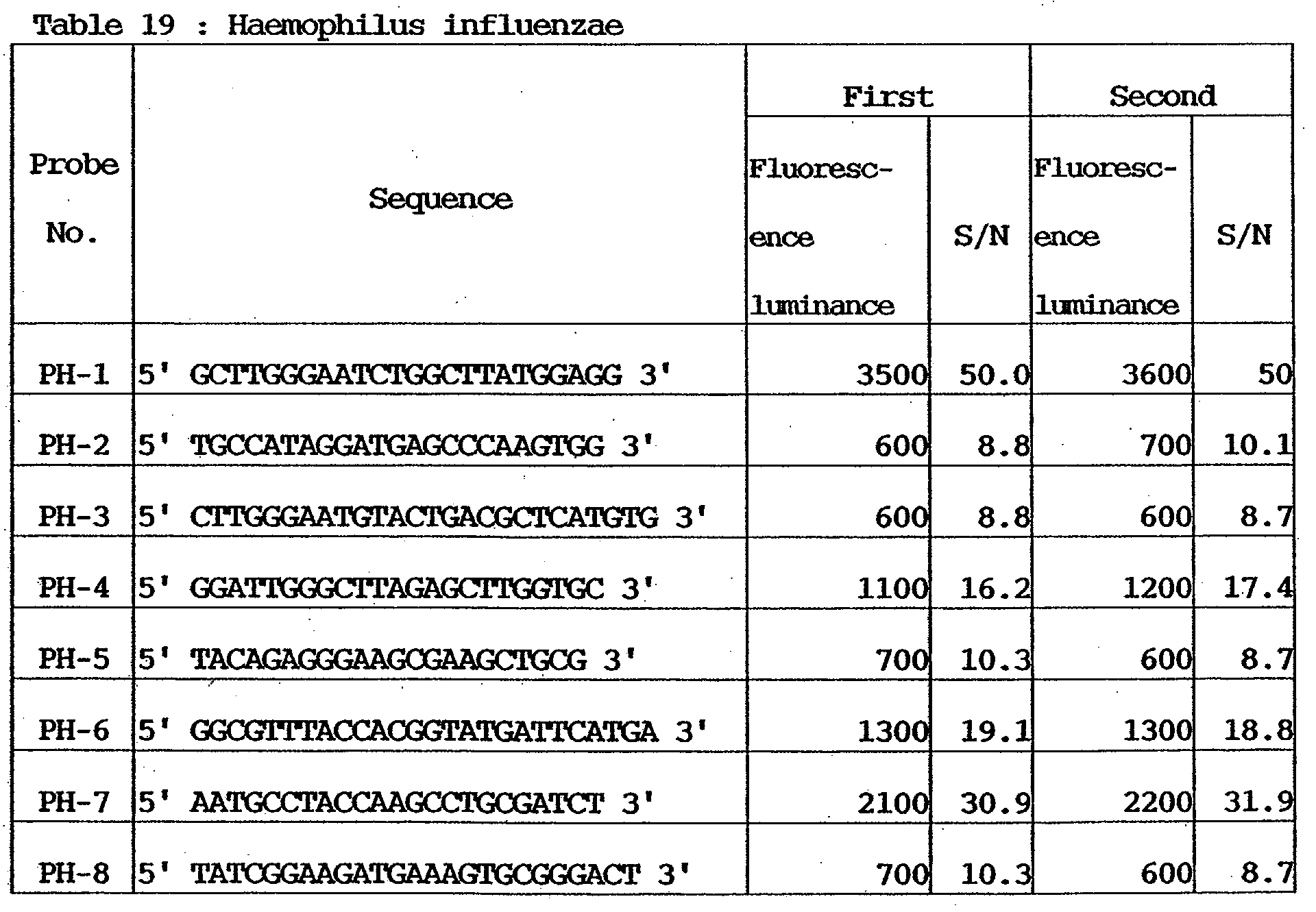
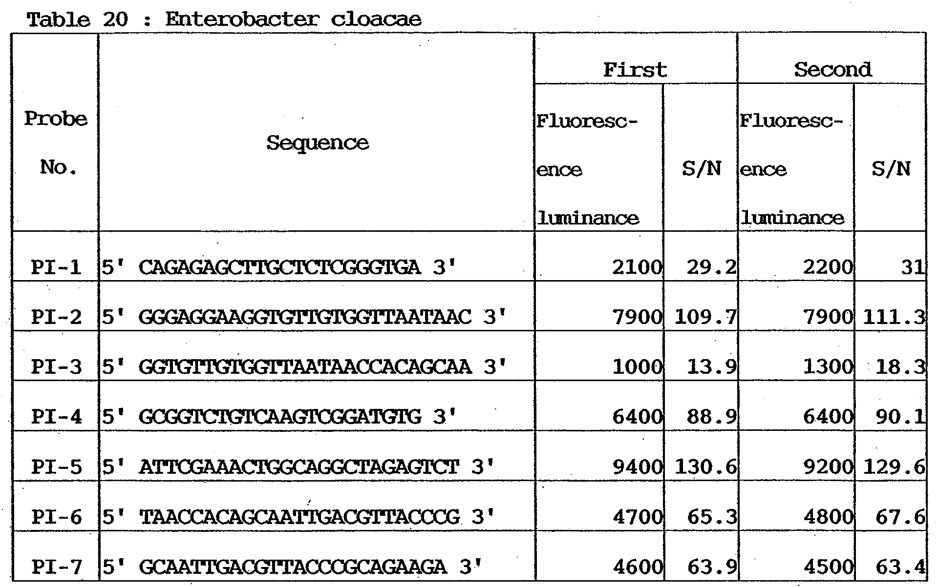

- The numerical values of florescent luminances(photomultiplier voltage: 400 V) in Tables 12 to 21 indicate average pixel luminances (resolution: 5 u m). The S/N ratios indicate values obtained by dividing the fluorescent luminances by background average values measured by analysis software (GenePix Pro Ver.3.0 available from Axon) attached to the measuring device.
- As is apparent from Tables 12 to 21, the respective etiologic agents can be detected with sufficient signals at a high reproducibility.
- As in Example 1, probe DNAs, specimen amplification PCR primers, the genome DNAs of etiologic agents, and DNA microarrays were prepared, and the following experiments were conducted.
- Amplification of microbial genes as specimens (1st PCR) and labeling (2nd PCR) reaction will be described below.
-
Premix PCR reagent (TAKARA ExTaq) 25 µl Template Genome DNA 2 µl (10ng) Forward Primer mix 2µl (20pmol/tube each) Reverse Primer mix 2 µl (20pmol/tube each) H20 19 µl Total 50 µl - Amplification reaction of the reaction solution having the above composition was caused by using a commercially available thermal cycler in accordance with the following protocol.

- After the end of reaction, purification was performed by using a purification column (QIAquick PCR Purification Kit available from QIAGEN). Then, determination of the amplified products was executed.
-
(QIAGEN Hotstar Taq Polymerase) 0.5 µl (2.5u) Template DNA (1st PCR Product) 10 µl ( 30ng) dNTP mix (Low dTTP ) 2 µl Cy- 3 DUTP (1mM) 2 µl (2nml/tube) Reverse Primer mix 5 µl (50gmol/tube each) 10 x Buffer 5 µl H2O 25.5 µl Total 50 µl - Amplification reaction of the reaction solution having the above composition was caused by using a commercially available thermal cycler in accordance with the following protocol.

- After the end of reaction, purification was performed by using a purification column (QIAquick PCR Purification Kit available from QIAGEN) to obtain labeled specimens.
- Hybridization was done in accordance with the same procedures as in Example 1.
- Fluorometry was executed for the DNA microarrays after the end of hybridization reaction by using a DNA microarray fluorescent detector (GenePix 4000B available from Axon). Tables 22 to 31 show the measurement results.
- Even in this example, fluorometry was executed twice for each DNA microarray. The results are shown in Tables 22 to 31.



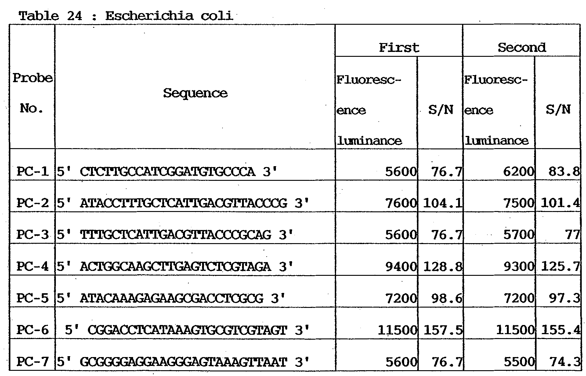


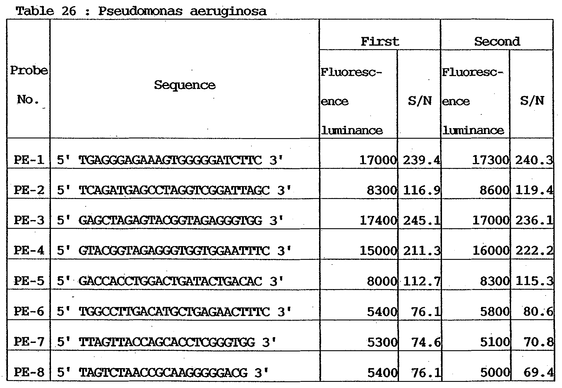


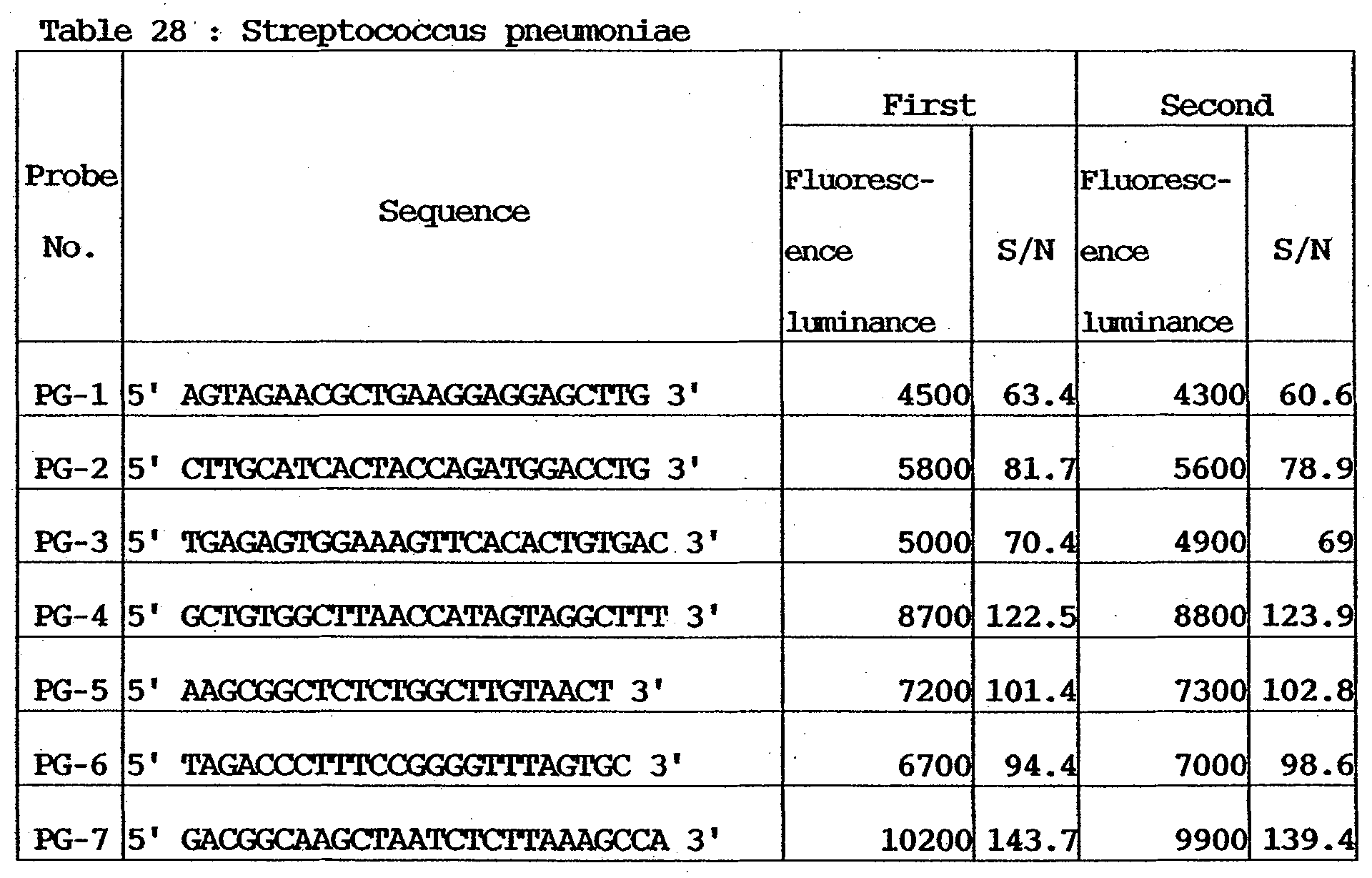

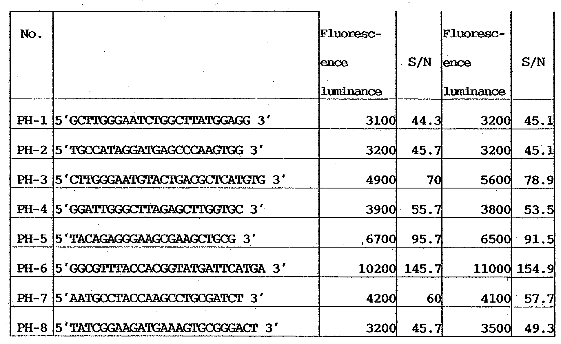
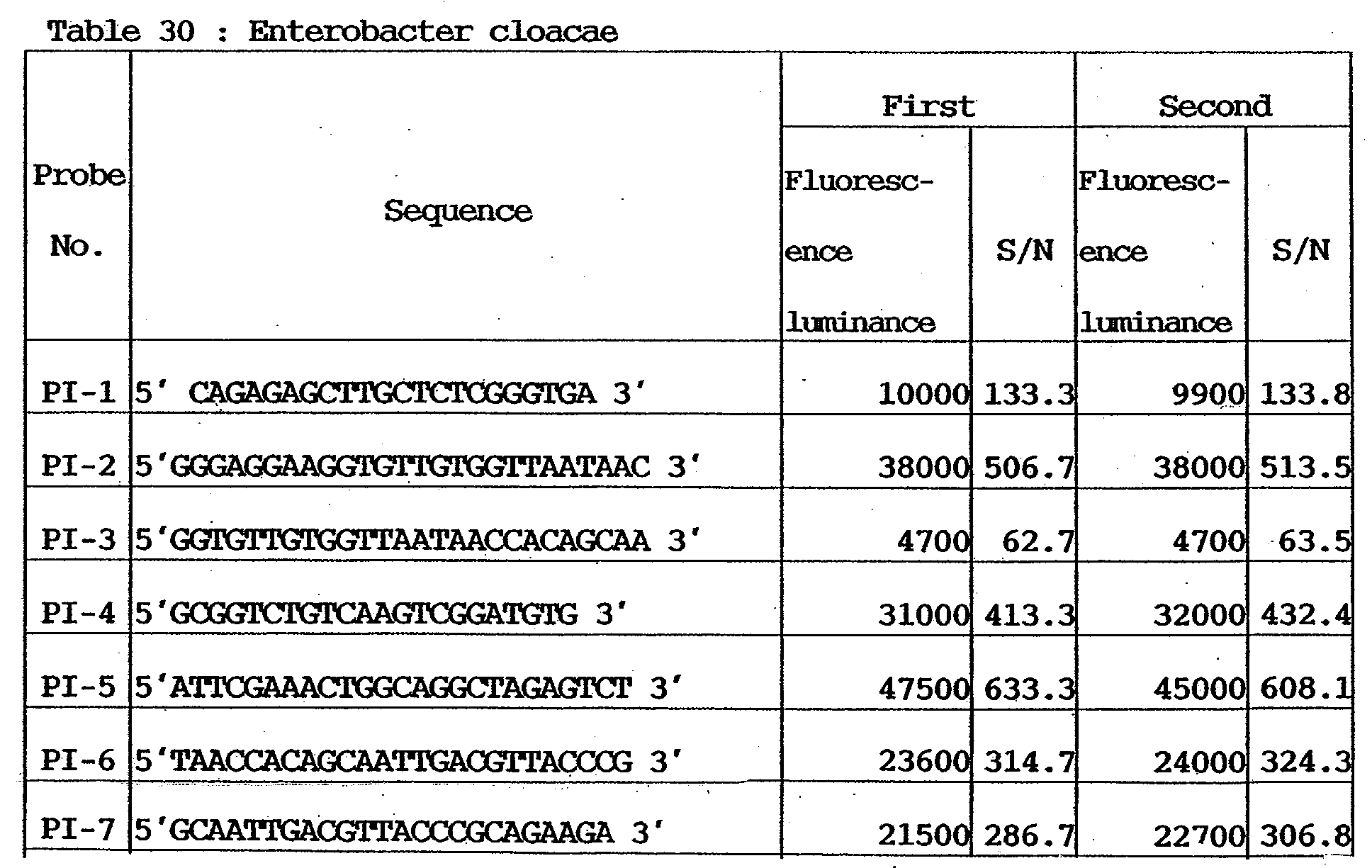

- The numerical values of florescent luminances(photomultiplier voltage: 400 V) in Tables 22 to 31 indicate average pixel luminances (resolution: 5 µm). The S/N ratios indicate values obtained by dividing the fluorescent luminances by background average values measured by analysis software (GenePix Pro Ver.3.0 available from Axon) attached to the measuring device.
- As is apparent from Tables 22 to 31, the respective etiologic agents can be detected with sufficient signals at a high reproducibility.
- As in Examples 1 and 2, probe DNAs, specimen amplification PCR primers, the genome DNAs of etiologic agents, and DNA microarrays were prepared, and the following experiments were conducted.
- Amplification of microbial genes as specimens (1st PCR) and labeling (2nd PCR) reaction will be described below.
-
AmpliTaq Gold LD(5U/µL) 0.5µL Template DNA variable dNTP mis(2.5mM/each) 4.0µL x10 PCR buffer 5.0µL 25mM MgCl2 7.0µL Forward Primer Mix(10µM/each) 0.25µL Reverse Primer Mix(10µM/each) 0.25pL H2O variable Total 50µL - Amplification reaction of the reaction solution having the above composition was caused by using a commercially available thermal cycler in accordance with the following protocol.

- After the end of reaction, purification was performed by using a purification column (QIAquick PCR Purification Kit available from QIAGEN). Then, determination of the amplified products was executed.
-
Premix PCR reagent (TAKARA ExTaq) 25 µl Template DNA (1st PCR Product) Variable ( 30ng/tube ) Cy3 Labeled Reverse primer Mix 5 µl H2O Variable Total 50 µl - Amplification reaction of the reaction solution having the above composition was caused by using a commercially available thermal cycler in accordance with the following protocol.
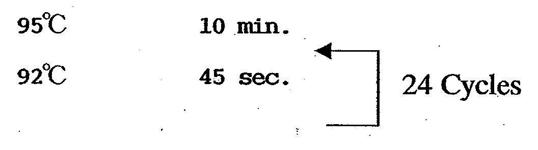

- After the end of reaction, purification was performed by using a purification column (QIAquick PCR Purification Kit available from QIAGEN) to obtain labeled specimens.
- Hybridization was done in accordance with the same procedures as in Example 1.
- Fluorometry was executed for the DNA microarrays after the end of hybridization reaction by using a DNA microarray fluorescent detector (GenePix 4000B available from Axon). Tables 32 to 41 show the measurement results.
- Note that, in this example, fluorometry was executed once twice for each DNA microarray. The results are shown in Tables 32 to 41.
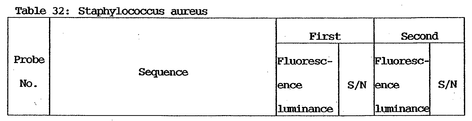
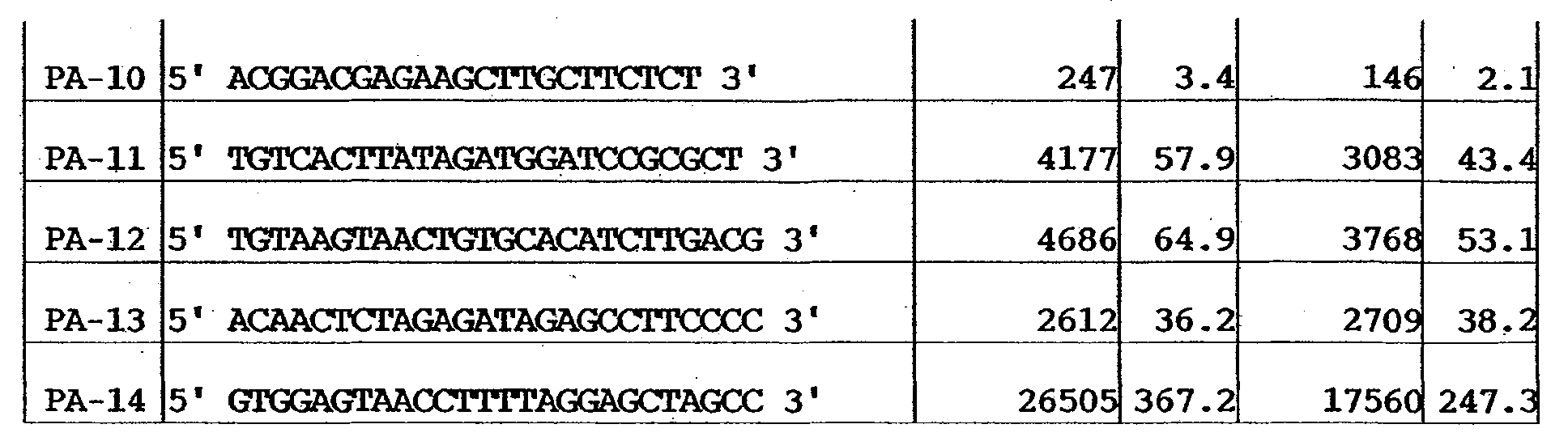

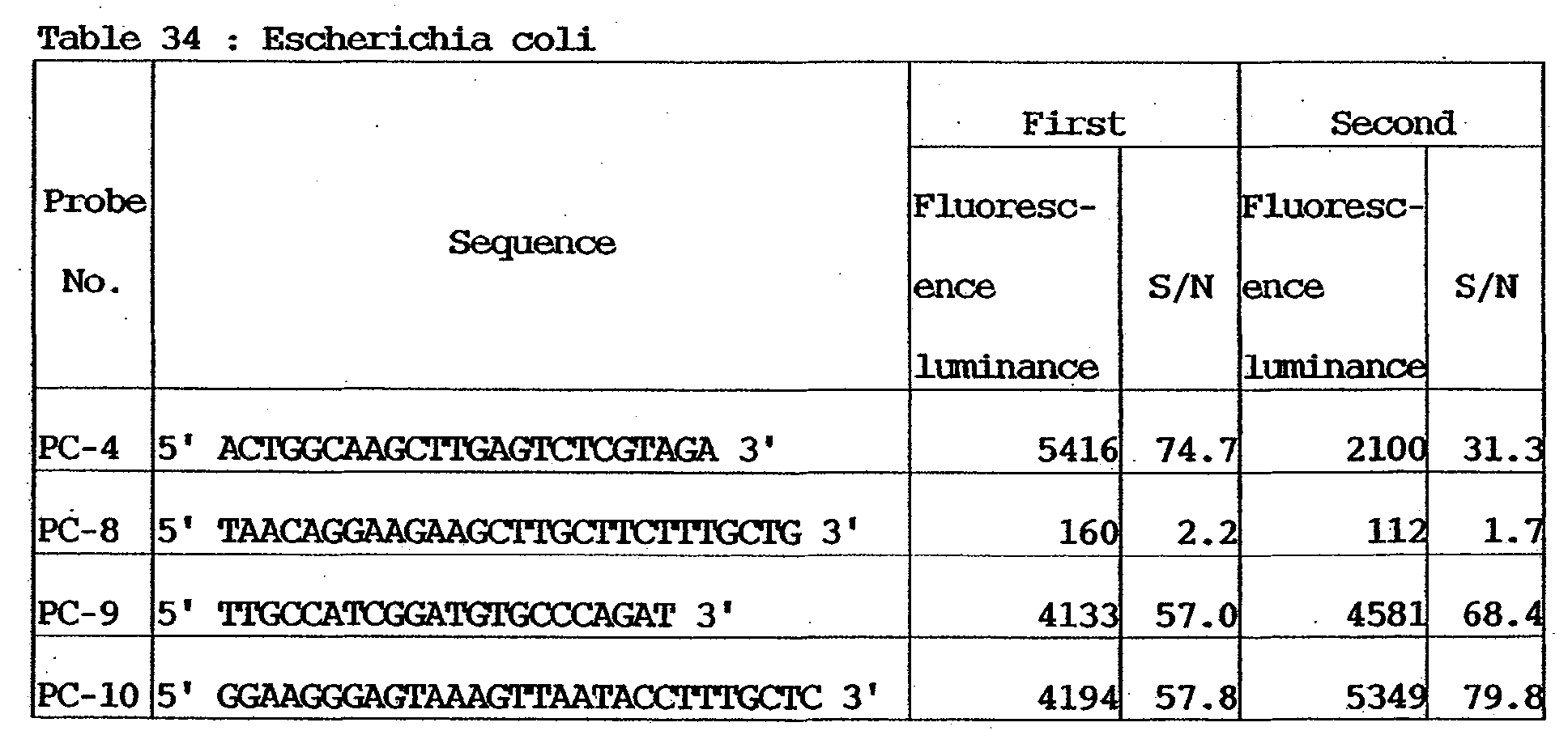

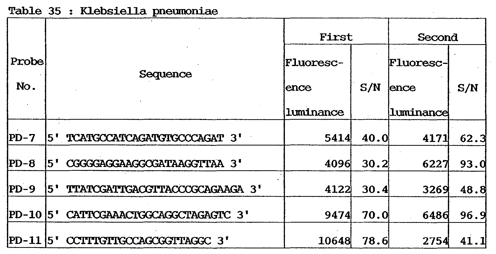
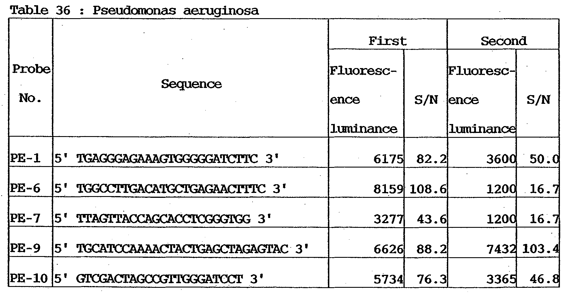
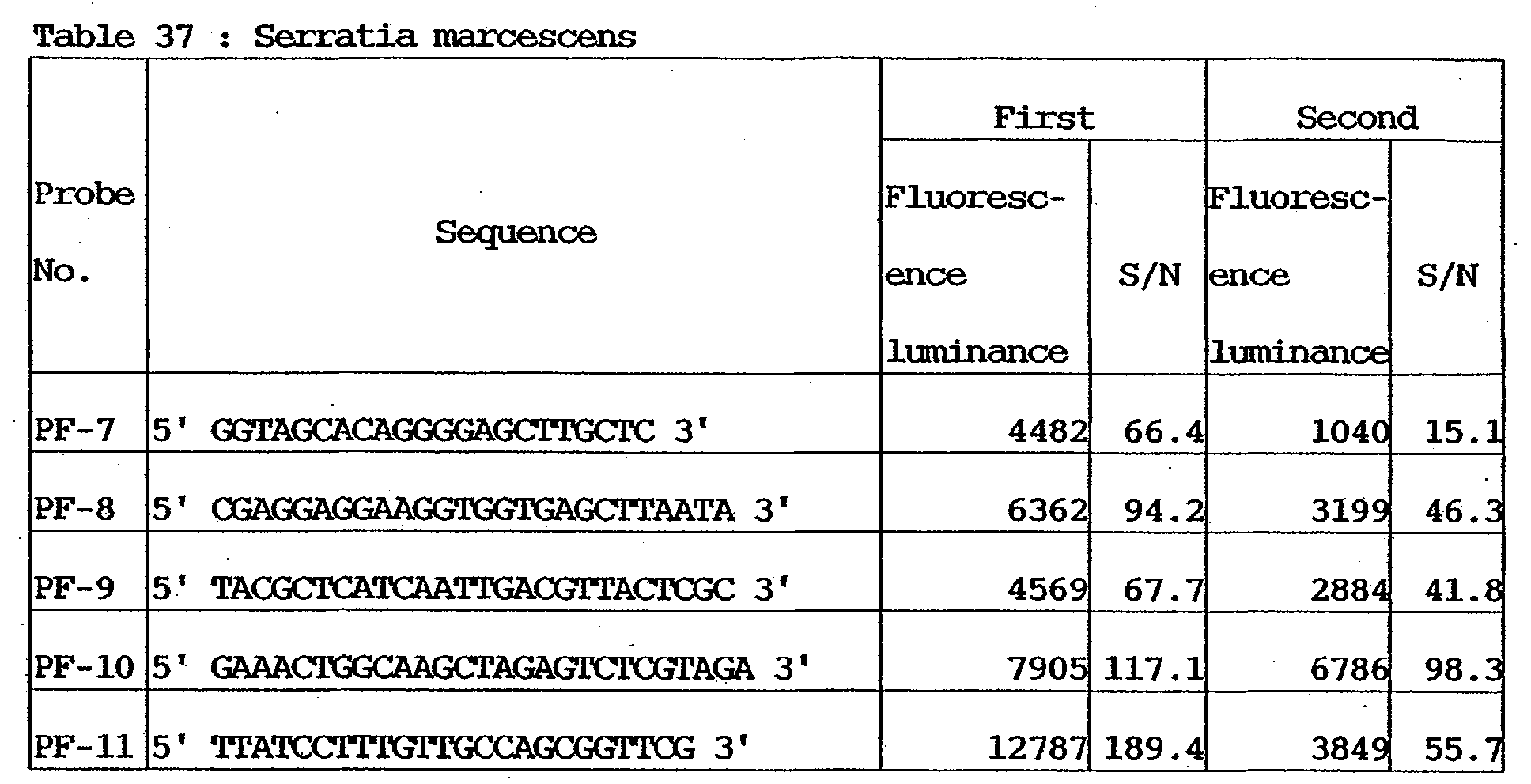
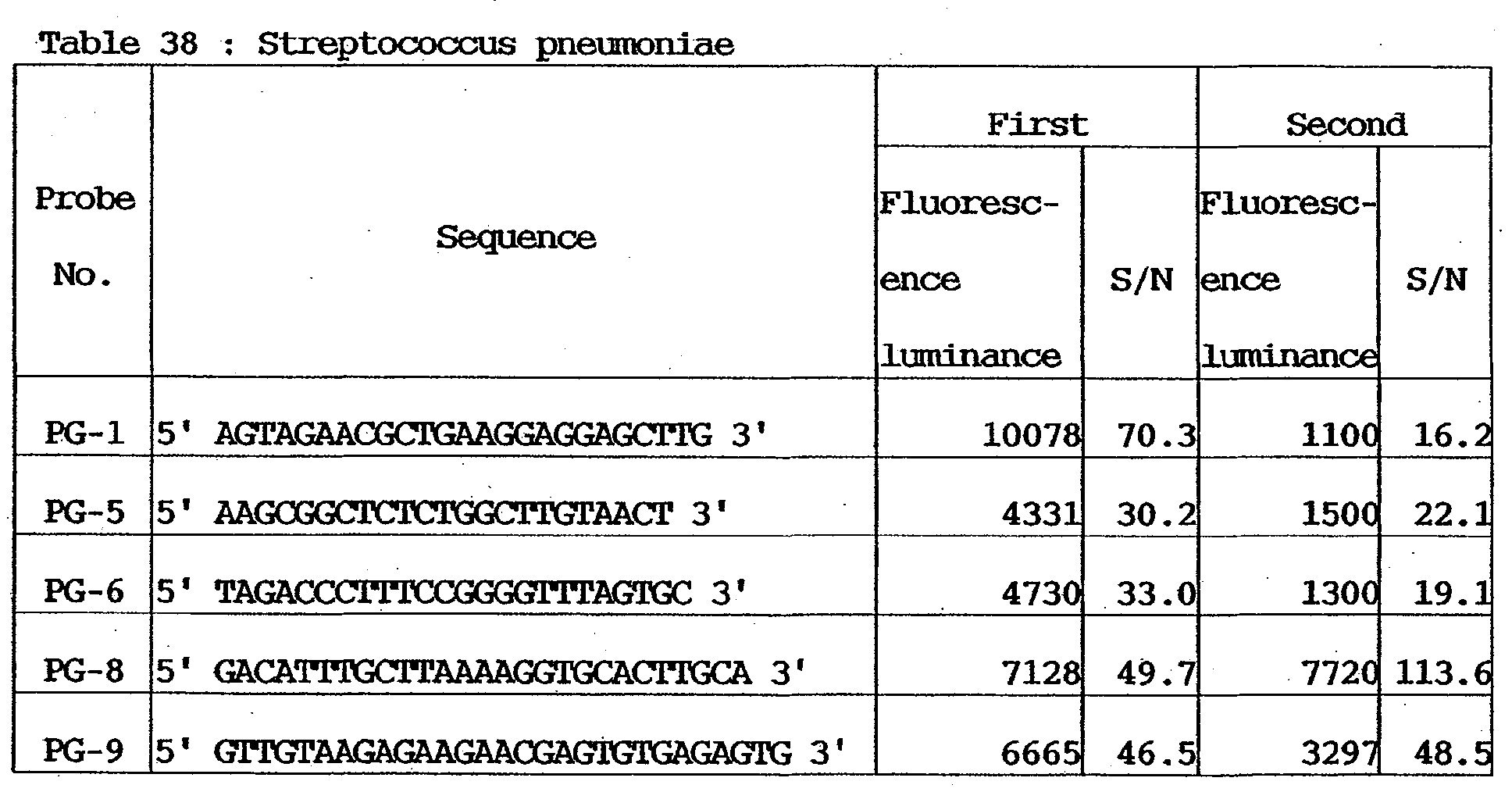

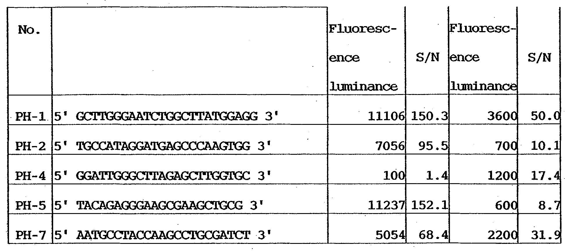


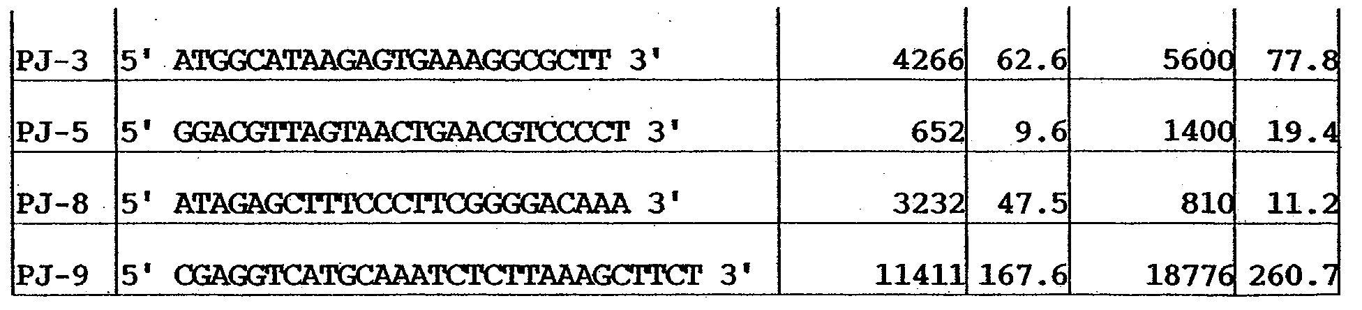
- As is apparent from Tables 32 to 41, the respective etiologic agents can be detected with sufficient signals at a high reproducibility.
- As described above, according to the examples, an infectious etiologic agent can be identified by using microarrays on which probe sets capable of detecting the 10 bacteria, i.e., Staphylococcus aureus, Staphylococcus epidermidis, Escherichia coli, Klebsiella pneumoniae, Pseudomonas aeruginosa, Serratia marcescens, Streptococcus pneumoniae, Haemophilus influenzae, Enterobacter cloacae, and Enterococcus faecalis are separately immobilized or combined. Hence, the problems of the DNA probe of microbial origin are solved. More specifically, because of the small number of bases, oligonucleotide probes can chemically be mass-produced, and purification and concentration control are possible. In addition, a probe set can be provided, which allows to detect bacteria in the same species all together and discriminatingly detect bacteria in other species for the purpose of classifying the bacteria depending on the species.
- Furthermore, a probe set can be provided, which also considers the stability of a hybrid body between a probe and a specimen so that the difference between the species can accurately be evaluated on a DNA microarray. A carrier on which the probe DNAs are immobilized to make the probe DNAs react with specimens can also be provided. Also, a carrier can be provided, on which the probe DNAs are chemically immobilized so that the probe DNAs are stably immobilized on the carrier, and a detection result with high reproducibility can be obtained in the process of reaction between a specimen solution and the probes and probe sets.
- According to the above examples, 16s rRNA gene arrangements in the genes of infectious etiologic agents can be detected in proper quantities. Hence, the presence of an infectious etiologic agent can efficiently and accurately be determined.
- The primer sets (Table 11) used in the above examples to amplify the 16s rRNA gene arrangements of one or some of Staphylococcus aureus, Staphylococcus epidermidis, Escherichia coli, Klebsiella pneumoniae, Pseudomonas aeruginosa, Serratia marcescens, Streptococcus pneumoniae, Haemophilus influenzae, Enterobacter cloacae, and Enterococcus faecalis will be described.
- The primer sets of this example are designed to give a satisfactory amplification result in PCR reaction executed to identify an infectious etiologic agent. "Satisfactory" means not only that the target 16s rRNAs are sufficiently amplified but also that no products other than the 16s rRNAs are generated.
- "Satisfactory" also means that only the 16s rRNAs of an infectious etiologic agent are amplified without amplifying human genome genes originated in a specimen, which are contained in the specimen.
- Any specimen in which bacteria may be present, and for example, body fluids originated in animals such as human and livestock, including blood, spinal fluid, phlegm, stomach fluid, vaginal discharge, and intraoral mucus, and excretion such as urine and feces are used in this example. All media which can be contaminated by bacteria can also be used in this example, including food , drink water and hot spring water in the natulal environment, which may cause food poisoning by contamination, filters from air and water cleaners , and so forth. Animals and plants which should be quarantined in import/export are also used as specimens.
- The PCR reaction used in this example includes PCR reaction which uses an extracted nucleic acid itself as a template, asymmetrical PCR reaction which uses primers on one side of SEQ ID Nos. 107 to 109 (F1 to F3 in Table 11) or SEQ ID Nos. 110 to 112 (R1 to R3 in Table 11), and PCR which executes labeling for visualization.
- As 16s rRNA gene (target gene) amplification PCR primers for etiologic agent detection, nucleic acid sequences shown in Table 11 were designed.
- More specifically, probe sets which specifically amplify the genome parts coding the 16s rRNAs, i.e., primers for which the specific melting points were made uniform as much as possible at the two end portions of the 16s rRNA coding region of a base length of 1,500 were designed. In order to simultaneously amplify variants or a plurality of 16s rRNA coding regions on genomes, a plurality of kinds of primers were designed.
- The primers shown in Table 11 were purified by HPLC (High Performance Liquid Chromatography) after synthesis. All of three forward primers and three reverse primers were mixed and dissolved in a TE buffer solution such that each primer concentration had an ultimate concentration of 10 pmol/µl. In this example, all the forward primers and reverse primers were used. Alternatively, one to three forward primers and one to three reverse primers may be used.
- By using a thus prepared solution of forward primers and reverse primers (forward primer mix and reverse primer mix), genome DNAs extracted by the method described in [3. Extraction of Genome DNAs (Model Specimens) of Etiologic Agents] were amplified by the method described in [5. Amplification and Labeling of Specimens (PCR Amplification & Fluorescent Labeling)].
- After the end of reaction, the primers were removed by using a purification column (QIAquick PCR Purification Kit available from QIAGEN). Then, the amplified products were examined by gel electrophoresis. One band was detected in 1,500 base pair regions, and it was confirmed that satisfactory PCR reaction was executed. No byproducts were generated.
- When the primers shown in Table 11 were used, satisfactory PCR amplification results were obtained in, e.g., all of the above-described 10 infectious etiologic agents (Staphylococcus aureus, Staphylococcus epidermidis, Escherichia coli, Klebsiella pneumoniae, Pseudomonas aeruginosa, Serratia marcescens, Streptococcus pneumoniae, Haemophilus influenzae, Enterobacter cloacae, and Enterococcus faecalis).
- Bacteremia model systems were prepared by adding 103 104, and 105 Enterobacter cloacae, which was cultured in accordance with the procedures described in Example 1, to 200-µl human blood (collected EDTA blood). An N-acetyl muramidase solution (0.2 mg/ml in Enzyme Buffer) was added to each solution. The solutions were heated to 37°C for 30 min. After that, DNAs were extracted by using Qiamp Blood mini Kit (available from QIAGEN) to prepare templates for PCR reaction.
- PCR reaction was caused for these DNAs by using the primers shown in Table 11, as in Example 4.
- As a result, as in Example 4, one band was detected in 1,500 base pair regions, and it was confirmed that satisfactory PCR reaction was executed. No byproducts were generated. The amount of PCR amplified products obtained from the band was proportional to the added cell amount. This indicates that when the primer sets were used, only the 16s rRNAs of Enterobacter cloacae were amplified without generating any PCR byproduct of human genome.
- As described above, according to this example, the 16s rRNA parts in the genes of a plurality of kinds of infectious etiologic agents can efficiently be amplified at a high purity. In addition, even when human genome DNAs are present, only the 16s rRNAs of an infectious etiologic agent can efficiently be amplified.
- As has been described above, according to the present invention, an infection detection probe which allows mass preparation at a time and identification of a species in similar species can be provided. More specifically, an infection detection probe which can suitably be used to classify a plurality of kinds of etiologic agents of an infection on the basis of the species can be provided.
- Alternatively, an infection detection probe suitable for detection of, e.g., the above-described 10 bacteria as the etiologic agents of infections can be provided.
- A probe set can also be provided, which also considers the stability of a hybrid body between an infection detection probe and a specimen so that the difference between similar species can accurately be evaluated on a DNA chip.
- In addition, a carrier on which the infection detection probe is immobilized to make the infection detection probe react with the specimen can be provided.
- Furthermore, a carrier can be provided, on which the infection detection probes are chemically immobilized so that the infection detection probes are stably immobilized on the carrier, and a detection result with high reproducibility can be obtained in the process of reaction with a specimen solution.
- According to the present invention, there is also provided a PCR reaction primer which amplifies the 16s rRNAs of an etiologic agent in a specimen in order to detect and/or identify an infectious etiologic agent.
- According to the present invention, there is also provided a primer set which can commonly be used for a plurality of species and effectively amplify the 16s rRNAs of an etiologic agent even when the species is unknown.
- According to the present invention, there is also provided a primer set which can amplify the 16s rRNAs of a plurality of kinds of etiologic agents under the same PCR conditions.
- As many apparently widely different embodiments of the present invention can be made without departing from the spirit and scope thereof, it is to be understood that the invention is not limited to the specific embodiments thereof except as defined in the appended claims.
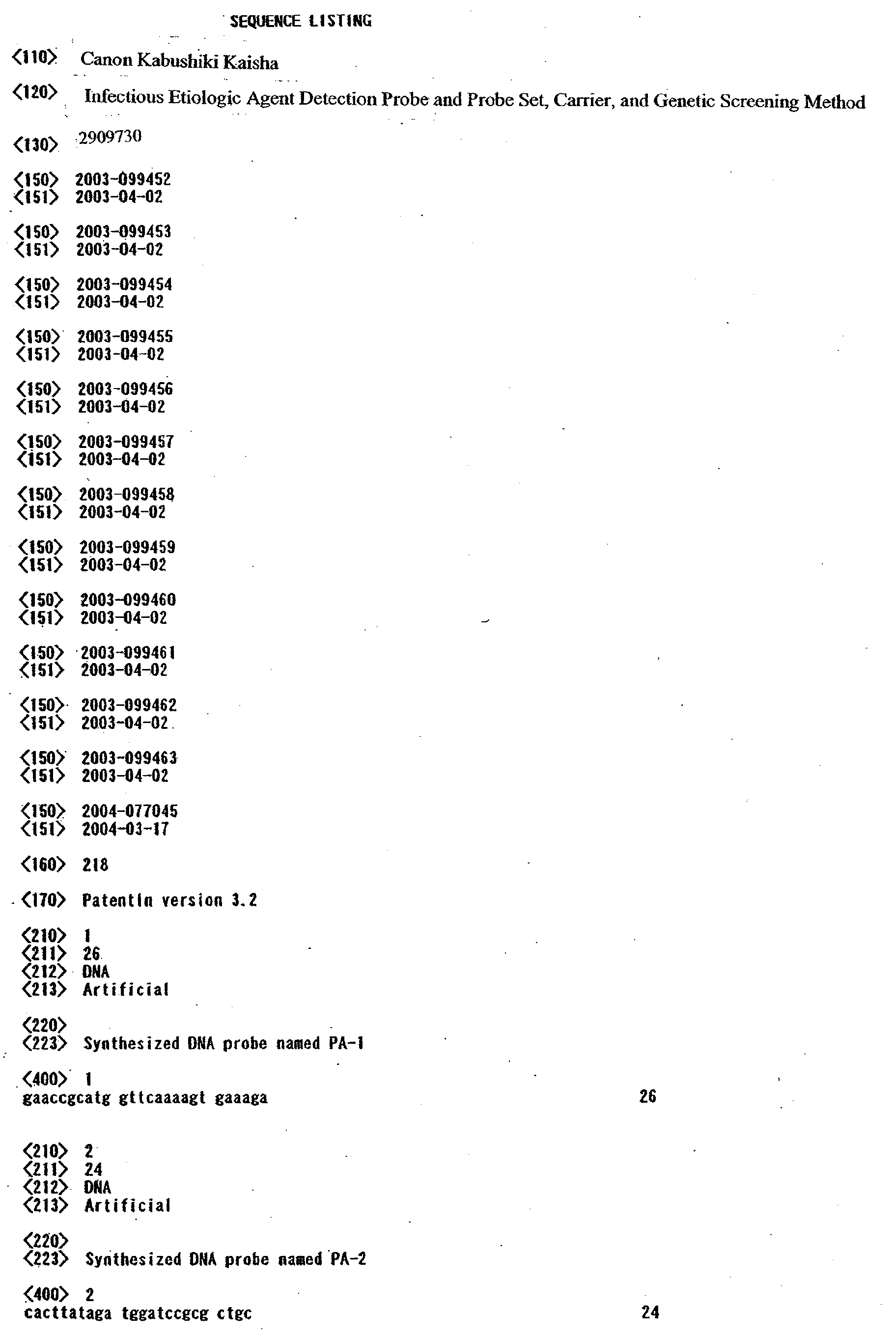


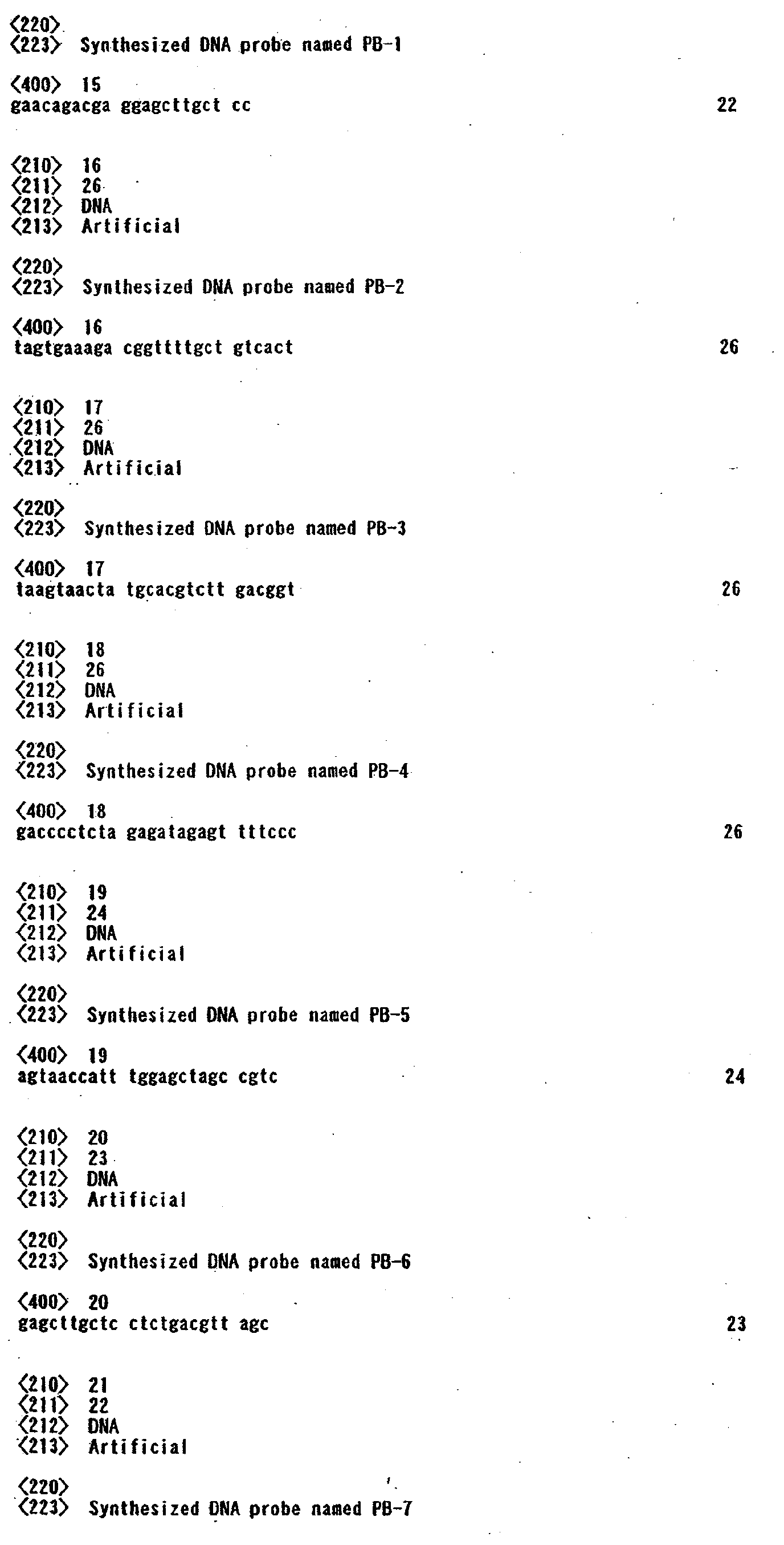


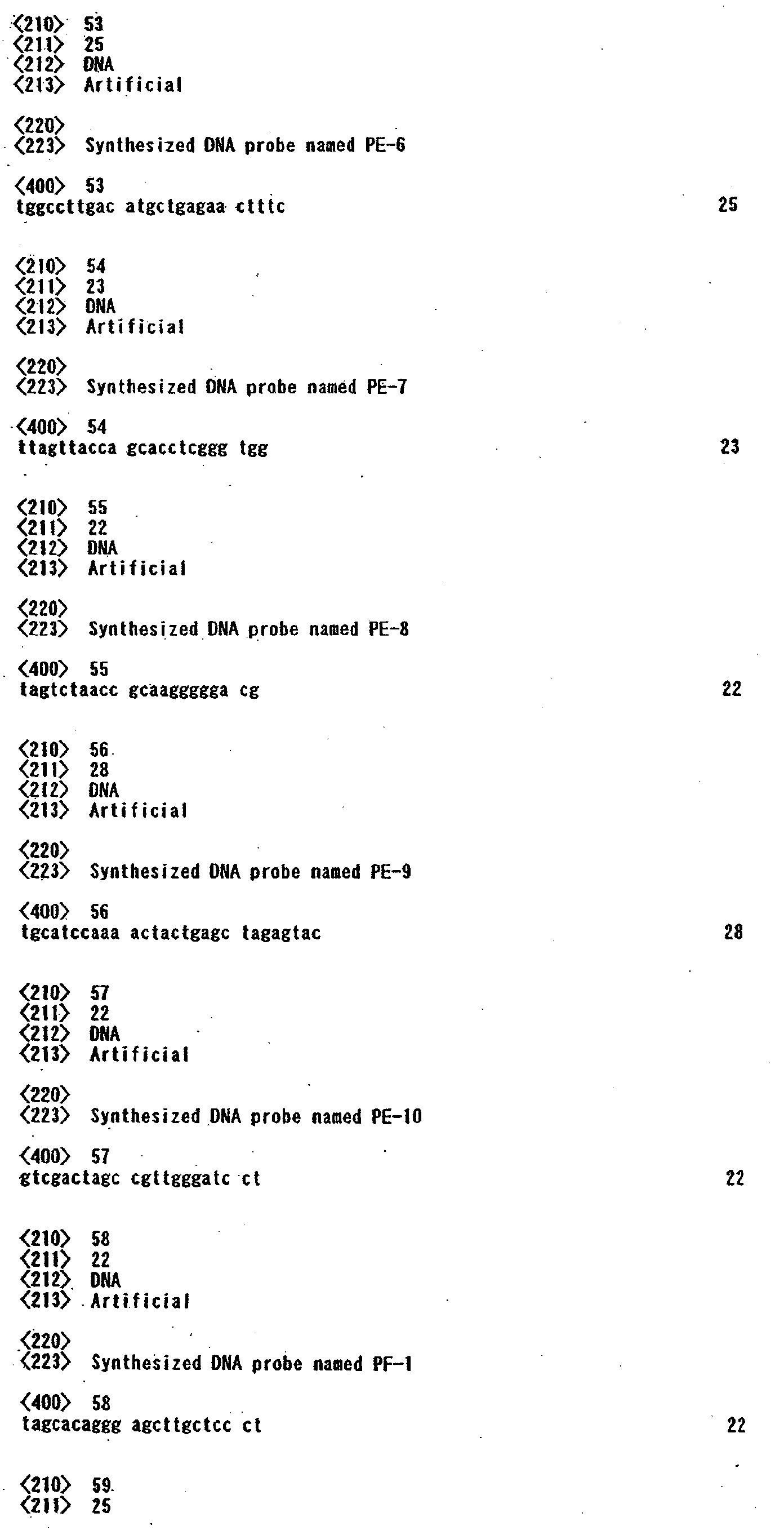


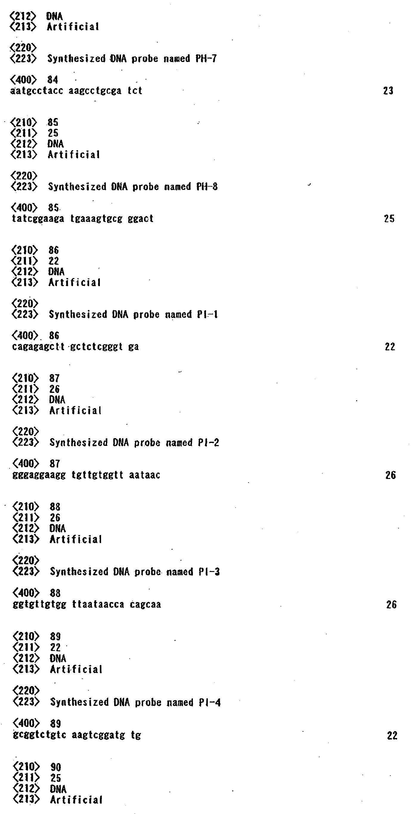
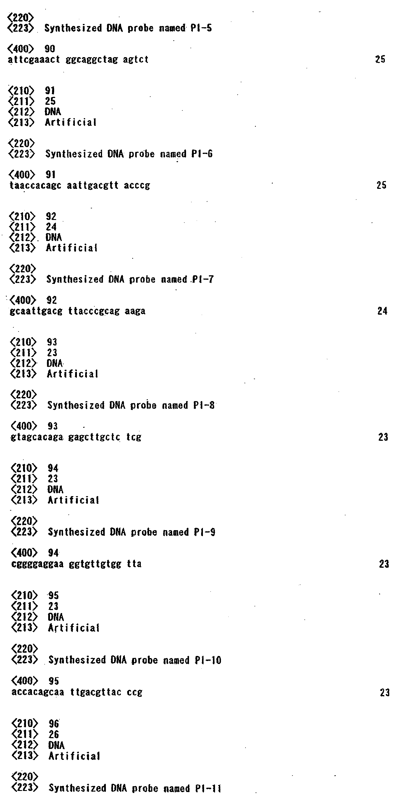
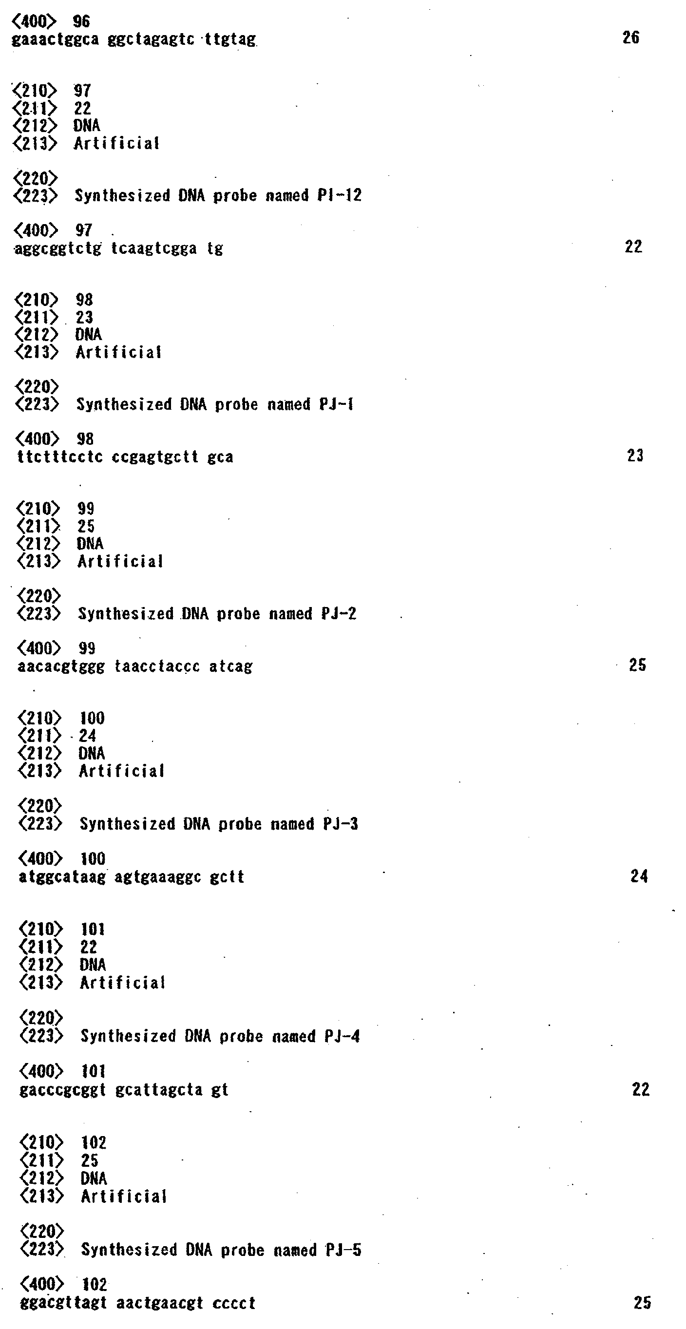


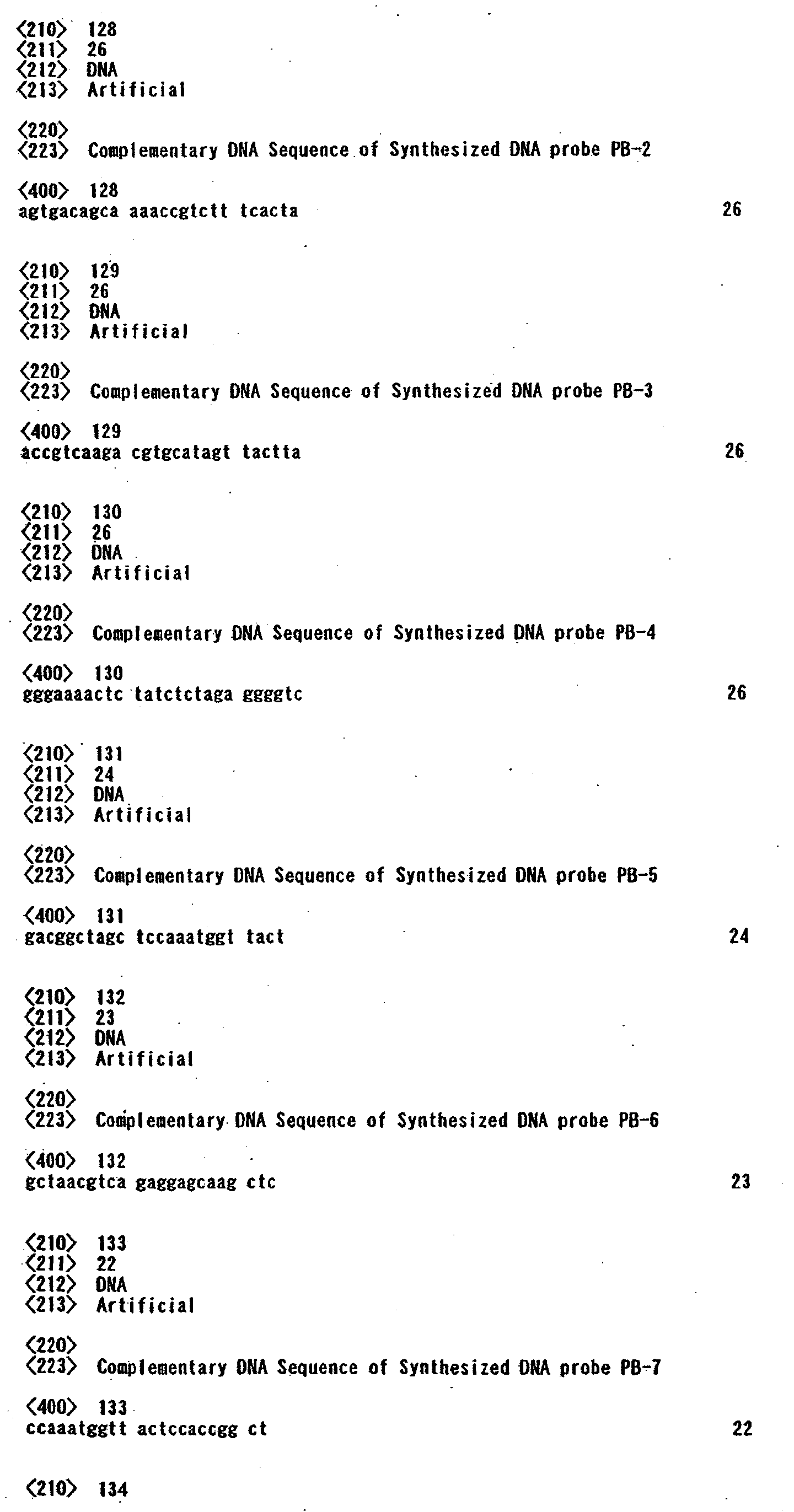

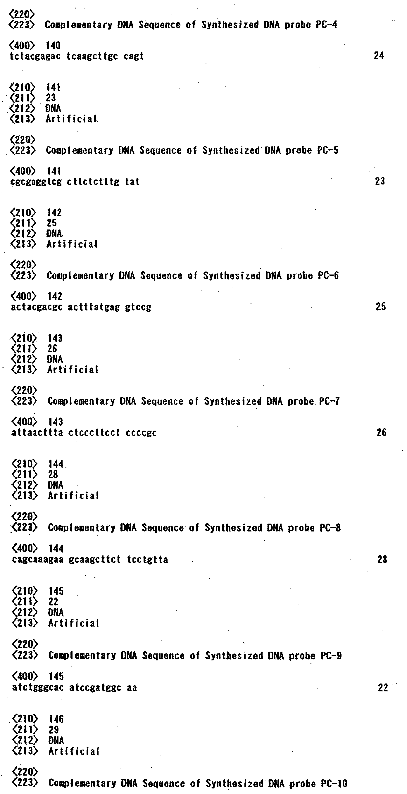
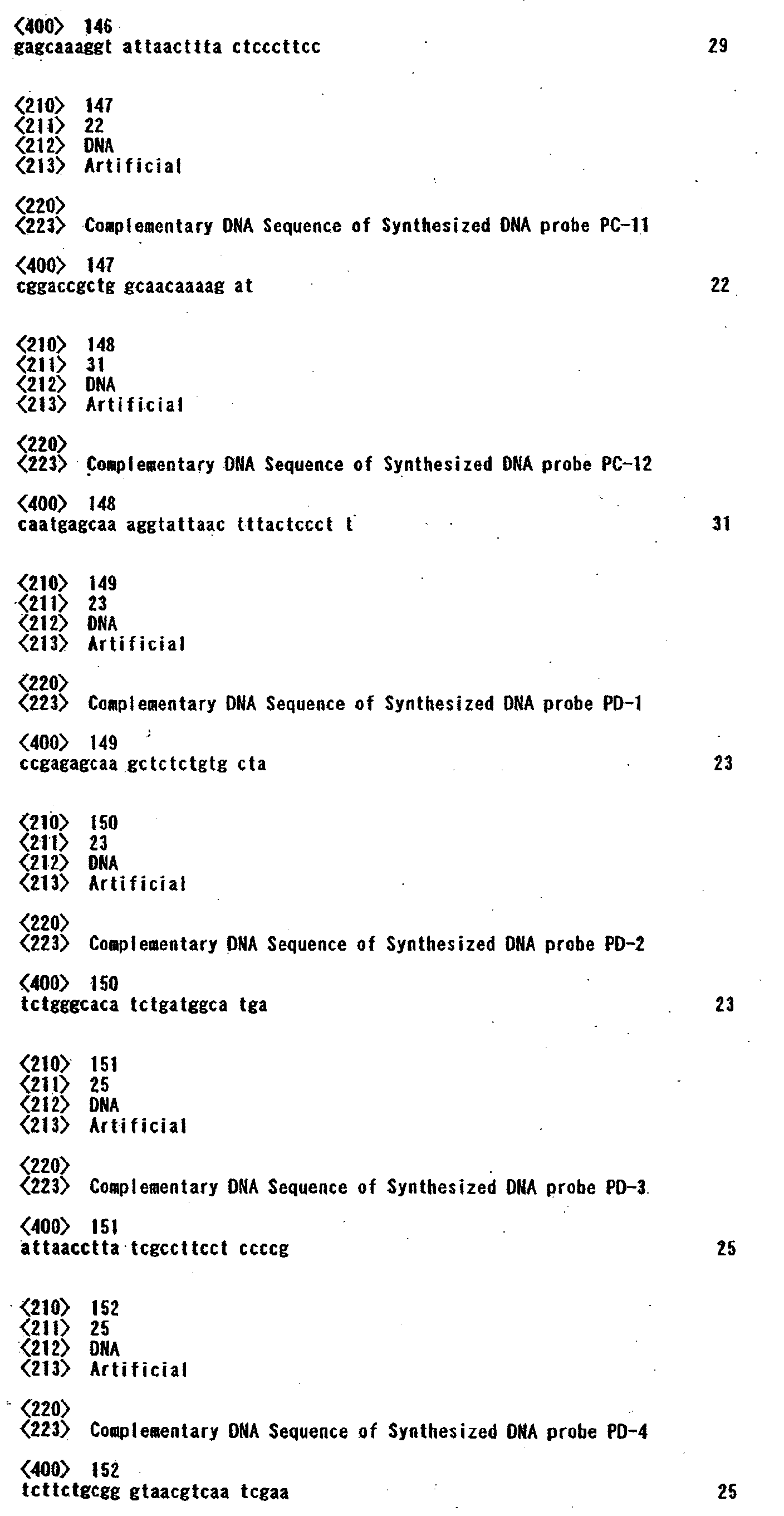


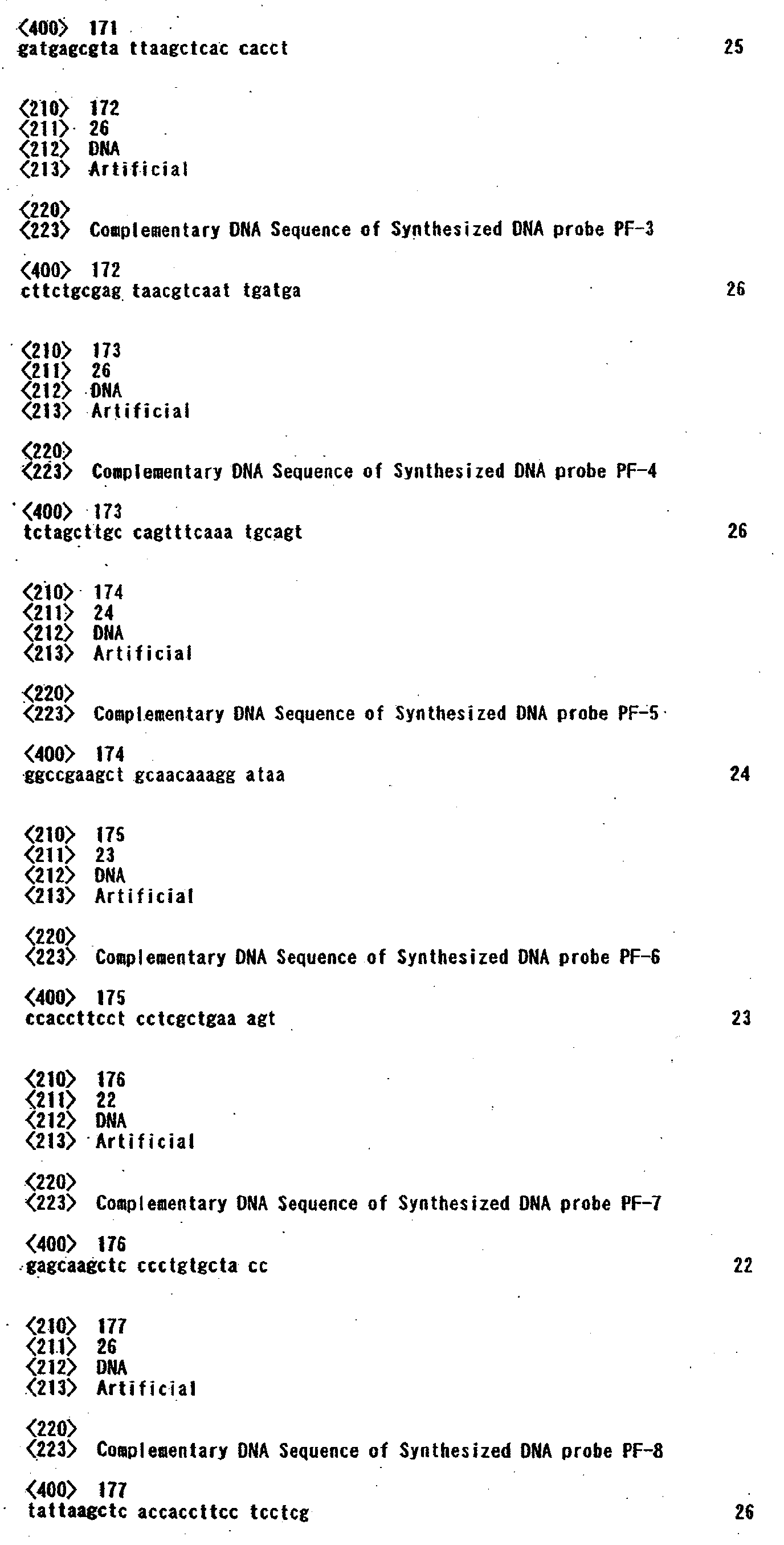

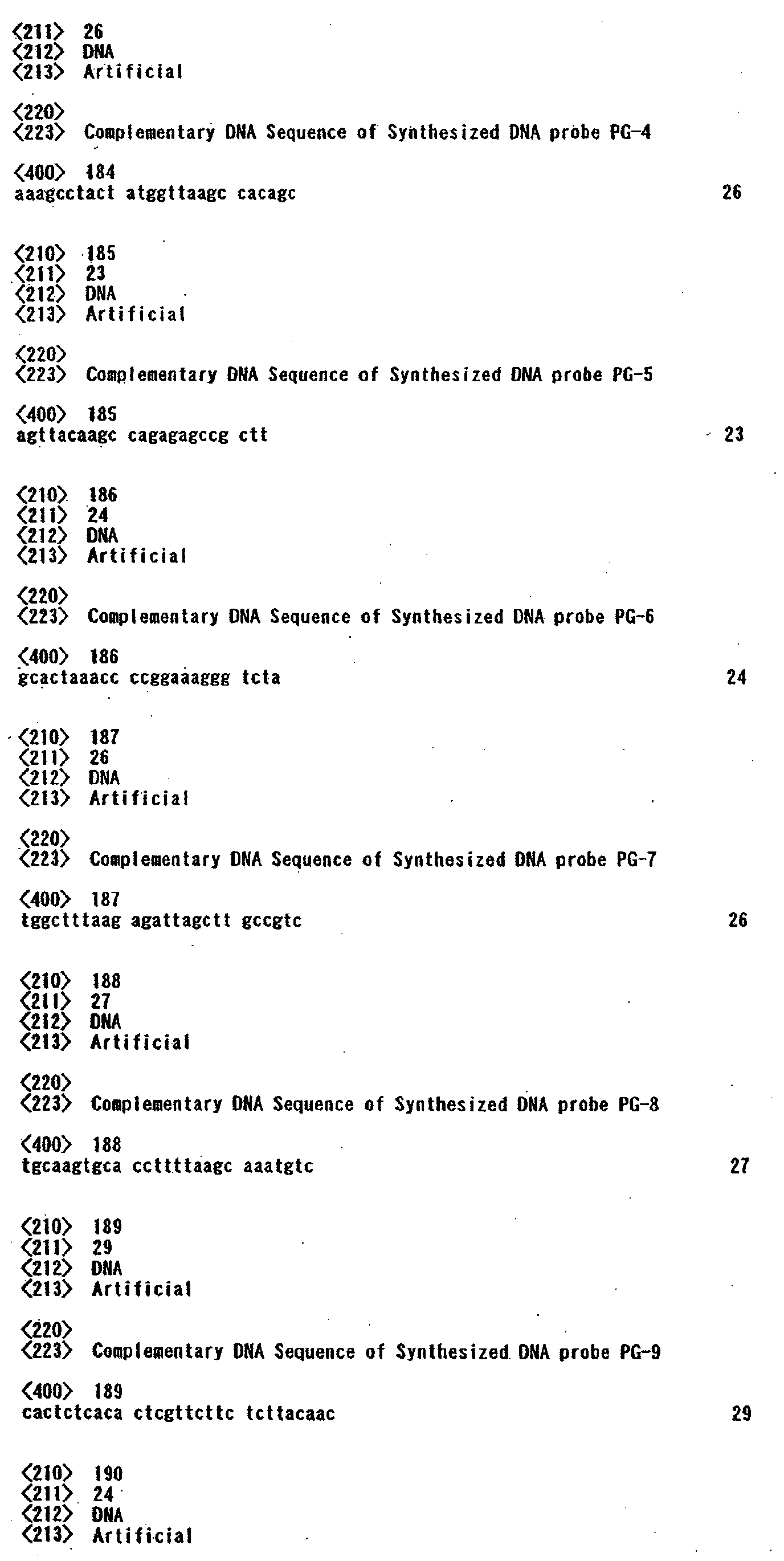
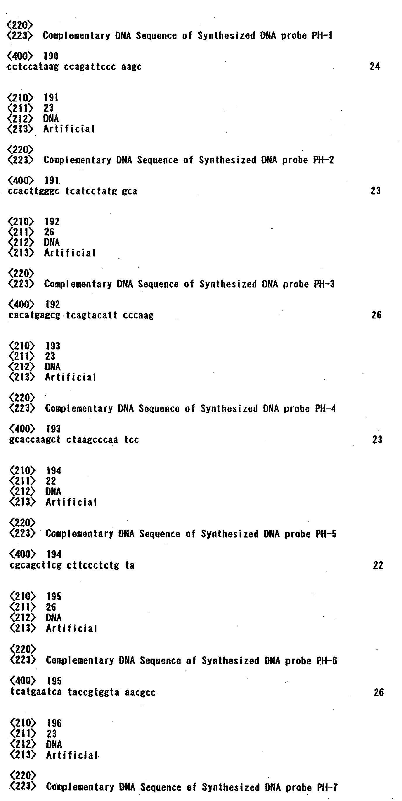
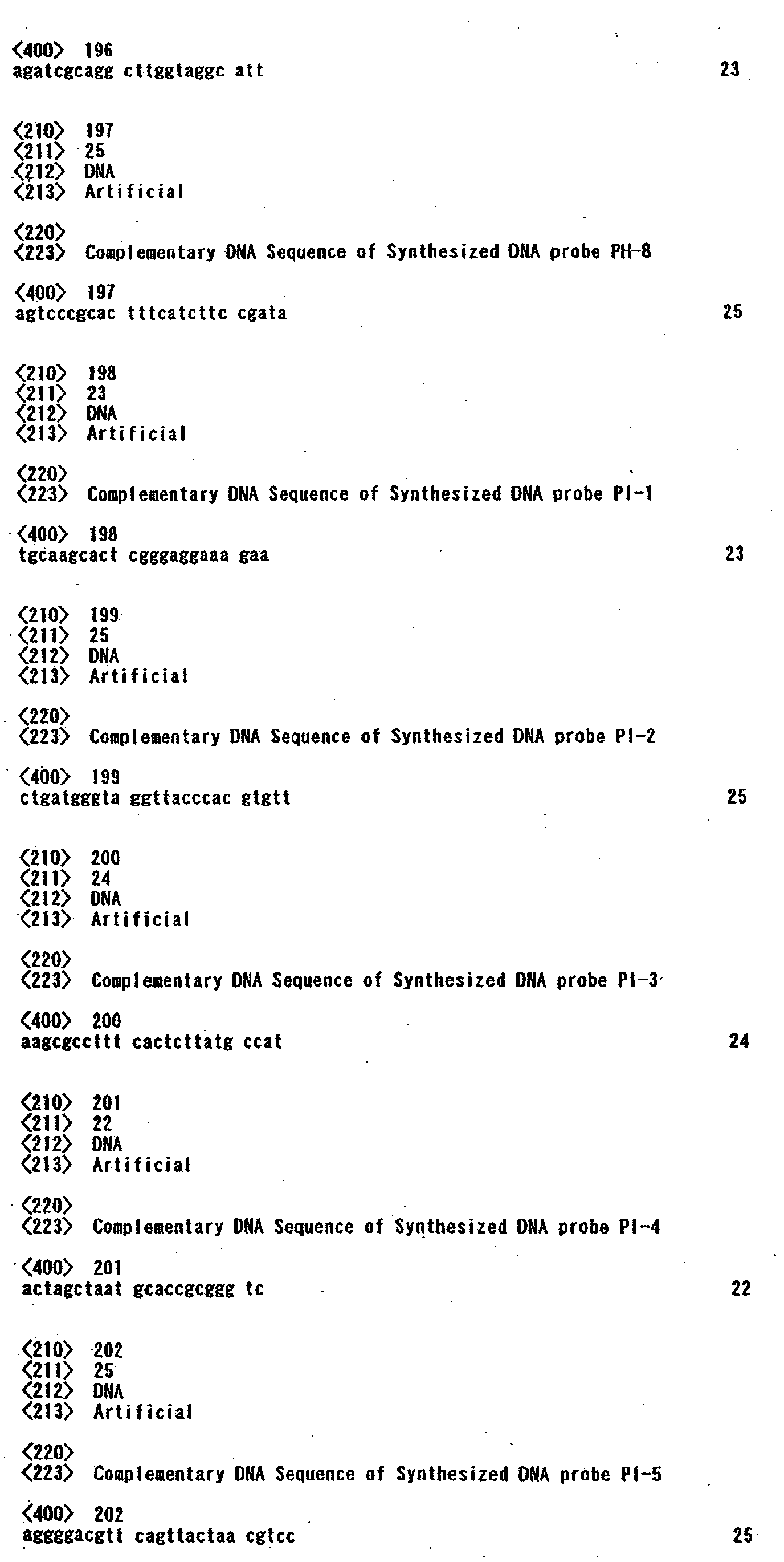
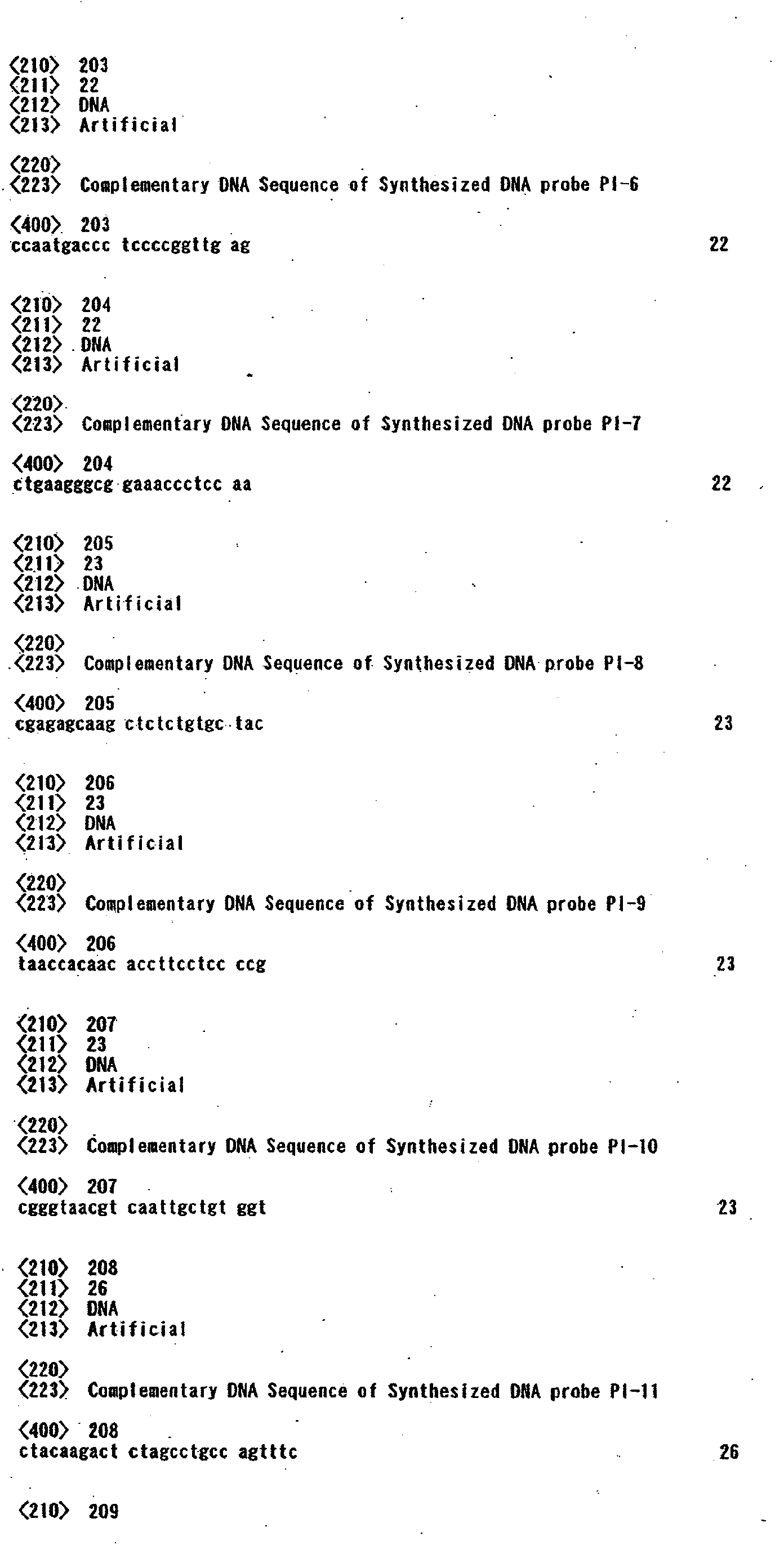


Claims (49)
- An infectious etiologic agent detection probe set which detects an infectious etiologic agent gene, comprising:a plurality of kinds of probes including oligonucleotide having base sequences selected from each of a plurality of groups selected from a first group including base sequences of SEQ ID Nos. 1 to 14 and complementary sequences thereof, a second group including base sequences of SEQ ID Nos. 15 to 24 and complementary sequences thereof, a third group including base sequences of SEQ ID Nos. 25 to 36 and complementary sequences thereof, a fourth group including base sequences of SEQ ID Nos. 37 to 47 and complementary sequences thereof, a fifth group including base sequences of SEQ ID Nos. 48 to 57 and complementary sequences thereof, a sixth group including base sequences of SEQ ID Nos. 58 to 68 and complementary sequences thereof, a seventh group including base sequences of SEQ ID Nos. 69 to 77 and complementary sequences thereof, an eighth group including base sequences of SEQ ID Nos. 78 to 85 and complementary sequences thereof, a ninth group including base sequences of SEQ ID Nos. 86 to 97 and complementary sequences thereof, and a 10th group including base sequences of SEQ ID Nos. 98 to 106 and complementary sequences thereof.
- A carrier on which a probe included in an infectious etiologic agent detection probe set of claim 1 is chemically immobilized.
- A genetic screening method of detecting an infectious etiologic agent gene by using a carrier of claim 2.
- An infection detection probe which can detect a gene originated in Staphylococcus aureus, which includes oligonucleotide having one of the base sequences of SEQ ID Nos. 1 to 14 and complementary sequences thereof.
- A probe set which can detect a gene originated in Staphylococcus aureus and includes at least one of infection detection probes each comprising oligonucleotide having one of the base sequences of SEQ ID Nos. 1 to 14 and complementary sequences thereof.
- A carrier on which an infection detection probe included in the probe set of claim 5 is chemically immobilized.
- A genetic screening method of detecting a gene originated in Staphylococcus aureus by using a carrier of claim 6.
- An infection detection probe which can detect a gene originated in Staphylococcus epidermidis, which includes oligonucleotide having one of the base sequences of SEQ ID Nos. 15 to 24 and complementary sequences thereof.
- A probe set which can detect a gene originated in Staphylococcus epidermidis and includes at least one of infection detection probes each including oligonucleotide having one of the base sequences of SEQ ID Nos. 15 to 24 and complementary sequences thereof.
- A carrier on which an infection detection probe included in the probe set of claim 9 is chemically immobilized.
- A genetic screening method of detecting a gene originated in Staphylococcus epidermidis by using a carrier of claim 10.
- An infection detection probe which can detect a gene originated in Escherichia coli, which includes oligonucleotide having one of the base sequences of SEQ ID Nos. 25 to 36 and complementary sequences thereof.
- A probe set which can detect a gene originated in Escherichia coli and includes at least one of infection detection probes each including oligonucleotide having one of the base sequences of SEQ ID Nos. 25 to 36 and complementary sequences thereof.
- A carrier on which at least one kind of infection detection probe of infection detection probes of claim 13 is chemically immobilized.
- A genetic screening method of detecting a gene originated in Escherichia coli by using a carrier of claim 14.
- An infection detection probe which can detect a gene originated in Klebsiella pneumoniae, which includes oligonucleotide having one of the base sequences of SEQ ID Nos. 37 to 47 and complementary sequences thereof.
- A probe set which can detect a gene originated in Klebsiella pneumoniae and includes at least one of infection detection probes each including oligonucleotide having one of the base sequences of SEQ ID Nos. 37 to 47 and complementary sequences thereof.
- A carrier on which at least one kind of infection detection probe of infection detection probes of claim 17 is chemically immobilized.
- A genetic screening method of detecting a gene originated in Klebsiella pneumoniae by using a carrier of claim 18.
- An infection detection probe which can detect a gene originated in Pseudomonas aeruginosa, which includes oligonucleotide having one of the base sequences of SEQ ID Nos. 48 to 57 and complementary sequences thereof.
- A probe set which can detect a gene originated in Pseudomonas aeruginosa and includes at least one of infection detection probes each including oligonucleotide having one of the base sequences of SEQ ID Nos. 48 to 57 and complementary sequences thereof.
- A carrier on which an infection detection probe included in the probe set of claim 21 is chemically immobilized.
- A genetic screening method of detecting a gene originated in Pseudomonas aeruginosa by using a carrier of claim 22.
- An infection detection probe which can detect a gene originated in Serratia marcescens, which includes oligonucleotide having one of the base sequences of SEQ ID Nos. 58 to 68 and complementary sequences thereof.
- A probe set which can detect a gene originated in Serratia marcescens and includes at least one of infection detection probes each including oligonucleotide having one of the base sequences of SEQ ID Nos. 58 to 68 and complementary sequences thereof.
- A carrier on which an infection detection probe included in the probe set of claim 25 is chemically immobilized.
- A genetic screening method of detecting a gene originated in Serratia marcescens by using a carrier of claim 26.
- An infection detection probe which can detect a gene originated in Streptococcus pneumoniae, which includes oligonucleotide having one of the base sequences of SEQ ID Nos. 69 to 77 and complementary sequences thereof.
- A probe set which can detect a gene originated in Streptococcus pneumoniae and includes at least one of infection detection probes each including oligonucleotide having one of the base sequences of SEQ ID Nos. 69 to 77 and complementary sequences thereof.
- A carrier on which at least one kind of infection detection probe of infection detection probes of claim 29 is chemically immobilized.
- A genetic screening method of detecting a gene originated in Streptococcus pneumoniae by using a carrier of claim 30.
- An infection detection probe which can detect a gene originated in Haemophilus influenzae, which includes oligonucleotide having one of the base sequences of SEQ ID Nos. 78 to 85 and complementary sequences thereof.
- A probe set which can detect a gene originated in Haemophilus influenzaeand includes at least one of infection detection probes each including oligonucleotide having one of the base sequences of SEQ ID Nos. 78 to 85 and complementary sequences thereof.
- A carrier on which at least one kind of infection detection probe of infection detection probes of claim 33 is chemically immobilized.
- A genetic screening method of detecting a gene originated in Haemophilus influenzaeby using a carrier of claim 34.
- An infection detection probe which can detect a gene originated in Enterobacter cloacae, which includes oligonucleotide having one of the base sequences of SEQ ID Nos. 86 to 97 and complementary sequences thereof.
- A probe set which can detect a gene originated in Enterobacter cloacae and includes at least one of infection detection probes each including oligonucleotide having one of the base sequences of SEQ ID Nos. 86 to 97 and complementary sequences thereof.
- A carrier on which at least one kind of infection detection probe of infection detection probes of claim 37 is chemically immobilized.
- A genetic screening method of detecting a gene originated in Enterobacter cloacae by using a carrier of claim 38.
- An infection detection probe which can detect a gene originated in Enterococcus faecalis, which includes oligonucleotide having one of the base sequences of SEQ ID Nos. 98 to 106 and complementary sequences thereof.
- A probe set which can detect a gene originated in Enterococcus faecalis and includes at least one of infection detection probes each including oligonucleotide having one of the base sequences of SEQ ID Nos. 98 to 106 and complementary sequences thereof.
- A carrier on which at least one kind of infection detection probe of infection detection probes of claim 41 is chemically immobilized.
- A genetic screening method of detecting a gene originated in Enterococcus faecalis by using a carrier of claim 42.
- An infectious etiologic agent amplification reaction primer used to PCR-amplify a 16s rRNA gene arrangement of an infectious etiologic agent, comprising:oligonucleotide having one of base sequences of SEQ ID Nos. 107 to 112.
- The primer according to claim 44, wherein the sequence is different from a base sequence of a human genome DNA by not less than three bases.
- An infectious etiologic agent amplification reaction primer set used to PCR-amplify a 16s rRNA gene arrangement of an infectious etiologic agent, comprising:a plurality of primers each comprising oligonucleotide having a plurality of base sequences including at least one of base sequences of SEQ ID Nos. 107 to 109 and at least one of base sequences of SEQ ID Nos. 110 to 112.
- The primer set according to claim 46, wherein PCR reaction is caused for a human blood specimen by using all the primer sets simultaneously.
- An infectious etiologic agent detection method of detecting an infectious etiologic agent by a DNA probe by executing a PCR amplification process using an infectious etiologic agent amplification reaction primer set of claim 46.
- An infectious etiologic agent detection probe set which detects an infectious etiologic agent gene, comprising:a plurality of kinds of probes including oligonucleotide having base sequences selected from each of a plurality of groups selected from a first group including base sequences of SEQ ID Nos. 1 to 9 and complementary sequences thereof, a second group including base sequences of SEQ ID Nos. 15 to 21 and complementary sequences thereof, a third group including base sequences of SEQ ID Nos. 25 to 31 and complementary sequences thereof, a fourth group including base sequences of SEQ ID Nos. 37 to 42 and complementary sequences thereof, a fifth group including base sequences of SEQ ID Nos. 48 to 55 and complementary sequences thereof, a sixth group including base sequences of SEQ ID Nos. 58 to 62 and complementary sequences thereof, a seventh group including base sequences of SEQ ID Nos. 69 to 75 and complementary sequences thereof, an eighth group including base sequences of SEQ ID Nos. 78 to 84 and complementary sequences thereof, a ninth group including base sequences of SEQ ID Nos. 86 to 92 and complementary sequences thereof, and a 10th group including base sequences of SEQ ID Nos. 98 to 101 and complementary sequences thereof.
Priority Applications (1)
| Application Number | Priority Date | Filing Date | Title |
|---|---|---|---|
| EP06076492A EP1717323B1 (en) | 2003-04-02 | 2004-03-29 | Infectious etiologic agent detection probe set, carrier, and genetic screening method |
Applications Claiming Priority (26)
| Application Number | Priority Date | Filing Date | Title |
|---|---|---|---|
| JP2003099454 | 2003-04-02 | ||
| JP2003099459 | 2003-04-02 | ||
| JP2003099460 | 2003-04-02 | ||
| JP2003099463 | 2003-04-02 | ||
| JP2003099462 | 2003-04-02 | ||
| JP2003099458 | 2003-04-02 | ||
| JP2003099457 | 2003-04-02 | ||
| JP2003099452 | 2003-04-02 | ||
| JP2003099463 | 2003-04-02 | ||
| JP2003099456 | 2003-04-02 | ||
| JP2003099455 | 2003-04-02 | ||
| JP2003099453 | 2003-04-02 | ||
| JP2003099454 | 2003-04-02 | ||
| JP2003099461 | 2003-04-02 | ||
| JP2003099460 | 2003-04-02 | ||
| JP2003099462 | 2003-04-02 | ||
| JP2003099457 | 2003-04-02 | ||
| JP2003099458 | 2003-04-02 | ||
| JP2003099456 | 2003-04-02 | ||
| JP2003099459 | 2003-04-02 | ||
| JP2003099452 | 2003-04-02 | ||
| JP2003099461 | 2003-04-02 | ||
| JP2003099455 | 2003-04-02 | ||
| JP2003099453 | 2003-04-02 | ||
| JP2004077045A JP2004313181A (en) | 2003-04-02 | 2004-03-17 | Probe and probe set for detecting causative bacteria of infectious disease, carrier and gene testing method |
| JP2004077045 | 2004-03-17 |
Related Child Applications (2)
| Application Number | Title | Priority Date | Filing Date |
|---|---|---|---|
| EP06076492A Division EP1717323B1 (en) | 2003-04-02 | 2004-03-29 | Infectious etiologic agent detection probe set, carrier, and genetic screening method |
| EP06076492.5 Division-Into | 2006-07-31 |
Publications (3)
| Publication Number | Publication Date |
|---|---|
| EP1464710A2 true EP1464710A2 (en) | 2004-10-06 |
| EP1464710A3 EP1464710A3 (en) | 2004-12-22 |
| EP1464710B1 EP1464710B1 (en) | 2011-05-11 |
Family
ID=32854684
Family Applications (2)
| Application Number | Title | Priority Date | Filing Date |
|---|---|---|---|
| EP06076492A Expired - Lifetime EP1717323B1 (en) | 2003-04-02 | 2004-03-29 | Infectious etiologic agent detection probe set, carrier, and genetic screening method |
| EP04251980A Expired - Lifetime EP1464710B1 (en) | 2003-04-02 | 2004-03-29 | Infectious etiologic agent detection probe and probe set, carrier, and genetic screening method |
Family Applications Before (1)
| Application Number | Title | Priority Date | Filing Date |
|---|---|---|---|
| EP06076492A Expired - Lifetime EP1717323B1 (en) | 2003-04-02 | 2004-03-29 | Infectious etiologic agent detection probe set, carrier, and genetic screening method |
Country Status (5)
| Country | Link |
|---|---|
| US (1) | US8080381B2 (en) |
| EP (2) | EP1717323B1 (en) |
| JP (1) | JP2004313181A (en) |
| KR (4) | KR100834587B1 (en) |
| CN (2) | CN101768638B (en) |
Cited By (7)
| Publication number | Priority date | Publication date | Assignee | Title |
|---|---|---|---|---|
| EP1801239A3 (en) * | 2005-12-20 | 2007-08-15 | Millipore Corporation | Composition for increasing microorganism wall permeability and method for detecting said microorganisms on a membrane |
| WO2007114518A1 (en) * | 2006-03-31 | 2007-10-11 | Canon Kabushiki Kaisha | Probe, probe set, probe-immobilized carrier, and genetic testing method |
| WO2007114517A1 (en) * | 2006-03-31 | 2007-10-11 | Canon Kabushiki Kaisha | Probe, probe set, probe-immobilized carrier, and genetic testing method |
| WO2007114519A1 (en) * | 2006-03-31 | 2007-10-11 | Canon Kabushiki Kaisha | Probe, probe set, probe-immobilized carrier, and genetic testing method |
| WO2008054432A3 (en) * | 2005-12-30 | 2008-10-30 | Honeywell Int Inc | Oligonucleotide microarray for identification of pathogens |
| WO2010089541A1 (en) * | 2009-02-03 | 2010-08-12 | Q Chip Limited | Assay products, devices and method (klebsiella) |
| EP3625358A4 (en) * | 2017-05-17 | 2021-02-24 | Microbio Pty Ltd | BIOMARKERS AND USES OF THE LATEST |
Families Citing this family (76)
| Publication number | Priority date | Publication date | Assignee | Title |
|---|---|---|---|---|
| JP2004337042A (en) * | 2003-05-14 | 2004-12-02 | Canon Inc | Nucleic acid array |
| JP4458327B2 (en) * | 2003-08-28 | 2010-04-28 | キヤノン株式会社 | Method for quantifying target substance and probe carrier used in the method |
| US7964344B2 (en) * | 2003-09-17 | 2011-06-21 | Canon Kabushiki Kaisha | Stable hybrid |
| US8227192B2 (en) * | 2003-11-26 | 2012-07-24 | Advandx, Inc. | Peptide nucleic acid probes for analysis of certain Staphylococcus species |
| WO2005063985A1 (en) | 2003-12-25 | 2005-07-14 | Canon Kabushiki Kaisha | Probe set and method for identifying hla allele |
| JP4508632B2 (en) | 2003-12-25 | 2010-07-21 | キヤノン株式会社 | Nucleic acid detection method and liquid composition |
| JP2005190427A (en) * | 2003-12-26 | 2005-07-14 | Canon Inc | Method for extracting a set of mutation elements to identify a sequence |
| JP4592060B2 (en) * | 2004-04-26 | 2010-12-01 | キヤノン株式会社 | PCR amplification reaction apparatus and PCR amplification reaction method using the apparatus |
| JP2006094760A (en) * | 2004-09-29 | 2006-04-13 | Canon Inc | Cell screening method and oral cancer cell detection method |
| US7763426B2 (en) * | 2004-11-01 | 2010-07-27 | The Regents Of The University Of California | Probes and methods for detection of Escheridia coli and antibiotic resistance |
| US8535914B2 (en) * | 2005-01-21 | 2013-09-17 | Canon Kabushiki Kaisha | Probe, probe set and information acquisition method using the same |
| JP2006322741A (en) * | 2005-05-17 | 2006-11-30 | Canon Inc | Hybridization data processing method using probe array |
| JP2007159411A (en) | 2005-12-09 | 2007-06-28 | Canon Inc | Probe set, probe fixing carrier, and genetic testing method |
| US20090011413A1 (en) * | 2005-12-14 | 2009-01-08 | Canon Kabushiki Kaisha | Method for screening colon cancer cells and gene set used for examination of colon cancer |
| US20090298069A1 (en) * | 2006-03-31 | 2009-12-03 | Canon Kabushiki Kaisha | Probe, probe set, probe-immobilized carrier, and genetic testing method |
| WO2007114516A1 (en) * | 2006-03-31 | 2007-10-11 | Canon Kabushiki Kaisha | Probe, probe set, probe-immobilized carrier, and genetic testing method |
| US8129109B2 (en) | 2006-03-31 | 2012-03-06 | Canon Kabushiki Kaisha | Probe, probe set, probe-immobilized carrier, and genetic testing method |
| WO2007114506A1 (en) * | 2006-03-31 | 2007-10-11 | Canon Kabushiki Kaisha | Probe, probe set, probe-immobilized carrier, and genetic testing method |
| WO2007114504A1 (en) * | 2006-03-31 | 2007-10-11 | Canon Kabushiki Kaisha | Probe, probe set, probe-immobilized carrier, and genetic testing method |
| WO2007114510A1 (en) * | 2006-03-31 | 2007-10-11 | Canon Kabushiki Kaisha | Probe, probe set, probe-immobilized carrier, and genetic testing method |
| WO2007114509A1 (en) * | 2006-03-31 | 2007-10-11 | Canon Kabushiki Kaisha | Probe, probe set, probe-immobilized carrier, and genetic testing method |
| US8187816B2 (en) | 2006-03-31 | 2012-05-29 | Canon Kabushiki Kaisha | Probe set, probe-immoblized carrier, and genetic testing method for detecting Anaerococcus prevotii |
| WO2007114508A1 (en) * | 2006-03-31 | 2007-10-11 | Canon Kabushiki Kaisha | Probe, probe set, probe-immobilized carrier, and genetic testing method |
| JP2008118907A (en) | 2006-11-10 | 2008-05-29 | Canon Inc | Probe, probe set, probe fixing carrier, and genetic testing method |
| JP2008118914A (en) * | 2006-11-10 | 2008-05-29 | Canon Inc | Probe, probe set, probe fixing carrier, and genetic testing method |
| JP5596892B2 (en) | 2006-11-10 | 2014-09-24 | キヤノン株式会社 | Probe set, probe fixing carrier, and genetic testing method |
| JP5596894B2 (en) | 2006-11-10 | 2014-09-24 | キヤノン株式会社 | Probe set, probe fixing carrier, and genetic testing method |
| JP5037905B2 (en) | 2006-11-10 | 2012-10-03 | キヤノン株式会社 | Probe, probe set, probe fixing carrier, and genetic testing method |
| JP2008118904A (en) | 2006-11-10 | 2008-05-29 | Canon Inc | Probe, probe set, probe fixing carrier, and genetic testing method |
| JP5037906B2 (en) | 2006-11-10 | 2012-10-03 | キヤノン株式会社 | Probe, probe set, probe fixing carrier, and genetic testing method |
| JP2008118908A (en) | 2006-11-10 | 2008-05-29 | Canon Inc | Probe, probe set, probe fixing carrier, and genetic testing method |
| JP5201818B2 (en) | 2006-11-10 | 2013-06-05 | キヤノン株式会社 | Probe set, probe fixing carrier, and genetic testing method |
| JP5596891B2 (en) | 2006-11-10 | 2014-09-24 | キヤノン株式会社 | Probe set, probe fixing carrier, and genetic testing method |
| JP5596893B2 (en) | 2006-11-10 | 2014-09-24 | キヤノン株式会社 | Probe set, probe fixing carrier, and genetic testing method |
| US20090035767A1 (en) * | 2006-11-28 | 2009-02-05 | Canon Kabushiki Kaisha | Primer for bacterium genome amplification reaction |
| JP2008228585A (en) * | 2007-03-16 | 2008-10-02 | Canon Inc | Cell culture container and cell culture device |
| US8080372B2 (en) | 2007-04-11 | 2011-12-20 | Canon Kabushiki Kaisha | Method for detecting nucleic acid in sample, method for designing probes, system for designing probes therefor |
| JP5196862B2 (en) | 2007-05-14 | 2013-05-15 | キヤノン株式会社 | Probe set, probe carrier and inspection method |
| JP5196857B2 (en) | 2007-05-14 | 2013-05-15 | キヤノン株式会社 | Probe set, probe carrier and inspection method |
| JP5196853B2 (en) | 2007-05-14 | 2013-05-15 | キヤノン株式会社 | Probe set, probe carrier and inspection method |
| JP5196856B2 (en) | 2007-05-14 | 2013-05-15 | キヤノン株式会社 | Probe set, probe carrier and inspection method |
| JP5196843B2 (en) | 2007-05-14 | 2013-05-15 | キヤノン株式会社 | Probe set, probe carrier and inspection method |
| JP5196855B2 (en) * | 2007-05-14 | 2013-05-15 | キヤノン株式会社 | Probe set, probe carrier and inspection method |
| JP5196846B2 (en) | 2007-05-14 | 2013-05-15 | キヤノン株式会社 | Probe set, probe carrier, test method and DNA detection kit |
| JP5196860B2 (en) | 2007-05-14 | 2013-05-15 | キヤノン株式会社 | Probe set, probe carrier and inspection method |
| JP5196847B2 (en) | 2007-05-14 | 2013-05-15 | キヤノン株式会社 | Probe set, probe carrier and inspection method |
| JP5196844B2 (en) | 2007-05-14 | 2013-05-15 | キヤノン株式会社 | Probe set, probe carrier and inspection method |
| JP5196859B2 (en) | 2007-05-14 | 2013-05-15 | キヤノン株式会社 | Probe set, probe carrier, test method and DNA detection kit |
| JP5196861B2 (en) * | 2007-05-14 | 2013-05-15 | キヤノン株式会社 | Probe set, probe carrier and inspection method |
| JP5196866B2 (en) | 2007-05-14 | 2013-05-15 | キヤノン株式会社 | Probe set, probe carrier and inspection method |
| JP5317430B2 (en) | 2007-05-14 | 2013-10-16 | キヤノン株式会社 | Probe set, probe carrier, and fungal discrimination identification method |
| JP5196849B2 (en) | 2007-05-14 | 2013-05-15 | キヤノン株式会社 | Probe set, probe carrier and inspection method |
| JP5196845B2 (en) | 2007-05-14 | 2013-05-15 | キヤノン株式会社 | Probe set, probe carrier and inspection method |
| JP5196858B2 (en) | 2007-05-14 | 2013-05-15 | キヤノン株式会社 | Probe set, probe carrier, test method and DNA detection kit |
| JP5196854B2 (en) | 2007-05-14 | 2013-05-15 | キヤノン株式会社 | Probe set, probe carrier and inspection method |
| JP5196850B2 (en) | 2007-05-14 | 2013-05-15 | キヤノン株式会社 | Probe set, probe carrier and inspection method |
| JP5196864B2 (en) | 2007-05-14 | 2013-05-15 | キヤノン株式会社 | Probe set, probe carrier and inspection method |
| JP5196863B2 (en) | 2007-05-14 | 2013-05-15 | キヤノン株式会社 | Probe set, probe carrier and inspection method |
| JP5196867B2 (en) | 2007-05-14 | 2013-05-15 | キヤノン株式会社 | Probe set, probe carrier and inspection method |
| JP5196865B2 (en) | 2007-05-14 | 2013-05-15 | キヤノン株式会社 | Probe set, probe carrier and inspection method |
| JP5196852B2 (en) * | 2007-05-14 | 2013-05-15 | キヤノン株式会社 | Probe set, probe carrier and inspection method |
| JP5279198B2 (en) * | 2007-05-14 | 2013-09-04 | キヤノン株式会社 | Probe set, probe carrier and inspection method |
| JP5196851B2 (en) | 2007-05-14 | 2013-05-15 | キヤノン株式会社 | Probe set, probe carrier and inspection method |
| JP5196848B2 (en) | 2007-05-14 | 2013-05-15 | キヤノン株式会社 | Probe set, probe carrier, test method and DNA detection kit |
| JP2009034052A (en) * | 2007-08-02 | 2009-02-19 | Canon Inc | Hybridization method and apparatus |
| JP5403573B2 (en) * | 2007-08-09 | 2014-01-29 | 国立大学法人山口大学 | Primer set for detecting pneumonia |
| CN101403003B (en) * | 2008-09-26 | 2011-03-23 | 广州华峰生物科技有限公司 | Rapid diagnosis reagent kit and detection method for staphylococcus epidermidis gene |
| JP5729904B2 (en) | 2009-06-02 | 2015-06-03 | キヤノン株式会社 | Methods for preparing proteins, DNA, RNA from cells |
| CN103667251B (en) * | 2012-09-03 | 2016-03-23 | 中国科学院上海生命科学研究院 | A kind of method of high throughput testing food-borne pathogens |
| KR101343324B1 (en) * | 2013-04-29 | 2014-01-23 | 주식회사 코젠바이오텍 | Detection set for detecting staphylococcus aureus enterotoxin using multiplex pcr and detection method thereof |
| CN104419703B (en) * | 2013-09-11 | 2017-10-24 | 中国科学院上海生命科学研究院 | A kind of method of high flux quick detection common pathogen |
| CN105506070A (en) * | 2015-08-20 | 2016-04-20 | 江苏省家禽科学研究所 | Specific probe and method thereof for rapid and sensitive quantification of staphylococcus aureus |
| CN105368961A (en) * | 2015-12-15 | 2016-03-02 | 天津脉络生物科技有限公司 | Shewanella oneidensis quantitative detection fluorescent PCR reagent kit and application thereof |
| CN107236795A (en) * | 2017-06-06 | 2017-10-10 | 江南大学 | A kind of method that Magneto separate RCA synthetic dnases detect staphylococcus aureus |
| CN111411162B (en) * | 2020-04-15 | 2023-03-17 | 深圳市人民医院 | Method for rapidly detecting enterobacter shenghuai strain level |
| CN116254354A (en) * | 2022-12-27 | 2023-06-13 | 圣湘生物科技股份有限公司 | Composition, kit and use thereof for detection of pathogens causing colorectal cancer |
Citations (3)
| Publication number | Priority date | Publication date | Assignee | Title |
|---|---|---|---|---|
| JP2558420B2 (en) | 1992-10-23 | 1996-11-27 | 扶桑薬品工業株式会社 | Infectious disease diagnostic probe |
| JP2965544B2 (en) | 1992-07-07 | 1999-10-18 | 扶桑薬品工業株式会社 | Infectious disease diagnostic probe |
| JP2001299396A (en) | 2000-02-18 | 2001-10-30 | Wako Pure Chem Ind Ltd | Method of identifying microorganism |
Family Cites Families (47)
| Publication number | Priority date | Publication date | Assignee | Title |
|---|---|---|---|---|
| US5635348A (en) * | 1990-10-05 | 1997-06-03 | Hoffmann-La Roche Inc. | Method and probes for identifying bacteria found in blood |
| US5620847A (en) * | 1990-10-05 | 1997-04-15 | Hoffman-La Roche Inc. | Methods and reagents for detection of bacteria in cerebrospinal fluid |
| EP0497464A1 (en) | 1991-01-31 | 1992-08-05 | Amoco Corporation | Rapid microbial diagnostics by in situ hybridization in aqueous suspension |
| US5738987A (en) | 1991-09-04 | 1998-04-14 | Gen-Probe Incorporated | 16S ribosomal nucleic acid probes to streptococcus pneumoniae |
| US5474796A (en) * | 1991-09-04 | 1995-12-12 | Protogene Laboratories, Inc. | Method and apparatus for conducting an array of chemical reactions on a support surface |
| JP3746083B2 (en) | 1993-05-28 | 2006-02-15 | 日本製粉株式会社 | Method for detecting RNA using two adjacent nucleic acid probes |
| FR2707010B1 (en) * | 1993-06-25 | 1995-09-29 | Bio Merieux | |
| CA2145750A1 (en) | 1993-08-18 | 1995-02-23 | Michael J. Conrad | Applications of fluorescent n-nucleosides and fluorescent structural analogs of n-nucleosides |
| US5582978A (en) | 1994-01-21 | 1996-12-10 | Amoco Corporation | Nucleic acid probes for the detection of Haemophilus influenzae |
| US5654141A (en) | 1994-11-18 | 1997-08-05 | Thomas Jefferson University | Amplification based detection of bacterial infection |
| EP0870034A1 (en) | 1995-12-11 | 1998-10-14 | Smithkline Beecham Plc | Novel nagpu |
| GB9604045D0 (en) | 1996-02-26 | 1996-04-24 | Smithkline Beecham Plc | Novel compounds |
| DE19616750A1 (en) | 1996-04-26 | 1997-11-06 | Newlab Diagnostic Systems Gmbh | Method for the detection of microorganisms in mixtures |
| AU4672097A (en) | 1996-10-01 | 1998-04-24 | Advanced Research And Technology Institute, Inc. | Methods and compositions for inhibiting bacterial growth |
| US6001558A (en) * | 1997-06-25 | 1999-12-14 | Ortho Clinical Diagnostics, Inc. | Amplification and detection of HIV-1 and/or HIV 2 |
| JP4313861B2 (en) * | 1997-08-01 | 2009-08-12 | キヤノン株式会社 | Manufacturing method of probe array |
| FR2766825B1 (en) * | 1997-08-04 | 2001-04-13 | Pasteur Institut | SPECIFIC OLIGONUCLEOTIDE OF THE SPECIES ESCHERICHIA COLI AND METHOD FOR THE DETECTION AND VISUALIZATION OF THE BACTERIA OF THIS SPECIES |
| US6380370B1 (en) | 1997-08-14 | 2002-04-30 | Genome Therapeutics Corporation | Nucleic acid and amino acid sequences relating to Staphylococcus epidermidis for diagnostics and therapeutics |
| AU1337099A (en) * | 1997-10-29 | 1999-05-17 | Mira Diagnostica Gmbh | Method for identifying micro-organisms |
| EP0957175A1 (en) | 1998-04-20 | 1999-11-17 | Academisch Ziekenhuis Groningen | Method for the rapid determination of bacteria |
| JP2000135085A (en) | 1998-10-29 | 2000-05-16 | Canon Inc | Ps. Using the 16S rRNA gene and the DNA sequence in the gene. alcaligenes KB2 strain detection method |
| DE19915141C2 (en) | 1999-03-26 | 2002-11-21 | Artus Ges Fuer Molekularbiolog | Detection of nucleic acid amplificates |
| EP1177317B1 (en) * | 1999-05-03 | 2006-08-09 | Gen-Probe Incorporated | POLYNUCLEOTIDE PROBES FOR exclusive DETECTION AND QUANTITATION OF STAPHYLOCOCCUS |
| CA2370255C (en) | 1999-05-03 | 2011-03-29 | Gen-Probe Incorporated | Polynucleotide matrix-based method of identifying microorganisms |
| AU5371099A (en) | 1999-07-22 | 2001-02-13 | Artus Gesellschaft Fur Molekularbiologische Diagnostik Und Entwicklung Mbh | Method for the species-specific detection of organisms |
| DE19941359C2 (en) | 1999-08-31 | 2002-12-05 | Gsf Forschungszentrum Umwelt | Fast, highly specific detection of Pseudomonas aeruginosa by multiple probe detection |
| WO2001034809A2 (en) * | 1999-11-09 | 2001-05-17 | Glaxo Group Limited | Staphylococcus epidermidis nucleic acids and proteins |
| JP4427806B2 (en) | 1999-11-12 | 2010-03-10 | 財団法人函館地域産業振興財団 | Oligonucleotide for detecting enteric bacteria and method for detecting enteric bacteria |
| JP2003516151A (en) * | 1999-11-29 | 2003-05-13 | エイブイアイ バイオファーマ, インコーポレイテッド | Uncharged antisense oligonucleotides targeted to bacterial 16S and 23S rRNA and uses thereof |
| DE10027113A1 (en) * | 1999-12-23 | 2001-09-27 | Andreas Hoeft | Method for determining microbial DNA / RNA, kit therefor and use of the method |
| CN1137999C (en) | 2000-07-04 | 2004-02-11 | 清华大学 | Integrated Microarray Device |
| AU2001272629A1 (en) * | 2000-07-06 | 2002-01-14 | Bio Merieux | Method for controlling the microbiological quality of an aqueous medium and kit therefor |
| JP2002051783A (en) | 2000-08-09 | 2002-02-19 | Denso Corp | Detection and quantification of eubacteria |
| JP2002238563A (en) | 2001-01-31 | 2002-08-27 | Univ Catholique De Louvain | Identification of mycobacterial-specific nucleotide sequences and development of differential diagnostic methods for mycobacterial species |
| US6699670B2 (en) * | 2001-03-01 | 2004-03-02 | The Johns Hopkins University | Quantitative assay for the simultaneous detection and speciation of bacterial infections |
| US7047141B2 (en) | 2001-03-22 | 2006-05-16 | Ge Healthcare Bio-Sciences Ab | Ratio-based oligonucleotide probe selection |
| JP2002355084A (en) | 2001-03-27 | 2002-12-10 | National Institute Of Advanced Industrial & Technology | Novel nucleic acid probe and novel nucleic acid measurement method using the same |
| JP2002330768A (en) | 2001-05-11 | 2002-11-19 | Hitachi Software Eng Co Ltd | Probe designing method and biochip |
| US20020160401A1 (en) * | 2001-03-29 | 2002-10-31 | Yasuyuki Nozaki | Biochip and method of designing probes |
| JPWO2002086160A1 (en) | 2001-04-18 | 2004-08-12 | 三菱レイヨン株式会社 | Hybridization probe |
| JP3979793B2 (en) * | 2001-05-29 | 2007-09-19 | 日立ソフトウエアエンジニアリング株式会社 | Probe design apparatus and probe design method |
| US20020187490A1 (en) | 2001-06-07 | 2002-12-12 | Michigan State University | Microbial identification chip based on DNA-DNA hybridization |
| US20040010129A1 (en) * | 2001-11-01 | 2004-01-15 | Po-Hsiang Hsu | Nucleic acid kit for bacterial pathogen diagnosis and method for using the same |
| KR100781558B1 (en) * | 2002-05-09 | 2007-12-03 | 메디제네스(주) | Nucleic Acid Probes for Detection of Actinomyces israelii |
| CN101012478B (en) * | 2003-04-02 | 2011-10-12 | 佳能株式会社 | Probes and probe sets for detection of infectious pathogens and method for vector and gene detection |
| JP4250554B2 (en) * | 2003-04-02 | 2009-04-08 | キヤノン株式会社 | DNA probe design apparatus and information processing method for DNA probe design |
| PT1660458E (en) * | 2003-08-15 | 2012-04-27 | Novartis Ag | 2, 4-pyrimidinediamines useful in the treatment of neoplastic diseases, inflammatory and immune system disorders |
-
2004
- 2004-03-17 JP JP2004077045A patent/JP2004313181A/en active Pending
- 2004-03-29 US US10/810,550 patent/US8080381B2/en not_active Expired - Fee Related
- 2004-03-29 EP EP06076492A patent/EP1717323B1/en not_active Expired - Lifetime
- 2004-03-29 EP EP04251980A patent/EP1464710B1/en not_active Expired - Lifetime
- 2004-04-02 CN CN200910224930.4A patent/CN101768638B/en not_active Expired - Fee Related
- 2004-04-02 CN CNA2004100333720A patent/CN1539994A/en active Pending
- 2004-04-02 KR KR1020040022793A patent/KR100834587B1/en not_active Expired - Fee Related
-
2006
- 2006-09-11 KR KR1020060087217A patent/KR101005673B1/en not_active Expired - Fee Related
-
2007
- 2007-05-08 KR KR1020070044398A patent/KR100838646B1/en not_active Expired - Fee Related
- 2007-10-22 KR KR1020070106023A patent/KR101100594B1/en not_active Expired - Fee Related
Patent Citations (4)
| Publication number | Priority date | Publication date | Assignee | Title |
|---|---|---|---|---|
| JP2965544B2 (en) | 1992-07-07 | 1999-10-18 | 扶桑薬品工業株式会社 | Infectious disease diagnostic probe |
| JP3026789B2 (en) | 1992-07-07 | 2000-03-27 | 扶桑薬品工業株式会社 | Infectious disease diagnostic probe |
| JP2558420B2 (en) | 1992-10-23 | 1996-11-27 | 扶桑薬品工業株式会社 | Infectious disease diagnostic probe |
| JP2001299396A (en) | 2000-02-18 | 2001-10-30 | Wako Pure Chem Ind Ltd | Method of identifying microorganism |
Non-Patent Citations (1)
| Title |
|---|
| WILSON KENNETH H. ET AL.: "High-Density Microarry of Small-Subunit Ribosomal DNA Probes", APPLIED AND ENVIRONMENTAL MOCROBIOLOGY, vol. 68, no. 5, pages 2535 - 2541, XP002289520, DOI: doi:10.1128/AEM.68.5.2535-2541.2002 |
Cited By (13)
| Publication number | Priority date | Publication date | Assignee | Title |
|---|---|---|---|---|
| US8148081B2 (en) | 2005-12-20 | 2012-04-03 | Emd Millipore Corporation | Method for detecting microorganisms on a membrane |
| EP1801239A3 (en) * | 2005-12-20 | 2007-08-15 | Millipore Corporation | Composition for increasing microorganism wall permeability and method for detecting said microorganisms on a membrane |
| WO2008054432A3 (en) * | 2005-12-30 | 2008-10-30 | Honeywell Int Inc | Oligonucleotide microarray for identification of pathogens |
| US8741565B2 (en) | 2005-12-30 | 2014-06-03 | Honeywell International Inc. | Oligonucleotide microarray for identification of pathogens |
| WO2007114519A1 (en) * | 2006-03-31 | 2007-10-11 | Canon Kabushiki Kaisha | Probe, probe set, probe-immobilized carrier, and genetic testing method |
| US8148063B2 (en) | 2006-03-31 | 2012-04-03 | Canon Kabushiki Kaisha | Probe, probe set, probe-immobilized carrier, and genetic testing method |
| WO2007114517A1 (en) * | 2006-03-31 | 2007-10-11 | Canon Kabushiki Kaisha | Probe, probe set, probe-immobilized carrier, and genetic testing method |
| US8187803B2 (en) | 2006-03-31 | 2012-05-29 | Canon Kabushiki Kaisha | Probe, probe set, probe-immobilized carrier, and genetic testing method |
| WO2007114518A1 (en) * | 2006-03-31 | 2007-10-11 | Canon Kabushiki Kaisha | Probe, probe set, probe-immobilized carrier, and genetic testing method |
| WO2010089541A1 (en) * | 2009-02-03 | 2010-08-12 | Q Chip Limited | Assay products, devices and method (klebsiella) |
| EP3625358A4 (en) * | 2017-05-17 | 2021-02-24 | Microbio Pty Ltd | BIOMARKERS AND USES OF THE LATEST |
| US11739389B2 (en) | 2017-05-17 | 2023-08-29 | Microbio Pty Ltd | Biomarkers and uses thereof |
| US12344904B2 (en) | 2017-05-17 | 2025-07-01 | Microbio Pty Ltd | Biomarkers and uses thereof |
Also Published As
| Publication number | Publication date |
|---|---|
| KR20070112744A (en) | 2007-11-27 |
| KR100834587B1 (en) | 2008-06-09 |
| KR20060106793A (en) | 2006-10-12 |
| EP1717323A2 (en) | 2006-11-02 |
| US20040241643A1 (en) | 2004-12-02 |
| JP2004313181A (en) | 2004-11-11 |
| KR101100594B1 (en) | 2011-12-29 |
| KR20040086612A (en) | 2004-10-11 |
| KR20070057732A (en) | 2007-06-07 |
| KR101005673B1 (en) | 2011-01-05 |
| US8080381B2 (en) | 2011-12-20 |
| CN101768638B (en) | 2014-04-30 |
| CN101768638A (en) | 2010-07-07 |
| EP1717323B1 (en) | 2011-09-14 |
| EP1464710B1 (en) | 2011-05-11 |
| EP1464710A3 (en) | 2004-12-22 |
| KR100838646B1 (en) | 2008-06-16 |
| CN1539994A (en) | 2004-10-27 |
| EP1717323A3 (en) | 2006-12-20 |
Similar Documents
| Publication | Publication Date | Title |
|---|---|---|
| EP1464710A2 (en) | Infectious etiologic agent detection probe and probe set, carrier, and genetic screening method | |
| JP5596893B2 (en) | Probe set, probe fixing carrier, and genetic testing method | |
| JP5596892B2 (en) | Probe set, probe fixing carrier, and genetic testing method | |
| JP5596891B2 (en) | Probe set, probe fixing carrier, and genetic testing method | |
| EP1803823B1 (en) | Probe set, probe immobilized carrier and gene examination method | |
| JP5201818B2 (en) | Probe set, probe fixing carrier, and genetic testing method | |
| JP2008118904A (en) | Probe, probe set, probe fixing carrier, and genetic testing method | |
| JP2008118914A (en) | Probe, probe set, probe fixing carrier, and genetic testing method | |
| JP5037905B2 (en) | Probe, probe set, probe fixing carrier, and genetic testing method | |
| WO2007114507A1 (en) | Probe, probe set, probe-immobilized carrier, and genetic testing method | |
| WO2007114504A1 (en) | Probe, probe set, probe-immobilized carrier, and genetic testing method | |
| WO2007114510A1 (en) | Probe, probe set, probe-immobilized carrier, and genetic testing method | |
| WO2007114508A1 (en) | Probe, probe set, probe-immobilized carrier, and genetic testing method | |
| JP5596894B2 (en) | Probe set, probe fixing carrier, and genetic testing method | |
| JP2006129810A (en) | Probe set and carrier for detecting infectious disease-causing bacteria and genetic testing method | |
| JP2008118908A (en) | Probe, probe set, probe fixing carrier, and genetic testing method | |
| JP2008118907A (en) | Probe, probe set, probe fixing carrier, and genetic testing method | |
| JP2006129828A (en) | Probe set and carrier for detecting infectious disease-causing bacteria and genetic testing method | |
| JP2006055028A (en) | Nucleic acid preparation method, and nucleic acid amplification method, nucleic acid labeling method and nucleic acid detection method using the same | |
| EP1690947A2 (en) | Base sequence for control probe and method of designing the same | |
| JP2007014351A (en) | Infectious disease detection probe, gene detection probe, infectious disease pathogen amplification reaction primer, infectious disease pathogen amplification reaction primer set, and infectious disease pathogen detection method | |
| JP5137442B2 (en) | Probe set, probe fixing carrier, and inspection method | |
| JP5137443B2 (en) | Probe set, probe fixing carrier, and genetic testing method | |
| JP5121281B2 (en) | Probe set, probe fixing carrier, and inspection method | |
| JP5078409B2 (en) | Probe set, probe fixing carrier, and inspection method |
Legal Events
| Date | Code | Title | Description |
|---|---|---|---|
| PUAI | Public reference made under article 153(3) epc to a published international application that has entered the european phase |
Free format text: ORIGINAL CODE: 0009012 |
|
| AK | Designated contracting states |
Kind code of ref document: A2 Designated state(s): AT BE BG CH CY CZ DE DK EE ES FI FR GB GR HU IE IT LI LU MC NL PL PT RO SE SI SK TR |
|
| AX | Request for extension of the european patent |
Extension state: AL LT LV MK |
|
| PUAL | Search report despatched |
Free format text: ORIGINAL CODE: 0009013 |
|
| AK | Designated contracting states |
Kind code of ref document: A3 Designated state(s): AT BE BG CH CY CZ DE DK EE ES FI FR GB GR HU IE IT LI LU MC NL PL PT RO SE SI SK TR |
|
| AX | Request for extension of the european patent |
Extension state: AL LT LV MK |
|
| 17P | Request for examination filed |
Effective date: 20050511 |
|
| AKX | Designation fees paid |
Designated state(s): CH DE FR GB LI |
|
| 17Q | First examination report despatched |
Effective date: 20060119 |
|
| GRAP | Despatch of communication of intention to grant a patent |
Free format text: ORIGINAL CODE: EPIDOSNIGR1 |
|
| GRAS | Grant fee paid |
Free format text: ORIGINAL CODE: EPIDOSNIGR3 |
|
| GRAA | (expected) grant |
Free format text: ORIGINAL CODE: 0009210 |
|
| AK | Designated contracting states |
Kind code of ref document: B1 Designated state(s): CH DE FR GB LI |
|
| REG | Reference to a national code |
Ref country code: GB Ref legal event code: FG4D |
|
| REG | Reference to a national code |
Ref country code: CH Ref legal event code: EP |
|
| REG | Reference to a national code |
Ref country code: DE Ref legal event code: R096 Ref document number: 602004032592 Country of ref document: DE Effective date: 20110622 |
|
| PLBE | No opposition filed within time limit |
Free format text: ORIGINAL CODE: 0009261 |
|
| STAA | Information on the status of an ep patent application or granted ep patent |
Free format text: STATUS: NO OPPOSITION FILED WITHIN TIME LIMIT |
|
| 26N | No opposition filed |
Effective date: 20120214 |
|
| REG | Reference to a national code |
Ref country code: DE Ref legal event code: R097 Ref document number: 602004032592 Country of ref document: DE Effective date: 20120214 |
|
| REG | Reference to a national code |
Ref country code: CH Ref legal event code: PL |
|
| GBPC | Gb: european patent ceased through non-payment of renewal fee |
Effective date: 20120329 |
|
| REG | Reference to a national code |
Ref country code: FR Ref legal event code: ST Effective date: 20121130 |
|
| PG25 | Lapsed in a contracting state [announced via postgrant information from national office to epo] |
Ref country code: LI Free format text: LAPSE BECAUSE OF NON-PAYMENT OF DUE FEES Effective date: 20120331 Ref country code: GB Free format text: LAPSE BECAUSE OF NON-PAYMENT OF DUE FEES Effective date: 20120329 Ref country code: CH Free format text: LAPSE BECAUSE OF NON-PAYMENT OF DUE FEES Effective date: 20120331 Ref country code: FR Free format text: LAPSE BECAUSE OF NON-PAYMENT OF DUE FEES Effective date: 20120402 |
|
| PGFP | Annual fee paid to national office [announced via postgrant information from national office to epo] |
Ref country code: DE Payment date: 20170331 Year of fee payment: 14 |
|
| REG | Reference to a national code |
Ref country code: DE Ref legal event code: R119 Ref document number: 602004032592 Country of ref document: DE |
|
| PG25 | Lapsed in a contracting state [announced via postgrant information from national office to epo] |
Ref country code: DE Free format text: LAPSE BECAUSE OF NON-PAYMENT OF DUE FEES Effective date: 20181002 |