EP1448246B1 - Homogeneously coated device having osteoinductive and osteoconductive properties - Google Patents
Homogeneously coated device having osteoinductive and osteoconductive properties Download PDFInfo
- Publication number
- EP1448246B1 EP1448246B1 EP02803343A EP02803343A EP1448246B1 EP 1448246 B1 EP1448246 B1 EP 1448246B1 EP 02803343 A EP02803343 A EP 02803343A EP 02803343 A EP02803343 A EP 02803343A EP 1448246 B1 EP1448246 B1 EP 1448246B1
- Authority
- EP
- European Patent Office
- Prior art keywords
- protein
- carrier
- osteoinductive
- bone
- gdf
- Prior art date
- Legal status (The legal status is an assumption and is not a legal conclusion. Google has not performed a legal analysis and makes no representation as to the accuracy of the status listed.)
- Expired - Lifetime
Links
Images


Classifications
-
- A—HUMAN NECESSITIES
- A61—MEDICAL OR VETERINARY SCIENCE; HYGIENE
- A61L—METHODS OR APPARATUS FOR STERILISING MATERIALS OR OBJECTS IN GENERAL; DISINFECTION, STERILISATION OR DEODORISATION OF AIR; CHEMICAL ASPECTS OF BANDAGES, DRESSINGS, ABSORBENT PADS OR SURGICAL ARTICLES; MATERIALS FOR BANDAGES, DRESSINGS, ABSORBENT PADS OR SURGICAL ARTICLES
- A61L27/00—Materials for grafts or prostheses or for coating grafts or prostheses
- A61L27/14—Macromolecular materials
- A61L27/22—Polypeptides or derivatives thereof, e.g. degradation products
- A61L27/227—Other specific proteins or polypeptides not covered by A61L27/222, A61L27/225 or A61L27/24
-
- A—HUMAN NECESSITIES
- A61—MEDICAL OR VETERINARY SCIENCE; HYGIENE
- A61L—METHODS OR APPARATUS FOR STERILISING MATERIALS OR OBJECTS IN GENERAL; DISINFECTION, STERILISATION OR DEODORISATION OF AIR; CHEMICAL ASPECTS OF BANDAGES, DRESSINGS, ABSORBENT PADS OR SURGICAL ARTICLES; MATERIALS FOR BANDAGES, DRESSINGS, ABSORBENT PADS OR SURGICAL ARTICLES
- A61L24/00—Surgical adhesives or cements; Adhesives for colostomy devices
- A61L24/02—Surgical adhesives or cements; Adhesives for colostomy devices containing inorganic materials
-
- A—HUMAN NECESSITIES
- A61—MEDICAL OR VETERINARY SCIENCE; HYGIENE
- A61L—METHODS OR APPARATUS FOR STERILISING MATERIALS OR OBJECTS IN GENERAL; DISINFECTION, STERILISATION OR DEODORISATION OF AIR; CHEMICAL ASPECTS OF BANDAGES, DRESSINGS, ABSORBENT PADS OR SURGICAL ARTICLES; MATERIALS FOR BANDAGES, DRESSINGS, ABSORBENT PADS OR SURGICAL ARTICLES
- A61L24/00—Surgical adhesives or cements; Adhesives for colostomy devices
- A61L24/04—Surgical adhesives or cements; Adhesives for colostomy devices containing macromolecular materials
- A61L24/10—Polypeptides; Proteins
- A61L24/108—Specific proteins or polypeptides not covered by groups A61L24/102 - A61L24/106
-
- A—HUMAN NECESSITIES
- A61—MEDICAL OR VETERINARY SCIENCE; HYGIENE
- A61L—METHODS OR APPARATUS FOR STERILISING MATERIALS OR OBJECTS IN GENERAL; DISINFECTION, STERILISATION OR DEODORISATION OF AIR; CHEMICAL ASPECTS OF BANDAGES, DRESSINGS, ABSORBENT PADS OR SURGICAL ARTICLES; MATERIALS FOR BANDAGES, DRESSINGS, ABSORBENT PADS OR SURGICAL ARTICLES
- A61L27/00—Materials for grafts or prostheses or for coating grafts or prostheses
- A61L27/02—Inorganic materials
- A61L27/12—Phosphorus-containing materials, e.g. apatite
-
- A—HUMAN NECESSITIES
- A61—MEDICAL OR VETERINARY SCIENCE; HYGIENE
- A61L—METHODS OR APPARATUS FOR STERILISING MATERIALS OR OBJECTS IN GENERAL; DISINFECTION, STERILISATION OR DEODORISATION OF AIR; CHEMICAL ASPECTS OF BANDAGES, DRESSINGS, ABSORBENT PADS OR SURGICAL ARTICLES; MATERIALS FOR BANDAGES, DRESSINGS, ABSORBENT PADS OR SURGICAL ARTICLES
- A61L27/00—Materials for grafts or prostheses or for coating grafts or prostheses
- A61L27/28—Materials for coating prostheses
- A61L27/34—Macromolecular materials
-
- A—HUMAN NECESSITIES
- A61—MEDICAL OR VETERINARY SCIENCE; HYGIENE
- A61P—SPECIFIC THERAPEUTIC ACTIVITY OF CHEMICAL COMPOUNDS OR MEDICINAL PREPARATIONS
- A61P19/00—Drugs for skeletal disorders
-
- A—HUMAN NECESSITIES
- A61—MEDICAL OR VETERINARY SCIENCE; HYGIENE
- A61P—SPECIFIC THERAPEUTIC ACTIVITY OF CHEMICAL COMPOUNDS OR MEDICINAL PREPARATIONS
- A61P19/00—Drugs for skeletal disorders
- A61P19/08—Drugs for skeletal disorders for bone diseases, e.g. rachitism, Paget's disease
Definitions
- the present invention relates to a device having osteoinductive and osteoconductive properties in vivo comprising a carrier containing calcium phosphate and an osteoinductive protein, wherein said carrier is homogeneously coated with said protein. Moreover, the present invention relates to a method for the production of a device having osteoinductive and osteoconductive properties in vivo.
- the invention encompasses a pharmaceutical composition comprising the device of the invention or a device which is obtainable by the method of the invention and relates to the use of said device for the preparation of a pharmaceutical composition to be used for bone augmentation, for treating bone defects, for treating degenerative and traumatic disc disease, for sinus floor elevation and for treatment of bone dehiscence.
- the invention relates to a kit comprising the device of the invention or a device which is obtainable by the method of the invention.
- Beta-TCP beta-tricalcium phosphate
- alpha-TCP alpha-tricalcium phosphate
- HA hydroxy apatite
- Beta-TCP for example, is suitable both as granulate and in pieces (blocks) for the treatment of bone defects.
- the bone replacements materials containing calcium phosphate are usually used when the regeneration of the bone is not possible any more or is possible with difficulties only.
- bone replacement materials are used when the formation of additional bone is a prerequisite for a subsequent setting of an implant.
- the calcium phosphates exhibit an osteoconductive effect, i.e.
- the presence of bones or different mesenchymal cells is a precondition for the new formation of bones.
- the effect of calcium phosphates can be significantly increased by adding bone chips.
- the bones are not only osteoconductive but also osteogenic (stimulation of bone cells for the neosynthesis of bone material) and osteoinductive, i.e. they cause the transformation of undifferentiated mesenchymal stem cells in osteoblasts and chondrocytes.
- osteoinductive i.e. they cause the transformation of undifferentiated mesenchymal stem cells in osteoblasts and chondrocytes.
- autogenic bone chips are preferred to the allogenic or xenogenic preparations.
- the production of autogenic bones however, always involves a second surgical procedure, which, in many cases, is not accepted by the patient.
- US 5,385,887 describes formulations of bone morphogenic proteins which are said to have improved solubility and/or stability. Said improved solubility and stability is described to be achieved using a composition of sucrose, glycine and glutaminic acid.
- the technical problem underlying the present invention is to provide means and methods for efficiently and reliably treating bone defects comprising bone augmentation.
- the technical problem is solved by the embodiments characterized in the claims.
- the present invention relates to a device having osteoinductive and osteoconductive properties in vivo comprising a carrier containing calcium phosphate and an osteoinductive protein, wherein said carrier is entirely coated with said osteoinductive protein and wherein essentially identical amounts of said osteoinductive protein are present in each and every area of the surface of said carrier.
- the term "device” as used in accordance to the present invention refers to a entity which comprises at least two components.
- One of said components is a carrier matrix.
- said carrier matrix consists of inorganic ceramics. Said ceramics have a particularly high surface due to the presence of macro- and micro pores.
- said macro- pores have a diameter of approximately 100 to 400 nm white the micro- pores have a diameter of less than 10 nm.
- said carrier is a calcium phosphate as referred to infra.
- Another component of said device is a protein or polypeptide which has osteoinductive properties as will be explained in detail below. The protein or polypeptide is immobilized on the surface of the carrier.
- the osteoinductive proteins and polypeptides applied in accordance with the present invention have a particular high affinity for inorganic carrier matrices such as calcium phosphate.
- the binding of said protein or polypeptide to the carrier is reversible.
- dissolution of said protein is allowed once the device has been brought into a suitable in vivo surrounding, such as a bone cavity.
- said dissolution of the proteins is slow release allowing diffusion of the protein into the tissue which surrounds the device.
- the device serves as an in vivo source for osteoinductive proteins which are slowly released and which can be thereby efficiently distributed into the surrounding tissues or have an effect in the immobilized form.
- the device may, moreover, comprise additional excipients.
- excipients serve to stabilization of the protein, e.g., saccharides, amino acids, polyols or detergents or maintenance of the pH, e.g., buffer substances.
- Preferred excipients encompassed by this invention are discussed in detail below.
- osteoinductive refers to the capability of the transformation of mesenchymal stem cells into osteoblasts and chondrocytes.
- a prerequisite for osteoinduction is a signal which is distributed by the device into the surrounding tissues where the aforementioned osteoblast precursors become activated.
- Osteoinduction as used herein encompasses the differentiation of mesenchymal cells into the bone precursor cells, the osteblasts.
- osteoinduction also comprises the differentiation of said osteoblasts into osteocytes, the mature cells of the bone.
- osteinduction is the differentiation of mesenchymal cells into chondrocytes.
- the chondroblasts and the chondrocytes residing in the perichondrium of the bone can also differentiate into osteocytes.
- osteoinduction requires differentiation of undifferentiated or less-differentiated cells into osteocytes which are capable of forming the bone.
- a prerequisite for osteoinduction is a signal which is distributed by the device into the surrounding tissues where the aforementioned osteocyte precursors usually reside.
- the osteoinductive proteins used in accordance with the present invention are slowly released from the device after implantation and are distributed efficiently in the surrounding tissues.
- the proteins and polypeptides encompassed by the present invention have osteoinductive properties in vivo.
- TGF- ⁇ Transforming Growth Factor- ⁇
- Individual members of said TGF- ⁇ superfamily which have particular well osteoinductive properties are listed infra.
- the osteoinductive proteins of the device of the present invention after having been released from the carrier serve as an osteoinductive signal for the osteocyte precursors of the tissue surrounding the side of implantation of the device.
- osteoogenic describes the synthesis of new bone by osteoblasts.
- preexisting bone in the surrounding of the side of implantation of the device grows into the device using the structure of the device as a matrix onto which the osteocytes can adhere.
- carrier encompasses three dimensional matrices, such as the ceramics referred to above. Moreover, as described above, said carrier, preferably, has an enlarged surface due to formation of macro- and micro-pores.
- the carrier material has a high affinity for osteoinductive proteins but nevertheless allows release of said proteins in vivo.
- said carrier is, preferably, a calcium phosphate.
- the carrier comprised by the device of the invention may be brought into a suitable form for administration of the device in vivo, such as ceramics in form of granules, blocks, cubes, cements and amorphic pastes.
- the carrier may be coated onto a metallic surface.
- calcium phosphate encompasses compositions comprising calcium ions, phosphate ions and, optionally, further ions or atoms which are suitable for the carrier of the present invention.
- the calcium phosphates as used in accordance with the present invention are crystals having a three dimensional structure suitable for the device of the present invention as set forth above. A list of preferred and well known calcium phosphates is given infra.
- osteoinductive protein refers to Transforming Growth Factor- ⁇ (TGF- ⁇ ) superfamily members which have osteoinductive properties, such as Growth and Differentiation Factor-5; see infra. These osteoinductive proteins exhibit a high affinity to calcium phosphates.
- Calcium phosphate can be present e.g. in the form of beta-TCP, ⁇ -TCP or hydroxy apatite.
- these inorganic minerals absorb aqueous solutions.
- proteins such as GDF-5 or BMP-2 are adsorbed tightly onto the surface of the carrier. An important precondition for this process is a sufficient solubility of the proteins in the coating solution.
- homogeneously coated means that the surface of the carrier is entirely coated with the said osteoinductive protein, whereby essentially identical amounts of protein are present in each and every area of the surface of said carrier.
- a homogeneously coated carrier in accordance with this invention preferably, exhibits a maximum covering with the osteoinductive protein on its surface.
- Homogenous coating is a prerequisite for efficient release and homogenous distribution and activity of the osteoinductive protein into the tissue surrounding the site of implantation.
- the osteoinductive proteins are not aggregated and partially or entirely inactivated due to precipitation or micro-precipitation, rather attachment of biologically active, non-aggregated proteins is to be achieved by homogenous coating.
- Said homogenous coating can be achieved by the method of the present invention and as described in the accompanied Examples. Further, means and methods for controlling homogeneous coating, quantification and characterization of the immobilized protein are described in the accompanied Examples.
- the above described device of the present invention has improved and reliable osteoinductive and osteoconductive properties in vivo after implantation into a subject, preferably a human.
- a prerequisite for such a device is a homogenous coating of the carrier with biologically active, non-aggregated osteoinductive protein. It has been found that even aggregation caused by micro-precipitation leads to an inhomogenous coat resulting in at least significantly decreased osteoinductive properties as described for other devices in the prior art, e.g., in WO98/21972.
- undesirable side effects such as inflammation and toxic reactions of the subject after implantation, can be avoided by the device of the present invention which is free of toxic impurities or infectious contaminants.
- the use of protecting proteins such as e.g. gelatine
- solubility mediator is totally unnecessary for the device of the present invention.
- the present invention relates to a method for the production of the device of the present invention having osteoinductive and osteoconductive properties in vivo comprising the steps of:
- drying encompasses means for removing liquids, such as excess buffer solution, which are still present after coating of the carrier with the osteoinductive protein.
- drying is achieved by vaccum- or freeze-drying.
- buffer which keeps said protein dissolved for a time to allow homogenous coating refers to a buffer in which the osteoinductive proteins can be efficiently dissolved and which is capable of balancing the increase of pH caused by contacting the buffer solution with the calcium phosphate carrier so that the protein does not immediately precipitate, e.g., due to a pH increase.
- Said buffer can be composed by the person skilled in the art based on the solubility of the osteoinductive protein which depends on the pH, the ionic strength and the influence of the carrier on said parameters after contacting the carrier with said buffer solution.
- a suitable buffer for the method of the present invention comprises a buffer substance in a low concentration, a weak acid, an alcohol or a saccharide.
- An advantage of the present invention is the homogenous coating which is achieved by limitation of the pH increase of the coating solution during the coating process.
- the described process allows the homogenous distribution and immobilization of the osteoinductive protein onto the said carrier.
- the efficacy of the coating process is, furthermore, supported by the carrier due to capillary forces resulting from the presence of the numerous macro- and micro-pores which due to their size are capable of soaking the solution into the pores.
- the osteoinducitve protein or polypeptide is according to the method of the present invention applied by attachment to the carriers rather than by precipitation or micro-precipitation.
- the pH increase taking place during the coating of calcium phosphates is decelerated sufficiently by the use of a weak acid, such as acetic acid.
- a weak acid such as acetic acid.
- organic combinations such as ethanol or sucrose proves to be additionally advantageous.
- a low ionic strength are an important precondition for successful coating.
- our tests show that the volume of the coating solution, too, has a considerable effect on the quality of the coating.
- the method of the present invention aims to avoid harmful organic solvents, such as acetonitrile, which are routinely used in the methods described in the art. By avoiding said harmful organic solvents, the safety profile and local tolerability of the device of the present invention can be improved.
- said buffer has a buffer concentration of less than 100 mmol/l, less than 50 mmol/l or less than 20 mmol/l.
- said buffer contains a weak acid.
- weak acid refers to organic or inorganic compounds containing at least one ionogenically bound hydrogen atom. Weak acids are well known in the art and are described in standard text books, such as Römpp, lexicon of chemistry. Preferably, said weak acids which have low dissociation degrees and are described by pK values between 3 and 7, preferred between 4 and 6.
- said weak acid is acetic acid or succinic acid.
- said buffer further comprises saccharides.
- saccharides encompasses mono-, di- and polysaccharides. The structure and composition of mono-, di, and polysaccharides are well known in the art and are described in standard text books, such as Römpp, lexicon of chemistry.
- said saccharide is a disaccharide.
- said dissaccharide is sucrose or trehalose.
- said buffer comprises an alcohol.
- Suitable alcohols are well known in the art and are described in standard text books, such as Römpp, lexicon of chemistry.
- said alcohol is ethanol or mannitol.
- said calcium phosphate is beta tricalcium phosphate, alpha tricalcium phosphate, apatite or a calcium phosphate containing cement.
- Said calcium phosphates are particularly well suited as carriers for the device of the present invention. Their in vivo properties have been described in Hotz, 1994, Gao, 1996, and in WO98/21972.
- said osteoinductive protein is a member of the TGF- ⁇ family.
- the TGF- ⁇ family of growth and differentiation factors has been shown to be involved in numerous biological processes comprising bone formation. All members of said family are secreted polypeptides comprising a characteristic domain structure. On the very N-terminus, the TGF- ⁇ family members comprise a signal peptide or secretion leader. This sequence is followed at the C-terminus by the prodomain and by the sequence of the mature polypeptide. The sequence of the mature polypeptide comprises seven conserved cysteins, six of which are required for the formation of intramolecular disulfide bonds whereas one is required for dimerization of two polypeptides.
- the biologically active TGF- ⁇ family member is a dimer, preferably composed of two mature polypeptides.
- the TGF- ⁇ family members are usually secreted as proproteins comprising in addition to the mature sequence the prodomain.
- the prodomains are extracellularly cleaved off and are not part of the signalling molecule. It has been reported, however, that the prodomain(s) may be required for extracellular stabilization of the mature polypeptides.
- the term "TGF- ⁇ family member" or the proteins of said family referred to below encompass all biologically active variants of the said proteins or members and all variants as well as their inactive precursors.
- proteins comprising merely the mature sequence as well as proteins comprising the mature protein and the prodomain or the mature protein, the prodomain and the leader sequence are within the scope of the invention as well as biologically active fragments thereof.
- Whether a fragment of a TGF- ⁇ member has the biological activity can be easily determined by biological assays described, e.g. in: Katagiri T, Yamaguchi A, Ikeda T, Yoshiki S, Wozney JM, Rosen V, Wang EA, Tanka H, Omura S, Suda T, (1990): The non-osteogenic mouse pluripotent cell line, C3H10T1/2, is induced to differentiate into osteoblastic cells by recombinant human bone morphogenetic protein-2. Biochem. Biophys. Res.
- the biological activity according to the invention can be determined by in vivo models as described in the accompanied Examples.
- variants of the TGF- ⁇ members which have an amino acid sequences being at least 75%, at least 80%, at least 90%, at least 95%, at least 96%, at least 97%, at least 98% or at least 99% identical to the amino acid sequences of the members of the TGF- ⁇ family.
- said member of the TGF- ⁇ family is a member of the BMP subfamily.
- the members of the Bone Morphogenetic Protein (BMP) subfamily have been shown to be involved, inter alia, in the induction and re-modelling of bone tissue. BMPs were originally isolated from bone matrix. These proteins are characterized by their ability to induce new bone formation at ectopic sites. Various in vivo studies demonstrated the promotion of osteogenesis and chondrogenesis of precursor cells by BMPs and raise the possibility that each BMP molecule has distinct role during the skeletal development. More details about the molecular and biological properties of the BMPs are described in:
- said member of the BMP family is BMP-2 or BMP-7.
- the amino acid sequence for the preproform of BMP-2 is deposited under Swiss-Prot Accession number P12643 and is shown below.
- Amino acids 1 to 23 correspond to the signal sequence
- amino acids 24 to 282 correspond to the propeptide
- amino acids 283 to 396 correspond to the mature protein.
- the amino acid sequence for the preproform of BMP-7 is deposited under Swiss-Prot Accession number P 18075 or shown in SEQ ID No: 2.
- Amino acids 1 to 29 correspond to the leader sequence
- amino acids 30 to 292 correspond to the proform
- amino acids 293 to 431 correspond to the mature protein.
- BMP-2 or BMP-7 refers to the preproform, to the proform or to the mature BMP-2 or BMP-7 peptide, respectively.
- fragments of said proteins having essentially the same biological activity, prefrably osteoinductive properties More sequence information for BMP-2 and BMP-7 is provided below.
- said member of the TGF- ⁇ family is a GDF. Growth and Differentiation Factor (GDF) have been also shown to be involved, inter alia, in the induction and re-modelling of bone tissue.
- GDF Growth and Differentiation Factor
- GDF-5 also known as cartilage-derived morphogenetic protein 1 (CDMP-1) is a member of subgroup of the BMP family, which also includes other related proteins, preferably, GDF-6 and GDF-7.
- the mature form of the protein is a 27 kDa homodimer.
- GDF-5 also known as cartilage-derived morphogenetic protein 1 (CDMP-1) is a member of subgroup of the BMP family, which also includes other related proteins, preferably, GDF-6 and GDF-7.
- the mature form of the protein is a 27 kDa homodimer.
- GDF-5 also known as cartilage-derived morphogenetic protein 1 (CDMP-1) is a member of subgroup of the BMP family, which also includes other related proteins, preferably, GDF-6 and GDF-7.
- the mature form of the protein is a 27 kDa homodimer.
- GDF-5 also known as cartilage-derived morphogenetic protein 1 (CDMP-1) is a member of subgroup of
- said member of the GDF subfamily is GDF-5.
- the amino acid sequence for the preproform of GDF-5 is deposited under Swiss-Prot Accession number P 43026 or shown in SEQ ID No: 3.
- Amino acids 1 to 27 correspond to the leader sequence
- amino acids 28 to 381 correspond to the proform
- amino acids 382 to 501 correspond to the mature protein.
- GDF-5 refers to the preproform, to the proform or to the mature GDF-5 peptide.
- said device is free of toxic substances.
- toxic substances preferably, encompasses those toxic organic solvents and additives which are used by the methods described in the art, e.g. actetonitrile. Said substances may cause inflammation and other reactions after implantation of devices containing said substances. Said devices are therapeutically less acceptable due to said undesirable side effects which can not be avoided by the coating methods described in the art.
- the international guidance for the development of therapeutic proteins require that in the manufacturing process harmful and toxic substances should be avoided (for details see: International Conference on Harmonisation (ICH), Topic Q3C; www. emea.eu.int/ ).
- the device of the present invention or a device which is obtainable by the method of the present invention is, advantageously, free of said toxic substances and, therefore, therapeutically well acceptable and fulfills the requirements of the regulatory authorities.
- said device is free of infectious material.
- infectious material comprised by the device may cause severe infections in a subject into which the device has been transplanted.
- Potentially infectious gelatin derived from bovine or procine bones is, however, used as a protecting protein in many state of the art methods (Und, 1996).
- the invention encompasses a pharmaceutical composition comprising the device of the invention or a device which is obtainable by the method of the invention.
- the product of the present invention can be formulated as a pharmaceutical composition or a medical device.
- the composition of said product may comprise additional compounds like stabilizers, buffer substances and other excipients.
- the amount of the product of the present invention applied to the patient will be determined by the attending physician and other clinical factors; preferably in accordance with any of the above described methods. As it is well known in the medical arts, the amount applied to a patient depends upon many factors, including the patient's size, body surface area, age, sex, time and route of administration, general health, and other drugs being administered concurrently. Progress can be monitored by periodic assessment.
- the present invention it is possible to treat various bone defects including large cavities.
- large cavities could not or only under use of autogenous bone material be efficiently treated.
- due to the reliable and efficient osteoinductive and the osteoconductive properties of the device of the present invention or a device which can be obtained by the method of the invention treatment of bone defects which requires extensive bone augmentation or repair has now become possible without a second surgery.
- the invention also encompasses the use of the device of the invention or a device which is obtainable by the method of the invention for the preparation of a pharmaceutical composition to be used for bone augmentation.
- the definitions of the terms referred to above apply mutatis mutandis to the aforementioned use of the present invention and those described infra.
- the term "bone augmentation” refers to the therapeutic formation of bone, which is indicated in order to treat bone defects, cavities in bones, or diseases and disorders accompanied with loss of bone tissue or to prepare the subsequent setting of an implant.
- the diseases and disorders described in the following are well known in the art and are described in detail in standard medical text books such as Pschyrembel or Stedman.
- said bone augmentation follows traumatic, malignant or artificial defects.
- Another embodiment of the present invention relates to the use of the device of the invention or a device which is obtainable by the method of the invention for the preparation of a pharmaceutical composition for treating bone defects.
- said bone defects are long bone defects or bone defects following apicoectomy, extirpation of cysts or tumors, tooth extraction, or surgical removal of retained teeth.
- the invention also relates to the use of the device of the invention or a device which is obtainable by the method of the invention for filling of cavities and support guided tissue regeneration in periodontology.
- Another embodiment of the present invention relates to the use of the device of the invention or a device which is obtainable by the method of the invention for the preparation of a pharmaceutical composition to be used for sinus floor elevation, augmentation of the atrophied maxillary and mandibulary ridge and stabilization of immediate implants.
- a method for treating one or more of the diseases referred to in accordance with the uses of the present invention comprises at least the step of administering the device of the invention or a device which can be obtained by the method of the invention in a pharmaceutically acceptable form to a subject.
- said subject is a human.
- the invention relates to a kit comprising the device of the invention or a device which is obtainable by the method of the invention.
- the parts of the kit of the invention can be packaged individually in vials or other appropriate means depending on the respective ingredient or in combination in suitable containers or multicontainer units.
- Manufacture of the kit follows preferably standard procedures which are known to the person skilled in the art.
- the GDF-5 content was determined by reversed phase (RP-) HPLC-analysis. Aliquots of the sample were analysed using a Poros C8-18 column (R2/10, 2.1* 30 mm, Applied Biosystems). 0.1 % formic acid in 21% acetonitrile (solvent A) and 0.1 % formic acid in 84 % acetonitrile (solvent B) were used as solvents at a flow rate of 0.4 ml/min. The elution profile was recorded by measuring the absorbance at 220 nm. The amounts of GDF-5 were calculated form the peak area at 220 nm using a standard curve.
- RP- reversed phase
- Coated beta-TCP (40 mg) were suspended in 700 ⁇ l solution matrix (1.22 mol/l citric acid, 1.22 mol/l HCl, 8 mol/l urea) and incubated for 60 min at 4°C. After centrifugation (13200* g, 2 min) 50 ⁇ l of the supernatant was analysed by RP_HPLC (see example 1). The standard curve was taken with various amounts of GDF-5 in the respective matrix solution.
- Coated beta-TCP (40 mg) were suspended in 700 ⁇ l solution matrix (10 mmol/l Tris/HCl, pH 7.4, 8 mol/l urea, 100 mmol/l EDTA) an incubated for 60 min at 4°C and centrifuged (5 min at 13,500 * g). Subsequently, the supernatant is quantified as described in method A or analysed further.
- GDF-5 is adjusted to a concentration of 4 mg/ml 10 mmol/l HCl.
- Aliquots (50 ⁇ l) of the stock solution are diluted 1:100 with 5 mmol/l acetic acid, 5 mmol/l H 3 PO 4 /NaOH, 150 mmol/l NaCl with different pH values each.
- the samples were incubated for 15 min and centrifuged (2 min at 13,200 * g). The pH value and the protein content in the supernatant were determined.
- the stock solution was diluted with 20 mmol/l arginine/HOAc with different pH values each.
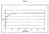
- Example 4 Solubility of GDF-5 in two buffers having different ionic strength at different pH values
- GDF-5 is adjusted to a concentration of 4 mg/ml 10 mmol/l HCl. Aliquots (50 ⁇ l) of the stock solution are diluted 1:100 with 10 mmol/l acetic acid/NaOH and with 5 mmol/l acetic acid, 5 mmol/l H 3 PO 4 /NaOH, 150 mmol/l NaCl with different pH values each. The samples were incubated for 15 min and centrifuged (2 min at 13,200 * g). The pH value and the protein content in the supernatant are determined. The data in Figure 3 show that with all the pH values measured, the solubility of GDF-5 in a buffer with higher ionic strength which corresponds to the physiological condition is significantly lower than in the buffer with low ionic strength (about 10 mmol/l).
- Freeze-dried GDF-5 was dissolved in pure acetonitrile, incubated for 15 min at room temperature and centrifuged (13,200* g, 2 min). No GDF-5 was detectable in the supernatant.
- Freeze-dried GDF-5 (50 ⁇ g) was dissolved with 50 ⁇ l 75% acetonitrile, incubated for 15 min at room temperature and centrifuged (13,200 * g, 2 min). In the supernatant, the pH was measured and the content of GDF-5 was determined. 100% of the GDF-5 used were detected, the pH value of the solution was 3.0. Subsequently, the pH value was adjusted to pH 7.4 by adding NaOH, incubated for 15 min at room temperature again and centrifuged. Only 3 ⁇ g/ml corresponding to a solubility of 60 ⁇ g/ml, were detected.
- the adsorbed protein is made visible by staining with Coomassie Brilliant Blue on the carrier.
- the distribution of the blue colour correlates with the distribution of the respective protein on the beta-TCP carrier.
- 3-4 coated granules are incubated with 200 ⁇ l staining solution (60% PBS, 40% methanol, 0.4% Coomassie Brilliant Blue R250) in a cavity of a 96-well plate and incubated for 30 min at room temperature.
- An uncoated carrier is treated in the same way as control.
- the surplus staining agent is removed by washing with 60% PBS, 40% methanol until the uncoated carrier used as control is completely destained.
- the stained carrier is dried at 40°C and documented photographically.
- ⁇ -TCP 200 mg ⁇ -TCP (0.5 - 1.0 mm granule size) are placed in a dry form in a 2R-glass.
- the stock solution of rhGDF-5 (4 mg/ml in 10 mM HCl) is diluted to 1 ⁇ g/ml with the means of the corresponding coating buffer.
- 200 ⁇ l of the GDF-5 solution obtained in that manner are pipetted on the beta-TCP and absorbed.
- the damp granulate is incubated for 1 hour at 25°C and then vacuum-dried.
- ⁇ -TCP 200 mg ⁇ -TCP (0.5 - 1.0 mm granule size) are placed in a dry form in a 2R-glass.
- the stock solution of rhGDF-5 (4 mg/ml in 10 mM HCl) is diluted to 1 ⁇ g/ml with the means of the corresponding coating buffer.
- 200 ⁇ l of the GDF-5 solution obtained in that manner are pipetted on the beta-TCP and absorbed.
- the damp granulate is incubated for 1 hour at 25°C and then lyophilised.
- a beta-TCP block having a mass of 360 mg is put into a suitable reaction vessel (Eppendorf), mixed with 500 ⁇ l of the coating solution, incubated for one hour and then vacuum- or freeze-dried.
- Beta-TCP coated with rhGDF-5 (50 ⁇ g/25 mg of beta-TCP) was manufactured using different coating buffers (20 mmol/l glycin/NaOH, pH 10 (C1) and 60% ethanol (C2)). Rats were anaesthetized by intramuscular injection of Tiletamine-Zolazepam (ZOLETIL® VIRBAC, CARROS, France, 50 mg/kg, IM). The dorsal part of the cranium was clipped free of fur. Then, the skin was scrubbed with a germicidal soap (VETEDINE®, VETOQUINOL, LURE, France).
- a germicidal soap VETEDINE®, VETOQUINOL, LURE, France.
- the surgical site was scrabbed with an antiseptic such as Povidone iodine (VETEDINE® solution, VETOQUINOL, LURE, France).
- an antiseptic such as Povidone iodine (VETEDINE® solution, VETOQUINOL, LURE, France).
- a 20 mm long incision in the scalp along the sagittal suture was realized and the skin, musculature and periosteum were reflected, exposing the parietal bones.
- a 6 mm trephined bur (COVELY, GENAY, France) was used to create the defect in the dorsal part of the parietal bone lateral to the sagittal suture under constant irrigation with sterile physiologic solution (AGUETTANT, LYON, France). Two identical defects were created per animal.
- beta TCP 200 mg beta TCP (0.5 - 1.0 mm granule size) are filled in a 2R-glass.
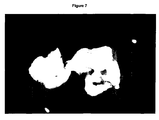
- the stock solution of BMP-2 is diluted to 1 mg/ml with the corresponding coating buffer (10 mmol/l acetic acid, 10 % sucrose). 200 ⁇ l of the coating solution are incubated with the beta-TCP (1 hr, 4 °C) and freeze dried. The distribution of BMP-2 on the coated carrier was shown by Commassie staining ( Figure 12).
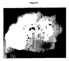
- Example 14 Comparison of the coating process in the presence of sucrose and trehalose
- beta-TCP 200 mg beta-TCP (0.5 - 1.0 mm granule size) are filled in a 2R-glass.
- the stock solution of GDF-5 (4 mg/ml in 10 mmol/l HCl) is diluted to 1 mg/ml with the corresponding coating buffer.
- the two variants of the coating buffer are containing 10 % sucrose and 10 % trehalose, respectively.
- 200 ⁇ l of the coating solution are incubated with the beta-TCP (1 hr, 4 °C) and freeze dried.
- the distribution of GDF-5 on the coated carrier was shown by Commassie staining ( Figure 13).
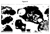
- Example 15 Comparison of the coating in the presence of ethanol and mannitol
- beta-TCP 200 mg beta-TCP (0.5 - 1.0 mm granule size) are filled in a 2R-glass.
- the stock solution of GDF-5 (4 mg/ml in 10 mmol/l HCl) is diluted to 1 mg/ml with the corresponding coating buffer.
- the two variants of the coating buffer contain 60 % ethanol and 10 % mannitol, 10 mmol/l acetic acid, respectively.
- 200 ⁇ l of the coating solution are incubated with the beta-TCP (1 hr, 4 °C) and freeze dried.
- the distribution of GDF-5 on the coated carrier was shown by Commassie staining ( Figure 14).
Abstract
Description
An alternative to the use of autogenic bones is the use of specific bone growth and differentiation factors such as GDF-5 or different bone morphogenetic proteins (BMPs). These protein factors have an osteoinductive effect which, however, they can only exert if they are used in an immobilized form. In the literature, both calcium phosphates, collagen and mineralised collagen (collagen-containing calcium phosphate) are described as carriers (hydroxy apatite and beta-TCP (Hotz, 1994), hydroxylic apatite from algae extracts (Gao, 1996), bone extracts (Gombotz, 1996) and collagen (Friess, 1999). The analyses of the potency of the coated carriers, which are described in the literature, do not present a uniform picture but exhibit significant variations which are a consequence of either the carrier type selected or the coating method (Terheyden et al. (1997)). Various methods are described. In WO 98/21972 coating is achieved by rapid precipitation of GDF-5 onto beta-TCP is achieved by first dissolving GDF-5 in an organic solvent and then precipitating it by adding water. Due to the toxicity of many solvents, however, such a process is not preferred for the production of pharmaceutical compositions. Lind et al. (1996) carry out the coating of various calcium phosphate ceramics in the presence of gelatine (usually obtained from bovine or pig bones) as protection protein. Due to the increased risk of infection, however, the use of animal substances should be avoided for the production of pharmaceutical compositions and medicinal products. Friess et al. (1999) and Gao et al. (1996) describe the coating of collagens with BMP-2. Due to the low compressive strength of collagens, such carriers, however, are not suitable for all indications. This particularly applies to indications with which the newly-formed bone has to sustain a later pressure load. Furthermore, pharmaceutical qualities of collagen are so far available from animal sources only. DE 196 47 853 describes bone implants comprising a matrix of calcium phosphate coated with protein (e.g. BMP, GDF). Said implants are used for bone augmentation following artifical defects, bone defects following extripation of cysts or tumors, for treating disc disease and for sinuslift.
The technical problem is solved by the embodiments characterized in the claims.
Another component of said device is a protein or polypeptide which has osteoinductive properties as will be explained in detail below. The protein or polypeptide is immobilized on the surface of the carrier. The osteoinductive proteins and polypeptides applied in accordance with the present invention have a particular high affinity for inorganic carrier matrices such as calcium phosphate. Preferably, the binding of said protein or polypeptide to the carrier is reversible. Thereby, dissolution of said protein is allowed once the device has been brought into a suitable in vivo surrounding, such as a bone cavity. Preferably, said dissolution of the proteins is slow release allowing diffusion of the protein into the tissue which surrounds the device. Thus, the device serves as an in vivo source for osteoinductive proteins which are slowly released and which can be thereby efficiently distributed into the surrounding tissues or have an effect in the immobilized form.
The device may, moreover, comprise additional excipients. These excipients serve to stabilization of the protein, e.g., saccharides, amino acids, polyols or detergents or maintenance of the pH, e.g., buffer substances. Preferred excipients encompassed by this invention are discussed in detail below.
The term "saccharides" encompasses mono-, di- and polysaccharides. The structure and composition of mono-, di, and polysaccharides are well known in the art and are described in standard text books, such as Römpp, lexicon of chemistry.
Suitable alcohols are well known in the art and are described in standard text books, such as Römpp, lexicon of chemistry.
Said calcium phosphates are particularly well suited as carriers for the device of the present invention. Their in vivo properties have been described in Hotz, 1994, Gao, 1996, and in WO98/21972.
The TGF-β family of growth and differentiation factors has been shown to be involved in numerous biological processes comprising bone formation. All members of said family are secreted polypeptides comprising a characteristic domain structure. On the very N-terminus, the TGF-β family members comprise a signal peptide or secretion leader. This sequence is followed at the C-terminus by the prodomain and by the sequence of the mature polypeptide. The sequence of the mature polypeptide comprises seven conserved cysteins, six of which are required for the formation of intramolecular disulfide bonds whereas one is required for dimerization of two polypeptides. The biologically active TGF-β family member is a dimer, preferably composed of two mature polypeptides. The TGF-β family members are usually secreted as proproteins comprising in addition to the mature sequence the prodomain. The prodomains are extracellularly cleaved off and are not part of the signalling molecule. It has been reported, however, that the prodomain(s) may be required for extracellular stabilization of the mature polypeptides.
In the context of the present invention, the term "TGF-β family member" or the proteins of said family referred to below encompass all biologically active variants of the said proteins or members and all variants as well as their inactive precursors. Thus, proteins comprising merely the mature sequence as well as proteins comprising the mature protein and the prodomain or the mature protein, the prodomain and the leader sequence are within the scope of the invention as well as biologically active fragments thereof. Whether a fragment of a TGF-β member has the biological activity can be easily determined by biological assays described, e.g. in: Katagiri T, Yamaguchi A, Ikeda T, Yoshiki S, Wozney JM, Rosen V, Wang EA, Tanka H, Omura S, Suda T, (1990): The non-osteogenic mouse pluripotent cell line, C3H10T1/2, is induced to differentiate into osteoblastic cells by recombinant human bone morphogenetic protein-2. Biochem. Biophys. Res. Commun. 172: 295-299 or Nishitoh H, Ichijo H, Kimura M, Matsumoto T, Makishima F, Yamaguchi A, Yamashita H, Enomoto S, Miyazono K (1996): Identification of type I and type II serine/threonine kinase receptors for growth/ differentiation factor-5. J. Biol. Chem. 271: 21345-21352.
The members of the Bone Morphogenetic Protein (BMP) subfamily have been shown to be involved, inter alia, in the induction and re-modelling of bone tissue. BMPs were originally isolated from bone matrix. These proteins are characterized by their ability to induce new bone formation at ectopic sites. Various in vivo studies demonstrated the promotion of osteogenesis and chondrogenesis of precursor cells by BMPs and raise the possibility that each BMP molecule has distinct role during the skeletal development. More details about the molecular and biological properties of the BMPs are described in:
The amino acid sequence for the preproform of BMP-2 is deposited under Swiss-Prot Accession number P12643 and is shown below.
Also more preferably, said member of the TGF-β family is a GDF. Growth and Differentiation Factor (GDF) have been also shown to be involved, inter alia, in the induction and re-modelling of bone tissue. Growth Differentiation Factor 5 (GDF-5), also known as cartilage-derived morphogenetic protein 1 (CDMP-1) is a member of subgroup of the BMP family, which also includes other related proteins, preferably, GDF-6 and GDF-7. The mature form of the protein is a 27 kDa homodimer. Various in vivo and in vitro studies demonstrate the role of GDP-5 during the formation of different morphological features in the mammalian skeleton. Mutations of GDF-5 are responsible for skeletal abnormalities including decrease of the length of long bones of limbs, abnormal joint development in the limb and sternum (Storm & Kingsley (1999), Development Biology, 209, 11-27). The amino acid sequence between mouse and human is highly conserved.
The amino acid sequence for the preproform of GDF-5 is deposited under Swiss-Prot Accession number P 43026 or shown in SEQ ID No: 3.
The term "toxic substances", preferably, encompasses those toxic organic solvents and additives which are used by the methods described in the art, e.g. actetonitrile. Said substances may cause inflammation and other reactions after implantation of devices containing said substances. Said devices are therapeutically less acceptable due to said undesirable side effects which can not be avoided by the coating methods described in the art. Moreover, the international guidance for the development of therapeutic proteins require that in the manufacturing process harmful and toxic substances should be avoided (for details see: International Conference on Harmonisation (ICH), Topic Q3C; www. emea.eu.int/). However, the device of the present invention or a device which is obtainable by the method of the present invention is, advantageously, free of said toxic substances and, therefore, therapeutically well acceptable and fulfills the requirements of the regulatory authorities.
Besides toxic substances, infectious material comprised by the device may cause severe infections in a subject into which the device has been transplanted. Potentially infectious gelatin derived from bovine or procine bones is, however, used as a protecting protein in many state of the art methods (Und, 1996).
The product of the present invention can be formulated as a pharmaceutical composition or a medical device. The composition of said product may comprise additional compounds like stabilizers, buffer substances and other excipients. The amount of the product of the present invention applied to the patient will be determined by the attending physician and other clinical factors; preferably in accordance with any of the above described methods. As it is well known in the medical arts, the amount applied to a patient depends upon many factors, including the patient's size, body surface area, age, sex, time and route of administration, general health, and other drugs being administered concurrently. Progress can be monitored by periodic assessment.
The definitions of the terms referred to above apply mutatis mutandis to the aforementioned use of the present invention and those described infra.
The term "bone augmentation" refers to the therapeutic formation of bone, which is indicated in order to treat bone defects, cavities in bones, or diseases and disorders accompanied with loss of bone tissue or to prepare the subsequent setting of an implant. The diseases and disorders described in the following are well known in the art and are described in detail in standard medical text books such as Pschyrembel or Stedman.
The parts of the kit of the invention can be packaged individually in vials or other appropriate means depending on the respective ingredient or in combination in suitable containers or multicontainer units. Manufacture of the kit follows preferably standard procedures which are known to the person skilled in the art.
| Key | From | | Length |
| SIGNAL | |||
| 1 | 23 | 23 | |
| PROPEP | 24 | 282 | 259 |
| hBMP2 | 283 | 396 | 114 |

MEDLINE=89072730; PubMed=3201241;
Wozney J.M., Rosen V., Celeste A.J., Mitsock L.M., Whitters M.J., Kriz R.W., Hewick R.M., Wang E.A.;
"Novel regulators of bone formation: molecular clones and activities.";
Science 242:1528-1534(1988).
MEDLINE=99175323; PubMed=10074410;
Scheufler C., Sebald W., Huelsmeyer M.;
"Crystal structure of human bone morphogenetic protein-2 at 2.7 A resolution.";
J. Mol. Biol. 287:103-115(1999).
| Key | From |
| Length |
| SIGNAL | |||
| 1 | 29 | 29 | |
| PROPEP | 30 | 292 | 263 |
| hBMP-7 | 293 | 431 | 139 |

TISSUE=Placenta;
MEDLINE=90291971; PubMed=2357959;
Oezkaynak E., Rueger D.C., Drier E.A., Corbett C., Ridge R.J., Sampath T.K., Oppermann H.; "OP-1 cDNA encodes an osteogenic protein in the TGF-beta family."; EMBO J. 9:2085-2093(1990).
MEDLINE=91088608; PubMed=2263636;
Celeste A.J., lannazzi J.A., Taylor R.C., Hewick R.M., Rosen V., Wang E.A., Wozney J.M.; "Identification of transforming growth factor beta family members present in bone-inductive protein purified from bovine bone.";
Proc. Natl. Acad. Sci. U.S.A. 87:9843-9847(1990).
MEDLINE=96149402; PubMed=8570652;
Griffith D.L., Keck P.C., Sampath T.K., Rueger D.C., Carlson W.D.;
"Three-dimensional structure of recombinant human osteogenic protein 1: structural paradigm for the transforming growth factor beta superfamily.";
Proc. Natl. Acad. Sci. U.S.A. 93:878-883(1996).
| Key | From | | Length |
| SIGNAL | |||
| 1 | 27 | 27 | |
| PROPEP | 28 | 381 | 354 |
| hGDF-5 | 382 | 501 | 120 |

TISSUE=Placenta;
MEDLINE=95071375; PubMed=7980526;
Hoetten G., Neidhardt H., Jacobowsky B., Pohl J.;
"Cloning and expression of recombinant human growth/
Biochem. Biophys. Res. Commun. 204:646-652(1994).
TISSUE=Articular cartilage;
MEDLINE=95050604; PubMed=7961761;
Chang S., Hoang B., Thomas J.T., Vukicevic S., Luyten F.P., Ryba N.J.P., Kozak C.A., Reddi A.H., Moos M.;
"Cartilage-derived morphogenetic proteins. New members of the transforming growth factor-beta superfamily predominantly expressed in long bones during human embryonic development.";
J. Biol. Chem. 269:28227-28234(1994).
The use of an acetate-buffered coating solution (Figure 6) causes a reduction of the pH increase during coating. While the pH increases to up to 8 in the unbuffered solutions, the pH of the acetate (40-80 mmol/l )-buffered coating solution reaches its maximum at pH 5.4. Thus, a sufficient solubility during the coating process is guaranteed. The GDF-5 used can spread evenly and bind to the carrier without precipitation taking place (see Example 7).
A delay of the pH increase is achieved also by using 60% ethanol. The delay is sufficient to achieve an even distribution of GDF-5 across the carrier (see Example 7).
After a follow-up of 6 weeks, the animals were anesthetized by intramuscular injection of ZOLETIL® (50 mg/kg) then euthanatized by lethal dosis injection of DOLETHALND (Pentobarbital sodique, VETOQUINOL, LURE, France).
The explants were sampled and fixed in 10 % buffered formalin solution. Afterwards samples were dehydrated in alcohol solutions of increased concentrations and embedded in PMMA (polymethylmetacrylate, Merck KGaA, Darmstadt, Germany). A section of 20 µm thickness was obtained by a microcutting and grinding technique adapted from Donath (Donath K. Breuner G., A method for the study of undecalcified bone and teeth with attached soft tissues. J. Oral. Pathol. 11, 318-326, 1982). A section was stained with modified Paragon for qualitative and semi-quantitative light microscopy analysis.
Histological sections were observed using a Polyvar microscope (REICHERT) fitted with a x4, x10, x25 and x40 objective.
| Sample | Bone tissue % | Implant % | Lacunae tissue % | Fibrous |
| C1 | ||||
| 8,9 | 39,2 | 2,1 | 49,8 | |
| | 43,4 | 18,5 | 21,4 | 16,7 |
<151> 2001-11-19
<211> 396
<212> PRT
<213> Homo Sapiens


<211> 431
<212> PRT
<213> Homo Sapiens


<211> 501
<212> PRT
<213> Homo Sapiens


Claims (26)
- A device having osteoinductive and osteoconductive properties in vivo comprising a carrier containing calcium phosphate and an osteoinductive protein, wherein said carrier is entirely coated with said osteoinductive protein and wherein essentially identical amounts of said osteoinductive protein are present in each and every area of the surface of said carrier.
- A method for the production of the device of claim 1 having osteoinductive and osteoconductive properties in vivo comprising the steps of:(a) providing a solution comprising dissolved osteoinductive protein and a buffer containing a weak acid having a pK value between 3 and 7, preferably between 4 and 6, said buffer keeping said protein dissolved for a time sufficient to allow homogenous coating of a carrier containing calcium phosphate when brought into contact with said carrier and said buffer being capable of balancing the increase of pH caused by contacting the buffer solution with the calcium phosphate carrier so that the protein does not immediately precipitate because of said pH increase;(b) contacting the solution of step (a) with a carrier containing calcium phosphate;(c) allowing homogenous coating of the surface of said carrier with said dissolved protein; and(d) drying of the coated carrier obtained in step (c).
- The method of claim 2, wherein said buffer has a buffer concentration of less than 100 mmol/l, less than 50 mmol/l or less than 20 mmol/l.
- The method of claim 3, wherein said weak acid is acetic acid or succinic acid.
- The method of any one of claims 2 to 4, wherein said buffer further comprises saccharides.
- The method of claim 5, wherein said saccharide is a disaccharide.
- The method of claim 6, wherein said dissaccharide is sucrose or trehalose.
- The method of any one of claims 2 to 7, wherein said buffer comprises an alcohol.
- The method of claim 8, wherein said alcohol is ethanol or mannitol.
- The device of claim 1 or the method of any one of claims 2 to 9, wherein said calcium phosphate is beta tricalcium phosphate, alpha tricalcium phosphate, apatite or a calcium phosphate containing cement.
- The device of claim 1, the method of any one of claims 2 to 9 or the device or method of claim 10, wherein said osteoinductive protein is a member of the TGF-β family.
- The device or method of claim 11, wherein said member of the TGF-β family is a member of the BMP subfamily.
- The device or method of claim 12, wherein said member of the BMP family is BMP-2 or BMP-7.
- The device or method of claim 11, wherein said member of the TGF-β family is a .. protein of the group of GDF-5, GDF-6 and GDF-7.
- The device or method of claim 14, wherein said GDF is GDF-5.
- The device of claim 1, the method of any one of claims 2 to 9 or the device or method of any one of claims 10 to 15, wherein said device is free of toxic substances.
- The device of claim 1, the method of any one of claims 2 to 9 or the device or method of any one of claims 10 to 16, wherein said device is free of infectious material.
- A pharmaceutical composition comprising the device of any one of claims 1, or 10 to 17 or which is obtainable by the method of any one of claims 2 to 17.
- Use of the device of any one of claims 1, or 10 to 17 or which is obtainable by the method of any one of claims 2 to 17 for the preparation of a pharmaceutical composition to be used for bone augmentation.
- The use of claim 19, wherein said bone augmentation follows traumatic, malignant or artificial defects or is a prerequisite for the subsequent setting of an implant.
- Use of the device of any one of claims 1, or 10 to 17 or which is obtainable by the method of any one of claims 2 to 17 for the preparation of a pharmaceutical composition for treating bone defects.
- The use of claim 21, wherein said bone defects are long bone defects, defects in the maxillofacial area or bone defects following apicoectomy, extirpation of cysts or tumors, tooth extraction, or surgical removal of retained teeth.
- Use of the device of any one of claims 1, or 10 to 17 or which is obtainable by the method of any one of claims 2 to 17 for the preparation of a pharmaceutical composition for treating degenerative and traumatic disc disease.
- Use of the device of any one of claims 1, or 10 to 17 or which is obtainable by the method of any one of claims 2 to 17 for the preparation of a pharmaceutical composition for treating bone dehiscence.
- Use of the device of any one of claims 1, or 10 to 17 or which is obtainable by the method of any one of claims 2 to 17 for the preparation of a pharmaceutical composition to be used for sinus floor elevation or augmentation of the atrophied maxillary or mandibular ridge.
- A kit comprising the device of any one of claims 1, or 10 to 17 or which is obtainable by the method of any one of claims 2 to 17.
Priority Applications (4)
| Application Number | Priority Date | Filing Date | Title |
|---|---|---|---|
| SI200230206T SI1448246T2 (en) | 2001-11-19 | 2002-03-27 | Homogeneously coated device having osteoinductive and osteoconductive properties |
| EP02803343.9A EP1448246B2 (en) | 2001-11-19 | 2002-03-27 | Method for producing a homogeneously coated device having osteoinductive and osteoconductive properties |
| DE60205937.2T DE60205937T3 (en) | 2001-11-19 | 2002-03-27 | Method for producing a homogeneously coated device with osteoinductive and osteoconductive properties |
| CY20051101348T CY1105664T1 (en) | 2001-11-19 | 2005-11-04 | HOMOGENEOUSLY COATED DISTRIBUTION HAVING OSTEOPULATIVE AND OSTEOCONDUCTIVE PROPERTIES |
Applications Claiming Priority (4)
| Application Number | Priority Date | Filing Date | Title |
|---|---|---|---|
| EP01127573 | 2001-11-19 | ||
| EP01127573 | 2001-11-19 | ||
| EP02803343.9A EP1448246B2 (en) | 2001-11-19 | 2002-03-27 | Method for producing a homogeneously coated device having osteoinductive and osteoconductive properties |
| PCT/EP2002/003463 WO2003043673A1 (en) | 2001-11-19 | 2002-03-27 | Device having osteoinductive and osteoconductive properties |
Publications (3)
| Publication Number | Publication Date |
|---|---|
| EP1448246A1 EP1448246A1 (en) | 2004-08-25 |
| EP1448246B1 true EP1448246B1 (en) | 2005-08-31 |
| EP1448246B2 EP1448246B2 (en) | 2015-09-09 |
Family
ID=8179288
Family Applications (1)
| Application Number | Title | Priority Date | Filing Date |
|---|---|---|---|
| EP02803343.9A Expired - Lifetime EP1448246B2 (en) | 2001-11-19 | 2002-03-27 | Method for producing a homogeneously coated device having osteoinductive and osteoconductive properties |
Country Status (16)
| Country | Link |
|---|---|
| US (2) | US8546334B2 (en) |
| EP (1) | EP1448246B2 (en) |
| JP (2) | JP4473576B2 (en) |
| CN (1) | CN100425285C (en) |
| AT (1) | ATE303169T1 (en) |
| AU (1) | AU2002308204B9 (en) |
| BR (1) | BR0214275A (en) |
| CA (1) | CA2466947C (en) |
| CY (1) | CY1105664T1 (en) |
| DE (1) | DE60205937T3 (en) |
| DK (1) | DK1448246T4 (en) |
| ES (1) | ES2247423T5 (en) |
| HK (1) | HK1071315A1 (en) |
| PT (1) | PT1448246E (en) |
| SI (1) | SI1448246T2 (en) |
| WO (1) | WO2003043673A1 (en) |
Cited By (2)
| Publication number | Priority date | Publication date | Assignee | Title |
|---|---|---|---|---|
| US7947649B2 (en) | 2008-04-14 | 2011-05-24 | Advanced Technologies And Regenerative Medicine, Llc | Liquid buffered GDF-5 formulations |
| US8435943B2 (en) | 2006-12-14 | 2013-05-07 | Advanced Technogies And Regenerative Medicine, Llc | Protein stabilization formulations |
Families Citing this family (19)
| Publication number | Priority date | Publication date | Assignee | Title |
|---|---|---|---|---|
| EP1462126A1 (en) * | 2003-03-28 | 2004-09-29 | BIOPHARM GESELLSCHAFT ZUR BIOTECHNOLOGISCHEN ENTWICKLUNG VON PHARMAKA mbH | Improved Osteoinductive Materials |
| ATE478008T1 (en) | 2004-03-10 | 2010-09-15 | Scil Technology Gmbh | COVERED IMPLANTS, THEIR PRODUCTION AND USE THEREOF |
| EP1604694A1 (en) * | 2004-06-09 | 2005-12-14 | Scil Technology GmbH | Composite device having osteoinductive and osteoconductive properties |
| EP1604693A1 (en) | 2004-06-09 | 2005-12-14 | Scil Technology GmbH | In situ forming scaffold, its manufacturing and use |
| WO2005120454A2 (en) * | 2004-06-09 | 2005-12-22 | Scil Technology Gmbh | Composite material for use as protein carrier |
| US20060286171A1 (en) * | 2005-06-17 | 2006-12-21 | Tianhong Zhou | Bone morphogenetic protein formulations |
| KR101105890B1 (en) * | 2005-12-14 | 2012-01-16 | 에스씨아이엘 테크놀로지 게엠베하 | A moldable biomaterial for bone regeneration |
| US20070299520A1 (en) * | 2006-06-26 | 2007-12-27 | Warsaw Orthopedic, Inc. | Surface treatment of implantable devices |
| DE102006060958A1 (en) * | 2006-12-20 | 2008-06-26 | Jennissen, Herbert P., Prof. Dr. | Process for the preparation of a polymer matrix, implants made thereof and their use |
| US20080221681A1 (en) * | 2007-03-09 | 2008-09-11 | Warsaw Orthopedic, Inc. | Methods for Improving Fatigue Performance of Implants With Osteointegrating Coatings |
| US7678764B2 (en) | 2007-06-29 | 2010-03-16 | Johnson & Johnson Regenerative Therapeutics, Llc | Protein formulations for use at elevated temperatures |
| CA2695697A1 (en) | 2007-08-07 | 2009-02-12 | Advanced Technologies And Regenerative Medicine, Llc | Protein formulations comprising gdf-5 in aqueous acidic solution |
| US20090061002A1 (en) * | 2007-09-05 | 2009-03-05 | Venbrocks Rudolf A | Calcium phospate based delivery of growth and differentiation factors to compromised bone |
| DE102007051914A1 (en) * | 2007-10-29 | 2009-05-07 | Herbert Prof. Dr. Jennissen | Process for the preparation of particles loaded with growth factors and the particles thus obtained |
| US20110059149A1 (en) * | 2008-06-16 | 2011-03-10 | Little Marisa A | Process for depositing calcium phosphate therapeutic coatings with different release rates and a prosthesis coated via the process |
| US9861725B2 (en) | 2009-05-28 | 2018-01-09 | Addbio Ab | Multilayer protein films, methods of making, and drug delivery devices and biomedical implants employing the films |
| CA2806143A1 (en) * | 2010-07-30 | 2012-02-02 | Biopharm Gesellschaft Zur Biotechnologischen Entwicklung Von Pharmaka Mb H | Drug delivery devices and growth factor formulations for accelerated wound healing |
| WO2012093939A1 (en) | 2011-01-05 | 2012-07-12 | Vereniging Voor Christelijk Hoger Onderwijs, Wetenschappelijk Onderzoek En Patiëntenzorg | Particles comprising calcium phosphate and use thereof |
| EP3682931A1 (en) * | 2019-01-21 | 2020-07-22 | Universitätsklinikum Hamburg-Eppendorf | Thoracic catheter |
Family Cites Families (139)
| Publication number | Priority date | Publication date | Assignee | Title |
|---|---|---|---|---|
| DE3329128A1 (en) | 1983-08-11 | 1985-02-28 | Bayer Ag, 5090 Leverkusen | 3-CYCLOALKYL-1- (1,3-DIOXAN-5-YL) -2- (1,2,4-TRIAZOL-1-YL) -PROPAN-1-ONE AND -PROPAN-1-OLE |
| US4596574A (en) | 1984-05-14 | 1986-06-24 | The Regents Of The University Of California | Biodegradable porous ceramic delivery system for bone morphogenetic protein |
| US5939388A (en) | 1986-07-01 | 1999-08-17 | Rosen; Vicki A. | Methods of administering BMP-5 compositions |
| ZA874681B (en) | 1986-07-01 | 1988-04-27 | Genetics Inst | Novel osteoinductive factors |
| US5459047A (en) | 1986-07-01 | 1995-10-17 | Genetics Institute, Inc. | BMP-6 proteins |
| US5108922A (en) | 1986-07-01 | 1992-04-28 | Genetics Institute, Inc. | DNA sequences encoding BMP-1 products |
| US5543394A (en) | 1986-07-01 | 1996-08-06 | Genetics Institute, Inc. | Bone morphogenetic protein 5(BMP-5) compositions |
| US5013649A (en) | 1986-07-01 | 1991-05-07 | Genetics Institute, Inc. | DNA sequences encoding osteoinductive products |
| US6432919B1 (en) | 1986-07-01 | 2002-08-13 | Genetics Institute, Inc. | Bone morphogenetic protein-3 and compositions |
| US5187076A (en) | 1986-07-01 | 1993-02-16 | Genetics Institute, Inc. | DNA sequences encoding BMP-6 proteins |
| US5366875A (en) | 1986-07-01 | 1994-11-22 | Genetics Institute, Inc. | Methods for producing BMP-7 proteins |
| US5106748A (en) | 1986-07-01 | 1992-04-21 | Genetics Institute, Inc. | Dna sequences encoding 5 proteins |
| US5631142A (en) | 1986-07-01 | 1997-05-20 | Genetics Institute, Inc. | Compositions comprising bone morphogenetic protein-2 (BMP-2) |
| US4877864A (en) | 1987-03-26 | 1989-10-31 | Genetics Institute, Inc. | Osteoinductive factors |
| IL83003A (en) | 1986-07-01 | 1995-07-31 | Genetics Inst | Osteoinductive factors |
| US6150328A (en) | 1986-07-01 | 2000-11-21 | Genetics Institute, Inc. | BMP products |
| US4975526A (en) | 1989-02-23 | 1990-12-04 | Creative Biomolecules, Inc. | Bone collagen matrix for zenogenic implants |
| US5011691A (en) | 1988-08-15 | 1991-04-30 | Stryker Corporation | Osteogenic devices |
| US6586388B2 (en) | 1988-04-08 | 2003-07-01 | Stryker Corporation | Method of using recombinant osteogenic protein to repair bone or cartilage defects |
| US5344654A (en) | 1988-04-08 | 1994-09-06 | Stryker Corporation | Prosthetic devices having enhanced osteogenic properties |
| US5258494A (en) | 1988-04-08 | 1993-11-02 | Stryker Corporation | Osteogenic proteins |
| US5354557A (en) | 1988-04-08 | 1994-10-11 | Stryker Corporation | Osteogenic devices |
| US5162114A (en) | 1989-02-23 | 1992-11-10 | Stryker Corporation | Bone collagen matrix for xenogenic implants |
| DE68929472T2 (en) | 1988-04-08 | 2004-05-13 | Stryker Corp., Kalamazoo | Biosynthetic bone-forming proteins and bone-forming devices containing them |
| US5670336A (en) | 1988-04-08 | 1997-09-23 | Stryker Corporation | Method for recombinant production of osteogenic protein |
| US5266683A (en) | 1988-04-08 | 1993-11-30 | Stryker Corporation | Osteogenic proteins |
| US5324819A (en) | 1988-04-08 | 1994-06-28 | Stryker Corporation | Osteogenic proteins |
| US5258029A (en) | 1988-09-29 | 1993-11-02 | Collagen Corporation | Method for improving implant fixation |
| US5207710A (en) † | 1988-09-29 | 1993-05-04 | Collagen Corporation | Method for improving implant fixation |
| US5108436A (en) | 1988-09-29 | 1992-04-28 | Collagen Corporation | Implant fixation |
| ATE162223T1 (en) | 1989-03-28 | 1998-01-15 | Genetics Inst | OSTEOINDUCTIVE COMPOSITIONS |
| US5422340A (en) * | 1989-09-01 | 1995-06-06 | Ammann; Arthur J. | TGF-βformulation for inducing bone growth |
| US5158934A (en) | 1989-09-01 | 1992-10-27 | Genentech, Inc. | Method of inducing bone growth using TGF-β |
| JP2845346B2 (en) | 1989-10-17 | 1999-01-13 | ストライカー・コーポレーション | Bone former |
| DK27390D0 (en) | 1990-02-02 | 1990-02-02 | Troels Torp Andreassen | METHOD AND APPARATUS FOR ADMINISTRATING BIOLOGICALLY ACTIVE SUBSTANCES |
| CA2094027C (en) | 1990-10-18 | 2001-12-25 | Hermann Oppermann | Osteogenic peptides |
| US6211146B1 (en) | 1991-03-11 | 2001-04-03 | Curis, Inc. | 60A protein-induced morphogenesis |
| US6090776A (en) | 1991-03-11 | 2000-07-18 | Creative Bio Molecules, Inc. | Morphogen treatment of organ implants |
| US6800603B2 (en) | 1991-03-11 | 2004-10-05 | Curis, Inc. | Morphogen-induced neural cell adhesion |
| US6399569B1 (en) | 1991-03-11 | 2002-06-04 | Curis, Inc. | Morphogen treatments for limiting proliferation of epithelial cells |
| US6495513B1 (en) | 1991-03-11 | 2002-12-17 | Curis, Inc. | Morphogen-enhanced survival and repair of neural cells |
| US6506729B1 (en) | 1991-03-11 | 2003-01-14 | Curis, Inc. | Methods and compositions for the treatment and prevention of Parkinson's disease |
| US6395883B1 (en) | 1991-03-11 | 2002-05-28 | Curis, Inc. | Soluble morphogenic protein complex compositions of matter |
| US6723698B2 (en) | 1991-03-11 | 2004-04-20 | Curis, Inc. | Methods and compositions for the treatment of motor neuron injury and neuropathy |
| US5972884A (en) | 1991-03-11 | 1999-10-26 | Creative Biomolecules, Inc. | Morphogen treatment of gastrointestinal ulcers |
| US5849686A (en) | 1991-03-11 | 1998-12-15 | Creative Biomolecules, Inc. | Morphogen-induced liver regeneration |
| JP3627985B2 (en) | 1991-03-11 | 2005-03-09 | キュリス インコーポレイテッド | Protein-induced morphogenesis |
| US5652337A (en) | 1991-03-11 | 1997-07-29 | Creative Biomolecules, Inc. | OP-3-induced morphogenesis |
| US5656593A (en) | 1991-03-11 | 1997-08-12 | Creative Biomolecules, Inc. | Morphogen induced periodontal tissue regeneration |
| US5650276A (en) | 1991-03-11 | 1997-07-22 | Creative Biomolecules, Inc. | Morphogenic protein screening method |
| CA2363965C (en) | 1991-03-11 | 2010-05-18 | Curis, Inc. | Protein-induced morphogenesis |
| US5707810A (en) | 1991-03-11 | 1998-01-13 | Creative Biomolecules, Inc. | Method of diagnosing renal tissue damage or disease |
| US6194376B1 (en) | 1991-03-11 | 2001-02-27 | Creative Biomolecules, Inc. | Method for modulating inflammatory response comprising administering morphogen |
| US6077823A (en) | 1991-03-11 | 2000-06-20 | Creative Biomolecules, Inc. | Method for reducing tissue damage associated with ischemia-reperfusion or hypoxia injury |
| US5674844A (en) | 1991-03-11 | 1997-10-07 | Creative Biomolecules, Inc. | Treatment to prevent loss of and/or increase bone mass in metabolic bone diseases |
| US5652118A (en) | 1991-03-11 | 1997-07-29 | Creative Biomolecules, Inc. | Nucleic acid encoding a novel morphogenic protein, OP-3 |
| US5563124A (en) | 1991-04-22 | 1996-10-08 | Intermedics Orthopedics/ Denver, Inc. | Osteogenic product and process |
| US5290763A (en) | 1991-04-22 | 1994-03-01 | Intermedics Orthopedics/Denver, Inc. | Osteoinductive protein mixtures and purification processes |
| US5177406A (en) | 1991-04-29 | 1993-01-05 | General Motors Corporation | Active matrix vacuum fluorescent display with compensation for variable phosphor efficiency |
| DE4121043A1 (en) | 1991-06-26 | 1993-01-07 | Merck Patent Gmbh | BONE REPLACEMENT MATERIAL WITH FGF |
| EP0523926A3 (en) | 1991-07-15 | 1993-12-01 | Smith & Nephew Richards Inc | Prosthetic implants with bioabsorbable coating |
| JP4235689B2 (en) | 1991-08-30 | 2009-03-11 | ストライカー・コーポレーション | Bone morphogenetic protein for treating bone disease |
| JP3693338B2 (en) | 1991-08-30 | 2005-09-07 | キュリス インコーポレイテッド | Regulation of inflammatory response by induction of tissue morphogenetic factor |
| US6022853A (en) | 1991-08-30 | 2000-02-08 | Creative Biomolecules, Inc. | Morphogen-enriched dietary composition |
| SE469653B (en) | 1992-01-13 | 1993-08-16 | Lucocer Ab | POROEST IMPLANT |
| KR0172186B1 (en) | 1992-02-12 | 1999-02-01 | 미하엘 파울리스타 | Dna sequences encoding novel growth/differentiation factors |
| US6120760A (en) | 1992-02-12 | 2000-09-19 | Biopharm Gesellschaft Zur Biotechnologischen Entwicklung | Growth/differentiation factors of the TGF-β family |
| US5876452A (en) | 1992-02-14 | 1999-03-02 | Board Of Regents, University Of Texas System | Biodegradable implant |
| US6013853A (en) | 1992-02-14 | 2000-01-11 | The University Of Texas System | Continuous release polymeric implant carrier |
| CA2117379C (en) | 1992-02-14 | 1999-11-16 | Kypriacos A. Athanasiou | Multi-phase bioerodible implant/carrier and method of manufacturing and using same |
| US5610021A (en) | 1992-02-21 | 1997-03-11 | Creative Biomolecules, Inc. | Compositions and methods for identification and use of soluble complex forms of osteogenic proteins |
| US6071695A (en) | 1992-02-21 | 2000-06-06 | Creative Biomolecules, Inc. | Methods and products for identification of modulators of osteogenic protein-1 gene expression |
| US5397235A (en) | 1993-07-02 | 1995-03-14 | Dental Marketing Specialists, Inc. | Method for installation of dental implant |
| WO1994015949A1 (en) | 1993-01-12 | 1994-07-21 | Johns Hopkins University School Of Medicine | Growth differentiation factor-5 |
| EP0679097B1 (en) | 1993-01-12 | 1997-05-28 | Genentech, Inc. | Tgf-beta formulation for inducing bone growth |
| US5385887A (en) * | 1993-09-10 | 1995-01-31 | Genetics Institute, Inc. | Formulations for delivery of osteogenic proteins |
| PT733109E (en) | 1993-12-07 | 2006-07-31 | Genetics Inst Llc | MORPHOGENETIC PROTEINS OF 0SS0S PMO-12 AND PMO-13 AND THEIR COMPOSITIONS FOR TENDAO INDUCTION |
| US6027919A (en) | 1993-12-07 | 2000-02-22 | Genetics Institute, Inc. | BMP-12 and BMP-13 proteins and DNA encoding them |
| US6013517A (en) | 1994-05-09 | 2000-01-11 | Chiron Corporation | Crossless retroviral vectors |
| US5681746A (en) | 1994-12-30 | 1997-10-28 | Chiron Viagene, Inc. | Retroviral delivery of full length factor VIII |
| JPH11501029A (en) | 1995-03-01 | 1999-01-26 | クリエイティブ バイオモレキュールズ,インコーポレイテッド | Morphogen-induced dentin regeneration |
| US5782919A (en) * | 1995-03-27 | 1998-07-21 | Sdgi Holdings, Inc. | Interbody fusion device and method for restoration of normal spinal anatomy |
| US5635372A (en) | 1995-05-18 | 1997-06-03 | Genetics Institute, Inc. | BMP-15 compositions |
| US6027742A (en) | 1995-05-19 | 2000-02-22 | Etex Corporation | Bioresorbable ceramic composites |
| US5676976A (en) | 1995-05-19 | 1997-10-14 | Etex Corporation | Synthesis of reactive amorphous calcium phosphates |
| US6541037B1 (en) | 1995-05-19 | 2003-04-01 | Etex Corporation | Delivery vehicle |
| US6287341B1 (en) | 1995-05-19 | 2001-09-11 | Etex Corporation | Orthopedic and dental ceramic implants |
| US8333996B2 (en) | 1995-05-19 | 2012-12-18 | Etex Corporation | Calcium phosphate delivery vehicle and adjuvant |
| US6117456A (en) | 1995-05-19 | 2000-09-12 | Etex Corporation | Methods and products related to the physical conversion of reactive amorphous calcium phosphate |
| US6132463A (en) | 1995-05-19 | 2000-10-17 | Etex Corporation | Cell seeding of ceramic compositions |
| ES2440441T3 (en) | 1995-06-05 | 2014-01-29 | Genetics Institute, Llc | Use of bone morphogenetic proteins for healing and repair of connective tissue junctions |
| EP0883410B1 (en) * | 1996-02-29 | 2004-08-18 | Bioactive Bone Substitute OY, AB | An osteogenic device and a method for preparing the device |
| EP0806212B1 (en) † | 1996-05-10 | 2003-04-02 | IsoTis N.V. | Device for incorporation and release of biologically active agents |
| US6143948A (en) | 1996-05-10 | 2000-11-07 | Isotis B.V. | Device for incorporation and release of biologically active agents |
| EP0806211B1 (en) | 1996-05-10 | 2002-10-23 | IsoTis N.V. | Implant material and process for producing it |
| CA2205104A1 (en) | 1996-05-10 | 1997-11-10 | Eugenia Ribeiro De Sousa Fidalgo Leitao | Device for incorporation and release of biologically active agents |
| US6953594B2 (en) | 1996-10-10 | 2005-10-11 | Etex Corporation | Method of preparing a poorly crystalline calcium phosphate and methods of its use |
| DE69729647T2 (en) | 1996-10-16 | 2005-07-07 | Etex Corp., Cambridge | Process for the preparation of calcium phosphate of low crystallinity and process for its use |
| DE19647853A1 (en) * | 1996-11-19 | 1998-05-20 | Bioph Biotech Entw Pharm Gmbh | Compounds with improved cartilage and / or bone inducing activity |
| US20020082224A1 (en) | 1997-01-14 | 2002-06-27 | Douglas J. Jolly | Non-immunogenic prodrugs and selectable markers for use in gene therapy |
| EA001579B1 (en) | 1997-01-30 | 2001-06-25 | Хехст Марион Рассел Лтд. | Lyophilized composition of bone morphogenic factor human mp52 and process for preparation the same |
| EP2332564A1 (en) | 1997-02-07 | 2011-06-15 | Stryker Corporation | Matrix-free osteogenic devices, implants and methods thereof |
| US20020098222A1 (en) | 1997-03-13 | 2002-07-25 | John F. Wironen | Bone paste |
| US20030032586A1 (en) | 1997-05-15 | 2003-02-13 | David C. Rueger | Compositions for morphogen-induced osteogenesis |
| US6129928A (en) | 1997-09-05 | 2000-10-10 | Icet, Inc. | Biomimetic calcium phosphate implant coatings and methods for making the same |
| ES2245042T3 (en) | 1997-09-24 | 2005-12-16 | The Regents Of The University Of California | VECTORS OF NON-PRIMATE LENTIVIRUS AND PACKAGING SYSTEMS. |
| CA2318575A1 (en) | 1998-01-16 | 1999-07-22 | Chiron Corporation | Feline immunodeficiency virus gene therapy vectors |
| US20020048805A1 (en) | 1998-01-16 | 2002-04-25 | Julie C. Johnston | Feline immunodeficiency virus gene therapy vectors |
| EP1076715B1 (en) | 1998-05-13 | 2007-07-18 | Genetix Pharmaceuticals Inc. | Lentiviral packaging cells |
| US6261322B1 (en) | 1998-05-14 | 2001-07-17 | Hayes Medical, Inc. | Implant with composite coating |
| KR100298178B1 (en) | 1998-06-29 | 2001-08-07 | 박종섭 | Photodiode in image sensorr |
| US6328765B1 (en) | 1998-12-03 | 2001-12-11 | Gore Enterprise Holdings, Inc. | Methods and articles for regenerating living tissue |
| US6423544B1 (en) | 1998-12-31 | 2002-07-23 | Chiron Corporation | Compositions and methods for producing recombinant virions |
| AU772682B2 (en) | 1999-02-04 | 2004-05-06 | Warsaw Orthopedic, Inc. | Highly-mineralized osteogenic sponge compositions, and uses thereof |
| WO2000045870A1 (en) | 1999-02-04 | 2000-08-10 | Sdgi Holdings, Inc. | Osteogenic paste compositions and uses thereof |
| US6730297B1 (en) | 1999-05-28 | 2004-05-04 | Chiron Corporation | Use of recombinant gene delivery vectors for treating or preventing lysosomal storage disorders |
| SE514202C2 (en) | 1999-05-31 | 2001-01-22 | Nobel Biocare Ab | Layers arranged on bone or tissue structure implants and such implants and methods for applying the layer |
| SE515695C2 (en) | 1999-05-31 | 2001-09-24 | Nobel Biocare Ab | Implant with coating of bone growth stimulant for bone application and procedure in such implant |
| SE514323C2 (en) | 1999-05-31 | 2001-02-12 | Nobel Biocare Ab | Implants and procedure and use in implants |
| JP2003505206A (en) * | 1999-07-28 | 2003-02-12 | リジェネレーション テクノロジーズ インク. | Graft of tissue with reduced antigenicity (RAT) |
| US6696410B1 (en) * | 1999-09-27 | 2004-02-24 | Stryker Corporation | Compositions and therapeutic methods using morphogenic proteins, hormones and hormone receptors |
| DE60012557T2 (en) | 1999-10-15 | 2005-08-04 | Genetics Institute, LLC, Cambridge | HYALURONIC ACID COMPOSITIONS FOR THE DISPOSAL OF OSTEOGENIC PROTEINS |
| EP1220693B1 (en) | 1999-10-15 | 2004-12-08 | Genetics Institute, LLC | Formulations for delivery of osteogenic proteins |
| DE19950386A1 (en) | 1999-10-19 | 2001-05-10 | Miladin Lazarov | Biocompatible item |
| EP1251889B1 (en) † | 2000-02-04 | 2003-07-02 | IsoTis N.V. | Proteinaceous coating |
| US20020022885A1 (en) * | 2000-05-19 | 2002-02-21 | Takahiro Ochi | Biomaterial |
| WO2001091801A2 (en) | 2000-05-26 | 2001-12-06 | Chiron Corporation | Methods of transducing neural cells using lentivirus vectors |
| US6533821B1 (en) | 2000-06-22 | 2003-03-18 | Thomas Lally | Bio-adhesive composition, method for adhering objects to bone |
| US7419829B2 (en) | 2000-10-06 | 2008-09-02 | Oxford Biomedica (Uk) Limited | Vector system |
| WO2002042482A2 (en) | 2000-11-27 | 2002-05-30 | Chiron Corporation | Functional lentiviral vector from an mlv-based backbone |
| US20020082679A1 (en) | 2000-12-22 | 2002-06-27 | Avantec Vascular Corporation | Delivery or therapeutic capable agents |
| US6972122B2 (en) | 2001-01-05 | 2005-12-06 | Duke University | Contrast enhancement agent for magnetic resonance imaging |
| US20030049328A1 (en) | 2001-03-02 | 2003-03-13 | Dalal Paresh S. | Porous beta-tricalcium phosphate granules and methods for producing same |
| US6949251B2 (en) | 2001-03-02 | 2005-09-27 | Stryker Corporation | Porous β-tricalcium phosphate granules for regeneration of bone tissue |
| EP1492475B1 (en) | 2001-04-16 | 2011-12-21 | Wright Medical Technology, Inc. | Dense/porous structures for use as bone substitutes |
| DE60230739D1 (en) | 2001-05-01 | 2009-02-26 | Amedica Corp | X-RAY BONE TRANSPLANT |
| US20030039636A1 (en) | 2001-05-01 | 2003-02-27 | Genetix Pharmaceuticals, Inc. | Novel self-inactivating (SIN) lentiviral vectors |
| SE519566C2 (en) | 2001-07-04 | 2003-03-11 | Nobel Biocare Ab | Method of Treating Implants by Coating with Calcium Phosphate and Bone Growth Stimulants |
| SE519564C2 (en) | 2001-07-04 | 2003-03-11 | Nobel Biocare Ab | Implants, for example dental implants, coated with bone growth stimulants |
-
2002
- 2002-03-27 AU AU2002308204A patent/AU2002308204B9/en not_active Ceased
- 2002-03-27 JP JP2003545351A patent/JP4473576B2/en not_active Expired - Fee Related
- 2002-03-27 SI SI200230206T patent/SI1448246T2/en unknown
- 2002-03-27 DK DK02803343.9T patent/DK1448246T4/en active
- 2002-03-27 US US10/496,399 patent/US8546334B2/en not_active Expired - Fee Related
- 2002-03-27 WO PCT/EP2002/003463 patent/WO2003043673A1/en active IP Right Grant
- 2002-03-27 ES ES02803343.9T patent/ES2247423T5/en not_active Expired - Lifetime
- 2002-03-27 BR BR0214275-9A patent/BR0214275A/en not_active IP Right Cessation
- 2002-03-27 DE DE60205937.2T patent/DE60205937T3/en not_active Expired - Lifetime
- 2002-03-27 CN CNB028192745A patent/CN100425285C/en not_active Expired - Fee Related
- 2002-03-27 PT PT02803343T patent/PT1448246E/en unknown
- 2002-03-27 AT AT02803343T patent/ATE303169T1/en active
- 2002-03-27 CA CA2466947A patent/CA2466947C/en not_active Expired - Fee Related
- 2002-03-27 EP EP02803343.9A patent/EP1448246B2/en not_active Expired - Lifetime
-
2005
- 2005-05-13 HK HK05104014.9A patent/HK1071315A1/en not_active IP Right Cessation
- 2005-11-04 CY CY20051101348T patent/CY1105664T1/en unknown
-
2009
- 2009-12-29 JP JP2009299102A patent/JP5054091B2/en not_active Expired - Fee Related
-
2013
- 2013-09-27 US US14/040,500 patent/US20150080303A1/en not_active Abandoned
Cited By (3)
| Publication number | Priority date | Publication date | Assignee | Title |
|---|---|---|---|---|
| US8435943B2 (en) | 2006-12-14 | 2013-05-07 | Advanced Technogies And Regenerative Medicine, Llc | Protein stabilization formulations |
| US8895506B2 (en) | 2006-12-14 | 2014-11-25 | DePuy Synthes Products, LLC | Protein stabilization formulations |
| US7947649B2 (en) | 2008-04-14 | 2011-05-24 | Advanced Technologies And Regenerative Medicine, Llc | Liquid buffered GDF-5 formulations |
Also Published As
| Publication number | Publication date |
|---|---|
| CN1561235A (en) | 2005-01-05 |
| DK1448246T3 (en) | 2005-12-27 |
| ES2247423T3 (en) | 2006-03-01 |
| US20150080303A1 (en) | 2015-03-19 |
| SI1448246T1 (en) | 2005-12-31 |
| CN100425285C (en) | 2008-10-15 |
| HK1071315A1 (en) | 2005-07-15 |
| BR0214275A (en) | 2004-09-21 |
| ES2247423T5 (en) | 2015-12-30 |
| WO2003043673A1 (en) | 2003-05-30 |
| ATE303169T1 (en) | 2005-09-15 |
| DE60205937T3 (en) | 2016-01-21 |
| DE60205937T2 (en) | 2006-06-14 |
| DE60205937D1 (en) | 2005-10-06 |
| CA2466947A1 (en) | 2003-05-30 |
| JP5054091B2 (en) | 2012-10-24 |
| JP2010075737A (en) | 2010-04-08 |
| US20060088565A1 (en) | 2006-04-27 |
| CY1105664T1 (en) | 2010-12-22 |
| PT1448246E (en) | 2005-11-30 |
| US8546334B2 (en) | 2013-10-01 |
| AU2002308204B2 (en) | 2007-09-27 |
| CA2466947C (en) | 2012-05-22 |
| EP1448246B2 (en) | 2015-09-09 |
| JP4473576B2 (en) | 2010-06-02 |
| SI1448246T2 (en) | 2015-12-31 |
| DK1448246T4 (en) | 2015-12-21 |
| EP1448246A1 (en) | 2004-08-25 |
| AU2002308204B9 (en) | 2008-06-12 |
| JP2005509494A (en) | 2005-04-14 |
| AU2002308204A1 (en) | 2003-06-10 |
Similar Documents
| Publication | Publication Date | Title |
|---|---|---|
| US20150080303A1 (en) | Device having osteoinductive and osteoconductive properties | |
| US7365051B2 (en) | Compounds with improved cartilage-inducing and/or bone-inducing activity | |
| EP1604693A1 (en) | In situ forming scaffold, its manufacturing and use | |
| KR101105890B1 (en) | A moldable biomaterial for bone regeneration | |
| EP1753396A2 (en) | Composite material for use as protein carrier | |
| EP1539261B1 (en) | Metal implant coated under reduced oxygen concentration with osteoinductive protein | |
| CA2868992C (en) | System and method for multiphasic release of growth factors | |
| EP2200537B1 (en) | Calcium phosphate based delivery of growth and differentiation factors to compromised bone | |
| EP3863691B1 (en) | Bioactive bone repair particles | |
| EP1604649A1 (en) | Composite material for use as protein carrier | |
| EP1604694A1 (en) | Composite device having osteoinductive and osteoconductive properties |
Legal Events
| Date | Code | Title | Description |
|---|---|---|---|
| PUAI | Public reference made under article 153(3) epc to a published international application that has entered the european phase |
Free format text: ORIGINAL CODE: 0009012 |
|
| 17P | Request for examination filed |
Effective date: 20040205 |
|
| AK | Designated contracting states |
Kind code of ref document: A1 Designated state(s): AT BE CH CY DE DK ES FI FR GB GR IE IT LI LU MC NL PT SE TR |
|
| AX | Request for extension of the european patent |
Extension state: AL LT LV MK RO SI |
|
| 17Q | First examination report despatched |
Effective date: 20041103 |
|
| GRAP | Despatch of communication of intention to grant a patent |
Free format text: ORIGINAL CODE: EPIDOSNIGR1 |
|
| RTI1 | Title (correction) |
Free format text: HOMOGENEOUSLY COATED DEVICE HAVING OSTEOINDUCTIVE AND OSTEOCONDUCTIVE PROPERTIES |
|
| GRAS | Grant fee paid |
Free format text: ORIGINAL CODE: EPIDOSNIGR3 |
|
| GRAA | (expected) grant |
Free format text: ORIGINAL CODE: 0009210 |
|
| RIN1 | Information on inventor provided before grant (corrected) |
Inventor name: HELLERBRAND, KLAUS Inventor name: POEHLING, SYLKE Inventor name: HAPPERSBERGER, PETER Inventor name: KOHNERT, ULRICH |
|
| AK | Designated contracting states |
Kind code of ref document: B1 Designated state(s): AT BE CH CY DE DK ES FI FR GB GR IE IT LI LU MC NL PT SE TR |
|
| AX | Request for extension of the european patent |
Extension state: AL LT LV MK RO SI |
|
| REG | Reference to a national code |
Ref country code: GB Ref legal event code: FG4D Ref country code: CH Ref legal event code: EP |
|
| REG | Reference to a national code |
Ref country code: IE Ref legal event code: FG4D |
|
| REF | Corresponds to: |
Ref document number: 60205937 Country of ref document: DE Date of ref document: 20051006 Kind code of ref document: P |
|
| REG | Reference to a national code |
Ref country code: SE Ref legal event code: TRGR |
|
| REG | Reference to a national code |
Ref country code: GR Ref legal event code: EP Ref document number: 20050403367 Country of ref document: GR |
|
| REG | Reference to a national code |
Ref country code: DK Ref legal event code: T3 |
|
| REG | Reference to a national code |
Ref country code: CH Ref legal event code: NV Representative=s name: BOVARD AG PATENTANWAELTE |
|
| REG | Reference to a national code |
Ref country code: ES Ref legal event code: FG2A Ref document number: 2247423 Country of ref document: ES Kind code of ref document: T3 |
|
| ET | Fr: translation filed | ||
| PLBI | Opposition filed |
Free format text: ORIGINAL CODE: 0009260 |
|
| PLAX | Notice of opposition and request to file observation + time limit sent |
Free format text: ORIGINAL CODE: EPIDOSNOBS2 |
|
| 26 | Opposition filed |
Opponent name: STRYKER CORPORATION Effective date: 20060531 |
|
| NLR1 | Nl: opposition has been filed with the epo |
Opponent name: STRYKER CORPORATION |
|
| PLAF | Information modified related to communication of a notice of opposition and request to file observations + time limit |
Free format text: ORIGINAL CODE: EPIDOSCOBS2 |
|
| PLBB | Reply of patent proprietor to notice(s) of opposition received |
Free format text: ORIGINAL CODE: EPIDOSNOBS3 |
|
| RDAF | Communication despatched that patent is revoked |
Free format text: ORIGINAL CODE: EPIDOSNREV1 |
|
| APBM | Appeal reference recorded |
Free format text: ORIGINAL CODE: EPIDOSNREFNO |
|
| APBP | Date of receipt of notice of appeal recorded |
Free format text: ORIGINAL CODE: EPIDOSNNOA2O |
|
| APAH | Appeal reference modified |
Free format text: ORIGINAL CODE: EPIDOSCREFNO |
|
| APBQ | Date of receipt of statement of grounds of appeal recorded |
Free format text: ORIGINAL CODE: EPIDOSNNOA3O |
|
| PLBP | Opposition withdrawn |
Free format text: ORIGINAL CODE: 0009264 |
|
| REG | Reference to a national code |
Ref country code: CH Ref legal event code: PFA Owner name: SCIL TECHNOLOGY GMBH Free format text: SCIL TECHNOLOGY GMBH#FRAUNHOFERSTRASSE 15#82152 MARTINSRIED (DE) -TRANSFER TO- SCIL TECHNOLOGY GMBH#FRAUNHOFERSTRASSE 15#82152 MARTINSRIED (DE) |
|
| PG25 | Lapsed in a contracting state [announced via postgrant information from national office to epo] |
Ref country code: DE Free format text: LAPSE BECAUSE OF NON-PAYMENT OF DUE FEES Effective date: 20111001 |
|
| APBU | Appeal procedure closed |
Free format text: ORIGINAL CODE: EPIDOSNNOA9O |
|
| PLAY | Examination report in opposition despatched + time limit |
Free format text: ORIGINAL CODE: EPIDOSNORE2 |
|
| PLAH | Information related to despatch of examination report in opposition + time limit modified |
Free format text: ORIGINAL CODE: EPIDOSCORE2 |
|
| PLAH | Information related to despatch of examination report in opposition + time limit modified |
Free format text: ORIGINAL CODE: EPIDOSCORE2 |
|
| PLBC | Reply to examination report in opposition received |
Free format text: ORIGINAL CODE: EPIDOSNORE3 |
|
| PLAY | Examination report in opposition despatched + time limit |
Free format text: ORIGINAL CODE: EPIDOSNORE2 |
|
| PLBC | Reply to examination report in opposition received |
Free format text: ORIGINAL CODE: EPIDOSNORE3 |
|
| PLAY | Examination report in opposition despatched + time limit |
Free format text: ORIGINAL CODE: EPIDOSNORE2 |
|
| PLBC | Reply to examination report in opposition received |
Free format text: ORIGINAL CODE: EPIDOSNORE3 |
|
| RIC2 | Information provided on ipc code assigned after grant |
Ipc: A61L 27/34 20060101ALI20140915BHEP Ipc: A61L 24/10 20060101ALI20140915BHEP Ipc: A61L 24/02 20060101ALI20140915BHEP Ipc: A61L 27/12 20060101AFI20140915BHEP Ipc: A61L 27/22 20060101ALI20140915BHEP |
|
| REG | Reference to a national code |
Ref country code: FR Ref legal event code: PLFP Year of fee payment: 14 |
|
| PGFP | Annual fee paid to national office [announced via postgrant information from national office to epo] |
Ref country code: MC Payment date: 20150323 Year of fee payment: 14 Ref country code: PT Payment date: 20150319 Year of fee payment: 14 Ref country code: LU Payment date: 20150323 Year of fee payment: 14 Ref country code: CH Payment date: 20150325 Year of fee payment: 14 Ref country code: FI Payment date: 20150320 Year of fee payment: 14 Ref country code: DK Payment date: 20150325 Year of fee payment: 14 Ref country code: IE Payment date: 20150319 Year of fee payment: 14 Ref country code: ES Payment date: 20150325 Year of fee payment: 14 Ref country code: NL Payment date: 20150323 Year of fee payment: 14 |
|
| PGFP | Annual fee paid to national office [announced via postgrant information from national office to epo] |
Ref country code: AT Payment date: 20150320 Year of fee payment: 14 Ref country code: GR Payment date: 20150319 Year of fee payment: 14 Ref country code: FR Payment date: 20150319 Year of fee payment: 14 Ref country code: GB Payment date: 20150324 Year of fee payment: 14 Ref country code: TR Payment date: 20150318 Year of fee payment: 14 Ref country code: SE Payment date: 20150324 Year of fee payment: 14 |
|
| PGFP | Annual fee paid to national office [announced via postgrant information from national office to epo] |
Ref country code: DE Payment date: 20150422 Year of fee payment: 14 Ref country code: CY Payment date: 20150319 Year of fee payment: 14 |
|
| PUAH | Patent maintained in amended form |
Free format text: ORIGINAL CODE: 0009272 |
|
| STAA | Information on the status of an ep patent application or granted ep patent |
Free format text: STATUS: PATENT MAINTAINED AS AMENDED |
|
| PGFP | Annual fee paid to national office [announced via postgrant information from national office to epo] |
Ref country code: BE Payment date: 20150318 Year of fee payment: 14 Ref country code: IT Payment date: 20150330 Year of fee payment: 14 |
|
| 27A | Patent maintained in amended form |
Effective date: 20150909 |
|
| AK | Designated contracting states |
Kind code of ref document: B2 Designated state(s): AT BE CH CY DE DK ES FI FR GB GR IE IT LI LU MC NL PT SE TR |
|
| AX | Request for extension of the european patent |
Extension state: AL LT LV MK RO SI |
|
| REG | Reference to a national code |
Ref country code: DE Ref legal event code: R102 Ref document number: 60205937 Country of ref document: DE |
|
| REG | Reference to a national code |
Ref country code: CH Ref legal event code: AELC |
|
| REG | Reference to a national code |
Ref country code: SE Ref legal event code: RPEO |
|
| REG | Reference to a national code |
Ref country code: DK Ref legal event code: T4 Effective date: 20151217 |
|
| REG | Reference to a national code |
Ref country code: ES Ref legal event code: DC2A Ref document number: 2247423 Country of ref document: ES Kind code of ref document: T5 Effective date: 20151230 |
|
| REG | Reference to a national code |
Ref country code: NL Ref legal event code: FP |
|
| REG | Reference to a national code |
Ref country code: AT Ref legal event code: UEP Ref document number: 303169 Country of ref document: AT Kind code of ref document: T Effective date: 20150909 |
|
| REG | Reference to a national code |
Ref country code: GR Ref legal event code: EP Ref document number: 20150402582 Country of ref document: GR Effective date: 20160219 |
|
| PG25 | Lapsed in a contracting state [announced via postgrant information from national office to epo] |
Ref country code: BE Free format text: LAPSE BECAUSE OF NON-PAYMENT OF DUE FEES Effective date: 20160331 |
|
| REG | Reference to a national code |
Ref country code: DE Ref legal event code: R119 Ref document number: 60205937 Country of ref document: DE |
|
| REG | Reference to a national code |
Ref country code: DK Ref legal event code: EBP Effective date: 20160331 |
|
| REG | Reference to a national code |
Ref country code: LT Ref legal event code: MM9D Effective date: 20160327 |
|
| PG25 | Lapsed in a contracting state [announced via postgrant information from national office to epo] |
Ref country code: MC Free format text: LAPSE BECAUSE OF NON-PAYMENT OF DUE FEES Effective date: 20160331 Ref country code: LU Free format text: LAPSE BECAUSE OF NON-PAYMENT OF DUE FEES Effective date: 20160327 Ref country code: FI Free format text: LAPSE BECAUSE OF NON-PAYMENT OF DUE FEES Effective date: 20160327 |
|
| REG | Reference to a national code |
Ref country code: CH Ref legal event code: PL |
|
| REG | Reference to a national code |
Ref country code: SE Ref legal event code: EUG |
|
| REG | Reference to a national code |
Ref country code: AT Ref legal event code: MM01 Ref document number: 303169 Country of ref document: AT Kind code of ref document: T Effective date: 20160327 |
|
| REG | Reference to a national code |
Ref country code: NL Ref legal event code: MM Effective date: 20160401 |
|
| GBPC | Gb: european patent ceased through non-payment of renewal fee |
Effective date: 20160327 |
|
| PG25 | Lapsed in a contracting state [announced via postgrant information from national office to epo] |
Ref country code: PT Free format text: LAPSE BECAUSE OF NON-PAYMENT OF DUE FEES Effective date: 20160927 Ref country code: CY Free format text: LAPSE BECAUSE OF NON-PAYMENT OF DUE FEES Effective date: 20160327 Ref country code: SE Free format text: LAPSE BECAUSE OF NON-PAYMENT OF DUE FEES Effective date: 20160328 |
|
| REG | Reference to a national code |
Ref country code: IE Ref legal event code: MM4A |
|
| REG | Reference to a national code |
Ref country code: SI Ref legal event code: KO00 Effective date: 20161121 Ref country code: FR Ref legal event code: ST Effective date: 20161130 |
|
| PG25 | Lapsed in a contracting state [announced via postgrant information from national office to epo] |
Ref country code: LI Free format text: LAPSE BECAUSE OF NON-PAYMENT OF DUE FEES Effective date: 20160331 Ref country code: FR Free format text: LAPSE BECAUSE OF NON-PAYMENT OF DUE FEES Effective date: 20160331 Ref country code: NL Free format text: LAPSE BECAUSE OF NON-PAYMENT OF DUE FEES Effective date: 20160401 Ref country code: GB Free format text: LAPSE BECAUSE OF NON-PAYMENT OF DUE FEES Effective date: 20160327 Ref country code: IE Free format text: LAPSE BECAUSE OF NON-PAYMENT OF DUE FEES Effective date: 20160327 Ref country code: CH Free format text: LAPSE BECAUSE OF NON-PAYMENT OF DUE FEES Effective date: 20160331 |
|
| REG | Reference to a national code |
Ref country code: GR Ref legal event code: ML Ref document number: 20150402582 Country of ref document: GR Effective date: 20161006 |
|
| PG25 | Lapsed in a contracting state [announced via postgrant information from national office to epo] |
Ref country code: GR Free format text: LAPSE BECAUSE OF NON-PAYMENT OF DUE FEES Effective date: 20161006 Ref country code: AT Free format text: LAPSE BECAUSE OF NON-PAYMENT OF DUE FEES Effective date: 20160327 Ref country code: IT Free format text: LAPSE BECAUSE OF NON-PAYMENT OF DUE FEES Effective date: 20160327 |
|
| PG25 | Lapsed in a contracting state [announced via postgrant information from national office to epo] |
Ref country code: DK Free format text: LAPSE BECAUSE OF NON-PAYMENT OF DUE FEES Effective date: 20160331 |
|
| PG25 | Lapsed in a contracting state [announced via postgrant information from national office to epo] |
Ref country code: ES Free format text: LAPSE BECAUSE OF NON-PAYMENT OF DUE FEES Effective date: 20160328 Ref country code: DE Free format text: LAPSE BECAUSE OF NON-PAYMENT OF DUE FEES Effective date: 20161001 |
|
| REG | Reference to a national code |
Ref country code: ES Ref legal event code: FD2A Effective date: 20181204 |
|
| PG25 | Lapsed in a contracting state [announced via postgrant information from national office to epo] |
Ref country code: TR Free format text: LAPSE BECAUSE OF FAILURE TO SUBMIT A TRANSLATION OF THE DESCRIPTION OR TO PAY THE FEE WITHIN THE PRESCRIBED TIME-LIMIT Effective date: 20050831 |