EP1432819B1 - Methode zur bestimmung der zellzyklusposition - Google Patents
Methode zur bestimmung der zellzyklusposition Download PDFInfo
- Publication number
- EP1432819B1 EP1432819B1 EP02760417A EP02760417A EP1432819B1 EP 1432819 B1 EP1432819 B1 EP 1432819B1 EP 02760417 A EP02760417 A EP 02760417A EP 02760417 A EP02760417 A EP 02760417A EP 1432819 B1 EP1432819 B1 EP 1432819B1
- Authority
- EP
- European Patent Office
- Prior art keywords
- cell
- cell cycle
- reporter
- control element
- nucleic acid
- Prior art date
- Legal status (The legal status is an assumption and is not a legal conclusion. Google has not performed a legal analysis and makes no representation as to the accuracy of the status listed.)
- Expired - Lifetime
Links
Images


Classifications
-
- C—CHEMISTRY; METALLURGY
- C07—ORGANIC CHEMISTRY
- C07K—PEPTIDES
- C07K14/00—Peptides having more than 20 amino acids; Gastrins; Somatostatins; Melanotropins; Derivatives thereof
- C07K14/435—Peptides having more than 20 amino acids; Gastrins; Somatostatins; Melanotropins; Derivatives thereof from animals; from humans
- C07K14/46—Peptides having more than 20 amino acids; Gastrins; Somatostatins; Melanotropins; Derivatives thereof from animals; from humans from vertebrates
- C07K14/47—Peptides having more than 20 amino acids; Gastrins; Somatostatins; Melanotropins; Derivatives thereof from animals; from humans from vertebrates from mammals
- C07K14/4701—Peptides having more than 20 amino acids; Gastrins; Somatostatins; Melanotropins; Derivatives thereof from animals; from humans from vertebrates from mammals not used
- C07K14/4738—Cell cycle regulated proteins, e.g. cyclin, CDC, INK-CCR
-
- C—CHEMISTRY; METALLURGY
- C12—BIOCHEMISTRY; BEER; SPIRITS; WINE; VINEGAR; MICROBIOLOGY; ENZYMOLOGY; MUTATION OR GENETIC ENGINEERING
- C12N—MICROORGANISMS OR ENZYMES; COMPOSITIONS THEREOF; PROPAGATING, PRESERVING, OR MAINTAINING MICROORGANISMS; MUTATION OR GENETIC ENGINEERING; CULTURE MEDIA
- C12N15/00—Mutation or genetic engineering; DNA or RNA concerning genetic engineering, vectors, e.g. plasmids, or their isolation, preparation or purification; Use of hosts therefor
- C12N15/09—Recombinant DNA-technology
- C12N15/63—Introduction of foreign genetic material using vectors; Vectors; Use of hosts therefor; Regulation of expression
Definitions
- the present invention relates to a novel, non-destructive and dynamic process for determining the cell cycle position of living cells.
- Eukaryotic cell division proceeds through a highly regulated cell cycle comprising consecutive phases termed G 1, S, G2 and M.
- Disruption of the cell cycle or cell cycle control can result in cellular abnormalities or disease states such as cancer which arise from multiple genetic changes that transform growth-limited cells into highly invasive cells that are unresponsive to normal control of growth. Transition of normal cells into cancer cells can arise arise though loss of correct function in DNA replication and DNA repair mechanisms. All dividing cells are subject to a number of control mechanisms, known as cell-cycle checkpoints, which maintain genomic integrity by arresting or inducing destruction of aberrant cells. Investigation of cell cycle progression and control is consequently of significant interest in designing anticancer drugs. ( Flatt, P.M. and Pietenpol, J.A. Drug Metab. Rev., (2000), 32(3-4), 283-305 ; Buolamwini, J.K. Current Pharmaceutical Design, (2000), 6, 379-392 ).
- cell cycle status for cell populations has been determined by flow cytometry using fluorescent dyes which stain the DNA content of cell nuclei ( Barlogie, B. et al, Cancer Res., (1983), 43(9), 3982-97 ).
- Flow cytometry yields quantitative information on the DNA content of cells and hence allows determination of the relative numbers of cells in the G1, S and G2 + M phases of the cell cycle.
- this analysis is a destructive non-dynamic process and requires serial sampling of a population to determine cell cycle status with time.
- standard flow cytometry techniques examine the total cell population in the sample and yield limited data on individual cells, which precludes study of cell cycle status of different cell types that may be present within the sample under analysis.
- EP 798386 describes a method for the analysis of the cell cycle of cell sub-populations present in heterogeneous cell samples. This method uses sequential incubation of the sample with fluorescently labelled monoclonal antibodies to identify specific cell types and a fluorochrome that specifically binds to nucleic acids. This permits determination of the cell cycle distribution of sub-populations of cells present in the sample.
- this method utilises flow cytometry, it still yields only non-dynamic data and requires serial measurements to be performed on separate samples of cells to determine variations in the cell cycle status of a cell population with time following exposure to an agent under investigation for effects on cell cycle progression.
- a further disadvantage of flow cytometry techniques relates to the indirect, and inferred assignment of cell cycle position of cells based on DNA content. Since the DNA content of cell nuclei varies through the cell cycle in a reasonably predictable fashion, ie. cells in G2 or M have twice the DNA content of cells in G1, and cells undergoing DNA synthesis in S phase have an intermediate amount of DNA, it is possible to monitor the relative distribution of cells between different phases of the cell cycle.
- the technique does not allow precision in determining the cell cycle position of any individual cell due to ambiguity in assigning cells to G2 or M phases and to further imprecision arising from inherent variation in DNA content from cell to cell within a population which can preclude precise discrimination between cells which are close to the boundary between adjacent phases of the cell cycle.
- Cell cycle progression is tightly regulated by defined temporal and spatial expression, localisation and destruction of a number of cell cycle regulators which exhibit highly dynamic behaviour during the cell cycle ( Pines, J., Nature Cell Biology, (1999), 1, E73-E79 ). For example, at specific cell cycle stages some proteins translocate from the nucleus to the cytoplasm, or vice versa, and some are rapidly degraded. For details of known cell cycle control components and interactions, see Kohn, Molecular Biology of the Cell (1999), 10, 2703-2734 .
- Cyclin B1 expression is driven by a cell cycle phase specific promoter which initiates expression at the end of S phase and peaks during G2. Once expressed, this protein constantly shuttles between the nucleus and the cytoplasm during the G2 phase, but it is primarily cytoplasmic because the rate of its export is much greater than its import.
- cyclin B1 rapidly translocates into the nucleus, when its rate of import substantially increases, and its export decreases, in a phosphorylation dependent manner ( Figure 1).
- cyclin B1 in the cell can be used to mark the transition from G2 phase to mitosis. Once a cell reaches metaphase, or, more accurately, when the spindle assembly checkpoint is satisfied, cyclin B1 is very rapidly degraded. Cyclin B1 destruction continues throughout the following G1 phase but stops once cells begin DNA replication. These events have been visualised in real time by micro-injection of fluorescently labelled cyclin B1 into living cells ( Clute and Pines, Nature Cell Biology, (1999), 1, 82-87 ).
- yeast (2000), 16, 1313-1323 describe expression of a destabilised GFP protein in yeast as a reporter gene to study cell cycle mediated gene expression.
- the construct consists of a GFP fused to the PEST domain from the C-terminus of the yeast G 1 cyclin CLn2 and expressed under the control of the CLn2 promoter. When expressed in synchronous cultures of yeast cells this construct was observed to yield expression kinetics consistent with those previously reported for Cln2; appearance of GFP via activation of the CLn2 promoter and decay of the destabilized GFP via ubiquitin-dependent degradation.
- the flow-cytometry method described is specific for yeast cells and would not be suitable for determining the cell cycle position of individual cells in asynchronous cultures.
- WO 00/29602 describes use of a cyclin A promoter to drive expression of GFP as a selectable marker for dividing transgenic stem cells to allow dividing cells to be isolated from a background of non-dividing cells by fluorescence activated cell sorting. While this method allows identification and selection of cells which have progressed past a certain stage in the cell cycle, it does not yield information on the cell cycle status of any given cell, other than historical information that the cell has or has not passed through the G2 phase of the cell cycle at some time in the past.
- US 6048693 describes a method for screening for compounds affecting cell cycle regulatory proteins, wherein expression of a reporter gene is linked to control elements which are acted on by cyclins or other cell cycle control proteins.
- temporal expression of a reporter gene product is driven in a cell cycle specific fashion and compounds acting on one or more cell cycle control components may increase or decrease expression levels. Since the assay system contains no elements which provide for the destruction of the reporter gene product nor for destruction of any signal arising from the reporter gene, the method cannot yield information on the cell cycle position of any cells in the assay and reports only on general perturbations of cell cycle control mechanisms.
- US 5849508 and US 6103887 describe methods for determining the proliferative status of cells by use of antibodies which bind to cyclin A. These methods provide means for determining the percentage of proliferating cells in a test population relative to a control population.
- US 6159691 relates to a method for assaying for putative regulators of cell cycle progression.
- nuclear localisation signals derived from the cell cycle phase specific transcription factors DP-3 and E2F-1 are used to assay the activity of compounds which act as agonists or antagonists to increase or decrease nuclear localisation of an NLS fused to a detectable marker.
- the present invention describes a method which utilises key components of the cell cycle regulatory machinery in defined combinations to provide novel means of determining cell cycle status for individual living mammalian cells in a non-destructive process providing dynamic read out.
- the present invention provides DNA constructs, and cell lines containing such constructs, that exhibit activation and destruction of a detectable reporter molecule in a cell cycle phase specific manner, by direct linkage of reporter signal switching to temporal and spatial expression and destruction of cell cycle components. This greatly improves the precision of determination of cell cycle phase status and allows continuous monitoring of cell cycle progression in individual cells. Furthermore, it has been found that key control elements can be isolated and abstracted from functional elements of the cell cycle control mechanism to permit design of cell cycle phase reporters which are dynamically regulated and operate in concert with, but independently of, endogenous cell cycle control components, and hence provide means for monitoring cell cycle position without influencing or interfering with the natural progression of the cell cycle.
- nucleic acid reporter construct comprising a nucleic acid sequence encoding a detectable live-cell reporter molecule operably linked to and under the control of:
- an in vitro method for determining the position in the cell cycle of a mammalian cell comprising:
- the nucleic acid reporter construct is a DNA construct.
- the cell cycle phase-specific expression control element is typically a DNA sequence that controls transcription and/or translation of one or more nucleic acid sequences and permits the cell cycle specific control of expression. Any expression control element that is specifically active in one or more phases of the cell cycle may suitably be used for construction of the cycle position reporter construct.
- the cell cycle phase specific expression control element may be selected from cell cycle specific promoters and other elements that influence the control of transcription or translation in a cell cycle specific manner.
- the expression control element is a promoter
- the choice of promoter will depend on the phase of the cell cycle selected for study.
- Suitable promoters include: cyclin B1 promoter ( Cogswell et al, Mol. Cell Biol., (1995), 15(5), 2782-90 , Hwang et al, J.Biol.Chem., (1995), 270(47), 28419-24 , Piaggio et al, Exp.
- Cdc25B promoter Komer et al, J.Biol.Chem., (2001), 276(13), 9662-9 ); cyclin A2 promoter ( Henglein et al, Proc.Nat.Acad.Sci.USA, (1994), 91 (12), 5490-4 , Zwicker et al, Embo J., (1995), 14(18), 4514-22 ); Cdc2 promoter ( Tommasi and Pfeifer, Mol.
- Cdc25C promoter Komer and Muller, J.Biol.Chem., (2000), 275(25), 18676-81 , Korner et al, Nucl. Acids Res., (1997), 25(24), 4933-9 ); cyclin E promoter ( Botz et al, Mol. Cell Biol., (1996), 16(7), 3401-9 , Korner and Muller, J.Biol.Chem., (2000), 275(25), 18676-81 ); Cdc6 promoter ( Hateboer et al, Mol.
- DHFR promoter Shimada et al, J.Biol.Chem., (1986), 261 (3), 1445-52 , Shimada and Nienhuis, J.Biol.Chem., (1985), 260(4), 2468-74
- histones promoters van Wijnen et al, Proc.Nat.Acad.Sci.USA, (1994), 91, 12882-12886 ).
- the cell cycle phase specific expression control element may be selected from cell cycle specific IRES elements and other elements that influence the control of translation in a cell cycle specific manner.
- An IRES element is an internal ribosomal entry site that allows the binding of a ribosome and the initiation of translation to occur at a region of mRNA which is not the 5'-capped region.
- a cell cycle-specific IRES element restricts cap-independent initiation of translation to a specific stage of the cell cycle ( Sachs, A.B., Cell, (2000), 101, 243-5 ).
- the expression control element is selected to be an IRES, suitably its selection will depend on the cell cycle phase under study. In this case, a constitutively expressed (eg.
- CMV or SV40 CMV or SV40
- inducible promoter may be used to control the transcription of the bicistronic mRNA ( Sachs, A.B., Cell, (2000), 101, 243-5 ).
- a non cell cycle phase-dependent IRES element eg. the EMCV IRES found in pIRES vectors, BD Clontech
- more precise control of expression of the reporter may be obtained by using a cell cycle phase specific promoter in conjunction with a cell cycle phase specific IRES element.
- IRES elements suitable for use in the invention include: G2-IRES ( Cornelis et al, Mol. Cell, (2000), 5(4), 597-605 ); HCV IRES ( Honda et al, Gastroenterology, (2000), 118, 152-162 ); ODC IRES ( Pyronet et al, Mol. Cell, (2000), 5, 607-616 ); c-myc IRES ( Pyronnet et al, Mol. Cell, (2000), 5(4), 607-16 ) and p58 PITSLRE IRES ( Cornelis et al, Mol. Cell, (2000), 5(4), 597-605 ).
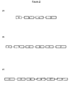
- Table 1 lists some preferred expression control elements that may be used in accordance with the invention, and indicates the cell cycle phase in which each element is activated.
- Table 1 Cell Cycle Phase-Specific Expression Control Elements Element Timing Element Timing Cyclin B1 promoter G2 DHFR promoter late G1 Cdc25B promoter S/G2 Histones promoters late G1/S Cyclin A2 promoter S G2-IRES G2 Cdc2 promoter S HCV IRES M Cdc25C promoter S ODC IRES G2/M Cyclin E promoter late G1 c-myc IRES M Cdc6 promoter late G1 p58 PITSLRE IRES G2/M
- the destruction control element is a DNA sequence encoding a protein motif that controls the destruction of proteins containing that sequence.
- the destruction control element may be cell cycle mediated, for example: Cyclin B1 D-box ( Glotzer et al, Nature, (1991), 349, 132-138 , Yamano et al, EMBO J., (1998), 17(19), 5670-8 , Clute and Pines, Nature Cell Biology, (1999), 1, 82 - 87 ); cyclin A N-terminus ( den Elzen and Pines, J. Cell Biol., (2001), 153(1), 121-36 , Geley et al, J.
- Cyclin B1 D-box Glotzer et al, Nature, (1991), 349, 132-138 , Yamano et al, EMBO J., (1998), 17(19), 5670-8 , Clute and Pines, Nature Cell Biology, (1999), 1, 82 - 87 ); cyclin A N-terminus ( den Elzen and Pines, J. Cell Biol., (2001), 153(1), 121-36 , Geley e
- Table 2 lists destruction control elements that may be used according to the invention and indicates the cell cycle phase in which each element is activated.
- the destruction control element may be non cell-cycle mediated, such as PEST sequences as described by Rogers et al, Science, (1986), 234, 364-8 .
- non cell-cycle mediated destruction control elements include sequences derived from casein, ornithine decarboxylase and proteins that reduce protein half-life. Use of such non cell-cycle mediated destruction control sequences in the method of the invention provides means for determining the persistence time of the cell cycle reporter following induction of expression by a cell cycle specific promoter.
- the live-cell reporter molecule encoded by the nucleic acid sequence may be selected from the group consisting of fluorescent proteins and enzymes.
- Preferred fluorescent proteins include Green Fluorescent Protein (GFP) from Aequorea victoria and derivatives of GFP such as functional GFP analogues in which the amino acid sequence of wild type GFP has been altered by amino acid deletion, addition, or substitution.
- Suitable GFP analogues for use in the present invention include EGFP ( Cormack, B.P. et al, Gene, (1996), 173, 33-38 ); EYFP and ECFP ( US 6066476, Tsien, R. et al ); F64L-GFP ( US 6172188, Thastrup, O. et al ); BFP, ( US 6077707, Tsien, R. et al ).
- Other fluorescent proteins include DsRed, HcRed and other novel fluorescent proteins ( BD Clontech and Labas, Y.A. et al, Proc Natl Acad Sci U S A (2002), 99, 4256-61 ) and Renilla GFP (Stratagene).
- Suitable enzyme reporters are those which are capable of generating a detectable (e.g. a fluorescent or a luminescent) signal in a substrate for that enzyme.
- Particularly suitable enzyme/substrates include: nitroreductase/Cy-Q (as disclosed in WO 01/57237 ) and ⁇ -lactamase/CCF4.
- the nucleic acid reporter construct includes a cell cycle phase-specific spatial localisation control element comprising a DNA sequence encoding a protein motif that is capable of controlling the sub-cellular localisation of the protein in a cell cycle specific manner.
- a localisation control element is required to determine the sub-cellular localisation of the reporter either to ensure its effective operation and/or destruction. More precise determination of the cell cycle position may be possible using a localisation control element since this will permit measurement of both intensity and location of the reporter signal.
- Suitable spatial localisation control elements include those that regulate localisation of a cell cycle control protein, for example the cyclin B1 CRS.
- operably linked indicates that the elements are arranged so that they function in concert for their intended purposes, e.g. transcription initiates in a promoter and proceeds through the DNA sequence coding for the fluorescent protein of the invention.
- Figure 2 (2A/2B/2C) illustrates the general construction of a DNA construct according to the invention.
- the construct comprises a cyclin B1 promoter, a cyclin B1 destruction box (D-box), a cyclin B1 cytoplasmic retention sequence (CRS) and a green fluorescent protein (GFP).
- D-box a cyclin B1 destruction box
- CRS cyclin B1 cytoplasmic retention sequence
- GFP green fluorescent protein
- the nucleic acid reporter construct comprises an expression vector comprising the following elements:
- the nucleic acid reporter construct additionally contains a eukaryotic drug resistance gene, preferably a mammalian drug resistance gene.
- Expression vectors may also contain other nucleic acid sequences, such as polyadenylation signals, splice donor/splice acceptor signals, intervening sequences, transcriptional enhancer sequences, translational enhancer sequences and the like.
- the drug resistance gene and the reporter gene may be operably linked by an internal ribosome entry site (IRES), which is either cell cycle specific ( Sachs, et al, Cell, (2000), 101, 243-245 ) or cell cycle independent ( Jang et al, J. Virology, (1988), 62, 2636-2643 and Pelletier and Sonenberg, Nature, (1988), 334, 320-325 ), rather than the two genes being driven from separate promoters.
- IRES internal ribosome entry site
- the pIRES-neo and pIRES-puro vectors commercially available from Clontech may be used.
- the nucleic acid reporter construct is assembled from a DNA sequence encoding the cyclin B1 promoter operably linked to DNA sequences encoding 171 amino acids of the amino terminus of cyclin B1 and a DNA sequence encoding a green fluorescent protein (GFP) ( Figure 3). Motifs controlling the localisation and destruction of cyclin B1 have all been mapped to ⁇ 150 amino acids in the amino terminus of the molecule. Consequently, an artificial cell cycle marker can be constructed using only sequences from the amino terminus of cyclin B1, which will not interfere with cell cycle progression since it lacks a specific sequence, termed the cyclin box, ( Nugent et al, J. Cell.
- cyclin B1 a nine amino acid motif termed the destruction box (D-box). This is necessary to target cyclin B1 to the ubiquitination machinery and, in conjunction with at least one C-terminal lysine residue, this is also required for its cell-cycle specific degradation; ii) an approximately ten amino acid nuclear export signal (NES).
- D-box destruction box
- This motif is recognised, either directly or indirectly, by exportin 1 and is sufficient to maintain the bulk of cyclin B1 in the cytoplasm throughout interphase; iii) approximately four mitosis-specific phosphorylation sites that are located in and adjacent to the NES and confer rapid nuclear import and a reduced nuclear export at mitosis.
- exportin 1 When expressed in a eukaryotic cell, the construct will exhibit cell cycle specific expression and destruction of the GFP reporter which parallels the expression and degradation of endogenous cyclin B1.
- GFP fluorescence intensity permits identification of cells in the G2/M phase of the cell cycle ( Figure 4).
- nucleic acid reporter construct according to the first aspect may be constructed by selecting suitable alternative cell cycle control elements, for example from those shown in Tables 1 and 2, to design cell cycle phase reporters which report a desired section of the cell cycle.
- Suitable vector backbones which include bacterial and mammalian drug resistance genes and a bacterial origin of replication include, but are not limited to: pCl-neo (Promega), pcDNA (Invitrogen) and pTriEx1 (Novagen).
- Suitable bacterial drug resistance genes include genes encoding for proteins that confer resistance to antibiotics including, but not restricted to: ampicillin, kanamycin, tetracyclin and chloramphenicol.
- Eurkaryotic drug selection markers include agents such as: neomycin, hygromycin, puromycin, zeocin, mycophenolic acid, histidinol, gentamycin and methotrexate.
- the DNA construct may be prepared by the standard recombinant molecular biology techniques of restriction digestion, ligation, transformation and plasmid purification by methods familiar to those skilled in the art and are as described in Sambrook, J. et al (1989), Molecular Cloning - A Laboratory Manual, Cold Spring Harbor Laboratory Press .
- the construct can be prepared synthetically by established methods, eg. the phosphoramidite method described by Beaucage and Caruthers, (Tetrahedron Letters, (1981), 22, 1859-1869 ) or the method described by Matthes et al (EMBO J., (1984), 3, 801-805 ).
- oligonucleotides are synthesised, eg.
- DNA construct may also be prepared by polymerase chain reaction (PCR) using specific primers, for instance, as described in US4683202 or by Saiki et al (Science, (1988), 239, 487-491 ). A review of PCR methods may be found in PCR protocols, (1990), Academic Press, San Diego, California, U.S.A .
- PCR polymerase chain reaction
- the gene sequence encoding the reporter must be joined in frame with the cell cycle phase specific destruction control element and the spatial localisation control element.
- the resultant DNA construct should then be placed under the control of one or more suitable cell cycle phase specific expression control elements.
- a host cell which is not a human embryonic cell, transfected with a nucleic acid reporter construct according to the present invention.
- the host cell into which the construct or the expression vector containing such a construct is introduced may be any mammalian cell which is capable of expressing the construct.
- the prepared DNA reporter construct may be transfected into a host cell using techniques well known to the skilled person.
- One approach is to temporarily permeabilise the cells using either chemical or physical procedures. These techniques may include: electroporation ( Tur-Kaspa et al, Mol. Cell Biol. (1986), 6, 716-718 ; Potter et al, Proc.Nat.Acad.Sci.USA, (1984), 81, 7161-7165 ), a calcium phosphate based method (eg. Graham and Van der Eb, Virology, (1973), 52, 456-467 and Rippe et al, Mol. Cell Biol., (1990), 10, 689-695 ) or direct microinjection.
- electroporation Tur-Kaspa et al, Mol. Cell Biol. (1986), 6, 716-718 ; Potter et al, Proc.Nat.Acad.Sci.USA, (1984), 81, 7161-7165
- a calcium phosphate based method
- cationic lipid based methods may be used to introduce DNA into cells ( Stewart et al, Human Gene Therapy, (1992), 3, 267 ; Torchilin et al, FASEB J, (1992), 6, 2716 ; Zhu et al, Science, (1993), 261, 209-211 ; Ledley et al, J. Pediatrics, (1987), 110, 1 ; Nicolau et al, Proc.Nat. Acad.Sci.,USA, (1983), 80, 1068 ; Nicolau and Sene, Biochem.Biophys.Acta, (1982), 721, 185-190 ).
- Jiao et al, Biotechnology, (1993), 11, 497-502 describe the use of bombardment mediated gene transfer protocols for transferring and expressing genes in brain tissues which may also be used to transfer the DNA into host cells.
- a further alternative method for transfecting the DNA construct into cells utilises the natural ability of viruses to enter cells.
- Such methods include vectors and transfection protocols based on, for example, Herpes simplex virus ( U.S. Pat 5288641 ), cytomegalovirus ( Miller, Curr. Top. Microbiol. Immunol., (1992), 158, 1 ), vaccinia virus ( Baichwal and Sugden, 1986, in Gene Transfer, ed. R. Kucherlapati, New York, Plenum Press, p117-148 ), and adenovirus and adeno-associated virus ( Muzyczka, Curr. Top. Microbiol. Immunol., (1992), 158, 97-129 ).
- suitable recombinant host cells include HeLa cells, Vero cells, Chinese Hamster ovary (CHO), U2OS, COS, BHK, HepG2, NIH 3T3 MDCK, RIN, HEK293 and other mammalian cell lines that are grown in vitro.
- Such cell lines are available from the American Tissue Culture Collection (ATCC), Bethesda, Maryland, U.S.A.
- ATCC American Tissue Culture Collection
- U.S.A U.S.A.
- Cells other than human embryonic cells, from primary cell lines that have been established after removing cells from a mammal followed by culturing the cells for a limited period of time are also intended to be included in the present invention.
- Cell lines which exhibit stable expression of a cell cycle position reporter may also be used in establishing xenografts of engineered cells in host animals using standard methods. ( Krasagakis, K.J et al, Cell Physiol., (2001), 187(3), 386-91 ; Paris, S. et al, Clin.Exp.Metastasis, (1999), 17(10), 817-22 ). Xenografts of tumour cell lines engineered to express cell cycle position reporters will enable establishment of model systems to study tumour cell division, stasis and metastasis and to screen new anticancer drugs.
- cells transfected with the DNA reporter construct may be cultured under conditions and for a period of time sufficient to allow expression of the reporter molecule at a specific stage of the cell cycle.
- expression of the reporter molecule will occur between 16 and 72 hours post transfection, but may vary depending on the culture conditions.
- the reporter molecule is based on a green fluorescent protein sequence the reporter may take a defined time to fold into a conformation that is fluorescent. This time is dependent upon the primary sequence of the green fluorescent protein derivative being used.
- the fluorescent reporter protein may also change colour with time (see for example, Terskikh, Science, (2000), 290, 1585-8 ) in which case imaging is required at specified time intervals following transfection.
- the cell cycle position of the cells may be determined by monitoring the expression of the reporter molecule and detecting signals emitted by the reporter using an appropriate detection device. If the reporter molecule produces a fluorescent signal, then, either a conventional fluorescence microscope, or a confocal based fluorescence microscope may be used. If the reporter molecule produces luminescent light, then a suitable device such as a luminometer may be used. Using these techniques, the proportion of cells expressing the reporter molecule may be determined. If the DNA construct contains translocation control elements and the cells are examined using a microscope, the location of the reporter may also be determined.
- the fluorescence of cells transformed or transfected with the DNA construct may suitably be measured by optical means in for example; a spectrophotometer, a fluorimeter, a fluorescence microscope, a cooled charge-coupled device (CCD) imager (such as a scanning imager or an area imager), a fluorescence activated cell sorter, a confocal microscope or a scanning confocal device, where the spectral properties of the cells in culture may be determined as scans of light excitation and emission.
- a spectrophotometer a fluorimeter, a fluorescence microscope, a cooled charge-coupled device (CCD) imager (such as a scanning imager or an area imager), a fluorescence activated cell sorter, a confocal microscope or a scanning confocal device, where the spectral properties of the cells in culture may be determined as scans of light excitation and emission.
- CCD charge-coupled device
- the nucleic acid reporter construct comprises a drug resistance gene
- cells expressing the modified reporter gene may be selected by growing the cells in the presence of an antibiotic for which transfected cells are resistant due, to the presence of a selectable marker gene.
- the purpose of adding the antibiotic is to select for cells that express the reporter gene and that have, in some cases, integrated the reporter gene, with its associated promoter, IRES elements, enhancer and termination sequences into the genome of the cell line.
- a clonal cell line expressing the construct can be isolated using standard techniques. The clonal cell line may then be grown under standard conditions and will express reporter molecule and produce a detectable signal at a specific point in the cell cycle.
- Cells transfected with the nucleic acid reporter construct according to the present invention may be grown in the absence and/or the presence of a test compound consisting of a drug, nucleic acid, hormone, protein or peptide to be studied and whose effect on the cell cycle of a cell is to be determined.
- a test compound consisting of a drug, nucleic acid, hormone, protein or peptide to be studied and whose effect on the cell cycle of a cell is to be determined.
- an in vitro method of determining the effect of a test compound consisting of a drug, nucleic acid, hormone, protein or peptide on the cell cycle position of a mammalian cell comprising:
- an in vitro method of determining the effect of a test compound consisting of a drug, nucleic acid, hormone, protein or peptide on the cell cycle position of a mammalian cell comprising:
- an in vitro method of determining the effect of a test compound consisting of a drug, nucleic acid, hormone, protein or peptide on the cell cycle position of a mammalian cell comprising:
- test compound it is intended to mean an agent such as a drug, hormone, protein, peptide, nucleic acid and the like, to which the cell is exposed.
- the test compound may be an agent such as a nucleic acid, peptide or protein that is expressed in the cell under study.
- cells transfected with the nucleic acid reporter constructs according to the present invention may be used to determine whether expression of cDNA containing constructs encoding proteins under study have an effect on the cell cycle position of a cell.
- a series of cDNAs, inserted into a mammalian expression vector may be transiently transfected into a cell stably expressing the cell cycle position reporter. By monitoring the expression and location of the nucleic acid reporter construct in these transfected cells, it is possible to determine the effects of the proteins encoded by the cDNAs on the cell cycle.
- the cell cycle position nucleic acid reporter constructs according to the present invention may also be used in a method to determine the effect of the cell cycle position on a cellular process, or to determine the effect of the cell cycle position on the action of a test substance on a cellular process. It is well known that many cellular processes, including those that respond to external stimuli, are influenced by the cell cycle so as to operate or respond differently at different stages of the cell cycle. For example, endothelin receptors have been shown to be expressed at different levels during different phases of the cell cycle ( Okazawa etal.
- cell cycle position reporter constructs will allow cell to cell variations in a biological assay, measured using an appropriate assay reporter, to be correlated with the signal from a cell cycle position reporter in order to determine if any variations in the assay signal correlate with the cell cycle position reporter signal and hence determine any cell cycle dependence of the assay signal.
- assays may be devised in which the amount of a red fluorescently labelled ligand bound to a cell surface receptor is correlated with cell cycle status determined using a GFP cell cycle position reporter.
- cellular process it is meant the normal processes which living cells undergo and includes: biosynthesis, uptake, transport, receptor binding, metabolism, fusion, biochemical response, growth and death.
- Two or more cell cycle position nucleic acid reporter constructs may be used in combination in applications that include reporting on transition through two or more cell cycle phases within the same cell.
- two or more different constructs are engineered and expressed in the same cell, wherein each reporter construct comprises a different combination of control elements linked to a different and distinguishable reporter.
- cellular expression of a construct comprising a cyclin B1 promoter and cyclin B1 D-box operably linked to GFP in combination with expression in the same cell of a second construct comprising a cyclin A2 promoter and cyclin B1 D-box operably linked to blue fluorescent protein (BFP) will allow discrimination of cells in S phase (blue fluorescence) from cells in G2/M phase (blue and green fluorescence).
- BFP blue fluorescent protein
- cell cycle position nucleic acid reporter constructs and assay methods according to the present invention may be used in a variety of additional applications, for example:
- U20S cells (ATCC HTB-96) were cultured in wells of a 96 well microtitre plate.
- Cells were transfected with a cell cycle reporter construct prepared according to Example 1, comprising a cyclin B1 promoter operably linked to sequences encoding the cyclin B1 D-box, the cyclin B1 CRS, and GFP in a pCORON4004 vector (Amersham Biosciences) using Fugene 6 (Roche) as the transfection agent.
- cell cycle phase reporters of the current invention are suitable for detecting agents which modulate cell cycle progression in a transient system and furthermore such reporters permit identification of the phase of the cell cycle in which cells are blocked.
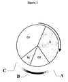
- HeLa cells were micro-injected with the construct prepared according to Example 1 and examined by time lapse microscopy, as shown in Figure 6.
- Differential interference contrast (DIC) images are shown on the left with the corresponding fluorescence image on the right.
- Frame A shows a cell (arrowed) in metaphase which shows bright fluorescence in the nucleus.
- Frames B and C show the same cell at later times in anaphase (B) and late anaphase (C).
- the DIC images of B and C show the division of the cell into two daughter cells (indicated by 2 arrows), the corresponding fluorescence images show the loss of fluorescence accompanying destruction of the fluorescent construct as the cell cycle progresses.
- U2-OS cells (ATCC HTB-96) were transfected with the construct described in example 1 and grown for several months in culture media containing 1 mg/ml geneticin to select for cells stably expressing the construct. A number of clones were picked by standard methods (e.g. described in Freshney, Chapter 11 in Culture of Animal Cells, (1994) Wiley-Liss Inc ) and a clone containing fluorescent cells was isolated. This cell line was maintained at 37°C in culture media containing 25 mM HEPES and a fluorescence and transmitted image of the cells taken every 15 minutes over a period of 24 hours using a standard xenon lamp at 488nm. Figure 7 shows 5 frames from a portion of the image that indicates that the cell line is behaving as expected. Cells in G2 exhibit green fluorescence in the cytoplasm, cells in early mitosis have fluorescence predominantly in the nucleus and following mitosis the reporter gene is degraded and the cells lose their fluorescence.
- Figure 8 shows the fate of a cell from the same clone that was monitored over 48 hours and that underwent two cell divisions to produce four granddaughter cells. For each time point the average intensity of each of the cells' progeny is measured and plotted against time. As can be seen the original cell enters mitosis at - 4 hours, one of the daughters divides at 32 hours and the other at 42 hours into the experiment. As cells leave S-phase and enter G2 there is a steady increase in average intensity until the cell enters mitosis when the cell rounds up and the average intensity increases dramatically.
- the green fluorescent protein reporter sequence in the vector described in example 1 was replaced with enhanced GFP (EGFP; Cormack, B.P. et al, Gene, (1996), 173, 33-38 ; BD Clontech) by standard methods.
- the EGFP gene is a brighter form of GFP containing the mutations F64L and S65T.
- EGFP contains codons that have been altered to optimise expression in mammalian cells.
- This new construct was transfected into U2-OS cells and a number of colonies were isolated by selection with geneticin followed by sorting of single cells using a fluorescence activated cell sorter. These clones showed brighter fluorescence than those generated in example 4 and as expected fluorescence intensity and location appeared to vary according to the cell cycle phase of the cell.
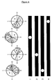
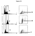
- the cells were prepared for FACS analysis by standard methods. Briefly the cells were fixed and permeabilised using CytoFix/CytoPerm (Becton Dickinson) according to the manufacturers procedures. The cells were then treated with 50 ⁇ g/ml RNAse and 0.4 % Triton X-100 and counterstained with 100 ⁇ g/ml propidium iodide.
- the degree of propidium iodide staining is proportional to the amount of DNA in the cell and therefore a measure of the cell cycle phase of the cell. As can be seen in Figure 9, as expected, the degree of red propidium iodide staining and the amount of green GFP fluorescence appear to be proportional in the cells.
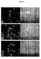
- Example 5 The cells prepared in Example 5 were grown in 25 cm 2 flasks and treated with either 100ng/ml demecolcine (Sigma) or 1 mM mimosine (Sigma) for 24 hours. The cells were then fixed, permeabilised and stained with propidium iodide as described in example 5. FACS analysis revealed that, as expected, cells treated with the colchicine analogue arrested in G2/M and cells treated with mimosine arrested at the G1/S boundary. As is also expected the cells that had been arrested in G2/M were brighter than the cells that had been arrested at G1/S (Figure 10).
Landscapes
- Health & Medical Sciences (AREA)
- Life Sciences & Earth Sciences (AREA)
- Genetics & Genomics (AREA)
- Chemical & Material Sciences (AREA)
- Organic Chemistry (AREA)
- Engineering & Computer Science (AREA)
- Zoology (AREA)
- Biochemistry (AREA)
- Wood Science & Technology (AREA)
- General Health & Medical Sciences (AREA)
- Biophysics (AREA)
- Bioinformatics & Cheminformatics (AREA)
- Molecular Biology (AREA)
- General Engineering & Computer Science (AREA)
- Biotechnology (AREA)
- Biomedical Technology (AREA)
- Cell Biology (AREA)
- Physics & Mathematics (AREA)
- Toxicology (AREA)
- Proteomics, Peptides & Aminoacids (AREA)
- Plant Pathology (AREA)
- Medicinal Chemistry (AREA)
- Microbiology (AREA)
- Gastroenterology & Hepatology (AREA)
- Measuring Or Testing Involving Enzymes Or Micro-Organisms (AREA)
- Micro-Organisms Or Cultivation Processes Thereof (AREA)
- Investigating Or Analysing Biological Materials (AREA)
- Fuel Cell (AREA)
Claims (23)
- Nukleinsäure-Reporter-Konstrukt, umfassend eine Nukleinsäuresequenz, kodierend ein detektierbares Lebendzellen-Reportermolekül, das betreibbar verbunden ist mit und unter der Steuerung steht von:i) mindestens einem Zellzyklusphasen-spezifischen Expressions-Steuerelement,ii) einem Zerstörungs-Steuerelement, undiii) einem Zellzyklusphasen-spezifischen Raumlokalisations-Steuerelement.
- Konstrukt nach Anspruch 1, wobei das Expressions-Steuerelement die Transskription in einer Zellzyklus-spezifischen Weise steuert.
- Konstrukt nach Anspruch 1 oder 2, wobei das Expressions-Steuerelement die Translation in einer Zellzyklus-spezifischen Weise steuert.
- Konstrukt nach Ansprüchen 1 bis 3, wobei das Expressions-Steuerelement ausgewählt ist aus einem Zellzyklus-spezifischen Promotor und einem Zellzyklus-spezifischen IRES.
- Konstrukt nach Anspruch 4, wobei der Promotor ausgewählt ist aus der Gruppe, die besteht aus Cyclin-B1-Promotor, Cdc25B-Promotor, Cyclin-A2-Promotor, Cdc2-Promotor, Cdc25C-Promotor, Cyclin-E-Promotor, Cdc6-Promotor, DHFR-Promotor und Histone-Promotoren.
- Konstrukt nach Anspruch 4 oder 5, wobei das IRES ausgewählt ist aus der Gruppe, die besteht aus G2-IRES, HCV-IRES, ODC-IRES, C-myc-IRES und p58-PITSLRE-IRES.
- Konstrukt nach einem der Ansprüche 1 bis 6, wobei das Zerstörungs-Steuerelement ausgewählt ist aus der Gruppe, die besteht aus Cyclin-B1-D-Box, Cyclin-A-N-Terminus, KEN-Box, Cyclin-E und p27Kip1.
- Konstrukt nach einem der Ansprüche 1 bis 7, wobei das Zellzyklusphasenspezifische Raumlokalisations-Steuerelement die zytoplasmische Cyclin-B1-Retentionssequenz (CRS) einschließlich seiner NES ist.
- Konstrukt nach einem der Ansprüche 1 bis 8, wobei das Lebendzellen-Reportermolekül ausgewählt ist aus der Gruppe, die besteht aus fluoreszierendem Protein und Enzym.
- Konstrukt nach Anspruch 9, wobei das fluoreszierende Protein ausgewählt ist aus Grün Fluoreszierendem Protein (GFP) und einem funktionellen GFP-Analogon, in dem die Aminosäuresequenz des Wild-Typ-GFP geändert wurde durch Aminosäure-Deletion, -Addition oder-Substitution.
- Konstrukt nach Anspruch 9, wobei der Enzymreporter ausgewählt ist aus der Gruppe, die besteht aus β-Lactamase und Nitroreduktase.
- Konstrukt nach einem der Ansprüche 1 bis 11, umfassend einen Cyclin-B1-Promotor, eine Cyclin-B1-Zerstörungsbox (D-Box), eine zytoplasmatische Cyclin-B1-Retentionssequenz (CRS) und ein Grün Fluoreszierendes Protein (GFP).
- Nukleinsäure-Reporterkonstrukt, umfassend einen Expressionsvektor, umfassend:a) ein Vektor-Rückgrat, umfassendi) einen bakteriellen Ursprung der Replikation; undii) ein bakterielles Arzneimittel-Resistenz-Gen;b) ein Zellzyklusphasen-spezifisches Expressions-Steuerelement;c) ein Zerstörungs-Steuerelement;d) ein Zellzyklusphasen-spezifisches Raumlokalisations-Steuerelment; unde) eine Nukleinsäuresequenz, die ein Reportermolekül kodiert.
- Konstrukt nach Anspruch 13, zusätzlich enthaltend ein eukaryontisches Arzneimittel-Resistenz-Gen.
- Wirtszelle, die keine menschliche Embryonenzelle ist, transfiziert mit einem Konstrukt nach einem der Ansprüche 1 bis 14.
- In-vitro-Verfahren zum Bestimmen der Zellzyklus-Position einer Säugerzelle, wobei das Verfahren umfasst:a) Exprimieren in einer Zelle ein Nukleinsäure-Reporter-Konstrukt nach einem der Ansprüche 1 bis 14; undb) Bestimmen der Zellzyklusposition durch Überwachen von Signalen, die von dem Reportermolekül emittiert werden.
- In-vitro-Verfahren des Bestimmens einer Testverbindung, bestehend aus einem Arzneimittel, einer Nukleinsäure, einem Hormon, einem Protein oder einem Peptid auf der Zellzyklusposition einer Säugerzelle, wobei das Verfahren umfasst:a) Exprimieren in der Zelle bei Abwesenheit und bei Vorhandensein der Testverbindung ein Nukleinsäure-Reporter-Konstrukt, umfassend eine Nukleinsäuresequenz, die ein detektierbares Lebendzellen-Reportermolekül codiert, das betreibbar verbunden ist mit und unter der Steuerung steht voni) einem Zellzyklusphasen-spezifischen Expressions-Steuerelement,ii) einem Zerstörungs-Steuerelement, undiii) einem Zellzyklusphasen-spezifischen Raumlokalisations-Steuerelement;wobei das Reporterkonstrukt exprimiert wird in einer Zelle an einem vorbestimmten Punkt im Zellzyklus; undb) Bestimmen der Zellzyklusposition durch Überwachen von Signalen, die von dem Reportermolekül emittiert wird, wobei eine Differenz zwischen den emittierten Signalen, die bei Abwesenheit und bei Vorhandensein der Testverbindung gemessen werden, den Effekt der Testverbindung auf die Zellzyklusposition der Zelle anzeigt.
- In-vitro-Verfahren des Bestimmens des Effekts einer Testverbindung, bestehend aus einem Arzneimittel, einer Nukleinsäure, einem Hormon, einem Protein oder einem Peptid auf der Zellzyklusposition einer Säugerzelle, wobei das Verfahren umfasst:a) Exprimieren in der Zelle bei Vorhandensein der Testverbindung ein Nukleinsäure-Reporterkonstrukt, umfassend eine Nukleinsäuresequenz, die ein detektierbares Lebendzellen-Reportermolekül codiert, das betreibbar verbunden ist mit und unter der Steuerung steht von:i) ein Zellzyklusphasen-spezifisches Expressions-Steuerelement,ii) ein Zerstörungs-Steuerelement, undiii) ein Zellzyklusphasen-spezifischen Raumlokalisations-Steuerelement;wobei das Reporterkonstrukt exprimiert wird in einer Zelle an einem vorbestimmten Punkt im Zellzyklus; undb) Bestimmen der Zellzyklusposition durch Überwachen von Signalen, die von dem Reportermolekül emittiert werden,c) Vergleichen des emittierten Signals bei Vorhandensein der Testverbindung mit einem bekannten Wert für das emittierte Signal in Abwesenheit der Testverbindung;wobei eine Differenz zwischen dem emittierten Signal, das bei Vorhandensein der Testverbindung gemessen wird, und dem bekannten Wert in Abwesenheit der Testverbindung den Effekt der Testverbindung auf die Zellzyklusposition der Zelle anzeigt.
- In-vitro-Verfahren des Bestimmens des Effekts einer Testverbindung, bestehend aus einem Arzneimittel, einer Nukleinsäure, einem Hormon, einem Protein oder einem Peptid auf der Zellzyklusposition einer Säugerzelle, wobei das Verfahren umfasst:a) Bereitstellen von Zellen, die ein Nukleinsäure-Reporterkonstrukt enthalten, das eine Nukleinsäuresequenz umfasst, die ein detektierbares Lebendzellen-Reportermolekül kodiert, das betreibbar verbunden ist mit und unter der Steuerung steht von:i) einem Zellzyklusphasen-spezifischen Expressions-Steuerelement,ii) einem Zerstörungs-Steuerelement, undiii) einem Zellzyklusphase-spezifischen Raumlokalisations-Steuerelement;wobei das Reporterkonstrukt exprimiert wird in einer Zelle an einem vorbestimmten Punkt in dem Zellzyklus;b) Kultivieren erster und zweiter Populationen der Zellen jeweils bei Vorhandensein und in Abwesenheit der Testverbindung und unter Bedingungen, die die Expression des Nukleinsäure-Reporterkonstrukts erlauben; undc) Messen der Signale, die von dem Reportermolekül in den ersten und zweiten Zellpopulationen emittiert werden;wobei eine Differenz zwischen den emittierten Signalen, die in der ersten und der zweiten Zellpopulation gemessen werden, den Effekt der Testverbindung auf die Zellzyklusposition der Zelle anzeigt.
- In-vitro-Verfahren des Bestimmens des Effektes des Säugerzellenzyklus auf die Expression, Translokation oder sub-zellulären Verteilung eines ersten detektierbaren Reporters, der dafür bekannt ist, dass er in Antwort auf eine Testverbindung variiert, die besteht aus einem Arzneimittel, einer Nukleinsäure, einem Hormon, einem Protein oder einem Peptid, wobei das Verfahren umfasst:a) Exprimieren in der Zelle bei Vorhandensein der Testverbindung ein zweites Nukleinsäure-Reporterkonstrukt, umfassend eine Nukleinsäuresequenz, die ein detektierbares Lebendzellen-Reportermolekül codiert, das betreibbar verbunden ist mit und unter der Steuerung steht von:i) ein Zellzyklusphasen-spezifisches Expressions-Steuerelement,ii) ein Zerstörungs-Steuerelement, undiii) ein Zellzyklusphasen-spezifisches Raumlokalisations-Steuerelement; wobei das Reporterkonstrukt exprimiert wird in einer Zelle bei einem vorbestimmten Punkt im Zellzyklus;b) Bestimmen der Zellzyklusposition durch Überwachen von Signalen, die von dem zweiten Reportermolekül emittiert werden; undc) Überwachen der Signale, die durch den ersten detektierbaren Reporter emittiert werden,wobei die Beziehung zwischen der Zellzyklusposition, die durch b) bestimmt wird und dem Signal, das von dem ersten detektierbaren Reporter emittiert wird, anzeigt, ob oder ob nicht die Expression, Translokation oder subzelluläre Verteilung des ersten detektierbaren Reporters Zellzyklus-abhängig ist.
- In-vitro-Verfahren nach einem der Ansprüche 16 bis 20, wobei das Nukleinsäure-Reporterkonstrukt das Konstrukt des Anspruchs 12 umfasst.
- In-vitro-Verfahren nach einem der Ansprüche 17 bis 21, wobei die Testverbindung ein Mittel ist, ausgewählt aus einem Peptid oder Protein, das in der studierten Zelle exprimiert wird.
- Nicht-menschlicher transgener Organismus, umfassend eine Zelle nach Anspruch 15.
Applications Claiming Priority (3)
| Application Number | Priority Date | Filing Date | Title |
|---|---|---|---|
| GB0123856 | 2001-10-05 | ||
| GBGB0123856.7A GB0123856D0 (en) | 2001-10-05 | 2001-10-05 | Method for determining cell cycle position |
| PCT/GB2002/004258 WO2003031612A2 (en) | 2001-10-05 | 2002-09-12 | Method for determining cell cycle position |
Publications (2)
| Publication Number | Publication Date |
|---|---|
| EP1432819A2 EP1432819A2 (de) | 2004-06-30 |
| EP1432819B1 true EP1432819B1 (de) | 2007-08-01 |
Family
ID=9923245
Family Applications (1)
| Application Number | Title | Priority Date | Filing Date |
|---|---|---|---|
| EP02760417A Expired - Lifetime EP1432819B1 (de) | 2001-10-05 | 2002-09-12 | Methode zur bestimmung der zellzyklusposition |
Country Status (11)
| Country | Link |
|---|---|
| US (2) | US7235401B2 (de) |
| EP (1) | EP1432819B1 (de) |
| JP (1) | JP4607454B2 (de) |
| AT (1) | ATE368753T1 (de) |
| AU (1) | AU2002326036B2 (de) |
| CA (1) | CA2461133C (de) |
| DE (1) | DE60221550T2 (de) |
| ES (1) | ES2290326T3 (de) |
| GB (1) | GB0123856D0 (de) |
| IL (1) | IL160908A0 (de) |
| WO (1) | WO2003031612A2 (de) |
Families Citing this family (9)
| Publication number | Priority date | Publication date | Assignee | Title |
|---|---|---|---|---|
| GB0307684D0 (en) * | 2003-04-02 | 2003-05-07 | Amersham Biosciences Uk Ltd | Determining cell cycle phase data |
| US7369696B2 (en) * | 2003-04-02 | 2008-05-06 | Ge Healthcare Uk Limited | Classification of cells into subpopulations using cell classifying data |
| US20050022259A1 (en) * | 2003-05-08 | 2005-01-27 | Pruitt Steven C. | Transgenic animal model for cancer and stem cells |
| US7745123B2 (en) | 2004-07-23 | 2010-06-29 | Ge Healthcare Uk Limited | Cell cycle reporting cell line |
| CN101052646B (zh) * | 2004-07-23 | 2015-10-21 | 通用电气医疗集团英国有限公司 | 细胞周期阶段标志物 |
| AU2005263988B2 (en) * | 2004-07-23 | 2009-10-01 | Ge Healthcare Uk Limited | Cell cycle phase markers |
| JP5408839B2 (ja) * | 2006-10-20 | 2014-02-05 | オリンパス株式会社 | 細胞周期解析方法 |
| EP2275567A1 (de) | 2009-07-15 | 2011-01-19 | Rheinische Friedrich-Wilhelms-Universität Bonn | Nukleinsäureexpressionskonstrukt und dessen Verwendung als ein Zellproliferationsmarker |
| AU2012284259A1 (en) | 2011-07-15 | 2014-03-06 | Sarepta Therapeutics, Inc. | Methods and compositions for manipulating translation of protein isoforms from alternative initiation start sites |
Family Cites Families (10)
| Publication number | Priority date | Publication date | Assignee | Title |
|---|---|---|---|---|
| US5288641A (en) * | 1984-06-04 | 1994-02-22 | Arch Development Corporation | Herpes Simplex virus as a vector |
| US4683202A (en) * | 1985-03-28 | 1987-07-28 | Cetus Corporation | Process for amplifying nucleic acid sequences |
| FR2658204A1 (fr) * | 1990-02-12 | 1991-08-16 | Inst Nat Sante Rech Med | Nouvelles compositions de cycline a humaine, sequence nucleotidique correspondante, et agents de detection ou de diagnostic de mutations hepatocellulaires induites par le virus hbv. |
| US6159691A (en) * | 1996-05-15 | 2000-12-12 | Prolifix Limited | Assay for a putative regulator of cell cycle progression |
| US6124128A (en) * | 1996-08-16 | 2000-09-26 | The Regents Of The University Of California | Long wavelength engineered fluorescent proteins |
| US6048693A (en) * | 1996-10-16 | 2000-04-11 | Bittech, Inc. | Phenotypic assays of cyclin/cyclin-dependent kinase function |
| US6461813B2 (en) * | 1998-09-21 | 2002-10-08 | Rigel Pharmaceuticals, Inc. | Multiparameter FACS assays to detect alterations in cell cycle regulation |
| DE19831420A1 (de) * | 1998-07-14 | 2000-01-20 | Hoechst Marion Roussel De Gmbh | Expressionssysteme enthaltend chimäre Promotoren mit Bindungsstellen für rekombinante Transkriptionsfaktoren |
| US6764852B2 (en) * | 1999-01-26 | 2004-07-20 | Vlaams Interuniversitair Instituut Voor Biotechnologie Vzw | Internal ribosome entry site, vector containing same and uses thereof |
| GB0002261D0 (en) * | 2000-02-02 | 2000-03-22 | Amersham Pharm Biotech Uk Ltd | Fluorescent detection method & reagent |
-
2001
- 2001-10-05 GB GBGB0123856.7A patent/GB0123856D0/en not_active Ceased
-
2002
- 2002-09-12 WO PCT/GB2002/004258 patent/WO2003031612A2/en active IP Right Grant
- 2002-09-12 AU AU2002326036A patent/AU2002326036B2/en not_active Ceased
- 2002-09-12 DE DE60221550T patent/DE60221550T2/de not_active Expired - Lifetime
- 2002-09-12 IL IL16090802A patent/IL160908A0/xx unknown
- 2002-09-12 ES ES02760417T patent/ES2290326T3/es not_active Expired - Lifetime
- 2002-09-12 CA CA002461133A patent/CA2461133C/en not_active Expired - Fee Related
- 2002-09-12 JP JP2003534582A patent/JP4607454B2/ja not_active Expired - Fee Related
- 2002-09-12 AT AT02760417T patent/ATE368753T1/de not_active IP Right Cessation
- 2002-09-12 EP EP02760417A patent/EP1432819B1/de not_active Expired - Lifetime
- 2002-09-12 US US10/491,762 patent/US7235401B2/en not_active Expired - Fee Related
-
2007
- 2007-06-12 US US11/761,441 patent/US20070238123A1/en not_active Abandoned
Non-Patent Citations (1)
| Title |
|---|
| None * |
Also Published As
| Publication number | Publication date |
|---|---|
| DE60221550T2 (de) | 2008-04-17 |
| CA2461133C (en) | 2009-02-10 |
| WO2003031612A3 (en) | 2003-10-30 |
| US7235401B2 (en) | 2007-06-26 |
| ATE368753T1 (de) | 2007-08-15 |
| DE60221550D1 (de) | 2007-09-13 |
| AU2002326036B2 (en) | 2007-09-13 |
| JP4607454B2 (ja) | 2011-01-05 |
| ES2290326T3 (es) | 2008-02-16 |
| CA2461133A1 (en) | 2003-04-17 |
| WO2003031612A2 (en) | 2003-04-17 |
| JP2005504550A (ja) | 2005-02-17 |
| US20070238123A1 (en) | 2007-10-11 |
| IL160908A0 (en) | 2004-08-31 |
| EP1432819A2 (de) | 2004-06-30 |
| GB0123856D0 (en) | 2001-11-28 |
| US20040248133A1 (en) | 2004-12-09 |
Similar Documents
| Publication | Publication Date | Title |
|---|---|---|
| US20070238123A1 (en) | Method for determining cell cycle position | |
| AU751163B2 (en) | Rapidly degrading GFP-fusion proteins and methods of use | |
| Niedenthal et al. | Green fluorescent protein as a marker for gene expression and subcellular localization in budding yeast | |
| JP2011015685A (ja) | 新規な発現ベクター | |
| AU2005263993B2 (en) | Cell cycle reporting cell line | |
| EP1071952A1 (de) | I kappa b und verbessertes grün-fluoreszierendes protein | |
| JP2005504550A5 (de) | ||
| US20040053328A1 (en) | Monitoring proteins for the activities of low-molecular- weight gtp-binding proteins | |
| WO2011090159A1 (ja) | 蛋白質の分解活性を測定するためのプローブ試薬 | |
| US7745123B2 (en) | Cell cycle reporting cell line | |
| Brini et al. | Targeting of aequorin for calcium monitoring in intracellular compartments | |
| KR100791859B1 (ko) | p53 종양 억제단백질과 형광단백질의 융합단백질을발현하는 형질전환 세포주 및 이를 이용한 p53 종양억제단백질의 활성과 관련된 물질의 검색방법 | |
| Bhat et al. | The tobacco BY-2 cell line as a model system to understand in planta nuclear coactivator interactions | |
| Heald | Regulation of nuclear lamina disassembly in mitosis | |
| CA2320894A1 (en) | Protein interaction and transcription factor trap |
Legal Events
| Date | Code | Title | Description |
|---|---|---|---|
| PUAI | Public reference made under article 153(3) epc to a published international application that has entered the european phase |
Free format text: ORIGINAL CODE: 0009012 |
|
| 17P | Request for examination filed |
Effective date: 20040312 |
|
| AK | Designated contracting states |
Kind code of ref document: A2 Designated state(s): AT BE BG CH CY CZ DE DK EE ES FI FR GB GR IE IT LI LU MC NL PT SE SK TR |
|
| AX | Request for extension of the european patent |
Extension state: AL LT LV MK RO SI |
|
| RAP1 | Party data changed (applicant data changed or rights of an application transferred) |
Owner name: CANCER RESEARCH TECHNOLOGY LIMITED Owner name: GE HEALTHCARE UK LIMITED |
|
| GRAP | Despatch of communication of intention to grant a patent |
Free format text: ORIGINAL CODE: EPIDOSNIGR1 |
|
| GRAS | Grant fee paid |
Free format text: ORIGINAL CODE: EPIDOSNIGR3 |
|
| GRAA | (expected) grant |
Free format text: ORIGINAL CODE: 0009210 |
|
| AK | Designated contracting states |
Kind code of ref document: B1 Designated state(s): AT BE BG CH CY CZ DE DK EE ES FI FR GB GR IE IT LI LU MC NL PT SE SK TR |
|
| REG | Reference to a national code |
Ref country code: GB Ref legal event code: FG4D |
|
| REG | Reference to a national code |
Ref country code: CH Ref legal event code: EP |
|
| REG | Reference to a national code |
Ref country code: IE Ref legal event code: FG4D |
|
| REF | Corresponds to: |
Ref document number: 60221550 Country of ref document: DE Date of ref document: 20070913 Kind code of ref document: P |
|
| REG | Reference to a national code |
Ref country code: SE Ref legal event code: TRGR |
|
| REG | Reference to a national code |
Ref country code: CH Ref legal event code: NV Representative=s name: ISLER & PEDRAZZINI AG |
|
| PG25 | Lapsed in a contracting state [announced via postgrant information from national office to epo] |
Ref country code: BG Free format text: LAPSE BECAUSE OF FAILURE TO SUBMIT A TRANSLATION OF THE DESCRIPTION OR TO PAY THE FEE WITHIN THE PRESCRIBED TIME-LIMIT Effective date: 20071101 Ref country code: FI Free format text: LAPSE BECAUSE OF FAILURE TO SUBMIT A TRANSLATION OF THE DESCRIPTION OR TO PAY THE FEE WITHIN THE PRESCRIBED TIME-LIMIT Effective date: 20070801 |
|
| REG | Reference to a national code |
Ref country code: ES Ref legal event code: FG2A Ref document number: 2290326 Country of ref document: ES Kind code of ref document: T3 |
|
| ET | Fr: translation filed | ||
| PG25 | Lapsed in a contracting state [announced via postgrant information from national office to epo] |
Ref country code: AT Free format text: LAPSE BECAUSE OF FAILURE TO SUBMIT A TRANSLATION OF THE DESCRIPTION OR TO PAY THE FEE WITHIN THE PRESCRIBED TIME-LIMIT Effective date: 20070801 |
|
| PG25 | Lapsed in a contracting state [announced via postgrant information from national office to epo] |
Ref country code: BE Free format text: LAPSE BECAUSE OF FAILURE TO SUBMIT A TRANSLATION OF THE DESCRIPTION OR TO PAY THE FEE WITHIN THE PRESCRIBED TIME-LIMIT Effective date: 20070801 |
|
| PG25 | Lapsed in a contracting state [announced via postgrant information from national office to epo] |
Ref country code: MC Free format text: LAPSE BECAUSE OF NON-PAYMENT OF DUE FEES Effective date: 20070930 Ref country code: DK Free format text: LAPSE BECAUSE OF FAILURE TO SUBMIT A TRANSLATION OF THE DESCRIPTION OR TO PAY THE FEE WITHIN THE PRESCRIBED TIME-LIMIT Effective date: 20070801 Ref country code: GR Free format text: LAPSE BECAUSE OF FAILURE TO SUBMIT A TRANSLATION OF THE DESCRIPTION OR TO PAY THE FEE WITHIN THE PRESCRIBED TIME-LIMIT Effective date: 20071102 |
|
| PG25 | Lapsed in a contracting state [announced via postgrant information from national office to epo] |
Ref country code: SK Free format text: LAPSE BECAUSE OF FAILURE TO SUBMIT A TRANSLATION OF THE DESCRIPTION OR TO PAY THE FEE WITHIN THE PRESCRIBED TIME-LIMIT Effective date: 20070801 Ref country code: CZ Free format text: LAPSE BECAUSE OF FAILURE TO SUBMIT A TRANSLATION OF THE DESCRIPTION OR TO PAY THE FEE WITHIN THE PRESCRIBED TIME-LIMIT Effective date: 20070801 Ref country code: PT Free format text: LAPSE BECAUSE OF FAILURE TO SUBMIT A TRANSLATION OF THE DESCRIPTION OR TO PAY THE FEE WITHIN THE PRESCRIBED TIME-LIMIT Effective date: 20080102 |
|
| PLBE | No opposition filed within time limit |
Free format text: ORIGINAL CODE: 0009261 |
|
| STAA | Information on the status of an ep patent application or granted ep patent |
Free format text: STATUS: NO OPPOSITION FILED WITHIN TIME LIMIT |
|
| 26N | No opposition filed |
Effective date: 20080506 |
|
| PG25 | Lapsed in a contracting state [announced via postgrant information from national office to epo] |
Ref country code: IE Free format text: LAPSE BECAUSE OF NON-PAYMENT OF DUE FEES Effective date: 20070912 |
|
| PG25 | Lapsed in a contracting state [announced via postgrant information from national office to epo] |
Ref country code: EE Free format text: LAPSE BECAUSE OF FAILURE TO SUBMIT A TRANSLATION OF THE DESCRIPTION OR TO PAY THE FEE WITHIN THE PRESCRIBED TIME-LIMIT Effective date: 20070801 |
|
| PG25 | Lapsed in a contracting state [announced via postgrant information from national office to epo] |
Ref country code: CY Free format text: LAPSE BECAUSE OF FAILURE TO SUBMIT A TRANSLATION OF THE DESCRIPTION OR TO PAY THE FEE WITHIN THE PRESCRIBED TIME-LIMIT Effective date: 20070801 |
|
| PG25 | Lapsed in a contracting state [announced via postgrant information from national office to epo] |
Ref country code: LU Free format text: LAPSE BECAUSE OF NON-PAYMENT OF DUE FEES Effective date: 20070912 |
|
| PG25 | Lapsed in a contracting state [announced via postgrant information from national office to epo] |
Ref country code: TR Free format text: LAPSE BECAUSE OF FAILURE TO SUBMIT A TRANSLATION OF THE DESCRIPTION OR TO PAY THE FEE WITHIN THE PRESCRIBED TIME-LIMIT Effective date: 20070801 |
|
| REG | Reference to a national code |
Ref country code: DE Ref legal event code: R082 Ref document number: 60221550 Country of ref document: DE Representative=s name: J D REYNOLDS & CO., GB |
|
| PGFP | Annual fee paid to national office [announced via postgrant information from national office to epo] |
Ref country code: ES Payment date: 20140926 Year of fee payment: 13 |
|
| REG | Reference to a national code |
Ref country code: FR Ref legal event code: PLFP Year of fee payment: 15 |
|
| PGFP | Annual fee paid to national office [announced via postgrant information from national office to epo] |
Ref country code: NL Payment date: 20160926 Year of fee payment: 15 Ref country code: CH Payment date: 20160927 Year of fee payment: 15 Ref country code: GB Payment date: 20160927 Year of fee payment: 15 |
|
| PGFP | Annual fee paid to national office [announced via postgrant information from national office to epo] |
Ref country code: FR Payment date: 20160926 Year of fee payment: 15 Ref country code: SE Payment date: 20160928 Year of fee payment: 15 |
|
| PGFP | Annual fee paid to national office [announced via postgrant information from national office to epo] |
Ref country code: DE Payment date: 20160928 Year of fee payment: 15 |
|
| PGFP | Annual fee paid to national office [announced via postgrant information from national office to epo] |
Ref country code: IT Payment date: 20160923 Year of fee payment: 15 |
|
| REG | Reference to a national code |
Ref country code: ES Ref legal event code: FD2A Effective date: 20170303 |
|
| PG25 | Lapsed in a contracting state [announced via postgrant information from national office to epo] |
Ref country code: ES Free format text: LAPSE BECAUSE OF NON-PAYMENT OF DUE FEES Effective date: 20150913 |
|
| REG | Reference to a national code |
Ref country code: DE Ref legal event code: R082 Ref document number: 60221550 Country of ref document: DE |
|
| REG | Reference to a national code |
Ref country code: DE Ref legal event code: R119 Ref document number: 60221550 Country of ref document: DE |
|
| REG | Reference to a national code |
Ref country code: CH Ref legal event code: PL |
|
| REG | Reference to a national code |
Ref country code: SE Ref legal event code: EUG |
|
| REG | Reference to a national code |
Ref country code: NL Ref legal event code: MM Effective date: 20171001 |
|
| GBPC | Gb: european patent ceased through non-payment of renewal fee |
Effective date: 20170912 |
|
| PG25 | Lapsed in a contracting state [announced via postgrant information from national office to epo] |
Ref country code: NL Free format text: LAPSE BECAUSE OF NON-PAYMENT OF DUE FEES Effective date: 20171001 |
|
| REG | Reference to a national code |
Ref country code: FR Ref legal event code: ST Effective date: 20180531 |
|
| PG25 | Lapsed in a contracting state [announced via postgrant information from national office to epo] |
Ref country code: DE Free format text: LAPSE BECAUSE OF NON-PAYMENT OF DUE FEES Effective date: 20180404 Ref country code: CH Free format text: LAPSE BECAUSE OF NON-PAYMENT OF DUE FEES Effective date: 20170930 Ref country code: LI Free format text: LAPSE BECAUSE OF NON-PAYMENT OF DUE FEES Effective date: 20170930 Ref country code: GB Free format text: LAPSE BECAUSE OF NON-PAYMENT OF DUE FEES Effective date: 20170912 |
|
| PG25 | Lapsed in a contracting state [announced via postgrant information from national office to epo] |
Ref country code: FR Free format text: LAPSE BECAUSE OF NON-PAYMENT OF DUE FEES Effective date: 20171002 Ref country code: IT Free format text: LAPSE BECAUSE OF NON-PAYMENT OF DUE FEES Effective date: 20170912 |
|
| PG25 | Lapsed in a contracting state [announced via postgrant information from national office to epo] |
Ref country code: SE Free format text: LAPSE BECAUSE OF NON-PAYMENT OF DUE FEES Effective date: 20170913 |