EP1424082A1 - Method of regenerating bone/chondral tissues by transferring transcriptional factor gene - Google Patents
Method of regenerating bone/chondral tissues by transferring transcriptional factor gene Download PDFInfo
- Publication number
- EP1424082A1 EP1424082A1 EP02741405A EP02741405A EP1424082A1 EP 1424082 A1 EP1424082 A1 EP 1424082A1 EP 02741405 A EP02741405 A EP 02741405A EP 02741405 A EP02741405 A EP 02741405A EP 1424082 A1 EP1424082 A1 EP 1424082A1
- Authority
- EP
- European Patent Office
- Prior art keywords
- cells
- bone
- transcription factor
- osteo
- chondro
- Prior art date
- Legal status (The legal status is an assumption and is not a legal conclusion. Google has not performed a legal analysis and makes no representation as to the accuracy of the status listed.)
- Withdrawn
Links
Images






Classifications
-
- A—HUMAN NECESSITIES
- A61—MEDICAL OR VETERINARY SCIENCE; HYGIENE
- A61L—METHODS OR APPARATUS FOR STERILISING MATERIALS OR OBJECTS IN GENERAL; DISINFECTION, STERILISATION OR DEODORISATION OF AIR; CHEMICAL ASPECTS OF BANDAGES, DRESSINGS, ABSORBENT PADS OR SURGICAL ARTICLES; MATERIALS FOR BANDAGES, DRESSINGS, ABSORBENT PADS OR SURGICAL ARTICLES
- A61L27/00—Materials for grafts or prostheses or for coating grafts or prostheses
- A61L27/36—Materials for grafts or prostheses or for coating grafts or prostheses containing ingredients of undetermined constitution or reaction products thereof, e.g. transplant tissue, natural bone, extracellular matrix
- A61L27/38—Materials for grafts or prostheses or for coating grafts or prostheses containing ingredients of undetermined constitution or reaction products thereof, e.g. transplant tissue, natural bone, extracellular matrix containing added animal cells
- A61L27/3895—Materials for grafts or prostheses or for coating grafts or prostheses containing ingredients of undetermined constitution or reaction products thereof, e.g. transplant tissue, natural bone, extracellular matrix containing added animal cells using specific culture conditions, e.g. stimulating differentiation of stem cells, pulsatile flow conditions
-
- A—HUMAN NECESSITIES
- A61—MEDICAL OR VETERINARY SCIENCE; HYGIENE
- A61L—METHODS OR APPARATUS FOR STERILISING MATERIALS OR OBJECTS IN GENERAL; DISINFECTION, STERILISATION OR DEODORISATION OF AIR; CHEMICAL ASPECTS OF BANDAGES, DRESSINGS, ABSORBENT PADS OR SURGICAL ARTICLES; MATERIALS FOR BANDAGES, DRESSINGS, ABSORBENT PADS OR SURGICAL ARTICLES
- A61L27/00—Materials for grafts or prostheses or for coating grafts or prostheses
- A61L27/36—Materials for grafts or prostheses or for coating grafts or prostheses containing ingredients of undetermined constitution or reaction products thereof, e.g. transplant tissue, natural bone, extracellular matrix
- A61L27/38—Materials for grafts or prostheses or for coating grafts or prostheses containing ingredients of undetermined constitution or reaction products thereof, e.g. transplant tissue, natural bone, extracellular matrix containing added animal cells
- A61L27/3804—Materials for grafts or prostheses or for coating grafts or prostheses containing ingredients of undetermined constitution or reaction products thereof, e.g. transplant tissue, natural bone, extracellular matrix containing added animal cells characterised by specific cells or progenitors thereof, e.g. fibroblasts, connective tissue cells, kidney cells
-
- A—HUMAN NECESSITIES
- A61—MEDICAL OR VETERINARY SCIENCE; HYGIENE
- A61L—METHODS OR APPARATUS FOR STERILISING MATERIALS OR OBJECTS IN GENERAL; DISINFECTION, STERILISATION OR DEODORISATION OF AIR; CHEMICAL ASPECTS OF BANDAGES, DRESSINGS, ABSORBENT PADS OR SURGICAL ARTICLES; MATERIALS FOR BANDAGES, DRESSINGS, ABSORBENT PADS OR SURGICAL ARTICLES
- A61L27/00—Materials for grafts or prostheses or for coating grafts or prostheses
- A61L27/36—Materials for grafts or prostheses or for coating grafts or prostheses containing ingredients of undetermined constitution or reaction products thereof, e.g. transplant tissue, natural bone, extracellular matrix
- A61L27/38—Materials for grafts or prostheses or for coating grafts or prostheses containing ingredients of undetermined constitution or reaction products thereof, e.g. transplant tissue, natural bone, extracellular matrix containing added animal cells
- A61L27/3839—Materials for grafts or prostheses or for coating grafts or prostheses containing ingredients of undetermined constitution or reaction products thereof, e.g. transplant tissue, natural bone, extracellular matrix containing added animal cells characterised by the site of application in the body
- A61L27/3843—Connective tissue
- A61L27/3847—Bones
-
- A—HUMAN NECESSITIES
- A61—MEDICAL OR VETERINARY SCIENCE; HYGIENE
- A61L—METHODS OR APPARATUS FOR STERILISING MATERIALS OR OBJECTS IN GENERAL; DISINFECTION, STERILISATION OR DEODORISATION OF AIR; CHEMICAL ASPECTS OF BANDAGES, DRESSINGS, ABSORBENT PADS OR SURGICAL ARTICLES; MATERIALS FOR BANDAGES, DRESSINGS, ABSORBENT PADS OR SURGICAL ARTICLES
- A61L27/00—Materials for grafts or prostheses or for coating grafts or prostheses
- A61L27/36—Materials for grafts or prostheses or for coating grafts or prostheses containing ingredients of undetermined constitution or reaction products thereof, e.g. transplant tissue, natural bone, extracellular matrix
- A61L27/38—Materials for grafts or prostheses or for coating grafts or prostheses containing ingredients of undetermined constitution or reaction products thereof, e.g. transplant tissue, natural bone, extracellular matrix containing added animal cells
- A61L27/3839—Materials for grafts or prostheses or for coating grafts or prostheses containing ingredients of undetermined constitution or reaction products thereof, e.g. transplant tissue, natural bone, extracellular matrix containing added animal cells characterised by the site of application in the body
- A61L27/3843—Connective tissue
- A61L27/3852—Cartilage, e.g. meniscus
-
- A—HUMAN NECESSITIES
- A61—MEDICAL OR VETERINARY SCIENCE; HYGIENE
- A61P—SPECIFIC THERAPEUTIC ACTIVITY OF CHEMICAL COMPOUNDS OR MEDICINAL PREPARATIONS
- A61P19/00—Drugs for skeletal disorders
-
- C—CHEMISTRY; METALLURGY
- C12—BIOCHEMISTRY; BEER; SPIRITS; WINE; VINEGAR; MICROBIOLOGY; ENZYMOLOGY; MUTATION OR GENETIC ENGINEERING
- C12N—MICROORGANISMS OR ENZYMES; COMPOSITIONS THEREOF; PROPAGATING, PRESERVING, OR MAINTAINING MICROORGANISMS; MUTATION OR GENETIC ENGINEERING; CULTURE MEDIA
- C12N5/00—Undifferentiated human, animal or plant cells, e.g. cell lines; Tissues; Cultivation or maintenance thereof; Culture media therefor
- C12N5/06—Animal cells or tissues; Human cells or tissues
- C12N5/0602—Vertebrate cells
- C12N5/0652—Cells of skeletal and connective tissues; Mesenchyme
- C12N5/0654—Osteocytes, Osteoblasts, Odontocytes; Bones, Teeth
-
- C—CHEMISTRY; METALLURGY
- C12—BIOCHEMISTRY; BEER; SPIRITS; WINE; VINEGAR; MICROBIOLOGY; ENZYMOLOGY; MUTATION OR GENETIC ENGINEERING
- C12N—MICROORGANISMS OR ENZYMES; COMPOSITIONS THEREOF; PROPAGATING, PRESERVING, OR MAINTAINING MICROORGANISMS; MUTATION OR GENETIC ENGINEERING; CULTURE MEDIA
- C12N5/00—Undifferentiated human, animal or plant cells, e.g. cell lines; Tissues; Cultivation or maintenance thereof; Culture media therefor
- C12N5/06—Animal cells or tissues; Human cells or tissues
- C12N5/0602—Vertebrate cells
- C12N5/0652—Cells of skeletal and connective tissues; Mesenchyme
- C12N5/0662—Stem cells
- C12N5/0663—Bone marrow mesenchymal stem cells (BM-MSC)
-
- A—HUMAN NECESSITIES
- A61—MEDICAL OR VETERINARY SCIENCE; HYGIENE
- A61K—PREPARATIONS FOR MEDICAL, DENTAL OR TOILETRY PURPOSES
- A61K35/00—Medicinal preparations containing materials or reaction products thereof with undetermined constitution
- A61K35/12—Materials from mammals; Compositions comprising non-specified tissues or cells; Compositions comprising non-embryonic stem cells; Genetically modified cells
-
- A—HUMAN NECESSITIES
- A61—MEDICAL OR VETERINARY SCIENCE; HYGIENE
- A61K—PREPARATIONS FOR MEDICAL, DENTAL OR TOILETRY PURPOSES
- A61K48/00—Medicinal preparations containing genetic material which is inserted into cells of the living body to treat genetic diseases; Gene therapy
-
- A—HUMAN NECESSITIES
- A61—MEDICAL OR VETERINARY SCIENCE; HYGIENE
- A61L—METHODS OR APPARATUS FOR STERILISING MATERIALS OR OBJECTS IN GENERAL; DISINFECTION, STERILISATION OR DEODORISATION OF AIR; CHEMICAL ASPECTS OF BANDAGES, DRESSINGS, ABSORBENT PADS OR SURGICAL ARTICLES; MATERIALS FOR BANDAGES, DRESSINGS, ABSORBENT PADS OR SURGICAL ARTICLES
- A61L2430/00—Materials or treatment for tissue regeneration
- A61L2430/02—Materials or treatment for tissue regeneration for reconstruction of bones; weight-bearing implants
-
- A—HUMAN NECESSITIES
- A61—MEDICAL OR VETERINARY SCIENCE; HYGIENE
- A61L—METHODS OR APPARATUS FOR STERILISING MATERIALS OR OBJECTS IN GENERAL; DISINFECTION, STERILISATION OR DEODORISATION OF AIR; CHEMICAL ASPECTS OF BANDAGES, DRESSINGS, ABSORBENT PADS OR SURGICAL ARTICLES; MATERIALS FOR BANDAGES, DRESSINGS, ABSORBENT PADS OR SURGICAL ARTICLES
- A61L2430/00—Materials or treatment for tissue regeneration
- A61L2430/06—Materials or treatment for tissue regeneration for cartilage reconstruction, e.g. meniscus
-
- C—CHEMISTRY; METALLURGY
- C12—BIOCHEMISTRY; BEER; SPIRITS; WINE; VINEGAR; MICROBIOLOGY; ENZYMOLOGY; MUTATION OR GENETIC ENGINEERING
- C12N—MICROORGANISMS OR ENZYMES; COMPOSITIONS THEREOF; PROPAGATING, PRESERVING, OR MAINTAINING MICROORGANISMS; MUTATION OR GENETIC ENGINEERING; CULTURE MEDIA
- C12N2501/00—Active agents used in cell culture processes, e.g. differentation
- C12N2501/60—Transcription factors
-
- C—CHEMISTRY; METALLURGY
- C12—BIOCHEMISTRY; BEER; SPIRITS; WINE; VINEGAR; MICROBIOLOGY; ENZYMOLOGY; MUTATION OR GENETIC ENGINEERING
- C12N—MICROORGANISMS OR ENZYMES; COMPOSITIONS THEREOF; PROPAGATING, PRESERVING, OR MAINTAINING MICROORGANISMS; MUTATION OR GENETIC ENGINEERING; CULTURE MEDIA
- C12N2510/00—Genetically modified cells
Definitions
- the present invention relates to a method for regeneration of bone/cartilage tissues by transfection of osteo-/choridro-inducible transcription factor genes.
- the present invention relates to a method of transfection of osteo-/chondro-inducible transcription factor genes to accelerate cell differentiation into the target tissues, thereby effectively constructing bone/cartilage tissues and substitutional bone/cartilage implants produced by the aforementioned method.
- porous biodegradable materials such as porous ceramics as scaffolds has provided sufficient results in the regeneration of hard tissues such as bone and cartilage tissues.
- cytokines responsible for cell differentiation have been directly donated to cells or an expression vector carrying cDNA of the target cytokine has been transfected into cells by lipofection method and so on. A certain level of success has been achieved by such methods.
- transcription factors particularly transcription factors of runt and Helix-Loop-Helix (HLH) types
- HHLH Helix-Loop-Helix
- a runt transcription factor Pebp2alphaA (Pebp2 ⁇ A)/Cbfa1
- an HLH transcription factor such as Scleraxis, Id-1, and I-mfa
- Kunikazu Tsuji et al. "Runt transcription factor associated with osteoblast differentiation, Cbfa1/Pebp2 ⁇ A, and Sox9 function in osteogenesis and chondrogenesis and unusual osteogenesis” Experimental Medicine, 16(11), 25-32, 1998).
- Special attempts in tissue regeneration utilizing functions of these transcription factors have not yet been reported.
- the present inventors have conducted concentrated studies. As a result, they have found that cells can be efficiently induced to differentiate into bone/cartilage tissues by transfecting osteo-/chondro-inducible transcription factors into bone-marrow-derived cells and having them express therein. Further, use of an adenoviral or a retroviral vector for the transfection of a transcription factor results in effective infection to the primary adhesive cells such as bone/cartilage cells, and very effective induction of differentiation at approximately 99% can be achieved. This has led to the completion of the present invention.
- the present invention provides the following (1) to (13).
- the present invention relates to a method of transfection of the osteo-/chondro-inducible (osteo-inducible and/or chondro-inducible) transcription factor gene to isolated cells to construct bone/cartilage (bone and/or cartilage) tissues in vitro by inducing differentiation and proliferation of the cells.
- the transcription factors employed in the present invention are the osteo-/chondro-inducible transcription factors that induce immatured cells to differentiate into bone and/or cartilage cells.
- Examples thereof include Cbfa1, Dlx-5, Bapx1, Msx2, Scleraxis, and Sox9.
- Cbfa1 is a transcription factor that was cloned by Ogawa et al. at Kyoto University in 1993 and proved to be indispensable for inducing differentiation of the mesenchymal stem cell into the osteoblast by Komori et al. at Osaka University ( Komori, T. et al., (1997) Cell 89, 755-764).
- This Cbfa1 has two isoforms, til-1 and pebp2 ⁇ A.
- Dlx-5 is a homolog of the Drosophila distalless (Dll) gene and a transcription factor associated with the ossification of perichondrium and endosporium (Acampora, D. et al., 1999, Development 126, 3795-3809).
- Bapx1 is a homolog of the Drosophila bagpipe homeobox gene and is associated particularly with differentiation of the mesenchymal stem cell into cartilage cells in the spinal cord and is considered to be a regulatory gene of the Cbfa1 gene (Tribioli, C. et al., 1999, Development 126, 5699-5711).
- Msx2 is a homolog of the Drosophila muscle segment homeobox (Msh) gene.
- Scleraxis is a transcription factor that is associated with induction of differentiation from the mesenchymal stem cell to cartilage cell or connective tissue (Cserjesi, P. et al., 1995, Development 121, 1099-1110). Sox9 is expressed in cartilage and it regulates expression of the genes associated with cartilage differentiation of, for example, type II collagen (Ng, L. J. et al., 1997, Dev. Biol. 183, 108-121).
- the above described osteo-/chondro-inducible transcription factor genes can be prepared by conventional techniques, using their known sequences.
- cDNA of a target transcription factor can be prepared by extracting RNA from the osteoblast, preparing primers based on a known sequence of the transcription factor gene, and cloning the cDNA by PCR using the RNA and the primers.
- the cells that are employed in the present invention are immatured cells with differentiation and proliferation ability isolated from a body.
- Examples thereof include the ES (embryonic stem) cell, the mesenchymal stem cell, and the preosteoblast differentiated from the mesenchymal stem cell.
- a bone-marrow-derived mesenchymal stem cell is particularly preferable, and the osteoblast is more preferable.
- the above-described cell is isolated from the body of a patient. This cell is preferably prepared by removing connective tissues with a conventional technique. Alternatively, cells may grow by primary culture before use.
- the cells should be cultured in an appropriate medium for inducing cell differentiation, constructing target tissues.
- an appropriate medium for inducing cell differentiation for example, at least one member selected from the group consisting of dexamethasone, immunosuppressive factors such as FK-506 and cyclosporine, bone morphogenetic proteins (BMP) such as BMP-2, BMP-4, BMP-5, BMP-6, BMP-7, and BMP-9, and osteogenic humoral factors such as TGF ⁇ is preferably supplemented to the medium to induce cell differentiation into bone cells.
- BMP bone morphogenetic proteins
- osteogenic humoral factors such as TGF ⁇
- the osteo-/chondro-inducible transcription factor gene can be transfected into the target cell by a method that is commonly employed for transfection to animal cells.
- the methods that can be employed include the calcium phosphate method, lipofection, electroporation, microinjection, and a method using an adenoviral, a retroviral, or a baculoviral vector. From the viewpoints of safety and transfection efficiency, a method using an adenoviral or a retroviral vector is preferable, and a method using an adenoviral vector is most preferable.
- adenoviral or retroviral vector can be constructed according to a known technique.
- an adenoviral vector can be constructed according to the method of Miyake et al. (Miyake, S. et al., Proc. Natl. Acad. Sci. 93: 1320-1324, 1993).
- a commercially available kit such as the Adenovirus Cre/loxP Kit (Takara Shuzo Co., Ltd.), can be used. This kit is used for constructing a recombinant adenoviral vector utilizing a novel regulation system of gene expression (Kanegae Y. et al., 1995, Nucl. Acids Res.
- the multiplicity of infection (moi) of adenovirus infection is 10 or more, preferably 50 to 200, and more preferably around 100 (approximately 80 to 120).
- the transfected cells should grow in suitable scaffolds in order to construct a complete three-dimensional structure.
- suitable scaffolds include hydroxyapatite or ⁇ -TCP (tricalcium phosphate), porous ceramics such as ⁇ -TCP, collagen, polylactic acid, polyglycolic acid, and a complex thereof (e.g., a complex of polylactic acid, polyglycolic acid resin, and collagen).
- ⁇ -TCP tricalcium phosphate
- porous ceramics such as ⁇ -TCP
- collagen polylactic acid
- polyglycolic acid resin e.g., a complex of polylactic acid, polyglycolic acid resin, and collagen
- These scaffolds may be composed of a simple kind of material or combinations of two or more kinds.
- Porous ceramics are particularly preferable for scaffolds for tissue regeneration because of their high mechanical strength.
- the above-mentioned scaffolds are preferably porous for uniform inoculation of cells.
- the term "porous" refers to a porosity of 40% or higher.
- the pore size is not particularly limited, although a diameter of 200 ⁇ m to 500 ⁇ m is preferable for facilitated bone regeneration.
- Cells can be proliferated by being inoculated and cultured in the scaffold in accordance with a conventional technique.
- Cells may be simply inoculated into the scaffold, or inoculated in the form of mixtures with a liquid such as a buffer, physiological saline, a solvent for injection, or a collagen solution.
- a liquid such as a buffer, physiological saline, a solvent for injection, or a collagen solution.
- the number of cells to be inoculated is determined in accordance with the types of cells or scaffolds in order to regenerate tissues more efficiently, maintaining the morphology of the cells.
- the inoculation density is preferably 1,000,000 cells/ml or higher in the case of osteoblasts.
- a conventional medium for cell culture such as MEM medium, ⁇ -MEM medium, or DMEM medium
- MEM medium can be suitably selected depending on the type of cell to be cultured and then used for cell culture.
- FBS Sigma
- antibiotics such as Antibiotic-Antimycotic (GIBCO BRL) or other substances may be added to the medium.
- Culture is preferably conducted in the presence of 3% to 10% CO 2 at 30°C to 40°C, and particularly preferably in the presence of 5% CO 2 at 37°C.
- the culture period is not particularly limited, and it is at least 4 days, preferably at least 7 days, and more preferably at least 2 weeks.
- the bone/cartilage tissues constructed by the above mentioned method are transplanted or injected into a body together with or separately from the scaffolds. Thus, they can be used as substitutional bone/cartilage implants.
- the present invention provides implants containing bone/cartilage tissues that are constructed in vitro.
- Bone/cartilage tissues may be transplanted into the body separately from scaffolds, although transplantation with scaffolds is preferable in the present invention.
- the most suitable scaffold for a given purpose and application site for implants may be selected from the above-mentioned scaffolds.
- hydroxyapatite is preferable for a transplantation site (or a surgical technique) that requires mechanical strength.
- biodegradable ⁇ -TCP or the like is preferable for a transplantation site (or a surgical technique) that does not require mechanical strength.
- Implants can take any desired configurations or shapes, such as sponges, meshes, unwoven fabric products, discs, films, sticks, particles, or pastes. These configurations and shapes may be suitably selected depending on their applications.
- the implants of the present invention can suitably contain other components in addition to the bone/cartilage tissues and scaffolds within the scope of the present invention.
- growth factors such as basic fibroblast growth factors (bFGF), platelet-derived growth factors (PDGF), insulin and insulin-like growth factors (IGF), hepatocyte growth factor, (HGF), glial-derived neurotrophic factors (GDNF), neurotrophic factors (NF), hormones, cytokines, bone morphogenetic factors (BMP), transforming growth factors (TGF), and vascular endothelial growth factors (VEGF); bone morphogenetic proteins; inorganic salts such as St, Mg, Ca, and CO 3 ; organic substances such as citric acid and phospholipid; and drugs.
- bFGF basic fibroblast growth factors
- PDGF platelet-derived growth factors
- IGF insulin and insulin-like growth factors
- HGF hepatocyte growth factor
- GDNF glial-derived neurotrophic factors
- NF neurotrophic factors
- BMP
- Bone/cartilage tissues of the implants of the present invention are constructed from the cells to which transcription factor genes have been transfected. Bone/cartilage tissues may be constructed not only prior to the transplantation (in vitro) but also after transplantation into bone defects (in vivo).
- the implants of the present invention are highly compatible with bones and of high bone formation ability. Thus, they can be integrated with biological bones and can then restore bone defects immediately after they have been transplanted into a body.
- Example 1 Bone tissue regeneration by transfecting Cbfa1 genes into rat osteoblasts using an adenoviral vector
- cDNAs of Cbfa1 i.e., type I: pebp2 ⁇ A/Cbfa1 (SEQ ID NO: 1) and type II/III: til-1/Cbfa1 (SEQ ID NO: 2), were prepared from two types of plasmids *1 (pSG5/IT (mPEBP2aA) and pSG5/KS (mtil-1)) provided by Sumitomo Pharmaceuticals Co., Ltd., ORF of mPEBP2aA was cleaved therefrom using a Bam HI restriction enzyme, and ORF of mtil-1 was cleaved therefrom using a Bgl II restriction enzyme (Fig. 1).
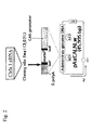
- cDNAs of Cbfa1 were inserted into the SwaI site of the cosmid vector pAxCALNLw using the commercially available Adenovirus Cre/loxP Kit (Takara Shuzo Co., Ltd.), and a recombinant adenoviral vector was prepared according to the manufacturer's instructions attached to the kit (Fig. 2).
- the titer of the prepared virus was approximately 10 11 PFU/ml, and infection efficiency was very high.
- RBMO rat bone marrow osteoblast
- the sampled cells were cultured and in MEM medium (Nacalai Tesque) containing 15% FBS (Sigma) and Antibiotic-Antimycotic (GIBCO BRL) until they became confluent. Subsequently, cells were subcultured into the above mentioned medium containing 5 nM dexamethasone (Sigma), 10 mM ⁇ -glycerophosphate (Sigma), and 50 ⁇ g/ml ascorbic acid phosphate (Wako) in ⁇ 3.5 cm dishes, and they were cultured until the number of cells per dish became approximately 400,000. The subcultured rat osteoblast was infected with the recombinant adenoviral vector prepared as mentioned above at a multiplicity of infection (moi) of 500 on the next day. Uninfected cells or cells infected with the mock vector were prepared as a control.
- the rat osteoblasts 3 days to 2 weeks after adenovirus infection were washed with 100 mM Tris (pH 7.5) and 5 mM MgCl 2 , collected using a scraper, suspended in 500 ⁇ l of 100 mM Tris (pH 7.5), 5 mM MgCl 2 , and 1% Triton X-100, and then disrupted by sonication. After the sonication, cells were centrifuged at 6,000 g for 5 minutes to recover the supernatant.
- the supernatant (5 ⁇ l in each case) was added to 0.056M 2-amino-2-methyl-1,3-propanediol (pH 9.9), 10 mM p-nitrophenyl phosphate, and 2 mM MgCl 2 , the resultants were incubated at 37°C for 30 minutes, and then absorbance at 405 nm was measured using a microplate reader immediately thereafter.
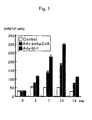
- the enzyme activity was determined based on the calibration curve prepared using p-nitrophenol from the obtained absorbance (Fig. 5).
- the rat osteoblasts 1 to 3 weeks after adenovirus infection were fixed with 3.7% formalin buffer and then decalcified with 0.6M HCl all day and night.
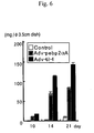
- the decalcified solution was diluted, and the calcium level was assayed using a commercially available o-cresol phthalein complexon-containing reagent for calcium analysis (# 587, 360-11, Sigma) according to the manufacturer's instructions (Fig. 6).
- Total RNA was extracted from the rat osteoblasts after adenovirus infection using a commercially available TRIzol reagent (# 15596-10551, GIBCO BRL) according to the manufacturer's instructions.
- Total RNA (10 ⁇ g) was separated on a 1% agarose/5.5% formaldehyde gel and transferred in 20x SSC to the HybondTM-XL membrane (Amersham Pharmacia Biotech). Thereafter, the membrane was heated at 80°C for 2 hours and the irradiated with ultraviolet rays for 2 minutes.
- the cDNA probes of GAPDH and Cbfa1 were labeled with ⁇ - 32 PdCTP (3,000 Ci/mmol, Amersham Pharmacia Biotech) using RediprimeTM (Amersham Pharmacia Biotech), and the ⁇ - 32 PdCTP that had not been incorporated was removed using a MicroSpinTM G-25 Column (Amersham Pharmacia Biotech). This membrane was incubated in the PerfectHybTM Plus Hybridization Buffer (Sigma) at 68°C for 30 minutes, the labeled cDNA probe (2x10 6 cpm/ml) was added, and incubation was further carried out at 68°C for 1 hour.
- the membrane was washed with 2x SSC/0.1% SDS at room temperature for 5 minutes and then washed twice with 0.5x SSC/0.1% SDS at 68°C (20 minutes each). Thereafter, the membrane was exposed to the Kodak XAR film at -80°C overnight (Fig. 7).
- the osteoblasts 3 days to 2 weeks after adenovirus infection were immobilized with PBS containing 1% glutaraldehyde for 5 minutes, washed twice with distilled water, and then stained with 1% crystal violet at room temperature for 30 minutes. After an excess amount of dye was removed by washing three times with distilled water, decolorization was carried out with 10% acetic acid and 1% Triton X-100. The destaining solution was diluted, and absorbance at 595 nm was measured. The number of cells was determined based on the calibration curve *2 from the obtained absorbance (Fig. 8).
- the osteoblasts 1 to 3 weeks after adenovirus infection were fixed with 3.7% formalin buffer for 5 minutes and then rinsed with diluted water. An aqueous solution of 5% silver nitrate was added, and incubation was then carried out for 20 minutes in the dark. Thereafter, incubation was further carried out for 15 minutes in the light, and the resultant was washed thoroughly with distilled water. The results were scanned in a scanner and then compared (Fig. 9).
- the osteoblasts 1 to 3 weeks after infection were fixed with 3.7% formalin buffer for 5 minutes and then rinsed with diluted water. An aqueous solution of 1% Alizarin Red was added thereto, and incubation was then carried out for 2 minutes. Thereafter, the resultant was washed thoroughly with distilled water. The results were scanned in a scanner and then compared (Fig. 10).
- the expression level of the LacZ genes reached its peak 3 days after infection, and the LacZ genes were expressed in approximately 90% of cells. Thereafter, the number of LacZ-expressing cells gradually decreased, although the expression level was maintained in some of the cells (approximately 20%) even 2 weeks after infection.
- the alkaline phosphatase activity of the cell in which Cbfa1 (pebp2 ⁇ A, til-1) had been excessively expressed was higher than that of the control.
- the enzyme activity 10 days after infection became as high as approximately 6 times that of the control.
- the virus titer of the Cbfa1 and Cre recombinant adenovirus infected cell was twice compared to that of the uninfected cell(control). There was no substantial difference in cell proliferation.
- cDNA of Cbfa1 was very effectively transfected into the osteoblasts with the aid of an adenoviral vector, and the transfected Cbfa1 gene would significantly accelerate the osteoblast differentiation into bone tissues.
- This culture system was also found to be capable of providing effective ways for in vitro tissue regeneration through the use of adequate scaffolds such as porous ceramics.
- Example 2 Experimentation for transplanting the Cbfa1 gene-transfected osteoblasts into a rat's back subcutaneously
- the Cbfa1 gene-transfected rat osteoblasts were cultured using a commercially available ⁇ -TCP (tricalcium phosphate) porous block (average pore size: 200 ⁇ m in diameter, 5 mm x 5 mm x 5 mm, Olympus) as a scaffold in the following manner, and the composite was transplanted subcutaneously to a rat's back.
- the confluent rat osteoblasts were treated with trypsin and the cell suspension was then soaked into the ⁇ -TCP block under low pressure (100 mHg) at a cell density of 1,000,000/ml.
- the cells were cultured for an additional 2 weeks, they were infected with the Cbfa1 (til-1)-recombinant adenoviral vector prepared in Example 1 at an moi of 500, and the infected cells were transplanted subcutaneously to a rat's back on the next day.
- the control blocks comprising cells infected with the mock adenoviral vector were similarly transplanted. The blocks were excised 2, 4, and 8 weeks after transplantation and then subjected to a variety of assays and observations.
- the excised blocks were fixed with 4% paraformaldehyde and 0.05% glutaraldehyde by microwave radiation. On the next day, the resultant was subjected to decalcification in 10% EDTA and 100 mM Tris (pH 7.4), and decalcification was continued for about 1 week. After the decalcification, the blocks were dehydrated in ethanol and then embedded in paraffin. Sctions prepared to a thickness of 5 ⁇ m each were deparaffinized and stained with hematoxylin and eosin (Figs. 11 and 12).
- the osteocalcin level in the Cbfa1 (til-1) overexpressed cell was higher than that in the control.
- the osteocalcin level 4 weeks after transplantation became as high as approximately 2.5 times that of the control.
- Example 3 Experimentation for transplanting Cbfa1 gene-transfected osteoblasts to rat's back and bone defects
- the rat osteoblasts were infected with the Cbfa1 (til-1)-recombinant adenovirus vector at a moi of 500. Twenty four hours later, the cells were trypsinized and the cell suspension was soaked into the commercially available ⁇ -TCP block (average pore size: 200 ⁇ m in diameter, 5 mm x 5 mm x 5 mm (for subcutaneous transplantation), 2 mm x 2 mm x 2 mm (for bone defects transplantation), Olympus) at a cell density of 2,000,000 cells/ml. Next day, the blocks were transplanted to a rat's back and into the bone defects.
- Transplantation into the bone defects was conducted by producing holes (diameter: 2 mm, depth: 3 to 4 mm) in the frontal ends of left and right femora and by embedding the blocks in the holes (bone defects).
- holes diameter: 2 mm, depth: 3 to 4 mm
- blocks containing uninfected cells were similarly transplanted.
- the subcutaneously transplanted block was excised 3 or 5 weeks after transplantation, the alkaline phosphatase activity and the osteocalcin level were assayed, and tissue sections were then observed.
- the block that had been transplanted into the femur was excised 3 or 8 weeks after transplantation, and tissue sections were then observed.
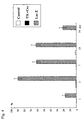
- the alkaline phosphatase activity in the block with the Cbfa1 (til-1) overexpressed cell was approximately 2.0 times and approximately 4.7 times higher than that of the control 3 and 5 weeks after transplantation, respectively (Fig. 16).
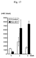
- the osteocalcin level in the cell in which Cbfa1 (til-1) had been excessively expressed was approximately 2.0 times and approximately 1.5 times higher than that of the control 3 and 5 weeks after transplantation, respectively (Fig. 17).
- the present invention provides a method for effective in vitro tissue culture for bone/cartilage tissue regeneration using bone marrow cells. This method enables ultimate therapy for damaged tissues that cannot be regenerated in vivo, wherein a patient's own cells extracted from his/her body are cultured and organized in vitro to reconstruct tissues that are as similar as possible to those in the body, and the composite is returned to the body.
Landscapes
- Health & Medical Sciences (AREA)
- Life Sciences & Earth Sciences (AREA)
- Engineering & Computer Science (AREA)
- Biomedical Technology (AREA)
- Chemical & Material Sciences (AREA)
- Zoology (AREA)
- General Health & Medical Sciences (AREA)
- Cell Biology (AREA)
- Organic Chemistry (AREA)
- Bioinformatics & Cheminformatics (AREA)
- Chemical Kinetics & Catalysis (AREA)
- Medicinal Chemistry (AREA)
- Animal Behavior & Ethology (AREA)
- Wood Science & Technology (AREA)
- Public Health (AREA)
- Veterinary Medicine (AREA)
- Genetics & Genomics (AREA)
- Biotechnology (AREA)
- Oral & Maxillofacial Surgery (AREA)
- Transplantation (AREA)
- Epidemiology (AREA)
- Botany (AREA)
- Dermatology (AREA)
- Developmental Biology & Embryology (AREA)
- Rheumatology (AREA)
- Orthopedic Medicine & Surgery (AREA)
- Microbiology (AREA)
- Biochemistry (AREA)
- General Engineering & Computer Science (AREA)
- Vascular Medicine (AREA)
- Hematology (AREA)
- Immunology (AREA)
- Nuclear Medicine, Radiotherapy & Molecular Imaging (AREA)
- Pharmacology & Pharmacy (AREA)
- General Chemical & Material Sciences (AREA)
- Physical Education & Sports Medicine (AREA)
- Urology & Nephrology (AREA)
- Micro-Organisms Or Cultivation Processes Thereof (AREA)
- Materials For Medical Uses (AREA)
Abstract
This invention provides a method of rapid and adequate culture of cells isolated
from a body to effectively construct bone/cartilage tissues and implants containing the
bone/cartilage tissues constructed by the aforementioned method. In this method,
osteo-/chondro-inducible transcription factor genes are transfected into
bone-marrow-derived cells isolated from a body using an adenoviral or a retroviral
vector to grow on adequate scaffolds. The constructed bone/cartilage tissues are
transplanted into a body together with the scaffolds. Thus, they can be used as
substitutional bone/cartilage implants.
Description
The present invention relates to a method for regeneration of bone/cartilage
tissues by transfection of osteo-/choridro-inducible transcription factor genes.
Particularly, the present invention relates to a method of transfection of
osteo-/chondro-inducible transcription factor genes to accelerate cell differentiation into
the target tissues, thereby effectively constructing bone/cartilage tissues and
substitutional bone/cartilage implants produced by the aforementioned method.
Recently, in the field of regenerative medicine, in vitro culture and organization
of cells from a body have been attempted for reconstructing tissues that are as similar as
possible to those in the body and replacing the tissues in the body. In such tissue
regeneration, the provision of scaffolds for cell proliferation and acceleration of cell
differentiation are indispensable, along with the preservation of cells.
In general, cells exist in a body by adhering to the extracellular matrix, which
plays as a scaffold for differentiation and proliferation. Accordingly, suitable scaffolds
for cell proliferation and differentiation are necessary in addition to cells, for in vitro
culture and complete three-dimensional tissue organization. Up to the present, the use
of porous biodegradable materials such as porous ceramics as scaffolds has provided
sufficient results in the regeneration of hard tissues such as bone and cartilage tissues.
In contrast, a method for rapid differentiation into target tissues in vitro is
important in the tissue engineering approach for tissue regeneration. Formally,
however, cytokines (humoral factors) responsible for cell differentiation have been
directly donated to cells or an expression vector carrying cDNA of the target cytokine
has been transfected into cells by lipofection method and so on. A certain level of
success has been achieved by such methods.
Transfection of cell growth factors, however, did not provide sufficient results
by several problems. For example, the target cytokine did not always fully and
specifically affect the tissue, and the transfection efficiency was almost 0 in primary
cells although gene transfection by lipofection was somewhat successful in established
cell lines.
Recently, many researchers have found that transcription factors, particularly
transcription factors of runt and Helix-Loop-Helix (HLH) types, play significant roles in
osteoblast differentiation. For example, a runt transcription factor, Pebp2alphaA
(Pebp2αA)/Cbfa1, and an HLH transcription factor, such as Scleraxis, Id-1, and I-mfa,
have been reported to have significant roles in osteoblast differentiation (e.g., Kunikazu
Tsuji et al., "Runt transcription factor associated with osteoblast differentiation,
Cbfa1/Pebp2αA, and Sox9 function in osteogenesis and chondrogenesis and unusual
osteogenesis" Experimental Medicine, 16(11), 25-32, 1998). Special attempts in tissue
regeneration utilizing functions of these transcription factors, however, have not yet
been reported.
The present inventors have conducted concentrated studies. As a result, they
have found that cells can be efficiently induced to differentiate into bone/cartilage
tissues by transfecting osteo-/chondro-inducible transcription factors into
bone-marrow-derived cells and having them express therein. Further, use of an
adenoviral or a retroviral vector for the transfection of a transcription factor results in
effective infection to the primary adhesive cells such as bone/cartilage cells, and very
effective induction of differentiation at approximately 99% can be achieved. This has
led to the completion of the present invention.
The present invention provides the following (1) to (13).
The present invention is hereafter described in detail.
The present invention relates to a method of transfection of the
osteo-/chondro-inducible (osteo-inducible and/or chondro-inducible) transcription factor
gene to isolated cells to construct bone/cartilage (bone and/or cartilage) tissues in vitro
by inducing differentiation and proliferation of the cells.
The transcription factors employed in the present invention are the
osteo-/chondro-inducible transcription factors that induce immatured cells to
differentiate into bone and/or cartilage cells. Examples thereof include Cbfa1, Dlx-5,
Bapx1, Msx2, Scleraxis, and Sox9. Cbfa1 is a transcription factor that was cloned by
Ogawa et al. at Kyoto University in 1993 and proved to be indispensable for inducing
differentiation of the mesenchymal stem cell into the osteoblast by Komori et al. at
Osaka University (Komori, T. et al., (1997) Cell 89, 755-764). This Cbfa1 has two
isoforms, til-1 and pebp2αA. Dlx-5 is a homolog of the Drosophila distalless (Dll)
gene and a transcription factor associated with the ossification of perichondrium and
endosporium (Acampora, D. et al., 1999, Development 126, 3795-3809). Bapx1 is a
homolog of the Drosophila bagpipe homeobox gene and is associated particularly with
differentiation of the mesenchymal stem cell into cartilage cells in the spinal cord and is
considered to be a regulatory gene of the Cbfa1 gene (Tribioli, C. et al., 1999,
Development 126, 5699-5711). Msx2 is a homolog of the Drosophila muscle segment
homeobox (Msh) gene. It is associated with the ossification of calvaria and is
considered to be a regulatory gene of the Cbfa1 gene (Satokata, I. et al., 2000, Nature
Genet. 24, 391-395). Scleraxis is a transcription factor that is associated with induction
of differentiation from the mesenchymal stem cell to cartilage cell or connective tissue
(Cserjesi, P. et al., 1995, Development 121, 1099-1110). Sox9 is expressed in cartilage
and it regulates expression of the genes associated with cartilage differentiation of, for
example, type II collagen (Ng, L. J. et al., 1997, Dev. Biol. 183, 108-121).
In the present invention, the above described osteo-/chondro-inducible
transcription factor genes can be prepared by conventional techniques, using their known
sequences. For example, cDNA of a target transcription factor can be prepared by
extracting RNA from the osteoblast, preparing primers based on a known sequence of
the transcription factor gene, and cloning the cDNA by PCR using the RNA and the
primers.
The cells that are employed in the present invention are immatured cells with
differentiation and proliferation ability isolated from a body. Examples thereof include
the ES (embryonic stem) cell, the mesenchymal stem cell, and the preosteoblast
differentiated from the mesenchymal stem cell. A bone-marrow-derived mesenchymal
stem cell is particularly preferable, and the osteoblast is more preferable. Particularly,
when the present invention is applied in the field of regenerative medicine, the
above-described cell is isolated from the body of a patient. This cell is preferably
prepared by removing connective tissues with a conventional technique. Alternatively,
cells may grow by primary culture before use.
The cells should be cultured in an appropriate medium for inducing cell
differentiation, constructing target tissues. In the case of bone tissue regeneration, for
example, at least one member selected from the group consisting of dexamethasone,
immunosuppressive factors such as FK-506 and cyclosporine, bone morphogenetic
proteins (BMP) such as BMP-2, BMP-4, BMP-5, BMP-6, BMP-7, and BMP-9, and
osteogenic humoral factors such as TGFβ is preferably supplemented to the medium to
induce cell differentiation into bone cells.
In the present invention, the osteo-/chondro-inducible transcription factor gene
can be transfected into the target cell by a method that is commonly employed for
transfection to animal cells. Examples of the methods that can be employed include the
calcium phosphate method, lipofection, electroporation, microinjection, and a method
using an adenoviral, a retroviral, or a baculoviral vector. From the viewpoints of safety
and transfection efficiency, a method using an adenoviral or a retroviral vector is
preferable, and a method using an adenoviral vector is most preferable.
The above-mentioned adenoviral or retroviral vector can be constructed
according to a known technique. For example, an adenoviral vector can be constructed
according to the method of Miyake et al. (Miyake, S. et al., Proc. Natl. Acad. Sci. 93:
1320-1324, 1993). Alternatively, a commercially available kit, such as the Adenovirus
Cre/loxP Kit (Takara Shuzo Co., Ltd.), can be used. This kit is used for constructing a
recombinant adenoviral vector utilizing a novel regulation system of gene expression
(Kanegae Y. et al., 1995, Nucl. Acids Res. 23, 3816) that employs Cre recombinase of
P1 phage and its recognition sequence, loxP. This kit can be used to simply construct a
recombinant adenoviral vector carrying the transcription factor gene. The multiplicity
of infection (moi) of adenovirus infection is 10 or more, preferably 50 to 200, and more
preferably around 100 (approximately 80 to 120).
The transfected cells should grow in suitable scaffolds in order to construct a
complete three-dimensional structure. Examples of materials that can be used for
scaffolds include hydroxyapatite or β-TCP (tricalcium phosphate), porous ceramics such
as α-TCP, collagen, polylactic acid, polyglycolic acid, and a complex thereof (e.g., a
complex of polylactic acid, polyglycolic acid resin, and collagen). These scaffolds may
be composed of a simple kind of material or combinations of two or more kinds.
Porous ceramics are particularly preferable for scaffolds for tissue regeneration because
of their high mechanical strength.
The above-mentioned scaffolds are preferably porous for uniform inoculation of
cells. In the present description, the term "porous" refers to a porosity of 40% or higher.
The pore size is not particularly limited, although a diameter of 200 µm to 500 µm is
preferable for facilitated bone regeneration.
Cells can be proliferated by being inoculated and cultured in the scaffold in
accordance with a conventional technique. Cells may be simply inoculated into the
scaffold, or inoculated in the form of mixtures with a liquid such as a buffer,
physiological saline, a solvent for injection, or a collagen solution. When the cells do
not smoothly enter into a pore because of its porous structure of the material, cells may
be inoculated under low pressure.
Preferably, the number of cells to be inoculated (inoculation density) is
determined in accordance with the types of cells or scaffolds in order to regenerate
tissues more efficiently, maintaining the morphology of the cells. For example, the
inoculation density is preferably 1,000,000 cells/ml or higher in the case of osteoblasts.
A conventional medium for cell culture, such as MEM medium, α-MEM
medium, or DMEM medium, can be suitably selected depending on the type of cell to be
cultured and then used for cell culture. FBS (Sigma) and antibiotics such as
Antibiotic-Antimycotic (GIBCO BRL) or other substances may be added to the medium.
Culture is preferably conducted in the presence of 3% to 10% CO2 at 30°C to 40°C, and
particularly preferably in the presence of 5% CO2 at 37°C. The culture period is not
particularly limited, and it is at least 4 days, preferably at least 7 days, and more
preferably at least 2 weeks.
The bone/cartilage tissues constructed by the above mentioned method are
transplanted or injected into a body together with or separately from the scaffolds.
Thus, they can be used as substitutional bone/cartilage implants. Specifically, the
present invention provides implants containing bone/cartilage tissues that are
constructed in vitro.
Bone/cartilage tissues may be transplanted into the body separately from
scaffolds, although transplantation with scaffolds is preferable in the present invention.
The most suitable scaffold for a given purpose and application site for implants may be
selected from the above-mentioned scaffolds. For example, hydroxyapatite is
preferable for a transplantation site (or a surgical technique) that requires mechanical
strength. In contrast, biodegradable β-TCP or the like is preferable for a
transplantation site (or a surgical technique) that does not require mechanical strength.
The configurations and shapes of the implants of the present invention are not
particularly limited. Implants can take any desired configurations or shapes, such as
sponges, meshes, unwoven fabric products, discs, films, sticks, particles, or pastes.
These configurations and shapes may be suitably selected depending on their
applications.
The implants of the present invention can suitably contain other components in
addition to the bone/cartilage tissues and scaffolds within the scope of the present
invention. Examples of such components include: growth factors such as basic
fibroblast growth factors (bFGF), platelet-derived growth factors (PDGF), insulin and
insulin-like growth factors (IGF), hepatocyte growth factor, (HGF), glial-derived
neurotrophic factors (GDNF), neurotrophic factors (NF), hormones, cytokines, bone
morphogenetic factors (BMP), transforming growth factors (TGF), and vascular
endothelial growth factors (VEGF); bone morphogenetic proteins; inorganic salts such as
St, Mg, Ca, and CO3; organic substances such as citric acid and phospholipid; and drugs.
Bone/cartilage tissues of the implants of the present invention are constructed
from the cells to which transcription factor genes have been transfected. Bone/cartilage
tissues may be constructed not only prior to the transplantation (in vitro) but also after
transplantation into bone defects (in vivo). The implants of the present invention are
highly compatible with bones and of high bone formation ability. Thus, they can be
integrated with biological bones and can then restore bone defects immediately after they
have been transplanted into a body.
This description includes a part or all of the contents as disclosed in the
description of Japanese Patent Application No. 2001-227979, which is a priority
document of the present application.
The present invention is hereafter described in more detail with reference to the
examples, although the technical scope of the present invention is not limited thereto.
Two types of cDNAs of Cbfa1, i.e., type I: pebp2αA/Cbfa1 (SEQ ID NO: 1) and
type II/III: til-1/Cbfa1 (SEQ ID NO: 2), were prepared from two types of plasmids*1
(pSG5/IT (mPEBP2aA) and pSG5/KS (mtil-1)) provided by Sumitomo Pharmaceuticals
Co., Ltd., ORF of mPEBP2aA was cleaved therefrom using a BamHI restriction enzyme,
and ORF of mtil-1 was cleaved therefrom using a BglII restriction enzyme (Fig. 1).
Two types of cDNAs of Cbfa1 were inserted into the SwaI site of the cosmid
vector pAxCALNLw using the commercially available Adenovirus Cre/loxP Kit (Takara
Shuzo Co., Ltd.), and a recombinant adenoviral vector was prepared according to the
manufacturer's instructions attached to the kit (Fig. 2). The titer of the prepared virus
was approximately 1011 PFU/ml, and infection efficiency was very high.
The rat bone marrow osteoblast (RBMO) was sampled from the back femur of a
6-week-old Fisher rat (male) in accordance with the method of Maniatopoulos et al.
(Maniatopoulos, C., Sodek, J., and Melcher, A. H., 1988, Cell Tissue Res. 254,
317-330).
The sampled cells were cultured and in MEM medium (Nacalai Tesque)
containing 15% FBS (Sigma) and Antibiotic-Antimycotic (GIBCO BRL) until they
became confluent. Subsequently, cells were subcultured into the above mentioned
medium containing 5 nM dexamethasone (Sigma), 10 mM β-glycerophosphate (Sigma),
and 50 µg/ml ascorbic acid phosphate (Wako) in 3.5 cm dishes, and they were cultured
until the number of cells per dish became approximately 400,000. The subcultured rat
osteoblast was infected with the recombinant adenoviral vector prepared as mentioned
above at a multiplicity of infection (moi) of 500 on the next day. Uninfected cells or
cells infected with the mock vector were prepared as a control.
Expression of LacZ in the rat osteoblasts 3 days to 2 weeks after adenovirus
infection were observed by X-gal staining in accordance with the method of Scholer et al.
(Scholer, H.R. et al., 1989, EMBO J., 8, 2551-2557) (Fig. 3). Further, the stained cells
were subjected to image analysis using the NIH image, and the number of expressed
cells was determined to evaluate the efficiency of gene transfection (Fig. 4).
The rat osteoblasts 3 days to 2 weeks after adenovirus infection were washed
with 100 mM Tris (pH 7.5) and 5 mM MgCl2, collected using a scraper, suspended in
500 µl of 100 mM Tris (pH 7.5), 5 mM MgCl2, and 1% Triton X-100, and then disrupted
by sonication. After the sonication, cells were centrifuged at 6,000 g for 5 minutes to
recover the supernatant. The supernatant (5 µl in each case) was added to 0.056M
2-amino-2-methyl-1,3-propanediol (pH 9.9), 10 mM p-nitrophenyl phosphate, and 2 mM
MgCl2, the resultants were incubated at 37°C for 30 minutes, and then absorbance at 405
nm was measured using a microplate reader immediately thereafter. The enzyme
activity was determined based on the calibration curve prepared using p-nitrophenol
from the obtained absorbance (Fig. 5).
The rat osteoblasts 1 to 3 weeks after adenovirus infection were fixed with 3.7%
formalin buffer and then decalcified with 0.6M HCl all day and night. The decalcified
solution was diluted, and the calcium level was assayed using a commercially available
o-cresol phthalein complexon-containing reagent for calcium analysis (# 587, 360-11,
Sigma) according to the manufacturer's instructions (Fig. 6).
Total RNA was extracted from the rat osteoblasts after adenovirus infection
using a commercially available TRIzol reagent (# 15596-10551, GIBCO BRL) according
to the manufacturer's instructions. Total RNA (10 µg) was separated on a 1%
agarose/5.5% formaldehyde gel and transferred in 20x SSC to the Hybond™-XL
membrane (Amersham Pharmacia Biotech). Thereafter, the membrane was heated at
80°C for 2 hours and the irradiated with ultraviolet rays for 2 minutes. The cDNA
probes of GAPDH and Cbfa1 were labeled with α-32PdCTP (3,000 Ci/mmol, Amersham
Pharmacia Biotech) using Rediprime™ (Amersham Pharmacia Biotech), and the
α-32PdCTP that had not been incorporated was removed using a MicroSpin™ G-25
Column (Amersham Pharmacia Biotech). This membrane was incubated in the
PerfectHyb™ Plus Hybridization Buffer (Sigma) at 68°C for 30 minutes, the labeled
cDNA probe (2x10⁶ cpm/ml) was added, and incubation was further carried out at 68°C
for 1 hour. The membrane was washed with 2x SSC/0.1% SDS at room temperature for
5 minutes and then washed twice with 0.5x SSC/0.1% SDS at 68°C (20 minutes each).
Thereafter, the membrane was exposed to the Kodak XAR film at -80°C overnight (Fig.
7).
The osteoblasts 3 days to 2 weeks after adenovirus infection were immobilized
with PBS containing 1% glutaraldehyde for 5 minutes, washed twice with distilled water,
and then stained with 1% crystal violet at room temperature for 30 minutes. After an
excess amount of dye was removed by washing three times with distilled water,
decolorization was carried out with 10% acetic acid and 1% Triton X-100. The
destaining solution was diluted, and absorbance at 595 nm was measured. The number
of cells was determined based on the calibration curve *2 from the obtained absorbance
(Fig. 8).
The conditions of calcification caused by mineral components secreted from the
osteoblasts were observed by Von Kossa staining and Alizarin Red staining.
The osteoblasts 1 to 3 weeks after adenovirus infection were fixed with 3.7%
formalin buffer for 5 minutes and then rinsed with diluted water. An aqueous solution
of 5% silver nitrate was added, and incubation was then carried out for 20 minutes in the
dark. Thereafter, incubation was further carried out for 15 minutes in the light, and the
resultant was washed thoroughly with distilled water. The results were scanned in a
scanner and then compared (Fig. 9).
The osteoblasts 1 to 3 weeks after infection were fixed with 3.7% formalin
buffer for 5 minutes and then rinsed with diluted water. An aqueous solution of 1%
Alizarin Red was added thereto, and incubation was then carried out for 2 minutes.
Thereafter, the resultant was washed thoroughly with distilled water. The results were
scanned in a scanner and then compared (Fig. 10).
As is apparent from Fig. 3 and Fig. 4, the expression level of the LacZ genes
reached its peak 3 days after infection, and the LacZ genes were expressed in
approximately 90% of cells. Thereafter, the number of LacZ-expressing cells gradually
decreased, although the expression level was maintained in some of the cells
(approximately 20%) even 2 weeks after infection.
As is apparent from Fig. 5, the alkaline phosphatase activity of the cell in which
Cbfa1 (pebp2αA, til-1) had been excessively expressed was higher than that of the
control. Particularly, the enzyme activity 10 days after infection became as high as
approximately 6 times that of the control.
As is apparent from Fig. 6, the amount of calcium deposited in the cell in which
Cbfa1 (pebp2αA, til-1) overexpressed cell was considerably larger than that in the
control.
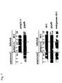
As is apparent from Fig. 7, a very high level of Cbfa1 gene expression was
observed in the cell 3 days after the Cbfa1 (pebp2αA, til-1) recombinant adenovirus
infection. Expression of endogenous Cbfa1 genes was observed in the til-1-infected
cell.
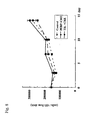
As is apparent from Fig. 8, the virus titer of the Cbfa1 and Cre recombinant
adenovirus infected cell was twice compared to that of the uninfected cell(control).
There was no substantial difference in cell proliferation.
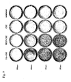
As is apparent from the results of Von Kossa staining (Fig. 9) and Alizarin Red
staining (Fig. 10), the level of calcification was more advanced in the cell in which
Cbfa1 (pebp2αA, til-1) overexpressed cell.
Accordingly, cDNA of Cbfa1 was very effectively transfected into the
osteoblasts with the aid of an adenoviral vector, and the transfected Cbfa1 gene would
significantly accelerate the osteoblast differentiation into bone tissues. This culture
system was also found to be capable of providing effective ways for in vitro tissue
regeneration through the use of adequate scaffolds such as porous ceramics.
The Cbfa1 gene-transfected rat osteoblasts were cultured using a commercially
available β-TCP (tricalcium phosphate) porous block (average pore size: 200 µm in
diameter, 5 mm x 5 mm x 5 mm, Olympus) as a scaffold in the following manner, and
the composite was transplanted subcutaneously to a rat's back. The confluent rat
osteoblasts were treated with trypsin and the cell suspension was then soaked into the
β-TCP block under low pressure (100 mHg) at a cell density of 1,000,000/ml. After the
cells were cultured for an additional 2 weeks, they were infected with the Cbfa1
(til-1)-recombinant adenoviral vector prepared in Example 1 at an moi of 500, and the
infected cells were transplanted subcutaneously to a rat's back on the next day. As the
control, blocks comprising cells infected with the mock adenoviral vector were similarly
transplanted. The blocks were excised 2, 4, and 8 weeks after transplantation and then
subjected to a variety of assays and observations.
The excised blocks were fixed with 4% paraformaldehyde and 0.05%
glutaraldehyde by microwave radiation. On the next day, the resultant was subjected to
decalcification in 10% EDTA and 100 mM Tris (pH 7.4), and decalcification was
continued for about 1 week. After the decalcification, the blocks were dehydrated in
ethanol and then embedded in paraffin. Sctions prepared to a thickness of 5 µm each
were deparaffinized and stained with hematoxylin and eosin (Figs. 11 and 12).
The alkaline phosphatase activities of the excised blocks were assayed in the
same manner as in Example 1 (Fig. 13).
After the assay of alkaline phosphatase activity, the sediments of the blocks
were disrupted by sonication in 20% formic acid, and incubated at 4°C for 4 days.
After desalting with the use of a PD-10 column (Amersham Pharmacia Biotech),
osteocalcin was extracted with 10% EDTA and 100 mM Tris (pH 7.4) and assayed the
osteocalcin level using the Rat Osteocalcin EIA Kit (Biomedical Technologies Inc.) (Fig.
14).
According to the results of hematoxylin-eosin staining of the transplanted blocks
(Figs. 11 and 12), the number of cells existing in pores of the blocks was larger and the
size of ossification area was larger in the Cbfa1 (til-1) overexpressed cell.
As is apparent from Fig. 13, the alkaline phosphatase activity of the cell in
which Cbfa1 (til-1) overexpressed cell was higher than that of the control. Particularly,
the enzyme activity 4 weeks after transplantation became as high as approximately 3
times that of the control.
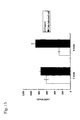
As is apparent from Fig. 14, the osteocalcin level in the Cbfa1 (til-1)
overexpressed cell was higher than that in the control. Particularly, the osteocalcin
level 4 weeks after transplantation became as high as approximately 2.5 times that of the
control.
The rat osteoblasts were infected with the Cbfa1 (til-1)-recombinant adenovirus
vector at a moi of 500. Twenty four hours later, the cells were trypsinized and the cell
suspension was soaked into the commercially available β-TCP block (average pore size:
200 µm in diameter, 5 mm x 5 mm x 5 mm (for subcutaneous transplantation), 2 mm x 2
mm x 2 mm (for bone defects transplantation), Olympus) at a cell density of 2,000,000
cells/ml. Next day, the blocks were transplanted to a rat's back and into the bone
defects. Transplantation into the bone defects was conducted by producing holes
(diameter: 2 mm, depth: 3 to 4 mm) in the frontal ends of left and right femora and by
embedding the blocks in the holes (bone defects). As the control, blocks containing
uninfected cells were similarly transplanted.
The subcutaneously transplanted block was excised 3 or 5 weeks after
transplantation, the alkaline phosphatase activity and the osteocalcin level were assayed,
and tissue sections were then observed. The block that had been transplanted into the
femur was excised 3 or 8 weeks after transplantation, and tissue sections were then
observed.
The number of cells existing in pores of the blocks with the Cbfa1 (til-1)
overexpressed cell was larger, and bone formation was more advanced than that in the
control, both 3 and 5 weeks after transplantation (Fig. 15A).
3 weeks after transplantation, bone formation was advanced, and the dissected
area of the femur was almost completely replaced with new bone tissue in the block with
the Cbfa1 (til-1) overexpressed cell. In contrast, bone formation did not substantially
occur in the case of the control. In the block with the Cbfa1 (til-1) overexpressed cell,
bone formation was further advanced, and the bone defect was almost completely
replaced with bone marrow tissues 8 weeks after transplantation. In contrast, the
transplanted block still remained as it had been in the control (Fig. 15B).
The alkaline phosphatase activity in the block with the Cbfa1 (til-1)
overexpressed cell was approximately 2.0 times and approximately 4.7 times higher than
that of the control 3 and 5 weeks after transplantation, respectively (Fig. 16). The
osteocalcin level in the cell in which Cbfa1 (til-1) had been excessively expressed was
approximately 2.0 times and approximately 1.5 times higher than that of the control 3
and 5 weeks after transplantation, respectively (Fig. 17).
All publications, patents, and patent applications cited herein are incorporated
herein by reference in their entirety.
The present invention provides a method for effective in vitro tissue culture for
bone/cartilage tissue regeneration using bone marrow cells. This method enables
ultimate therapy for damaged tissues that cannot be regenerated in vivo, wherein a
patient's own cells extracted from his/her body are cultured and organized in vitro to
reconstruct tissues that are as similar as possible to those in the body, and the composite
is returned to the body.



Claims (13)
- A method for constructing bone/cartilage tissues, wherein an osteo-/chondro-inducible transcription factor gene is transfected into isolated cells, and the isolated cells are allowed to differentiate and proliferate.
- The method according to claim 1, wherein the cells are derived from bone marrow.
- The method according to claim 2, wherein the bone-marrow-derived cells are mesenchymal stem cells.
- The method according to claim 2, wherein the bone-marrow-derived cells are osteoblasts.
- The method according to any one of claims 1 to 4, wherein the cells are primary cells.
- The method according to claim 5, wherein the cells are isolated from a patient.
- The method according to any one of claims 1 to 6, wherein the osteo-/chondro-inducible transcription factor is at least one member selected from the group consisting of Cbfa1, Dlx-5, Bapx1, Msx2, Scleraxis, and Sox9.
- The method according to any one of claims 1 to 7, wherein the osteo-/chondro-inducible transcription factor gene is transfected into the cells using an adenoviral or a retroviral vector.
- The method according to any one of claims 1 to 8, wherein the cells transfected by the osteo-/chondro-inducible transcription factor genes proliferate in at least one scaffold selected from the group consisting of porous ceramics, collagen, polylactic acid, polyglycolic acid, and a complex thereof.
- A method for constructing bone/cartilage tissues comprising the following procedures of:1) inducing differentiation of bone-marrow-derived cells isolated from a body with the aid of at least one member selected from the group consisting of dexamethasone, immunosuppressive factors, bone morphogenetic proteins, and osteogenic cytokines;2) transfection of the osteo-/chondro-inducible transcription factor gene into the cells using an adenoviral or retroviral vector; and3) proliferation of the cells in at least one scaffold selected from the group consisting of porous ceramics, collagen, polylactic acid, polyglycolic acid, and a complex thereof.
- Implants containing the bone/cartilage tissues constructed by the method according to any one of claims 1 to 10.
- The implants according to claim 11, which further contain at least one scaffold selected from the group consisting of porous ceramics, collagen, polylactic acid, polyglycolic acid, and a complex thereof.
- Implants containing cells, in which the osteo-/chondro-inducible transcription factor gene is transfected, and at least one member selected from the group consisting of porous ceramics, collagen, polylactic acid, polyglycolic acid, and a complex thereof.
Applications Claiming Priority (3)
| Application Number | Priority Date | Filing Date | Title |
|---|---|---|---|
| JP2001227979 | 2001-07-27 | ||
| JP2001227979 | 2001-07-27 | ||
| PCT/JP2002/006727 WO2003011343A1 (en) | 2001-07-27 | 2002-07-03 | Method of regenerating bone/chondral tissues by transferring transcriptional factor gene |
Publications (2)
| Publication Number | Publication Date |
|---|---|
| EP1424082A1 true EP1424082A1 (en) | 2004-06-02 |
| EP1424082A4 EP1424082A4 (en) | 2010-03-10 |
Family
ID=19060554
Family Applications (1)
| Application Number | Title | Priority Date | Filing Date |
|---|---|---|---|
| EP02741405A Withdrawn EP1424082A4 (en) | 2001-07-27 | 2002-07-03 | TECHNIQUE FOR REGENERATING BONE / CARTILAGINOUS TISSUES BY TRANSFERRING THE GENE OF TRANSCRIPTION FACTOR |
Country Status (4)
| Country | Link |
|---|---|
| US (1) | US20050063959A1 (en) |
| EP (1) | EP1424082A4 (en) |
| JP (1) | JP4921692B2 (en) |
| WO (1) | WO2003011343A1 (en) |
Families Citing this family (14)
| Publication number | Priority date | Publication date | Assignee | Title |
|---|---|---|---|---|
| WO2004074489A1 (en) * | 2003-02-18 | 2004-09-02 | Japan Science And Technology Agency | Method of forming bone/cartilage tissue with use of fat cell |
| GB0307011D0 (en) | 2003-03-27 | 2003-04-30 | Regentec Ltd | Porous matrix |
| US8029755B2 (en) | 2003-08-06 | 2011-10-04 | Angstrom Medica | Tricalcium phosphates, their composites, implants incorporating them, and method for their production |
| US8016865B2 (en) | 2003-09-29 | 2011-09-13 | Depuy Mitek, Inc. | Method of performing anterior cruciate ligament reconstruction using biodegradable interference screw |
| WO2005035014A1 (en) * | 2003-10-15 | 2005-04-21 | Japan Science And Technology Agency | Implant for regenerating bone or cartilage with the use of transcriptional factor |
| JP2006263459A (en) * | 2005-02-25 | 2006-10-05 | Japan Science & Technology Agency | Bone tissue regeneration method |
| WO2006090710A1 (en) * | 2005-02-25 | 2006-08-31 | Japan Science And Technology Agency | Method for bone tissue regeneration |
| JP5789799B2 (en) | 2009-08-21 | 2015-10-07 | 国立研究開発法人農業生物資源研究所 | Porous body manufacturing method, cell or tissue supply support manufacturing method, and tissue supply body manufacturing method |
| JP2012165719A (en) * | 2011-02-16 | 2012-09-06 | National Institute Of Advanced Industrial Science & Technology | Method for constructing three-dimensional carcinoma tissue model under pseudo-microgravity environment |
| WO2014093836A1 (en) * | 2012-12-13 | 2014-06-19 | University Of Georgia Research Foundation, Inc. | Ossification-inducing composition and methods of use thereof |
| EP3678602A4 (en) | 2017-09-08 | 2021-10-06 | Pioneer Surgical Technology, Inc. | INTERVERTEBRAL IMPLANTS, INSTRUMENTS AND PROCESSES |
| USD907771S1 (en) | 2017-10-09 | 2021-01-12 | Pioneer Surgical Technology, Inc. | Intervertebral implant |
| CN113116810B (en) * | 2021-03-29 | 2023-08-29 | 中国人民解放军空军军医大学 | Traditional Chinese medicine monomer and high-expression recombinant human bone morphogenetic protein cell gel preparation and preparation method and application thereof |
| CN113832109B (en) * | 2021-10-14 | 2023-11-21 | 中国人民解放军陆军军医大学第一附属医院 | A kind of bone marrow mesenchymal stem cell exosome and its preparation method and application |
Family Cites Families (7)
| Publication number | Priority date | Publication date | Assignee | Title |
|---|---|---|---|---|
| US6432711B1 (en) * | 1993-11-03 | 2002-08-13 | Diacrin, Inc. | Embryonic stem cells capable of differentiating into desired cell lines |
| US5591625A (en) * | 1993-11-24 | 1997-01-07 | Case Western Reserve University | Transduced mesenchymal stem cells |
| JPH10194987A (en) * | 1997-01-14 | 1998-07-28 | Youtai Iwamoto | Composition for ossification and chondrogenesis |
| US6077987A (en) * | 1997-09-04 | 2000-06-20 | North Shore-Long Island Jewish Research Institute | Genetic engineering of cells to enhance healing and tissue regeneration |
| DE19837438A1 (en) * | 1998-08-18 | 2000-02-24 | Biotechnolog Forschung Gmbh | Recombinant human cell for forming bone and cartilage cells from undifferentiated mesenchyme is capable of expressing a member of the T-box family of transcription factors |
| ES2304359T3 (en) * | 1999-12-28 | 2008-10-16 | The Regents Of The University Of Michigan | PROCEDURE FOR EX-VIVO TRAINING OF MAMPHERE BONES AND USES OF THE SAME |
| JP4246994B2 (en) * | 2001-01-30 | 2009-04-02 | オーソジーン, インコーポレイテッド | Compositions and methods for the treatment and repair of defects or lesions in articular cartilage using synovial tissue or cells |
-
2002
- 2002-07-03 JP JP2003516573A patent/JP4921692B2/en not_active Expired - Lifetime
- 2002-07-03 WO PCT/JP2002/006727 patent/WO2003011343A1/en not_active Ceased
- 2002-07-03 EP EP02741405A patent/EP1424082A4/en not_active Withdrawn
- 2002-07-03 US US10/484,699 patent/US20050063959A1/en not_active Abandoned
Also Published As
| Publication number | Publication date |
|---|---|
| US20050063959A1 (en) | 2005-03-24 |
| JP4921692B2 (en) | 2012-04-25 |
| EP1424082A4 (en) | 2010-03-10 |
| JPWO2003011343A1 (en) | 2004-11-18 |
| WO2003011343A1 (en) | 2003-02-13 |
Similar Documents
| Publication | Publication Date | Title |
|---|---|---|
| US20050063959A1 (en) | Method of regenerating bone/chondral tissues by transferring transcriptional factor gene | |
| Krebsbach et al. | Gene therapy-directed osteogenesis: BMP-7-transduced human fibroblasts form bone in vivo | |
| Oliveira et al. | An improved collagen scaffold for skeletal regeneration | |
| Yang et al. | Induction of human osteoprogenitor chemotaxis, proliferation, differentiation, and bone formation by osteoblast stimulating factor‐1/pleiotrophin: Osteoconductive biomimetic scaffolds for tissue engineering | |
| Zhang et al. | The effects of pore architecture in silk fibroin scaffolds on the growth and differentiation of mesenchymal stem cells expressing BMP7 | |
| JP4125241B2 (en) | Tooth-derived pluripotent embryonic-like stem cells and use thereof | |
| KR100968164B1 (en) | Adipose-derived hepatocytes and lattice | |
| US20120128739A1 (en) | Three-dimensional nanostructured hybrid scaffold and manufacture thereof | |
| US20050220773A1 (en) | Implant containing cells having growhfactor gene transferred thereinto | |
| Xiao et al. | Development and transplantation of a mineralized matrix formed by osteoblasts in vitro for bone regeneration | |
| CN107446885B (en) | A scaffold material for in vitro osteogenic differentiation of mesenchymal stem cells and its application | |
| Zhang et al. | Platelet‐derived growth factor BB gene‐released scaffolds: biosynthesis and characterization | |
| US20140178346A1 (en) | Cellular compositions for tissue engineering | |
| Pan et al. | Role and mechanism of BMP4 in regenerative medicine and tissue engineering | |
| Zhou et al. | Constructing Tissue‐Engineered Dressing Membranes with Adipose‐Derived Stem Cells and Acellular Dermal Matrix for Diabetic Wound Healing: A Comparative Study of Hypoxia‐or Normoxia‐Culture Modes | |
| JP4426532B2 (en) | Implants for bone and cartilage regeneration using transcription factors | |
| KR101733662B1 (en) | A scaffold with extracellular matrix for bone regeneration | |
| JP4491611B2 (en) | Method for producing bone and cartilage tissue using adipocytes | |
| CN109675108B (en) | Method for directly regenerating hypertrophic cartilage tissue by using adipose tissue and scaffold material | |
| JPWO2003008592A1 (en) | Adipose tissue-derived pluripotent stem cells | |
| JP2006263459A (en) | Bone tissue regeneration method | |
| WO2006090710A1 (en) | Method for bone tissue regeneration | |
| JP2004016109A (en) | Chondrocyte-like cell and method for making the same | |
| Sharma et al. | ENDOGENOUS REGENRATIVE TECHNOLOGY IN PERIODONTICS-A BRIEF REVIEW. | |
| Mondal et al. | Gene Therapy in Tissue Engineering: Prospects and Challenges |
Legal Events
| Date | Code | Title | Description |
|---|---|---|---|
| PUAI | Public reference made under article 153(3) epc to a published international application that has entered the european phase |
Free format text: ORIGINAL CODE: 0009012 |
|
| 17P | Request for examination filed |
Effective date: 20040227 |
|
| AK | Designated contracting states |
Kind code of ref document: A1 Designated state(s): AT BE BG CH CY CZ DE DK EE ES FI FR GB GR IE IT LI LU MC NL PT SE SK TR |
|
| A4 | Supplementary search report drawn up and despatched |
Effective date: 20100208 |
|
| STAA | Information on the status of an ep patent application or granted ep patent |
Free format text: STATUS: THE APPLICATION HAS BEEN WITHDRAWN |
|
| 18W | Application withdrawn |
Effective date: 20100415 |