EP1390516B1 - Novel expression vectors and uses thereof - Google Patents
Novel expression vectors and uses thereof Download PDFInfo
- Publication number
- EP1390516B1 EP1390516B1 EP02722308A EP02722308A EP1390516B1 EP 1390516 B1 EP1390516 B1 EP 1390516B1 EP 02722308 A EP02722308 A EP 02722308A EP 02722308 A EP02722308 A EP 02722308A EP 1390516 B1 EP1390516 B1 EP 1390516B1
- Authority
- EP
- European Patent Office
- Prior art keywords
- vector
- protein
- dna
- dna sequence
- cells
- Prior art date
- Legal status (The legal status is an assumption and is not a legal conclusion. Google has not performed a legal analysis and makes no representation as to the accuracy of the status listed.)
- Expired - Lifetime
Links
Images


Classifications
-
- C—CHEMISTRY; METALLURGY
- C12—BIOCHEMISTRY; BEER; SPIRITS; WINE; VINEGAR; MICROBIOLOGY; ENZYMOLOGY; MUTATION OR GENETIC ENGINEERING
- C12N—MICROORGANISMS OR ENZYMES; COMPOSITIONS THEREOF; PROPAGATING, PRESERVING, OR MAINTAINING MICROORGANISMS; MUTATION OR GENETIC ENGINEERING; CULTURE MEDIA
- C12N15/00—Mutation or genetic engineering; DNA or RNA concerning genetic engineering, vectors, e.g. plasmids, or their isolation, preparation or purification; Use of hosts therefor
- C12N15/09—Recombinant DNA-technology
- C12N15/63—Introduction of foreign genetic material using vectors; Vectors; Use of hosts therefor; Regulation of expression
- C12N15/79—Vectors or expression systems specially adapted for eukaryotic hosts
- C12N15/85—Vectors or expression systems specially adapted for eukaryotic hosts for animal cells
-
- A—HUMAN NECESSITIES
- A61—MEDICAL OR VETERINARY SCIENCE; HYGIENE
- A61P—SPECIFIC THERAPEUTIC ACTIVITY OF CHEMICAL COMPOUNDS OR MEDICINAL PREPARATIONS
- A61P31/00—Antiinfectives, i.e. antibiotics, antiseptics, chemotherapeutics
-
- A—HUMAN NECESSITIES
- A61—MEDICAL OR VETERINARY SCIENCE; HYGIENE
- A61P—SPECIFIC THERAPEUTIC ACTIVITY OF CHEMICAL COMPOUNDS OR MEDICINAL PREPARATIONS
- A61P31/00—Antiinfectives, i.e. antibiotics, antiseptics, chemotherapeutics
- A61P31/12—Antivirals
-
- A—HUMAN NECESSITIES
- A61—MEDICAL OR VETERINARY SCIENCE; HYGIENE
- A61P—SPECIFIC THERAPEUTIC ACTIVITY OF CHEMICAL COMPOUNDS OR MEDICINAL PREPARATIONS
- A61P31/00—Antiinfectives, i.e. antibiotics, antiseptics, chemotherapeutics
- A61P31/12—Antivirals
- A61P31/14—Antivirals for RNA viruses
- A61P31/18—Antivirals for RNA viruses for HIV
-
- A—HUMAN NECESSITIES
- A61—MEDICAL OR VETERINARY SCIENCE; HYGIENE
- A61P—SPECIFIC THERAPEUTIC ACTIVITY OF CHEMICAL COMPOUNDS OR MEDICINAL PREPARATIONS
- A61P35/00—Antineoplastic agents
-
- A—HUMAN NECESSITIES
- A61—MEDICAL OR VETERINARY SCIENCE; HYGIENE
- A61K—PREPARATIONS FOR MEDICAL, DENTAL OR TOILETRY PURPOSES
- A61K39/00—Medicinal preparations containing antigens or antibodies
- A61K2039/51—Medicinal preparations containing antigens or antibodies comprising whole cells, viruses or DNA/RNA
- A61K2039/53—DNA (RNA) vaccination
-
- A—HUMAN NECESSITIES
- A61—MEDICAL OR VETERINARY SCIENCE; HYGIENE
- A61K—PREPARATIONS FOR MEDICAL, DENTAL OR TOILETRY PURPOSES
- A61K48/00—Medicinal preparations containing genetic material which is inserted into cells of the living body to treat genetic diseases; Gene therapy
-
- C—CHEMISTRY; METALLURGY
- C12—BIOCHEMISTRY; BEER; SPIRITS; WINE; VINEGAR; MICROBIOLOGY; ENZYMOLOGY; MUTATION OR GENETIC ENGINEERING
- C12N—MICROORGANISMS OR ENZYMES; COMPOSITIONS THEREOF; PROPAGATING, PRESERVING, OR MAINTAINING MICROORGANISMS; MUTATION OR GENETIC ENGINEERING; CULTURE MEDIA
- C12N2800/00—Nucleic acids vectors
- C12N2800/10—Plasmid DNA
- C12N2800/108—Plasmid DNA episomal vectors
-
- C—CHEMISTRY; METALLURGY
- C12—BIOCHEMISTRY; BEER; SPIRITS; WINE; VINEGAR; MICROBIOLOGY; ENZYMOLOGY; MUTATION OR GENETIC ENGINEERING
- C12N—MICROORGANISMS OR ENZYMES; COMPOSITIONS THEREOF; PROPAGATING, PRESERVING, OR MAINTAINING MICROORGANISMS; MUTATION OR GENETIC ENGINEERING; CULTURE MEDIA
- C12N2830/00—Vector systems having a special element relevant for transcription
- C12N2830/42—Vector systems having a special element relevant for transcription being an intron or intervening sequence for splicing and/or stability of RNA
-
- C—CHEMISTRY; METALLURGY
- C12—BIOCHEMISTRY; BEER; SPIRITS; WINE; VINEGAR; MICROBIOLOGY; ENZYMOLOGY; MUTATION OR GENETIC ENGINEERING
- C12N—MICROORGANISMS OR ENZYMES; COMPOSITIONS THEREOF; PROPAGATING, PRESERVING, OR MAINTAINING MICROORGANISMS; MUTATION OR GENETIC ENGINEERING; CULTURE MEDIA
- C12N2840/00—Vectors comprising a special translation-regulating system
- C12N2840/20—Vectors comprising a special translation-regulating system translation of more than one cistron
- C12N2840/203—Vectors comprising a special translation-regulating system translation of more than one cistron having an IRES
Definitions
- Vaccination has proven to be a highly effective and economical method to prevent a disease caused by infectious agents. Since the introduction of the Vaccinia virus as an attenuated vaccine against the smallpox virus (Variola), vaccines against a multitude of human pathogens have been developed and taken into routine use. Today small pox has been eradicated by vaccinations and the same is to be expected shortly for the poliovirus. Several childhood diseases, such as pertussis, diphtheria and tetanus, can be effectively prevented by vaccinations.
- these functional origin fragments contain an E2 binding site, which is essential for the initiation of DNA replication in vivo in most cases (Ustav, E., et al., supra).
- the E2 protein facilitates the first step of the origin recognition by E1. After the initial binding of monomeric E1 to the origin the multimerization of E1 is initiated. This leads to the formation of the complex with the ori melting activity. It has been suggested that E2 has no influence on the following stages of the initiation of the DNA replication [ Lusky, M., et al., Proc Natl Acad Sci USA 91 (1994) 8895-8899 ].
- E2 binding sites in the BPV1 genome and up to four sites in the HPV genomes which play a crucial role in the initiation of viral DNA replication (Ustav, E., et al., supra) and in the regulation of viral gene expression ( Howley, P. M., Papillomavirinae: the viruses and their replication, in Virology, Fields, B. C., Knipe, D. M., Howley, P. M., Eds., Philadelphia: Lippincott-Raven Publishers, 1996. 2. edition, p. 2045 - 2076 ). Structural and mutational analyses have revealed three distinct functional domains in the full size E2 protein.
- the N-terminal part (residues 1 to 210) is an activation domain for transcription and replication. It is followed by the unstructured hinge region (residues 211 to 324) and the carboxy-terminal DNA binding-dimerization domain (residues 325 to 410) [ Dostatni, N., et al., EMBO J 7 (1988) 3807 - 3816 ; Haugen, T., et al.
- the functional elements of the transactivation domain of E2 have a very high structural integrity as confirmed by mutational analysis [ Abroi, A., et al., J Virol 70 (1996) 6169 - 6179 ; Brokaw, J., et al., J Virol 71 (1996) 23 - 29 ; Grossel, M., et al., J Virol 70 (1996) 7264 - 7269 ; Ferguson, M. and Botchan, M., J Virol 70 (1996) 4193-4199 ] and by X-ray crystallography [ Harris, S., and Botchan, M.R., Science 284 (1999) 1673-1677 and Antson, A. et al., Nature 403 (2000) 805-809 ].
- X-ray crystallography shows that the N-terminal domain of the E2 protein forms a dimeric structure, where Arg 37 has an important function in dimer formation (Antson, A., et al., supra).
- an optimal vaccine must produce protective immunity with minimal adverse effects.
- the vaccine should be devoid of components, which are toxic and/or cause symptoms of the disease to the recipient.
- an optimal vaccine must induce a pathogen-specific immune response, i.e. it must elicit a strong and measurable immune response to the desired pathogen without causing an immune response to other components of the vaccine.
- the vector produces several other papilloma proteins, which may elicit undesired immune responses and which induce a risk of the vector's integration in the recipient.
- the vector is replicable, since it contains the E1 gene. Additionally, it is large in size and therefore subject to bacterial modification during preparation.
- vectors containing an MME consisting essentially of ten E2 binding sites are disclosed in some examples. These vectors require the presence of the E1 protein either in the host or in the vector for the expression. This imparts the replication function to the vectors. These vectors also express the E1 protein in addition to the gene of interest and the E2 protein and contain sequences, such as rabbit ⁇ -globin sequences, which are partially homologous to human sequences causing a serious risk of integration to human genome, which reduces the potential of these vectors as DNA vaccines.
- E1 and E2 are the only viral proteins necessary for the episomal long-term replication of the vectors.
- the maintenance function of the BPV1 genome is associated with the presence of minimal ori (MO), which is stated to be necessary, although not sufficient, for the long-term persistence or the stable maintenance of the vectors the cells.
- MO minimal ori
- the cis-elements, i.e. the Minichromosome Maintenance Elements of the BPV1 are stated to be required for the stable replication of BPV1.
- multimeric E2 binding sites (E2BS) are stated to be necessary for the stable maintenance of the vectors.
- Yet another object of the invention is to provide novel vectors, which are suitable for a large-scale production in bacterial cell.
- An additional object of the invention is to provide novel vectors, which are useful as carrier vectors for a gene or genes of interest,
- the present invention discloses novel vectors, which meet the requirements of a carrier vector of a gene or genes of interest or of an optimal DNA vaccination vector and which are preferably devoid of drawbacks and side effects of prior art vectors.
- the present invention discloses novel vectors useful as carrier vectors of a gene or genes of interest, in DNA vaccination and gene therapy and as gene therapeutic agents.
- said vectors are capable of spreading and, if desired, of expressing a gene or genes of interest in an increasing number of cells for an extended time.
- the vectors of the present invention preferably express only a nuclear-anchoring protein, and, if desired, the gene or genes of interest, and optionally a selectable marker. However, they preferably lack any redundant, oncogenically transforming or potentially toxic sequences, thereby avoiding a severe drawback of the vectors previously disclosed or suggested for use as DNA vaccines, i.e. hypersensitivity reactions against other viral components. In certain embodiments of the invention, this is achieved by low level of the expressed nuclear-anchoring protein in the cells.
- the vectors of the present invention induce both humoral and cellular immune responses, where the gene or genes of interest is included in the vector.
- the vectors of the present invention are advantageous for use both in vitro (e.g., in the production level) and in vivo (e.g., vaccination).
- the present invention relates to expression vectors comprising: (a) a DNA sequence encoding a nuclear-anchoring protein operatively linked to a heterologous promoter, said nuclear-anchoring protein comprising (i) a DNA binding domain which binds to a specific DNA sequence, and (ii) a functional domain of the E2 protein of Bovine Papilloma Virus type 1 that binds to a nuclear component, and (b) a multimerized DNA binding sequence for the nuclear anchoring protein, wherein said vector lacks a an origin of replication functional in mammalian cells.
- the nuclear component can be mitotic chromatin, the nuclear matrix, nuclear domain 10 (ND10), or nuclear domain POD.
- the nuclear anchoring-protein is a chromatin-anchoring protein, and said functional domain binds mitotic chromatin.
- the nuclear-anchoring protein contains a hinge or linker region.
- the nuclear-anchoring protein is a natural protein of viral origin.
- the nuclear-anchoring protein is that of a papilloma virus.
- the nuclear-anchoring protein is the E2 protein of Bovine Papilloma Virus type 1.
- the nuclear-anchoring protein is a non-natural protein.
- the nuclear-anchoring protein is a recombinant protein, a fusion protein, or a protein obtained by molecular modeling techniques.
- the recombinant protein, fusion protein, or protein obtained by molecular modeling techniques contains any combination of a DNA binding domain which binds to said specific DNA sequence and a functional domain which binds to a nuclear component, wherein said functional domain which binds to a nuclear component is that of E2 protein of Bovine Papilloma Virus type 1.
- the vector further comprises one or more expression cassettes of a DNA sequence of interest.
- the DNA sequence of interest is that of a bacterium.
- the bacterium is selected from the group consisting of Chlamydia trachomatis, Mycobacterium tuberculosis, and Mycoplasma pneumonia.
- the bacterium is Salmonella.
- the DNA sequence of interest is that of a fungal pathogen.
- the fungal pathogen is Candida albigans.
- the DNA sequence of interest is of HIV origin.
- the DNA sequence of interest encodes a non-structural regulatory protein of HIV.
- the non-structural regulatory protein of HIV is Nef, Tat and/or Rev.
- the non-structural regulatory protein of HIV is Nef.
- the DNA sequence of interest encodes a structural protein of HIV.
- the DNA sequence of interest is the gene encoding HIV gp120/gp160.
- the vector of the invention comprises a first expression cassette comprising a DNA sequence of interest which encodes Nef, Tat and/or Rev, and a second expression cassette comprising a DNA sequence of interest which encodes Nef, Tat and/or Rev.
- the vector of the invention comprises a first expression cassette comprising a DNA sequence of interest which encodes Nef, Tat and/or Rev, and a second expression cassette comprising a DNA sequence of interest which encodes a structural protein of HIV.
- the DNA sequence of interest encodes a protein associated with cancer.
- the DNA sequence of interest encodes a protein associated with immune maturation, regulation of immune responses, or regulation of autoimmune responses.
- the protein is APECED.
- the DNA sequence of interest is the Aire gene.
- the DNA sequence of interest encodes a protein that is defective in any hereditary single gene disease.
- the DNA sequence of interest encodes a macromolecular drug.
- the DNA sequence of interest encodes a cytokine.
- the cytokine is an interleukin selected from the group consisting of IL1, IL2, IL4, IL6 and IL12.
- the DNA sequence of interest encodes an interferon.
- the DNA sequence of interest encodes a biologically active RNA molecule.
- the biologically active RNA molecule is selected from the group consisting of inhibitory antisense and ribozyme molecules.
- the inhibitory antisense or ribozyme molecules antagonize the function of an oncogene.
- a vector of the invention is suitable for the use for the production of a therapeutic macromolecular agent in vivo.
- the invention provides a vector for use as a medicament.
- the invention provides a vector for use as a carrier vector for a gene, genes, or a DNA sequence or DNA sequences of interest, such as a gene, genes, or a DNA sequence or DNA sequences encoding a protein or peptide of an infectious agent, a therapeutic agent, a macromolecular drug, or any combination thereof.
- the invention provides a vector for use as a medicament for treating inherited or acquired genetic defects.
- the invention provides a vector for use as a therapeutic DNA vaccine against an infectious agent.
- the invention provides a vector for use as a therapeutic agent.
- the invention further relates to methods for providing a protein to a subject, said method comprising administering to the subject a vector of the invention, wherein said vector (i) further comprises a second DNA sequence encoding the protein to be provided to the subject, which second DNA sequence is operably linked to a second promoter, and (ii) does not encode Bovine Papilloma Virus protein E1, and wherein said subject does not express Bovine Papilloma Virus protein E1.
- the invention further relates to methods for inducing an immune response to a protein in a subject, said method comprising administering to the subject a vector of the invention wherein said vector (i) further comprises a second DNA sequence encoding said protein, which second DNA sequence is operably linked to a second promoter, and (ii) does not encode Bovine Papilloma Virus protein E1, and wherein said subject does not express Bovine Papilloma Virus protein E1.
- the invention further relates to methods for treating an infectious disease in a subject in need of said treatment, said method comprising administering to said subject a therapeutically effective amount of a vector of the invention, wherein the DNA sequence of interest encodes a protein comprising an immunogenic epitope of an infectious agent.
- the invention further relates to methods for treating an inherited or acquired genetic defect in a subject in need of said treatment, said method comprising: administering to said subject a therapeutically effective amount of a vector of the invention, wherein said DNA sequence of interest encodes a protein which is affected by said inherited or acquired genetic defect.
- the invention further relates to methods for expressing a DNA sequence in a subject, said method comprising administering a vector of the invention to said subject.
- a vector of the invention is used for production of a protein encoded by said DNA sequence of interest in a cell or an organism.
- the invention further provides a method for the preparation of a vector of claim 1, 2, or 17 comprising: (a) cultivating a host cell containing said vector and (b) recovering the vector.
- the method for preparing a vector of the invention further comprises before step (a) a step of transforming said host cell with said vector.
- the host cell is a prokaryotic cell.
- the host cell is an Escherichia coli.
- the invention further relates to a host cell that is characterized by containing a vector of the invention.
- the host cell is a bacterial cell.
- the host cell is a mammalian cell.
- the invention further relates to carrier vectors containing a vector of the invention.
- the invention further relates to a pharmaceutical composition
- a pharmaceutical composition comprising a vector of the invention and a suitable pharmaceutical vehicle.
- the invention further relates to a gene therapeutic agent containing a vector of the invention.
- the invention further relates to a method for the preparation of a DNA vaccine, said method comprising combining a vector of the invention with a suitable pharmaceutical vehicle.
- the invention further relates to a method for the preparation of an agent for use in gene therapy, said method comprising combining a vector of the invention with a suitable pharmaceutical vehicle.
- the present invention is based on the unexpected finding that expression vectors, which carry (A) an expression cassette of a gene of a nuclear-anchoring protein that binds both to (i) a specific DNA sequence and (ii) to a suitable nuclear component and (B) a multimerized DNA binding sequence for said nuclear-anchoring protein as disclosed in claim 1 are capable of spreading in a proliferating cell population.
- nuclear-anchoring proteins include, but are not limited to, chromatin-anchoring proteins, such as the Bovine Papilloma Virus type 1 E2 protein (BPV1 E2; SEQ ID NO: 50).
- the DNA binding sequences can be, but are not limited to, multimerized E2 binding sites.
- nuclear-anchoring protein refers to a protein, which binds to a specific DNA sequence and capable of providing a nuclear compartmentalization function to the vector, i.e., to a protein, which is capable of anchoring or attaching the vector to a specific nuclear compartment.
- the nuclear-anchoring protein is a natural protein. Examples of such nuclear compartments are the mitotic chromatin or mitotic chromosomes, the nuclear matrix, nuclear domains like ND10 and POD etc.
- the nuclear-anchoring protein of the invention is a recombinant protein.
- the nuclear-anchoring protein is a fusion protein, a chimeric protein, or a protein obtained by molecular modeling.
- a fusion protein, or a protein obtained by molecular modeling in connection with the present invention is characterized by its ability to bind to a nuclear component and by its ability to bind sequence-specifically to DNA.
- such a fusion protein is encoded by a vector of the invention which also contains the specific DNA sequence to which the fusion/chimeric protein binds.
- Nuclear components include, but are not limited to chromatin, the nuclear matrix, the ND10 domain and POD.
- the DNA binding domain and the corresponding DNA sequence is preferably non-endogenous to the host cell/host organism.
- Such domains include, but are not limited to, the DNA binding domain of the Bovine Papilloma Virus type 1 (BPV1) E2 protein (SEQ ID NO: 50), Epstein-Barr Virus Nuclear Antigen 1 (EBNA1; SEQ ID NO: 52), and High Mobility Group (HMG) proteins (HMG box).
- the vector of the invention can further comprise a "DNA sequence of interest", that encodes a protein (including a peptide or polypeptide), e.g., that is an immunogen or a therapeutic.
- the DNA sequence of interest encodes a biologically active RNA molecule, such as an antisense RNA molecule or a ribozyme.
- the expression vectors of the invention carrying an expression cassette for a gene of a nuclear-anchoring protein and multimerized binding sites for said nuclear-anchoring protein spread in a proliferating host cell population. This means that a high copy-number of vectors or plasmids are delivered into the target cells and the use of the segregation/partitioning function of the nuclear-anchoring protein and its multimerized binding sites assures the distribution of the vector to the daughter cells during cell division.
- the vector of the invention lacks a papilloma virus origin of replication, providing a vector which lacks an origin of replication functional in mammalian cells.
- the omission of an origin of replication functional in mammalian cells constitutes an improvement over prior art vectors for several reasons.
- Omission of the origin of replication reduces the size of the vector of the invention compared to prior art vectors. Such a reduction in size increases the stability of the vector and facilitates uptake by the host cell.
- Omission of the origin of replication reduces the risk for recombination with the host cell's genome, thereby reducing the risk of unwanted side effects.
- the omission of the origin of replication allows to control the dosage simply by adjusting the amount of vector administered.
- the mechanism of the spreading function is due to the dual function of the E2 protein: the capacity of the E2 protein to attach to mitotic chromosomes through the N-terminal domain of the protein and the sequence-specific binding capacity of the C-terminal domain of the E2 protein, which assures the tethering of vectors, which contain a multimerized E2 binding site, to mitotic chromosomes. A segregation/partitioning function is thus provided to the vectors.
- the expression cassette of a gene of the chromatin-anchoring protein comprises a gene of any suitable protein of cellular, viral or recombinant origin having analogous properties to E2 of the BPV1, i.e., the ability to attach to the mitotic chromatin through one domain and to cooperatively bind DNA through another domain to multimerized binding sites specific for this DNA binding domain.
- the multimerized DNA binding sequences may be composed of any suitable multimeric specific sequences capable of inducing the cooperative binding of the protein to the plasmid, such as those of the EBNA1 or a suitable HMG protein.
- 21x30bp repeats of binding sites for EBNA-1 are localized in the region spanning from nucleotide position 7421 to nucleotide position 8042 of the Epstein-Barr virus genome (SEQ ID NO:51).
- the selectable marker can be any suitable marker allowable in DNA vaccines, such a kanamycin or neomycin, and others.
- other positive and negative selection markers can be included in the vectors of the invention, where applicable.
- the vectors of the present invention are not host specific, since the expression of the nuclear-anchoring protein, such as the E2 protein, is controlled by non-native or heterologous promoters. Depending on the particular promoter chosen, these promoters may be functional in a broad range of mammalian cells or they can be cell or tissue specific. Examples of promoters for the nuclear-anchoring protein, such as for the E2 protein, useful in the vectors of the present invention are thymidine kinase promoters, Human Cytomegalovirus Immediate Early Promoter, Rous Sarcoma Virus LTR and like. For the expression of the gene of interest, preferred promoters are strong promoters assuring high levels of expression of the gene of interest, an example for such a promoter is the Human Cytomegalovirus Immediate Early Promoter.
- a gene, genes or a DNA sequence or DNA sequences to be expressed via a vector of the invention can be any DNA sequence of interest, whose expression is desired.
- the vectors may contain a gene or genes or a DNA sequence or DNA sequences from infectious microbial pathogens, such as viruses, against which live attenuated vaccines or inactivated vaccines cannot be prepared or used.
- DNA sequences of interest include genes or DNA sequences from viruses, such as Human Immunodeficiency Virus (HIV), Herpex Simplex Virus (HSV), Hepatitis C Virus, Influenzae Virus, Enteroviruses etc.; intracellular bacterial, such as Chlamydia trachomatis, Mycobacterium tuberculosis, Mycoplasma pneumonia etc.; extracellular bacteria, such as Salmonella; or fungi, such as Candida albigans.
- viruses such as Human Immunodeficiency Virus (HIV), Herpex Simplex Virus (HSV), Hepatitis C Virus, Influenzae Virus, Enteroviruses etc.
- intracellular bacterial such as Chlamydia trachomatis, Mycobacterium tuberculosis, Mycoplasma pneumonia etc.
- extracellular bacteria such as Salmonella
- fungi such as Candida albigans.
- the vectors contain a gene encoding early regulatory proteins of HIV, i.e. the nonstructural regulatory proteins Nef, Tat or Rev, preferably Nef.
- the vectors of the invention contain genes encoding structural proteins of the HIV.
- the vectors of the present invention contain two or more genes encoding any combination of early regulatory proteins and/or structural proteins of HIV.
- the vaccines of the present invention contain a vector of the present invention or a mixture of said vectors in a suitable pharmaceutical carrier.
- the vaccine may for instance contain a mixture of vectors containing genes for the three different regulatory proteins of the HIV and/or structural proteins of the HIV.
- the vectors of the present invention can be used for the expression of the specific cytokines, like interleukines (IL1, IL2, IL4, IL6, IL12 and others) or interferon, with the aim of modulating the specific immune responses of the organism (immunotherapy) against foreign antigens or boosting of the activity of the immune system against the mutated self-antigens.
- the vectors of the present invention are also useful in complementing malfunctioning of the brain due to the loss of specific dopamine-ergic neurons leading to the irreversible neurodegeneration, which is cause for Parkinson's disease, by expressing genes involved into synthesis of dopamine, like tyrosine hydroxylase, as well as other genes deficiency of which would have the similar effect.
- DNA sequences encoding molecules comprising epitopes of known parasites are used.
- DNA sequences encoding antigenic epitopes may be prepared from parasites including, but not limited to, chlamydia and rickettsia.
- Both constitutive and inducible regulatory regions may be used for expression of the DNA sequence of interest. It may be desirable to use inducible promoters when the conditions optimal for growth of the host cells and the conditions for high level expression of the DNA sequence of interest are different. Examples of useful regulatory regions are provided below (section 5.4.4).
- the vector comprising a DNA sequence of interest operably linked to a regulatory region can be directly introduced into appropriate host cells for expression of the DNA sequence of interest without further cloning.
- a variety of regulatory regions can be used, for example, the SV40 early and late promoters, the cytomegalovirus (CMV) immediate early promoter, and the Rous sarcoma virus long terminal repeat (RSV-LTR) promoter.
- Inducible promoters that may be useful in mammalian cells include but are not limited to those associated with the metallothionein II gene, mouse mammary tumor virus glucocorticoid responsive long terminal repeats (MMTV-LTR), ⁇ -interferon gene, and hsp70 gene [ Williams et al., Cancer Res. 49 (1989) 2735-42 ; Taylor et al., Mol. Cell Biol., 10 (1990) 165-75 ]. It may be advantageous to use heat shock promoters or stress promoters to drive expression of the DNA sequence of interest in recombinant host cells.
- Preferred mammalian host cells include but are not limited to those derived from humans, monkeys and rodents, (see, for example, Kriegler M. in “Gene Transfer and Expression: A Laboratory Manual", New York, Freeman & Co. 1990 ), such as monkey kidney cell line transformed by SV40 (COS-7, ATCC CRL 1651); human embryonic kidney line (293, 293-EBNA, or 293 cells subcloned for growth in suspension culture, Graham et al., J. Gen. Virol., 36 (1977) 59 ; baby hamster kidney cells (BHK, ATCC CCL 10); chinese hamster ovary-cells-DHFR [CHO, Urlaub and Chasin. Proc. Natl. Acad. Sci.
- a vector of the invention comprising an expression cassette of a DNA sequence of interest is administered to a subject to induce an immune response.
- the DNA sequence of interest encodes a protein (for example, a peptide or polypeptide), which induces a specific immune response upon its expression. Examples of such proteins are discussed in section 5.3.
- a vector of the invention for use as a vaccine, methods may be selected from among those known in the art and/or described in section 5.4.6.
- a vector of the invention comprising an expression cassette comprising DNA sequences of interest is administered to treat, or prevent various diseases.
- the DNA sequence of interest may encode a protein and/or a biologically active RNA molecule.
- Gene therapy refers to therapy performed by the administration to a subject of an expressed or expressible DNA sequence.
- the DNA sequences produce their encoded protein or RNA molecule that mediates a therapeutic effect.
- mice mammary tumor virus control region which is active in testicular, breast, lymphoid and mast cells [ Leder et al., Cell 45 (1986) 485-495 ], albumin gene control region which is active in the liver [ Pinkert et al., Genes and Devel. 1 (1987) 268-276 ], alpha-fetoprotein gene control region which is active in the liver [ Krumlauf et al., Mol. Cell. Biol. 5 (1985)1639-1648 ; Hammer et al., Science 235 (1987) 53-58 ; alpha 1-antitrypsin gene control region which is active in the liver [ Kelsey et al., Genes and Devel.
- beta-globin gene control region which is active in myeloid cells [ Mogram et al., Nature 315 (1985) 338-340 ; Kollias et al., Cell 46 (1986) 89-94 ]; myelin basic protein gene control region which is active in oligodendrocyte cells in the brain [ Readhead et al., Cell 48 (1987) 703-712 ]; myosin light chain-2 gene control region which is active in skeletal muscle [ Sani, Nature 314 (1985) 283-286 ], and gonadotropic releasing hormone gene control region which is active in the hypothalamus [ Mason et al., Science 234 (1986)1372-1378 ].
- a vector of the invention contains a DNA sequence of interest that encodes an antisense or ribozyme RNA molecule. Techniques for the production and use of such molecules are well known to those of skill in the art.
- Antisense RNA molecules act to directly block the translation of mRNA by hybridizing to targeted mRNA and preventing protein translation.
- Antisense approaches involve the design of oligonucleotides that are complementary to a target gene mRNA. The antisense oligonucleotides will bind to the complementary target gene mRNA transcripts and prevent translation. Absolute complementarity, although preferred, is not required.
- a sequence "complementary" to a portion of an RNA means a sequence having sufficient complementarity to be able to hybridize with at least the non-polyA portion of an RNA, forming a stable duplex; in the case of double-stranded antisense nucleic acids, a single strand of the duplex DNA may thus be tested, or triplex formation may be assayed.
- the ability to hybridize will depend on both the degree of complementarity and the length of the antisense nucleic acid. Generally, the longer the hybridizing nucleic acid, the more base mismatches with an RNA it may contain and still form a stable duplex (or triplex, as the case may be).
- One skilled in the art can ascertain a tolerable degree of mismatch by use of standard procedures to determine the melting point of the hybridized complex.
- Antisense nucleic acids should be at least six nucleotides in length, and are preferably oligonucleotides ranging from 6 to about 50 nucleotides in length. In specific aspects the oligonucleotide is at least 10 nucleotides, at least 17 nucleotides, at least 25 nucleotides or at least 50 nucleotides. In other embodiments of the invention, the antisense nucleic acids are at least 100, at least 250, at least 500, and at least 1000 nucleotides in length.
- antisense DNA sequences complementary to the target gene coding region sequence could be used, those complementary to the transcribed, untranslated region are most preferred.
- the DNA sequence encoding the biologically active RNA molecule is operatively linked to a strong pol III or pol II promoter.
- a vector of the invention can be introduced, e.g ., such that it is taken up by a cell and directs the transcription of an antisense RNA.
- Such vectors can be constructed by recombinant DNA technology methods standard in the art.
- Expression of the sequence encoding the antisense RNA can be by any promoter known in the art to act in mammalian, preferably human cells.
- Such promoters can be inducible or constitutive.
- Such promoters include but are not limited to: the SV40 early promoter region [ Bernoist and Chambon, Nature 290 (1981) 304-310 ], the promoter contained in the 3 long terminal repeat of Rous sarcoma virus [ Yamamoto, et al., Cell 22 (1980) 787-797 ], the herpes thymidine kinase promoter [ Wagner, et al., Proc. Natl. Acad. Sci. U.S.A. 78 (1981) 1441-1445 ], the regulatory sequences of the metallothionein gene [ Brinster, et al., 1982, Nature 296 (1982) 39-42 ], etc .
- a vector of the invention contains a DNA sequence, which encodes a ribozyme.
- Ribozyme molecules designed to catalytically cleave target gene mRNA transcripts can also be used to prevent translation of a target gene mRNA and, therefore, expression of a target gene product [see, e.g ., PCT International Publication WO90/11364, published October 4, 1990 ; Sarver, et al., Science 247 (1990) 1222-1225 ].
- Ribozymes are enzymatic RNA molecules capable of catalyzing the specific cleavage of RNA. [For a review, see Rossi, Current Biology 4 (1994) 469-471 ]. The mechanism of ribozyme action involves sequence specific hybridization of the ribozyme molecule to complementary target RNA, followed by an endonucleolytic cleavage event. The composition of ribozyme molecules must include one or more sequences complementary to the target gene mRNA, and must include the well known catalytic sequence responsible for mRNA cleavage. For this sequence, see, e.g ., U.S. Patent No. 5,093,246 .
- ribozymes that cleave mRNA at site-specific recognition sequences can be used to destroy target gene mRNAs
- the use of hammerhead ribozymes is preferred.
- Hammerhead ribozymes cleave mRNAs at locations dictated by flanking regions that form complementary base pairs with the target mRNA. The sole requirement is that the target mRNA have the following sequence of two bases: 5'-UG-3'.
- the ribozyme is engineered so that the cleavage recognition site is located near the 5' end of the target gene mRNA, i.e ., to increase efficiency and minimize the intracellular accumulation of non-functional mRNA transcripts.
- the ribozymes of the present invention also include RNA endoribonucleases (hereinafter "Cech-type ribozymes”) such as the one that occurs naturally in Tetrahymena thermophila (known as the IVS, or L-19 IVS RNA) and that has been extensively described by Thomas Cech and collaborators [ Zaug, et al., Science, 224 (1984) 574-578 ; Zaug and Cech, Science, 231 (1986) 470-475 ; Zaug, et al., Nature, 324 (1986) 429-433 ; published International patent application No. WO 88/04300 by University Patents Inc.; Been & Cech, Cell, 47 (1986) 207-216 ].
- Cech-type ribozymes such as the one that occurs naturally in Tetrahymena thermophila (known as the IVS, or L-19 IVS RNA) and that has been extensively described by Thomas Cech and collaborators [ Zaug, et al., Science
- the Cech-type ribozymes have an eight base pair active site, which hybridizes to a target RNA sequence whereafter cleavage of the target RNA takes place.
- the invention encompasses those Cech-type ribozymes, which target eight base-pair active site sequences that are present in the target gene.
- ribozyme can be under the control of a strong constitutive pol III or pol II promoter, so that transfected cells will produce sufficient quantities of the ribozyme to destroy endogenous target gene messages and inhibit translation. Because ribozymes unlike antisense molecules, are catalytic, a lower intracellular concentration is required for efficiency.
- nucleic acid molecules that encode and express target gene polypeptides exhibiting normal target gene activity may, be introduced into cells via gene therapy methods such as those described, below, in Section 5.4.4 that do not contain sequences susceptible to whatever antisense, ribozyme, or triple helix treatments are being utilized.
- the target gene encodes an extracellular protein
- a pharmaceutical of the invention comprises a substantially purified vector of the invention (e.g ., substantially free from substances that limit its effect or produce undesired side-effects).
- the subject to whom the pharmaceutical is administered in the methods of the invention is preferably an animal, including but not limited to animals such as cows, pigs, horses, chickens, cats, dogs, etc ., and is preferably a mammal, and most preferably a human.
- the vector of the invention is directly administered in vivo, where the DNA sequence of interest is expressed to produce the encoded product. This can be accomplished by any of numerous methods known in the art.
- the vectors of the invention can be administered so that the nucleic acid sequences become intracellular.
- the vectors of the invention can be administered by direct injection of naked DNA; use of microparticle bombardment (e.g ., a gene gun; Biolistic, Dupont); coating with lipids or cell-surface receptors ortransfecting agents; encapsulation in microparticles or microcapsules; administration in linkage to a peptide which is known to enter the nucleus; administration in linkage to a ligand subject to receptor-mediated endocytosis [see, e.g ., Wu and Wu, J. Biol. Chem. 262 (1987) 4429-4432 ] (which can be used to target cell types specifically expressing the receptors); etc .
- microparticle bombardment e.g ., a gene gun; Biolistic, Dupont
- coating with lipids or cell-surface receptors ortransfecting agents encapsulation in microparticles or microcapsules
- administration in linkage to a peptide which is known to enter the nucleus administration in linkage to a
- the compound or composition can be delivered in a vesicle, in particular a liposome [see Langer, Science 249 (1990) 1527-1533 ; Treat et al., 1989, in Liposomes in the Therapy of Infectious Disease and Cancer, Lopez-Berestein and Fidler (eds.), Liss, New York, pp. 353- 365 ; Lopez-Berestein, ibid., pp. 317-327 ].
- a liposome see Langer, Science 249 (1990) 1527-1533 ; Treat et al., 1989, in Liposomes in the Therapy of Infectious Disease and Cancer, Lopez-Berestein and Fidler (eds.), Liss, New York, pp. 353- 365 ; Lopez-Berestein, ibid., pp. 317-327 ].
- the vector of the invention is coated with lipids or cell-surface receptors or transfecting agents, or linked to a homeobox- like peptide which is known to enter the nucleus [see e.g ., Joliot et al., Proc. Natl. Acad. Sci. USA 88 (1991) 1864-1868 ], etc .
- nucleic acid-ligand complexes can be formed in which the ligand comprises a fusogenic viral peptide to disrupt endosomes, allowing the nucleic acid to avoid lysosomal degradation.
- the vector of the invention can be targeted in vivo for cell specific uptake and expression, by targeting a specific receptor (see, e.g ., PCT Publications WO 92/06 180 ; WO 92/22635 ; W092/20316 ; W093/14188 , and WO 93/20221 ).
- Methods for use with the invention include, but are not limited to, intradermal, intramuscular, intraperitoneal, intravenous, subcutaneous, intranasal, epidural, and oral routes. Methods for use with the invention further include administration by any convenient route, for example by infusion or bolus injection, by absorption through epithelial or mucocutaneous linings ( e.g ., oral mucosa, rectal and intestinal mucosa, etc.).
- a vector of the invention may be desirable to administer a vector of the invention by injection, by means of a catheter, by means of a suppository, or by means of an implant, said implant being of a porous, non-porous, or gelatinous material, including a membrane, such as a sialastic membrane, or a fiber. Care must be taken to use materials to which the vector does not absorb. Administration can be systemic or local.
- a vector of the invention is administered together with other-biologically active agents such as chemotherapeutic agents or agents that augment the immune system.
- methods for use with the invention include delivery via a controlled release system.
- a pump may be used [see Langer, supra; Sefton, CRC Crit. Ref. Biomed. Eng. 14 (1989) 201 ; Buchwald et al., Surgery 88 (1980) 507 ; Saudek et al., N. Engl. J. Med. 321 (1989) 574 ].
- polymeric materials can be used [see Medical Applications of Controlled Release, 1974, Langer and Wise (eds.), CRC Pres., Boca Raton, Florida ; Controlled Drug Bioavailability, Drug Product Design and Performance, 1984, Smolen and Ball (eds.), Wiley, New York ; Ranger and Peppas, Macromol. Sci. Rev. Macromol. Chem. 23 (1983) 61 ; see also Levy et al., Science 228 (1985) 190 ; During et al., Ann. Neurol. 25 (1989) 351 ; Howard et al., J. Neurosurg. 71 (1989) 105 ].
- compositions of the invention comprise a therapeutically effective amount of a vector of the invention, and a suitable pharmaceutical vehicle.
- suitable pharmaceutical vehicle means approved by a regulatory agency of the Federal or a state government or listed in the U.S. Pharmacopeia or other generally recognized pharmacopeia for use in animals, and more particularly in humans.
- vehicle refers to a diluent, adjuvant, excipient, or vehicle with which the therapeutic is administered.
- suitable pharmaceutical vehicles can be sterile liquids, such as water and oils, including those of petroleum, animal, vegetable or synthetic origin, such as peanut oil, soybean oil, mineral oil, sesame oil and the like. Water is a preferred carrier when the pharmaceutical composition is administered intravenously.
- Saline solutions and aqueous dextrose and glycerol solutions can also be employed as liquid carriers, particularly for injectable solutions.
- suitable pharmaceutical excipients include starch, glucose, lactose, sucrose, gelatin, malt, rice, flour, chalk, silica gel, sodium stearate, glycerol monostearate, talc, sodium chloride, dried skim milk, glycerol, propylene, glycol, water, ethanol and the like.
- the composition if desired, can also contain minor amounts of wetting or emulsifying agents, or pH buffering agents. These compositions can take the form of solutions, suspensions, emulsion, tablets, pills, capsules, powders, sustained-release formulations and the like.
- compositions can be formulated as a suppository, with traditional binders and carriers such as triglycerides.
- Oral formulation can include standard carriers such as pharmaceutical grades of mannitol, lactose, starch, magnesium stearate, sodium saccharine, cellulose, magnesium carbonate, etc. Examples of suitable pharmaceutical carriers are described in "Remington's Pharmaceutical Sciences” by E.W. Martin.
- Such compositions will contain a therapeutically effective amount of the nucleic acid or protein of the invention, preferably in purified form, together with a suitable amount of carrier so as to provide the form for proper administration to the patient.
- the formulation should suit the mode of administration.
- the pharmaceutical of the invention is formulated in accordance with routine procedures as a pharmaceutical composition adapted for intravenous administration to human beings.
- compositions for intravenous administration are solutions in sterile isotonic aqueous buffer.
- the pharmaceutical of the invention may also include a solubilizing agent and a local anesthetic such as lignocaine to ease pain at the site of the injection.
- the ingredients are supplied either separately or mixed together in unit dosage form, for example, as a dry lyophilized powder or water free concentrate in a hermetically sealed container such as an ampoule or sachette indicating the quantity of active agent.
- the pharmaceutical of the invention is to be administered by infusion, it can be dispensed with an infusion bottle containing sterile pharmaceutical grade water or saline.
- an ampoule of sterile water for injection or saline can be provided so that the ingredients may be mixed prior to administration.
- compositions may take the form of tablets or lozenges formulated in conventional manner.
- the compounds for use according to the present invention are conveniently delivered in the form of an aerosol spray presentation from pressurized packs or a nebulizer, with the use of a suitable propellant, e.g., dichlorodifluoromethane, trichlorofluoromethane, dichlorotetrafluoroethane, carbon dioxide or other suitable gas.
- a suitable propellant e.g., dichlorodifluoromethane, trichlorofluoromethane, dichlorotetrafluoroethane, carbon dioxide or other suitable gas.
- the dosage unit may be determined by providing a valve to deliver a metered amount.
- Capsules and cartridges of e.g. gelatin for use in an inhaler or insufflator may be formulated containing a powder mix of the compound and a suitable powder base such as lactose or starch.
- the amount of a vector of the invention which will be effective in the treatment or prevention of the indicated disease, can be determined by standard clinical techniques.
- in vitro assays may optionally be employed to help identify optimal dosage ranges.
- the precise dose to be employed in the formulation will also depend on the route of administration, and the stage of indicated disease, and should be decided according to the judgment of the practitioner and each patient's circumstances. Effective doses may be extrapolated from dose-response curves derived from in vitro or animal model test systems.
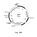
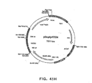
- vector plasmids super6 ( Figure 1 ) and super6wt were prepared from previous generation based gene vaccination vectors VI ( Figure 2 ) and Vlwt, respectively.
- Vectors VI and Vlwt are principally synthetic bacterial plasmids that contain a transposon Tn903 derived kanamycin resistance marker gene [ Oka, A., et al., J Mol Biol 147 (1981) 217-226 ] and a modified form of pMB1 replicon [ Yanisch-Perron, C., et al., Gene 33 (1985) 103-119 ] needed for the propagation in Escherichia coli cells.
- Vectors VI and Vlwt also contain a Cytomegalovirus Immediately Early Promoter combined with a HSV1 TK leader sequence and rabbit ⁇ -globin gene sequences, which both are derived from plasmid pCG [ Tanaka, M., et al., 60 (1990) Cell 375-386 ].
- the latter elements are needed for expressing from the nef coding sequence derived from a HAN2 isolate of the HIV-1 strain [ Sauermann, U., et al., AIDS Research. Hum. Retrov. 6 (1990) 813-823 ].
- the expression vectors for the Nef carry clustered ten high affinity E2 binding sites (derived from plasmid pUC1910BS, unpublished) just upstream of the CMV promoter.
- the parent vector VI contains a modified E2 coding sequence: the hinge region of E2 (amino acids 192-311) is replaced with four glycine-alanine repeats from EBNA1 protein of Epstein-Barr Virus [ Baer, R. J., et al., Nature 310 (1984) 207-211 ].
- the protein encoded by this sequence was named as E2d192-311+4G.
- the parent vector Vlwt contains an expression cassette for wild type E2 protein of the bovine papilloma virus type 1 with point mutations introduced for the elimination E3 and E4 open reading frame (ORF) coding capacity by two stop codons into both these ORFs.
- the E2 coding sequences are cloned between a Rous sarcoma virus proviral 5' LTR [ Long, E. O., et al., Hum. Immunol. 31 (1991) 229-235 ] and bovine growth hormone polyadenylation region [ Chesnut, J. D., et al., J Immunol Methods 193 (1996) 17-27 ].
- Plasmid vectors super6 and super6wt were constructed by deleting from the respective parent vectors VI and Vlwt all beta-globin sequences downstream of the nef gene except the second intron of the rabbit beta-globin gene.
- the beta-globin sequences (especially the fragment of the exon) show some homology with sequences in the human beta-globin gene, whereas the intron lacks any significant homology to human genomic sequences.
- the intron was amplified by PCR from the plasmid pCG [ Tanaka, M.
- the expression properties of the Nef and E2- proteins expressed by the plasmid vectors super6 and super6wt were analyzed and compared with the expression properties of the Nef and E2 proteins expressed by VI and Vlwt by Western blotting [ Towbin et al., Proc Natl Acad Sci USA 76 (1979) 4350-4354 ] with monoclonal antibodies against Nef and E2.
- Jurkat cells a human T-cell lymphoblast cell line
- were transfected by electroporation [ Ustav et al. EMBO J 2 (1991) 449-457 ] with 1 ⁇ g of super6, super6WT or equimolar amounts of the plasmids VI, Vlwt.
- the cells were lysed by treating with a sample buffer containing 50mM Tris-HCl pH 6.8; 2% SDS, 0.1 % bromophenol blue, 100mM dithiothreitol, and 10% (v/v) glycerol.
- the lysates were run on a 10% or 12.5% SDS-polyacrylamide gel and subsequently transferred onto a 0.45 ⁇ m PVDF nitrocellulose membrane (Millipore).
- the membrane was first blocked overnight with a blocking solution containing 5% dry milk (fat-free), 0.1 % Tween 20 in 50 mM Tris-HCl pH 7.5; 150mM NaCl and thereafter incubated for 1 h with diluted monoclonal anti-Nef antisera (1:100) or anti-E2-antisera (1:1000) in the blocking solution. After each incubation step, unbound proteins were removed by washing strips three times with TBS - 0.1 % Tween-20.
- the binding of primary immunoglobulins was detected by incubating the strips with horse raddish peroxidase conjugate anti-mouse IgG (Labas, Estonia) followed by visualization using a chemoluminesence detection system (Amersham Pharmacia Biotech, United Kingdom).
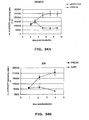
- Nef expressed from the plasmids super6, super6wt, VI and Vlwt (lanes 1-4 in Figure 4A ) are quite similar ( Figure 4 , panel A, lanes 1 to 4). Much less protein is produced from plasmid II (lane 5). The expression levels of the Nef protein are higher from vectors containing wtE2 (cf. lane 1 compared with lane 2 and lane 3 compared with lane 4). This is in accordance with the expression levels of E2 and E2d192-311 +4GA proteins from these plasmids ( Figure 4 , panel B).
- the new plasmids were named as the product1 ( Figure 5 ), and product1wt respectively.
- An unsuccessful attempt to reinsert the ten E2 binding sites back into the blunted Nhel site upstream of the CMV promoter of the product1 resulted in vector New Vector NNV, respectively, with only two binding sites integrated in the plasmid.
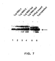
- the expression properties of the Nef protein from the NNV plasmids i.e. NNV-1, NNV-2, NNVwt and NNV-2wt, after the transfection of Jurkat cells by electroporation at a concentration of 1 ig of the plasmid were analyzed and compared with the expression properties of the Nef proteins from super6 and super6wt by Western blotting essentially as described in Example 1.
- the amounts of super6 and super6wt used for the transfection were 0,95 and 1 ⁇ g, respectively. The results are shown in Figure 7 .
- NNV-1 and NNV-2 vectors have expression potential similar to plasmid super6 as evident from the comparison of lanes 1 and 2 on figure 8 with lane 5.
- the plasmids expressing wt E2 produce more Nef protein than E2d192-311+4GA vectors do (compare lane 1 with lane 3 and lane 2 with lane 4 in figure 7 ).
- vector NNV-2wt was selected for further tests.
- NNV-2wt human T-cell lymphoblasts
- P815 mouse mastocytoma cells
- CHO Choinese Hamster Ovary cells
- RD human embryo rhabdomyosarcoma cells
- Each cell line was transfected with different amounts of the vector DNA by electroporation essentially as described in Example 1. Time-points were taken approximately two and five days after transfection. The results of analyses are presented in figures 8 to10.
- the Jurkat cells were transfected with 0.5 ⁇ g or 2 ⁇ g of the NNV-2wt (lanes 1,2, 8, and 9 in Figure 8 ) and equal amounts of the plasmids NNV-2wtFS (lanes 3, 4, 10, and 11 in Figure 8 ) and product1wt (lanes 5, 6,12, and13 in Figure 8 ) or carrier only (lanes 7 and 14 in Figure 8 ).
- Time-points were taken 44 hours (lanes 1-7) and 114 hours (lanes 8-14) aftertransfection: The expression of the Nef and E2 proteins was analyzed by Western blotting essentially as described in Example 1.
- the P815 cells were transfected with 0.5 ⁇ g or 2 ⁇ g of the NNV-2wt (lanes 1,2, 8, and 9 in Figure 9 ) and equal amounts of the plasmids NNV-2wtFS (lanes 3,4,10, and 11 in Figure 9 ) and product1wt (lanes 5, 6,12, and 13 in Figure 9 ) or carrier only (lanes 7 and 14 in Figure 9 ).
- Time-points were taken 45, hours (lanes 1-7) and 119 hours (lanes 8-14) aftertransfection:
- the expression of the Nef proteins was analyzed by Western blotting essentially as described in Example 1. The blot with anti-E2 antibodies 119h post-transfection is not shown, because no special signal could be detected.
- the expression level of the Nef protein correlated with the expression level of E2 protein in these cells, which confirms the fact that the function of the E2 protein is to activate the transcription and to help the plasmid to be maintained for a longer time in the proliferating cells.
- the CHO cells were transfected with 0.5 ⁇ g or 2 ⁇ g of the NNV-2wt (lanes 1,2, 8, and 9 in Figure 10 ) and equal amounts of the plasmids NNV-2wtFS (lanes 3,4,10, and 11 in Figure 10 ) and product1wt (lanes 5,6,12, and 13 in Figure 10 ) or carrier only (lanes 7 and 14 in Figure 10 ).
- Time-points were taken 48 hours (lanes 1-7) and 114 hours (lanes 8-14) after transfection.
- the expression of the Nef and E2 proteins was analyzed by Western blotting essentially as described in Example 1.
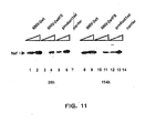
- the RD cells were transfected with 0.5 ⁇ g or 2 ⁇ g of the NNV-2wt (lanes 1,2,8, and 9 in Figure 11 ) and equal amounts of the plasmids NNV-2wtFS (lanes 3,4, 10, and 11 in Figure 11 ) and product1 wt (lanes 5,6,12, and 13 in Figure 11 ) or carrier only (lanes 7 and 14 in Figure 11 ).
- Time-points were taken 39 hours (lanes 1-7) and 110 hours (lanes 8-14) after transfection.
- the expression of the Nef protein was analyzed by Western blotting essentially as described in Example 1.
- RNA from these plasmids was also analyzed using the Northern analysis [ Alwine, J. C, et al., Proc Natl Acad Sci U S A 74 (1977) 5350-5354 ] for the NNV-2wt vector.
- Jurkat and CHO cells were transfected with 2 ⁇ g of the NNV-2wt.
- P815 cells 10 ⁇ g of NNV-2wt were used. The transfections were made essentially as described in Example 1.
- the running buffer contained the same components except formaldehyde.
- the samples were loaded in a buffer containing formamide and formaldehyde.
- RNAs were blotted onto the HybondN+ membrane (Amersham Pharmacia Biotech, United Kingtom) and hybridization with a radio-labeled nef coding sequence, E2 coding sequence or whole vector probes was carried out.
- the RNA from cells transfected with the carrier was used as a control.
- the results of the Northern blot analyses are shown in figure 12 .
- FISH fluoresence in situ hybridisation
- the CHO cells by electroporation with 1 ⁇ g of NNV-2wt or with equimolar amounts of the control plasmids NNV-2wtFS and product1wt (performed essentially as described in Example 1) the cultures were treated with colchicin (Gibco) for arresting the cells in metaphase of the mitosis. Briefly, cells were exposed to colchicine added to medium at final concentration of 0.1 ⁇ g/ml for 1-4 h to block the cell cycle at mitosis.
- colchicin Gabco
- Blocked cells were harvested by a trypsin treatment and suspended in a 0.075M KCI solution, incubated at room temperature for 15 min, and fixed in ice-cold methanol-glacial acetic acid (3:1, vol/vol).
- the spread-out chromosomes at metaphase and nuclei at interphase for fluorescence in situ hybridization analyses were prepared by dropping the cell suspension on wet slides. Several slides from one culture were prepared.
- Hybridization probes were generated by nick-translation, using biotin-16-dUTP as a label and plasmid Product1wt as template.
- a typical nick-translation reaction mixture contained a nick-translation buffer, unlabeled dNTPs, biotin-16-dUTP, and E.coli DNA polymerase.
- Chromosome preparations were denatured at 70°C in 70% formamide (pH 7.0-7.3) for 5 min, then immediately dehydrated in a series of washes (70%, 80%, and 96% ice-cold ethanol washes for 3 min each), and air-dried.
- the hybridization mixture (18 ⁇ l per slide) was composed of 50% formamide in 2xSSC (1xSSC is 0.15 M NaCl plus 0.015 M sodium citrate), 10% dextran sulfate, 150 ng of biotinylated plasmid probe DNA and 10 ⁇ g of herring sperm carrier DNA.
- probe DNA was applied to each slide, sealed under a coverslip, and hybridized for overnight at 37°C in a moist chamber.
- the slides were washed with three changes of 2xSSC, nd 2xSSC containing 0.1 % IGEPAL CA-630 (Sigma Chemical Co.) at 45°C.
- 2xSSC 2xSSC
- nd 2xSSC containing 0.1 % IGEPAL CA-630 (Sigma Chemical Co.) at 45°C.
- slides Prior to the immunofluorescence detection, slides were preincubated for 5 min in PNM a buffer [PN buffer (25.2 g Na 2 HPO 4 •7 H 2 O, 083 g NaH 2 PO4•_ ⁇ H 2 O and 0.6 ml of IGEPAL CA-630 in 1 liter of H 2 O] with 5% nonfat dried milk and 0.02% sodium azide).
- the vector is stable during the passage in Escherichia coli cells: no colonies with re-arrangements were observed when compared with the DNA used for transformation (lane 9).
- the samples were digested with different restriction endonucleases: with Eco81I (Fermentas, Lithuania) that has two recognition sites on the plasmid, with HindIII (Fermentas, Lithuania) that does not cut the NNV-2wt DNA and with DpnI (New England Biolabs, USA) that digest only DNA synthesized in Escherichia coli cells. Restricted DNAs were separated on TAE agarose electrophoresis and analyzed by Southern blotting [ Southern, E.M. J. Mol. Biol. 98 (1975) 503-517 ] with a vector specific radiolabeled probe. The results are illustrated on figure 14 .
- bovine papillomavirus type 1 E2 protein in trans and its multiple binding sites in cis are both necessary and sufficient for the chromatin attachment of the episomal genetic elements.
- the phenomenon is suggested to provide a mechanism for partitioning viral genome during viral infection in the dividing cells [ Ilves, I., et al., J Virol. 73 (1999) 4404-4412 ]. Because both functional elements are also included into our vector system, the aim of this study was analyze the importance of the E2 protein and oligomerized binding sites for maintenance of the transcriptionally active vector element in population of dividing cells.
- the vector gf10bse2 is derived from this plasmid by replacing the Nef coding sequence containing NdeI-Bst1107I fragment with d1 EGFP coding sequence containing fragment from 2wtd1 EGFP, cut out with same enzymes.
- Human and mouse AIRE-gene PCR-products were also digested with NotI restriction enzyme.
- 26 ⁇ l of PCR product 3 ⁇ l of an appropriate enzyme buffer and 10U of NotI restriction enzyme (the buffer and enzyme from MBI Fermentas) was used.
- the digestion was carried out at +37°C for 2 hours, after which digested PCR-products were purified and dissolved in sterile water to a volume of 10 ⁇ l.
- Miniprep DNA preparations from selected colonies were purified using Qiagen's Plasmid Midi Kit and dissolved to a volume of 50 ⁇ l of sterile water. The presence and size of the insert was checked with NotI and BamHI digestion.10 ⁇ l of miniprep DNA was taken for digestion, 5U of NotI and 5U of BamHI enzymes, 2 ml of R+ enzyme buffer and sterile water was added to a final volume of 20 ⁇ l. The digestion was carried out at +37°C for 1 hour.
- the generated vectors were sequenced for approximately 500 bp from both ends to verify the orientation and correctedness of the insert.
- the sequencing was performed using the dideoxy method with PE Biosystem's Big Dye Terminator RR-mix, which contains the four different terminating dideoxynucletide triphosphates labeled with different fluorescent labels.
- Plasmids containing the AIRE gene and AIRE gene fragments were inserted into selected cell lines to check the expression of the protein with Western blot after the transfection.
- Cos-1 cells were harvested with trypsin-EDTA (Bio Whittaker Europe) solution and suspended 10x106 cells/ml into Dulbecco's MEM (Life Technologies) medium and 250 ⁇ l of cell suspension was taken for transfection.
- the transfection of Cos-1 cells was performed using electroporation with 2.5x10⁶ cells, 50 ⁇ g of salmon sperm DNA as a carrier and 5 ⁇ g of appropriate vector.
- the transfections were made with pS6wthAIRE, pS6wtmAIRE, Super6wt, pCAIRE, psiAIRE and pCAIRE S1-4.
- pCAIRE and psiAIRE are positive human AIRE controls
- pCAIRE S1-4 is a positive mouse AIRE control
- Super6wt is a negative control.
- the electroporation was done using Biorad's Gene Pulser with capacitance 960 ⁇ Fd, 240 V and 1 pulse. After the pulse the cells were kept at room temperature for 10 minutes and 400 ⁇ l of medium was added. The cells were transferred to 5 ml of medium and centrifuged for 5 minutes with 1000 rpm. Cells were plated and grown for 3 days at +37°C, 5% CO2.
- the membrane was blocked in 5% milk in TBS (0.05 M Tris-Cl, 0.15 M NaCl, pH 7.5) for 30 minutes at room temperature.
- the membrane was washed two times with 0.1 % Tween in TBS for 5 minutes and once with TBS for 5 minutes.
- the secondary antibody, biotinylated anti-mouse IgG at a dilution of 1:500 in 5% milk in TBS was incubated for 1 hour at room temperature.
- Nef antibody ELISA was performed as previously described (Tähtinen et al., 2001). Briefly, Nunc Maxi Sorp plates were coated with 50 ng of Nef (isolate HAN), blocked with 2% BSA in phosphate buffered saline (PBS), and the sera in a dilution of 1:100 to 1:25000 were added in duplicate wells for an overnight incubation. After extensive washings, the secondary antibody, peroxidase conjugated anti-mouse IgG or IgM (DAKO), was added, and the plates were incubated for two hours and then washed.
- PBS phosphate buffered saline
- T-cell and B-cell assays were performed.
- T cell proliferation assay The spleen cells were suspended to a final concentration of 1 x 10⁶/ml RPMI-1640 (GibcoBRL) supplemented with 10% FCS (GibcoBRL), 1% penicillin-streptomycin (GibcoBRL) and 50 ⁇ M betamercaptoethanol (Sigma). Cells were incubated in microtitre plates at 200 ⁇ l/well with media only or with different stimuli. The final concentrations of stimuli were: Con A 5 ⁇ g/ml, HIV-Nef-protein at a concentration of 1 and 10 ⁇ g/ml, and a negative control antigen HIV-gag at a concentration of 1 and 10 ⁇ g/ml.
- CTL assays Mouse splenocytes were co-cultured with fixed antigen presenting cells (P-815 cells infected with MVA-HIV-nef or control MVA-F6) for five days after which they were tested in a standard 4 hour 51 chromium release assay [ Hiserodt, J., et al., J Immunol 135 (1995) 53-59 ; Lagranderie, M., et al., J Virol 71 (1997) 2303-2309 ) against MVA-HIV-nef infected or control target cells. In CTL assays the specific lysis of 10% or more was considered positive.
- Cytokine assay IFN-gamma and IL-10 were measured from antigen-stimulated cell culture supernatants in order to analyze, whether immunized mice develop a Th1 type or Th2 response. The supernatants were collected from antigen-stimulated cells as described above. Pro-inflammatory cytokines TNF-alfa and IL-10 were measured in the sera of the immunized mice. All cytokines were measured with commercial ELISA kits (Quantikine, R&D Systems).
- Spontaneous proliferation was detected by 3H-thymidine uptake of the cells cultured in the medium only for 6 days.
- dsDNA antibodies Anti-double strand (ds) DNA antibodies.
- dsDNA antibodies were measured in the sera of immunized mice, positive control mice (mrl/Ipr, a generous gift from Dr. Gene Shearer, NIH, USA) and normal mice. The antibodies were assayed with ELISA on poly-L-Lysine bounded lambda phage dsDNA. The results are shown in Tables 2 and 3.
- Table 2 shows complete immunological results of the mice immunized with HIV-Nef plasmid DNA.
- negative control protein HIV-gag did not induce any T cell response. Only the T cells of the mice in the group that had nef-specific proliferation also produced nef-specific IFN-gamma.
- Table 2 Mice HIV-1 HIV-1 IFN-g IL-10 HIV-1 E2 nef gag nef SI* SI Th1 Th2 Ab Ab NNV-Nef 8 1 6 1 - - ++ - 2 8 1 - - ++ - 3 13 2 - - ++ - 4 15 1 - - ++ - 5 7 1 - - ++ - Mean 9.8 1.2 NNV-NEF 0.4 1 24 1 + - + - 2 112 1 + - + - 3 83 1 + - + - 4 73 1 + - + - 5 69 1 + - + - Mean 72.2 1 NNV- ⁇ Nef 8 1 6 1 - - - - 2 nt nt - - - - 3 11 1 -
- the investigational vaccine NNV-2-Nef was prepared according to Example 2 with the Manufacturing License No. LLDnro 756/30/2000 (issued by the Finnish National Agency for Medicines on 21.12.2000).
- the manufacturing processes performed fulfilled the current Good Manufacturing Practices (cGMP) requirements and provided plasmid DNA preparations suitable for use in clinical phase I and II studies.
- the manufacturing process consisted of four steps:
- NNV-2-Nef was produced in E. coli bacteria.
- the Master Cell Banks (MCBs) and Working Cell Banks (WCBs) containing E. coli DH5 alpha T1 phage resistant cell strain were established in accordance with the specific Standard Operating Procedure from pure cultured and released Research Cell Banks.
- HIV-1 infected patients undergoing Highly Active Anti-Retroviral therapy were immunized with the experimental DNA vaccine NN2-Nef, expressing the HIV-1 Nef gene (Clade B).
- NN2-Nef expressing the HIV-1 Nef gene
- the doses were 1 and 20 micrograms/injection.
- Blood samples were drawn at -4, 0, 1, 2, 4, 8 and 12 weeks. The samples were analyzed for humoral (ELISA, Western blot) and cell mediated immune response (T-cell subsets, T-cell proliferation, ELISPOT, cytokine expression, intracellular cytokines).
- a clinical examination was performed to each patient participating the study.
- the clinical examination included a patient interview (anamnesis) and weight determination. Cardiac and pulmonar functions were checked by auscultation and percussion, the blood pressure and heart rate were recorded. Enlargement of lymph nodes, liver and thyroid gland were determined by palpation.
- LPA Lymphocyte proliferation assay
- PBMC Peripheral blood mononuclear cells
- Quadruplicate cultures were then set up in flat-bottomed micro titer plates (1x10 5 PBMC/well) and the cells were incubated for 6 days in the presence or absence of the following stimuli: rNef (0.2, 1 and 5 ⁇ g/ml), GST (0.2, 1 and 5 ⁇ g/ml), purified protein derivative of tuberculin (PPD, 12.5 ⁇ g/ml; Statens Seruminstitut), Candida albicans antigen (20 ⁇ g/ml; Greer Laboratories) and Phytohaemagglutinin (PHA; 5 ⁇ g/ml; Life Technologies).
- PPD purified protein derivative of tuberculin
- PPD purified protein derivative of tuberculin
- Candida albicans antigen (20 ⁇ g/ml; Greer Laboratories
- PHA Phytohaemagglutinin
- results are expresses as delta cpm (cpm in the presence of antigen-cpm without antigen) or as stimulation index (cpm in the presence of antigen/cpm without antigen).
- the type of immune response (Th1/Th2) induced by the vaccine was evaluated by measuring interferon-gamma (IFN- y) released in 6 days old culture supernatant after antigen (rNef, rGST, PPD) or mitogen (PHA) stimulation of PBMC.
- IFN- y interferon-gamma
- PPD antigen
- PHA mitogen
- commercial ELISA kits R&D Quantikine
- the assay employ the quantitative sandwich enzyme immunoassay technique where a monoclonal antibody specific for IFN- y has been coated onto a microplate. Standards and samples are pipetted into the wells and any IFN- y present is bound by the immobilized antibody. After washing away any unbound substances, an enzyme-linked polyclonal antibody specific for IFN- ⁇ is added to the wells. Following a wash to remove any unbound antibody-enzyme reagent, a substrate solution is added to the wells and color develops in proportion to the amount of the cytokine
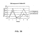
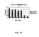
- IFN- ⁇ response data from patient # 1 is shown in Figure 28 .
- the vaccinee responded to the rNef antigen by marked IFN- ⁇ response correlated with the T-cell proliferation, indicating that the response seen in the vaccinee is in fact of the Th1 type.
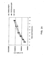
- HIV-1 infection is characterized by low or totally lacking cell-mediated immune response towards all HIV proteins.
- the results show that it is possible to induce a robust CMI in such patients with exceptionally low doses of the DNA vaccine NN2-Nef.
- the doses used were minimal to what has generally been required with DNA vaccines. Thus, for instance, Merch announced recently good results with their experimental HIV vaccine but the doses required were from 1000 to 5000 micrograms (IAVI report, 2002).
- EXAMPLE 12 CONSTRUCTION OF THE PLASMID EXPRESSING EPSTEIN-BARR VIRUS (EBV) EBNA-1 PROTEIN AND CONTAINING 20 BINDING SITES FOR EBNA-1 (FR ELEMENT)
- Epstein-Barr virus (EBV) EBNA-1 protein containing 20 binding sites for EBNA-1 (FR element)
- BPV-1 E2 binding sites were first replaced by EBV EBNA-1 binding sites (oriP without DS element).
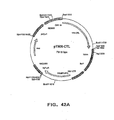
- Plasmid FRE2d1 EGFP ( Figure 29 ) was constructed by isolating the Xmil(Accl)/Eco321(EcoRV) DNA fragment (blunt-ended with Klenow enzyme) of pEBO LPP plasmid ( Figure 29A ) (the fragment contains 20 binding sites for EBNA-1) and inserting it by blunt end ligation into the Spel/Nhel site of s6E2d1 EGFP ( Figure 29B ) (blunt-ended with Klenow enzyme).
- the constructed plasmid FRE2d1EGFP ( Figure 29 ) was used as a negative control in further experiments. It contains binding sites for EBNA-1 protein instead of the BPV1 E2 10 binding sites, expressing E2, but not EBNA-1.
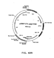
- the sequence encoding BPV-1 E2 protein in FRE2d1EGFP plasmid was replaced by a sequence encoding EBV EBNA-1 protein as follows.
- the Xmil(Accl)/EcoRI fragment of pEBO LPP plasmid was isolated and blunt-ended with Klenow enzyme and inserted into the XbaI/XbaI site of FRE2d1EGFP plasmid (blunted with Klenow enzyme).
- the vector FRE2d1EGFP was previously grown in Escherichia coli strain DH5 ⁇ lacking Dam - methylation, because one XbaI site is sensitive for methylation.
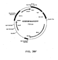
- the constructed plasmid FREBNAd1EGFP ( Figure 30 ) expresses EBNA-1 protein and contains 20 binding sites for EBNA-1.
- Jurkat, human embryonic kidney cell line 293 (ATCC CRL 1573) and mouse fibroblast cell line 3T6 cells (ATCC CCL 96) were maintained in Iscove's modified Dulbecco's medium (IMDM) supplemented with 10% fetal calf serum (FCS).
- IMDM Iscove's modified Dulbecco's medium
- FCS fetal calf serum
- 75% confluent dishes 293) or 1 ⁇ 4 of 75% confluent dishes (3T6) were used for each transfection, which were carried out by electroporation as follows. Cells were harvested by centrifugation (1000 rpm, 5 min, at 20°C, Jouan CR 422), and resuspended in a complete medium containing 5mM Na-BES buffer (pH 7.5).
- 250 ⁇ l cell of the cell suspension was mixed with 50 ⁇ g of carrier DNA (salmon sperm DNA) and 1 ⁇ g (in the case of Jurkat and 3T6) or 5 ⁇ g (in the case of 293) of plasmid DNA and electroporated at 200 V and 1000 ⁇ F for Jurkat cells, 170 V and 950 ⁇ F for 293 cells and 230 V and 975 ⁇ F for 3T6 cells.
- the transfected Jurkat cells were plated on 6-cm dishes with 5 ml of medium; 1/3 of transfected 293 and 3T6 cells were plated on a 6-cm dishes with 5 ml of medium and 2/3 of the cells were plated on a 10-cm dishes with 10 ml of medium.
- the transfected cells were analysed for the expression of d1EGFP protein (modified enhanced green fluorescent protein). All of the constructed plasmids expressed d1EGFP protein, which was detected by measuring the fluorescence using a flow cytometer. Because of the short half-life of the d1EGFP protein, it does not accumulate, and the expression of this protein reflects the presence of transcriptionally active plasmids in the cells. Becton-Dickinson FACSCalibur system was used. The volume of the Jurkat cell suspension was measured before each time-point (approximately after every 24 hour) and if the volume was less than 5 ml, the missing volume of medium was added. Depending on the cell suspension density the appropriate volume was taken for measuring (1 or 2 ml) and replaced with the same amount of medium. This was later taken into consideration when the dilution was calculated.
- d1EGFP protein modified enhanced green fluorescent protein
- the first time-point 293 cells from the 6-cm dish were suspended in 5 ml of medium for measuring. In every following time-point half of the cells were taken from the 10-cm dish, suspended in 5 ml of medium and then measured. An appropriate volume was added to the rest of the cell suspension.
- 3T6 cells from the 6-cm dish were suspended in 1 ml of trypsine, which was then inactivated with 100 ⁇ l of FCS.
- cells from the 10-cm dish were suspended in 2 ml of trypsin. 1 ml of this suspension was treated as described previously. 9 ml of medium was added to the rest of the suspension. The analyzed cells were taken out of the incubator immediately before the measurement. The appropriate flow speed (500-1000 cells/sec) was determined before each time-point using cells transfected only with carrier DNA as a control. Three different parameters were used to measure size, surface structure and fluorescence of the cells.
- Percentages of the d1EGFP expressing cells were calculated using cells transfected with the carrier only as a negative control for background fluorescence. As shown in Figure 33 , the two vectors were maintained in the cells with different kinetics.
- the number of the d1EGFP expressing cells was calculated taking the dilutions into consideration using cells transfected with the carrier only as a negative control for background fluorescence. As seen from figure 53, the plasmids expressing EBNA-1 and carrying EBNA-1 specific multimeric binding sites are maintained very efficiently in the transfected cells. At day 1 after transfection approximately 8x10 4 cells expressed EGFP. At day 8, in the case of maintenance vector (FREBNAd1EGFP), the number of the plasmid positive d1EGFP expressing cells had increased ten times to 8 x 10 5 .
- the immunogenicity of six different multi-gene vaccine constructs prepared in Example 12, i.e. GTU-1-RNT, GTU-1-TRN, GTU-1-RNT-CTL, GTU-1-TRN-CTL, GTU-1-TRN-optgag-CTL, and GTU-1-TRN-CTL-optgag vectors were tested in mice.
- the vectors were transformed into TOP10 or DH5alpha cells, and the MegaPreps were prepared using commercial Qiagen columns. Endotoxins were removed with Pierce Endotoxin Removal Gel.
- test articles were coated on 1 ⁇ m gold particles according to the instructions given by the manufacturer (Bio-Rad) with slight modifications.
- Balb/c mice were immunized with a Helios Gene Gun using a pressure of 400 psi and 0.5 mg gold/cartridge. Mice were immunized three times at weeks 0,1, and 3. Mice were sacrificed two weeks after the last immunization.
- mice were divided into six test groups (5 mice/group), which received 3 x 1 ⁇ g DNA as follows:
- the humoral response was followed from tail-blood samples from each mouse.
- First pre-immunization sample was taken from anesthetized mice before the first immunization was given.
- Second sample was taken from anesthetized mice before the third immunization.
- whole blood sample was used for white blood cell counting, and serum was collected for humoral immunity tests.
- the blood samples were tested for antibodies with ELISA using a standard procedure.
- Nunc Maxi Sorp plates were coated with 100 ng of Nef, Rev, Tat, Gag, CTL or E2 proteins, blocked and sera at a dilution of 1:100 were added in duplicate wells for an overnight incubation. After washing, the plates were incubated for 2 hours with a diluted (1:500) secondary antibody, peroxidase conjugated anti-mouse IgG (DAKO).
- DAKO peroxidase conjugated anti-mouse IgG
- Color intensity produced from the substrate (2,2'-azino-bis(3-ethylbenzthiazoline-6-sulfonic acid) (ABTS) in phosphate-citrate buffer was measured at 405 nm using Labsystems ELISA-plate reader.
- mice immunized with GTU-1-RNT or GTU-1-RNT-CTL also developed Tat antibodies ( Fig. 36 and Table 4).
- mice immunized with vectors containing the optgag sequence developed also Gag antibodies, but the construct GTU-1-TRN-optgag-CTL was a better antibody inducer that the construct GTU-1-TRN-CTL-optgag ( Figure 37 and Table 4).
- the antibodies induced were mainly of the IgG1 class indicating a Th2 type of response usually seen with gene gun immunization.
- the antibody assays shown below were done from the sera collected when mice were sacrificed.
- EXAMPLE 14 EXPRESSION OF HYBRID PROTEIN EXPRESSING NEF, REV AND TAT IN DIFFERENT COMBINATIONS (MULTIREG)
- GTU-1 vector with a multicloning site ( Figure 38A ) was used as a backbone.
- Intact Nef, Rev and Tat coding sequences were amplified by the polymerase chain reaction (PCR) and attached to each other in various orders to multi-regulatory (multireg) antigen coding reading frames (Nef-Tat-Rev, Tat-Rev-Nef, Rev-Tat-Nef, Tat-Nef-Rev and Rev-Nef-Tat; Sequences Id. No. 1 to 5, respectively). These sequences were cloned to the Bsp119I and NotI sites of the GTU-1 vector.
- Nef protein expressing GTU-2 and GTU-3 vectors ( Figure 38B and 38C ; see also Figure 6B for NNV-2wt)) were also used as backbones for the production of HIV multigene vectors.
- the vector super6wt expressing destabilized enhanced green fluorescent protein or d1EGFP (super6wtd1 EGFP; Figure 17 and Figure 38D ) and plasmid utilizing the EBNA-1 protein and its binding sites (FREBNAd1EGFP; Figure 38E ) were used as a Gene Transfer Unit (GTU) platform.
- a regular cytomegalovirus (CMV) vector NNV-Rev expressing Rev and a plasmid EBNA-1 and E2BS containing d1EGFP plasmid were used as backbones.
- GTU-1, GTU-2 and GTU-3 vectors which express the structural proteins encoded by the gag gene or an artificial polyprotein composed by previously described CTL epitopes, were prepared.
- the dgag contains truncated p17 (start at 13 aa ) +p24+p2+p7 (p1 and p6 are excluded) (Sequence Id. No. 11) of gag region of the Han2 isolate.
- the synp17/24 (Sequence Id. No. 12) codes for the p17+p24 polypeptide of the Han2 HIV-1. The codon usage is modified to be optimal in human cells. Also, previously identified AU rich RNA instability elements were removed by this way.
- the optp17/24 coding (Sequence Id. No. 13) region is very similar to the synp17/24 with the exception that the two synonymous mutations made therein do not change the protein composition but remove a potential splicing donor site.
- a set of the multi-HIV vectors which contain both a multireg antigen and structural antigens as a single polyprotein, were created: pTRN-CTL, pRNT-CTL, pTRN-dgag, pTRN-CTL-dgag, pRNT-CTL-dgag, pTRN-dgag-CTL, pRNT-dgag-CTL, pTRN-optp17/24-CTL, pTRN-CTL-optp17/24, and pRNT-CTL-optp17/24; p2TRN-optp17/24-CTL, p2RNT-optp17/24-CTL, p2TRN-CTL-optp17/24, p2RNT-CTL-optp17/24, p2TRN-CTL-optp17/24, p2TRN-CTL-optp17/24, p2TRN-CT
- the STOP codon was removed from the regulatory multi-antigen coding sequences. Then the structural antigen coding sequences were added by cloning into the NotI site at the end of the frame so that a Notl site was reconstituted. If both CTL and gag were added, the first antigen coding sequence was without the STOP codon.
- the clonings were made in context of GTU-1 and for making the respective GTU-2 (p2%) and GTU-3 (p3...) vectors, the Nef gene in the plasmids GTU-2Nef and GTU-2Nef was replaced using sites for Ndel and Pag I. However, the RNT-optp17/24-CTL antigen was built up directly in GTU-2 vector.
- the HIV multi-antigen was cloned to the vectors super6wtd1EGFP and FREBNAd1EGFP instead of the d1EGFP using sites for Eco105I and NotI (super6wt-RNT-CTL-optp17/24 and FREBNA-RNT-CTL-optp17/24; Figures 43V and 42U , respectively). If indicated, the IRES and mouse mGM-CSF were cloned into the GTU-2 and GTU-3 vectors behind the E2 coding sequence into the sites Mph1103I and Eco91I from pTRN-iE2-rnGMCSF (cut out using same restrictases).
- non-GTU control vector E2BSEBNA-RNT-CTL-optp17/24 ( Figure 42W ) for the system utilizing EBNA-1 (contains EBNA-1 expression cassette with E2 binding sites) was made in a similar way as the FREBNA-RNT-CTL-optp17/24.
- the regular CMV vector pCMV-RNT-CTL-optp17/24 expressing the multi HIV antigen ( Figure 42D ) was made by cloning the multi-HIV coding fragment from respective GTU-1 vector using sites for Ndel and Pag I.
- EXAMPLE 15 EXPRESSION PROPERTIES OF THE MULTIREG ANTIGENS CARRYING ONLY IMMUNODOMINANT REGIONS OF THE REGULATORY PROTEINS.
- MultiREG antigens expressed by the vectors of the invention was studied by in situ immunofluorescence in RD cells using monoclonal antibodies against Nef, Rev and Tat proteins essentially as described in Example 4. The results are summarized in Table 5 and illustrated in figure 45. All antigens that are comprised of intact Nef, Rev and Tat proteins showed exclusive localization in cytoplasm. The aberrant protein initially designed as N(ef)T(at)R(ev), which has a frame-shift before the Rev sequence, showed only the nuclear localization. MultiREG antigens carrying truncated sequences of the regulatory proteins were localized in cytoplasm. In this cases distinct structures like "inclusion bodies" were frequently observed.
- nuclei and cytoplasm inclusions in nuclei and cytoplasm pMV2N11TR only inclusions in nuclei and in cytopi only inclusions in nuclei and in cytopi only inclusions in nuclei and in cytopi.
- the intracellular localization of dgag and p17+p24 proteins was also analyzed in RD cells by immunofluorescence with monoclonal anti p24 antibodies.
- the dgag could not be detected.
- the p17/24 protein showed localization in plasma membranes ( Figure 45). The localization of CTL protein was not analyzed, because no suitable antibody was available.
- the intracellular localization was also analyzed in RD cells by immunofluorescence with anti p24 Mab. Similar to the western blot results in Jurkat cells, the dgag could not be detected.
- the p17/24 protein showed localization in plasma membranes ( Figure 47). The localization of CTL protein was not analyzed caused by lacking of suitable antibody.
- the expression of the MultiHIV antigenes consisting of both, regular multigene together with gag encoded protein and/or CTL multiepitope as single polypeptide was analysed.
- the Western blot shows the expression of several multiHIV-antigenes expressing vectors transfected to the Cos-7 cells. It is clearly seen that the expression levels of all regulatory+structural multi-antigenes are significantly lower than of the RNT or TRN proteins. All tested MultiHIV antigenes migrate in the gel as distinct bands near the position of predicted size (73kD for multireg+CTL; 95kD for multireg+dgag and 120kD for multireg+CTL+dgag).
- the RNT-CTL migrates more slowly than TRN-CTL. Also, in cases of both TRN and RNT constructs, the MultiREG-CTL-dgag combination showed higher expression level than MultiREG-dgag-CTL.
- MultiHIV antigenes More detailed analysis of the multiHIV antigenes was performed in Jurkat cells. For this reason, most of the constructed MultiHIV antigenes (multireg+structural), included the MultiREG+CTL+optp17/24 (with predicted size 113kD) were analyzed by Western blotting using antibodies against different parts of the antigene. The results are presented on Figure 49 are principially similar to those were reported in previous section in case of Cos-7 cells. As it was seen in the previous experiments, the dgag containing multi-antigenes express very low levels of the hybrid protein in Jurkat cells. The expression from the vector pTRNdgag was undetectable on all blots.
- the maintenance of the plasmid in a population of dividing cells was proved using the green fluorescent protein and Nef protein as markers.
- the maintenance of the expression of the RNT-CTL-optp17/24 antigen produced from different GTU or non-GTU vectors was also analyzed.
- GTU-1 p RNT-CTL-optp17/24
- GTU-2 p2 RNT-CTL-optp17/24
- GTU-3 p3 RNT-CTL-optp17124
- super6wt super6wt-RNT-CTL-optp17/24 vectors each utilize the E2 protein and its binding sites for the plasmid maintenance activity.
- EBNA-1 and its binding site utilizing GTU vector FREBNA-RNT-CTL-optp17/24 was included.
- "non-GTU" plasmid containing a mixed pair of the EBNA-1 expression cassette together with E2 binding sites (E2BSEBNA- RNT-CTL-optp17/24) was used.
- regular CMV expression vector pCMV- RNT-CTL-optp17/24 was used.
- Jurkat cells were transfected with equimolar amounts of the plasmids and the antigen expression was studied at 2 and 5 days post-transfection using a monoclonal anti-Nef antibodies. Transfection with carrier DNA only was used as a negative control. The results are presented in Figure 51.
- the expression is detectable only from GTU vectors at the second time-point.
- the antigen expression from the FREBNA- RNT-CTL-optp17/24 was lower at both time-points, because, unlike E2, the EBNA-1 does not have transcription activation ability.
Description
- The present invention relates to novel vectors, to DNA vaccines and gene therapeutics containing said vectors, to methods for the preparation of the vectors and DNA vaccines and gene therapeutics containing the vectors, and to therapeutic uses of said vectors. More specifically, the present invention relates to novel vectors comprising (a) an expression cassette of a gene of a nuclear-anchoring protein, which contains (i) a DNA binding domain which binds to a specific DNA sequence and (ii) a functional domain of the E2 protein of the Boving
Papilloma Virus type 1 that binds to a nuclear component and (b) a multimerized DNA binding sequence for the anchoring protein of a nuclear-anchoring protein, and optionally (c) one or more expression cassettes of a DNA sequence of interest, wherein the vectors lack an origin of replication functional in mammalian cells. The invention further relates to methods for expressing a DNA sequence of interest in a subject. - Transfer of autologous or heterologous genes into animal or human organisms with suitable vectors is emerging as a technique with immense potential to cure diseases with a genetic background or to prevent or cure infectious diseases. Several types of viral and non-viral vectors have been developed and tested in animals and in human subjects to deliver a gene/genes that are defective by mutations and therefore non-functional. Examples of such vectors include Adenovirus vectors, Herpes virus vectors, Retrovirus vectors, Lentivirus vectors and Adeno-associated vectors.
- Vaccination has proven to be a highly effective and economical method to prevent a disease caused by infectious agents. Since the introduction of the Vaccinia virus as an attenuated vaccine against the smallpox virus (Variola), vaccines against a multitude of human pathogens have been developed and taken into routine use. Today small pox has been eradicated by vaccinations and the same is to be expected shortly for the poliovirus. Several childhood diseases, such as pertussis, diphtheria and tetanus, can be effectively prevented by vaccinations.
- In general, the most successful viral vaccines are live avirulent mutants of the disease-causing viruses. The key to the success of this approach is the fact that a living virus targets the same organs, the same type and similar number of cells, and therefore, by multiplying in the recipient, elicits a long-lasting immune response without causing the disease or causing only a mild disease. In effect, a live attenuated vaccine produces a subclinical infection, the nature's own way of immunizing. As a result, a full immune response will be induced, including humoral, cellular and innate responses, providing a long lasting and sometimes a life-long immune protection against the pathogen.
- Although live attenuated vaccines are most potent, they can cause harmful side effects. Thus, an attenuated viral vaccine can revert to a virulent strain or in cases where the attenuated virus is apathogenic in adults it can still cause a disease in infants or in disabled persons. This is true in the case of viruses causing chronic infections, such as Human
Immunodeficiency Virus type - The strength and type of immune response is, however, also dependent on how the viral proteins are processed and how they are presented to the immune system by antigen presenting cells (APCs), such as macrophages and dendritic cells. Protein and peptide antigens are taken up by APCs via endocytosis, processed to small immunogenic peptides through an endosomal pathway and presented to T-lymphocytes (T-cells) by MHC (major histocompatibility complex) class II antigens [in man HLAs (human leukocyte antigens) class II]. In contrast, proteins synthesized de novo in APCs or in possible target cells for an immune response, will be processed through a cytoplasmic pathway and presented to T-cells by MHC class I antigens (in man HLAs class I). In general, the presentation of immunogenic peptides through the class II pathway will lead to the activation of the helper/inducer T-cells, which in turn will lead to the activation of B-cells and to antibody response. In contrast, presentation through class I MHC favors the induction of cytotoxic T-lymphocytes (CTLs), which are capable of recognition and destruction of virally infected cells.
- In early 1990's, a method to mimic the antigen processing and presentation that was normally achieved by live attenuated vaccines was introduced [Ulmer, J.B. et al Science 259 (1993) 1745-1749]. It was shown that an injection of eukaryotic expression vectors in the form of circular DNA into the muscle induced take-up of this DNA by the muscle cells (and probably others) and was able to induce the expression of the gene of interest, and to raise an immune response, especially a cellular immune response in the form of CTLs, to the protein encoded by the inserted gene. Since that observation, DNA immunization has become a standard method to induce immune responses to foreign proteins in experimental animals and human studies with several DNA vaccines are underway.
- Generally, the DNA vectors used in these vaccine studies contain a cloning site for the gene of interest, a strong viral promoter, such as the immediate early promoter of the CMV virus, in order to drive the expression of the gene of interest, a polyadenylation region, and an antibiotic resistance gene and a bacterial replication origin for the propagation of the DNA vector (plasmid) in bacterial cells.
- With the vectors described above it is possible to obtain a detectable level of expression of the gene of interest after administering the vector to experimental animals or to humans, either by a direct injection to muscle or to skin with a particle bombardment technique or by applying the vector in a solution directly to mucous membranes. However, the expression obtained by these vectors is short lived: the vectors tend to disappear from the transfected cells little by little and are not transferred to daughter cells in a dividing cell population. The short-term expression of the gene of interest and limited number of cells targeted are probably the major reasons, why only temporary immune responses are observed in subjects immunized with DNA vectors described above. Thus, for example, Boyer et al. observed only temporary immune responses to HIV-1 Env and Rev proteins in human subjects, who were immunized several times with a vector similar to the those described above [Boyer, J. D., J Infect Dis 181 (2000) 476 - 483].
- There is a growing interest in developing novel products useful in gene therapy and DNA vaccination. For instance papilloma virus vectors carrying the expression cassette for the gene of interest have been suggested to be useful candidates.
- To date more than 70 subtypes of human papilloma viruses (HPVs) and many different animal papilloma viruses have been identified [zur Hausen, H. and de Villiers E., Annu Rev Microbiol 48 (1994) 427 - 447; Bernard, H., et al., Curr Top Microbiol Immunol 186 (1994) 33 - 54]. All papilloma viruses share a similar genome organization and the positioning of all of the translational open reading frames (ORFs) is highly conserved.
- Papilloma viruses infect squamous epithelial cells of skin or mucosa at different body sites and induce the formation of benign tumors, which in some cases can progress to malignancy. The papilloma virus genomes are replicated and maintained in the infected cells as multicopy nuclear plasmids. The replication, episomal maintenance, expression of the late genes and virus assembly are tightly coupled to the differentiation of the epithelial tissue: the papilloma virus DNA episomal replication takes place during the initial amplificational replication and the second, i.e. latent, and the third, i.e. vegetative, replications in the differentiating epithelium [Howley, P. M.; Papillomavirinae: the viruses and their replication. In Virology, Fields, B. C., Knipe, D. M., Howley, P. M., Eds., Lippincott-Raven Publishers, Philadelphia, USA, 1996, 2. Edition, p. 2045 - 2076].
- Two viral factors encoded by the E1 and E2 open reading frames have been shown to be necessary and sufficient for the initiation of the DNA replication from the papilloma virus origin in the cells [Ustav, M. and Stenlund, A., EMBO J 10 (1991) 449 - 57; Ustav, M., et al., EMBO J 10 (1991) 4321 - 4329; Ustav, E., et al., Proc Natl Acad Sci USA 90 (1993) 898 - 902].
- Functional origins for the initiation of the DNA replication have been defined for BPV1 [Ustav, M., et al., EMBO J 10 (1991) 4321 - 4329], HPV1a [Gopalakrishnan, V. and Khan, S., supra], HPV11 [Russell, J., Botchan, M., J Virol 69 (1995) 651 -660], HPV18 [Sverdrup, F. and Khan, S., J Virol 69 (1995) 1319 - 1323:Sverdrup, F. and Khan, S., J Virol 68 (1994) 505-509] and many others. Characteristically, all these origin fragments have a high A/T content, and they contain several overlapping individual E1 protein recognition sequences, which together constitute the E1 binding site [Ustav, M., et al., EMBO J 10 (1991) 4321 -4329; Holt, S., et al., J Virol 68 (1994) 1094 - 1102; Holt, S. and Wilson, V., J Virol 69 (1995) 6525 - 3652; Sedman, T., et al. J Virol 71 (1997) 2887 - 2996]. In addition, these functional origin fragments contain an E2 binding site, which is essential for the initiation of DNA replication in vivo in most cases (Ustav, E., et al., supra). The E2 protein facilitates the first step of the origin recognition by E1. After the initial binding of monomeric E1 to the origin the multimerization of E1 is initiated. This leads to the formation of the complex with the ori melting activity. It has been suggested that E2 has no influence on the following stages of the initiation of the DNA replication [Lusky, M., et al., Proc Natl Acad Sci USA 91 (1994) 8895-8899].
- The BPV1 E2 ORF encodes three proteins that originate from selective promoter usage and alternative mRNA splicing [Lambert, P., et al., Annu Rev Genet 22 (1988) 235 - 258]. All these proteins can form homo- and heterodimers with each other and bind specifically to a 12 bp interrupted palindromic sequence 5'-ACCNNNNNNGGT-3' [Androphy, E., et al., Nature 325 (1987) 70 - 739].
- There are 17 E2 binding sites in the BPV1 genome and up to four sites in the HPV genomes, which play a crucial role in the initiation of viral DNA replication (Ustav, E., et al., supra) and in the regulation of viral gene expression (Howley, P. M., Papillomavirinae: the viruses and their replication, in Virology, Fields, B. C., Knipe, D. M., Howley, P. M., Eds., Philadelphia: Lippincott-Raven Publishers, 1996. 2. edition, p. 2045 - 2076). Structural and mutational analyses have revealed three distinct functional domains in the full size E2 protein. The N-terminal part (
residues 1 to 210) is an activation domain for transcription and replication. It is followed by the unstructured hinge region (residues 211 to 324) and the carboxy-terminal DNA binding-dimerization domain (residues 325 to 410) [Dostatni, N., et al., EMBO J 7 (1988) 3807 - 3816; Haugen, T., et al. EMBO J 7 (1988) 4245 - 4253; McBride, A., et al., EMBO J 7 (1988) 533 - 539; McBride, A., et al., Proc Natl Acad Sci USA 86 (1989) 510-514]. On the basis of X-ray crystallographical data, the DNA binding-dimerization domain of E2 has a structure of a dyad-symmetric eight-stranded antiparallel beta barrel, made up of two identical "half-barrel" subunits [Hegde, R., et al., Nature 359 (1992) 505- 512; Hegde, R., J Nucl Med 36(6 Suppl) (1995) 25S - 27S]. The functional elements of the transactivation domain of E2 have a very high structural integrity as confirmed by mutational analysis [Abroi, A., et al., J Virol 70 (1996) 6169 - 6179; Brokaw, J., et al., J Virol 71 (1996) 23 - 29; Grossel, M., et al., J Virol 70 (1996) 7264 - 7269; Ferguson, M. and Botchan, M., J Virol 70 (1996) 4193-4199] and by X-ray crystallography [Harris, S., and Botchan, M.R., Science 284 (1999) 1673-1677 and Antson, A. et al., Nature 403 (2000) 805-809]. In addition, X-ray crystallography shows that the N-terminal domain of the E2 protein forms a dimeric structure, where Arg 37 has an important function in dimer formation (Antson, A., et al., supra). - As has been described previously,
bovine papillomavirus type 1 E2 protein in trans and its multiple binding sites in cis are both necessary and sufficient for the chromatin attachment of the episomal genetic elements. The phenomenon is suggested to provide a mechanism for partitioning viral genome during viral infection in the dividing cells [Ilves, I., et al., J Virol. 73 (1999) 4404-4412]. - None of the papilloma vectors or other vectors disclosed so far fulfills the criteria and requirements set forth for an optimal vaccine, which are the same for DNA vaccines and for conventional vaccines. (It should be noted that these requirements are preferred but not necessary for use as a vaccine.) First, an optimal vaccine must produce protective immunity with minimal adverse effects. Thus the vaccine should be devoid of components, which are toxic and/or cause symptoms of the disease to the recipient. Second, an optimal vaccine must induce a pathogen-specific immune response, i.e. it must elicit a strong and measurable immune response to the desired pathogen without causing an immune response to other components of the vaccine. These two requirements imply that a vector to be used as a DNA vaccine should optimally only express the desired gene(s) and optimally should not replicate in the host or contain any sequences homologous with those of the recipient, since nucleotide sequences that are homologous between the vector and the host's genome may effect the integration of the vector into the host's genome. Third, an optimal vaccine must induce a right type of immune response; i.e. it must raise both humoral and cellular immune responses in order to act on the intracellular and extracellular pathogen. Finally, an optimal vaccine must be stable, i.e. it must retain its potency for a sufficiently long time in the body to raise the immune response in a vaccine formulation for use in various demanding circumstances during storage and preparation. Additionally, vaccines should be of reasonable price. Further, the route and the method of inoculation are important considerations for optimizing a DNA immunization.
- When developing a DNA vaccine the stability of the expression of the desired gene is sometimes a major problem. Thus, the maintenance function or the persistance of the vector in the recipient cell has been focused on in the prior art, however, often at the cost of the safety. For example, Ohe, Y., et al. ][Hum Gene Ther 6(3) (1995) 325-333] disclose a papilloma virus vector capable of stable, high-level gene expression, which is suggested for use in gene therapy. Transforming early genes E5, E6, and E7 have been deleted from said vector, but it still contains nucleotide sequences encoding other papilloma viral genes, such as the E1 and E2 genes, which are involved in the replication of the virus. Thus, the vector produces several other papilloma proteins, which may elicit undesired immune responses and which induce a risk of the vector's integration in the recipient. Also, the vector is replicable, since it contains the E1 gene. Additionally, it is large in size and therefore subject to bacterial modification during preparation.
- International Patent Application
PCT/EE96/00004 WO 97/24451 ca 15 kb): at the preparation stage in a bacterial cell, the bacterial replication machinery tends to modify the vector by random slicing of the vector, which leads to unsatisfactory expression products including products totally lacking the gene of interest. - International Patent Application
PCT/EE96/00004 WO 97/24451 - There is a clear need for improved novel vectors, which would be useful as DNA vaccines.
- An object of the invention is therefore to provide novel vectors, which are capable of a long-term maintenance in a large and increasing number of different cells of the host's body and thereby capable of providing a stable expression of the desired antigen(s).
- Another object of the invention is to provide novel vectors, which are maintained for a long period of time in the cells that originally received the vector and transferred it to the daughter cells after mitotic cell division.
- Yet another object of the invention is to provide novel vectors, which express in addition to the gene or genes of interest preferably only a gene necessary for a long-term maintenance in the recipient cells and thus are devoid of components that are toxic or cause symptoms of the disease to the recipient.
- A further object of the invention is to provide novel vectors, which mimic attenuated live viral vaccines, especially in their function of multiplying in the body, without inducing any considerable signs of disease and without expressing undesired proteins, which may induce adverse reactions in a host injected with the DNA vaccine.
- Still a further object of the invention is to provide novel vectors, which do not replicate in the recipient.
- Still another object of the invention is to provide novel vectors, which induce both humoral and cellular immune responses when used as DNA vaccines.
- Yet another object of the invention is to provide novel vectors, which are suitable for a large-scale production in bacterial cell.
- Yet another object of the invention is to provide novel vectors, which are not host specific and thus enable the production in various bacterial cells.
- An additional object of the invention is to provide novel vectors, which are useful as carrier vectors for a gene or genes of interest,
- A further object of the invention is to provide novel vectors, which are useful in gene therapy and as gene therapeutic agents and for the production of macromolecular drugs in vivo.
- The present invention discloses novel vectors, which meet the requirements of a carrier vector of a gene or genes of interest or of an optimal DNA vaccination vector and which are preferably devoid of drawbacks and side effects of prior art vectors.
- The present invention is based on the surprising finding that a vector (plasmid) carrying (i) an expression cassette of a DNA sequence encoding a nuclear-anchoring protein comprising a functional domain of the E2 protein of the Bovin
Papilloma Virus Type 1 and a DNA binding domain which binds to a specific DNA sequence ; and (ii) multiple copies of high affinity binding sites for said nuclear-anchoring protein spreads in proliferating cells. As a result, the number of vector-carrying cells increases even without the replication of the vector. When the vector additionally carries a gene or genes of interest, the number of such cells that express a gene or genes of interest similarly increases without the replication of the vector. Thus, the vector of the invention lacks an origin of replication functional in mammalian cells. - Accordingly, the present invention discloses novel vectors useful as carrier vectors of a gene or genes of interest, in DNA vaccination and gene therapy and as gene therapeutic agents. In a specific embodiment, said vectors are capable of spreading and, if desired, of expressing a gene or genes of interest in an increasing number of cells for an extended time. The vectors of the present invention preferably express only a nuclear-anchoring protein, and, if desired, the gene or genes of interest, and optionally a selectable marker. However, they preferably lack any redundant, oncogenically transforming or potentially toxic sequences, thereby avoiding a severe drawback of the vectors previously disclosed or suggested for use as DNA vaccines, i.e. hypersensitivity reactions against other viral components. In certain embodiments of the invention, this is achieved by low level of the expressed nuclear-anchoring protein in the cells. At the same time, the vectors of the present invention induce both humoral and cellular immune responses, where the gene or genes of interest is included in the vector.
- The vectors of the present invention are advantageous for use both in vitro (e.g., in the production level) and in vivo (e.g., vaccination).
- The present invention relates to the subject matter of the invention as set forth in the attached claims.
- The present invention relates to expression vectors comprising: (a) a DNA sequence encoding a nuclear-anchoring protein operatively linked to a heterologous promoter, said nuclear-anchoring protein comprising (i) a DNA binding domain which binds to a specific DNA sequence, and (ii) a functional domain of the E2 protein of Bovine
Papilloma Virus type 1 that binds to a nuclear component, and (b) a multimerized DNA binding sequence for the nuclear anchoring protein, wherein said vector lacks a an origin of replication functional in mammalian cells. - The nuclear component can be mitotic chromatin, the nuclear matrix, nuclear domain 10 (ND10), or nuclear domain POD.
- In certain specific embodiments, the nuclear anchoring-protein is a chromatin-anchoring protein, and said functional domain binds mitotic chromatin.
- In certain embodiments, the nuclear-anchoring protein contains a hinge or linker region.
- In certain embodiments, the nuclear-anchoring protein is a natural protein of viral origin.
- In certain embodiments, the nuclear-anchoring protein is that of a papilloma virus.
- In a specific embodiment, the nuclear-anchoring protein is the E2 protein of Bovine
Papilloma Virus type 1. - In certain embodiments, the nuclear-anchoring protein is a non-natural protein.
- In certain embodiments, the nuclear-anchoring protein is a recombinant protein, a fusion protein, or a protein obtained by molecular modeling techniques.
- In certain specific embodiments, the recombinant protein, fusion protein, or protein obtained by molecular modeling techniques contains any combination of a DNA binding domain which binds to said specific DNA sequence and a functional domain which binds to a nuclear component, wherein said functional domain which binds to a nuclear component is that of E2 protein of Bovine
Papilloma Virus type 1. - In certain embodiments, the vector further comprises one or more expression cassettes of a DNA sequence of interest.
- In certain embodiments, the DNA sequence of interest is that of an infectious pathogen. In certain embodiments, the infectious pathogen is a virus. In certain specific embodiments, the virus is selected from the group consisting of Human Immunodeficiency Virus (HIV), Herpex Simplex Virus (HSV), Hepatitis C Virus, Influenzae Virus, and Enterovirus.
- In certain embodiments, the DNA sequence of interest is that of a bacterium. In certain embodiments, the bacterium is selected from the group consisting of Chlamydia trachomatis, Mycobacterium tuberculosis, and Mycoplasma pneumonia. In a specific embodiment, the bacterium is Salmonella.
- In certain embodiments, the DNA sequence of interest is that of a fungal pathogen. In certain embodiments, the fungal pathogen is Candida albigans.
- In certain embodiments, the DNA sequence of interest is of HIV origin.
- In specific embodiments, the DNA sequence of interest encodes a non-structural regulatory protein of HIV. In more specific embodiments, the non-structural regulatory protein of HIV is Nef, Tat and/or Rev. In a specific embodiment, the non-structural regulatory protein of HIV is Nef.
- In certain embodiments, the DNA sequence of interest encodes a structural protein of HIV. In a specific embodiment, the DNA sequence of interest is the gene encoding HIV gp120/gp160.
- In certain embodiments, the vector of the invention comprises a first expression cassette comprising a DNA sequence of interest which encodes Nef, Tat and/or Rev, and a second expression cassette comprising a DNA sequence of interest which encodes Nef, Tat and/or Rev.
- In certain embodiments, the vector of the invention comprises a first expression cassette comprising a DNA sequence of interest which encodes Nef, Tat and/or Rev, and a second expression cassette comprising a DNA sequence of interest which encodes a structural protein of HIV.
- In certain embodiments, the DNA sequence of interest encodes a protein associated with cancer.
- In certain embodiments, the DNA sequence of interest encodes a protein associated with immune maturation, regulation of immune responses, or regulation of autoimmune responses. In a specific embodiment, the protein is APECED.
- In a specific embodiment, the DNA sequence of interest is the Aire gene.
- In certain embodiments, the DNA sequence of interest encodes a protein that is defective in any hereditary single gene disease.
- In certain embodiments, the DNA sequence of interest encodes a macromolecular drug.
- In certain embodiments, the DNA sequence of interest encodes a cytokine. In certain specific embodiments, the cytokine is an interleukin selected from the group consisting of IL1, IL2, IL4, IL6 and IL12. In certain other specific embodiments, the DNA sequence of interest encodes an interferon.
- In certain embodiments, the DNA sequence of interest encodes a biologically active RNA molecule. In certain specific embodiments, the biologically active RNA molecule is selected from the group consisting of inhibitory antisense and ribozyme molecules. In certain specific embodiments, the inhibitory antisense or ribozyme molecules antagonize the function of an oncogene.
- A vector of the invention is suitable for the use for the production of a therapeutic macromolecular agent in vivo.
- In certain embodiments, the invention provides a vector for use as a medicament.
- In certain embodiments, the invention provides a vector for use as a carrier vector for a gene, genes, or a DNA sequence or DNA sequences of interest, such as a gene, genes, or a DNA sequence or DNA sequences encoding a protein or peptide of an infectious agent, a therapeutic agent, a macromolecular drug, or any combination thereof.
- In certain specific embodiments, the invention provides a vector for use as a medicament for treating inherited or acquired genetic defects.
- In certain embodiments, the invention provides a vector for use as a therapeutic DNA vaccine against an infectious agent.
- In certain embodiments, the invention provides a vector for use as a therapeutic agent.
- The invention further relates to methods for providing a protein to a subject, said method comprising administering to the subject a vector of the invention, wherein said vector (i) further comprises a second DNA sequence encoding the protein to be provided to the subject, which second DNA sequence is operably linked to a second promoter, and (ii) does not encode Bovine Papilloma Virus protein E1, and wherein said subject does not express Bovine Papilloma Virus protein E1.
- The invention further relates to methods for inducing an immune response to a protein in a subject, said method comprising administering to the subject a vector of the invention wherein said vector (i) further comprises a second DNA sequence encoding said protein, which second DNA sequence is operably linked to a second promoter, and (ii) does not encode Bovine Papilloma Virus protein E1, and wherein said subject does not express Bovine Papilloma Virus protein E1.
- The invention further relates to methods for treating an infectious disease in a subject in need of said treatment, said method comprising administering to said subject a therapeutically effective amount of a vector of the invention, wherein the DNA sequence of interest encodes a protein comprising an immunogenic epitope of an infectious agent.
- The invention further relates to methods for treating an inherited or acquired genetic defect in a subject in need of said treatment, said method comprising: administering to said subject a therapeutically effective amount of a vector of the invention, wherein said DNA sequence of interest encodes a protein which is affected by said inherited or acquired genetic defect.
- The invention further relates to methods for expressing a DNA sequence in a subject, said method comprising administering a vector of the invention to said subject.
- The invention further relates to methods for expressing a DNA sequence in a subject, treating an inherited or acquired genetic defect, treating an infectious disease, inducing an immune-response to a protein, and providing a protein to a subject, wherein the vector of the invention does not encode Bovine Papilloma Virus protein E1, and wherein said subject does not express Bovine Papilloma Virus protein E1.
- In certain embodiments, a vector of the invention is used for production of a protein encoded by said DNA sequence of interest in a cell or an organism.
- The invention further provides a method for the preparation of a vector of
claim - The invention further relates to a host cell that is characterized by containing a vector of the invention. In certain embodiments, the host cell is a bacterial cell. In a certain other embodiments, the host cell is a mammalian cell.
- The invention further relates to carrier vectors containing a vector of the invention.
- The invention further relates to a pharmaceutical composition comprising a vector of the invention and a suitable pharmaceutical vehicle.
- The invention further relates to a DNA vaccine containing a vector of the invention.
- The invention further relates to a gene therapeutic agent containing a vector of the invention.
- The invention further relates to a method for the preparation of a DNA vaccine, said method comprising combining a vector of the invention with a suitable pharmaceutical vehicle.
- The invention further relates to a method for the preparation of an agent for use in gene therapy, said method comprising combining a vector of the invention with a suitable pharmaceutical vehicle.
-
-
Figure 1 shows the schematic map of plasmid super6. -
Figure 2 shows the schematic map of plasmid VI. -
Figure 3 shows the schematic map of plasmid II. -
Figure 4 shows the expression of the Nef and E2 proteins from the vectors super6, super6wt, VI, Vlwt, and II in Jurkat cells. -
Figure 5 shows the schematic map of plasmid product1. -
Figure 6A shows the schematic map of the plasmids NNV-1 and NNV-2 andFigure 6B shows the schematic map of plasmid and NNV-2wt. -
Figure 7 shows the expression of the Nef protein from the plasmids NNV-1, NNV-2, NNV-1wt, NNV-2-wt, super6, and super6wt in Jurkat cells. -
Figure 8 shows the expression of the Nef and E2 proteins from the plasmids NNV-2-wt, NNV-2-wtFS, and product I in Jurkat cells. -
Figure 9 shows the expression of the Nef and E2 proteins from the plasmids NNV-2-wt, NNV-2-wtFS, and product I in P815 cells. -
Figure 10 shows the expression of the Nef and E2 proteins from the plasmids NNV-2-wt, NNV-2-wtFS, and product I in CHO cells. -
Figure 11 shows the expression of the Nef protein from the plasmids NNV-2-wt, NNV-2-wtFS, and product I in RD cells. -
Figure 12 shows the expression of the RNA molecules NNV-2wt in CHO, Jurkat cells, and P815 cells. -
Figure 13 shows the stability of NNV-2wt in bacterial cells. -
Figure 14 shows the Southern blot analysis of stability of the NNV-2wt as non-replicating episomal element in CHO and Jurkat cell lines. -
Figure 15 shows that the vectors NNV2wt, NNV2wtFS and product1 are unable to HPV-11 replication factor-dependent replication. -
Figure 16 shows the schematic map of the plasmid 2wtd1 EGFP. -
Figure 17 shows the schematic map of the plasmid gf10bse2 -
Figure 18 shows the schematic map of the plasmid 2wtd1 EGFPFS. -
Figure 19 shows the schematic map of the plasmid NNVd1EGFP. -
Figure 20 shows the growth curves of the Jurkat cells transfected with the plasmids 2wtd1EGFP, 2wtd1EGFPFS, NNVd1EGFP or with carrier DNA only. -
Figure 21 shows the growth curves of the Jurkat cells transfected with the plasmids 2wtd1EGFP, 2wtd1EGFPFS, gf10bse2 or with carrier DNA only. -
Figure 22 shows the change in the percentage ofd 1 EGFP positive cells in a population of Jurkat cells transfected with the vectors 2wtd1EGFP, 2wtd1 EGFPFS or NNVd1EGFP. -
Figure 23 shows the change in percentage of the d1 EGFP positive cells in a population of Jurkat cells transfected with the vectors 2wtd1EGFP, 2wtd1EGFPFS orgf10bse2. -
Figure 24 shows the change in the number of d1EGFP expressing cells in a population of Jurkat cells transfected with the vectors 2wtd1EGFP, 2wtd1EGFPFS or NNVd1EGFP. -
Figure 25 shows the change in the number of d1EGFP expressing cells in a population of Jurkat cells transfected with the vectors 2wtd1 EGFP, 2wtd1EGFPFS or gf10bse2. -
Figure 26 . T-cell responses towards recombinant Nef proteins (5 micrograms/well), measured by T-cell proliferation in five patients immunized with 1 microgram of GTU-Nef. -
Figure 27 . T-cell responses towards recombinant Nef proteins (5 micrograms/well), measured by T-cell proliferation in five patients immunized with 20 micrograms of GTU-Nef. -
Figure 28 . T-cell responses towards recombinant Nef proteins (5 micrograms/well), measured by T-cell proliferation inpatient # 1 immunized with 1 microgram of GTU-Nef. The results are given as stimulation index of the T-cell proliferation assay (Nef SI) and as IFN-Gamma secretion to the supernatant. -
Figure 29 . (A) plasmid pEBO LPP; (B) plasmid s6E2d1 EGFP; (C) plasmid FRE2d1 EGFP -
Figure 30 . Plasmid FREBNAd1EGFP -
Figure 31 . Vectors did not interfere with cell proliferation -
Figure 32 . Vectors were maintained in the cells with different kinetics -
Figure 33 . Change of the number of d1EGFP expressing cells in time in transfected total population of cells -
Figure 34 . Change of the number of d1 EGFP expressing cells in time in transfected total population of cells. (A) humanembryonic cell line 293; (B) mouse cell line 3T6 -
Figure 35 . Nef and E2 antibody response -
Figure 36 . Rev and Tat antibody response -
Figure 37 . Gag and CTL response -
Figure 38 . (A) GTU-1; (B) GTU-2Nef; (C) GTU-3Nef; (D) super6wtd1EGFP; (E) FREBNAd1EGFP; (F) E2BSEBNAd1EGFP; (G) NNV-Rev -
Figure 39 . (A) pNRT; (B) pTRN; (C) pRTN; (D) pTNR; (E) pRNT; (F) p2TRN; (G) p2RNT; (H) p3RNT; (I) pTRN-iE2-GMCSF; (J) pTRN-iMG-GMCSF -
Figure 40 . (A) pMV1NTR; (B) pMV2NTR; (C) pMV1N11TR; (D)pMV2N 11 TR -
Figure 41 . (A) pCTL; (B) pdgag; (C) psynp17/24; (D) poptp17/24; (E) p2mCTL; (F) p2optp17/24; (G) p3mCTL; (H) p3optp17/24 -
Figure 42 . (A) pTRN-CTL; (B) pRNT-CTL; (C) pTRN-dgag; (D) pTRN-CTL-dgag; (E) pRNT-CTL-dgag; (F) pTRN-dgag-CTL; (G) pRNT-dgag-CTL; (H) pTRN-optp17/24-CTL; (I) pTRN-CTL-optp17/24; (J) pRNT-CTL-optp17/24; (K) p2TRN-optp17/24-CTL; (L) p2RNT-optp17/24-CTL; (M) p2TRN-CTL-optp17/24; (N) p2RNT-CTL-optp17/24; (O) p2TRN-CTL-optp17/24-iE2-mGMCSF; (P) p2RNT-CTL-optp17/24-iE2-mGMCSF; (Q) p3TRN-CTL-optp17/24; (R) p3RNT-CTL-optp17/24; (S) p3TRN-CTL-optp17/24-iE2-mGMCSF; (T) p3RNT-CTL-optp17/24-iE2-mGMCSF; (U) FREBNA-RNT-CTL-optp17/24; (V) super6wt-RNT-CTL-optp17/24; (W) E2BSEBNA- RNT-CTL-optp17/24; (X) pCMV- RNT-CTL-optp17/24 - Figure 43. Analysis of expression of the multireg antigens.
- Figure 44. Analysis of expression of the multireg antigens comprised of immunodominant parts of the proteins.
- Figure 45. Analysis of intracellular localization of multireg antigens by immunofluorescence.
- Figure 46. Analysis of expression of the gag coded structural proteins and the CTL multi-epitope.
- 47. The p17/24 protein localization in membranes of RD cells.
- Figure 48. Analysis expression of the dgag and CTL containing multi-genes in Cos-7 cells.
- Figure 49. Western blot analyses of multiHIV antigens expressed in Jurkat cells.
- Figure 50. Analysis of the expression of the TRN-CTL-optp17/24 and RNT-CTL-optp17/24 antigens as well E2 protein from the GTU-1, GTU-2 and GTU-3 vector.
- Figure 51. The maintenance of the multiHIV antigen expression from different vectors.
- Figure 52. Intracellular localization of the multiHIV antigens in RD cells.
- The present invention is based on the unexpected finding that expression vectors, which carry (A) an expression cassette of a gene of a nuclear-anchoring protein that binds both to (i) a specific DNA sequence and (ii) to a suitable nuclear component and (B) a multimerized DNA binding sequence for said nuclear-anchoring protein as disclosed in
claim 1 are capable of spreading in a proliferating cell population. Such nuclear-anchoring proteins include, but are not limited to, chromatin-anchoring proteins, such as the BovinePapilloma Virus type 1 E2 protein (BPV1 E2; SEQ ID NO: 50). The DNA binding sequences can be, but are not limited to, multimerized E2 binding sites. On the basis of prior art, it could not be expected that a segregation/partitioning function of, for instance, the papilloma viruses could be expressed separately and that an addition of such segregation/partitioning function to the vaccine vectors would assure the distribution of the vector in the proliferating cell population. Additionally, on the basis of the prior art, it could not have been expected that functional vectors acting independently of the replication origin can be constructed. - The term "nuclear-anchoring protein" as used in the present invention refers to a protein, which binds to a specific DNA sequence and capable of providing a nuclear compartmentalization function to the vector, i.e., to a protein, which is capable of anchoring or attaching the vector to a specific nuclear compartment. In certain embodiments of the invention, the nuclear-anchoring protein is a natural protein. Examples of such nuclear compartments are the mitotic chromatin or mitotic chromosomes, the nuclear matrix, nuclear domains like ND10 and POD etc. Examples of nuclear-anchoring proteins are the Bovine Papilloma Virus type 1 (BPV1) E2 protein, EBNA1 (Epstein-Barr
Virus Nuclear Antigen 1; SEQ ID NO: 52), and High Mobility Group (HMG) proteins etc. The term "functional equivalent of a nuclear-anchoring protein" as used herein refers to a protein or a polypeptide of natural or non-natural origin having the properties of the nuclear-anchoring protein. - In certain other embodiments of the invention, the nuclear-anchoring protein of the invention is a recombinant protein. In certain specific embodiments of the invention, the nuclear-anchoring protein is a fusion protein, a chimeric protein, or a protein obtained by molecular modeling. A fusion protein, or a protein obtained by molecular modeling in connection with the present invention is characterized by its ability to bind to a nuclear component and by its ability to bind sequence-specifically to DNA. In a preferred embodiment of the invention, such a fusion protein is encoded by a vector of the invention which also contains the specific DNA sequence to which the fusion/chimeric protein binds. Nuclear components include, but are not limited to chromatin, the nuclear matrix, the ND10 domain and POD. In order to reduce the risk of interference with the expression of genes endogenous to the host cell, the DNA binding domain and the corresponding DNA sequence is preferably non-endogenous to the host cell/host organism. Such domains include, but are not limited to, the DNA binding domain of the Bovine Papilloma Virus type 1 (BPV1) E2 protein (SEQ ID NO: 50), Epstein-Barr Virus Nuclear Antigen 1 (EBNA1; SEQ ID NO: 52), and High Mobility Group (HMG) proteins (HMG box).
- The vector of the invention can further comprise a "DNA sequence of interest", that encodes a protein (including a peptide or polypeptide), e.g., that is an immunogen or a therapeutic. In certain embodiments of the invention, the DNA sequence of interest encodes a biologically active RNA molecule, such as an antisense RNA molecule or a ribozyme.
- The expression vectors of the invention carrying an expression cassette for a gene of a nuclear-anchoring protein and multimerized binding sites for said nuclear-anchoring protein spread in a proliferating host cell population. This means that a high copy-number of vectors or plasmids are delivered into the target cells and the use of the segregation/partitioning function of the nuclear-anchoring protein and its multimerized binding sites assures the distribution of the vector to the daughter cells during cell division.
- In one embodiment the vector of the invention lacks a papilloma virus origin of replication, providing a vector which lacks an origin of replication functional in mammalian cells. The omission of an origin of replication functional in mammalian cells constitutes an improvement over prior art vectors for several reasons. (1) Omission of the origin of replication reduces the size of the vector of the invention compared to prior art vectors. Such a reduction in size increases the stability of the vector and facilitates uptake by the host cell. (2) Omission of the origin of replication reduces the risk for recombination with the host cell's genome, thereby reducing the risk of unwanted side effects. (3) The omission of the origin of replication allows to control the dosage simply by adjusting the amount of vector administered. In contrast, with a functioning origin of replication, replication of the vector has to be taken into consideration when determining the required dosage. (4) If the vector is not administered to a host organism continually, the lack of an origin of replication allows the host organism to clear itself of the vector, thus providing more control over the levels of DNA sequences to be expressed in the host organism. Further, the ability of the organism to clear itself of the vector will be advantageous if the presence of the vector is required only during the course of a therapy but is undesirable in a healthy individual.
- The gene of a nuclear-anchoring protein useful in the vectors of the present invention can be any suitable DNA sequence as defined in
claim 1 encoding a natural or artificial protein, such as a recombinant protein, a fusion protein or a protein obtained by molecular modeling techniques, having the required properties. Thus the gene of a natural nuclear-anchoring protein, which contains a DNA binding domain which binds to a specific DNA sequence and a functional domain of the E2 protein of the BovinePapilloma Virus type 1 that binds to a nuclear component, can be that of a viral protein, such as the E2 protein of Bovine Papilloma Virus. Alternatively, the gene of a nuclear-anchoring protein, which contains a DNA binding domain which binds to a specific DNA sequence and a functional domain of the E2 protein of the BovinePapilloma Virus type 1 that binds to a nuclear component, can also be comprised of DNA sequences, which encode a domain from a cellular protein having the ability to attach to a suitable nuclear structure, such as to mitotic chromosomes, the nuclear matrix or nuclear domains like ND10 or POD. - Alternatively, the DNA sequence, which encodes a non-natural or artificial protein, such as a recombinant protein or a fusion protein or a protein obtained by molecular modeling, contains a DNA binding domain which binds a specific DNA sequence of, e.g., a papilloma virus, such as the DNA binding domain of the E2 protein of the BPV1. Similarly, DNA sequences, which encode a recombinant protein or a fusion protein, which contains a functional domain that binds to a nuclear component, which functional domain is terminal functional domain of the E2 protein of the BPV1, but in which the C-terminal DNA-binding dimerization domain of the nuclear-anchoring protein, i.e., that of the E2 protein, has been replaced with domains of any protein of a sufficient DNA-binding strength, e.g., the DNA binding domain of the BPV-1 E2 protein and the EBNA-1, can be used.
- In a preferred embodiment of the invention, the nuclear-anchoring protein is a chromatin-anchoring protein, which contains a DNA binding domain, which binds to a specific DNA sequence, and a functional domain of the E2 protein of the Bovine
Papilloma Virus type 1 that binds to mitotic chromatin. A preferred example of such a chromatin-anchoring protein and its multimerized binding sites useful in the present invention are the E2 protein of BovinePapilloma Virus type 1 and E2 protein multimerized binding sites. In the case of E2, the mechanism of the spreading function is due to the dual function of the E2 protein: the capacity of the E2 protein to attach to mitotic chromosomes through the N-terminal domain of the protein and the sequence-specific binding capacity of the C-terminal domain of the E2 protein, which assures the tethering of vectors, which contain a multimerized E2 binding site, to mitotic chromosomes. A segregation/partitioning function is thus provided to the vectors. - In another preferred embodiment of the invention, the expression cassette of a gene of the chromatin-anchoring protein comprises a gene of any suitable protein of cellular, viral or recombinant origin having analogous properties to E2 of the BPV1, i.e., the ability to attach to the mitotic chromatin through one domain and to cooperatively bind DNA through another domain to multimerized binding sites specific for this DNA binding domain.
- In a specific embodiment, sequences obtained from BPV1, are used in the vectors of the present invention, they are extensively shortened in size to include just two elements from BPV1. First, they include the E2 protein coding sequence transcribed from a heterologous eukaryotic promoter and polyadenylated at the heterologous polyadenylation site. Second, they include E2 protein multiple binding sites incorporated into the vector as a cluster, where the sites can be as head-to-tail structures or can be included into the vector by spaced positioning. Both of these elements are necessary and, surprisingly, sufficient for the function of the vectors to spread in proliferating cells. Similarly, when DNA sequences based of other suitable sources are used in the vectors of the present invention, the same principles are applied.
- According to the present invention, the expression cassette of a gene of a nuclear-anchoring protein, which contains a DNA binding domain which binds to a specific DNA sequence and a functional domain of the E2 protein of Bovine
Papilloma Virus type 1 that binds to a nuclear component, such as an expression cassette of a gene of a chromatin-anchoring protein, like BPV1 E2, comprises a heterologous eukaryotic promoter, the nuclear-anchoring protein coding sequence, such as a chromatin-anchoring protein coding sequence, for instance the BPV1 E2 protein coding sequence, and a poly A site. Different heterologous, eukaryotic promoters, which control the expression of the nuclear-anchoring protein, can be used. Nucleotide sequences of such heterologous, eukaryotic promoters are well known in the art and are readily available. Such heterologous eukaryotic promoters are of different strength and tissue-specificity. In a preferred embodiment, the nuclear anchoring protein is expressed at low levels. - The multimerized DNA binding sequences, i.e., DNA sequences containing multimeric binding sites, as defined in the context of the present invention, are the region, to which the DNA binding dimerization domain binds. The multimerized DNA binding sequences of the vectors of the present invention can contain any suitable DNA binding site, provided that it fulfills the above requirements.
- In a preferred embodiment, the multimerized DNA binding sequence of a vector of the present invention can contain any one of known 17 different affinity E2 binding sites as a hexamer or a higher oligomer, as a octamer or a higher oligomer, as a decamer or higher oligomer. Oligomers containing different E2 binding sites are also applicable. Specifically preferred E2 binding sites useful in the vectors of the present invention are the BPV1
high affinity sites affinity site 9 being most preferred. When a higher oligomer is concerned, its size is limited only by the construction circumstances and it may contain from 6 to 30 identical binding sites. Preferred vectors of the invention contain 10 BPV-1E2 binding sites 9 in tandem. When the multimerized DNA binding sequences are comprised of different E2 binding sites, their size and composition is limited only by the method of construction practice. Thus they may contain two or more different E2 binding sites attached to a series of 6 to 30, most preferably 10, E2 binding sites. The BovinePapilloma Virus type 1 genome (SEQ ID NO: 49) contains 17 E2 protein binding sites which differ in their affinity to E2. The E2 binding sites are described in Li et al. [Genes Dev 3(4) (1989) 510-526]. - Alternatively, the multimerized DNA binding sequences may be composed of any suitable multimeric specific sequences capable of inducing the cooperative binding of the protein to the plasmid, such as those of the EBNA1 or a suitable HMG protein. 21x30bp repeats of binding sites for EBNA-1 are localized in the region spanning from nucleotide position 7421 to nucleotide position 8042 of the Epstein-Barr virus genome (SEQ ID NO:51). These EBNA-1 binding sites are described in the following references: Rawlins et al., Cell 42(3) (1985) 859-868; Reisman et al., Mol Cell Biol 5(8) (1985) 1822-1832; and Lupton and Levine, Mol Cell Biol 5(10) (1985) 2533-2542.
- The position of the multimerized DNA binding sequences relative to the expression cassette for the DNA binding dimerization domain is not critical and can be any position in the plasmid. Thus the multimerized DNA binding sequences can be positioned either downstream or upstream relative to the expression cassette for the gene of interest, a position close to the promoter of the gene of interest being preferred.
- The vectors of the invention also contain, where appropriate, a suitable promoter for the transcription of the gene or genes or the DNA sequences of interest, additional regulatory sequences, polyadenylation sequences and introns. Preferably the vectors may also include a bacterial plasmid origin of replication and one or more genes for selectable markers to facilitate the preparation of the vector in a bacterial host and a suitable promoter for the expression the gene for antibiotic selection.
- The selectable marker can be any suitable marker allowable in DNA vaccines, such a kanamycin or neomycin, and others. In addition, other positive and negative selection markers can be included in the vectors of the invention, where applicable.
- The vectors of the present invention only comprise the DNA sequences, for instance BPV1 DNA sequences, which are necessary and sufficient for long-term maintenance. All superfluous sequences, which may induce adverse reactions, such as oncogenic sequences, have been deleted. Thus in preferred vectors of the invention the E2 coding sequence is modified by mutational analysis so that this expresses only E2 protein and overlapping E3, E4 and E5 sequences have been inactivated by the introduction of mutations, which inactivate the translation from Open Reading Frames for E3, E4 and E5. The vector of the invention does not contain an origin of replication functional in mammalian cells or a mammal.
- Furthermore, the vectors of the present invention are not host specific, since the expression of the nuclear-anchoring protein, such as the E2 protein, is controlled by non-native or heterologous promoters. Depending on the particular promoter chosen, these promoters may be functional in a broad range of mammalian cells or they can be cell or tissue specific. Examples of promoters for the nuclear-anchoring protein, such as for the E2 protein, useful in the vectors of the present invention are thymidine kinase promoters, Human Cytomegalovirus Immediate Early Promoter, Rous Sarcoma Virus LTR and like. For the expression of the gene of interest, preferred promoters are strong promoters assuring high levels of expression of the gene of interest, an example for such a promoter is the Human Cytomegalovirus Immediate Early Promoter.
- A gene, genes or a DNA sequence or DNA sequences to be expressed via a vector of the invention can be any DNA sequence of interest, whose expression is desired. Thus the vectors may contain a gene or genes or a DNA sequence or DNA sequences from infectious microbial pathogens, such as viruses, against which live attenuated vaccines or inactivated vaccines cannot be prepared or used. Such DNA sequences of interest include genes or DNA sequences from viruses, such as Human Immunodeficiency Virus (HIV), Herpex Simplex Virus (HSV), Hepatitis C Virus, Influenzae Virus, Enteroviruses etc.; intracellular bacterial, such as Chlamydia trachomatis, Mycobacterium tuberculosis, Mycoplasma pneumonia etc.; extracellular bacteria, such as Salmonella; or fungi, such as Candida albigans.
- In a preferred embodiment of the invention, the vectors contain a gene encoding early regulatory proteins of HIV, i.e. the nonstructural regulatory proteins Nef, Tat or Rev, preferably Nef. In another preferred embodiment of the invention the vectors of the invention contain genes encoding structural proteins of the HIV. In another preferred embodiment the vectors of the present invention contain two or more genes encoding any combination of early regulatory proteins and/or structural proteins of HIV. Illustrative examples of such combinations are a combination of a gene encoding the Nef protein and a DNA sequence encoding the Tat protein, possibly together with a DNA sequence encoding outer envelope glycoprotein of HIV, gp120/gp160 or a combination of any immunogenic epitopes of the proteins of pathogens incorporated into artificial recombinant protein.
- Alternatively, the vectors of the invention may contain genes or DNA sequences for inherited or acquired genetic defects, such as sequences of differentiation antigens for melanoma, like a Tyrosinase A coding sequence or a coding sequence of beta-catenins.
- In a preferred embodiment of the invention, the vectors contain a gene encoding proteins relating to cancer or other mutational diseases, preferably diseases related to immune maturation and regulation of immune response towards self and nonself, such as the APECED gene.
- In another preferred embodiment of the invention, the vectors contain any DNA sequence coding for a protein that is defective in any hereditary single gene hereditary disease.
- In another preferred embodiment of the invention, the vectors contain any DNA sequence coding for a macromolecular drug to be delivered and produced in vivo.
- The method of the invention for the preparation of the vectors of the invention comprises the following steps: (A) cultivating a host cell containing a vector of the invention, and (B) recovering the vector. In certain specific embodiments, step (A) is preceded by transforming a host cell with a vector of the invention.
- The vectors of the invention are preferably amplified in a suitable bacterial host cell, such as Escherichia coli. The vectors of the invention are stable and replicate at high copy numbers in bacterial cells. If a vector of the invention is to be amplified in a bacterial host cell, the vector of the invention contains a bacterial origin of replication. Nucleotide sequences of bacterial origins of replication are well known to the skilled artisan and can readily be obtained.
- Upon transfection into a mammalian host in high copy number, the vector spreads along with cell divisions and the number of cells carrying the vector increases without the replication of the vector, each cell being capable of expressing the protein of interest.
- The vectors of the invention result in high expression of the desired protein. For instance, as demonstrated in Examples 4, 7 -10: a high expression of the Nef protein of the HIV, green fluorescent protein (EGFP) and the AIRE protein could be demonstrated in many different cell lines and the data indicate that not only the number of positive cells, but the quantity of the protein encoded by the gene of interest is increasing in time.
- The vectors of the invention also induce both humoral and cellular response as demonstrated in Examples 9 and 10. The results indicate that the vectors of the present invention can effectively be used as DNA vaccines.
- The vaccines of the present invention contain a vector of the present invention or a mixture of said vectors in a suitable pharmaceutical carrier. The vaccine may for instance contain a mixture of vectors containing genes for the three different regulatory proteins of the HIV and/or structural proteins of the HIV.
- The vaccines of the invention are formulated using standard methods of vaccine formulation to produce vaccines to be administered by any conventional route of administration, i.e. intramuscularily, intradermally and like.
- The vectors of the invention may contain the ISS stimulatory sequences in order to activate the immune response of the body.
- The vaccines of the invention can be used in a conventional preventive manner to protect an individual from infections, Alternatively, the vaccines of the invention can be used as therapeutical vaccines, especially in the case of viral infections, together with a conventional medication.
- As mentioned above, the vectors of the present invention carrying the mechanism of spreading in the host cell find numerous applications as vaccines, in gene therapy, in gene transfer and as therapeutic immunogens. The vectors of the invention can be used to deliver a normal gene to a host having a gene defect, thus leading to a cure or therapy of a genetic disease. Furthermore, the vectors can deliver genes of immunogenic proteins of foreign origin, such as those from microbes or autologous tumor antigens, to be used in the development of vaccines against microbes or cancer. Furthermore, the vectors of the invention can deliver suitable genes of marker substances to nucleus, to be used in studies of cellular function or in diagnostics. Finally, the vectors of the invention can be used to specifically deliver a gene of macromolecular drug to the nucleus, thus enabling the development of novel therapeutic principles to treat and cure diseases, where the expression of the drug in the site of action, the cell nucleus, is of importance. These drugs can be chemical macromolecules, such as any proteins or polypeptides with therapeutic or curative effect, which interfere with any of the nuclear mechanisms, such as the replication or transcription or the transport of substances to and from the nucleus.
- Specifically, the vectors of the present invention can be used for the expression of the specific cytokines, like interleukines (IL1, IL2, IL4, IL6, IL12 and others) or interferon, with the aim of modulating the specific immune responses of the organism (immunotherapy) against foreign antigens or boosting of the activity of the immune system against the mutated self-antigens. The vectors of the present invention are also useful in complementing malfunctioning of the brain due to the loss of specific dopamine-ergic neurons leading to the irreversible neurodegeneration, which is cause for Parkinson's disease, by expressing genes involved into synthesis of dopamine, like tyrosine hydroxylase, as well as other genes deficiency of which would have the similar effect. The vectors of the present invention are also useful for the expression of proteins and peptides regulating the brain activity, like dopamine receptors, CCK-A and CCK-B receptors, as well as neurotrophic factors, like GDNF, BDNF and other proteins regulating the brain activity. Further, the vectors of the present invention are useful for a long-term expression of factor IX in hepatocytes and alfa1-antitrypsin in muscle cells with the aim of complementing respective deficiencies of the organism.
- In certain embodiments, a vector of the invention is used as a vaccine. In certain embodiments, a vector of the invention contains a DNA sequence of interest that encodes a protein or a peptide. Upon administering of such a vector to a subject, the protein or peptide encoded by the DNA sequence of interest is expressed and stimulates an immune response specific to the protein or peptide encoded by the DNA sequence of interest.
- In specific embodiments, the vector of the invention is used to treat and/or prevent an infectious disease and/or a condition caused by an infectious agent. Such diseases and conditions include, but are not limited to, infectious diseases caused by bacteria, viruses, fungi, protozoa, helminths, and the like. In a more specific embodiment of the invention, the infectious disease is Acquired Immunodeficiency Syndrome.
- Preferably, where it is desired to treat or prevent viral diseases, DNA sequences encoding molecules comprising epitopes of known viruses are used. For example, such DNA sequences encoding antigenic epitopes may be prepared from viruses including, but not limited to, hepatitis type A, hepatitis type B, hepatitis type C, influenza, varicella, adenovirus, herpes simplex type I (HSV-I), herpes simplex type II (HSV-II), rinderpest, rhinovirus, echovirus, rotavirus, respiratory syncytial virus, papilloma virus, papova virus, cytomegalovirus, echinovirus, arbovirus, huntavirus, coxsackie virus, mumps virus, measles virus, rubella virus, polio virus, human immunodeficiency virus type I (HIV-I), and human immunodeficiency virus type II (HIV-II).
- Preferably, where it is desired to treat or prevent bacterial infections, DNA sequences encoding molecules comprising epitopes of known bacteria are used. For example, such DNA sequences encoding antigenic epitopes may be prepared from bacteria including, but not limited to, mycobacteria rickettsia, mycoplasma, neisseria and legionella.
- Preferably, where it is desired to treat or prevent protozoal infections, DNA sequences encoding molecules comprising epitopes of known protozoa are used. For example, such DNA sequences encoding antigenic epitopes may be prepared from protozoa including, but not limited to, leishmania, kokzidioa, and trypanosoma.
- Preferably, where it is desired to treat or prevent parasitic infections, DNA sequences encoding molecules comprising epitopes of known parasites are used. For example, such DNA sequences encoding antigenic epitopes may be prepared from parasites including, but not limited to, chlamydia and rickettsia.
- In other specific embodiments, the vector of the invention is used to treat and/or prevent a neoplastic disease in a subject. In these embodiments, the DNA sequence of interest encodes a protein or peptide that is specific to or associated with the neoplastic disease. By way of non-limiting example, the neoplastic disease can be a fibrosarcoma, myxosarcoma, liposarcoma, chondrosarcoma, osteogenic sarcoma, chordoma, angiosarcoma, endotheliosarcoma, lymphangiosarcoma, lymphangioendotheliosarcoma, synovioma, mesothelioma, Ewing's tumor, leiomyosarcoma, rhabdomyosarcoma, colon carcinoma, pancreatic cancer, breast cancer, ovarian cancer, prostate cancer, squamous cell carcinoma, basal cell carcinoma, adenocarcinoma, sweat gland carcinoma, sebaceous gland carcinoma, papillary carcinoma, papillary adenocarcinomas, cystadenocarcinoma, medullary carcinoma, bronchogenic carcinoma, renal cell carcinoma, hepatoma, bile duct carcinoma, choriocarcinoma, seminoma, embryonal carcinoma, Wilms' tumor, cervical cancer, testicular tumor, lung carcinoma, small cell lung carcinoma, bladder carcinoma, epithelial carcinoma, glioma, astrocytoma, medulloblastoma, craniopharyngioma, ependymoma, pinealoma, hemangioblastoma, acoustic neuroma, oligodendroglioma, meningioma, melanoma, neuroblastoma, retinoblastoma; leukemias, e.g., acute lymphocytic leukemia and acute myelocytic leukemia (myeloblastic, promyelocytic, myelomonocytic, monocytic and erythroleukemia); chronic leukemia (chronic myelocytic (granulocytic) leukemia and chronic lymphocytic leukemia); and polycythemia vera, lymphoma (Hodgkin's disease and non-Hodgkin's disease), multiple myeloma, Waldenström's macroglobulinemia, and heavy chain disease, etc.
- In certain other embodiments of the invention, the DNA sequence of interest encodes a protein that is non-functional or malfunctioning due to an inherited disorder or an acquired mutation in the gene encoding the protein. Such genetic diseases include, but are not limited to, metabolic diseases, e.g., Atherosclerosis (affected gene: APOE); cancer, e.g., Familial Adenomatous Polyposis Coli (affected gene: APC gene); auto-immune diseases, e.g., autoimmune polyendocrinopathy-candidosis-ectodermal dysplasia (affected gene: APECED); disorders of the muscle, e.g., Duchenne muscular dystrophyvaccines (affected gene: DMD); diseases of the nervous system, e.g., Alzheimer's Disease (affected genes: PS1 and PS2).
- In even other embodiments, the vectors of the invention are used to treat and/or prevent diseases and disorders caused by pathologically high activity of a protein. In these embodiments of the invention, the DNA sequence of interest encodes an antagonist of the overactive protein. Such antagonists include, but are not limited to, antisense RNA molecules, ribozymes, antibodies, and dominant negative proteins. In specific embodiments of the invention, the DNA sequence of interest encodes an inhibitor of an oncogene.
- In certain embodiments, the DNA sequence of interest encodes a molecule that antagonizes neoplastic growth. In specific embodiments of the invention, the DNA sequence of interest encodes a tumor suppressor, such as, but not limited to, p53. In other specific embodiments, the DNA sequence of interest encodes an activator of apoptosis, such as but not limited to, a Caspase.
- The invention provides methods, whereby a DNA sequence of interest is expressed in a subject. In certain embodiments, a vector containing one or more expression cassettes of a DNA sequence of interest is administered to the subject, wherein the subject does not express the Bovine Papilloma Virus E1 protein.
- In various embodiments of the invention, the vector of the invention comprises one or more expression cassettes comprising a DNA sequence of interest. The DNA sequence of interest can encode a protein and/or a biologically active RNA molecule. In either case, the DNA sequence is inserted into the vector of the invention for expression in recombinant cells or in cells of the host in the case of gene therapy.
- An expression cassette, as used herein, refers to a DNA sequence of interest operably linked to one or more regulatory regions or enhancer/promoter sequences which enables expression of the protein of the invention in an appropriate host cell. "Operably-linked" refers to an association in which the regulatory regions and the DNA sequence to be expressed are joined and positioned in such a way as to permit transcription, and in the case of a protein, translation.
- The regulatory regions necessary for transcription of the DNA sequence of interest can be provided by the vector of the invention. In a compatible host-construct system, cellular transcriptional factors, such as RNA polymerase, will bind to the regulatory regions of the vector to effect transcription of the DNA sequence of interest in the host organism. The precise nature of the regulatory regions needed for gene expression may vary from host cell to host cell. Generally, a promoter is required which is capable of binding RNA polymerase and promoting the transcription of an operably-associated DNA sequence. Such regulatory regions may include those 5'-non-coding sequences involved with initiation of transcription and translation, such as the TATA box, capping sequence, CAAT sequence, and the like. The non-coding region 3' to the coding sequence may contain transcriptional termination regulatory sequences, such as terminators and polyadenylation sites.
- Both constitutive and inducible regulatory regions may be used for expression of the DNA sequence of interest. It may be desirable to use inducible promoters when the conditions optimal for growth of the host cells and the conditions for high level expression of the DNA sequence of interest are different. Examples of useful regulatory regions are provided below (section 5.4.4).
- In order to attach DNA sequences with regulatory functions, such as promoters, to the DNA sequence of interest or to insert the DNA sequence of interest into the cloning site of a vector, linkers or adapters providing the appropriate compatible restriction sites may be ligated to the ends of the cDNAs by techniques well known in the art [Wu et al., Methods in Enzymol 152 (1987) 343-349). Cleavage with a restriction enzyme can be followed by modification to create blunt ends by digesting back or filling in single-stranded DNA termini before ligation. Alternatively, a desired restriction enzyme site can be introduced into a fragment of DNA by amplification of the DNA by use of PCR with primers containing the desired restriction enzyme site.
- The vector comprising a DNA sequence of interest operably linked to a regulatory region (enhancer/promoter sequences) can be directly introduced into appropriate host cells for expression of the DNA sequence of interest without further cloning.
- For expression of the DNA sequence of interest in mammalian host cells, a variety of regulatory regions can be used, for example, the SV40 early and late promoters, the cytomegalovirus (CMV) immediate early promoter, and the Rous sarcoma virus long terminal repeat (RSV-LTR) promoter. Inducible promoters that may be useful in mammalian cells include but are not limited to those associated with the metallothionein II gene, mouse mammary tumor virus glucocorticoid responsive long terminal repeats (MMTV-LTR), β-interferon gene, and hsp70 gene [Williams et al., Cancer Res. 49 (1989) 2735-42; Taylor et al., Mol. Cell Biol., 10 (1990) 165-75]. It may be advantageous to use heat shock promoters or stress promoters to drive expression of the DNA sequence of interest in recombinant host cells.
- In addition, the expression vector may contain a selectable or screenable marker gene for initially isolating, identifying or tracking host cells that contain the vector. A number of selection systems may be used for mammalian cells, including but not limited to the Herpes simplex virus thymidine kinase [Wigler et al., Cell 11 (1977) 223], hypoxanthine-guanine phosphoribosyltransferase [Szybalski and Szybalski, Proc. Natl. Acad. Sci. USA 48 (1962) 2026], and adenine phosphoribosyltransferase [Lowy et al., Cell 22 (1980) 817] genes can be employed in tk-, hgprt- or aprt- cells, respectively. Also, antimetabolite resistance can be used as the basis of selection for dihydrofolate reductase (dhfr), which confers resistance to methotrexate [Wigler et al., Natl. Acad. Sci. USA 77 (1980) 3567; O'Hare et al., Proc. Natl. Acad. Sci. USA 78 (1981) 1527]; gpt, which confers resistance to mycophenolic acid [Mulligan & Berg, Proc. Natl. Acad. Sci. USA 78 (1981) 2072]; neomycin phosphotransferase (neo), which confers resistance to the aminoglycoside G-418 [Colberre-Garapin et al., J. Mol. Biol. 150 (1981) 1]; and hygromycin phosphotransferase (hyg), which confers resistance to hygromycin [Santerre et al.,1984, Gene 30 (1984)147]. Other selectable markers, such as but not limited to histidinol and Zeocin® can also be used.
- For use with the methods of the invention, the host cell and/or the host organism preferably does not express the Bovine Papilloma Virus E1 protein. Furthermore, preferably the vector of the invention does not encode the Bovine Papilloma Virus E1 protein.
- Preferred mammalian host cells include but are not limited to those derived from humans, monkeys and rodents, (see, for example, Kriegler M. in "Gene Transfer and Expression: A Laboratory Manual", New York, Freeman & Co. 1990), such as monkey kidney cell line transformed by SV40 (COS-7, ATCC CRL 1651); human embryonic kidney line (293, 293-EBNA, or 293 cells subcloned for growth in suspension culture, Graham et al., J. Gen. Virol., 36 (1977) 59; baby hamster kidney cells (BHK, ATCC CCL 10); chinese hamster ovary-cells-DHFR [CHO, Urlaub and Chasin. Proc. Natl. Acad. Sci. 77 (1980) 4216]; mouse sertoli cells [Mather, Biol. Reprod. 23 (1980) 243-251]; mouse fibroblast cells (NIH-3T3), monkey kidney cells (CVI ATCC CCL 70); african green monkey kidney cells (VERO-76, ATCC CRL-1587); human cervical carcinoma cells (HELA, ATCC CCL 2); canine kidney cells (MDCK, ATCC CCL 34); buffalo rat liver cells (BRL 3A, ATCC CRL 1442); human lung cells (W138, ATCC CCL 75); human liver cells (Hep G2, HB 8065); and mouse mammary tumor cells (MMT 060562, ATCC CCL51).
- The vectors of the invention may be synthesized and assembled from known DNA sequences by well-known techniques in the art. The regulatory regions and enhancer elements can be of a variety of origins, both natural and synthetic. Some host cells may be obtained commercially.
- The vectors of the invention containing a DNA sequence of interest can be introduced into the host cell by a variety of techniques known in the art, including but not limited to, for prokaryotic cells, bacterial transformation (Hanahan,1985, in DNA Cloning, A Practical Approach, 1:109-136), and for eukaryotic cells, calcium phosphate mediated transfection [Wigler et al., Cell 11 (1977) 223-232], liposome-mediated transfection [Schaefer-Ridder et al., Science 215 (1982) 166-168], electroporation [Wolff et al., Proc Natl Acad Sci 84 (1987)3344], and microinjection [Cappechi, Cell 22 (1980) 479-4889].
- In a specific embodiment, cell lines that express the DNA sequence of the invention may be engineered by using a vector that contains a selectable marker. By way of example but not limitation, following the introduction of the vector, engineered cells may be allowed to grow for 1-2 days in an enriched media, and then are switched to a selective media. The selectable marker in the vector confers resistance to the selection and optimally allows only cells that contain the vector with the selectable marker to grow in culture.
- In certain embodiments, a vector of the invention comprising an expression cassette of a DNA sequence of interest is administered to a subject to induce an immune response. Specifically, the DNA sequence of interest encodes a protein (for example, a peptide or polypeptide), which induces a specific immune response upon its expression. Examples of such proteins are discussed in section 5.3.
- For the delivery of a vector of the invention for use as a vaccine, methods may be selected from among those known in the art and/or described in section 5.4.6.
- In a specific embodiment, a vector of the invention comprising an expression cassette comprising DNA sequences of interest is administered to treat, or prevent various diseases. The DNA sequence of interest may encode a protein and/or a biologically active RNA molecule. Gene therapy refers to therapy performed by the administration to a subject of an expressed or expressible DNA sequence. In this embodiment of the invention, the DNA sequences produce their encoded protein or RNA molecule that mediates a therapeutic effect.
- Any of the methods for gene therapy available in the art can be used according to the present invention. Exemplary methods are described below.
- For general reviews of the method of gene therapy, see, Goldspiel et al., Clinical Pharmacy 12 (1993) 488-505; Wu and Wu, Biotherapy 3 (1991) 87-95; Tolstoshev, Ann. Rev. Pharmacol. Toxicol. 32 (1993) 573-596; Mulligan, Science 260 (1993) 926-932; Morgan and Anderson, Ann. Rev. Biochem. 62 (1993) 191-217; May, . Methods commonly known in the art of recombinant DNA technology which can be used are described in Ausubel et al. (eds.), Current Protocols in Molecular Biology, John Wiley & Sons, NY (1993); and Kriegler, Gene Transfer and Expression, A Laboratory Manual, Stockton Press, NY (1990).
- The following animal regulatory regions, which exhibit tissue specificity and have been utilized in transgenic animals, can be used for expression of the DNA sequence of interest in a particular tissue type: elastase I gene control region which is active in pancreatic acinar cells [Swift et al., Cell 38 (1984) 639-646; Ornitz et al., Cold Spring Harbor Symp. Quant. Biol. 50 (1986) 399-409; MacDonald, Hepatology 7 (1987) 425-515]; insulin gene control region which is active in pancreatic beta cells [Hanahan, Nature 315 (1985)115-122], immunoglobulin gene control region which is active in lymphoid cells [Grosschedl et al., Cell 38 (1984) 647-658; Adames et al., Nature 318 (1985) 533-538; Alexander et al., Mol. Cell. Biol. 7 (1987) 1436-1444], mouse mammary tumor virus control region which is active in testicular, breast, lymphoid and mast cells [Leder et al., Cell 45 (1986) 485-495], albumin gene control region which is active in the liver [Pinkert et al., Genes and Devel. 1 (1987) 268-276], alpha-fetoprotein gene control region which is active in the liver [Krumlauf et al., Mol. Cell. Biol. 5 (1985)1639-1648; Hammer et al., Science 235 (1987) 53-58; alpha 1-antitrypsin gene control region which is active in the liver [Kelsey et al., Genes and Devel. 1 (1987) 161-171], beta-globin gene control region which is active in myeloid cells [Mogram et al., Nature 315 (1985) 338-340; Kollias et al., Cell 46 (1986) 89-94]; myelin basic protein gene control region which is active in oligodendrocyte cells in the brain [Readhead et al., Cell 48 (1987) 703-712]; myosin light chain-2 gene control region which is active in skeletal muscle [Sani, Nature 314 (1985) 283-286], and gonadotropic releasing hormone gene control region which is active in the hypothalamus [Mason et al., Science 234 (1986)1372-1378].
- Methods of delivery for gene therapy approaches are well known in the art and/or described in section 5.4.6.
- In certain embodiments of the invention a vector of the invention contains a DNA sequence of interest that encodes an antisense or ribozyme RNA molecule. Techniques for the production and use of such molecules are well known to those of skill in the art.
- Antisense RNA molecules act to directly block the translation of mRNA by hybridizing to targeted mRNA and preventing protein translation. Antisense approaches involve the design of oligonucleotides that are complementary to a target gene mRNA. The antisense oligonucleotides will bind to the complementary target gene mRNA transcripts and prevent translation. Absolute complementarity, although preferred, is not required.
- A sequence "complementary" to a portion of an RNA, as referred to herein, means a sequence having sufficient complementarity to be able to hybridize with at least the non-polyA portion of an RNA, forming a stable duplex; in the case of double-stranded antisense nucleic acids, a single strand of the duplex DNA may thus be tested, or triplex formation may be assayed. The ability to hybridize will depend on both the degree of complementarity and the length of the antisense nucleic acid. Generally, the longer the hybridizing nucleic acid, the more base mismatches with an RNA it may contain and still form a stable duplex (or triplex, as the case may be). One skilled in the art can ascertain a tolerable degree of mismatch by use of standard procedures to determine the melting point of the hybridized complex.
- Antisense nucleic acids should be at least six nucleotides in length, and are preferably oligonucleotides ranging from 6 to about 50 nucleotides in length. In specific aspects the oligonucleotide is at least 10 nucleotides, at least 17 nucleotides, at least 25 nucleotides or at least 50 nucleotides. In other embodiments of the invention, the antisense nucleic acids are at least 100, at least 250, at least 500, and at least 1000 nucleotides in length.
- Regardless of the choice of target sequence, it is preferred that in vitro studies are first performed to quantitate the ability of the antisense oligonucleotide to inhibit gene expression. It is preferred that these studies utilize controls that distinguish between antisense gene inhibition and nonspecific biological effects of oligonucleotides. It is also preferred that these studies compare levels of the target RNA or protein with that of an internal control RNA or protein. Additionally, it is envisioned that results obtained using the antisense DNA sequence are compared with those obtained using a control DNA sequence. It is preferred that the control DNA sequence is of approximately the same length as the test oligonucleotide and that the DNA sequence of the oligonucleotide differs from the antisense sequence no more than is necessary to prevent specific hybridization to the target sequence.
- While antisense DNA sequences complementary to the target gene coding region sequence could be used, those complementary to the transcribed, untranslated region are most preferred.
- For expression of the biologically active RNA, e.g., an antisense RNA molecule, from the vector of the invention the DNA sequence encoding the biologically active RNA molecule is operatively linked to a strong pol III or pol II promoter. The use of such a construct to transfect target cells in the patient will result in the transcription of sufficient amounts of single stranded RNAs that will form complementary base pairs with the endogenous target gene transcripts and thereby prevent translation of the target gene mRNA. For example, a vector of the invention can be introduced, e.g., such that it is taken up by a cell and directs the transcription of an antisense RNA. Such vectors can be constructed by recombinant DNA technology methods standard in the art. Expression of the sequence encoding the antisense RNA can be by any promoter known in the art to act in mammalian, preferably human cells. Such promoters can be inducible or constitutive. Such promoters include but are not limited to: the SV40 early promoter region [Bernoist and Chambon, Nature 290 (1981) 304-310], the promoter contained in the 3 long terminal repeat of Rous sarcoma virus [Yamamoto, et al., Cell 22 (1980) 787-797], the herpes thymidine kinase promoter [Wagner, et al., Proc. Natl. Acad. Sci. U.S.A. 78 (1981) 1441-1445], the regulatory sequences of the metallothionein gene [Brinster, et al., 1982, Nature 296 (1982) 39-42], etc.
- In certain embodiments of the invention, a vector of the invention contains a DNA sequence, which encodes a ribozyme. Ribozyme molecules designed to catalytically cleave target gene mRNA transcripts can also be used to prevent translation of a target gene mRNA and, therefore, expression of a target gene product [see, e.g.,
PCT International Publication WO90/11364, published October 4, 1990 - Ribozymes are enzymatic RNA molecules capable of catalyzing the specific cleavage of RNA. [For a review, see Rossi, Current Biology 4 (1994) 469-471]. The mechanism of ribozyme action involves sequence specific hybridization of the ribozyme molecule to complementary target RNA, followed by an endonucleolytic cleavage event. The composition of ribozyme molecules must include one or more sequences complementary to the target gene mRNA, and must include the well known catalytic sequence responsible for mRNA cleavage. For this sequence, see, e.g.,
U.S. Patent No. 5,093,246 . - While ribozymes that cleave mRNA at site-specific recognition sequences can be used to destroy target gene mRNAs, the use of hammerhead ribozymes is preferred. Hammerhead ribozymes cleave mRNAs at locations dictated by flanking regions that form complementary base pairs with the target mRNA. The sole requirement is that the target mRNA have the following sequence of two bases: 5'-UG-3'. The construction and production of hammerhead ribozymes is well known in the art and is described more fully in Myers, 1995, Molecular Biology and Biotechnology: A Comprehensive Desk Reference, VCH Publishers, New York, (see especially Figure 4, page 833) and in Haseloff & Gerlach, Nature, 334 1988) 585-591.
- Preferably the ribozyme is engineered so that the cleavage recognition site is located near the 5' end of the target gene mRNA, i.e., to increase efficiency and minimize the intracellular accumulation of non-functional mRNA transcripts.
- The ribozymes of the present invention also include RNA endoribonucleases (hereinafter "Cech-type ribozymes") such as the one that occurs naturally in Tetrahymena thermophila (known as the IVS, or L-19 IVS RNA) and that has been extensively described by Thomas Cech and collaborators [Zaug, et al., Science, 224 (1984) 574-578; Zaug and Cech, Science, 231 (1986) 470-475; Zaug, et al., Nature, 324 (1986) 429-433; published International patent application No.
WO 88/04300 - Expression of a ribozyme can be under the control of a strong constitutive pol III or pol II promoter, so that transfected cells will produce sufficient quantities of the ribozyme to destroy endogenous target gene messages and inhibit translation. Because ribozymes unlike antisense molecules, are catalytic, a lower intracellular concentration is required for efficiency.
- In instances wherein the antisense and/or ribozyme molecules described herein are utilized to inhibit mutant gene expression, it is possible that the technique may so efficiently reduce or inhibit the translation of mRNA produced by normal target gene alleles that the possibility may arise wherein the concentration of normal target gene product present may be lower than is necessary for a normal phenotype. In such cases, to ensure that substantially normal levels of target gene activity are maintained, therefore, nucleic acid molecules that encode and express target gene polypeptides exhibiting normal target gene activity may, be introduced into cells via gene therapy methods such as those described, below, in Section 5.4.4 that do not contain sequences susceptible to whatever antisense, ribozyme, or triple helix treatments are being utilized. Alternatively, in instances whereby the target gene encodes an extracellular protein, it may be preferable to co-administer normal target gene protein in orderto maintain the requisite level of target gene activity.
- Methods of administering the ribozyme and antisense RNA molecules are well known in the art and/or described in section 5.4.6.
- In a preferred aspect, a pharmaceutical of the invention comprises a substantially purified vector of the invention (e.g., substantially free from substances that limit its effect or produce undesired side-effects). The subject to whom the pharmaceutical is administered in the methods of the invention is preferably an animal, including but not limited to animals such as cows, pigs, horses, chickens, cats, dogs, etc., and is preferably a mammal, and most preferably a human.
- In certain embodiments, the vector of the invention is directly administered in vivo, where the DNA sequence of interest is expressed to produce the encoded product. This can be accomplished by any of numerous methods known in the art. The vectors of the invention can be administered so that the nucleic acid sequences become intracellular. The vectors of the invention can be administered by direct injection of naked DNA; use of microparticle bombardment (e.g., a gene gun; Biolistic, Dupont); coating with lipids or cell-surface receptors ortransfecting agents; encapsulation in microparticles or microcapsules; administration in linkage to a peptide which is known to enter the nucleus; administration in linkage to a ligand subject to receptor-mediated endocytosis [see, e.g., Wu and Wu, J. Biol. Chem. 262 (1987) 4429-4432] (which can be used to target cell types specifically expressing the receptors); etc. In a specific embodiment, the compound or composition can be delivered in a vesicle, in particular a liposome [see Langer, Science 249 (1990) 1527-1533; Treat et al., 1989, in Liposomes in the Therapy of Infectious Disease and Cancer, Lopez-Berestein and Fidler (eds.), Liss, New York, pp. 353- 365; Lopez-Berestein, ibid., pp. 317-327].
- In certain embodiments, the vector of the invention is coated with lipids or cell-surface receptors or transfecting agents, or linked to a homeobox- like peptide which is known to enter the nucleus [see e.g., Joliot et al., Proc. Natl. Acad. Sci. USA 88 (1991) 1864-1868], etc.
- In certain other embodiments, nucleic acid-ligand complexes can be formed in which the ligand comprises a fusogenic viral peptide to disrupt endosomes, allowing the nucleic acid to avoid lysosomal degradation.
- In yet other embodiments, the vector of the invention can be targeted in vivo for cell specific uptake and expression, by targeting a specific receptor (see, e.g.,
PCT Publications WO 92/06 180 WO 92/22635 W092/20316 W093/14188 WO 93/20221 - Methods for use with the invention include, but are not limited to, intradermal, intramuscular, intraperitoneal, intravenous, subcutaneous, intranasal, epidural, and oral routes. Methods for use with the invention further include administration by any convenient route, for example by infusion or bolus injection, by absorption through epithelial or mucocutaneous linings (e.g., oral mucosa, rectal and intestinal mucosa, etc.). In a specific embodiment, it may be desirable to administer a vector of the invention by injection, by means of a catheter, by means of a suppository, or by means of an implant, said implant being of a porous, non-porous, or gelatinous material, including a membrane, such as a sialastic membrane, or a fiber. Care must be taken to use materials to which the vector does not absorb. Administration can be systemic or local.
- In certain embodiments, a vector of the invention is administered together with other-biologically active agents such as chemotherapeutic agents or agents that augment the immune system.
- In yet another embodiment, methods for use with the invention include delivery via a controlled release system. In one embodiment, a pump may be used [see Langer, supra; Sefton, CRC Crit. Ref. Biomed. Eng. 14 (1989) 201; Buchwald et al., Surgery 88 (1980) 507; Saudek et al., N. Engl. J. Med. 321 (1989) 574]. In another embodiment, polymeric materials can be used [see Medical Applications of Controlled Release, 1974, Langer and Wise (eds.), CRC Pres., Boca Raton, Florida; Controlled Drug Bioavailability, Drug Product Design and Performance, 1984, Smolen and Ball (eds.), Wiley, New York; Ranger and Peppas, Macromol. Sci. Rev. Macromol. Chem. 23 (1983) 61; see also Levy et al., Science 228 (1985) 190; During et al., Ann. Neurol. 25 (1989) 351; Howard et al., J. Neurosurg. 71 (1989) 105].
- Other controlled release systems are discussed in the review by Langer, Science 249 (1990)1527-1533.
- Pharmaceutical compositions of the invention comprise a therapeutically effective amount of a vector of the invention, and a suitable pharmaceutical vehicle. In a specific embodiment, the term "suitable pharmaceutical vehicle" means approved by a regulatory agency of the Federal or a state government or listed in the U.S. Pharmacopeia or other generally recognized pharmacopeia for use in animals, and more particularly in humans. The term "vehicle" refers to a diluent, adjuvant, excipient, or vehicle with which the therapeutic is administered. Such suitable pharmaceutical vehicles can be sterile liquids, such as water and oils, including those of petroleum, animal, vegetable or synthetic origin, such as peanut oil, soybean oil, mineral oil, sesame oil and the like. Water is a preferred carrier when the pharmaceutical composition is administered intravenously. Saline solutions and aqueous dextrose and glycerol solutions can also be employed as liquid carriers, particularly for injectable solutions. Suitable pharmaceutical excipients include starch, glucose, lactose, sucrose, gelatin, malt, rice, flour, chalk, silica gel, sodium stearate, glycerol monostearate, talc, sodium chloride, dried skim milk, glycerol, propylene, glycol, water, ethanol and the like. The composition, if desired, can also contain minor amounts of wetting or emulsifying agents, or pH buffering agents. These compositions can take the form of solutions, suspensions, emulsion, tablets, pills, capsules, powders, sustained-release formulations and the like. The composition can be formulated as a suppository, with traditional binders and carriers such as triglycerides. Oral formulation can include standard carriers such as pharmaceutical grades of mannitol, lactose, starch, magnesium stearate, sodium saccharine, cellulose, magnesium carbonate, etc. Examples of suitable pharmaceutical carriers are described in "Remington's Pharmaceutical Sciences" by E.W. Martin. Such compositions will contain a therapeutically effective amount of the nucleic acid or protein of the invention, preferably in purified form, together with a suitable amount of carrier so as to provide the form for proper administration to the patient. The formulation should suit the mode of administration.
- In a specific embodiment, the pharmaceutical of the invention is formulated in accordance with routine procedures as a pharmaceutical composition adapted for intravenous administration to human beings. Typically, compositions for intravenous administration are solutions in sterile isotonic aqueous buffer. Where necessary, the pharmaceutical of the invention may also include a solubilizing agent and a local anesthetic such as lignocaine to ease pain at the site of the injection. Generally, the ingredients are supplied either separately or mixed together in unit dosage form, for example, as a dry lyophilized powder or water free concentrate in a hermetically sealed container such as an ampoule or sachette indicating the quantity of active agent. Where the pharmaceutical of the invention is to be administered by infusion, it can be dispensed with an infusion bottle containing sterile pharmaceutical grade water or saline. Where the pharmaceutical of the invention is administered by injection, an ampoule of sterile water for injection or saline can be provided so that the ingredients may be mixed prior to administration.
- For buccal administration the compositions may take the form of tablets or lozenges formulated in conventional manner.
- For administration by inhalation, the compounds for use according to the present invention are conveniently delivered in the form of an aerosol spray presentation from pressurized packs or a nebulizer, with the use of a suitable propellant, e.g., dichlorodifluoromethane, trichlorofluoromethane, dichlorotetrafluoroethane, carbon dioxide or other suitable gas. In the case of a pressurized aerosol the dosage unit may be determined by providing a valve to deliver a metered amount. Capsules and cartridges of e.g. gelatin for use in an inhaler or insufflator may be formulated containing a powder mix of the compound and a suitable powder base such as lactose or starch.
- The amount of a vector of the invention, which will be effective in the treatment or prevention of the indicated disease, can be determined by standard clinical techniques. In addition, in vitro assays may optionally be employed to help identify optimal dosage ranges. The precise dose to be employed in the formulation will also depend on the route of administration, and the stage of indicated disease, and should be decided according to the judgment of the practitioner and each patient's circumstances. Effective doses may be extrapolated from dose-response curves derived from in vitro or animal model test systems.
- The present invention may be better understood by reference to the following non-limiting Examples, which are provided as exemplary of the invention. The following examples are presented in order to more fully illustrate the preferred embodiments of the invention. They should in no way be construed, however, as limiting the broad scope of the invention.
- The vector plasmids super6 (
Figure 1 ) and super6wt were prepared from previous generation based gene vaccination vectors VI (Figure 2 ) and Vlwt, respectively. Vectors VI and Vlwt are principally synthetic bacterial plasmids that contain a transposon Tn903 derived kanamycin resistance marker gene [Oka, A., et al., J Mol Biol 147 (1981) 217-226] and a modified form of pMB1 replicon [Yanisch-Perron, C., et al., Gene 33 (1985) 103-119] needed for the propagation in Escherichia coli cells. Vectors VI and Vlwt also contain a Cytomegalovirus Immediately Early Promoter combined with a HSV1 TK leader sequence and rabbit β-globin gene sequences, which both are derived from plasmid pCG [Tanaka, M., et al., 60 (1990) Cell 375-386]. The latter elements are needed for expressing from the nef coding sequence derived from a HAN2 isolate of the HIV-1 strain [Sauermann, U., et al., AIDS Research. Hum. Retrov. 6 (1990) 813-823]. The expression vectors for the Nef carry clustered ten high affinity E2 binding sites (derived from plasmid pUC1910BS, unpublished) just upstream of the CMV promoter. - The parent vector VI contains a modified E2 coding sequence: the hinge region of E2 (amino acids 192-311) is replaced with four glycine-alanine repeats from EBNA1 protein of Epstein-Barr Virus [Baer, R. J., et al., Nature 310 (1984) 207-211]. The protein encoded by this sequence was named as E2d192-311+4G. The parent vector Vlwt contains an expression cassette for wild type E2 protein of the bovine
papilloma virus type 1 with point mutations introduced for the elimination E3 and E4 open reading frame (ORF) coding capacity by two stop codons into both these ORFs. In the vectors the E2 coding sequences are cloned between a Rous sarcoma virus proviral 5' LTR [Long, E. O., et al., Hum. Immunol. 31 (1991) 229-235] and bovine growth hormone polyadenylation region [Chesnut, J. D., et al., J Immunol Methods 193 (1996) 17-27]. - Plasmid vectors super6 and super6wt were constructed by deleting from the respective parent vectors VI and Vlwt all beta-globin sequences downstream of the nef gene except the second intron of the rabbit beta-globin gene. The beta-globin sequences (especially the fragment of the exon) show some homology with sequences in the human beta-globin gene, whereas the intron lacks any significant homology to human genomic sequences. The intron was amplified by PCR from the plasmid pCG [Tanaka, M. et al., Cell 60 (1990) 375-386] using oligonucleotides with some mismatches for modifying the sequences of splicing donor and acceptor sites of the intron to the perfect match to consensus motifs. The Herpes
Simplex Virus type 1 thymidine kinase gene polyadenylation region from pHook [Chesnut, J. D., et al., J Immunol Methods 193 (1996) 17-27] was then cloned just next to the 3'-end of the intron, because in parent plasmids the rabbit β-globin polyadenylation signal were used. - The expression properties of the Nef and E2- proteins expressed by the plasmid vectors super6 and super6wt were analyzed and compared with the expression properties of the Nef and E2 proteins expressed by VI and Vlwt by Western blotting [Towbin et al., Proc Natl Acad Sci USA 76 (1979) 4350-4354] with monoclonal antibodies against Nef and E2. First, Jurkat cells (a human T-cell lymphoblast cell line) were transfected by electroporation [Ustav et al. EMBO J 2 (1991) 449-457] with 1 µg of super6, super6WT or equimolar amounts of the plasmids VI, Vlwt. As a control an equimolar amount of vector II (
Figure 3 ), which contains an identical Nef cassette but no E2 coding sequence, was used. Carrier DNA was used as a negative control. Briefly, the plasmid and carrier DNA were mixed with the cell suspension in a 0.4 cm electroporation cuvette (BioRad Laboratories, Hercules, USA) followed an electric pulse (200V; 1 mF) using Gene Pulser IITM with capacitance extender (BioRad Laboratories, Hercules, USA). - Forty-nine hours post-transfection the cells were lysed by treating with a sample buffer containing 50mM Tris-HCl pH 6.8; 2% SDS, 0.1 % bromophenol blue, 100mM dithiothreitol, and 10% (v/v) glycerol. The lysates were run on a 10% or 12.5% SDS-polyacrylamide gel and subsequently transferred onto a 0.45 µm PVDF nitrocellulose membrane (Millipore). The membrane was first blocked overnight with a blocking solution containing 5% dry milk (fat-free), 0.1
% Tween 20 in 50 mM Tris-HCl pH 7.5; 150mM NaCl and thereafter incubated for 1 h with diluted monoclonal anti-Nef antisera (1:100) or anti-E2-antisera (1:1000) in the blocking solution. After each incubation step, unbound proteins were removed by washing strips three times with TBS - 0.1 % Tween-20. The binding of primary immunoglobulins was detected by incubating the strips with horse raddish peroxidase conjugate anti-mouse IgG (Labas, Estonia) followed by visualization using a chemoluminesence detection system (Amersham Pharmacia Biotech, United Kingdom). - The results are shown in
Figure 4 . The expression of the Nef protein is shown on panel A and the expression of E2 protein on panel B. The arrows indicate the right molecular sizes of the Nef and E2 proteins. The expression level of the E2d192-311 +4GA is very low and for this reason cannot be seen on the blot presented inFigure 4 . - The amounts of Nef expressed from the plasmids super6, super6wt, VI and Vlwt (lanes 1-4 in
Figure 4A ) are quite similar (Figure 4 , panel A,lanes 1 to 4). Much less protein is produced from plasmid II (lane 5). The expression levels of the Nef protein are higher from vectors containing wtE2 (cf.lane 1 compared withlane 2 andlane 3 compared with lane 4). This is in accordance with the expression levels of E2 and E2d192-311 +4GA proteins from these plasmids (Figure 4 , panel B). - To increase the copy number of the vectors super 6 and super6wt in Escherichia coli further modifications were made in these vectors. The Tn903 kanamycin resistance gene, pMB1 replicon and ten E2 binding sites were removed by HindIII/NheI digestion followed by replacing with the Hind III/Nhel fragment from retroviral vector pBabe Neo [Morgenstern, J.P. and Land, H., Nucleic Acids Research 18 (1990) 3587 - 3596]. This fragment contains a modified pMB1 replicon and the Tn5 kanamycin resistance gene that allow relaxed high copy-number replication of the plasmids in bacteria. The new plasmids were named as the product1 (
Figure 5 ), and product1wt respectively. An unsuccessful attempt to reinsert the ten E2 binding sites back into the blunted Nhel site upstream of the CMV promoter of the product1 resulted in vector New Vector NNV, respectively, with only two binding sites integrated in the plasmid. - Additional ten E2 binding sites were inserted from plasmid pUC1910BS into the New Vector in just downstream the E2 expression cassette. These new vectors were named NNV-1 and NNV-2 (
Figure 6A ). For replacing the E2d192-311 +4GA with wt E2 (with deleted E3 and E4 ORFs), the E2d192-311+4GA coding sequence containing Bsp120I fragment was replaced with wtE2 containing an analogous Bsp120I fragment from the super6wt. Generated plasmids were named NNV-1wt and NNV-2wt (Figure 6B ), respectively. Thenumbers - The expression properties of the Nef protein from the NNV plasmids, i.e. NNV-1, NNV-2, NNVwt and NNV-2wt, after the transfection of Jurkat cells by electroporation at a concentration of 1 ig of the plasmid were analyzed and compared with the expression properties of the Nef proteins from super6 and super6wt by Western blotting essentially as described in Example 1. The amounts of super6 and super6wt used for the transfection were 0,95 and 1 ìg, respectively. The results are shown in
Figure 7 . - NNV-1 and NNV-2 vectors have expression potential similar to plasmid super6 as evident from the comparison of
lanes figure 8 withlane 5. The same applies to vectors NNV-1wt, NNV-2wt and super6wt (comparelanes lane 6 onfigure 7 ). In accordance with the previous results the plasmids expressing wt E2 produce more Nef protein than E2d192-311+4GA vectors do (comparelane 1 withlane 3 andlane 2 withlane 4 infigure 7 ). In view of this and since the Nef expression from NNV-2wt was slightly higher than that from NNV-1wt, vector NNV-2wt was selected for further tests. - To analyse the expression properties of NNV-2wt, four different cell lines, i.e. the Jurkat (human T-cell lymphoblasts), P815 (mouse mastocytoma cells), CHO (Chinese Hamster Ovary cells) lines and RD (human embryo rhabdomyosarcoma cells), were transfected by electroporation and analyzed for their expression of Nef of and E2. To reveal the transcription activation and maintenance properties mediated by E2 protein and E2 oligomerized binding sites product1wt, which lacks the E2 binding sites (
Figure 5 ), was used as a control. An additional control plasmid was plasmid NNV-2wtFS, which differs from NNV-2wt by containing a frameshift introduced into E2 coding sequence, whereby it does not express functional E2 protein. - Each cell line was transfected with different amounts of the vector DNA by electroporation essentially as described in Example 1. Time-points were taken approximately two and five days after transfection. The results of analyses are presented in
figures 8 to10. - The Jurkat cells were transfected with 0.5 µg or 2 µg of the NNV-2wt (
lanes Figure 8 ) and equal amounts of the plasmids NNV-2wtFS (lanes Figure 8 ) and product1wt (lanes Figure 8 ) or carrier only (lanes Figure 8 ). Time-points were taken 44 hours (lanes 1-7) and 114 hours (lanes 8-14) aftertransfection: The expression of the Nef and E2 proteins was analyzed by Western blotting essentially as described in Example 1. - The P815 cells were transfected with 0.5 µg or 2 µg of the NNV-2wt (
lanes Figure 9 ) and equal amounts of the plasmids NNV-2wtFS (lanes Figure 9 ) and product1wt (lanes Figure 9 ) or carrier only (lanes Figure 9 ). Time-points were taken 45, hours (lanes 1-7) and 119 hours (lanes 8-14) aftertransfection: The expression of the Nef proteins was analyzed by Western blotting essentially as described in Example 1. The blot withanti-E2 antibodies 119h post-transfection is not shown, because no special signal could be detected. Generally, the expression level of the Nef protein correlated with the expression level of E2 protein in these cells, which confirms the fact that the function of the E2 protein is to activate the transcription and to help the plasmid to be maintained for a longer time in the proliferating cells. - The CHO cells were transfected with 0.5 µg or 2 µg of the NNV-2wt (
lanes Figure 10 ) and equal amounts of the plasmids NNV-2wtFS (lanes Figure 10 ) and product1wt (lanes Figure 10 ) or carrier only (lanes Figure 10 ). Time-points were taken 48 hours (lanes 1-7) and 114 hours (lanes 8-14) after transfection. The expression of the Nef and E2 proteins was analyzed by Western blotting essentially as described in Example 1. - The RD cells were transfected with 0.5 µg or 2 µg of the NNV-2wt (
lanes Figure 11 ) and equal amounts of the plasmids NNV-2wtFS (lanes Figure 11 ) and product1 wt (lanes Figure 11 ) or carrier only (lanes Figure 11 ). Time-points were taken 39 hours (lanes 1-7) and 110 hours (lanes 8-14) after transfection. The expression of the Nef protein was analyzed by Western blotting essentially as described in Example 1. - In all four cell lines the expression level of the Nef protein, taken at earlier time points (lanes 1-7 in
figures 8-11 ) and at later time points (lanes 8-14 inFigures 8-11 ) hours, from the NNV-2wt was higher than from control vectors. The superiority of the NNV-2wt was more obvious at later time-points as evident from the comparision oflane 8 withlanes Figure 8 , and also from the comparision oflane 9 withlanes figures 8 ,9 and10 . - The expression pattern of RNA from these plasmids was also analyzed using the Northern analysis [Alwine, J. C, et al., Proc Natl Acad Sci U S A 74 (1977) 5350-5354] for the NNV-2wt vector. For this purpose, Jurkat and CHO cells were transfected with 2 µg of the NNV-2wt. For the transfection of
P815 cells 10 µg of NNV-2wt were used. The transfections were made essentially as described in Example 1. Forty-eight hours post-transfection total RNA was extracted using RNAeasy kit (Qiagen) and samples containing 21 µg (P815), 15 µg (CHO) or 10 µg (Jurkat) of the RNA were analysed by electrophoresis under the denaturing conditions (1.3% agarose gel containing 20mM MOPS pH 7.0; 2mM NaOAc; 1 mM EDTA pH 8.0; 2.2M formaldehyde). The running buffer contained the same components except formaldehyde. The samples were loaded in a buffer containing formamide and formaldehyde. After the electrophoresis the separated RNAs were blotted onto the HybondN+ membrane (Amersham Pharmacia Biotech, United Kingtom) and hybridization with a radio-labeled nef coding sequence, E2 coding sequence or whole vector probes was carried out. The RNA from cells transfected with the carrier was used as a control. The results of the Northern blot analyses are shown infigure 12 . - The results indicate that no other RNA species than complementary mRNAs for E2 and nef are expressed from the vector, since no additional signals can be detected with the whole vector probe compared with nef and E2 specific hybridizations (compare lanes 1-12 with lanes 13-18 in
Figure 12 ). - The attachment of the NNV-2wt to mitotic chromosomes in CHO cells was analyzed by fluoresence in situ hybridisation (FISH) [Tucker J.D., et al., In: J.E.Celis (ed.), Cell Biology: A Laboratory Handbook, .].
- Thirty-six hours post-transfection the CHO cells by electroporation with 1 µg of NNV-2wt or with equimolar amounts of the control plasmids NNV-2wtFS and product1wt (performed essentially as described in Example 1) the cultures were treated with colchicin (Gibco) for arresting the cells in metaphase of the mitosis. Briefly, cells were exposed to colchicine added to medium at final concentration of 0.1 µg/ml for 1-4 h to block the cell cycle at mitosis. Blocked cells were harvested by a trypsin treatment and suspended in a 0.075M KCI solution, incubated at room temperature for 15 min, and fixed in ice-cold methanol-glacial acetic acid (3:1, vol/vol). The spread-out chromosomes at metaphase and nuclei at interphase for fluorescence in situ hybridization analyses were prepared by dropping the cell suspension on wet slides. Several slides from one culture were prepared.
- Hybridization probes were generated by nick-translation, using biotin-16-dUTP as a label and plasmid Product1wt as template. A typical nick-translation reaction mixture contained a nick-translation buffer, unlabeled dNTPs, biotin-16-dUTP, and E.coli DNA polymerase.
- Chromosome preparations were denatured at 70°C in 70% formamide (pH 7.0-7.3) for 5 min, then immediately dehydrated in a series of washes (70%, 80%, and 96% ice-cold ethanol washes for 3 min each), and air-dried. The hybridization mixture (18 µl per slide) was composed of 50% formamide in 2xSSC (1xSSC is 0.15 M NaCl plus 0.015 M sodium citrate), 10% dextran sulfate, 150 ng of biotinylated plasmid probe DNA and 10 µg of herring sperm carrier DNA. After 5 min of denaturation at 70°C, probe DNA was applied to each slide, sealed under a coverslip, and hybridized for overnight at 37°C in a moist chamber. The slides were washed with three changes of 2xSSC, nd 2xSSC containing 0.1 % IGEPAL CA-630 (Sigma Chemical Co.) at 45°C. Prior to the immunofluorescence detection, slides were preincubated for 5 min in PNM a buffer [PN buffer (25.2 g Na2HPO4•7 H2O, 083 g NaH2PO4•_□ H2O and 0.6 ml of IGEPAL CA-630 in 1 liter of H2O] with 5% nonfat dried milk and 0.02% sodium azide).
- Subsequently, the probe was detected with fluorescein isothiocyanate (FITC)-conjugated extravidin. The signal was amplified with biotinylated antiavidin antibody and a second round of extravidin-FITC tretment. Between each of the steps, the slides were washed in PN buffer containing 0.05% IGEPAL CA-630 at room temperature for 2x5 min. Chromosomes were counterstained with propidium iodide and mounted in p-phenylenediamine antifade mounting medium.
- Slides were analyzed with a Olympus VANOX-S fluorescence microscope equipped with appropriate filter set.
- The results are shown in Table 1.
TABLE 1. Chromosomal attachment of the NNV-2wt. Culture Metaphases with episomal signal on chromosomes Analyzed metaphases % 0.5µg 11 158 7 NNV-2wt 0.5 µg 0 100 0 NNV-2wtFS 0.48 µg 0 100 0 product1wt carrier 0 100 0 - The data indicate clearly that the E2 protein and its binding sites are needed for the chromosomal attachment because only the NNV-2wt but not two other vectors have this ability.
- The stability of NNV-2wt during propagation in bacterial cells was tested. The plasmid NNV-2wt was mixed with competent Escherichia coli cells of the DH5alpha strain [prepared as described in Inoue, H., et al., Gene 96 (1990) 23-28] and incubated on ice for 30 minutes. Subsequently, the cell suspension was subjected to a heat-shock for 3 minutes at 37°C followed by a rapid cooling on ice. One milliliter of LB medium was added to the sample and the mixture was incubated for 45 minutes at 37°C with vigorous shaking. Finally, a portion of the cells was plated onto dishes containing LB medium with 50 µg/ml of kanamycin. On the next day, the cells from a single colony were transferred onto the new dishes containing the same medium. This procedure was repeated until four generations of bacteria had been grown, and the plasmid DNA from the colonies of each generation was analyzed.
- One colony from each generation was used for an inoculation of 2 ml LB medium containing 50 µg/ml of kanamycin followed by an overnight incubation at 37°C with vigorous shaking. The cells were harvested and the plasmid DNA was extracted from the cell using classical lysis by boiling. [Sambrook, S., et al., Molecular Cloning A Laboratory Manual. Second ed. Cold Spring Harbor Laboratory Press, Cold Spring Harbor, New York]. The samples were digested with restriction endonuclease Xbal (Fermentas, Lithuania) and analyzed by agarose gel electrophoresis in comparison with the original DNA used for transformation. The results are shown in
Figure 13 . - As can be seen in
Figure 13 , the vector is stable during the passage in Escherichia coli cells: no colonies with re-arrangements were observed when compared with the DNA used for transformation (lane 9). - The stability of the plasmid NNV-2wt as a non-replicating episomal element was also analyzed in eukaryotic cells. For this purpose the CHO and Jurkat cells were transfected with 2 µg of NNV-2wt. Total DNAs of the cells were extracted at 24, 72 or 96 hours post-transfection. Briefly, the cells were lyzed in 20mM Tris-HCl pH 8.0; 10mM EDTA pH 8.0; 100mM NaCl; 0.2% SDS; in presence of 200 µg/ml of proteinase K (Fermentas, Lithuania). Next, the samples were extracted sequentially with phenol and with chloroform and precipitated with ethanol. The nucleic acids were resuspended in 10mM Tris-HCl pH 8.0; 1mM EDTA pH 8.0; 20 µg/ml of RNase A (Fermentas, Lithuania) and incubated for 1 hour at 37°C. Finally the DNA was re-precipitated with ammonium acetate and ethanol, washed with 70% ethanol and resuspended in 10mM Tris-HCl pH 8.0; 1 mM EDTA pH 8.0. The samples were digested with different restriction endonucleases: with Eco81I (Fermentas, Lithuania) that has two recognition sites on the plasmid, with HindIII (Fermentas, Lithuania) that does not cut the NNV-2wt DNA and with DpnI (New England Biolabs, USA) that digest only DNA synthesized in Escherichia coli cells. Restricted DNAs were separated on TAE agarose electrophoresis and analyzed by Southern blotting [Southern, E.M. J. Mol. Biol. 98 (1975) 503-517] with a vector specific radiolabeled probe. The results are illustrated on
figure 14 . As obvious from comparison of the fragment sizes of Eco81I digestion (lanes figure 14 ) with respective marker lanes no arrangements of the vector were detected in the assay. Neither were signals observed at a position different from the marker lanes in cases of the Hind III (lanes figure 14 ) or HindIII/DpnI (lanes figure 14 ) digestion indicating that integration and/or replication events were not observed. - It has been demonstrated previously that papillomaviral proteins are able to initiate the replication of heterologous ori-containing plasmids from many other human and animal papillomaviruses [Chiang, C. M., et al., Proc Natl Acad Sci U S A 89 (1992) 5799-5803]. Although NNV-2wt does not contain an intact viral origin of replication, it was tested how the replication is initiated in the presence of
human papillomavirus type 1 E1 and E2 proteins. CHO cells were transfected with one microgram of either plasmids NNV-2wt, NNV-2wtFS orproduct 1 alone or with 4.5 µg of the HPV-11 E1 expression vector pMT/E1 HPV11 or with same amount of pMT/E1 HPV11 and 4.5 µg HPV-11 E2 protein expression vector pMT/E2 HPV 11 as indicated on the top of thefigure 15 . Transfections were done essentially as described in Example 1. E1 and E2 expression vectors are described previously (Chiang, C. M. et al., supra). An equimolar amount of HPV-11 replication origin containing plasmid HPV11 ORI was transfected with the same expression vectors as a positive control. - Low-molecular weight DNA was extracted by modified Hirt lysis [Ustav, et al., EMBO J 2 (1991) 449-457] at 67 hours post-transfection. Briefly, the cells washed with PBS were lyzed on ice at 5 minutes by adding alkaline lysis solutions I (50mM glucose; 25mM Tris-HCl, pH 8.0; 10mM EDTA, pH 8.0) and II (0.2M NaOH; 1 % SDS) in a ratio of 1:2 onto the dishes. The lysates were neutralized by 0.5 vol solution III (a mixture of potassium acetate and acetic acid, 3M with respect to potassium and 5M with respect to acetate). After centrifugation the supernatant was precipitated with isopropanol, resuspended and incubated at 55°C in 20mM Tris-HCl pH 8.0; 10mM EDTA pH 8.0; 100mM NaCl; 0.2% SDS; in presence of 200 µg/ml of proteinase K (Fermentas, Lithuania). Next, the samples were extracted sequentially with phenol and with chloroform followed by precipitation with ethanol. The nucleic acids were resuspended in 10mM Tris-HCl pH 8.0; 1 mM EDTA pH 8.0; 20 µg/ml RNase A (Fermentas, Lithuania) and incubated for 30 min at 65°C. The samples were digested with linearizing endonuclease (Ndel; Fermentas, Lithuania) in case of the vectors or HindIII (Fermentas, Lithuania) in case of the HPV11ORI) and Dpnl (New England Biolabs, USA) (breaks non-replicated DNA), followed by Southern blotting performed essentially as described earlier using a vector specific radiolabeled probe. For positive control of hybridization appropriate markers of the linearized vectors and HPV11ORI were used (la-(lanes marked as M on
figure 15 ). As seen from the results set forth infigure 15 , no replication signal was detected in case of any vector plasmids. - As has been described previously,
bovine papillomavirus type 1 E2 protein in trans and its multiple binding sites in cis are both necessary and sufficient for the chromatin attachment of the episomal genetic elements. The phenomenon is suggested to provide a mechanism for partitioning viral genome during viral infection in the dividing cells [Ilves, I., et al., J Virol. 73 (1999) 4404-4412]. Because both functional elements are also included into our vector system, the aim of this study was analyze the importance of the E2 protein and oligomerized binding sites for maintenance of the transcriptionally active vector element in population of dividing cells. - For this purpose the Nef coding sequence of the vectors NNV-2wt and super6wt was replaced with coding sequence of the destabilized form of green fluorescent protein (d1 EGFP) derived from vector pd1EGFP-N1 (Clontech Laboratories). Because the half-life of this protein is as short as 1 hour, it does not accumulate in the cells and the d1EGFP expression detected by flow cytometer correlates with the presence of transcriptionally active vector in these cells.
- From NNV-2wt the nef coding sequence was removed and Smal-Notl fragment from the pd1EGFP-N1 was inserted instead of it. New vector was named as 2wtd1 EGFP (
Figure 16 ). Similar replacement was made in case of super6wt for generation gf10bse2 (Figure 17 ), respectively. The recognition sequence for restrictional endonuclease Spel was introduced into the EcoRI site in the super6wt just upstrem the ten E2BS. The vector gf10bse2 is derived from this plasmid by replacing the Nef coding sequence containing NdeI-Bst1107I fragment with d1 EGFP coding sequence containing fragment from 2wtd1 EGFP, cut out with same enzymes. - Negative control plasmids lacking either functional E2 coding sequence or its binding sites were also made: The frameshift was introduced into the E2 coding sequence in context of the 2wtd1EGFP by replacing E2 coding sequence containing Bsp120I-Bsp120I with similar fragment from plasmid NNV-2wtFS. The resulting vector was named as 2wtd1EGFPFS (
Figure 18 ). For the construction the control plasmid NNVd1EGFP (Figure 19 ) the whole E2 expression cartridge (as well bacterial replicon) from the 2wtd1EGFP was removed by Bst1107 and Nhel digestion. The replicon was reconstituted from plasmid product1 as HindIII (filled in)-Nhel fragment. - Jurkat cells were transfected by electroporation with 1µg of the vector 2wtd1EGFP or with equimolar amounts of the plasmids 2wtd1EGFPFS, NNVd1EGFP, gf10bse2 or with carrier DNA only as described in Example 1. At different time-points post-transfection the equal aliquots of the cell suspension were collected for analysis and the samples were diluted thereafter with the fresh medium. At every time-point total number of the cells as well the number of the d1EGFP expressing cells were counted by flow cytometer (Becton-Dickinson FACSCalibur System). With these data, the percentages of d1EGFP expressing cells, alterations of total numbers of cells and numbers of d1EGFP expressing cells in samples were calculated using the carrier-only transfected cells as a negative control for background fluorescence. The calculations of cell numbers were done in consideration of the dilutions made. Finally, the error values were calculated based on technical data of the cytometer about fluctuations of speed of the flow.
- Two independent experiments were done. First, the maintenance of d1EGFP expressed from the plasmids 2wtd1 EGFP, 2wtd1 EGFPFS and NNVd1EGFP were analyzed during the eight days post-transfection. In the second experiment the maintenance of d1EGFP expressed from the plasmids 2wtd1 EGFP, 2wtd1EGFPFS and gf10bse2 were analyzed during the thirteen days post-transfection.
- As is obvious from
Figures 20 and21 , there was no difference of the growth speed of the cells transfected with any vector or carrier only. It means that differences in the d1EGFP expression maintenance are not caused by influences of transfected vectors themselves on the dividing of the cells. Also, during the assay the logarithmic growth of the cells were detected, except the period until second time-point in the experiment represented inFigure 20 . This lag period of the growth is probably caused by the electroporation shock of the cells, because the first time-point was taken already 19 hours after the transfection. - As illustrated in
Figures 22 and23 , the percentages of green fluorescent protein expressing cells decrease in all populations transfected with either plasmid, because the vectors do not replicate in the cells. However, as is seen on the charts, the fraction of positive cells declines more rapidly in cases of control vectors, if compared with the2wtd 1 EGFP or gf10bse2. If compared with each other, the gf10bse2 have clear benefit to 2wtd1 EGFP (Figure 23 .). There is also a notable difference of maintenance between control plasmids 2wtFSd1EGFP and NNVd1EGFP (Figure 22 ). - These differences between the vectors become much more obvious, if the data are represented as alterations of the numbers of the d1EGFP expressing cells in the populations (
Figures 24 and25 ). The numbers of the positive cells in cases of the control plasmids are not notable changed during the assay. In contrast, in case of the 2wtd1 EGFP the number of d1EGFP expressing cells increases during the first week after the transfection becoming approximately five to ten times higher than in control samples (Figure 24 ). After this time-point the number start to decrease (Figure 25 ). The difference of maintenance is strongest in the case of the gf10bse2 vector. The number of positive cells increases continuously during the analyses period. After two weeks it is 6 times higher than in the sample transfected with 2wtd1EGFP and 45 times higher than in the population transfected with frameshift mutant (Figure 25 ). - The data demonstrate clearly that the vector system of the present invention has active mechanism of segregation based on a nuclear-anchoring protein, i.e.
bovine papillomavirus type 1 E2 protein and its binding sites that promotes its maintenance in a population of proliferating cells as a transcriptionally active element. - The AIRE gene coding for the AIRE protein (AIRE = autoimmune regulator) is mutated in an autosomally heredited syndrome APECED (Autoimmune polyendocrinopathy candidiasis ectodermal dystrophy). AIRE is expressed in rare epithelial cells in the medulla of thymus and in the dendritic cells in peripheral blood and in peripheral lymphoid organs. APECED could thus be treated by transferring the non-mutated AIRE gene ex vivo to peripheral blood dendritic cells, followed by the introduction of the corrected dendritic cells back to the patient. To test this possibility human AIRE gene and the homologous murine AIRE gene were transferred to COS-1 cells.
- For cloning of the AIRE gene into Super6wt a maxi-preparation of the vector was prepared. First a transfection with Super6wt was done to TOP10 -cells (chemically competent Escherichia coli by Invitrogen) according to manufacturer's protocol. Briefly, the cells were incubated on ice for 30 minutes, after which a heat shock was performed in a water bath at +42°C for 30 seconds. The cells were then transferred directly on ice for 2 minutes and grown in 250 µl of SOC-medium (2% tryptone, 0.5% yeast extract, 10 mM NaCl, 2.5 mM KCI, 10 mM MgCl2, 10 mM MgSO4, 20 mM glucose) at +37°C with shaking for 1 hour.
- Plating was done on LB-plates using kanamycin (50 µg/ml) for selection. For maxiprep, colonies were transferred to 150 ml of LB-solution containing kanamycin (50 µg/ml) and grown overnight at +37°C with shaking. Preparation of maxiprep was done using Qiagen's Plasmid Maxi Kit according to manufacturer's protocol.
- A digestion with BamHI and Sall restriction enzymes was used to check the vector. The reaction mixture contained 500 ng of Super6w, 5 U of BamHI, 5 U of Sall, 2 µl of 2XTANGO buffer (both the restriction enzymes and buffer from Fermentas) and sterile water in total volume of 10 µl. The digestion was carried out at +37°C for one hour.
- The digested vector was checked with 1% agarose gel containing
ethidium bromide 1 µg/ml in 1X TAE-buffer. - For cloning of the PCR amplified AIRE gene and Aire fragments into the Super6wt, 4 µg of Super6wt was digested with 10 U Notl restriction enzyme (MBI Fermentas, in 2 µl enzyme buffer and sterile water added to a final volume of 20 µl. The digestion was carried at +37°C for 1.5 hours, after which 1U of ZIP-enzyme (alkaline phosphatase) was added to the reaction mixture and incubated further for 30 minutes. The ZIP-enzyme treatment was done to facilitate the insertion of the AIRE gene into the vector by preventing the self-ligation of the vector back to a circular mode. After the digestion the vector was purified using GFXTM PCR DNA and Gel Band Purification Kit (Amersham Pharmacia Biotech) and dissolved in to a concentration of 0.2 micrograms/microliter.
- Human and mouse AIRE-gene PCR-products were also digested with NotI restriction enzyme. To the digestion, 26 µl of PCR product, 3 µl of an appropriate enzyme buffer and 10U of NotI restriction enzyme (the buffer and enzyme from MBI Fermentas) was used. The digestion was carried out at +37°C for 2 hours, after which digested PCR-products were purified and dissolved in sterile water to a volume of 10 µl.
- The PCR amplified and digested human and mouse AIRE genes were ligated to Super6wt by a T4 DNA ligase (MBI Fermentas). The digested insert DNA was taken (a total volume of 10 µl), 1.5 µl of ligase buffer (MBI Fermentas), 5U of T4 DNA ligase and sterile water was added to a final concentration of 15 µl. The ligation was carried out at +17°C overnight.
- After the
ligation 10 µl of ligation reaction mixture was taken for transfection into TOP10 cells according to manufacturer's protocol. The cells were incubated on ice for 30 minutes, after which a heat shock was performed in a water bath at +42°C for 30 seconds. The cells were then transferred directly on ice for 2 minutes and grown in 250 µl of SOC-medium (2% tryptone, 0.5% yeast extract, 10 mM NaCl, 2.5 mM KCl, 10 mM MgCl2, 10 mM MgSO4, 20 mM glucose) at +37°C with shaking for 1 hour. - The transfected bacterial cells were plated onto LB-kanamycin plates and colonies were picked on the following day to 2 ml of LB-medium (1% tryptone, 0.5% yeast extract, 170 mM NaCl) with kanamycin and grown overnight at +37°C.
- Miniprep DNA preparations from selected colonies were purified using Qiagen's Plasmid Midi Kit and dissolved to a volume of 50 µl of sterile water. The presence and size of the insert was checked with NotI and BamHI digestion.10 µl of miniprep DNA was taken for digestion, 5U of NotI and 5U of BamHI enzymes, 2 ml of R+ enzyme buffer and sterile water was added to a final volume of 20 µl. The digestion was carried out at +37°C for 1 hour.
- The orientation of the insert was analysed with BamHI restriction enzyme. Ten µl of minprep DNA was taken, 5 U of BamHI, 2 µl of BamHI buffer (MBI Fermentas) and sterile water was added to a final volume of 20 µl. The digestion was carried out for 1 hour at +37°C and the products were checked on a1% agarose gel with EtBr in 1XTAE.
- On the basis on these results, a plasmid containing a mouse AIRE-gene and a plasmid containing a human AIRE-gene were picked and maxipreps were prepared. Briefly, 0.5 ml of E. coli cell suspension containing the plasmid of interest or a miniprep culture was added to a 150 ml LB-medium containing kanamycin (50 µg/ml) and grown overnight at +37°C. Maxiprep DNAs were prepared using Qiagen's Plasmid Maxi Kit.
- The plasmid containing the mouse AIRE-gene was designated as pS6wtmAIRE and plasmid containing the human AIRE-gene as pS6wthAIRE.
- The generated vectors were sequenced for approximately 500 bp from both ends to verify the orientation and correctedness of the insert. The sequencing was performed using the dideoxy method with PE Biosystem's Big Dye Terminator RR-mix, which contains the four different terminating dideoxynucletide triphosphates labeled with different fluorescent labels.
- Plasmids containing the AIRE gene and AIRE gene fragments were inserted into selected cell lines to check the expression of the protein with Western blot after the transfection.
- Cos-1 cells were harvested with trypsin-EDTA (Bio Whittaker Europe) solution and suspended 10x106 cells/ml into Dulbecco's MEM (Life Technologies) medium and 250 µl of cell suspension was taken for transfection. The transfection of Cos-1 cells was performed using electroporation with 2.5x10⁶ cells, 50 µg of salmon sperm DNA as a carrier and 5 µg of appropriate vector. The transfections were made with pS6wthAIRE, pS6wtmAIRE, Super6wt, pCAIRE, psiAIRE and pCAIRE S1-4. pCAIRE and psiAIRE are positive human AIRE controls, pCAIRE S1-4 is a positive mouse AIRE control and Super6wt is a negative control.
- The electroporation was done using Biorad's Gene Pulser with capacitance 960 µFd, 240 V and 1 pulse. After the pulse the cells were kept at room temperature for 10 minutes and 400 µl of medium was added. The cells were transferred to 5 ml of medium and centrifuged for 5 minutes with 1000 rpm. Cells were plated and grown for 3 days at +37°C, 5% CO2.
- The cells were harvested with trypsin-EDTA and centrifuged. Then Cells were then washed once with 500 ml of 1XPBS (0.14 mM NaCl, 2.7 mM KCI, 7.9 µM Na2HP04, 1.5 µM KH2PO4). 50 µl of PBS and 100 µl of SDS loading buffer (5% mercaptoethanol, 16 µM Bromphenolblue, 20 µM Xylene Cyanol,1.6 mM Ficoll 400) was added and cells were heated at +95°C for 10 minutes.
- For the western blot analysis SDS-PAGE was prepared with 10% separation and 5% stacking gels in a SDS running buffer (25 mM Tris, 250 mM glysin, 0.1 % SDS). Cell samples and biotinylated molecular weigh marker were loaded on the gel and electrophoresis was performed with 150 V for 1
h 50 minutes. The transfer of proteins to a nitrocellulose membrane was performed at 100 V for 1.5 hours at room temperature with a cooler in transfer buffer. - The membrane was blocked in 5% milk in TBS (0.05 M Tris-Cl, 0.15 M NaCl, pH 7.5) for 30 minutes at room temperature. A primary antibody mixture, anti-AIRE6.1 (human) and anti-AIRE8.1 (mouse) antibodies at a dilution of 1:100 in 5% milk in TBS, was added onto membrane and incubated overnight at +4°C. The membrane was washed two times with 0.1 % Tween in TBS for 5 minutes and once with TBS for 5 minutes. The secondary antibody, biotinylated anti-mouse IgG at a dilution of 1:500 in 5% milk in TBS was incubated for 1 hour at room temperature. The membrane was washed and horseradish peroxidase avidin D at a dilution of 1:1000 in 5% milk in TBS was added. The membrane was incubated at room temperature for 1 hour and washed. A substrate for the peroxidase was prepared of 5 ml chloronaphtol, 20 ml TBS and 10 µl hydrogen peroxide and added onto membrane. After the development of the color the membrane was washed with TBS and dried.
- The antibody detecting with human AIRE (anti-AIRE6.1) detected the AI-RE protein expression in the preparates transfected with pS6wthAIRE, pCAIRE and psiAIRE. The antibody detecting murine AIRE detected likewise the murine AIRE in cells transfected with pS6wtmAIRE and pCAIRE S1-4. The negative control (Super6wt) showed no AIRE/aire proteins.
- To further study the induction of humoral immunity by the vectors of the inventions, 5-8 weeks old both male and female BALB/c (H-2d) mice were used. For the DNA immunizations, the mice were anaesthetized with 1,2 mg of pentobarbital (i.p) and DNA was inoculated on shaved abdominal skin using plasmid DNA coated gold particles. The inoculation was made with Helios Gene Gun (Bio-Rad) using the pressure of 300 psi. The gold particles were 1 µm in diameter, ∼1 µg of DNA/cartridge. The mice were immunized twice (on
day 0 and day 7) with a total amount of DNA of 0,4 or 8 µg/mouse. The control mice were immunized with 8 µg of the plain vector without the nef-gene, i.e.NNV-deltanef. - A blood sample was taken from the tail of the mice two weeks after the last immunization. The mice were sacrificed four weeks after the last immunization and blood samples (100 µl) were collected to Eppendorf tubes containing 10 µl of 0,5 M blotting (++ vs. +) and in ELISA (higher OD, more mice in higher-dose above cut-off EDTA. The absolute number of leukocytes/ml of blood was calculated from these samples for each mouse. The sera were collected for antibody assays and stored at -20°C. The spleens were removed aseptically, weighted and then homogenized to single cell suspensions for use in T, B and NK cell assays and staining.
- For the detection of Nef-specific antibodies by Western blotting, serum samples from mice immunized with the vector constructs of the invention were diluted 1:100 to 5% milk in TBS and applied on nitrocellulose strips made with recombinant HIV-1 Nef protein. For the preparation of the nitrocellulose strips, the purified recombinant protein was boiled in a sample buffer containing 1% SDS and 1% 2-mercaptoethanol, then run on a 10 or 12.5 % polyacrylamide gel and subsequently transferred onto a 0.45 µm nitrocellulose paper. The strips were first blocked with 2% BSA in 5% defatted milk-TBS and thereafter incubated with diluted sera (1:100) overnight. After incubation, unbound proteins were removed by washing the strips three times with TBS - 0.05% Tween-20 and twice with water. After washings, the strips were probed with a 1:500 dilution of biotinylated anti-mouse IgG (Vector Laboratories, USA) for 2 hour. After further washings, horseradish peroxidase-avidin in a dilution of 1:1000 (Vector Laboratories, USA) was added for 1 h, the strips were washed again and the bound antibodies were detected with a hydrogen peroxidase substrate, 4-chloro-1-naphtol (Sigma, USA).
- The sera were also tested in ELISA to determine the exact antibody titers induced by each construct. Nef antibody ELISA was performed as previously described (Tähtinen et al., 2001). Briefly, Nunc Maxi Sorp plates were coated with 50 ng of Nef (isolate HAN), blocked with 2% BSA in phosphate buffered saline (PBS), and the sera in a dilution of 1:100 to 1:25000 were added in duplicate wells for an overnight incubation. After extensive washings, the secondary antibody, peroxidase conjugated anti-mouse IgG or IgM (DAKO), was added, and the plates were incubated for two hours and then washed. Color intensity produced from the substrate (2,2'-azino-bis(3-ethylbenzthiazoline-6-sulfonic acid, ABTS, Sigma) in a phosphate-citrate buffer was measured at 405 nm using a Labsystems Multiscan Plus ELISA-plate reader. The optical density cut-off value for positive antibody reactions was determined as follows:

- Detection of the cellular immunogenicity of the vectors of the invention
- To analyze the capacity of the vectors of the invention to induce cellular immunity, T-cell and B-cell assays as well as cell surface staining were performed.
- T cell proliferation assay. The spleen cells were suspended to a final concentration of 1 x 10⁶/ml RPMI-1640 (GibcoBRL) supplemented with 10% FCS (GibcoBRL), 1% penicillin-streptomycin (GibcoBRL) and 50 µM betamercaptoethanol (Sigma). Cells were incubated in microtitre plates at 200 µl/well with media only or with different stimuli. The final concentrations of stimuli were:
Con A 5 µg/ml, HIV-Nef-protein at a concentration of 1 and 10 µg/ml, and a negative control antigen HIV-gag at a concentration of 1 and 10 µg/ml. All reactions were made in quadruplicates. On the sixth day of theincubation 100 µl of supernatant from each well was collected and stored at -80°C for cytokine assays. Six hours before harvesting 1 µCi of 3H-thymidine (Amersham Pharmacia Biotech) was added to each well. The cells were harvested and radioactivity incorporated (cpm) was measured in a scintillation counter. The stimulation indexes (SI) were calculated as follows
- Lymphocyte activation. T and B cell activation was detected by double surface staining of fresh splenocytes with anti-CD3-FITC plus anti-CD69-PE (early activation marker) and anti-CD19-FITC plus anti-CD69-PE antibodies (all from Pharmingen). Stainings were analyzed with flow cytometer (FACScan, Becton Dickinson).
- CTL assays. Mouse splenocytes were co-cultured with fixed antigen presenting cells (P-815 cells infected with MVA-HIV-nef or control MVA-F6) for five days after which they were tested in a standard 4 hour 51 chromium release assay [Hiserodt, J., et al., J Immunol 135 (1995) 53-59; Lagranderie, M., et al., J Virol 71 (1997) 2303-2309) against MVA-HIV-nef infected or control target cells. In CTL assays the specific lysis of 10% or more was considered positive.
- Cytokine assay. IFN-gamma and IL-10 were measured from antigen-stimulated cell culture supernatants in order to analyze, whether immunized mice develop a Th1 type or Th2 response. The supernatants were collected from antigen-stimulated cells as described above. Pro-inflammatory cytokines TNF-alfa and IL-10 were measured in the sera of the immunized mice. All cytokines were measured with commercial ELISA kits (Quantikine, R&D Systems).
- Spontaneous proliferation. Spontaneous splenocyte proliferation was detected by 3H-thymidine uptake of the cells cultured in the medium only for 6 days.
- Anti-double strand (ds) DNA antibodies. dsDNA antibodies were measured in the sera of immunized mice, positive control mice (mrl/Ipr, a generous gift from Dr. Gene Shearer, NIH, USA) and normal mice. The antibodies were assayed with ELISA on poly-L-Lysine bounded lambda phage dsDNA. The results are shown in Tables 2 and 3.
- Table 2 shows complete immunological results of the mice immunized with HIV-Nef plasmid DNA. Although HIV-1 Nef recombinant protein, which was used for in vitro T cell stimulation, induced some non-HIV-specific proliferation of the cells in each immunized group, there was a significant increase in the mean SI of mice immunized with 0,4 µg of the plasmid (mean SI=72,2) compared to others. Furthermore, negative control protein HIV-gag did not induce any T cell response. Only the T cells of the mice in the group that had nef-specific proliferation also produced nef-specific IFN-gamma. None of the immunized mice had cells producing IL-10, which shows that the T cell response in the immunized mice was of Th1 type and not of Th2 type. In contrast to the T cell response, mice immunized with the higher concentration of nef plasmid DNA (8 µg) had a stronger B cell response compared to mice immunized with 0,4 µg: the humoral response in mice immunized with the higher dose was detectable already three weeks after the last immunization and the response detected was stronger both in Western-). The antibodies detected belonged to IgG-class, no IgM response was detected. None of the mice developed E2 specific antibody.
- The mice immunized with 0.4 µg of HIV-nef plasmid DNA had an increased number of leukocytes (6.38x10⁶/ml) in the peripheral blood compared to other groups of immunized mice and normal mice (3.8x10⁶/ml) (Table 3). The same mice had twice as much activated T cells (21 %, CD3+CD69+) compared to other mice (9% and 10%). This finding is in correlation with the positive T cell response to HIV-Nef (Table 2), since the mice with a positive T cell response to Nef also had an increased number of activated T cells in their spleens. The results of Table 3 also show that none of the immunized mice developed anti-dsDNA antibodies as compared to positive control sera (OD=1,208) indicating that there is no adverse effect of the immunization.
Table 2 Mice HIV-1 HIV-1 IFN-g IL-10 HIV-1 E2 nef gag nef SI* SI Th1 Th2 Ab Ab NNV- Nef 81 6 1 - - ++ - 2 8 1 - - ++ - 3 13 2 - - ++ - 4 15 1 - - ++ - 5 7 1 - - ++ - Mean 9.8 1.2 NNV-NEF 0.4 1 24 1 + - + - 2 112 1 + - + - 3 83 1 + - + - 4 73 1 + - + - 5 69 1 + - + - Mean 72.2 1 NNV- ΔNef 81 6 1 - - - - 2 nt nt - - - - 3 11 1 - - - - 4 23 2 - - - - 5 12 1 - - - - Mean 13 1.25 *SI=stimulation inde)
nt=not tested
-', negative
+', postitive
+'+', strong positiveTable 3 Mice WBC CD3 CD3+CD69+ CD19 CD19+CD69+ anti-dsDNA % % % % ab x106/ml spleen spleen spleen spleen OD (1:10 dil) NM 1 0.355 2 0.255 3 0.231 Mean 0.280 NNV- Nef 81 5 nt nt nt nt 0.387 2 4.3 50 4 11 3 0.457 3 4.9 57 4 15 4 0.514 4 4.3 55 6 15 4 0.367 5 5.1 54 5 7 0 0.478 mean 4.72 54 4.75 (9%) 12 2.75 0.441 NNV-Nef 0.4 1 3.9 nt nt nt nt 0.418 2 8 41 9 18 5 0.263 3 7.5 39 8 25 9 0.375 4 5 46 9 16 6 0.285 5 7.5 43 10 13 7 0.396 mean 6.38 42.25 9 (21%) 18 6.75 0.347 NNV- Δ Nef 81 4.5 61 4 9 2 0.413 2 4.6 59 4 15 1 0.353 3 3.8 50 6 17 5 0.382 4 3.1 46 7 25 8 0.448 5 3.5 nt nt nt nt 0.501 mean 3.9 54 5.25 (10%) 16.5 4 0.419 Normal mouse mean WBC=3.8x10⁶/ml.
nt, not tested
a-dsDNA positive control sera OD was 1.208
(1:10 dil) - The investigational vaccine NNV-2-Nef was prepared according to Example 2 with the Manufacturing License No. LLDnro 756/30/2000 (issued by the Finnish National Agency for Medicines on 21.12.2000).
- The manufacturing processes performed fulfilled the current Good Manufacturing Practices (cGMP) requirements and provided plasmid DNA preparations suitable for use in clinical phase I and II studies. The manufacturing process consisted of four steps:
- a) Establishment of Master Cell Banks and Working Cell Banks
- b) Fermentation
- c) Purification
- d) Aseptic filling of the vaccine
- In detail, NNV-2-Nef was produced in E. coli bacteria. The Master Cell Banks (MCBs) and Working Cell Banks (WCBs) containing E. coli DH5 alpha T1 phage resistant cell strain were established in accordance with the specific Standard Operating Procedure from pure cultured and released Research Cell Banks.
- a) Establishment of Master Cell Banks and Working Cell Banks
The schematic procedure for establishing the cell bank system is illustrated below:- Thaw of one vial of Research Cell Bank [E. coli DH5 alpha T1 phage resistant cell strain (Gibco RBL) transformed with the NNV-2-Nef plasmid.
- Inoculate of the culture on modified Luria Bertani medium plate (containing 25 µg/ml of kanamycin)
- Incubate overnight (14-16h) at 37°C
- Select of a single colony from the plate and inoculation into 50 ml of modified Luria Bertani medium (containing 25 µg/ml of kanamycin)
- Incubate overnight (14-16h) at 37°C
- Measure optical density of the bacterial culture (OD600= 2.0 - 6.0)
- Add glycerol to bacterial culture
- Divide the culture-glycerol mix to aliquots
- Label and store the Master Cell Banks
- Following the same diagram, the Working Cell Bank was established using one vial of the Master Cell Bank as the starting material. The routine tests performed on the MCB and WCB were: microbiological characterization, absence of contamination, assessment of the plasmid stability by replica plating and the plasmid identity (restriction enzyme digestion and sequencing).
- b) Fermentation. In the fermentation the DH5 alpha T1 phage resistant E. coli strain (Gibco RBL, UK) transformed with NNV-2-Nef (WCB) was first cultured on plate. From the plate a single colony was inoculated to a 100 ml liquid pre-culture before the actual fermentation in the fermentation reactor. The fermentation was carried out in a 5 l fermentor (B. Braun Medical) on a fed-batch system basis, after which cells were harvested. The culture medium composition for one litre contained 7g of yeast extract, 8g of peptone from soy meal, 10g of NaCl, 800ml of water for injection (WFI), 1N NaOH, pH 7.0, kanamycin 50mg/ml (Sigma), silicon anti-foaming agent (Merck), 1M K2PO4 (BioWhittaker).
In the beginning of the fermentation run, a 1 ml sample was taken through the harvesting tube to determine the initial cell density (OD600). The pre-culture was used to inoculate the fermentation medium. During the fermentation, fresh culture medium and 1 M potassium phosphate buffer, pH 6.5 - 7.3, were fed to the reactor with the pumps. Addition of the medium allows replenishment of essential nutrients before they run out and phosphate buffer maintains the pH constant. When the fermentation process had continued for approximately 5 hours and at the end of the fermentation run (after approximately 10 hours of fermentation), samples of 1 ml were taken as above and the cell density was measured. After the fermentation, the culture medium was centrifuged (10,000 rpm, 30 minutes, +4°C) and the bacterial pellet (50-60g) was recovered. - c) Purification. The methodology used for the purification of DNA was based on the QIAGEN process scale technology (Qiagen
Plasmid Purification Handbook 11/98). The NN2-Nef was purified using the following steps:- Resuspend the bacterial pellet in the resuspension buffer (100-150ml, RT)
- Lyse with the lysis buffer (100-150ml, 5 minutes, RT)
- Neutralize with the neutralization buffers (100-150ml, +4°C)
- Incubate (30minutes, +4°C)
- Centrifugate (10,000 rpm, 30 minutes, +4°C)
- Filtrate supernatant (0.22 micrometers)
- Remove endotoxins with Endotoxin removal buffer (60-90ml)
- Equilibrate Ultrapure column with Equilibration buffer (350ml, flow rate 10ml/min)
- Load lysate to the column (flow rate 4-6ml/min)
- Wash the column with Wash buffer (31, overnight, flow rate 4-6ml/min)
- Elute the plasmid DNA with Elution buffer (400ml, flow rate 3.1ml/min)
- Filtrate the eluate (0.22 micrometer)
- Precipitate DNA with isopropanol
- Centrifuge (20000g, 30 minutes, 4°C)
- Purified plasmid DNA
Buffers used within the purification were as follows. The resuspension buffer contained 50mM Tris-Cl, pH 8.0, plus RNase A (50mg); the lysis buffer was 200mM NaOH; the neutralization buffer was 3M potassium acetate, pH 5.5; the endotoxin removal buffer contained 750mM NaCl, 10% Triton X-100; 50mM MOPS, pH7.0; the equilibration buffer contained 750mM NaCl, 50mM MOPS, pH 7.0; the wash buffer contained 1 M NaCl, 50mM MOPS, pH 7.0, 15% isopropanol; and elution buffer contained 1.6 M NaCl, 50mM MOPS, pH 7.0, 15% isopropanol. - d) Aseptic filling
The purified DNA representing the final bulk was dissolved in 0.9% sterile physiological saline to a final concentration of 1 mg/ml and sterile filtered (0.22 micrometer) during the same day. The purified bulk was filled manually (filling volume 0.5 ml) inSchott Type 1 plus glass vials using a steam sterilized Finnpipette® and sterile endotoxin-free tips. The vials filled with the NN2-Nef vaccine were closed immediately, labelled and packed in accordance to the specific Standard Operating Procedure (SOP). - Ten HIV-1 infected patients undergoing Highly Active Anti-Retroviral therapy (HAART) were immunized with the experimental DNA vaccine NN2-Nef, expressing the HIV-1 Nef gene (Clade B). For immunizations, two intramuscular injections in the gluteal muscle were given two weeks apart. The doses were 1 and 20 micrograms/injection. Blood samples were drawn at -4, 0, 1, 2, 4, 8 and 12 weeks. The samples were analyzed for humoral (ELISA, Western blot) and cell mediated immune response (T-cell subsets, T-cell proliferation, ELISPOT, cytokine expression, intracellular cytokines).
- A clinical examination was performed to each patient participating the study. The clinical examination included a patient interview (anamnesis) and weight determination. Cardiac and pulmonar functions were checked by auscultation and percussion, the blood pressure and heart rate were recorded. Enlargement of lymph nodes, liver and thyroid gland were determined by palpation.
- Laboratory tests to evaluate the safety of the vaccine were performed at each visit. These tests included:
- Hematology: red blood cell count, haemoglobin, total and differential WBC, platelet count, prothrombin time and activated partial thromboplastin time at baseline; mean erythrocyte corpuscular volume and hemoglobin content has been calculated.
- Immunology: nuclear and ds-DNA antibodies.
- Serum chemistries: total bilirubin, alkaline phosphatase, SGOT/SLT or SGPT/ALT, serum creatinine, protein electrophoresis, total serum cholesterol, triglycerides, glucose (at baseline), sodium, potassium, and calcium.
- Urine analysis: dipstick protein, glucose, ketones, occult blood, bile pigments, pH, specific gravity and microscopic examination of urinary sediment (RBC, WBC, epithelial cells, bacteria, casts), when dipstick determination showed one or more abnormal values.
- Viral load: Increases of more than one
log 10 should be followed by a confirmatory viral load estimate after two weeks. - None of the patients experienced subjective or objective adverse reactions to the vaccination. No adverse laboratory abnormalities were observed in the panel of clinical chemistry tests (see material and methods for details) performed repeatedly during the vaccination period.
- The following immunological studies were performed:
- Peripheral blood mononuclear cells (PBMC) were isolated from heparinized venous blood by Ficoll-Hypaque density-gradient (Pharmacia) centrifugation and resuspended at 1x10⁶ cells/ml in RPMI 1640 medium (Gibco) supplemented with 5% pooled, heat-inactivated AB+ serum (Sigma), antibiotics (100 U/ml penicillin and 100 µg/ml streptomycin; Gibco) and L-glutamine (complete medium, CM). Quadruplicate cultures were then set up in flat-bottomed micro titer plates (1x10⁵ PBMC/well) and the cells were incubated for 6 days in the presence or absence of the following stimuli: rNef (0.2, 1 and 5 µg/ml), GST (0.2, 1 and 5 µg/ml), purified protein derivative of tuberculin (PPD, 12.5 µg/ml; Statens Seruminstitut), Candida albicans antigen (20 µg/ml; Greer Laboratories) and Phytohaemagglutinin (PHA; 5 µg/ml; Life Technologies). For the last 6 h of the incubation period 3H-thymidine (1 µCi/well; Amersham) was added to the cultures and the cells were harvested onto glass fiber filters and incorporated radioactivity was measured in a γ-counter. Results are expresses as delta cpm (cpm in the presence of antigen-cpm without antigen) or as stimulation index (cpm in the presence of antigen/cpm without antigen).
- The results are shown in
Figures 26 and27 . None of the vaccinees showed significant T-cell proliferative response to the test antigen, HIV-1 Nef before the vaccination. In contrast, 2 out of 5 vaccinees in the group that had received 1 microgram dose of the test vaccine (patients 1 and 3) (Figure 26 ) and2 out of 5 in the group receiving 20 micrograms of the test vaccine (patients 9 and 10) (Figure 27 ) showed a strong T-cell proliferative response after the first vaccination. After the second vaccination, one (patient 2) vaccinee responded in the 1-microgram group. - The type of immune response (Th1/Th2) induced by the vaccine was evaluated by measuring interferon-gamma (IFN- y) released in 6 days old culture supernatant after antigen (rNef, rGST, PPD) or mitogen (PHA) stimulation of PBMC. For determinations, commercial ELISA kits (R&D Quantikine) were used. The assay employ the quantitative sandwich enzyme immunoassay technique where a monoclonal antibody specific for IFN- y has been coated onto a microplate. Standards and samples are pipetted into the wells and any IFN- y present is bound by the immobilized antibody. After washing away any unbound substances, an enzyme-linked polyclonal antibody specific for IFN- γ is added to the wells. Following a wash to remove any unbound antibody-enzyme reagent, a substrate solution is added to the wells and color develops in proportion to the amount of the cytokine bound in the initial step. The color development is stopped and the intensity of the color is measured.
- IFN-γ response data from
patient # 1 is shown inFigure 28 . As can be seen, the vaccinee responded to the rNef antigen by marked IFN-γ response correlated with the T-cell proliferation, indicating that the response seen in the vaccinee is in fact of the Th1 type. - HIV-1 infection is characterized by low or totally lacking cell-mediated immune response towards all HIV proteins. The results show that it is possible to induce a robust CMI in such patients with exceptionally low doses of the DNA vaccine NN2-Nef. The doses used were minimal to what has generally been required with DNA vaccines. Thus, for instance, Merch announced recently good results with their experimental HIV vaccine but the doses required were from 1000 to 5000 micrograms (IAVI report, 2002).
- To construct a plasmid expressing Epstein-Barr virus (EBV) EBNA-1 protein and containing 20 binding sites for EBNA-1 (FR element), BPV-1 E2 binding sites were first replaced by EBV EBNA-1 binding sites (oriP without DS element). Plasmid FRE2d1 EGFP (
Figure 29 ) was constructed by isolating the Xmil(Accl)/Eco321(EcoRV) DNA fragment (blunt-ended with Klenow enzyme) of pEBO LPP plasmid (Figure 29A ) (the fragment contains 20 binding sites for EBNA-1) and inserting it by blunt end ligation into the Spel/Nhel site of s6E2d1 EGFP (Figure 29B ) (blunt-ended with Klenow enzyme). The constructed plasmid FRE2d1EGFP (Figure 29 ) was used as a negative control in further experiments. It contains binding sites for EBNA-1 protein instead of theBPV1 E2 10 binding sites, expressing E2, but not EBNA-1. - Next, the sequence encoding BPV-1 E2 protein in FRE2d1EGFP plasmid was replaced by a sequence encoding EBV EBNA-1 protein as follows. The Xmil(Accl)/EcoRI fragment of pEBO LPP plasmid was isolated and blunt-ended with Klenow enzyme and inserted into the XbaI/XbaI site of FRE2d1EGFP plasmid (blunted with Klenow enzyme). The vector FRE2d1EGFP was previously grown in Escherichia coli strain DH5α lacking Dam- methylation, because one XbaI site is sensitive for methylation. The constructed plasmid FREBNAd1EGFP (
Figure 30 ) expresses EBNA-1 protein and contains 20 binding sites for EBNA-1. - For expression, Jurkat, human embryonic kidney cell line 293 (ATCC CRL 1573) and mouse fibroblast cell line 3T6 cells (ATCC CCL 96) were maintained in Iscove's modified Dulbecco's medium (IMDM) supplemented with 10% fetal calf serum (FCS). Four million cells (Jurkat), 75% confluent dishes (293) or ¼ of 75% confluent dishes (3T6) were used for each transfection, which were carried out by electroporation as follows. Cells were harvested by centrifugation (1000 rpm, 5 min, at 20°C, Jouan CR 422), and resuspended in a complete medium containing 5mM Na-BES buffer (pH 7.5). 250 µl cell of the cell suspension was mixed with 50 µg of carrier DNA (salmon sperm DNA) and 1 µg (in the case of Jurkat and 3T6) or 5 µg (in the case of 293) of plasmid DNA and electroporated at 200 V and 1000 µF for Jurkat cells, 170 V and 950 µF for 293 cells and 230 V and 975 µF for 3T6 cells. The transfected Jurkat cells were plated on 6-cm dishes with 5 ml of medium; 1/3 of transfected 293 and 3T6 cells were plated on a 6-cm dishes with 5 ml of medium and 2/3 of the cells were plated on a 10-cm dishes with 10 ml of medium.
- The transfected cells were analysed for the expression of d1EGFP protein (modified enhanced green fluorescent protein). All of the constructed plasmids expressed d1EGFP protein, which was detected by measuring the fluorescence using a flow cytometer. Because of the short half-life of the d1EGFP protein, it does not accumulate, and the expression of this protein reflects the presence of transcriptionally active plasmids in the cells. Becton-Dickinson FACSCalibur system was used. The volume of the Jurkat cell suspension was measured before each time-point (approximately after every 24 hour) and if the volume was less than 5 ml, the missing volume of medium was added. Depending on the cell suspension density the appropriate volume was taken for measuring (1 or 2 ml) and replaced with the same amount of medium. This was later taken into consideration when the dilution was calculated.
- For the first time-point, 293 cells from the 6-cm dish were suspended in 5 ml of medium for measuring. In every following time-point half of the cells were taken from the 10-cm dish, suspended in 5 ml of medium and then measured. An appropriate volume was added to the rest of the cell suspension. For the first time-point, 3T6 cells from the 6-cm dish were suspended in 1 ml of trypsine, which was then inactivated with 100 µl of FCS. For every following time-point, cells from the 10-cm dish were suspended in 2 ml of trypsin. 1 ml of this suspension was treated as described previously. 9 ml of medium was added to the rest of the suspension. The analyzed cells were taken out of the incubator immediately before the measurement. The appropriate flow speed (500-1000 cells/sec) was determined before each time-point using cells transfected only with carrier DNA as a control. Three different parameters were used to measure size, surface structure and fluorescence of the cells.
- The results are presented as graphs in
Figure 31 . Cells transfected only with carrier DNA were used to measure the auto-fluorescence of the cell-line. 1% of this auto-fluorescence was considered as background fluorescence and was subtracted later from the d1EGFP fluorescence. The received data was analyzed using Microsoft Excel program. - Percentages of the d1EGFP expressing cells were calculated using cells transfected with the carrier only as a negative control for background fluorescence. As shown in
Figure 33 , the two vectors were maintained in the cells with different kinetics. - The number of the d1EGFP expressing cells was calculated taking the dilutions into consideration using cells transfected with the carrier only as a negative control for background fluorescence. As seen from figure 53, the plasmids expressing EBNA-1 and carrying EBNA-1 specific multimeric binding sites are maintained very efficiently in the transfected cells. At
day 1 after transfection approximately 8x10⁴ cells expressed EGFP. Atday 8, in the case of maintenance vector (FREBNAd1EGFP), the number of the plasmid positive d1EGFP expressing cells had increased ten times to 8 x 10⁵. With the plasmid lacking EBNA-1 expression (FRE2d1 EGFP) or having no EBNA-1 binding sites, the number of plasmid positive cells was retained or in many cases decreased. This fact reflects the mechanism for segregation/partitioning Epstein-Barr virus. Maintenance and segregation function by EBNA1 and EBNA-1 binding sites provides maintenance function to the plasmid if EBNA-1 is expressed and plasmid carries EBNA-1 binding sites. The same mechanism and the same components actually provide the segregation function to Epstein-Barr Virus in the latent phase of life-cycle. - Similar results were obtained also in human
embryonic cell line 293 and mouse cell line 3T6 (Figure 34 ). As a control for the maintenance for 293 and 3T6 cells, s6HPV11 and 2wtFS, respectively, were used. - The immunogenicity of six different multi-gene vaccine constructs prepared in Example 12, i.e. GTU-1-RNT, GTU-1-TRN, GTU-1-RNT-CTL, GTU-1-TRN-CTL, GTU-1-TRN-optgag-CTL, and GTU-1-TRN-CTL-optgag vectors were tested in mice. The vectors were transformed into TOP10 or DH5alpha cells, and the MegaPreps were prepared using commercial Qiagen columns. Endotoxins were removed with Pierce Endotoxin Removal Gel.
- The test articles were coated on 1 µm gold particles according to the instructions given by the manufacturer (Bio-Rad) with slight modifications. Balb/c mice were immunized with a Helios Gene Gun using a pressure of 400 psi and 0.5 mg gold/cartridge. Mice were immunized three times at
weeks - Mice were divided into six test groups (5 mice/group), which received 3 x 1 µg DNA as follows:
-
Group 1. GTU-1-RNT -
Group 2. GTU-1-TRN -
Group 3. GTU-1-RNT-CTL -
Group 4. GTU-1-TRN-CTL -
Group 5. GTU-1-TRN-optgag-CTL -
Group 6. GTU-1-TRN-CTL-optgag -
Group 7. Control mice immunized with empty gold particles not loaded with DNA. - The humoral response was followed from tail-blood samples from each mouse. First pre-immunization sample was taken from anesthetized mice before the first immunization was given. Second sample was taken from anesthetized mice before the third immunization. At sacrifice, whole blood sample was used for white blood cell counting, and serum was collected for humoral immunity tests.
- The blood samples were tested for antibodies with ELISA using a standard procedure. Nunc Maxi Sorp plates were coated with 100 ng of Nef, Rev, Tat, Gag, CTL or E2 proteins, blocked and sera at a dilution of 1:100 were added in duplicate wells for an overnight incubation. After washing, the plates were incubated for 2 hours with a diluted (1:500) secondary antibody, peroxidase conjugated anti-mouse IgG (DAKO). Color intensity produced from the substrate (2,2'-azino-bis(3-ethylbenzthiazoline-6-sulfonic acid) (ABTS) in phosphate-citrate buffer was measured at 405 nm using Labsystems ELISA-plate reader.
- All vectors induced Nef antibodies in all mice, whereas none of the mice showed E2, CTL or Rev antibodies (
Fig. 35 ,36 , and Table 4). Some of the mice immunized with GTU-1-RNT or GTU-1-RNT-CTL also developed Tat antibodies (Fig. 36 and Table 4). Furthermore, mice immunized with vectors containing the optgag sequence developed also Gag antibodies, but the construct GTU-1-TRN-optgag-CTL was a better antibody inducer that the construct GTU-1-TRN-CTL-optgag (Figure 37 and Table 4). The antibodies induced were mainly of the IgG1 class indicating a Th2 type of response usually seen with gene gun immunization. The antibody assays shown below were done from the sera collected when mice were sacrificed. - The results show that a multigene construct, expressing several HIV genes as a fusion protein, can induce an immune response to most of the gene products. The orientation and order of the genes in the multigene and corresponding proteins in the fusion proteins affects the results, however, dramatically. Thus, a response against Tat was seen only when the Tat gene was placed inside the fusion protein (vectors with RNT motif) and not when Tat was the amino terminal protein (vectors with the TRN motif). Response to the Gag proteins was seen only with the vector, where Gag was placed before the CTL containing a stretch of Th and CTL epitopes.
Table 4. Immuno III A mice ELISA results (OD mean of five mice) Immunogen Group number Nef (own prot) Tat Rev Gag CTL GTU-1- RNT 1 2.194 1.391 0.31 0.155 0.36 GTU-1- TRN 2 1.849 0.197 0.252 0.302 0.38 GTU-1-RNT- CTL 3 1.922 0.555 0.295 0.154 0.439 GTU-1-TRN- CTL 4 1.677 0.211 0.298 0.14 0.425 GTU-1-TRN-optgag- CTL 5 1.722 0.182 0.24 0.667 0.381 GTU-1-TRN-CTL- optgag 6 0.547 0.225 0.322 0.228 0.43 Controls 7 0.316 0.226 0.282 0.16 0.405 Immunogen Group Percent of Nef response Tat response Rev response Gag response CTL response GTU-1- RNT 1 100 80 0 0 0 GTU-1- TRN 2 100 0 0 20 0 GTU-1-RNT- CTL 3 100 40 0 0 0 GTU-1-TRN- CTL 4 100 0 0 0 0 GTU-1-TRN-optgag- CTL 5 80 0 0 60 0 GTU-1-TRN-CTL- optgag 6 100 0 0 20 0 - For the production of HIV multi-gene vectors, GTU-1 vector with a multicloning site (
Figure 38A ) was used as a backbone. Intact Nef, Rev and Tat coding sequences were amplified by the polymerase chain reaction (PCR) and attached to each other in various orders to multi-regulatory (multireg) antigen coding reading frames (Nef-Tat-Rev, Tat-Rev-Nef, Rev-Tat-Nef, Tat-Nef-Rev and Rev-Nef-Tat; Sequences Id. No. 1 to 5, respectively). These sequences were cloned to the Bsp119I and NotI sites of the GTU-1 vector. - Similarly, Nef protein expressing GTU-2 and GTU-3 vectors (
Figure 38B and38C ; see alsoFigure 6B for NNV-2wt)) were also used as backbones for the production of HIV multigene vectors. Additionally, the vector super6wt expressing destabilized enhanced green fluorescent protein or d1EGFP (super6wtd1 EGFP;Figure 17 andFigure 38D ) and plasmid utilizing the EBNA-1 protein and its binding sites (FREBNAd1EGFP;Figure 38E ) were used as a Gene Transfer Unit (GTU) platform. For control "non-GTU"vectors, a regular cytomegalovirus (CMV) vector NNV-Rev expressing Rev and a plasmid EBNA-1 and E2BS containing d1EGFP plasmid (NNV-Rev and E2BSEBNAd1 EGFP, respectively;Figures 38G and F ) were used as backbones. - For the preparation of different GTU-2 and GTU-3 vectors (pNRT, pTRN, pRTN, pTNR and pRNT; and p2TRN and p2RNT; and p3RNT,
Figures 39A-E ,39F-G and39H , respectively), the Nef gene in vectors GTU-2Nef and GTU-3Nef was substituted by the respective multireg antigen using Ndel and Pag I sites. The sequence of the letters N(ef), R(ev) and T(at) in the name shows the position of respective coding sequences of the protein in the multi-gene. Also two vectors, which contain the IRES element placed into the Sall sites following either the multi-antigen or E2 coding sequences, were prepared (pTRN-iE2-GMCSF and pTRN-iMG-GMCSF, respectively;Figures 39I and J ). The latter sequence, which controls the translation of the coding sequence of the mouse granulocyte-magrophage colony stimulating factor (GM-CSF), was cloned into the single BspTI site introduced with IRES. - Additionally, a set of the vectors, in which only immunodominant parts of the regulatory proteins were used for building up the polyproteins, were cloned into the Bsp119I and NotI sites of the GTU-1 (pMV1NTR, pMV2NTR, pMV1N11TR and pMV2N11TR;
Figures 40A-D ). In case of the pMV2 constructs, linkers that could be digested by intracellular proteases separate the regions of the multi-antigene derived from different regulatory proteins. - Further, GTU-1, GTU-2 and GTU-3 vectors, which express the structural proteins encoded by the gag gene or an artificial polyprotein composed by previously described CTL epitopes, were prepared. The coding sequences were cloned as Bsp119I and Not I digested PCR fragments into the GTU-1 vector (pCTL = BNmCTL, pdgag = pBNdgag, psynp17/24 = pBNsynp17+24, poptp17/24= pBNoptp17/24;
Figures 41A-D ), and transferred in a Nde I-Pac I fragment to the GTU-2 (p2mCTL and p2optp17/24;Figures 41E and F ) and GTU-3 (p3mCTL and p3optp17/24;Figures 41G and H ). - The coding segment designated as CTL (Sequence Id. No.10) contains fragments from pol and env regions involving many previously identified CTL epitopes. The codon usage is optimized so that only codons used frequently in human cells are involved. This coding sequence also contains a well-characterized mouse CTL epitope used in potency assay and an epitope for recognition by anti-mouse CD43 antibody. Also, a dominant SIV p27 epitope was included for use in potency studies in macaques.
- The dgag contains truncated p17 (start at 13 aa ) +p24+p2+p7 (p1 and p6 are excluded) (Sequence Id. No. 11) of gag region of the Han2 isolate. The synp17/24 (Sequence Id. No. 12) codes for the p17+p24 polypeptide of the Han2 HIV-1. The codon usage is modified to be optimal in human cells. Also, previously identified AU rich RNA instability elements were removed by this way. The optp17/24 coding (Sequence Id. No. 13) region is very similar to the synp17/24 with the exception that the two synonymous mutations made therein do not change the protein composition but remove a potential splicing donor site.
- Further, a set of the multi-HIV vectors, which contain both a multireg antigen and structural antigens as a single polyprotein, were created: pTRN-CTL, pRNT-CTL, pTRN-dgag, pTRN-CTL-dgag, pRNT-CTL-dgag, pTRN-dgag-CTL, pRNT-dgag-CTL, pTRN-optp17/24-CTL, pTRN-CTL-optp17/24, and pRNT-CTL-optp17/24; p2TRN-optp17/24-CTL, p2RNT-optp17/24-CTL, p2TRN-CTL-optp17/24, p2RNT-CTL-optp17/24, p2TRN-CTL-optp17/24-iE2-mGMCSF, and p2RNT-CTL-optp17/24-iE2-mGMCSF; and p3TRN-CTL-optp17/24, p3RNT-CTL-optp17/24, p3TRN-CTL-optp17/24-iE2-mGMCSF, and p3RNT-CTL-optp17/24-iE2-mGMCSF,
Figures 42A-T . - For cloning, as a first step the STOP codon was removed from the regulatory multi-antigen coding sequences. Then the structural antigen coding sequences were added by cloning into the NotI site at the end of the frame so that a Notl site was reconstituted. If both CTL and gag were added, the first antigen coding sequence was without the STOP codon. Generally, the clonings were made in context of GTU-1 and for making the respective GTU-2 (p2...) and GTU-3 (p3...) vectors, the Nef gene in the plasmids GTU-2Nef and GTU-2Nef was replaced using sites for Ndel and Pag I. However, the RNT-optp17/24-CTL antigen was built up directly in GTU-2 vector.
- The HIV multi-antigen was cloned to the vectors super6wtd1EGFP and FREBNAd1EGFP instead of the d1EGFP using sites for Eco105I and NotI (super6wt-RNT-CTL-optp17/24 and FREBNA-RNT-CTL-optp17/24; Figures 43V and
42U , respectively). If indicated, the IRES and mouse mGM-CSF were cloned into the GTU-2 and GTU-3 vectors behind the E2 coding sequence into the sites Mph1103I and Eco91I from pTRN-iE2-rnGMCSF (cut out using same restrictases). - Finally, "non-GTU" control vector E2BSEBNA-RNT-CTL-optp17/24 (
Figure 42W ) for the system utilizing EBNA-1 (contains EBNA-1 expression cassette with E2 binding sites) was made in a similar way as the FREBNA-RNT-CTL-optp17/24. The regular CMV vector pCMV-RNT-CTL-optp17/24 expressing the multi HIV antigen (Figure 42D ) was made by cloning the multi-HIV coding fragment from respective GTU-1 vector using sites for Ndel and Pag I. - The intracellular localization of MultiREG antigens expressed by the vectors of the invention was studied by in situ immunofluorescence in RD cells using monoclonal antibodies against Nef, Rev and Tat proteins essentially as described in Example 4. The results are summarized in Table 5 and illustrated in figure 45. All antigens that are comprised of intact Nef, Rev and Tat proteins showed exclusive localization in cytoplasm. The aberrant protein initially designed as N(ef)T(at)R(ev), which has a frame-shift before the Rev sequence, showed only the nuclear localization. MultiREG antigens carrying truncated sequences of the regulatory proteins were localized in cytoplasm. In this cases distinct structures like "inclusion bodies" were frequently observed. The same was true for antigens, which carried the protease sites expressed from pMV2 vectors. However in these cases the proteins in nucleus were also detected (Figure 45).
Table 5. Intracellular localization in multireg antigens Construct anti-Nef anti-Rev anti-Tat empty GTU-1 negative negative negative pTRN strong staining in cytoplasm good staining in cytoplasm positive staining in cytoplasm pNTR strong staining in nucleus, nucleolus negative positive staining in nucleus pRNT strong staining in cytoplasm good staining in cytoplasm good staining in cytoplasm pNRT strong, cytoplasmic good staining in cytoplasm good staining in cytoplasm pRTN strong, cytoplasmic good staining in positive staining in cytoplasm cytoplasm pTNR strong, cytoplasmic good staining in cytoplasm good staining in cytoplasm pMV1 NTR strong, cytoplasmic cytoplasmic+inclusions cytoplasmic+inclusions pMV1N11TR strong cytoplasmic+inclusions cytoplasmic+inclusions cytoplasmic+inclusions pMV2NTR inclusions in nuclei and in cytopi. inclusions in nuclei and cytoplasm inclusions in nuclei and cytoplasm pMV2N11TR only inclusions, in nuclei and in cytopi only inclusions in nuclei and in cytopi only inclusions in nuclei and in cytopi. - The intracellular localization of dgag and p17+p24 proteins was also analyzed in RD cells by immunofluorescence with monoclonal anti p24 antibodies. In accordance with the Western blot results in Jurkat cells, the dgag could not be detected. However, the p17/24 protein showed localization in plasma membranes (Figure 45). The localization of CTL protein was not analyzed, because no suitable antibody was available.
- Analysis of expression of the vectors expressing CTL cds or proteins from the gag region were performed by western blot. As seen on Figure 46A and 46B, the CTL and dgag expression was clearly demonstrated in Cos-7 cells as predicted size proteins (25kD and 47kD, respectively). The co-transfection of the Nef, Rev and Tat significantly enhanced the expression of the dgag protein. We interpret this as a result of REV protein action on the GAG mRNA expression We also tried to express the dgag protein from GTU-1 vector in Jurkat cells, but we failed to detect any signal (Figure 46C). The analysis of the codon usage showed that wt GAG sequence had not optimal codon usage for human cells. When the codon usage was optimized (constructs psynp17/24 and poptp17/24), strong p17+p24 (40kD) protein expression was detected in Jurkat cells (Figure 46C and 46D).
- For dgag and p17+p24 proteins, the intracellular localization was also analyzed in RD cells by immunofluorescence with anti p24 Mab. Similar to the western blot results in Jurkat cells, the dgag could not be detected. The p17/24 protein showed localization in plasma membranes (Figure 47). The localization of CTL protein was not analyzed caused by lacking of suitable antibody.
- As next step, the expression of the MultiHIV antigenes consisting of both, regular multigene together with gag encoded protein and/or CTL multiepitope as single polypeptide was analysed. On Figure 48, the Western blot shows the expression of several multiHIV-antigenes expressing vectors transfected to the Cos-7 cells. It is clearly seen that the expression levels of all regulatory+structural multi-antigenes are significantly lower than of the RNT or TRN proteins. All tested MultiHIV antigenes migrate in the gel as distinct bands near the position of predicted size (73kD for multireg+CTL; 95kD for multireg+dgag and 120kD for multireg+CTL+dgag). Similar to the RNT and TRN, the RNT-CTL migrates more slowly than TRN-CTL. Also, in cases of both TRN and RNT constructs, the MultiREG-CTL-dgag combination showed higher expression level than MultiREG-dgag-CTL.
- More detailed analysis of the multiHIV antigenes was performed in Jurkat cells. For this reason, most of the constructed MultiHIV antigenes (multireg+structural), included the MultiREG+CTL+optp17/24 (with predicted size 113kD) were analyzed by Western blotting using antibodies against different parts of the antigene. The results are presented on Figure 49 are principially similar to those were reported in previous section in case of Cos-7 cells. As it was seen in the previous experiments, the dgag containing multi-antigenes express very low levels of the hybrid protein in Jurkat cells. The expression from the vector pTRNdgag was undetectable on all blots. In lanes loaded material from cells transfected with other dgag containing antigene expression vectors, very faint signals only on the Nef Mab hybridized blot were detected at positions of predicted sizes. In contrast, if the dgag part is replaced with the codon optimized p17/24, the expression level increase was observed. Because the TRN-CTL-optp17/24 and RNT-optp17/24 were initially chosen for further analysis, the expression of the antigenes was analyzed from all GTU vectors containing these expression cassettes. Also, the E2 protein expression from these plasmids was analyzed. The results are illustrated on Figure 50. There are no big differences between the vectors in expression levels of both multi-antigene and the E2 protein. The E2 expression level is not significantly influenced by presence of IRES element followed mouse GM-CSF gene in the plasmid, translated from the same mRNA as the E2.
- The maintenance of the plasmid in a population of dividing cells was proved using the green fluorescent protein and Nef protein as markers. The maintenance of the expression of the RNT-CTL-optp17/24 antigen produced from different GTU or non-GTU vectors was also analyzed. Specifically, GTU-1 (p RNT-CTL-optp17/24), GTU-2 (p2 RNT-CTL-optp17/24), GTU-3 (p3 RNT-CTL-optp17124), super6wt (super6wt-RNT-CTL-optp17/24) vectors each utilize the E2 protein and its binding sites for the plasmid maintenance activity. In this experiment, also EBNA-1 and its binding site utilizing GTU vector FREBNA-RNT-CTL-optp17/24 was included. As negative controls, "non-GTU" plasmid containing a mixed pair of the EBNA-1 expression cassette together with E2 binding sites (E2BSEBNA- RNT-CTL-optp17/24) was used. Also, regular CMV expression vector pCMV- RNT-CTL-optp17/24 was used.
- Jurkat cells were transfected with equimolar amounts of the plasmids and the antigen expression was studied at 2 and 5 days post-transfection using a monoclonal anti-Nef antibodies. Transfection with carrier DNA only was used as a negative control. The results are presented in Figure 51.
- As it seen from figure 51, the expression is detectable only from GTU vectors at the second time-point. The antigen expression from the FREBNA- RNT-CTL-optp17/24 was lower at both time-points, because, unlike E2, the EBNA-1 does not have transcription activation ability.
- Also the intracellular localization of the multireg+structural polyproteins was studied by in situ immunofluorescence analysis in RD cells essentially as described in Example 4. The results are presented in figure 52.
- In all cases localization only in cytoplasm was detected using either monoclonal anti-Nef or anti-p24 antibodies. In accordance with Western blot data, the expression level of optp17/24 containing proteins was much stronger than dgag fragment containing antigens.
-
- <110> FIT Biotech Oyj Plc
- <120> Novel expression vectors and uses thereof
- <130> 11041-006-228 (2010149)
- <140> To be assigned
<141> 2002-05-03 - <150>
FI 20010922
<151> 2001-05-03 - <160> 52
- <170> PatentIn version 3.1
- <210> 1
<211> 1260
<212> DNA
<213> Artificial Sequence - <220>
<223> Hybrid protein comprised of Nef-Tat-Rev (NTR) - <400> 1

- <210> 2
<211> 1260
<212> DNA
<213> Artificial Sequence - <220>
<223> Hybrid protein comprised of Tat-Rev-Nef (TRN) - <400> 2


- <210> 3
<211> 1260
<212> DNA
<213> Artificial Sequence - <220>
<223> Hybrid protein comprised of Rev-Tat-Nef (RTN) - <400> 3

- <210> 4
<211> 1260
<212> DNA
<213> Artificial Sequence - <220>
<223> Hybrid protein comprised of Tat-Nef-Rev (TNR) - <400> 4


- <210> 5
<211> 1260
<212> DNA
<213> Artificial Sequence - <220>
<223> Hybrid protein comprised of Rev-Nef-Tat (RNT) - <400> 5

- <210> 6
<211> 1164
<212> DNA
<213> Artificial Sequence - <220>
<223> Protein comprised of Immunodominant parts of the Nef-Tat-Rev(NTR) - <400> 6


- <210> 7
<211> 1173
<212> DNA
<213> Artificial Sequence - <220>
<223> Protein comprised of Immunodominant parts of the Nef-Tat-Rev separated by protease sites(NTR) - <400> 7

- <210> 8
<211> 1161
<212> DNA
<213> Artificial Sequence - <220>
<223> Protein comprised of Immunodominant parts of the regulatory proteins Nef-Tat-Rev started from aal of Nef (N11TR) - <400> 8


- <210> 9
<211> 1170
<212> DNA
<213> Artificial Sequence - <220>
<223> Protein comprised of Immunodominant parts of the regulatory proteins Nef-Tat-Rev started from aal of Nef separated by protease sites (N11TR) - <400> 9

- <210> 10
<211> 663
<212> DNA
<213> Artificial Sequence - <220>
<223> Protein comprised of Cytotoxic T-cell epitopes of Pol and Env genes (CTL) - <400> 10


- <210> 11
<211> 1266
<212> DNA
<213> Artificial Sequence - <220>
<223> Truncated Gag protein sequence (dgag) - <400> 11

- <210> 12
<211> 1092
<212> DNA
<213> Artificial Sequence - <220>
<223> Synthetic coding sequence for p17/24 protein of Gag gene (syn 17/24) - <400> 12


- <210> 13
<211> 1092
<212> DNA
<213> Artificial Sequence - <220>
<223> Synthetic coding sequence for p17/24 protein of Gag gene optimized for expression in eukaryotic cells (optp17/24) - <400> 13

- <210> 14
<211> 1926
<212> DNA
<213> Artificial Sequence - <220>
<223> Hybrid protein cds comprised of Tat-Rev-Nef and CTL (TRN-CTL) - <400> 14


- <210> 15
<211> 1926
<212> DNA
<213> Artificial Sequence - <220>
<223> Hybrid protein cds comprised of Rev-Nef-Tat and CTL (RNT-CTL) - <400> 15

- <210> 16
<211> 2529
<212> DNA
<213> Artificial Sequence - <220>
<223> Hybrid protein cds comprised of Tat-Rev-Nef and truncated Gag protein (TRN-dgag) - <400> 16

- <210> 17
<211> 3195
<212> DNA
<213> Artificial Sequence - <220>
<223> Hybrid protein cds comprised of Tat-Rev-Nef, CTL and truncated Gag protein (TRN-CTL-dgag) - <400> 17


- <210> 18
<211> 3195
<212> DNA
<213> Artificial Sequence - <220>
<223> Hybrid protein cds comprised of Rev-Nef-Tat, CTL and truncated Gag protein (RNT-CTL-dgag) - <400> 18


- <210> 19
<211> 3195
<212> DNA
<213> Artificial Sequence - <220>
<223> Hybrid protein cds comprised of Tat-Rev-Nef, truncated Gag protein and CTL (TRN-dgag-CTL) - <400> 19


- <210> 20
<211> 3195
<212> DNA
<213> Artificial Sequence - <220>
<223> Hybrid protein cds comprised of Rev-Nef-Tat, truncated Gag protein and CTL (RNT-dgag-CTL) - <400> 20


- <210> 21
<211> 3020
<212> DNA
<213> Artificial Sequence - <220>
<223> Hybrid protein cds comprised of Tat-Rev-Nef, truncated Gag protein and CTL (TRN-optp17/24-CTL) - <400> 21

- <210> 22
<211> 3021
<212> DNA
<213> Artificial Sequence - <220>
<223> Hybrid protein cdscomprised of Tat-Rev-Nef, CTL and truncated Gag protein (TRN-CTL-optp17/24) - <400> 22


- <210> 23
<211> 3021
<212> DNA
<213> Artificial Sequence - <220>
<223> Hybrid protein cds comprised of Rev-Nef-Tat, CTL and truncated Gag protein (RNT-CTL-optp17/24) - <400> 23


- <210> 24
<211> 3021
<212> DNA
<213> Artificial Sequence - <220>
<223> Hybrid protein cds comprised of Rev-Nef-Tat, truncated Gag protein and CTL (RNT-optp17/24-CTL) - <400> 24


- <210> 25
<211> 419
<212> PRT
<213> Artificial Sequence - <220>
<223> Hybrid protein comprised of Nef-Tat-Rev (NTR) - <400> 25


- <210> 26
<211> 419
<212> PRT
<213> Artificial Sequence - <220>
<223> Hybrid protein comprised of Tat-Rev-Nef (TRN) - <400> 26


- <210> 27
<211> 419
<212> PRT
<213> Artificial Sequence - <220>
<223> Hybrid protein comprised of Rev-Tat-Nef (RTN) - <400> 27


- <210> 28
<211> 419
<212> PRT
<213> Artificial Sequence - <220>
<223> Hybrid protein comprised of Tat-Nef-Rev (TNR) - <400> 28


- <210> 29
<211> 419
<212> PRT
<213> Artificial Sequence - <220>
<223> Hybrid protein comprised of Rev-Nef-Tat (RNT) - <400> 29


- <210> 30
<211> 387
<212> PRT
<213> Artificial Sequence - <220>
<223> Protein comprised of Immunodominant parts of the Nef-Tat-Rev(NTR) - <400> 30


- <210> 31
<211> 390
<212> PRT
<213> Artificial Sequence - <220>
<223> Protein comprised of Immunodominant parts of the Nef-Tat-Rev separated by protease sites(NTR) - <400> 31


- <210> 32
<211> 386
<212> PRT
<213> Artificial Sequence - <220>
<223> Protein comprised of Immunodominant parts of the regulatory proteins Nef-Tat-Rev started from aal of Nef(N11TR) - <400> 32


- <210> 33
<211> 389
<212> PRT
<213> Artificial Sequence - <220>
<223> Protein comprised of Immunodominant parts of the regulatory proteins Nef-Tat-Rev started from aal of Nef separated by protease sites(N11TR) - <400> 33


- <210> 34
<211> 220
<212> PRT
<213> Artificial Sequence - <220>
<223> Protein comprised of Cytotoxic T-cell epitopes of Pol and Env genes(CTL) - <400> 34


- <210> 35
<211> 421
<212> PRT
<213> Artificial Sequence - <220>
<223> Truncated Gag protein sequence(dgag) - <400> 35


- <210> 36
<211> 363
<212> PRT
<213> Artificial Sequence - <220>
<223> Synthetic p17/24 protein of Gag gene(Syn 17/24) - <400> 36


- <210> 37
<211> 363
<212> PRT
<213> Artificial Sequence - <220>
<223> Synthetic p17/24 protein of Gag gene optimized for expression in eukaryotic cells(optp 17/24) - <400> 37


- <210> 38
<211> 641
<212> PRT
<213> Artificial Sequence - <220>
<223> Hybrid protein comprised of Tat-Rev-Nef and CTL(TRN-CTL) - <400> 38


- <210> 39
<211> 641
<212> PRT
<213> Artificial Sequence - <220>
<223> Hybrid protein comprised of Rev-Nef-Tat and CTL(RNT-CTL) - <400> 39


- <210> 40
<211> 842
<212> PRT
<213> Artificial Sequence - <220>
<223> Hybrid protein cds comprised of Tat-Rev-Nef and truncated Gag protein(TRN-dgag) - <400> 40


- <210> 41
<211> 1064
<212> PRT
<213> Artificial Sequence - <220>
<223> Hybrid protein cds comprised of Tat-Rev-Nef, CTL and truncated Gag protein(TRN-TCL-dgag) - <400> 41


- <210> 42
<211> 1064
<212> PRT
<213> Artificial Sequence - <220>
<223> Hybrid protein comprised of Tat-Rev-Nef, CTL and truncated Gag protein(TRN-CTL-dgag) - <400> 42


- <210> 43
<211> 1064
<212> PRT
<213> Artificial Sequence - <220>
<223> Hybrid protein comprised of Tat-Rev-Nef, truncated Gag protein and CTL (TRN-dgag-CTL) - <400> 43


- <210> 44
<211> 1064
<212> PRT
<213> Artificial Sequence - <220>
<223> Hybrid protein comprised of Rev-Nef-Tat, truncated Gag protein and CTL(RNT-dgag-CTL) - <400> 44


- <210> 45
<211> 1006
<212> PRT
<213> Artificial Sequence - <220>
<223> Hybrid protein cds comprised of Tat-Rev-Nef, truncated Gag protein and CTL(TRN-optp17/24-CTL) - <400> 45


- <210> 46
<211> 1006
<212> PRT
<213> Artificial Sequence - <220>
<223> Hybrid protein cdscomprised of Tat-Rev-Nef, CTL and truncated Gag protein (TRN-CTL-optp17/24) - <400> 46


- <210> 47
<211> 1006
<212> PRT
<213> Artificial Sequence - <220>
<223> Hybrid protein comprised of Rev-Nef-Tat, CTL and truncated Gag protein (RNT-CTL-optp17/24) - <400> 47


- <210> 48
<211> 1006
<212> PRT
<213> Artificial Sequence - <220>
<223> Hybrid protein comprised of Rev-Nef-Tat, truncated Gag protein and CTL (RNT-optp17/24-CTL) - <400> 48


- <210> 49
<211> 7945
<212> DNA
<213> Bovine papillomavirus - <220>
<221> misc_feature
<222> 1205
<223> n = A,T,C or G - <400> 49


- <210> 50
<211> 306
<212> PRT
<213>bovine papillomavirus type 1 - <400> 50

- <210> 51
<211> 622
<212> DNA
<213>Human herpesvirus 4 - <400> 51


- <210> 52
<211> 641
<212> PRT
<213>Human herpesvirus 4 - <400> 52


Claims (63)
- An expression vector comprising:a. a DNA sequence encoding a nuclear-anchoring protein operatively linked to a heterologous promoter, said nuclear-anchoring protein comprising(i) a DNA binding domain which binds to a specific DNA sequence, and(ii) a functional domain of the E2 protein of the Bovine Papilloma Virus type 1 that binds to a nuclear component; andb. a multimerized DNA binding sequence for the nuclear anchoring protein,wherein said vector lacks an origin of replication functional in mammalian cells.
- The vector of claim 1, wherein said nuclear anchoring-protein is a chromatin-anchoring protein, and said functional domain binds mitotic chromatin.
- The vector of any of claims 1-2, wherein said nuclear-anchoring protein contains a hinge or linker region.
- The vector of any of claims 1-3, wherein the DNA binding domain is the DNA binding domain of the E2 protein of the Bovine Papilloma Virus type 1,
- The vector of any of claims 1-4, wherein the multimerized DNA binding sequence comprises multimerized E2 binding sites.
- The vector of any of claims 1-5, wherein said nuclear-anchoring protein is a natural protein of viral origin.
- The vector of any of claims 1-5, wherein said nuclear-anchoring protein is a recombinant protein, a fusion protein, or a protein obtained by molecular modeling techniques.
- The vector of any of claims 1-7, wherein said vector further comprises one or more expression cassettes of a DNA sequence of interest.
- The vector of claim 8, wherein said DNA sequence of interest is that of a gene obtained from an infectious pathogen.
- The vector of claim 9, wherein said infectious pathogen is a virus.
- The vector of claim 10, wherein said virus is selected from the group consisting of Human Immunodeficiency Virus (HIV), Herpex Simplex Virus (HSV), Hepatitis C Virus, Influenzae Virus, and Enterovirus.
- The vector of claim 8, wherein said DNA sequence of interest is that of a gene obtained from a bacterium.
- The vector of claim 12, wherein said bacterium is selected from the group consisting of Chlamydia trachomatis, Mycobacterium tuberculosis, and Mycoplasma pneumonia.
- The vector of claim 12, wherein said bacterium is Salmonella.
- The vector of claim 8, wherein said DNA sequence of interest is that of a gene obtained from a fungal pathogen.
- The vector of claim 15, wherein said fungal pathogen is Candida albigans.
- The vector of claim 8, wherein said DNA sequence of interest is of HIV origin.
- The vector of claim 17, wherein said DNA sequence of interest encodes a non-structural regulatory protein of HIV.
- The vector of claim 18, wherein said non-structural regulatory protein of HIV is Nef, Tat or Rev.
- The vector of claim 19, wherein said nonstructural regulatory protein of HIV is Nef.
- The vector of claim 8, wherein said DNA sequence of interest encodes a structural protein of HIV.
- The vector of claim 21, wherein said DNA sequence of interest is the gene encoding HIV gp120/gp160.
- The vector of claim 8, wherein a first said expression cassette comprises a DNA sequence of interest which encodes Nef, Tat or Rev, and wherein a second said expression cassette comprises a DNA sequence of interest which encodes Nef, Tat or Rev.
- The vector of claim 8, wherein a first said expression cassette comprises a DNA sequence of interest which encodes Nef, Tat or Rev, and wherein a second said expression cassette comprises a DNA sequence of interest which encodes a structural protein of HIV.
- The vector of claim 8, wherein the DNA sequence of interest encodes a protein associated with cancer.
- The vector of claim 8, wherein the DNA sequence of interest encodes a protein associated with immune maturation, regulation of immune responses, or regulation of autoimmune responses.
- The vector of claim 26, wherein said protein is APECED.
- The vector of claim 8, wherein the DNA sequence of interest is the Aire gene.
- The vector of claim 8, wherein the DNA sequence of interest encodes a protein that is defective in any hereditary single gene disease.
- The vector of claim 8, wherein the DNA sequence of interest encodes a macromolecular drug.
- The vector of claim 8, wherein the DNA sequence of interest encodes a cytokine.
- The vector of claim 31, wherein said cytokine is an interleukin selected from the group consisting of IL1, IL2, IL4, IL6 and IL12.
- The vector of claim 31, wherein the DNA sequence of interest encodes an interferon.
- The vector of claim 8, wherein said DNA sequence of interest encodes a biologically active RNA molecule.
- The vector of claim 34, wherein said biologically active RNA molecule is selected from the group consisting of inhibitory antisense and ribozyme molecules.
- The vector of claim 35, wherein said inhibitory antisense or ribozyme molecules inhibit the function of an oncogene.
- The vector of any of claims 1-36 for use as a medicament.
- The vector of any of claims 1-36 for use as a carrier vector for a gene, genes, or a DNA sequence or DNA sequences of interest, such as a gene, genes, or a DNA sequence or DNA sequences encoding a protein or peptide of an infectious agent, a therapeutic agent, a macromolecular drug, or any combination thereof.
- The vector of any of claims 1-36 for use as a medicament for treating inherited or acquired genetic defects.
- The vector of any of claims 1-36 for use as a therapeutic DNA vaccine against an infectious agent.
- The vector of any of claims 1-36 for use as a therapeutic agent.
- The vector of any of claims 1-36 for the use for production of a protein encoded by said DNA sequence of interest in a cell or an organism.
- The vector of any of claims 1-36 for the use for production of a therapeutic macromolecular agent in vivo.
- Use of the vector of any of claims 1-8, for manufacturing a pharmaceutical composition for providing a protein to a subject, wherein said vector (i) further comprises a second DNA sequence encoding the protein to be provided to a subject, which second DNA sequence is operably linked to a second promoter, and (ii) does not encode Bovine Papilloma Virus protein E1, and wherein said subject does not express Bovine Papilloma Virus protein E1.
- Use of the vector of any of claims 1-8, for manufacturing a pharmaceutical composition for inducing an immune response to a protein in a subject, wherein said vector (i) further comprises a second DNA sequence encoding said protein, which second DNA sequence is operably linked to a second promoter, and (ii) does not encode Bovine Papilloma Virus protein E1, and wherein said subject does not express Bovine Papilloma Virus protein E1.
- Use of the vector of any of claims 1-8, for manufacturing a pharmaceutical composition for treating an infectious disease in a subject in need of said treatment, wherein said DNA sequence of interest encodes a protein comprising an immunogenic epitope of an infectious agent.
- Use of the vector of any of claims 1-8, for manufacturing a pharmaceutical ' composition for treating an inherited or acquired genetic defect in a subject in need of said treatment, wherein said DNA sequence of interest encodes a protein which is affected by said inherited or acquired genetic defect.
- Use of the vector of any of claims 1-36, for manufacturing a pharmaceutical composition for expressing a DNA sequence in a subject.
- Use of the vector according to any of claims 1-11, for manufacturing a pharmaceutical composition for treating HIV.
- A vector according to any of claims 1-11, for use in treating HIV.
- A method for the preparation of a vector of any of claims 1 to 36 comprising:(a) cultivating a host cell containing said vector; and(b) recovering the vector.
- The method of claim 51, further comprising before step (a) a step of transforming said host cell with said vector.
- The method of claim 51, wherein said host cell is a prokaryotic cell.
- The method of claim 51, wherein said host cell is an Escherichia coli.
- A host cell, characterized by containing the vector of any of claims 1-36.
- The host cell of claim 55, wherein said host cell is a bacterial cell.
- The host cell of claim 55, wherein said host cell is a mammalian cell.
- A carrier vector containing the vector of any of claims 1 to 36.
- A pharmaceutical composition comprising the vector of any of claims 1 to 36 and a suitable pharmaceutical carrier.
- A DNA vaccine containing the vector of any of claims 1 to 36.
- A DNA vaccine according to claim 60, wherein said vaccine is a preventive vaccine.
- A gene therapeutic agent containing the vector of any of claims 1 to 36.
- A method for the preparation of the DNA vaccine of claim 60 or 61, said method comprising combining said vector with a suitable pharmaceutical vehicle.
Priority Applications (2)
| Application Number | Priority Date | Filing Date | Title |
|---|---|---|---|
| SI200230827T SI1390516T1 (en) | 2001-05-03 | 2002-05-03 | Novel expression vectors and uses thereof |
| CY20091100588T CY1114047T1 (en) | 2001-05-03 | 2009-06-02 | NEW EXPRESSIONS AND THEIR USES |
Applications Claiming Priority (5)
| Application Number | Priority Date | Filing Date | Title |
|---|---|---|---|
| FI20010922 | 2001-05-03 | ||
| FI20010922A FI116851B (en) | 2001-05-03 | 2001-05-03 | Expression vector, its uses and process for its preparation and products containing it |
| US10/138,098 US7498314B2 (en) | 2001-05-03 | 2002-05-03 | Expression vectors and uses thereof |
| PCT/FI2002/000379 WO2002090558A1 (en) | 2001-05-03 | 2002-05-03 | Novel expression vectors and uses thereof |
| US138098 | 2002-05-03 |
Publications (2)
| Publication Number | Publication Date |
|---|---|
| EP1390516A1 EP1390516A1 (en) | 2004-02-25 |
| EP1390516B1 true EP1390516B1 (en) | 2009-03-18 |
Family
ID=26161168
Family Applications (1)
| Application Number | Title | Priority Date | Filing Date |
|---|---|---|---|
| EP02722308A Expired - Lifetime EP1390516B1 (en) | 2001-05-03 | 2002-05-03 | Novel expression vectors and uses thereof |
Country Status (23)
| Country | Link |
|---|---|
| EP (1) | EP1390516B1 (en) |
| JP (1) | JP5016779B2 (en) |
| CN (1) | CN100430482C (en) |
| AP (1) | AP2368A (en) |
| AT (1) | ATE426035T1 (en) |
| AU (1) | AU2002253198B2 (en) |
| BR (1) | BRPI0209416A8 (en) |
| CA (1) | CA2446260C (en) |
| CZ (1) | CZ301691B6 (en) |
| DE (1) | DE60231613D1 (en) |
| DK (1) | DK1390516T3 (en) |
| EA (1) | EA009388B1 (en) |
| EE (1) | EE05691B1 (en) |
| ES (1) | ES2324095T3 (en) |
| HK (1) | HK1063486A1 (en) |
| HU (1) | HU227667B1 (en) |
| IL (2) | IL158730A0 (en) |
| MX (1) | MXPA03009978A (en) |
| NZ (1) | NZ529327A (en) |
| PL (1) | PL206097B1 (en) |
| SI (1) | SI1390516T1 (en) |
| SK (1) | SK287471B6 (en) |
| WO (1) | WO2002090558A1 (en) |
Families Citing this family (9)
| Publication number | Priority date | Publication date | Assignee | Title |
|---|---|---|---|---|
| AU2003292280A1 (en) * | 2002-12-20 | 2004-07-14 | Strom & Gulliksson Ip Ab | Vectors lacking an origin of replication functioning in mammalian cells and expressing growth factors |
| FI116068B (en) * | 2003-09-15 | 2005-09-15 | Fit Biotech Oyj Plc | New selection system, useful vector, bacterial strains and cell selection method |
| WO2005117997A1 (en) * | 2004-06-01 | 2005-12-15 | The Ohio State University | Ligands having metal binding ability and targeting properties |
| WO2009019612A2 (en) | 2007-08-03 | 2009-02-12 | Institut Pasteur | Lentiviral gene transfer vectors and their medicinal applications |
| EP2047861B1 (en) * | 2007-10-12 | 2019-07-31 | Institut Pasteur | Lentiviral gene transfer vectors suitable for iterative administration and their medicinal applications |
| RU2507253C2 (en) * | 2009-03-27 | 2014-02-20 | Айдгенозише Технише Хохшуле Цюрих | N-glycan from c jejuni presented in salmonella enterica and its derivatives |
| JP6607784B2 (en) | 2012-12-06 | 2019-11-20 | ピーアイエヌ ファーマ インコーポレイテッド | Treatment of inflammation, autoimmunity and neurodegenerative diseases using immunosuppressive Tat derivative polypeptides |
| CN107344962B (en) * | 2016-05-04 | 2021-03-02 | 中国科学院微生物研究所 | Repressor protein, regulatory element group, gene expression regulatory system and construction method thereof |
| WO2018185110A1 (en) | 2017-04-03 | 2018-10-11 | Fit Biotech Oy | Novel expression vectors and uses thereof |
Family Cites Families (2)
| Publication number | Priority date | Publication date | Assignee | Title |
|---|---|---|---|---|
| US4987071A (en) | 1986-12-03 | 1991-01-22 | University Patents, Inc. | RNA ribozyme polymerases, dephosphorylases, restriction endoribonucleases and methods |
| DE69704206T2 (en) * | 1996-08-16 | 2001-08-30 | Medical Res Council London | SELF-REPLICATING EPISOMAL EXPRESSION VECTORS, WHICH CONVEY TISSUE-SPECIFIC EXPRESSION |
-
2002
- 2002-05-03 MX MXPA03009978A patent/MXPA03009978A/en active IP Right Grant
- 2002-05-03 HU HU0304053A patent/HU227667B1/en not_active IP Right Cessation
- 2002-05-03 BR BRPI0209416A patent/BRPI0209416A8/en not_active IP Right Cessation
- 2002-05-03 AU AU2002253198A patent/AU2002253198B2/en not_active Ceased
- 2002-05-03 AP APAP/P/2003/002914A patent/AP2368A/en active
- 2002-05-03 CN CNB028133048A patent/CN100430482C/en not_active Expired - Fee Related
- 2002-05-03 IL IL15873002A patent/IL158730A0/en unknown
- 2002-05-03 EE EEP200300483A patent/EE05691B1/en unknown
- 2002-05-03 SI SI200230827T patent/SI1390516T1/en unknown
- 2002-05-03 DK DK02722308T patent/DK1390516T3/en active
- 2002-05-03 WO PCT/FI2002/000379 patent/WO2002090558A1/en active Application Filing
- 2002-05-03 PL PL367126A patent/PL206097B1/en unknown
- 2002-05-03 NZ NZ529327A patent/NZ529327A/en not_active IP Right Cessation
- 2002-05-03 SK SK1469-2003A patent/SK287471B6/en not_active IP Right Cessation
- 2002-05-03 EP EP02722308A patent/EP1390516B1/en not_active Expired - Lifetime
- 2002-05-03 DE DE60231613T patent/DE60231613D1/en not_active Expired - Lifetime
- 2002-05-03 JP JP2002587618A patent/JP5016779B2/en not_active Expired - Fee Related
- 2002-05-03 CA CA2446260A patent/CA2446260C/en not_active Expired - Lifetime
- 2002-05-03 EA EA200301207A patent/EA009388B1/en not_active IP Right Cessation
- 2002-05-03 AT AT02722308T patent/ATE426035T1/en active
- 2002-05-03 CZ CZ20033201A patent/CZ301691B6/en not_active IP Right Cessation
- 2002-05-03 ES ES02722308T patent/ES2324095T3/en not_active Expired - Lifetime
-
2003
- 2003-11-03 IL IL158730A patent/IL158730A/en active IP Right Grant
-
2004
- 2004-08-19 HK HK04106222.3A patent/HK1063486A1/en not_active IP Right Cessation
Also Published As
Similar Documents
| Publication | Publication Date | Title |
|---|---|---|
| US9725486B2 (en) | Methods of treating HIV diseases using novel expression vectors | |
| AU784605B2 (en) | Chimeric immunogenic compositions and nucleic acids encoding them | |
| JP4580410B2 (en) | Expression vector capable of inducing improved immune response and method of using this vector | |
| JP2004508064A (en) | Enhanced first generation adenovirus vaccine expressing codon-optimized HIV1-GAG, POL, NEF and modifications | |
| CA2258568A1 (en) | Vaccines comprising synthetic genes | |
| KR20020013537A (en) | Compositions And Methods For Identifying Antigens Which Elicit An Immune Response | |
| AU745805B2 (en) | Compositions and methods for delivery of genetic material | |
| EP1390516B1 (en) | Novel expression vectors and uses thereof | |
| KR20230015914A (en) | Capping compounds, compositions and methods of use thereof | |
| KR20040007567A (en) | Replicons derived from positive strand RNA virus genomes useful for the production of heterologous proteins | |
| KR20230019450A (en) | Encapsulated RNA Replicons and Methods of Use | |
| AU2002253198A1 (en) | Novel expression vectors and uses thereof | |
| JP2003516741A (en) | Polynucleotide vaccines expressing codon-optimized HIV-1Nef and modified HIV-1Nef | |
| EP0870047B1 (en) | Episomal vector and uses thereof | |
| JP2003520786A (en) | Polynucleotide vaccines expressing codon-optimized and modified HIV-1 POL | |
| AU762034B2 (en) | Episomal vector and uses thereof |
Legal Events
| Date | Code | Title | Description |
|---|---|---|---|
| PUAI | Public reference made under article 153(3) epc to a published international application that has entered the european phase |
Free format text: ORIGINAL CODE: 0009012 |
|
| 17P | Request for examination filed |
Effective date: 20031124 |
|
| AK | Designated contracting states |
Kind code of ref document: A1 Designated state(s): AT BE CH CY DE DK ES FI FR GB GR IE IT LI LU MC NL PT SE TR |
|
| AX | Request for extension of the european patent |
Extension state: AL LT LV MK RO SI |
|
| REG | Reference to a national code |
Ref country code: HK Ref legal event code: DE Ref document number: 1063486 Country of ref document: HK |
|
| 17Q | First examination report despatched |
Effective date: 20050808 |
|
| GRAP | Despatch of communication of intention to grant a patent |
Free format text: ORIGINAL CODE: EPIDOSNIGR1 |
|
| GRAS | Grant fee paid |
Free format text: ORIGINAL CODE: EPIDOSNIGR3 |
|
| GRAA | (expected) grant |
Free format text: ORIGINAL CODE: 0009210 |
|
| RAP1 | Party data changed (applicant data changed or rights of an application transferred) |
Owner name: FIT BIOTECH OY |
|
| AK | Designated contracting states |
Kind code of ref document: B1 Designated state(s): AT BE CH CY DE DK ES FI FR GB GR IE IT LI LU MC NL PT SE TR |
|
| AX | Request for extension of the european patent |
Extension state: AL LT LV MK RO SI |
|
| REG | Reference to a national code |
Ref country code: GB Ref legal event code: FG4D |
|
| REG | Reference to a national code |
Ref country code: CH Ref legal event code: EP |
|
| REG | Reference to a national code |
Ref country code: IE Ref legal event code: FG4D |
|
| REF | Corresponds to: |
Ref document number: 60231613 Country of ref document: DE Date of ref document: 20090430 Kind code of ref document: P |
|
| REG | Reference to a national code |
Ref country code: CH Ref legal event code: NV Representative=s name: E. BLUM & CO. AG PATENT- UND MARKENANWAELTE VSP |
|
| REG | Reference to a national code |
Ref country code: PT Ref legal event code: SC4A Free format text: AVAILABILITY OF NATIONAL TRANSLATION Effective date: 20090618 |
|
| REG | Reference to a national code |
Ref country code: SE Ref legal event code: TRGR |
|
| REG | Reference to a national code |
Ref country code: DK Ref legal event code: T3 Ref country code: GR Ref legal event code: EP Ref document number: 20090401561 Country of ref document: GR |
|
| REG | Reference to a national code |
Ref country code: ES Ref legal event code: FG2A Ref document number: 2324095 Country of ref document: ES Kind code of ref document: T3 |
|
| PG25 | Lapsed in a contracting state [announced via postgrant information from national office to epo] |
Ref country code: FI Free format text: LAPSE BECAUSE OF FAILURE TO SUBMIT A TRANSLATION OF THE DESCRIPTION OR TO PAY THE FEE WITHIN THE PRESCRIBED TIME-LIMIT Effective date: 20090318 |
|
| REG | Reference to a national code |
Ref country code: HK Ref legal event code: GR Ref document number: 1063486 Country of ref document: HK |
|
| PLBE | No opposition filed within time limit |
Free format text: ORIGINAL CODE: 0009261 |
|
| STAA | Information on the status of an ep patent application or granted ep patent |
Free format text: STATUS: NO OPPOSITION FILED WITHIN TIME LIMIT |
|
| 26N | No opposition filed |
Effective date: 20091221 |
|
| PG25 | Lapsed in a contracting state [announced via postgrant information from national office to epo] |
Ref country code: IT Free format text: LAPSE BECAUSE OF NON-PAYMENT OF DUE FEES Effective date: 20090503 |
|
| PGRI | Patent reinstated in contracting state [announced from national office to epo] |
Ref country code: IT Effective date: 20110616 |
|
| PGFP | Annual fee paid to national office [announced via postgrant information from national office to epo] |
Ref country code: GR Payment date: 20140528 Year of fee payment: 13 |
|
| PGFP | Annual fee paid to national office [announced via postgrant information from national office to epo] |
Ref country code: CY Payment date: 20140425 Year of fee payment: 13 |
|
| PG25 | Lapsed in a contracting state [announced via postgrant information from national office to epo] |
Ref country code: GR Free format text: LAPSE BECAUSE OF NON-PAYMENT OF DUE FEES Effective date: 20151208 |
|
| PG25 | Lapsed in a contracting state [announced via postgrant information from national office to epo] |
Ref country code: CY Free format text: LAPSE BECAUSE OF NON-PAYMENT OF DUE FEES Effective date: 20150503 |
|
| REG | Reference to a national code |
Ref country code: GR Ref legal event code: ML Ref document number: 20090401561 Country of ref document: GR Effective date: 20151208 |
|
| REG | Reference to a national code |
Ref country code: FR Ref legal event code: PLFP Year of fee payment: 15 |
|
| REG | Reference to a national code |
Ref country code: FR Ref legal event code: PLFP Year of fee payment: 16 |
|
| PGFP | Annual fee paid to national office [announced via postgrant information from national office to epo] |
Ref country code: TR Payment date: 20170406 Year of fee payment: 16 |
|
| REG | Reference to a national code |
Ref country code: FR Ref legal event code: PLFP Year of fee payment: 17 |
|
| PGFP | Annual fee paid to national office [announced via postgrant information from national office to epo] |
Ref country code: PT Payment date: 20180413 Year of fee payment: 17 |
|
| REG | Reference to a national code |
Ref country code: LT Ref legal event code: MM9D Effective date: 20180503 |
|
| PG25 | Lapsed in a contracting state [announced via postgrant information from national office to epo] |
Ref country code: MC Free format text: LAPSE BECAUSE OF NON-PAYMENT OF DUE FEES Effective date: 20180601 |
|
| REG | Reference to a national code |
Ref country code: SI Ref legal event code: KO00 Effective date: 20190108 |
|
| PG25 | Lapsed in a contracting state [announced via postgrant information from national office to epo] |
Ref country code: LU Free format text: LAPSE BECAUSE OF NON-PAYMENT OF DUE FEES Effective date: 20180503 |
|
| PG25 | Lapsed in a contracting state [announced via postgrant information from national office to epo] |
Ref country code: PT Free format text: LAPSE BECAUSE OF NON-PAYMENT OF DUE FEES Effective date: 20191104 |
|
| PGFP | Annual fee paid to national office [announced via postgrant information from national office to epo] |
Ref country code: FR Payment date: 20200312 Year of fee payment: 19 |
|
| PGFP | Annual fee paid to national office [announced via postgrant information from national office to epo] |
Ref country code: IE Payment date: 20200513 Year of fee payment: 19 Ref country code: DE Payment date: 20200422 Year of fee payment: 19 Ref country code: NL Payment date: 20200417 Year of fee payment: 19 Ref country code: CH Payment date: 20200515 Year of fee payment: 19 Ref country code: DK Payment date: 20200512 Year of fee payment: 19 |
|
| PGFP | Annual fee paid to national office [announced via postgrant information from national office to epo] |
Ref country code: SE Payment date: 20200512 Year of fee payment: 19 Ref country code: GB Payment date: 20200422 Year of fee payment: 19 Ref country code: BE Payment date: 20200416 Year of fee payment: 19 Ref country code: IT Payment date: 20200414 Year of fee payment: 19 |
|
| PGFP | Annual fee paid to national office [announced via postgrant information from national office to epo] |
Ref country code: AT Payment date: 20200428 Year of fee payment: 19 |
|
| REG | Reference to a national code |
Ref country code: ES Ref legal event code: FD2A Effective date: 20210927 |
|
| PG25 | Lapsed in a contracting state [announced via postgrant information from national office to epo] |
Ref country code: ES Free format text: LAPSE BECAUSE OF NON-PAYMENT OF DUE FEES Effective date: 20200504 |
|
| REG | Reference to a national code |
Ref country code: DE Ref legal event code: R119 Ref document number: 60231613 Country of ref document: DE |
|
| REG | Reference to a national code |
Ref country code: DK Ref legal event code: EBP Effective date: 20210531 |
|
| REG | Reference to a national code |
Ref country code: SE Ref legal event code: EUG |
|
| REG | Reference to a national code |
Ref country code: CH Ref legal event code: PL |
|
| REG | Reference to a national code |
Ref country code: NL Ref legal event code: MM Effective date: 20210601 |
|
| REG | Reference to a national code |
Ref country code: AT Ref legal event code: MM01 Ref document number: 426035 Country of ref document: AT Kind code of ref document: T Effective date: 20210503 |
|
| GBPC | Gb: european patent ceased through non-payment of renewal fee |
Effective date: 20210503 |
|
| PG25 | Lapsed in a contracting state [announced via postgrant information from national office to epo] |
Ref country code: LI Free format text: LAPSE BECAUSE OF NON-PAYMENT OF DUE FEES Effective date: 20210531 Ref country code: CH Free format text: LAPSE BECAUSE OF NON-PAYMENT OF DUE FEES Effective date: 20210531 Ref country code: AT Free format text: LAPSE BECAUSE OF NON-PAYMENT OF DUE FEES Effective date: 20210503 Ref country code: SE Free format text: LAPSE BECAUSE OF NON-PAYMENT OF DUE FEES Effective date: 20210504 |
|
| REG | Reference to a national code |
Ref country code: BE Ref legal event code: MM Effective date: 20210531 |
|
| PG25 | Lapsed in a contracting state [announced via postgrant information from national office to epo] |
Ref country code: IE Free format text: LAPSE BECAUSE OF NON-PAYMENT OF DUE FEES Effective date: 20210503 Ref country code: GB Free format text: LAPSE BECAUSE OF NON-PAYMENT OF DUE FEES Effective date: 20210503 Ref country code: DK Free format text: LAPSE BECAUSE OF NON-PAYMENT OF DUE FEES Effective date: 20210531 Ref country code: DE Free format text: LAPSE BECAUSE OF NON-PAYMENT OF DUE FEES Effective date: 20211201 |
|
| PG25 | Lapsed in a contracting state [announced via postgrant information from national office to epo] |
Ref country code: NL Free format text: LAPSE BECAUSE OF NON-PAYMENT OF DUE FEES Effective date: 20210601 Ref country code: FR Free format text: LAPSE BECAUSE OF NON-PAYMENT OF DUE FEES Effective date: 20210531 |
|
| PG25 | Lapsed in a contracting state [announced via postgrant information from national office to epo] |
Ref country code: TR Free format text: LAPSE BECAUSE OF NON-PAYMENT OF DUE FEES Effective date: 20180503 |
|
| PG25 | Lapsed in a contracting state [announced via postgrant information from national office to epo] |
Ref country code: BE Free format text: LAPSE BECAUSE OF NON-PAYMENT OF DUE FEES Effective date: 20210531 |
|
| PG25 | Lapsed in a contracting state [announced via postgrant information from national office to epo] |
Ref country code: IT Free format text: LAPSE BECAUSE OF NON-PAYMENT OF DUE FEES Effective date: 20210531 |