EP1381846B1 - Verfahren zur analyse einer biologischen probe - Google Patents
Verfahren zur analyse einer biologischen probe Download PDFInfo
- Publication number
- EP1381846B1 EP1381846B1 EP02745294A EP02745294A EP1381846B1 EP 1381846 B1 EP1381846 B1 EP 1381846B1 EP 02745294 A EP02745294 A EP 02745294A EP 02745294 A EP02745294 A EP 02745294A EP 1381846 B1 EP1381846 B1 EP 1381846B1
- Authority
- EP
- European Patent Office
- Prior art keywords
- image
- acquisition modality
- biological sample
- sub
- modality
- Prior art date
- Legal status (The legal status is an assumption and is not a legal conclusion. Google has not performed a legal analysis and makes no representation as to the accuracy of the status listed.)
- Expired - Lifetime
Links
Images


Classifications
-
- G—PHYSICS
- G06—COMPUTING OR CALCULATING; COUNTING
- G06V—IMAGE OR VIDEO RECOGNITION OR UNDERSTANDING
- G06V20/00—Scenes; Scene-specific elements
- G06V20/60—Type of objects
- G06V20/69—Microscopic objects, e.g. biological cells or cellular parts
-
- G—PHYSICS
- G01—MEASURING; TESTING
- G01N—INVESTIGATING OR ANALYSING MATERIALS BY DETERMINING THEIR CHEMICAL OR PHYSICAL PROPERTIES
- G01N15/00—Investigating characteristics of particles; Investigating permeability, pore-volume or surface-area of porous materials
- G01N15/10—Investigating individual particles
- G01N15/14—Optical investigation techniques, e.g. flow cytometry
- G01N15/1468—Optical investigation techniques, e.g. flow cytometry with spatial resolution of the texture or inner structure of the particle
-
- G—PHYSICS
- G06—COMPUTING OR CALCULATING; COUNTING
- G06T—IMAGE DATA PROCESSING OR GENERATION, IN GENERAL
- G06T7/00—Image analysis
- G06T7/0002—Inspection of images, e.g. flaw detection
- G06T7/0012—Biomedical image inspection
-
- G—PHYSICS
- G01—MEASURING; TESTING
- G01N—INVESTIGATING OR ANALYSING MATERIALS BY DETERMINING THEIR CHEMICAL OR PHYSICAL PROPERTIES
- G01N15/00—Investigating characteristics of particles; Investigating permeability, pore-volume or surface-area of porous materials
- G01N15/10—Investigating individual particles
- G01N15/14—Optical investigation techniques, e.g. flow cytometry
- G01N15/1429—Signal processing
- G01N15/1433—Signal processing using image recognition
-
- G—PHYSICS
- G01—MEASURING; TESTING
- G01N—INVESTIGATING OR ANALYSING MATERIALS BY DETERMINING THEIR CHEMICAL OR PHYSICAL PROPERTIES
- G01N15/00—Investigating characteristics of particles; Investigating permeability, pore-volume or surface-area of porous materials
- G01N15/10—Investigating individual particles
- G01N15/14—Optical investigation techniques, e.g. flow cytometry
- G01N2015/1477—Multiparameters
-
- G—PHYSICS
- G01—MEASURING; TESTING
- G01N—INVESTIGATING OR ANALYSING MATERIALS BY DETERMINING THEIR CHEMICAL OR PHYSICAL PROPERTIES
- G01N15/00—Investigating characteristics of particles; Investigating permeability, pore-volume or surface-area of porous materials
- G01N15/10—Investigating individual particles
- G01N15/14—Optical investigation techniques, e.g. flow cytometry
- G01N2015/1488—Methods for deciding
-
- G—PHYSICS
- G01—MEASURING; TESTING
- G01N—INVESTIGATING OR ANALYSING MATERIALS BY DETERMINING THEIR CHEMICAL OR PHYSICAL PROPERTIES
- G01N15/00—Investigating characteristics of particles; Investigating permeability, pore-volume or surface-area of porous materials
- G01N15/10—Investigating individual particles
- G01N15/14—Optical investigation techniques, e.g. flow cytometry
- G01N2015/1497—Particle shape
Definitions
- the present invention relates to a method for Analysis of a biological sample, and in particular for one Method of analyzing a tissue sample for benign or malignant tissue changes in the course of early detection and Avoid cancer detection.
- the cancer screenings are largely based on the cytological and histological analysis of tissue samples Under the microscope.
- the interpretation of the microscopic Images require the highest level of expertise and a lot Routine.
- the processes of digital image processing and pattern recognition offer highly complex classification and sometimes very rare findings for use captivating benefits as they are neutral and independent of the subjective condition of the viewer. Nevertheless, the Cell analysis - based on gray scale images - the required Classification security cannot be achieved.
- cervix gynecological smear of the cervix (cervix) as well as for the evaluation of bronchial secretion samples automated processes were developed, but only with very limited sensitivity or specificity Can recognize tumor cells.
- PAP test cytological Staining procedure for the identification of premalignants and malignant (malignant) cells in cervical smears
- PAP test cytological Staining procedure for the identification of premalignants and malignant (malignant) cells in cervical smears
- One method uses an automated thin film cell preparation technique, with the dysplastic cells in only very small amounts of conspicuous cell material are present can be identified much better. This method was approved by the American FDA (Food and Drug Association) as a replacement for the conventional PAP tests and is said to be from clinical trial results improve the detection of dysplasia by 65%.
- HPV human papilloma viruses
- HPV typing is considered in western industrialized countries the additional diagnostic examination in women with mild and middle dysplasia recognized.
- the problem of the HPV test for cervical cancer screening lies in his high rate of false positives. Up to 80% all women get HPV infection during their lifetime, but only about 2 to 4% of all women actually show Dysplasia and only about 1 to 2% of these women fall ill from a cervical carcinoma.
- NMP Nuclear Matrix Protein Technology
- Another cervical cancer screening procedure is Identification of specific protein markers used in dysplastic Cells and overexpressed in tumor cells. Clinical trial results related to this procedure are not known.
- EP 0 647 844 A describes a method for interactive described automatic cytology, in which an automated System and an operator create a diagnosis in parallel, and in a final step, the system diagnosis is compared with the operator diagnosis.
- This method has the disadvantage that here too a diagnosis is made an operator must be created.
- U.S. patents 5,7557,954 and 5,978,497 are automatic methods Segmentation of an image, which is a biological sample represents, described in more detail, with individual objects on the Be classified based on predetermined features.
- US 5 162 990 A describes a method for analyzing a biological sample described in which images captured in the case of shredded recording modalities are processed. The processed images are combined into one image, which is further processed and analyzed.
- the object of the present invention is to provide an improved automatic procedure for the safe analysis of a biological sample to create benign or malignant Changes compared to a healthy sample safely and quickly recognizable.
- an immunocytometric procedure is created which makes it possible to clearly identify tumor cells and the progression of dysplastic cells in tumor cells to forecast.
- the morphometric Image information is obtained by transmitted light microscopy. Determining the amount of DNS damage in detail Cells take place in the fluorescence microscope by measuring the Fluorescence signals from specific to defined DNA damage (8-Oxoguanine) bound antibodies are sent.
- the advantage of the present invention is that a significant gain compared to existing processes of image information with new knowledge-based, trainable image evaluation method is combined, and thus one with a view to classification security and application sensitivity unprecedented process engineering was developed.
- the invention Procedure its application in the analysis of Cervical smears (cervix). As progress towards Analysis of this sample becomes an optimized preparation process of these samples performed while the mucus was removed cells are separated on the carrier, one highly accurate reproducible preparation and treatment with Antibodies is carried out. This is followed by this Embodiment the acquisition of microscopic transmitted light, Color contrast and fluorescence images of high Goodness by means of a computer-aided image evaluation system automatic detection of dysplasia and their Prognosis or of suspected tumor cells in the microscopic Take pictures.
- the present invention is not based on the study of cervical smears limited, but is also limited to the Early detection of other types of cancer, such as B. that Bladder, breast, lung and thyroid carcinoma, as well for malignant effusions and the like.
- One way to identify tumor cells is to record their oxidative DNA damage.
- the tumor cells always show higher oxidative DNA damage than cells of the corresponding normal tissue.
- This is the result of a series of publications on the content of the mutagenic base damage 8-oxoguanine and other oxidative DNA changes in tumor and normal tissues of the gastrointestinal tract and of the lungs, prostate, brain, ovaries, breast, etc., such as that in Olinski ua (1992) FEBS Lett. 309, 193-198; Jaruga et al. (1994) FEBS Lett. 341, 59-64; Musarrat et al. (1996) Eur. J. Cancer 32A, 1209-1214; Malins et al. (1996) Proc. Natl.
- sample preparation is required for the method according to the invention required, which according to an embodiment an immunocytochemical method is used.
- immunocytochemistry So far, procedures have only been used in diagnostics limited use, for which there are two reasons gives.
- Paraffin-embedded tissues is immunocytochemistry more difficult to standardize, and quality of the immunostained cytological specimens shows strong Fluctuations.
- the present Invention applies a method in which quality assured immunocytochemical stains and their Reproduction are standardized so that the morphology of the processed cells is better preserved and an image evaluation done reliably.
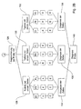
- Fig. 1 is a schematic representation of a first Embodiment of the method according to the invention shown.
- the individual process steps are in the Blocks shown in the left column, and parallel to that the left column are the corresponding ones in the right column Objects or elements depicted on the individual Take steps.
- the method according to the invention is basically divided according to the illustrated embodiment in a first Sub-step I and a second sub-step II.
- step I the sample array is first in step 100 102 scanned or scanned.
- the sample array 102 includes a plurality of incremental samples 104, whereby due to the Step 100 a biological sample 104 'from the array 102 is selected, as illustrated by arrow 106 is. After a single sample 104 'is selected, it is done in step 108 an analysis of individual images regarding the sample 104 ', as explained in more detail below becomes.
- Step 110 the individual images are first segmented in such a way that partial images with individual cells arise, as is exemplary is shown in Fig. 1 at 112, which is a field with two individual cells 114 and 116.
- Step 110 the individual images are first segmented in such a way that partial images with individual cells arise, as is exemplary is shown in Fig. 1 at 112, which is a field with two individual cells 114 and 116.
- Arrow 118 is shown being a cell in the shown Example, cell 114 selected and carried out in step 120 an analysis of this suspect cell 114.
- the result of the analysis can be used to determine whether the tumor suspect cell is actually an existing one Tumor cell, or whether it is is a benign or malignant dysplasia.
- pre-classified are Reference data sets mathematical feature operators iteratively determines the captured images of the biological Classify samples.
- a separation of samples with one dominant feature type each Determination of an associated operator set.
- Can be mutually orthogonal operators clearly the interested Assign characteristic types, so they can be differentiated can be realized within an image data set.
- this selection is made in substep I and in substep II.
- step I the selection of the operators becomes global applied at the level of an image 104 ', under one such image 104 'a unit can be understood that a cytologist as a whole - possibly with different ones Magnifications - through the eyepiece of his microscope. From a normal cytological sample, depending on Save magnification factor between 12 and 500 such images.
- the automatic parameter selection can be done independently of each other on frames of different Modality such as transmitted light, phase contrast and fluorescence as well as on different cell types (cervix, bladder, Lungs, etc.). With a sufficiently safe pre-classified Image sample gives a good selection result.
- step II if suspicious cells are present - object segmentation and partial analysis follows on the Single cell level with maximum resolution. Be here Cell dimension, shape and structural features recorded, wherein here too the approach of texture parameter selection is used becomes. Morphometric parameters are also determined.
- cytologists have many years of experience and cytology assistants for assessment and evaluation of microscopic histological findings in the Selection of the ones to be used for the classification of a cell Cell image and cell parameters with a more objective Approach to automatic parameter selection coupled.
- cytologists have many years of experience and cytology assistants for assessment and evaluation of microscopic histological findings in the Selection of the ones to be used for the classification of a cell Cell image and cell parameters with a more objective Approach to automatic parameter selection coupled.
- There a complete, optimal search to select ideal ones Parameters require a computing time of several years would take, the existing expert knowledge becomes syntactic and semantically in the automatic selection - coupled with a weighting function - integrated.
- Fig. 2 is a more detailed representation of the basis of the Fig. 1 described embodiment of the present Invention shown. From the sample array 102 with the plurality from incremental samples 104 a incremental sample 104 'becomes Analysis selected.
- the first partial image is generated by means of a first exposure modality, namely the microscopic transmitted light exposure modality (DL).
- the second field is obtained using the phase contrast (PK) recording modality
- the third field is obtained using the fluorescence (FL) recording modality.
- Each obtained partial image DL, PK, FL is classified separately from one another by means of a selected parameter set, as indicated by the blocks K 1 , K 2 and K 3 .
- Corresponding classification results for each partial image result from the classification of the individual partial images, as indicated by the blocks E 1 , E 2 and E 3 .
- step 122 it is determined on the basis of the results E 1 , E 2 and E 3 whether there is a change in the sample 104 'compared to a healthy sample. If this is not the case, the method according to the exemplary embodiment shown ends in step 124. For example, if a suspected tumor is affirmed, the method continues to step 126 in FIG. 2B. In step 126, the previously described segmentation of the sample 104 'is carried out in such a way that the sample is divided into individual regions which essentially comprise a cell or only individual cells, as is shown schematically at 128, 130 and 132 in FIG. 2B ,
- the detailed embodiment described with reference to FIG. 2 first includes the pre-classification on the Image plane as shown in Fig. 2A.
- three individual images are generated, the transmitted light (DL), phase contrast by means of the recording modalities (PK) and fluorescence (FL) can be obtained.
- This three pictures are initially separated from each other on the Image plane, d. H. each classified as a whole. in this connection becomes an expanded and modified automatic texture analysis method used, the extension of which is that an addition is integrated through texture processes which is especially necessary for cytological preparations.
- an automatic selection takes place the necessary and sufficient for these classification tasks Parameters from a pool of approx.
- Texture parameters color, statistical, texture, shape parameters.
- the required parameters are based on a representative image sample through an intelligent selection determined, including the quality, the informative value and the selectivity of the selected parameters in the Classification play a role.
- DL, PK and FL are each one independently own parameter set determined for classification.
- the pre-classification results are correlated from several recording modalities.
- the independent classification results K 1 , K 2 , K 3 of the three recording modalities are correlated with one another in order to make a decision as to whether the biological sample 104 'is to be classified as healthy or suspected of being tumor.
- this type of preclassification also ensures that approximately 75% of the samples are sorted out with high certainty as not suspected of being a tumor. All samples classified as suspect of tumor are subjected to a further classification level at the cell level in the following steps, which are shown in FIG. 2B.
- step 126 first shows the cell segmentation carried out. To all tumor suspect image samples more precisely to be investigated and assigned to the enemy classification The individual cells are first segmented from these image samples. The cells are segmented again on all three excerpts DL, PK and FL separated from each other. The result of this segmentation is the smallest Unit image sections with - ideally - one each single cell. With a suitable preparation of the samples in advance it is ensured that only a few cell conglomerates, d. H. Cell overlaps can be seen.
- the segmented cells are then classified. First, each individual cell is classified separately, whereby the method described above for automatic, knowledge-based Parameter selection is also used here comes. At the cell level there are not only color, texture parameters and statistical parameters used, but also shape parameters of the segmented cell considered. Here too, using intelligent selection from a pool of 600 to 1000 descriptive cell parameters selected those with whom in the different Recording modalities with emerging cell types can be distinguished from each other with a high degree of certainty.
- the classification results are then merged and a decision.
- the classification at cell level is initially separate for each image modality performed, and then both the segmented Cells of the three views registered with each other are offset as well as the classification results in order to arrive at a differentiated diagnosis.
- the Drawing files with the different recording modalities for the segmented cells either due to the already in the The first main step generated drawing files by simple segmentation this can be obtained, or a corresponding one Segmentation of the sample and renewed generation of the images in the second section is carried out.
- the invention Method not limited to the preferred embodiment is. Rather, the method according to the invention enables an analysis of biological samples to determine whether these changes compared to healthy tissue exhibit. Based on this classification of a sample different options for further analysis are possible, if this is desired at all. For some applications it may be sufficient to note that changes the sample against healthy tissue or healthy Rehearsals have taken place.
- the third option is full automation, as described above with reference to the figures.
Landscapes
- Engineering & Computer Science (AREA)
- Health & Medical Sciences (AREA)
- General Health & Medical Sciences (AREA)
- Physics & Mathematics (AREA)
- General Physics & Mathematics (AREA)
- Theoretical Computer Science (AREA)
- Life Sciences & Earth Sciences (AREA)
- Chemical & Material Sciences (AREA)
- Analytical Chemistry (AREA)
- Nuclear Medicine, Radiotherapy & Molecular Imaging (AREA)
- Molecular Biology (AREA)
- Dispersion Chemistry (AREA)
- Biomedical Technology (AREA)
- Biochemistry (AREA)
- Immunology (AREA)
- Pathology (AREA)
- Medical Informatics (AREA)
- Multimedia (AREA)
- Radiology & Medical Imaging (AREA)
- Quality & Reliability (AREA)
- Computer Vision & Pattern Recognition (AREA)
- Investigating Or Analysing Biological Materials (AREA)
- Investigating, Analyzing Materials By Fluorescence Or Luminescence (AREA)
- Sampling And Sample Adjustment (AREA)
- Image Input (AREA)
- Image Processing (AREA)
- Image Analysis (AREA)
Description
- Fig. 1
- eine schematische Darstellung eines ersten Ausführungsbeispiels des erfindungsgemäßen Verfahrens; und
- Fig. 2A und B
- eine detailliertere Darstellung der einzelnen Schritte des anhand der Fig. 1 beschriebenen Ausführungsbeispiels.
- eine bestehende Tumorzelle zu identifizieren, oder
- zu prognostizieren, ob sich eine Dysplasie zu einem Tumor weiter entwickeln wird.
- Die Parameterselektion wird anhand einer großen vorklassifizierten Stichprobe in mehreren Lernphasen durchgeführt. Variationen zur Optimierung können neu trainiert werden und in Konfigurationsdateien abgespeichert werden.
- Durch die Erstellung mehrerer Konfigurationsdatensätze lassen sich mit einem Klassifikationssystem unterschiedliche zytologische Präparate (Zervix, Blase, Lunge, ...) untersuchen und die erforderlichen Algorithmen adaptieren. Dadurch sind solche Klassifikationssysteme nicht mehr auf eine Anwendung limitiert und ermöglichen die Bearbeitung eines Großteils der zytologischen und histologischen Prüfaufgaben.
Claims (7)
- Rechner implementiertes Verfahren zur Analyse einer biologischen Probe (104') um eine Veränderung gegenüber einer gesunden biologischen Probe zu bestimmen, mit folgenden Schritten:Erzeugen eines ersten Bildes der biologischen Probe bei einer ersten Aufnahmemodalität (DL);Erzeugen eines zweiten Bildes der biologischen Probe bei einer zweiten Aufnahmemodalität (PK), wobei die erste Aufnahmemodalität und die zweite Aufnahmemodalität unterschiedlich sind, und wobei die erste Aufnahmemodalität und die zweite Aufnahmemodalität aus einer Gruppe von Aufnahmemodalitäten ausgewählt sind, die eine Durchlichtaufnahmemodalität, eine Fluoreszenzaufnahmemodalität und eine Phasenkontrastaufnahmemodalität umfaßt;Klassifizieren (K1) des ersten Bildes anhand eines ersten vorbestimmten Parameters;Klassifizieren (K2) des zweiten Bildes anhand eines zweiten vorbestimmten Parameters, wobei das erste Bild unabhängig von dem zweiten Bild klassifiziert wird; undabhängig von den Klassifikationsergebnisse (E1, E2), Bestimmen (122) ob die biologische Probe (104') eine Veränderung gegenüber einer gesunden biologischen Probe aufweist.
- Verfahren nach Anspruch 1, mit folgenden Schritten:Erzeugen eines dritten Bildes der biologischen Probe (104') bei einer dritten Aufnahmemodaltität (FL); undKlassifizierten (K3) des dritten Bildes anhand eines dritten vorbestimmten Parameters, wobei das dritte Bild unabhängig von dem ersten Bild und von dem zweiten Bild klassifiziert wird.
- Verfahren nach Anspruch 1 oder 2, das bei der Bestimmung einer Veränderung der biologischen Probe (104') die folgenden Schritte aufweist, um zu bestimmen, ob die Veränderung der biologischen Probe gutartig oder bösartig ist:Festlegen (126) zumindest eines Abschnitts (128, 130, 132) der biologischen Probe (104'), wobei der Abschnitt im wesentlichen ein Element oder im wesentlichen einzelne Elemente aufweist;Erzeugen eines ersten Teilbildes (DL) des Abschnitts der biologischen Probe bei einer vierten Aufnahmemodalität;Erzeugen eines zweiten Teilbildes (PK) des Abschnitts der biologischen Probe bei einer fünften Aufnahmemodalität;Klassifizieren (K) des ersten Teilbildes anhand eines vierten vorbestimmten Parameters;Klassifizieren (K) des zweiten Teilbildes anhand eines fünften vorbestimmten Parameters, wobei das erste Teilbild unabhängig von dem zweiten Teilbild klassifiziert wird; undabhängig von den Klassifikationsergebnissen (E), Bestimmen, ob die Veränderung der biologischen Probe gutartig oder bösartig ist.
- Verfahren nach Anspruch 3, mit folgenden Schritten:Erzeugen eines dritten Teilbildes (FL) des Abschnitts der biologischen Probe mit einer sechsten Aufnahmemodalität; undKlassifizieren des dritten Teilbildes anhand eines sechsten vorbestimmten Parameters, wobei das dritte Teilbild unabhängig von dem ersten Teilbild und von dem zweiten Teilbild klassifiziert wird.
- Verfahren nach einem der Ansprüche 3 oder 4, bei dem die biologische Probe eine Mehrzahl von Zellen umfaßt, wobei das erste Teilbild, das zweite Teilbild und das dritte Teilbild im wesentlichen eine Zelle oder im wesentlichen einzelne Zellen umfaßt.
- Verfahren nach einem der Ansprüche 1 bis 5, bei dem die erste Aufnahmemodalität, die zweite Aufnahmemodalität, die dritte Aufnahmemodalität, die vierte Aufnahmemodalität, die fünfte Aufnahmemodalität und die sechste Aufnahmemodalität aus einer Gruppe ausgewählt sind, die Durchlicht, Phasenkontrast und Fluoreszenz umfaßt, und bei dem der erste vorbestimmte Parameter, der zweite vorbestimmte Parameter, der dritte vorbestimmte Parameter, der vierte vorbestimmte Parameter, der fünfte vorbestimmte Parameter und der sechste vorbestimmte Parameter aus einer Gruppe ausgewählt sind, die Zelldimensions-, Form-, Struktur-, Farb-, Textur-Merkmale und statistische Merkmale, morphometrische Daten und Daten der oxidativen Schädigung von Zellen umfaßt.
- Verfahren nach Anspruch 6, bei dem die erste Aufnahmemodalität und die vierte Aufnahmemodalität die Durchsichtaufnahmemodalität sind, die zweite Aufnahmemodalität und die fünfte Aufnahmemodalität die Phasenkontrastaufnahmemodalität ist, und die dritte Aufnahmemodalität und die sechste Aufnahmemodalität die Fluoreszenzaufnahmemodalität sind, wobei für jede Aufnahmemodalität unabhängig voneinander jeweils ein eigener Parametersatz aus der Gruppe von vorbestimmten Parametern zur Klassifikation bestimmt wird.
Applications Claiming Priority (3)
| Application Number | Priority Date | Filing Date | Title |
|---|---|---|---|
| DE10124340A DE10124340A1 (de) | 2001-05-18 | 2001-05-18 | Verfahren zur Analyse einer biologischen Probe |
| DE10124340 | 2001-05-18 | ||
| PCT/EP2002/005316 WO2002095394A2 (de) | 2001-05-18 | 2002-05-14 | Verfahren zur analyse einer biologischen probe |
Publications (2)
| Publication Number | Publication Date |
|---|---|
| EP1381846A2 EP1381846A2 (de) | 2004-01-21 |
| EP1381846B1 true EP1381846B1 (de) | 2004-09-29 |
Family
ID=7685337
Family Applications (1)
| Application Number | Title | Priority Date | Filing Date |
|---|---|---|---|
| EP02745294A Expired - Lifetime EP1381846B1 (de) | 2001-05-18 | 2002-05-14 | Verfahren zur analyse einer biologischen probe |
Country Status (9)
| Country | Link |
|---|---|
| US (1) | US7257243B2 (de) |
| EP (1) | EP1381846B1 (de) |
| JP (1) | JP3897701B2 (de) |
| CN (1) | CN1226609C (de) |
| AT (1) | ATE278184T1 (de) |
| AU (1) | AU2002316904A1 (de) |
| DE (2) | DE10124340A1 (de) |
| ES (1) | ES2229158T3 (de) |
| WO (1) | WO2002095394A2 (de) |
Families Citing this family (10)
| Publication number | Priority date | Publication date | Assignee | Title |
|---|---|---|---|---|
| US8050477B2 (en) * | 2006-02-01 | 2011-11-01 | Koninklijke Philips Electronics N.V. | Radiation therapy flanning procedure |
| DE102007022014B4 (de) * | 2007-05-08 | 2018-08-30 | Leica Biosystems Nussloch Gmbh | Gewebeeinbettvorrichtung und Verfahren zum Betreiben einer Gewebeeinbettvorrichtung |
| DE102007062067A1 (de) * | 2007-12-21 | 2009-07-02 | Fraunhofer-Gesellschaft zur Förderung der angewandten Forschung e.V. | Optisches Verfahren und Vorrichtung zur Klassifizierung von Partikeln |
| US8762072B2 (en) * | 2008-10-02 | 2014-06-24 | Koninklijke Philips N.V. | Method of determining a reliability indicator for signatures obtained from clinical data and use of the reliability indicator for favoring one signature over the other |
| EP2344982A4 (de) * | 2008-10-10 | 2012-09-19 | Sti Medical Systems Llc | Verfahren zur gewebeklassifizierung in der zervikalen bildgebung |
| EP2562246B1 (de) * | 2010-04-23 | 2018-09-05 | Hamamatsu Photonics K.K. | Zellbeobachtungsvorrichtung und zellbeobachtungsverfahren |
| WO2011132586A1 (ja) * | 2010-04-23 | 2011-10-27 | 浜松ホトニクス株式会社 | 細胞観察装置および細胞観察方法 |
| AU2015331579A1 (en) * | 2014-10-17 | 2017-05-25 | Cireca Theranostics, Llc | Methods and systems for classifying biological samples, including optimization of analyses and use of correlation |
| AT525533B1 (de) * | 2021-11-02 | 2023-06-15 | West Medica Produktions Und Handels Gmbh | Verfahren und System zur Analyse einer Blutprobe |
| US20250147298A1 (en) * | 2023-11-08 | 2025-05-08 | Idexx Laboratories, Inc. | Methods and systems for verifying biological analysis devices |
Family Cites Families (23)
| Publication number | Priority date | Publication date | Assignee | Title |
|---|---|---|---|---|
| US4500509A (en) * | 1981-03-11 | 1985-02-19 | Lawrence Kass | Metachromatic dye sorption and fluorescent light emmisive means for differential determination of developmental stages of neutrophilic granulocytic cells and other leukocytes |
| US5016283A (en) * | 1985-11-04 | 1991-05-14 | Cell Analysis Systems, Inc. | Methods and apparatus for immunoploidy analysis |
| US4998284A (en) * | 1987-11-17 | 1991-03-05 | Cell Analysis Systems, Inc. | Dual color camera microscope and methodology for cell staining and analysis |
| US5252487A (en) * | 1989-05-19 | 1993-10-12 | Cell Analysis Systems, Inc. | Method and apparatus for determining the amount of oncogene protein product in a cell sample |
| US5162990A (en) | 1990-06-15 | 1992-11-10 | The United States Of America As Represented By The United States Navy | System and method for quantifying macrophage phagocytosis by computer image analysis |
| US5733721A (en) * | 1992-11-20 | 1998-03-31 | The Board Of Regents Of The University Of Oklahoma | Cell analysis method using quantitative fluorescence image analysis |
| US5381224A (en) * | 1993-08-30 | 1995-01-10 | A. E. Dixon | Scanning laser imaging system |
| CA2132269C (en) | 1993-10-12 | 2000-02-01 | Rainer Hermann Doerrer | Interactive automated cytology method and system |
| US5978497A (en) | 1994-09-20 | 1999-11-02 | Neopath, Inc. | Apparatus for the identification of free-lying cells |
| DE19538004A1 (de) | 1995-03-31 | 1996-10-02 | Fraunhofer Ges Forschung | Verfahren zur Grobklassifizierung von Objekten |
| US6091842A (en) | 1996-10-25 | 2000-07-18 | Accumed International, Inc. | Cytological specimen analysis system with slide mapping and generation of viewing path information |
| US5732150A (en) * | 1995-09-19 | 1998-03-24 | Ihc Health Services, Inc. | Method and system for multiple wavelength microscopy image analysis |
| US5755954A (en) | 1996-01-17 | 1998-05-26 | Technic, Inc. | Method of monitoring constituents in electroless plating baths |
| DE19612465C2 (de) | 1996-03-28 | 1998-12-24 | Fraunhofer Ges Forschung | Automatisches Optimieren von Objekt-Erkennungssystemen |
| DE19616997A1 (de) * | 1996-04-27 | 1997-10-30 | Boehringer Mannheim Gmbh | Verfahren zur automatisierten mikroskopunterstützten Untersuchung von Gewebeproben oder Körperflüssigkeitsproben |
| JPH10185911A (ja) | 1996-10-23 | 1998-07-14 | K O Denshi Kogyo Kk | 細胞解析装置及びその方法 |
| US6081740A (en) * | 1997-04-23 | 2000-06-27 | Accumed International, Inc. | Method and apparatus for imaging and sampling diseased tissue |
| US5999844A (en) * | 1997-04-23 | 1999-12-07 | Accumed International, Inc. | Method and apparatus for imaging and sampling diseased tissue using autofluorescence |
| US5986271A (en) * | 1997-07-03 | 1999-11-16 | Lazarev; Victor | Fluorescence imaging system |
| US6005256A (en) * | 1998-02-12 | 1999-12-21 | Raytheon Company | Method and apparatus for performing cell analysis based on simultaneous multiple marker emissions from neoplasia (CASMMEN) |
| US6462770B1 (en) * | 1998-04-20 | 2002-10-08 | Xillix Technologies Corp. | Imaging system with automatic gain control for reflectance and fluorescence endoscopy |
| DE19834718C2 (de) | 1998-07-31 | 2003-06-12 | Fraunhofer Ges Forschung | Digitale Bildverarbeitung für ein Qualitätskontrollsystem |
| US6143512A (en) * | 1998-08-17 | 2000-11-07 | Markovic; Nenad | Cap-pap test |
-
2001
- 2001-05-18 DE DE10124340A patent/DE10124340A1/de not_active Withdrawn
-
2002
- 2002-05-14 WO PCT/EP2002/005316 patent/WO2002095394A2/de not_active Ceased
- 2002-05-14 AT AT02745294T patent/ATE278184T1/de active
- 2002-05-14 US US10/478,482 patent/US7257243B2/en not_active Expired - Lifetime
- 2002-05-14 CN CNB028132629A patent/CN1226609C/zh not_active Expired - Fee Related
- 2002-05-14 JP JP2002591817A patent/JP3897701B2/ja not_active Expired - Fee Related
- 2002-05-14 EP EP02745294A patent/EP1381846B1/de not_active Expired - Lifetime
- 2002-05-14 DE DE50201173T patent/DE50201173D1/de not_active Expired - Lifetime
- 2002-05-14 ES ES02745294T patent/ES2229158T3/es not_active Expired - Lifetime
- 2002-05-14 AU AU2002316904A patent/AU2002316904A1/en not_active Abandoned
Also Published As
| Publication number | Publication date |
|---|---|
| ATE278184T1 (de) | 2004-10-15 |
| US7257243B2 (en) | 2007-08-14 |
| ES2229158T3 (es) | 2005-04-16 |
| DE10124340A1 (de) | 2002-12-05 |
| US20040235180A1 (en) | 2004-11-25 |
| AU2002316904A1 (en) | 2002-12-03 |
| WO2002095394A3 (de) | 2003-02-20 |
| CN1522362A (zh) | 2004-08-18 |
| JP2004529360A (ja) | 2004-09-24 |
| EP1381846A2 (de) | 2004-01-21 |
| JP3897701B2 (ja) | 2007-03-28 |
| WO2002095394A2 (de) | 2002-11-28 |
| CN1226609C (zh) | 2005-11-09 |
| DE50201173D1 (de) | 2004-11-04 |
Similar Documents
| Publication | Publication Date | Title |
|---|---|---|
| DE68926796T2 (de) | System und Verfahren für automatische Zellenklassifizierung, basierend auf ein Neuronalnetzwerk | |
| DE69232413T2 (de) | Verfahren und Vorrichtung zum automatischen Test biologischer Proben | |
| DE69322498T2 (de) | Verfahren zur identifizierung von normalen biomedizinischen proben | |
| DE2823490C2 (de) | ||
| DE69329554T2 (de) | Verfahren zur identifizierung von objekten unter verwendung von datenverarbeitungstechniken | |
| DE19747415C2 (de) | Verfahren zur Unterstützung eines Betrachters bei der Durchmusterung einer Probe und zytologisches Probenanalysiersystem | |
| DE69422128T2 (de) | Verfahren zur interaktiven automatischen Zytologie | |
| EP2130174B1 (de) | Verfahren und vorrichtung zur ermittlung einer zellkontur einer zelle | |
| DE69126839T2 (de) | Automatisches zellenklassifikationssystem und verfahren | |
| EP2656277B1 (de) | Vorrichtung und verfahren zur bestimmung von objekten in einer farbaufnahme | |
| DE69532276T2 (de) | Automatisiertes cytologisches probenklassifizierungsverfahren | |
| DE102010009853B4 (de) | Bestimmung von Gewebezuständen mittels bildgebender Massenspektrometrie | |
| DE19616997A1 (de) | Verfahren zur automatisierten mikroskopunterstützten Untersuchung von Gewebeproben oder Körperflüssigkeitsproben | |
| DE1498824A1 (de) | Vorrichtung zur maschinellen Krebsdiagnose | |
| DE102013216362A1 (de) | Analyseverfahren zur Klassifikationsunterstützung | |
| DE4211904C2 (de) | Automatisches Verfahren zum Erstellen einer Liste unterschiedlicher Arten für eine flüssige Probe | |
| EP3430565B1 (de) | Verfahren zur untersuchung verteilter objekte unter segmentierung eines übersichtbildes | |
| EP2400458A2 (de) | Verfahren und Vorrichtung zur Segmentierung von biologischen Zellen in einer Aufnahme | |
| EP1381846A2 (de) | Verfahren zur analyse einer biologischen probe | |
| DE112007001907T5 (de) | Verfahren für die Zellanalyse | |
| EP2440920B1 (de) | Lokalisierung eines validen bereiches eines blutausstriches | |
| DE69329324T2 (de) | Automatische Detektion von kanzerogenem oder vorkanzerogenem Gewebe durch Messung maligner Veränderungen | |
| DE19637741B4 (de) | Automatisches Zytologisches Probenanalysesystem unter Verwendung von individualisierten Patientendaten | |
| EP3540631A1 (de) | In-vitro-verfahren zum markierungsfreien bestimmen eines zelltyps einer weissen blutzelle | |
| DE69831573T2 (de) | Inspektionssystem mit probenvorschau |
Legal Events
| Date | Code | Title | Description |
|---|---|---|---|
| PUAI | Public reference made under article 153(3) epc to a published international application that has entered the european phase |
Free format text: ORIGINAL CODE: 0009012 |
|
| 17P | Request for examination filed |
Effective date: 20031114 |
|
| AK | Designated contracting states |
Kind code of ref document: A2 Designated state(s): AT BE CH CY DE DK ES FI FR GB GR IE IT LI LU MC NL PT SE TR |
|
| AX | Request for extension of the european patent |
Extension state: AL LT LV MK RO SI |
|
| GRAP | Despatch of communication of intention to grant a patent |
Free format text: ORIGINAL CODE: EPIDOSNIGR1 |
|
| GRAS | Grant fee paid |
Free format text: ORIGINAL CODE: EPIDOSNIGR3 |
|
| GRAA | (expected) grant |
Free format text: ORIGINAL CODE: 0009210 |
|
| AK | Designated contracting states |
Kind code of ref document: B1 Designated state(s): AT BE CH CY DE DK ES FI FR GB GR IE IT LI LU MC NL PT SE TR |
|
| PG25 | Lapsed in a contracting state [announced via postgrant information from national office to epo] |
Ref country code: TR Free format text: LAPSE BECAUSE OF FAILURE TO SUBMIT A TRANSLATION OF THE DESCRIPTION OR TO PAY THE FEE WITHIN THE PRESCRIBED TIME-LIMIT Effective date: 20040929 Ref country code: FI Free format text: LAPSE BECAUSE OF FAILURE TO SUBMIT A TRANSLATION OF THE DESCRIPTION OR TO PAY THE FEE WITHIN THE PRESCRIBED TIME-LIMIT Effective date: 20040929 Ref country code: IE Free format text: LAPSE BECAUSE OF FAILURE TO SUBMIT A TRANSLATION OF THE DESCRIPTION OR TO PAY THE FEE WITHIN THE PRESCRIBED TIME-LIMIT Effective date: 20040929 Ref country code: NL Free format text: LAPSE BECAUSE OF FAILURE TO SUBMIT A TRANSLATION OF THE DESCRIPTION OR TO PAY THE FEE WITHIN THE PRESCRIBED TIME-LIMIT Effective date: 20040929 |
|
| REG | Reference to a national code |
Ref country code: GB Ref legal event code: FG4D Free format text: NOT ENGLISH |
|
| REG | Reference to a national code |
Ref country code: CH Ref legal event code: EP |
|
| REG | Reference to a national code |
Ref country code: IE Ref legal event code: FG4D Free format text: GERMAN |
|
| REF | Corresponds to: |
Ref document number: 50201173 Country of ref document: DE Date of ref document: 20041104 Kind code of ref document: P |
|
| GBT | Gb: translation of ep patent filed (gb section 77(6)(a)/1977) |
Effective date: 20041104 |
|
| PG25 | Lapsed in a contracting state [announced via postgrant information from national office to epo] |
Ref country code: GR Free format text: LAPSE BECAUSE OF FAILURE TO SUBMIT A TRANSLATION OF THE DESCRIPTION OR TO PAY THE FEE WITHIN THE PRESCRIBED TIME-LIMIT Effective date: 20041229 Ref country code: SE Free format text: LAPSE BECAUSE OF FAILURE TO SUBMIT A TRANSLATION OF THE DESCRIPTION OR TO PAY THE FEE WITHIN THE PRESCRIBED TIME-LIMIT Effective date: 20041229 Ref country code: DK Free format text: LAPSE BECAUSE OF FAILURE TO SUBMIT A TRANSLATION OF THE DESCRIPTION OR TO PAY THE FEE WITHIN THE PRESCRIBED TIME-LIMIT Effective date: 20041229 |
|
| LTIE | Lt: invalidation of european patent or patent extension |
Effective date: 20040929 |
|
| NLV1 | Nl: lapsed or annulled due to failure to fulfill the requirements of art. 29p and 29m of the patents act | ||
| REG | Reference to a national code |
Ref country code: ES Ref legal event code: FG2A Ref document number: 2229158 Country of ref document: ES Kind code of ref document: T3 |
|
| REG | Reference to a national code |
Ref country code: IE Ref legal event code: FD4D |
|
| PG25 | Lapsed in a contracting state [announced via postgrant information from national office to epo] |
Ref country code: LU Free format text: LAPSE BECAUSE OF NON-PAYMENT OF DUE FEES Effective date: 20050514 Ref country code: CY Free format text: LAPSE BECAUSE OF FAILURE TO SUBMIT A TRANSLATION OF THE DESCRIPTION OR TO PAY THE FEE WITHIN THE PRESCRIBED TIME-LIMIT Effective date: 20050514 |
|
| PG25 | Lapsed in a contracting state [announced via postgrant information from national office to epo] |
Ref country code: MC Free format text: LAPSE BECAUSE OF NON-PAYMENT OF DUE FEES Effective date: 20050531 Ref country code: BE Free format text: LAPSE BECAUSE OF NON-PAYMENT OF DUE FEES Effective date: 20050531 |
|
| ET | Fr: translation filed | ||
| PLBE | No opposition filed within time limit |
Free format text: ORIGINAL CODE: 0009261 |
|
| STAA | Information on the status of an ep patent application or granted ep patent |
Free format text: STATUS: NO OPPOSITION FILED WITHIN TIME LIMIT |
|
| 26N | No opposition filed |
Effective date: 20050630 |
|
| BERE | Be: lapsed |
Owner name: FRAUNHOFER-GESELLSCHAFT ZUR FORDERUNG DER ANGEWAND Effective date: 20050531 |
|
| BERE | Be: lapsed |
Owner name: *FRAUNHOFER-GESELLSCHAFT ZUR FORDERUNG DER ANGEWAN Effective date: 20050531 |
|
| PG25 | Lapsed in a contracting state [announced via postgrant information from national office to epo] |
Ref country code: PT Free format text: LAPSE BECAUSE OF NON-PAYMENT OF DUE FEES Effective date: 20050228 |
|
| REG | Reference to a national code |
Ref country code: FR Ref legal event code: PLFP Year of fee payment: 14 |
|
| PGFP | Annual fee paid to national office [announced via postgrant information from national office to epo] |
Ref country code: GB Payment date: 20150521 Year of fee payment: 14 Ref country code: CH Payment date: 20150520 Year of fee payment: 14 Ref country code: ES Payment date: 20150520 Year of fee payment: 14 |
|
| PGFP | Annual fee paid to national office [announced via postgrant information from national office to epo] |
Ref country code: FR Payment date: 20150519 Year of fee payment: 14 Ref country code: IT Payment date: 20150519 Year of fee payment: 14 Ref country code: AT Payment date: 20150520 Year of fee payment: 14 |
|
| REG | Reference to a national code |
Ref country code: CH Ref legal event code: PL |
|
| REG | Reference to a national code |
Ref country code: AT Ref legal event code: MM01 Ref document number: 278184 Country of ref document: AT Kind code of ref document: T Effective date: 20160514 |
|
| GBPC | Gb: european patent ceased through non-payment of renewal fee |
Effective date: 20160514 |
|
| PG25 | Lapsed in a contracting state [announced via postgrant information from national office to epo] |
Ref country code: LI Free format text: LAPSE BECAUSE OF NON-PAYMENT OF DUE FEES Effective date: 20160531 Ref country code: CH Free format text: LAPSE BECAUSE OF NON-PAYMENT OF DUE FEES Effective date: 20160531 |
|
| PG25 | Lapsed in a contracting state [announced via postgrant information from national office to epo] |
Ref country code: IT Free format text: LAPSE BECAUSE OF NON-PAYMENT OF DUE FEES Effective date: 20160514 Ref country code: AT Free format text: LAPSE BECAUSE OF NON-PAYMENT OF DUE FEES Effective date: 20160514 |
|
| REG | Reference to a national code |
Ref country code: FR Ref legal event code: ST Effective date: 20170131 |
|
| PG25 | Lapsed in a contracting state [announced via postgrant information from national office to epo] |
Ref country code: FR Free format text: LAPSE BECAUSE OF NON-PAYMENT OF DUE FEES Effective date: 20160531 |
|
| PG25 | Lapsed in a contracting state [announced via postgrant information from national office to epo] |
Ref country code: GB Free format text: LAPSE BECAUSE OF NON-PAYMENT OF DUE FEES Effective date: 20160514 |
|
| PG25 | Lapsed in a contracting state [announced via postgrant information from national office to epo] |
Ref country code: ES Free format text: LAPSE BECAUSE OF NON-PAYMENT OF DUE FEES Effective date: 20160515 |
|
| REG | Reference to a national code |
Ref country code: ES Ref legal event code: FD2A Effective date: 20180627 |
|
| PGFP | Annual fee paid to national office [announced via postgrant information from national office to epo] |
Ref country code: DE Payment date: 20200525 Year of fee payment: 19 |
|
| REG | Reference to a national code |
Ref country code: DE Ref legal event code: R119 Ref document number: 50201173 Country of ref document: DE |
|
| PG25 | Lapsed in a contracting state [announced via postgrant information from national office to epo] |
Ref country code: DE Free format text: LAPSE BECAUSE OF NON-PAYMENT OF DUE FEES Effective date: 20211201 |