EP1336088B1 - Step-wise process to recover or eliminate biological substances by flotation from underlayered colloidal medium - Google Patents
Step-wise process to recover or eliminate biological substances by flotation from underlayered colloidal medium Download PDFInfo
- Publication number
- EP1336088B1 EP1336088B1 EP01997697A EP01997697A EP1336088B1 EP 1336088 B1 EP1336088 B1 EP 1336088B1 EP 01997697 A EP01997697 A EP 01997697A EP 01997697 A EP01997697 A EP 01997697A EP 1336088 B1 EP1336088 B1 EP 1336088B1
- Authority
- EP
- European Patent Office
- Prior art keywords
- density
- sample
- centrifugation
- mixture
- medium
- Prior art date
- Legal status (The legal status is an assumption and is not a legal conclusion. Google has not performed a legal analysis and makes no representation as to the accuracy of the status listed.)
- Expired - Lifetime
Links
Images
Classifications
-
- C—CHEMISTRY; METALLURGY
- C12—BIOCHEMISTRY; BEER; SPIRITS; WINE; VINEGAR; MICROBIOLOGY; ENZYMOLOGY; MUTATION OR GENETIC ENGINEERING
- C12Q—MEASURING OR TESTING PROCESSES INVOLVING ENZYMES, NUCLEIC ACIDS OR MICROORGANISMS; COMPOSITIONS OR TEST PAPERS THEREFOR; PROCESSES OF PREPARING SUCH COMPOSITIONS; CONDITION-RESPONSIVE CONTROL IN MICROBIOLOGICAL OR ENZYMOLOGICAL PROCESSES
- C12Q1/00—Measuring or testing processes involving enzymes, nucleic acids or microorganisms; Compositions therefor; Processes of preparing such compositions
- C12Q1/68—Measuring or testing processes involving enzymes, nucleic acids or microorganisms; Compositions therefor; Processes of preparing such compositions involving nucleic acids
- C12Q1/6806—Preparing nucleic acids for analysis, e.g. for polymerase chain reaction [PCR] assay
-
- G—PHYSICS
- G01—MEASURING; TESTING
- G01N—INVESTIGATING OR ANALYSING MATERIALS BY DETERMINING THEIR CHEMICAL OR PHYSICAL PROPERTIES
- G01N15/00—Investigating characteristics of particles; Investigating permeability, pore-volume, or surface-area of porous materials
- G01N15/04—Investigating sedimentation of particle suspensions
- G01N15/05—Investigating sedimentation of particle suspensions in blood
Definitions
- the present invention relates to a method for discontinuous density gradient centrifugation to separate a minimum of two substances in a sample.
- use is made of colloidal density media and low-density solutions.
- cells and micro-organisms often need to be separated from each other and from different types of samples (e.g. clinical, industrial, food, environmental and forensic samples) in such a way that they can be used for, for example, cultivation, inoculation, cell fusion, clinical therapy and biochemical analysis without interference from inhibiting activities of the sample or inhibiting activities added during the separation process.
- These inhibiting activities may be, for example, endogenous antibodies, proteases, nucleases, endotoxins, and antibiotics.
- Micro-organisms, such as bacteria and virus comprise gene sequences analogous with eukaryotic gene sequences and this may lead to an erroneous result if both of these are present simultaneously in a sample.
- Density media of both the low molecular weight (Nycodenz etc.) and of the colloidal type (BactXtractor, Percoll, PureSperm) has been used.
- the separation is based on the density of the inherent components in the used density media after centrifugation in a continuous density gradient.
- the sample is loaded on the density media and the desired fraction is recovered from the bottom part of the resulting lower phase.
- the centrifugation is performed in an angle centrifuge and this means that the bacteria, or other desired cells or fractions, may be harmed when they hit the wall of the centrifuge tube during the sedimentation down to their density level and this may lead to bacterial death and leakage of their DNA/RNA content.
- the sample is loaded on the colloidal media there is a risk of contamination of the underlying phases. This contamination is partly due to wall effects, i.e. the sample flows down along the tube wall and may be mixed with underlying phases, and partly due to too rapid, especially initially, loading of the sample leading to admixture with underlying phases.
- FR 2 561 256 describes a method of purification of biological particles, such as antigen core particles, using flotation ultracentrifugation on a density gradient. More specifically, the sample is placed on top of a preformed density gradient, which is then centrifuged to allow for collection of the sample in the top layer while contaminants etc have migrated to the lower layers of the gradient. Before this purification process, the sample needs to be concentrated and prepurified.
- the density gradient is provided by a low molecular compound, namely cesium chloride.
- the g-force required during the centrifugation in this process is at least 20 000 x g.
- US A 5 437 987 relates to a triple gradient process with antibody panning to recover nucleated fetal cells from maternal blood. This document teaches use of multiple layers of density gradient medium.
- US A 4 927 750 relates to a cell separation process involving centrifugation. This document describes separation of cells from various biological specimen and is not concerned with flotation of one or more desired substances to an uppermost solution of low density.
- US A 5 962 237 relates to a method of enriching rare cells using centrifugation.
- the centrifugation steps may be repeated for enrichment purposes, ie during the same time and same g-force. This document does not teach flotation of different desired substances.
- WO 91 04318A relates to separation of different living cells using low molecular solutions which will diffuse into each other and form a continuous or discontinuous gradient.
- US A 4 971 801 relates to a biological response modifier comprising natural membrane vesicles and ribosomes in a suspending buffer.
- Product isolation is described using gradient centrifugation.
- the desired fraction is pelleted, ie collected at the bottom of the centrifuge tube.
- non-ionic gradient media e.g. sucrose gradients
- sucrose gradients have also been suggested for separation.
- sugars such as sucrose
- these methods will entail the same sort of drawbacks as mentioned above, i.e. unstable gradients wherein the boundaries become more and more diffuse with time, an osmotic pressure that can harm micro-organisms, cells etc.
- One object of the present invention is to avoid one or more of the above drawbacks by providing a new method for discontinuous density gradient centrifugation which relies on a new principle, namely allowing the desired substances to float after one or more centrifugation steps.
- a further object of the invention is to provide a method of discontinuous density gradient centrifugation, which includes less risk of contamination of the desired substance(s) than prior art methods.
- This is achieved by a step-wise method, wherein a lower part of a centrifuged sample is passed over to one or more subsequent steps, repeating the same round of mixing and centrifugation as defined in claim 1 but at different densities and at different g-forces. Since the lower part is further processed, the actual sample requires no pipetting between steps, which eliminates one common way of contamination.
- the novel step-wise method provides a superior flexibility as compared to the prior art, since a series of rounds can be designed, each round being performed during conditions specifically separating substances that are known or suspected to be present in the sample.
- the invention provides a method of discontinuous density gradient centrifugation for separating a minimum of two substances from each other in a sample.
- the method is characterised by the subsequent steps of
- the above described round of steps (i)-(iv) can be repeated, such as one, two, three or any number of times, including a second, third, fourth etc mixture of a defined density, and centrifuged at a second, third, fourth etc g-force.
- the skilled person in this field can select the appropriate combinations of g-forces and densities based on the densities of the substance(s) that are to be separated from the sample.
- Such a systematic variation of g-forces has not been suggested in the context of density gradient centrifugation in the prior art.
- the advantages of using a colloidal centrifugation medium to this end were quite unexpected, since a method designed according to the present invention can provide purities that are superior to the results of prior art methods applied to the wide variety of samples that the present invention is useful with.
- a colloidal density gradient centrifugation medium of a lower density than the mixture in a) is placed between the mixture in a) and the low-density solution in b). This gives an extra barrier between the mixed sample and the low-density solution and improves and facilitates the recovery following separation.
- discontinuous refers to a successive layering of solutions of different density e.g. in a centrifuge tube.
- the centrifugation medium used according to the invention should be a heavy medium, such as a colloidal silica-based material.
- the medium should be inert, autoclavable in the presence of salt, have a low or no endotoxin level, an as low osmotic pressure as possible, preferably below 20 mOsm/kg, a low viscosity in salt ( ⁇ 5cP), at high density (>1.3 g/ml; RG).
- a suitable medium is ReadyGrade® (in general denoted RG and available from Amersham Biosciences, Uppsala, Sweden).
- silica particles can be bought from commercial sources, such as Nyacol, and silanised according to well known techniques by the user.
- the unique properties of the medium make it possible to retain the native characterstics of substances after separation. Further details regarding the medium will be given below. Accordingly, the present method is advantageous as compared to the prior art since it can reduce costs to a substantial degree.
- the solution in b) is preferably an aqueous solution, such as a buffer solution.
- the density in a) is higher than that of the desired substance(s) to be separated.
- the skilled person will understand that the terms "low density” and "high density” are used in relation to each other and are not intended to be compared with any other densities. Illustrative densities will be given below.
- the steps (i)-(iv) are repeated once or more depending on which substance is sought after or which contaminants are to be removed.
- the g-force in the centrifugation step(s) and the density of the colloidal media are varied according to the substance(s) to be separated.
- the desired substance(s) to be separated is/are cells, micro-organisms, virus, and fractions thereof, nucleic acids and any proteins.
- the sample is processed into a mixture with the medium in order to provide an appropriate density thereof.
- the sample may be of practically any origin, such as a biological sample, e.g. cells, blood etc, faeces, e.g.
- the desired substance(s) is/are derivatized before centrifugation to improve separation of two substances of similar density.
- An example of this is to treat the desired substance(s) with an affinity reagent linked to a high or low-density particle, such as mAb coated beads of suitable density.
- the method described above is specifically adapted to separate different cell types, such as X- and Y-sperm cells, from each other in a sample on the basis of their buoyant density, shape, permeability, movement, and/or viscosity in a density gradient medium by centrifugation.
- the method comprises the subsequent steps according to claim 1, with the sample being a semen sample and the desired substances being the separated cells.
- the density medium of this embodiment is the kind of colloidal density medium discussed above.
- the sample is mixed with high density medium and, optionally, said medium in step A) is centrifuged to form a density gradient before said sample is added to said centrifuge tube.
- the gradient is preferably very flat or essentially planar, i.e. it comprises a very small density difference in the centrifuge tube, at least at the centre of the gradient.
- the gradient is discontinuous. It is important that the gradient provides the necessary separation which means that it should be possible to recover fractions where X and Y sperms, respectively, are enriched to at least 70% or more, preferably 100%.
- the semen sample is purified before separation.
- This purification may be in any desired way, such as by discontinuous centrifugation.
- the density referred to above is close to that of the X and Y sperm cells, i.e. about 1.120g/mL.
- the corresponding values for human cells are substantially the same. These densities are applicable under the experimental conditions mentioned below in the experimental part.
- the present invention does not relate to centrifugations during which human X- and Y-sperm cells, respectively, have densities centred around 1.185 g/mL.
- the separation may be improved by manipulating the density of X and Y sperm cells, for instance by altering pH, conductivity of the sample and/or medium.
- the manipulation may comprise swelling of X- and Y-sperm cells.
- centrifugations for separation of X and Y cells from each other in a density gradient as defined above will be performed depending on choice of gradient solution, gradient shape, centrifuge rotor, centrifugation time, g-force at known viscosity, osmolarity and concentration levels.
- the invention also relates to a method for separation as above, wherein said sample is mixed with said medium to achieve a density of said mixed sample-medium which lies close to the density of X and Y sperm cells.
- the separation is mainly achieved by isopycnic centrifugation.
- the separation pattern obtained in this variant of the invention will thus preferentially be based on the different buoyant densities of the sperm cells.
- the colloidal density gradient medium comprises a suspension of particles having an average particle size of 2-40nm, preferably a hydrated particle of 10-30 nm.
- the particles are derivatized silica particles, and more preferably silanised silica particles.
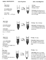
- Fig. 1 represents a schematic view of the working principle of the method according to the present invention.
- four centrifugation steps have been performed.
- the number of centrifugation steps and g-force is varied according to the nature of the substances to be separated.
- RG an example of one colloidal medium according to the invention
- the resulting lower phase, resuspended and mixed with RG to a density of 1.200 g/ml, is placed at the bottom of a new centrifuge tube, is optionally overlaid with RG having a density of 1.090 g/ml and on top of this a solution of 0.150 M NaCl is applied. Then the tube is centrifuged for 1 to 5 minutes at 1,000 x g av . Any cells and protozoans from the sample are recovered in the uppermost phase layer (2, Figure 1).
- the resulting lower phase is resuspended and transferred to a new centrifuge tube, is optionally overlaid with RG having a density of 1.130 g/ml and on top of this a solution of 0.150 M NaCl is applied. Then the tube is centrifuged for 1 to 5 minutes at 10,000 x g av . Any bacteria from the sample are now recovered in the uppermost phase layer (3, Figure 1).
- the resulting lower phase is resuspended and transferred to a new centrifuge tube, is optionally overlaid with RG having a density of 1.130 g/ml and on top of this a solution of 0.150 M NaCl is applied. Then the tube is centrifuged for 1 to 5 minutes at 50,000 x g av . Any viruses from the sample are now recovered in the uppermost phase layer (4, Figure 1).
- the light density material in the sample matrix is floating in step 1 ( Figure 1).
- step 1 the cells/micro-organisms no longer have to sediment through a possibly formed plug but will in this step remain in the lower phase because of their density ( ⁇ > 1.057).
- the average hydrostatic pressure resulting from centrifugation will be higher than in prior art techniques. This is due to that the distance to the rotational centre is longer when the sample is in the bottom of the tube as compared to when the sample is loaded on the top. This fact contributes to an increased yield when employing the present method compared with prior art.
- steps 2, 3, and 4 desirable cells, bacteria and viruses are floating from the underlying sample phase and are conveniently recovered from the uppermost phase layer. Hereby all manipulations with the sample phase is avoided and thus recontamination with e.g. inhibitory activities from the sample solution is eliminated.
- the sample containing cells / micro-organisms is located in the lower part of the centrifugation tube in every centrifugation step, the sedimentation towards the wall or bottom of the tube of the cells/micro-organisms is avoided. This reduces the cell damage frequency and increases the yield as compared to prior art techniques.
- the mixture was transferred to the bottom of a centrifuge tube (1.5 ml), overlaid with 0.5 ml of a RG solution with a density of 1.030 g/ml and on top of this with 0.2 ml of a 0.15 M NaCl solution. Then, centrifugation was performed in a table centrifuge at 10,000 x g av for 1 minute. Then, the lower phase was aspirated into a syringe whereby the cannula first penetrated the unpipettable plug formed by the minced meat matrix and was transferred to a new tube.
- the upper phases were aspirated with a Pasteur pipette and discarded and then the lower phase was resuspended, transferred to a new tube, overlaid with 0.2 ml of a RG solution with a density of 1.130 g/ml and 0.1 ml of a 0.15 M NaCl solution. Then, centrifugation was performed at 10,000 x g av for 1 minute.
- the upper phases were fractionated with an automatic pipette in three portions of 75 ⁇ l and then analysed.
- the yield with the spiked samples was 80-100% as compared to 50-80% with the two older techniques.
- the yield in the controls was somewhat lower in every case as compared to the samples. This phenomenon has been reported earlier and is very likely due to the protective effect, which the endogenous flora in the minced meat has on the bacteria added in the samples.
- the mixture was transferred to the bottom of a centrifuge tube (1.5 ml), overlaid with 0.5 ml of a RG solution with a density of 1.030 g/ml and on top with 0.2 ml of a 0.15 M NaCl solution. Then, centrifugation was performed in a table centrifuge at 10,000 x g av for 1 minute. Thereafter the lower phase was aspirated into a syringe, whereby the cannula first penetrated the unpipettable plug formed by the sample matrix, and was transferred to a new tube. A small pellet was observed and to obtain the pelleted material (with bound bacteria) the remaining 100 ⁇ l was resuspended before being drawn into the syringe.
- the lower phase thus obtained was mixed with 585 ⁇ l RG to obtain a density of 1.200 g/ml, then overlaid with 0.2 ml of a RG solution with a density of 1.090 g/ml and on of this top 0.1 ml of a 0.15 M NaCl solution was added. Then centrifugation was performed at 1,000 x g av for 1 minute.
- the upper phases were aspirated with a Pasteur pipette and discarded and then the lower phase was resuspended, transferred to a new tube, overlaid with 0.2 ml of a RG solution with a density of 1.130 g/ml and with 0.1 ml of a 0.15 M NaCl solution. Then, centrifugation was performed at 10,000 x g av for 1 minute.
- the upper phases were fractionated with an automatic pipette in three portions of 75 ⁇ l and then analysed.
- the yield with the spiked samples was 80-100% as compared to 50-80% with the two older techniques.
- the yield in the controls was somewhat lower in every case as compared to the samples. This phenomenon has been reported earlier and is very likely due to the protective effect, which the endogenous flora in the faeces sample has on the bacteria added in the samples.
- Spiked samples and corresponding controls were slurred and mixed with 85 ⁇ l of RG to a final density of 1.057 g/ml.
- the mixture was transferred to the bottom of a centrifuge tube (1.5 ml), overlaid with 0.5 ml of a RG solution with a density of 1.030 g/ml and on top of this with 0.2 ml of a 0.15 M NaCl solution. Then, centrifugation was performed in a table centrifuge at 10,000 x g av for 1 minute. The upper phases were aspirated with a Pasteur pipette and discarded.
- the lower phase was resuspended before transferring it to a new tube.
- the lower phase obtained was mixed with 585 ⁇ l RG to obtain a density of 1.200 g/ml, then overlaid with 0.2 ml of a RG solution with a density of 1.090 g/ml and on top 0.1 ml of a 0.15 M NaCl solution was added. Then centrifugation was performed at 1,000 x g av for 1 minute.
- the upper phases were aspirated with a Pasteur pipette and discarded and then the lower phase including the pellet was resuspended, transferred to a new tube, overlaid with 0.2 ml of a RG solution with a density of 1.130 g/ml and 0.1 ml of a 0.15 M NaCl solution. Then, centrifugation was performed at 10,000 x g av for 1 minute.
- the upper phases were fractionated with an automatic pipette in three portions of 75 ⁇ l and then analysed.
- E. coli For determination of the amount of E. coli by culturing, 2 x 10 ⁇ l of varying dilutions from the different samples were spread on plates. Manual counting of the colonies and calculation of the amount were performed after incubation over night. For detection of E. coli by a DNA technique 10 ⁇ l from each sample was used in a PCR test.
- the yield with the spiked samples was 80-100% as compared to 10-20% with the two older techniques.
- the yield in the controls was somewhat lower in every case as compared to the samples. This phenomenon has been reported earlier and is very likely due to the protective effect, which the endogenous flora in the soil sample has on the added bacteria in the samples.
- a fresh semen sample from a healthy bull was divided after dilution by 1:10 with 0.15 M NaCl, into 16 fractions of 0.45 ml each containing about 10 7 sperms.
- Varying amounts of BVDV (bovine virus diarrhoea virus; 10 6 , 10 5 , 10 4 ) and Campylobacter fetus (10 6 , 10 5 , 10 4 ), respectively, in 50 ⁇ l of 0.15 M NaCl were added to 12 of the tubes and to four of the tubes 50 ⁇ l of 0.15 M NaCl were added as control.
- Spiked samples (6 tubes) and controls (2 tubes) were mixed with 85 ⁇ l of RG to a final density of 1.057 g/ml.
- the mixture was transferred to the bottom of a centrifuge tube (3.0 ml), overlaid with 0.5 ml of a RG solution with a density of 1.030 g/ml and on top with 0,5 ml of a 0.15 M NaCl solution. Then, centrifugation was performed in a swing-out rotor at 10,000 x g av for 1 minute. The upper phases were aspirated by a Pasteur pipette and discarded.
- the lower phase was resuspended, transferred to a new tube and mixed with 585 ⁇ l RG to obtain a density of 1.200 g/ml, overlaid with 0.5 ml of a RG solution with a density of 1.090 g/ml and on top with 0.5 ml of a 0.15 M NaCl solution. Then centrifugation was performed at 1,000 x g av for 1 minute.
- the upper phases were aspirated by a Pasteur pipette and discarded and then the lower phase was resuspended, transferred to a new tube, overlaid with 0.5 ml RG solution with a density of 1.130 g/ml and with 0.5 ml of a 0.15 M NaCl solution.
- samples (6 tubes) and controls (2 tubes) were stored respectively on top of a discontinuous gradient consisting of 0.5 ml of 80% and 0.5 ml of 40% SpermXtractor, respectively. Then, the samples were centrifuged at 400 x g av for 10 minutes. The gradients were fractionated from above by a Pasteur pipette into 6 fractions (the upper phase, the first interface, the second phase, the second interface, the third phase, and the pellet). The fractions were analysed regarding sperms, BVDV and Campylobacter fetus .
- the method of the invention may also be used for, for example testing of bacterial content in an enzyme fraction from a fermentation process.
- Another example is purification of dissolved substances (toxins, antibiotics, hormones) from food stuff.
- aspiration of the suitable layer analysis is performed by GC-MS, ELISA, Dip-Stick etc.
- a further example is purification of dissolved substances (toxins, antibiotics, hormones) from food stuff by mixing the sample before centrifugation with mAb-coated beads of suitable density and analysing after loosening by GC-MS, ELISA, Dip-Stick etc.
- the invention may also be used in criminal investigation/forensic medicine, for example purification of DNA from bloodstains or purification of cells/DNA from vacuumed samples.
- Another example is purification of sperms/DNA from stains/clinical samples.
- the invention is also useful for purification of nucleic acids and proteins for analytical and/or preparative purposes.
- the extraordinary purity of a separated substance obtained by the present invention makes it especially advantageous for all applications where high purity is a requirement, such as in most purification of DNA and RNA. Even ribosomes and other cell components can be obtained as true solutions by an appropriate design of the present method. It is also advantageous in the subsequent handling of such samples since they are normally subject to PCR, in which case a first step is lysis.
- the present invention can also be used to separate living cells from dead cells, in which case the cell type can be the same, since properties change when the cell cease to live. More specifically, the present method can be used to differentiate between active and not active cells. In some cases, cells are viable but still not culturable, and such can now be separated from dead cells. In this context, it is essential that the density of a living cell is not an absolute value, but will vary depending on its environment, such as osmolality (salt level). Thus, by an appropriate manipulation of the environment, the density of the living cells can be changed, but the dead cell will keep their density, and accordingly the present method will be readily applicable.
Abstract
Description
The density in a) is higher than that of the desired substance(s) to be separated. In the present context, the skilled person will understand that the terms "low density" and "high density" are used in relation to each other and are not intended to be compared with any other densities. Illustrative densities will be given below.
The desired substance(s) to be separated is/are cells, micro-organisms, virus, and fractions thereof, nucleic acids and any proteins. Before centrifugation, the sample is processed into a mixture with the medium in order to provide an appropriate density thereof. The sample may be of practically any origin, such as a biological sample, e.g. cells, blood etc, faeces, e.g. to test for food poisoning, such as salmonella bacteria, foodstuffs, samples useful to analyse the environment, e.g. suspected pollutions or genetically manipulated organisms, etc. Accordingly, a great advantage of the present method is that it is applicable on much more complex samples than the prior art. The invention can be applied within a wide range of fields, such as for clinical analyses, forensic medicine, routine testing and many more. A few specific fields where the present method is especially advantageous will be described in the last paragraph of the present specification.
Preferably the particles are derivatized silica particles, and more preferably silanised silica particles.
For determination of the E. coli amount by culturing, 2 x 10 µl of varying dilutions from the different samples were spread on plates. Manual counting of the colonies and calculation of the amount were performed after incubation over night. For detection of E. coli with a DNA technique, 10 µl from each sample was used in a PCR test.
The spiked samples were left to stand for 30 minutes before use so that any binding of the bacteria to the sample matrix could occur. Spiked samples and corresponding controls were slurred and mixed with 85µl of RG to a final density of 1.057 g/ml. The mixture was transferred to the bottom of a centrifuge tube (1.5 ml), overlaid with 0.5 ml of a RG solution with a density of 1.030 g/ml and on top of this with 0.2 ml of a 0.15 M NaCl solution. Then, centrifugation was performed in a table centrifuge at 10,000 x gav for 1 minute. The upper phases were aspirated with a Pasteur pipette and discarded. A large pellet was formed and to obtain all the pelleted material (with bound bacteria) the lower phase was resuspended before transferring it to a new tube. Thus, the lower phase obtained was mixed with 585 µl RG to obtain a density of 1.200 g/ml, then overlaid with 0.2 ml of a RG solution with a density of 1.090 g/ml and on top 0.1 ml of a 0.15 M NaCl solution was added. Then centrifugation was performed at 1,000 x gav for 1 minute. The upper phases were aspirated with a Pasteur pipette and discarded and then the lower phase including the pellet was resuspended, transferred to a new tube, overlaid with 0.2 ml of a RG solution with a density of 1.130 g/ml and 0.1 ml of a 0.15 M NaCl solution. Then, centrifugation was performed at 10,000 x gav for 1 minute. The upper phases were fractionated with an automatic pipette in three portions of 75 µl and then analysed.
Claims (8)
- A method of discontinuous density gradient centrifugation for separating at least two substances from each other in a sample, which method comprises the subsequent steps ofwherein the centrifugation medium is a colloidal density gradient medium.(i) mixing the sample with a high density medium to obtain a specific density of the mixture;(ii) placinga) the mixture of a high density centrifugation medium and the sample comprising the substances to be separated at the bottom of a centrifuge tube, in which mixture the density is defined and is higher than that of the desired substance(s) to be separated,b) a solution of low density on top of the mixture in a);(iii) centrifuging said tube under a defined g-force so to allow the desired substance(s) or solute(s) in said sample to float to the low density solution whereas other substance(s) in said sample remain in the bottom denser media;(iv) collecting separately the desired substance(s),
- A method according to claim 1, wherein a colloidal density gradient centrifugation medium of a lower density than the density of the mixture in a) is placed between the mixture in a) and the low density solution in b).
- A method according to claim 1 or 2, wherein the solution in b) is an aqueous solution.
- A method according to any of the above claims, wherein the colloidal density gradient medium comprises a suspension of particles having an average particle size of 2-40 nm, preferably a hydrated diameter of 10-30 nm.
- A method according to any of the above claims, wherein the lower phase is collected after step (iv) and resuspended to provide a second mixture of a defined density, with which second mixture steps (i)-(iv) are repeated, wherein both the density of the second mixture and/or the g-force are different during the repetition of steps (i)-(iv) as compared to the previous round of steps (i)-(iv).
- A method according to claim 5, wherein at least the steps (ii)-(iv) are repeated any number of times depending on the nature of the desired substance.
- A method according to any of claims 5 or 6, wherein each combination of a g-force during the centrifugation step and a defined density of the colloidal medium is selected for each round of steps (i)-(iv) to enable separation of one or more desired substance(s).
- A method according to any of the above claims, wherein the desired substance(s) to be separated is/are cells, micro-organisms, virus, and fractions thereof, nucleic acids and proteins.
Applications Claiming Priority (5)
| Application Number | Priority Date | Filing Date | Title |
|---|---|---|---|
| SE0004271 | 2000-11-22 | ||
| SE0004271A SE0004271D0 (en) | 2000-11-22 | 2000-11-22 | Method and kit for discontinuous density gradient centrifugation |
| SE0004777 | 2000-12-22 | ||
| SE0004777A SE0004777D0 (en) | 2000-12-22 | 2000-12-22 | Separation of X and Y sperm cells |
| PCT/EP2001/013551 WO2002042767A2 (en) | 2000-11-22 | 2001-11-21 | Step-wise process to recover or eliminate biological substances by flotation from underlayered colloidal medium |
Publications (2)
| Publication Number | Publication Date |
|---|---|
| EP1336088A2 EP1336088A2 (en) | 2003-08-20 |
| EP1336088B1 true EP1336088B1 (en) | 2005-08-10 |
Family
ID=26655316
Family Applications (1)
| Application Number | Title | Priority Date | Filing Date |
|---|---|---|---|
| EP01997697A Expired - Lifetime EP1336088B1 (en) | 2000-11-22 | 2001-11-21 | Step-wise process to recover or eliminate biological substances by flotation from underlayered colloidal medium |
Country Status (9)
| Country | Link |
|---|---|
| US (1) | US20040038356A1 (en) |
| EP (1) | EP1336088B1 (en) |
| JP (1) | JP4684533B2 (en) |
| AT (1) | ATE301830T1 (en) |
| AU (1) | AU2002223035A1 (en) |
| CA (1) | CA2428041C (en) |
| DE (1) | DE60112604T2 (en) |
| ES (1) | ES2249494T3 (en) |
| WO (1) | WO2002042767A2 (en) |
Families Citing this family (5)
| Publication number | Priority date | Publication date | Assignee | Title |
|---|---|---|---|---|
| US20070238088A1 (en) * | 2006-03-29 | 2007-10-11 | General Electric Company | Hydrophilic functionalized colloidal silica compositions, methods of making, and uses therefor |
| CN102822191B (en) * | 2010-03-30 | 2015-06-10 | 诺维信公司 | Crystal metabolite recovery |
| US9217697B2 (en) | 2012-11-30 | 2015-12-22 | Rarecyte, Inc. | Apparatus, system, and method for collecting a target material |
| US9513291B2 (en) | 2012-11-30 | 2016-12-06 | Rarecyte, Inc. | Apparatus, system, and method for collecting a target material |
| CN108624552B (en) * | 2018-03-27 | 2020-11-27 | 中国农业科学院北京畜牧兽医研究所 | Method for obtaining high-purity chicken sperms |
Family Cites Families (6)
| Publication number | Priority date | Publication date | Assignee | Title |
|---|---|---|---|---|
| US4927750A (en) * | 1986-04-09 | 1990-05-22 | Jeanette Simpson | Cell separation process |
| US4927749A (en) * | 1986-04-09 | 1990-05-22 | Jeanette Simpson | Reagent for cell separation |
| US4957963A (en) * | 1988-08-05 | 1990-09-18 | Dow Corning Corporation | Silicone water based elastomers |
| DE69027686T2 (en) * | 1989-09-20 | 1997-01-23 | Vivorx Inc | SEPARATION OF DIFFERENT CELLS |
| US5275933A (en) * | 1992-09-25 | 1994-01-04 | The Board Of Trustees Of The Leland Stanford Junior University | Triple gradient process for recovering nucleated fetal cells from maternal blood |
| EP0891550A1 (en) * | 1996-04-05 | 1999-01-20 | The Johns Hopkins University School Of Medicine | A method of enriching rare cells |
-
2001
- 2001-11-21 ES ES01997697T patent/ES2249494T3/en not_active Expired - Lifetime
- 2001-11-21 WO PCT/EP2001/013551 patent/WO2002042767A2/en active IP Right Grant
- 2001-11-21 AU AU2002223035A patent/AU2002223035A1/en not_active Abandoned
- 2001-11-21 AT AT01997697T patent/ATE301830T1/en not_active IP Right Cessation
- 2001-11-21 CA CA2428041A patent/CA2428041C/en not_active Expired - Fee Related
- 2001-11-21 US US10/416,440 patent/US20040038356A1/en not_active Abandoned
- 2001-11-21 DE DE60112604T patent/DE60112604T2/en not_active Expired - Lifetime
- 2001-11-21 JP JP2002544656A patent/JP4684533B2/en not_active Expired - Fee Related
- 2001-11-21 EP EP01997697A patent/EP1336088B1/en not_active Expired - Lifetime
Also Published As
| Publication number | Publication date |
|---|---|
| JP4684533B2 (en) | 2011-05-18 |
| WO2002042767A2 (en) | 2002-05-30 |
| DE60112604D1 (en) | 2005-09-15 |
| JP2004514548A (en) | 2004-05-20 |
| WO2002042767A3 (en) | 2002-09-26 |
| EP1336088A2 (en) | 2003-08-20 |
| ES2249494T3 (en) | 2006-04-01 |
| DE60112604T2 (en) | 2006-06-08 |
| US20040038356A1 (en) | 2004-02-26 |
| CA2428041A1 (en) | 2002-05-30 |
| CA2428041C (en) | 2012-08-07 |
| ATE301830T1 (en) | 2005-08-15 |
| AU2002223035A1 (en) | 2002-06-03 |
Similar Documents
| Publication | Publication Date | Title |
|---|---|---|
| US7498133B2 (en) | FTA-coated media for use as a molecular diagnostic tool | |
| Lantz et al. | Sample preparation methods in PCR-based detection of food pathogens | |
| RU2654666C2 (en) | System and method for collecting sample of nucleic acid | |
| US6958392B2 (en) | Methods for the isolation of nucleic acids and for quantitative DNA extraction and detection for leukocyte evaluation in blood products | |
| Towner | Purification of DNA | |
| JP2564335B2 (en) | Method for isolating and purifying nucleic acids from biological samples | |
| JP2020190560A (en) | System and method for collecting nucleic acid sample | |
| US20090043087A1 (en) | DNA purification and recovery from high particulate and solids samples | |
| JPH06315374A (en) | Complementing method and device | |
| WO1998018005A1 (en) | Use of anti-embryonic hemoglobin antibodies to identify fetal cells | |
| US20140255271A1 (en) | Device for Rapid Urine Concentration | |
| WO2011103163A2 (en) | Nucleic acid extraction from complex matrices | |
| Ugelstad et al. | Biochemical and biomedical application of monodisperse polymer particles | |
| ES2291199T3 (en) | HALF COVERED BY FTA FOR USE AS A MOLECULAR DIAGNOSTIC TOOL. | |
| EP1336088B1 (en) | Step-wise process to recover or eliminate biological substances by flotation from underlayered colloidal medium | |
| HUE028915T2 (en) | Method for separating target molecules or particles from fibrinogen-containing samples including blood components | |
| US20170121705A1 (en) | Methods and kits for nucleic acid isolation | |
| CA2348054C (en) | Processes and means for the isolation and purification of nucleic acids at surfaces | |
| US20030228600A1 (en) | DNA isolation method and kit | |
| CN109852611B (en) | Blood cell lysate and method for extracting nucleic acid in blood by using lysate | |
| Corbin | A Comparison of Collection Materials and Differential DNA Extraction Methods Used to Extract Sperm and Buccal Cell Mixtures | |
| JP2009089612A (en) | Method for separating cell information |
Legal Events
| Date | Code | Title | Description |
|---|---|---|---|
| PUAI | Public reference made under article 153(3) epc to a published international application that has entered the european phase |
Free format text: ORIGINAL CODE: 0009012 |
|
| 17P | Request for examination filed |
Effective date: 20030415 |
|
| AK | Designated contracting states |
Designated state(s): AT BE CH CY DE DK ES FI FR GB GR IE IT LI LU MC NL PT SE TR |
|
| AX | Request for extension of the european patent |
Extension state: AL LT LV MK RO SI |
|
| 17Q | First examination report despatched |
Effective date: 20030923 |
|
| GRAP | Despatch of communication of intention to grant a patent |
Free format text: ORIGINAL CODE: EPIDOSNIGR1 |
|
| GRAJ | Information related to disapproval of communication of intention to grant by the applicant or resumption of examination proceedings by the epo deleted |
Free format text: ORIGINAL CODE: EPIDOSDIGR1 |
|
| GRAP | Despatch of communication of intention to grant a patent |
Free format text: ORIGINAL CODE: EPIDOSNIGR1 |
|
| GRAS | Grant fee paid |
Free format text: ORIGINAL CODE: EPIDOSNIGR3 |
|
| GRAA | (expected) grant |
Free format text: ORIGINAL CODE: 0009210 |
|
| AK | Designated contracting states |
Kind code of ref document: B1 Designated state(s): AT BE CH CY DE DK ES FI FR GB GR IE IT LI LU MC NL PT SE TR |
|
| PG25 | Lapsed in a contracting state [announced via postgrant information from national office to epo] |
Ref country code: BE Free format text: LAPSE BECAUSE OF FAILURE TO SUBMIT A TRANSLATION OF THE DESCRIPTION OR TO PAY THE FEE WITHIN THE PRESCRIBED TIME-LIMIT Effective date: 20050810 Ref country code: FI Free format text: LAPSE BECAUSE OF FAILURE TO SUBMIT A TRANSLATION OF THE DESCRIPTION OR TO PAY THE FEE WITHIN THE PRESCRIBED TIME-LIMIT Effective date: 20050810 Ref country code: AT Free format text: LAPSE BECAUSE OF FAILURE TO SUBMIT A TRANSLATION OF THE DESCRIPTION OR TO PAY THE FEE WITHIN THE PRESCRIBED TIME-LIMIT Effective date: 20050810 Ref country code: TR Free format text: LAPSE BECAUSE OF FAILURE TO SUBMIT A TRANSLATION OF THE DESCRIPTION OR TO PAY THE FEE WITHIN THE PRESCRIBED TIME-LIMIT Effective date: 20050810 |
|
| REG | Reference to a national code |
Ref country code: GB Ref legal event code: FG4D |
|
| REG | Reference to a national code |
Ref country code: CH Ref legal event code: EP |
|
| REG | Reference to a national code |
Ref country code: IE Ref legal event code: FG4D |
|
| REF | Corresponds to: |
Ref document number: 60112604 Country of ref document: DE Date of ref document: 20050915 Kind code of ref document: P |
|
| PG25 | Lapsed in a contracting state [announced via postgrant information from national office to epo] |
Ref country code: GR Free format text: LAPSE BECAUSE OF FAILURE TO SUBMIT A TRANSLATION OF THE DESCRIPTION OR TO PAY THE FEE WITHIN THE PRESCRIBED TIME-LIMIT Effective date: 20051110 Ref country code: DK Free format text: LAPSE BECAUSE OF FAILURE TO SUBMIT A TRANSLATION OF THE DESCRIPTION OR TO PAY THE FEE WITHIN THE PRESCRIBED TIME-LIMIT Effective date: 20051110 |
|
| REG | Reference to a national code |
Ref country code: CH Ref legal event code: NV Representative=s name: ISLER & PEDRAZZINI AG |
|
| PG25 | Lapsed in a contracting state [announced via postgrant information from national office to epo] |
Ref country code: CY Free format text: LAPSE BECAUSE OF FAILURE TO SUBMIT A TRANSLATION OF THE DESCRIPTION OR TO PAY THE FEE WITHIN THE PRESCRIBED TIME-LIMIT Effective date: 20051121 Ref country code: IE Free format text: LAPSE BECAUSE OF NON-PAYMENT OF DUE FEES Effective date: 20051121 |
|
| REG | Reference to a national code |
Ref country code: SE Ref legal event code: TRGR |
|
| PG25 | Lapsed in a contracting state [announced via postgrant information from national office to epo] |
Ref country code: MC Free format text: LAPSE BECAUSE OF NON-PAYMENT OF DUE FEES Effective date: 20051130 Ref country code: LU Free format text: LAPSE BECAUSE OF NON-PAYMENT OF DUE FEES Effective date: 20051130 |
|
| PG25 | Lapsed in a contracting state [announced via postgrant information from national office to epo] |
Ref country code: PT Free format text: LAPSE BECAUSE OF FAILURE TO SUBMIT A TRANSLATION OF THE DESCRIPTION OR TO PAY THE FEE WITHIN THE PRESCRIBED TIME-LIMIT Effective date: 20060110 |
|
| REG | Reference to a national code |
Ref country code: CH Ref legal event code: PFA Owner name: GE HEALTHCARE BIO-SCIENCES AB Free format text: AMERSHAM BIOSCIENCES AB#BJORKGATAN 30#715 84 UPPSALA (SE) -TRANSFER TO- GE HEALTHCARE BIO-SCIENCES AB##751 84 UPPSALA (SE) |
|
| RAP2 | Party data changed (patent owner data changed or rights of a patent transferred) |
Owner name: GE HEALTHCARE BIO-SCIENCES AB |
|
| REG | Reference to a national code |
Ref country code: ES Ref legal event code: FG2A Ref document number: 2249494 Country of ref document: ES Kind code of ref document: T3 |
|
| NLT1 | Nl: modifications of names registered in virtue of documents presented to the patent office pursuant to art. 16 a, paragraph 1 |
Owner name: GE HEALTHCARE BIO-SCIENCES AB |
|
| ET | Fr: translation filed | ||
| NLT2 | Nl: modifications (of names), taken from the european patent patent bulletin |
Owner name: GE HEALTHCARE BIO-SCIENCES AB Effective date: 20060322 |
|
| PLBE | No opposition filed within time limit |
Free format text: ORIGINAL CODE: 0009261 |
|
| STAA | Information on the status of an ep patent application or granted ep patent |
Free format text: STATUS: NO OPPOSITION FILED WITHIN TIME LIMIT |
|
| 26N | No opposition filed |
Effective date: 20060511 |
|
| REG | Reference to a national code |
Ref country code: IE Ref legal event code: MM4A |
|
| REG | Reference to a national code |
Ref country code: FR Ref legal event code: CD |
|
| REG | Reference to a national code |
Ref country code: CH Ref legal event code: PCAR Free format text: ISLER & PEDRAZZINI AG;POSTFACH 1772;8027 ZUERICH (CH) |
|
| REG | Reference to a national code |
Ref country code: DE Ref legal event code: R082 Ref document number: 60112604 Country of ref document: DE Representative=s name: J D REYNOLDS & CO., GB |
|
| PGFP | Annual fee paid to national office [announced via postgrant information from national office to epo] |
Ref country code: SE Payment date: 20141128 Year of fee payment: 14 Ref country code: DE Payment date: 20141128 Year of fee payment: 14 Ref country code: ES Payment date: 20141126 Year of fee payment: 14 Ref country code: CH Payment date: 20141127 Year of fee payment: 14 Ref country code: FR Payment date: 20141118 Year of fee payment: 14 |
|
| PGFP | Annual fee paid to national office [announced via postgrant information from national office to epo] |
Ref country code: NL Payment date: 20141126 Year of fee payment: 14 |
|
| PGFP | Annual fee paid to national office [announced via postgrant information from national office to epo] |
Ref country code: IT Payment date: 20141127 Year of fee payment: 14 |
|
| REG | Reference to a national code |
Ref country code: DE Ref legal event code: R119 Ref document number: 60112604 Country of ref document: DE |
|
| REG | Reference to a national code |
Ref country code: CH Ref legal event code: PL |
|
| PG25 | Lapsed in a contracting state [announced via postgrant information from national office to epo] |
Ref country code: LI Free format text: LAPSE BECAUSE OF NON-PAYMENT OF DUE FEES Effective date: 20151130 Ref country code: CH Free format text: LAPSE BECAUSE OF NON-PAYMENT OF DUE FEES Effective date: 20151130 Ref country code: IT Free format text: LAPSE BECAUSE OF NON-PAYMENT OF DUE FEES Effective date: 20151121 |
|
| REG | Reference to a national code |
Ref country code: NL Ref legal event code: MM Effective date: 20151201 |
|
| REG | Reference to a national code |
Ref country code: FR Ref legal event code: ST Effective date: 20160729 |
|
| PG25 | Lapsed in a contracting state [announced via postgrant information from national office to epo] |
Ref country code: SE Free format text: LAPSE BECAUSE OF NON-PAYMENT OF DUE FEES Effective date: 20151122 |
|
| PG25 | Lapsed in a contracting state [announced via postgrant information from national office to epo] |
Ref country code: NL Free format text: LAPSE BECAUSE OF NON-PAYMENT OF DUE FEES Effective date: 20151201 |
|
| PG25 | Lapsed in a contracting state [announced via postgrant information from national office to epo] |
Ref country code: DE Free format text: LAPSE BECAUSE OF NON-PAYMENT OF DUE FEES Effective date: 20160601 |
|
| PG25 | Lapsed in a contracting state [announced via postgrant information from national office to epo] |
Ref country code: FR Free format text: LAPSE BECAUSE OF NON-PAYMENT OF DUE FEES Effective date: 20151130 |
|
| PG25 | Lapsed in a contracting state [announced via postgrant information from national office to epo] |
Ref country code: ES Free format text: LAPSE BECAUSE OF NON-PAYMENT OF DUE FEES Effective date: 20151122 |
|
| REG | Reference to a national code |
Ref country code: ES Ref legal event code: FD2A Effective date: 20180629 |
|
| PGFP | Annual fee paid to national office [announced via postgrant information from national office to epo] |
Ref country code: GB Payment date: 20201021 Year of fee payment: 20 |
|
| REG | Reference to a national code |
Ref country code: GB Ref legal event code: PE20 Expiry date: 20211120 |
|
| PG25 | Lapsed in a contracting state [announced via postgrant information from national office to epo] |
Ref country code: GB Free format text: LAPSE BECAUSE OF EXPIRATION OF PROTECTION Effective date: 20211120 |